Journal of Neurochemistry Raven Press. New York 0 1985 International Society for Neurochemistry Leptinotoxin-h Action in Synaptosomes, Neurosecretory Cells, and Artificial Membranes: Stimulation of Ion Fluxes Luisa Madeddu, “Tullio Pozzan, tMauro Robello, tRanieri Rolandi, 8Ting H. Hsiao, and Jacopo Meldolesi Departments of Pharmacology, CNR Center of Cytophurmacology , University of Milano, Milano, Italy; “General Pathology, CNR Center for the Physiology of Mitochondriri, University of Padova, Padova, Italy; fPhysical Sciences, University of Genova, Genoia, Italy; arid $Biology, Utah State University, Logan, Utah, U.S.A. Abstract: Leptinotoxin-h (LPTx), a neurotoxin (other- wise designated P-leptinotarsin-h) known to stimulate the release of neurotransmitters from synapses, was puri- fied from the hemolymph of the potato beetle, Leptino- tarsa haldemani, by a simplification of the procedure originally developed by Crosland et al. [Biochemistry 23, 734-741, (1984)l. Highly and partially purified prepara- tions of the toxin were applied to guinea pig synapto- somes and neurosecretory (PC12) cells. When applied in a Ca*+-containing Ringer medium, at concentrations in the 10-”-lO-’o M range, the toxin induced: (a) rapid depolarization of the plasma membrane, which was not inhibited by organic blockers of voltage-dependent Na + and Ca2+ channels (tetrodotoxin or verapamil); (b) large 4SCa influx; and (c) increased free cytosolic Ca” con- centration. These latter two effects were unaffected by verapamil. In Ca*+-free media the effects of the toxin were different in the two systems investigated. In syn- aptosomes, depolarization was still observed, even if the toxin concentrations needed were higher (-10 x ) than those effective in the complete medium. In contrast, in PC12 cells no effect of the toxin on membrane potential was observed. Binding of LPTx to its cellular targets could not be investigated directly because the toxin was inactivated by the procedures used for its labeling. Indi- rect evidence suggested however that Ca2+ is necessary for toxin binding to PC12 cells. Interaction of LPTx with airfwater interfaces, as well as with cholesterol/phospho- lipid mono- and bilayer membranes was investigated. The results indicate that the toxin has affinity for hydro- phobic surfaces, but lacks the capacity to insert across membranes unless transpositive voltage is applied. Our results are inconsistent with the previous conclusion of Crosland et al. (l984), who suggested opening of the Ca2+ channel as the mechanism of action of LPTx. The effects of the toxin resemble those of a-latrotoxin (a-LTx) of the black widow spider venom, and therefore the two toxins might act by similar mechanisms. However, the sites recognized by the two toxins might be different, be- cause LPTx does not inhibit a-LTx binding. Key Words: Leptinotoxin-h-a-Latrotoxin-synaptosomes- PC 12 cells-Artificial membranes-Ca2+ channel. Ma- deddu L. et al. Leptinotoxin-h action in synaptosomes, neurosecretory cells, and artificial membranes: Stimula- tion of ion fluxes. J. Nerrrochem. 45, 1708-1718 (1985). Toxins of natural origin have been widely used in neurobiological studies during the last few decades (see Clernenti and Ceccarelli, 1979; Hucho and Ovchinnikov, 1983). Many of these substances proved to be clean experimental tools, because of their high affinity and selectivity for specific rnolec- ular targets, and their capacity to affect individual functions of nerve cells. A class of neurotoxins that has attracted interest is constituted by high Mr pro- teins devoid of enzyme activity, which act presyn- aptically by inducing release of neurotransmitters (Howard and Gundersen, 1980). The best known representative of these neurotoxins is a-latrotoxin (a-LTx), a major component of the black widow spider venom, which acts at vertebrate synapses and some neurosecretory cells (Hurlbut and Cec- carelli, 1979; Howard and Gundersen, 1980; Scheer et al., 1984). Received February 4, 1985; accepted May 30, 1985. Address correspondence and reprint requests to Prof. J. Mel- dolesi at Department of Pharmacology. University of Milano, via Vanvitelli 32, 20129 Milano. Italy. Abbreviations used: [Ca”],. free cytoplasmic Ca2- concen- tration; U3H]DA, ~-[7,8-~H]3,4-dihydroxyphenylethylamine ([’Hldopamine); FCCP, carbonylcyanide-p-trifluoromethoxy- phenylhydrazone; KR medium, Krebs-Ringer medium; LPTx, leptinotoxin-h; a-LTx, u-latrotoxin; SDS-PAGE, sodium do- decyl sulfate-polyacrylamide gel electrophoresis; I3H1TPMP+, [‘Hltriphenylmethyl phosphonium. I 708

Welcome message from author
This document is posted to help you gain knowledge. Please leave a comment to let me know what you think about it! Share it to your friends and learn new things together.
Transcript

Journal of Neurochemistry Raven Press. New York 0 1985 International Society for Neurochemistry
Leptinotoxin-h Action in Synaptosomes, Neurosecretory Cells, and Artificial Membranes: Stimulation of Ion Fluxes
Luisa Madeddu, “Tullio Pozzan, tMauro Robello, tRanieri Rolandi, 8Ting H. Hsiao, and Jacopo Meldolesi
Departments of Pharmacology, C N R Center of Cytophurmacology , University of Milano, Milano, Italy; “General Pathology, C N R Center for the Physiology of Mitochondriri, University of Padova, Padova, Italy; fPhysical Sciences, University of Genova, Genoia, Italy; arid $Biology, Utah State University, Logan, Utah, U . S . A .
Abstract: Leptinotoxin-h (LPTx), a neurotoxin (other- wise designated P-leptinotarsin-h) known to stimulate the release of neurotransmitters from synapses, was puri- fied from the hemolymph of the potato beetle, Leptino- tarsa haldemani, by a simplification of the procedure originally developed by Crosland et al. [Biochemistry 23, 734-741, (1984)l. Highly and partially purified prepara- tions of the toxin were applied to guinea pig synapto- somes and neurosecretory (PC12) cells. When applied in a Ca*+-containing Ringer medium, at concentrations in the 10-”-lO-’o M range, the toxin induced: (a) rapid depolarization of the plasma membrane, which was not inhibited by organic blockers of voltage-dependent Na +
and Ca2+ channels (tetrodotoxin or verapamil); (b) large 4SCa influx; and (c) increased free cytosolic Ca” con- centration. These latter two effects were unaffected by verapamil. In Ca*+-free media the effects of the toxin were different in the two systems investigated. In syn- aptosomes, depolarization was still observed, even if the toxin concentrations needed were higher (-10 x ) than those effective in the complete medium. In contrast, in PC12 cells no effect of the toxin on membrane potential was observed. Binding of LPTx to its cellular targets
could not be investigated directly because the toxin was inactivated by the procedures used for its labeling. Indi- rect evidence suggested however that Ca2+ is necessary for toxin binding to PC12 cells. Interaction of LPTx with airfwater interfaces, as well as with cholesterol/phospho- lipid mono- and bilayer membranes was investigated. The results indicate that the toxin has affinity for hydro- phobic surfaces, but lacks the capacity to insert across membranes unless transpositive voltage is applied. Our results are inconsistent with the previous conclusion of Crosland et al. (l984), who suggested opening of the Ca2+ channel as the mechanism of action of LPTx. The effects of the toxin resemble those of a-latrotoxin (a-LTx) of the black widow spider venom, and therefore the two toxins might act by similar mechanisms. However, the sites recognized by the two toxins might be different, be- cause LPTx does not inhibit a -LTx binding. Key Words: Leptinotoxin-h-a-Latrotoxin-synaptosomes- PC 12 cells-Artificial membranes-Ca2+ channel. Ma- deddu L. et al. Leptinotoxin-h action in synaptosomes, neurosecretory cells, and artificial membranes: Stimula- tion of ion fluxes. J . Nerrrochem. 45, 1708-1718 (1985).
Toxins of natural origin have been widely used in neurobiological studies during the last few decades (see Clernenti and Ceccarelli, 1979; Hucho and Ovchinnikov, 1983). Many of these substances proved to be clean experimental tools, because of their high affinity and selectivity for specific rnolec- ular targets, and their capacity to affect individual functions of nerve cells. A class of neurotoxins that has attracted interest is constituted by high Mr pro-
teins devoid of enzyme activity, which act presyn- aptically by inducing release of neurotransmitters (Howard and Gundersen, 1980). The best known representative of these neurotoxins is a-latrotoxin (a-LTx), a major component of the black widow spider venom, which acts at vertebrate synapses and some neurosecretory cells (Hurlbut and Cec- carelli, 1979; Howard and Gundersen, 1980; Scheer et al., 1984).
Received February 4, 1985; accepted May 30, 1985. Address correspondence and reprint requests to Prof. J. Mel-
dolesi at Department of Pharmacology. University of Milano, via Vanvitelli 32, 20129 Milano. Italy.
Abbreviations used: [Ca”],. free cytoplasmic Ca2- concen- tration; U3H]DA, ~-[7,8-~H]3,4-dihydroxyphenylethylamine
([’Hldopamine); FCCP, carbonylcyanide-p-trifluoromethoxy- phenylhydrazone; KR medium, Krebs-Ringer medium; LPTx, leptinotoxin-h; a-LTx, u-latrotoxin; SDS-PAGE, sodium do- decyl sulfate-polyacrylamide gel electrophoresis; I3H1TPMP+, [‘Hltriphenylmethyl phosphonium.
I 708

LEPTINOTOXIN-h ACTION ON ION FLUXES 1709
One or two additional components of the black widow venom resemble a-LTx in their effects, but are specific to the synapses of invertebrates and insects, instead of vertebrates (Frontali e t al., 1976; Fritz e t al., 1980). Another toxin has been partially purified from homogenates of venom glands of the marine worm, Glycera convoluta (Morel e t al . , 1983), and recently compared to a-LTx for its target specificity and mechanism of action (Madeddu et al., 1984). Finally, neurotoxic proteins, denomi- nated leptinotarsins, are known to exist in the he- molymph of various insects of the genus Lepti- notarsa, the potato beetle. These neurotoxins were originally discovered by Hsiao and his associates (Hsiao and Fraenkel, 1969; Hsiao, 19781, and later studied in collaboration with the group of McClure (McClure et al., 1980; Yoshino et al., 1980; Cros- land et al., 1984). Partially purified preparations were found to induce release of acetylcholine when applied to both rat brain synaptosomes and neuro- muscular junctions. These effects were completely unaffected by tetrodotoxin, and partially dependent on extracellular Ca2+ (McClure e t al., 1980; Yoshino e t al . , 1980). Recently, Crosland e t al. (1984) developed procedures to yield considerable purification of a 57K, slightly acidic protein from the hemolymph of Leptinotarsa haldemani, which they called p-leptinotarsin-h. Application of the pu- rified toxin to synaptosomes induced the expected, massive, tetrodotoxin-insensitive release of acetyl- choline, and also proved to be active on y-amino- butyric acid (GABA) and noradrenaline release. In addition, membrane depolarization and increased Ca2+ influx were observed. In Ca2+-free, EGTA- containing medium the toxin-induced depolariza- tion was greatly decreased, and stimulated acetyl- choline release abolished. Transmitter release was inhibited also by inorganic blockers of the Ca2+ channel, Co2+ and Cd2+. These findings seemed to differentiate P-leptinotarsin-h from a-LTx and the other toxins of its class. In particular, it was sug- gested that p-leptinotarsin-h could act by activating specifically the voltage-dependent Ca2+ channel (Crosland et al., 1984).
The mechanism of action of p-leptinotarsin-h (from hereon referred to as leptinotoxin-h, LPTx) was now investigated in two cellular systems: syn- aptosomes of guinea pig brain cortex and PC12 cells, a line of neurosecretory cells derived from a rat pheochromocytoma. A variety of techniques previously employed for the study of a-LTx were used. These included, in the present work, mea- surement of membrane potential, 45Ca fluxes, and free cytoplasmic Ca2 + concentration ([Ca2 + ] J , as well as the study of artificial, lipid bilayer and monolayer membranes and in the companion article (Madeddu et al., 1985) the study of transmitter re- lease and electron microscopy of synaptosomes and PC12 cells.
MATERIALS AND METHODS Purification of LPTx
To purify LPTx, the procedure described by Crosland et al. (1984) was simplified by omitting the ion-exchange chromatography steps. In each run, 30-50 mg of frozen- dried hemolymph of Leptinotarsa haldemani were stirred for 5 min with 1 ml of ice-cold, 50 mM phosphate buffer, pH 7, then centrifuged at 10,500 rprn for 15 rnin. The pellet was washed by resuspension in 0.3 ml of the same buffer and recentrifugation. The combined supernatants were applied to a Sephadex G150 column (130 ml, 1.5 x 70 cm) equilibrated with the phosphate buffer. The eluted fractions (1.6 ml) were analyzed for protein (optical den- s i ty 280) and for the capacity to induce release of ~-[7,8-~Hl3,4-dihydroxyphenylethylamine ([3H]DA, L3H1dopamine) from rat stnatal synaptosomes, measured as described below. The fractions showing the highest release activity were pooled and loaded onto a small, 1.5- ml column of Blue Sepharose CL-6B, equilibrated with the 50 mM phosphate buffer. The loaded column was first washed with the phosphate buffer (10 fractions, 1.6 ml each); the bound material was then eluted by application of a continuous salt gradient obtained by mixing, in a gradient-forming chamber, 20 ml of the phosphate buffer and 20 ml of the same buffer containing NaC1,0.4 M . The peak [3H]DA release activity eluted at approximately 0.12 M NaC1. Dissolved hemolymph, (31.50 active pool (from hereon referred to as the partially purified LPTx), and purified LPTx preparations were analyzed by one- and two-dimensional sodium dodecyl sulfate-polyacrylamide gel electrophoresis (SDS-PAGE) carried out as described by Maize1 (1971) and O'Farrell et al. (1977), respectively. Gels were stained by the Coomassie Brilliant Blue and silver staining procedures.
Synaptosomes and PC12 cells Crude synaptosomes were isolated from the corpora
striata of the rat by differential centrifugation (Jones and Matus, 1974), and purified synaptosomes from the cortex of the guinea pig by differential centrifugation followed by isopycnic gradient centrifugation in a sucrose-Ficoll step gradient (Scott and Nicholls, 1980). To isolate plasma membranes, purified synaptosomes were swollen at 0°C in 5 mM Tris buffer, pH 8.2, for 20 min, then sheared mechanically in a tight-fitting glass homogenizer, and fi- nally centrifuged through a sucrose step gradient (Jones and Matus, 1974).
PC12 cells were cultured as monolayers in polystyrene flasks and dishes according to Greene and Tischler (1976). Immediately before use the monolayers were de- tached from the plastic surfaces, and the large cell clumps were dissociated as described (Meldolesi et al., 1983). Incubations were carried out in a complete Krebs-Ringer (KR) medium that contained (in mmol/L): NaCI, 125; KCI, 6; MgSO, and K,HPO,, 1.2; CaCl,, 2; N-2-hydroxy- ethylpiperazine-N'-2-ethanesulfonic acid (HEPES)- NaOH buffer, pH 7.4, 25; glucose, 6. In the Ca2+-free media the concentration of MgSO, was raised to 2.4 mM. Substitutions, omissions, and further additions to the media are specified in the text in figure legends. Protein was measured according to Lowry et al. (1951).
r3H]DA release Suspensions of crude striatal synaptosomes were
loaded for 15 min with [3H]DA in the presense of par-
J . Neurochem., Vol. 45, N o . 6 , 1985

I710 L. MADEDDU ET AL.
gyline and ascorbic acid, then diluted with complete KR medium, recovered by centrifugation, and resuspended. To measure the transmitter release activity of the various fractions eluted from the Sephadex G150 and Blue Seph- arose CLdB columns, 2-4-pI aliquots of these fractions were mixed with 200 p,l of the loaded synaptosome sus- pensions (30-40 pg of protein) and incubated at 30°C for 10 min in the presence of desmethylimipramine, 1 pM. The incubations were terminated by chilling and rapid centrifugation of the samples. The radioactivity re- maining in the rinsed pellets was measured. For further details see Meldolesi (1982) and Meldolesi et al. (1983).
Binding of a-LTx Specific binding of a-LTx to guinea pig synaptosomal
plasma membranes was measured as described by Mel- dolesi (1982).
Membrane potential Different techniques were used for measuring mem-
brane potential in purified guinea pig brain cortex syn- aptosomes and PC12 cells. The distribution of [3H]tri- phenylmethylphosphonium ([3H]TPMP+, 0.01 p,M; 1 pCi/ml) between synaptosomes and medium was mea- sured in the presence of [14C]sucrose (1.4 pCi/ml) to in- dicate extracellular space contamination of the pellets (Nicholls et al., 1982). Synaptosomes were first incubated for 15 min at 30°C in a shaking bath together with the tracers and tetraphenylboron, 3 pM, and then exposed to LPTx. Discrimination between the membrane potential across the plasma and inner mitochondria1 membranes was achieved by applying either high (40 mM) KCI or the mitochondrial uncoupler carbonylcyanide-p-trifluoro- methyl-phenylhydrazone (FCCP, 2 pM) to parallel ali- quots of both control and experimental samples, 5 min after the time of LPTx addition. At the timepoints indi- cated in Fig. 3, aliquots of the incubated samples were withdrawn and synaptosomes centrifuged through oil layers. Radioactivity was measured in the pellets (Scott and Nicholls, 1980).
The plasma membrane potential changes of PC12 cells were qualitatively indicated by bis(oxonol), which re- sponds to depolarization with an increase of fluorescence (excitation: 540 +- 2 nm; emission: 550 2 5 nm) (see Melodolesi et al., 1984).
45Ca transport Pellets of synaptosomes and PC12 cells were resus-
pended in KR medium without CaClz added, and incu- bated at either 30" or 37°C for 15 min as described above, after which a mixture of CaCI2, 4SCa, and ['H]sucrose (space marker) was added to the final concentrations of 2.0 mM, and 0.85 and 1.5 pCi/ml, respectively. Additions (verapamil; LPTx; high K') were made as detailed in the legend to Fig. 5. Aliquots of the incubated suspensions, withdrawn from the incubation vessels at the appropriate timepoints, were mixed with EGTA and ruthenium red (2.5 mh4 and 5 p M , final concentrations) to remove su- perficial Caz+, and immediately centrifuged through oil (Scott et a\., 1980).
Concentration of free cytosolic Ca2+ [Ca2+ Ii nique exactly as described by Meldolesi et al. (1984).
[CaZ+l, was measured in PC12 cells by the quin2 tech-
Planar lipid bilayers Black lipid membranes were prepared by spreading a
solution of lipids (20 mg/ml) in n-decane over a circular hole (0.5-1 mm in diameter) made in a Teflon partition separating the two chambers of a Teflon cell (Mueller et al., 1962). Mixtures ( l : l , molar ratio) of either dioleyl- phosphatidylcholine or egg lecithin, and cholesterol were used. Membranes free of solvent were prepared by the Montal and Mueller (1972) method. In this case mem- branes were assembled from two monolayers across a 200-pm diameter hole in a 12-pm thick partition sepa- rating two chambers of a Teflon cell. Mixtures in a molar ratio of 3:1 of egg lecithin and cholesterol were spread over 2 ml of 0.1 M NaCl buffered at pH 7.5 with 10 mM Tris-HC1 in each chamber. Before the experiments, hole edges were not exposed to treatments with solvents. LPTx was added to either the cis or to both chambers of the cell. In some experiments the toxin was applied after the lipid was already spread onto the ionic solution and before forming the membrane. Two Ag/AgCl electrodes were used to apply external voltage and to record mem- brane current under voltage-clamp conditions as previ- ously described (Robello et al., 1984). All experiments were carried out at room temperature.
Monolayer experiments Surface pressure area isotherms were performed at
room temperature using a Fromherz monofilmmeter (Mayer Feintechnique, Gottingen, F.R.G.) (Fromherz, 1975). Monofilm area was reduced in a continuous way, at a rate of 1 cm/s. Surface pressure and area were si- multaneously recorded by a x-y plotter. The variations of the surface pressure of lipid films at constant area after toxin addition were measured by a Cahn electrobalance RTL model (Cahn Division, Ventron Instruments) used in the Wilhelmy balance configuration, and were re- corded on a strip chart potentiometer recorder. A strip of filter paper (15 mm wide) fishing into a glass through a 40-cmZ area containing 16 ml of the NaC1-Tris solution was used as the hydrophilic plate.
Materials a-LTx and [1Z51]a-LTx were prepared as described else-
where (Meldolesi, 1982). Bis(oxono1) was the kind gift of Dr. R. Y. Tsien, Department of Physiology and Anatomy, University of California, Berkeley, CA, U.S.A. Commercially available substances were purchased from the sources indicated below: [3H]TPMP+, New England Nuclear, Dreieich, ER.G.; ['4C]- and [ 3 H l ~ ~ c r ~ ~ e , 45Ca, and [3H]DA, Amersham International, Buckinghamshire, U.K.; FCCP and veratridine, Sigma Chemical, St. Louis, MO, U.S.A.; tetrodotoxin, Boehringer AG, Mannheim, F.R.G.; quin2, egg lecithin, and cholesterol, Calbiochem, La Jolla, CA, U.S.A.; dioleyl-phosphatidylcholine, Serva, Heidelberg, F.R.G.; Sephadex (3150 and Blue Sepharose CL-6B, Pharmacia, Uppsala, Sweden. Vera- pamil hydrochloride was obtained from Knoll AG, Lud- wigshaffen, F.R.G. ; pargyline and desmethylimipramine were from Ciba Geigy, Basel, Switzerland.
RESULTS Purification of LPTx
T h e procedure recently developed by Crosland et al. (1984) for the purification of LPTx includes a gel
J . Neurochem., Vol. 45, No. 6, 1985

LEPTINOTOXIN-h ACTION ON ION FLUXES 1711
filtration step on a Sephadex G150 column followed by two ion-exchange chromatography steps (in se- quence, on DEAE Sephacel and phosphocellulose) and by an affinity chromatography step on Reactive Blue 2-agarose. However, the two subsequent ion- exchange columns yielded a considerable loss of the toxin (recoveries of -25%), with only 10-fold pu- rification. Attempts were therefore made to sim- plify the procedure of Crosland et al. (1984) by leaving out the ion-exchange steps. The LPTx-con- taining fractions of the G150 Sephadex column were applied directly to the affinity column, which in our experiments was made up of Pharmacia Blue Seph- arose CL-6B.
Because of the low amounts of hemolymph avail- able, and because of the instability of the purified toxin (see below), experiments were carried out not only with the purified LPTx, but also with the prep- arations obtained by simple gel filtration on Seph- adex G1.50 column (partially purified LPTx). The concentrations of LPTx in the purified preparations were in all cases too small for us to measure protein accurately. Our results are therefore expressed on a protein basis only in the case of the partially pu- rified toxin, whereas for purified LPTx they are ex-
pressed as protein equivalent (protein Eq) in the partially purified preparation, calculated after cor- rection for incomplete recovery. On the basis of the data of the present work and that of Crosland et al. (1984), the actual concentration of protein in the purified LPTx preparations we used was calculated to be -2 orders of magnitude lower than the equiv- alent concentration indicated in the figures of this and the subsequent articles.
Figure 1 shows the elution profiles of the gel fil- tration and affinity chromatography columns used, and Fig. 2 the two-dimensional SDS-PAGE of the dissolved hemolymph and purified LPTx. As can be seen, the activity peak resolved by the Sephadex G15O column was discrete and sharp (estimated M, 50-60,000), with no indication of heterogeneity, at variance with the suggestion of Crosland et al. (1984). Part of the release stimulatory activity leaked out of the Blue Sepharose column during washing, but the bulk was recovered in a sharp peak
j:l 0 1 u 20 30 40 50 60 70 80 J
Fraction number
p u h e d L P i x lracI8on~ 25-28
0 4
Fraction number
FIG. 1. Purification of LPTx. A: Elution profile of the Sephadex Gf50 column loaded with total hemolymph: 50 mg of frozen-dried hemolymph redissolved in 1.3 ml of 50 mM phosphate buffer, pH 7. The column was equilibrated and eluted with this same buffer. Pooled fractions 36-44 consti- tuted the partially purified LPTx preparation. B: Elution pro- file of the Blue Sepharose CL-6B column. Fourteen milliliters of partially purified LPTx were loaded onto a small column equilibrated with the phosphate buffer. After washing (frac- tions 11-19), a gradient of NaCl in the phosphate buffer was passed through the column. Pooled fractions 25-28 are the purified LPTx preparation. Volume of the fraction in (A) and (B) was 1.6 ml.
FIG. 2. Two-dimensional SDS-PAGE of total hemolymph (A) and purified LPTx (B). Samples of either dissolved total he- molymph (60 pg of protein) or purified LPTx (60 pg of protein equivalent) were mixed with the solubilization mixture and run in the first and second dimensions as recommended by O'Farrell et al. (1977). The gel in (A) was stained with Coo- rnassie Brilliant Blue; the area of migration of LPTx is circled. The gel in (B) was silver-stained; LPTx spot is indicated by an arrow.
J . Neurochem., Vol. 45, No. 6 , 1985

1712 L . MADEDDU ET AL.
eluted at approximately 0.12 M NaCl. Recovery of the activity, calculated with respect to the dissolved hemolymph, was close to 100% after Sephadex (3150, and -20% after Blue Sepharose. This latter value compares favorably with that ( I 1%) of Cros- land et al. (1984). In the two-dimensional electro- phoretogram of total hemolymph almost 100 dis- crete spots appeared, most of which localized in the region of 5-6.5 apparent isoelectric point (Fig. 2A). Over 20 of these spots were present in the partially purified LPTx preparations (not shown). In the pu- rified preparations one major spot appeared, which had apparent M, of -55K, and isoelectric point of 5.5 (Fig. 2B), in good agreement with our column chromatography data and with the previous data of Hsiao (1978) and Crosland et al. (1984). Two or three minor spots, with the same isoelectric point as LPTx and lower M, (Fig. 2B), were also seen. In different preparations, a similar electrophoretic pattern has been proven to be the result of partial proteolysis of a single protein (see e.g., Winkler et al., 1984). That this was the case in our experiments is suggested by the fact that the spots with lower apparent M, were hardly detectable in freshly iso- lated preparations, and tended to increase with storage. One or two additional, very minor spots (probably contaminants) were occasionally seen in some purified preparations (not shown). Minor con- taminants were present also in the preparations of Crosland et al. (1984). Comparison of the hemo- lymph and LPTx electrophoretograms revealed that in the former the toxin could be either one of the two tiny spots encircled in Fig. 2A.
Binding experiments Attempts were made to radiolabel purified LF’Tx
in order to carry out binding experiments. Two techniques were used: direct iodination by the chlo- ramine T procedure, and indirect iodination by ac- ylation with the Bolton-Hunter reagent. The latter procedure is used successfully in our laboratory to label a-LTx of the black widow spider venom (Mel- dolesi, 1982). With LPTx, however, the results were discouraging. After labeling, the toxin lost its bio- logical activity and no specific binding to synapto- soma1 plasma membranes could be measured.
In view of the similarities between LPTx and a- LTx, an additional series of experiments was car- ried out to investigate whether LPTx had the ca- pacity to affect binding of a-LTx. Guinea pig syn- aptosomal membranes were first incubated at 37°C for 2 min with increasing concentrations of either partially purified LPTx (0.5- 10 pglml) or a-LTx in complete KR medium. 1251-a-LTx was then added, and the incubations continued for an additional 10 min (for further details on these experiments see Madeddu et a]., 1984). As shown in Fig. 3 , the binding of 12SI-a-LTx was unmodified even by high concentration of LPTx, whereas it was competed
- log I a L T x l 11 10 9 r
a
I 0 1 1 10
LPTx. pg protein/ml
FIG. 3. Effects of partially purified LPTx and a-LTx on 1251- aLTx specific binding to brain cortex snynaptosome mem- branes. Mixtures (200 pI of KR) containing synaptosome membranes (45 pg proteinhl) and either partially purified LPTx (0) or a-LTx (O), at the concentrations specified on the abscissa, were incubated at 30°C. After 5 min 1251-a-LTx (10 x l o -” M; 28,000 cpm) was added, and the incubation was continued for 10 min longer. Values shown are averages of two (0) or four (0) experiments. Specific binding is the dif- ference between total and unspecific binding. The latter ranged between 5 and 10% of 1251-~-LTx input.
out by a-LTx. This latter result confirms the pre- vious extensive work of our laboratory on a-LTx binding (Meldolesi, 1982; Meldolesi et al., 1983; Madeddu et al., 1984; Valtorta et al., 1984).
Plasma membrane potential Different techniques were employed to measure
membrane potential in synaptosomes and PC12 cells. In fact, the technique based on the distribu- tion of the lipophilic cation [3H]TPMP+ gives ex- cellent results in synaptosomes (Nicholls et al., 1982) but is less satisfactory in cells, probably be- cause of their larger size and the variable activity coefficient of the tracer in the cytoplasm. The op- posite situation holds for bis(oxonol), which in our hands proved to be a valuable tool in cells, but failed to indicate membrane potential in synapto- somes (Meldolesi et al., 1984; T. Pozzan and J. Mel- dolesi, unpublished observations). Figure 4 illus- trates the results obtained with synaptosomes. Pu- rified LPTx, applied at 0.25 pg protein Eq/ml (a concentration that induces half-maximal release ef- fect in synaptosomes; see Madeddu et al., 1985) had the capacity to induce a rapid drop of membrane potential, which was primarily confined to the plasma membrane, as shown by the decreased ef- fectiveness of high K + applied after the toxin (com- pare Fig. 4A and 3). In contrast, the mitochondria1 membrane potential was not greatly affected by the toxin, because the uncoupler drug FCCP main- tained its activity after LPTx (Fig. 4A and B). Pre-
J . Neurochem., Vol. 45, No. 6, 1985
40
-* r 20
a a
c
LEPTINOTOXIN-h ACTION ON ION FLUXES 1713
2 0 1 , 'b, 1, ,
*.* 5 15 25 5 15 25
minutes
FIG. 4. Membrane potential of purified guinea pig brain cortex synaptosomes. Aliquots of synaptosomes suspen- sions (0.96 mg proteidrnl) were incubated at 30°C with [3H]TPMP+, ['4C]sucrose, and tetraphenylboron for 15 min, after which LPTx was added (arrows). To discriminate be- tween plasma membrane and inner mitochondria1 membrane potential, KCI (0) and FCCP (*) (40mM and 2 pM, final con- centrations) were added (arrowheads) to parallel aliquots of synaptosomes 5 min after LPTx addition. A: Control (no LPTx addition). B: Purified LPTx, 0.25 pg of protein Eqlml (m). C: Condition as in (B), but verapamil(lO0 pM) was added before purified LPTx (A). D: Conditions as in (B) except for the in- cubation medium (which was KR without CaCI, added and containing EGTA, 1 mM) and for the concentration of LPTx, which was 10-fold higher (2.5 pg protein Eq/ml).
treatment of synaptosomes with organic blockers of the Na+ and Ca2+ channels, tetrodotoxin (not shown; the concentration used, 0.5 p M , blocks completely the depolarizing effect of veratridine ap- plied together with ouabain; see Nicholls et al., 1982 and Madeddu et al., 1984) and verapamil (0.1 mM; Fig. 4C), failed to inhibit the depolarizing ef- fect of LPTx. Actually, verapamil potentiated slightly the effect of the toxin. LPTx applied to syn- aptosomes suspended in a Ca2+-free, EGTA-con- taining medium at the concentration used in the ex- periments shown in Fig. 4A-C failed to induce any detectable membrane depolarization (not shown). However, a 10-fold increase of the toxin concentra- tion (from 0.25 to 2.5 kg of protein Eq/ml; the latter Concentration is slightly above maximal for stimu- lation of transmitter release in KR medium; Ma- deddu et d., 1985) resulted in the appearance of a clear, although somewhat slow, depolarization (Fig. 4D).
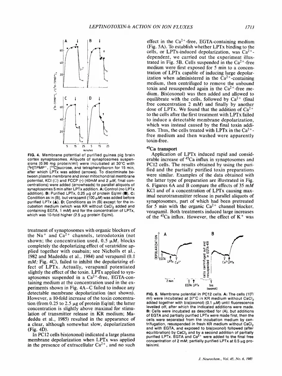
In PC12 cells bis(oxono1) indicated a large plasma membrane depolarization when LPTx was applied in the presence of extracellular Ca2+, and no such
effect in the Ca2+-free, EGTA-containing medium (Fig. 5A). To establish whether LPTx binding to the cells, or LPTx-induced depolarization, was Ca2 +-
dependent, we carried out the experiment illus- trated in Fig. 5B. Cells suspended in the Ca2+-free medium were first exposed for 5 min to a concen- tration of LPTx capable of inducing large depolar- ization when administered in the Ca2 +-containing medium, then centrifuged to remove the unbound toxin and resuspended again in the Ca2+-free me- dium. Bis(oxono1) was then added and allowed to equilibrate with the cells, followed by Ca2+ (final free concentration 2 mM) and finally by another dose of LPTx. We found that the addition of Ca2+ to the cells after the first treatment with LPTx failed to induce a detectable membrane depolarization, which was instead caused by the final toxin addi- tion. Thus, the cells treated with LPTx in the Ca2+- free medium and then washed were apparently toxin-free.
45Ca transport Application of LPTx induced rapid and consid-
erable increase of 45Ca influx in synaptosomes and PC12 cells. The results obtained by using the puri- fied and the partially purified toxin preparations were similar. Examples of the data obtained with the latter type of preparation are illustrated in Fig. 6. Figures 6A and B compare the effects of 35 mM KCI and of a concentration of LPTx causing max- imal neurotransmitter release in parallel aliquots of synaptosomes, part of which had been pretreated for 5 min with the organic Ca2+ channel blocker, verapamil. Both treatments induced large increases of the 45Ca influx. However, the effect of K+ was
B f
_ _ f t--3 t_
2 min EGTA LPTx bis
FIG. 5. Membrane potential in PC12 cells. A: The cells (lo5/ ml) were incubated at 37°C in KR medium without CaCI, added together with bis(oxono1) (0.1 pM) until fluorescence levelled off, after which the indicated additions were made. B: Cells were incubated as described for (A), but additions of EGTA and partially purified LPTx were made first, then the cells were separated from the incubation medium by cen- trifugation, resuspended in fresh KR medium without CaCI, and with EGTA, and exposed to bis(oxono1) followed (after equilibration) by CaCI, and by a second addition of partially purified LPTx. EGTA and Ca2+ were added to the final free concentration of 2 mM; partially purified LPTx at 0.5 wg pro- tein/ml.
loxonol)
J . Neurochem., Vol. 45, No. 6, 1985

L . MADEDDU ET AL.
u 15 20 25
B
i - minutes
FIG. 6. Ca“ transport in purified guinea pig cortex synap- tosomes (A and B) and PC12 cells (C). Purified synaptosomes (in KR medium without CaCI, added, 0.65 mg proteinhl) were preincubated at 30°C for 15 min after which a mixture of 45Ca (0.85 pCi/ml; 2mM) and [3H]sucrose (1.5 pCi/ml) was added, and the incubation continued at the same tempera- ture. Verapamil (100 pM) was added to half of the samples (open symbols) at the 10th min. At the 15th min the samples in (A) received KCI, 35 rnM final concentration; those in (B), partially purified LPTx, 0.6 pg protein/ml. In the experiment illustrated in (C), PC12 cells (5 x 10⁶/ml) were preincubated for 2 min 2 verapamil (25 pM, open and closed symbols) after which the 45Ca/[3H]sucrose mixture was added, fol- lowed immediately by partially purified LPTx (1 pg protein/ ml).
>70% reduced by verapamil, whereas the effect of LPTx was almost unaffected by verapamil. The re- sults with PC12 cells (Fig. 6C) were similar to those with synaptosomes: the increased influx of 45Ca in- duced by the toxin was large and rapid, and vera- pamil pretreatment was without effect.
Free cytoplasmic Ca2+ Figure 7 illustrates the results obtained by mea-
suring [Ca2+Ii in PC12 cells exposed to purified (trace A) or partially purified (traces B-F) LPTx. In the cells incubated in complete KR medium [Ca2+Ii rose without appreciable delay after the ap- plication of LPTx, from the resting level of -100 nM to high levels, which were maintained for several minutes. The size of the LPTx-induced rises de- pended on the concentration of the applied toxin. A concentration of 0.15 pg/ml of partially purified toxin-which was still subthreshold for the stimu- lation of transmitter release (Madeddu et al., 1985)-induced a threefold rise of [Ca2+Ii (trace B); with 0.5 Fg/ml (30% of the maximal release re- sponse) [CaZfli rose to 700 nM (trace C), and with 1 pg/ml (>50% of the release response) to approx- imately 1 p M (trace D). When LPTx was applied in a medium free of added CaZ+, without (trace E) or with (trace F) EGTA added, it failed to modify [Ca2+Ii in PC12 cells. In these cells exposed to LPTx in Ca2+-free media, reintroduction of Ca2+ into the medium caused the expected rise of [Ca2 +I i (traces E and F).
Interaction with artificial membranes Purified or partially purified LPTx (concentra-
tions ranging between 0.1 and 4 pg of protein Eq/ ml, and between 0.25 and 10 pg/ml, respectively) were added to the salt solution bathing either the cis side, or both sides of lipid bilayer membranes formed by the Mueller-Rudin method (Mueller et al., 1962). In many such experiments no changes in membrane conductance were observed. Only in a few cases fluctuations of different amplitude and kinetics appeared. Usually these fluctuations were recorded after a previous membrane had broken (not shown). Current fluctuations were consistently observed in membranes formed by the Montal- Mueller technique (Montal and Mueller, 1972) from lipid monolayers spread over a bulk solution con- taining partially purified LPTx (0.2-2 pgiml). The fluctuations observed at low level of activity were of two types: (a) burst-like fluctuations; and (b) square pulse-like events of different conductance (Fig. 8). The behavior of the current-voltage char- acteristics of LPTx-treated membranes (Fig. 9) was very reproducible. The marked conductance in- crease for high transpositive potentials was accom- panied by a large level of noise. At transnegative potentials conductance was low, and going from - 100 to - 110 the trace exhibited a segment of neg-
EGTA A L
EGTA
10-j- EGTA 7 x -
C 3 5 x 10⁻⁷ - B F,
10‘~-
f r 1 115x i
2 min
FIG. 7. [Ca’-], in PC12 cells. Aliquots of PC12 cells loaded with quin2 as described by Meldolesi et al. (1984) were re- suspended (0.8 x 10⁶/ml) and exposed to LPTx. In the ex- periments illustrated by traces A-D the initial incubation me- dium was complete KR; trace D: KR without added CaCI,; trace E: this last medium containing in addition EGTA, 1 mM. Purified LPTx (0.1 pg of protein Eqiml) was used in experi- ment A; partially purified LPTx in the others: 6 = 0.12 pg proteiniml; C, E, and F = 0.5 pgirnl; D = 1 pgiml. Calibration of [Ca*-], is indicated to the left of each trace.
J . Neurochem , Vol. 45, No. 6, 1985

LEPTINOTOXIN-h ACTION ON ION FLUXES I715
20 - E \" c 21 0 -
10-
A
-
I"SL 10 5
FIG. 8. Changes in conductance induced by partially purified LPTx in planar lipid bilayers. A: Square pulse-like conduc- tance fluctuations in a virtually solvent-free membrane com- posed of cholesterol and egg lecithin, prepared by the tech- nique of Montal-Mueller. LPTx (0.75 pg/ml) was applied in the cis chamber of the Teflon cell. A 40 mV transpositive potential was applied. 8: Burst-like conductance fluctuations in a membrane composed of cholesterol and dioleyl-phos- phatidylcholine in n-decane. LPTx (0.5 kg/ml) was applied on either chamber of the Teflon cell. A 20 mV transpositive potential was applied.
ative slope (=transition to a lower conductance state) which could be due to either abrupt closing of the channels or to negative voltage-induced ex- trusion of LPTx molecules from the bilayer.
In a further series of experiments the effects of
400 -
I IY'J I J /
00 200
V (mV)
-200 1 FIG. 9. Current-voltage characteristics of a Montal-Mueller (1972) planar lipid bilayer membrane after addition of par- tially purified LPTx (0.25 pg/ml) in the cis-side of the bathing solution. The applied triangolar potential change had a Hz frequency (see the inset). Other conditions were as de- scribed in the legend to Fig. 7A. The difference between the foward and backward traces is due to a hysteresis in the membrane response. The step at -110 mV (transnegative potential) is a 900 pS transition to a lower conductance state. At transpositive potential larger than 165 mV the current was out of the recorder scale.
LPTx on surface tension of salt solutions and lipid monolayers were investigated. With salt solutions (0.1 M NaCl; pH 7 . 9 , large changes of the surface tension (17 dyne/cm, area: 40 cm2) were observed after the application of 0.1 pg/ml of partially puri- fied LPTx (Fig. lo). After the superficial layer of the solution was taken off by sucking with a water pump, the effect of the toxin was no longer evident. These results suggest that LPTx molecules are pref- erentially distributed as a monolayer at the air-salt solution interface, and are therfore consistent with the presence of hydrophobic domains in the toxin molecule. The results obtained by adding the same amount of LPTx to a solution underlying a tightly packed monomolecular lipid film (area: 40 cm2; ini- tial surface tension: 10 dyne/cm) were different, in- asmuch as no toxin-induced changes of surface ten- sion were recorded under these conditions. Only with films of low (3 dyne/cm) surface tension (loosely packed films) slow increases were ob- served. After 7 min, when a quasi-steady state had been reached, the surface tension increase was 3.2 dyne/cm (not shown in figures).
DISCUSSION
Purification and use of LPTx In the present work LPTx was purified from the
hemolymph of the Colorado potato beetle, Lepri- notarsa haldemani, by a simplified version of the procedure originally developed by Crosland et al. (1984). The preparation that had high transmitter release stimulating activity (Madeddu et al., 1985) contained one major component, -55K in M, and -5.5 in isoelectric point. Taken together with pre- vious findings (Hsiao, 1978; Crosland et al., 1984) these results strongly indicate that the major com-
FIG. 10. Surface pressure-area isotherm of a preparation of partially purified LPTx in salt solution (NaCI 0.1 M, Tris 0.01 M, pH 7.5) at room temperature (22°C). The toxin (4 pg in 100 pl of an aqueous solution) was added to 40 ml of substrate. The initial available area was 94 cm2. Measurements were initiated 10 min after LPTx addition. Areasimolecule were calculated based on a toxin M, of 55,000.
J . Neurochem., Voi. 45, N o . 6, 1985

1716 L . MADEDDU ET A L .
ponent is the neurotoxic principle of the Leptino- tarsa hemolymph, previously called by Crosland et al. (1984) p-leptinotarsin-h.
Compared to the original procedure (Crosland et al., 1984) our purification scheme offers distinct ad- vantages because it is more rapid, and because the purity of the final preparation is at least as high, and the yield almost twice as great. The amount of hemolymph that we had available was limited, the concentration of LPTx in the hemolymph is very low (of the order of O.I%), and the purified LPTx is very unstable. We chose therefore to work not only with pure LPTx, but also with the partially purified preparation (concentration of LPTx: - 1'36, obtained by simple gel filtration of the redissolved hemolymph), which contains over 20 major contam- inant polypeptides. The transmitter release activity of this preparation is much more stable than that of purified LPTx. With the sole exception of some studies with artificial membranes, the data with the partially purified preparation were however always duplicated using pure LPTx. We feel confident therefore to attribute the results we obtained to one single active principle, LPTx, characterized by a defined M, and isoelectric point.
As previously mentioned, the estimated protein concentration of purified LPTx preparations was I / 100 of their protein Eq concentration. Thus, the cel- lular effects of the toxin analyzed in the present paper (depolarization, Ca'+ fluxes; [Ca2+Ii rises) were obtained at LPTx concentrations as low as 1 - 10 nglml. This means that LPTx is a very potent neurotoxin, active in the 10-*l-lO-'o M range. For comparison it might be mentioned that a-LTx works in the 10p'o-10-9 M range (Hurlbut and Cec- carelli, 1979; Grasso et al., 1982; Meldolesi, 1982; Meldolesi et al., 1983; Nicholls et al., 1982).
Binding experiments During the last several years, many protein toxins
have been successfully radiolabeled and used to carry out binding experiments. However, all our at- tempts to radiolabel LPTx resulted in its complete inactivation, and therefore direct binding experi- ments could not be carried out. By using 1251-a-LTx (which maintains the activity of native a-LTx, Mel- dolesi, 1982) we were able to investigate the pos- sible interaction of the two toxins at the level of their binding. The results obtained failed to show any competition of LPTx for a-LTx binding to its specific receptors in synaptosomal membranes. Thus, the two toxins might be addressed to two separate and distinct binding sites. Depolarization, increased Caz+ influx, and [Ca2+Ii
Our results confirm the previous observation of Crosland et al. (1984) that LPTx causes membrane depolarization and stimulates Ca2+ influx in rat brain synaptosomes. In addition, we showed these
effects in the neurosecretory cell line PC12, and demonstrated that in the cells the toxin causes a persistent elevation of [Ca2+Ii, as measured by the quin2 technique. Two aspects of these effects of LPTx are worth emphasizing. The first concerns their size and kinetic features. Depolarization and stimulated Ca2+ influx were pronounced, but not as massive as those induced by other toxins of the presynaptic stimulatory class, a-LTx and glycero- toxin (Grasso et al., 1980; Nicholls et al., 1982; Ma- deddu et al., 1984). The LPTx-induced increases of [Ca2+Ii, on the other hand, were large and dose- dependent, and were maintained for several min- utes after toxin application, suggesting the estab- lishment of a new, pump-and-leak equilibrium be- tween Ca2+ influx (stimulated by LPTx) and Ca2+ extrusion mechanisms (Meldolesi et al., 1984). Ef- fects of a-LTx (depolarization; Ca2+ influx; [Ca2+Ii rise; phosphoinositide turnover; stimulation of transmitter release) are known to initiate after a delay following the application, which is believed to be due to the time needed for the toxin to bind to its specific receptors (Nicholls et al., 1982; Mel- dolesi et al., 1984; Vicentini and Meldolesi, 1984). No such delay was observed with LPTx, suggesting that the rate of its binding is faster than that of a- LTx.
The second aspect of LPTx action to be dealt with is Ca2+ dependency. Crosland et al. (1984), working on rat brain cortex synaptosomes, failed to observed any effects (depolarization, stimulated acetylcholine release) when the toxin was applied in a Ca2+-free EGTA-containing medium. In addi- tion, they reported that the release responses were blocked by millimolar concentrations of the inor- ganic blockers of the voltage-dependent Ca2+ channel, Co2+ and Cd2+. These two findings seem to meet requirements for a direct involvement of the Ca2+ channel in LPTx action (Hagiwara and Byerly, 1981). Crosland et al. (1984) proposed there- fore that the mechanism of action of LPTx consists in the opening of that channel. The data that we have now obtained cast doubt on this interpreta- tion. We found that, in both synaptosomes and PC 12 cells, verapamil inhibits neither depolariza- tion nor Ca2+ fluxes induced by LPTx. Verapamil is a lipophilic drug that elicits a wide spectrum of actions. At the high concentrations that we used, it certainly inhibits voltage-dependent Ca2+ channels (Fig. 6 ; see also Akerman and Nicholls, 1981; Nicholls et al., 1982; Meldolesi et al., 1984) and in addition it is known to interfere with other chan- nels, receptors, as well as with uptake and biosyn- thetic processes (see Miller and Freedman, 1984). The lack of effect of verapamil on LPTx-induced ion fluxes indicates therefore that neither the voltage-dependent Ca2 + channel nor the other tar- gets of verapamil are involved in the toxin action. The independence of LPTx action from the Ca2+
I . Neurochern., Vol. 45, N o . 6 , 1985

LEPTINOTOXIN-h ACTION ON ION FLUXES 1717
channel is supported also by transmitter release re- sults reported in the companion article (Madeddu et al., 1985). On the other hand, the effects of the Ca2 +-free, EGTA-containing medium were different in the two experimental systems used. In guinea pig brain cortex synaptosomes we observed a shift to the right of LPTx potency (i.e., larger concentra- tions of the toxin were needed to obtain depolar- ization in the Ca2+-free, EGTA-containing medium) whereas under these conditions LPTx had no de- tectable effect on membrane potential in PC12 cells. Because of our failure to radiolabel LPTx, we were unable to investigate directly whether Ca2+ is needed for the action or for the binding of the toxin. Indirect evidence suggesting that Ca2+ is needed for LPTx binding come from the experiments in which PC12 cells failed to depolarize after having been treated with the toxin in Ca2+-free medium, then centrifuged, resuspended, and exposed to Ca2+ (Fig. 5B). Alternatively, this result could be due to rapid dissociation (during washing) of the toxin mol- ecules bound to the cells during the initial incuba- tion in Ca2+-free medium. This possibility seems unlikely in view of the high potency of LPTx, which probably reflects a high binding affinity. Additional evidence suggesting the involvement of Ca2+ in LPTx binding arose from the results of quin2 ex- periments. We observed that [Ca2+Ii failed to rise when LPTx was applied not only in the Ca2+-free, EGTA-containing medium, but also in the medium without CaCI, added. In this medium the [Ca2+], can be estimated to be around 10 p M , which implies the existence of an inward-directed electrochemical gradient for Ca2+ sufficient to cause a measurable [Ca2+Ii rise if LPTx binding had activated Ca2+ transport.
Interaction with artificial membranes a-LTx has been known for many years to have
the capacity to insert across lipid bilayers, thus giving rise to discrete, high conductance channels, transporting mono- and divalent cations (Finkel- stein et al., 1976). Recently, this effect of a-LTx has been further investigated by us. Under voltage- clamp conditions the toxin-induced ionic currents were found to be dependent on both the voltage and the transbilayer orientation of the toxin molecules (Robello et al. , 1984). Channel-forming capacity has been described recently also for the toxin contained in the venom of the worm Glycera dibranchiata (Kagan et al., 1982). Results very similar to those of Kagan et al. (1982) were obtained by us using another member of the presynaptic stimulatory toxin class, the toxin of Glycera convoluta (M. Ro- bello, R. Rolandi, L. Madeddu, and J. Meldolesi, unpublished results). Although the functional sig- nificance of the channel-forming capacity of these toxins remains undefined in intact cells, we consid- ered it of interest to investigate the interaction of
LPTx with air-water interfaces as well as lipid mono- and bilayer membranes.
From our measurements of surface pressure in salt solutions and lipid monolayers it appears that LPTx molecules have the capacity to partition at the air-water interface, but not to penetrate lipid layers in such a way to change their surface pres- sure. On the other hand, the I-V data of Fig. 8 show that the application of a transpositive potential to LPTx-treated lipid bilayers results in conductance increases, which document the formation of trans- membrane channels. The voltage dependence of LPTx-induced currents resembles that of mellitin and trichotoxin A40 (Hanke et al., 1983). Similar to these two toxins, LPTx induced burst-like conduc- tance changes in lipid bilayer membranes. On the other hand, the voltage dependence of LPTx effects was quite different from that of a-LTx, which in cholesterol-containing membranes causes high con- ductance at transnegative potentials (Robello et al., 1984). Taken together all our data suggest that LPTx molecules have affinity for hydrophobic surfaces, but lack the capacity to insert across membranes unless transpositive voltage is applied.
Conclusions The profile of LPTx, as it emerges from the re-
sults of the experiments reported in the present paper, casts doubt on the hypothesis recently pro- posed of its being a specific activator of presynaptic voltage-dependent Ca2+ channels (Crosland et al., 1984). Except for the data on artificial membranes, which document differences in the capacity of LPTx and a- LTx to insert across lipid bilayers, our data revealed distinct similarities between the ef- fects of the two toxins, and suggest therefore that they could have similar, although not identical, mechanisms of action. Thus, the two toxins could bind to plasma membranes (probably at separate, specific sites) and initiate a series of events among which is the activation of tetrodotoxin and vera- pamil-insensitive cation fluxes. The activation of these fluxes would then be responsible for the changes of ionic homeostasis that we have re- ported. Additional evidence supporting this general interpretation is given in the companion article, which deals with the effects of LPTx on the release of neurotransmitters and ultrastructure in synapto- somes and PC12 cells (Madeddu et al., 1985).
Acknowledgment: L. Madeddu is a fellow of the Anna Villa Rusconi Foundation. This work was supported in part by grants of the Italian Department of Education, Membrane Biology and Pathology Project (to J. Meldo- lesi) and Italian National Research Council, Special Project “Secondary Fine Chemistry” (to M. Robello and R. Rolandi).
REFERENCES Akerman K. E. 0. and Nicholls D. G. (1981) Calcium transport
by intact synaptosomes: the voltage-dependent calcium
J . Neurochem., Vol. 45, No. 6, 1985

1718 L . MADEDDU ET A L
channel and a reevaluation of the role of sodiumicalcium exchange. Eur. J . Biochem. 117, 491-497.
Clementi F. and Ceccarelli B. (1979) Neurotoxim, Tools in Neu- robiology. Raven Press, New York.
Crosland R. D., Hsiao T. H., and McClure W. 0. (1984) Purifi- cation and characterization of pleptinotarsin-h, an activator of presynaptic calcium channels. Biochemist/? 23, 734-741.
Finkelstein A, , Rubin L . L . , and Tzeng M. C. (1976) Black widow spider venom. Effect of the purified toxin on lipid bilayer membranes. Science 193, 1009-I01 1 .
Fritz L. C., Tzeng M. C., and Mauro A. (1980) Different com- ponents of black widow spider venom mediate transmitter release at vertebrate and lobster neuromuscular junctions. Nature 283, 486-487.
Fromherz P. ( 1975) Instrumentation for handling monomolecular films at an air-water interface. Rev. Sci. Znstrrtm. 46, 1380- 1385.
Frontali N., Ceccarelli B., Gorio A,. Mauro A,, Siekevitz P., Tzeng M. C., and Hurlbut W. P. (1976) Purification from black widow spider venom of a protein factor causing the depletion of synaptic vesicles of neuromuscular junction. J . Cell Biol. 68, 462-479.
Grasso A,, Alema’ S . , Rufini S . , and Senni M. I . (1980) Black widow spider toxin-induced calcium fluxes and transmitter release in a neurosecretory cell line. Nature 283, 774-776.
Grasso A., Pelliccia M., and Alema’ S. (1982) Characterization of alatrotoxin interaction with rat brain synaptosomes and PC12 cells. Toxicon 20, 149-156.
Greene L. A. and Tischler A. S. (1976) Establishment of a nor- adrenergic clonal line of rat adrenal pheochromocytoma cells which respond to nerve growth factor. Proc. Natl. Acad. Sci. USA 73, 2474-2478.
Hagiwara S. and Byerly L. (1981) Calcium channel. Annrr. Re\.. Neurosci. 4, 69-125.
Hanke W., Methfessel C.. Wilmsen H., Katz E.. Jung G.. and Boheim G. (1983) Mellitin and a chemically modified tricho- toxin from alamethicin-type multistate pores. Biochim. Bio-
Howard B. D. and Gundersen C. B. (1980) Effects and mecha- nisms of polypeptide neurotoxins that act presynaptically. Annu. Rev. Pharmacol. Toxicol. 20, 307-336.
Hsiao T. H. (1978) Comparative studies on hemolymph protein toxins of leptinotarsa beetles, in Toxins: Animal, Plant und Microbial (Rosenberg P., ed), pp. 675-688. Pergamon Press, Oxford and New York.
Hsiao T. H. and Fraenkel G. (1969) Properties of leptinotarsin, a toxic hemolymph protein from the colorado potato beetle. Toxicon 7, 119- 130.
Hucho F. and Ovchinnikov Y. A. (1983) Toxins as Tools in Neu- rochemistry. W. De Gruyter, Berlin and New York.
Hurlbut W. P. and Ceccarelli B. (1979) Use of black widow spider venom to study the release of neurotransmitters. in Neurotoxins, Tools in Neurobiology (Ceccarelli B. and Cle- menti F. eds), pp. 87- 115. Raven Press, New York.
Jones D. M. and Matus A. I. (1974) Isolation of synaptic plasma membrane from brain by combined flotation-sedimentation density gradient centrifugation. Biochim. Biophys. Acra
Kagan B. L., Pollard H. B., and Hanna R. B. (1982) Induction ion-permeable channels by the venom of the fanged blood- worm Glycera dihranchiafa. Toxicon 20, 887-893.
Lowry 0. H., Rosenbrough N. J., Farr A. L., and Randall R. J. (1951) Protein measurement with the Fohn phenol reagent. J . Biol Chem. 193, 265-275.
Madeddu L., Meldolesi J., Pozzan T., Cardona E., and Bon C. (1984) aLatrotoxin and glycerotoxin differ in target speci- ficity and in the mechanism of their neurotransmitter re- leasing action. Neuroscience 12, 939-949.
Madeddu L., Saito I., Hsiao T. H., and Meldolesi J. (1985) Lep- tinotoxin-h action in synaptosomes and neurosecretory cells. Stimulation of neurotransmitter release. J . Neuro- chem. 45, 1719-1730.
p h y s . Act0 727, 108- 114.
356, 276-281.
Maize1 J. V. (1971) Polyacrylamide gel electrophoresis of viral proteins. Methods Virol 5, 179-246.
McClure W. 0.. Abbott B. C., Baxter D. E., Hsiao T. H., Satin L. S. , Siger A., and Yoshino J. E. (1980) Leptinotarsin: a new neurotoxin that stimulates acetylcholine release. Proc. Nnrl. Acad. Sci. USA 77, 1219-1223.
Meldolesi J. (1982) Studies on a-latrotoxin receptors in rat brain synaptosomes: correlation between toxin binding and stim- ulation of transmitter release. J . Neurochem. 38, 1559- 1569.
Meldolesi J . , Madeddu L., Torda M., Gatti G., and Niutta E. (1983) The effect of alatrotoxin on the neurosecretory PC12 cell line. Studies on toxin binding and stimulation of trans- mitter release. Neuroscience 10, 997- 1009.
Meldolesi J . , Huttner W. B., Tsien R. Y., and Pozzan T. (1984) Free cytoplasmic Ca2+ and neurotransmitter release: studies on PC 12 cells and synaptosomes exposed to a-latro- toxin. Proc. Narl. Acad. Sci. USA 81, 620-624.
Miller R. J . and Freedman S . B. (1984) Are dihydropyridine binding sites voltage sensitive calcium channels? Life Sci.
Montal M. and Mueller P. (1972) Formation of biomolecular membranes from lipid monolayers and a study of their elec- trical properties. Proc. Natl. Acad. Sci. USA 69, 3561- 3966.
Morel N., Thieffry M., and Manaranche R. (1983) Binding of a Glycera convoluta neurotoxin to cholinergic nerve terminal plasma membranes. J . Cell B id . 97, 1737-1744.
Mueller P., Rudin D. O., Ti Tien H . , and Wescott W. C. (1962) Reconstitution of excitable membrane structure in vitro. Circulation 26, 1167- 1172.
Nicholls D. G., Rugolo M., Scott I. G., and Meldolesi J. (1982) aLatrotoxin of black widow spider depolarizes the plasma membrane, induces massive calcium influx and stimulates transmitter release in guinea pig synaptosomes. Proc. Natl. Acad. Sci. USA 79, 7924-7928.
O’Farrell P. Z . , Goodman H. M., and O’Farrell P. H. (1977) High resolution two-dimensional electrophoresis of basic as well as acidic proteins. Cell 12, 1133-1142.
Robello M., Rolandi R., Alema’s. , and Grasso A. (1984) Trans- bilayer orientation and voltage dependence of alatrotoxin- induced channels. Proc. R . Soc. Lond. [Biol.] 220,477-487.
Scheer H., Madeddu L., Dozio N., Gatti G., Vicentini L. M., and Meldolesi J. (1984) aLatrotoxin of black widow spider venom: an interesting neurotoxin and a tool for investigating the process of neurotransmitter release. J . Physiol. 79,216- 221.
Scott 1. D. and Nicholls D. G. (1980) Energy transduction in intact synaptosomes. Influence of plasma membrane depo- larization on the respiration and membrane potential of in- teral mitochondria determined in situ. Biochem J . 186, 21- 33.
Scott I. D., Akerman K. E. O., and Nicholls D. G. (1980) Cal- cium-ion transport by intact synaptosomes. Biochem. J . 192, 873-880.
Valtorta F., Madeddu L., Meldolesi J., and Ceccarelli B. (1984) Specific localization of the alatrotoxin receptor in the nerve terminal plasma membrane. J. Cell Biol. 99, 124-132.
Vicentini L. M. and Meldolesi J. (1984) aLatrotoxin of black widow spider venom binds to a specific receptor coupled to phosphoinositide breakdown in PC12 cells. Biochim. Bio- phys. Res. Commun. 121, 538-544.
Winkler H., Falkensammer G., Patzak A., Fischer-Colbie R., Schober M., and Weber A. (1984) Life cycle of the cate- cholaminergic vesicle: from biogenesis to secretion, in Reg- ulation of Transmitter Function: Basic and Clinical Aspects (Vizi E . S . and Magyar K . , ed), pp. 65-73; Akademiai Kiado, Budapest.
Yoshino J. E., Baxter D. E., Hsiao T. H., and McClure W. 0. (1980) Release of acetylcholine from rat brain synaptosomes stimulated with leptinotarsin, a new neurotoxin. J . Neu- rocliem. 34, 635 -642.
34, 1205-1221.
J . Neurochem., Val. 45, No. 6. 1985
Related Documents


![Evidence for a relationship between B-50 (GAP-43) and [3H]noradrenaline release in rat brain synaptosomes](https://static.cupdf.com/doc/110x72/634462d3596bdb97a9086fc8/evidence-for-a-relationship-between-b-50-gap-43-and-3hnoradrenaline-release.jpg)
