1 Leprosy Terry L Dwelle MD MPTHM

Welcome message from author
This document is posted to help you gain knowledge. Please leave a comment to let me know what you think about it! Share it to your friends and learn new things together.
Transcript
1
LeprosyTerry L Dwelle MD MPTHM
Presenter
Presentation Notes
This talk is on leprosy.
2
Introduction
►Leprosy is associated with a stigma
Presenter
Presentation Notes
Leprosy disease and the visible physical deformities contribute to an intense social stigma resulting in discrimination of patients and their families. I’m sure many of us remember stories from the Bible about how leprosy patients needed to stay away from others often in separate communities. They needed to cover themselves and their faces when they traveled yelling “unclean” It may be good to just discuss the possible difference between leprosy in the old testament and the disease we call leprosy today.
3
Introduction
►Leprosy in the Old Testament is not one disease
Original Hebrew “tsara’ath” – group of diseasesTranslated to “lepra” in Greek – 100 BC1384 Wycliffe translated “lepra” to “leprosy” a disease seen in Europe at that time and described as a “unholy and loathsome condition”
Presenter
Presentation Notes
Leprosy in the Old Testament is not one disease but a cluster of skin diseases of which the modern leprosy may have been one. The original Hebrew word for leprosy is “tsara’ath” and referred to a group of diseases That was translated to “lepra” in Greek by 100 BC In 1384 Wycliffe translated “lepra” to “leprosy” a disease seen in Europe at that time and described as a “unholy and loathsome condition” I think a main point is that whatever the association of ancient leprosy and the disease we know of as leprosy today caused by a bacteria called Mycobacterium Leprae there has always been a stigma assocaited with the disease. When completing my studies in New Orleans I worked at the national leprosarium. An elderly black woman had lived most of her life at the leprosarium. It was her home. She recalled how in the early part of the 20th century that her and her family were transmitted by a coal barge in chains up the river to the leprosarium at Carville. They were not given shelter or covering and it was cold and rainy. They were lead at gun point to the facility.
4
General
►One of the leading causes of permanent physical disability in the world
►Afflicts individuals in their most productive stage of life
►Multi-drug therapy (MDT) can eliminate leprosy as a public health problem (prevalence < 1/10,000)
►MDT can also bring about cure without disability
Presenter
Presentation Notes
Some general comments include; Leprosy is still one of the leading causes of permanent physical disability in the world It also afflicts individuals in their most productive stage of life Multidrug therapy (MDT) can if done properly eliminate leprosy as a public health problem. By definition that means the prevalence would drop to < 1/10,000 population. MDT can also bring about cure without disability due to the efficacy of the regimens particularly for paucibacillary forms of the disease.
5
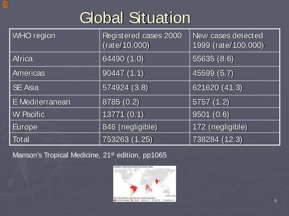
Global SituationWHO region Registered cases 2000
(rate/10,000)New cases detected 1999 (rate/100,000)
Africa 64490 (1.0) 55635 (8.6)
Americas 90447 (1.1) 45599 (5.7)
SE Asia 574924 (3.8) 621620 (41.3)
E Mediterranean 8785 (0.2) 5757 (1.2)
W Pacific 13771 (0.1) 9501 (0.6)
Europe 846 (negligible) 172 (negligible)
Total 753263 (1.25) 738284 (12.3)
Manson’s Tropical Medicine, 21st edition, pp1065
Presenter
Presentation Notes
Leprosy currently impacts about 750000 persons worldwide. Most cases come from SE Asia with a 3.8 / 10,000 prevalence rate. Also note that new cases detected in 1999 were about 738000 with the greatest incidence being found in SE Asia and then Africa.

6
Prevalence of Leprosy - top 11 countriesCountry Registered cases
2000Prevalence / 10000 New cases during
1999Detection rate / 100000
India 495073 5.0 537956 54.3
Brazil 78068 4.3 42055 25.9
Myanmar 28404 5.9 30479 62.9
Indonesia 23156 1.1 17477 8.3
Nepal 13572 5.7 18693 78.7
Madagascar 7865 4.7 8704 51.6
Ethiopia 7764 1.3 4457 7.4
Mozambique 7403 3.9 5488 28.7
D.R. Congo 5031 1.0 4221 8.6
Tanzania 4701 1.4 5081 15.4
Guinea 1559 2.0 2475 32.0
Total 672596 4.1 677086 41.7
Presenter
Presentation Notes
This table lists the prevalence of leprosy in the 11 countries with the highest prevalence in the worlds. Of those India, Brazil, and Myanmar have the highest prevalence ranging from 5.0 to 5.9 / 10,000 population. The new case detection rate in 1999 per 100000 is highest in Nepal at 78.7, followed by Myanmar 62.9 and Madagascar 51.6.

7
Registered Cases
8.4 8.8
12
1.25
0
2
4
6
8
10
12
14
1966 1976 1985 2000
Prev Rate per 10000
Target Rate 1/10,000
Presenter
Presentation Notes
The yellow line shows the change in registered leprosy cases worldwide from 1966 to 2000. There was a steady increase in registered cases from 1966 to 1985. Since 1985 there has been a steady decline of registered cases representing an 89.6% decrease in prevalence since 1985. This may be due to several factors over the past 2 decades including; In 1981 the WHO recommended MDT as the standard treatment for leprosy. In 1991 the 44th World Health Assembly passed the resolution to eliminate leprosy as a public health problem by the end of year 2000. That obviously didn’t happen but there has been significant progress with the overall prevalence rate per 10000 being 1.25 down from 12 in 1985.
8
Organism
►Mycobacterium leprae – acid fast bacillus►Primarily found in masses within
macrophages►Intra and extra-cellular globi
Presenter
Presentation Notes
Leprosy is caused by Mycobacterium leprae an acid fast bacillus that looks like M Tuberculosis. Primarily found in masses within macrophages often grouped like a pack of cigars or a palisade type structure. There can also be intra and extracellular masses of organisms called globi coverd by a capsular material.
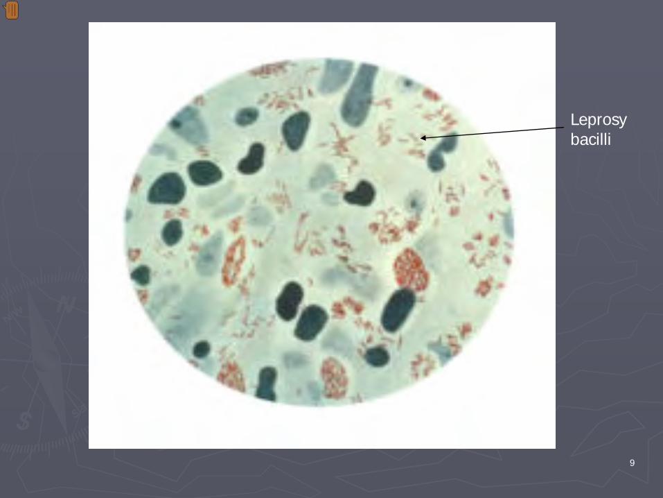
9
Leprosy bacilli
Presenter
Presentation Notes
This is a slide show multiple leprosy bacilli free within the tissues and in macrophages. As can be seen in this slide in many infectious patients the number of organisms can be overwhelming. When patients are treated with standard MDT a high proportion of the bacilli are killed within days and this immense release of antigens from dead organisms can manifest as several symptoms of leprosy disease including the erythema nodosum leprosum reaction. We will briefly cover this later.
10
Transmission
►Two portals of exitSkinNasal mucosa►Majority of lepromatous patients have bacilli in nasal
secretions from blowing the nose►Can yield as many as 10 million viable organisms per
day
►MDT renders a person non-infective after a few doses
Presenter
Presentation Notes
There seem to be two portals of exit for M Leprae from the body including; One of the potential portal is Skin – Job et al found large numbers of organisms in the superficial keritin layer of skin of lepromatous leprosy patients suggesting that the organism could exit via the sebaceous glands. Another is the Nasal mucosa The nasal mucosa’s importance as a point of exit was recognized in 1898 by schaffer. Pedley reported that a majority of lepromatous patients have leprosy bacilli in their nasal secretions collected via blowing the nose the nose. Rees indicated that nasal secretions from a lepromatous patient could yield as many as 10 million viable organisms per day. It is felt that nasal secretions are the main route of leprosy transmission in humans. MDT (multidrug therapy) renders a person non-infective after only a few doses of medication

11
Viability of M. leprae
► 36 hours to 9 days►Contaminated fomites and clothing could be
a source of infection
Presenter
Presentation Notes
M leprae can survive up to 36 hours in nasal secretions in temporate climates. That can increase to 9 days in tropical situations. This environmental survival could suggest that contaminated fomites and clothing could be a source of infection

12
Portal of Entry
►Skin►Upper respiratory tract – most likely route►Others?
Breast MilkPlacental
Presenter
Presentation Notes
The two most likely portals of entry for infection in the human are likely skin and upper respiratory tract. Historically many felt the skin via ulcers and other lesions was the most likely route of infection. Recent studies in mice have demonstrated infectious transfer via aerosols as well as topical application of organisms in nude mice, so both routes seem plausible.. Currently most leprologists feel that the respiratory route is the most like portal of entry of the organism due to the large numbers or organisms in nasal discharge of infected patients. Other potential routes of infection could be breast milk and the placenta.

13
In vitro culture
►No substantiated in vitro culture of the bacillus
Presenter
Presentation Notes
No substantiated in vitro culture of the bacillus has been confirmed. Mycobacterial organisms have been recovered and cultured from the skin and other tissues of leprosy patients. Most of these organisms in culture appear to be M. avian complex organisms vs M. Leprae.
14
In vivo culture
►Mouse footpad culture is the standard►Use of the footpad culture method
Culture diagnosis of patientsMinimum concentration of treatment drugsSensitivity to new drugsDrug resistance in patients
Presenter
Presentation Notes
The mouse footpad culture is the standard culture method for leprosy with this technique. 10,000 organisms are injected into the hind footpad of a mouse. This yields 1 million organisms in 5-6 months. No clinical disease develops in the mouse. Mean generation time is 14 days consistent with the natural history of disease in man. The organisms slowly degenerates after 6 months in the mouse. Its interesting that thymectomized irradiated mice develop lepromatous leprosy similar to humans. This mouse model is particularly useful to detect small numbers of organisms in specimens. This is a cumbersome laboratory method as you can suspect. The footpad culture method is used for the following; 1. Culture diagnosis of patient specimens. 2. Drug treatment decisions like; the minimum concentration of treatment drugs to kill the organism (MBC – mimimum bacteriocidal concentration) determining the sensitivity to new drugs the development of drug resistance
15
Nine banded Armadillo
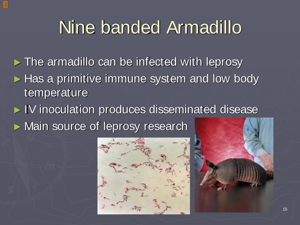
►The armadillo can be infected with leprosy►Has a primitive immune system and low body
temperature► IV inoculation produces disseminated disease►Main source of leprosy research
Presenter
Presentation Notes
It is assumed that almost all of the sources of infections for humans are other humans yet other animals like armadillos and some primates could be reservoirs of infection. The nine-banded armadillo seen in much of the Southern US can be infected with leprosy. Its estimated that 30% of the armadillos in Texas are infected. We had one patient that was likely infected via armadillos. She came from Texas and her only risk factor was armadillos in her garden. The Armadillo has a primitive immune system and low body temperature. Leprosy organisms like low temperatures therefore concentrate in areas like the distal extremities, ears, nose etc. in man. Armadillos are a main source of biologic and immunologic leprosy research. In Carville La., workers would drive the roads every morning looking for armadillo road kills. Armadillos have a bad habit of jumping up to the level of car grills when startled. Workers would cut of the noses and ears of these dead armadillos to supply the organisms for research. The pictures show a nine band armadillow and the left slide (Kinyoun) shows numerous leprosy bacilli taken from an infected armadillo.
16
Other animals
►Chimpanzee in Sierra Leone►Mangabey monkey in West Africa
Presenter
Presentation Notes
Other animals particularly primates can also be infected with leprosy including; Chimpanzee in Sierra Leone Mangabey monkey in West Africa

17
Family tree
►M. leprae has the longest doubling time of all known bacteria – extreme case of reductive evolution
► Less than half the genome contains functional genes eliminating many important metabolic activities
►There are 1500 genes common to TB and M leprae
►TB and M leprae derived from a common ancestor and likely had gene pools of similar size
►Many of the genes of M leprae have been lost.
Presenter
Presentation Notes
M. leprae has the longest doubling time of all known bacteria and demonstrates an extreme case of reductive evolution. Less than half the genome contains functional genes eliminating many important metabolic activities. This may explain its less than hardy infectivity and slow doubling time. There are 1500 genes common to TB and M leprae TB and M leprae derived from a common ancestor and likely originally had gene pools of similar size Many of the genes or at least the activity of the genes of M leprae have been lost. This is a lazy organism eliminating anything that looks like extra work.

18
Epidemiology►Transmission = close contact with leprosy patients
Cebu – 6.2/1000/yearSouth India – 55.8/1000/year
►Upper respiratory route most likely►Other factors for clinical expression;
GeneticsRoute of entryMalnutritionPrior exposure to other mycobacterial organisms
Presenter
Presentation Notes
Some comments about the epidemiology of leprosy. Transmission generally increases with close contact with leprosy patients. There is quite a variation regionally. In Cebu the transmission rate within households is 6.2/1000/year In S India it was much higher at 55.8/1000/year. The reason for this difference is unknown. As previously mentioned the upper respiratory route, nasal secretions are the most likely route of tranmission since this has been demonstrated in mice and there are such high concentrations of organisms in nasal secretions. There are likely other factors involved for clinical expression of the disease including; Genetics since there is a clustering of leprosy around certain families. Is that due to increased contact only or underlying genetic predisposition. Route of entry. In the animal model ID administration in mice doesn’t result in disseminated disease vs IV administration. Could it be that lepromatous patients have a different route of entry vs tuberculoid patients? Other factors that may enter the picture include malnutrition and possibly the prior exposure to other mycobacterial organisms that might alter the immune response to leprosy.

19
Leprosy an Immune Disease
0
20
40
60
80
100
120
LL BL BB BT TT Healthy
CMI to MLNo. OrgAntibody
Presenter
Presentation Notes
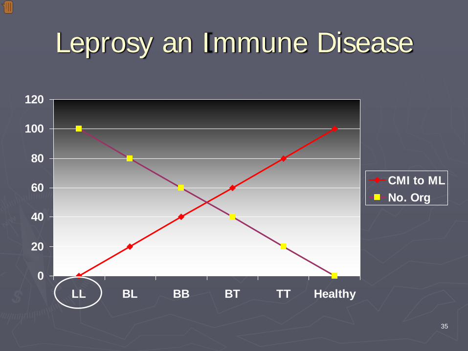
Leprosy is an immune disease. This chart demonstrates the cell mediated immunity to M leprae in the spectrum of leprosy disease. On the left of the chart we have Lepromatous patients. BL stands for borderline lepromatous BB is borderline borderline BT is borderline tuberculoid TT is tuberculoid And then healthy at the far right. You can see the inverse relationship of CMI to the number of organisms found on skin snips. Lepromatous patients have huge numbers of organisms vs those with tuberculoid disease. As expected borderline situations have varying numbers of organisms depending on which way they lean. Individuals also can shift especially with treatment from lets say the LL pole toward the TT pole This shift in CMI response may cause incredible clinical symptoms, complications and reactions like the Type II ENL reaction we will talk about later.. You will also notice that the antibody response is vigorous across the spectrum. The role of antibody leprosy is unclear since it doesn’t seem to be correlated with disease.
20
Epidemiology - Age at onset
►Mainly young adults►Range of infections from 3 weeks old to 141
years old
Presenter
Presentation Notes
Leprosy is mainly a disease of young adults in their productive years but the range of infection onset is from 3 weeks old from a child in Martinique to a 141 year old individual from Nepal.

21
Epidemiology - Gender
►Males affected more than females – 2:1 ratio
►In many parts of Africa there is an equal gender distribution
►In Uganda, Nigeria, Malawi, Gambia, Burkina Faso, Zambia, Thialand, and Japan there is a female predominance
Presenter
Presentation Notes
Generally males are affected more than females. The ratio is generally 2:1 and studies in the Philippines point out that this is a true difference. Yet in many parts of Africa there is an equal gender distribution or in some parts of the world there is a female predominance including the countries we have listed here; Uganda, Nigeria, Malawi, Gambia, Burkina Faso, Zambia, Thialand, and Japan. No one knows the reason for this.
22
Epidemiology - Incubation period
►Few weeks to 30 years +►The average – 3-5 years
Presenter
Presentation Notes
The incubation period has been difficult to determine due to limitations of the immunologic and diagnostic tools. Most experts feel the range is from a few weeks (eg the baby at 3 weeks of age with disease from Martinique unless he was infected via the placenta) to as long a 30 years or more. Most experts feel the average incubation time is 3-5 years.

23
Epidemiology – Sub Clinical Infection
►Sub-clinical infection is far more common than overt disease
►The factors influencing the onset of disease may be different from those associated with infection
Presenter
Presentation Notes
As with many infectious agents subclinical infection is far more common than overt disease The factors influencing the onset of disease ( immune response to the bacillus) may be different from those associated with infection (family members with infection).
24
Epidemiology – Household contacts
►Household contacts of leprosy patients are at greater risk of developing leprosy disease vs non-household contacts
►Household contacts contribute only a limited proportion of all new cases
Presenter
Presentation Notes
Household contacts of leprosy patients are at greater risk of developing leprosy disease vs non-household contacts. Therefore it is important that all household contacts should be examined for leprosy. They should also be educated on the early signs of the disease and the importance of seeking care if skin or nervous lesions occur. Yet we must also remember that household contacts contribute only a limited proportion of all new cases

25
Epidemiology - HIV
►No association of HIV and leprosy
Presenter
Presentation Notes
Unlike with TB, leprosy does not seem to be associated with HIV. Clincal manifestations and relapses do not differ significantly between HIV negative and HIV positive patients. Additional studies are needed in this area.

26
Epidemiology - BCG
►BCG seems to provide some protection against leprosy
Field trials – Malawi, Myanmar, Papua New Guinea, Uganda, Venezuela, IndiaProtective efficacy – 20-30% Myanmar, 50% Venezuela, 80% in UgandaGreater effect if vaccinated < 15 yoBooster doses seems to increase protectionAddition of killed M leprae organisms does not increase protectionUse of BCG may be contributing to the decline of leprosy worldwide
Presenter
Presentation Notes
This slide summarizes information of BCG with leprosy. Protective efficacy ranges from 20 to 80%. It seems there is a greater effect if vaccination is given < 15 yo. Boosters seem to increase protection yet widespread use of repeat doses are not recommended for several reasons including; poor cost effectiveness, lack of acceptability, operational difficulties, contraindications in symptomatic HIV individuals. Some studies using killed M Leprae organisms with BCG did not increase protection. One last comment is that the use of BCG may be a contributing factor to the decline of leprosy worldwide though this is not proven.
27
Epidemiolgy - Disability
►2 million worldwide with leprosy disability►Men►Multibacillary forms►Age►Duration of disease►Significantly reduced with MDT
Presenter
Presentation Notes
It is estimated that 2 million people worldwide with leprosy have disability Men, patients with multibacillary forms, those of older age, those with longer duration of disease have more disability as might be expected. Disability is significantly reduced with MDT.
28
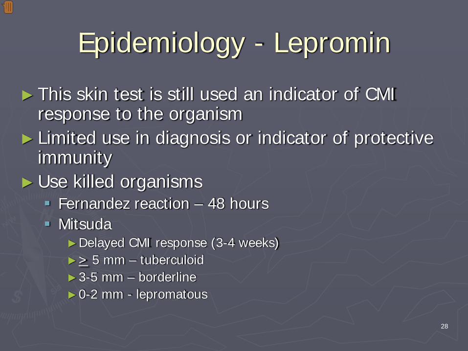
Epidemiology - Lepromin
►This skin test is still used an indicator of CMI response to the organism
► Limited use in diagnosis or indicator of protective immunity
►Use killed organismsFernandez reaction – 48 hoursMitsuda ►Delayed CMI response (3-4 weeks)►> 5 mm – tuberculoid►3-5 mm – borderline►0-2 mm - lepromatous
Presenter
Presentation Notes
The lepromin skin test is still used an indicator of CMI response to the organism. It’s usefulness in diagnosis and as an indicator of protective immunity is quite limited. This test uses killed leprosy bacilli. It is injected intradermally like a PPD There are 2 general type of reactions; the Fernandez and Mitsuda reactions. The Fernandez is seen within 48 hours and experts are not sure what it means. The Mitsuda on the other hand is the late CMI delayed hypersensitivity response that takes 3-4 weeks to occur. The Lepromin skin test is used by clinicians to help classify the immune response in leprosy patients but once again is not used for diagnosis or determining protective immunity. > or equal to 5 mm is usually seen with tuberculoid patients. 3-5 mm is usually seen in borderline patients, with 0-2 mm seen in lepromatous patients.

29
Epidemiology - Mortality
►Rarely the immediate cause of death►Indian and Philippines lepromatous patients
had a 4X and non-lepromatous patients have a 2X increased mortality vs the general population
Presenter
Presentation Notes
Rarely is leprosy the immediate cause of death though complications of the disease increase mortality due to other causes like infection and suicide etc. A study from India and the Philippines demonstrated a 4X increased mortality for lepromatous patients and a 2X increase for non-lepromatous patients with other diseases vs the general population.

30
Clinical - General► Majority of people significantly exposed experience
infection but develop no signs or symptoms ► Onset is quite variable and progression is usually insidious
Skin lesions – hypopigmented or erythematous patch with anesthesia, single, multiple or diffuseSpontaneous healing is common in childhood and some communitiesUnlike TB there is an absence of toxicity with large numbers of organisms presentAt any stage sudden exanthems may be seen associated with feverChronic onset is so gradual and insidious that it is usually far advanced on presentationAcute onset (less common) presents often with multiple lesions that spread rapidly and contain numerous bacilli, often associated with another stressor
Presenter
Presentation Notes
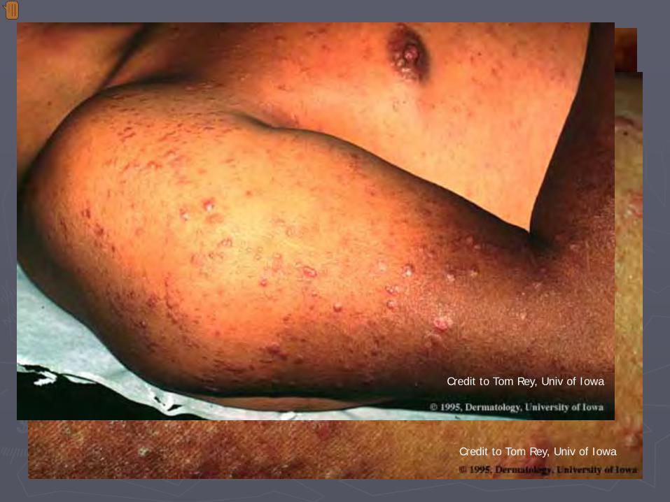
A majority of people significantly exposed (close household contact) experience infection (via lymphocyte transformation tests) but develop no signs or symptoms of disease and somehow eliminate the infection. A majority of those manifesting clinical disease will have a strong CMI response and manifest tuberculoid leprosy. A minority will manifest a weak CMI therefore manifesting lepromatous disease. The rest will have varying degrees of CMI and develop indeterminate disease. These indeterminate and borderline cases are the ones that swing more frequently across the spectrum downgrading or upgrading with changes of the immune response. Onset is quite variable. It may present with skin lesions that can be either hypopigmented or erythematous patch with anesthesia and may be single, multiple or diffuse involvement of the skin. Please note that the nerves are often involved with leprosy therefore commonly present with nerve involvement of anesthesia or weakness along with the rash. Spontaneous healing of the skin lesions is common in childhood and moreso in some communities though if diagnosed these folks should be treated not just observed. Unlike TB there is an absence of toxicity with large numbers of organisms present. Many times a person with bacilli all over the body is totally asymptomatic At any stage sudden exanthems (rashes) may be seen associated with fever Onset can be chronic or acute. Chronic onset is much more common and is so gradual and insidious that it is usually far advanced on presentation Acute onset (less common) presents often with multiple lesions that spread rapidly and contain numerous bacilli, Often this acute onset is associated with another stressor (eg puberty, birthing process, menopause, acute illness, psychological stress, etc.)
31Credit to Dr Hardin, Univ of Iowa
Credit to Tom Rey Univ of Iowa
Credit to Tom Rey, Univ of Iowa
Credit to Tom Rey, Univ of Iowa
Credit to Tom Rey, Univ of Iowa
Presenter
Presentation Notes
This is a 19 year old patient diagnosed with borderline leprosy. These localized facial erythematous plaques had been present for 4 months. This is another facial rash showing more diffuse plaque like lesions. A trunckel rash of papules and plaques. This is a close up picture of the papules of dermal leprosy. Once again a papular rash associated with leprosy. The rash associated with leprosy can mimic many other rashes associated with other conditions.
32
Case Definition
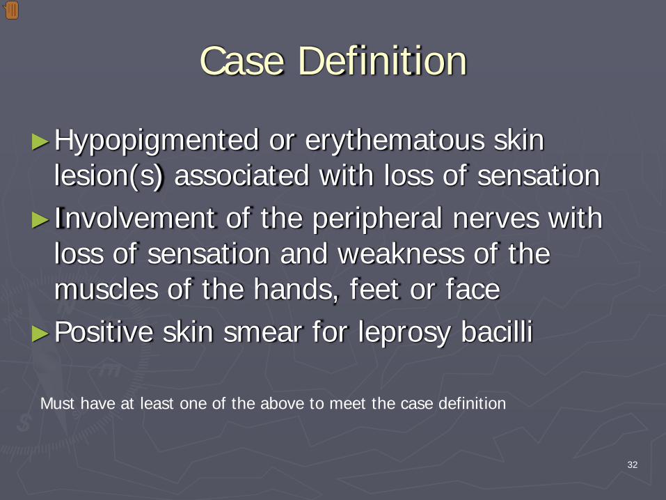
►Hypopigmented or erythematous skin lesion(s) associated with loss of sensation
►Involvement of the peripheral nerves with loss of sensation and weakness of the muscles of the hands, feet or face
►Positive skin smear for leprosy bacilli
Must have at least one of the above to meet the case definition
Presenter
Presentation Notes
At least one of the following must be met to meet the case definition of leprosy; 1. Hypopigmented or erythematous skin lesion(s) associated with loss of sensation 2. Involvement of the peripheral nerves with loss of sensation and weakness of the muscles of the hands, feet or face. There is usually loss of sensation to pin prick and / or light touch. One can also see weakness of muscles supplied b y affected peripheral nerves. Often the peripheral nerves become thickened and clinically palpable. Nerve thickening of itself without sensory loss or muscle weakness is not a reliable sign of leprosy. 3. Positive skin smear for leprosy bacilli. These are obtained by smears taken from affected skin (skin slips and smears) and stained for leprosy bacilli. Generally leprosy is diagnosed by clinical signs and symptoms; classic skin presentations plus peripheral nerve involvement. Rarely is there need to use laboratory to confirm the diagnosis.

33
Ridley-Jopling ClassificationSign or Test Type of Leprosy
TT BT BB-BL LL Indeterminate
No of lesions Usually single Single or few Several or many
Very many Vague hypopigmented or ery macules
Size of lesions Variable Variable Variable Small Variable
Surface of lesions
Very dry, scaly Dry Shiny Shiny Variable
Hair in lesions Absent Moderately diminished
Slightly diminished
Non-affected Variable
Sensation Completely lost Moderate-marked loss
Slight-moderate loss
No loss early Variable
AFB in smears None None or scanty Several – many Very many plus globi
None or scanty
Nasal AFB None None None (scanty rarely)
Very many plus globi
Negative or scanty
Lepromin test +++ + or ++ Negative Negative Negative or +
Presenter
Presentation Notes
This chart gives the Ridley-Jopling classification of Leprosy. There are essentially 5 different categories; TT, BT, BB and BL, LL and indeterminate. The classification is based on the number of, size, surface characteristics, hair presence in lesions, sensory nerve involvement, AFB in smears and in nasal smears, and the result of the lepromin test (Mitsuda reaction)3-4 weeks after ID injection of the killed bacilli. Remember that 5 mm or greater is generally TT, BL cases are usually 3-5 mm, where LL is usually 0-2. Indeteminate is 3-5 mm like the BL cases. TT cases have usually single lesions that are dry, with no hair in the lesion. Sensory nerves are involved with decreased sensation, AFB are not seen in smears of the slit smears or nasal secretions or scrapings. The lepromin test is > or equal to 5 mm in size at 3-4 weeks. The LL patients have multiple and symmetric lesions. They tend to be smaller than the TT lesions, are shiny, hair continues to grow in the lesions (exception is the hair of the eyebrows and lashes and in the pure diffuse LL cases where one occasionally sees alopecia and total body hair loss) . There is generally no loss of sensation early though as the disease progresses the nerves become more involved due to the mass of organisms resulting in sensory and motor losses. AFB are found in the slit smears and in the nose. The Lepromin test is negative (0-2 mm at 3-4 weeks). Borderline cases fall in between these poles. The indeterminate cases often present with vague hypopigmented or erythematous macultes that have variable other traits. AFB are rarely seen in slit smears or nasal secretions and the lepromin test is like the Borderline cases reacting at 3-5 mm. These Indeterminate cases may spontaneiously heal or remain unchanged for years. They may have regional nerve enlargement or tenderness.
34
Presenter
Presentation Notes
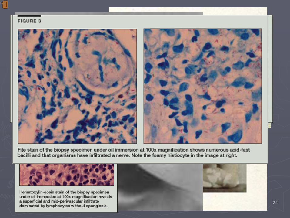
This is a picture of a 24 year old Samoan worman who had a 7 month history of an expanding placquelike lesion on her right cheek. Also notice the prominence of an acessory nerve on the neck over the sternomastoid muscle. This is a hypopigmented area of anesthiia in a young girl with leprosy. A child with hypopigmented anesthethic lesions. Another papular lesion of leprosy. These are two pictures of women with classic hypopigmented lesions of leprosy. The woman on the left has classic plaques. Another child with hypopigmented lesions of leprosy. This is a picture showing the obvious swelling of a nerve in the neck due to leprosy. This is a picture of a patient being tested for light touch sensation using a feather on the skin. This is a picture of an 84 year old man who presented to a clinician in the southern US with these plaquelike lesions. He was treated with steroids with no improvement. A 4 mm puch biopsy was obtained that showed perivascular infiltrates of lymphocytes with no spongiosis (click for the picture). A month later the patient complained of numbness in his arms with no other neurologic complaints. The biopsy specimen was pulled and stained for acid fast bacilli. And what do you know they saw acid fast bacilli. This patient has leprosy.
35
Leprosy an Immune Disease
0
20
40
60
80
100
120
LL BL BB BT TT Healthy
CMI to MLNo. Org
Presenter
Presentation Notes
We are now going to focus on the various forms of leprosy in a bit more detail starting with lepromatous form. This again is a form associated with little CMI against the organism.
36
Lepromatous Leprosy
►Wide disseminationSkinNervesReticuloendothelial systemEyesTestesBonesMucous membranesMouthNosePhargnxTrachea
Presenter
Presentation Notes
Due to the limited resistance to the organism it is widely disseminated in this form of leprosy and includes; Skin Nerves Reticuloendothelial system Eyes Testes Bones Mucous membranes Mouth Nose Phargnx Trachea
37
Lepromatous Leprosy - Skin► Multiple, symmetric macules (flat), plaques (elevated), papules and
nodules► Macules are usually the first seen most commonly seen on the face,
buttocks and extremities► Macules may be erythematous in light skin and faintly hypopigmented
in dark skin► Plaques are elevated and do not appear on the palms and soles► Papules and nodules occur as the disease advances and favor the face,
ears and buttocks► Leonine facies – enhanced wrinkles, loss of eyebrows ► Nodules and plaques may ulcerate on legs when associated with
lymphedema► Pure Diffuse – skin of the whole body becomes infiltrated and
resembles scleroderma, also can be associated with Lucio’s phenomena
Presenter
Presentation Notes
This slide lists the skin lesions of lepromaous leprosy. Generally one sees multiple, symmetric macules, plaques, papules and nodules. All of these forms may be present in the same patient at the same time once established. Macules are usually the first seen most commonly seen on the face, buttocks and extremities Macules may be erythematous in light skin and faintly hypopigmented in dark skin Plaques are elevated and do not appear on the palms and soles due to the tightness and thickness of the skin in those areas. The plaques are not necessarily anesthetic unless the peripheral nerve is involved in that area. Nerve involvement doesn’t always follow the distibution of the skin lesions in LL patients vs TT patients. Papules and nodules occur as the disease advances and favor the face, ears and buttocks. The ear lobes are more constantly afffected and therefore should be examined carefully. These areas are of lower temperature therefore desired by the bacillus. Leonine facies is common with enhanced wrinkles due to infliltration of the surrounding skin. The eyebrows and eyelashes are often lost. The loss first occurs laterally. We call this supercilliary and ciliary madarosis. Nodules and plaques may ulcerate on legs when associated with lymphedema. The lymphedema is due to massive bacillary invasion of the lymphatics. There is a form of lepromatous leprosy called Pure Diffuse where the skin of the whole body becomes infiltrated, shiny and thick and resembles scleroderma. It was first described by Lucio and Alvarado in 1852 and later by Latapi in 1938. There is no obvious disfirgurement. No early macules are seen in this form. Madarosis is seen. One often sees widespread telangiectasias. Nasal destruction can be seen. Sometimes there is alopecia and general loss of body hair. Cutaneous nodules are absent. Occasionally largngeal ulcerations are seen. Occasionally these pure diffuse patients develop painful, purpuric ulcerating patches on the skin that crust and leave scars. This is called Lucio’s phenomena. This is not associated with fever and leukocytosis, tender lesions and doesn’t respond to anti leprosy drugs like Erythema Nodosum Leprosum. It also is unique histologically from ENL.

38
Skin slit
Presenter
Presentation Notes
This is a classic picture of a lady with lepromatous leprosy. Notice the diffuse plaques, nodules, loss of eyebrows. This is what would be called a Leonine face This is another picture of a lady with LL. Notive the marked nodularity and symmetric distribution of the lesions. This is a man who has LL. Notice the leonine facies and involvement of the hands and legs. The peripheral nerve involvement with loss of sensation results in trauma, secondary infection and loss of fingers, toes, etc. as seen in this picture. This is another picture of a classic LL leonine face. This patient still has some eyebrows and eyelashes. They would lose those laterally first. The name of that loss is madarosis. This is a picture of an 8 year old girl with diffuse LL. She is losing her eyebrows. She is of Burmese and Scottish ancestory. Another picture of the classice leonine face of LL. And one more LL facial picture. This shows the ear involvement in LL.You can see a skin slit on the ear used to get a smear for organisms.
39
Lepromatous Leprosy – Nerve Involvement
► The nerves are not involved without the skin► Nerves are damaged later in LL ► Sensory loss is predominant► Nerve thickening is symmetric (great auricular,
supraclavicular, ulnar, antebrachial in the forearm, radial and median at the wrist, femoral cutaneous, common peroneals, superficial peroneal at the front of the ankles, posterior tibial below the internal malleolus)
► Sensory disturbance – paresthesia, hyperesthesiae, hyperalgesia, anesthesia (light touch, temperature, pain)
Presenter
Presentation Notes
This just lists some considerations for nerve involement in Leprosy. The nerves are almost never if ever involved without the skin. Skin lesions are always present in LL when the nerves are involved. Nerves are damaged later in LL than in TT due to less vigorous inflammatory responses in LL. In LL the nerve may be damaged by the volume of bacilli infecting the peripheral nerve vs the inflammatory swelling and damage of TT. Sensory loss is predominant vs motor loss. This results in continued use while not recognizing injury or damage and results in loss of extremity parts. Nerve thickening is symmetric in LL. This just lists the major nerves that we check for when evaluating for peripheral nerve thickening. Sensory disturbance – paresthesia (tingling), hyperesthesiae (increased sensation), hyperalgesia (increased pain), anesthesia (decreases sensitivity to light touch, temperature, pain) Loss of position sense (proprioception), vibration sense, and tendon reflexes may occur but are not common. Muscle wasting due to denervation can occur. This can result in claw hand (ulnar nerve), foot drop (common peroneal), facial palsy-Bell’s Palsy (CN 7). Muscle wasting occurs long before total paralysis. Autonomic nerves can be affected and result in slight edema of the hands and feet. More marked vasomotor problems develop later causing puffy and cyanosed hands.
40
Presenter
Presentation Notes
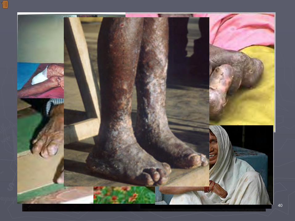
These two pictures show the thickening of the posterior auricular nerve in leprosy. This picture shows the incredible damage that can occur to extremities when sensation is lost and yet use of the extremity continues. You can see the extrusion of the bones on the 4th and 5th fingers. When making rounds at Carville, La we had a patient like this and when examining the finger the bone just popped out and fell to the floor. This is a picture of significant foot ulcers due to leprosy and nerve damage. This picture shows the claw hand deformity of leprosy due to damage to the ulnar nerve. These are some additional pictures of feet damaged by leprosy. Some additional pictures of the incredible damage that can occur to the hands with this disease. Another picture showing the damage to the feet and also possible some early lymphedema. You can see the need for physical therapy and prothetics in any good leprosy program.
41
Other tissues involved with LL
►Nails of fingers and toes – dry, lusterless, narrowed, longitudinally ridged
►Mucous membranesNose – discharge, blocked airway, swollen mucosa, nodules / ulcers on the septum, septal perforation (saddle nose)Mouth – nodules / ulcerations on lips, tongue, palateLargnx – nodules / ulcerations, altered voice, stidorGlottis – edema, obstruction
Presenter
Presentation Notes
These next few slides describe some of the other tissues involved with lepromatous leprosy Nails of fingers and toes become dry, lusterless, narrowed, longitudinally ridged Mucous membranes Nasal discharge is seen and the discharge is often bloody. It can result in a blocked airway. Examination often shows a swollen mucosa with nodules / ulcers on the septum. Septal perforation can result in collapse of the nose resulting in a saddle nose deformity. Nodules / ulcerations on lips, tongue, palate can be seen Nodules / ulcerations of the largnx can result in an altered voice, stridor (inspiratory sounds) The back of the throat can have swelling or edema and obstruction. This can be an emergency situation calling for tracheostomy and was more common prior to MDT.
42
Other tissues involved with LL
►EyeCorneal changesIridocyclitis►Acute►Insidious
Cataracts
Presenter
Presentation Notes
Eye Visual impairment and blindness frequently occur in advanced cases of LL. Leprosy is the third leading cause of blindness worldwide This slide lists the 3 major complications leading to blindness in leprosy including; 1. Corneal changes due to loss of corneal sensation predisposing the cornea to injury and resultant ulceration and scarring. 2. Iris changes can be seen often due to iridocyclitis (inflammation of the iris) as part of the ENL (erythema nodosum leprosum) reaction of LL ENL is an immune complex reaction often seen after a few months of treatment of LL. 3. This chronic form leads to iris atrophy and a pinpoint pupil due to synechiae formation (fibrous tissue). Cataracts – may be caused by iridiocyclitis, the use of steroids, or invasion of the eye with the L bacillus Any leprosy program must have an aggressive program to handle eye complications. Iridocyclitis is the most common cause of blindness and needs to be treated effectively with dilating drugs and antiinflammatory drugs Patients with nerve damage where they can’t blink will need artificial tears and protection from injury to preserve vision.
43
Other tissues involved with LL
►Musculoskeletal systemSkull, arms and legsMultiple factors►Bacilli invading bones – cysts and perostitis►Neurotrophic changes – localized to the phalanges►Repeat trauma►Disuse atrophy►Secondary infections►Generalized osteoporosis
Presenter
Presentation Notes
Musculoskeletal system problems in leprosy are primarily related to bone changes in the skull and arms and legs The skull changes are due to atrophy of the anterior nasal spine and maxillary alveolar process This can result in collapse of the nose and loss of the incisor teeth and even perforation of the palate.

44
Other tissues involved with LL
►Reticuloendothelial system (RES)LymphadenopathyHepatosplenomegalyLymphedema of the lower legs - elephantiasis
►Testes – testicular atrophy►Kidneys – glomerulonephritis, interstitial
nephritis, pyelonephritis, renal amyloidosis
Presenter
Presentation Notes
Other tissues involved with leprosy include; Reticuloendothelial system (RES) Lymph nodes Liver and spleen Lymph channels of lower legs resulting in elephantiasis like that caused by filiariasis. Testicular atrophy is common and can result in hormonal imbalance and exacerbate osteoporosis. Kidneys can be affected

45
Leprosy an Immune Disease
0
20
40
60
80
100
120
LL BL BB BT TT Healthy
CMI to MLNo. Org
Presenter
Presentation Notes
Lets now discuss briefly the tuberculoid form of the disease. This again is a form associated with a vigorous CMI against the organism.
46
Tuberculoid Leprosy
►Good immune response►May be neural or neural and dermal►Localized
Presenter
Presentation Notes
This pole of leprosy may manifest as neural disease only or a combination of neural and dermal disease. This spectrum of disease is never widespread and is localized to one or a few areas. They are always asymmetric.

47
Tuberculoid Leprosy
►Neural DiseaseThickened nervesSensory and motor involvementMotor changes affect the face, intrinsic muscles of the hand and dorsiflexors of the feetAbscesses along affected nerves can be seenEye can be involved due to damage to the facial nerve
Presenter
Presentation Notes
Neural Disease has these characteristics Thickened nerves Sensory and motor involvement due to inflammation and thickening of nerves. Motor changes that affect the face, intrinsic muscles of the hand and dorsiflexors of the feet Abscesses along affected nerves can be seen Eye involvement is also seen in TT due to the damage to the nerves associated with blinking of the eyelids.

48
Tuberculoid Leprosy
►Dermal DiseaseMacules and plaquesAsymmetricFace, extensor surfaces of limbs, back, buttocks
Presenter
Presentation Notes
Dermal Disease is quite different from the lepromatous skn lesions Macules and plaques (infiltrations are see). These plaques are anaesthetic and have no hair. Remember that LL skin lesions usually at presentation have no anesthesia. They are asymmetric They affect the face, extensor surfaces of limbs, back, buttocks

49
Presenter
Presentation Notes
This is a man with TT leprosy with asymmtric plaques on the face. These plaques in TT leprosy are anesthetic and have no hair in them. These are four pictures of TT leprosy. Notice the asymetric plaques and papules on the face and foot. Also notice the swollen posterior auricular nerve in the neck. This is a picture of TT skin lesion on the back of a 35 you man. This lesion was hypopigmented, anesthetic and anhidrotic. This is another hypopigmented anestheic anhidrotic skin lesion of TT leprosy. This is a picture of neuropathic hands due to nerve involvement of a patient with TT leprosy. This is extensive foot ulcer associated with TT leprosy. Another foot ulcer of TT leprosy.
50
Borderline Leprosy
►Not strictly localized like TT or as widespread as LL
►Nerve involvement can always be demonstrated and often preceded skin lesions
►Nerves are thickened and show sensory and motor involvement
Presenter
Presentation Notes
Borderline leprosy is just that; in between TT and LL. The immune response is also in between that of no immunity in LL to aggressive immunity in TT. Borderline leprosy is not strictly localized like TT or as widespread as LL Nerve involvement can almost always be demonstrated and often precedes skin lesions Nerves are thickened and show sensory and motor involvement

51
Presenter
Presentation Notes
We’ve seen this picture before of a 19 yo with a borderline leprosy rash. You can appreciate how difficult this is to separate from either LL or TT. There is generally nerve involvement in these lesions.
52
Indeterminate Leprosy
►Early phase and has not yet committed to either TT or LL
►Single macule, uncharacteristic histology, absence of bacilli
Presenter
Presentation Notes
Indeterminate leprosy is leprosy in a early phase of development. The situation is not yet well defined or committed to either the TT or LL poles. This usually presents with a single macule. Biopsy shows uncharacteristic histology. There are no leprosy bacilli seen in a smear.

53
Presenter
Presentation Notes
This is a picture of a single macule of indeterminate leprosy.
54
Antileprosy Medications
►StandardRifampicin (rifampin, rifadin, rimactane)ClofazamineDapsone (4,4’ diaminodiphenylsulfone)
►Special situationsOfloxacinMinocyclineClarithromycin
Presenter
Presentation Notes
This just lists the drugs used to treat leprosy. With adequate treatment the prognosis is good in nearly all patients whether with PB or MB disease. I’ve divided them into standard drugs and those for special situations. Standard drugs include; Rifampicin (rifampin, rifadin, rimactane). Rifampicin inhibits RNA synthesis and is the most effective bacteriocidal drug against leprosy. Steroids effects are reduced by Rifampicin. Clofazamine. It is bacteriocidal against M. Leprae and also has anti-inflammatory activity. There can be some side effects when used in higher doses. One of the side effects is discoloration of the skin particularly within the skin lesions. These are usually clear spontaneously within 6-12 months. Dapsone (4,4’ diaminodiphenylsulfone). This drug acts at the level of inhibiting the folic acid synthesizing enzyme. It is weakly bacteriocidal. All of the above drugs are relatively safe. Special situation drugs include. Ofloxacin. Is a fluoroquinolone and interfers with DNA replication by inhibiting DNA gyrase. It is modestly bacteriocidal. Minocycline. Is a tetracycline. It inhibits protein synthesis. It is bacteriocidal. Clarithromycin. It is a macrolide antibiotic. It inhibits protein synthesis. It is significantly bacteriocidal.

55
Standard MDT regimens
►Multibacillary – Rx for 12 months (US 1-3 years)Rifampicin: 600 mg once / month, supervisedDapsone: 100 mg once / month, self administeredClofazamine: 300 mg once / month, supervised + 50 mg daily, self administered
► Paucibacillary – Rx for 6 months (US 1 year)Rifampicin: 600 mg once / month, supervisedDapsone: 100 mg daily, self administered
Pregnancy and Lactation – Leprosy is exacerbated in pregnancy therefore the above regimens are recommended unchanged for pregnancy and lactation. Some of the medications are excreted in small quantities in breast milk but no adverse reactions have been noted.
HIV patients respond to standard MDT
Presenter
Presentation Notes
This slide lists the multi drug therapy (MDT) standard regimens to Rx leprosy. MDT was developed due to the emergence of dapsone resistant organisms. MDT as with TB therapy effectively prevents the development of resistance to any single drug. The US Rx of leprosy is prolonged vs that recommended by the WHO. For multibacillary the Rx is for 12 months (US 1-3 years) with three drugs (R, Dapsone, and Clofazamine). Some of this is supervised like DOT of TB and some self administered. For paucibacillary disease the Rx course is for 6 months (US 1 year) with R supervised once per month and Dapsone daily self administered. For pregnancy and lactation the MDT therapy should continue unchanged. Pregnancy exacerbates leprosy. Some of the medications are excreted in breast milk in small quantities but no adverse effects have been reported. HIV patients respond like other patients to standard MDT therapy.

56
Pediatric Doses
►Dapsone – 1 mg /kg►Rifampin – 10 mg / kg►Clofazamine – 1 mg / kg
Presenter
Presentation Notes
This slide just lists the pediatric doses of the standard drugs.
57
Special Situations
► Single lesion paucibacillary leprosySingle dose therapy (ROM)►Rifampicin 600 mg►Ofloxacin 400 mg►Minocycline 100 mg►Marginally less effective than standard MDT for paucibacillary
disease
►Can’t take RipampicinTreat for 24 months►Clofazamine 50 mg / day + ►Two of the following / day (Ofloxacin 100 mg, Minocycline 100
mg, or Clarithromycin 500 mg) for 6 months then►Daily Clofazamine 50 mg + either Minocycline 100 mg or
Ofloxacin 400 mg for 18 months
Presenter
Presentation Notes
These next few slides list some alternate treatments for special situations. I will not cover these in detail. They are here for your reference if needed.
58
Special Situations
►Multibacillary patients Refusing ClofazamineStandard MDT but replacing Clofazamine by Ofloxacin 400 mg daily or Minocycline 100 mg daily for 12 months24 month regimen (ROM) of Rifamipcin 600 mg / month, Ofloxacin 400 mg / month and Minocycline 100 mg / month
Presenter
Presentation Notes
This slide just lists some alternate regimens for those patients refusing to take clofazamine due to the temporary skin discoloration adverse reaction.

59
Special Situations
►Can’t take DapsoneWith multibacillary disease just stop the Dapsone and continue the other meds (rifampicin and clofazamine)With paucibacillary disease substitute clofazamine for dapsone
Presenter
Presentation Notes
This slide lists the alternatives for patients unable to take Dapsone.
60
Reactions►Lepra reactions – immune mediated
inflammation►5% of PB and 20% of MB patients►Two major types
Reversal (type 1)ENL (erythema nodosum leprosum) (type 2)
►ENL less common with MDT
Presenter
Presentation Notes
During therapy it is common to observe immune mediated reactions that cause significant inflammation associated with serious complications. These reactions are called lepra reactions. They are seen in 5% of PB patients and 20% of MB patients. There are two major types of lepra reactions Reversal (type 1) seen in both PB and MB disease ENL (erythema nodosum leprosum) (type 2) seen exclusively in advanced MB disease. ENL is less common with current MDT regimens vs when Dapsone was used alone. This is probably due to anti inflammatory activity of Clofazamine in the current standard MDT regimens.
61
Reversal Type 1
►Lesion changes – Erythema, edema, pain, tenderness over nerves
►High risk of nerve damage►Treat – Steroids (1 mg / kg / day, max 40-
60 mg of prednisolone)►Taper off steroids over a 12 week period
Presenter
Presentation Notes
Type 1 reversal reactions are manifest by increasing redness of the skin lesions with increasing edema and inflammation and swelling of the nerves and may be associated with pain and tenderness over the nerves. Type 1 reversal reactions are associated with a high risk of nerve damage therefore decreased sensation over nerve distribution areas must be monitored closely. Early aggressive treatment with steroids is essential in Type 1 reactions.. The reaction is often controlled within a few days with this therapy. The dose is then gradually reduced over a 12 week period.
62
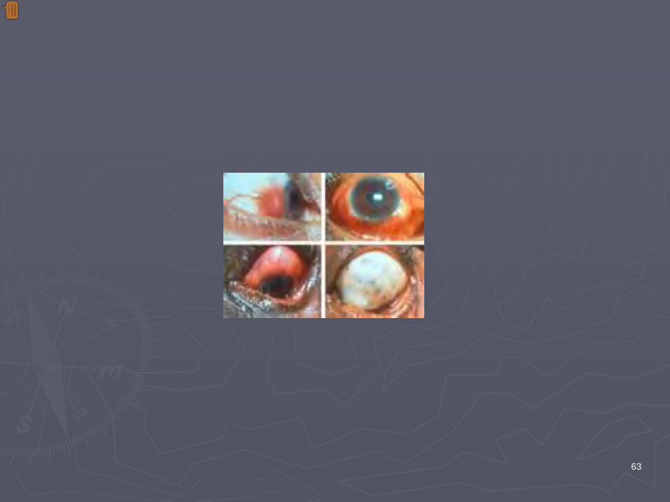
ENL type 2
►Varies in severity duration and organ involvement►Rapid onset of erythematous nodules, fever, joint
inflammation, iridocyclitis, ulceration of the skin, glomerulonephritis and amyloidosis
►Mild ENL Rx – Aspirin► Severe ENL with neuritis – Rx with prednisolone as
for Type 1 reactions►Clofazamine may be useful when withdrawing
steroids (dose 300 mg / day divided doses)► Iridocyclitis – add topical steroids to regimen
Presenter
Presentation Notes
For ENL (erythema nodosum leprosum) type 2 reactions The situation varies in severity, duration and organ involvement There can be a rapid onset of redness of the skin, fever, joint inflammation, iridocyclitis (eye inflammation), ulceration of the skin, and kidney involvement like glomerulonephritis and amyloidosis (protein deposits in the kidney) Mild ENL may respond well to Aspirin Severe ENL with neuritis may require Rx with steroids as for Type 1 reversal reactions Clofazamine may be useful when withdrawing steroids (dose 300 mg / day divided doses to decrease GI irritation. Clofazamine should never be used solely to treat type 2 reactions since it may take 4-6 weeks to have a effect. With Iridocyclitis – add topical steroids to regimen
63
Presenter
Presentation Notes
These four pictures show an ENL reaction eyes with marked inflammation of various tissues.

64
Presenter
Presentation Notes
This is a picture of a Lucio reaction. This is painful, ulcerating patch on the skin that will crust and leave scars. This is a form of lepra reaction that seems to be unique to those patients having the pure diffuse type of leprosy described by Lucio and Alvarado in Mexico in 1852 and later by Latapi in 1938. This reaction can be differentiated from ENL by the absence of fever and increased wbc counts (leucocytosis), absence of tender lesions and a good response to antileprosy drugs. There is also a unique histological apprearance to biopsies of these lesions.

65
Lepra Reaction – Program Recommendations
►Patients taught to recognize Lepra reactions and report promptly
►Clinicians able to diagnose and promptly treat reactions
►Adequate stocks of medications to treat reactions
►Continue MDT without interruption during a lepra reaction
Presenter
Presentation Notes
These are some general guidelines for leprosy programs regarding lepra reactions. Patients need to be taught to recognize Lepra reactions and report them promptly. This is a disease management concept. The patient is an integral part of the team in treating chronic disease. Clinicians must be able to diagnose and promptly treat reactions Adequate stocks of medications to treat reactions must be readily available It is extremely important to continue MDT without interruption during a lepra reaction
66
Relapse
►0.1% relapse rate►All M leprae from relapse patients remain
susceptible to rifampicin and clofazamine and respond favorably to a second course of MDT
Presenter
Presentation Notes
A few comments about efficacy of MDT. MDT is associated with only a a 0.1% relapse rate Historically all M leprae organisms from relapse patients remain susceptible to rifampicin and clofazamine and respond favorably to a second course of MDT MDT is an incredible development in the world of leprosy.
67
Strategies for eliminating Leprosy as a public health problem
► Identification of endemic districts►MDT services integrated into general health
facilities►Monitoring the elimination at the district level► Promoting community action► Social marketing / advocacy►Remotivating the research community► Prevention of disabilities and rehabilitation
Presenter
Presentation Notes
These are the main strategies for eliminating leprosy as a public health problem (prevalence < 1/10000 population) We are still at about 1.25 / 10000 worldwide (much higher in localized areas like the top 11 countries where the prevalence is 4.1)). 1. Identification of all endemic districts. We just don’t have good data at this level in much of the world therefore making it hard to plan. 2. MDT services integrated into general health facilities. This will be essential. Many clinicians worldwide don’t know how to diagnose and treat this disease. 3. Monitoring the elimination at the district level. Evaluation is key to everything. 4. Promoting community action. This is essential to change the image of leprosy and remove the stigma. It also will be important in much of the world for providing MDT. This is community engagement. 5. Social marketing / advocacy. This will be important for patients seeking care early in the disease process. 6. Remotivating the research community. We need new strategies to move us along. This will take some new innovative research. 7. Prevention of disabilities and rehabilitation. This is key for managing the complications of this disease. Questions?
Related Documents