Leishmanicidal activity of the Agaricus blazei Murill in different Leishmania species Diogo G. Valadares a, 1 , Mariana C. Duarte b, 1 , Jamil S. Oliveira a , Miguel A. Chávez-Fumagalli c , Vivian T. Martins a , Lourena E. Costa b , João Paulo V. Leite d , Marcelo M. Santoro a , Wiliam C.B. Régis e, f, 2 , Carlos A.P. Tavares a, 2 , Eduardo A.F. Coelho b, ⁎ , 2 a Departamento de Bioquímica e Imunologia, Instituto de Ciências Biológicas, Universidade Federal de Minas Gerais, Belo Horizonte, Minas Gerais, Brazil b Departamento de Patologia Clínica COLTEC, Universidade Federal de Minas Gerais, Belo Horizonte, Minas Gerais, Brazil c Programa de Pós-Graduação em Medicina Molecular, Universidade Federal de Minas Gerais, Belo Horizonte, Minas Gerais, Brazil d Departamento de Bioquímica e Biologia Molecular, Universidade Federal de Viçosa, Viçosa, Minas Gerais, Brazil e Minasfungi do Brasil LTDA, Belo Horizonte, Minas Gerais, Brazil f Pontifícia Universidade Católica de Minas Gerais, Belo Horizonte, Minas Gerais, Brazil abstract article info Article history: Received 10 January 2011 Received in revised form 31 May 2011 Accepted 2 June 2011 Available online 24 June 2011 Keywords: Leishmania Agaricus blazei Murill mushroom Leishmanicidal activity Treatment Leishmaniasis is a major public health problem, and the alarming spread of parasite resistance underlines the importance of discovering new therapeutic products. The present study aims to investigate the in vitro leishmanicidal activity of an Agaricus blazei Murill mushroom extract as compared to different Leishmania species and stages. The water extract proved to be effective against promastigote and amastigote-like stages of Leishmania amazonensis, L. chagasi, and L. major, with IC 50 (50% inhibitory concentration) values of 67.5, 65.8, and 56.8 μg/mL for promastigotes, and 115.4, 112.3, and 108.4 μg/mL for amastigotes-like respectively. The infectivity of the three Leishmania species before and after treatment with the water extract was analyzed, and it could be observed that 82%, 57%, and 73% of the macrophages were infected with L. amazonensis, L. major, and L. chagasi, respectively. However, when parasites were pre-incubated with the water extract, and later used to infect macrophages, they were able to infect only 12.7%, 24.5%, and 19.7% of the phagocytic cells for L. amazonensis, L. chagasi, and L. major, respectively. In other experiments, macrophages were infected with L. amazonensis, L. chagasi, or L. major, and later treated with the aforementioned extract, presented reductions of 84.4%, 79.6%, and 85.3% in the parasite burden after treatment. A confocal microscopy revealed the loss of the viability of the parasites within the infected macrophages after treatment with the water extract. The applied extract presented a low cytotoxicity in murine macrophages and a null hemolytic activity in type O + human red blood cells. No nitric oxide (NO) production, nor inducible nitric oxide syntase expression, could be observed in macrophages after stimulation with the water extract, suggesting that biological activity may be due to direct mechanisms other than macrophage activation by means of NO production. In conclusion, the results demonstrate that the A. blazei Murill water extract can potentially be used as a therapeutic alternative on its own, or in association with other drugs, to treat Visceral and Cutaneous Leishmaniasis. © 2011 Elsevier Ireland Ltd. All rights reserved. 1. Introduction Protozoan parasites of the Leishmania genus are the etiological agents of a vector-borne disease that has presented high morbidity and mortality throughout the world. Leishmaniasis has affected around 12 million people and is present in 88 countries, mainly in tropical and subtropical areas. The approximately 2 million new cases per year and the nearly 350 million people living in endemic regions reveal the importance of this neglected disease [1,2]. Historically, chemotherapy to treat leishmaniasis has been based on the use of pentavalent antimonial drugs. One of the available anti- leishmanial drugs, meglumine antimoniate (Glucantime ® ), is clinically unsatisfactory, once that it is not very effective and may have severe side effects. Furthermore, a high number of relapses of the disease have been described [3,4]. Pentamidine, another anti-leishmanial drug, is unsuitable as a first line treatment due to its toxicity. Amphotericin B and its liposomal formulation are effective, though such drugs are expensive and their use requires hospitalization. Reported clinical results using oral miltefosine treatment are encouraging; however, this drug is linked to potential teratogenicity and should not be given to pregnant women or to those of childbearing age [5]. The number of reported VL cases is increasing. Moreover, VL has emerged as an opportunistic infection in human immunodeficiency virus-infected (HIV) patients [6]. Therefore, the development of cost-effective alternative therapeutic strategies has become a high-priority. Parasitology International 60 (2011) 357–363 ⁎ Corresponding author at: Laboratório de Biotecnologia Aplicada ao Estudo de Doenças Parasitárias, Departamento de Patologia Clínica, COLTEC, Universidade Federal de Minas Gerais, Avenida Antônio Carlos, 6627, Belo Horizonte, Minas Gerais, Brazil, 31.270-901. Tel./fax: + 55 31 3409 4983. E-mail address: [email protected] (E.A.F. Coelho). 1,2 These authors contributed equally to this work. 1383-5769/$ – see front matter © 2011 Elsevier Ireland Ltd. All rights reserved. doi:10.1016/j.parint.2011.06.001 Contents lists available at ScienceDirect Parasitology International journal homepage: www.elsevier.com/locate/parint

Welcome message from author
This document is posted to help you gain knowledge. Please leave a comment to let me know what you think about it! Share it to your friends and learn new things together.
Transcript
Parasitology International 60 (2011) 357–363
Contents lists available at ScienceDirect
Parasitology International
j ourna l homepage: www.e lsev ie r.com/ locate /par in t
Leishmanicidal activity of the Agaricus blazei Murill in different Leishmania species
Diogo G. Valadares a,1, Mariana C. Duarte b,1, Jamil S. Oliveira a, Miguel A. Chávez-Fumagalli c,Vivian T. Martins a, Lourena E. Costa b, João Paulo V. Leite d, Marcelo M. Santoro a, Wiliam C.B. Régis e,f,2,Carlos A.P. Tavares a,2, Eduardo A.F. Coelho b,⁎,2
a Departamento de Bioquímica e Imunologia, Instituto de Ciências Biológicas, Universidade Federal de Minas Gerais, Belo Horizonte, Minas Gerais, Brazilb Departamento de Patologia Clínica COLTEC, Universidade Federal de Minas Gerais, Belo Horizonte, Minas Gerais, Brazilc Programa de Pós-Graduação em Medicina Molecular, Universidade Federal de Minas Gerais, Belo Horizonte, Minas Gerais, Brazild Departamento de Bioquímica e Biologia Molecular, Universidade Federal de Viçosa, Viçosa, Minas Gerais, Brazile Minasfungi do Brasil LTDA, Belo Horizonte, Minas Gerais, Brazilf Pontifícia Universidade Católica de Minas Gerais, Belo Horizonte, Minas Gerais, Brazil
⁎ Corresponding author at: Laboratório de BiotecnoDoenças Parasitárias, Departamento de Patologia Clínicade Minas Gerais, Avenida Antônio Carlos, 6627, Belo H31.270-901. Tel./fax: +55 31 3409 4983.
E-mail address: [email protected],2 These authors contributed equally to this work.
1383-5769/$ – see front matter © 2011 Elsevier Irelanddoi:10.1016/j.parint.2011.06.001
a b s t r a c t
a r t i c l e i n f oArticle history:Received 10 January 2011Received in revised form 31 May 2011Accepted 2 June 2011Available online 24 June 2011
Keywords:LeishmaniaAgaricus blazei Murill mushroomLeishmanicidal activityTreatment
Leishmaniasis is a major public health problem, and the alarming spread of parasite resistance underlines theimportance of discovering new therapeutic products. The present study aims to investigate the in vitroleishmanicidal activity of an Agaricus blazei Murill mushroom extract as compared to different Leishmania speciesand stages. The water extract proved to be effective against promastigote and amastigote-like stages of Leishmaniaamazonensis, L. chagasi, and L.major, with IC50 (50% inhibitory concentration) values of 67.5, 65.8, and56.8 μg/mL forpromastigotes, and 115.4, 112.3, and 108.4 μg/mL for amastigotes-like respectively. The infectivity of the threeLeishmania species before and after treatment with the water extract was analyzed, and it could be observed that82%, 57%, and 73% of the macrophages were infected with L. amazonensis, L. major, and L. chagasi, respectively.However, when parasites were pre-incubated with the water extract, and later used to infect macrophages, theywere able to infect only 12.7%, 24.5%, and 19.7% of the phagocytic cells for L. amazonensis, L. chagasi, and L. major,respectively. In other experiments, macrophages were infectedwith L. amazonensis, L. chagasi, or L. major, and latertreatedwith theaforementionedextract, presented reductionsof 84.4%, 79.6%, and85.3% in theparasiteburdenaftertreatment. A confocalmicroscopy revealed the loss of the viability of the parasites within the infectedmacrophagesafter treatmentwith thewater extract. The applied extract presented a lowcytotoxicity inmurinemacrophages anda null hemolytic activity in type O+ human red blood cells. No nitric oxide (NO) production, nor inducible nitricoxide syntase expression, could be observed in macrophages after stimulation with the water extract, suggestingthat biological activity may be due to direct mechanisms other than macrophage activation by means of NOproduction. In conclusion, the results demonstrate that the A. blazeiMurill water extract can potentially be used as atherapeutic alternative on its own, or in associationwith other drugs, to treat Visceral and Cutaneous Leishmaniasis.
logia Aplicada ao Estudo de, COLTEC, Universidade Federalorizonte, Minas Gerais, Brazil,
(E.A.F. Coelho).
Ltd. All rights reserved.
© 2011 Elsevier Ireland Ltd. All rights reserved.
1. Introduction
Protozoan parasites of the Leishmania genus are the etiologicalagents of a vector-borne disease that has presented highmorbidity andmortality throughout the world. Leishmaniasis has affected around12 million people and is present in 88 countries, mainly in tropical andsubtropical areas. The approximately 2 million new cases per year andthe nearly 350 million people living in endemic regions reveal theimportance of this neglected disease [1,2].
Historically, chemotherapy to treat leishmaniasis has been based onthe use of pentavalent antimonial drugs. One of the available anti-leishmanial drugs, meglumine antimoniate (Glucantime®), is clinicallyunsatisfactory, once that it is not very effective andmayhave severe sideeffects. Furthermore, a high number of relapses of the disease havebeen described [3,4]. Pentamidine, another anti-leishmanial drug, isunsuitable as a first line treatment due to its toxicity. Amphotericin Band its liposomal formulation are effective, though such drugs areexpensive and their use requires hospitalization. Reported clinicalresults using oral miltefosine treatment are encouraging; however, thisdrug is linked to potential teratogenicity and should not be given topregnant women or to those of childbearing age [5]. The number ofreported VL cases is increasing. Moreover, VL has emerged as anopportunistic infection in human immunodeficiency virus-infected(HIV) patients [6]. Therefore, the development of cost-effectivealternative therapeutic strategies has become a high-priority.

358 D.G. Valadares et al. / Parasitology International 60 (2011) 357–363
Prior research has been carried out concerning natural products andtheir biological effects, such as fungicidal, antimicrobial, antimalarial,antimycobacterial, and antiviral activities. However, only a few studieshave investigated the biological potential of Brazilian mushrooms.Agaricus blazei Murill is a commonly found mushroom in Brazil. Itsuse has been associated with folk medicine in the treatment ofsome diseases, including diabetes mellitus and arterial hypertension[7–13]. This mushroom presents compounds, such as β-D-glucans,glycoproteins, cerebrosides, polysaccharides, steroids, ergosterol, andgraxs acids, which can activate and/or modulate the immune responseof the hosts [14–17].
The present study assessed the in vitro leishmanicidal activity of an A.blazeiMurill water extract against L. amazonensis, L. chagasi, and L. majorpromastigote and amastigote-like stages. Studies were extended toestablish their minimum inhibitory concentrations (IC50), leishmanicidaleffects on intra-macrophages Leishmania stages, nitric oxide (NO)production, inducible nitric oxide syntase (iNOS) expression, as well astheir cytotoxic effects onmurinemacrophages andhuman redblood cells.
2. Material and methods
2.1. A. blazei Murill water extract
TheA. blazeiMurillwater extractwasprepared bymacerating50 g offresh mushrooms in 50 mL of sterile milli-Q water using a Waring–Blendor homogenizer. Next, the macerated mixture was centrifuged at10.000 × g for 20 min at 4 °C (Sorval, LC5Cmodel). The supernatantwasthen sterilized by passing through a 0.22 μm membrane in a laminarflow hood under sterile conditions and stored at−20 °C, until use. Thisprocedure has been patented at the Federal University of Minas Gerais(UFMG) (PI 014100001550/CTI&T).
2.2. Parasites and mice
Leishmania amazonensis (IFLA/BR/1967/PH-8), L. chagasi (MHOM/BR/1970/BH46), and L. major (MHOM/IL/1980/Friedlin) were used inthis study. Parasites were grown at 24 °C in Schneider's medium (Sigma,St. Louis,MO, USA), supplementedwith 20% heat-inactivated fetal bovineserum (FBS, Sigma), 20 mM L-glutamine, 200 U/mL penicillin, and100 μg/mL streptomycin, at a 7.4 pH [18]. Stationary-phase promastigoteand amastigote-like of Leishmania used in this work were prepared asdescribed [19]. Murine macrophages were collected from peritonealcavities of female BALB/c mice (6-weeks old), which were purchasedfrom Institute of Biological Sciences of UFMG. The Animal Use Committeeof the UFMG approved experimental protocols.
2.3. Leishmanicidal in vitro activity
Inhibition of cell growth was assessed in vitro by cultivatingpromastigote and amastigote-like (4×10⁵, each one) of L. amazonensis,L. chagasi, and L. major in the presence of different individualconcentrations (25 to 200 μg/mL) of an A. blazei Murill water extract in96-well culture plates (Corning Life Sciences, Corning, NY, USA), for 48 hat 24 °C. Amphotericin B was used as a positive control (10 µg/mL).Cell viability was assessed bymeasuring the cleavage of 2 mg/mL of MTT[3-(4.5-dimethylthiazol-2-yl)-2.5-diphenyl tetrazolium bromide](Sigma). Absorbances were measured by using a multi-well scanningspectrophotometer (LABTRADE, model 660) at a wavelength of 570 nm.Analyses were performed in triplicate, and results were expressed as themean percentage reduction of the parasites compared to non-treatedcontrol wells. The 50% inhibitory concentration (IC50)was determined byapplying the sigmoidal regression of the concentration–response curves.Data shown are representative of three different experiments, performedin triplicate, which presented similar results.
2.4. Chemical characterization of the A. blazei Murill water extract
Chemical analysis of the water extract was performed on chroma-tography plates coated with silica gel GF 254® (Merck, Darmstadt,Germany) for the detection of tannins, coumarins, flavonoids, anthra-quinones, triterpenes, steroids, saponins, cardiotonic glycosides, andalkaloids. Different mobile phases and detection reagents were used inaccordance with the protocol described by Wagner et al. [20]. Proteinswere detected in SDS-PAGE 10% gels by silver staining, whileglycoproteins presence were demonstrated in SDS-PAGE 10% stainedby periodic acid Schiff. The presence of carbohydrates was investigatedusing a phenol-sulphuric acid method [21].
2.5. Inhibition of infection in phagocytic cells
The inhibitory effect of the A. blazei Murill water extract on theLeishmania invasion of macrophages was evaluated in promastigotes ofL. amazonensis, L. chagasi, and L. major. Parasites were pre-incubatedwith the water extract (25 μg/mL), for 1 h at 24 °C. Next, cells werewashed three times with RPMI 1640medium and further incubated for4 h with murine macrophages at a ratio of 10 Leishmanias per 1macrophage. After, cells were again washed, set, and stained, todetermine the percentages of infected macrophages by counting 100cells in triplicate. Additionally, an optical microscopy was used to viewthe Leishmania infection profiles within the murine macrophages.
2.6. Treatment of infected macrophages
Macrophageswere plated on roundglass coverslips inside thewells ofa 24-well culture plate at a concentration of 5×10⁵cells per coverslip inan RPMI 1640 medium supplemented with 20% FBS, 2 mM L-glutamine,200 U/mL penicillin, and 100 μg/mL streptomycin, at a 7.4 pH. After 2 h ofincubation at 37 °C in 5% CO2; promastigotes of L. chagasi, L. major, andL. amazonensis 5×10⁶ were added to the wells and the cultures wereincubated for 4 h at 37 °C, 5% CO2. Next, free parasites were removed byextensive washing with an RPMI 1640 medium, and infected macro-phages were treated for 48 h with the A. blazei Murill water extract(50 μg/mL). Cells were washed in RPMI 1640 and incubated with 4%paraformaldehyde for 15 min, at which time they were treated with 70%ethanol in an ice-bath for 4 h, and again washed three times with sterilePBS. The percentage of the inhibition of Leishmania intra-macrophagesviabilitywasdeterminedbycounting100cells in triplicateandcomparingthis to the infected and non-treated cells. Additionally, a confocalmicroscopy was performed to view the inhibition of Leishmania viabilitywithin intra-macrophages. For this, an RNaseA solution was added(200 μg/mL), and incubation occurred for 30 min at 37 °C, after whichtime, a solution comprised of 370 μL of 0.1 MHCl in 148 μL of PBS 1×wasadded perwell. After 45 s, 1.5 mLof an acridine orange solution (3 μg/mL,pH 5.0) was added and incubation occurred for 5 min, at which time thecells were analyzed. Data shown are representative of three separateexperiments, performed in triplicate, which presented similar results.
2.7. Nitric oxide (NO) production and iNOS Western blot
To visualize the macrophage activation via NO production aftertreatment with an A. blazei Murill water extract, cells (4×10⁵) wereincubated alone in an RPMI 1640 medium (background control) orseparately stimulated with the water extract (50 μg/mL) or concanavalinA (ConA; 5 μg/mL), at 37°C in 5% CO2 for 24, 48, and 72 h. Followingincubation, 100 µL of culture supernatant was mixed with an equalvolume of Griess reagent (Sigma). After an incubation of 30 min at roomtemperature, nitrite concentration was calculated using a standard curveof known concentrations. Data were expressed as μM per 4×10⁵ cells.Data shown are representative of three different experiments, performedin triplicate, which presented similar results.

Fig. 1. Leishmanicidal activity of the water extract of Agaricus blazei Murill againstpromastigotes of L. chagasi, L.major, and L. amazonensis. Parasites (4×10⁵)were incubatedwith different individual concentrations (25 to 200 μg/mL) of the A. blazei Murill waterextract, and cell viability was analyzed by MTT assay, after incubation for 48 h at 24 °C.Amphotericin B (AmB, 10 µg/mL)was used as a positive control. Bars indicate themean±standard deviation of the inhibition of Leishmania viability in each concentration tested.Differences were considered statistically significant (Pb0.05) when comparing theparasites treated with: (a) 25 μg/mL, (b) 50 μg/mL, (c) 100 μg/mL, or (d) 200 μg/mL ofthe water extract, and the parasites treated with AmB (x).
Fig. 2. Leishmanicidal activity of the water extract of Agaricus blazei Murill againstamastigotes-like of L. chagasi, L. major, and L. amazonensis. Parasites (4×10⁵)were incubatedwith different individual concentrations (25 to 200 μg/mL) of the A. blazei Murill waterextract, and cell viability was analyzed by MTT assay, after incubation for 48 h at 24 °C.Amphotericin B (AmB, 10 µg/mL) was used as a positive control. Bars indicate the mean±standard deviation of the inhibition of Leishmania viability in each concentration tested.Differenceswere considered statistically significant (Pb0.05) when comparing the parasitestreated with: (a) 25 μg/mL, (b) 50 μg/mL, (c) 100 μg/mL, or (d) 200 μg/mL of the waterextract, and the parasites treated with AmB (x).
359D.G. Valadares et al. / Parasitology International 60 (2011) 357–363
After separation of the supernatants, and in order to detect iNOSexpression, cellswere lysed in an ice-cold lyses buffer (100 mMTris; pH8.0; 2 mM EDTA; 100 mM NaCl; 1% Triton X-100), which wassupplemented by a complete proteases inhibitors cocktail (RocheDiagnostics, Indianapolis, IN, USA). Lysates were centrifuged at14,000 × g for 10 min at 4 °C, and the protein concentration wasdetermined using the DC Protein Assay Reagents Package (Bio-RadLaboratories, Hercules, CA, USA), employing bovine serum albumin(BSA) as a standard. The proteic extract (20 μg) was analyzed bymeansof a SDS-PAGE 10% gel, and proteins were transferred onto cellulosemembranes using a semi-dry electroblotting apparatus (Loccus Bio-technology). Themembraneswere blockedwith a 5% BSA in Tris Saline-Tween 20 0.05% (TBS-T: 10 mM Tris–HCl; pH 7.4; 0.15 M NaCl; 0.05%Tween 20 0.05%), for 2 h at room temperature. Blots were incubatedwith 1:1,000 monoclonal anti-mouse iNOS (123 kDa) and beta-actin(45 kDa) antibodies (Santa Cruz Biotechnology, Santa Cruz, CA, USA)overnight, under constant shaking, at 4 °C. After having been washed 5times with TBS-T, membranes were incubated using an anti-mouse IgGgoat antibody (Cell Signaling Technology, Danvers, MA, USA, dilution1:1,000 diluted in 1% non-fat dry milk) for 2 h at room temperature.After having been washed 7 times with TBS-T, blots were developedusing chloronaphtol (12.5 mg), diaminobenzidine (25 mg), and H2O2
30 vol (20 μL), for 30 min, and the reaction development was ended byadding distilled water.
2.8. Cytotoxicity and hemolytic activity
Cytotoxicity was evaluated by cultivating macrophages (4×10⁵)with different concentrations (25 to 200 μg/mL) of the A. blazei Murillwater extract in 96-well plates, for 24 h at 4 °C. Cell viability wasassessedby theMTTassay, and resultswere expressed as thepercentageof cell lyses compared to cultures treated with amphotericin B (AmB,1 µg/mL). The hemolytic activity was investigated by incubating the A.blazei Murill water extract (25 to 200 μg/mL) with a 5% red blood cell(human O+) suspension for 1 h at 37 °C. The erythrocyte suspensionwas centrifuged (1000 × g for 10 min), and cell lysis was determinedspectrophotometrically (540 nm), as described [22]. The absence(blank) or 100% presence of hemolysis (positive control) wasdetermined by replacing the extract for an equal volume of PBS 1x ordistilled water, respectively. The results were determined by thepercentage of hemolysis compared to thenegative andpositive controls.Data shown are representative of three separate experiments,performed in triplicate, which presented similar results.
2.9. Statistical analysis
Statistical analysis was performed using the GraphPad Prismsoftware (version 5.0 for Windows). The differences among thediverse treatments (25, 50, 100 and 200 μg/mL) carried out using theA. blazei Murill water extract, as well as the reduction of thepercentage of infected macrophages, were evaluated by the one-way ANOVA analysis, followed by Bonferroni's post-test for multiplecomparisons. Differences were considered significant when Pb0.05.
3. Results
3.1. Leishmanicidal activity of the water extract of A. blazei Murill inLeishmania
The in vitro leishmanicidal effect of the A. blazei Murill water extractwas tested against promastigote and amastigote-like stages ofL. amazonensis, L. chagasi, and L. major. Parasites were separatelyincubated with different extract concentrations (25 to 200 μg/mL) for48 h at 24 °C. In the evaluation of the results, it could be observed that theleishmanicidal activitywas based on a dependent-dose application, giventhat that a lower dose (25 μg/mL) of the extract induced the inhibition of
Leishmaniaviability percentages in theorder of 26.5%, 29.3%, and23.2% forL. amazonensis, L. chagasi, and L.majorpromastigotes (Fig. 1), respectively,whereas in the amastigote-like stage, the inhibition of cell viabilitypercentages were of 10.5%, 12.3%, and 13.2%, respectively (Fig. 2) Thehigher dose of the water extract (200 μg/mL) evoked higher inhibitionof Leishmania viability percentages in the order of 85%, 81.6%, and 85% forL. amazonensis, L. chagasi, and L. major promastigotes, respectively, and of61.5%, 63.6%, and 65.6% for amastigote-like stages, respectively, for thethree species. In the evaluation of IC50, promastigotes proved to be moresusceptible than amastigote-like as they presented IC50 values of 67.5,65.8, and 56.8 μg/mL for L. amazonensis, L. chagasi, and L. major,respectively, while the values for amastigote-like stages were 115.4,112.3, and 108.4 μg/mL for L. amazonensis, L. chagasi, and L. major,respectively (Table 1). Amphotericin B (10 μg/mL), used as a positivecontrol, presented significant reductions in the viability of the threeLeishmania species evaluated (Figs. 1 and 2).
To provide additional information concerning the chemical entitiesin the A. blazei Murill water extract that can be responsible for thebiological effects observed in this study, a chemical characterizationwasperformed, as described in theMaterial andmethods section. The resultof this characterization illustrated the presence of tannins, saponins,
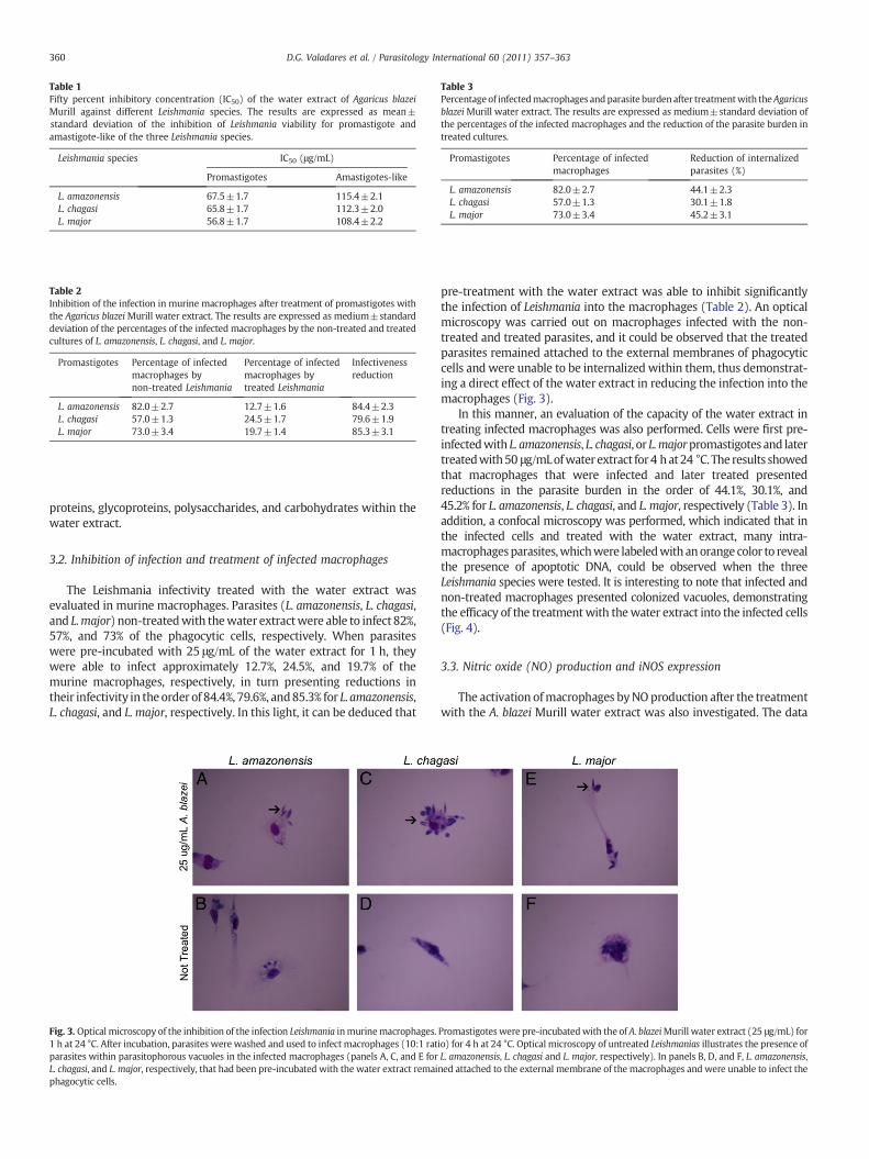
Table 1Fifty percent inhibitory concentration (IC50) of the water extract of Agaricus blazeiMurill against different Leishmania species. The results are expressed as mean±standard deviation of the inhibition of Leishmania viability for promastigote andamastigote-like of the three Leishmania species.
Leishmania species IC50 (μg/mL)
Promastigotes Amastigotes-like
L. amazonensis 67.5±1.7 115.4±2.1L. chagasi 65.8±1.7 112.3±2.0L. major 56.8±1.7 108.4±2.2
Table 2Inhibition of the infection in murine macrophages after treatment of promastigotes withthe Agaricus blazeiMurill water extract. The results are expressed as medium±standarddeviation of the percentages of the infected macrophages by the non-treated and treatedcultures of L. amazonensis, L. chagasi, and L. major.
Promastigotes Percentage of infectedmacrophages bynon-treated Leishmania
Percentage of infectedmacrophages bytreated Leishmania
Infectivenessreduction
L. amazonensis 82.0±2.7 12.7±1.6 84.4±2.3L. chagasi 57.0±1.3 24.5±1.7 79.6±1.9L. major 73.0±3.4 19.7±1.4 85.3±3.1
Table 3Percentageof infectedmacrophages andparasite burden after treatmentwith theAgaricusblazei Murill water extract. The results are expressed as medium±standard deviation ofthe percentages of the infected macrophages and the reduction of the parasite burden intreated cultures.
Promastigotes Percentage of infectedmacrophages
Reduction of internalizedparasites (%)
L. amazonensis 82.0±2.7 44.1±2.3L. chagasi 57.0±1.3 30.1±1.8L. major 73.0±3.4 45.2±3.1
360 D.G. Valadares et al. / Parasitology International 60 (2011) 357–363
proteins, glycoproteins, polysaccharides, and carbohydrates within thewater extract.
3.2. Inhibition of infection and treatment of infected macrophages
The Leishmania infectivity treated with the water extract wasevaluated in murine macrophages. Parasites (L. amazonensis, L. chagasi,and L.major) non-treatedwith thewater extractwere able to infect 82%,57%, and 73% of the phagocytic cells, respectively. When parasiteswere pre-incubated with 25 μg/mL of the water extract for 1 h, theywere able to infect approximately 12.7%, 24.5%, and 19.7% of themurine macrophages, respectively, in turn presenting reductions intheir infectivity in the order of 84.4%, 79.6%, and85.3% for L. amazonensis,L. chagasi, and L. major, respectively. In this light, it can be deduced that
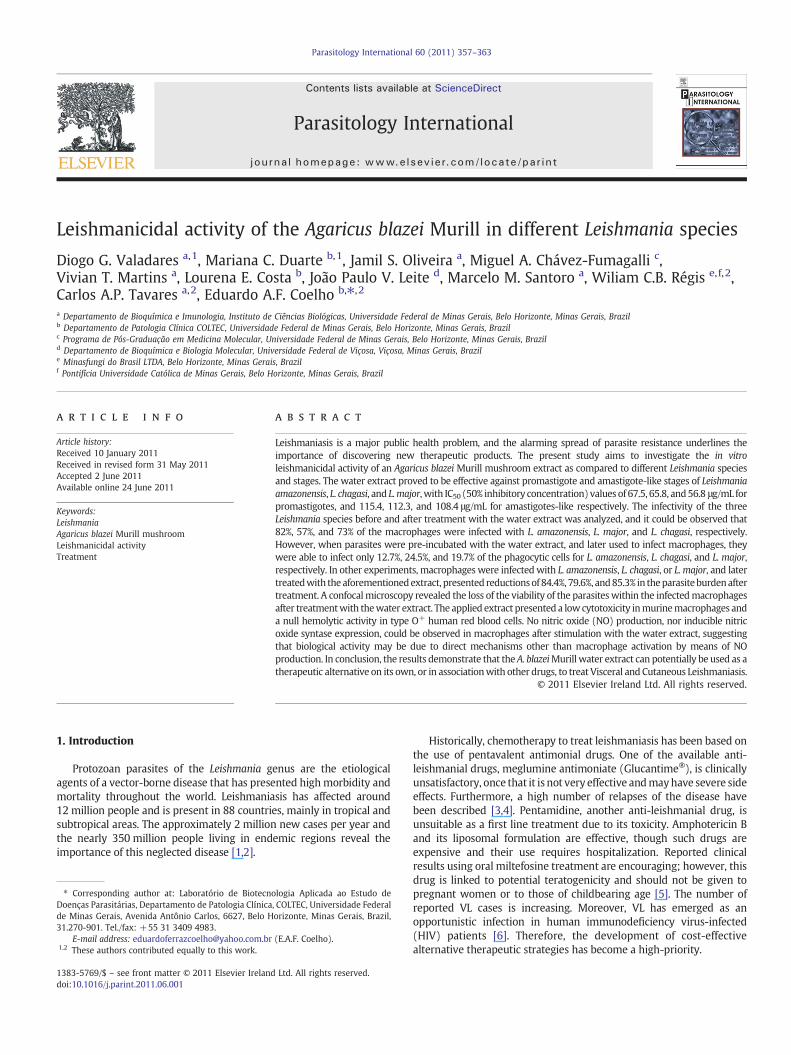
Fig. 3. Optical microscopy of the inhibition of the infection Leishmania inmurinemacrophages.1 h at 24 °C. After incubation, parasites were washed and used to infect macrophages (10:1 ratparasites within parasitophorous vacuoles in the infected macrophages (panels A, C, and E forL. chagasi, and L. major, respectively, that had been pre-incubated with the water extract remaiphagocytic cells.
pre-treatment with the water extract was able to inhibit significantlythe infection of Leishmania into the macrophages (Table 2). An opticalmicroscopy was carried out on macrophages infected with the non-treated and treated parasites, and it could be observed that the treatedparasites remained attached to the external membranes of phagocyticcells and were unable to be internalized within them, thus demonstrat-ing a direct effect of the water extract in reducing the infection into themacrophages (Fig. 3).
In this manner, an evaluation of the capacity of the water extract intreating infected macrophages was also performed. Cells were first pre-infectedwith L. amazonensis, L. chagasi, or L.majorpromastigotes and latertreatedwith50 μg/mLofwater extract for4 hat24 °C. The results showedthat macrophages that were infected and later treated presentedreductions in the parasite burden in the order of 44.1%, 30.1%, and45.2% for L. amazonensis, L. chagasi, and L. major, respectively (Table 3). Inaddition, a confocal microscopy was performed, which indicated that inthe infected cells and treated with the water extract, many intra-macrophagesparasites,whichwere labeledwith anorange color to revealthe presence of apoptotic DNA, could be observed when the threeLeishmania species were tested. It is interesting to note that infected andnon-treated macrophages presented colonized vacuoles, demonstratingthe efficacy of the treatmentwith thewater extract into the infected cells(Fig. 4).
3.3. Nitric oxide (NO) production and iNOS expression
The activation ofmacrophages byNOproduction after the treatmentwith the A. blazei Murill water extract was also investigated. The data
Promastigoteswere pre-incubatedwith the of A. blazeiMurill water extract (25 μg/mL) forio) for 4 h at 24 °C. Optical microscopy of untreated Leishmanias illustrates the presence ofL. amazonensis, L. chagasi and L. major, respectively). In panels B, D, and F, L. amazonensis,ned attached to the external membrane of the macrophages and were unable to infect the
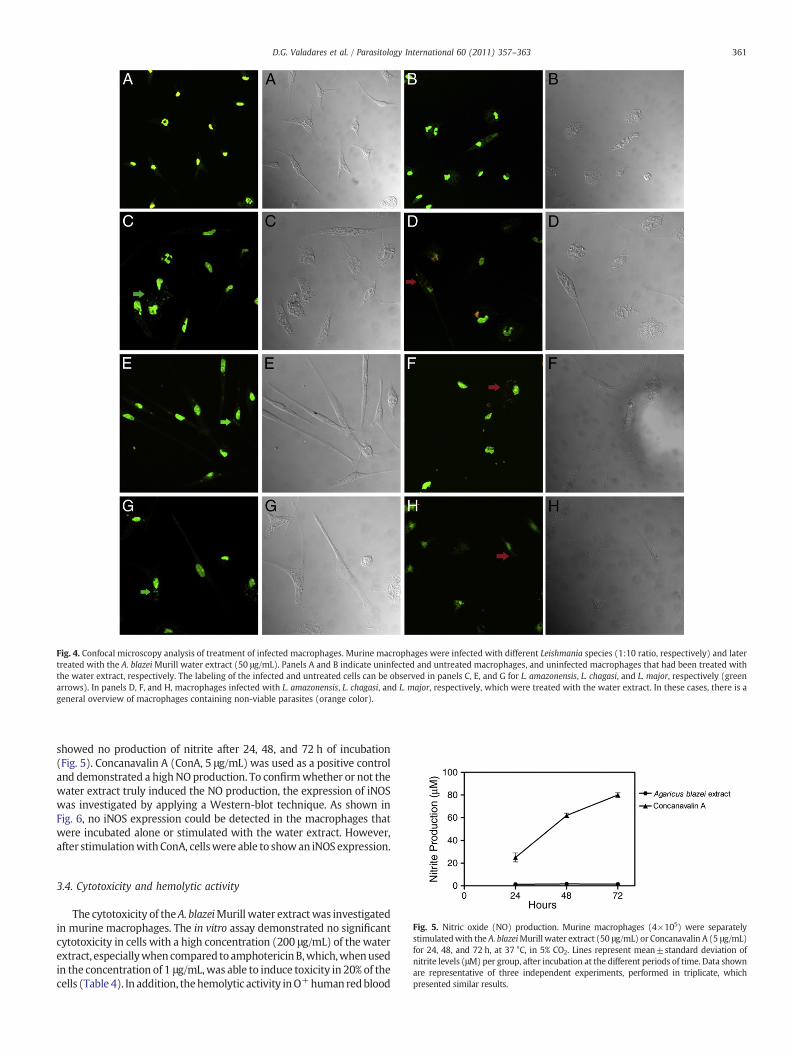
Fig. 4. Confocal microscopy analysis of treatment of infected macrophages. Murine macrophages were infected with different Leishmania species (1:10 ratio, respectively) and latertreated with the A. blazei Murill water extract (50 μg/mL). Panels A and B indicate uninfected and untreated macrophages, and uninfected macrophages that had been treated withthe water extract, respectively. The labeling of the infected and untreated cells can be observed in panels C, E, and G for L. amazonensis, L. chagasi, and L. major, respectively (greenarrows). In panels D, F, and H, macrophages infected with L. amazonensis, L. chagasi, and L. major, respectively, which were treated with the water extract. In these cases, there is ageneral overview of macrophages containing non-viable parasites (orange color).
361D.G. Valadares et al. / Parasitology International 60 (2011) 357–363
showed no production of nitrite after 24, 48, and 72 h of incubation(Fig. 5). Concanavalin A (ConA, 5 μg/mL) was used as a positive controland demonstrated a highNOproduction. To confirmwhether or not thewater extract truly induced the NO production, the expression of iNOSwas investigated by applying a Western-blot technique. As shown inFig. 6, no iNOS expression could be detected in the macrophages thatwere incubated alone or stimulated with the water extract. However,after stimulationwithConA, cellswere able to showan iNOS expression.
Fig. 5. Nitric oxide (NO) production. Murine macrophages (4×10⁵) were separatelystimulatedwith the A. blazeiMurill water extract (50 μg/mL) or Concanavalin A (5 μg/mL)for 24, 48, and 72 h, at 37 °C, in 5% CO2. Lines represent mean±standard deviation ofnitrite levels (μM) per group, after incubation at the different periods of time. Data shownare representative of three independent experiments, performed in triplicate, whichpresented similar results.
3.4. Cytotoxicity and hemolytic activity
The cytotoxicity of theA. blazeiMurill water extractwas investigatedin murine macrophages. The in vitro assay demonstrated no significantcytotoxicity in cells with a high concentration (200 μg/mL) of the waterextract, especiallywhencompared toamphotericinB,which,whenusedin the concentration of 1 μg/mL,was able to induce toxicity in 20% of thecells (Table4). In addition, thehemolytic activity inO+human redblood

Fig. 6. Inducible nitric oxide syntase (iNOS) expression. Murine macrophages wereincubated alone (medium; background control, in 1) or separately stimulated with theA. blazeiMurill water extract (50 μg/mL, in 2) or Concanavalin A (5 μg/mL, in 3) for 24 h at37 °C, in 5%CO2. Cellswere lysed, and iNOS expression (123 kDa) levelsweremeasured byWestern-blot assayusing specific antibodies.Βeta-actin expression (45 kDa)wasusedas astandard.
362 D.G. Valadares et al. / Parasitology International 60 (2011) 357–363
cells was also determined as a cytotoxic parameter, and no significantdamage to human erythrocytes could be observed after incubationwith200 μg/mL of the water extract (Table 4).
4. Discussion
Current treatment for Visceral and Cutaneous Leishmaniasis isconsidered inadequate, given that drugs present high toxicity levels,high cost, a growing parasite resistance, and long-term treatments[23]. Compounds extracted from natural products represent animportant source for research concerning new drugs to be used indeveloping novel therapeutic agents [24–30]. In this light, the purposeof this study was to evaluate the leishmanicidal activity of an A. blazeiMurill water extract against different Leishmania species, namely,L. amazonensis, L. major, and L. chagasi promastigote and amastigote-like stages.
The water extract showed a good leishmanicidal activity against thedifferent Leishmania species and in both parasite stages. It proved to beeffective in inhibiting the infection of macrophages by parasites that hadbeen pre-incubated with the water extract, as well as in reducing theparasite burden in infectedmacrophages that were later treated with thewater extract. In addition, the extract presented a low cytotoxicity inmurinemacrophages and no hemolytic activity in human red blood cells.The biological activity of the mushroom can be considered relevant, as itwas effective against Leishmania species found in different parts of theworld. In this light, L. major, an important etiological agent of Old WorldCutaneous Leishmaniasis, was the strain that proved to be mostsusceptible to the water extract, presenting IC50 values of 56.8 and108.4 μg/mL for promastigote and amastigote-like stages, respectively.Similarly, the water extract also proved to be effective against L. chagasi,presenting IC50 values of 65.8 and 112.3 μg/mL, as well as againstL. amazonensis, with IC50 values of 67.5 and 115.4 μg/mL for promastigoteand amastigote-like stages, respectively. Results from the present studyshowed that the A. blazei Murill was more potent against Leishmaniapromastigotes than against amastigotes-like since the IC50 values werehigher in inducing leishmanicidal activity in the amastigote-like stage.These differences can be, at least in part, explained by the distinctmorphological and biochemical characteristics of both stages, given thatthe amastigotes can modulate the immune response of the hosts and,consequently, be more resistant than the promastigotes forms [31].
Table 4Cytotoxicity and hemolytic activity. The results are expressed as medium±standarddeviation of the percentage of the cytotoxic and hemolytic activities of the A. blazeiMurill water extract. ND: not done.
Treatment (μg/mL) Cytotoxicity (%) Hemolysis (%)
A. blazei extract (25) 3.15±1.15 3.51±1.04A. blazei extract (50) 6.02±0.89 2.73±1.39A. blazei extract (100) 12.31±1.78 3.51±1.17A. blazei extract (200) 15.55±1.98 4.32±1.22Amphotericin B (1) 20.00±4.00 ND
Macrophages represent themainhostphagocytic cells in leishmaniasisand play a relevant role in the immunological control of intracellularparasitism through the production of oxygen derivative metabolites[32]. Through the up-regulation of NO production within the cells,macrophages can trigger an intracellular killing mechanism of theinternalized parasites [33]. Mushrooms are known for their immuneproperties that can be associated with the polysaccharide fractions. Inparticular, β-glucans have been reported to activate leukocytes and canincrease the phagocytic activity of the cells, the production of reactiveoxygen intermediates, as well as the production of inflammatorymediators, such as cytokines and chemokines [14,34,35]. In a chemicalcharacterization performed with the water extract used in this study, thepresenceof the tannins, saponins, proteins, glycoproteins, polysaccharidesand carbohydrates was observed. Therefore, the present study aimed toinvestigate the NO production and the iNOS expression by macrophagestreatedwith theA. blazeiMurillwater extract. It couldbeobserved that thewater extract did not induce NO production, nor iNOS expression,suggesting that the leishmanicidal activity observed may be due to amechanismother thanNOproduction bymacrophages. Recently, Volmanet al. (2010) reported that the Agaricus bisporus mushroom was able toinduce a strong immune-stimulating effect on bone marrow-derivedmacrophages andNOproduction, whereas the A. blazeiMurill was unableto induce theNOproduction [36]. This result is inaccordancewithfindingsfrom the present study.
Tempone et al. (2001) showed that snake venom presentedleishmanicidal activitywithout inducingNOproductionby thephagocyticcells [37]. According to this data, together with those described in thiswork, it can be speculated that other mechanisms than NO productionmay well be involved in the elimination of parasites without requiringmacrophage activation via iNOS expression. Thisfinding suggests that theproduct can transpose the membrane of the macrophages and reach theinternalized parasites through a direct effect of thewater extract onto theparasites. A confocalmicroscopy revealed that infectedmacrophages thatwere later treated with the water extract, as compared to infected andnon-treated controls, presented significant reductions in the parasiteburden. An optical microscopy showed that parasites that were pre-incubated with the water extract, as compared to the controls,encountered several difficulties in infecting macrophages. As a lowcytotoxicity in the macrophages and a poor hemolytic activity in humanred blood cells were observed, it could be infered that thewater extract issafe for mammalian cells, considering that a high concentration of thisproduct (200 μg/mL) presented low cytotoxicity in the host cells.Amphotericin B, when applied in a low doses (10 μg), proved to bequite efficient in eliminating parasites, but it was highly cytotoxic inmammalian cells, thus demonstrating some of its reported adverseeffects.
In conclusion, this study described the leishmanicidal activity of anA. blazei Murill water extract in different Leishmania species and in theboth stages. This study showed that the extract's activity was notmediated by NO production and that parasite elimination may well bedue to a direct effect into the infected macrophages. Further studiesmay lead to the characterization of molecular mechanisms and toidentification of isolated compounds of A. blazei Murill mushroom thatcan be responsible to the leishmanicidal activity observed in thisresearch. Thus, further exploration of this mushroom is recommendedhow an attempt to find suitable chemotherapeutic alternatives for thetreatment of Visceral and Cutaneous Leishmaniasis.
Acknowledgments
This study was supported by grants from Pró-Reitoria de Pesquisa(PRPq) of UFMG (Edital 07/2010), Fundação de Amparo à Pesquisa doEstado de Minas Gerais (FAPEMIG; CBB-APQ-01322-08 and CBB-APQ-02364-08), Conselho Nacional de Desenvolvimento Científico eTecnológico (CNPq; APQ-577483/2008-0), and Instituto Nacional deCiência e Tecnologia em Nanobiofarmacêutica (INCT/Nano-BIOFAR).
363D.G. Valadares et al. / Parasitology International 60 (2011) 357–363
DGV and EAFC are grant recipient of CNPq, while MACF is a grantrecipient of Coordenação de Aperfeiçoamento de Pessoal de NívelSuperior (CAPES).
References
[1] Grimaldi Jr G, Tesh RB. Leishmaniasis of the New World: current concepts andimplications for future research. Clin Microbiol Rev 1993;6:230–50.
[2] World Health Organization. Leishmaniasis: the disease and its impact. WHOcommunicable diseases surveillance and response. Geneva: WHO; 2003 http://www.who.int/emc/diseases/leish/leisdis1.html.
[3] Thakur CP, Sinha GP, Pandey AK, KumarN, Kumar P, Hassan SM, Narain S, Roy RK. Dothe diminishing efficacy and increasing toxicity of sodium stibogluconate in thetreatment of visceral leishmaniasis in Bihar, India, justify its continued use as a first-line drug? An observational study of 80 cases. Ann Trop Med Parasitol 1998;92:561–9.
[4] Sundar S, More DK, Singh MK, Singh VP, Sharma S, Makharia A, Kumar PC, MurrayHW. Failure of pentavalent antimony in visceral leishmaniasis in India: reportfrom the center of the Indian epidemic. Clin Infect Dis 2000;31:1104–7.
[5] Bhattacharya G, Herman J, Delfín D, SalemMM, Barszcz T, Mollet M, Riccio G, BrunR,Werbovetz KA. Synthesis and antitubulin activity of N1- and N4-substituted 3,5-dinitro sulfanilamides against African trypanosomes and Leishmania. J Med Chem2004;47:1823–32.
[6] Tremblay M, Olivier M, Bernier R. Leishmania and the pathogenesis of HIVinfection. Parasitol Today 1996;12:257–61.
[7] Grinde B, Hetland G, Johnson E. Effects on gene expression and viral load of amedicinal extract from Agaricus blazei Murill in patients with chronic hepatitis Cinfection. Int Immunopharmacol 2006;6:1311–4.
[8] Talcott JA, Clark JA, Lee IP. Measuring perceived effects of drinking an extract ofbasidiomycetes Agaricus blazei Murill: a survey of Japanese consumers withcancer. BMC Complement Altern Med 2007;29:7–32.
[9] Firenzuoli F, Gori L, Lombardo G. The medicinal mushroom Agaricus blazei Murill:review of literature and pharmaco-toxicological problems. Evid Based Comple-ment Altern Med 2008;5:3–15.
[10] Hsu CH, Hwang KC, Chiang YH, Chou P. The mushroom Agaricus blazei Murillextract normalizes liver function in patients with chronic hepatitis B. J AlternComplement Med 2008;14:299–301.
[11] Sorimachi K, Akimoto K, Koge T. Inhibitory effect of Agaricus blazei Murillcomponents on abnormal collagen fiber formation in human hepatocarcinomacells. Biosci Biotechnol Biochem 2008;72:621–3.
[12] Gonzaga ML, Bezerra DP, Alves AP, De Alencar NM, Mesquita RO, LimaMW, SoaresSA, Pessoa C, DeMoraes MO, Costa-Lotufo LV. In vivo growth-inhibition of sarcoma180 by an alpha-(1–N4)-glucan-beta-(1–N6)-glucan-protein complex polysac-charide obtained from Agaricus blazei Murill. Nat Med (Tokyo) 2009;63:32–40.
[13] Kim CF, Jiang JJ, Leung KN, Fung KP, Lau CB. Inhibitory effects of Agaricus blazeiMurill extracts on human myeloid leukemia cells. J Ethnopharmacol 2009;122:320–6.
[14] Wasser SP, Weis AL. Medicinal properties of substances occurring in higherbasidiomycetes mushrooms: current perspectives (review). Int J Med 1999;1:31–62.
[15] Hashimoto T, Nonaka Y, Minato K, Kawakami S, Mizuno M, Fukuda I, Kanazawa K,Ashida H. Suppressive effect of polysaccharides from the edible and medicinalmushrooms, Lentinus edodes and Agaricus blazei Murill, on the expression ofcytochrome P450s in mice. Biosci Biotechnol Biochem 2002;66:1610–4.
[16] Urben AF. Caracterização morfológica e fisiológica de acessos de Agaricus blazei eA. sylvaticus. V Congresso latino americano de Micologia, Brasília; 2005. p. 203–5.
[17] Padilha MM, Ávila AA, Sousa PJ, Cardoso LG, Perazzo FF, Carvalho JC. Anti-inflammatory activity of water and alkaline extracts from mushrooms (Agaricusblazei Murill). J Med Food 2009;12:359–64.
[18] Coelho EAF, Tavares CA, Carvalho FA, Chaves KF, Teixeira KN, Rodrigues RC,Charest H, Matlashewski G, Gazzinelli RT, Fernandes AP. Immune responsesinduced by the Leishmania (Leishmania) donovani A2 antigen, but not by the LACKantigen, are protective against experimental Leishmania (Leishmania) amazonensisinfection. Infect Immun 2003;71:3988–94.
[19] Coelho EAF. Avaliação dos níveis de proteção e da resposta imune induzida pelaimunização com os antígenos A2 e LACK na infecção experimental com Leishmania(Leishmania) major e Leishmania (Leishmania) amazonensis. Doctoral Thesis.Department of Biochemistry and Immunology. Institute of Biological Sciences (ICB),UFMG. 2004.
[20] Wagner H, Bland S, Zgainski EM. Plant drug analysis. Berlim: Springer-Verlag; 1984.320 pp.
[21] Dubois M, Gilles KA, Hamilton JK, Rebers PA, Smith F. Colorimetric method fordetermination of sugars and related substances. Anal Chem 1956;28:350–6.
[22] Löfgren SE, Miletti LC, Steindel M, Bachère E, Barracco MA. Trypanocidal andleishmanicidal activities of different antimicrobial peptides (AMPs) isolated fromaquatic animals. Exp Parasitol 2008;118:197–202.
[23] Tasdemir D, Kaiser M, Brun R, Yardley V, Schmidt TJ, Tosun F, Rüedi P. Anti-trypanosomal and leishmanicidal activities of flavonoids and their analogues: invitro, in vivo, structure–activity relationship, and quantitative structure–activityrelationship studies. Antimicrob Agents Chemother 2006;50:1352–64.
[24] Araújo CAC, Alegrio LV, Leon LL. Leishmanicidal activity of compounds extractedand characterized from Centrolobium sclerophyllum. Phytochemistry 1998;49:751–4.
[25] Akendengue B, Ngou-Milama E, Laurens A, Hocquemiller R. Recent advances in thefight against leishmaniasis with natural products. Parasite 1999;6:3–8.
[26] Delorenzi JC, Attias M, Gattass CR, Andrade M, Rezende C, Pinto AC, Henriques AT,Bou-Habib DC, Saraiva BEM. Leishmanicidal activity of indole alkaloid fromPeschiera australis. Antimicrob Agents Chemother 2001;45:1349–54.
[27] Hatimi S, Boudouma M, Bichichi M, Chaib N, Idrissi NG. In vitro evaluation ofantileishmania activity of Artemisia herba alba Asso. Bull Soc Pathol Exot 2001;94:29–31.
[28] Ferreira ICP, Lonardoni MVC, Machado GMC, Leon LL, Gobbi-Filho L, Pinto LHB,Oliveira AJB. Anti-leishmanial activity of alkaloidal extract from Aspidospermaramiflorum. Mem Inst Oswaldo Cruz 2004;99:1–3.
[29] Buckner FS, Wilson AJ. Colorimetric assay for screening compounds againstLeishmania amastigotes grown in macrophages. Am J Trop Med Hyg 2005;72:600–5.
[30] Campos FF, Rosa LH, Cota BB, Caligiorne RB, Rabello ALT, Alves TMA, Rosa CA, ZanilCL. Leishmanicidal metabolites from Cochliobolus spp., an endophytic fungusisolated from Piptadenia adiantoides (Fabaceae). PLoS Negl Trop Dis 2008;12:e348.
[31] Kima PE. The amastigote forms of Leishmania are experts at exploiting host cellprocesses to establish infection and persist. Int J Parasitol 2007;37:1087–96.
[32] Balaraman S, Tewary P, Singh VK, Madhubala R. Leishmania donovani inducesinterferon regulatory factor in murine macrophages: a host defense response.Biochem Biophys Res Commun 2004;317:639–47.
[33] Mauël J, Ransijn A. Leishmania spp.: mechanisms of toxicity of nitrogen oxidationproducts. Exp Parasitol 1997;87:98–111.
[34] Mizuno T. Bioactive biomolecules of mushrooms: food function and medicinaleffect of mushroom fungi. Food Rev Int 1995;11:7–21.
[35] Brown GD, Gordon S. Fungal beta-glucans and mammalian immunity. Immunity2003;19:311–5.
[36] Volman JJ, Mensink RP, Van Griensven LJ, Plat J. Effects of alpha-glucans fromAgaricus bisporus on ex vivo cytokine production by LPS and PHA-stimulatedPBMCs; a placebo-controlled study in slightly hypercholesterolemic subjects. Eur JClin Nutr 2010;64:720–6.
[37] Tempone AG, Andrade HF, Spencer PJ, Lourenço CO, Rogero JR, Nascimento N.Bothrops moojeni venom kills Leishmania spp. with hydrogen peroxide generatedby its L-amino acid oxidase. Biochem Biophys Res Commun 2001;280:620–4.
Related Documents