Leishmania (Viannia) panamensis: An in vitro assay using the expression of GFP for screening of antileishmanial drug Rubén Eduardo M Varela a,c , Diana Lorena Muñoz a,c , Sara M. Robledo a,d , Bala K. Kolli b , Sujoy Dutta b , Kwang Poo Chang b , and Carlos Muskus a,*,d a Programa de Estudio y Control de Enfermedades Tropicales-PECET, Universidad de Antioquia, Calle 62 No 52-59, Lab. 632, Medellín, Colombia b Department of Microbiology and Immunology, Chicago Medical School, Rosalind Franklin University, 3333 Green Bay Road, North Chicago, IL 60064, USA Abstract Promastigotes of Leishmania (Viannia) panamensis were successfully transfected with p6.5-egfp to express green fluorescent protein. The transfectants remained infective to macrophages, providing an in vitro model for screening antileishmanial drugs. This was demonstrated by flow cytometry of macrophage-associated GFP after exposure of infected cultures to known anti- leishmanial drugs, i. e. amphotericin B and glucantime®. Fluorescence of GFP diminished progressively from infected cells with increasing drug concentrations used in both cases. The availability of this fluorescent assay for infection of macrophages by L. (V.) panamensis facilitates drug discovery program for the Viannia species, which differ significantly from those of the Leishmania subgenus. Index descriptor L. (V.) panamensis; L. (L.) amazonensis; L. (L.) major; L. (L.) donovani; Viannia subgenus; Leishmania subgenus; leishmaniasis; green fluorescent protein; antileishmanial drug screening Introduction Leishmania spp. are responsible for leishmaniasis - a major parasitic disease of more than 12 million people in 88 countries. There are two million new cases annually, i. e. 1.5 million cutaneous cases and 500,000 visceral cases, respectively (WHO, 2004). Colombia is one of the major endemic places where 99% of the cases are cutaneous leishmaniasis and 70% are caused by Leishmania (V.) panamensis. Chemotherapy of this disease has relied mainly on pentavalent antimonials, but also on amphotericin B and pentamidine as the second line alternative drugs. Development of leishmanial resistance to antimonials (Rojas et al., 2006) necessitates prolonged treatments of patients with increasing dosages, resulting in severe side effects including death (Cesur et al., 2002; Oliveira et al., 2005). Recent evaluation of miltefosine yielded promising results (Soto and Soto, 2006), but the development of * Correspondence: Carlos Muskus, Programa de Estudio y Control de Enfermedades Tropicales (PECET), Universidad de, Antioquia, Medellín, Colombia. Calle 62 No 52-59, Lab 632., Phone: (574) 219-6507, Fax (574) 219-6511, [email protected]. c contributed equally d Joint senior author Publisher's Disclaimer: This is a PDF file of an unedited manuscript that has been accepted for publication. As a service to our customers we are providing this early version of the manuscript. The manuscript will undergo copyediting, typesetting, and review of the resulting proof before it is published in its final form. Please note that during the production process errors may be discovered which could affect the content, and all legal disclaimers that apply to the journal pertain. NIH Public Access Author Manuscript Exp Parasitol. Author manuscript; available in PMC 2013 May 01. Published in final edited form as: Exp Parasitol. 2009 June ; 122(2): 134–139. doi:10.1016/j.exppara.2009.02.012. NIH-PA Author Manuscript NIH-PA Author Manuscript NIH-PA Author Manuscript

Welcome message from author
This document is posted to help you gain knowledge. Please leave a comment to let me know what you think about it! Share it to your friends and learn new things together.
Transcript

Leishmania (Viannia) panamensis: An in vitro assay using theexpression of GFP for screening of antileishmanial drug
Rubén Eduardo M Varelaa,c, Diana Lorena Muñoza,c, Sara M. Robledoa,d, Bala K. Kollib,Sujoy Duttab, Kwang Poo Changb, and Carlos Muskusa,*,d
aPrograma de Estudio y Control de Enfermedades Tropicales-PECET, Universidad de Antioquia,Calle 62 No 52-59, Lab. 632, Medellín, ColombiabDepartment of Microbiology and Immunology, Chicago Medical School, Rosalind FranklinUniversity, 3333 Green Bay Road, North Chicago, IL 60064, USA
AbstractPromastigotes of Leishmania (Viannia) panamensis were successfully transfected with p6.5-egfpto express green fluorescent protein. The transfectants remained infective to macrophages,providing an in vitro model for screening antileishmanial drugs. This was demonstrated by flowcytometry of macrophage-associated GFP after exposure of infected cultures to known anti-leishmanial drugs, i. e. amphotericin B and glucantime®. Fluorescence of GFP diminishedprogressively from infected cells with increasing drug concentrations used in both cases. Theavailability of this fluorescent assay for infection of macrophages by L. (V.) panamensis facilitatesdrug discovery program for the Viannia species, which differ significantly from those of theLeishmania subgenus.
Index descriptorL. (V.) panamensis; L. (L.) amazonensis; L. (L.) major; L. (L.) donovani; Viannia subgenus;Leishmania subgenus; leishmaniasis; green fluorescent protein; antileishmanial drug screening
IntroductionLeishmania spp. are responsible for leishmaniasis - a major parasitic disease of more than 12million people in 88 countries. There are two million new cases annually, i. e. 1.5 millioncutaneous cases and 500,000 visceral cases, respectively (WHO, 2004). Colombia is one ofthe major endemic places where 99% of the cases are cutaneous leishmaniasis and 70% arecaused by Leishmania (V.) panamensis. Chemotherapy of this disease has relied mainly onpentavalent antimonials, but also on amphotericin B and pentamidine as the second linealternative drugs. Development of leishmanial resistance to antimonials (Rojas et al., 2006)necessitates prolonged treatments of patients with increasing dosages, resulting in severeside effects including death (Cesur et al., 2002; Oliveira et al., 2005). Recent evaluation ofmiltefosine yielded promising results (Soto and Soto, 2006), but the development of
*Correspondence: Carlos Muskus, Programa de Estudio y Control de Enfermedades Tropicales (PECET), Universidad de, Antioquia,Medellín, Colombia. Calle 62 No 52-59, Lab 632., Phone: (574) 219-6507, Fax (574) 219-6511, [email protected] equallydJoint senior author
Publisher's Disclaimer: This is a PDF file of an unedited manuscript that has been accepted for publication. As a service to ourcustomers we are providing this early version of the manuscript. The manuscript will undergo copyediting, typesetting, and review ofthe resulting proof before it is published in its final form. Please note that during the production process errors may be discoveredwhich could affect the content, and all legal disclaimers that apply to the journal pertain.
NIH Public AccessAuthor ManuscriptExp Parasitol. Author manuscript; available in PMC 2013 May 01.
Published in final edited form as:Exp Parasitol. 2009 June ; 122(2): 134–139. doi:10.1016/j.exppara.2009.02.012.
NIH
-PA Author Manuscript
NIH
-PA Author Manuscript
NIH
-PA Author Manuscript

resistance to this drug in laboratory studies predicts its emergence in the clinics. There isthus always an urgent need to find new antileishmanial drugs.
Candidate compounds are usually screened against the amastigote as the clinically relevantstage by quantitative microscopy and/or by other assays for cell viability, e. g. propidiumiodide permeability (Kamau et al., 2001) or radioactive biosynthetic labeling (Goto et al.,1995). Automation of such assays has been facilitated by transfection of Leishmania toexpress reporter genes encoding, for example, green fluorescent protein (GFP), luciferaseand β-galactosidase that can be used for rapid and high-throughput drug screening. GFP ofthe jellyfish, Aequorea victoria, is often the marker of choice because it is non-toxic andauto-fluorescent for easy imaging by fluorescent microscopy and ready quantification byflow cytometry or fluorimetry. GFP also compares favorably for the simplicity of its useagainst other markers, i. e. luciferase, β-lactamase and β-galactosidase. Recently, GFPexpressing transfectants have been developed from many species in the Leishmaniasubgenus, e. g. L. (L.) infantum (Kamau et al., 2001), L. (L.) donovani (Ha et al., 1996;Singh and Dube., 2004; Dube et al., 2005) and L. (L.) amazonensis (Chan et al., 2003;Okuno et al., 2003; Dutta et al., 2005). This is also true for other markers, e. g. luciferase inL. (L.) donovani (Roy et al., 2000; Gupta el al., 2005; Ashutosh et al., 2005), L. (L.)infantum (Sereno et al., 2001), L. (L.) major (Roy et al., 2000) and L. (L.) amazonensis(Lang et al., 2005), and β-lactamase and β-galactosidase in L.(L.) amazonensis (Bucknerand Wilson, 2005; Okuno et al., 2003). In contrast, similar transfectants have not been madeavailable for those in the Viannia subgenus, except two luciferase-transfected L. (V.)panamensis for drug studies (Romero et al., 2005; Henao et al., 2004).
Leishmania and Viannia are thought to have diverged 40 to 80 million years ago(McMahon-Pratt et al., 1992; McMahon-Pratt and Alexander, 2004). Leishmaniapanamensis of the Viannia subgenus differs significantly from its counterpart in theLeishmania subgenus, i. e. L. (L.) major in infectivity to laboratory animals (Goto et al.,1995; Guevara-Mendoza et al., 1997). Different species within the Viannia subgenus alsovary among themselves in pathogenicity (Colmenares et al., 2002; McMahon-Pratt andAlexander, 2004), producing a spectrum of simple cutaneous, diffused cutaneous andmucocutaneous leishmaniasis. Intra-species heterogeneity is further noted in the Vianniasubgenus (Cupolillo et al., 1997; Muskus et al., 1997; Pacheco et al., 2000; Saravia et al.,2002), including strain-specific drug sensitivity (Croft and Brun, 2003). Thus, additionalmodels are needed for drug-screening to develop Viannia-specific chemotherapy.
In this study we report the development of GFP-transfectants from promastigotes of L. (V.)panamensis. These transfectants retain their infectivity to macrophage cell lines andcontinue to express GFP intracellularly in vitro. We demonstrated the utility of this infectionmodel for potential high throughput screening of antileishmanial drugs in vitro by flowcytometry.
Materials and methodsParasites and transfection
Promastigotes of L. (V.) panamensis (MHOM/87/CO/UA140), a Colombian isolate, wascultured at 25°C in Schneider’s medium (Gibco) with 10% HIFBS in 25 cm2 tissue cultureflasks (Cellstar). For transfection, these cells grown to late-log phase were washed bycentrifugation at 3,500 g at 4°C for 10 min once with PBS and twice in chilledelectroporation buffer (25 mM HEPES, 137 mM NaCl, 5 mM KCl, 0.7 mM NaH2PO4 and 6mM glucose). The parasite suspension (0.4 ml at 50 × 106 cells/ml) was mixed with ~30 μgof p6.5-egfp (Dutta et al., 2005) in a 2 mm-gapped cuvette (BioRad) for electroporation in aGene Pulser (BioRad) at 0.45 kV and 500 μF (Chen et al., 2000). After ~24 hrs of
Varela et al. Page 2
Exp Parasitol. Author manuscript; available in PMC 2013 May 01.
NIH
-PA Author Manuscript
NIH
-PA Author Manuscript
NIH
-PA Author Manuscript
incubation for recovery in drug-free medium with 20% HIFBS, the transfectants wereselected for resistance to tunicamycin at 1.25, 2.5 and 5 μg/ml. Cells were microscopicallymonitored daily for viability and growth (Nikon Labophot-2).
Transfectants obtained were cloned by limiting dilution in 96-well tissue culture plates.Fluorescent clones emerged were picked and further selected with increasing concentrationsof tunicamycin from 5 to 25 μg/ml. Transfectants grown at increasing selective pressureswere harvest and fixed with 0.5% paraformaldehyde in PBS for flow cytometry (Beckman–coulter Epics XL) using an argon laser at 488 nm for excitation and 525 nm for emission.GFP-expressing promastigotes were analyzed flow cytometrically in 10,000 gated eventsand the numeric data were processed by using WinMDI software.
Infection of macrophages with GFP transfectants and determination of their medianinfective dose (IC50)
Monocyte-derived macrophages of the human cell line U937 were used to determine thisafter infection with GFP-transfectants (Sundstrom and Nilsson, 1976). The non-infectedmonocytes were cultured in RPMI 1640 medium (Sigma) plus 10% HIFBS at 37°C in 5%CO2 and passaged every 2 days. These monocytes were treated at 3 × 10⁵ cell/ml per wellwith 1 μM of phorbol myristate acetate (PMA) (Sigma) for 48 hr at 37°C in 24-well cultureplates (Fisher). The macrophages so obtained were infected for two hrs at 34 °C with theGFP-transfectants at different parasite-to-host ratios. The cultures were washed three timeswith pre-warmed fresh medium to remove un-ingested parasites and further incubated for 24hrs at 34°C. Cells were then removed from individual wells by vigorous aspiration with coldmedium for reaction at 4 °C for 30 min with PE conjugated anti-CD33 (Pharmingen) (5 μlper sample) specific to the monocytic/myeloid lineage (Simmons and Seed, 1988). PE-conjugated isotype control was included to estimate the non-specific binding of the primaryantibody to CD33. After washing, individual cell samples were subjected to two-color flowcytometric analysis, i. e. emission at 488 nm and excitation at 530 nm for GFP and 575 nmfor PE. Aliquots of these samples at different parasite-to-macrophage ratios weresimultaneously collected for microscopic evaluation of the percentage of infection afterfixation in methanol and staining with 10% Giemsa (Merck). Median infective dose or IC50was calculated from the data obtained by both methods using the probit method (Finney,1971). Macrophages of the J774 line were also infected with the GFP-transfectants underconditions as described (Dutta et al., 2005).
Utility of the GFP tansfectant-infected cultures for screening antileishmanial drugs byfluorescent assay
Two known antileishmanial drugs were included for comparison, i. e. meglumineantimoniate at 0.3125, 1.25, 5 and 20 μg/ml (Glucantime®, Aventis Pharma Ltd, Brasil) andamphotericin B at 0.1563, 0.625, 2.5 and 10 μg/ml (Bristol-Myers Squibb). Controls werediluents alone at the highest concentration used for each compound. The macrophagescultured in 24-well plates as described were infected for 2 hrs at 34 °C with the stationaryphase GFP transfectants at the IC50 determined, i. e. a promastigote-to-macrophage ratio of35:1. Cultures were washed to remove un-ingested promastigotes as before and then treatedfor 96 hrs at 34°C with the drugs at different concentrations as indicated. The drug effectswere analyzed by flow cytometry of cell associated GFP and by microscopy as described.The EC50 was calculated from the fluorescence intensities of CD33 and GFP positiveinfected macrophages by using a logarithmic regression with the probit method (Finney,1971).
Varela et al. Page 3
Exp Parasitol. Author manuscript; available in PMC 2013 May 01.
NIH
-PA Author Manuscript
NIH
-PA Author Manuscript
NIH
-PA Author Manuscript
ResultsExpression of GFP in transfected L. (V.) panamensis
Fluorescent transfectants emerged after electroporation of cells under the initial selectivepressures of ≤5 ug tunicamycin/ml, reaching a level of 50–60% of the total population byday 25. The proportion of GFP fluorescent cells increased significantly to 96% when cloneswere obtained and further selected for resistance to higher concentrations of tunicamycinfrom 10 to 25 μg/ml. The GFP fluorescence of individual cells appeared largely cytosolicand varied in intensity (Fig. 1A–D). Transfectants grown continuously with increasingselective pressures from 0 to 25 μg of tunicamycin/ml showed an increase in the fluorescentcells from 0 to 96% of the population (Fig. 2).
Assay for the activities of known antileishmanial drugs based on Leishmania GFPfluorescence intensities in infected macrophages
The transfectants retained their infectivity to the macrophages and therein differentiated intoamastigotes, which remained fluorescent for the duration of the experiments, as seen by bothfluorescent microscopy (Fig. 3) and flow cytometry (Fig. 4). GFP fluorescence wasassociated strictly with intracellular Leishmania in the parasitophorous vacuoles (Fig. 3A–D). Flow cytometry of the macrophages infected for 4 days showed that the percentage ofGFP+ parasites increased in the CD33+ cells proportionally with increasing promastigote-to-macrophage ratios, indicative of increasing infection, as expected (Fig. 4A). The presenceof CD33-negative cells (Fig. 4A, Lower left quadrant) suggests that a low percentage ofPMA-treated U937 cells did not react with the anti-CD33-PE used. Estimation of theinfection rate by the GFP fluorescence of the CD33+ cells is comparable to that obtained bymicroscopic enumeration of intracellular Leishmania in the transfectant-infected U937 cells,showing an increase with increasing promastigote-to-macrophage ratios from 19% to 74%by microscopy (data not shown) and from 17% to 58%, by flow citometry. The medianinfective dose of the GFP transfectants is estimated to fall within the range of their numberused at a parasite-to-macrophage ratio between 25:1 and 35:1.
Macrophages infected with the median infective dose of the GFP-transfectants weresubsequently treated with meglumine antimoniate and amphotericin B for ~4 days atincreasing drug concentrations mentioned. The EC50s’ of both antileishmanial drugs weredetermined by flow cytometry of GFP fluorescence levels in the CD33+ cells. Thefluorescence intensities of GFP in arbitrary fluorescent unit (FU) decreased in these CD33+populations gradually from 95.5 FU of the infected and untreated control to 48.17 and 41.03FU, in those exposed to the highest concentrations of antimoniate and amphotericin B,respectively (data not shown). When these FU were converted to percentage of viableintracellular parasites, the viability decreased from 100% to 52% with antimoniate and from100% to 0.8% with amphotericin B, in the infected cells exposed to the highestconcentrations of these antileishmanial drugs (Fig. 4B). Namely, the CD33+GFP+ doublefluorescent cells or infected macrophages decreased from 36.77% of the untreated controlproportionally with increasing drug concentrations to 13.31% (Fig. 4C 1–3) and 7.61% (Fig.4C 1, 4–5) for antimoniate and amphotericin B, respectively. The GFP+ CD33− populationswere present in all cases, apparently representing amastigotes, which were inadvertentlyreleased from infected cells during their removal from the culture flasks by vigorousaspiration or as result of the infection itself. The proportion of CD33+ cells was reducedslightly with increasing concentration of both drugs, indicating that they are marginally toxicto U937 cells under the experimental conditions. However, the drug concentrations usedwere always below the LC50. The EC50s’ of the antileishmanial compounds weredetermined to be 20 μg/ml and 0.078 μg/ml for meglumine antimoniate and AmphotericinB, respectively. These values are comparable, although not identical, to those previously
Varela et al. Page 4
Exp Parasitol. Author manuscript; available in PMC 2013 May 01.
NIH
-PA Author Manuscript
NIH
-PA Author Manuscript
NIH
-PA Author Manuscript
determined for wildtype cells by the conventional methods, e. g. an EC50 value of 0.132 ±0.09 μg/ml for amphotericin B (Unpublished).
DiscussionGenetic engineering of Leishmania to express fluorescent markers, such as GFP, facilitatesdrug screening automation. We produced such transfectants for L. (V.) panamensis with thep6.5-egfp constructs, which have been used previously only for species of the Leishmaniasubgenus (Chan et al., 2003; Dutta et al., 2005). Clonal variation in GFP expression wasevident among the emerging transfectants in the initial selection. Stable clones weresubsequently selected with increasing drug concentrations, giving intensive GFPfluorescence in almost 100% of the population (Figs 1 and 2). The vector p6.5 containsDNA sequences from a member of the Leishmania subgenus, L. (L.) amazonensis, includingnagt as the selective marker for tunicamycin-resistance (Liu and Chang, 1992) and theprecise deletion of a constitutively expressed gene (p36) 2.3 kb downstream of nagt as theexpression site (Liu and Chang, 1994). Previously, p6.5 has been shown to express foreigngenes with high yield in L. (L.) amazonensis (Sah et al., 2002) and other members of theLeishmania subgenus (Dutta et al, 2008). The work presented here represents first report forthe utility of this Leishmania subgenus-derived vector for transfection of a member in theViannia subgenus. Cross-subgenus functionality of the selective marker and the expressionsite in p6.5 is thus demonstrated. Whether this may contribute to the initial heterogeneity ofGFP expression seen, is unknown. Inherent clonal variations of L. (V.) panamensis in geneexpression and low selective pressures used initially are alternative possibilities, pendingfurther investigation. The egfp-transfectants of L. (V.) panamensis retain not only itsinfectivity in vitro to human monocyte-derived macrophages of the cell line U937 and J774macrophages of murine origin but also the ability to differentiate from promastigotes intoamastigotes in these macrophages. More importantly, the intracellular parasites containsufficient GFP fluorescence readily detectable by fluorescent microscopy (Fig. 3) and byflow cytometry (Figs. 4). There is a good correlation between parasite burdens andfluorescence quantitatively determined by microscopy and by flow cytometry. The lattermethod is less laborious, more accurate and higher in capacity to evaluate a large number ofcells than possible by microscopy. This may be further simplified by fluorimetry of cellsuspensions for quantitative measurement of GFP fluorescence.
Here, we also demonstrate the utility of L. (V.) panamensis transfected with GFP forevaluating their response to known antileishmanial compounds in the in vitro infectionmodel described. The GFP-expression by the transfectants allows us to evaluate theirinfection of macrophages by measuring fluorescence, as shown previously for otherintracellular pathogens, e. g. Mycobacterium tuberculosis (Collins et al., 1998), Toxoplasmagondii (Gubbels et al., 2004) and Leishmania subgenus species (Kamau et al., 2001; Singhand Dube, 2004; Dube et al., 2005). In our case, the stability of GFP fluorescence in theamastigotes of L. (V.) panamensis in infected cells makes it possible to show a dose-dependent antileishmanial activities of both glucantime® and amphotericin B. Theirdifferential antileishmanial activities in favor of the latter drug are in agreement withprevious reports (Escobar et al., 2001; Sereno et al., 2000).
In summary, data presented show that L. (V.) panamensis can be transfected to produce GFPin p6.5 expression vector, which has been shown to express foreign proteins efficiently inmembers of the Leishmania subgenus. This is the first report for such transfection of L. (V.)panamensis and its utility to evaluate antileishmanial compounds. The availability of thesetransfectants will facilitate the screening of drugs specific for the Viannia group and also thestudy of host-parasite interactions.
Varela et al. Page 5
Exp Parasitol. Author manuscript; available in PMC 2013 May 01.
NIH
-PA Author Manuscript
NIH
-PA Author Manuscript
NIH
-PA Author Manuscript
AcknowledgmentsThis work was financed by the Colombian funding agency, Colciencias (contract numbers 110 and 257, projectcodes identification 11150412982 and 11150416514, respectively) and USA NIH AI-20486 to KPC. We thankMiguel Toro for assistance with the figure preparation.
Abbreviations
GFP Green fluorescent protein
HIFBS heat-inactivated fetal bovine serum
PMA phorbol myristate acetate
PE phycoerythrin
nagt N-acetylglucosamine-1-phosphate transferase gene
ReferencesAshutosh Gupta S, Ramesh Sundar S, Goyal N. Use of Leishmania donovani field isolates expressing
the luciferase reporter gene in in vitro drug screening. Antimicrobial Agents and Chemotherapy.2005; 49:3776–3783. [PubMed: 16131481]
Buckner FS, Wilson AJ. Colorimetric assay for screening compounds against Leishmania amastigotesgrown in macrophages. American Journal of Tropical Medicine and Hygiene. 2005; 72:600–605.[PubMed: 15891135]
Chan MM, Bulinski JC, Chang KP, Fong D. A microplate assay for Leishmania amazonensispromastigotes expressing multimeric green fluorescent protein. Parasitology Research. 2003;89:266–271. [PubMed: 12632163]
Chen DQ, Kolli BK, Yadava N, Lu HG, Gilman-Sachs A, Peterson DA, Chang KP. Episomalexpression of specific sense and antisense mRNAs in Leishmania amazonensis: modulation of gp63level in promastigotes and their infection of macrophages in vitro. Infection and Immunity. 2000;68:80–86. [PubMed: 10603371]
Cesur S, Bahar K, Erekul S. Death from cumulative sodium stibogluconate toxicity on Kala-Azar.Clinical Microbiololy and Infection. 2002; 8:606.
Collins LA, Torrero MN, Franzblau SG. Green fluorescent protein reporter microplate assay for high-throughput screening of compounds against Mycobacterium tuberculosis. Antimicrobial Agents andChemotherapy. 1998; 4:344–347. [PubMed: 9527783]
Colmenares M, Kar S, Goldsmith-Pestana K, McMahon-Pratt D. Mechanisms of pathogenesis:differences amongst Leishmania species. Transactions of the Royal Society of Tropical Medicineand Hygiene. 2002; 96:S3–S7. [PubMed: 12055848]
Croft, SL.; Brun, R. In vitro and in vivo models for the identification and evaluation of drugs activeagainst Trypanosoma and Leishmania. In: Fairlamb, AH.; Ridley, R.; Vial, H., editors. Drugsagainst parasitic diseases: Rmethodologies and issues [Online]. World Health Organization;Geneva, Switzerland: 2003. p. 165-175.
Cupolillo E, Grimaldi G Jr, Momen H. Genetic diversity among Leishmania (Viannia) parasites.Annals of Tropical Medicine and Parasitology. 1997; 91:617–626. [PubMed: 9425364]
Dube A, Singh N, Sundar S, Singh N. Refractoriness to the treatment of sodium stibogluconate inIndian kala-azar field isolates persists in in vitro and in vivo experimental models. ParasitologyResearch. 2005; 96:216–223. [PubMed: 15868188]
Dutta S, Ray D, Kolli BK, Chang KP. Photodynamic sensitization of Leishmania amazonensis in bothextracellular and intracellular stages with aluminum phthalocyanine chloride for photolysis invitro. Antimicrobial Agents and Chemotherapy. 2005; 49:4474–4484. [PubMed: 16251285]
Dutta S, Furuyama K, Sassa S, Chang KP. Leishmania spp.: Delta-aminolevulinate-inducibleporphyria by genetic complementation of incomplete heme biosynthesis. ExperimentalParasitology. 2008; 118:629–636. [PubMed: 18164705]
Varela et al. Page 6
Exp Parasitol. Author manuscript; available in PMC 2013 May 01.
NIH
-PA Author Manuscript
NIH
-PA Author Manuscript
NIH
-PA Author Manuscript
Escobar P, Yardley V, Croft SL. Activities of hexadecylphosphocholine (miltefosine), AmBisome, andsodium stibogluconate (Pentostam) against Leishmania donovani in immunodeficient scid mice.Antimicrobial Agents and Chemotherapy. 2001; 45:1872–1875. [PubMed: 11353640]
Finney JD. Statisical logic in the monitoring of reactions to therapeutic drugs. Methods of Informationin Medicine. 1971; 10:237–245. [PubMed: 5124593]
Fumarola L, Spinelli R, Brandonisio O. In vitro assays for evaluation of drug activity againstLeishmania spp. Research in Microbiology. 2004; 155:224–230. [PubMed: 15142618]
Goto H, Rojas JI, Sporrong L, De Carreira P, Sanchez C, Orn A. Leishmania (Viannia) panamensis-induced cutaneous leishmaniasis in susceptible and resistant mouse strains. Revista do Instituto deMedicina Tropical de São Paulo. 1995; 37:475–481. [PubMed: 8731258]
Gubbels MJ, Striepen B. Studying the cell biology of apicomplexan parasites using fluorescentproteins. Microscopy and Microanalysis. 2004; 10:568–579. [PubMed: 15525431]
Guevara-Mendoza O, Une C, Franceschi Carreira P, Orn A. Experimental infection of Balb/c micewith Leishmania panamensis and Leishmania mexicana: induction of early IFN-gamma but notIL-4 is associated with the development of cutaneous lesions. Scandinavian Journal ofImmunology. 1997; 46:35–40. [PubMed: 9246206]
Ha DS, Schwarz JK, Turco SJ, Beverley SM. Use of the green fluorescent protein as a marker intransfected Leishmania. Molecular and Biochemical Parasitology. 1996; 77:57–64. [PubMed:8784772]
Henao HH, Osorio Y, Saravia NG, Gomez A, Travi B. Efficacy and toxicity of pentavalentantimonials (Glucantime and Pentostam) in an American cutaneous leishmaniasis animal model:luminometry application. Biomedica. 2004; 24:393–402. [PubMed: 15678803]
Kamau SW, Grimm F, Hehl AB. Expression of green fluorescent protein as a marker for effects ofantileishmanial compounds in vitro. Antimicrobial Agents and Chemotherapy. 2001; 45:3654–3656. [PubMed: 11709363]
Lang T, Goyard S, Lebastard M, Milon G. Bioluminescent Leishmania expressing luciferase for rapidand high throughput screening of drugs acting on amastigote-harbouring macrophages and forquantitative real-time monitoring of parasitism features in living mice. Cellular Microbiology.2005; 7:383–392. [PubMed: 15679841]
Liu X, Chang KP. The 63-kilobase circular amplicon of tunicamycin-resistant Leishmaniaamazonensis contains a functional N-acetylglucosamine-1-phosphate transferase gene that can beused as a dominant selectable marker in transfection. Molecular and Cellular Biology. 1992;12:4112–4122. [PubMed: 1324414]
Liu X, Chang KP. Identification by extrachromosomal amplification and overexpression of a zeta-crystallin/NADPH-oxidoreductase homologue constitutively expressed in Leishmania spp.Molecular and Biochemical Parasitology. 1994; 66:201–210. [PubMed: 7808470]
McMahon-Pratt D, Alexander J. Does the L. major paradigm of pathogenesis and protection hold forNew World cutaneous leishmaniases or the visceral disease? Immunological Review. 2004;201:206–224.
McMahon-Pratt D, Traub-Cseko Y, Lohman KL, Rogers DD, Beverly SM. Loss of the GP46/M-2surface membrane glycoprotein gene family in the Leishmania braziliensis complex. Molecularand Biochemical Parasitology. 1992; 50:151–160. [PubMed: 1542309]
Misslitz A, Mottram JC, Overath P, Aebischerm T. Targeted integration into a rRNA locus results inuniform and high level expression of transgenes in Leishmania amastigotes. Molecular andBiochemical Parasitology. 2000; 107:251–261. [PubMed: 10779601]
Muskus C, Segura I, Oddone R, Turco SJ, Leiby DA, Toro L, Robledo S, Saravia NG. Carbohydrateand LPG expression in Leishmania Viannia subgenus. Journal of Parasitology. 1997; 83:671–678.[PubMed: 9267410]
Okuno T, Goto Y, Matsumoto Y, Otsuka H, Masumoto Y. Applications of recombinant Leishmaniaamazonensis expressing egfp or the beta-galactosidase gene for drug screening andhistopathological analysis. Experimental Animal. 2003; 52:109–118.
Oliveira MC, Amorim RF, de Freitas RA, de Costa AL. A fatal case of mucocutaneous leishmaniasisafter pentavalent antimonial use. Revista da Sociedade Brasileira de Medicina Tropical. 2005;38:258–260. [PubMed: 15895180]
Varela et al. Page 7
Exp Parasitol. Author manuscript; available in PMC 2013 May 01.
NIH
-PA Author Manuscript
NIH
-PA Author Manuscript
NIH
-PA Author Manuscript
Pacheco RS, Fernandes O, Salinas G, Segura I, Momen H, Degrave W, Saravia NG, Campbell DA.Intraspecies-specific heterogeneity in the mini-exon gene localization of Leishmania (Viannia)panamensis and Leishmania (Viannia) guyanensis from Colombia. Journal of Parasitology. 2000;86:1250–1253. [PubMed: 11191900]
Rojas R, Valderrama L, Valderrama M, Varona MX, Ouellette M, Saravia NG. Resistance to antimonyand treatment failure in human Leishmania (Viannia) infection. Journal of Infectious Diseases.2006; 193:1375–1383. [PubMed: 16619185]
Romero IC, Saravia NG, Walker J. Selective action of fluoroquinolones against intracellularamastigotes of Leishmania (Viannia) panamensis in vitro. Journal of Parasitology. 2005; 91:1474–1479. [PubMed: 16539034]
Roy G, Dumas C, Sereno D, Wu Y, Singh AK, Tremblay MJ, Ouellette M, Olivier M, PapadopoulouB. Episomal and stable expression of the luciferase reporter gene for quantifying Leishmania spp.infections in macrophages and in animal models. Molecular and Biochemical Parasitology. 2000;110:195–206. [PubMed: 11071276]
Sah JF, Ito H, Kolli BK, Peterson DA, Sassa S, Chang KP. Genetic rescue of Leishmania deficiency inporphyrin biosynthesis creates mutants suitable for analysis of cellular events in uroporphyria andfor photodynamic therapy. Journal of Biological Chemistry. 2002; 277:14902–14909. [PubMed:11836252]
Saravia NG, Weigle K, Navas C, Segura I, Valderrama L, Valencia AZ, Escorcia B, McMahon-PrattD. Heterogeneity, geographic distribution, and pathogenicity of serodemes of Leishmania Vianniain Colombia. American Journal of Tropical Medicine and Hygiene. 2002; 66:738–744. [PubMed:12224583]
Sereno D, Cordeiro da Silva A, Mathieu-Daude F, Ouaissi A. Advances and perspectives inLeishmania cell based drug-screening procedures. Parasitology International. 2007; 56:3–7.[PubMed: 17079188]
Sereno D, Holzmuller P, Lemesre JL. Efficacy of second line drugs on antimonyl-resistant amastigotesof Leishmania infantum. Acta Tropica. 2000; 74:25–31. [PubMed: 10643904]
Sereno D, Roy G, Lemesre JL, Papadopoulou B, Ouellette M. DNA transformation of Leishmaniainfantum axenic amastigotes and their use in drug screening. Antimicrobial Agents andChemotherapy. 2001; 45:1168–1173. [PubMed: 11257031]
Simmons D, Seed B. Isolation of a cDNA encoding CD33, a differentiation antigen of myeloidprogenitor cells. Journal of Immunology. 1988; 141:2797–2800.
Singh N, Dube A. Short report: fluorescent Leishmania: application to anti-leishmanial drug testing.American Journal of Tropical Medicine and Hygiene. 2004; 71:400–402. [PubMed: 15516633]
Soto J, Soto P. Miltefosine: oral treatment of leishmaniasis. Expert Review of Anti-infective Therapy.2006; 4:177–185. [PubMed: 16597200]
Sundstrom C, Nilsson K. Establishment and characterization of a human histiocytic lymphoma cellline (U-937). International Journal of Cancer. 1976; 17:565–577.
World Health Organization. Tropical disease research: progress Leishmaniasis. 2003–2004. [Online.]http://www.who.int/tdr/publications/Publications/pdf/pr17/leishmaniasis.pdf
Varela et al. Page 8
Exp Parasitol. Author manuscript; available in PMC 2013 May 01.
NIH
-PA Author Manuscript
NIH
-PA Author Manuscript
NIH
-PA Author Manuscript
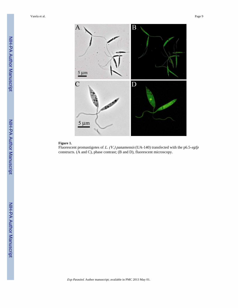
Figure 1.Fluorescent promastigotes of L. (V.) panamensis (UA-140) transfected with the p6.5-egfpconstructs. (A and C), phase contrast; (B and D), fluorescent microscopy.
Varela et al. Page 9
Exp Parasitol. Author manuscript; available in PMC 2013 May 01.
NIH
-PA Author Manuscript
NIH
-PA Author Manuscript
NIH
-PA Author Manuscript
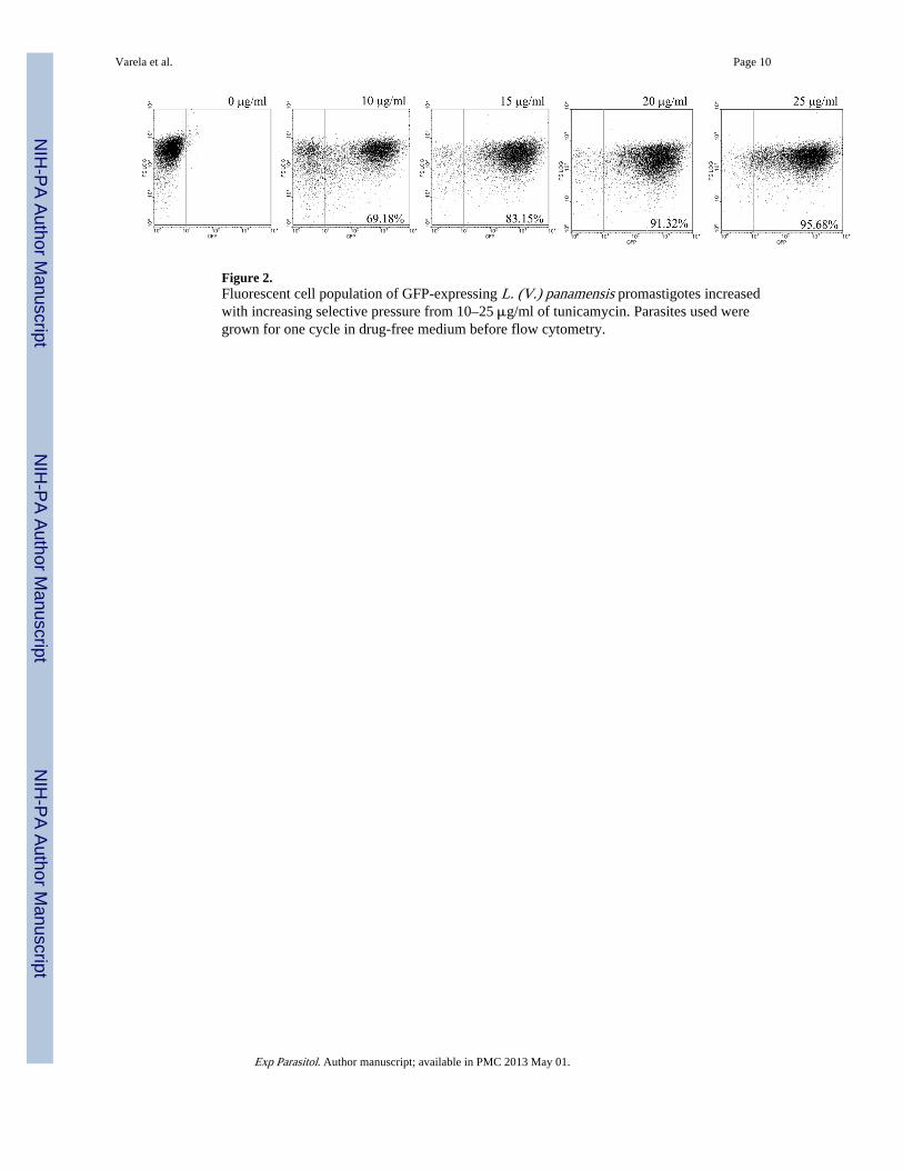
Figure 2.Fluorescent cell population of GFP-expressing L. (V.) panamensis promastigotes increasedwith increasing selective pressure from 10–25 μg/ml of tunicamycin. Parasites used weregrown for one cycle in drug-free medium before flow cytometry.
Varela et al. Page 10
Exp Parasitol. Author manuscript; available in PMC 2013 May 01.
NIH
-PA Author Manuscript
NIH
-PA Author Manuscript
NIH
-PA Author Manuscript
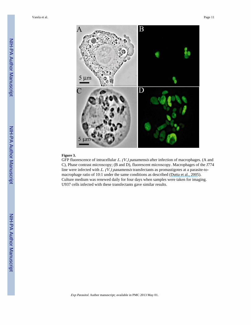
Figure 3.GFP fluorescence of intracellular L. (V.) panamensis after infection of macrophages. (A andC), Phase contrast microscopy; (B and D), fluorescent microscopy. Macrophages of the J774line were infected with L. (V.) panamensis transfectants as promastigotes at a parasite-to-macrophage ratio of 10:1 under the same conditions as described (Dutta et al., 2005).Culture medium was renewed daily for four days when samples were taken for imaging.U937 cells infected with these transfectants gave similar results.
Varela et al. Page 11
Exp Parasitol. Author manuscript; available in PMC 2013 May 01.
NIH
-PA Author Manuscript
NIH
-PA Author Manuscript
NIH
-PA Author Manuscript
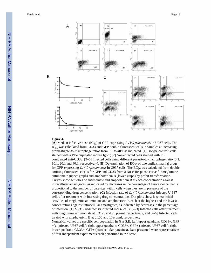
Figure 4.(A) Median infective dose (IC50) of GFP-expressing L.(V.) panamensis in U937 cells. TheIC50 was calculated from CD33 and GFP double-fluorescent cells in samples at increasingpromastigote-to-macrophage ratios from 0:1 to 40:1 as indicated. [1] Isotype control: cellsstained with a PE-conjugated mouse IgG1; [2] Non-infected cells stained with PEconjugated anti-CD33; [3–6] Infected cells using different parasite-to-macrophage ratio (5:1,10:1, 20:1 and 40:1, respectively). (B) Determination of EC50 of two antileishmanial drugsfor GFP-expressing L. (V.) panamensis in U937 cells. The EC50 was calculated from doubleemitting fluorescence cells for GFP and CD33 from a Dose-Response curve for meglumineantimoniate (upper graph) and amphotericin B (lower graph) by probit transformation.Curves show activities of antimoniate and amphotericin B at each concentration againstintracellular amastigotes, as indicated by decreases in the percentage of fluorescence that isproportional to the number of parasites within cells when they are in presence of thecorresponding drug concentration. (C) Infection rate of L. (V.) panamensis infected U-937cells after treatment with increasing drug concentrations. Dot plots show leishmanicidalactivities of meglumine antimoniate and amphotericin B each at the highest and the lowestconcentrations against intracellular amastigotes, as indicated by decreases in the percentageof infection. [1] L. (V.) panamensis infected U-937 cells; [2–3] Infected cells after treatmentwith meglumine antimoniate at 0.3125 and 20 μg/ml, respectively, and [4–5] Infected cellstreated with amphotericin B at 0.156 and 10 μg/ml, respectively.Numerical values are specific cell population in % ± S.E. Left upper quadrant: CD33+, GFP−(uninfected U937 cells); right upper quadrant: CD33+, GFP+ (infected U937 cells); rightlower quadrant: CD33−, GFP+ (extracellular parasites). Data presented were representativesof four independent experiments each performed in triplicate.
Varela et al. Page 12
Exp Parasitol. Author manuscript; available in PMC 2013 May 01.
NIH
-PA Author Manuscript
NIH
-PA Author Manuscript
NIH
-PA Author Manuscript
Related Documents