Leishmania major parasites and their interaction with human macrophages Dissertation

Welcome message from author
This document is posted to help you gain knowledge. Please leave a comment to let me know what you think about it! Share it to your friends and learn new things together.
Transcript
Leishmania major parasitesand their interaction with human macrophages
Dissertation
Zur Erlangung des Grades
Doktor der Naturwissenschaften
Am Fachbereich Biologie
der Johannes Gutenberg-Universität Mainz
Elena Bank
geb. am 07.06.1983 in Dshiginka
Mainz, 2012


Dissertation von Elena Bank
Leishmania major parasitesand their interaction with human macrophages
Dekan:
1. Gutachter:
2. Gutachter:


Contents
1 Introduction 71.1 Leishmaniasis . . . . . . . . . . . . . . . . . . . . . . . . . . . . . . . . . 7
1.2 Life cycle of L. major parasites . . . . . . . . . . . . . . . . . . . . . . . 8
1.3 Stage-speci�c characteristics of L. major . . . . . . . . . . . . . . . . . . 9
1.4 Apoptosis in L. major . . . . . . . . . . . . . . . . . . . . . . . . . . . . 11
1.4.1 Drug induced apoptosis . . . . . . . . . . . . . . . . . . . . . . . 12
1.5 Leishmania and the adaptive immune system . . . . . . . . . . . . . . . 14
1.6 The interaction with human MF . . . . . . . . . . . . . . . . . . . . . . . 14
1.7 Defense of Leishmaniasis . . . . . . . . . . . . . . . . . . . . . . . . . . . 16
1.8 Aim of the study . . . . . . . . . . . . . . . . . . . . . . . . . . . . . . . 19
2 Material and Methods 232.1 Material . . . . . . . . . . . . . . . . . . . . . . . . . . . . . . . . . . . . 23
2.1.1 Chemicals . . . . . . . . . . . . . . . . . . . . . . . . . . . . . . . 23
2.1.2 Culture media and bu�ers . . . . . . . . . . . . . . . . . . . . . . 26
2.1.3 Westernblot bu�ers and solutions . . . . . . . . . . . . . . . . . . 28
2.1.4 Leishmania strains . . . . . . . . . . . . . . . . . . . . . . . . . . 31
2.1.5 Human leukocytes . . . . . . . . . . . . . . . . . . . . . . . . . . 32
2.1.6 Ready-to-use kits . . . . . . . . . . . . . . . . . . . . . . . . . . . 32
2.1.7 Anti-Human antibodies . . . . . . . . . . . . . . . . . . . . . . . . 32
2.1.8 Anti-Leishmania antibodies . . . . . . . . . . . . . . . . . . . . . 33
2.1.9 Oligonucleotides . . . . . . . . . . . . . . . . . . . . . . . . . . . . 34
2.1.10 Enzymes . . . . . . . . . . . . . . . . . . . . . . . . . . . . . . . . 35
2.1.11 Laboratory supplies . . . . . . . . . . . . . . . . . . . . . . . . . . 35
2.1.12 Instruments . . . . . . . . . . . . . . . . . . . . . . . . . . . . . . 36
2.1.13 Software . . . . . . . . . . . . . . . . . . . . . . . . . . . . . . . . 38
3
Contents
2.2 Methods . . . . . . . . . . . . . . . . . . . . . . . . . . . . . . . . . . . . 39
2.2.1 Cell culture . . . . . . . . . . . . . . . . . . . . . . . . . . . . . . 39
2.2.1.1 Cultivation of L. major promastigotes . . . . . . . . . . 39
2.2.1.2 Isolation of metacyclic L. major promastigotes . . . . . 39
2.2.1.3 Generation and cultivation of L. major amastigotes in
vitro . . . . . . . . . . . . . . . . . . . . . . . . . . . . . 40
2.2.1.4 Isolation of L. major amastigotes from infected MF . . . 41
2.2.1.5 Isolation of human peripheral blood mononuclear cells
(PBMC) . . . . . . . . . . . . . . . . . . . . . . . . . . . 41
2.2.1.6 Generation of blood derived MF . . . . . . . . . . . . . 42
2.2.1.7 Co-incubation of macrophages with L. major parasites . 42
2.2.1.8 End-point titration . . . . . . . . . . . . . . . . . . . . . 43
2.2.1.9 Cytocentrifuging cells . . . . . . . . . . . . . . . . . . . 44
2.2.1.10 Di� QUIK staining . . . . . . . . . . . . . . . . . . . . . 44
2.2.2 FACS analysis . . . . . . . . . . . . . . . . . . . . . . . . . . . . . 44
2.2.2.1 Extracellular FACS analysis of (infected) MF . . . . . . 44
2.2.2.2 Intracellular FACS analysis of (infected) MF . . . . . . . 45
2.2.2.3 Extracellular FACS analysis of L. major . . . . . . . . . 45
2.2.2.4 Intracellular FACS analysis of L. major . . . . . . . . . 45
2.2.3 ELISA analysis . . . . . . . . . . . . . . . . . . . . . . . . . . . . 46
2.2.3.1 TNF alpha . . . . . . . . . . . . . . . . . . . . . . . . . 46
2.2.3.2 IL-12 . . . . . . . . . . . . . . . . . . . . . . . . . . . . 46
2.2.3.3 IL-10 . . . . . . . . . . . . . . . . . . . . . . . . . . . . 47
2.2.4 Arginase assay . . . . . . . . . . . . . . . . . . . . . . . . . . . . 47
2.2.5 Molecular biology methods . . . . . . . . . . . . . . . . . . . . . . 48
2.2.5.1 Transfection of primary cells with siRNA . . . . . . . . . 48
2.2.5.2 DNA isolation . . . . . . . . . . . . . . . . . . . . . . . 48
2.2.5.3 Ampli�cation of the ribosomal internal transcribed spacer
1 (ITS1) . . . . . . . . . . . . . . . . . . . . . . . . . . . 49
2.2.5.4 Restriction digest . . . . . . . . . . . . . . . . . . . . . . 50
2.2.5.5 RNA isolation . . . . . . . . . . . . . . . . . . . . . . . . 50
2.2.5.6 Test-PCR . . . . . . . . . . . . . . . . . . . . . . . . . . 51
2.2.5.7 cDNA synthesis . . . . . . . . . . . . . . . . . . . . . . . 52
4
Contents
2.2.5.8 Quantitative real-time PCR . . . . . . . . . . . . . . . . 53
2.2.6 Transmission electron microscopy (EM) . . . . . . . . . . . . . . . 55
2.2.7 Westernblot analysis . . . . . . . . . . . . . . . . . . . . . . . . . 55
2.2.7.1 Sample preparation . . . . . . . . . . . . . . . . . . . . . 55
2.2.7.2 SDS-PAGE . . . . . . . . . . . . . . . . . . . . . . . . . 55
2.2.7.3 Band detection . . . . . . . . . . . . . . . . . . . . . . . 56
2.2.7.4 Coomassie staining . . . . . . . . . . . . . . . . . . . . . 56
2.2.8 Statistical analysis . . . . . . . . . . . . . . . . . . . . . . . . . . 56
3 Results 573.1 Con�rmation of the L. major species . . . . . . . . . . . . . . . . . . . . 57
3.2 Characterisation of L. major FEBNI parasites . . . . . . . . . . . . . . . 57
3.2.1 Morphological characteristics of L. major promastigotes . . . . . 58
3.2.2 Morphological characteristics of L. major amastigotes . . . . . . . 59
3.2.3 Annexin binding . . . . . . . . . . . . . . . . . . . . . . . . . . . 61
3.2.4 Stage-speci�c gene expression levels . . . . . . . . . . . . . . . . . 61
3.2.5 The surface marker lipophosphoglycan (LPG) . . . . . . . . . . . 63
3.2.6 Generation of axenic amastigotes in other L. major strains . . . . 64
3.3 Detection of Apoptotic characteristics in L. major parasites . . . . . . . 68
3.3.1 Apoptosis mechanisms in promastigotes . . . . . . . . . . . . . . 68
3.3.2 Apoptosis mechanisms in amastigotes . . . . . . . . . . . . . . . . 70
3.3.3 FACS analysis of apoptotic parasites . . . . . . . . . . . . . . . . 72
3.4 Interaction of L. major with human MF . . . . . . . . . . . . . . . . . . 75
3.4.1 Infection of di�erent phenotypes of MF with L. major . . . . . . 75
3.4.2 Infection with eGFP expressing L. major . . . . . . . . . . . . . . 78
3.4.2.1 L. major eGFP parasites . . . . . . . . . . . . . . . . . 78
3.4.2.2 Parasite development in infected MF . . . . . . . . . . . 78
3.5 Phenotype and parasites stage-speci�c MF surface marker expression . . 81
3.5.1 CD163 . . . . . . . . . . . . . . . . . . . . . . . . . . . . . . . . . 81
3.5.2 CD206 . . . . . . . . . . . . . . . . . . . . . . . . . . . . . . . . . 82
3.5.3 MHC class II (MHC II) . . . . . . . . . . . . . . . . . . . . . . . 85
3.5.4 CD86 . . . . . . . . . . . . . . . . . . . . . . . . . . . . . . . . . 86
3.6 Phenotype and parasites stage-speci�c cytokine production . . . . . . . . 87
3.6.1 TNF alpha . . . . . . . . . . . . . . . . . . . . . . . . . . . . . . 87
5
Contents
3.6.2 IL-12 . . . . . . . . . . . . . . . . . . . . . . . . . . . . . . . . . . 89
3.6.3 CCL3 and CCL4 . . . . . . . . . . . . . . . . . . . . . . . . . . . 89
3.6.4 IL-10 . . . . . . . . . . . . . . . . . . . . . . . . . . . . . . . . . . 92
3.7 Phosphorylation of MAP kinases (MAPK) after L. major infection . . . 93
3.7.1 p38 MAP kinases . . . . . . . . . . . . . . . . . . . . . . . . . . . 95
3.7.2 ERK1/2 MAP kinases . . . . . . . . . . . . . . . . . . . . . . . . 95
3.8 L. major parasite escape from phagolysosomes . . . . . . . . . . . . . . . 98
3.9 Arginase in infected MF . . . . . . . . . . . . . . . . . . . . . . . . . . . 100
3.10 Cathelicidin (LL-37) in infected MF . . . . . . . . . . . . . . . . . . . . . 100
3.10.1 Di�erent LL-37 expression in MF I and MF II . . . . . . . . . . . 102
3.10.2 Killing e�ect of rhLL-37 on L. major promastigotes . . . . . . . . 102
3.10.3 No e�ect of rhLL-37 on L. major amastigotes . . . . . . . . . . . 104
3.10.4 Knockdown of LL-37 in human MF . . . . . . . . . . . . . . . . . 106
3.10.4.1 Infection and parasite burden . . . . . . . . . . . . . . . 106
3.10.4.2 Survival of L. major parasites in knockdown MF . . . . 108
4 Discussion 1114.1 Di�erent life stages of the parasite L. major . . . . . . . . . . . . . . . . 111
4.2 In vitro culture method for axenic L. major amastigotes . . . . . . . . . 113
4.3 Apoptosis in L. major parasites . . . . . . . . . . . . . . . . . . . . . . . 114
4.4 Interaction of L. major with human MF . . . . . . . . . . . . . . . . . . 120
4.5 Clearance of L. major in human MF . . . . . . . . . . . . . . . . . . . . 123
4.6 Concluding remarks . . . . . . . . . . . . . . . . . . . . . . . . . . . . . . 127
5 Summary 129
6 Zusammenfassung 131
Abbreviations 163
Acknowledgements 167
6

1 Introduction
1.1 Leishmaniasis
Leishmaniasis is a parasitic infection with the protozoan genus Leishmania which is
endemic in 88 countries, mainly prevalent in the tropical and subtropical regions of
the world. Currently approximately 12 million people are su�ering from Leishmaniasis,
however about 350 million people are worldwide threatened and the estimated incidence
of 2 million new cases arises each year [215, 4]. Up to date there are about 21 Leishmania
species known to be pathogenic for humans [91]. According to the Leishmania species
initiating infection and the immunologic status, humans can develop a large spectrum of
symptoms ranging from self-healing lesions to a severe organ-in�ltrating manifestation of
the disease. Four major forms of human Leishmaniasis have been described: cutaneous,
di�use cutaneous, mucocutaneous and visceral Leishmaniasis. The localized cutaneous
Leishmaniasis (LCL), which is primarily caused by Leishmania major (L. major) and
Leishmania tropica (L. tropica) produces self-healing skin ulcers on exposed parts of
the body, which is also known as �Aleppo boil�. On the other hand, chronic di�use
cutaneous Leishmaniasis (DCL) caused by Leishmania aethiopica (L. aethiopica) and
Leishmania mexicana amazonensis (L. mexicana amazonensis) produces widespread
skin lesions all over the body which resemble leprosy [143]. Another more severe form
is the mucocutaneous Leishmaniasis (MCL) characterized by the in�ltration of the mu-
cousal membranes, especially those of the nose and mouth leading to extensive tissue
damage and dis�guration (Espundia). The causative agents of MCL are Leishmania
braziliensis (L. braziliensis) and Leishmania mexicana pifanoi (L. mexicana pifanoi)
[102]. The most severe and life threatening form is the visceral Leishmaniasis (VL) also
named �Kala azar� caused by the Leishmania donovani complex, including Leishmania
7
1 Introduction
donovani, Leishmania infantum and Leishmania chagasi (L. donovani, L. infantum and
L. chagasi). This form a�ects internal organs such as the lymph nodes, the liver, the
spleen and the bone marrow and is lethal if untreated [17].
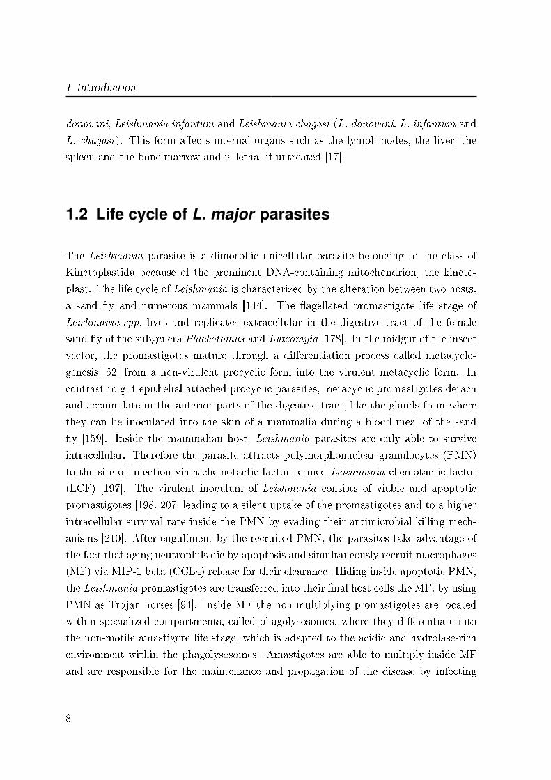
1.2 Life cycle of L. major parasites
The Leishmania parasite is a dimorphic unicellular parasite belonging to the class of
Kinetoplastida because of the prominent DNA-containing mitochondrion, the kineto-
plast. The life cycle of Leishmania is characterized by the alteration between two hosts,
a sand �y and numerous mammals [144]. The �agellated promastigote life stage of
Leishmania spp. lives and replicates extracellular in the digestive tract of the female
sand �y of the subgenera Phlebotomus and Lutzomyia [178]. In the midgut of the insect
vector, the promastigotes mature through a di�erentiation process called metacyclo-
genesis [62] from a non-virulent procyclic form into the virulent metacyclic form. In
contrast to gut epithelial attached procyclic parasites, metacyclic promastigotes detach
and accumulate in the anterior parts of the digestive tract, like the glands from where
they can be inoculated into the skin of a mammalia during a blood meal of the sand
�y [159]. Inside the mammalian host, Leishmania parasites are only able to survive
intracellular. Therefore the parasite attracts polymorphonuclear granulocytes (PMN)
to the site of infection via a chemotactic factor termed Leishmania chemotactic factor
(LCF) [197]. The virulent inoculum of Leishmania consists of viable and apoptotic
promastigotes [198, 207] leading to a silent uptake of the promastigotes and to a higher
intracellular survival rate inside the PMN by evading their antimicrobial killing mech-
anisms [210]. After engulfment by the recruited PMN, the parasites take advantage of
the fact that aging neutrophils die by apoptosis and simultaneously recruit macrophages
(MF) via MIP-1 beta (CCL4) release for their clearance. Hiding inside apoptotic PMN,
the Leishmania promastigotes are transferred into their �nal host cells the MF, by using
PMN as Trojan horses [94]. Inside MF the non-multiplying promastigotes are located
within specialized compartments, called phagolysosomes, where they di�erentiate into
the non-motile amastigote life stage, which is adapted to the acidic and hydrolase-rich
environment within the phagolysosomes. Amastigotes are able to multiply inside MF
and are responsible for the maintenance and propagation of the disease by infecting
8
1.3 Stage-speci�c characteristics of L. major
surrounding phagocytes causing Leishmaniasis [22]. The life cycle is completed when
a sand �y take a blood meal from an infected mammal. The free or in MF resident
amastigotes are then able to rapidly di�erentiate into the promastigote life stage in the
gut of the insect. The adaptation to both an arthropod vector and a mammalian host
is a typical feature of the obligatory intracellular Leishmania parasite and is illustrated
in �g. 1.
Figure 1: Life cycle of Leishmania spp.
[Source: http://en.wikipedia.org/wiki/File:Leishmaniasis_life_cycle_diagram_en.
svg, accessed on 04/05/2012]
1.3 Stage-specific characteristics of L. major
Due to the alteration between two completely di�erent hosts, Leishmania parasites had
to evolve di�erent strategies to survive in both environmental conditions. The two life
stages of Leishmania display distinct morphologic and metabolic characteristics con-
sistent with a stage-speci�c expression of parasitic genes and proteins of which some
9

1 Introduction
are shown to be important for the survival and di�erentiation in the insect vector and
a successful infection in the mammalian host. The lipophosphoglycan (LPG), which
forms a dense glycocalyx around the parasitic body, is demonstrated to play an im-
portant role in the attachment of promastigotes in the digestive tract of the sand �y
[147]. During metacyclogenesis the LPG molecules are modi�ed by capping the galac-
tosyl side chains of procyclic LPG with arabinosyl residues [164, 165]. Thus, metacyclic
promastigotes lose their binding capacity to the midgut epithelium and are released for
transmission [163, 113]. Inside the mammalian host LPG also inhibits the complement-
mediated lysis of the parasites by blocking the insertion of the lytic C5b-9 complex
into the promastigote membrane [149] and is involved in the mechanism to inhibit
the protein kinase C (PKC) resulting in a delay of endosomal compartment fusion
[134, 122, 185]. After the transformation of Leishmania parasites in the amastigote life
stage they show a strongly reduced LPG expression compared to the infective metacyclic
promastigotes, which correlates with the absence of a glycocalyx on the amastigote sur-
face [112, 63, 194, 121, 11, 171, 146]. The major surface glycoprotein GP63 (GP63) is a
metalloprotease on the surface of Leishmania promastigotes, which is known to cleave
the C3b form of the complement system into the inactive C3bi form and thus block
complement-mediated lysis inside the mammalian host [27, 39]. In addition, GP63 is
able to directly interact with the macrophage surface receptor complement receptor
type 3 (CR3, or CD11b/CD18). The opsonized parasites with C3bi are targeting for
phagocytosis by MF via the corresponding CR3 [214, 183]. In the amastigote life stage
of L. major the metalloprotease GP63 is found not to be expressed [170]. A well charac-
terized marker for the Leishmania promastigote life stage is the gene expression of the
small hydrophilic ER-associated protein (SHERP). SHERP is essential for the parasite
development during the metacyclogenesis in the sand �y and may therefore be essential
for transmission [86, 175]. Another stage-speci�c marker for Leishmania parasites is the
structural protein gene alpha-tubulin which is up-regulated in the promastigote life stage
[98]. The ABC-transporter homologue (ABC) belongs to a gene family coding for pro-
teins involved in the ATP-dependent transport of a variety of molecules across biological
membranes, including amino acids, sugars, peptides, lipids, ions, and chemotherapeu-
tic drugs [68]. ABC is demonstrated to be speci�c up-regulated in the amastigote life
stage of L. major [98]. Furthermore, quinonoid-dihydropteridine reductase (QDPR) is
the key enzyme in Leishmania for the regeneration of H4biopterin which is essential
10

1.4 Apoptosis in L. major
for parasite growth and di�erentiation [40, 128]. QDPR is required for the reduction
of quinonoid-dihydropteridine to restore the intracellular H4biopterin pool [105]. The
expression of QDPR in stat-phase promastigotes is reduced compared to both log-phase
promastigotes and amastigotes [105]. An additional stage-speci�c marker is the cysteine
protease b (Cbp) which is a multicopy gene family belonging to the cysteine proteases
and is required for parasite replication and virulence [37, 114, 123]. Cpb is shown to
be up-regulated in the amastigote life stage for several Leishmania species, including
L. tropica and L. mexicana [129].
1.4 Apoptosis in L. major
Cell death is historically classi�ed into regulated or programmed cell death (PCD), also
termed apoptosis and the unregulated cell death often called necrosis. Apoptosis is
a mechanism which is absolutely essential to remain homeostasis in multicellular or-
ganisms [188]. Apoptosis is a well-organized multi-step process with distinct events to
occur. An early marker for apoptosis is the externalisation of phosphatidylserine (PS)
from the inner to the outer lea�et of the cell membrane. The asymmetric distribution
of phospholipids of the plasma membrane gets lost and PS is translocated to the outer
lea�et of the plasma membrane via a �ip-�op mechanism [110]. Simultaneously prote-
olytic enzymes termed caspases are sequentially activated which lead to the subsequent
breakdown of the cell content. The mitochondria act as key players during this pro-
cess releasing pro-apoptotic factors like cytochrome c into the cytoplasm accompanied
by disruption of the mitochondrial membrane potential leading to caspase activation
[219, 53]. Additionally, reactive oxygen species (ROS) which are highly reactive inter-
mediates in the reduction of oxygen to water are generated and released [173]. As a
result the cell rounds up followed by cell shrinkage while the integrity of the plasma
membrane remains fully intact throughout the entire apoptotic process [82, 85, 127]. In
the end the nucleus condensates and the DNA is degraded by fragmentation [216]. Since
in the past apoptosis was claimed only for multicellular organisms, many researchers
doubt the existence of a programmed cell death in unicellular life forms. But during
the last years, there have been reports for di�erent phyla of protists demonstrating
several markers of apoptosis [44, 199, 95]. Many characteristics of metazoan apopto-
11
1 Introduction
sis have been described to occur in Leishmania parasites when apoptotic death was
induced by diverse stimuli. The externalization of PS was demonstrated for several
di�erent species, including L. major. Furthermore, the loss of mitochondrial membrane
potential as well as the formation of ROS and the release of cytochrome c was observed
after apoptosis induction. Although no proteolytic caspases were found in Leishmania,
several caspase-like proteases were shown to be present [96, 43, 131]. Moreover, the
maintenance of the plasma membrane integrity, cell shrinkage and nuclear chromatin
condensation and fragmentation of the DNA were observed [9, 42, 176, 130, 117, 5].
These �ndings demonstrate the existence of all characteristics of an apoptosis program
in Leishmania. However the exact mechanisms responsible for apoptosis regulation are
still not known.
1.4.1 Drug induced apoptosis
The above mentioned apoptotic characteristics were also found after the treatment of
Leishmania parasites with di�erent drugs to induce apoptosis, including staurosporine,
camptothecin and the anti-leishmanial drug miltefosine.
Staurosporine:
Staurosporine was originally isolated in 1977 from the bacterium Streptomyces stau-
rosporeus and was found to strongly induce apoptosis. The main biological activity
of this compound in mammalian cells is the inhibition of protein kinases through the
prevention of ATP binding to the kinase. This is achieved through the stronger a�nity
of staurosporine to the ATP-binding site on the kinases, though with little selectivity
[80]. The underlying mechanism of apoptosis induction via staurosporine is reported to
be the mitochondrial apoptotic pathway [190, 191]. For Leishmania spp. staurosporine
was demonstrated to induce cell death with several cytoplasmic and nuclear characteris-
tics of apoptosis, including cell shrinkage, PS externalisation, cytochrome c release and
DNA fragmentation [13, 9].
Camptothecin:
Camptothecin is a cytotoxic alkaloid isolated from the bark and stem of Camptotheca
acuminata (Happy tree) in 1966. It is a potent inhibitor of both DNA as well as
12
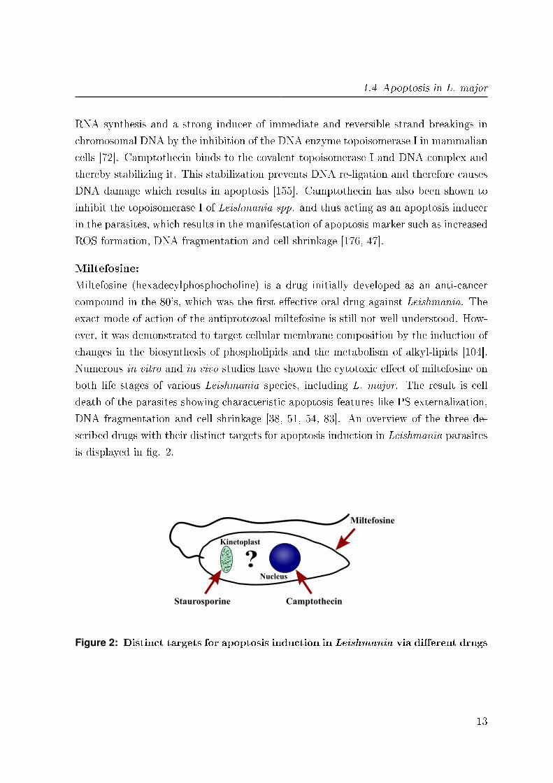
1.4 Apoptosis in L. major
RNA synthesis and a strong inducer of immediate and reversible strand breakings in
chromosomal DNA by the inhibition of the DNA enzyme topoisomerase I in mammalian
cells [72]. Camptothecin binds to the covalent topoisomerase I and DNA complex and
thereby stabilizing it. This stabilization prevents DNA re-ligation and therefore causes
DNA damage which results in apoptosis [155]. Camptothecin has also been shown to
inhibit the topoisomerase I of Leishmania spp. and thus acting as an apoptosis inducer
in the parasites, which results in the manifestation of apoptosis marker such as increased
ROS formation, DNA fragmentation and cell shrinkage [176, 47].
Miltefosine:
Miltefosine (hexadecylphosphocholine) is a drug initially developed as an anti-cancer
compound in the 80's, which was the �rst e�ective oral drug against Leishmania. The
exact mode of action of the antiprotozoal miltefosine is still not well understood. How-
ever, it was demonstrated to target cellular membrane composition by the induction of
changes in the biosynthesis of phospholipids and the metabolism of alkyl-lipids [104].
Numerous in vitro and in vivo studies have shown the cytotoxic e�ect of miltefosine on
both life stages of various Leishmania species, including L. major. The result is cell
death of the parasites showing characteristic apoptosis features like PS externalization,
DNA fragmentation and cell shrinkage [38, 51, 54, 83]. An overview of the three de-
scribed drugs with their distinct targets for apoptosis induction in Leishmania parasites
is displayed in �g. 2.
Staurosporine
Miltefosine
?Nucleus
Kinetoplast
Camptothecin
Figure 2: Distinct targets for apoptosis induction in Leishmania via di�erent drugs
13
1 Introduction
1.5 Leishmania and the adaptive immune system
Leishmania parasites were used as a model organism by immunologists to study the dif-
ferent mechanism of the immune system for the last 40 years. The basic pathogenesis of
Leishmania infection has been investigated using experimental mouse infection models
with mouse strains having di�erent genetic backgrounds. After the successful transfer
of the parasites into the �nal host cells, the MF, the immune response in Leishmaniasis
is mediated by T lymphocytes [2]. Resistance of the disease is mediated by a T-helper 1
(Th-1) immune response, whereas disease development is associated with a sustained
T-helper 2 (Th-2) response [156, 166]. In mouse infection models with L. major this
polarized Th response distribution is re�ected in resistant C57BL/6 mice which are able
to control parasite replication e�ciently and the susceptible Balb/c mice, which develop
a severe course of disease (reviewed by [22]). For C57BL/6 mice a Th-1 dominated re-
sponse was demonstrated, characterized by the release of pro-in�ammatory cytokines
like INF gamma, TNF alpha and IL-12 after infection. These cytokines are responsible
for MF activation and the clearance of intracellular parasites. In contrast, susceptible
Balb/c mice produce high levels of IL-4 and low amounts of INF gamma and TNF al-
pha. Additionally, in patients with di�erent forms of Leishmaniasis the most commonly
cytokine to �nd was not IL-4, but IL-10 which leads to down regulation of INF gamma
and prevents e�ective parasite elimination [6, 7]. C57BL/6 mice with a protective Th-1
response are thought to be a model for the self-healing human cutaneous Leishmaniasis
[2], whereas Balb/c mice and a strong Th-2 response are associated with non-healing
forms of the human disease such as �Kala azar� or di�use cutaneous Leishmaniasis
[25, 24]. However, it has not been possible to associate a Th-2 polarity completely with
non-healing or systemic forms of Leishmaniasis in humans [6]. Taken together, these
data demonstrate the importance of the Th-1/Th-2 balance for the infection outcome
in experimental Leishmaniasis.
1.6 The interaction with human MF
In contrast to PMN where Leishmania promastigotes do not di�erentiate into the ama-
stigote life stage, MF are known to be the �nal host cells [212]. Inside MF promastigotes
14
1.6 The interaction with human MF
transform into amastigotes and start to multiply. However, MF are a heterogeneous
population of cells with various immune and homeostatic functions in the human body.
They consist of mature MF and circulating immature monocytes which can migrate
into tissue and di�erentiate after stimulation by di�erent signals into tissue resident
MF, such as microglia in the central nervous system or alveolar MF in the alveoli of
the lung. For cutaneous Leishmaniasis it is not known which distinct subtype of MF is
infected. Blood derived human monocytes were shown to be able to di�erentiate into
two di�erent phenotypes of MF in vitro after stimulation with di�erent growth factors.
These phenotypes were termed type I and type II MF [184, 202].
Type I MF (MF I):
The incubation with GM-CSF polarizes human blood monocytes into type I MF or
classical activated MF. The morphology of this phenotype is fried egg-shaped and the
cells are CD14 positive, but CD163 negative. MF I produce pro-in�ammatory cytokines
when stimulated such as TNF alpha, IL-1, IL-23, and IL-12(p40). In addition they are
e�cient producers of antimicrobial e�ector molecules like reactive oxygen and nitrogen
intermediates. Type I MF play an important role in the clearance of apoptotic cells and
support the Th-1 response by the secretion of IL-12 and IL-6 [109, 202, 203]. Therefore,
type I MF are termed pro-in�ammatory phagocytes.
Type II MF (MF II):
M-CSF incubation leads to the di�erentiation of monocytes into type II MF or alter-
native activated MF. MF II are wide stretched cells and show a spindle-like shape. In
contrast to MF I, MF II have a higher phagocytosis capacity [217, 218]. Furthermore,
they play an important role in the clearance of necrotic cells [168]. A particular feature
of MF II cells is the expression of CD14 and the scavenger receptor CD163, which is
supposed to be involved in anti-in�ammatory processes [30]. The MF II phenotype is
hallmarked by a lack of microbicidal activity as well as IL-12(p40) secretion and release
anti-in�ammatory IL-10 as the signature cytokine upon activation [202, 203]. Moreover,
MF II down-regulate the IL-12 production [88], have poor antigen presentation and were
shown to secrete TGF beta upon uptake of apoptotic cells [168, 50]. Therefore, type
II MF are termed anti-in�ammatory phagocytes. Despite the recognition and charac-
terization of these distinct subtypes of human MF, the consequences of such di�erently
polarized MF interacting with L. major like uptake and parasite propagation remain
15
1 Introduction
unclear.
1.7 Defense of Leishmaniasis
The �rst defense of Leishmania inside a mammalian host is mediated by the comple-
ment system. Extracellular parasites are targeted by complement compounds such as
C3b and C5b leading to complement-mediated lysis. Even though Leishmania para-
sites evolved mechanisms to block an e�ective lysis via LPG and GP63 as described
above, a killing e�ect of the parasites was described to some extent [164]. Remain-
ing parasites are engulfed by professional phagocytes, which try to eliminate them by
diverse antimicrobial e�ector mechanisms. One of these mechanisms is the induction
of several reactive oxygen intermediates (ROI) like O−2 or H2O2 which are generated
by the NADPH oxidase and superoxide dismutase [90]. For the experimental mouse
model, the production of nitric oxide (NO) which is produced by the inducible isoform
of inducible nitric oxide synthase (iNOS) is known to play an important role in the
killing of Leishmania parasites [100, 66]. However, for the human system NO induction
inside infected MF remains controversial [132]. Another eliminating mechanism for in-
tracellular pathogens is the microbicidal e�ect of several antimicrobial proteins such as
defensins (alpha, beta), cathepsins (B, C, D, H, L) or cathelicidins. Defensins are small
arginine rich cationic proteins which are present in cells of the immune system to assist
in killing of phagocytosed pathogens, for example in granules and phagolysosomes of
PMN but also in monocytes and MF [59, 84]. In vitro, defensins were shown to have
antimicrobial activities against bacteria [59], fungi [1], and distinct viruses [41]. Most
defensins function by binding to the microbial cell membrane and forming pores leading
to an e�ux of essential ions and nutrients resulting in cell lysis [78, 97]. Cathepsins
are predominantly endoproteases which are located in lysosomes and phagolysosomes.
There are approximately a dozen members of this family, which are distinguished by
their structure mainly into cysteine and aspartyl proteases [154]. Most of the members
are produced in a pro-form, which becomes activated at a low pH (3 - 5.5 pH) found in
mature phagolysosomes [196]. Cathepsins are involved in pathogen degradation which
is essential for processing of antigens and further presentation by major histocompatibil-
ity class II (MHC II) molecules to establish an immune response [195, 148]. Moreover,
16
1.7 Defense of Leishmaniasis
e�ective clearance of Leishmania is correlated with the release of pro-in�ammatory cy-
tokines such as INF gamma, TNF alpha and IL-12 acting as potent activators of MF
and leading to an activation loop and an improved parasite killing [177].
Cathelicidins (LL-37) comprise another important group of mammalian antimicrobial
peptides with a potent antimicrobial activity for bacteria, fungi and viruses [48, 84].
In mammalia, up to seven di�erent protein members were isolated of the cathelicidin
family. However, in mouse (CRAMP), rhesus monkey and humans (hCAP18) only
one single cathelicidin is present [58, 223, 92]. Cathelicidin is translated as a proform.
The hallmark of the cathelicidin family is a highly conserved cathelin-like domain and a
variable C-terminal cathelicidin peptide domain representing the proform ot the protein.
The C-terminal antimicrobial peptide LL-37 becomes active when released from the pro-
region [93, 61]. The microbicidal activity of LL-37 is based on its binding to LPS residues
and the subsequent disruption of the foreign cell membrane. Similar to defensins, LL-37
has also a chemotactic activity to neutrophils, monocytes and lymphocytes [29]. An
overview of known and potential elimination mechanisms for Leishmania parasites is
displayed in �g. 3.
17

1 Introduction
cytokines
surface marker
Lysosome
Phagolysosome
Phagosome
antimicrobialproteins
antimicrobialmolecules
H2O2, O2-, NO
complementsystem
vATPasepH
Figure 3: Overview of possible mechanisms for the clearance of Leishmania
18
1.8 Aim of the study
1.8 Aim of the study
Leishmania major (L. major) is the disease causing agent of human cutaneous Leishma-
niasis. The dimorphic parasite is characterized by an alteration between two di�erent
hosts, a sand �y as the insect vector and the phagocytes of mammalian hosts. The focus
of this study was both the stage-speci�c characterization of the L. major parasite itself
and the interaction of the parasites with di�erent phenotypes of human macrophages
(MF) as their �nal host cells.
Part 1
The adaption of L. major to the two di�erent hosts resulted in two distinct life stages:
disease inducing promastigotes and infection propagating amastigotes. For decades it
has been tried to generate the amastigote life stage of L. major in a pure host free
system. As mentioned above our group was the �rst to successful establish such an in
vitro method for axenic amastigote generation and culture. We suggest these axenic
parasites to represent the multiplying amastigote life stage of L. major, which develops
inside infected MF and is responsible for disease propagation.
Aim 1: Therefore our �rst aim was to characterize the generated axenic amastigotes
for morphology, state-speci�c gene expression and surface composition, and compare
them with MF-derived amastigotes as well as promastigotes, in order to con�rm the
generated axenic amastigotes to be the viable amastigote life stage of L. major.
Furthermore, the virulent inoculum of L. major was demonstrated to consist of viable
and apoptotic parasites. However as described above, the machinery for apoptosis in
unicellular organisms is up to now poorly understood. Since Leishmania parasites lack
the metazoan caspases, we speculate that there have to be other proteins responsible
for a regulated course of apoptosis.
19
1 Introduction
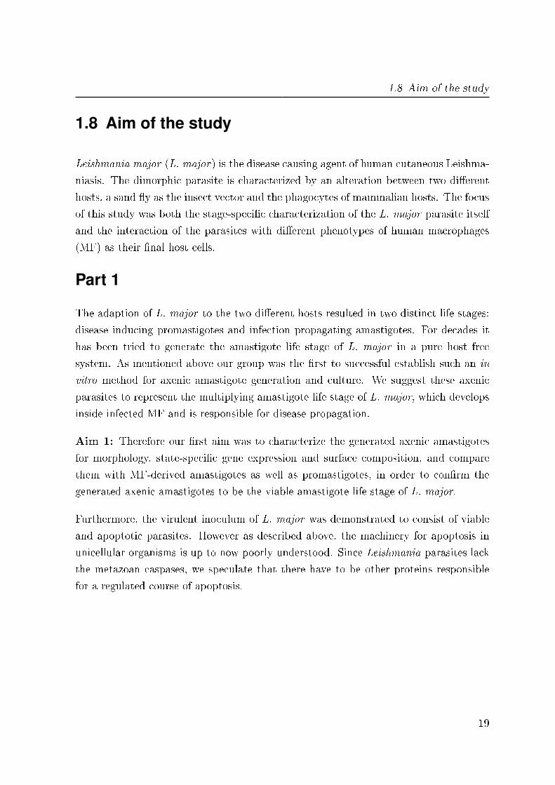
Aim 2: Consequently we intended to investigate the di�erent steps of the apoptotic
cell death in a chronological order for both promastigotes as well as amastigotes and
additionally identify new proteins involved in the apoptotic machinery of L. major.
unidentifiedproteins?
ROS
?
Nucleus
KinetoplastPS
Figure 4: Involved proteins during apoptosis in L. major parasites
Part 2
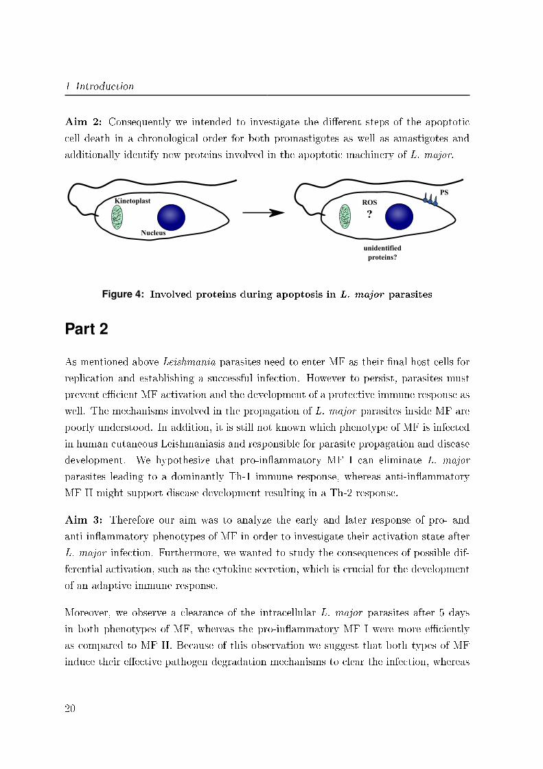
As mentioned above Leishmania parasites need to enter MF as their �nal host cells for
replication and establishing a successful infection. However to persist, parasites must
prevent e�cient MF activation and the development of a protective immune response as
well. The mechanisms involved in the propagation of L. major parasites inside MF are
poorly understood. In addition, it is still not known which phenotype of MF is infected
in human cutaneous Leishmaniasis and responsible for parasite propagation and disease
development. We hypothesize that pro-in�ammatory MF I can eliminate L. major
parasites leading to a dominantly Th-1 immune response, whereas anti-in�ammatory
MF II might support disease development resulting in a Th-2 response.
Aim 3: Therefore our aim was to analyze the early and later response of pro- and
anti-in�ammatory phenotypes of MF in order to investigate their activation state after
L. major infection. Furthermore, we wanted to study the consequences of possible dif-
ferential activation, such as the cytokine secretion, which is crucial for the development
of an adaptive immune response.
Moreover, we observe a clearance of the intracellular L. major parasites after 5 days
in both phenotypes of MF, whereas the pro-in�ammatory MF I were more e�ciently
as compared to MF II. Because of this observation we suggest that both types of MF
induce their e�ective pathogen degradation mechanisms to clear the infection, whereas
20
1.8 Aim of the study
MF I are expected to kill L. major parasites more e�ciently as compared to MF II.
Aim 4: Our aim was to identify the degrading mechanisms which are involved in the
elimination of L. major parasites in infected human MF. In addition, we wanted to
know whether there are di�erences in the responsible mechanisms of parasite clearance
in MF I compared to MF II.
Healing
Pro-inflammatoryMF I
L. majorparasites
Disease development
parasiteclearance
?
Anti-inflammatoryMF II
Figure 5: Hypothesis for L. major parasite propagation in di�erent phenotypes ofhuman MF
21

2 Material and Methods
2.1 Material
2.1.1 Chemicals
α-Isonitrosopropiophenone Sigma, Deisenhof (Ger)
β-Mercaptoethanol Sigma, Deisenhof (Ger)
Acrylamide-Bis 30 % Serva, Heidelberg (Ger)
Adenin Sigma, Deisenhof (Ger)
Agarose Sigma, Deisenhof (Ger)
Aminocaproic acid Sigma, Steinheim (Ger)
Ammoniumchloride Sigma Chemical, St. Louis (USA)
Ammoniumpersulfat (APS) Serva, Heidelberg (Ger)
Ampuwa H2O Fresenius Kabi, Bad Homburg (Ger)
Annexin-V-FITC Responsif AG, Karlsruhe (Ger)
Annexin-V-Fluos Roche Applied Science, Mannheim (Ger)
Annexin-V-Alexa 647 Molecular Probes, Eugene (USA)
Biotin Sigma, Deisenhof (Ger)
Biotinylated Peroxidase Invitrogen, Camarillo (USA)
Bovine Serum Albumin (BSA) Sigma, Deisenhof (Ger)
Bromphenol blue dye Serva, Heidelberg (Ger)
Camptothecin Bio Vision, San Francisco (USA)
23
2 Material and Methods
Concanamycin A Sigma, St. Luois (USA)
D-galactose Sigma, Steinheim (Ger)
2',7'-Dichlorofuorescein diacetate (H2DCFDA) Sigma, Steinheim (Ger)
DifcoTM
Brain Heart Infusion Agar BD, Sparks (USA)
Di�-QUIK R© Medion Diagnostics, Düdingen (CH)
Dimethylsulfoxid (DMSO) Serva, Heidelberg (Ger)
Dithiothreitol (DTT) Sigma, Steinheim (Ger)
DMEM-Medium (10x) Biochrom AG, Berlin (Ger)
1 kb DNA Ladder Promega, Madison (USA)
100 bp DNA Ladder Promega, Madison (USA)
ECL Blocking Agent GE Healhcare, Buckinghamshire (UK)
ECL Western Blotting Detection Reagents GE Healhcare, Buckinghamshire (UK)
EDTA Sigma, Deisenhof (Ger)
Ethanol, absolut (EtOH) VWR, Bruchsal (Ger)
Full-Range Rainbow Molecular Weight Marker GE Healhcare,Buckinghamshire (UK)
Foetal Calf Serum (FCS) Sigma, Deisenhof (Ger)
Glutamine (L-Glutamine) Biochrom AG, Berlin (Ger)
Glycerol (99 %) Sigma, Deisenhof (Ger)
Glycine Sigma, Steinheim (Ger)
Hemin Sigma, Deisenhof (Ger)
Hepes-Bu�er (1M) Biochrom AG, Berlin (Ger)
Histopaque R©1119 Sigma, Deisenhof (Ger)
Human, recombinant Granolucyte MacrophageColony Stimulating Factor (GM-CSF)
Genzyme Onkology, NeuIsenburg (Ger)
Human, recombinant Macrophage Colony Stim-ulating Factor (M-CSF)
R&D Systems, Minneapolis (USA)
Hydrochloric acid, 37 % (HCl) VWR, Bruchsal (Ger)
Hygromycin B, solution Invitrogen, San Diego (USA)
24
2.1 Material
Immersion Oil Carl Zeiss, Jena (Ger)
L-Arginine Sigma, Steinheim (Ger)
Lectin from Arachis hypogaea (peanut) Sigma, Steinheim (Ger)
Lipopolysaccharides from E. coli (LPS) Sigma, Steinheim (Ger)
Lymphocyte Separation Medium 1077 (LSM1077)
PAA, Pasching (Aut)
Manganese chloride (MnCl2) Sigma, Deisenhof (Ger)
Medium 199 Sigma, Deisenhof (Ger)
Methanol Sigma, Deisenhof (Ger)
Miltefosine Calbiochem, Darmstadt (Ger)
Human Serum Type AB Lonza, Walkersville (USA)
Paraformaldehyde (PFA) Sigma, Deisenhof (Ger)
Peanut Lectin Sigma, Deisenhof (Ger)
Penicillin/Streptomycin Biochrom AG, Berlin (Ger)
Phorbol 12-myristate 13-acetate (PMA) Sigma, Deisenhof (Ger)
Phosphate Bu�ered Saline (PBS), 1x PAA, Pasching (Aut)
Phosphoric acid (H3PO4) Merck, Darmstadt (Ger)
Rabbit Blood, de�brinated Elocin-Lab GmbH, Gladbeck (Ger)
Ringer B. Braun B. Braun Melsungen, Melsungen (Ger)
RNase AWAY VWR, Darmstadt (Ger)
Roswell Park Memorial Institute (RPMI) 1640Medium
Sigma, Deisenhof (Ger)
Roti R©-Blue Carl Roth, Karlsruhe (Ger)
Saponin from Quillaja bark Sigma, Steinheim (Ger)
Sodium Acetat Sigma, Deisenhof (Ger)
Sodium Azide Sigma, Deisenhof (Ger)
Sodium Chloride Sigma, Deisenhof (Ger)
Sodium Dodecyl Sulfate (SDS) Sigma, Deisenhof (Ger)
25

2 Material and Methods
Sodium Hydroxide, 1M (NaOH) Merck, Darmstadt (Ger)
Staurosporine Sigma, Steinheim (Ger)
Streptavidin Invitrogen, Camarillo (USA)
Sulfuric acid (H2SO4) Merck, Darmstadt (Ger)
TMB Substrate Solution Thermo Fisher Scienti�c, Bonn (Ger)
TEMED Serva, Heidelberg (Ger)
Trishydroxylmethylaminomethan (Tris) Sigma, Deisenhof (Ger)
Triton X-100 Sigma, Steinheim (Ger)
Tween 20 Sigma, Steinheim (Ger)
Urea Roth, Karlsruhe (Ger)
2.1.2 Culture media and buffers
Alex-Amastigote-Medium (AAM) RPMI 1640 Medium
10 % FCS
3 mM L-Glutamine
100 U/ml Penicillin
100 µg/ml Streptomycin
pH 5.5, adjusted with 38 % HCl
sterile �ltered
FACS-Bu�er 1 x PBS
1 % Human Serum
1 % Foetal Calf Serum
1 % Bovine Serum Albumin
FACS-Bu�er II 1 x PBS
1 % Human Serum
26
2.1 Material
1 % Foetal Calf Serum
1 % Bovine Serum Albumin
0.5 % Saponin
sterile �ltered
Lm-FACS-Bu�er I 1 x Ringer Solution
1 % Bovine Serum Albumin
Lm-FACS-Bu�er II 1 x PBS
1 % Foetal Calf Serum
1 % Bovine Serum Albumin
Lm-Medium RPMI 1640 Medium
5 % FCS
2 mM L-Glutamine
50 µM β-Mercaptoethanol
100 U/ml Penicillin
100 µg/ml Streptomycin
10 mM Hepes Bu�er
Lm-Suspension-Medium Medium 199
10 % FCS
100 U/ml Penicillin
100 µg/ml Streptomycin
40 mM Hepes Bu�er
5 ml 10 mM Adenine, in 50 mM Hepes
1 ml 0.25 % Hemin, in 50 % Triethanolamine
0.5 ml 0.1 % Biotin, in 95 % Ethanol
27
2 Material and Methods
Novy-Nicolle-McNeal Blood Agar Medium 16.6 % Rabbit blood, de�brinated
16.6 % 1 x PBS
66.2 % Brain Heart Infusion Agar
66.2 U/ml Penicillin
66.2 µg/ml Streptomycin
Complete-Medium RPMI 1640 Medium
10 % FCS
2 mM L-Glutamine
50 µM β-Mercaptoethanol
100 U/ml Penicillin
100 µg/ml Streptomycin
10 mM Hepes Bu�er
Wash-Bu�er 1 x PBS
5 % Complete-Medium
MACS-Bu�er 1 x PBS
2 mM EDTA
0.5 % Bovine Serum Albumin
pH 7.2
2.1.3 Westernblot buffers and solutions
6 x Lämmli-Bu�er A. bidest
4.125 M Glycerol
10 % SDS
28
2.1 Material
0.6 M DTT
180 µM Bromphenol blue
Running-Bu�er A. bidest
25 mM Tris
0.1 % SDS
1.44 % Glycine
Separation-Gel-Bu�er A. bidest
1.5 M Tris
0.4 % SDS
pH 6.8 with HCl
Stacking-Gel-Bu�er A. bidest
500 mM Tris
0.4 % SDS
pH 8.8 with HCl
TBST-Solution A. bidest
0.5 % Tween
0.14 M NaCl
10 mM Tris
1 mM NaN3
pH 8
Anode-Bu�er I A. bidest
20 % Methanol
300 mM Tris
29
2 Material and Methods
Anode-Bu�er II A. bidest
20 % Methanol
25 mM Tris
Cathode-Bu�er A. bidest
20 % Methanol
40 mM Aminocaproic acid
0.01 % SDS
WB-Block-Solution TBST-Solution
5 % Blocking reagent
Bu�er for primary antibody TBST-Solution
2 % Bovine Serum Albumin
0.02 % NaN3
Coomassie gel-�xing solution A. bidest
1 % Phos (85 %)
20 % Methanol
Coomassie staining solution A. bidest
20 % Roti R©-Blue
20 % Methanol
Coomassie gel-washing solution A. bidest
25 % Methanol
30

2.1 Material
Gels:
Chemicals Separation gel (30 ml) Stacking gel (10 ml)
15 % 12 % 3.3 %
Aqua bidest 7.5 ml 10.5 ml 6.1 ml
Separation-Gel-Bu�er 7.5 ml 7.5 ml -
Stacking-Gel-Bu�er - - 2.5 ml
Acrylamide stock 30 % 15 ml 12 ml 1.3 ml
TEMED 20 µl 20 µl 10 µl
10 % APS 100 µl 100 µl 50 µl
2.1.4 Leishmania strains
Leishmania major isolate MHOM/IL/81/FEBNI: Originally isolated from a skin biopsy
of an israeli patient and kindly provided by Dr. Frank Ebert (Bernhard Nocht Institute
for Tropical Medicine, Hamburg, Germany).
Leishmania major isolate MHOM/IL/81/FEBNI eGFP: MHOM/IL/81/FEBNI isolate
genetically transfected with the green �uorescent eGFP gene.
Leishmania major isolate MHOM/IL/81/FEBNI DsRed: MHOM/IL/81/FEBNI iso-
late genetically transfected with the red �uorescent DsRed gene.
Leishmania major isolate MHOM/IL/80/Friedlin: Originally isolated from a skin biopsy
of an israeli patient with cutaneous leishmaniasis and kindly provided by the Pasteur
Institute (Paris, France).
Leishmania donovani isolate MHOM/IN/80/DD8: Originally obtained from an indian
Kala-azar patient.
31
2 Material and Methods
Leishmania tropica isolate MHOM/SU/74/K27: Originally isolated from a skin biopsy
of a patient with leishmaniasis in the former Soviet Union.
2.1.5 Human leukocytes
Human peripheral blood mononuclear cells (PBMC) and macrophages (MF) were ob-
tained from bu�ycoats of healthy donors from the blood bank of the University Hospital
of Ulm and the DRK-Blutspendedienst in Frankfurt. Subsequently, cells were isolated
as described in Methods 2.2.1.
2.1.6 Ready-to-use kits
CD14 MicroBeads, human Miltenyi Biotec, Bergisch Gladbach (Ger)
DNeasy Blood and Tissue Kit Qiagen, Hilden (Ger)
Human TNF-alpha Quantikine Elisa kit R&D Systems, Minneapolis (USA)
Human IL-12/IL-23 p40 Quantikine Elisa kit R&D Systems, Minneapolis (USA)
Human IL-10 Duoset Quantikine Elisa kit R&D Systems, Minneapolis (USA)
ImProm-II Reverse Transkription System Promega, Mannheim (Ger)
LightCycler R© FastStart Master Plus SYBERGreen I kit
Roche Applied Science, Mannheim (Ger)
RNeasy Plus Mini kit Qiagen, Hilden (Ger)
StemfectTM
RNA Transfection kit Stemgent, San Diego (USA)
2.1.7 Anti-Human antibodies
Isotype (FITC), IgG1, MOPC-21 (1:100) BD Pharmingen, Heidelberg (Ger)
Isotype (FITC), IgG2b, 27-35 (1:25) BD Pharmingen, Heidelberg (Ger)
Isotype (APC), IgG1, MOPC-21 (1:100) Caltag Laboratories, Hamburg (Ger)
Isotype (PE), IgG1, MOPC-21 (1:100) BD Pharmingen, Heidelberg (Ger)
32
2.1 Material
Isotype (PerCP), IgG2a, X39 (1:100) BD Pharmingen, Heidelberg (Ger)
Chicken anti-mouse (Alexa 488) (1:100) Molecular Probes, Eugene (USA)
Mouse anti-CD163 (PE), IgG1, GHI/61 (1:10) BD Pharmingen, Heidelberg (Ger)
Mouse anti-CD206 (PE), IgG1, 19.2 (1:20) BD Pharmingen, Heidelberg (Ger)
Mouse anti-CD11b (PE), IgG1, ICRF44 (1:100) BD Pharmingen, Heidelberg (Ger)
Mouse anti-CD14 (FITC), IgG2b, (1:25) BD Bioscience, Heidelberg (Ger)
Mouse anti-CD35 (FITC), IgG1, E11 (1:25) BD Pharmingen, Heidelberg (Ger)
Mouse anti-MHC II (PerCP), IgG2a, L243(1:10)
BD Pharmingen, Heidelberg (Ger)
Mouse anti-CD40 (APC), IgG1, HB14 (1:100) Caltag Laboratories, Hamburg (Ger)
Mouse anti-CD86 (APC), IgG1, 2331 (1:100) BD Pharmingen, Heidelberg (Ger)
Mouse anti-phospho-p44/42 MAPK (ERK 1/2)(Alexa Fluor 488), IgG1, 20A (1:10)
BD Pharmingen, Heidelberg (Ger)
Mouse anti-phospho-p38 MAPK (PE), IgG1,36/p38 (1:10)
BD Pharmingen, Heidelberg (Ger)
Mouse anti-phospho-p38 MAPK , IgG1, 36/p38(1:2500)
BD Bioscience, Heidelberg (Ger)
Rabbit anti-p38 MAPK , IgG, (1:1000) Cell Signaling, Danvers (USA)
Rabbit anti-phospho-p44/42 MAPK (ERK1/2), IgG1, 197G2 (1:1000)
Cell Signaling, Danvers (USA)
Mouse anti-p44/42 MAPK (ERK 1/2), IgG1,3A7 (1:2000)
Cell Signaling, Danvers (USA)
Goat anti-rabbit-HRP, IgG, (1:4000) Cell Signaling, Danvers (USA)
Horse anti-mouse-HRP, IgG, (1:4000) Cell Signaling, Danvers (USA)
Mouse anti-β-Actin, IgG, AC-15 (1:1000) Sigma, Steinheim (Ger)
2.1.8 Anti-Leishmania antibodies
Mouse anti-LPG (WIC79.3) (1:3000) Kind gift of Dr. G. Späth, InstitutePasteur, Paris (Fra)
33

2 Material and Methods
2.1.9 Oligonucleotides
Primer Sequence
45rRNA fwd 5'- CCT ACC ATG CCG TGT CCT TCT A -3'
45rRNA rev 5'- AAC GAC CCC TGC AGC AAT AC -3'
ABC-Transp. homologue fwd 5'- CGG GTT TGT CTT TCA GTC GT -3'
ABC-Transp. homologue rev 5'- CAC CAG AGA GCA TTG ATG GA -3'
Sherp fwd 5'- GAC GCT CTG CCC TTC ACA TAC -3'
Sherp rev 5'- TCT CTC AGC TCT CGG ATC TTG TC -3'
QDPR fwd 5'- ATG AAA AAT GTA CTC CTC ATC G -3'
QDPR rev 5'- TTC ACC CTG CGT ACT GAA CAC AT -3'
alpha-tubulin fwd 5'- ATG CGT GAG GCT ATC TGC ATC CAC AT -3'
alpha-tubulin rev 5'- TAG TGG CCA CGA GCG TAG TTG TTC G -3'
GP63 fwd 5'- ACT GCC CGT TTG TTA TCG AC -3'
GP63 rev 5'- CCG GCG TAC GAC TTG ACT AT -3'
Cpb fwd 5'- TGA TGC GGT GGA CTG GC -3'
Cpb rev 5'- CCA CTC GAA TGC CTG CAG C -3'
GAPDH fwd 5'- GAG TCA ACG GAT TTG GTC GT -3'
GAPDH rev 5'- TTG ATT TTG GAG GGA TCT CG -3'
LL37 fwd 5'- GGA CCC AGA CAC GCC AAA -3'
LL37 rev 5'- GCA CAC TGT CTC CTT CAC TGT GA -3'
ITS1 fwd (LITSR) 5'- CTG GAT CAT TTT CCG ATG -3'
ITS1 rev (L5.8S) 5'- TGA TAC CAC TTA TCG CAC TT -3'
Table 1: Primer for PCR
Primer:
Table 1 contains the used oligonucleotide primer and their sequence. All oligonucleotides
were purchased from Thermo Fisher Scienti�c in Ulm (Ger).
34
2.1 Material
siRNA:
Stealth RNAi siRNA Negative Control Med
GC
Invitrogen, Darmstadt (Ger)
ON-TARGET plus SMART pool Human
CAMP (LL-37)
Thermo Scienti�c Dharmacon, Bonn (Ger)
2.1.10 Enzymes
FideliTaq PCR Master Mix (2x) A�ymetrix, Santa Clara (USA)
HaeIII (restriction enzyme) New England Biolabs, Frankfurt am Main(Ger)
Phusion High Fidelity DNA Polymerase Finnzymes, Vantaa (Fin)
Phusion High-Fidelity PCR kit New England Biolabs, Frankfurt am Main(Ger)
Peroxidase Invitrogen, Camarillo (USA)
Recombinant DNase I Roche, Mannheim (Ger)
RNaseOUTTM
recombinant RNase Inhibitor Invitrogen, Darmstadt (Ger)
2.1.11 Laboratory supplies
Carbon-coated sapphire discs (3 mm in di-ameter)
Engineering O�ce M. Wohlwend GmbH,Sennwald (CH)
Cell culture �asks (25 cm2; 75 cm2) BD labware Europe, Le Pont de Claix (Fra)
Cell culture plates (6-well; 24-well; 96-well) BD labware Europe, Le Pont de Claix (Fra)
Centrifuge tubes (15 ml; 50 ml) BD labware Europe, Le Pont de Claix (Fra)
Cryo tubes Greiner Bio-one, Frickenhausen (Ger)
FACS tubes (2 ml) Micronic, Lelystad (Ned)
FACS tubes BD labware Europe, Le Pont de Claix (Fra)
35

2 Material and Methods
High performance chemiluminescence �lm(Hyper�lm
TM
ECL)GE Healhcare, Buckinghamshire (UK)
Hybond ECL blot membrane VWR, Darmstadt (Ger)
LightCycler R© capillaries 20 µl Roche Applied Science, Mannheim (Ger)
Microtest plates, 96-well (V-Bottom) Sarstedt, Nümbrecht (Ger)
Microtest plates, 96-well (Flat-Bottom) Sarstedt, Nümbrecht (Ger)
Milipore stericup sterile vacuum �lter units Millipore, Schwalbach (Ger)
Multiplier tubes (0.65 ml), biopure Sarstedt, Nümbrecht (Ger)
Nunc-ImmunoTM
plate, Maxisorp NUNC, Langenselbold (Ger)
Pipette �lter tips (1-10 µl, 10-100 µl, 50-200µl, 100-1000 µl)
Sarstedt, Nümbrecht (Ger)
Reaction tubes (0.5 ml; 1.5 ml; 2.0 ml) Eppendorf, Hamburg (Ger)
Serological pipettes, steril (2.5 ml; 5 ml; 10ml; 25 ml)
Corning Inc., Corning, New York (USA)
Transfer membrane immobilon-P (PVDF) Millipore, Billerica (USA)
Transfer pipette (3.5 ml) Sarstedt, Nümbrecht (Ger)
Whatman paper gel blotting VWR, Darmstadt (Ger)
2.1.12 Instruments
AutoMACS Pro separator Miltenyi Biotec, Bergisch Gladbach (Ger)
Analytical balance AG204 Mettler Toledo, Giessen (Ger)
Balance KERN470 Kern & Sohn GmbH, Balingen-Frommern(Ger)
Centrifuge 5471 Eppendorf, Hamburg (Ger)
CO2-Incubator Forma 3010 Thermo Scienti�c, Marietta (USA)
CO2-Incubator Heraeus BBD 6220 Thermo, Dreieich (Ger)
CO2-Incubator Forma Series II Water Jacket Thermo Scienti�c, Marietta (USA)
Cytocentrifuge Cytospin3 Shandon, Frankfurt (Ger)
DNA Engine R© Peltier Thermal Cycler Bio-Rad, München (Ger)
36
2.1 Material
Easy-castTM
Electrophoresis System Thermo Scienti�c Owl Separation, Rochester(Ger)
Electrophoresis power supply EPS 600 Amersham Pharmacia, Uppsala (Swe)
EM10 transmission electron microscope Carl Zeiss, Jena (Ger)
Flow-Cytometer FACS-Calibur II Becton Dickinson, Heidelberg (Ger)
Flow-Cytometer LSR II Becton Dickinson, Heidelberg (Ger)
Freezer -20◦C Bosch, Stuttgart (Ger)
Freezer Herafreeze -80◦C Heraeus Sepatech GmbH, Osterode (Ger)
Gel dryer model 543 Bio-Rad, München (Ger)
Gel electrophoresis system SE600 Hoefer, San Francisco (USA)
HPF 01 apparatus Engineering O�ce M. Wohlwend GmbH,Sennwald (CH)
Laminar �ow workbench MSC-Advantage Thermo Scienti�c, Dreieich (Ger)
LightCycler R© Roche Applied Science, Mannheim (Ger)
Magnetic stirrer MR3002 Heidolph, Leverkusen (Ger)
Microscope Axio Imager M2 Carl Zeiss, Jena (Ger)
Microscope Axiovert 40 CFL Carl Zeiss, Jena (Ger)
Microscope Primo Star Carl Zeiss, Jena (Ger)
Multichannel Pipette Eppendorf, Hamburg, (Ger)
Multifuge 3 SR Heraeus, Thermo, Dreieich (Ger)
Neubauer cell counting chamber depth0.1mm
VWR, Darmstadt (Ger)
Neubauer cell counting chamber depth0.02mm
VWR, Darmstadt (Ger)
Power supply Power Pac 300 Angewandte Gentechnologie SystemeGmbH, Heidelberg (Ger)
pH-Meter pH525 WTW, Wellheim (Ger)
Pipettes Eppendorf, Hamburg, (Ger)
Platform shaker Polymax 1040 Heidolph, Schwabach (Ger)
37

2 Material and Methods
Shaker VWR, Darmstadt (Ger)
Shake Table GFL-3016 GFL, Burgwedel (Ger)
Semi-dry transfer unit TE 77 PWR Amersham Biosciences, Freiburg (Ger)
Table-top processor Curix 60 AGFA, Berlin (Ger)
Tecan in�nite M200 Tecan Austria GmbH, Grödig (Aut)
Thermostatic circulator 2219 Multitemp II LKB Bromma, Stockholm (Swe)
Variofuge 3.OR Heraeus, Thermo, Dreieich (Ger)
Water bath GFL, Burgwedel (Ger)
2.1.13 Software
Axiovision 4.7 Carl Zeiss, Jena (Ger)
BD Diva software v6.1.3 Becton Dickinson, Heidelberg (Ger)
CellQuest R© Pro Becton Dickinson, Heidelberg (Ger)
Inkscape v0.48 OpenSource (http://www.inkscape.org)
ImageJ OpenSource (http://rsbweb.nih.gov/ij/)
LightCycler R© software v3.5 Roche Applied Science, Mannheim (Ger)
Microsoft R© O�ce 2010 Microsoft, Redmont (USA)
Tecan I-ControlTM
v1.6 Tecan Austria GmbH, Grödig (Aut)
38

2.2 Methods
2.2 Methods
2.2.1 Cell culture
The cells were treated and passaged under sterile conditions in endotoxin free environ-
ments and all cell cultures were kept in humidi�ed incubators with 5 % CO2 .
2.2.1.1 Cultivation of L. major promastigotes
L. major promastigotes were cultured either in biphasic Novy-Nicolle-McNeal (NNN)
blood agar medium or in Lm-Suspension-Medium at 27◦C. In the stationary growth
phase (stat-phase) after 7 days L. major promastigote cultures were passaged up to ten
serial passages before the cultures were discarded.
For long-time storage stationary-phase (stat-phase) L. major were pelleted at 2400 x g
for 8 min and resuspended in ice-cold Lm-Medium supplemented with 20 % FCS and
10 % DMSO. The cell density was adjusted to 2 x 10⁸ L. major/ml. The cells were
transferred into Cryo Tubes and were put in a styropore box at -80◦C overnight before
they could be stored in liquid nitrogen.
L. major parasites were thawed at 37◦C in a water-bath and added drop-wise to Lm-
Medium to dilute the DMSO. After pelleting the parasites were washed one more time
before the pellet was resuspended in 10 ml Lm-Medium and added on biphasic NNN
blood agar medium 100 µl/well. The culture was not used until the second passage.
For eGFP and DsRed promastigotes Lm-Medium was supplemented with 20 µg/ml
hygromycin B.
2.2.1.2 Isolation of metacyclic L. major promastigotes
4 x 10⁸ of stat-phase L. major promastigotes were resuspended in 1 ml RPMI supple-
mented with 100 µg/ml lectin (peanut), incubated at room temperature for 30 min and
centrifuged at 545 x g for 10 min. The supernatant was collected and washed with
DMEM supplemented with 20 mM D-galactose at 2400 x g for 10 min. The isolated
metacyclic parasites were resuspended in Lm-Medium and counted for further analysis.
39

2 Material and Methods
2.2.1.3 Generation and cultivation of L. major amastigotes in vitro
Promastigote pre-culture
4 wells of logarithmic growth phsase (log-phase) (day 3 - 4 of NNN blood agar culture)
L. major promastigotes were cultured in 5 ml Lm-Suspensions-Medium + 0.5 ml FCS
for 3 days at 27◦C.
For eGFP and DsRed promastigotes Lm-Medium was supplemented with 20 µg/ml
hygromycin B.
Amastigote pre-culture
The log-phase promastigote pre-culture was harvested and pelleted at 1450 x g for 8
min. The pellet was resuspended in 10 ml AAM and centrifuged for 8 min at 1450 x
g. This step was repeated with 2400 x g. The pellet was resuspended in AAM and
adjusted to 2 x 10⁷ L. major/ml. The cells were incubated in 25 cm2 culture �asks for
10 - 12 days at 33◦C.
Amastigote isolation
To separate the amastigotes from the remaining promastigotes and dead parasites a
discontinous Histopaque R© 1119 density gradient was used. The amastigote pre-culture
was harvested and pelleted at 2400 x g for 8 min. The pellet was resuspended in 50 %
(1,0595 g/ml) Histopaque R© 1119 and fractionated on a discontinous Histopaque R© 1119
density gradient consisting of layers with densities of (from top to bottom) 1,0833 g/ml
(70 %), 1,0952 g/ml (80 %), 1,1071 g/ml (90 %) and 1,119 g/ml (100 %). The gradient
was centrifuged at 2400 x g for 35 min (with acceleration and deceleration set at the
lowest level). The interphases between 80 - 90 % and 90 - 100% were collected, washed
twice in AAM and adjusted to 2 x 10⁷ L. major/ml. The purity of the amastigotes
was monitored by analysing Di� QUIK R© stained cytospin slides. The puri�ed L. major
amastigotes were cultured in 25 cm2 culture �asks at a density of 2 x 10⁷ L. major/ml
at 33◦C. The culture is stable for 7 days.
Amastigote retransformation
To assure a constant virulence of the parasites, amastigote-passages were performed
using the in vitro culture method to generate axenic amastigotes. L. major promasti-
gotes were transformed into amastigotes and subsequently, were cultured on biphasic
40
2.2 Methods
NNN blood agar medium, where the parasites transformed back to the promastigote
stage.
2.2.1.4 Isolation of L. major amastigotes from infected MF
Macrophages (MF) were infected as described in 2.2.1.7 with a multiplicity of infection
of 1:20 and incubated for 2 - 4 days at 37◦C to allow the parasites to di�erentiate into
amastigotes. The infected cells were washed with warm RPMI without supplements
and the cell walls were lysed in RPMI supplemented with 0.02 % SDS for 3 min at 37◦C
to release the intracellular amastigotes. The lysis was stopped with AAM supplemented
with 20 % FCS and the MF lysate was washed once at 2400 x g for 8 min. To collect
the free parasites the pellet was resuspended in AAM supplemented with 20 % FCS and
centrifuged at 75 x g for 8 min. The supernatant was removed and the centrifugation
step was repeated twice. The puri�ed amastigotes were pelleted at 2400 x g for 8
min, resuspented in AAM and adjusted to 2 x 10⁷ L. major/ml. The purity of the
amastigotes was monitored by analysing Di� QUIK R© stained cytospin slides.
2.2.1.5 Isolation of human peripheral blood mononuclear cells (PBMC)
PBMC were isolated from bu�ycoats of healthy donors. The bu�ycoats were diluted
1:5 with sterile PBS, layered on top of 15 ml Lymphocyte Separation Medium 1077
and centrifuged at 545 x g for 30 min (with acceleration and deceleration set at the
lowest level). Plasma and the interphase mainly consisting of PBMC were collected
and washed with Wash-Bu�er at 1024 x g for 8 min. The pellet was resuspendet and
washed with Wash-Bu�er �rst at 545 x g and then at 135 x g for 8 min. The pellet
was resuspended in 10 ml 0.15 M Ammoniunchloride and the ery-lysis was performed
for 10 - 15 min at room temperature. Subsequently, the cells were washed twice with
Wash-Bu�er at 135 x g for 8 min to remove thrombocytes. After pooling the cells were
counted and adjusted to a density of 1 x 10⁷ PBMC/ml in Complete-Medium.
41
2 Material and Methods
2.2.1.6 Generation of blood derived MF
Plastic adherence
Fresh isolated PBMC were incubated in 25 cm2 culture �asks at a density of 1 x 10⁷
PBMC/ml in Complete-Medium supplemented with 1 % human serum for 90 min at
37◦C. The supernatant was discarded and the non-adherent cells were removed by wash-
ing 2 times with pre-warm Wash-Bu�er. The adherent monocytes were cultured in
Complete-Medium supplemented with 10 ng/ml GM-CSF (generation of type 1 MF) or
30 ng/ml M-CSF (generation of type 2 MF) for 5 - 7 days at 37◦C.
AutoMACS separation
100 x 106 PBMC fresh isolated PBMC were washed with 10 ml cold MACS-Bu�er
at 300 x g for 8 min. The pellet was resuspended in 400 µl MACS-Bu�er, 100 µl
CD14-Beads were added to the cells and the mixture was incubated at 4◦ C for 15
min. Subsequently, the cells were washed with 10 ml cold MACS-Bu�er at 300 x g
for 8 min and the pellet was resuspended in 500 µl MACS-Bu�er. The labbeled cells
were placed into an AutoMACS device and the separation program posseld was run.
After seperation the isolated monocytes were counted and incubated in 6-well plates
at a density of 1.6 x 10⁶ cells/ml in Complete-Medium supplemented with 10 ng/ml
GM-CSF (generation of type 1 MF) or 30 ng/ml M-CSF (generation of type 2 MF) for
5 - 7 days at 37◦ C with a medium exchange after 3 days.
2.2.1.7 Co-incubation of macrophages with L. major parasites
After 5 - 7 days of culture in the presence of either GM-CSF or M-CSF MF were
harvested with a cell scraper and counted. Co-incubation of MF with L. major parasites
was performed by two di�erent methods.
Co-incubation in cell culture plates
1 x 10⁶ MF/ml were transfered into 12-well (1 x 10⁶ MF) or 96-well (1 x 10⁵ MF)
cell culture plates. The cells were left to adhere in the cell culture plates for 60 min
and the non-adherend cells were removed by discarding the supernatant. Stat-phase
L. major promastigotes or 1 - 3 days old L. major amastigotes were added to the MF
42
2.2 Methods
with a multiplicity of infection (MOI) of 1:10. The cell culture plates were centrifuged
at 304 x g for 3 min before incubating for 3 h at 37◦C. After co-incubation extracellular
parasites were removed by washing the cells with Wash-Bu�er. Cells and supernatants
were collected after 18, 48 or 72 h culture at 37◦C for further analyses.
Co-incubation in centrifuge tubes
1 x 10⁷ MF/ml were transfered into 15 ml centrifuge tubes or 1.5 ml reaction tubes.
Stat-phase L. major promastigotes or 1 - 3 days old L. major axenic amastigotes were
added to the MF with a MOI of 1:10. After 3 h of co-incubation with the parasites
either extracellular parasites were removed by washing the cells with Wash-Bu�er at
135 x g for 10 min and the cells were cultured for another 18 h at 37◦C. Or 1 ml
Complete-Medium was added per 1 x 10⁶ MF to the cells and after another incubation
for 18 h at 37◦C the extracellular parasites were removed by centrifuging at 135 x g for
10 min. Cells were collected after 18, 48 or 72 h culture at 37◦C for further analyses.
Infection rates were determined by counting at least 200 MF on Di� QUIK R© stained
cytospin slides. The parasite burdens were evaluated by counting intracellular L. major
parasites in 20 infected MF.
2.2.1.8 End-point titration
Axenic L. major parasites
The amount of viable L. major parasites was determined 72 h after the treatment with
di�erent drugs by end-point titration. End-point titration experiments were carried out
by using 1 x 10⁵ parasites in octuplicate wells and a dilution factor of 10. The number
of viable L. major was assessed after 7 days of incubation on biphasic NNN blood agar
medium at 27◦C and calculated from the last dilution that showed parasitic growth.
Viable L. major parasites inside infected MF
The amount of viable intracellular L. major parasites inside human MF was determined
18 and 48 h after 3 h of co-incubation with the parasites by end-point titration.
End-point titration experiments were carried out by using 2000 MF in quadruplicate
wells and a dilution factor of 1.5. The number of viable intracellular L. major parasites
per 1000 MF was assessed after 7 days of incubation on biphasic NNN Blood Agar
43
2 Material and Methods
Medium at 27◦C and calculated from the last dilution that showed parasitic growth and
equals 1.5 exp (mean dilution with parasitic growth).
2.2.1.9 Cytocentrifuging cells
2 x 10⁶ L. major parasites or 1 x 10⁵ MF were washed in Medium and resuspended in
100 µl PBS. Cells were centrifuged on slides in a Cytocentrifuge at 500 x g for 10 min
for Leishmania and at 75 x g for 5 min for MF. Subsequetly, the slides were air-dried
for further use.
2.2.1.10 Diff QUIK staining
Air-dried cytospin slides were incubated for 1 min in Fixation Solution of a Qi� QUIK R©
kit. Subsequently, incubated for 1 min in Staining Solution I followed by 1 min in
Staining Solution II. The slides were rinsed in tap water, air-dried and used for further
microscopical analyses.
2.2.2 FACS analysis
FACS stainings were performed in 96-well Microtestplates (V-Bottom) in the dark on
ice.
2.2.2.1 Extracellular FACS analysis of (infected) MF
2 x 10⁵ MF were washed in FACS-Bu�er and incubated with α-CD163-PE, α-CD-11b-
PE, α-CD14-FITC, α-CD35-FITC, α-CD40-APC, α-CD86-APC, α-CD206-PE and α-
MHC II-PerCP in FACS-Bu�er for 30 min. For isotype controls, MF were incubated
with PE-, FITC-, APC- or PerCP- conjugated matched mouse IgG1 and mouse IgG2
antibodies. The cells were washed in FACS-Bu�er, resuspended in 400 µl FACS-Bu�er
and analysed by a �ow cytometer (FACS-Calibur II with CellQuest R© Pro software or
LSR II with BD Diva software).
44

2.2 Methods
2.2.2.2 Intracellular FACS analysis of (infected) MF
5 x 10⁵ MF were �rst washed in FACS-Bu�er and then in FACS-Bu�er II to permeabilize
the cell membrane. Subsequently the cells were incubated with α-phospho Tyr-FITC,
α-phospho p38-PE and α-phospho ERK-Alexa 488 in FACS-Bu�er II for 30 min. After
a washing step with FACS-Bu�er II, the MF were washed with FACS-Bu�er, resus-
pended in 400 µl FACS-Bu�er and analysed by a �ow cytometer (FACS-Calibur II with
CellQuest R© Pro software).
2.2.2.3 Extracellular FACS analysis of L. major
5 x 10⁶ L. major parasites were washed in Lm-FACS-Bu�er I and incubated with α-
LPG (WIC79.3) in Lm-FACS-Bu�er I for 30 min. After a washing step with Lm-FACS-
Bu�er I, the cells were incubated with chicken α-mouse-Alexa 488 in Lm-FACS-Bu�er
I for 30 min. The parasites were washed in Lm-FACS-Bu�er I, resuspended in 400 µl
FACS-Bu�er and analysed by a �ow cytometer (FACS-Calibur II with CellQuest R© Pro
software or LSR II with BD Diva software).
Annexin-binding
5 x 10⁶ L. major parasites were washed in Lm-FACS-Bu�er I and incubated with 0.1
µg/ml Annexin A5 (AnxA5)-FITC or AnxA5-Fluos in Lm-FACS-Bu�er I for 20 min.
The parasites were washed in Lm-FACS-Bu�er I, resuspended in 400 µl Lm-FACS-Bu�er
I and analysed by a �ow cytometer (FACS-Calibur II with CellQuest R© Pro software or
LSR II with BD Diva software).
2.2.2.4 Intracellular FACS analysis of L. major
ROS-detection
5 x 10⁶ L. major parasites were washed in Lm-FACS-Bu�er II and incubated with 50 nM
2',7'-Dichlorofuorescein diacetate (H2DCFDA) in Lm-FACS-Bu�er II for 20 min. The
parasites were washed in Lm-FACS-Bu�er II, resuspended in 400 µl Lm-FACS-Bu�er
II and analysed by a �ow cytometer (FACS-Calibur II with CellQuest R© Pro software).
45
2 Material and Methods
2.2.3 ELISA analysis
Macrophages were cultured for 18 h in Complete-Medium alone, co-incubated with
stat-phase L. major promastigotes or with 1-3 days old L. major axenic amastigotes
at a MOI of 1:10. Supernatants were collected and stored at -80◦C until cytokine
determination using an enzyme-linked immunosorbent assay (ELISA).
2.2.3.1 TNF alpha
TNF alpha content was measured using sandwich ELISA (Human TNF-alpha Quan-
tikine Elisa kit) according to the manufacturer's instructions. Brie�y, a 96-well ImmunoTM
plate was coated with 2 µg/ml anti-TNF alpha antibody, blocked and washed. The sam-
ples were applied to the plate together with a dilution series of a protein standard (for a
standard curve) for 1 h at room temperature. TNF alpha was detected using 0.5 µg/ml
of a biotinyled anti-human TNF alpha antibody for 1 h. During the incubation the
streptavidin-HRP complex was prepared and diluted 1:10000. The plate was washed
and incubated with the streptavidin-HRP complex for 30 min. The TMB substrate
solution was added after washing to the wells and incubated for 15 - 30 min in the dark.
The reaction was stopped using 0.18 M (H2SO4) and the optical density was determined
with a Tecan in�nite M200, wavelength set to 450 nm. The TNF alpha concentration in
each well was calculated using the standard curve generated with the optical densities
of the standard dilution series.
2.2.3.2 IL-12
Il-12 content was measured using sandwich ELISA (Human IL-12/IL-23 p40 Quantikine
Elisa kit) according to the manufacturer's instructions. Brie�y, a 96-well ImmunoTM
plate was coated with 3 µg/ml anti-IL-12/IL-23 p40 antibody, blocked and washed. The
samples were applied to the plate together with a dilution series of a protein standard
(for a standard curve) for 2 h at room temperature. The plate was washed and IL-12
was detected using 0.2 µg/ml of a biotinyled anti-human IL-12 antibody for 1 h. During
the incubation the streptavidin-HRP complex was prepared and diluted 1:10000. The
46
2.2 Methods
plate was washed and incubated with the streptavidin-HRP complex for 30 min. The
TMB substrate solution was added after washing to the wells and incubated for 15 -
30 min in the dark. The reaction was stopped using 0.18 M (H2SO4) and the optical
density was determined with a Tecan in�nite M200, wavelength set to 450 nm. The
IL-12 concentration in each well was calculated using the standard curve generated with
the optical densities of the standard dilution series.
2.2.3.3 IL-10
Il-10 content was measured using sandwich ELISA (Human IL-10 Duoset Quantikine
Elisa kit) according to the manufacturer's instructions. Brie�y, a 96-well ImmunoTM
plate was coated with 3 µg/ml anti-IL-10 antibody, blocked and washed. The samples
were applied to the plate together with a dilution series of a protein standard (for a
standard curve) for 2 h at room temperature. The plate was washed and IL-10 was
detected using 0.2 µg/ml of a biotinyled anti-human IL-10 antibody for 2 h. During
the incubation the streptavidin-HRP complex was prepared and diluted 1:10000. The
plate was washed and incubated with the streptavidin-HRP complex for 30 min. The
TMB substrate solution was added after washing to the wells and incubated for 15 -
30 min in the dark. The reaction was stopped using 0.18 M (H2SO4) and the optical
density was determined with a Tecan in�nite M200, wavelength set to 450 nm. The
IL-10 concentration in each well was calculated using the standard curve generated with
the optical densities of the standard dilution series.
2.2.4 Arginase assay
Macrophages were cultured for 18 h in Complete-Medium alone, co-incubated with stat-
phase L. major promastigotes or with 1 - 3 days old L. major axenic amastigotes at a
MOI of 1:10. Cells were washed with 10 ml PBS at 1024 x g for 8 min and the pellet was
lysed in 100 µl 0.1 % Triton X-100 for 30 min at RT while stirring. 100 µl 25 mM Tris-
HCl and 20 µl 10 mM MnCl2 were added to the lysate and the enzyme was activated by
incubation for 10 min at 56◦C. Subsequently 100 µl 0.5 M L-arginine was added to the
whole mixture and incubated for 90 min at 37◦C. For a standard curve a dilution series
47
2 Material and Methods
of a urea protein standard was prepared. 400 µl acid mixture (H2SO4/ H3PO4/ H2O
(1/3/7)) and 25 µl 9 % α-isonitrosopropiophenone were added to the protein standard
and incubated for 30 min at 95◦C. The enzyme reaction in the samples was stopped by
adding 800 µl acid mixture and 40 µl 9 % α-isonitrosopropiophenone and incubating for
30 min at 95◦C. 200 µl of both samples and protein standard were applied to a 96-well
plate and the optical density was determined with a Tecan in�nite M200, wavelength
set to 540 nm after 10 min in the dark. The arginase activity in each well was calculated
using the standard curve generated with the optical densities of the standard dilution
series.
2.2.5 Molecular biology methods
2.2.5.1 Transfection of primary cells with siRNA
Human MF were generated from bu�ycoats of healthy donors by AutoMACS separation
using a CD14 MicroBeads-isolation kit. For transfection the MF were washed with 1 ml
RPMI-Medium without supplements and 1 ml RPMI-Medium without supplements was
added per well. For each well: 4 µl of 20 µM siRNA (80 pmole) was mixed with 20 µl of
Stemfect Bu�er and 4.6 µl of Stemfect Reagent was mixed with 20 µl Stemfect Bu�er.
Both compounds were mixed together within 5 min and incubated for 20 min at room
temperature. Subsequently the whole mixture was added to the MF and incubated for 7
h at 37◦C. After incubation the transfection mixture was removed from the cells, 2.5 ml
Complete-Medium was added per well and incubated for 2 days at 37◦C. After 2 days
of siRNA transfection, MF were harvested and proceeded with further experiments.
2.2.5.2 DNA isolation
Genomic DNA was isolated using the DNeasy Blood and Tissue Kit according to the
manufacturer's instructions. Brie�y, 1 x 10⁸ stationary phase L. major (MHOM/IL/81/
FEBNI), L. tropica (MHOM/SU/74/K27) and L. donovani (MHOM/IN/80/DD8) pro-
mastigotes were washed in cold PBS and resuspended in 200 µl PBS. 20 µl proteinase
K and 200 µl Bu�er AL were added and the cells were lysed for 10 min at 56◦C. 200
48
2.2 Methods
µl ethanol was added to the lysate, vortexed, transfered to a DNeasy Mini spin column
and centrifuged at 6000 x g for 1 min. The �ow-through was discarded and the column
washed with 500 µl Bu�er AW1 by centrifugation at 6000 x g for 1 min. Another wash-
ing step was performed with 500 µl Bu�er AW2 by centrifugation at 18000 x g for 3
min and the column was dried by centrifugation in a fresh collection tube at 18000 x g
for 1 min. Subsequently the spin column was placed in a fresh 1.5 ml reaction tube, 200
µl Bu�er AE was added to the column, incubated fot 1 min and centrifuged at 6000 x
g for 1 min. The isolated DNA was stored at -20◦C.
2.2.5.3 Amplification of the ribosomal internal transcribed spacer 1 (ITS1)
The ITS1 of the di�erent Leishmania spp. was ampli�ed in a PCR with speci�c oligonu-
cleotides, which are listed in section 2.1.9 on page 34. For one reaction:
Volume [µl] Reagent
19 Nuclease-free H2O
2 Primer fwd (10 µM)
2 Primer rev (10 µM)
25 Phusion High Fidelity DNA Polymerase
48 µl
+ 150 ng DNA
Tubes were centrifuged at 800 x g, placed into a PCR cycler and the following program
started.
49

2 Material and Methods
Temp [◦C] Time
Denaturation 98 30 sec
Ampli�cation (45 cycles)
Denaturation 98 30 sec
Annealing 53 30 sec
Elongation 72 20 sec
Elongation 72 10 min
Cooling 4 ∞
The products were subjected to electrophoresis on a 0.7 % agarose gel and visualized
under ultraviolet light.
2.2.5.4 Restriction digest
The PCR products of the di�erent Leishmania spp. were incubated with 10 U HaeIII
for restriction digest according to the manufacturer's instructions at 37◦C for 1 h. The
restriction fragments were subjected to electrophoresis on a 0.7 % agarose gel and visu-
alized under ultraviolet light.
2.2.5.5 RNA isolation
RNA was isolated using the RNeasy Plus Mini Kit according to the manufacturer's
instructions. Brie�y, either 1 x 10⁸ L. major parasites or 1 x 10⁶ MF were washed
with cold PBS and the pellet lysed in 350 µl RLT Bu�er by pipetting. The lysate
was transfered to a gDNA Eliminator spin column and centrifuged at 18000 g for 30
sec. 350 µl 70 % ethanol was added to the �ow-through, transfered to a RNeasy spin
column and centrifuged at 13000 rpm for 30 sec. The �ow-through was discarded and
the column was washed with 700 µl RW1 Bu�er at 18000 g for 30 sec. The washing
50
2.2 Methods
was repeated twice with 500 µl RPE Bu�er and the column was dried by centrifugation
in a fresh collection tube at 20000 g for 1 min. Subsequently the spin column was
placed in a fresh 1.5 ml reaction tube, 40 µl RNase-free water was added to the column
and centrifuged at 18000 g for 1 min. The isolated RNA was treated with DNase I to
eliminate remaining genomic DNA and stored at -80◦C.
DNase digestion
Up to 10 µg RNA was incubated with 1 µl recombinant DNase I (10 U/µl) and 1 µl
RNaseOUTTM
recombinant RNase Inhibitor (40 U/µl) for 20 min at 37◦C. Enzymes
were subsequently inactivated for 10 min at 75◦C. The DNase digestion was performed
twice to ensure the elimination of remaining genomic DNA.
2.2.5.6 Test-PCR
A Test-PCR was performed to check the isolated RNA for genomic DNA contamination.
The speci�c primer for Test-PCR were GAPDH for human MF and 45rRNA for L. major
parasites, which are listed in section 2.1.9 on page 34. For one reaction:
Volume [µl] Reagent
19 Nuclease-free H2O
2 Primer fwd (10 µM)
2 Primer rev (10 µM)
25 FideliTaq PCR Master Mix
48 µl
+ 150 ng RNA
Tubes were centrifuged at 800 x g, placed into a PCR cycler and the following program
started.
51
2 Material and Methods
Temp [◦C] Time
Denaturation 98 30 sec
Ampli�cation (30 cycles)
Denaturation 98 30 sec
Annealing 60 30 sec
Elongation 72 30 sec
Elongation 72 10 min
Cooling 4 ∞
The products were subjected to electrophoresis on a 0.7 % agarose gel and visualized
under ultraviolet light. Only the positive control with cDNA as template should show a
product. Otherwise the RNA ist contaminated with genomic DNA. Then an additional
DNase I digestion step is required.
2.2.5.7 cDNA synthesis
For cDNA synthesis the ImProm-II Reverse Transcription SystemTM
was used according
to the manufacturer's instructions. For one reaction:
Volume [µl] Reagent
1 ImProm-IITM
Random Primer Mix
100 - 150 ng Template RNA
ad 5 µl Nuclease-free H2O
5 µl
The primer/template RNA mix is thermally denatured for 5 min at 70◦C and subse-
quently chilled on ice. A reverse transcription reaction mix was assembled on ice and
52

2.2 Methods
added to the mixture. For one reaction:
Volume [µl] Reagent
6.5 Nuclease-free H2O
4 ImProm-IITM
5X Reaction Bu�er
2 MgCl2
1 dNTP Mix
0.5 Recombinant RNasin R© Ribonuclease Inhibitor
1 ImProm-IITM
Reverse Transcriptase
15 µl
Tubes were centrifuged at 800 x g, placed into a PCR cycler and the following program
started.
Temp [◦C] Time
Annealing 25 5 min
cDNA synthesis 42 60 min
Inactivation 70 15 min
Cooling 4 ∞
2.2.5.8 Quantitative real-time PCR
For quantitative real-time PCR the LightCycler R© FastStart DNA MasterPLUS SYBR
Green I kit was used according to the manufacturer's instructions. The speci�c primer
for real-time PCR are listed in section 2.1.9 on page 34. For one reaction:
53
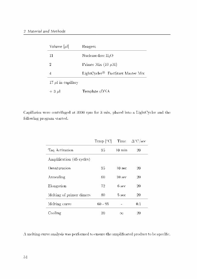
2 Material and Methods
Volume [µl] Reagent
11 Nuclease-free H2O
2 Primer Mix (10 µM)
4 LightCycler R© FastStart Master Mix
17 µl in capillary
+ 3 µl Template cDNA
Capillaries were centrifuged at 3000 rpm for 3 min, placed into a LightCycler and the
following program started.
Temp [◦C] Time ∆◦C/sec
Taq Activation 95 10 min 20
Ampli�cation (45 cycles)
Denaturation 95 10 sec 20
Annealing 60 10 sec 20
Elongation 72 6 sec 20
Melting of primer dimers 80 5 sec 20
Melting curve 60 - 95 - 0.1
Cooling 20 ∞ 20
A melting curve analysis was performed to ensure the ampli�cated product to be speci�c.
54
2.2 Methods
2.2.6 Transmission electron microscopy (EM)
Macrophages were adhered on carbon-coated sapphire discs (3 mm in diameter) in 6-
well cell culture plates and infected with stat-phase L. major promastigotes or 1 - 3
days old L. major amastigotes with a MOI of 1:10. After co-incubation for 18 h extra-
cellular parasites were removed by washing the cells with Wash-Bu�er and cells were
frozen from the living state by high-pressure freezing with an HPF 01 appatus. Samples
were freeze substituted in acetone containing 0.1 % (w/v) uranyl acetate, 0.2 % (w/v)
osmium tetroxide and 5 % (v/v) water and embedded in epon (as previously described
by [205, 32]). Ultrathin sections were prepared on copper grids for transmission elec-
tron microscopy. The samples were imaged with the Zeiss EM10 transmission electron
microscope at an acceleration voltage of 80 kV.
2.2.7 Westernblot analysis
2.2.7.1 Sample preparation
1 x 10⁸ L. major parasites or 1 x 10⁶ MF were lysed in 100 µl 1 x Lämmli-Bu�er by
heating at 95◦C for 10 min and centrifuged at 12900 x g for 3 min.
2.2.7.2 SDS-PAGE
15 % or 12 % SDS-polyacrylamide gels were prepared according to a standard protocol,
see section 2.1.3 on page 28. Either 25 µg/50 µg of total protein or a total number of 0.5
x 106 MF were diluted in 1 x Lämmli-Bu�er and loaded onto the gel. The electrophoresis
was performed with constant 12 Watt for protein passage through the stacking gel and
with constant 24 Watt for the separation gel.
The separated proteins were blotted onto a Transfer membrane (PVDF) at 139 mA
constant voltage for 2 h. To ensure an equal loading of protein, the gel was stained
after blotting with Coommassie staining solution and dried using a gel dryer.
55
2 Material and Methods
2.2.7.3 Band detection
The membrane was blocked with WB-Block-Solution for 2 h at room temperature or
over nigth at 4◦C, washed with WB-Wash-Bu�er and subsequently exposed to a primary
antibody for 2 h at room temperature or over nigth at 4◦C by gentle agitation on a shake
table. After extensive washing with WB-Wash-Bu�er the membrane was incubated with
a HRP-conjugated secondary antibody for 1 h at room temperature on a shake table.
The menbrane was washed once more and the protein bands were detected using an
ECL substrate, high performance ECL �lms and a processor.
2.2.7.4 Coomassie staining
After electrophoresis gel was incubated in 200 ml Coomassie Gel-�xing solution at room
temperature for 1 h, transfered into 200 ml Coomassie staining solution for 2 - 15 h
while shaking and destained in 100 ml Coomassie Gel-washing solution for 5 - 15 min
while shaking. The stained gel was scanned for further analysis.
2.2.8 Statistical analysis
Data are depicted as mean value ± SEM or standard deviation. To determine whether
di�erences were statistically signi�cant the results were analyzed with student's t test
by using a two-tailed distribution and Microsoft Excel 2010 software. * indicates sta-
tistically di�erent at p < 0.05 and ** at p < 0.005.
56
3 Results
Part 1
3.1 Confirmation of the L. major species
This study focused on the Leishmania parasite species L. major. To ensure the identity
of the used Leishmania species, a taxonomy analysis was performed. Schönian and
colleagues showed the restriction fragment length polymorphism (RFLP) analysis of the
internal transcribed spacer 1 (ITS1) marker to be the appropriate method to distinguish
between di�erent Leishmania species [172]. Therefore the amplicons of ITS1 of the used
Leishmania strain were digested with the restriction enzyme HaeIII and compared
with two other Leishmania species. In concordance with the literature we found that
the di�erent Leishmania species showed distinct di�erential fragment patterns. The
obtained fragments varied in size and numbers. We detected two bands of 220 and
140 bp for Leishmania major (Lm), two fragments of 200 and 45 bp for Leishmania
tropica (Lt) and three bands of 200, 65 and 40 bp for Leishmania donovani (�g. 6). In
conformity with Schönian et al. these fragment patterns are species speci�c and can be
used to identify the di�erent Leishmania species.
3.2 Characterisation of L. major FEBNI parasites
The lifecycle of Leishmania includes two di�erent life stages of the parasite: the disease
inducing promastigote form, which replicates in the insect vector and the disease prop-
agating obligate intracellular amastigote form, which multiplies in a mammalian host.
57

3 Results
RFLP of Leishmania ITS1
50
200
75
150
Lm Lt Ld M bp
200
150
75
50
Figure 6: RFLP analysis of the Leishmania spp. ITS1 marker: Genomic DNA iso-lated from log-phase promastigotes of 3 di�erent Leishmania species was ampli�ed with ITS1speci�c oligonucleotides. Products were digested with the restriction enzyme HaeIII and dis-played by gel electrophoresis. L. major MHOM/IL/81/FEBNI (Lm) in lane 2, L. tropica
MHOM/SU/74/K27 (Lt) in lane 3 and L. donovani MHOM/IN/80/DD8 (Ld) in lane 4 wereanalyzed and a representative restriction pattern is depicted. A 100 bp DNA ladder was usedas molecular size marker (M) in lane 1.
Molecular biology methods, �ow cytometry and di�erent microscopic techniques were
used to characterize both parasite forms, studying the morphology, the expression of
distinct surface markers and mRNA expression levels.
3.2.1 Morphological characteristics of L. major promastigotes
In vitro cultured logarithmic-phase (log-phase) promastigotes were analyzed microscop-
ically (�g. 7). Light microscopy of Di� QUIK stained log-phase promastigotes (�g. 7
A) and phase contrast microscopy (�g. 7 B) showed an elongated body with a length of
10 - 15 µm, a diameter of 1 - 2 µm and one apical �agellum (F). Transmission electron
microscopy (EM) revealed that the �agellum is anchored within the �agellum pocket
(FP) (�g. 7 C). In addition, the nucleus (N) and the characteristic kinetoplast (KP)
are visible in EM and Di� QUIK stained micrographs (�g. 7 A + C).
The virulent inoculum of L. major consists of viable and apoptotic promastigotes [198]
as represented by in vitro cultured stationary-phase (stat-phase) parasites (�g. 8).
About 50 % of the cells are apoptotic and show di�erent morphological features like cell
shrinkage and rounding of the cell body (�g. 8 A, black arrow 2). Another characteristic
58
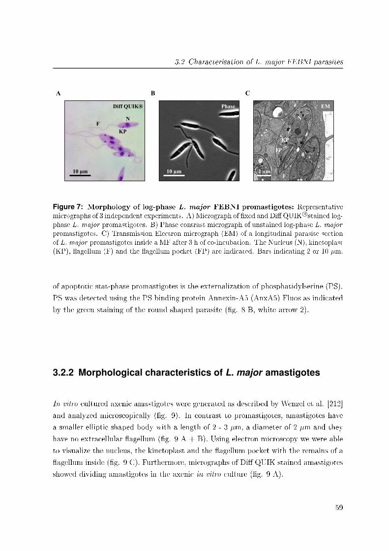
3.2 Characterisation of L. major FEBNI parasites
10 µm
Diff QUIK®
2 µm
EM Phase
10 µm
A B C
FP
N
F
KP
N F
KP
Figure 7: Morphology of log-phase L. major FEBNI promastigotes: Representativemicrographs of 3 independent experiments. A) Micrograph of �xed and Di� QUIK R©stained log-phase L. major promastigotes. B) Phase contrast micrograph of unstained log-phase L. major
promastigotes. C) Transmission Electron micrograph (EM) of a longitudinal parasite sectionof L. major promastigotes inside a MF after 3 h of co-incubation. The Nucleus (N), kinetoplast(KP), �agellum (F) and the �agellum pocket (FP) are indicated. Bars indicating 2 or 10 µm.
of apoptotic stat-phase promastigotes is the externalization of phosphatidylserine (PS).
PS was detected using the PS binding protein Annexin-A5 (AnxA5) Fluos as indicated
by the green staining of the round shaped parasite (�g. 8 B, white arrow 2).
3.2.2 Morphological characteristics of L. major amastigotes
In vitro cultured axenic amastigotes were generated as described by Wenzel et al. [212]
and analyzed microscopically (�g. 9). In contrast to promastigotes, amastigotes have
a smaller elliptic shaped body with a length of 2 - 3 µm, a diameter of 2 µm and they
have no extracellular �agellum (�g. 9 A + B). Using electron microscopy we were able
to visualize the nucleus, the kinetoplast and the �agellum pocket with the remains of a
�agellum inside (�g. 9 C). Furthermore, micrographs of Di� QUIK stained amastigotes
showed dividing amastigotes in the axenic in vitro culture (�g. 9 A).
59
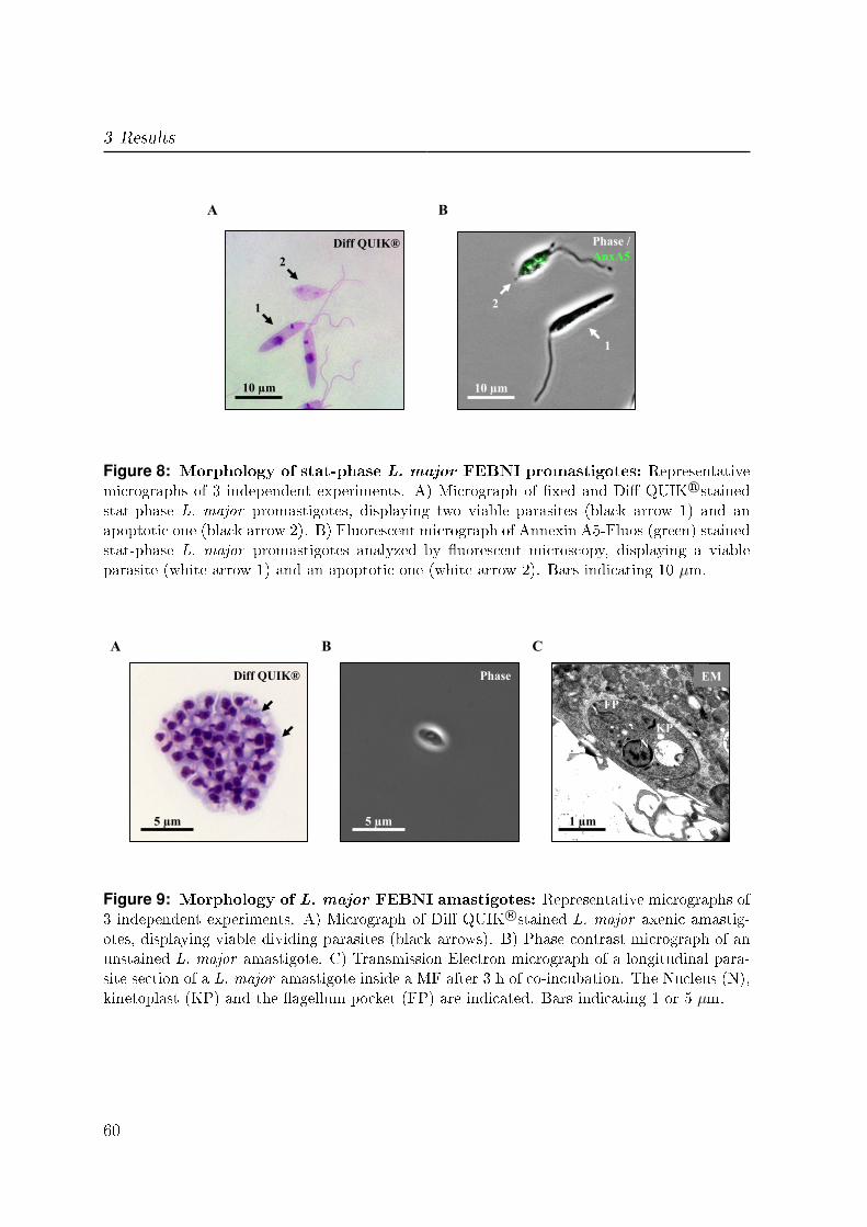
3 Results
10 µm 10 µm
Diff QUIK® Phase /
AnxA5
A B
1
2
1
2
Figure 8: Morphology of stat-phase L. major FEBNI promastigotes: Representativemicrographs of 3 independent experiments. A) Micrograph of �xed and Di� QUIK R©stainedstat-phase L. major promastigotes, displaying two viable parasites (black arrow 1) and anapoptotic one (black arrow 2). B) Fluorescent micrograph of Annexin A5-Fluos (green) stainedstat-phase L. major promastigotes analyzed by �uorescent microscopy, displaying a viableparasite (white arrow 1) and an apoptotic one (white arrow 2). Bars indicating 10 µm.
5 µm 5 µm 1 µm
Diff QUIK® EM Phase
A B C
FP
N
KP
Figure 9: Morphology of L. major FEBNI amastigotes: Representative micrographs of3 independent experiments. A) Micrograph of Di� QUIK R©stained L. major axenic amastig-otes, displaying viable dividing parasites (black arrows). B) Phase contrast micrograph of anunstained L. major amastigote. C) Transmission Electron micrograph of a longitudinal para-site section of a L. major amastigote inside a MF after 3 h of co-incubation. The Nucleus (N),kinetoplast (KP) and the �agellum pocket (FP) are indicated. Bars indicating 1 or 5 µm.
60
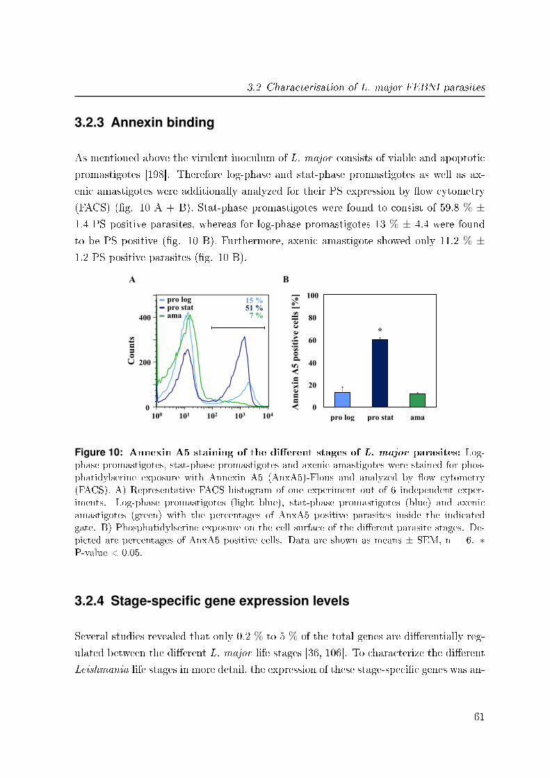
3.2 Characterisation of L. major FEBNI parasites
3.2.3 Annexin binding
As mentioned above the virulent inoculum of L. major consists of viable and apoptotic
promastigotes [198]. Therefore log-phase and stat-phase promastigotes as well as ax-
enic amastigotes were additionally analyzed for their PS expression by �ow cytometry
(FACS) (�g. 10 A + B). Stat-phase promastigotes were found to consist of 59.8 % ±1.4 PS positive parasites, whereas for log-phase promastigotes 13 % ± 4.4 were found
to be PS positive (�g. 10 B). Furthermore, axenic amastigote showed only 11.2 % ±1.2 PS positive parasites (�g. 10 B).
A B
An
nex
in A
5 p
osi
tiv
e ce
lls
[%]
pro log pro stat ama
0
20
40
60
80
100
*
101 102 103 104
Co
un
ts
0
pro stat pro log
ama
100
400
200
Annexin A5-Fluos
15 % 51 %
7 %
Figure 10: Annexin A5 staining of the di�erent stages of L. major parasites: Log-phase promastigotes, stat-phase promastigotes and axenic amastigotes were stained for phos-phatidylserine exposure with Annexin A5 (AnxA5)-Flous and analyzed by �ow cytometry(FACS). A) Representative FACS histogram of one experiment out of 6 independent exper-iments. Log-phase promastigotes (light blue), stat-phase promastigotes (blue) and axenicamastigotes (green) with the percentages of AnxA5 positive parasites inside the indicatedgate. B) Phosphatidylserine exposure on the cell surface of the di�erent parasite stages. De-picted are percentages of AnxA5 positive cells. Data are shown as means ± SEM, n = 6. ∗P-value < 0.05.
3.2.4 Stage-specific gene expression levels
Several studies revealed that only 0.2 % to 5 % of the total genes are di�erentially reg-
ulated between the di�erent L. major life stages [36, 106]. To characterize the di�erent
Leishmania life stages in more detail, the expression of these stage-speci�c genes was an-
61
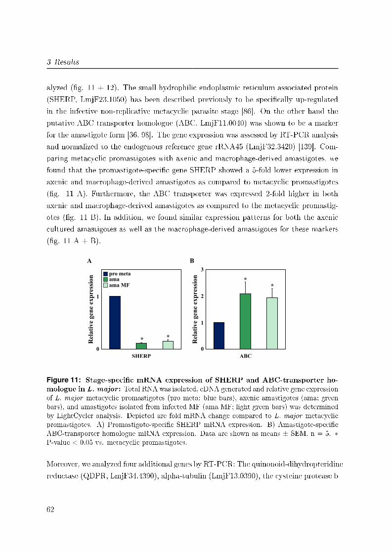
3 Results
alyzed (�g. 11 + 12). The small hydrophilic endoplasmic reticulum-associated protein
(SHERP, LmjF23.1050) has been described previously to be speci�cally up-regulated
in the infective non-replicative metacyclic parasite stage [86]. On the other hand the
putative ABC transporter homologue (ABC, LmjF11.0040) was shown to be a marker
for the amastigote form [36, 98]. The gene expression was assessed by RT-PCR analysis
and normalized to the endogenous reference gene rRNA45 (LmjF32.3420) [139]. Com-
paring metacyclic promastigotes with axenic and macrophage-derived amastigotes, we
found that the promastigote-speci�c gene SHERP showed a 5-fold lower expression in
axenic and macrophage-derived amastigotes as compared to metacyclic promastigotes
(�g. 11 A). Furthermore, the ABC transporter was expressed 2-fold higher in both
axenic and macrophage-derived amastigotes as compared to the metacyclic promastig-
otes (�g. 11 B). In addition, we found similar expression patterns for both the axenic
cultured amastigotes as well as the macrophage-derived amastigotes for these markers
(�g. 11 A + B).
0
1
SHERP
pro meta ama ama MF
Rel
ati
ve
gen
e ex
pre
ssio
n
0
1
2
3
ABC
Rel
ati
ve
gen
e ex
pre
ssio
n
*
* *
A B
*
Figure 11: Stage-speci�c mRNA expression of SHERP and ABC-transporter ho-mologue in L. major : Total RNA was isolated, cDNA generated and relative gene expressionof L. major metacyclic promastigotes (pro meta; blue bars), axenic amastigotes (ama; greenbars), and amastigotes isolated from infected MF (ama MF; light green bars) was determinedby LightCycler analysis. Depicted are fold mRNA change compared to L. major metacyclicpromastigotes. A) Promastigote-speci�c SHERP mRNA expression. B) Amastigote-speci�cABC-transporter homologue mRNA expression. Data are shown as means ± SEM, n = 5. ∗P-value < 0.05 vs. metacyclic promastigotes.
Moreover, we analyzed four additional genes by RT-PCR: The quinonoid-dihydropteridine
reductase (QDPR, LmjF34.4390), alpha-tubulin (LmjF13.0390), the cysteine protease b
62

3.2 Characterisation of L. major FEBNI parasites
(Cpb, LmjF08.1040) and the major surface glycoprotein (GP63; LmjF10.0470) as shown
in �g. 12 [105, 98, 129, 116]. For QDPR we found a signi�cantly higher expression in
axenic amastigotes compared to stat-phase promastigotes, whereas alpha-tubulin was
signi�cantly down-regulated in amastigotes (�g. 12 A). Furthermore, Cpb and GP63
were both signi�cantly higher expressed in the amastigote stage compared to stat-phase
and log-phase promastigotes (�g. 12 B). These data demonstrate that the di�erent life
stages of L. major show a stage-speci�c gene expression.
pro log pro stat ama
*
*
QDPR α-Tubulin
0
1
2
3
Rel
ati
ve
gen
e ex
pre
ssio
n
A B
0
1
2
3
4
Cpb GP63
Rel
ati
ve
gen
e ex
pre
ssio
n
*
*
Figure 12: Stage-speci�c mRNA expression of QDPR, alpha-tubulin, Cpb andGP63 in L. major : Total RNA was isolated, cDNA generated and relative gene expres-sion of log-phase L. major promastigotes (pro log; light blue bars), stat-phase promastigotes(pro stat; blue bars) and axenic amastigotes (ama; green bars) was determined by LightCycleranalysis. Depicted are fold mRNA change compared to stat-phase L. major promastigotes.A) Gene expression of QDPR and alpha-tubulin. B) Gene expression of Cpb and GP63. Dataare shown as means ± SEM, n = 3. ∗ P-value < 0.05 vs. stat-phase promastigotes.
3.2.5 The surface marker lipophosphoglycan (LPG)
The most abundant surface macromolecule on the promastigote stage of Leishmania
parasites is the polymorphic lipophosphoglycan (LPG). Glaser et al. demonstrated
that this promastigote-speci�c LPG is nearly absent on the amastigote life stage [63].
The expression of promastigote-speci�c LPG was analyzed on the parasite surface of
the di�erent life stages by FACS analysis (�g. 13) and by western blot (�g. 14).
LPG was detected with the murine WIC79.3 antibody (Ab) and visualized with Alexa
63

3 Results
488-labeled anti-mouse Ab using FACS analysis. In concordance with the literature,
log-phase promastigotes and axenic amastigotes were found to express a signi�cant
lower level of LPG as compared to stat-phase promastigotes (�g. 13 B). These results
were con�rmed by western blot analysis (�g. 14 A). Parasite lysates were separated by
SDS-PAGE and LPG was detected by the murine WIC79.3 Ab. The band intensity was
normalized with the whole protein amount gained by coomassie staining (�g. 14 B).
LPG
Geo
met
ric
mea
n (
x 1
00
0)
20
30
10
0
* *
pro log pro stat ama
101 102 103 104 105
Co
un
ts
0
pro stat pro log
ama control
A B
LPG-Alexa 488
Figure 13: Stage-speci�c protein expression of LPG on the cell surface of L. ma-
jor : Log-phase promastigotes, stat-phase promastigotes and axenic amastigotes were stainedfor lipophosphoglycan (LPG) expression with mouse anti-LPG WIC79.3 (1:3000), detectedwith Alexa 488-labeled chicken anti-mouse Ab (2 µg) and analyzed by �ow cytometry (FACS)A) Representative FACS histogram of one experiment out of 3 independent experiments.Log-phase promastigotes (light blue), stat-phase promastigotes (blue) and axenic amastig-otes (green) with the corresponding control (grey). B) LPG expression on the cell surface ofthe di�erent parasite stages. Depicted are geometric means x 1000. Data are shown as means± SEM, n = 3. ∗ P-value < 0.05 vs. stat-phase promastigotes.
3.2.6 Generation of axenic amastigotes in other L. major strains
To validate the method for amastigote generation described by Wenzel et al. [212],
several other L. major strains were transformed into the amastigote stage. Using our
standardized protocol we found that for the transfected L. major FEBNI eGFP strain
the incubation time of the amastigote pre-culture in AAM was too long, resulting in
many dead parasites in the culture. In contrast, for the L. major FEBNI DsRed strain
the transformation into amastigotes was not fully completed after the pre-culture, which
64

3.2 Characterisation of L. major FEBNI parasites
0
100
200
300
400
pro log pro stat ama
LP
G b
an
d i
nte
nsi
ty
pro
stat ama
pro
log M
Coomassie WB
102 76
52
38
31
24
17
kDa pro
stat ama
pro
log M
A B
Figure 14: Stage-speci�c protein expression of LPG in L. major : Log-phase pro-mastigotes (pro log), stat-phase promastigotes (pro stat) and axenic amastigotes (ama) werelysed in 1x-Lämmli Bu�er and subjected to SDS-PAGE. LPG was detected with mouse anti-LPG WIC79.3 (1:1000) and Anti-mouse HRP (1:1000). Gel was stained with Coomassie blue.A) Coomassie staining and LPG western blot of the di�erent parasite stages. A full rangerainbow marker was used as molecular size marker (M). B) LPG band intensity of the di�erentparasite stages after normalization with the whole protein amount. Log-phase promastigotes(light blue), stat-phase promastigotes (blue) and axenic amastigotes (green), n = 1.
led to a high contamination with promastigotes. Therefore small modi�cations in the
transformation procedure had to be assessed. We de�ned a pre-culture of amastigotes
to be completely transformed and viable, when the developed amastigotes started di-
viding. Focusing on this characteristic feature, di�erent time points were tested for
the amastigote pre-culture to achieve a complete transformation resulting in diving
amastigotes. We could successfully convert the genetically modi�ed �uorescent strains
L. major FEBNI eGFP and DsRed into amastigotes by adjusting the incubation time
(data not shown). For the transgenic L. major FEBNI eGFP strain the incubation
time was shorted to 7 - 10 days of amastigote pre-culture at 33◦C. Whereas in case of
the transgenic L. major FEBNI DsRed strain 12 - 14 days of amastigote pre-culture at
33◦C were required to get dividing amastigotes.
In addition to L. major FEBNI, we wanted also to transform the well characterized
L. major strain MHOM/IL/80/Friedlin. This was the �rst Leishmania strain whose
genome was completely sequenced [125, 76]. To �nd the adequate conditions for the
amastigote transformation of the Friedlin strain, di�erent parameters like the incubation
65
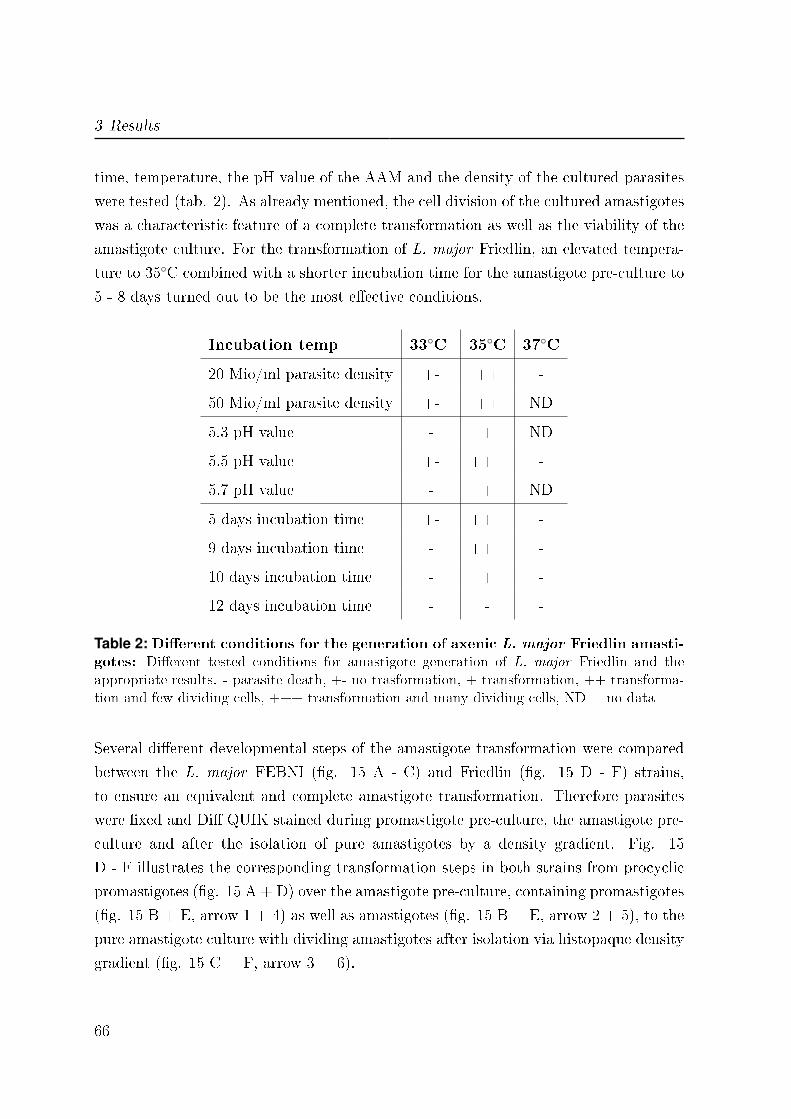
3 Results
time, temperature, the pH value of the AAM and the density of the cultured parasites
were tested (tab. 2). As already mentioned, the cell division of the cultured amastigotes
was a characteristic feature of a complete transformation as well as the viability of the
amastigote culture. For the transformation of L. major Friedlin, an elevated tempera-
ture to 35◦C combined with a shorter incubation time for the amastigote pre-culture to
5 - 8 days turned out to be the most e�ective conditions.
Incubation temp 33◦C 35◦C 37◦C
20 Mio/ml parasite density +- ++ -
50 Mio/ml parasite density +- ++ ND
5.3 pH value - + ND
5.5 pH value +- +++ -
5.7 pH value - + ND
5 days incubation time +- +++ -
9 days incubation time - +++ -
10 days incubation time - + -
12 days incubation time - - -
Table 2: Di�erent conditions for the generation of axenic L. major Friedlin amasti-gotes: Di�erent tested conditions for amastigote generation of L. major Friedlin and theappropriate results. - parasite death, +- no trasformation, + transformation, ++ transforma-tion and few dividing cells, +++ transformation and many dividing cells, ND = no data
Several di�erent developmental steps of the amastigote transformation were compared
between the L. major FEBNI (�g. 15 A - C) and Friedlin (�g. 15 D - F) strains,
to ensure an equivalent and complete amastigote transformation. Therefore parasites
were �xed and Di� QUIK stained during promastigote pre-culture, the amastigote pre-
culture and after the isolation of pure amastigotes by a density gradient. Fig. 15
D - F illustrates the corresponding transformation steps in both strains from procyclic
promastigotes (�g. 15 A + D) over the amastigote pre-culture, containing promastigotes
(�g. 15 B + E, arrow 1 + 4) as well as amastigotes (�g. 15 B + E, arrow 2 + 5), to the
pure amastigote culture with dividing amastigotes after isolation via histopaque density
gradient (�g. 15 C + F, arrow 3 + 6).
66
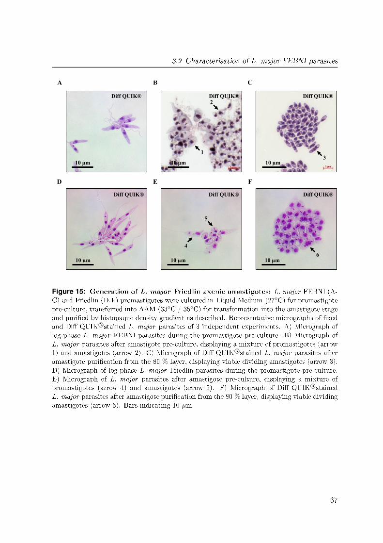
3.2 Characterisation of L. major FEBNI parasites
Diff QUIK®
10 µm 10 µm 10 µm
D E F
Diff QUIK® Diff QUIK®
4
5
6
Diff QUIK® Diff QUIK® Diff QUIK®
10 µm 10 µm 10 µm
1
2
3
A B C
Figure 15: Generation of L. major Friedlin axenic amastigotes: L. major FEBNI (A-C) and Friedlin (D-F) promastigotes were cultured in Liquid Medium (27◦C) for promastigotepre-culture, transferred into AAM (33◦C / 35◦C) for transformation into the amastigote stageand puri�ed by histopaque density gradient as described. Representative micrographs of �xedand Di� QUIK R©stained L. major parasites of 3 independent experiments. A) Micrograph oflog-phase L. major FEBNI parasites during the promastigote pre-culture. B) Micrograph ofL. major parasites after amastigote pre-culture, displaying a mixture of promastigotes (arrow1) and amastigotes (arrow 2). C) Micrograph of Di� QUIK R©stained L. major parasites afteramastigote puri�cation from the 80 % layer, displaying viable dividing amastigotes (arrow 3).D) Micrograph of log-phase L. major Friedlin parasites during the promastigote pre-culture.E) Micrograph of L. major parasites after amastigote pre-culture, displaying a mixture ofpromastigotes (arrow 4) and amastigotes (arrow 5). F) Micrograph of Di� QUIK R©stainedL. major parasites after amastigote puri�cation from the 80 % layer, displaying viable dividingamastigotes (arrow 6). Bars indicating 10 µm.
67
3 Results
3.3 Detection of Apoptotic characteristics in L. major
parasites
In multicellular organisms, apoptosis is a tightly regulated pathway of the cell to die.
An early sign is the externalization of PS from the inner to the outer lea�et of the cell
membrane [110]. Another early marker which is connected to apoptosis is the intracellu-
lar formation of reactive oxygen species (ROS) [160, 176]. To investigate the presence of
extracellular expression of PS and intracellular expression of ROS in L. major parasites
after apoptosis induction we used �ow cytometry.
3.3.1 Apoptosis mechanisms in promastigotes
L. major promastigotes were treated with three di�erent drugs to induce apoptosis:
Staurosporine which triggers the mitochondrial pathway, the anti-leishmanial drug mil-
tefosine which a�ects the parasite membrane and camptothecin, a nucleus dependent
apoptosis inducer. After the treatment the parasites were analyzed for PS exposure with
the PS recognizing protein AnxA5, labeled with Alexa 647 and ROS was detected with 5-
(and-6)-chloromethyl-2',7'-dichlorodihydro�uorescein diacetate (H2DCFDA). We found
a signi�cant higher PS expression of 72.7 % ± 6.9 for the miltefosine treated promastig-
otes as compared to 28.3 % ± 2.6 positive parasites for the untreated promastigotes after
18 hours (�g. 18 A). However, 42 hours of treatment resulted in a signi�cant higher
level of PS positive promastigotes in both miltefosine (81.3 % ± 2) and staurosporine
(61 % ± 8.9) treated parasites. Camptothecin had no e�ect on the PS externalization.
Detecting the formation of ROS, we found signi�cant higher percentages of ROS pos-
itive parasites only for staurosporine treated promastigotes compared to the low ROS
level in the untreated parasites (�g. 18 B). 18 hours after treatment there was a max-
imum of 60 % ± 4.1 ROS positive parasites and then decreased to 39.7 % ± 10 ROS
positivity after 42 hours. Moreover, these ROS positive parasites were detected to have
a signi�cant higher mean �uorescence intensity (MFI) for ROS after 18 hours (�g. 18
C). In contrast, miltefosine induced a non-signi�cant increase in ROS over time (p-value
= 0.078), whereas camptothecin showed no ROS induction.
68
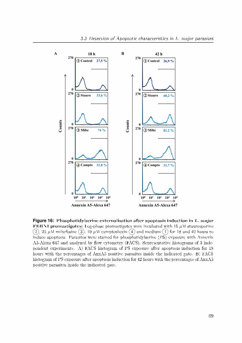
3.3 Detection of Apoptotic characteristics in L. major parasites
Co
un
ts
Annexin A5-Alexa 647
A 18 h
33,6 % Stauro
102 101 103 104 100
74 % Milte
31,8 % Campto
Control 27,5 % 26,9 %
60,2 %
81,2 %
31,7 %
270
Co
un
ts
Annexin A5-Alexa 647
B
0
42 h
Stauro
102 101 103 104
0
100
Milte
Campto
Control
270
0
270
0
270
270
0
0
270
0
270
0
270
1 1
2 2
3 3
4 4
Figure 16: Phosphatidylserine externalisation after apoptosis induction in L. major
FEBNI promastigotes: Log-phase promastigotes were incubated with 15 µM staurosporine2 , 25 µM miltefosine 3 , 10 µM camptothecin 4 and medium 1 for 18 and 42 hours toinduce apoptosis. Parasites were stained for phosphatidylserine (PS) exposure with AnnexinA5-Alexa 647 and analyzed by �ow cytometry (FACS). Representative histograms of 3 inde-pendent experiments. A) FACS histogram of PS exposure after apoptosis induction for 18hours with the percentages of AnxA5 positive parasites inside the indicated gate. B) FACShistogram of PS exposure after apoptosis induction for 42 hours with the percentages of AnxA5positive parasites inside the indicated gate.
69

3 Results
Co
un
ts
A 18 h
58,1 % Stauro
102 101 103 104 100
19,7 % Milte
9,8 % Campto
Control 12,6 % 15,3 %
45,7 %
26,5 %
18,5 %
270
Co
un
ts
B
0
42 h
Stauro
102 101 103 104
0
100
Milte
Campto
Control
270
0
270
0
270
270
0
0
270
0
270
0
270
ROS (H2DCFDA) ROS (H2DCFDA)
1
2
3
1
2
3
4 4
Figure 17: Formation of reactive oxygen species after apoptosis induction in L.
major FEBNI promastigotes: Log-phase promastigotes were incubated with 15 µM stau-rosporine 2 , 25 µM miltefosine 3 , 10 µM camptothecin 4 and medium control 1 for 18and 42 hours to induce apoptosis. Parasites were stained for reactive oxygen species (ROS)with 5-(and-6)-chloromethyl-2',7'-dichlorodihydro�uorescein diacetate (H2DCFDA) and ana-lyzed by �ow cytometry (FACS). Representative histograms of 3 independent experiments. A)FACS histograms of ROS presence after apoptosis induction for 18 hours with the percentagesof ROS positive parasites inside the indicated gate. B) FACS histograms of ROS presenceafter apoptosis induction for 42 hours with the percentages of ROS positive parasites insidethe indicated gate.
3.3.2 Apoptosis mechanisms in amastigotes
Corresponding to the apoptosis induction in promastigotes, the amastigote life stage
parasites were treated with the same drugs and concentrations: staurosporine, milte-
70
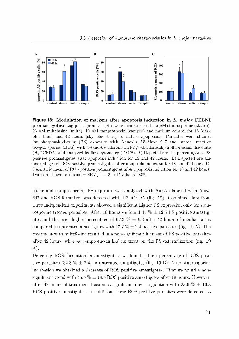
3.3 Detection of Apoptotic characteristics in L. major parasites
stauro milte campto control 0
20
40
60
80
An
nex
in A
5 p
osi
tiv
e ce
lls
[%]
A
stauro milte campto control 0
20
40
60
80
RO
S p
osi
tive
cell
s [%
]
B
stauro milte campto control
C
0
100
200
300
400
Geo
met
ric m
ean
of
RO
S
*
18 h 42 h
*
* *
*
*
Figure 18: Modulation of markers after apoptosis induction in L. major FEBNIpromastigotes: Log-phase promastigotes were incubated with 15 µM staurosporine (stauro),25 µM miltefosine (milte), 10 µM camptothecin (campto) and medium control for 18 (darkblue bars) and 42 hours (sky blue bars) to induce apoptosis. Parasites were stainedfor phosphatidylserine (PS) exposure with Annexin A5-Alexa 647 and present reactiveoxygen species (ROS) with 5-(and-6)-chloromethyl-2',7'-dichlorodihydro�uorescein diacetate(H2DCFDA) and analyzed by �ow cytometry (FACS). A) Depicted are the percentages of PSpositive promastigotes after apoptosis induction for 18 and 42 hours. B) Depicted are thepercentages of ROS positive promastigotes after apoptosis induction for 18 and 42 hours. C)Geometric mean of ROS positive promastigotes after apoptosis induction for 18 and 42 hours.Data are shown as means ± SEM, n = 3. ∗ P-value < 0.05.
fosine and camptothecin. PS exposure was analyzed with AnxA5 labeled with Alexa
647 and ROS formation was detected with H2DCFDA (�g. 19). Combined data from
three independent experiments showed a signi�cant higher PS expression only for stau-
rosporine treated parasites. After 18 hours we found 44 % ± 12.6 PS positive amastig-
otes and the even higher percentage of 62.3 % ± 6.3 after 42 hours of incubation as
compared to untreated amastigotes with 13.7 % ± 2.4 positive parasites (�g. 19 A). The
treatment with miltefosine resulted in a non-signi�cant increase of PS positive parasites
after 42 hours, whereas camptothecin had no e�ect on the PS externalization (�g. 19
A).
Detecting ROS formation in amastigotes, we found a high percentage of ROS posi-
tive parasites (82.3 % ± 2.4) in untreated amastigotes (�g. 19 B). After staurosporine
incubation we obtained a decrease of ROS positive amastigotes. First we found a non-
signi�cant trend with 45.5 % ± 18.6 ROS positive amastigotes after 18 hours. However,
after 42 hours of treatment became a signi�cant down-regulation with 23.6 % ± 10.8
ROS positive amastigotes. In addition, these ROS positive parasites were detected to
71
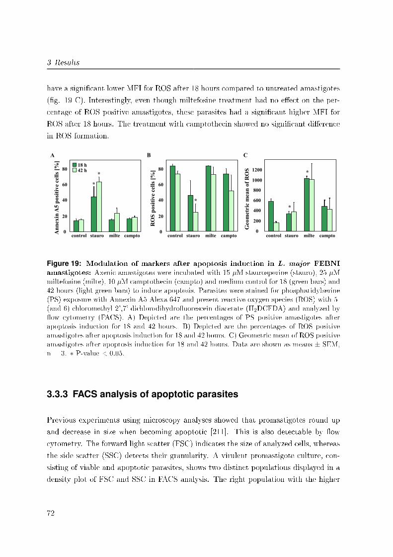
3 Results
have a signi�cant lower MFI for ROS after 18 hours compared to untreated amastigotes
(�g. 19 C). Interestingly, even though miltefosine treatment had no e�ect on the per-
centage of ROS positive amastigotes, these parasites had a signi�cant higher MFI for
ROS after 18 hours. The treatment with camptothecin showed no signi�cant di�erence
in ROS formation.
Geo
met
ric m
ean
of
RO
S
0
200
400
600
800
1000
1200
stauro milte campto control 0
20
40
60
80
An
nex
in A
5 p
osi
tiv
e ce
lls
[%]
A
18 h 42 h
stauro milte campto control 0
20
40
60
80
RO
S p
osi
tive
cell
s [%
] B
stauro milte campto control
C
*
* *
*
*
Figure 19: Modulation of markers after apoptosis induction in L. major FEBNIamastigotes: Axenic amastigotes were incubated with 15 µM staurosporine (stauro), 25 µMmiltefosine (milte), 10 µM camptothecin (campto) and medium control for 18 (green bars) and42 hours (light green bars) to induce apoptosis. Parasites were stained for phosphatidylserine(PS) exposure with Annexin A5-Alexa 647 and present reactive oxygen species (ROS) with 5-(and-6)-chloromethyl-2',7'-dichlorodihydro�uorescein diacetate (H2DCFDA) and analyzed by�ow cytometry (FACS). A) Depicted are the percentages of PS positive amastigotes afterapoptosis induction for 18 and 42 hours. B) Depicted are the percentages of ROS positiveamastigotes after apoptosis induction for 18 and 42 hours. C) Geometric mean of ROS positiveamastigotes after apoptosis induction for 18 and 42 hours. Data are shown as means ± SEM,n = 3. ∗ P-value < 0.05.
3.3.3 FACS analysis of apoptotic parasites
Previous experiments using microscopy analyses showed that promastigotes round up
and decrease in size when becoming apoptotic [211]. This is also detectable by �ow
cytometry. The forward light scatter (FSC) indicates the size of analyzed cells, whereas
the side scatter (SSC) detects their granularity. A virulent promastigote culture, con-
sisting of viable and apoptotic parasites, shows two distinct populations displayed in a
density plot of FSC and SSC in FACS analysis. The right population with the higher
72

3.3 Detection of Apoptotic characteristics in L. major parasites
density and size represents the viable promastigotes and the apoptotic parasites are
found in the other population (left) with the lower density and size. We found such two
distinct populations of parasites in FACS after the treatment of promastigotes with mil-
tefosine (�g. 20 A + B). Further analysis revealed the right population with the higher
density to consist of PS negative and thus viable promastigotes (�g. 20 B). In contrast,
we found the PS positive promastigotes in the population with the lower density and
size.
SS
C
FSC
viable apoptotic
102 101
270
103 104
Co
un
ts
0
viable
100
Annexin A5-Alexa 647
apoptotic
D
0,6 % 70,1 %
C
SS
C
FSC
vaible apoptotic
270
B
101 102 103 104
Co
un
ts
0
viable
100
Annexin A5-Alexa 647
5,5 % 84,2 % apoptotic
A
Figure 20: Viable and apoptotic L. major FEBNI parasites in �ow cytometry: Log-phase promastigotes were incubated with 25 µMmiltefosine and axenic amastigotes with 15 µMstaurosporine for 18 hours to induce apoptosis. Parasites were stained for phosphatidylserineexposure with Annexin A5-Alexa 647 and analyzed by �ow cytometry (FACS). Representa-tive plots of 3 independent experiments. A) FACS density plot of viable and apoptotic pro-mastigotes after apoptosis induction display two di�erent populations. B) FACS histogramsof promastigotes gated on the viable (dark blue) and apoptotic (blue) parasite population. C)FACS density plot of viable and apoptotic amastigotes after apoptosis induction display twodi�erent populations. D) FACS histograms of amastigotes gated on the viable (dark blue) andapoptotic (blue) parasite population.
73
3 Results
Furthermore, a similar distribution of the parasites was found in a density plot of FSC
and SSC for amastigotes after the induction of apoptosis (�g. 20 C + D). Two distinct
populations were detected after incubation of amastigotes with staurosporine. Analy-
zing these populations for PS revealed the right more dense population to consist of PS
negative viable amastigotes, while the left less dense population resulted to be mainly
PS positive parasites (�g. 20 D).
74

3.4 Interaction of L. major with human MF
Part 2
3.4 Interaction of L. major with human MF
In Leishmaniasis MF are known to be the �nal host cells. But it is still not clear which
type of MF is involved in disease development or parasite clearance. There are di�erent
phenotypes of human MF present in the human body, such as pro-in�ammatory type
I (MF I) and anti-in�ammatory type II (MF II) MF [203]. Both phenotypes, MF I as
well as MF II can be possible host cells for L. major parasites (�g. 21). Therefore, the
interaction of L. major promastigotes and amastigotes with pro-in�ammatory as well
as anti-in�ammatory human MF was investigated.
3.4.1 Infection of different phenotypes of MF with L. major
Initially, we characterized the infectivity of both L. major parasite life stages for the
di�erent phenotypes of MF. In pro-in�ammatory MF I we found a signi�cant lower
infection rate of 38.7 % ± 3.5 as compared to 51.5 % ± 4.5 for anti-in�ammatory MF
II after co-incubation with promastigotes (�g. 22 A). In addition, in pro-in�ammatory
MF I we found a signi�cant lower infection rate, after co-incubation with amastigotes
of 66.3 % ± 3.5 as compared to 80.6 % ± 1.5 for anti-in�ammatory MF II. Moreover
we found that amastigotes are more infective than promastigotes for both phenotypes
of MF. Next to infection we assessed the parasite burden by quantifying the number of
intracellular parasites per MF. In concordance with the MF phenotype speci�c as well
as parasite stage speci�c infection rates, we found a signi�cant lower parasite burden
of 2.8 ± 0.2 parasites/MF in pro-in�ammatory MF I as compared to 4.0 ± 0.5 in anti-
in�ammatory MF II after promastigote co-incubation (�g. 22 C). Furthermore, the
co-incubation with amastigotes resulted also in lower parasite uptake in MF I of 18.3
± 2.7 compared to 25.6 ± 2.2 in MF II (�g. 22 D), however in higher parasite burdens
than after promastigote co-incubation.
75

3 Results
MF II + Lm ama
3 h PI
MF II + Lm pro
3 h PI
MF I + Lm pro
3 h PI
MF I + Lm ama
3 h PI
A B
C D
Figure 21: Infected human MF with L. major parasites: Pro-in�ammatory type IMF (MF I) and anti-in�ammatory type II MF (MF II) were co-incubated with stat-phasepromastigotes (A and C) or axenic amastigotes (B and D). Extracellular parasites were removed3 hours post infection (PI) and the cells stained with Di� QUIK R©. Representative micrographsof 3 independent experiments. A) Micrograph of MF I infected with L. major promastigotes.B) Micrograph of MF I infected with L. major amastigotes. C) Micrograph of MF II infectedwith L. major promastigotes. D) Micrograph of MF II infected with L. major amastigotes.Magni�cation: 63x objective with 10x ocular.
76
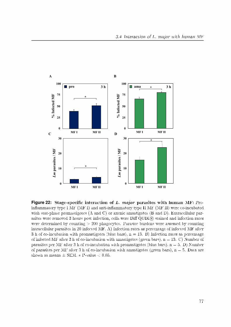
3.4 Interaction of L. major with human MF
MF I MF II
% I
nfe
cted
MF
MF I MF II
% I
nfe
cted
MF
A B
0
25
50
75
100 ama 3 h
0
25
50
75
100 pro 3 h
0
10
20
30
MF I MF II
*
Lm
pa
rasi
tes
/ M
F
0
10
20
30
MF I MF II
Lm
pa
rasi
tes
/ M
F
*
C D
*
*
Figure 22: Stage-speci�c interaction of L. major parasites with human MF: Pro-in�ammatory type I MF (MF I) and anti-in�ammatory type II MF (MF II) were co-incubatedwith stat-phase promastigotes (A and C) or axenic amastigotes (B and D). Extracellular par-asites were removed 3 hours post infection, cells were Di� QUIK R© stained and infection rateswere determined by counting > 200 phagocytes. Parasite burdens were assessed by countingintracellular parasites in 20 infected MF. A) Infection rates as percentage of infected MF after3 h of co-incubation with promastigotes (blue bars), n = 13. B) Infection rates as percentageof infected MF after 3 h of co-incubation with amastigotes (green bars), n = 13. C) Number ofparasites per MF after 3 h of co-incubation with promastigotes (blue bars), n = 5. D) Numberof parasites per MF after 3 h of co-incubation with amastigotes (green bars), n = 5. Data areshown as means ± SEM. ∗ P-value < 0.05.
77
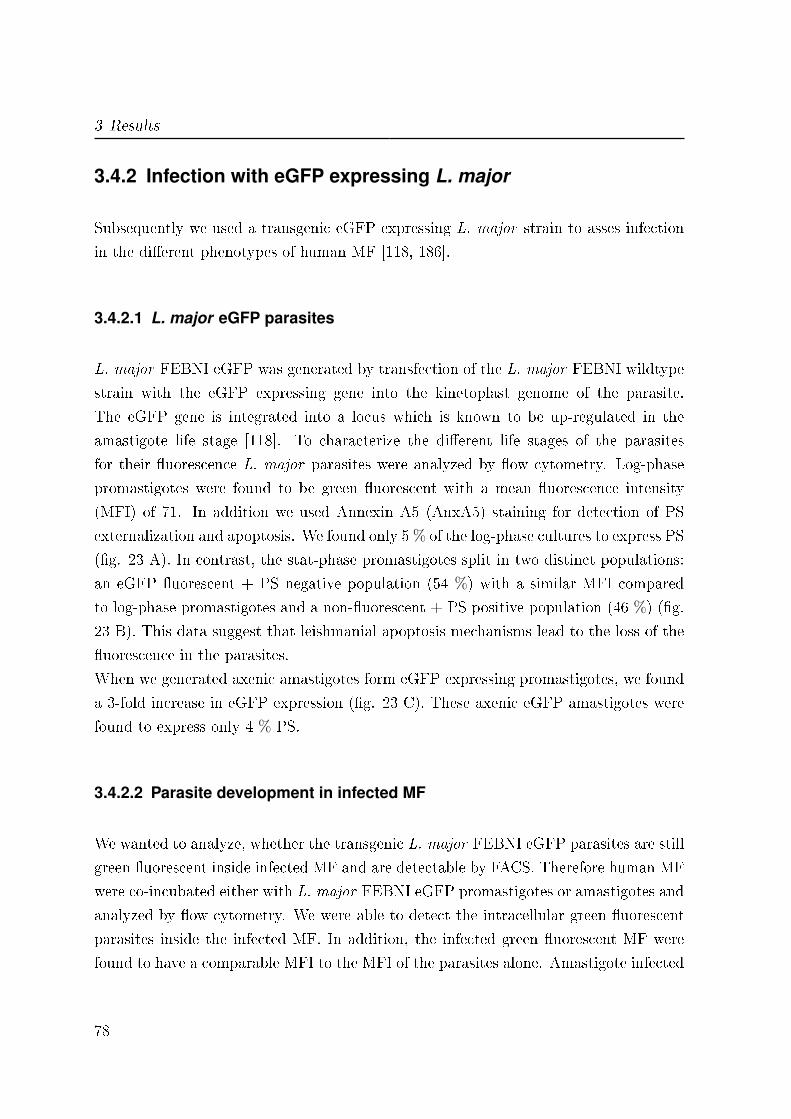
3 Results
3.4.2 Infection with eGFP expressing L. major
Subsequently we used a transgenic eGFP expressing L. major strain to asses infection
in the di�erent phenotypes of human MF [118, 186].
3.4.2.1 L. major eGFP parasites
L. major FEBNI eGFP was generated by transfection of the L. major FEBNI wildtype
strain with the eGFP expressing gene into the kinetoplast genome of the parasite.
The eGFP gene is integrated into a locus which is known to be up-regulated in the
amastigote life stage [118]. To characterize the di�erent life stages of the parasites
for their �uorescence L. major parasites were analyzed by �ow cytometry. Log-phase
promastigotes were found to be green �uorescent with a mean �uorescence intensity
(MFI) of 71. In addition we used Annexin A5 (AnxA5) staining for detection of PS
externalization and apoptosis. We found only 5 % of the log-phase cultures to express PS
(�g. 23 A). In contrast, the stat-phase promastigotes split in two distinct populations:
an eGFP �uorescent + PS negative population (54 %) with a similar MFI compared
to log-phase promastigotes and a non-�uorescent + PS positive population (46 %) (�g.
23 B). This data suggest that leishmanial apoptosis mechanisms lead to the loss of the
�uorescence in the parasites.
When we generated axenic amastigotes form eGFP expressing promastigotes, we found
a 3-fold increase in eGFP expression (�g. 23 C). These axenic eGFP amastigotes were
found to express only 4 % PS.
3.4.2.2 Parasite development in infected MF
We wanted to analyze, whether the transgenic L. major FEBNI eGFP parasites are still
green �uorescent inside infected MF and are detectable by FACS. Therefore human MF
were co-incubated either with L. major FEBNI eGFP promastigotes or amastigotes and
analyzed by �ow cytometry. We were able to detect the intracellular green �uorescent
parasites inside the infected MF. In addition, the infected green �uorescent MF were
found to have a comparable MFI to the MFI of the parasites alone. Amastigote infected
78
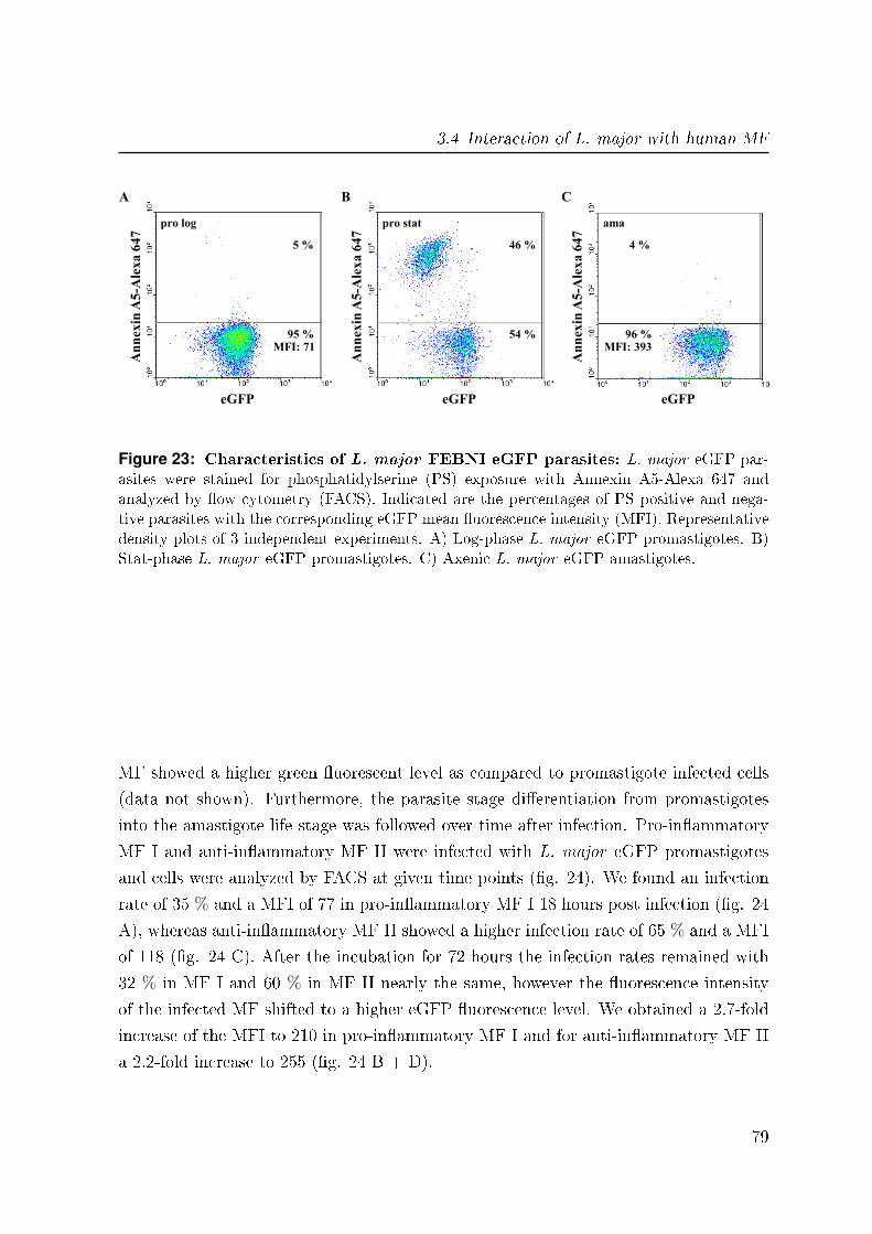
3.4 Interaction of L. major with human MF
5 %
95 %
An
nex
in A
5-A
lex
a 6
47
eGFP
A
pro log
MFI: 71
46 %
54 %
An
nex
in A
5-A
lex
a 6
47
eGFP
B
pro stat
4 %
96 %
An
nex
in A
5-A
lex
a 6
47
eGFP
C
ama
MFI: 393
Figure 23: Characteristics of L. major FEBNI eGFP parasites: L. major eGFP par-asites were stained for phosphatidylserine (PS) exposure with Annexin A5-Alexa 647 andanalyzed by �ow cytometry (FACS). Indicated are the percentages of PS positive and nega-tive parasites with the corresponding eGFP mean �uorescence intensity (MFI). Representativedensity plots of 3 independent experiments. A) Log-phase L. major eGFP promastigotes. B)Stat-phase L. major eGFP promastigotes. C) Axenic L. major eGFP amastigotes.
MF showed a higher green �uorescent level as compared to promastigote infected cells
(data not shown). Furthermore, the parasite stage di�erentiation from promastigotes
into the amastigote life stage was followed over time after infection. Pro-in�ammatory
MF I and anti-in�ammatory MF II were infected with L. major eGFP promastigotes
and cells were analyzed by FACS at given time points (�g. 24). We found an infection
rate of 35 % and a MFI of 77 in pro-in�ammatory MF I 18 hours post infection (�g. 24
A), whereas anti-in�ammatory MF II showed a higher infection rate of 65 % and a MFI
of 118 (�g. 24 C). After the incubation for 72 hours the infection rates remained with
32 % in MF I and 60 % in MF II nearly the same, however the �uorescence intensity
of the infected MF shifted to a higher eGFP �uorescence level. We obtained a 2.7-fold
increase of the MFI to 210 in pro-in�ammatory MF I and for anti-in�ammatory MF II
a 2.2-fold increase to 255 (�g. 24 B + D).
79
3 Results
35 % 62 %
MH
C I
I
eGFP
A
MF I 18 h
32 % 67 %
MH
C I
I
eGFP
B
MF I 72 h
65 % 34 %
MH
C I
I
eGFP
C
MF II 18 h
60 % 40 %
MH
C I
I
eGFP
D
MF II 72 h
MFI: 77 MFI: 210
MFI: 118 MFI: 255
Figure 24: L. major eGFP parasite development in di�erent types of human MF:Pro-in�ammatory MF I and anti-in�ammatory MF II were infected with stat-phase L. ma-
jor eGFP promastigotes. Extracellular parasites were removed 18 hours post infection andinfection rates as well as eGFP mean �uorescence intensity (MFI) were determined 18 and 72hours post infection by �ow cytometry (FACS). Indicated are the percentages of non-infectedand infected MF with the corresponding eGFP MFI. Representative density plots of 3 inde-pendent experiments. A) Infected pro-in�ammatory MF I 18 hours post infection. B) Infectedpro-in�ammatory MF I 72 hours post infection. C) Infected anti-in�ammatory MF II 18 hourspost infection. D) Infected anti-in�ammatory MF II 72 hours post infection.
80
3.5 Phenotype and parasites stage-speci�c MF surface marker expression
3.5 Phenotype and parasites stage-specific MF
surface marker expression
We found a MF phenotype and parasite stage-speci�c infection after co-incubation with
L. major. Now we wanted to know whether these di�erences have an e�ect on distinct
surface markers on the infected MF. Therefore, we infected pro-in�ammatory MF I
and anti-in�ammatory MF II with either L. major promastigotes or amastigotes and
analyzed the phenotypically expression of the following cell surface molecules by �ow
cytometry.
3.5.1 CD163
The scavenger receptor CD163 is a speci�c marker for anti-in�ammatory MF II and
plays a role in the resolution of in�ammation [119, 135]. We found CD163 expression
to be low on MF I (5.3 % ± 1.8) independent of an infection (�g. 26 A), whereas 52.4
% ± 5.5 of the uninfected MF II expressed CD163 on their cell surface. However, the
percentage of CD163 positive MF II decreased signi�cantly upon infection with both
L. major promastigotes and amastigotes, 36.1 % ± 5.8 and 36.8 % ± 5.7 respectively
(�g. 25 B). Furthermore, there was not only a reduction of CD163 positive MF II, but
also the MFI of infected and CD163 positive cells shifted to a signi�cant lower level upon
the infection with promastigotes (�g. 25 C + D) and amastigotes (data not shown).
In addition to the expression of CD163 on the cell surface, we monitored the relative
gene expression by RT-PCR analysis. We found no signi�cant di�erence in the CD163
mRNA expression of untreated MF I and II, 1.6 ± 0.2 and 1.3 ± 0.2 respectively (�g.
26 C + D). However, there was a signi�cant reduction of the CD163 mRNA expression
to 0.7 ± 0.5 for MF I and 0.9 ± 0.2 for MF II after the infection of both phenotypes
of MF with the promastigote life stage. The infection with amastigotes resulted in a
non-signi�cant decrease of CD163 expression.
81
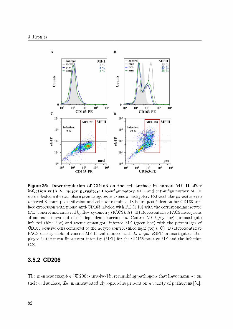
3 Results
A B
pro med
ama
control
pro med
ama
control 55 % 23 % 28 %
MF II 3 % 3 % 3 %
MF I
Co
un
ts
0
101 102 103 104 100
CD163-PE
Co
un
ts
0
101 102 103 104 100
CD163-PE
101 102 103 104 100
101
102
103
104
100
CD163-PE
eGF
P
101 102 103 104 100
101
102
103
104
100
CD163-PE
eGF
P
Infection:
0 %
Infection:
30 %
MFI: 120 MFI: 201 MF II MF II
C D
med pro
Figure 25: Downregulation of CD163 on the cell surface in human MF II afterinfection with L. major parasites: Pro-in�ammatory MF I and anti-in�ammatory MF IIwere infected with stat-phase promastigotes or axenic amastigotes. Extracellular parasites wereremoved 3 hours post infection and cells were stained 18 hours post infection for CD163 sur-face expression with mouse anti-CD163 labeled with PE (1:10) with the corresponding isotype(PE) control and analyzed by �ow cytometry (FACS). A)+B) Representative FACS histogramsof one experiment out of 6 independent experiments. Control MF (grey line), promastigoteinfected (blue line) and axenic amastigote infected MF (green line) with the percentages ofCD163 positive cells compared to the isotype control (�lled light grey). C)+D) RepresentativeFACS density plots of control MF II and infected with L. major eGFP promastigotes. Dis-played is the mean �uorescent intensity (MFI) for the CD163 positive MF and the infectionrate.
3.5.2 CD206
The mannose receptor CD206 is involved in recognizing pathogens that have mannose on
their cell surface, like mannosylated glycoproteins present on a variety of pathogens [31].
82

3.5 Phenotype and parasites stage-speci�c MF surface marker expression
0
20
40
60 C
D1
63
po
siti
ve
MF
[%
]
CD163
MF I med pro ama
*
0
20
40
60
CD163
MF II
*
CD
16
3 p
osi
tive
MF
[%
]
0
1
2 MF I
Rel
ati
ve
gen
e ex
pre
ssio
n MF II
0
1
2
Rel
ati
ve
gen
e ex
pre
ssio
n
*
*
A B
C D
Figure 26: Downregulation of CD163 on the cell surface and mRNA expression inhuman MF II after infection with L. major parasites: Pro-in�ammatory MF I andanti-in�ammatory MF II were infected with stat-phase promastigotes or axenic amastigotes.Extracellular parasites were removed 3 hours post infection and cells were harvested 18 hourspost infection from medium controls (grey bars), promastigote infected MF (blue bars) andamastigote infected MF (green bars). A)+B) MF I and II were stained for CD163 surfaceexpression with mouse anti-CD163 labeled with PE (1:10) with the corresponding isotype(PE) control and analyzed by �ow cytometry (FACS). Depicted are percentages of CD163positive cells, n = 6. C)+D) Total RNA was isolated, cDNA generated and the relative geneexpression was determined by LightCycler analysis. Depicted is the relative gene expressionof MF I and II, n = 5. Data are shown as means ± SEM. ∗ P-value < 0.05.
Therefor CD206 plays a role in receptor-mediated endocytosis and receptor-mediated
facilitated antigen presentation [167, 192, 31]. We found that there is no signi�cant
di�erence in CD206 expression on uninfected MF I and II, 49.7 % ± 12.3 and 55.1 %
± 9.3 respectively (�g. 27 C + D).
However, we did �nd signi�cant higher percentages of CD206 positive MF upon the
infection with both life stages of L. major parasites. For MF I we observed an up-
regulation of CD206 positive cells to 82.1 % ± 2.2 after the infection with promastigote
83
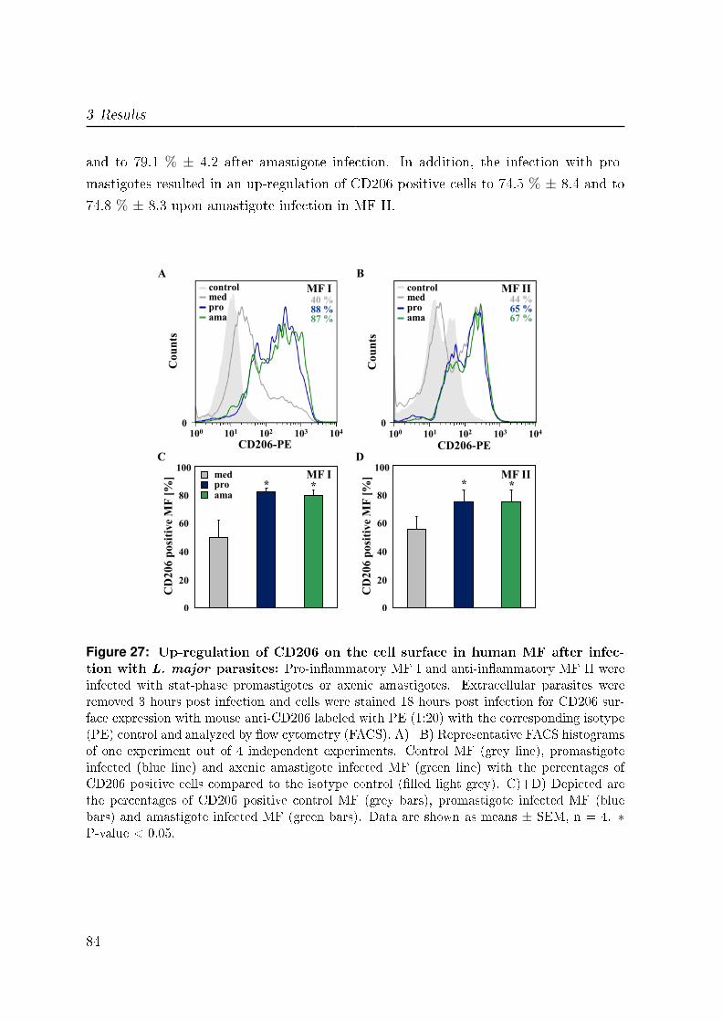
3 Results
and to 79.1 % ± 4.2 after amastigote infection. In addition, the infection with pro-
mastigotes resulted in an up-regulation of CD206 positive cells to 74.5 % ± 8.4 and to
74.8 % ± 8.3 upon amastigote infection in MF II.
* MF I *
CD
20
6 p
osi
tive
MF
[%
]
C
0
20
80
100
40
60
* MF II
*
CD
20
6 p
osi
tive
MF
[%
]
D
0
20
80
100
40
60
med pro ama
Co
un
ts
0
101 102 103 104 100
CD206-PE
pro med
ama
40 % 88 % 87 %
control
pro med
ama
control
Co
un
ts
0
101 102 103 104 100
CD206-PE
44 % 65 % 67 %
MF I MF II
A B
Figure 27: Up-regulation of CD206 on the cell surface in human MF after infec-tion with L. major parasites: Pro-in�ammatory MF I and anti-in�ammatory MF II wereinfected with stat-phase promastigotes or axenic amastigotes. Extracellular parasites wereremoved 3 hours post infection and cells were stained 18 hours post infection for CD206 sur-face expression with mouse anti-CD206 labeled with PE (1:20) with the corresponding isotype(PE) control and analyzed by �ow cytometry (FACS). A)+B) Representative FACS histogramsof one experiment out of 4 independent experiments. Control MF (grey line), promastigoteinfected (blue line) and axenic amastigote infected MF (green line) with the percentages ofCD206 positive cells compared to the isotype control (�lled light grey). C)+D) Depicted arethe percentages of CD206 positive control MF (grey bars), promastigote infected MF (bluebars) and amastigote infected MF (green bars). Data are shown as means ± SEM, n = 4. ∗P-value < 0.05.
84
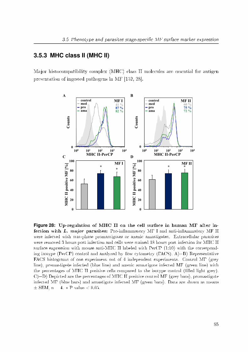
3.5 Phenotype and parasites stage-speci�c MF surface marker expression
3.5.3 MHC class II (MHC II)
Major histocompatibility complex (MHC) class II molecules are essential for antigen
presentation of ingested pathogens in MF [152, 28].
* MF I
MH
C I
I p
osi
tiv
e M
F [
%]
C
0
20
80
100
40
60
* MF II
MH
C I
I p
osi
tiv
e M
F [
%]
D
0
20
80
100
40
60
Co
un
ts
0
101 102 103 104 100
MHC II-PerCP
pro med
ama
41 % 87 % 82 %
control MF I
A
pro med
ama
control
Co
un
ts
0
101 102 103 104 100
60 % 75 % 72 %
MF II
B
MHC II-PerCP
* *
Figure 28: Up-regulation of MHC II on the cell surface in human MF after in-fection with L. major parasites: Pro-in�ammatory MF I and anti-in�ammatory MF IIwere infected with stat-phase promastigotes or axenic amastigotes. Extracellular parasiteswere removed 3 hours post infection and cells were stained 18 hours post infection for MHC IIsurface expression with mouse anti-MHC II labeled with PerCP (1:10) with the correspond-ing isotype (PerCP) control and analyzed by �ow cytometry (FACS). A)+B) RepresentativeFACS histograms of one experiment out of 4 independent experiments. Control MF (greyline), promastigote infected (blue line) and axenic amastigote infected MF (green line) withthe percentages of MHC II positive cells compared to the isotype control (�lled light grey).C)+D) Depicted are the percentages of MHC II positive control MF (grey bars), promastigoteinfected MF (blue bars) and amastigote infected MF (green bars). Data are shown as means± SEM, n = 4. ∗ P-value < 0.05.
85

3 Results
We found no signi�cant di�erences in MHC II expression of uninfected MF I and II, 51.9
%± 10.1 and 60.6 %± 8.9 respectively (�g. 28 C + D). However, after the infection with
both life stages of L. major parasites, we found signi�cant higher expression of MHC
II on the surface of both phenotypes of MF. For MF I we observed an up-regulation
of MHC II positive cells to 73.2 % ± 6.2 after the infection with promastigote and to
66.8 % ± 9.4 after amastigote infection. In addition, the infection with promastigotes
resulted in an up-regulation of MHC II positive cells to 73.5 % ± 7.3 and to 74.4 % ±7.5 upon amastigote infection in MF II.
3.5.4 CD86
CD86 is expressed on MF and other antigen-presenting cells and is involved in provid-
ing co-stimulatory signals necessary for T cell activation and survival [108, 179, 174].
Uninfected MF were found to show a signi�cant higher expression of CD86 of 37.4 %
± 6 on the cell surface of MF II as compared to 15 % ± 1.4 on MF I (�g. 29 A +
B). Furthermore, we found signi�cant higher expression of CD86 on the surface of both
phenotypes of MF after the infection with both life stages of L. major parasites. For
MF I we observed an up-regulation of CD86 positive cells to 28.8 % ± 5.2 after the
infection with promastigote and to 24.3 % ± 3.2 after amastigote infection. In addi-
tion, we found for MF II an up-regulation of CD86 positive cells to 59.3 % ± 3.9 after
promastigote infection and to 51.3 % ± 5.3 upon amastigote infection.
In addition, the analysis of complement receptor 3 (CD11b), a protein which plays a role
in phagocytosis, adhesion and migration revealed no signi�cant di�erences in CD11b
expression on uninfected MF I and II. Moreover, we found no in�uence of an infection
with L. major parasites on CD11b surface expression in MF I. However, MF II resulted
in a signi�cant higher CD11b expression upon the infection with both parasite life stages
of L. major (data not shown). Furthermore, we investigated the surface expression of
the co-stimulatory protein CD40, which is responsible for MF activation. We found a
high CD40 expression on the cell surface of both phenotypes of MF (94.8 % ± 0.8). The
infection with L. major parasites had no in�uence on the surface expression, neither for
MF I nor for MF II (data not shown).
86
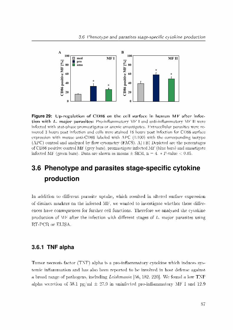
3.6 Phenotype and parasites stage-speci�c cytokine production
med pro ama
CD
86
po
siti
ve
MF
[%
]
A
0
20
80
100
40
60 *
MF II
B
*
CD
86
po
siti
ve
MF
[%
]
0
20
80
100
40
60
MF I
* *
Figure 29: Up-regulation of CD86 on the cell surface in human MF after infec-tion with L. major parasites: Pro-in�ammatory MF I and anti-in�ammatory MF II wereinfected with stat-phase promastigotes or axenic amastigotes. Extracellular parasites were re-moved 3 hours post infection and cells were stained 18 hours post infection for CD86 surfaceexpression with mouse anti-CD86 labeled with APC (1:100) with the corresponding isotype(APC) control and analyzed by �ow cytometry (FACS). A)+B) Depicted are the percentagesof CD86 positive control MF (grey bars), promastigote infected MF (blue bars) and amastigoteinfected MF (green bars). Data are shown as means ± SEM, n = 4. ∗ P-value < 0.05.
3.6 Phenotype and parasites stage-specific cytokine
production
In addition to di�erent parasite uptake, which resulted in altered surface expression
of distinct markers on the infected MF, we wanted to investigate whether these di�er-
ences have consequences for further cell functions. Therefore we analyzed the cytokine
production of MF after the infection with di�erent stages of L. major parasites using
RT-PCR or ELISA.
3.6.1 TNF alpha
Tumor necrosis factor (TNF) alpha is a pro-in�ammatory cytokine which induces sys-
temic in�ammation and has also been reported to be involved in host defense against
a broad range of pathogens, including Leishmania [56, 182, 220]. We found a low TNF
alpha secretion of 38.1 pg/ml ± 27.9 in uninfected pro-in�ammatory MF I and 12.9
87

3 Results
pg/ml ± 4.9 in anti-in�ammatory MF II. After the infection with L. major promastig-
otes, we could detect a signi�cant increase in TNF alpha production in both phenotypes
of MF, with a signi�cantly higher increase in MF II (1079 pg/ml ± 237) as compared
to MF I (113.3 pg/ml ± 53, �g. 30 A + B). In contrast, amastigote infection do not
induce TNF alpha, neither in MF I (20.2 pg/ml ± 10.4) nor in MF II (16.7 pg/ml ±5.8, �g. 30 A + B).
Rel
ati
ve
gen
e ex
pre
ssio
n
TN
F-α
in
pg
/ml
MF I MF II
0
400
800
1200
1600
Med Pro Ama
TN
F-α
in
pg
/ml
0
400
800
1200
1600
Med Pro Ama
A B
**
*
C D
0
1
2
Rel
ati
ve
gen
e ex
pre
ssio
n
0
1
2 MF I med
pro ama
TNF-α
MF II
TNF-α
*
*
Figure 30: Di�erent cytokine production of TNF alpha in human MF after infec-tion with L. major parasites: Pro-in�ammatory MF I and anti-in�ammatory MF II wereinfected with stat-phase promastigotes or axenic amastigotes. Extracellular parasites wereremoved 3 hours post infection and supernatants and cells were harvested 18 hours post infec-tion from medium controls (grey bars), promastigote infected MF (blue bars) and amastigoteinfected MF (green bars) for TNF alpha sandwich ELISA and mRNA expression analysis.Total RNA was isolated, cDNA generated and the relative gene expression was determined byLightCycler analysis. A) TNF alpha production of pro-in�ammatory MF I (n = 3). B) TNFalpha production of anti-in�ammatory MF II (n = 3). C) TNF alpha mRNA expression ofpro-in�ammatory MF I (n = 4). D) TNF alpha mRNA expression of anti-in�ammatory MFII (n = 4). Data are shown as means ± SEM. ∗ P-value < 0.05, ∗∗ P-value < 0.005.
88

3.6 Phenotype and parasites stage-speci�c cytokine production
In addition to TNF alpha production, we monitored the relative gene expression of TNF
alpha by RT-PCR analysis. We found no signi�cant di�erences in uninfected MF I and
II, 0.22 ± 0.02 and 0.35 ± 0.07 respectively (�g. 30 C + D). However, in concordance
with the cytokine expression on protein level, we found a signi�cant up-regulation of the
TNF alpha expression on the mRNA level after promastigote infection in MF I to 0.9 ±0.1 and to 1.3 ± 0.2 in MF II (�g. 30 C + D). Upon the infection with the amastigote
life stage we found, similar to the results of ELISA analysis, no signi�cant di�erences
in the TNF alpha expression in MF I and II.
3.6.2 IL-12
Interleukin 12 (IL-12) is a heterodimer consisting of the IL-12p40 and the IL-12p35
subunit [87]. IL-12 is a pro-in�ammatory regulatory cytokine and is shown to promote
the di�erentiation of naive T lymphocytes into Th-1 cells, which is crucial in determining
resistance and clearance of a particular pathogen [73, 107]. The IL-12p40 subunit was
detected by ELISA analysis for the IL-12 measurement (�g. 31).
Uninfected MF I and II showed a low level of IL-12 secretion of 3.8 pg/ml ± 1.2 in
pro-in�ammatory MF I and 1.3 pg/ml ± 0.1 in anti-in�ammatory MF II. We found a
signi�cant increase of IL-12 secretion to 778.6 pg/ml ± 307 only in pro-in�ammatory
MF I after the infection with promastigotes (�g. 31 A). The infection with amastigotes
in MF I as well as both parasite life stages in MF II had no e�ect on IL-12 production
(�g. 31 A + B).
3.6.3 CCL3 and CCL4
Macrophage in�ammatory protein-1 (MIP-1) alpha and MIP-1 beta belong both to the
family of chemotactic cytokines, which are known as chemokines and are now o�cially
named CCL3 and CCL4, respectively. Both CCL3 and CCL4 are demonstrated to
have a chemotactic activity towards monocytes and T lymphocytes, as well as pro-
in�ammatory e�ects [111]. The relative gene expression of CCL3 and CCL4 was an-
alyzed by RT-PCR (�g. 32). For CCL3 we found no signi�cant di�erences in the
89
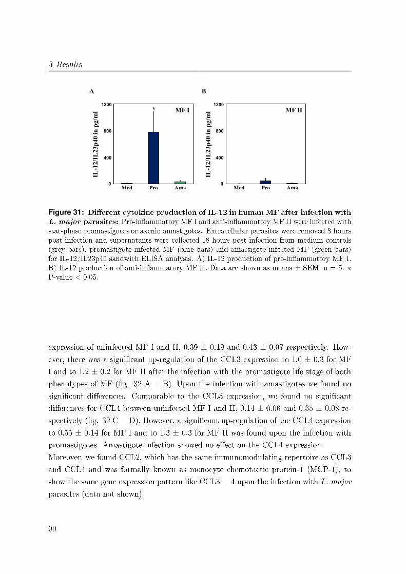
3 Results
0
400
800
1200
IL-1
2/I
L2
3p
40 i
n p
g/m
l MF I
Med Pro Ama
A
*
0
400
800
1200
MF II
Med Pro Ama
B
IL-1
2/I
L2
3p
40 i
n p
g/m
l Figure 31: Di�erent cytokine production of IL-12 in human MF after infection withL. major parasites: Pro-in�ammatory MF I and anti-in�ammatory MF II were infected withstat-phase promastigotes or axenic amastigotes. Extracellular parasites were removed 3 hourspost infection and supernatants were collected 18 hours post infection from medium controls(grey bars), promastigote infected MF (blue bars) and amastigote infected MF (green bars)for IL-12/IL23p40 sandwich ELISA analysis. A) IL-12 production of pro-in�ammatory MF I.B) IL-12 production of anti-in�ammatory MF II. Data are shown as means ± SEM, n = 5. ∗P-value < 0.05.
expression of uninfected MF I and II, 0.39 ± 0.19 and 0.43 ± 0.07 respectively. How-
ever, there was a signi�cant up-regulation of the CCL3 expression to 1.0 ± 0.3 for MF
I and to 1.2 ± 0.2 for MF II after the infection with the promastigote life stage of both
phenotypes of MF (�g. 32 A + B). Upon the infection with amastigotes we found no
signi�cant di�erences. Comparable to the CCL3 expression, we found no signi�cant
di�erences for CCL4 between uninfected MF I and II, 0.14 ± 0.06 and 0.35 ± 0.08 re-
spectively (�g. 32 C + D). However, a signi�cant up-regulation of the CCL4 expression
to 0.55 ± 0.14 for MF I and to 1.3 ± 0.3 for MF II was found upon the infection with
promastigotes. Amastigote infection showed no e�ect on the CCL4 expression.
Moreover, we found CCL2, which has the same immunomodulating repertoire as CCL3
and CCL4 and was formally known as monocyte chemotactic protein-1 (MCP-1), to
show the same gene expression pattern like CCL3 + 4 upon the infection with L. major
parasites (data not shown).
90

3.6 Phenotype and parasites stage-speci�c cytokine production
Rel
ati
ve
Gen
e E
xp
ress
ion
CCL3
MF I med pro ama
p = 0,07
Rel
ati
ve
Gen
e E
xp
ress
ion
CCL3
MF II
0
0,8
1,6
1,2
0,4
*
A B
0
0,8
1,6
1,2
0,4
*
Rel
ati
ve
Gen
e E
xp
ress
ion
CCL4
MF I med pro ama
*
Rel
ati
ve
Gen
e E
xp
ress
ion
CCL4
MF II
0
0,8
1,6
1,2
0,4
0
0,8
1,6
1,2
0,4
*
C D
Figure 32: Di�erent cytokine mRNA expression of CCL3 and CCL4 in human MFafter infection with L. major parasites: Pro-in�ammatory MF I and anti-in�ammatoryMF II were infected with stat-phase promastigotes or axenic amastigotes. Extracellular para-sites were removed 3 hours post infection and cells were harvested 18 hours post infection frommedium controls (grey bars), promastigote infected MF (blue bars) and amastigote infectedMF (green bars). Total RNA was isolated, cDNA generated and the relative gene expressionwas determined by LightCycler analysis. A) CCL3 mRNA expression of pro-in�ammatoryMF I. B) CCL3 mRNA expression of anti-in�ammatory MF II. C) CCL4 mRNA expressionof pro-in�ammatory MF I. D) CCL4 mRNA expression of anti-in�ammatory MF II. Data areshown as means ± SEM, n = 4. ∗ P-value < 0.05.
91
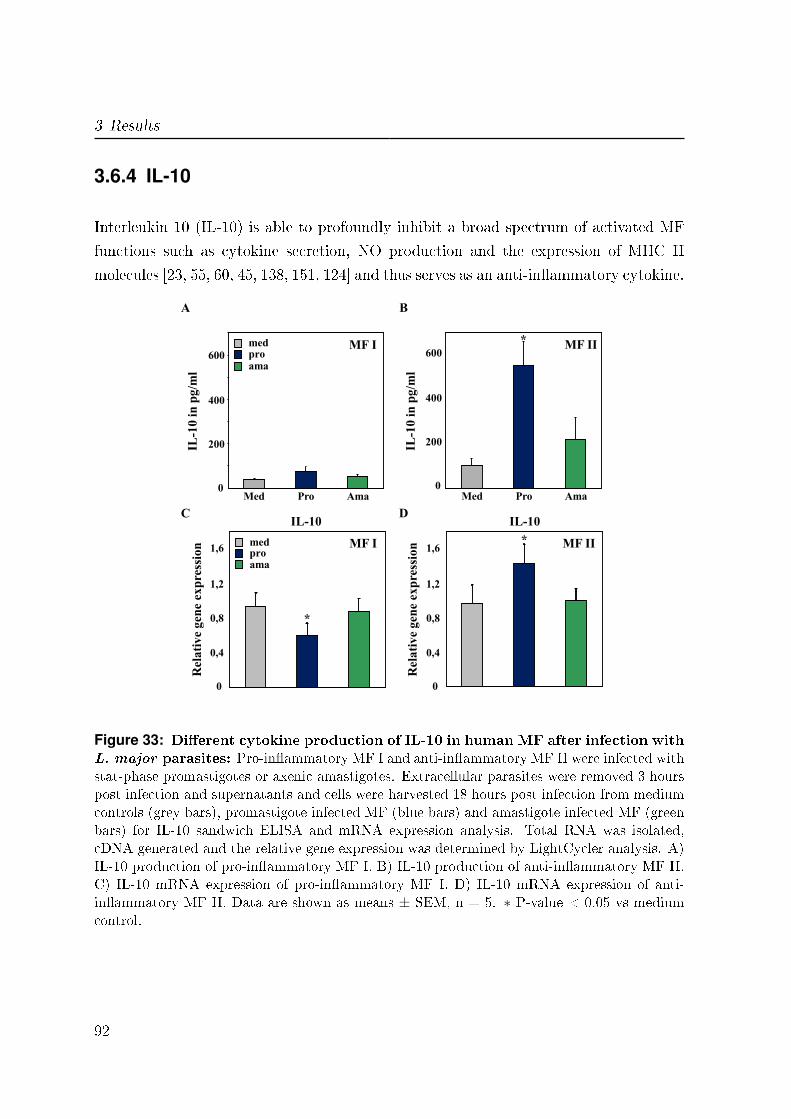
3 Results
3.6.4 IL-10
Interleukin 10 (IL-10) is able to profoundly inhibit a broad spectrum of activated MF
functions such as cytokine secretion, NO production and the expression of MHC II
molecules [23, 55, 60, 45, 138, 151, 124] and thus serves as an anti-in�ammatory cytokine.
0
200
400
600
0
200
400
600
IL-1
0 i
n p
g/m
l
MF I
Med Pro Ama
A
IL-1
0 i
n p
g/m
l
MF II
Med Pro Ama
B
*
Rel
ati
ve
gen
e ex
pre
ssio
n
IL-10
Rel
ati
ve
gen
e ex
pre
ssio
n MF II
IL-10 C D
MF I med pro ama
*
0
0,8
1,6
1,2
0,4
0
0,8
1,6
1,2
0,4
*
med pro ama
Figure 33: Di�erent cytokine production of IL-10 in human MF after infection withL. major parasites: Pro-in�ammatory MF I and anti-in�ammatory MF II were infected withstat-phase promastigotes or axenic amastigotes. Extracellular parasites were removed 3 hourspost infection and supernatants and cells were harvested 18 hours post infection from mediumcontrols (grey bars), promastigote infected MF (blue bars) and amastigote infected MF (greenbars) for IL-10 sandwich ELISA and mRNA expression analysis. Total RNA was isolated,cDNA generated and the relative gene expression was determined by LightCycler analysis. A)IL-10 production of pro-in�ammatory MF I. B) IL-10 production of anti-in�ammatory MF II.C) IL-10 mRNA expression of pro-in�ammatory MF I. D) IL-10 mRNA expression of anti-in�ammatory MF II. Data are shown as means ± SEM, n = 5. ∗ P-value < 0.05 vs mediumcontrol.
92

3.7 Phosphorylation of MAP kinases (MAPK) after L. major infection
In contrast to TNF alpha and IL-12, we found a signi�cant higher secretion of IL-10
in uninfected anti-in�ammatory MF II of 89 pg/ml ± 31 as compared to 34.4 pg/ml
± 7.6 in pro-in�ammatory MF I (�g. 33 A + B). Furthermore, MF II resulted in a
signi�cant increase in IL-10 production to 579.2 pg/ml ± 118 after the infection with
the promastigote life stage. Amastigote infection showed no in�uence on the secretion of
IL-10 in MF II, as well as both parasite life stages in MF I (�g. 33 A + B). Subsequently
to IL-10 production, the relative gene expression was monitored by RT-PCR analysis.
Interestingly, we found a signi�cant down-regulation of IL-10 expression from 0.9 ±0.15 in uninfected MF I to 0.6 ± 0.14 in promastigote infected pro-in�ammatory MF
I, whereas amastigotes had no in�uence on IL-10 expression in MF I (�g. 33 C). In
contrast, anti-in�ammatory MF II showed a signi�cant up-regulation upon promastigote
infection to 1.43 ± 0.2 as compared to 0.97 ± 0.2 in uninfected MF II. Amastigotes
showed no e�ect on the IL-10 expression in MF II (�g. 33 D).
3.7 Phosphorylation of MAP kinases (MAPK) after
L. major infection
In order to understand the di�erential cytokine production in the di�erent phenotypes
of MF as well as after infection with both life stages of the L. major parasite, we
investigated intracellular signal transduction pathways. Therefore we selected the fol-
lowing two pathways based on their involvement in the intracellular signaling network
[141, 35]: P38 mitogen-activated protein kinase (p38) and extracellular signal-regulated
kinases (ERK1/2). Both are mitogen-activated protein (MAP) kinases of the family of
protein kinase cascades. They belong to the serine/threonine-speci�c protein kinases
that respond to extracellular stimuli such as pro-in�ammatory cytokines, ultra violate
radiation, heat shock, growth factors and mitogens. MAPK activation is regulated by
phosphorylation cascades of a serial of molecules resulting in modulation of distinct
transcription factors with e�ects on various cellular activities like cell proliferation, dif-
ferentiation and cell survival/apoptosis [142]. For both there have been hints that they
might be a�ected in parasite infection [77, 16, 15].
Preliminary data (n = 2) from an intracellular FACS staining showed a slight in-
93

3 Results
crease in the phosphorylation of p38 and ERK1/2 after infection with L. major in
pro-in�ammatory MF I (�g. 34 C + D). Furthermore, we used western blot analysis,
which is a standard method to investigate the phosphorylation level of p38 and ERK1/2.
0
5
10
0
10
20
pp
38
po
siti
ve
MF
[%
]
C
MF I
Pro Ama Med
pE
RK
1/2
po
siti
ve
MF
[%
]
D
MF I
Pro Ama Med
15 min 30 min
101 102 103 104
Co
un
ts
0
pro med
ama
100
Phospho p38-PE
1,4 % 15,1% 1,5 %
101 102 103 104
Co
un
ts
0
pro med
ama
100
Phospho ERK1/2-FITC
0,9% 6,1% 0,9%
A B
Figure 34: Di�erent phosphorylation state of p38 and p44/42 (ERK1/2) MAPkinases (MAPK) in human type I MF after infection with L. major parasites: Pro-in�ammatory MF I were infected with stat-phase promastigotes or axenic amastigotes. Extra-cellular parasites were removed 15 and 30 minutes post infection and cells were harvested frommedium controls (med), promastigote infected MF (pro) and amastigote infected MF (ama).Intracellular staining was performed for phospho p38 and phospho ERK1/2 MAPK with mouseanti-phospho p38 MAPK labeled with PE (1:10) and anti-phospho ERK1/2 MAPK labeledwith Alexa Fluor 488 (1:10) and analyzed by FACS. A)+B) Representative FACS histogramsof one experiment out of 2 independent experiments. Control MF I (grey line), promastigoteinfected (blue line) and axenic amastigote infected (green line) with the percentages of phosphop38 and phospho ERK1/2 positive cells compared to the isotype control (�lled light grey) after30 minutes infection. C)+D) Positive MF I for phospho p38 and phospho ERK1/2 after 15(yellow bars) and 30 (brown bars) minutes of L. major infection. Depicted are percentages ofphospho p38 and phospho ERK1/2 positive cells. Data are shown as means ± SEM, n = 2.
94
3.7 Phosphorylation of MAP kinases (MAPK) after L. major infection
3.7.1 p38 MAP kinases
P38 MAPK is activated mainly by in�ammatory cytokines, like TNF alpha, TGF beta
and IL-1, as well as by lipopolysaccharides (LPS) [150]. It was demonstrated that the ac-
tivation of p38 leads to di�erent e�ects such as cytokine production, cell motility, apop-
tosis or elimination of pathogens [137, 136]. We found �rst a signi�cant up-regulation of
phospho p38 in promastigote infected MF I after 15 min (1.18 ± 0.04), however after 30
min there was a subsequent signi�cant decrease of phospho p38 (0.75 ± 0.05, �g 35 A).
Amastigote infection showed no signi�cant e�ect on p38 phosphorylation. In contrast,
we obtained a signi�cant up-regulation of phospho p38 only after 15 min of infection
with both parasite life stages in MF II, 1.59 ± 0.06 after promastigote and 2.17 ± 0.27
upon amastigote infection (�g. 35 B).
3.7.2 ERK1/2 MAP kinases
In contrast to p38, ERK1/2 MAPK are induced by hormones, growth factors and a vast
number of extracellular stimuli [35]. Activation of ERK1/2 leads to altered transcription
of genes that are important amongst others for the cell cycle. Feng et al. showed
Leishmania to activate ERK1/2 and subsequently decrease IL-12 production in MF
[52]. However, for MF I and II there are no data published. Preliminary data (n = 2)
showed for MF I a down-regulation of phospho ERK1/2 in promastigote infected cells
after 15 min (0.74 ± 0.02) as well as after 30 min (0.51 ± 0.02, �g. 36 A). An even
stronger decrease was found upon amastigote infection 15 min (0.38 ± 0.04) and 30 min
(0.23 ± 0.04) after co-incubation. However, MF II showed an up-regulation after 15
min in both promastigote (1.36 ± 0.01) and amastigote (1.82 ± 0.04) infected MF II
for phospho ERK1/2 (�g. 36 B). After 30 min of co-incubation no di�erences in ERK
1/2 phosphorylation could be detected any more.
95
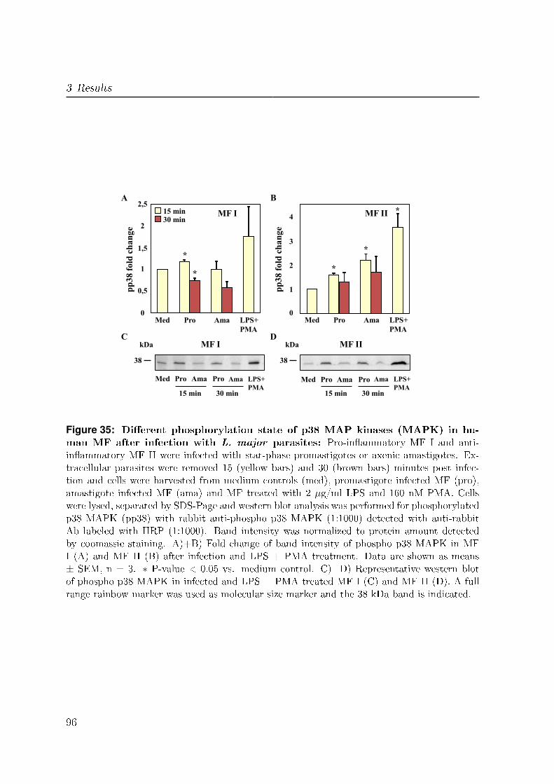
3 Results
pp
38
fo
ld c
ha
ng
e
A
MF I
Pro Ama Med
15 min 30 min
0
1
2,5
1,5
0,5
2
LPS+
PMA
pp
38
fo
ld c
ha
ng
e
B
MF II
Pro Ama Med 0
2
3
1
4
LPS+
PMA
*
* *
*
*
38
Pro Ama Med
15 min
kDa C D
Pro Ama
30 min
LPS+
PMA
38
Pro Ama Med
15 min
kDa
Pro Ama
30 min
LPS+
PMA
MF I MF II
Figure 35: Di�erent phosphorylation state of p38 MAP kinases (MAPK) in hu-man MF after infection with L. major parasites: Pro-in�ammatory MF I and anti-in�ammatory MF II were infected with stat-phase promastigotes or axenic amastigotes. Ex-tracellular parasites were removed 15 (yellow bars) and 30 (brown bars) minutes post infec-tion and cells were harvested from medium controls (med), promastigote infected MF (pro),amastigote infected MF (ama) and MF treated with 2 µg/ml LPS and 160 nM PMA. Cellswere lysed, separated by SDS-Page and western blot analysis was performed for phosphorylatedp38 MAPK (pp38) with rabbit anti-phospho p38 MAPK (1:1000) detected with anti-rabbitAb labeled with HRP (1:1000). Band intensity was normalized to protein amount detectedby coomassie staining. A)+B) Fold change of band intensity of phospho p38 MAPK in MFI (A) and MF II (B) after infection and LPS + PMA treatment. Data are shown as means± SEM, n = 3. ∗ P-value < 0.05 vs. medium control. C)+D) Representative western blotof phospho p38 MAPK in infected and LPS + PMA treated MF I (C) and MF II (D). A fullrange rainbow marker was used as molecular size marker and the 38 kDa band is indicated.
96

3.7 Phosphorylation of MAP kinases (MAPK) after L. major infection
pE
RK
1/2
fo
ld c
ha
ng
e
A
MF I
Pro Ama Med
15 min 30 min
0
1
2,5
1,5
0,5
2
LPS+
PMA
pE
RK
1/2
fo
ld c
ha
ng
e
B
MF II
Pro Ama Med 0
2
3
1
5
LPS+
PMA
4
38
Pro Ama Med
15 min
C D
Pro Ama
30 min
LPS+
PMA
38
Pro Ama Med
15 min
Pro Ama
30 min
LPS+
PMA
kDa kDa MF I MF II
Figure 36: Di�erent phosphorylation state of ERK1/2 MAP kinases (MAPK) inhuman MF after infection with L. major parasites: Pro-in�ammatory MF I and anti-in�ammatory MF II were infected with stat-phase promastigotes or axenic amastigotes. Extra-cellular parasites were removed 15 (yellow bars) and 30 (brown bars) minutes post infection andcells were harvested from medium controls (med), promastigote infected MF (pro), amastigoteinfected MF (ama) and MF treated with 2 µg/ml LPS and 160 nM PMA. Cells were lysed,separated by SDS-Page and western blot analysis was performed for phosphorylated ERK1/2MAPK (pERK 1/2) with rabbit anti-phospho ERK1/2 MAPK (1:1000) detected with anti-rabbit Ab labeled with HRP (1:1000). Band intensity was normalized to protein amountdetected by coomassie staining. A)+B) Fold change of band intensity of phospho ERK1/2MAPK in MF I (A) and MF II (B) after infection and LPS + PMA treatment. Data areshown as means ± SEM, n = 2. C)+D) Representative western blot of phospho ERK1/2MAPK in infected and LPS + PMA treated MF I (C) and MF II (D). A full range rainbowmarker was used as molecular size marker and the 38 kDa band is indicated.
97

3 Results
3.8 L. major parasite escape from phagolysosomes
After the analysis of the cytokine production and intracellular cell signaling of MF
upon the infection with L. major parasites, we wanted to look at the compartment
where the parasites reside inside MF. L. major promastigotes are known to end up
in phagolysosomes, where they need the special environmental conditions like a low
pH-value to start stage di�erentiation into the amastigote form and multiplication [22].
Concanamycin A is an inhibitor of the vacuolar ATPase, a proton pump that carries
H+ in the lumen of a compartment and acidi�es the pH inside this compartment [26,
57, 74]. Therefore, treatment of MF with concanamycin A leads to the inhibition of
the vacuolar ATPase in the phagolysosomal membrane by binding to the proteolipid
subunit, which results in an increase of the pH-value inside the phagolysosome after
treatment. Investigating the e�ect of such a pH increase on the fate of infection of
human MF, we analyzed infected MF I and II with L. major parasites after subsequently
treated with concanamycin A.
We found no signi�cant di�erences in the resulting infection rates for both phenotypes
of MF upon promastigote infection, neither after 18 hours (data not shown) nor after 42
hours any (�g. 37 A). In addition, the infection with amastigotes showed no di�erences
after 18 hours. However, after 42 hours we found a signi�cant higher percentage of
infected MF in concanamycin A treated cells as compared to the untreated control cells
in both MF I and MF II. For MF I the infection rate increased from 55.1 % ± 9.2 in
untreated control MF to 78 % ± 6.2 in concanamycin A treated cells and for MF II
from 80.4 % ± 4.5 to 90.6 % ± 2.1 respectively (�g. 37 B).
Subsequently, we analyzed the location of L. major parasites inside MF using electron
microscopy (EM) in infected MF I (data not shown) and MF II (�g. 37 C + D). We
found promastigote infected MF II to show two double membrane structures, one from
the host compartment (phagolysosome) and one parasite double membrane (�g. 37 C).
We found the promastigote double membrane to be associated with the characteristic
subpellicular microtubular structure of the L. major parasites surface. In contrast, for
amastigote infected MF II, we found some phagolysosomes with one intact parasite
membrane and a second disrupted host membrane around the parasite, in addition to
phagolysosomes with two double membranes (data not shown). Furthermore, some
infected MF II showed only one double membrane of the parasite without any host
98

3.8 L. major parasite escape from phagolysosomes
membranes of the MF (�g. 37 D, white arrows). In addition, L. major amastigotes
show the same characteristic subpellicular microtubular structure associated with the
parasite membrane as compared to the promastigote form.
20
40
60
80
100
Infe
ctio
n r
ate
[%
]
20
40
60
80
100
Infe
ctio
n r
ate
[%
]
MF I MF II
Concanamycin A Control
MF I MF II
*
*
Pro Ama A B
200 nm
200 nm
L. major
pro
MF
C D
200 nm
L. major
ama MF
Figure 37: Infection rates after concanamycin A treatment and parasite locationin human MF after L. major infection: A)+B) Pro-in�ammatory MF I and anti-in�ammatory MF II were infected with stat-phase promastigotes (A) or axenic amastigotes(B) in the presence of 22 nM Concanamycin A (brown bars) or medium (yellow bars). Extra-cellular parasites were removed 3 hours post infection and cells were Di� QUIK R© stained 42hours post infection. Infection rates were determined by counting > 200 phagocytes. Depictedare infection rates as percentages of infected MF. Data are shown as means ± SEM, n = 4. ∗P-value < 0.05. C)+D) Represatative EM micrpgraghs of 3 independent experiments of MF IIinfected with stat-phase promastigotes (C) or axenic amastigotes (D) 18 hours post infection.
99
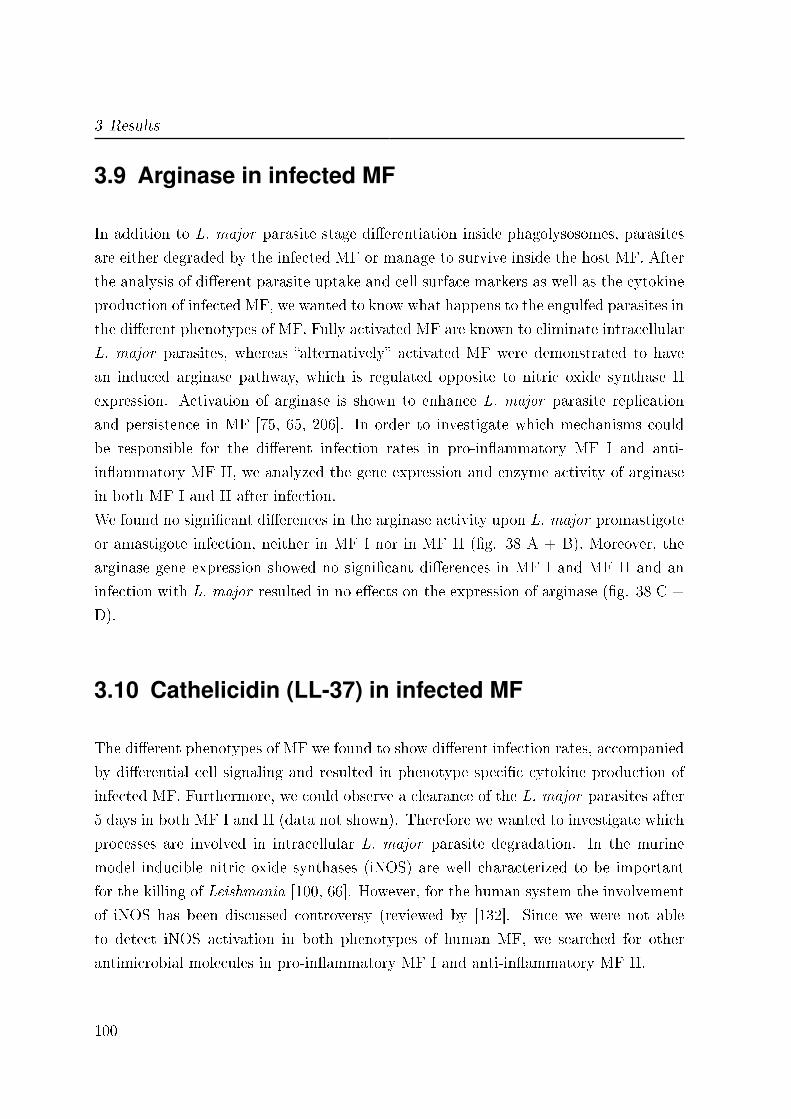
3 Results
3.9 Arginase in infected MF
In addition to L. major parasite stage di�erentiation inside phagolysosomes, parasites
are either degraded by the infected MF or manage to survive inside the host MF. After
the analysis of di�erent parasite uptake and cell surface markers as well as the cytokine
production of infected MF, we wanted to know what happens to the engulfed parasites in
the di�erent phenotypes of MF. Fully activated MF are known to eliminate intracellular
L. major parasites, whereas �alternatively� activated MF were demonstrated to have
an induced arginase pathway, which is regulated opposite to nitric oxide synthase II
expression. Activation of arginase is shown to enhance L. major parasite replication
and persistence in MF [75, 65, 206]. In order to investigate which mechanisms could
be responsible for the di�erent infection rates in pro-in�ammatory MF I and anti-
in�ammatory MF II, we analyzed the gene expression and enzyme activity of arginase
in both MF I and II after infection.
We found no signi�cant di�erences in the arginase activity upon L. major promastigote
or amastigote infection, neither in MF I nor in MF II (�g. 38 A + B). Moreover, the
arginase gene expression showed no signi�cant di�erences in MF I and MF II and an
infection with L. major resulted in no e�ects on the expression of arginase (�g. 38 C +
D).
3.10 Cathelicidin (LL-37) in infected MF
The di�erent phenotypes of MF we found to show di�erent infection rates, accompanied
by di�erential cell signaling and resulted in phenotype speci�c cytokine production of
infected MF. Furthermore, we could observe a clearance of the L. major parasites after
5 days in both MF I and II (data not shown). Therefore we wanted to investigate which
processes are involved in intracellular L. major parasite degradation. In the murine
model inducible nitric oxide synthases (iNOS) are well characterized to be important
for the killing of Leishmania [100, 66]. However, for the human system the involvement
of iNOS has been discussed controversy (reviewed by [132]. Since we were not able
to detect iNOS activation in both phenotypes of human MF, we searched for other
antimicrobial molecules in pro-in�ammatory MF I and anti-in�ammatory MF II.
100
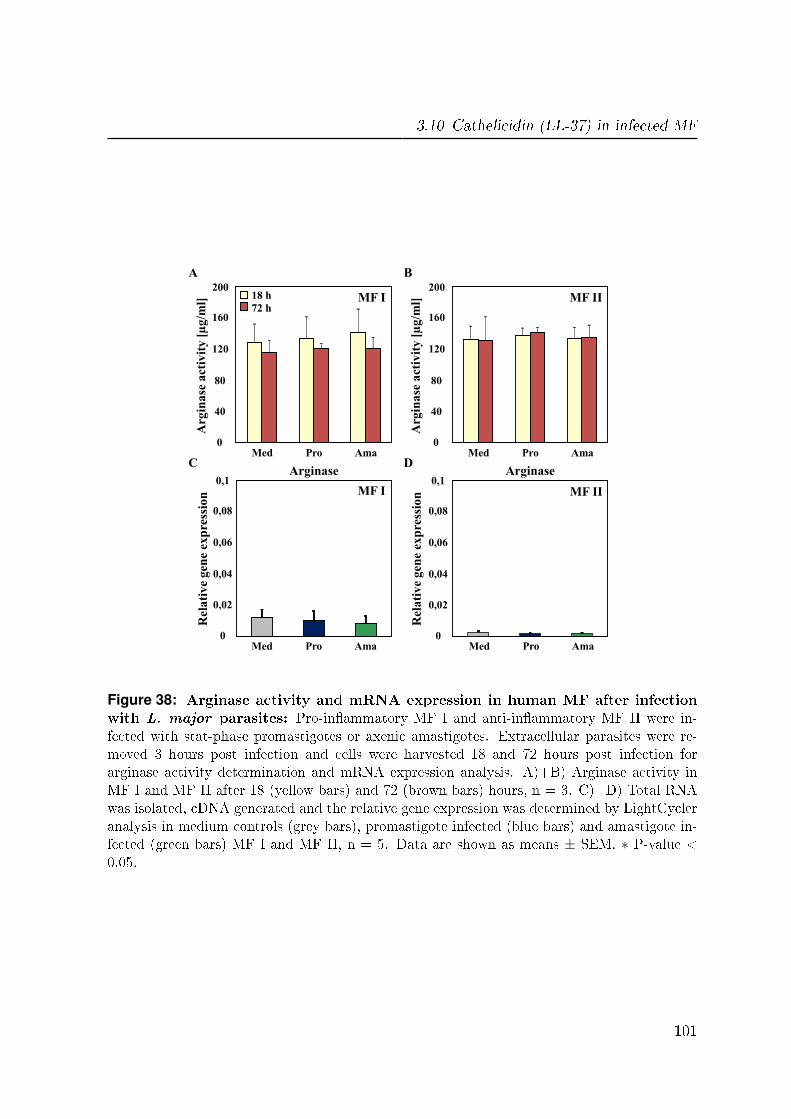
3.10 Cathelicidin (LL-37) in infected MF
0
40
80
120
160
200
Med Pro Ama 0
40
80
120
160
200
Med Pro Ama
18 h
72 h
A B
Arg
ina
se a
ctiv
ity
[µ
g/m
l]
Arg
ina
se a
ctiv
ity
[µ
g/m
l] MF I MF II
0
0,02
0,04
0,06
0,08
0,1
Med Pro Ama Med Pro Ama
C D
MF I MF II
Rel
ati
ve
gen
e ex
pre
ssio
n
0
0,02
0,04
0,06
0,08
0,1
Rel
ati
ve
gen
e ex
pre
ssio
n MF II
Arginase Arginase
Figure 38: Arginase activity and mRNA expression in human MF after infectionwith L. major parasites: Pro-in�ammatory MF I and anti-in�ammatory MF II were in-fected with stat-phase promastigotes or axenic amastigotes. Extracellular parasites were re-moved 3 hours post infection and cells were harvested 18 and 72 hours post infection forarginase activity determination and mRNA expression analysis. A)+B) Arginase activity inMF I and MF II after 18 (yellow bars) and 72 (brown bars) hours, n = 3. C)+D) Total RNAwas isolated, cDNA generated and the relative gene expression was determined by LightCycleranalysis in medium controls (grey bars), promastigote infected (blue bars) and amastigote in-fected (green bars) MF I and MF II, n = 5. Data are shown as means ± SEM. ∗ P-value <0.05.
101

3 Results
3.10.1 Different LL-37 expression in MF I and MF II
Using RT-PCR we found a signi�cant higher gene expression of LL-37 in pro-in�amma-
tory MF I as compared to anti-in�ammatory MF II (0.14 ± 0.07, �g. 39 A). Further-
more, we also monitored the LL-37 expression upon L. major infection and found no
signi�cant di�erences after the infection with both parasite life stages, neither in MF I
nor in MF II (�g. 39 B + C). However, human cathelicidin, also named LL-37, revealed
to be a promising candidate for L. major parasite degradation, because of its e�ective
antimicrobial activity against a variety of pathogens [187, 64, 12, 71].
0
0,5
1
MF I
Pro Ama Med
Rel
ati
ve
gen
e ex
pre
ssio
n
0
0,5
1
MF II
Pro Ama Med
Rel
ati
ve
gen
e ex
pre
ssio
n
0
0,5
1
MF I MF II
Rel
ati
ve
gen
e ex
pre
ssio
n
A B C
*
LL-37 LL-37 LL-37
Figure 39: Di�erent cathelicidin (LL-37) mRNA expression in human MF: Pro-in�ammatory MF I and anti-in�ammatory MF II were infected with stat-phase promastigotesor axenic amastigotes. Extracellular parasites were removed 3 hours post infection and cellswere harvested 18 hours post infection from medium controls (grey bars), promastigote infectedMF (blue bars) and amastigote infected MF (green bars). Total RNA was isolated, cDNAgenerated and the relative gene expression was determined by LightCycler analysis. Depictedare fold mRNA change compared to medium control of MF I. A) LL-37 mRNA expression ofcontrol pro-in�ammatory MF I (light grey) and anti-in�ammatory MF II (dark grey). B) LL-37mRNA expression of pro-in�ammatory MF I. C) LL-37 mRNA expression of anti-in�ammatoryMF II. Data are shown as means ± SEM, n = 5. ∗ P-value < 0.05.
3.10.2 Killing effect of rhLL-37 on L. major promastigotes
In order to investigate whether human LL-37 is able to be involved in the degradation
of L. major, the parasites were treated with di�erent concentrations of the recombinant
human LL-37 (rhLL-37).
102
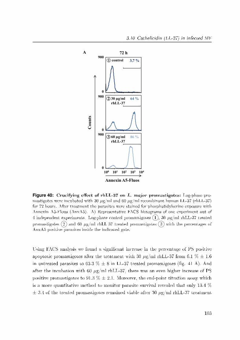
3.10 Cathelicidin (LL-37) in infected MF
30 µg/ml
rhLL-37
60 µg/ml
rhLL-37
64 %
86 %
Co
un
ts
Annexin A5-Fluos
A 72 h
102 101 103 104 100
control 3,7 % 900
0
0
900
0
900
1
2
3
Figure 40: Crucifying e�ect of rhLL-37 on L. major promastigotes: Log-phase pro-mastigotes were incubated with 30 µg/ml and 60 µg/ml recombinant human LL-37 (rhLL-37)for 72 hours. After treatment the parasites were stained for phosphatidylserine exposure withAnnexin A5-Flous (AnxA5). A) Representative FACS histograms of one experiment out of
4 independent experiments. Log-phase control promastigotes 1 , 30 µg/ml rhLL-37 treated
promastigotes 2 and 60 µg/ml rhLL-37 treated promastigotes 3 with the percentages ofAnxA5 positive parasites inside the indicated gate.
Using FACS analysis we found a signi�cant increase in the percentage of PS positive
apoptotic promastigotes after the treatment with 30 µg/ml rhLL-37 from 6.1 % ± 1.6
in untreated parasites to 63.3 % ± 8 in LL-37 treated promastigotes (�g. 41 A). And
after the incubation with 60 µg/ml rhLL-37, there was an even higher increase of PS
positive promastigotes to 91.3 % ± 2.1. Moreover, the end-point titration assay which
is a more quantitative method to monitor parasite survival revealed that only 13.4 %
± 3.4 of the treated promastigotes remained viable after 30 µg/ml rhLL-37 treatment
103
3 Results
(�g. 41 B). An even higher decrease of viable promastigotes to 0.07 % ± 0.03 was found
after the treatment with 60 µg/ml rhLL-37, compared to 100 % viable parasites in the
untreated control.
*
*
*
*
30 µg/ml 60 µg/ml control
40
0
20
60
80
100
Dea
d p
ara
site
s [%
]
30 µg/ml 60 µg/ml control
40
0
20
60
80
100
Via
ble
pa
rasi
tes
[%]
A B
Figure 41: Killing e�ect of rhLL-37 on L. major promastigotes: Log-phase promastig-otes were incubated with 30 µg/ml and 60 µg/ml recombinant human LL-37 (rhLL-37) for 72hours. After treatment parasites were stained for phosphatidylserine exposure with AnnexinA5-Flous (AnxA5) and end-point titration assay was performed. A) Phosphatidylserine ex-posure on the cell surface of log-phase control promastigotes (dark blue), 30 µg/ml rhLL-37treated promastigotes (blue) and 60 µg/ml rhLL-37 treated promastigotes (light blue). De-picted are percentages of AnxA5 positive parasites. B) Viability of the promastigotes afterthe di�erent treatments with rhLL-37. Depicted are percentages of viable parasites. Data areshown as means ± SEM, n = 4. ∗ P-value < 0.05.
In addition to the analysis of the PS staining by FACS, we analyzed the parasites via
�uorescence microscopy (�g. 42). Untreated control promastigotes were found to have
elongated bodies and show no green �uorescence of the AnxA5-Fluos, which indicates
them to be viable (�g. 42 A). In contrast, after the incubation with 30 µg/ml rhLL-37
the promastigotes revealed to be rounded up and PS positive, as shown by the green
AnxA5-Fluos staining which indicates this promastigotes to be apoptotic (�g. 42 B).
3.10.3 No effect of rhLL-37 on L. major amastigotes
For the amastigote life stage of L. major, we found no signi�cant di�erences in the
percentage of PS positive parasites compared to untreated amastigotes, neither after
the treatment with 30 nor 60 µg/ml rhLL-37 (�g. 43 A). In concordance to the FACS
104
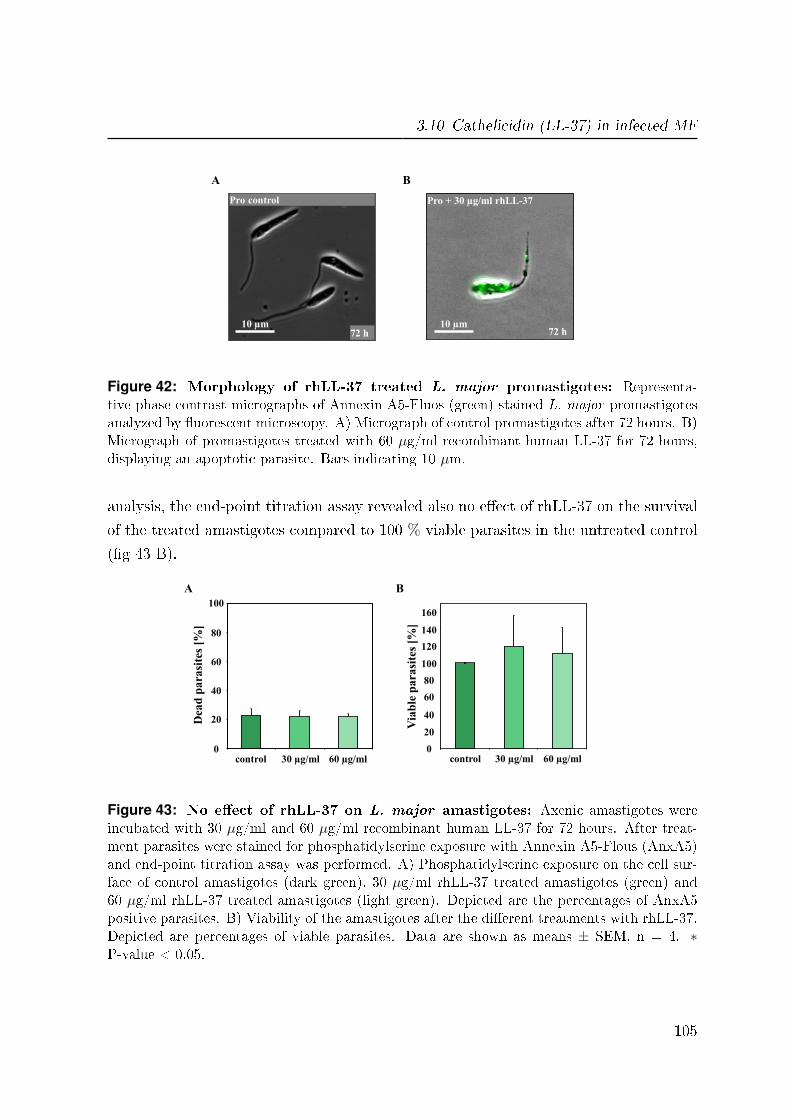
3.10 Cathelicidin (LL-37) in infected MF
Pro + 30 µg/ml rhLL-37
72 h
A B
10 µm
Pro control
72 h 10 µm
Figure 42: Morphology of rhLL-37 treated L. major promastigotes: Representa-tive phase contrast micrographs of Annexin A5-Fluos (green) stained L. major promastigotesanalyzed by �uorescent microscopy. A) Micrograph of control promastigotes after 72 hours. B)Micrograph of promastigotes treated with 60 µg/ml recombinant human LL-37 for 72 hours,displaying an apoptotic parasite. Bars indicating 10 µm.
analysis, the end-point titration assay revealed also no e�ect of rhLL-37 on the survival
of the treated amastigotes compared to 100 % viable parasites in the untreated control
(�g 43 B).
30 µg/ml 60 µg/ml control
Via
ble
pa
rasi
tes
[%]
40
0
20
60
80
160
100
120
140
B
30 µg/ml 60 µg/ml control
40
0
20
60
80
100
Dea
d p
ara
site
s [%
]
A
Figure 43: No e�ect of rhLL-37 on L. major amastigotes: Axenic amastigotes wereincubated with 30 µg/ml and 60 µg/ml recombinant human LL-37 for 72 hours. After treat-ment parasites were stained for phosphatidylserine exposure with Annexin A5-Flous (AnxA5)and end-point titration assay was performed. A) Phosphatidylserine exposure on the cell sur-face of control amastigotes (dark green), 30 µg/ml rhLL-37 treated amastigotes (green) and60 µg/ml rhLL-37 treated amastigotes (light green). Depicted are the percentages of AnxA5positive parasites. B) Viability of the amastigotes after the di�erent treatments with rhLL-37.Depicted are percentages of viable parasites. Data are shown as means ± SEM, n = 4. ∗P-value < 0.05.
105
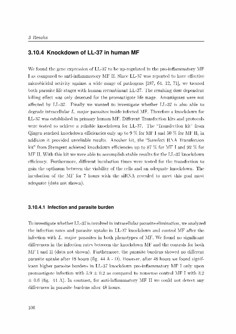
3 Results
3.10.4 Knockdown of LL-37 in human MF
We found the gene expression of LL-37 to be up-regulated in the pro-in�ammatory MF
I as compared to anti-in�ammatory MF II. Since LL-37 was reported to have e�ective
microbicidal activity against a wide range of pathogens [187, 64, 12, 71], we treated
both parasite life stages with human recombinant LL-37. The resulting dose dependent
killing e�ect was only detected for the promastigote life stage. Amastigotes were not
a�ected by LL-37. Finally we wanted to investigate whether LL-37 is also able to
degrade intracellular L. major parasites inside infected MF. Therefore a knockdown for
LL-37 was established in primary human MF. Di�erent Transfection kits and protocols
were tested to achieve a reliable knockdown for LL-37. The �Transfection kit� from
Qiagen reached knockdown e�ciencies only up to 9 % for MF I and 50 % for MF II, in
addition it provided unreliable results. Another kit, the �Stemfect RNA Transfection
kit� from Stemgent achieved knockdown e�ciencies up to 87 % for MF I and 92 % for
MF II. With this kit we were able to accomplish stable results for the LL-37 knockdown
e�ciency. Furthermore, di�erent incubation times were tested for the transfection to
gain the optimum between the viability of the cells and an adequate knockdown. The
incubation of the MF for 7 hours with the siRNA revealed to meet this goal most
adequate (data not shown).
3.10.4.1 Infection and parasite burden
To investigate whether LL-37 is involved in intracellular parasite elimination, we analyzed
the infection rates and parasite uptake in LL-37 knockdown and control MF after the
infection with L. major parasites in both phenotypes of MF. We found no signi�cant
di�erences in the infection rates between the knockdown MF and the controls for both
MF I and II (data not shown). Furthermore, the parasite burdens showed no di�erent
parasite uptake after 18 hours (�g. 44 A - D). However, after 48 hours we found signif-
icant higher parasite burdens in LL-37 knockdown pro-in�ammatory MF I only upon
promastigote infection with 5.9 ± 0.2 as compared to nonsense control MF I with 3.2
± 0.6 (�g. 44 A). In contrast, for anti-in�ammatory MF II we could not detect any
di�erences in parasite burdens after 48 hours.
106

3.10 Cathelicidin (LL-37) in infected MF
18 h 48 h
Pro
Lm
pa
rasi
tes
/ M
F
0
5
10
15
20
25
30
35
18 h 48 h
Lm
pa
rasi
tes
/ M
F
Ama
0
5
10
*
control
LL-37
18 h 48 h
Lm
pa
rasi
tes
/ M
F
0
5
10
0
5
10
15
20
25
30
35
18 h 48 h
Lm
pa
rasi
tes
/ M
F
control
LL-37
MF I
MF II
MF I
MF II
A B
C D
Figure 44: Parasite burden in LL-37 knockdown pro-in�ammatory and anti-in�ammatory human MF: MF I and MF II were treated with control nonsense siRNA(yellow bars) and LL-37 siRNA (brown bars) to maintain a knockdown. MF were co-incubatedwith stat-phase promastigotes or axenic amastigotes, extracellular parasites were removed 3hours post infection, 18 and 48 hours post infection cells were Di� QUIK R© stained and para-site burdens were assessed by counting intracellular parasites in 20 infected MF. Depicted arethe number of parasites per MF. A)+B) Parasite burdens in pro-in�ammatory MF I. C)+D)Parasite burdens in anti-in�ammatory MF II. Data are shown as means ± SEM, n = 3. ∗P-value < 0.05.
107
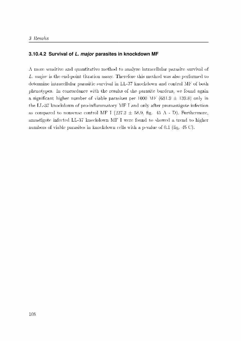
3 Results
3.10.4.2 Survival of L. major parasites in knockdown MF
A more sensitive and quantitative method to analyze intracellular parasite survival of
L. major is the end-point titration assay. Therefore this method was also performed to
determine intracellular parasitic survival in LL-37 knockdown and control MF of both
phenotypes. In concordance with the results of the parasite burdens, we found again
a signi�cant higher number of viable parasites per 1000 MF (681.9 ± 139.8) only in
the LL-37 knockdown of pro-in�ammatory MF I and only after promastigote infection
as compared to nonsense control MF I (227.2 ± 58.9, �g. 45 A - D). Furthermore,
amastigote infected LL-37 knockdown MF I were found to showed a trend to higher
numbers of viable parasites in knockdown cells with a p-value of 0.1 (�g. 45 C).
108
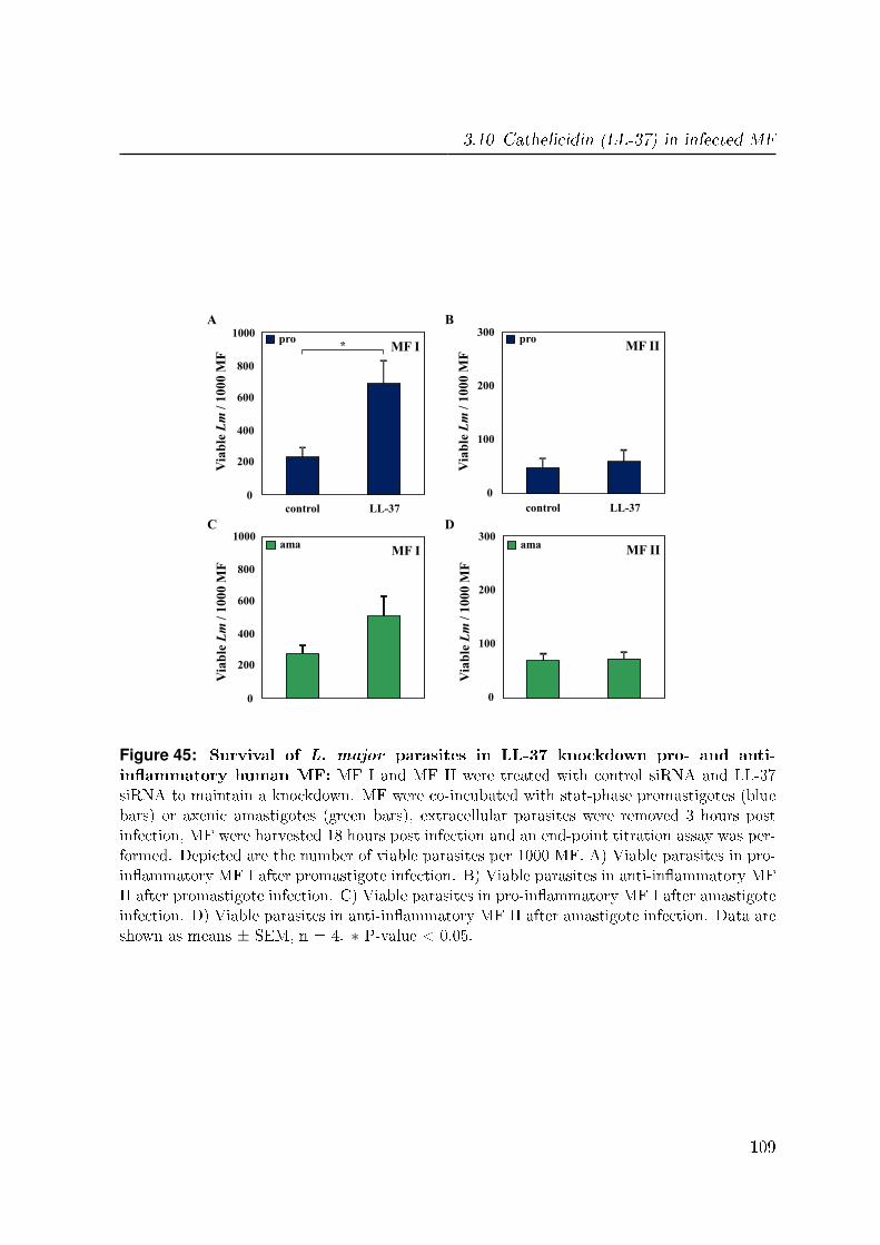
3.10 Cathelicidin (LL-37) in infected MF
control LL-37
0
200
400
600
800
1000 pro
Via
ble
Lm
/ 1
00
0 M
F *
control LL-37
Via
ble
Lm
/ 1
00
0 M
F
ama MF I
MF I
A
C
0
200
400
600
800
1000
0
100
200
300
Via
ble
Lm
/ 1
00
0 M
F
control LL-37
ama
0
100
200
300
Via
ble
Lm
/ 1
00
0 M
F
control LL-37
pro
MF II
MF II
B
D
Figure 45: Survival of L. major parasites in LL-37 knockdown pro- and anti-in�ammatory human MF: MF I and MF II were treated with control siRNA and LL-37siRNA to maintain a knockdown. MF were co-incubated with stat-phase promastigotes (bluebars) or axenic amastigotes (green bars), extracellular parasites were removed 3 hours postinfection, MF were harvested 18 hours post infection and an end-point titration assay was per-formed. Depicted are the number of viable parasites per 1000 MF. A) Viable parasites in pro-in�ammatory MF I after promastigote infection. B) Viable parasites in anti-in�ammatory MFII after promastigote infection. C) Viable parasites in pro-in�ammatory MF I after amastigoteinfection. D) Viable parasites in anti-in�ammatory MF II after amastigote infection. Data areshown as means ± SEM, n = 4. ∗ P-value < 0.05.
109


4 Discussion
First we characterized both life stages of L. major FEBNI parasites, promastigotes as
well as amastigotes. We found that in contrast to previous �ndings the virulence marker
GP63 was also expressed by axenic amastigotes. In addition to the L. major FEBNI
strain, we applied and successfully modi�ed our novel in vitro method to generate
axenic amastigotes of the L. major Friedlin and 5ASKH strains. Interestingly, these
L. major strains needed another temperature to be transferred into amastigotes in
the axenic culture system. Investigating apoptotic mechanisms in both parasite life
stages of L. major FEBNI we found both ROS dependent and ROS independent cell
death mechanisms. Focusing on promastigote and amastigote interaction with pro-
in�ammatory (MF I) and anti-in�ammatory (MF II) macrophages we found amastigotes
to be more infective as compared to promastigotes. Moreover, we could demonstrate
that pro-in�ammatory MF I were less susceptible to infection as compared to anti-
in�ammatory MF II. Finally we investigated parasite stage speci�c macrophages (MF)
responses and defense mechanisms against L. major. We identi�ed a new mechanism
in MF enabling killing of promastigotes. This mechanism depends on the antimicrobial
molecule cathelicidin, LL-37.
Part 1
4.1 Different life stages of the parasite L. major
Characterizing the generated axenic amastigotes on a genetic level, we could demon-
strate that they showed the same stage-speci�c gene expression as compared to MF-
derived amastigotes focusing on markers such as ABC and Sherp. Analyses of the
virulence factor GP63 revealed an up-regulation in the amastigote life stage. In con-
111
4 Discussion
trast, using mouse derived L. major amastigotes Schneider et al. reported GP63 to be
only expressed in promastigotes, but not in the amastigote life stage [170]. However,
for L. major, seven distinct genes are described on chromosome 10, which can encode
for the protein GP63 [204]. We analyzed the gene expression of the GP63 gene 3. Fur-
thermore, for all Leishmania species studied to date GP63 is reported to be encoded by
a multigene family [33, 208, 115, 204]. Here the GP63 genes are arranged in one gene
locus containing several tandemly repeated genes and additional dispersed genes from
this tandem array. We suggest that these distinct genes for GP63 could be expressed
di�erently during the di�erent life stages of the L. major parasites. In addition, such a
stage-speci�c expression of the structurally distinct GP63 genes was already shown for
L. mexicana [116, 115]. Here three di�erent gene classes were found for the GP63 gene
family: C1 - 3. Only the C1 gene class is up-regulated and transcribed in amastigotes,
while the C2/3 gene classes are expressed in promastigotes [161]. A comparable C1
gene class for GP63 was also found in L. donovani [153]. Although there are no reports
for such gene classes in L. major to date, these �ndings support our suggestion of a
stage-dependent expression of the di�erent GP63 genes in L. major.
Investigating GP63 on the protein level, we found GP63 to be almost absent on the
cell surface of amastigotes. In contrast, log-phase promastigotes showed a high surface
expression of GP63, which was decreased but still present on stat-phase promastigotes
(data not shown). GP63 is encoded by several distinct genes, which may be expressed
stage-speci�cally in L. major parasites. We hypothesize that the distinct GP63 genes
code for GP63 proteins, which are structurally di�erent in the amastigote and pro-
mastigote life stage. This may result in a di�erent surface distribution of the distinct
GP63 proteins. In agreement, Medina-Acosta et al. found in L. mexicana such a dif-
ferential posttranslational processing and localization of GP63 proteins in amastigotes
as compared to promastigotes [116, 115]. These alterations of GP63 in the di�erent life
stages could lead to an altered 3-dimensional conformation and surface distribution of
GP63, which may be stage-speci�c. Moreover, Medina-Acosta et al. reported a lack of
the promastigote characteristic �membrane-form� of GP63 on the amastigote cell sur-
face of L. mexicana, whereas it was present within the �agellar pocket of the parasite
[116]. In addition, they demonstrated that altered GP63 proteins were the most abun-
dant proteins on the surface of the amastigote life stage and that di�erent antibodies
raised against GP63 showed di�erent reactivity to both life stages, being promastigote
112
4.2 In vitro culture method for axenic L. major amastigotes
or amastigote speci�c [116]. The monoclonal antibody for GP63 used for FACS analysis
in this study was generated for L. major promastigote speci�c recombinant GP63 [34],
resulting in binding only to a speci�c GP63 protein structure. GP63 proteins which
are structurally di�erent cannot be detected by this antibody. This would explain the
absence of GP63 detection on axenic amastigotes by FACS analysis in our results.
As it was described for L. mexicana, there is a di�erent gene expression of the three
GP63 gene clusters C1 - 3 in promastigotes and amastigotes [161]. Our data suggest such
a stage-speci�c gene expression of GP63 genes also in L. major parasites. Furthermore,
we would suggest an organization of the seven GP63 genes in gene clusters as compared
to L. mexicana [161]. We suggest that the analyzed GP63 gene 3 belongs to a gene
cluster which is up-regulated during the amastigote life stage. Since there is no data
concerning such gene clusters for L. major parasites. This should be investigated using
our new in vitro culture method to generate axenic L. major amastigotes.
4.2 In vitro culture method for axenic L. major
amastigotes
Di�erent factors are of importance to transform L. major promastigotes into amastig-
otes. In our axenic culture for L. major, we found the temperature to be critical. Using
either 35◦C or 31◦C instead of 33◦C we were able to generate L. major Friedlin and
5ASKH amastigote cultures in addition to L. major FEBNI. The di�erent L. major
isolates used in this study originate from di�erent geographic regions, namely Israel
(Friedlin) and the former Soviet Union (5ASKH). It has been described previously that
Leishmania species di�er in their sensitivity to temperature stress [224]. For L. tropica
it was shown that amastigotes replicate more rapidly at 35◦C than at 37◦C, and are
completely eliminated at 39◦C [18]. Furthermore, the growth of L. mexicana is also
reported to be temperature sensitive [21]. While parasites of this species grow and pro-
liferate well within cultured MF at 34◦C, infection does not proceed at 37.5◦C. Similar
�ndings were reported for L. panamensis [222, 162], where parasite survival is restricted
to 33◦C.
113

4 Discussion
These reports all indicate that the temperature is of immense importance during the
stage di�erentiation of the di�erent L. major isolates, which are adapted to the local
temperatures of their origin. Therefore we suggest that that the complex process of
L. major stage di�erentiation and generation of axenic in vitro cultures is restricted to
a narrow temperature range and depends on exact temperature settings �tting to the
speci�c natural environmental conditions of the corresponding isolate and its origin.
4.3 Apoptosis in L. major parasites
To investigate the underlying processes of the apoptosis machinery in unicellular organ-
isms, we aimed to analyze the di�erent steps of the apoptotic cell death in L. major
parasites and identify new proteins involved in the leishmanial apoptotic machinery.
Inducing apoptosis with staurosporine we found a strong up-regulation of ROS accom-
panied by a time-delayed PS externalization and cell rounding in the promastigote life
stage. In Leishmania, staurosporine is reported to strongly induce apoptosis resulting in
PS externalization, cytochrome c release, DNA fragmentation and cell shrinkage [13, 9].
However, the exact mechanism of apoptosis induction is not known. Our data showed
that there was �rst a high ROS formation after staurosporine treatment followed by PS
externalization. This could indicate that Leishmania have a mitochondrial triggered
apoptotic pathway similar as in mammalian cells [190, 191]. For L. donovani such a
mitochondria-dependent ROS-mediated programmed cell death was reported by Roy
et al. [160]. They found �rst a stimulation of mitochondrial generated ROS, followed
by a depolarization of the mitochondrial membrane potential. Subsequently, the mito-
chondrial membrane disrupts, leading to the release of ROS together with cytochrome c
into the cytosol of the parasite and �nally resulting in DNA fragmentation [160]. These
�ndings �t with our results after staurosporine treatment. Therefore a similar mecha-
nism is conceivable also in L. major parasites for the induction of apoptosis by stau-
rosporine. Another mechanism for staurosporine-induced apoptosis in L. major could
be the induction of the endoplasmic reticulum (ER) stress-induced apoptotic pathway
[46]. Here the exposure of the ER to stress leads to the elevation of the cytosolic Ca2+
level inside L. major parasites, due the release from internal stores. This causes mito-
chondrial membrane potential depolarization and ATP loss resulting in ROS-dependent
114
4.3 Apoptosis in L. major parasites
release of cytochrome c and subsequent PS externalization, DNA fragmentation and
cell shrinkage [46]. We hypothesize staurosporine to be such an ER stress inducer re-
sulting in activation of this pathway. To investigate whether the ER stress-induced
apoptotic pathway is involved in the apoptotic process after staurosporine treatment,
the cytosolic Ca2+ level should be analyzed in the parasites. However, both mechanisms
are ROS-dependent and mediated by the mitochondrion of the parasite. This supports
our assumption that staurosporine induce a mitochondrial triggered apoptotic pathway
which is ROS-dependent.
Another apoptosis inducer for Leishmania parasites is the anti-leishmanial drug mil-
tefosine. Previous studies demonstrated miltefosine to induce cell death in L. major
promastigotes showing PS externalization, cell rounding and �nally DNA fragmentation
[51, 83]. The exact mechanism of miltefosine induced apoptosis is unknown. However
miltefosine is known to a�ect the parasite membrane composition via the induction of
changes in the biosynthesis of phospholipids [104]. Using miltefosine we found a strong
PS externalization in L. major parasites, which was higher and much faster than af-
ter staurosporine treatment. Moreover, we observed a signi�cant lower induction of
ROS as compared to staurosporine induced leishmanial apoptosis. Downstream of the
inhibition of phosphatidylcholine biosynthesis which is important for the integrity of
the cell membrane, miltefosine was additionally demonstrated to be responsible for the
increase of cellular ceramide after apoptosis induction in mammalian cells [213]. Such
a ceramide-mediated apoptotic pathway, which is independent from ROS would �t to
our results and could be a possible mechanism for apoptosis induction in L. major after
miltefosine treatment. Moreover ceramide induced cell death is independent of caspase
functions [193]. Since Leishmania do not have apoptosis regulating caspases [8], this
suggest miltefosine to have an e�ect on the parasite cell membrane biosynthesis [104]
and subsequent trigger a ceramide-mediated ROS independent apoptotic pathway. To
investigate whether ceramide is involved in the apoptotic machinery of L. major, the
cytosolic ceramide level needs to be analyzed. Another example for ROS independent
apoptosis was reported for L. donovani after the treatment with Aloe vera leaf exudate
(AVL) as a potent anti-leishmanial agent [49]. Here AVL was demonstrated to mediate
the loss of mitochondrial membrane potential, cytochrome c release into the cytosol,
PS externalisation and chromatin condensation. However, they found no increase in
cytosolic Ca2+ or generation of intracellular ROS, indicating a ROS independent apop-
115
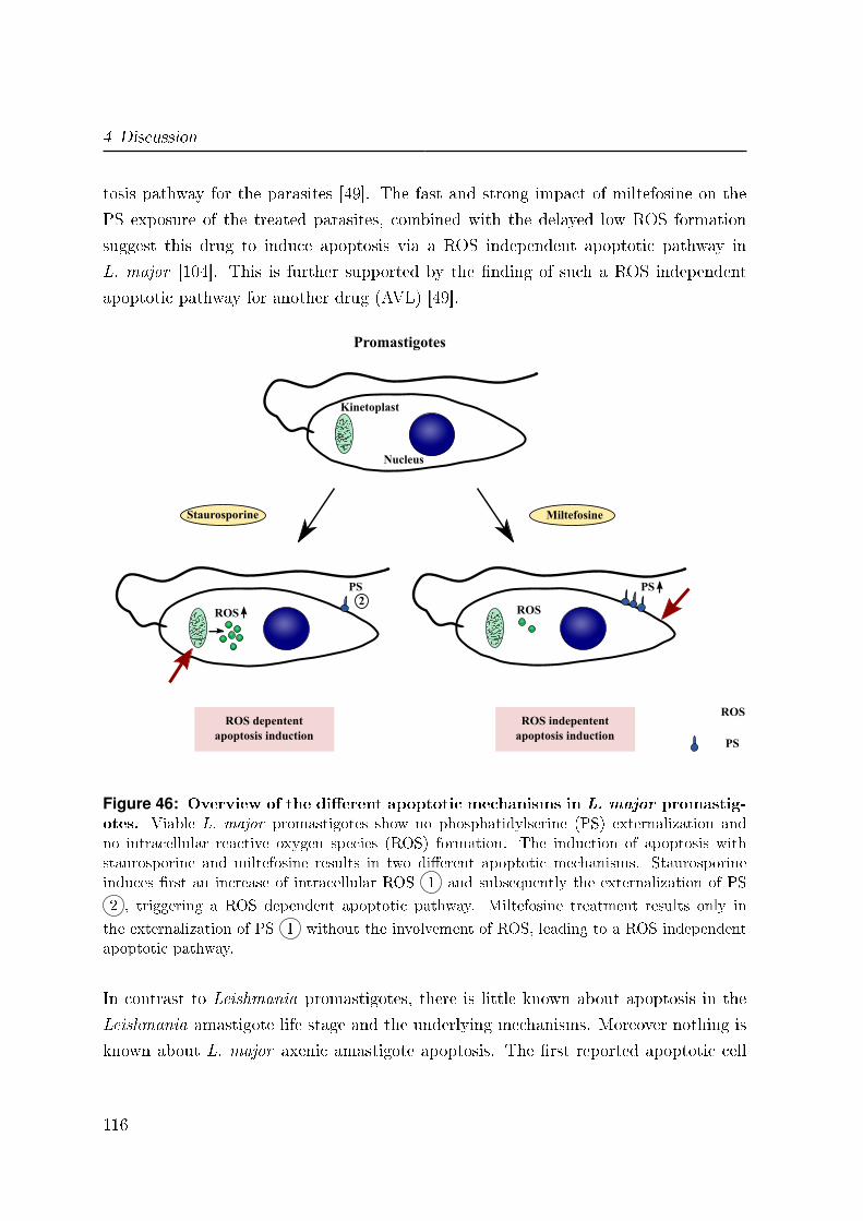
4 Discussion
tosis pathway for the parasites [49]. The fast and strong impact of miltefosine on the
PS exposure of the treated parasites, combined with the delayed low ROS formation
suggest this drug to induce apoptosis via a ROS independent apoptotic pathway in
L. major [104]. This is further supported by the �nding of such a ROS independent
apoptotic pathway for another drug (AVL) [49].
2
ROS depententapoptosis induction
ROS
Nucleus
Kinetoplast
PS
Staurosporine
PS
Miltefosine
ROS
ROS indepententapoptosis induction
Promastigotes
PS
ROS
Figure 46: Overview of the di�erent apoptotic mechanisms in L. major promastig-otes. Viable L. major promastigotes show no phosphatidylserine (PS) externalization andno intracellular reactive oxygen species (ROS) formation. The induction of apoptosis withstaurosporine and miltefosine results in two di�erent apoptotic mechanisms. Staurosporineinduces �rst an increase of intracellular ROS 1 and subsequently the externalization of PS
2 , triggering a ROS dependent apoptotic pathway. Miltefosine treatment results only in
the externalization of PS 1 without the involvement of ROS, leading to a ROS independentapoptotic pathway.
In contrast to Leishmania promastigotes, there is little known about apoptosis in the
Leishmania amastigote life stage and the underlying mechanisms. Moreover nothing is
known about L. major axenic amastigote apoptosis. The �rst reported apoptotic cell
116
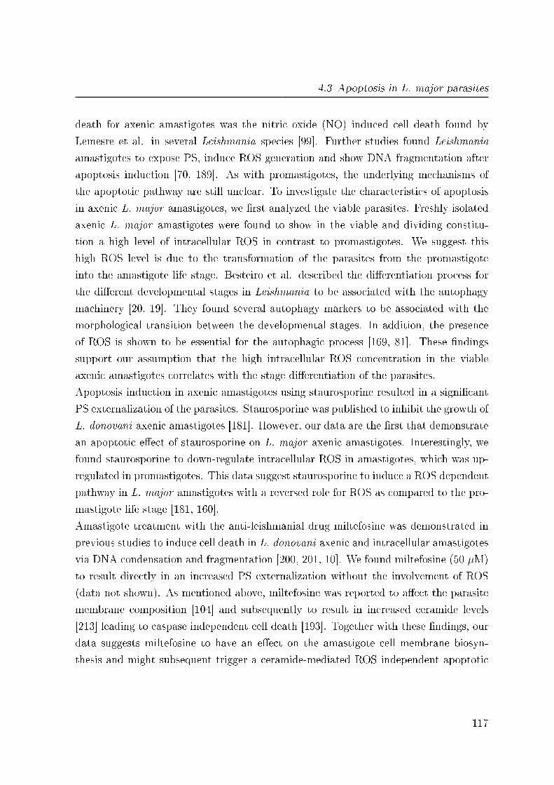
4.3 Apoptosis in L. major parasites
death for axenic amastigotes was the nitric oxide (NO) induced cell death found by
Lemesre et al. in several Leishmania species [99]. Further studies found Leishmania
amastigotes to expose PS, induce ROS generation and show DNA fragmentation after
apoptosis induction [70, 189]. As with promastigotes, the underlying mechanisms of
the apoptotic pathway are still unclear. To investigate the characteristics of apoptosis
in axenic L. major amastigotes, we �rst analyzed the viable parasites. Freshly isolated
axenic L. major amastigotes were found to show in the viable and dividing constitu-
tion a high level of intracellular ROS in contrast to promastigotes. We suggest this
high ROS level is due to the transformation of the parasites from the promastigote
into the amastigote life stage. Besteiro et al. described the di�erentiation process for
the di�erent developmental stages in Leishmania to be associated with the autophagy
machinery [20, 19]. They found several autophagy markers to be associated with the
morphological transition between the developmental stages. In addition, the presence
of ROS is shown to be essential for the autophagic process [169, 81]. These �ndings
support our assumption that the high intracellular ROS concentration in the viable
axenic amastigotes correlates with the stage di�erentiation of the parasites.
Apoptosis induction in axenic amastigotes using staurosporine resulted in a signi�cant
PS externalization of the parasites. Staurosporine was published to inhibit the growth of
L. donovani axenic amastigotes [181]. However, our data are the �rst that demonstrate
an apoptotic e�ect of staurosporine on L. major axenic amastigotes. Interestingly, we
found staurosporine to down-regulate intracellular ROS in amastigotes, which was up-
regulated in promastigotes. This data suggest staurosporine to induce a ROS dependent
pathway in L. major amastigotes with a reversed role for ROS as compared to the pro-
mastigote life stage [181, 160].
Amastigote treatment with the anti-leishmanial drug miltefosine was demonstrated in
previous studies to induce cell death in L. donovani axenic and intracellular amastigotes
via DNA condensation and fragmentation [200, 201, 10]. We found miltefosine (50 µM)
to result directly in an increased PS externalization without the involvement of ROS
(data not shown). As mentioned above, miltefosine was reported to a�ect the parasite
membrane composition [104] and subsequently to result in increased ceramide levels
[213] leading to caspase independent cell death [193]. Together with these �ndings, our
data suggests miltefosine to have an e�ect on the amastigote cell membrane biosyn-
thesis and might subsequent trigger a ceramide-mediated ROS independent apoptotic
117
4 Discussion
pathway as compared to the promastigote life stage.
ROS depententapoptosis induction
ROS
Nucleus
Kinetoplast
PS
Staurosporine
PS
Miltefosine
ROS
ROS indepententapoptosis induction
Amastigotes
2
Figure 47: Overview of the di�erent apoptotic mechanisms in L. major amastig-otes. Viable L. major amastigotes show no phosphatidylserine (PS) externalization and a highintracellular reactive oxygen species (ROS) level due to stage di�erentiation. The inductionof apoptosis with staurosporine and miltefosine results in two di�erent apoptotic mechanisms.Staurosporine induces �rst a decrease of intracellular ROS 1 and subsequently the exter-
nalization of PS 2 , triggering a ROS dependent apoptotic pathway. Miltefosine treatment
results only in the externalization of PS 1 without the involvement of ROS, leading to a ROSindependent apoptotic pathway.
Even though the exact biochemical mechanisms of the protozoan apoptotic machinery
are poorly understood, there are a few proteins believed to be involved in apoptotic
processes in Leishmania. For promastigotes it has been published that an activated
nuclease similar to the endonuclease G (EndoG), migrates from the kinetoplast to the
nucleus causing DNA degradation [157, 46]. Therefore Leishmania EndoG is suggested
to be a pro-apoptotic protein. Another enzyme, the mitochondrial ascorbate peroxidase
118
4.3 Apoptosis in L. major parasites
(LmAPX) is known to inactivate ROS, which indicates this protein to inhibit a ROS-
mediated cell death [47]. In order to elucidate the complex apoptotic machinery and
identify the involved proteins, we generated lysates of L. major parasites after apop-
tosis induction and quanti�ed a total of 707 proteins using mass spectrometry analysis
(data not shown). We found several up- and down-regulated proteins upon apoptosis
induction for both L. major life stages. These apoptosis dependent regulated proteins
are potential candidates involved in the apoptotic machinery of Leishmania. Further
analyses are under way to investigate their role in the course of apoptotic processes. The
elucidation of the molecular pathways responsible for leishmanial apoptotic cell death
might help to identify new target molecules for chemotherapeutic drug development and
therapeutic intervention for cutaneous Leishmaniasis.
In conclusion, we found for the di�erent parasite life stages of L. major two di�erent
pathways which could be involved in leishmanial apoptosis. One is ROS-mediated,
which is induced by staurosporine and the second is independent from the formation of
intracellular ROS, induced by miltefosine treatment. Furthermore, these mechanisms
were found to be di�erently regulated between the two life stages of L. major.
119
4 Discussion
Part 2
4.4 Interaction of L. major with human MF
Our aim was to investigate the interaction of L. major parasites with pro- and anti-
in�ammatory human MF in order to analyze which phenotype may be involved in disease
propagation or healing. CD163, at typical marker for MF 2 was found to be down-
regulated after treatment of MF II with LPS + INF gamma [126]. In addition, these
reprogrammed MF II showed a higher antimicrobial activity compared to untreated MF
II after the infection with L. mexicana. However, the adaption of the MF did not extend
to all functions, such as the production of the cytokine IL-12 [126]. Another group
demonstrated that the expression of CD163 is regulated by pro- and anti-in�ammatory
mediators like cytokines [30]. In monocytes CD163 expression was found to be strongly
up-regulated by stimuli such as IL-6 and IL-10. On the other side LPS, INF gamma,
and TNF alpha suppress the expression of CD163 [30]. These data suggest infected MF
II to sort of change their phenotype towards the MF I phenotype after the infection
with L. major. This was additionally supported by our �ndings of a down-regulation of
CD163 on MF II after the infection with L. major parasites and a down-regulation of
CD163 on mRNA level upon L. major promastigote infection.
Interestingly, uninfected MF I and MF II we found to show an equal expression of
CD163 on the mRNA level, however we could not detect the CD163 receptor on the
cell surface of MF I. A possible explanation could be a shedding of the receptor. Hintz
and colleagues showed for monocytes pro-in�ammatory stimuli like LPS to induce such
a shedding of CD163 from the surface [69]. Therefore we suggest a swift extracellular
shedding of CD163 to its soluble form, at the moment the protein reaches the cell
membrane in MF I.
Upon infection, MF activation is needed for an e�ective immune response. In order to
gain insights of possible MF activation, we analyzed the early intracellular activation
state after the infection with L. major via the phosphorylation of mitogen-activated
protein (MAP) kinases. We found in pro-in�ammatory MF I the p38 MAP kinase
to be activated after promastigote infection but down-regulated upon amastigote in-
fection. On the other hand, preliminary data showed the ERK1/2 MAP kinases to
120
4.4 Interaction of L. major with human MF
be down-regulated either after promastigote or amastigote infection. P38 activation is
demonstrated to be involved in the induction of IL-12, while ERK1/2 MAP kinases
(MAPK) suppress IL-12 transcription [52]. According to these �ndings, we detected
the production of IL-12 only in MF I after the infection with L. major promastigotes.
In contrast, the induction of IL-10 is shown to require the activation of both p38 and
ERK1/2 MAPK [103]. Anti-in�ammatory MF II were found to show such an activation
of p38 and ERK1/2 MAPK upon the infection with either promastigotes or amastig-
otes. However, we detected only after promastigote infection the secretion of IL-10.
This suggests the amastigote life stage of L. major to be able to inhibit the production
of IL-10 despite the activation of p38 and ERK1/2. The underlying mechanisms of this
phenomenon are still to be de�ned. However, our results showed that already after
15 min there are di�erent activation patterns of p38 and ERK1/2 MAPK in the two
phenotypes of MF after L. major infection. These data demonstrate that MF are able
to respond in a very quick manner after parasite contact.
Furthermore, we wanted to study the consequences of a di�erent susceptibility of both
phenotypes of MF to L. major infection. As cytokine secretion is crucial for the de-
velopment of an adaptive immune response, we investigate the cytokine pro�le and the
underlying gene expression pro�le during L. major infection. In contrast to amastig-
ote infection, promastigote infection induced an increase in TNF alpha in both MF I
and II. In addition, we found the chemokines CCL3 and CCL4 to be up-regulated in
MF I and II infected with promastigotes. These data demonstrate a speci�c MF acti-
vation occurring after promastigote uptake, whereas infection with amastigotes keeps
the MF silenced and non-activated. The pro-in�ammatory cytokine TNF alpha and
the chemokines CCL3 and CCL4 are reported to be essential for the induction of local
in�ammatory responses [158]. Our results �t into the concept that the initial MF re-
cruitment at the site of infection is mediated by TNF alpha and chemokines. However,
the persistent infection and disease propagation is based on further transmission of the
parasites to MF via the amastigote life stage inside the human host (reviewed by [79]).
Therefore a silencing e�ect of amastigotes on MF would be more advantageous for dis-
ease propagation as previously found in mouse models [14, 209]. Thus, we suggest the
amastigote life stage of L. major to be not involved in the initial TNF alpha mediated
in�ammatory response of infected MF.
As already reported, we also found the less susceptible MF I to produce the pro-
121
4 Discussion
in�ammatory cytokines TNF alpha and IL-12 [109, 202, 203]. These cytokines are
also known to induce MF e�ector mechanism activation, leading to host defense against
several pathogens including Leishmania [182, 56, 180, 3]. Moreover, IL-12 is a key cy-
tokine driving the development of a protective Th-1 immune response in Leishmaniasis
[156, 133]. Therefore we propose MF I to be the phenotype of MF involved in the initi-
ation of a protective Th-1 mediated immune response, associated with parasite control
and subsequent healing in human cutaneous Leishmaniasis.
Interestingly, anti-in�ammatory MF II were found to secrete both anti-in�ammatory
IL-10 [202, 203, 88] as well as the pro-in�ammatory TNF alpha upon L. major infec-
tion. This �nding was �rst surprisingly. However as already mentioned, MF II are able
to adapt their phenotype to pro-in�ammatory MF I to a certain extent [126]. This
might be the reason for pro-in�ammatory cytokine expression of infected MF II. An
explanation for the even higher TNF alpha secretion of MF II compared to MF I could
be the fact that MF II manifest a higher infection rate. The cytokine production from
a single MF II cell might be lower compared to MF I but the amount of cytokine pro-
ducing cells is higher for MF II resulting in a higher combined TNF alpha secretion.
In concordance with literature for the MF II adaption to the MF I phenotype [126],
we found no IL-12 production from the altered MF II. These data additionally support
our assumption of the ability of MF to adapt their phenotype upon the infection with
L. major. Furthermore, MF II were found to secrete anti-in�ammatory IL-10, which is
reported to be the most common cytokine in Leishmaniasis patients [6]. In addition,
IL-10 is demonstrated to decrease MF activation via down regulation of INF gamma
and prevents e�ective parasite elimination [6, 7]. Together with the higher susceptibility
of MF II to L. major parasites, these data indicate the MF II phenotype to be associ-
ated with a Th-2 mediated immune response, leading to parasite replication and disease
propagation. To investigate this suggestion further studies are necessary, including the
analyses of resulting T-cell activation and polarization after the infection with L. major
parasites in the di�erent phenotypes of human MF.
122

4.5 Clearance of L. major in human MF
4.5 Clearance of L. major in human MF
Another aim was to investigate the degrading mechanisms of human MF that are re-
sponsible for the elimination of L. major parasites inside infected cells. We found MF I
to clear L. major infection more e�ciently as compared to MF II after 5 days of infec-
tion. This �t into our proposed concept of the pro-in�ammatory MF I phenotype to be
involved in protective immune response with subsequent healing. Furthermore, previous
reports demonstrated in the murine system fully activated pro-in�ammatory MF I to
e�ectively eliminate intracellular L. major parasites, whereas �alternatively� activated
MF (MF II) were demonstrated to have an induced arginase pathway [75, 65, 206]. Such
activation of arginase was reported to be associated with an enhanced L. major parasite
replication and persistence in MF [206]. Therefore we assumed a down-regulation of
arginase in infected MF I, which would lead to a lower parasite replication or even more
an inhibition of parasite survival. However, we did not �nd any signi�cant di�erences
neither for the arginase activity after infection nor the gene expression of arginase in
MF I and II. This �nding led to the assumption that there must be other mechanisms
for parasite elimination in human MF.
An important killing mechanism in the mouse model of cutaneous Leishmaniasis is the
inducible nitric oxide synthases (iNOS), which was shown to be involved in the clearance
of L. major parasites [100]. There iNOS is responsible for the induction of nitric oxide
(NO) radicals which are involved in the killing of the Leishmania parasites [100, 66].
However, for the human system the contribution of iNOS has been discussed controversy,
as reviewed by Nussler et al. [132]. Since we were not able to detect iNOS activation
in both MF I and II, we focused on the elucidation of other potential mechanisms that
are involved in parasite clearance inside MF.
Interestingly we found a signi�cant higher expression of cathelicidin (LL-37) in pro-
in�ammatory MF I as compared to anti-in�ammatory MF II. LL-37 is a small anti-
microbial peptide with a potent antimicrobial activity against a variety of pathogens
[12, 48, 84]. The human CAP18 is processed into the active antimicrobial peptide LL-37
by the cleavage of its cathelicidin peptide domain with proteases like elastase or pro-
teinase 3 [187]. The name of LL-37 is based on its two leucine residues in the beginning
and its length of 37 amino acid residues [67]. The microbicidal activity of LL-37 re-
123
4 Discussion
sults due to its binding to LPS residues and the subsequent disruption of the foreign
cell membrane. Similar to defensins, LL-37 has also a chemotactic activity to neu-
trophils, monocytes and lymphocytes [29]. Originally LL-37 was found in neutrophils,
but more recent studies showed LL-37 also present in many other cells including the
(phago)lysosomes of MF [84], which makes LL-37 a potent candidate for Leishmania
degradation. To investigate whether LL-37 has a microbicidal activity for L. major, we
treated the parasites with recombinant human LL-37 (rhLL-37) and found indeed an
e�cient dose dependent killing e�ect for the promastigote life stage. Surprisingly, there
was no degradation of the amastigote life stage by rhLL-37. Antimicrobial molecules
such as LL-37 are cationic peptides and their charge is known to be an important mech-
anism for the binding to the attacked membrane [140, 221]. After binding to a foreign
cell membrane, this membrane is subsequently disrupted leading to cell lysis and the
elimination of the pathogen [221]. A possible explanation for the una�ected amastig-
otes could be a di�erence in their surface charge as compared to promastigotes. Such
changes occurring in the surface charge during stage di�erentiation from promastigotes
to amastigotes were demonstrated for L. mexicana [145]. Therefore we suggest that
LL-37 is not able to bind to the amastigote cell membrane of L. major, causing the inef-
�ciency of LL-37 for this life stage. Since cell-debris free and pure L. major amastigotes
were not available in previous studies, there are no data concerning the charge of the
cell membrane of L. major amastigotes. This should be investigated using our new in
vitro culture method to generate axenic L. major amastigotes.
Since LL-37 is a potent killer of L. major promastigotes, we wanted to know whether
LL-37 is able to eliminate intracellular parasites inside infected MF. Therefore a siRNA
knockdown was established for LL-37 in primary human MF I and II. After the suc-
cessful LL-37 knockdown, which was controlled by obtaining e�ciency rates of not less
than 85 %, the infection rates as well as the parasite survival was monitored. Anti-
in�ammatory MF II showed no di�erences in infection rates and parasite survival after
L. major infection. As already mentioned LL-37 expression in MF II was strongly re-
duced compared to MF I, therefore we did not expect any measurable e�ects after a
knockdown.
In contrast, we found a higher parasite survival in LL-37 knockdown of pro-in�ammatory
MF I compared to their corresponding nonsense controls, though we did not �nd any
di�erences in the infection rates. This higher parasite survival was only detected in
124

4.5 Clearance of L. major in human MF
L. major promastigote infected cells. Such a degradative in�uence of cathelicidin on
L. major parasites was recently also reported in the mouse model, with the correspond-
ing murine cathelicidin CAMP (or CRAMP) [89]. In mice the lack of CAMP expression
was associated with higher levels of anti-in�ammatory IL-10 and reduced production of
pro-in�ammatory IL-12 and IFN gamma. Furthermore, knockout mice for CAMP were
reported to develop exacerbated lesions combined with a higher parasites distribution
upon L. major infection as compared to wildtype mice [89]. These �ndings support
the assumption that LL-37 is involved in L. major promastigote elimination in human
MF I. In the knockdown MF I the intracellular LL-37 was strongly reduced, resulting
in a lower parasite killing and subsequent higher parasite survival in the knockdown
MF I. Moreover, we found a trend for higher parasite survival in knockdown MF I also
after the infection with L. major amastigotes. This suggests intracellular amastigotes
probably to be more sensitive towards LL-37 as compared to extracellular treatment.
We hypothesize that the low pH-value inside phagolysosomes could have an e�ect on
the amastigote surface and its charge leading to a more e�cient LL-37 binding [221]
and subsequent higher parasite elimination.
Interestingly, we did not �nd any up-regulation of the LL-37 gene expression upon
L. major infection. However, we monitored the gene expression only 18 hours post
infection. Therefore it would be interesting to analyze later time points. Furthermore,
the induction of LL-37 could be a potent strategy to enhance the intracellular parasite
elimination inside MF. An induction mechanism for LL-37 was demonstrated by Liu
et al. via the pro-vitamin D3 hormone [101]. The active form of vitamin D3 (1,25-
dihydroxyvitamin D3) which is generated by the enzymatic conversion of the inactive
pro-vitamin D3, binds to the vitamin-D receptor leading to an induction of LL-37 pro-
duction [101]. Moreover, the induction of this vitamin D-mediated up-regulation of
LL-37 is stimulated by the binding of IL-15 to the IL-15 receptor on the MF surface
[120]. Thus, further research is required to investigate whether such an induction of
LL-37 either via the stimulation of the vitamin-D receptor or the IL-15 receptor in�u-
ence the outcome of L. major infection in human MF.
125
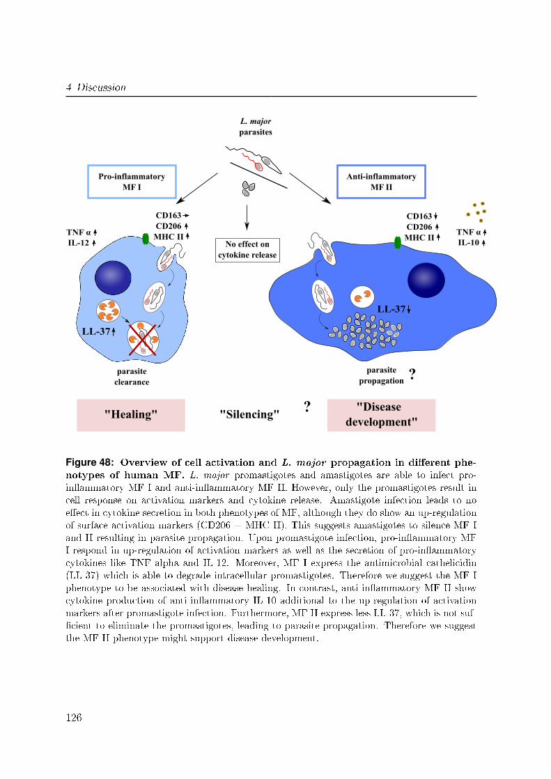
4 Discussion
"Healing"
L. majorparasites
"Disease development"
parasiteclearance
LL-37
TNF αIL-10
TNF αIL-12
LL-37
parasitepropagation
CD163CD206
MHC II
CD163CD206
MHC IINo effect on
cytokine release
Pro-inflammatoryMF I
Anti-inflammatoryMF II
"Silencing" ?
?
Figure 48: Overview of cell activation and L. major propagation in di�erent phe-notypes of human MF. L. major promastigotes and amastigotes are able to infect pro-in�ammatory MF I and anti-in�ammatory MF II. However, only the promastigotes result incell response on activation markers and cytokine release. Amastigote infection leads to noe�ect in cytokine secretion in both phenotypes of MF, although they do show an up-regulationof surface activation markers (CD206 + MHC II). This suggests amastigotes to silence MF Iand II resulting in parasite propagation. Upon promastigote infection, pro-in�ammatory MFI respond in up-regulation of activation markers as well as the secretion of pro-in�ammatorycytokines like TNF alpha and IL-12. Moreover, MF I express the antimicrobial cathelicidin(LL-37) which is able to degrade intracellular promastigotes. Therefore we suggest the MF Iphenotype to be associated with disease healing. In contrast, anti-in�ammatory MF II showcytokine production of anti-in�ammatory IL-10 additional to the up-regulation of activationmarkers after promastigote infection. Furthermore, MF II express less LL-37, which is not suf-�cient to eliminate the promastigotes, leading to parasite propagation. Therefore we suggestthe MF II phenotype might support disease development.
126
4.6 Concluding remarks
4.6 Concluding remarks
Taken together, the presented data demonstrates our axenic parasites to represent the
multiplying amastigote life stage of L. major, which develops inside infected MF and
is responsible for disease propagation. These parasites will be extremely useful for ad-
ditional biological, biochemical and molecular studies on the intracellular stage of this
parasite species itself as well as further investigations of the interaction with human
host cells. Furthermore, the �ndings of this study and recent publications demonstrate
L. major parasites to undergo apoptosis. In addition, we found at least two leishma-
nial cell death mechanisms, one dependent on ROS and one independent, which are
di�erently regulated between the promastigote and amastigote life stages.
This study shows the pro- and anti-in�ammatory phenotypes of human MF to respond
di�erently upon the infection with L. major parasites. Disease inducing promastigotes
result in pro-in�ammatory MF I in the secretion of the pro-in�ammatory cytokines
TNF alpha and IL-12 combined with a lower susceptibility for infection. Therefore we
suggest the MF I phenotype to be involved in the protective Th-1 mediated immune
response and subsequent healing. In contrast, anti-in�ammatory MF II produce anti-
in�ammatory IL-10 along with TNF alpha and show a higher susceptibility for L. major
infection. Thus MF II are proposed to be associated with a disease propagating Th-2
mediated immune response, leading to disease establishment. Furthermore, we demon-
strate the antimicrobial peptide LL-37 to e�ciently kill axenic L. major promastigotes
but not amastigotes in a dose dependent manner. Besides this, we found LL-37 to have
an e�ect on the survival of intracellular L. major parasites in MF I.
Taken together we are the �rst to demonstrate LL-37 to have a leishmanicidal activity
for L. major promastigotes in human primary MF. Further studies are required to inves-
tigate LL-37 as a potent candidate molecule for therapeutic intervention for cutaneous
Leishmaniasis.
127

5 Summary
This study focused on the characterization of both life stages of L. major FEBNI par-
asites, promastigotes as well as amastigotes and their interaction with di�erent pheno-
types of human macrophages (MF). In this context, a novel in vitro method to generate
L. major axenic amastigotes was applied and we found the in vitro generated L. major
axenic amastigotes showed the same stage-speci�c gene expression as compared to MF-
derived amastigotes. Moreover, we found the virulence factor GP63 to be stage-speci�c
expressed and up-regulated in L. major axenic amastigotes. In addition, we successfully
adapted the axenic culture system to generate axenic amastigotes from other L. major
isolates such as Friedlin and 5ASKH. Interestingly we found this process to be de-
pendent on isolate-speci�c temperature settings. Since there is only little know about
apoptotic machinery of protozoan parasites we induced apoptosis in both promastigotes
and amastigotes. We found two di�erent apoptotic mechanisms in L. major parasites.
One is dependent on reactive oxygen species (ROS) and one independent. These mech-
anisms were found to be regulated di�erentially in promastigotes and amastigotes.
In human disease it is still unclear which phenotype of MF is involved in disease de-
velopment. Therefore we analyzed pro-in�ammatory (MF I) and anti-in�ammatory
(MF II) MF. We found that L. major amastigotes infect both phenotypes of human
MF better as compared to promastigotes. Furthermore, anti-in�ammatory MF II are
more susceptible to infection as compared to pro-in�ammatory MF I. Analyzing possible
�killing signaling�, we found di�erential cytokine secretion in both phenotypes of MF.
Interestingly, we found only after promastigote infection the release of cytokines. For
MF I pro-in�ammatory cytokines like TNF alpha and IL-12 were detected which are
known to activate the cell and suggest MF I might clear parasite infection. In contrast,
MF II secreted anti-in�ammatory IL-10 upon infection indicating MF II to silence the
cell and resulting in parasite propagation. Furthermore, we searched for intracellular
129

5 Summary
killing mechanisms of MF and found the antimicrobial peptide cathelicidin (LL-37) to
be up-regulated in MF I as compared to MF II. LL-37 is located in lysosomes and is
shown to be involved in the innate host defense against pathogens. Consistent with
this, knockdown experiments for LL-37 demonstrated this peptide to be involved in the
intracellular degradation of L. major promastigotes in human MF I.
In conclusion, this study suggests that pro-in�ammatory MF I can eliminate L. major
parasites via LL-37, whereas anti-in�ammatory MF II might support disease develop-
ment.
130
6 Zusammenfassung
Der Fokus dieser Arbeit lag auf der Charakterisierung beider Lebensstadien des Para-
siten L. major FEBNI, der promastigoten und amastigoten Form, sowie deren Wech-
selwirkungen mit unterschiedlichen Phänotypen von humanen Makrophagen (MF). In
diesem Zusammenhang wurde eine neue in vitro Methode zur Herstellung von L. major
axenischen Amastigoten angewandt und dabei festgestellt, dass die in vitro generierten
axenischen Amastigoten die gleiche stadium-spezi�sche Genexpression aufweisen wie
Amastigote, die aus MF isoliert wurden. Des Weiteren konnte gezeigt werden, dass
der Virulenzfaktor GP63 stadium-spezi�sch exprimiert wird, wobei dieser in axenischen
Amastigoten hochreguliert ist. Zusätzlich wurde die in vitro Kultivierungsmethode der-
art angepasst, sodass Amastigote auch aus anderen L. major Isolaten wie Friedlin und
5ASKH erfolgreich generiert werden konnten. Interessanterweise war dieser Prozess
temperatursensitiv und abhängig von stamm-spezi�schen Temperaturbedingungen. Da
sehr wenig über das Apoptoseprogramm von einzelligen Parasiten bekannt ist, wurde
sowohl in Promastigoten als auch in Amastigoten Apoptose induziert und dabei zwei ver-
schiedene Mechanismen des programmierten Zelltods in L. major Parasiten gefunden.
Der erste wird durch reaktive Sauersto�spezies (ROS) vermittelt wohingegen der zweite
unabhängig davon ist. Beide Mechanismen werden in Promastigoten und Amastigoten
unterschiedlich reguliert.
In der humanen Leishmaniose ist bis heute nicht klar welcher Phänotyp von MF für
die Krankheitsentstehung relevant ist. Deshalb haben wir sowohl pro-in�ammatorische
MF (MF I) als auch anti-in�ammatorische MF (MF II) untersucht. Wir haben her-
ausgefunden, dass L. major Amastigoten beide Phänotypen humaner MF besser in-
�zieren als Promastigoten. Darüber hinaus sind anti-in�ammatorische MF II viel anfäl-
liger gegenüber einer Infektion verglichen mit pro-in�ammatorische MF I. Die Analyse
möglicher Signaltransduktionswege ergab Unterschiede in der Sekretion von Zytoki-
131
6 Zusammenfassung
nen der beiden MF Phänotypen. Interessanterweise haben wir nur nach Infektion mit
Promastigoten die Freisetzung von Zytokinen messen können. In MF I wurden pro-
in�ammatorische Zytokine wie TNF alpha und IL-12 nachgewiesen, die dafür bekannt
sind MF zu aktivieren und darauf hinweisen, dass MF I die Infektion abwehren könnten.
Im Gegensatz dazu sezernieren in�zierte MF II anti-in�ammatorisches IL-10 was darauf
hindeutet, dass MF II in ihrer Immunreaktion gehemmt werden, was zur Vermehrung
der Parasiten führen könnte. Darüber hinaus haben wir nach Abbaumechanismen von
intrazellulären Parasiten in MF gesucht und haben gefunden, dass das antimikrobielle
Peptid Cathelicidin (LL-37) in MF I im Vergleich zu MF II hochreguliert ist. LL-37
ist in Lysosomen zu �nden und an der angeborenen Immunantwort im Kampf gegen
Erreger beteiligt. Im Einklang dazu, haben unsere Knockdown Experimente für LL-37
gezeigt, dass LL-37 eine Rolle bei der intrazellulären Degradierung von L. major Pro-
mastigoten in MF I spielt.
Zusammenfassend legen die Ergebnisse dieser Dissertation nahe, dass pro-in�ammato-
rische MF I L. major Parasiten mit Hilfe von LL-37 abtöten, wohingegen anti-in�am-
matorische MF II die Krankheitsentwicklung fördern könnten.
132
List of Figures
1 Life cycle of Leishmania spp . . . . . . . . . . . . . . . . . . . . . . . . . 9
2 Distinct targets for apoptosis induction in Leishmania via di�erent drugs 13
3 Overview of possible mechanisms for the clearance of Leishmania . . . . 18
4 Involved proteins during apoptosis in L. major parasites . . . . . . . . . 20
5 Hypothesis for L. major parasite propagation in di�erent phenotypes of
human MF . . . . . . . . . . . . . . . . . . . . . . . . . . . . . . . . . . 21
6 RFLP analysis of the Leishmania spp. ITS1 marker . . . . . . . . . . . . 58
7 Morphology of log-phase L. major FEBNI promastigotes . . . . . . . . . 59
8 Morphology of stat-phase L. major FEBNI promastigotes . . . . . . . . . 60
9 Morphology of L. major FEBNI amastigotes . . . . . . . . . . . . . . . . 60
10 Annexin A5 staining of the di�erent stages of L. major parasites . . . . . 61
11 Stage-speci�c mRNA expression of SHERP and ABC-transporter homo-
logue in L. major . . . . . . . . . . . . . . . . . . . . . . . . . . . . . . . 62
12 Stage-speci�c mRNA expression of QDPR, alpha-tubulin, Cpb and GP63
in L. major . . . . . . . . . . . . . . . . . . . . . . . . . . . . . . . . . . 63
13 Stage-speci�c protein expression of LPG on the cell surface of L. major . 64
14 Stage-speci�c protein expression of LPG in L. major . . . . . . . . . . . 65
15 Generation of L. major Friedlin axenic amastigotes . . . . . . . . . . . . 67
16 Phosphatidylserine externalisation after apoptosis induction in L. major
FEBNI promastigotes . . . . . . . . . . . . . . . . . . . . . . . . . . . . . 69
17 Formation of reactive oxygen species after apoptosis induction in L. major
FEBNI promastigotes . . . . . . . . . . . . . . . . . . . . . . . . . . . . . 70
18 Modulation of markers after apoptosis induction in L. major FEBNI
promastigotes . . . . . . . . . . . . . . . . . . . . . . . . . . . . . . . . . 71
133
List of Figures
19 Modulation of markers after apoptosis induction in L. major FEBNI
amastigotes . . . . . . . . . . . . . . . . . . . . . . . . . . . . . . . . . . 72
20 Viable and apoptotic L. major FEBNI parasites in �ow cytometry . . . . 73
21 Infected human MF with L. major parasites . . . . . . . . . . . . . . . . 76
22 Stage-speci�c interaction of L. major parasites with human MF . . . . . 77
23 Characteristics of L. major FEBNI eGFP parasites . . . . . . . . . . . . 79
24 L. major eGFP parasite development in di�erent types of human MF . . 80
25 Downregulation of CD163 on the cell surface in human MF II after in-
fection with L. major parasites . . . . . . . . . . . . . . . . . . . . . . . 82
26 Downregulation of CD163 on the cell surface and mRNA expression in
human MF II after infection with L. major parasites . . . . . . . . . . . 83
27 Up-regulation of CD206 on the cell surface in human MF after infection
with L. major parasites . . . . . . . . . . . . . . . . . . . . . . . . . . . 84
28 Up-regulation of MHC II on the cell surface in human MF after infection
with L. major parasites . . . . . . . . . . . . . . . . . . . . . . . . . . . 85
29 Up-regulation of CD86 on the cell surface in human MF after infection
with L. major parasites . . . . . . . . . . . . . . . . . . . . . . . . . . . 87
30 Di�erent cytokine production of TNF alpha in human MF after infection
with L. major parasites . . . . . . . . . . . . . . . . . . . . . . . . . . . 88
31 Di�erent cytokine production of IL-12 in human MF after infection with
L. major parasites . . . . . . . . . . . . . . . . . . . . . . . . . . . . . . 90
32 Di�erent cytokine mRNA expression of CCL3 and CCL4 in human MF
after infection with L. major parasites . . . . . . . . . . . . . . . . . . . 91
33 Di�erent cytokine production of IL-10 in human MF after infection with
L. major parasites . . . . . . . . . . . . . . . . . . . . . . . . . . . . . . 92
34 Di�erent phosphorylation state of p38 and p44/42 (ERK1/2) MAP ki-
nases (MAPK) in human type I MF after infection with L. major parasites 94
35 Di�erent phosphorylation state of p38 MAP kinases (MAPK) in human
MF after infection with L. major parasites . . . . . . . . . . . . . . . . . 96
36 Di�erent phosphorylation state of ERK1/2 MAP kinases (MAPK) in
human MF after infection with L. major parasites . . . . . . . . . . . . . 97
37 Infection rates after concanamycin A treatment and parasite location in
human MF after L. major infection . . . . . . . . . . . . . . . . . . . . . 99
134
List of Figures
38 Arginase activity and mRNA expression in human MF after infection
with L. major parasites . . . . . . . . . . . . . . . . . . . . . . . . . . . 101
39 Di�erent cathelicidin (LL-37) mRNA expression in human MF . . . . . . 102
40 Crucifying e�ect of rhLL-37 on L. major promastigotes . . . . . . . . . . 103
41 Killing e�ect of rhLL-37 on L. major promastigotes . . . . . . . . . . . . 104
42 Morphology of rhLL-37 treated L. major promastigotes . . . . . . . . . . 105
43 No e�ect of rhLL-37 on L. major amastigotes . . . . . . . . . . . . . . . 105
44 Parasite burden in LL-37 knockdown pro-in�ammatory and anti-in�ammatory
human MF . . . . . . . . . . . . . . . . . . . . . . . . . . . . . . . . . . 107
45 Survival of L. major parasites in LL-37 knockdown pro-in�ammatory and
anti-in�ammatory human MF . . . . . . . . . . . . . . . . . . . . . . . . 109
46 Overview of the di�erent apoptotic mechanisms in L. major promastigotes116
47 Overview of the di�erent apoptotic mechanisms in L. major amastigotes 118
48 Overview of cell activation and L. major propagation in di�erent pheno-
types of human MF . . . . . . . . . . . . . . . . . . . . . . . . . . . . . . 126
135


Bibliography
[1] M. S. Alcouloumre, M. A. Ghannoum, A. S. Ibrahim, M. E. Selsted, and J. E.
Edwards. Fungicidal properties of defensin np-1 and activity against cryptococcus
neoformans in vitro. Antimicrob Agents Chemother, 37(12):2628�2632, Dec 1993.
[2] J. Alexander, A. R. Satoskar, and D. G. Russell. Leishmania species: models of
intracellular parasitism. J Cell Sci, 112 Pt 18:2993�3002, Sep 1999.
[3] Cindy Allenbach, Pascal Launois, Christoph Mueller, and Fabienne Tacchini-
Cottier. An essential role for transmembrane tnf in the resolution of the in�amma-
tory lesion induced by leishmania major infection. Eur J Immunol, 38(3):720�731,
Mar 2008.
[4] Jorge Alvar, Sergio Yactayo, and Caryn Bern. Leishmaniasis and poverty. Trends
Parasitol, 22(12):552�557, Dec 2006.
[5] J. F. Alzate, A. Alvarez-Barrientos, V. M. González, and A. Jiménez-Ruiz. Heat-
induced programmed cell death in leishmania infantum is reverted by bcl-x(l)
expression. Apoptosis, 11(2):161�171, Feb 2006.
[6] Charles F Anderson, Susana Mendez, and David L Sacks. Nonhealing infection
despite th1 polarization produced by a strain of leishmania major in c57bl/6 mice.
J Immunol, 174(5):2934�2941, Mar 2005.
[7] Charles F Anderson, Mohammed Oukka, Vijay J Kuchroo, and David Sacks.
Cd4(+)cd25(-)foxp3(-) th1 cells are the source of il-10-mediated immune suppres-
sion in chronic cutaneous leishmaniasis. J Exp Med, 204(2):285�297, Feb 2007.
137
Bibliography
[8] L. Aravind, V. M. Dixit, and E. V. Koonin. Apoptotic molecular machinery:
vastly increased complexity in vertebrates revealed by genome comparisons. Sci-
ence, 291(5507):1279�1284, Feb 2001.
[9] D. Arnoult, K. Akarid, A. Grodet, P. X. Petit, and J. Estaquier sand
J. C. Ameisen. On the evolution of programmed cell death: apoptosis of the
unicellular eukaryote leishmania major involves cysteine proteinase activation and
mitochondrion permeabilization. Cell Death Di�er, 9(1):65�81, Jan 2002.
[10] Samira Azzouz, Mimoun Maache, Ramon Gil Garcia, and Antonio Osuna. Leish-
manicidal activity of edelfosine, miltefosine and ilmofosine. Basic Clin Pharmacol
Toxicol, 96(1):60�65, Jan 2005.
[11] V. Bahr, Y. D. Stierhof, T. Ilg, M. Demar, M. Quinten, and P. Overath. Expression
of lipophosphoglycan, high-molecular weight phosphoglycan and glycoprotein 63
in promastigotes and amastigotes of leishmania mexicana. Mol Biochem Parasitol,
58(1):107�121, Mar 1993.
[12] R. Bals, X. Wang, M. Zaslo�, and J. M. Wilson. The peptide antibiotic ll-37/hcap-
18 is expressed in epithelia of the human lung where it has broad antimicrobial
activity at the airway surface. Proc Natl Acad Sci U S A, 95(16):9541�9546, Aug
1998.
[13] S. Becker and C. L. Ja�e. E�ect of protein kinase inhibitors on the growth,
morphology, and infectivity of leishmania promastigotes. Parasitol Res, 83(3):273�
280, 1997.
[14] Y. Belkaid, B. Butcher, and D. L. Sacks. Analysis of cytokine production by
in�ammatory mouse macrophages at the single-cell level: selective impairment of
il-12 induction in leishmania-infected cells. Eur J Immunol, 28(4):1389�1400, Apr
1998.
[15] Rym Ben-Othman, Koussay Dellagi, and Lamia Guizani-Tabbane. Leishmania
major parasites induced macrophage tolerance: implication of mapk and nf-
kappab pathways. Mol Immunol, 46(16):3438�3444, Oct 2009.
[16] Rym Ben-Othman, Lamia Guizani-Tabbane, and Koussay Dellagi. Leishmania ini-
tially activates but subsequently down-regulates intracellular mitogen-activated
138

Bibliography
protein kinases and nuclear factor-kappab signaling in macrophages. Mol Im-
munol, 45(11):3222�3229, Jun 2008.
[17] J. D. Berman. Human leishmaniasis: clinical, diagnostic, and chemotherapeutic
developments in the last 10 years. Clin Infect Dis, 24(4):684�703, Apr 1997.
[18] J. D. Berman and F. A. Neva. E�ect of temperature on multiplication of leish-
mania amastigotes within human monocyte-derived macrophages in vitro. Am J
Trop Med Hyg, 30(2):318�321, Mar 1981.
[19] Sébastien Besteiro, Roderick A M Williams, Graham H Coombs, and Jeremy C
Mottram. Protein turnover and di�erentiation in leishmania. Int J Parasitol,
37(10):1063�1075, Aug 2007.
[20] Sébastien Besteiro, Roderick A M Williams, Lesley S Morrison, Graham H
Coombs, and Jeremy C Mottram. Endosome sorting and autophagy are essential
for di�erentiation and virulence of leishmania major. J Biol Chem, 281(16):11384�
11396, Apr 2006.
[21] D. Biegel, G. Topper, and M. Rabinovitch. Leishmania mexicana: temperature
sensitivity of isolated amastigotes and of amastigotes infecting macrophages in
culture. Exp Parasitol, 56(3):289�297, Dec 1983.
[22] C. Bogdan and M. Röllingho�. The immune response to leishmania: mechanisms
of parasite control and evasion. Int J Parasitol, 28(1):121�134, Jan 1998.
[23] C. Bogdan, Y. Vodovotz, and C. Nathan. Macrophage deactivation by interleukin
10. J Exp Med, 174(6):1549�1555, Dec 1991.
[24] Glória Bom�m, Bruno B Andrade, Silvane Santos, Jorge Clarêncio, Manoel
Barral-Netto, and Aldina Barral. Cellular analysis of cutaneous leishmaniasis
lymphadenopathy: insights into the early phases of human disease. Am J Trop
Med Hyg, 77(5):854�859, Nov 2007.
[25] Eliane Bourreau, Jacques Gardon, Roger Pradinaud, Hervé Pascalis, Ghislaine
Prévot-Linguet, Amina Kariminia, and Launois Pascal. Th2 responses predomi-
nate during the early phases of infection in patients with localized cutaneous leish-
139
Bibliography
maniasis and precede the development of th1 responses. Infect Immun, 71(4):2244�
2246, Apr 2003.
[26] Barry J Bowman and Emma Jean Bowman. Mutations in subunit c of the vacuolar
atpase confer resistance to ba�lomycin and identify a conserved antibiotic binding
site. J Biol Chem, 277(6):3965�3972, Feb 2002.
[27] A. Brittingham, C. J. Morrison, W. R. McMaster, B. S. McGwire, K. P. Chang,
and D. M. Mosser. Role of the leishmania surface protease gp63 in complement
�xation, cell adhesion, and resistance to complement-mediated lysis. J Immunol,
155(6):3102�3111, Sep 1995.
[28] F. M. Brodsky, L. Lem, A. Solache, and E. M. Bennett. Human pathogen sub-
version of antigen presentation. Immunol Rev, 168:199�215, Apr 1999.
[29] Kelly L Brown and Robert E W Hancock. Cationic host defense (antimicrobial)
peptides. Curr Opin Immunol, 18(1):24�30, Feb 2006.
[30] C. Buechler, M. Ritter, E. Orsó, T. Langmann, J. Klucken, and G. Schmitz.
Regulation of scavenger receptor cd163 expression in human monocytes and
macrophages by pro- and antiin�ammatory stimuli. J Leukoc Biol, 67(1):97�103,
Jan 2000.
[31] E. Buentke, A. Zargari, L. C. He�er, J. Avila-Cariño, J. Savolainen, and
A. Scheynius. Uptake of the yeast malassezia furfur and its allergenic components
by human immature cd1a+ dendritic cells. Clin Exp Allergy, 30(12):1759�1770,
Dec 2000.
[32] C. Buser and P. Walther. Freeze-substitution: the addition of water to polar
solvents enhances the retention of structure and acts at temperatures around -60
degrees c. J Microsc, 230(Pt 2):268�277, May 2008.
[33] L. L. Button, D. G. Russell, H. L. Klein, E. Medina-Acosta, R. E. Karess, and
W. R. McMaster. Genes encoding the major surface glycoprotein in leishmania are
tandemly linked at a single chromosomal locus and are constitutively transcribed.
Mol Biochem Parasitol, 32(2-3):271�283, Jan 1989.
140
Bibliography
[34] L. L. Button, G. Wilson, C. R. Astell, and W. R. McMaster. Recombinant leish-
mania surface glycoprotein gp63 is secreted in the baculovirus expression system
as a latent metalloproteinase. Gene, 134(1):75�81, Nov 1993.
[35] M. H. Cobb and E. J. Goldsmith. How map kinases are regulated. J Biol Chem,
270(25):14843�14846, Jun 1995.
[36] Gabriela Cohen-Freue, Timothy R Holzer, James D Forney, and W. Robert Mc-
Master. Global gene expression in leishmania. Int J Parasitol, 37(10):1077�1086,
Aug 2007.
[37] G. H. Coombs and J. Baxter. Inhibition of leishmania amastigote growth by
antipain and leupeptin. Ann Trop Med Parasitol, 78(1):21�24, Feb 1984.
[38] S. L. Croft, D. Snowdon, and V. Yardley. The activities of four anticancer
alkyllysophospholipids against leishmania donovani, trypanosoma cruzi and try-
panosoma brucei. J Antimicrob Chemother, 38(6):1041�1047, Dec 1996.
[39] Anna C Cunningham. Parasitic adaptive mechanisms in infection by leishmania.
Exp Mol Pathol, 72(2):132�141, Apr 2002.
[40] M. L. Cunningham, R. G. Titus, S. J. Turco, and S. M. Beverley. Regulation of
di�erentiation to the infective stage of the protozoan parasite leishmania major
by tetrahydrobiopterin. Science, 292(5515):285�287, Apr 2001.
[41] K. A. Daher, M. E. Selsted, and R. I. Lehrer. Direct inactivation of viruses by
human granulocyte defensins. J Virol, 60(3):1068�1074, Dec 1986.
[42] M. Das, S. B. Mukherjee, and C. Shaha. Hydrogen peroxide induces apoptosis-like
death in leishmania donovani promastigotes. J Cell Sci, 114(Pt 13):2461�2469,
Jul 2001.
[43] Alain Debrabant, Nancy Lee, Sylvie Bertholet, Robert Duncan, and Hira L
Nakhasi. Programmed cell death in trypanosomatids and other unicellular or-
ganisms. Int J Parasitol, 33(3):257�267, Mar 2003.
[44] Marcel Deponte and Katja Becker. Plasmodium falciparum�do killers commit
suicide? Trends Parasitol, 20(4):165�169, Apr 2004.
141
Bibliography
[45] L. Ding, P. S. Linsley, L. Y. Huang, R. N. Germain, and E. M. Shevach. Il-
10 inhibits macrophage costimulatory activity by selectively inhibiting the up-
regulation of b7 expression. J Immunol, 151(3):1224�1234, Aug 1993.
[46] Subhankar Dolai, Swati Pal, Rajesh K Yadav, and Subrata Adak. Endoplasmic
reticulum stress-induced apoptosis in leishmania through ca2+-dependent and
caspase-independent mechanism. J Biol Chem, 286(15):13638�13646, Apr 2011.
[47] Subhankar Dolai, Rajesh K Yadav, Swati Pal, and Subrata Adak. Overexpression
of mitochondrial leishmania major ascorbate peroxidase enhances tolerance to
oxidative stress-induced programmed cell death and protein damage. Eukaryot
Cell, 8(11):1721�1731, Nov 2009.
[48] R. A. Dorschner, V. K. Pestonjamasp, S. Tamakuwala, T. Ohtake, J. Rudisill,
V. Nizet, B. Agerberth, G. H. Gudmundsson, and R. L. Gallo. Cutaneous injury
induces the release of cathelicidin anti-microbial peptides active against group a
streptococcus. J Invest Dermatol, 117(1):91�97, Jul 2001.
[49] Avijit Dutta, Suman Bandyopadhyay, Chitra Mandal, and Mitali Chatterjee. Aloe
vera leaf exudate induces a caspase-independent cell death in leishmania donovani
promastigotes. J Med Microbiol, 56(Pt 5):629�636, May 2007.
[50] S. Ehrt, D. Schnappinger, S. Bekiranov, J. Drenkow, S. Shi, T. R. Gingeras,
T. Gaasterland, G. Schoolnik, and C. Nathan. Reprogramming of the macrophage
transcriptome in response to interferon-gamma and mycobacterium tuberculosis:
signaling roles of nitric oxide synthase-2 and phagocyte oxidase. J Exp Med,
194(8):1123�1140, Oct 2001.
[51] Patricia Escobar, Sangeeta Matu, Cláudia Marques, and Simon L Croft. Sensitiv-
ities of leishmania species to hexadecylphosphocholine (miltefosine), et-18-och(3)
(edelfosine) and amphotericin b. Acta Trop, 81(2):151�157, Feb 2002.
[52] G. J. Feng, H. S. Goodridge, M. M. Harnett, X. Q. Wei, A. V. Nikolaev, A. P.
Higson, and F. Y. Liew. Extracellular signal-related kinase (erk) and p38 mitogen-
activated protein (map) kinases di�erentially regulate the lipopolysaccharide-
mediated induction of inducible nitric oxide synthase and il-12 in macrophages:
142

Bibliography
Leishmania phosphoglycans subvert macrophage il-12 production by targeting erk
map kinase. J Immunol, 163(12):6403�6412, Dec 1999.
[53] K. F. Ferri and G. Kroemer. Mitochondria�the suicide organelles. Bioessays,
23(2):111�115, Feb 2001.
[54] Y. Le Fichoux, D. Rousseau, B. Ferrua, S. Ruette, A. Leliévre, D. Grousson,
and J. Kubar. Short- and long-term e�cacy of hexadecylphosphocholine against
established leishmania infantum infection in balb/c mice. Antimicrob Agents
Chemother, 42(3):654�658, Mar 1998.
[55] D. F. Fiorentino, A. Zlotnik, T. R. Mosmann, M. Howard, and A. O'Garra. Il-10
inhibits cytokine production by activated macrophages. J Immunol, 147(11):3815�
3822, Dec 1991.
[56] J. L. Flynn, M. M. Goldstein, K. J. Triebold, J. Sypek, S. Wolf, and B. R. Bloom.
Il-12 increases resistance of balb/c mice to mycobacterium tuberculosis infection.
J Immunol, 155(5):2515�2524, Sep 1995.
[57] Michael Forgac. Vacuolar atpases: rotary proton pumps in physiology and patho-
physiology. Nat Rev Mol Cell Biol, 8(11):917�929, Nov 2007.
[58] R. L. Gallo, K. J. Kim, M. Bern�eld, C. A. Kozak, M. Zanetti, L. Merluzzi,
and R. Gennaro. Identi�cation of cramp, a cathelin-related antimicrobial peptide
expressed in the embryonic and adult mouse. J Biol Chem, 272(20):13088�13093,
May 1997.
[59] T. Ganz, M. E. Selsted, D. Szklarek, S. S. Harwig, K. Daher, D. F. Bainton, and
R. I. Lehrer. Defensins. natural peptide antibiotics of human neutrophils. J Clin
Invest, 76(4):1427�1435, Oct 1985.
[60] R. T. Gazzinelli, I. P. Oswald, S. L. James, and A. Sher. Il-10 inhibits parasite
killing and nitrogen oxide production by ifn-gamma-activated macrophages. J
Immunol, 148(6):1792�1796, Mar 1992.
[61] R. Gennaro and M. Zanetti. Structural features and biological activities of the
cathelicidin-derived antimicrobial peptides. Biopolymers, 55(1):31�49, 2000.
143

Bibliography
[62] M. S. Giannini. E�ects of promastigote growth phase, frequency of subculture,
and host age on promastigote-initiated infections with leishmania donovani in the
golden hamster. J Protozool, 21(4):521�527, Oct 1974.
[63] T. A. Glaser, S. F. Moody, E. Handman, A. Bacic, and T. W. Spithill. An
antigenically distinct lipophosphoglycan on amastigotes of leishmania major. Mol
Biochem Parasitol, 45(2):337�344, Apr 1991.
[64] Y. Jerold Gordon, Ling C Huang, Eric G Romanowski, Kathleen A Yates, Rita J
Proske, and Alison M McDermott. Human cathelicidin (ll-37), a multifunctional
peptide, is expressed by ocular surface epithelia and has potent antibacterial and
antiviral activity. Curr Eye Res, 30(5):385�394, May 2005.
[65] S. J. Green, S. Mellouk, S. L. Ho�man, M. S. Meltzer, and C. A. Nacy. Cellular
mechanisms of nonspeci�c immunity to intracellular infection: cytokine-induced
synthesis of toxic nitrogen oxides from l-arginine by macrophages and hepatocytes.
Immunol Lett, 25(1-3):15�19, Aug 1990.
[66] S. J. Green, C. A. Nacy, and M. S. Meltzer. Cytokine-induced synthesis of nitro-
gen oxides in macrophages: a protective host response to leishmania and other
intracellular pathogens. J Leukoc Biol, 50(1):93�103, Jul 1991.
[67] G. H. Gudmundsson, B. Agerberth, J. Odeberg, T. Bergman, B. Olsson, and
R. Salcedo. The human gene fall39 and processing of the cathelin precursor to
the antibacterial peptide ll-37 in granulocytes. Eur J Biochem, 238(2):325�332,
Jun 1996.
[68] C. F. Higgins. Abc transporters: from microorganisms to man. Annu Rev Cell
Biol, 8:67�113, 1992.
[69] Katharine A Hintz, Athos J Rassias, Kathleen Wardwell, Marcia L Moss, Peter M
Morganelli, Patricia A Pioli, Alice L Givan, Paul K Wallace, Mark P Yeager, and
Paul M Guyre. Endotoxin induces rapid metalloproteinase-mediated shedding
followed by up-regulation of the monocyte hemoglobin scavenger receptor cd163.
J Leukoc Biol, 72(4):711�717, Oct 2002.
[70] Philippe Holzmuller, Denis Sereno, Mireille Cavaleyra, Isabelle Mangot, Sylvie
Daulouede, Philippe Vincendeau, and Jean-Loup Lemesre. Nitric oxide-mediated
144
Bibliography
proteasome-dependent oligonucleosomal dna fragmentation in leishmania amazo-
nensis amastigotes. Infect Immun, 70(7):3727�3735, Jul 2002.
[71] Michael D Howell, James F Jones, Kevin O Kisich, Joanne E Streib, Richard L
Gallo, and Donald Y M Leung. Selective killing of vaccinia virus by ll-37: impli-
cations for eczema vaccinatum. J Immunol, 172(3):1763�1767, Feb 2004.
[72] Y. H. Hsiang, R. Hertzberg, S. Hecht, and L. F. Liu. Camptothecin induces
protein-linked dna breaks via mammalian dna topoisomerase i. J Biol Chem,
260(27):14873�14878, Nov 1985.
[73] C. S. Hsieh, S. E. Macatonia, C. S. Tripp, S. F. Wolf, A. O'Garra, and K. M.
Murphy. Development of th1 cd4+ t cells through il-12 produced by listeria-
induced macrophages. Science, 260(5107):547�549, Apr 1993.
[74] Markus Huss, Gudrun Ingenhorst, Simone König, Michael Gassel, Stefan Dröse,
Axel Zeeck, Karlheinz Altendorf, and Helmut Wieczorek. Concanamycin a,
the speci�c inhibitor of v-atpases, binds to the v(o) subunit c. J Biol Chem,
277(43):40544�40548, Oct 2002.
[75] Virginia Iniesta, Jesualdo Carcelén, Isabel Molano, Pablo M V Peixoto, Eloy
Redondo, Pilar Parra, Marina Mangas, Isabel Monroy, Maria Luisa Campo, Car-
los Gómez Nieto, and Inés Corraliza. Arginase i induction during leishmania major
infection mediates the development of disease. Infect Immun, 73(9):6085�6090,
Sep 2005.
[76] Alasdair C Ivens, Christopher S Peacock, Elizabeth AWorthey, Lee Murphy, Gau-
tam Aggarwal, Matthew Berriman, Ellen Sisk, Marie-Adele Rajandream, Ellen
Adlem, Rita Aert, Atashi Anupama, Zina Apostolou, Philip Attipoe, Nathalie Ba-
son, Christopher Bauser, Alfred Beck, Stephen M Beverley, Gabriella Bianchettin,
Katja Borzym, Gordana Bothe, Carlo V Bruschi, Matt Collins, Eithon Cadag,
Laura Ciarloni, Christine Clayton, Richard M R Coulson, Ann Cronin, An-
gela K Cruz, Robert M Davies, Javier De Gaudenzi, Deborah E Dobson, An-
dreas Duesterhoeft, Gholam Fazelina, Nigel Fosker, Alberto Carlos Frasch, Au-
drey Fraser, Monika Fuchs, Claudia Gabel, Arlette Goble, André Go�eau, David
Harris, Christiane Hertz-Fowler, Helmut Hilbert, David Horn, Yiting Huang, Sven
145

Bibliography
Klages, Andrew Knights, Michael Kube, Natasha Larke, Lyudmila Litvin, Angela
Lord, Tin Louie, Marco Marra, David Masuy, Keith Matthews, Shulamit Michaeli,
Jeremy C Mottram, Silke Müller-Auer, Heather Munden, Siri Nelson, Halina Nor-
bertczak, Karen Oliver, Susan O'neil, Martin Pentony, Thomas M Pohl, Claire
Price, Bénédicte Purnelle, Michael A Quail, Ester Rabbinowitsch, Richard Rein-
hardt, Michael Rieger, Joel Rinta, Johan Robben, Laura Robertson, Jeronimo C
Ruiz, Simon Rutter, David Saunders, Melanie Schäfer, Jacquie Schein, David C
Schwartz, Kathy Seeger, Amber Seyler, Sarah Sharp, Heesun Shin, Dhileep Sivam,
Rob Squares, Steve Squares, Valentina Tosato, Christy Vogt, Guido Volckaert,
Rolf Wambutt, Tim Warren, Holger Wedler, John Woodward, Shiguo Zhou, Wolf-
gang Zimmermann, Deborah F Smith, Jenefer M Blackwell, Kenneth D Stuart,
Bart Barrell, and Peter J Myler. The genome of the kinetoplastid parasite, leish-
mania major. Science, 309(5733):436�442, Jul 2005.
[77] Muthoni Junghae and John G Raynes. Activation of p38 mitogen-activated pro-
tein kinase attenuates leishmania donovani infection in macrophages. Infect Im-
mun, 70(9):5026�5035, Sep 2002.
[78] B. L. Kagan, M. E. Selsted, T. Ganz, and R. I. Lehrer. Antimicrobial defensin
peptides form voltage-dependent ion-permeable channels in planar lipid bilayer
membranes. Proc Natl Acad Sci U S A, 87(1):210�214, Jan 1990.
[79] M. M. Kane and D. M. Mosser. Leishmania parasites and their ploys to disrupt
macrophage activation. Curr Opin Hematol, 7(1):26�31, Jan 2000.
[80] Mazen W Karaman, Sanna Herrgard, Daniel K Treiber, Paul Gallant, Corey E
Atteridge, Brian T Campbell, Katrina W Chan, Pietro Ciceri, Mindy I Davis,
Philip T Edeen, Ra�aella Faraoni, Mark Floyd, Jeremy P Hunt, Daniel J Lock-
hart, Zdravko V Milanov, Michael J Morrison, Gabriel Pallares, Hitesh K Patel,
Stephanie Pritchard, Lisa M Wodicka, and Patrick P Zarrinkar. A quantitative
analysis of kinase inhibitor selectivity. Nat Biotechnol, 26(1):127�132, Jan 2008.
[81] Prasanthi Karna, Susu Zughaier, Vaishali Pannu, Robert Simmons, Satya
Narayan, and Ritu Aneja. Induction of reactive oxygen species-mediated au-
tophagy by a novel microtubule-modulating agent. J Biol Chem, 285(24):18737�
18748, Jun 2010.
146
Bibliography
[82] J. F. Kerr, A. H. Wyllie, and A. R. Currie. Apoptosis: a basic biological
phenomenon with wide-ranging implications in tissue kinetics. Br J Cancer,
26(4):239�257, Aug 1972.
[83] Shahram Khademvatan, Mohammad Javad Gharavi, Fakher Rahim, and Jasem
Saki. Miltefosine-induced apoptotic cell death on leishmania major and l. tropica
strains. Korean J Parasitol, 49(1):17�23, Mar 2011.
[84] Mary E Klotman and Theresa L Chang. Defensins in innate antiviral immunity.
Nat Rev Immunol, 6(6):447�456, Jun 2006.
[85] R. M. Kluck, E. Bossy-Wetzel, D. R. Green, and D. D. Newmeyer. The release of
cytochrome c from mitochondria: a primary site for bcl-2 regulation of apoptosis.
Science, 275(5303):1132�1136, Feb 1997.
[86] E. Knuepfer, Y. D. Stierhof, P. G. McKean, and D. F. Smith. Characterization of a
di�erentially expressed protein that shows an unusual localization to intracellular
membranes in leishmania major. Biochem J, 356(Pt 2):335�344, Jun 2001.
[87] M. Kobayashi, L. Fitz, M. Ryan, R. M. Hewick, S. C. Clark, S. Chan, R. Loudon,
F. Sherman, B. Perussia, and G. Trinchieri. Identi�cation and puri�cation of nat-
ural killer cell stimulatory factor (nksf), a cytokine with multiple biologic e�ects
on human lymphocytes. J Exp Med, 170(3):827�845, Sep 1989.
[88] Stephan R Krutzik, Belinda Tan, Huiying Li, Maria Teresa Ochoa, Philip T Liu,
Sarah E Sharfstein, Thomas G Graeber, Peter A Sieling, Yong-Jun Liu, Thomas H
Rea, Barry R Bloom, and Robert L Modlin. Tlr activation triggers the rapid di�er-
entiation of monocytes into macrophages and dendritic cells. Nat Med, 11(6):653�
660, Jun 2005.
[89] Manjusha M Kulkarni, Joseph Barbi, W. Robert McMaster, Richard L Gallo,
Abhay R Satoskar, and Bradford S McGwire. Mammalian antimicrobial peptide
in�uences control of cutaneous leishmania infection. Cell Microbiol, 13(6):913�
923, Jun 2011.
[90] Promod Kumar, Kalpana Pai, Haushila P Pandey, and Shyam Sundar. Nadh-
oxidase, nadph-oxidase and myeloperoxidase activity of visceral leishmaniasis pa-
tients. J Med Microbiol, 51(10):832�836, Oct 2002.
147

Bibliography
[91] R. Lainson. Ecological interactions in the transmission of the leishmaniases. Philos
Trans R Soc Lond B Biol Sci, 321(1207):389�404, Oct 1988.
[92] J. W. Larrick, M. Hirata, R. F. Balint, J. Lee, J. Zhong, and S. C. Wright. Human
cap18: a novel antimicrobial lipopolysaccharide-binding protein. Infect Immun,
63(4):1291�1297, Apr 1995.
[93] J. W. Larrick, J. Lee, S. Ma, X. Li, U. Francke, S. C. Wright, and R. F. Balint.
Structural, functional analysis and localization of the human cap18 gene. FEBS
Lett, 398(1):74�80, Nov 1996.
[94] Tamás Laskay, Ger van Zandbergen, and Werner Solbach. Neutrophil
granulocytes�trojan horses for leishmania major and other intracellular microbes?
Trends Microbiol, 11(5):210�214, May 2003.
[95] Carsten Gk Lüder, Jenny Campos-Salinas, Elena Gonzalez-Rey, and Ger van
Zandbergen. Impact of protozoan cell death on parasite-host interactions and
pathogenesis. Parasit Vectors, 3:116, 2010.
[96] N. Lee, S. Bertholet, A. Debrabant, J. Muller, R. Duncan, and H. L. Nakhasi.
Programmed cell death in the unicellular protozoan parasite leishmania. Cell
Death Di�er, 9(1):53�64, Jan 2002.
[97] R. I. Lehrer and T. Ganz. Antimicrobial peptides in mammalian and insect host
defence. Curr Opin Immunol, 11(1):23�27, Feb 1999.
[98] Kirk Leifso, Gabriela Cohen-Freue, Nisha Dogra, Angus Murray, and W. Robert
McMaster. Genomic and proteomic expression analysis of leishmania promastigote
and amastigote life stages: the leishmania genome is constitutively expressed. Mol
Biochem Parasitol, 152(1):35�46, Mar 2007.
[99] J. L. Lemesre, D. Sereno, S. Daulouède, B. Veyret, N. Brajon, and P. Vincendeau.
Leishmania spp.: nitric oxide-mediated metabolic inhibition of promastigote and
axenically grown amastigote forms. Exp Parasitol, 86(1):58�68, May 1997.
[100] F. Y. Liew, S. Millott, C. Parkinson, R. M. Palmer, and S. Moncada. Macrophage
killing of leishmania parasite in vivo is mediated by nitric oxide from l-arginine.
J Immunol, 144(12):4794�4797, Jun 1990.
148
Bibliography
[101] Philip T Liu, Ste�en Stenger, Huiying Li, Linda Wenzel, Belinda H Tan,
Stephan R Krutzik, Maria Teresa Ochoa, Jürgen Schauber, Kent Wu, Christoph
Meinken, Diane L Kamen, Manfred Wagner, Robert Bals, Andreas Steinmeyer,
Ulrich Zügel, Richard L Gallo, David Eisenberg, Martin Hewison, Bruce W Hollis,
John S Adams, Barry R Bloom, and Robert L Modlin. Toll-like receptor triggering
of a vitamin d-mediated human antimicrobial response. Science, 311(5768):1770�
1773, Mar 2006.
[102] P. J. Lohuis, M. M. Lipovsky, A. I. Hoepelman, G. J. Hordijk, and E. H. Huizing.
Leishmania braziliensis presenting as a granulomatous lesion of the nasal septum
mucosa. J Laryngol Otol, 111(10):973�975, Oct 1997.
[103] Mark Lucas, Xia Zhang, Vikram Prasanna, and David M Mosser. Erk activation
following macrophage fcgammar ligation leads to chromatin modi�cations at the
il-10 locus. J Immunol, 175(1):469�477, Jul 2005.
[104] H. Lux, N. Heise, T. Klenner, D. Hart, and F. R. Opperdoes. Ether�lipid (alkyl-
phospholipid) metabolism and the mechanism of action of ether�lipid analogues
in leishmania. Mol Biochem Parasitol, 111(1):1�14, Nov 2000.
[105] Lon-Fye Lye, Mark L Cunningham, and Stephen M Beverley. Characterization of
quinonoid-dihydropteridine reductase (qdpr) from the lower eukaryote leishmania
major. J Biol Chem, 277(41):38245�38253, Oct 2002.
[106] Miriam A Lynn and W. Robert McMaster. Leishmania: conserved evolution�
diverse diseases. Trends Parasitol, 24(3):103�105, Mar 2008.
[107] R. Manetti, P. Parronchi, M. G. Giudizi, M. P. Piccinni, E. Maggi, G. Trinchieri,
and S. Romagnani. Natural killer cell stimulatory factor (interleukin 12 [il-12])
induces t helper type 1 (th1)-speci�c immune responses and inhibits the develop-
ment of il-4-producing th cells. J Exp Med, 177(4):1199�1204, Apr 1993.
[108] S. P. Manickasingham, S. M. Anderton, C. Burkhart, and D. C. Wraith. Quali-
tative and quantitative e�ects of cd28/b7-mediated costimulation on naive t cells
in vitro. J Immunol, 161(8):3827�3835, Oct 1998.
[109] Alberto Mantovani, Antonio Sica, Silvano Sozzani, Paola Allavena, Annunci-
ata Vecchi, and Massimo Locati. The chemokine system in diverse forms of
149
Bibliography
macrophage activation and polarization. Trends Immunol, 25(12):677�686, Dec
2004.
[110] S. J. Martin, C. P. Reutelingsperger, A. J. McGahon, J. A. Rader, R. C. van
Schie, D. M. LaFace, and D. R. Green. Early redistribution of plasma membrane
phosphatidylserine is a general feature of apoptosis regardless of the initiating
stimulus: inhibition by overexpression of bcl-2 and abl. J Exp Med, 182(5):1545�
1556, Nov 1995.
[111] M. Maurer and E. von Stebut. Macrophage in�ammatory protein-1. Int J Biochem
Cell Biol, 36(10):1882�1886, Oct 2004.
[112] M. J. McConville and J. M. Blackwell. Developmental changes in the glycosylated
phosphatidylinositols of leishmania donovani. characterization of the promastigote
and amastigote glycolipids. J Biol Chem, 266(23):15170�15179, Aug 1991.
[113] M. J. McConville, S. J. Turco, M. A. Ferguson, and D. L. Sacks. Developmental
modi�cation of lipophosphoglycan during the di�erentiation of leishmania major
promastigotes to an infectious stage. EMBO J, 11(10):3593�3600, Oct 1992.
[114] J. H. McKerrow, E. Sun, P. J. Rosenthal, and J. Bouvier. The proteases and
pathogenicity of parasitic protozoa. Annu Rev Microbiol, 47:821�853, 1993.
[115] E. Medina-Acosta, R. E. Karess, and D. G. Russell. Structurally distinct genes for
the surface protease of leishmania mexicana are developmentally regulated. Mol
Biochem Parasitol, 57(1):31�45, Jan 1993.
[116] E. Medina-Acosta, R. E. Karess, H. Schwartz, and D. G. Russell. The promastig-
ote surface protease (gp63) of leishmania is expressed but di�erentially processed
and localized in the amastigote stage. Mol Biochem Parasitol, 37(2):263�273, Dec
1989.
[117] Ashish Mehta and Chandrima Shaha. Apoptotic death in leishmania donovani
promastigotes in response to respiratory chain inhibition: complex ii inhibition
results in increased pentamidine cytotoxicity. J Biol Chem, 279(12):11798�11813,
Mar 2004.
150

Bibliography
[118] A. Misslitz, J. C. Mottram, P. Overath, and T. Aebischer. Targeted integration
into a rrna locus results in uniform and high level expression of transgenes in
leishmania amastigotes. Mol Biochem Parasitol, 107(2):251�261, Apr 2000.
[119] Søren K Moestrup and Holger J Møller. Cd163: a regulated hemoglobin scavenger
receptor with a role in the anti-in�ammatory response. Ann Med, 36(5):347�354,
2004.
[120] Dennis Montoya, Daniel Cruz, Rosane M B Teles, Delphine J Lee, Maria Teresa
Ochoa, Stephan R Krutzik, Rene Chun, Mirjam Schenk, Xiaoran Zhang, Ben-
jamin G Ferguson, Anne E Burdick, Euzenir N Sarno, Thomas H Rea, Martin
Hewison, John S Adams, Genhong Cheng, and Robert L Modlin. Divergence of
macrophage phagocytic and antimicrobial programs in leprosy. Cell Host Microbe,
6(4):343�353, Oct 2009.
[121] S. F. Moody, E. Handman, M. J. McConville, and A. Bacic. The structure of
leishmania major amastigote lipophosphoglycan. J Biol Chem, 268(25):18457�
18466, Sep 1993.
[122] K. J. Moore, S. Labrecque, and G. Matlashewski. Alteration of leishmania dono-
vani infection levels by selective impairment of macrophage signal transduction.
J Immunol, 150(10):4457�4465, May 1993.
[123] J. C. Mottram, A. E. Souza, J. E. Hutchison, R. Carter, M. J. Frame, and G. H.
Coombs. Evidence from disruption of the lmcpb gene array of leishmania mexi-
cana that cysteine proteinases are virulence factors. Proc Natl Acad Sci U S A,
93(12):6008�6013, Jun 1996.
[124] E. E. Murphy, G. Terres, S. E. Macatonia, C. S. Hsieh, J. Mattson, L. Lanier,
M. Wysocka, G. Trinchieri, K. Murphy, and A. O'Garra. B7 and interleukin 12
cooperate for proliferation and interferon gamma production by mouse t helper
clones that are unresponsive to b7 costimulation. J Exp Med, 180(1):223�231, Jul
1994.
[125] P. J. Myler, S. M. Beverley, A. K. Cruz, D. E. Dobson, A. C. Ivens, P. D. McDon-
agh, R. Madhubala, S. Martinez-Calvillo, J. C. Ruiz, A. Saxena, E. Sisk, S. M.
Sunkin, E. Worthey, S. Yan, and K. D. Stuart. The leishmania genome project:
151

Bibliography
new insights into gene organization and function. Med Microbiol Immunol, 190(1-
2):9�12, Nov 2001.
[126] Katie J Mylonas, Meera G Nair, Lidia Prieto-Lafuente, Daniel Paape, and Ju-
dith E Allen. Alternatively activated macrophages elicited by helminth infection
can be reprogrammed to enable microbial killing. J Immunol, 182(5):3084�3094,
Mar 2009.
[127] S. Nagata. Apoptosis by death factor. Cell, 88(3):355�365, Feb 1997.
[128] B. Nare, L. W. Hardy, and S. M. Beverley. The roles of pteridine reductase 1
and dihydrofolate reductase-thymidylate synthase in pteridine metabolism in the
protozoan parasite leishmania major. J Biol Chem, 272(21):13883�13891, May
1997.
[129] Abedelmajeed Nasereddin, Carola Schweynoch, Gabriele Schonian, and Charles L
Ja�e. Characterization of leishmania (leishmania) tropica axenic amastigotes.
Acta Trop, 113(1):72�79, Jan 2010.
[130] Paul A Nguewa, Miguel A Fuertes, Victoria Cepeda, Salvador Iborra, Javier Car-
rión, Basilio Valladares, Carlos Alonso, and José M Pérez. Pentamidine is an
antiparasitic and apoptotic drug that selectively modi�es ubiquitin. Chem Bio-
divers, 2(10):1387�1400, Oct 2005.
[131] Paul A Nguewa, Miguel A Fuertes, Basilio Valladares, Carlos Alonso, and José M
Pérez. Programmed cell death in trypanosomatids: a way to maximize their
biological �tness? Trends Parasitol, 20(8):375�380, Aug 2004.
[132] A. K. Nussler, L. Rénia, V. Pasquetto, F. Miltgen, H. Matile, and D. Mazier. In
vivo induction of the nitric oxide pathway in hepatocytes after injection with irra-
diated malaria sporozoites, malaria blood parasites or adjuvants. Eur J Immunol,
23(4):882�887, Apr 1993.
[133] A. O'Garra. Cytokines induce the development of functionally heterogeneous t
helper cell subsets. Immunity, 8(3):275�283, Mar 1998.
[134] M. Olivier, R. W. Brownsey, and N. E. Reiner. Defective stimulus-response cou-
pling in human monocytes infected with leishmania donovani is associated with
152
Bibliography
altered activation and translocation of protein kinase c. Proc Natl Acad Sci U S
A, 89(16):7481�7485, Aug 1992.
[135] Gabriela Onofre, Martina Kolácková, Karolina Jankovicová, and Jan Krejsek.
Scavenger receptor cd163 and its biological functions. Acta Medica (Hradec
Kralove), 52(2):57�61, 2009.
[136] Bastian Opitz, Anja Püschel, Wiebke Beermann, Andreas C Hocke, Stefanie
Förster, Bernd Schmeck, Vincent van Laak, Trinad Chakraborty, Norbert Sut-
torp, and Stefan Hippenstiel. Listeria monocytogenes activated p38 mapk and
induced il-8 secretion in a nucleotide-binding oligomerization domain 1-dependent
manner in endothelial cells. J Immunol, 176(1):484�490, Jan 2006.
[137] Mary O'Riordan, Caroline H Yi, Ramona Gonzales, Kyung-Dall Lee, and Daniel A
Portnoy. Innate recognition of bacteria by a macrophage cytosolic surveillance
pathway. Proc Natl Acad Sci U S A, 99(21):13861�13866, Oct 2002.
[138] I. P. Oswald, R. T. Gazzinelli, A. Sher, and S. L. James. Il-10 synergizes with il-4
and transforming growth factor-beta to inhibit macrophage cytotoxic activity. J
Immunol, 148(11):3578�3582, Jun 1992.
[139] Meriam Ouakad, Narges Bahi-Jaber, Mehdi Chenik, Koussay Dellagi, and Hechmi
Louzir. Selection of endogenous reference genes for gene expression analysis in
leishmania major developmental stages. Parasitol Res, 101(2):473�477, Jul 2007.
[140] Emilios Andrew Papanastasiou, Quyen Hua, Aline Sandouk, U. Hyon Son, An-
drew James Christenson, Monique Louise Van Hoek, and Barney Michael Bishop.
Role of acetylation and charge in antimicrobial peptides based on human beta-
defensin-3. APMIS, 117(7):492�499, Jul 2009.
[141] A. Paul, S. Wilson, C. M. Belham, C. J. Robinson, P. H. Scott, G. W. Gould, and
R. Plevin. Stress-activated protein kinases: activation, regulation and function.
Cell Signal, 9(6):403�410, Sep 1997.
[142] G. Pearson, F. Robinson, T. Beers Gibson, B. E. Xu, M. Karandikar, K. Berman,
and M. H. Cobb. Mitogen-activated protein (map) kinase pathways: regulation
and physiological functions. Endocr Rev, 22(2):153�183, Apr 2001.
153

Bibliography
[143] R. D. Pearson and A. Q. Sousa. Clinical spectrum of leishmaniasis. Clin Infect
Dis, 22(1):1�13, Jan 1996.
[144] R. D. Pearson, D. A. Wheeler, L. H. Harrison, and H. D. Kay. The immunobiology
of leishmaniasis. Rev Infect Dis, 5(5):907�927, 1983.
[145] P. F. Pimenta and W. de Souza. Leishmania mexicana amazonensis: surface
charge of amastigote and promastigote forms. Exp Parasitol, 56(2):194�206, Oct
1983.
[146] P. F. Pimenta, E. M. Saraiva, and D. L. Sacks. The comparative �ne structure
and surface glycoconjugate expression of three life stages of leishmania major. Exp
Parasitol, 72(2):191�204, Feb 1991.
[147] P. F. Pimenta, S. J. Turco, M. J. McConville, P. G. Lawyer, P. V. Perkins, and
D. L. Sacks. Stage-speci�c adhesion of leishmania promastigotes to the sand�y
midgut. Science, 256(5065):1812�1815, Jun 1992.
[148] E. Prina, J. C. Antoine, B. Wiederanders, and H. Kirschke. Localization
and activity of various lysosomal proteases in leishmania amazonensis-infected
macrophages. Infect Immun, 58(6):1730�1737, Jun 1990.
[149] S. M. Puentes, R. P. Da Silva, D. L. Sacks, C. H. Hammer, and K. A. Joiner.
Serum resistance of metacyclic stage leishmania major promastigotes is due to
release of c5b-9. J Immunol, 145(12):4311�4316, Dec 1990.
[150] J. Raingeaud, S. Gupta, J. S. Rogers, M. Dickens, J. Han, R. J. Ulevitch, and
R. J. Davis. Pro-in�ammatory cytokines and environmental stress cause p38
mitogen-activated protein kinase activation by dual phosphorylation on tyrosine
and threonine. J Biol Chem, 270(13):7420�7426, Mar 1995.
[151] P. Ralph, I. Nakoinz, A. Sampson-Johannes, S. Fong, D. Lowe, H. Y. Min, and
L. Lin. Il-10, t lymphocyte inhibitor of human blood cell production of il-1 and
tumor necrosis factor. J Immunol, 148(3):808�814, Feb 1992.
[152] L. Ramachandra, R. Song, and C. V. Harding. Phagosomes are fully competent
antigen-processing organelles that mediate the formation of peptide:class ii mhc
complexes. J Immunol, 162(6):3263�3272, Mar 1999.
154
Bibliography
[153] R. Ramamoorthy, J. E. Donelson, K. E. Paetz, M. Maybodi, S. C. Roberts, and
M. E. Wilson. Three distinct rnas for the surface protease gp63 are di�erentially
expressed during development of leishmania donovani chagasi promastigotes to an
infectious form. J Biol Chem, 267(3):1888�1895, Jan 1992.
[154] Neil D Rawlings, Fraser R Morton, and Alan J Barrett. Merops: the peptidase
database. Nucleic Acids Res, 34(Database issue):D270�D272, Jan 2006.
[155] M. R. Redinbo, L. Stewart, P. Kuhn, J. J. Champoux, and W. G. Hol. Crystal
structures of human topoisomerase i in covalent and noncovalent complexes with
dna. Science, 279(5356):1504�1513, Mar 1998.
[156] S. L. Reiner and R. M. Locksley. The regulation of immunity to leishmania major.
Annu Rev Immunol, 13:151�177, 1995.
[157] Eva Rico, Juan Fernando Alzate, Andrés Augusto Arias, David Moreno, Joachim
Clos, Federico Gago, Inmaculada Moreno, Mercedes Domínguez, and Antonio
Jiménez-Ruiz. Leishmania infantum expresses a mitochondrial nuclease homolo-
gous to endog that migrates to the nucleus in response to an apoptotic stimulus.
Mol Biochem Parasitol, 163(1):28�38, Jan 2009.
[158] Uwe Ritter and Heinrich Körner. Divergent expression of in�ammatory dermal
chemokines in cutaneous leishmaniasis. Parasite Immunol, 24(6):295�301, Jun
2002.
[159] M. E. Rogers, M. L. Chance, and P. A. Bates. The role of promastigote secretory
gel in the origin and transmission of the infective stage of leishmania mexicana
by the sand�y lutzomyia longipalpis. Parasitology, 124(Pt 5):495�507, May 2002.
[160] Amit Roy, Agneyo Ganguly, Somdeb BoseDasgupta, Benu Brata Das, Chu-
rala Pal, Parasuraman Jaisankar, and Hemanta K Majumder. Mitochondria-
dependent reactive oxygen species-mediated programmed cell death induced by
3,3'-diindolylmethane through inhibition of f0f1-atp synthase in unicellular proto-
zoan parasite leishmania donovani. Mol Pharmacol, 74(5):1292�1307, Nov 2008.
[161] D. G. Russell. Immunoelectron microscopy of endosomal tra�cking in
macrophages infected with microbial pathogens. Methods Cell Biol, 45:277�288,
1994.
155
Bibliography
[162] D. L. Sacks, A. Barral, and F. A. Neva. Thermosensitivity patterns of old
vs. new world cutaneous strains of leishmania growing within mouse peritoneal
macrophages in vitro. Am J Trop Med Hyg, 32(2):300�304, Mar 1983.
[163] D. L. Sacks, T. N. Brodin, and S. J. Turco. Developmental modi�cation of the
lipophosphoglycan from leishmania major promastigotes during metacyclogenesis.
Mol Biochem Parasitol, 42(2):225�233, 1990.
[164] D. L. Sacks and P. V. Perkins. Identi�cation of an infective stage of leishmania
promastigotes. Science, 223(4643):1417�1419, Mar 1984.
[165] D. L. Sacks and P. V. Perkins. Development of infective stage leishmania pro-
mastigotes within phlebotomine sand �ies. Am J Trop Med Hyg, 34(3):456�459,
May 1985.
[166] David Sacks and Nancy Noben-Trauth. The immunology of susceptibility and
resistance to leishmania major in mice. Nat Rev Immunol, 2(11):845�858, Nov
2002.
[167] F. Sallusto, M. Cella, C. Danieli, and A. Lanzavecchia. Dendritic cells use
macropinocytosis and the mannose receptor to concentrate macromolecules in
the major histocompatibility complex class ii compartment: downregulation by
cytokines and bacterial products. J Exp Med, 182(2):389�400, Aug 1995.
[168] Nigel D L Savage, Tjitske de Boer, Kimberley V Walburg, Simone A Joosten,
Krista van Meijgaarden, Annemiek Geluk, and Tom H M Ottenho�. Human
anti-in�ammatory macrophages induce foxp3+ gitr+ cd25+ regulatory t cells,
which suppress via membrane-bound tgfbeta-1. J Immunol, 181(3):2220�2226,
Aug 2008.
[169] Ruth Scherz-Shouval, Elena Shvets, Ephraim Fass, Hagai Shorer, Lidor Gil, and
Zvulun Elazar. Reactive oxygen species are essential for autophagy and speci�cally
regulate the activity of atg4. EMBO J, 26(7):1749�1760, Apr 2007.
[170] P. Schneider, J. P. Rosat, J. Bouvier, J. Louis, and C. Bordier. Leishmania
major: di�erential regulation of the surface metalloprotease in amastigote and
promastigote stages. Exp Parasitol, 75(2):196�206, Sep 1992.
156
Bibliography
[171] P. Schneider, J. P. Rosat, A. Ransijn, M. A. Ferguson, and M. J. McConville.
Characterization of glycoinositol phospholipids in the amastigote stage of the pro-
tozoan parasite leishmania major. Biochem J, 295 ( Pt 2):555�564, Oct 1993.
[172] Gabriele Schönian, Abedelmajeed Nasereddin, Nicole Dinse, Carola Schweynoch,
Henk D F H Schallig, Wolfgang Presber, and Charles L Ja�e. Pcr diagnosis
and characterization of leishmania in local and imported clinical samples. Diagn
Microbiol Infect Dis, 47(1):349�358, Sep 2003.
[173] K. Schulze-Ostho�, R. Beyaert, V. Vandevoorde, G. Haegeman, and W. Fiers.
Depletion of the mitochondrial electron transport abrogates the cytotoxic and
gene-inductive e�ects of tnf. EMBO J, 12(8):3095�3104, Aug 1993.
[174] J. C. Schwartz, X. Zhang, A. A. Fedorov, S. G. Nathenson, and S. C. Almo.
Structural basis for co-stimulation by the human ctla-4/b7-2 complex. Nature,
410(6828):604�608, Mar 2001.
[175] Jovana Sádlová, Helen P Price, Barbara A Smith, Jan Vot?pka, Petr Volf, and
Deborah F Smith. The stage-regulated haspb and sherp proteins are essential for
di�erentiation of the protozoan parasite leishmania major in its sand �y vector,
phlebotomus papatasi. Cell Microbiol, 12(12):1765�1779, Dec 2010.
[176] N. Sen, B. B. Das, A. Ganguly, T. Mukherjee, G. Tripathi, S. Bandyopadhyay,
S. Rakshit, T. Sen, and H. K. Majumder. Camptothecin induced mitochondrial
dysfunction leading to programmed cell death in unicellular hemo�agellate leish-
mania donovani. Cell Death Di�er, 11(8):924�936, Aug 2004.
[177] Md Shadab and Nahid Ali. Evasion of host defence by leishmania donovani:
Subversion of signaling pathways. Mol Biol Int, 2011:343961, 2011.
[178] Umakant Sharma and Sarman Singh. Insect vectors of leishmania: distribution,
physiology and their control. J Vector Borne Dis, 45(4):255�272, Dec 2008.
[179] Arlene H Sharpe and Gordon J Freeman. The b7-cd28 superfamily. Nat Rev
Immunol, 2(2):116�126, Feb 2002.
[180] Lee M Shaughnessy and Joel A Swanson. The role of the activated macrophage
in clearing listeria monocytogenes infection. Front Biosci, 12:2683�2692, 2007.
157

Bibliography
[181] Orly Shimony and Charles L Ja�e. Rapid �uorescent assay for screening drugs on
leishmania amastigotes. J Microbiol Methods, 75(2):196�200, Oct 2008.
[182] J. S. Silva, G. N. Vespa, M. A. Cardoso, J. C. Aliberti, and F. Q. Cunha. Tu-
mor necrosis factor alpha mediates resistance to trypanosoma cruzi infection in
mice by inducing nitric oxide production in infected gamma interferon-activated
macrophages. Infect Immun, 63(12):4862�4867, Dec 1995.
[183] R. P. Da Silva, B. F. Hall, K. A. Joiner, and D. L. Sacks. Cr1, the c3b receptor,
mediates binding of infective leishmania major metacyclic promastigotes to human
macrophages. J Immunol, 143(2):617�622, Jul 1989.
[184] W. Smith, M. Feldmann, and M. Londei. Human macrophages induced in vitro
by macrophage colony-stimulating factor are de�cient in il-12 production. Eur J
Immunol, 28(8):2498�2507, Aug 1998.
[185] Gerald F Späth, L. A. Garraway, Salvatore J Turco, and Stephen M Beverley.
The role(s) of lipophosphoglycan (lpg) in the establishment of leishmania major
infections in mammalian hosts. Proc Natl Acad Sci U S A, 100(16):9536�9541,
Aug 2003.
[186] Meike Sörensen, Christoph Lippuner, Toralf Kaiser, Ana Misslitz, Toni Aebischer,
and Dirk Bumann. Rapidly maturing red �uorescent protein variants with strongly
enhanced brightness in bacteria. FEBS Lett, 552(2-3):110�114, Sep 2003.
[187] O. E. Sørensen, P. Follin, A. H. Johnsen, J. Calafat, G. S. Tjabringa, P. S.
Hiemstra, and N. Borregaard. Human cathelicidin, hcap-18, is processed to the
antimicrobial peptide ll-37 by extracellular cleavage with proteinase 3. Blood,
97(12):3951�3959, Jun 2001.
[188] A. Strasser, L. O'Connor, and V. M. Dixit. Apoptosis signaling. Annu Rev
Biochem, 69:217�245, 2000.
[189] G. Sudhandiran and Chandrima Shaha. Antimonial-induced increase in intracel-
lular ca2+ through non-selective cation channels in the host and the parasite is
responsible for apoptosis of intracellular leishmania donovani amastigotes. J Biol
Chem, 278(27):25120�25132, Jul 2003.
158

Bibliography
[190] M. Tafani, D. A. Minchenko, A. Serroni, and J. L. Farber. Induction of the
mitochondrial permeability transition mediates the killing of hela cells by stau-
rosporine. Cancer Res, 61(6):2459�2466, Mar 2001.
[191] Marco Tafani, Joshua A Cohn, Natalie O Karpinich, Ronald J Rothman, Matteo A
Russo, and John L Farber. Regulation of intracellular ph mediates bax activation
in hela cells treated with staurosporine or tumor necrosis factor-alpha. J Biol
Chem, 277(51):49569�49576, Dec 2002.
[192] M. C. Tan, A. M. Mommaas, J. W. Drijfhout, R. Jordens, J. J. Onderwater,
D. Verwoerd, A. A. Mulder, A. N. van der Heiden, D. Scheidegger, L. C. Oomen,
T. H. Ottenho�, A. Tulp, J. J. Neefjes, and F. Koning. Mannose receptor-mediated
uptake of antigens strongly enhances hla class ii-restricted antigen presentation
by cultured dendritic cells. Eur J Immunol, 27(9):2426�2435, Sep 1997.
[193] Lutz Thon, Heike Möhlig, Sabine Mathieu, Arne Lange, Elena Bulanova, Su-
pandi Winoto-Morbach, Stefan Schütze, Silvia Bulfone-Paus, and Dieter Adam.
Ceramide mediates caspase-independent programmed cell death. FASEB J,
19(14):1945�1956, Dec 2005.
[194] S. J. Turco and D. L. Sacks. Expression of a stage-speci�c lipophosphoglycan in
leishmania major amastigotes. Mol Biochem Parasitol, 45(1):91�99, Mar 1991.
[195] Boris Turk and Veronika Stoka. Protease signalling in cell death: caspases versus
cysteine cathepsins. FEBS Lett, 581(15):2761�2767, Jun 2007.
[196] V. Turk, B. Turk, and D. Turk. Lysosomal cysteine proteases: facts and oppor-
tunities. EMBO J, 20(17):4629�4633, Sep 2001.
[197] G. van Zandbergen, N. Hermann, H. Laufs, W. Solbach, and T. Laskay. Leishma-
nia promastigotes release a granulocyte chemotactic factor and induce interleukin-
8 release but inhibit gamma interferon-inducible protein 10 production by neu-
trophil granulocytes. Infect Immun, 70(8):4177�4184, Aug 2002.
[198] Ger van Zandbergen, Annalena Bollinger, Alexander Wenzel, Shaden Kamhawi,
Reinhard Voll, Matthias Klinger, Antje Müller, Christoph Hölscher, Martin Her-
rmann, David Sacks, Werner Solbach, and Tamás Laskay. Leishmania disease
159
Bibliography
development depends on the presence of apoptotic promastigotes in the virulent
inoculum. Proc Natl Acad Sci U S A, 103(37):13837�13842, Sep 2006.
[199] Ger van Zandbergen, Carsten G K Lüder, Volker Heussler, and Michael Duszenko.
Programmed cell death in unicellular parasites: a prerequisite for sustained infec-
tion? Trends Parasitol, 26(10):477�483, Oct 2010.
[200] Navin K Verma and Chinmoy S Dey. Possible mechanism of miltefosine-mediated
death of leishmania donovani. Antimicrob Agents Chemother, 48(8):3010�3015,
Aug 2004.
[201] Marieke Vermeersch, Raquel Inocêncio da Luz, Kim Toté, Jean-Pierre Timmer-
mans, Paul Cos, and Louis Maes. In vitro susceptibilities of leishmania donovani
promastigote and amastigote stages to antileishmanial reference drugs: practical
relevance of stage-speci�c di�erences. Antimicrob Agents Chemother, 53(9):3855�
3859, Sep 2009.
[202] Frank A W Verreck, Tjitske de Boer, Dennis M L Langenberg, Marieke A Ho-
eve, Matthijs Kramer, Elena Vaisberg, Robert Kastelein, Arend Kolk, René
de Waal-Malefyt, and Tom H M Ottenho�. Human il-23-producing type 1
macrophages promote but il-10-producing type 2 macrophages subvert immunity
to (myco)bacteria. Proc Natl Acad Sci U S A, 101(13):4560�4565, Mar 2004.
[203] Frank A W Verreck, Tjitske de Boer, Dennis M L Langenberg, Linda van der
Zanden, and Tom H M Ottenho�. Phenotypic and functional pro�ling of human
proin�ammatory type-1 and anti-in�ammatory type-2 macrophages in response
to microbial antigens and ifn-gamma- and cd40l-mediated costimulation. J Leukoc
Biol, 79(2):285�293, Feb 2006.
[204] B. R. Voth, B. L. Kelly, P. B. Joshi, A. C. Ivens, and W. R. McMaster. Di�eren-
tially expressed leishmania major gp63 genes encode cell surface leishmanolysin
with distinct signals for glycosylphosphatidylinositol attachment. Mol Biochem
Parasitol, 93(1):31�41, May 1998.
[205] P. Walther and A. Ziegler. Freeze substitution of high-pressure frozen samples:
the visibility of biological membranes is improved when the substitution medium
contains water. J Microsc, 208(Pt 1):3�10, Oct 2002.
160

Bibliography
[206] Nanchaya Wanasen and Lynn Soong. L-arginine metabolism and its impact on
host immunity against leishmania infection. Immunol Res, 41(1):15�25, 2008.
[207] João Luiz Mendes Wanderley, Lucia Helena Pinto da Silva, Poliana Deolindo,
Lynn Soong, Valéria Matos Borges, Deboraci Brito Prates, Ana Paula Almeida
de Souza, Aldina Barral, José Mario de Freitas Balanco, Michelle Tanny Cunha
do Nascimento, Elvira Maria Saraiva, and Marcello André Barcinski. Cooperation
between apoptotic and viable metacyclics enhances the pathogenesis of leishma-
niasis. PLoS One, 4(5):e5733, 2009.
[208] J. R. Webb, L. L. Button, and W. R. McMaster. Heterogeneity of the genes
encoding the major surface glycoprotein of leishmania donovani. Mol Biochem
Parasitol, 48(2):173�184, Oct 1991.
[209] N. Weinheber, M. Wolfram, D. Harbecke, and T. Aebischer. Phagocytosis of
leishmania mexicana amastigotes by macrophages leads to a sustained suppression
of il-12 production. Eur J Immunol, 28(8):2467�2477, Aug 1998.
[210] Alexander Wenzel and Ger Van Zandbergen. Lipoxin a4 receptor dependent leish-
mania infection. Autoimmunity, 42(4):331�333, May 2009.
[211] Ulf Alexander Wenzel. Stage speci�c interactions of Leishmania major with human
phagocytes. PhD thesis, Technology and Sciences of the University of Lübeck,
2009.
[212] Ulf Alexander Wenzel, Elena Bank, Christian Florian, Sabine Förster, Nicole Zi-
mara, Jochen Steinacker, Matthias Klinger, Norbert Reiling, Uwe Ritter, and Ger
van Zandbergen. Leishmania major parasite stage-dependent host cell invasion
and immune evasion. FASEB J, 26(1):29�39, Jan 2012.
[213] T. Wieder, C. E. Orfanos, and C. C. Geilen. Induction of ceramide-mediated
apoptosis by the anticancer phospholipid analog, hexadecylphosphocholine. J
Biol Chem, 273(18):11025�11031, May 1998.
[214] M. E. Wilson and R. D. Pearson. Roles of cr3 and mannose receptors in the attach-
ment and ingestion of leishmania donovani by human mononuclear phagocytes.
Infect Immun, 56(2):363�369, Feb 1988.
161
Bibliography
[215] World health organisation (WHO) http://www.who.int/leishmaniasis/en/,
accessed on 06/03/2012.
[216] A. H. Wyllie. Glucocorticoid-induced thymocyte apoptosis is associated with
endogenous endonuclease activation. Nature, 284(5756):555�556, Apr 1980.
[217] Wei Xu, Anja Roos, Nicole Schlagwein, Andrea M Woltman, Mohamed R Daha,
and Cees van Kooten. Il-10-producing macrophages preferentially clear early apop-
totic cells. Blood, 107(12):4930�4937, Jun 2006.
[218] Wei Xu, Nicole Schlagwein, Anja Roos, Timo K van den Berg, Mohamed R Daha,
and Cees van Kooten. Human peritoneal macrophages show functional charac-
teristics of m-csf-driven anti-in�ammatory type 2 macrophages. Eur J Immunol,
37(6):1594�1599, Jun 2007.
[219] Z. W. Yang and F. Y. Yang. Sensitivity of ca2+ transport of mitochondria to
reactive oxygen species. Biosci Rep, 17(6):557�567, Dec 1997.
[220] G. S. Yap, T. Scharton-Kersten, H. Charest, and A. Sher. Decreased resistance of
tnf receptor p55- and p75-de�cient mice to chronic toxoplasmosis despite normal
activation of inducible nitric oxide synthase in vivo. J Immunol, 160(3):1340�1345,
Feb 1998.
[221] Lois M Yin, Michelle A Edwards, Jessica Li, Christopher M Yip, and Charles M
Deber. Roles of hydrophobicity and charge distribution of cationic antimicrobial
peptides in peptide-membrane interactions. J Biol Chem, 287(10):7738�7745, Mar
2012.
[222] R. Zeledon. E�ecto de la temperature de la piel en la leishmaniasis cutanea
experimental. Rev. Soc. Brasil. Med., 25:121�138, 1971.
[223] C. Zhao, T. Nguyen, L. M. Boo, T. Hong, C. Espiritu, D. Orlov, W. Wang,
A. Waring, and R. I. Lehrer. Rl-37, an alpha-helical antimicrobial peptide of the
rhesus monkey. Antimicrob Agents Chemother, 45(10):2695�2702, Oct 2001.
[224] D. Zilberstein and M. Shapira. The role of ph and temperature in the development
of leishmania parasites. Annu Rev Microbiol, 48:449�470, 1994.
162

Abbreviations
A. bidest . . . . . . . . . . . . . . . . aqua bidestillata
AAM . . . . . . . . . . . . . . . . . . . alex aastigote medium
Ab . . . . . . . . . . . . . . . . . . . . . . antibody
ama . . . . . . . . . . . . . . . . . . . . . amastigotes
AnxA5 . . . . . . . . . . . . . . . . . . Annexin-A5
bp . . . . . . . . . . . . . . . . . . . . . . base pair
cDNA . . . . . . . . . . . . . . . . . . . complementary DNA
DNA . . . . . . . . . . . . . . . . . . . . desoxyribonucleic acid
EM . . . . . . . . . . . . . . . . . . . . . transmission electron microscopy
FACS . . . . . . . . . . . . . . . . . . . fluorescence activated cell sorting
g . . . . . . . . . . . . . . . . . . . . . . . . gramm or gravity
h . . . . . . . . . . . . . . . . . . . . . . . hour
HRP . . . . . . . . . . . . . . . . . . . . horseradish peroxidase
kb . . . . . . . . . . . . . . . . . . . . . . kilobase pairs
kDa . . . . . . . . . . . . . . . . . . . . . kilodalton
l . . . . . . . . . . . . . . . . . . . . . . . . liter
M . . . . . . . . . . . . . . . . . . . . . . . mol
m . . . . . . . . . . . . . . . . . . . . . . . milli or meter
163
Abbreviations
MFI . . . . . . . . . . . . . . . . . . . . Mean fluorescence of intensity
min . . . . . . . . . . . . . . . . . . . . . minute
mRNA . . . . . . . . . . . . . . . . . . messenger RNA
n . . . . . . . . . . . . . . . . . . . . . . . nano
p . . . . . . . . . . . . . . . . . . . . . . . pico
PCR . . . . . . . . . . . . . . . . . . . . polymerase chain reaction
PI . . . . . . . . . . . . . . . . . . . . . . post infection
pro . . . . . . . . . . . . . . . . . . . . . promastigotes
PS . . . . . . . . . . . . . . . . . . . . . . phosphatidylserin
RNA . . . . . . . . . . . . . . . . . . . . ribonucleic acid
ROS . . . . . . . . . . . . . . . . . . . . reactive oxigen species
rpm . . . . . . . . . . . . . . . . . . . . . rounds per minute
rRNA . . . . . . . . . . . . . . . . . . . ribosomal RNA
RT . . . . . . . . . . . . . . . . . . . . . . roomtemperature or reverse transkriptase
sec . . . . . . . . . . . . . . . . . . . . . . second
siRNA . . . . . . . . . . . . . . . . . . small interfering RNA
Temp. . . . . . . . . . . . . . . . . . . temperature
WB . . . . . . . . . . . . . . . . . . . . . westernblot
164
Declaration of Authorship:
I herewith declare that I have completed the present thesis independently making use
only of the speci�ed literature and aids. Sentences or parts of sentences quoted literally
are marked as quotations; identi�cation of other references with regard to the statement
and scope of the work is quoted. The thesis in this form or in any other form has not
been submitted to an examination body.
Elena Bank
Mainz, June 11. 2012
165

Acknowledgements
I would like to express my sincere gratitude to my supervisor for providing me with the
opportunity to carry out this work at the Paul-Ehrlich-Institute in Langen as well as for
his exemplary guidance and thoughtful supervision. His encouragement, his constructive
criticism, my discussions with him, and his insightful advice have helped me to mature
and develop my own scienti�c concepts.
I also thank my second dissertation advisor for his willingness to supervise my doctoral
research in behalf of the Faculty of Biology at the Johannes-Gutenberg University of
Mainz.
My warm thanks to Karin and Sabrina for their support, help, our funny co�ee breaks
and the warm atmosphere in the lab.
I especially thank Peter for many productive discussions and his constructive criticism
during the preparation of this thesis. I am grateful for his great support and help during
di�cult moments. Thanks for our �Frühstücks-Pause� and being a friend in and outside
the lab.
I am gratefully indebted to Ste� for her immense encouragement, her friendly support
and lots of wise words.
During this work I have collaborated with many colleagues for whom I have great regard,
and I wish to extend my warmest thanks to all present and former members of the lab:
Alex, Lisa, Cordula, Sebastian, Florian, Jochen, Stefan, Susi, Vanessa, Sabine, Meike
and Stephan for their support, technical assistance, stimulating discussions and the
167
Acknowledgements
friendly cooperation.
My warm and sincere thanks to Norbert from the Division of Microbial Interface Biology
at the Research Center Borstel, who performed parts of the quantitative real-time PCR
analyses and gave valuable advice and constructive comments.
For friendly cooperation I want to acknowledge the whole group from the Department
of Electron Microscopy at the University of Ulm for introducing me in the electron
microscopy, their friendly help and the opportunity to use the electron microscope
facility.
I would like to extent my regards to our �former Institute� the Institute for Medical
Microbiology and Hygiene at the University Hospital of Ulm.
I am deeply grateful to all my friends for their moral support and continuous help
throughout my work.
However, the greatest thank is owed to my husband Mathias and my family for their
enormous support, love and untiring help during the whole time. Without their en-
couragement and understanding it would have been impossible for me to �nish this
work.
168
Related Documents


