Legionella pneumophila Secretes a Mitochondrial Carrier Protein during Infection Pavel Dolezal 1,2 , Margareta Aili 3¤ , Janette Tong 1 , Jhih-Hang Jiang 1,3 , Carlo M. Marobbio 4 , Sau fung Lee 3 , Ralf Schuelein 3 , Simon Belluzzo 3 , Eva Binova 5 , Aurelie Mousnier 6 , Gad Frankel 6 , Giulia Giannuzzi 4 , Ferdinando Palmieri 4 , Kipros Gabriel 1 , Thomas Naderer 1 , Elizabeth L. Hartland 3 *, Trevor Lithgow 1 * 1 Department of Biochemistry and Molecular Biology, Monash University, Clayton, Australia, 2 Department of Parasitology, Charles University, Prague, Czech Republic, 3 Department of Microbiology and Immunology, University of Melbourne, Parkville, Australia, 4 Department of Pharmaco-Biology, Laboratory of Biochemistry and Molecular Biology, University of Bari, Bari, Italy, 5 Department of Tropical Medicine, 1st Faculty of Medicine, Charles University in Prague and Faculty Hospital Bulovka, Prague, Czech Republic, 6 Centre for Molecular Microbiology and Infection, Division of Cell and Molecular Biology, Imperial College London, London, United Kingdom Abstract The Mitochondrial Carrier Family (MCF) is a signature group of integral membrane proteins that transport metabolites across the mitochondrial inner membrane in eukaryotes. MCF proteins are characterized by six transmembrane segments that assemble to form a highly-selective channel for metabolite transport. We discovered a novel MCF member, termed Legionella nucleotide carrier Protein (LncP), encoded in the genome of Legionella pneumophila, the causative agent of Legionnaire’s disease. LncP was secreted via the bacterial Dot/Icm type IV secretion system into macrophages and assembled in the mitochondrial inner membrane. In a yeast cellular system, LncP induced a dominant-negative phenotype that was rescued by deleting an endogenous ATP carrier. Substrate transport studies on purified LncP reconstituted in liposomes revealed that it catalyzes unidirectional transport and exchange of ATP transport across membranes, thereby supporting a role for LncP as an ATP transporter. A hidden Markov model revealed further MCF proteins in the intracellular pathogens, Legionella longbeachae and Neorickettsia sennetsu, thereby challenging the notion that MCF proteins exist exclusively in eukaryotic organisms. Citation: Dolezal P, Aili M, Tong J, Jiang J-H, Marobbio CM, et al. (2012) Legionella pneumophila Secretes a Mitochondrial Carrier Protein during Infection. PLoS Pathog 8(1): e1002459. doi:10.1371/journal.ppat.1002459 Editor: Craig R. Roy, Yale University School of Medicine, United States of America Received June 10, 2011; Accepted November 9, 2011; Published January 5, 2012 Copyright: ß 2012 Dolezal et al. This is an open-access article distributed under the terms of the Creative Commons Attribution License, which permits unrestricted use, distribution, and reproduction in any medium, provided the original author and source are credited. Funding: This work was funded by grants from the Ministero dell’Universita ` e della Ricerca (MIUR), the Center of Excellence in Genomics (CEGBA), Fondazione Cassa di Risparmio di Puglia, the Italian Human ProteomeNet No. RBRN07BMCT_009 (MIUR), Czech Science Foundation grant P305/10/0651, Program Grant (606788) from the National Health and Medical Research Council (NHMRC) of Australia, the Australian Research Council (ARC) and the Wellcome Trust. MA was supported by a Wenner-Gren Foundation Fellowship, SB by an Australian Postgraduate Award and JHJ by an Endeavor Postgraduate Award. ELH is an ARC Future Fellow, TL is an ARC Federation Fellow. The funders had no role in study design, data collection and analysis, decision to publish, or preparation of the manuscript. Competing Interests: The authors have declared that no competing interests exist. * E-mail: [email protected] (ELH); [email protected] (TL) ¤ Current address: Uppsala BioCenter, Department of Plant Biology and Forest Genetics, The Swedish University of Agricultural Sciences (SLU), Uppsala, Sweden Introduction Legionella pneumophila is an intracellular pathogen and the major causative agent of Legionnaire’s disease, an acute form of pneumonia. The ability of the bacteria to replicate in environmental protozoa such as amoebae has equipped the bacteria with the capacity to replicate in human alveolar macrophages, leading to lung inflammation and disease [1,2]. Within macrophages and amoebae, the bacteria replicate within a membrane bound vacuole, block phagolysosome fusion and intercept vesicles trafficking in the secretory pathway [3,4]. Mitochondria are also transiently recruited to the L. pneumophila intracellular compartment [5]. The membrane of the mature Legionella-containing vacuole (LCV) shares many characteristics with membrane of the rough endoplasmic reticulum, reviewed in [6,7] but interactions with the endocytic pathway are also evident [8]. Therefore formation of the intracellular replicative niche of L. pneumophila results from extensive remodelling of the intracellular vacuole and multiple interactions with vesicle traffick- ing pathways within the host cell [8,9]. The formation of the LCV relies on a functional bacterial Dot/ Icm Type IVB secretion system, which delivers at least 275 effectors into the host cell cytosol [10–13]. The effectors target multiple host cell functions including GTPase activity, ubiquitination, phospho- inositide metabolism, eukaryotic protein translation, autophagy and apoptosis, reviewed in [6,14–17]. Many groups of effectors have overlapping and somewhat redundant activities making the use of reverse bacterial genetics to identify gene function difficult. Instead, many investigators have applied cell biology and protein biochem- istry techniques to understand the biochemical activity of Dot/Icm effectors and their possible role during LCV formation and L. pneumophila intracellular replication [18–21]. Genomics has revealed that a substantial number of Dot/Icm effectors share similarity with eukaryotic proteins [22]. For example, a large group of effectors contain multiple ankyrin repeat domains [23] and another group share similarity with F- box and U-box proteins involved in protein ubiquitination [24– 26]. One effector termed LegS2 shares amino acid sequence similarity with eukaryotic sphingosine-1-phosphate lyases and is targeted to mitochondria during infection [27], although the importance of this targeting to LegS2 function is unknown. In this study, we discovered that the genome of L. pneumophila strain 130b encodes a putative member of the Mitochondrial PLoS Pathogens | www.plospathogens.org 1 January 2012 | Volume 8 | Issue 1 | e1002459

Welcome message from author
This document is posted to help you gain knowledge. Please leave a comment to let me know what you think about it! Share it to your friends and learn new things together.
Transcript
Legionella pneumophila Secretes a Mitochondrial CarrierProtein during InfectionPavel Dolezal1,2, Margareta Aili3¤, Janette Tong1, Jhih-Hang Jiang1,3, Carlo M. Marobbio4, Sau fung Lee3,
Ralf Schuelein3, Simon Belluzzo3, Eva Binova5, Aurelie Mousnier6, Gad Frankel6, Giulia Giannuzzi4,
Ferdinando Palmieri4, Kipros Gabriel1, Thomas Naderer1, Elizabeth L. Hartland3*, Trevor Lithgow1*
1 Department of Biochemistry and Molecular Biology, Monash University, Clayton, Australia, 2 Department of Parasitology, Charles University, Prague, Czech Republic,
3 Department of Microbiology and Immunology, University of Melbourne, Parkville, Australia, 4 Department of Pharmaco-Biology, Laboratory of Biochemistry and
Molecular Biology, University of Bari, Bari, Italy, 5 Department of Tropical Medicine, 1st Faculty of Medicine, Charles University in Prague and Faculty Hospital Bulovka,
Prague, Czech Republic, 6 Centre for Molecular Microbiology and Infection, Division of Cell and Molecular Biology, Imperial College London, London, United Kingdom
Abstract
The Mitochondrial Carrier Family (MCF) is a signature group of integral membrane proteins that transport metabolitesacross the mitochondrial inner membrane in eukaryotes. MCF proteins are characterized by six transmembrane segmentsthat assemble to form a highly-selective channel for metabolite transport. We discovered a novel MCF member, termedLegionella nucleotide carrier Protein (LncP), encoded in the genome of Legionella pneumophila, the causative agent ofLegionnaire’s disease. LncP was secreted via the bacterial Dot/Icm type IV secretion system into macrophages andassembled in the mitochondrial inner membrane. In a yeast cellular system, LncP induced a dominant-negative phenotypethat was rescued by deleting an endogenous ATP carrier. Substrate transport studies on purified LncP reconstituted inliposomes revealed that it catalyzes unidirectional transport and exchange of ATP transport across membranes, therebysupporting a role for LncP as an ATP transporter. A hidden Markov model revealed further MCF proteins in the intracellularpathogens, Legionella longbeachae and Neorickettsia sennetsu, thereby challenging the notion that MCF proteins existexclusively in eukaryotic organisms.
Citation: Dolezal P, Aili M, Tong J, Jiang J-H, Marobbio CM, et al. (2012) Legionella pneumophila Secretes a Mitochondrial Carrier Protein during Infection. PLoSPathog 8(1): e1002459. doi:10.1371/journal.ppat.1002459
Editor: Craig R. Roy, Yale University School of Medicine, United States of America
Received June 10, 2011; Accepted November 9, 2011; Published January 5, 2012
Copyright: � 2012 Dolezal et al. This is an open-access article distributed under the terms of the Creative Commons Attribution License, which permitsunrestricted use, distribution, and reproduction in any medium, provided the original author and source are credited.
Funding: This work was funded by grants from the Ministero dell’Universita e della Ricerca (MIUR), the Center of Excellence in Genomics (CEGBA), FondazioneCassa di Risparmio di Puglia, the Italian Human ProteomeNet No. RBRN07BMCT_009 (MIUR), Czech Science Foundation grant P305/10/0651, Program Grant(606788) from the National Health and Medical Research Council (NHMRC) of Australia, the Australian Research Council (ARC) and the Wellcome Trust. MA wassupported by a Wenner-Gren Foundation Fellowship, SB by an Australian Postgraduate Award and JHJ by an Endeavor Postgraduate Award. ELH is an ARC FutureFellow, TL is an ARC Federation Fellow. The funders had no role in study design, data collection and analysis, decision to publish, or preparation of the manuscript.
Competing Interests: The authors have declared that no competing interests exist.
* E-mail: [email protected] (ELH); [email protected] (TL)
¤ Current address: Uppsala BioCenter, Department of Plant Biology and Forest Genetics, The Swedish University of Agricultural Sciences (SLU), Uppsala, Sweden
Introduction
Legionella pneumophila is an intracellular pathogen and the major
causative agent of Legionnaire’s disease, an acute form of
pneumonia. The ability of the bacteria to replicate in environmental
protozoa such as amoebae has equipped the bacteria with the
capacity to replicate in human alveolar macrophages, leading to
lung inflammation and disease [1,2]. Within macrophages and
amoebae, the bacteria replicate within a membrane bound vacuole,
block phagolysosome fusion and intercept vesicles trafficking in the
secretory pathway [3,4]. Mitochondria are also transiently recruited
to the L. pneumophila intracellular compartment [5]. The membrane
of the mature Legionella-containing vacuole (LCV) shares many
characteristics with membrane of the rough endoplasmic reticulum,
reviewed in [6,7] but interactions with the endocytic pathway are
also evident [8]. Therefore formation of the intracellular replicative
niche of L. pneumophila results from extensive remodelling of the
intracellular vacuole and multiple interactions with vesicle traffick-
ing pathways within the host cell [8,9].
The formation of the LCV relies on a functional bacterial Dot/
Icm Type IVB secretion system, which delivers at least 275 effectors
into the host cell cytosol [10–13]. The effectors target multiple host
cell functions including GTPase activity, ubiquitination, phospho-
inositide metabolism, eukaryotic protein translation, autophagy and
apoptosis, reviewed in [6,14–17]. Many groups of effectors have
overlapping and somewhat redundant activities making the use of
reverse bacterial genetics to identify gene function difficult. Instead,
many investigators have applied cell biology and protein biochem-
istry techniques to understand the biochemical activity of Dot/Icm
effectors and their possible role during LCV formation and L.
pneumophila intracellular replication [18–21].
Genomics has revealed that a substantial number of Dot/Icm
effectors share similarity with eukaryotic proteins [22]. For
example, a large group of effectors contain multiple ankyrin
repeat domains [23] and another group share similarity with F-
box and U-box proteins involved in protein ubiquitination [24–
26]. One effector termed LegS2 shares amino acid sequence
similarity with eukaryotic sphingosine-1-phosphate lyases and is
targeted to mitochondria during infection [27], although the
importance of this targeting to LegS2 function is unknown.
In this study, we discovered that the genome of L. pneumophila
strain 130b encodes a putative member of the Mitochondrial
PLoS Pathogens | www.plospathogens.org 1 January 2012 | Volume 8 | Issue 1 | e1002459

Carrier Family (MCF), termed LncP for Legionella nucleotide
carrier Protein. MCF proteins are a signature family of eukaryotic
proteins that evolved in the course of endosymbiosis, ultimately
giving rise to mitochondria [28]. MCF proteins are found in the
broadest distribution of eukaryotes, including humans, yeast,
plants and parasites such as trypanosomes and amoebae [29–32].
In humans, yeast and other eukaryotes, MCF proteins are
synthesized in the cytoplasm and enter mitochondria via a defined
‘‘carrier pathway’’. The proteins are chaperoned through the
cytosol by Hsp70/Hsp90 and delivered to the Tom70 receptor on
the mitochondrial surface [33]. After threading through the
channel in the outer mitochondrial membrane, unfolded MCFs
are bound by the TIM9:10 chaperone in the intermembrane space
and then assembled into the mitochondrial inner membrane by
the TIM22 complex (reviewed in [34–37]). Here we found that
LncP was translocated into host cells by the Dot/Icm type IV
secretion system and transported into the mitochondrial inner
membrane by the mitochondrial TIM9:10 chaperones and the
TIM22 complex. A yeast model system and biochemical transport
assays suggested that LncP mediated the unidirectional transport
of ATP. In this otherwise exclusively eukaryotic group of proteins,
LncP is the first example of a MCF member from bacteria that
may contribute to the persistence of L. pneumophila within
eukaryotic cells.
Results
Legionella pneumophila Encodes a Putative MitochondrialCarrier Protein
When the UniProt data set of protein sequences was screened
with a hidden Markov model for mitochondrial carrier family
(MCF) proteins, an expected number of MCF proteins were
detected in mammals, plants and fungi [38–42] and a smaller
number in protists such as Entamoeba histolytica [32]. Unexpectedly,
a handful of protein sequences was also retrieved from bacteria.
Two of these were encoded in the genome of the intracellular
pathogen Neorickettsia sennetsu, the causative agent of Sennetsu fever
[43,44]. Three other carriers were encoded in the genome of L.
longbeachae (Llo1924, Llo3082 and Llo1358), with Llo1924 having a
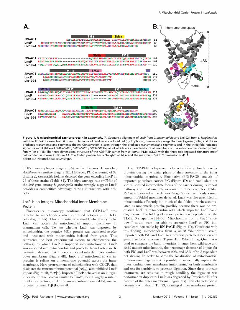
homolog (sequence identity of 57%; Figure 1A), encoded in the
genome of the related pathogen L. pneumophila strain 130b (open-
reading frame LPW_31961) [45,46]. The putative MCF protein
from L. pneumophila was subsequently termed LncP.
The crystal structure of the prototypical MCF, the adenine
nucleotide transporter from mammals, shows that the protein has
six transmembrane segments that are embedded in the mitochon-
drial inner membrane [47]. Bioinformatic analysis indicated that
the amino acid sequences of Llo1924 and LncP had six predicted
transmembrane segments and a three-fold repeated signature motif
(Figure 1A) which are the essential characteristics of members of the
MCF (Figure 1B) [38–42]. MCF proteins differ to nucleotide
carriers in the inner membranes of the Chlamydiales and the
Rickettsiales, which represent different family of proteins, referred
to as TLC ATP/ADP transporters (PF03219) [48,49]. This latter
group is of bacterial origin, and has spread from chlamydial
ancestors to other classes of bacteria and to chloroplasts via lateral
gene transfer events. TLC ATP/ADP transporters contain twelve
transmembrane segments and their nucleotide exchange properties
do not require membrane potential [50].
LncP Is Targeted to Mitochondria during Infection in aDot/Icm T4SS-dependent Manner
Many eukaryotic-type proteins from L. pneumophila are translo-
cated into infected cells via the Dot/Icm type IV secretion system.
To determine if LncP was a Dot/Icm effector, we generated a
translational fusion of the calmodulin-dependent adenylate cyclase
from Bordetella pertussis (CyaA), with the N-terminus of LncP (Cya-
LncP). The Cya-LncP fusion construct was introduced into wild
type L. pneumophila 130b or a dot/icm (dotA) mutant [51]. Upon
infection of THP-1 macrophages, Cya-LncP translocation was
detected by increased cyclic AMP (cAMP) production at levels
similar to the positive control (Cya-RalF) (Figure 2A). This
translocation was dependent on dotA indicating that LncP is a
Dot/Icm effector. Compared to eukaryotic MCF members, LncP
carries a short amino acid extension at the C-terminus (Figure 1A).
As the secretion signal for many Dot/Icm effectors lies in the C-
terminus of the protein [52,53], we tested whether this region
contained a Dot/Icm secretion signal, however deletion of the C-
terminal amino acid residues PTRKR had no effect on Dot/Icm
dependent translocation (Figure 2A).
To determine if LncP localized to mitochondria during infection
of macrophages, we generated a 4HA epitope-tagged version of
LncP for expression in L. pneumophila. The resulting expression
plasmid, p4HA-LncP, was transformed into wild type L.
pneumophila 130b and the dotA mutant. Upon infection of
macrophages for 5 h with 130b carrying p4HA-LncP, anti-HA
staining co-localized extensively with Mitotracker red in infected
cells (Figure 2B). Anti-HA staining was not observed in
macrophages infected with L. pneumophila 130b carrying the empty
vector, pICC562, or in macrophages infected with the dotA mutant
carrying p4HA-LncP (Figure 2B). Similar results were observed
upon L. pneumophila infection of HeLa cells (Figure S1). We
detected increasing amounts of LncP associated with mitochondria
over time (Figure 2C) and at earlier time points, we frequently
observed LncP staining at the poles of the bacterial cell where the
Dot/Icm secretion system is believed to be located (Figure 2C).
Altogether, this demonstrated that LncP was localized to
mitochondria during L. pneumophila infection and this event relied
upon a functional dot/icm system.
Many genes encoding Dot/Icm effectors are dispensable for
intracellular replication due to functional redundancy [9],
reviewed in [6]. Likewise here, the gene encoding LncP was
not required for L. pneumophila 130b intracellular replication in
Author Summary
Mitochondrial carrier proteins evolved during endosymbi-osis to transport substrates across the mitochondrial innermembrane. As such the proteins are associated exclusivelywith eukaryotic organisms. Despite this, we identifiedputative mitochondrial carrier proteins in the genomes ofdifferent intracellular bacterial pathogens, including Le-gionella pneumophila, the causative agent of Legionnaire’sdisease. We named the mitochondrial carrier protein fromL. pneumophila LncP and determined that the protein istranslocated into host cells during infection by thebacterial Dot/Icm type IV secretion system. From there,LncP accesses the classical mitochondrial import pathwayand is incorporated into the mitochondrial inner mem-brane as an integral membrane protein. Remarkably, LncPcrosses five biological membranes to reach its finallocation. Biochemically, LncP is a unidirectional nucleotidetransporter similar to Aac1 in yeast. Although not essentialfor intracellular replication, the high carriage rate of lncPamong isolates of L. pneumophila suggests that the abilityof the pathogen to manipulate mitochondrial ATPtransport assists survival of the bacteria in an intracellularenvironment.
A Mitochondrial Carrier Protein in Legionella
PLoS Pathogens | www.plospathogens.org 2 January 2012 | Volume 8 | Issue 1 | e1002459
THP-1 macrophages (Figure 3A) or in the model amoeba,
Acanthamoeba castellanii (Figure 3B). However, PCR screening of 37
distinct L. pneumophila isolates detected the gene encoding LncP in
28 of these strains (Table S1). The high carriage rate (,75%) of
the lncP gene among L. pneumophila strains strongly suggests LncP
provides a competitive advantage during interactions with host
cells.
LncP Is an Integral Mitochondrial Inner MembraneProtein
Fluorescence microscopy confirmed that GFP-LncP was
targeted to mitochondria when expressed ectopically in HeLa
cells (Figure 4A). This substantiates a model whereby cytosolic
LncP can access the mitochondrial import machinery in
mammalian cells. To test whether LncP was imported by
mitochondria, the putative MCF protein was translated in vitro
and incubated with mitochondria isolated from yeast. This
represents the best experimental system to characterize the
pathway by which LncP is imported into mitochondria. LncP
was imported into mitochondria and protected from Proteinase K
treatment showing that it is not imported into the mitochondrial
outer membrane (Figure 4B). Import of mitochondrial carrier
proteins is reliant on a membrane potential across the inner
membrane. Here pretreatment of mitochondria with CCCP, that
dissipates the transmembrane potential (Dym), also inhibited LncP
import (Figure 4B, ‘‘-Dy’’). Imported LncP behaved as an integral
inner membrane protein similar to Tim23, being largely resistant
to alkali extraction, unlike the non-membrane embedded, matrix
targeted protein, F1b (Figure 4C).
The TIM9:10 chaperone characteristically binds carrier
proteins during the initial phase of their assembly in the inner
mitochondrial membrane. Blue-native (BN)-PAGE analysis of
imported phosphate carrier PiC (Figure 4D) and Aac1 (data not
shown) showed intermediate forms of the carrier during its import
pathway and final assembly as a mature dimer complex. Folded
PiC mostly existed as the dimeric (Stage V) form with only a small
amount of folded monomer detected. LncP was also assembled in
mitochondria efficiently but much of the folded protein accumu-
lated as monomeric protein, possibly because there was no pre-
existing LncP in mitochondria with which imported LncP could
oligomerise. The folding of carrier proteins is dependent on the
TIM9:10 chaperone [54–56]. Mitochondria from a tim10 ‘‘shut-
down’’ strain were not able to assemble LncP or PiC into
complexes detectable by BN-PAGE (Figure 4D). Consistent with
this finding, mitochondria from a tim10 ‘‘shut-down’’ strain,
imported both PiC and LncP to a protease protected location at a
greatly reduced efficiency (Figure 4E). When ImageQuant was
used to compare the band intensities in lanes from wild-type and
tim10 mutant mitochondria, the percentage decrease of import for
both PiC and LncP was between 20% and 35% of wild-type (data
not shown). In order to show the localization of mitochondrial
proteins unambiguously it is possible to sequentially rupture the
mitochondrial outer membrane (mitoplasting) or both membranes
and test for sensitivity to protease digestion. Since these protease
treatments are sensitive to rough handling, the digestion was
performed in duplicate. LncP was degraded by Proteinase K after
rupture of the outer membrane (Figure 4G). This characteristic is
consistent with that of Tim23, an integral inner membrane protein
Figure 1. A mitochondrial carrier protein in Legionella. (A) Sequence alignment of LncP from L. pneumophila and Llo1924 from L. longbeachaewith the ADP/ATP carrier from Bos taurus. Amino acid residues are colored red (hydrophobic), blue (acidic), magenta (basic), green (polar) and the sixpredicted transmembrane segments shown. Conservation is seen through the predicted transmembrane segments and in the three-fold repeatedsignature motif (labeled SM1a-SM1b, SM2a-SM2b, SM3a-SM3b), all of which are characteristic of all members of the mitochondrial carrier proteinfamily [40,41]. (B) The three-dimensional structure of the ADP/ATP carrier from B. taurus (PDB: 1OKC), with the three-fold repeated signature motifcolor-coded as shown in Figure 1A. The folded protein has a ‘‘height’’ of 46 A and the maximum ‘‘width’’ dimension is 41 A.doi:10.1371/journal.ppat.1002459.g001
A Mitochondrial Carrier Protein in Legionella
PLoS Pathogens | www.plospathogens.org 3 January 2012 | Volume 8 | Issue 1 | e1002459

with domains exposed to the intermembrane space. The matrix
targeted protein, F1b was not degraded by Proteinase K unless the
inner membrane was also ruptured by the addition of detergent
(Figure 4G). Slight changes in band intensity from lane to lane
were not significant upon repetition, rather protease treatment
drastically altered the levels of susceptible proteins such as after
mitoplasting or treatment with detergent (Figure 4G).
LncP Is a Nucleotide Carrier ProteinSaccharomyces cerevisiae encodes 35 mitochondrial carrier
proteins, including four proteins that can transport ATP:
Aac1, Aac2, Aac3 and Sal1 [57] (Figure 5A). Yeast is a
powerful model system to study cellular phenotypes, and
fluorescence microscopy showed that ectopically expressed
LncP is targeted to mitochondria in yeast (Figure 5B). Mutant
yeast strains, each lacking one of these 35 carriers were
transformed with a plasmid-based LncP expression construct
and the transformed cells tested for growth complementation.
The mutants were scored under conditions where characteristic
growth defects were known. However no complementation was
observed upon LncP expression in any of the mutants tested.
For example, Dagc1 mutant cells lacking the amino acid
transporter Agc1 form only microcolonies on rich medium with
glycerol as a carbon source; expression of LncP did not
complement this growth defect (Figure 5C). However, we noted
a dominant-negative phenotype from expression of LncP in
wild-type cells which represented a 5-fold loss in viability on rich
growth medium, exacerbated to ,500-fold loss of viability on
minimal medium (Figure 5D). We therefore screened the carrier
mutant collection for mutants resistant to this LncP-induced
inhibition of cell viability. Only the Daac1 mutant was resistant
to the dominant-negative effect of LncP expression (Figure 5E).
In yeast, Sal1 is a Ca2+-dependent ATP-import carrier that co-
transports ATP and Mg2+ into the matrix during growth on
glucose [58,59], and the Aac1 transporter balances this effect by
ATP export. The most likely explanation for the Aac1-
dependent dominant-negative effect of LncP expression is that
combined export of ATP from the matrix by LncP and Aac1
Figure 2. LncP is translocated into macrophages by the Dot/Icm T4SS. (A) THP-1 macrophages were left uninfected or infected withderivatives of L. pneumophila 130b carrying the pEC34 vector or expressing the indicated Cya hybrid proteins. Following infection for 1 hour,macrophages were lysed and total intracellular cAMP was measure by ELISA. Results are expressed as fmol cAMP and are the mean 6 standarddeviation of three independent experiments, each performed in duplicate. Note Cya-LncPDPTRKR is a truncated protein lacking the C-terminal residues(PTRKR) of LncP. (B) Immortalized macrophages from C57BL/6 mice were infected with derivatives of L. pneumophila 130b for 5 h as indicated.Bacteria were visualized using anti-Legionella antibodies (blue) 4HA-LncP was visualized with antibodies to HA (green). Prior to fixation, cells werestained with MitoTracker Red. Cells were viewed by confocal microscopy under a 1006objective. White scale bars represent 5 mm. (C) Immortalizedmacrophages from C57BL/6 mice were infected with derivatives of L. pneumophila 130b for 30 min, 1 h, 2 h or 3 h as indicated, stained as above, andviewed by confocal microscopy under a 1006objective. White scale bars represent 5 mm. Arrows indicate LncP at the poles of the bacterial cell.doi:10.1371/journal.ppat.1002459.g002
A Mitochondrial Carrier Protein in Legionella
PLoS Pathogens | www.plospathogens.org 4 January 2012 | Volume 8 | Issue 1 | e1002459

leads to a growth defect. Thus, yeast can tolerate the expression
of Aac1 or LncP, but not both of these carrier proteins.
In order to measure directly substrate transport catalyzed by
LncP, purified recombinant protein was reconstituted into
liposomes. LncP transported nucleotides, phosphate and pyro-
phosphate, with a strong preference for ATP and GTP (Figure 6A).
The kinetic constants of purified reconstituted LncP were
determined by measuring the initial transport rate at various
external [3H]ATP or [3H]GTP concentrations in the presence of a
fixed saturating internal concentration of ATP or GTP, respec-
tively. The transport affinities (Km) of LncP for ATP and GTP
were 190637 and 183632 mM, respectively. The average Vmax
values for ATP and GTP were 9266216 and 6886213 mmol/min
x g of protein, respectively (mean values of 4 experiments).
Powerful inhibitors of the well-characterized ADP/ATP carrier,
which transports only ADP and ATP, fix the transporter in a
specific state: atractylosides (such as carboxyatractyloside; CAT)
fixes the transporter in the ‘‘cytosolic’’ c-state thereby inducing
swelling of mitochondria and apoptosis, and bongkrekic acid
(BKA) fixes the transporter in the ‘‘matrix’’ m-state thereby
suppressing induction of apoptosis [60]. LncP was not inhibited by
CAT or BKA (Figure 6B). It was also not inhibited by the SH
alkylating reagent N-ethylmaleimide (NEM; inhibitor of the
phosphate, glutamate and ornithine carriers). In contrast, ATP
transport catalyzed by LncP was effectively prevented by other
reagents such as mersalyl (MER), p-hydroxymercurybenzoate (p-
HMB) and HgCl2, which are organic mercurials, and by
pyridoxal-59-phosphate (PLP) and bathophenanthroline (BAT),
which alone or in combination inhibit the activity of several
mitochondrial carriers, although their mechanism of action is not
known. Therefore, both the substrate specificity (Figure 6A) and
the inhibitor sensitivity (Figure 6B) of LncP distinguish it
biochemically from the ADP/ATP carrier.
To characterize further the transport properties of LncP, the
kinetics of [3H]ATP and [3H]GTP uptake into proteoliposomes
were compared either as uniport (in the absence of internal
substrate) or as exchange (in the presence of internal ATP or GTP,
respectively) (Figure 6C). Both the exchange and the uniport
reactions of ATP and GTP uptake followed first-order kinetics,
isotopic equilibrium being approached exponentially. The ratio of
maximal substrate uptake by both reactions was 9.8 for ATP and
13.0 for GTP, in good agreement with the expected ratio of 10
from the intraliposomal concentrations at equilibrium (1 mM and
10 mM for uniport and exchange, respectively). The uniport mode
of transport of reconstituted LncP was also investigated by
measuring the efflux of [3H]ATP from pre-labeled proteolipo-
somes (Figure 6D) because this approach provides a more sensitive
assay for unidirectional transport [61]. A significant efflux of ATP
was observed in the absence of external substrate (filled circle) and
a more rapid and extensive efflux occurred upon addition of ATP
(open square) or phosphate (open triangle). Moreover, the ATP-
induced efflux of radioactivity was prevented by the presence of
the carrier inhibitors PLP and BAT (filled square). Similar results
were obtained using GTP as substrate (data not shown). Thus,
LncP was able to catalyze unidirectional transport of ATP and
GTP and a fast exchange reaction of substrates.
Discussion
Recently, 275 effectors of the Dot/Icm secretion system were
described in the Philadelphia-1 strain of L. pneumophila [13]. This
represents almost 10% of all open reading frames encoded in the
L. pneumophila genome. Given that there is also diversity in the
presence and range of effector genes among the different
sequenced L. pneumophila genomes and even greater differences
between Legionella species [22], the total Dot/Icm effector
repertoire is likely to be much larger. Here we describe a new
Dot/Icm effector from L. pneumophila, LncP, with sequence and
functional similarity to eukaryotic mitochondrial carrier proteins.
LncP was predicted to have six transmembrane domains, similar
to eukaryotic MCF members. Remarkably, this highly hydropho-
bic protein crosses five biological membranes to reach its final
destination in the mitochondrial inner membrane. Generally
bacterial membrane proteins are assembled into the cytoplasmic
membrane by YidC and SecYEG [62], reviewed in [63].
Chaperones for the Dot/Icm machinery, such as IcmS, IcmW
and LvgA [64–66], must be in active competition with the
bacterial YidC/SecYEG machinery to dictate which integral
membrane proteins will be assembled into the bacterial inner
membrane and which will be evacuated via the Dot/Icm T4SS.
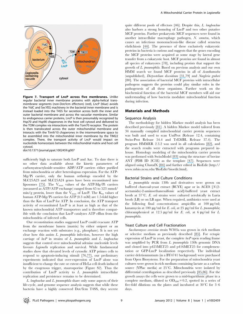
Therefore recognition of LncP by the Dot/Icm machinery
presumably allows this hydrophobic protein to avoid assembly
into the bacterial inner membrane by YidC/SecYEG (Figure 7).
Figure 3. Mutant L. pneumophila lacking LncP replicateproficiently in host cells. Two independent mutants of L.pneumophila 130b lacking LncP (lncP-3 and lncP-4) were tested, alongwith a dotA mutant lacking the Dot/Icm T4SS. Replication of L.pneumophila 130b (N),lncP-3 (D),lncP-4 (,) and dotA (%) within themacrophage cell-line THP-1 (A) and A. castellanii (B) is shown. Resultsare expressed as the log10CFU of viable bacteria present in theextracellular medium (and associated with cells for THP-1) at specifictime points after inoculation, mean 6 standard deviation of at leastthree independent experiments from duplicate wells.doi:10.1371/journal.ppat.1002459.g003
A Mitochondrial Carrier Protein in Legionella
PLoS Pathogens | www.plospathogens.org 5 January 2012 | Volume 8 | Issue 1 | e1002459

The mechanism by which this recognition occurs is unknown but
probably involves detection of a C-terminal Dot/Icm secretion
signal. Here we removed the C-terminal amino acids PTRKR
from LncP but found that this had no effect on LncP translocation.
Bioinformatic analysis of known Dot/Icm substrates has revealed a
preference for short acidic or negatively charged amino acids in
the C-terminal secretion signal [26,53]. Recently, a glutamate rich
region (E Block) was associated with the translocation signal of
Figure 4. LncP is transported to the mitochondrial inner membrane. (A) HeLa cells were transformed to express LncP-GFP or a control plasmid.The LncP-GFP cells were co-stained with tetramethylrhodamine methyl ester (TMRM) and viewed by confocal microscopy. The merge shows themitochondrial localization of LncP-GFP (B) Mitochondria (50 mg protein) from wild-type yeast cells were incubated with [35S]-labeled LncP. After theindicated time at 25uC, mitochondria were isolated, treated with Proteinase K to degrade protein that had not been imported, and analyzed by SDS-PAGE and fluorography. ‘‘T’’ represents non-Proteinase K treated control. ‘‘-Dym’’ indicates a sample where the mitochondria were pre-incubated withinhibitors and uncouplers to deplete the transmembrane potential (see Methods) (C) Mitochondria (100 mg protein) from wild-type cells were incubatedwith [35S]-labeled LncP. After 20 minutes at 25uC, mitochondria were isolated, extracted with 0.1 M Na2CO3 and the membrane-containing pellet (‘‘Pel’’)and extracted proteins in the supernatant (‘‘S/N’’) analyzed by SDS-PAGE and fluorography and immunoblot against a known membrane embeddedprotein (Tim23) and a non-membrane embedded protein matrix localized protein (F1b). A sample of mitochondria prior to extraction and representingthe total amount (‘‘Tot.’’) is shown for comparison. The right-hand panel shows the percentage distribution of LncP in the pellet and supernatantfractions after 5 repeat experiments 6 standard error. (D) Mitochondria (50 mg protein) from wild-type and Tim10 depleted (tim10Q) yeast cells wereresuspended in isotonic import buffer and incubated with [35S]-labeled LncP and PiC. After the indicated time at 25uC, mitochondria were isolated,solubilized in digitonin and analyzed by BN-PAGE and fluorography. Asterisk indicates bands formed by folded carrier proteins. The lower asteriskrepresents the folded monomer and the upper asterisk represents assembled carrier dimers (Stage V) (E) Mitochondria (50 mg protein) from wild-typeyeast or from tim10Q yeast depleted of Tim10 were incubated with [35S]-labeled LncP or PiC. After the indicated time at 25uC, the mitochondria weretreated with Proteinase K and then analysed by SDS-PAGE and fluorography. ‘‘-Dym’’ indicates a sample where the mitochondria were pre-incubatedwith inhibitors and uncouplers to deplete the transmembrane potential (see Methods). (F) Control western blots with mitochondria isolated from wild-type and tim10Q cells respectively showing that Tim10 has been selectively depleted. (G) The localization of LncP within mitochondria after import wasdetermined using a sequential proteolysis assay. After import of [35S]-labeled LncP at 25uC for 20 minutes, mitochondria were treated with hypotonicbuffer to induce mitoplasting, or Triton-X-100 to rupture both membranes and Proteinase K (50 mg/mL) as indicated (see Methods). ‘‘L’’ is lysate onlywithout mitochondria to show size of unimported protein. The control proteins, the inner membrane embedded protein (Tim23) and a non-membraneembedded protein matrix localized protein (F1b) were detected by immunoblot on the same membrane.doi:10.1371/journal.ppat.1002459.g004
A Mitochondrial Carrier Protein in Legionella
PLoS Pathogens | www.plospathogens.org 6 January 2012 | Volume 8 | Issue 1 | e1002459

many Dot/Icm effectors [67]. The E Block motif was located in
the C-terminal 30 amino acids of the effectors. LncP also contains
a putative E Block motif in the C-terminus that may contain the
signal for translocation (Figure 1A). However, the motif is
predicted to lie within the most distal transmembrane domain of
the carrier protein and likely contributes to correct protein folding
and function. Hence, further investigation of the LncP secretion
signal will require careful mutational analysis by amino acid
substitution rather than deletion to dissect the bona fide secretion
signal from the transmembrane domain.
The mechanism by which hydrophobic membrane proteins
such as LncP can be accommodated in the translocase channel
and assisted on the host cytoplasmic side of the Legionella-
containing vacuole membrane without aggregating is unknown.
When we analyzed the Dot/Icm effector repertoire of L.
pneumophila 130b using two independent hidden Markov model
approaches, HMMtop [68] and TMHMM v 2.0 [69], 71 effectors
were predicted by both methods to have one or more
transmembrane segments (Table S2). Thus the Dot/Icm T4SS
has evolved to handle the export of proteins with significant
hydrophobicity across at least three biological membranes.
Currently, mitochondrial localization of only one other Dot/
Icm effector, LegS2, has been reported, although the precise
mitochondrial compartment was not described. LegS2 has
sphingosine-1-phosphate lyase activity and it is not yet clear if
mitochondrial targeting plays any role in effector function [27].
Here we found that LncP was also targeted to mitochondria
during infection of eukaryotic cells with L. pneumophila and
assembled into the mitochondrial inner membrane, where the
effector appeared to act as a unidirectional nucleotide transporter.
Mitochondrial import required the TIM9:10 chaperones and
hence the TIM22 machinery, according to classical mitochondrial
protein transport mechanisms.
In the yeast model system, expression of LncP led to a
dominant-negative phenotype. Although not lethal, the expression
of LncP greatly slowed growth, particularly growth on minimal
media. This dominant-negative phenotype depended on the
activity of the endogenous MCF protein, Aac1. Whereas the
yeast MCFs Aac2 and Aac3 are classic ADP/ATP carriers that
regenerate cytoplasmic ATP levels (because ATP export can only
Figure 5. LncP generates a dominant negative phenotype,dependent on Aac1 ATP transport activity. (A) Yeast cells expressthree dominant carriers for adenine nucleotide transport: Aac1, Aac2and Sal1 in the mitochondrial inner membrane (IM). Aac3 is an isoform
expressed under anaerobic conditions [94]. The outer membrane (OM)of mitochondria is permeable to ATP due to the pores formed by VDAC.(B) Yeast cells transformed to express LncP-GFP were co-stained withMitoTracker Red and visualized by fluorescence microscopy. The mergeshows the mitochondrial localization of LncP-GFP. (C) Yeast mutants,each lacking one member of the carrier protein family weretransformed with either a plasmid encoding LncP (+) or the control (-)plasmid. The transformed cells were plated on selective medium andscored for growth using five-fold serial dilutions. As an example, theDagc1 mutant is shown: Agc1 is an amino acid transporter which actsboth as a glutamate uniporter and as an aspartate-glutamateexchanger; while viable on plates containing glycerol as a carbonsource, the Dagc1 mutant cells form only microcolonies before arrestinggrowth. Expression of LncP does not support glutamate-aspartatetransport and so does not rescue this phenotype. (D) Wild-type cellstransformed with either a plasmid encoding LncP (+) or the control (2)plasmid were plated on YPD medium with glucose as a carbon source(rich) or SD semi-synthetic medium with glucose as a carbon source(minimal) and scored for growth using five-fold serial dilutions. Thenumber of colonies represents cell viability. (E) Yeast mutants, eachlacking a distinct carrier protein, were transformed with either theplasmid encoding LncP (+) or the control (2) plasmid and scored forgrowth using five-fold serial dilutions. The Ddic1 mutant lacks thedicarboxylic acid transporter and is representative of carrier mutants inshowing the same dominant-negative phenotype as wild-type cells.Only in the Daac1 mutants is this phenotype suppressed.doi:10.1371/journal.ppat.1002459.g005
A Mitochondrial Carrier Protein in Legionella
PLoS Pathogens | www.plospathogens.org 7 January 2012 | Volume 8 | Issue 1 | e1002459

be achieved with a concomitant import of ADP), a distinguishing
feature of Aac1 is its propensity to export ATP from the
mitochondrial matrix [57]. Thus, the dominant-negative effect
seen in yeast is likely a cellular consequence of an imbalance of
ADP/ATP transport across the mitochondrial inner membrane.
We also observed ATP transport activity for LncP in
reconstituted liposomes. The kinetic parameters of ATP transport
by LncP were comparable to those of genuine ATP carriers. There
are two classes of transporters for ATP in the mitochondrial inner
membrane: the carboxyatractyloside-inhibitable ADP/ATP carri-
ers (Aac) and the ATP-Mg/Pi carriers (in humans named APC
and in yeast Sal1). Studies in which the Vmax of Aac has been
measured in reconstituted liposomes (either as ATP/ATP or
ADP/ADP exchange) using protein purified from mitochondria or
after heterologous expression, obtained Vmax values ranging from
360 and 1300 mmol/min/g protein [70–74]. Here we measured
the Vmax of ATP transport in LncP-reconstituted liposomes
(measured as ATP/ATP exchange) as 926 mmol/min/g protein.
This means that the ratio between the activity of LncP and the
activity of genuine ADP/ATP carriers varied from 2.6 to 0.7. The
Km for ATP of genuine mitochondrial ADP/ATP carriers,
measured in reconstituted liposomes, ranges between 9 and
120 mM, lower than the Km of LncP for ATP (190 mM). However,
the internal concentration of ATP in respiring mitochondria is
Figure 6. LncP is a nucleotide carrier with unique properties. (A) Liposomes reconstituted with LncP were preloaded internally with varioussubstrates (concentration, 10 mM). Transport was started by the addition of 0.2 mM [3H]ATP and terminated after 2 min. Values are means 6 S.D. ofat least three independent experiments. a-OG, a-oxoglutarate; Pi, phosphate; PPi, pyrophosphate. (B) Proteoliposomes were preloaded internally with10 mM ATP and transport was initiated by adding 0.2 mM [3H]ATP. The reaction time was 2 min. Thiol reagents were added 2 min before the labeledsubstrate; the other inhibitors were added together with the labeled substrate. The final concentrations of the inhibitors were 20 mM (PLP, pyridoxal-59-phosphate; BAT, bathophenanthroline), 0.2 mM (p-HMB, p-hydroxymercuribenzoate; MER, mersalyl), 1 mM (NEM, N-ethylmaleimide), 0.2% (TAN,tannic acid), 0.2 mM (BrCP, bromcresol purple), 25 mM (HgCl2, mercuric chloride) and 10 mM (BKA, bongkrekic acid; CAT, carboxyatractyloside). Theextent of inhibition (%) from representative experiments is given. (C) Uptake of [3H]ATP (&, %) and [3H]GTP (N, #) into liposomes reconstituted withLncP. 1 mM [3H]ATP or [3H]GTP was added to proteoliposomes containing 10 mM ATP or GTP, respectively (exchange, filled shapes), or 10 mM NaCland no substrate (uniport, open shapes). Similar results were obtained in three independent experiments. (D) Efflux of [3H]ATP from LncPproteoliposomes. The internal substrate (2 mM ATP) was labeled by carrier-mediated exchange equilibration. After removal of the external substrateby Sephadex G-75, the efflux of [3H]ATP was started by adding buffer A alone (filled circles), 5 mM ATP, 20 mM pyridoxal-59-phosphate and 10 mMbathophenanthroline in buffer A (filled squares), 5 mM ATP in buffer A (open squares) or 5 mM phosphate (open triangles). Similar results wereobtained in three independent experiments.doi:10.1371/journal.ppat.1002459.g006
A Mitochondrial Carrier Protein in Legionella
PLoS Pathogens | www.plospathogens.org 8 January 2012 | Volume 8 | Issue 1 | e1002459
sufficiently high to saturate both LncP and Aac. To date there is
no other data available about the kinetic parameters of
carboxyatractyloside-sensitive ADP/ATP carriers either purified
from mitochondria or after heterologous expression. For the ATP-
Mg/Pi carrier, only the human orthologs encoded by the
SLC25A23 and SLC25A24 genes have been reconstituted into
liposomes [75]. The Vmax values of the ATP-Mg/Pi carriers
(measured as ATP/ATP exchange) ranged from 65 to 523 mmol/
min/g protein, lower than the Vmax of LncP. The Km values of
human ATP-Mg/Pi carriers for ATP (0.3 mM) are 1.5-fold higher
than the Km of LncP for ATP. In conclusion, the ATP transport
activity of reconstituted LncP is at least as high as that of the
known mitochondrial ATP transporters and is therefore compat-
ible with the conclusion that LncP catalyzes ATP efflux from the
mitochondria of infected cells.
Our reconstitution studies suggested LncP could evacuate ATP
from the membrane lumen (matrix) by either uniport or an
exchange reaction with substrates (e.g., phosphate). It is not yet
clear how this assists L. pneumophila infection, however the high
carriage of lncP in strains of L. pneumophila and L. longbeachae
suggests that control over mitochondrial adenine nucleotide levels
favours Legionella replication and survival. While fundamental
studies show that elevated levels of cytosolic ATP primes cells to
respond to apoptosis-inducing stimuli [76,77], our preliminary
experiments indicated that over-expression of LncP alone was
insufficient to change the rate or extent of HeLa cell death induced
by the exogenous trigger, staurosporine (Figure S2). Thus the
contribution of LncP activity to L. pneumophila intracellular
replication and persistence remains to be determined.
L. longbeachae and L. pneumophila share only some aspects of their
life-cycle, and genome sequence analysis suggests that while these
bacteria have a highly conserved Dot/Icm T4SS, they secrete
quite different pools of effectors [46]. Despite this, L. longbeachae
also harbors a strong homolog of LncP and two other putative
MCF proteins. Further prokaryotic MCF sequences were found in
another intracellular macrophage pathogen, N. sennetsu, which
causes an infectious mononucleosis-like disease called sennetsu
ehrlichiosis [44]. The presence of these exclusively eukaryotic
proteins in bacteria is curious and suggests that the genes encoding
the MCF proteins were acquired at some stage by lateral gene
transfer from a eukaryotic host. MCF proteins are found in almost
all species of eukaryotes [78], including protists that support the
growth of L. pneumophila. Based on previous analysis and our own
HMM search we found MCF proteins in all of Acanthamoeba
(unpublished), Dictyostelium discoideum [31,79] and Naegleria gruberi
[80]. The association of bacterial MCF proteins with intracellular
pathogens suggests the proteins could play similar roles in the
pathogenesis of all these organisms. Further work on the
biochemical function of the bacterial MCF members will aid our
understanding of how bacteria modulate mitochondrial function
during infection.
Materials and Methods
Sequence AnalysisThe methodology for hidden Markov model analysis has been
described previously [81]. A hidden Markov model tailored from
34 manually compiled mitochondrial carrier protein sequences
was built and used to scan UniProt (Release 12.4, containing
Swiss-Prot Release 54.4 and TrEMBL Release 37.4). The
program HMMER 2.3.2 was used in all calculations [82], and
the search results were extracted with programs prepared in-
house. Homology modeling of the mitochondria carrier protein
was performed with SwissModel [83] using the structure of bovine
ANT (PDB ID 2C3E) as the template [47]. Sequences were
aligned using ClustalX [84] and further edited in BioEdit (http://
www.mbio.ncsu.edu/BioEdit/bioedit.html).
Bacterial Strains and Culture ConditionsL. pneumophila strain 130b and derivatives were grown on
buffered charcoal-yeast extract (BCYE) agar or in ACES [N-(2-
acetamido)-2-aminoethanesulfonic acid]-buffered yeast extract
broth at 37uC. E. coli strains were cultured aerobically in Luria
broth (LB) or on LB agar. When required, antibiotics were used at
the following final concentrations: ampicillin at 100 mg/ml;
kanamycin at 100 mg/ml for E. coli, at 25 mg/ml for L. pneumophila;
chloramphenicol at 12.5 mg/ml for E. coli, at 6 mg/ml for L.
pneumophila.
Yeast Culture and Cell FractionationSaccharomyces cerevisiae strain W303a was grown in rich medium
or selective medium as previously described [85]. For ectopic
expression of LncP in yeast, the complete lncP open reading frame
was amplified by PCR from L. pneumophila 130b genomic DNA
and cloned into p425MET25 and p416MET25 for complemen-
tation or GFP-LncP localization respectively. The individual
carrier deletionmutants (in a BY4741 background) were purchased
from Open Biosystems. For the preparation of mitochondria yeast
cultures were grown in rich medium containing lactate as a carbon
source (YPlac media) at 25uC. Mitochondria were isolated by
differential centrifugation as described previously [85,86]. For the
growth assays the cells were grown to a mid-logarithmic phase in a
complete medium, diluted to OD600 = 0.2, spotted in a series of
five-fold dilutions on the plates and incubated at 30uC for 3–6
days.
Figure 7. Transport of LncP across five membranes. Unlikeregular bacterial inner membrane proteins with alpha-helical trans-membrane segments (non-Dot/Icm effectors) (red), LncP (blue) avoidsthe YidC and SecYEG machinery in the bacterial inner membrane and isinstead loaded into the T4SS for secretion across both the inner andouter bacterial membrane and across the vacuolar membrane. Similarto endogenous carrier proteins, LncP is then presumably recognized byHsp70 and Hsp90 chaperones in the host cell cytosol and delivered tothe TOM complex via interactions with the Tom70 receptor. The proteinis then translocated across the outer mitochondrial membrane andinteracts with the Tim9/10 chaperones in the intermembrane space tobe assembled into the mitochondrial inner membrane by the TIM22complex. There, the transport activity of LncP would impact onnucleotide homeostasis between the mitochondrial matrix and host cellcytosol.doi:10.1371/journal.ppat.1002459.g007
A Mitochondrial Carrier Protein in Legionella
PLoS Pathogens | www.plospathogens.org 9 January 2012 | Volume 8 | Issue 1 | e1002459
Isolation of Yeast MitochondriaFor the isolation of wildtype and Tim10 depleted mitochondria
the Saccharomyces cerevisiae strains W303, PMET3Tim10 [87] were
grown in synthetic glucose media [0.67% (w/v) yeast nitrogen, 2%
(w/v) glucose, 0.01% (w/v) leucine, tryptophan, uracil, adenine
and histidine at 30uC for 10 hours as a pre-culture. The pre-
culture was diluted to A600 nm = 0.2units/mL in media supple-
mented with 0.2 mM methionine then grown for 2 days to reach
A600 nm = 1.0 before harvesting and mitochondrial isolation by
previously described methods [86].
Synthesis of [35S]-labeled LncPDNA encoding LncP was amplified and cloned into pSP73
(Promega) from genomic DNA isolated Legionella pneumophila (strain
130b). The oligonucleotides LncP-FW BamHI (GCGCGGATC-
CATGAAAGACAAAACAATA), and LncP-REV XhoI (GATCC
TCGAGCTACCTGTTCCTTGTTGG) were used to amplify
full length LncP DNA In vitro transcription was carried out as
previously described [88]. Rabbit reticulocyte lysate was pur-
chased from Promega and in vitro translation reactions were carried
out for 30–60 minutes in the presence of [35S]- methionine/
cysteine (MP Biomedicals) [88].
In vitro Import Reactions[35S]-Methionine/cysteine-labeled LncP or PiC were synthesized
in vitro and were incubated with mitochondria (50 mg per lane) for
the indicated time periods at 25uC in import buffer (0.6 M sorbitol,
50 mM Hepes (pH 7.4), 2 mM KPi (pH 7.4), 25 mM KCl, 10 mM
MgCl2, 0.5 mM EDTA, 1 mM dithiothreitol, 4 mM ATP, and
2 mM NADH). Samples were treated with Proteinase K (40 mg/
mL) in import buffer for 15 minutes on ice to remove un-imported
material before addition of protease inhibitor (1 mM PMSF). The
mitochondria were re-isolated by centrifugation at 10,000 g and this
was followed by either protein separation under denaturing gel
electrophoresis (SDS) or protein complexes separated by Blue
Native electrophoresis [89]. For Proteinase K shaving or mito-
chondrial membrane potential dissipation conditions, mitochondria
were treated with 40 mg/ml Proteinase K or AVO mix (8 mM
antimycin A, 1 mM valinomycin, and 20 mM oligomycin) respec-
tively. For preparation of mitoplasts (mitochondria with ruptured
outer mitochondrial membrane), post-import mitochondria were
subjected to osmotic shock by resuspension in 20 mM Hepes/
KOH, pH 7.4 with and without Proteinase K where indicated.
Sodium Carbonate ExtractionsAfter the completion of in vitro import reactions for 8 minutes at
25uC, mitochondria were re-isolated by centrifugation at 10,000 g
and resuspended in 200 mL of 100 mM sodium carbonate which
was adjusted to pH 11.5 and left on ice for 30 minutes with gentle
mixing every 5 to 10 minutes. A membrane pellet was then
separated from a supernatant by ultra-centrifugation at 100,000g
for 30 minutes at 4uC. The pellet was resuspended in 200 mL of
100 mM sodium carbonate and both the pellet and supernatant
were then subjected to Trichloroacetic acid precipitation. Each
experiment was conducted in duplicate with one set of pellet and
supernatant samples recombined to make the ‘‘total’’ sample. This
was repeated 5 times in order to assess statistical significance. LncP
protein level was quantified by densitometry of phosphorimages
using the Image Quant software.
Reconstitution of LncP into Liposomes and TransportMeasurements
Expression of recombinant LncP is detailed in the supporting
methods (Protocol S1). The recombinant, purified LncP was
reconstituted into liposomes by cyclic removal of the detergent
with a hydrophobic column of Amberlite beads (Fluka) [61]. The
composition of the initial mixture used for reconstitution was 35 ml
of purified LncP (15 mg of protein), 70 ml of 10% Triton X-114,
100 ml of 10% phospholipids in the form of sonicated liposomes,
10 mM ATP (except where otherwise indicated), 10 mM PIPES
(pH 7.0), 0.42 mg of the mitochondrial lipid cardiolipin (Sigma)
and water to a final volume of 700 ml. After vortexing, this mixture
was recycled 13 times through the Amberlite column (3.560.5 cm)
pre-equilibrated with a buffer containing 10 mM PIPES pH 7.0.
All steps were performed at 4uC, except for the passages through
Amberlite, which were carried out at room temperature.
External substrate was removed from proteoliposomes on
Sephadex G-75 columns pre-equilibrated with 50 mM NaCl
and 10 mM PIPES at pH 7.0 (buffer A) and 4uC. The eluted
proteoliposomes were distributed in reaction vessels and used for
transport measurements by the inhibitor-stop method [61].
Transport at 25uC was started by adding [3H]ATP (Perkin Elmer)
or [3H]GTP (American Radiolabeled Chemicals) to proteolipo-
somes and terminated by addition of 20 mM pyridoxal-59-
phosphate and 20 mM bathophenanthroline. In controls, the
inhibitors were added at the beginning together with the
radioactive substrate. Finally, the external radioactivity was
removed from each sample of proteoliposomes by a Sephadex
G-75 column; the proteoliposomes were eluted with buffer A and
their radioactivity was measured. The experimental values were
corrected by subtracting control values. The initial transport rate
was calculated from the radioactivity taken up by proteoliposomes
after 2 min (in the initial linear range of substrate uptake). For
efflux measurements, proteoliposomes containing 2 mM ATP or
GTP were labeled with 10 mM [3H]ATP or [3H]GTP, respec-
tively, by carrier-mediated exchange equilibration [61]. After
50 min, the external radioactivity was removed by passing the
proteoliposomes through Sephadex G-75 pre-equilibrated with
buffer A. Efflux was started by adding unlabeled external substrate
or buffer A alone to aliquots of proteoliposomes and terminated by
adding the inhibitors indicated above.
Disruption of LncP in L. pneumophila 130bAn insertional mutation in LncP was created via homologous
recombination. A ,1 kb fragment encompassing LncP was
amplified by PCR from L. pneumophila 130b genomic DNA using
the oligonucleotide primers, 59- caacggatcctatttcatttgtagtcccttg -39
and 59- tcctgtcgacctgaaatattttcatggaaac -39. The resulting product
was cloned into the BamHI and SalI sites of pPCRScript and a
kanamycin resistance gene from Tn5 was introduced into the
native PstI site of LncP. The construct was introduced into L.
pneumophila 130b via natural transformation, as described previ-
ously [90]. Kanamycin resistant clones were assessed by PCR
analysis and ampicillin sensitivity to detect replacement of lncP
with lncP::km and the loss of pCR-Script. Two independent L.
pneumophila lncP::km clones, LncP-3 and LncP-4, were chosen for
further analysis in host cell replication assays.
Macrophage and HeLa Cell Infection and Anti-HAImmunofluorescence
The human monocytic cell line, THP-1 was maintained in
RPMI 1640 supplemented with 10% fetal bovine serum in 5%
CO2 at 37uC. The cells were prepared for infection with
stationary-phase L. pneumophila as previously described [91].
THP-1 cells were infected at a multiplicity of infection (MOI) of
5 cells for 2 h in 5% CO2 at 37uC. Cells were then treated with
100 mg/ml gentamicin for 1 h to kill extracellular bacteria and
washed with PBS before being lysed with 0.01% digitonin.
A Mitochondrial Carrier Protein in Legionella
PLoS Pathogens | www.plospathogens.org 10 January 2012 | Volume 8 | Issue 1 | e1002459
Serial dilutions of the inoculum and bacteria recovered from
lysed cells were plated on BCYE agar and results were expressed
as the percentage of the inoculum that resisted killing by
gentamicin (mean 6 standard deviation of at least 3 indepen-
dent experiments).
Immortalized macrophages from wild type C57BL/6 mice [92]
were seeded at 26105 per coverslip 16 h prior infection. The B6
macrophages were a gift from Dr Ashley Mansell (Monash
Institute of Medical Research). Cells were maintained in DMEM
supplemented with 10% FCS, 2 mM glutamine, 100 U penicillin/
ml and 100 mg streptomycin/ml. Immediately prior to infection,
macrophages were washed and the medium replaced with DMEM
supplemented with 1 mM IPTG and lacking antibiotics. Macro-
phages were infected for 30 min, 1 h, 2 h, 3 h or 5 h with
derivatives of L. pneumophila 130b at a multiplicity of infection of
50. Bacterial strains for infection were grown overnight in ACES
broth supplemented with antibiotics where appropriate and 1 mM
IPTG. HeLa cells were infected using an identical protocol.
Following the infection period, cells were washed once with fresh
tissue culture medium and incubated with 500 nM MitotrackerHRed FM (Invitrogen) for 30 min at 37uC and 5% CO2. Labelled
cells were then fixed in 4% paraformaldehyde-PBS for 20 min and
permeabilized with 0.1% TritonX-100-PBS for 20 min. Cells were
incubated for 60 min in staining solution containing 0.2% BSA,
1:50 dilution of anti-HA.11 monoclonal antibody (Covance) and
1:75 dilution of rabbit raised anti-Legionella pneumophila antibody
(Acris). The bound primary antibodies were detected using 1:1000
dilution of Alexa Fluor 405-conjugated anti-rabbit antibody and
Alexa Fluor 488-conjugated anti-mouse antibody (Invitrogen)
respectively. Coverslips were mounted onto glass slides with Dako
Fluorescent Mounting Medium (Dako). Immunofluorescence
images were acquired using a confocal laser scanning microscope
(Leica TCS SP2 confocal imaging system) with a 100x/1.4 NA
HCX PL APO CS oil immersion objective.
Cya-LncP and 4HA Gene Fusions and Intracellular cAMPAssays
Adenylate cyclase (Cya) fusions with RalF and LncP were
generated as described previously [51]. Details are provided in the
supporting methods (Protocol S1). Hemagluttinin (HA) fusions
with LncP were generated as described in the supporting methods
(Protocol S1).
Infection of A. castellanii with L. pneumophilaA. castellanii ATCC 50739 was cultured in PYG 712 medium at
20uC for 72 h prior to harvesting for L. pneumophila infection. A.
castellanii cells were washed once with A.c. buffer (0.1% trisodium
citrate, 0.4 mM CaCl2, 2,5 mM KH2PO4, 4 mM MgSO4,
2.5 mM Na2HPO4, 0.005 mM ferric pyrophosphate) and seeded
into 24-well tissue culture trays (Sarstedt, Leicestershire, United
Kingdom) at a density of 105 cells/well. Stationary-phase L.
pneumophila was added at an MOI of 0.01 and incubated at 37uC.
At set time points, entire co-culture volumes were collected and
plated onto BCYE agar to count colony-forming units of L.
pneumophila.
LncP PurificationProteins were analyzed by SDS-PAGE or by Blue Native (BN)-
PAGE (Figure S3) as previously described [85]. N-terminal
sequencing was carried out as described previously [93]. Purified
LncP was quantified by laser densitometry of stained samples,
using carbonic anhydrase as the protein standard [93]. Protein
incorporation into liposomes was measured as described [93] and
varied between 20-30% of the protein added to the reconstitution
mixture.
Supporting Information
Figure S1 Localization of 4HA-LncP in macrophagesand HeLa cells. (A) Macrophages were infected with L.
pneumophila (either wild type 130b or the DdotA mutant) expressing
4HA-LncP. Bacteria were visualized using anti-Legionella antibod-
ies (blue) 4HA-LncP was visualized with antibodies to HA (green).
Prior to fixation, cell were stained with MitoTracker Red. Cells
were viewed by confocal microscopy under a 1006objective. The
merge shows the mitochondrial localization of 4HA-LncP. White
scale bars represent 10 mm (B) HeLa cells infected with L.
pneumophila (either wild type 130b or the DdotA mutant) expressing
4HA-LncP were analyzed as above.
(TIF)
Figure S2 LncP is targeted to mitochondria, but doesnot impact on apoptosis induced by staurosporinetreatment. (A) LncP-EGFP was expressed in HeLa cells. Cells
were immunostained with antibodies against GFP (green) and
active caspase-3 (red). Hoechst 33342 was used as a counterstain to
indicate nucleus (blue). The panel at the right shows the cells
treated with staurosporine for 5–6 hours. The left panel shows the
cells without staurosporine treatment. Scale bar: 10 mm. The
Table documents the analysis by cell counting. Total cells were
counted based on nucleus staining. LncP-EGFP expressing cells
were counted based on green color while cells with active caspase-
3 were counted based on red color. (B) HeLa cells were transfected
with LncP-EGFP (green) and then stained with tetramethylrho-
damine methyl ester (TMRM) (red). The right panel shows the
cells treated with vehicle DMSO for 5 hours while the left panel is
the cells without treatment. Scale bar: 50 mm (C) HeLa cells
transfected with LncP-EGFP (green) were stained with TMRM
(red) and then treated with staurosporine for up to 3 hours. The
panel at the right shows fluorescence images while the left panel
shows bright field images. Scale bar: 50 mm.
(TIF)
Figure S3 Recombinant expression and purification ofLncP. Proteins were separated by SDS-PAGE and stained with
Coomassie Blue. Markers in left-hand column (bovine serum
albumin, carbonic anhydrase, and cytochrome c); lanes 1–4,
Escherichia coli C0214 (DE3) containing the expression vector
without (lanes 1 and 3) and with (lanes 2 and 4) the coding
sequence of LncP. Samples were taken at the time of induction
(lanes 1 and 2) and 5 h later (lanes 3 and 4). The same number of
bacteria was analyzed in each sample. Lane 5, purified LncP
protein (5 mg) purified from E. coli shown in lane 4. The identity of
the purified protein was confirmed by N-terminal sequencing.
Approximately 55mg of purified protein per liter of culture were
obtained.
(TIF)
Protocol S1 Supporting methods.(DOC)
Table S1 Prevalence of lncP among strains of L.pneumophila. A range of L. pneumophila strains were tested for
carriage of lncP by Southern hybridisation as described previously
[90]. A digoxigenin (DIG)-labelled probe was generated by PCR
amplification according to the manufacturer’s instructions (Roche)
with the primer pair 59- caacggatcctatttcatttgtagtcccttg -39 and 59-
tcctgtcgacctgaaatattttcatggaaac -39 using L. pneumophila 130b
genomic DNA as a template [45].
(DOC)
A Mitochondrial Carrier Protein in Legionella
PLoS Pathogens | www.plospathogens.org 11 January 2012 | Volume 8 | Issue 1 | e1002459

Table S2 Predicted membrane proteins in the L.pneumophila Dot/Icm effector repertoire. Two hundred
and seventy-five proteins encoded in the genome of L. pneumophila
strain Philadelphia were identified as effector proteins in a recent
high-throughput study [13]. Here the protein sequences of these
effectors as well effectors unique to L. pneumophila strain 130b were
analyzed with HMMtop [68] and TMHMM v 2.0 [69] to predict
transmembrane segments. The two predictors have independent
means of assessing hydrophobicity and other characteristics of
alpha-helical transmembrane segments, yet concordant predic-
tions are seen for most proteins.
(DOC)
Acknowledgments
We thank Vladamir Likic, Ana Traven and Chaille Webb for expert
advice, and Victoria Hewitt and members of the Lithgow and Hartland
labs for critical comments on the manuscript. We are grateful to Patrice
Riedmaier for immunoblot analysis of 4HA-LncP, and to Craig Roy for
the gift of pEC34 (pCya). The authors acknowledge the facilities, scientific
and technical assistance of Monash Micro Imaging, Monash University,
Victoria, Australia.
Author Contributions
Conceived and designed the experiments: PD MA JT JHJ CMM EB FP
KG GG ELH TL. Performed the experiments: PD MA SFL RS JT JHJ
CMM EB GG. Analyzed the data: PD SB TN JT JHJ CMM EB FP KG
GG ELH TL. Contributed reagents/materials/analysis tools: AM GF.
Wrote the paper: PD ELH TL FP.
References
1. Horwitz MA (1983) The Legionnaires’ disease bacterium (Legionella pneumophila)inhibits phagosome-lysosome fusion in human monocytes. J Exp Med 158:
2108–2126.
2. Horwitz MA, Silverstein SC (1980) Legionnaires’ disease bacterium (Legionella
pneumophila) multiples intracellularly in human monocytes. J Clin Invest 66:441–450.
3. Kagan JC, Roy CR (2002) Legionella phagosomes intercept vesicular traffic from
endoplasmic reticulum exit sites. Nat Cell Biol 4: 945–954.
4. Horwitz MA (1983) Formation of a novel phagosome by the Legionnaires’
disease bacterium (Legionella pneumophila) in human monocytes. J Exp Med 158:1319–1331.
5. Tilney LG, Harb OS, Connelly PS, Robinson CG, Roy CR (2001) How theparasitic bacterium Legionella pneumophila modifies its phagosome and transforms
it into rough ER: implications for conversion of plasma membrane to the ERmembrane. J Cell Sci 114: 4637–4650.
6. Isberg RR, O’Connor TJ, Heidtman M (2009) The Legionella pneumophila replicationvacuole: making a cosy niche inside host cells. Nat Rev Microbiol 7: 13–24.
7. Roy CR, Tilney LG (2002) The road less traveled: transport of Legionella to the
endoplasmic reticulum. J Cell Biol 158: 415–419.
8. Urwyler S, Nyfeler Y, Ragaz C, Lee H, Mueller LN, et al. (2009) Proteome
analysis of Legionella vacuoles purified by magnetic immunoseparation revealssecretory and endosomal GTPases. Traffic 10: 76–87.
9. Dorer MS, Kirton D, Bader JS, Isberg RR (2006) RNA interference analysis ofLegionella in Drosophila cells: exploitation of early secretory apparatus dynamics.
PLoS Pathog 2: e34.
10. Segal G, Purcell M, Shuman HA (1998) Host cell killing and bacterial
conjugation require overlapping sets of genes within a 22-kb region of the
Legionella pneumophila genome. Proc Natl Acad Sci U S A 95: 1669–1674.
11. Segal G, Shuman HA (1997) Characterization of a new region required for
macrophage killing by Legionella pneumophila. Infect Immun 65: 5057–5066.
12. Vogel JP, Andrews HL, Wong SK, Isberg RR (1998) Conjugative transfer by the
virulence system of Legionella pneumophila. Science 279: 873–876.
13. Zhu W, Banga S, Tan Y, Zheng C, Stephenson R, et al. (2011) Comprehensive
identification of protein substrates of the Dot/Icm type IV transporter ofLegionella pneumophila. PLoS One 6: e17638.
14. Hubber A, Roy CR (2010) Modulation of host cell function by Legionella
pneumophila type IV effectors. Annu Rev Cell Dev Biol 26: 261–283.
15. Franco IS, Shuman HA, Charpentier X (2009) The perplexing functions andsurprising origins of Legionella pneumophila type IV secretion effectors. Cell
Microbiol 11: 1435–1443.
16. Newton HJ, Ang DK, van Driel IR, Hartland EL (2010) Molecular pathogenesisof infections caused by Legionella pneumophila. Clin Microbiol Rev 23: 274–298.
17. Weber SS, Ragaz C, Hilbi H (2009) Pathogen trafficking pathways and hostphosphoinositide metabolism. Mol Microbiol 71: 1341–1352.
18. Machner MP, Isberg RR (2007) A bifunctional bacterial protein links GDIdisplacement to Rab1 activation. Science 318: 974–977.
19. Shohdy N, Efe JA, Emr SD, Shuman HA (2005) Pathogen effector proteinscreening in yeast identifies Legionella factors that interfere with membrane
trafficking. Proc Natl Acad Sci U S A 102: 4866–4871.
20. Ingmundson A, Delprato A, Lambright DG, Roy CR (2007) Legionella
pneumophila proteins that regulate Rab1 membrane cycling. Nature 450:
365–369.
21. Weber SS, Ragaz C, Reus K, Nyfeler Y, Hilbi H (2006) Legionella pneumophila
exploits PI(4)P to anchor secreted effector proteins to the replicative vacuole.PLoS Pathog 2: e46.
22. Cazalet C, Rusniok C, Bruggemann H, Zidane N, Magnier A, et al. (2004)Evidence in the Legionella pneumophila genome for exploitation of host cell
functions and high genome plasticity. Nature Genet 36: 1165–1173.
23. Pan X, Luhrmann A, Satoh A, Laskowski-Arce MA, Roy CR (2008) Ankyrin
repeat proteins comprise a diverse family of bacterial type IV effectors. Science
320: 1651–1654.
24. Price CT, Al-Quadan T, Santic M, Jones SC, Abu Kwaik Y (2010) Exploitation
of conserved eukaryotic host cell farnesylation machinery by an F-box effector of
Legionella pneumophila. J Exp Med 207: 1713–1726.
25. Lomma M, Dervins-Ravault D, Rolando M, Nora T, Newton HJ, et al. (2010)
The Legionella pneumophila F-box protein Lpp2082 (AnkB) modulates ubiquitina-
tion of the host protein parvin B and promotes intracellular replication. Cell
Microbiol 12: 1272–1291.
26. Kubori T, Hyakutake A, Nagai H (2008) Legionella translocates an E3 ubiquitin
ligase that has multiple U-boxes with distinct functions. Mol Microbiol 67:
1307–1319.
27. Degtyar E, Zusman T, Ehrlich M, Segal G (2009) A Legionella effector acquired
from protozoa is involved in sphingolipids metabolism and is targeted to the host
cell mitochondria. Cell Microbiol 11: 1219–1235.
28. Alcock F, Clements A, Webb C, Lithgow T (2010) Evolution. Tinkering inside
the organelle. Science 327: 649–650.
29. Jarmuszkiewicz W, Czarna M, Swida A, Behrendt M (2004) Uncoupling
Proteins in Amoeboid Eukaryotes, Acanthamoeba castellanii, and Dictyostelium
discoideum. Toxicol Mech Methods 14: 3–6.
30. Chan KW, Slotboom DJ, Cox S, Embley TM, Fabre O, et al. (2005) A novel
ADP/ATP transporter in the mitosome of the microaerophilic human parasite
Entamoeba histolytica. Curr Biol 15: 737–742.
31. Satre M, Mattei S, Aubry L, Gaudet P, Pelosi L, et al. (2007) Mitochondrial
carrier family: repertoire and peculiarities of the cellular slime mould Dictyostelium
discoideum. Biochimie 89: 1058–1069.
32. Dolezal P, Dagley MJ, Kono M, Wolynec P, Likic VA, et al. (2010) The
essentials of protein import in the degenerate mitochondrion of Entamoeba
histolytica. PLoS Pathog 6: e1000812.
33. Young JC, Hoogenraad NJ, Hartl FU (2003) Molecular chaperones Hsp90 and
Hsp70 deliver preproteins to the mitochondrial import receptor Tom70. Cell
112: 41–50.
34. Koehler CM (2004) New developments in mitochondrial assembly. Annu Rev
Cell Dev Biol 20: 309–335.
35. Gabriel K, Pfanner N (2007) The mitochondrial machinery for import of
precursor proteins. Methods Mol Biol 390: 99–117.
36. Neupert W, Herrmann JM (2007) Translocation of proteins into mitochondria.
Annu Rev Biochem 76: 723–749.
37. Chacinska A, Koehler CM, Milenkovic D, Lithgow T, Pfanner N (2009)
Importing mitochondrial proteins: machineries and mechanisms. Cell 138:
628–644.
38. Picault N, Hodges M, Palmieri L, Palmieri F (2004) The growing family of
mitochondrial carriers in Arabidopsis. Trends Plant Sci 9: 138–146.
39. Palmieri F, Pierri CL (2010) Mitochondrial metabolite transport. Essays
Biochem 47: 37–52.
40. Palmieri F (2004) The mitochondrial transporter family (SLC25): physiological
and pathological implications. Pflugers Arch 447: 689–709.
41. Palmieri F, Pierri CL, De Grassi A, Nunes-Nesi A, Fernie AR (2011) Evolution,
structure and function of mitochondrial carriers: a review with new insights.
Plant J 66: 161–181.
42. Palmieri F, Pierri CL (2010) Structure and function of mitochondrial carriers -
role of the transmembrane helix P and G residues in the gating and transport
mechanism. FEBS Lett 584: 1931–1939.
43. Rikihisa Y (2003) Mechanisms to create a safe haven by members of the family
Anaplasmataceae. Ann N Y Acad Sci 990: 548–555.
44. Walker DH (2009) Sennetsu neorickettsiosis: a potentially prevalent, treatable,
acutely incapacitating tropical infectious disease. Am J Trop Med Hyg 81:
187–188.
45. Schroeder GN, Petty NK, Mousnier A, Harding CR, Vogrin AJ, et al. (2010)
Legionella pneumophila strain 130b possesses a unique combination of type IV
secretion systems and novel Dot/Icm secretion system effector proteins.
J Bacteriol 192: 6001–6016.
A Mitochondrial Carrier Protein in Legionella
PLoS Pathogens | www.plospathogens.org 12 January 2012 | Volume 8 | Issue 1 | e1002459

46. Cazalet C, Gomez-Valero L, Rusniok C, Lomma M, Dervins-Ravault D, et al.
(2010) Analysis of the Legionella longbeachae genome and transcriptome uncoversunique strategies to cause Legionnaires’ disease. PLoS Genet 6: e1000851.
47. Pebay-Peyroula E, Dahout-Gonzalez C, Kahn R, Trezeguet V, Lauquin GJ,et al. (2003) Structure of mitochondrial ADP/ATP carrier in complex with
carboxyatractyloside. Nature 426: 39–44.
48. Greub G, Raoult D (2003) History of the ADP/ATP-translocase-encoding gene,
a parasitism gene transferred from a Chlamydiales ancestor to plants 1 billion
years ago. App Environ Microbiol 69: 5530–5535.
49. Schmitz-Esser S, Linka N, Collingro A, Beier CL, Neuhaus HE, et al. (2004)
ATP/ADP translocases: a common feature of obligate intracellular amoebalsymbionts related to Chlamydiae and Rickettsiae. J Bacteriol 186: 683–691.
50. Trentmann O, Horn M, van Scheltinga AC, Neuhaus HE, Haferkamp I (2007)Enlightening energy parasitism by analysis of an ATP/ADP transporter from
chlamydiae. PLoS Biol 5: e231.
51. Cambronne ED, Roy CR (2007) The Legionella pneumophila IcmSW complex
interacts with multiple Dot/Icm effectors to facilitate type IV translocation.PLoS Pathog 3: e188.
52. Nagai H, Cambronne ED, Kagan JC, Amor JC, Kahn RA, et al. (2005) A C-terminal translocation signal required for Dot/Icm-dependent delivery of the
Legionella RalF protein to host cells. Proc Natl Acad Sci U S A 102: 826–831.
53. Burstein D, Zusman T, Degtyar E, Viner R, Segal G, et al. (2009) Genome-scale
identification of Legionella pneumophila effectors using a machine learning
approach. PLoS Pathog 5: e1000508.
54. Truscott KN, Wiedemann N, Rehling P, Muller H, Meisinger C, et al. (2002)
Mitochondrial import of the ADP/ATP carrier: the essential TIM complex ofthe intermembrane space is required for precursor release from the TOM
complex. Mol Cell Biol 22: 7780–7789.
55. Ryan MT, Muller H, Pfanner N (1999) Functional staging of ADP/ATP carrier
translocation across the outer mitochondrial membrane. J Biol Chem 274:20619–20627.
56. Kutik S, Rissler M, Guan XL, Guiard B, Shui G, et al. (2008) The translocatormaintenance protein Tam41 is required for mitochondrial cardiolipin
biosynthesis. J Cell Biol 183: 1213–1221.
57. Smith CP, Thorsness PE (2008) The molecular basis for relative physiological
functionality of the ADP/ATP carrier isoforms in Saccharomyces cerevisiae. Genetics
179: 1285–1299.
58. Cavero S, Traba J, Del Arco A, Satrustegui J (2005) The calcium-dependent
ATP-Mg/Pi mitochondrial carrier is a target of glucose-induced calciumsignalling in Saccharomyces cerevisiae. Biochem J 392: 537–544.
59. Traba J, Satrustegui J, del Arco A (2009) Characterization of SCaMC-3-like/slc25a41, a novel calcium-independent mitochondrial ATP-Mg/Pi carrier.
Biochem J 418: 125–133.
60. Marzo I, Brenner C, Zamzami N, Jurgensmeier JM, Susin SA, et al. (1998) Bax
and adenine nucleotide translocator cooperate in the mitochondrial control ofapoptosis. Science 281: 2027–2031.
61. Palmieri F, Indiveri C, Bisaccia F, Iacobazzi V (1995) Mitochondrial metabolitecarrier proteins: purification, reconstitution, and transport studies. Methods
Enzymol 260: 349–369.
62. Kol S, Nouwen N, Driessen AJ (2008) Mechanisms of YidC-mediated insertionand assembly of multimeric membrane protein complexes. J Biol Chem 283:
31269–31273.
63. Xie K, Dalbey RE (2008) Inserting proteins into the bacterial cytoplasmic
membrane using the Sec and YidC translocases. Nat Rev Microbiol 6: 234–244.
64. Ninio S, Zuckman-Cholon DM, Cambronne ED, Roy CR (2005) The Legionella
IcmS-IcmW protein complex is important for Dot/Icm-mediated proteintranslocation. Mol Microbiol 55: 912–926.
65. Bardill JP, Miller JL, Vogel JP (2005) IcmS-dependent translocation of SdeA intomacrophages by the Legionella pneumophila type IV secretion system. Mol
Microbiol 56: 90–103.
66. Vincent CD, Vogel JP (2006) The Legionella pneumophila IcmS-LvgA protein
complex is important for Dot/Icm-dependent intracellular growth. Mol
Microbiol 61: 596–613.
67. Huang L, Boyd D, Amyot WM, Hempstead AD, Luo ZQ, et al. (2011) The E
Block motif is associated with Legionella pneumophila translocated substrates. CellMicrobiol 13: 227–245.
68. Tusnady GE, Simon I (2001) The HMMTOP transmembrane topologyprediction server. Bioinformatics 17: 849–850.
69. Sonnhammer EL, von Heijne G, Krogh A (1998) A hidden Markov model forpredicting transmembrane helices in protein sequences. Proc Int Conf Intell Syst
Mol Biol 6: 175–182.
70. Knirsch M, Gawaz MP, Klingenberg M (1989) The isolation and reconstitution
of the ADP/ATP carrier from wild-type Saccharomyces cerevisiae. Identification ofprimarily one type (AAC-2). FEBS Lett 244: 427–432.
71. Gawaz M, Douglas MG, Klingenberg M (1990) Structure-function studies of
adenine nucleotide transport in mitochondria. II. Biochemical analysis of distinctAAC1 and AAC2 proteins in yeast. J Biol Chem 265: 14202–14208.
72. Heimpel S, Basset G, Odoy S, Klingenberg M (2001) Expression of the
mitochondrial ADP/ATP carrier in Escherichia coli. Renaturation, reconstitution,and the effect of mutations on 10 positive residues. J Biol Chem 276:
11499–11506.73. Spagnoletta A, De Santis A, Palmieri F, Genchi G (2002) Purification and
characterization of the reconstitutively active adenine nucleotide carrier from
mitochondria of Jerusalem artichoke (Helianthus tuberosus L.) tubers. J BioenergBiomembr 34: 465–472.
74. Dolce V, Scarcia P, Iacopetta D, Palmieri F (2005) A fourth ADP/ATP carrierisoform in man: identification, bacterial expression, functional characterization
and tissue distribution. FEBS Lett 579: 633–637.75. Fiermonte G, De Leonardis F, Todisco S, Palmieri L, Lasorsa FM, et al. (2004)
Identification of the mitochondrial ATP-Mg/Pi transporter. Bacterial expres-
sion, reconstitution, functional characterization, and tissue distribution. J BiolChem 279: 30722–30730.
76. Maeno E, Ishizaki Y, Kanaseki T, Hazama A, Okada Y (2000) Normotonic cellshrinkage because of disordered volume regulation is an early prerequisite to
apoptosis. Proc Natl Acad Sci U S A 97: 9487–9492.
77. Zamaraeva MV, Sabirov RZ, Maeno E, Ando-Akatsuka Y, Bessonova SV, et al.(2005) Cells die with increased cytosolic ATP during apoptosis: a biolumines-
cence study with intracellular luciferase. Cell Death Differ 12: 1390–1397.78. Tsaousis AD, Kunji ER, Goldberg AV, Lucocq JM, Hirt RP, et al. (2008) A
novel route for ATP acquisition by the remnant mitochondria of Encephalito-zoon cuniculi. Nature 453: 553–556.
79. Dolezal P, Dagley MJ, Kono M, Wolynec P, Likic VA, et al. (2010) The
essentials of protein import in the degenerate mitochondrion of Entamoeba
histolytica. PLoS Pathog 6: e1000812.
80. Fritz-Laylin LK, Prochnik SE, Ginger ML, Dacks JB, Carpenter ML, et al.(2010) The genome of Naegleria gruberi illuminates early eukaryotic versatility. Cell
140: 631–642.
81. Likic VA, Dolezal P, Celik N, Dagley M, Lithgow T (2010) Using hiddenmarkov models to discover new protein transport machines. Methods Mol Biol
619: 271–284.82. Eddy SR (1998) Profile hidden Markov models. Bioinformatics 14: 755–763.
83. Arnold K, Bordoli L, Kopp J, Schwede T (2006) The SWISS-MODELworkspace: a web-based environment for protein structure homology modelling.
Bioinformatics 22: 195–201.
84. Thompson JD, Gibson TJ, Plewniak F, Jeanmougin F, Higgins DG (1997) TheCLUSTAL_X windows interface: flexible strategies for multiple sequence
alignment aided by quality analysis tools. Nucleic Acids Res 25: 4876–4882.85. Chan NC, Lithgow T (2008) The peripheral membrane subunits of the SAM
complex function codependently in mitochondrial outer membrane biogenesis.
Mol Biol Cell 19: 126–136.86. Daum G, Bohni PC, Schatz G (1982) Import of proteins into mitochondria.
Cytochrome b2 and cytochrome c peroxidase are located in the intermembranespace of yeast mitochondria. J Biol Chem 257: 13028–13033.
87. Vergnolle MA, Alcock FH, Petrakis N, Tokatlidis K (2007) Mutation ofconserved charged residues in mitochondrial TIM10 subunits precludes TIM10
complex assembly, but does not abolish growth of yeast cells. J Mol Biol 371:
1315–1324.88. Foo JH, Culvenor JG, Ferrero RL, Kwok T, Lithgow T, et al. (2010) Both the
p33 and p55 subunits of the Helicobacter pylori VacA toxin are targeted tomammalian mitochondria. J Mol Biol 401: 792–798.
89. Gentle I, Gabriel K, Beech P, Waller R, Lithgow T (2004) The Omp85 family of
proteins is essential for outer membrane biogenesis in mitochondria andbacteria. J Cell Biol 164: 19–24.
90. Newton HJ, Sansom FM, Bennett-Wood V, Hartland EL (2006) Identification ofLegionella pneumophila-specific genes by genomic subtractive hybridization with
Legionella micdadei and identification of lpnE, a gene required for efficient host cell
entry. Infect Immun 74: 1683–1691.91. Newton HJ, Sansom FM, Dao J, Cazalet C, Bruggemann H, et al. (2008)
Significant role for ladC in initiation of Legionella pneumophila infection. InfectImmun 76: 3075–3085.
92. Hornung V, Bauernfeind F, Halle A, Samstad EO, Kono H, et al. (2008) Silicacrystals and aluminum salts activate the NALP3 inflammasome through
phagosomal destabilization. Nat Immunol 9: 847–856.
93. Fiermonte G, Dolce V, Palmieri F (1998) Expression in Escherichia coli, functionalcharacterization, and tissue distribution of isoforms A and B of the phosphate
carrier from bovine mitochondria. J Biol Chem 273: 22782–22787.94. Drgon T, Sabova L, Nelson N, Kolarov J (1991) ADP/ATP translocator is
essential only for anaerobic growth of yeast Saccharomyces cerevisiae. FEBS Lett
289: 159–162.
A Mitochondrial Carrier Protein in Legionella
PLoS Pathogens | www.plospathogens.org 13 January 2012 | Volume 8 | Issue 1 | e1002459
Related Documents