Massachusetts Institute of Technology Massachusetts Institute of Technology Harvard Medical School Harvard Medical School Brigham and Women Brigham and Women ’ ’ s Hospital s Hospital VA Boston Healthcare System VA Boston Healthcare System 2.79J/3.96J/20.441/HST522J 2.79J/3.96J/20.441/HST522J TISSUE TYPES TISSUE TYPES M. Spector, Ph.D. M. Spector, Ph.D.

Welcome message from author
This document is posted to help you gain knowledge. Please leave a comment to let me know what you think about it! Share it to your friends and learn new things together.
Transcript

Massachusetts Institute of TechnologyMassachusetts Institute of TechnologyHarvard Medical SchoolHarvard Medical School
Brigham and WomenBrigham and Women’’s Hospitals HospitalVA Boston Healthcare SystemVA Boston Healthcare System
2.79J/3.96J/20.441/HST522J2.79J/3.96J/20.441/HST522J
TISSUE TYPESTISSUE TYPES
M. Spector, Ph.D.M. Spector, Ph.D.

DEFINITIONSDEFINITIONS
OnOn--line Medical Dictionariesline Medical Dictionarieshttp://cancerweb.ncl.ac.uk/omd/http://cancerweb.ncl.ac.uk/omd/http://medicalhttp://medical--dictionary.thefreedictionary.com/dictionary.thefreedictionary.com/

The Cell and Its Membrane Molecules
Figures by MIT OpenCourseWare.

http://www.ns.purchase.edu/biology/bio1560lab/histology-1.htm
Viewing Histological SectionsEffects of the Plane of Sectioning
Figure by MIT OpenCourseWare.
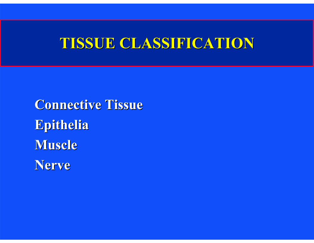
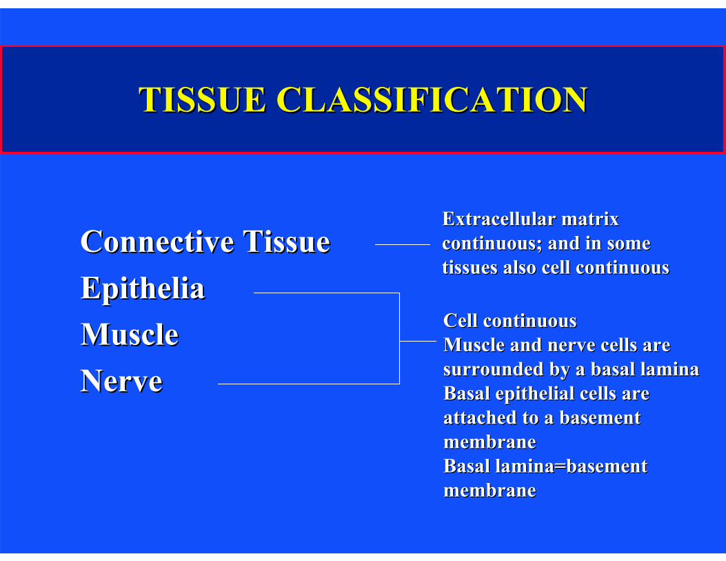
TISSUE CLASSIFICATIONTISSUE CLASSIFICATION
Connective TissueConnective TissueEpitheliaEpitheliaMuscleMuscleNerveNerve
TISSUE CLASSIFICATIONTISSUE CLASSIFICATION
Connective TissueConnective TissueEpitheliaEpitheliaMuscleMuscleNerveNerve
Extracellular matrixExtracellular matrix continuous; and in somecontinuous; and in some tissues also cell continuoustissues also cell continuous
Cell continuousCell continuousMuscle and nerve cells areMuscle and nerve cells are surrounded by a basal laminasurrounded by a basal lamina Basal epithelial cells areBasal epithelial cells are attached to a basementattached to a basement membranemembraneBasal lamina=basementBasal lamina=basement membranemembrane

BASEMENT MEMBRANEBASEMENT MEMBRANE
Continuous sheet, 50Continuous sheet, 50--300 nm thick300 nm thickNo cells contained within the BM; it is aNo cells contained within the BM; it is a
nonliving structurenonliving structurePrincipal protein constituentsPrincipal protein constituents
LamininLamininType IV collagenType IV collagenType XVIII collagenType XVIII collagen

L. Jeng
Basement Membrane
Diagram removed due to copyright restrictions.
FASEB J 2005

Image removed due to copyright restrictions. Diagrams of muscle, epithelial sheet, and kidney glomerulus structures.
B Alberts, et al, Mol Biol of the Cell, p. 819 (1989)

Scanning Electron Micrograph of the Cornea of a Chick Embryo
E: Epithelial cells BL: Basal lamina C: Collagen fibrils in
the underlying CT
Photo removed due to copyright restrictions.
B Alberts, et al, Mol Biol of the Cell, p. 819 (1989)
Connective Tissue
Image removed due to copyright restrictions.
Sketches from Illustrated Physiology, AB McNaught and R Callander, Williams and Wilkins, 1967

Connective Tissues
Image removed due to copyright restrictions. See http://cal.vet.upenn.edu/projects/histo/Index.htm
Loose and dense connective tissue from a cow's planum.

Loose Connective Tissue Dense Connective Tissue
Images removed due to copyright restrictions.
Ciba Collection, FH Netter, 1987

Connective Tissue: Adipose Tissue (Fat)
Images removed due to copyright restrictions.

Connective Tissue: Bone
Image removed due to copyright restrictions.
Ciba Collection, FH Netter, 1987

Connective Tissue: Cartilage
Hyaline Cartilage: Trachea Elastic Cartilage: Epiglottus
Image removed due to copyright restrictions. See http://cal.vet.upenn.edu/projects/histo/Index.htm
Fibrocartilage Cartilage identification is principally based on morphology; rounded cells in a lacuna; ECM is type I collagen (fibrocartilage) or type II cartilage (hyaline), with (elastic) or without elastin.
http://www.uoguelph.ca/zoology/devobio/210labs/epithelial1.html
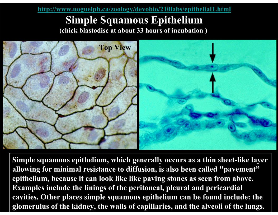
Simple Squamous Epithelium(chick blastodisc at about 33 hours of incubation )
Top View Cross-Sectional View
Simple squamous epithelium, which generally occurs as a thin sheet-like layer allowing for minimal resistance to diffusion, is also been called "pavement” epithelium, because it can look like like paving stones as seen from above. Examples include the linings of the peritoneal, pleural and pericardial cavities. Other places simple squamous epithelium can be found include: the glomerulus of the kidney, the walls of capillaries, and the alveoli of the lungs.
http://www.uoguelph.ca/zoology/devobio/210labs/epithelial1.html
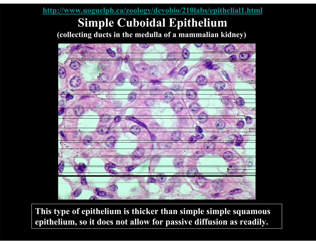
Simple Cuboidal Epithelium(collecting ducts in the medulla of a mammalian kidney)
This type of epithelium is thicker than simple simple squamous epithelium, so it does not allow for passive diffusion as readily.

http://www.uoguelph.ca/zoology/devobio/210labs/epithelial1.html
Simple Columnar Epithelium(small intestine)
Since columnar cells are quite thick, they do not readily allow passive diffusion. As a result, these cells use active transport to move nutrients through them from the intestine to the blood. This is what we commonly call "absorption." To help with this, they have numerous microvilli on their apical (lumenal) surface, which increases their surface area to allow for greater absorption.

Simple Columnar Epithelium
Image removed due to copyright considerations. See http://cal.vet.upenn.edu/projects/histo/Index.htm
This is a section through the edge of a gallbladder. There is a layer of simple columnar epithelium overlying the connective tissue as indicated by the arrows.

Stratified Squamous Epithelium
Image removed due to copyright restrictions. See http://cal.vet.upenn.edu/projects/histo/Index.htm
This is an example of stratified squamous epithelium from the esophagus of a cat. Arrows show nuclei of the outermost layer. This is normal for mucosa. Most stratified squamous cells in other areas, such as skin, lose their nuclei by the time they approach the outermost layers.
http://www.uoguelph.ca/zoology/devobio/210labs/epithelial1.html
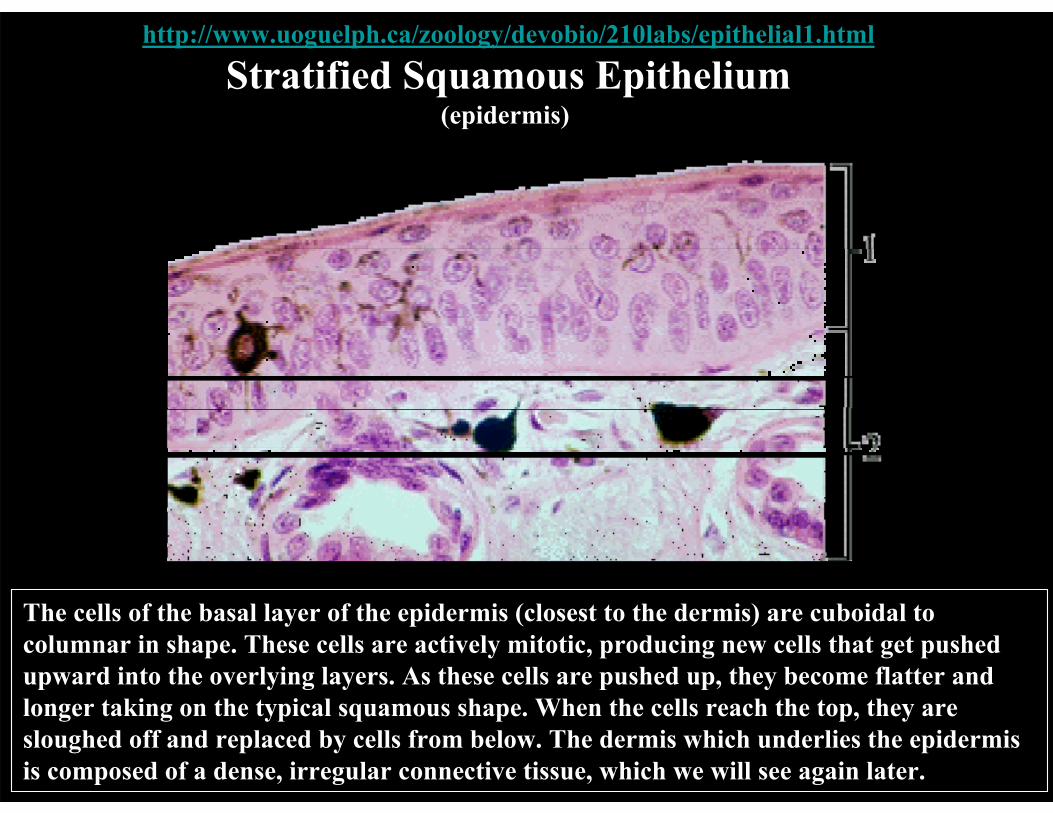
Stratified Squamous Epithelium(epidermis)
The cells of the basal layer of the epidermis (closest to the dermis) are cuboidal to columnar in shape. These cells are actively mitotic, producing new cells that get pushed upward into the overlying layers. As these cells are pushed up, they become flatter and longer taking on the typical squamous shape. When the cells reach the top, they are sloughed off and replaced by cells from below. The dermis which underlies the epidermis is composed of a dense, irregular connective tissue, which we will see again later.

Pseudostratified Columnar Epithelium
Image removed due to copyright restrictions. See http://cal.vet.upenn.edu/projects/histo/Index.htm
This is an example of ciliated pseudostratified columnar epithelium from the trachea. The arrows indicate the layer of cilia on the surface of the pseudostratified columnar cell layer.

Intestinal Epithelium
Photos removed due to copyright restrictions.
BM: basement membraneLu: lumen Cp: capillary Co: collagen fibrils SM: smooth muscle cell M: mitochondriaNF: nerve fibers
1μm KR Porter & MA Bonneville, Fine Structure of Cells and Tissues (1973)
Ciliated Epithelium
Photo removed due to copyright restrictions.
C: cilia SER: vesicles F: fibroblasts El: elastic fibers
1μm
KR Porter & MA Bonneville, Fine Structure of Cells and Tissues (1973)
Convoluted Tubule of the Kidney: Epithelium
Photo removed due to copyright restrictions.
1μm KR Porter & MA Bonneville, Fine Structure of Cells and Tissues (1973)
SEM of the Kidney
Photo removed due to copyright restrictions.
KR Porter & MA Bonneville, Fine Structure of Cells and Tissues (1973)
20μm

Kidney: Epithelium
Photo removed due to copyright restrictions.
US: urinary space
KR Porter & MA Bonneville, Fine Structure of Cells and Tissues (1973) 0.3 μm

Transmission Electron Micrograph of a Rat Kidney Glomerulus
Photo removed due to copyright restrictions.
From R Kessel and R. Kardon, Tiss and Org, p.233 (1979)

Muscle
Drawings removed due to copyright restrictions. 1. Smooth muscle 2. Cardiac muscle
3. Voluntary muscle fiber.
Ciba Collection, FH Netter, 1987

http://www.uoguelph.ca/zoology/devobio/210labs/epithelial1.html
MuscleSmooth (Involuntary) Muscle
Cardiac Muscle
Striated (Skeletal; Vountary) Muscle

Cardiac Muscle
Photo removed due to copyright restrictions.
SC: Schwann cell CT: connective tissue
0.3 μm
KR Porter & MA Bonneville, Fine Structure of Cells and Tissues (1973)

Nerve
Figure by MIT OpenCourseWare. After McNaught and Callander, Illustrated Physiology, Williams and Wilkins, 1967.
http://www.uoguelph.ca/zoology/devobio/210labs/epithelial1.html
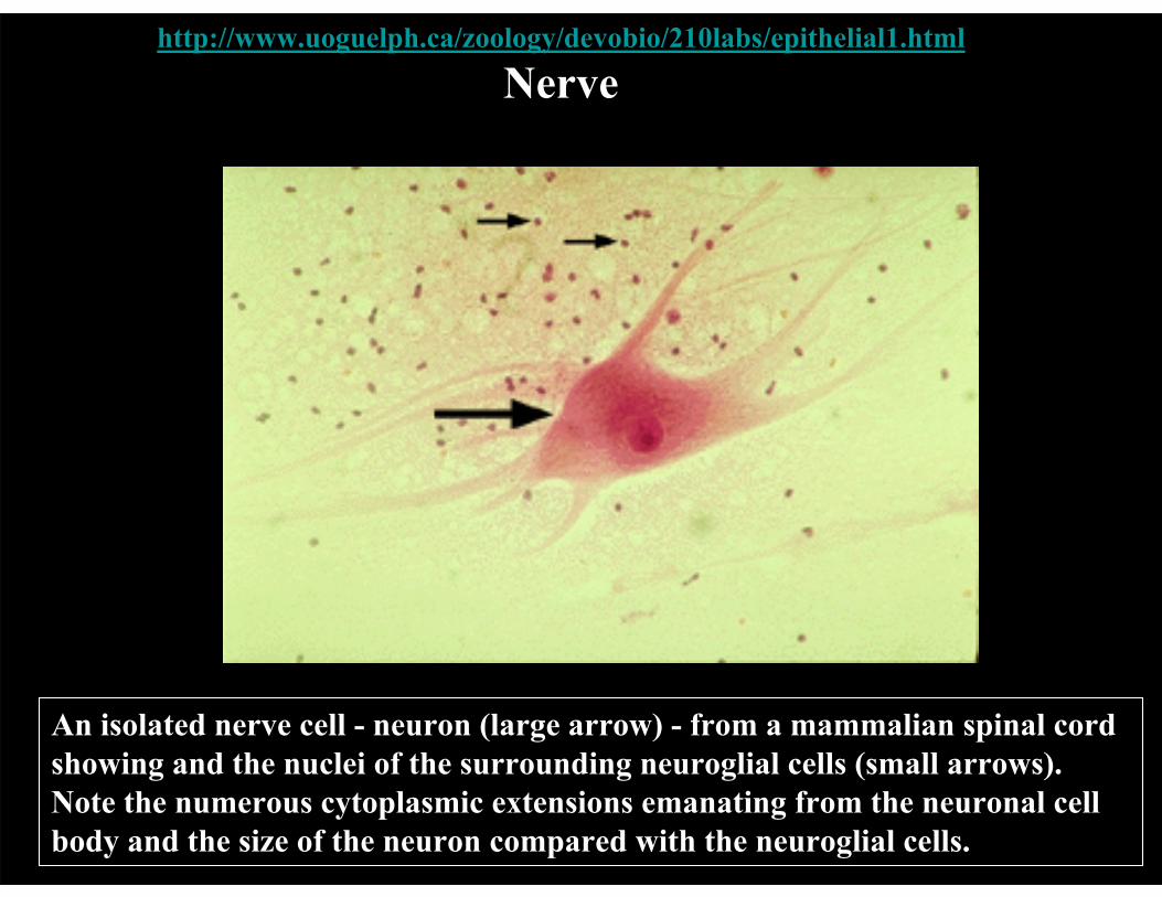
Nerve
An isolated nerve cell - neuron (large arrow) - from a mammalian spinal cord showing and the nuclei of the surrounding neuroglial cells (small arrows). Note the numerous cytoplasmic extensions emanating from the neuronal cell body and the size of the neuron compared with the neuroglial cells.
Peripheral Nerve: Rat Sciatic
Photo removed due to copyright restrictions.
Molecular Cell Biology, J Darnell, et al., 1990
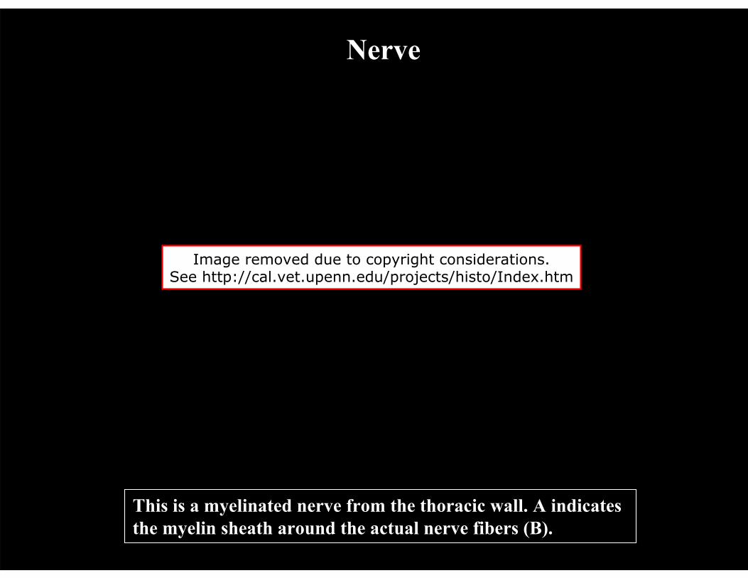
Nerve
Image removed due to copyright considerations. See http://cal.vet.upenn.edu/projects/histo/Index.htm
This is a myelinated nerve from the thoracic wall. A indicates the myelin sheath around the actual nerve fibers (B).
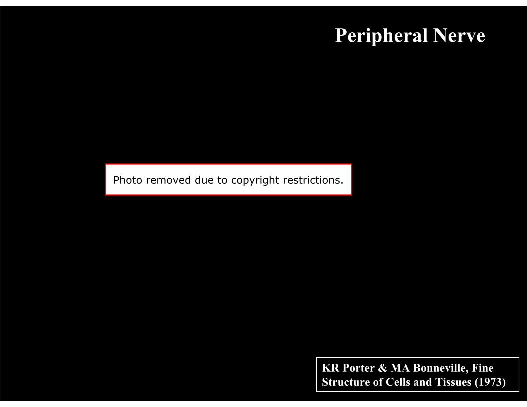
Peripheral Nerve
Photo removed due to copyright restrictions.
0.3 μm
KR Porter & MA Bonneville, Fine Structure of Cells and Tissues (1973)
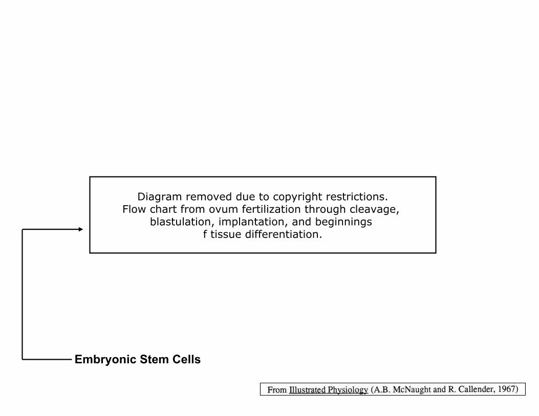
Diagram removed due to copyright restrictions.Flow chart from ovum fertilization through cleavage,
blastulation, implantation, and beginnings f tissue differentiation.
Embryonic Stem Cells

Embryonic Germ Disc
• Ectoderm becomes: – Epithelia of external surfaces – Nervous system tissues
• Mesoderm becomes: – Muscular tissues – Connective tissues – Urogenital system – Lining of body cavities and blood vessels
• Endoderm becomes: – Epithelia of most internal surfaces – Some glands (e.g. thyroid, pancreas, liver)

MIT OpenCourseWarehttp://ocw.mit.edu
20.441J / 2.79J / 3.96J / HST.522J Biomaterials-Tissue Interactions Fall 2009
For information about citing these materials or our Terms of Use, visit: http://ocw.mit.edu/terms.
Related Documents