Lectin Microarrays for Glycomic Analysis Garima Gupta, 1 Avadhesha Surolia, 2 and Srinivasa-Gopalan Sampathkumar 3 Abstract Glycomics is the study of comprehensive structural elucidation and characterization of all glycoforms found in nature and their dynamic spatiotemporal changes that are associated with biological processes. Glycocalyx of mammalian cells actively participate in cell–cell, cell–matrix, and cell–pathogen interactions, which impact embryogenesis, growth and development, homeostasis, infection and immunity, signaling, malignancy, and metabolic disorders. Relative to genomics and proteomics, glycomics is just growing out of infancy with great potential in biomedicine for biomarker discovery, diagnosis, and treatment. However, the immense diversity and complexity of glycan structures and their multiple modes of interactions with proteins pose great challenges for development of analytical tools for delineating structure function relationships and understanding glyco- code. Several tools are being developed for glycan profiling based on chromatography, mass spectrometry, glycan microarrays, and glyco-informatics. Lectins, which have long been used in glyco-immunology, printed on a microarray provide a versatile platform for rapid high throughput analysis of glycoforms of biological sam- ples. Herein, we summarize technological advances in lectin microarrays and critically review their impact on glycomics analysis. Challenges remain in terms of expansion to include nonplant derived lectins, standardization for routine clinical use, development of recombinant lectins, and exploration of plant kingdom for discovery of novel lectins. Introduction G lycomics may be defined as the study of comprehen- sive elucidation and characterization of all the glyco- forms (monosaccharides, oligosaccharides, polysaccharides, and their modifications) found either independently or con- jugated to other noncarbohydrate biomolecules (such as proteins, lipids, and nucleic acids) and their dynamic spatio- temporal changes that are associated with biological pro- cesses (Hirabayashi et al., 2001). A glycoform refers to a single homogeneous molecular species of a glycoconjugate carrying a set of specific structure of glycans, which are oligosaccharide chains—containing a glycoside bond between an anomeric carbon of one monosaccharide and oxygen of another monosaccharide—composed of various monosaccharide units, connected in a specific linkage and stereochemical ori- entation (Dell and Morris, 2001). As information carriers for coding biological processes, carbohydrates far exceed bases of nucleic acids and amino acids of proteins (Gabius, 2000). In mammalian systems, glycoconjugates are found in the form of glycoproteins, glycolipids, proteoglycans, and glycosylpho- sphatidyl inositol (GPI) anchors. Additionally, glycosamino- glycans (GAGs), which are composed of hexosamines and hexuronic acids in an alternating manner, are found either as free polysaccharides or as proteoglycans (Bishop et al., 2007; Cummings, 2009; Sasisekharan et al., 2006; Turnbull et al., 2001). Mammalian glycan chains are made up of monosaccharide building blocks, namely, hexoses (glucose, galactose, and mannose), N-acetyl-hexosamines (glucosamine, mannosa- mine, and galactosamine), deoxyhexoses (fucose), hexuronic acids (glucuronic acid, iduronic acid), pentoses (xylose), and nonuloses (sialic acids). The monosaccharides are found in the dextro-(d) rotatory form, except fucose, which is found in the levo-(l) rotatory form (Varki et al., 2008). All monosaccharide building blocks found in mammalian glycoconjugates exhibit hexopyranose ring structure. Although deoxyribonucleic ac- ids (DNA) and ribonucleic acids (RNA) do contain deoxyri- bose and ribose monosaccharide units in pentafuranose form in their backbone, they are not considered as glycans as they lack a glycosidic linkage. Both prokaryotes and plants—owing to their diverse, ver- satile, and easily adaptable biosynthetic machinery—exhibit a large repertoire of monosaccharide building blocks (e.g., l-rhamnose, N-acetyl-d-fucosamine, etc.) as part of their lipopolysaccharides (LPS), capsular polysaccharides (CPS), 1 Molecular Biophysics Unit, Indian Institute of Science, Bangalore, India. 2 Molecular Sciences Laboratory, National Institute of Immunology, New Delhi, India. 3 Laboratory of Chemical Glycobiology, National Institute of Immunology, New Delhi, India. OMICS A Journal of Integrative Biology Volume 14, Number 4, 2010 ª Mary Ann Liebert, Inc. DOI: 10.1089/omi.2009.0150 419

Welcome message from author
This document is posted to help you gain knowledge. Please leave a comment to let me know what you think about it! Share it to your friends and learn new things together.
Transcript

Lectin Microarrays for Glycomic Analysis
Garima Gupta,1 Avadhesha Surolia,2 and Srinivasa-Gopalan Sampathkumar3
Abstract
Glycomics is the study of comprehensive structural elucidation and characterization of all glycoforms found innature and their dynamic spatiotemporal changes that are associated with biological processes. Glycocalyx ofmammalian cells actively participate in cell–cell, cell–matrix, and cell–pathogen interactions, which impactembryogenesis, growth and development, homeostasis, infection and immunity, signaling, malignancy, andmetabolic disorders. Relative to genomics and proteomics, glycomics is just growing out of infancy with greatpotential in biomedicine for biomarker discovery, diagnosis, and treatment. However, the immense diversityand complexity of glycan structures and their multiple modes of interactions with proteins pose great challengesfor development of analytical tools for delineating structure function relationships and understanding glyco-code. Several tools are being developed for glycan profiling based on chromatography, mass spectrometry,glycan microarrays, and glyco-informatics. Lectins, which have long been used in glyco-immunology, printed ona microarray provide a versatile platform for rapid high throughput analysis of glycoforms of biological sam-ples. Herein, we summarize technological advances in lectin microarrays and critically review their impact onglycomics analysis. Challenges remain in terms of expansion to include nonplant derived lectins, standardizationfor routine clinical use, development of recombinant lectins, and exploration of plant kingdom for discovery ofnovel lectins.
Introduction
Glycomics may be defined as the study of comprehen-sive elucidation and characterization of all the glyco-
forms (monosaccharides, oligosaccharides, polysaccharides,and their modifications) found either independently or con-jugated to other noncarbohydrate biomolecules (such asproteins, lipids, and nucleic acids) and their dynamic spatio-temporal changes that are associated with biological pro-cesses (Hirabayashi et al., 2001). A glycoform refers to a singlehomogeneous molecular species of a glycoconjugate carryinga set of specific structure of glycans, which are oligosaccharidechains—containing a glycoside bond between an anomericcarbon of one monosaccharide and oxygen of anothermonosaccharide—composed of various monosaccharideunits, connected in a specific linkage and stereochemical ori-entation (Dell and Morris, 2001). As information carriers forcoding biological processes, carbohydrates far exceed bases ofnucleic acids and amino acids of proteins (Gabius, 2000). Inmammalian systems, glycoconjugates are found in the form ofglycoproteins, glycolipids, proteoglycans, and glycosylpho-sphatidyl inositol (GPI) anchors. Additionally, glycosamino-glycans (GAGs), which are composed of hexosamines and
hexuronic acids in an alternating manner, are found either asfree polysaccharides or as proteoglycans (Bishop et al., 2007;Cummings, 2009; Sasisekharan et al., 2006; Turnbull et al.,2001).
Mammalian glycan chains are made up of monosaccharidebuilding blocks, namely, hexoses (glucose, galactose, andmannose), N-acetyl-hexosamines (glucosamine, mannosa-mine, and galactosamine), deoxyhexoses (fucose), hexuronicacids (glucuronic acid, iduronic acid), pentoses (xylose), andnonuloses (sialic acids). The monosaccharides are found in thedextro-(d) rotatory form, except fucose, which is found in thelevo-(l) rotatory form (Varki et al., 2008). All monosaccharidebuilding blocks found in mammalian glycoconjugates exhibithexopyranose ring structure. Although deoxyribonucleic ac-ids (DNA) and ribonucleic acids (RNA) do contain deoxyri-bose and ribose monosaccharide units in pentafuranose formin their backbone, they are not considered as glycans as theylack a glycosidic linkage.
Both prokaryotes and plants—owing to their diverse, ver-satile, and easily adaptable biosynthetic machinery—exhibita large repertoire of monosaccharide building blocks (e.g.,l-rhamnose, N-acetyl-d-fucosamine, etc.) as part of theirlipopolysaccharides (LPS), capsular polysaccharides (CPS),
1Molecular Biophysics Unit, Indian Institute of Science, Bangalore, India.2Molecular Sciences Laboratory, National Institute of Immunology, New Delhi, India.3Laboratory of Chemical Glycobiology, National Institute of Immunology, New Delhi, India.
OMICS A Journal of Integrative BiologyVolume 14, Number 4, 2010ª Mary Ann Liebert, Inc.DOI: 10.1089/omi.2009.0150
419
antibiotic glycosides, and plant-derived polysaccharides,which in contrast, are not found in humans. Consequently, thestudy of glycomics of prokaryotes and the plant kingdom isindeed a daunting task in relation to mammalian genome(Varki et al., 2008). The unique monosaccharide structuresand glycans found on bacteria are proving extremely valuabledeterminants for identification, serotyping, and classification.However, there are commonalities found with respect tobiosynthetic protein N-glycosylation pathways between plantand mammalian systems, with the exception of sialic acidswhich are unique to mammals (Wilson, 2002).
Advances in genomics have already reached a level ofcommercial viability as evidenced by extensive use ofgenome-wide and focussed DNA microarrays in research andclinic. A logical follow-up was the study of proteomics, whichhas reached a level of sophistication and automation sufficientto be translated to the end user for biomarker discovery.However, proteomics relies for a large part on differentialisotopic labeling, enzymatic digestion, resulting in a mixtureof peptides followed by mass spectrometry rather than anal-ysis of whole proteins (Blackstock and Weir, 1999; Zaia, 2008).Both genomics and proteomics are made ‘‘relatively’’ simplethanks to their template-driven nature arising out of base-paircomplementarity and tri-nucleotide codon-driven transcrip-tional and translational processes. The fruits of more than halfa century of research efforts in automated chemical synthesisof peptides and nucleic acid primers, cloning, and expressionof proteins, gene silencing technology, epigenetics, and tran-scriptome studies have greatly enabled the current level ofsophistication in genomics and proteomics. The past decadehas seen a growing realization of the importance of glycomicsperspective to immunology and biomedical challenges (Baumand Crocker, 2009; Sharon and Lis, 1993).
In comparison to genomics and proteomics—where auto-mated synthesis, amplification, expression, and character-ization have become routine—the tools available for study ofglycomics are few. This necessitated the development of un-ique tools that are currently reaching a reasonable level ofsophistication for use by nonspecialists (Bertozzi and Kies-sling, 2001; Pilobello and Mahal, 2007; Prescher and Bertozzi,2006). The enormous diversity of structures that can be cre-ated using a limited number of monosaccharides buildingblocks coupled with its non template-driven nature has madethe task of system-wide glycan profiling a challenging task(Fig. 1) (Cummings, 2009; Gabius et al., 2004). A major ob-stacle for the advancement of glycomics has been the lack ofsensitive and easy-to-use high-throughput analytical tools.
The availability of well-characterized enzymes with knownsubstrate specificities, such as PNGase-F (useful for cleavingthe intact N-linked glycans out of the asparagine (Asn) sidechain), endo-H (useful for cleaving the internal b-(1? 4)-GlcNAc residue of N-linked glycans leaving one GlcNAc at-tached to peptide backbone) and several plant and bacterialglycosidases (useful for distinguishing specific anomericlinkages), and analytical chromatographic methods, based onelectrochemical detection and fluorophore derivatization ofthe reducing end anomeric carbon (Domann et al., 2007), en-abled detailed and thorough characterization of the total oli-gosaccharide structures and linkages (Ghesquiere et al., 2006;Ramachandran et al., 2006). Parallel advances in stereo-chemically controlled synthesis and purification of oligosac-charides produced homogenous glycans that served as
reference standards for biological specimen-derived glycans,haptens for conjugation, production of monoclonal anti-bodies, and vaccine development (Buskas et al., 2006; See-berger and Werz, 2007; Wang et al., 2007). However, thecharacterization of a complete structure of a glycan usingenzymes and chromatography involves tedious and time-consuming efforts (Ding et al., 2009; Patwa et al., 2009; Zhang,2007).
The advent of proteomics and evolution of powerful yetuser-friendly mass spectrometry technologies, includingmethodologies coupled to chromatography (known as hy-phenated techniques) and bioinformatics, stimulated the rapidanalysis of glycans (Bielik and Zaia, 2010; von der Lieth et al.,2006; Werz et al., 2007; Zaia, 2008). Mass spectrometry has theadvantage to rapidly scan and analyze samples in a highthroughput manner, once the glycans or glycopeptides havebeen isolated, with or without permethylation, or enrichedusing lectin-based affinity chromatography. However, unlikepeptides, which are made of amino acid as building blockswith distinct molecular mass enabling easy identification ofunique peptide sequences, the isobaric nature of variousmonosaccharide components of glycans complicates the as-signment. So, although one could easily predict the size of theoligosaccharides and general components such as number ofhexosamines, hexoses, sialic acids, etc., using mass spec-trometry, the specific monosaccharides and their stereo-chemical linkages have to be deduced based on knownbiochemical pathways (North et al., 2009; von der Lieth et al.,2006). High-end techniques such as tandem mass spectrom-etry [MS]n, which study fragmentation of a given molecularion peak and generation of fingerprint-like patterns, are beingcurrently developed to identify the component monosaccha-rides, their linkage positions, and anomeric orientations. Theinherently charged high-energy nature of molecular ions inmass spectrometry leads to loss of posttranslational modifi-cations (such as O-acetylation of sialic acids) and loss of sen-sitive terminal sialic acid residues. To simplify the vastnumber of structures possible for a given oligosaccharidepurely based on mathematical permutations and for matchingof a given mass spectrometry ion peak to relate to a minimalset of glycans, qualified databases were developed usingknown mammalian biosynthetic glycosylation pathways aslimiting parameters (Cooper et al., 2001; Goldberg et al., 2005;McDonald et al., 2009). Rigorous mathematical models cor-relating expression levels and activities of enzymes involvedin N-glycan biosynthetic pathways to the overall mass spec-trometry profile of glycans have been developed to analyzedifferential glycosylation patterns (Krambeck et al., 2009).Notwithstanding intense efforts by multiple research groups,the use of mass spectrometry as a stand-alone technique forcomplete characterization of glycans is still far from complete.
To evaluate and validate the best possible combination oftechniques for glycome profiling as part of Human ProteomeOrganization (HUPO) and Human Disease Glycomics/Proteome Initiative (HGPI), a highly coordinated multi-insti-tutional study of analysis of N-linked glycans was conductedrecently in 20 different labs using identical samples of humanIgG and transferrin. The study found that although there weredifferences in sensitivities [limit of detection (LoD)] and thederived glycoform profiles between various chromatographic(trypsin or PNGase-F digestion, fluorophore derivatization,electrochemical detection, and permethylation using LC or
420 GUPTA ET AL.

GC) and mass spectrometric techniques (MALDI or ESI of freeor permethylated glycans or glycopeptides); the outcome wasstrikingly comparable considering the microheterogeneity ofglycans (Wada et al., 2007). Although MALDI-MS of per-methylated glycans was found to be as good as chromato-graphic techniques, it suffered from frequent loss of terminalsialic acid residues. On the other hand, LC/ESI-MS was foundto retain sialic acids intact. The masses have to be mappedwith an online MALDI-MS glycomics database. In anotherinter-laboratory study for structural analysis of N-linkedglycans, wide variations were found for levels of sialylation,fucosylation, and antennary branching, highlighting methodvariability and the need for well-characterized referencestandards (Thobhani et al., 2009).
Clearly, a comprehensive elucidation of intact glycoformstructures and their biological significance requires the in-volvement of application of several complementary technol-ogies. Although chromatography and mass spectrometry arethe mainstays for glycomics research, alternate technologiessuitable for rapid, high-throughput user-friendly analysisat the ‘‘point-of-care’’ need to be developed. Toward thisend microarray-based technologies have come to the rescue.Microarrays based on immobilized glycan ligands, either syn-thetic, isolated from biological samples, or neoglycoconju-gates have been developed and shown to be useful for thedetection and characterization of human lectins and bacteria(Hsu and Mahal, 2009; Ito et al., 2009; Song et al., 2009). Thedevelopments and achievements of carbohydrate–ligand-based microarrays have been reviewed regularly and exten-sively (Feizi and Chai, 2004; Hirabayashi, 2003; Horlacher andSeeberger, 2006, 2008; Krishnamoorthy and Mahal, 2009;Liang et al., 2008; Oyelaran and Gildersleeve, 2009; Paulsonet al., 2006). Microarrays wherein immobilized antibodies(including anti-carbohydrate antibodies) were used to capturea targeted set of glycoproteins and subsequent detection with
labeled lectins have been valuable in studying cancer-associ-ated glycan alterations (Chen et al., 2007a). However, in thecase of anti-carbohydrate antibodies in particular, the reportedsupplier-dependent variations and crossreactivity must betaken in to account (Manimala et al., 2007). A complementaryapproach is the design and development of microarray tech-niques using immobilized lectins, which is the topic of thisreview. The analysis of glycans seems to have taken a full circlewith the advent of lectin-based microarrays (Sharon, 1998).
Lectin and Carbohydrate Recognition
Early analysis of glycoforms relied heavily on screening ofbiological samples using a wide variety of plant lectins. Lec-tins, derived from Latin legere to pick out or choose, are de-fined as proteins—that bind carbohydrates present onglycoconjugates as ligands—that are neither enzymes (car-bohydrate processing), antibodies (anticarbohydrate anti-bodies), nor proteins that bind small carbohydrate molecules(energy metabolism) (Goldstein et al., 1980). Lectins, whichare available in plenty in plant seeds and could be isolated inenriched forms relatively easily, induced agglutination inblood cells and were instrumental in the study of blood groupclassification and as affinity matrices for purification of gly-coproteins and cell separations (Rudiger and Gabius, 2001). Itwas known as early as in the 1950s that the ligands to whichlectins bind on the mammalian cell surface were composed ofcarbohydrate, as shown by competitive inhibition using spe-cific monosaccharides (Morgan, 1970; Morgan and Watkins,2000; Watkins and Morgan, 1957). Two classic examples arethe characterization of blood group antigens and the dem-onstration of leukocyte homing based on glycan-derived re-ceptors (Gesner and Ginsburg, 1964; Rosen, 2004). Pioneeringwork on lectins and their carbohydrate binding specificitiesby Sharon, Goldstein, Kornfeld, Brewer, Surolia, and several
FIG. 1. Complexity and diversity of mammalian glycome. The glycome represents the largest class of posttranslationallymodified molecules. The information flow from genome to glycome increases exponentially (diagram not drawn to scale).Glycans amplify the genomic information content of cells providing functional diversity originating from only a single gene.Genome, transcriptome, and proteome are governed by template-driven processes using unidirectional (nucleotide base pairsindicated using single letter codes) or bidirectional building blocks (amino acids indicated by standard single-letter codes). Incontrast, glycome is generated by complex biosynthetic glycosylation pathways in a non template-driven manner, governedby availability of carbohydrate substrates, expression and activity levels of enzymes and sugar–nucleotide transporters. Themonosaccharide building blocks (shown using standard abbreviations) of mammalian glycans are combined in a multidi-rectional manner and exhibit linkage and stereo isomerism and frequent branching.
LECTIN MICROARRAYS FOR GLYCOMIC ANALYSIS 421

others have set a strong biochemical foundation (Brewer et al.,2002; Dam and Brewer, 2002, 2010; Lis and Sharon, 1986;Sacchettini et al., 2001; Sharon, 2007, 2008; Sharon and Lis,2004). In mammalian physiology, endogenous lectins (e.g.,galectins, siglecs, calreticulin, calnexin, etc.) regulate keyprocesses such as pattern recognition of pathogens, cell–cellcommunications, correct folding of glycoproteins and qualitycontrol (Caramelo and Parodi, 2007; Gabius et al., 2002;Shanmugham et al., 2006). Lectins are known to play vitalroles in plant physiology, development, and stress response.As part of the innate immune system, sometimes coupledwith ribosome inactivating proteins, for example, ricin, lectinsward off attacks by pathogens, parasites, and predators (DeHoff et al., 2009). On the other hand, lectins act as tools forscreening and agglutination, via interaction with bacterialpolysaccharides including Nod factors, for attachment ofsymbiotic bacteria (e.g., rhizobia) and fungi, which help innitrogen fixation and facilitate mutualism.
Origin of Studies on Microheterogeneity,Structure of Glycans, and Lectins
The classic works of Sharon and coworkers (Lis and Shar-on, 1986; Sharon, 2008; Sharon and Lis, 2004), Goldstein andcoworkers (Goldstein, 2009; Shibuya et al., 1988), Cummingsand Kornfeld (1982), Debray and coworkers (1981), Baenzigerand Fiete (1979a, 1979b, 1982), and Surolia and coworkers(Mitra et al., 2003; Surolia et al., 1974, 1980) with lectins set thepace for defining glycan structures and heterogeneities in-volved therein. Sharon (2007) has pioneered the structure andcarbohydrate specificities of lectins particularly soy bean ag-glutinin (SBA) and Erythrina corallodendron lectin (ECorL)and the importance of their glycosylation. Goldstein and co-workers have studied the interactions of Conconavalin A(Con A) with several simple and complex carbohydrates. Intheir classic article they demonstrate that Con A precipitateswith purified monoclonal IgM and also binds the variouscarbohydrate moieties present on the surface of lectins fromwaxbean and soybean, which contain terminal nonreducinga-d-mannopyranosyl, a-d-glucopyranosyl, and b-d-fructo-furanosyl residues (Shibuya et al., 1988). Cummings andKornfeld (1982) demonstrated that serial lectin–agarosechromatography (SLAC) is a rapid, sensitive, and specifictechnique that can be used for fractionating and analyzing N-linked oligosaccharides. The fractionation scheme involvedserial chromatography on Con A– Sepharose, pea lectin–Sepharose, and leukoagglutinating phytohemagglutinin–Sepharose. The various fractions eluted from the columnscontained relatively pure populations of glycopeptides thatcould be further used for structural analysis. Debray and co-workers (1981) elucidated the interactions of various oligo-saccharides with 12 different lectins in an exhaustive mannerto obtain carbohydrate specificities. Surolia and coworkers(1980) have demonstrated the power of lectin binding meth-odology for identifying the various glycoforms and micro-heterogeneity in glycoproteins before glycan massspectrometry was in vogue. The use of monovalent lectin-affinity purification was demonstrated by deliberately gen-erating microheterogeneity by subjecting fetuin to partialhydrolysis. Partial hydrolysis of fetuin resulted in variousasialoglycoforms that stoichiometrically bound monovalentradioactive-labeled ricin, in a manner proportional to the
number of asialo-glycan units per protein chain. The variousricin–fetuin complexes could be easily separated by gel fil-tration. However, the technique itself being cumbersome andtime consuming, the studies in this direction were not pur-sued further. Studies by Baenziger and Fiete (1982) describedin detail the binding affinities of a number of N- and O-linkedoligosaccharides with several lectins, as well as the usage ofphotoactivable glycopeptides reagents for site-specific label-ing of lectins. They synthesized photoactivable iodinatedglycopeptides bearing oligosaccharides of defined structure.Con A, Ricinus communis agglutinin and toxin, RCAI andRCAII, respectively, and Gal/GalNAc-specific lectin fromhuman and rat hepatocytes were used for the study. Theydemonstrated that covalent incorporation upon photoactiva-tion only occurs with a glycopeptide that is specifically boundby the lectin and inhibited only by appropriate monosaccha-rides. This high degree of affinity and specificity of photo-activable glycopeptides identified them as promising agentsfor examination of lectins. Extensive studies by Brewer andcoworkers, Surolia and coworkers, and others using iso-thermal calorimetry have provided enormous data on thethermodynamics of lectin–carbohydrate interactions andmultivalency (Dam and Brewer, 2002; Mitra et al., 2003). In anelegant study highlighting importance of both carbohydrateligand density as well as relative spatial orientation anddynamics of carbohydrate–lectin interactions, Srinivas andcoworkers (2002, 2005) synthesized photo-switchable glyco-ligands with an azobenzene core moiety which undergoescis–trans isomerism upon irradiation. The existence of co-operativity in lectin–ligand binding was shown using bi-phasic binding curve of PNA with azobenzene-bis-lactosederivative, by isothermal calorimetry (ITC). Vast amount ofknowledge gathered over four decades on structure of lectins,carbohydrate ligands, and their binding selectivities, andthermodynamic parameters using solution-based methodol-ogies, and studies on multivalency or clustering effect for in-creased avidity of lectin–carbohydrate interactions (Lee andLee, 1995, 2000; Lundquist and Toone, 2002) have togetherhelped take lectins to the next level in the form of lectin arrayto address the challenge of glycan profiling.
Lectin Microarrays—Evolution of a PlatformTechnology
In tune and keeping pace with the dawn of 21st century anddevelopment of high-throughput technologies in genomicsand proteomics, it was logical for glycomics analysis toquickly follow suit. The time was opportune to put the largeamount of knowledge accumulated in lectin biochemistryover four decades to fruitful applications. Toward this end,lectin microarrays were developed wherein several lectinswere immobilized to a suitable solid phase (microscopic slide)optimized using multiple chemical methodologies, conditions(humidity, buffer, and temperature), spot size and spot mor-phology. Table 1 illustrates the number of studies performedto date along with total number of lectins per array, method ofdetection, and the nature of analyte (ranging from mono- andoligo-saccharides, neo-glycoconjugates, purified glycans,glycoproteins, crude lysates, membrane preparation, whole-cell bacteria, and intact mammalian cells). Methods for lectinimmobilization rely on various methods such as (1) carbeneinsertion, (2) biotin–avidin bridge, (3) attachment of amine
422 GUPTA ET AL.

functional group of lysine side chains of protein-backboneof lectins for immobilization to solid surface throughepoxy-functionalized (glycidyl derivative) or N-hydro-xysuccinimidyl (NHS)-derived esters, (4) exploitation of self-assembled monolayers of thiols on gold-coated surfaces, and(5) 3D hydrogel surfaces (Fig. 2). In all methods there is a lackof absolute control in guaranteeing the optimal orientation,native multimeric quarternary structure, optimal multivalentclustering of carbohydrate recognition domains (CRD) oflectins and their metal ion requirements, such as Caþ2, Mgþ2,etc. Curiously, the carbohydrate portion of lectins (exceptWGA, Con A, PNA, which are not glycosylated) has not beenexploited as an anchor point. A mild periodate-based oxida-tion of glycan chains of lectin, as employed for glycosylatedantibody microarrays (Chen et al., 2007a), followed by an-choring to hydroxylamine or hydrazine containing solidsurfaces should be a useful approach. This method wouldleave the CRD of lectins intact, tuck away the crossreactingglycan portions of lectin, and might greatly improve optimalorientation of lectins in their native multimeric forms. Suchhydrazide or hydroxylamine based methodologies have beenused to capture glycans from glycoproteins and mammaliancells (Wollscheid et al., 2009).
Detection and differentiation of glycoforms on proteins
In a first proof-of-concept demonstration of feasibility oflectin arrays for distinct detection of terminal carbohydratemoieties on glycoproteins Angeloni and coworkers (2005)designed a miniarray. Employing a set of four lectins, namely,peanut agglutinin (PNA), Sambucus nigra lectin (SNA), Dan-tura stramonia lectin (DSA), and Ulex europaeus lectin (UEA), itwas shown that the differences in terminal sugar epitopescould be detected with good sensitivity. Biotinylated lectinswere immobilized on Opto-Dex–biotin plates using neu-travidin as a bridge. A selected set of purified glycoproteinslabeled either with Cy3 or Cy5 were used as analytes. Thefluorescent labeling of glycoproteins was performed with anexcess of the dye and purified by size exclusion chroma-tography. For quantitative studies, it will be important toknow the exact stoichiometry of number of fluorophores co-valently linked for each molecule of glycoprotein, as thefluorescence emission and quantum efficiency may be afunction of number of fluorophores per molecule. A numberof fluorophores in close proximity may lead either to mutualenhancement of emission signal intensity or internalquenching depending on the microenvironment of the gly-coprotein under consideration (Yamaguchi et al., 2008). It isnot clear if these glycoproteins [excluding neoglycoconjugatesof bovine serum albumin (BSA) and dextrose] consisted ofmultiple glycoforms. If so, it was at least not detected in thisarray with limited number of lectins or there was selectivecapturing of some glycoforms, whereas others were lost dueto washing. Notably, in an alternative methodology, whenphotochemical activation of diazirine moieties on modifieddextran was employed for covalent crosslinking of lectins, thecarbohydrate binding activity of lectins was found to be lost.In addition to structural rearrangement of CRD upon carbeneinsertion reaction, the carbohydrate binding by lectin mayhave been compromised by the multiple points of attachmentof a lectin on dextran. The aspect regarding the correct, if not,at least flexible or adaptable orientation of the carbohydrate-
binding moiety on the surface of immobilization cannot beoveremphasized for routine use of lectin microarray tech-nologies. In comparison to cDNA microarray employed forgenomics where true sequence-dependent and strong bindingor ‘‘hybridization’’ occurs owing to complementary base pairsand hydrogen bonds, the interactions between glycan epi-topes and lectins depend heavily on weak, albeit selective,complementarity of three-dimensional binding spacesthrough hydrogen bonds and van der Waals forces.
In another effort for rapid analysis of glycopatterns ofproteins, Pilobello and coworkers reported a lectin arrayconsisting of nine commonly available lectins. The lectinswere covalently immobilized directly on to aldehyde or epoxide-derivatized glass slides. The array was probed using Cy3-conjugated chicken ovalbumin and found to exhibit selectivebinding consistent with earlier reports of N-linked glycanspresent on ovalbumin. The sample of ovalbumin containedmany accessible terminal GlcNAc glycans predominantlyas shown by their exclusive binding to Con A, Griffoniasimplifolica lectin (GSL-II) and wheat germ agglutinin (WGA)(Pilobello et al., 2005). Although the lectins were selectedto cover a wide range of substrate specificities, the above-mentioned three lectins possess overlapping selectivity forGlcNAc. The epoxy-derivatized slides seem to be somewhatbetter than aldehyde derivatized slides in terms of spotmorphology and homogeneity; both the array substratesprovided qualitatively similar results. To establish selectivebinding with terminal GlcNAc competitive inhibition studieswere conducted using free GlcNAc and Gal. In all the threelectins, Con A, GSL-II, and WGA, the binding, and hencefluorescence, was abolished by GlcNAc but not Gal, whichwas as expected. It is not clear if other monosaccharides werestudied for competitive inhibition. For example, use of man-nose (Man) as a competing monosaccharide control, in ad-dition to Gal, might have shown a higher differential affinityfor Con A binding of Cy3-ovalbumin. Additional studies us-ing Cy-3 labeled porcine gastric mucin and bovine salivarymucin exhibited qualitative differences in lectin binding in-dicating the feasibility of microarrays. The sensitivity of de-tection and pattern was found to be conserved when the spotsize was reduced to *80 microns using automated arrayerfrom *750 microns with manual arrayer. However, the lackof efficient binding of mucins to SNA was attributed to con-flicting reports of selective substrates for SNA and supplierdependent variations. It is not clear if the sample of Cy-3-conjugated ovalbumin (analyte) used, which could exhibitbatch-to-batch variations in microheterogeneity of glyco-forms, was independently tested for presence of sialic acidcontaining N-glycans using routine periodate-resorcinol,thiobarbituric acid or other assays. It is possible the Cy-3 (afluorophore with a positive charge) derivatization of oval-bumin sterically and electrostatically prohibited the interac-tion of sialoglycans with SNA. Importantly, the stoichiometryof number of fluorophore molecules per molecule of albuminmay be crucial in achieving consistent results. Chicken oval-bumin has 20 lysine residues, although not all are equallyaccessible to Cy-3-NHS ester, and an N-terminal aminogroup. The average pattern of Cy-3 derivatization could alterthe selective binding characteristics to lectins. The aspect ofweak and overlapping binding affinities among the lectinsselected restricted use of other glycoproteins with heteroge-neous glycoforms. Also, the efficiency of interaction of solid
LECTIN MICROARRAYS FOR GLYCOMIC ANALYSIS 423

Ta
bl
e1.
Co
mp
ar
iso
no
fN
um
be
ro
fL
ec
tin
s,
Me
th
od
so
fIm
mo
bil
iz
at
io
n,
an
dS
ub
st
ra
te
sA
na
ly
ze
dU
sin
gL
ec
tin
Mic
ro
ar
ra
ys
S.
No.
No.
ofle
ctin
s
Mod
eof
atta
chm
ent,
incu
bati
onte
mp
erat
ure
and
hu
mid
ity
for
hy
brid
izat
ion
and
bloc
kin
gag
ent
(av
erag
ed
iam
eter
ofle
ctin
spot
inmm
)A
nal
yte
Met
hod
ofd
etec
tion
Ap
pli
cati
on(r
efer
ence
)
14
Bio
tin
yla
ted
lect
in-n
eutr
avid
in(1
50mm
)G
lyco
pro
tein
san
dn
eo-
gly
coco
nju
gat
es–C
y5-
dex
-lac
tose
,C
y3-
asia
lofe
tuin
,C
y5-
fetu
in,
Cy
3-L
acN
Ac-
BS
A,
Cy
5-2
(-fu
cosy
llac
tose
-BS
A,
Cy
5-tr
ansf
erri
n
Flu
ore
scen
cesc
ann
erP
roo
f-o
f-co
nce
pt
(An
gel
on
iet
al.,
2005
)
29
Ep
ox
ide
and
ald
ehy
de
gla
sssl
ides
(700
mm)
Gly
cop
rote
ins–
Cy
3-co
nju
gat
edch
ick
eno
val
bu
min
,p
orc
ine
gas
tric
mu
cin
,an
db
ov
ine
sali
var
ym
uci
n
Cy
3-co
nju
gat
ion
flu
ore
scen
cesc
ann
erP
roo
f-o
f-co
nce
pt
(Pil
ob
ello
etal
.,20
05)
36
Go
ld–t
hio
l–li
pid
–NH
S-d
eriv
edco
val
ent
lin
kin
g–S
AM
–sel
f-as
sem
ble
dm
on
ola
yer
s;B
SA
;H
um
idch
amb
er(h
and
spo
tted
usi
ng
0.5mL
vo
lum
e)
Gly
cop
rote
in–F
luo
resc
ein
-ta
gg
edch
ick
eno
val
bu
min
.W
ho
lem
amm
alia
nce
lls–
BH
K-2
1an
dC
aco
-2ce
lls
Flu
ore
scen
cean
db
rig
ht-
fiel
dm
icro
sco
py
Pro
of-
of-
pri
nci
ple
(Zh
eng
etal
.,20
05)
413
Gel
slid
es;
no
nco
val
ent
abso
rpti
on
(150
mm)
Asi
alo
fetu
inan
da-
feto
pro
tein
Cy
3-la
bel
edg
lyco
pro
tein
sG
lyco
pro
fili
ng
of
hep
ato
cell
ula
rca
rcin
om
ab
iom
ark
er(C
hen
etal
.,20
08)
539
Ep
ox
yco
ated
slid
es(3
-g
lyci
do
xy
pro
py
ltri
met
ho
xy
-si
lan
e);
1%
BS
Ain
PB
S.
Pu
rifi
edg
lyco
pro
tein
san
do
lig
osa
cch
arid
esC
y3-
lab
eled
gly
cop
rote
ins
and
TM
R-l
abel
edo
lig
osa
cch
arid
es.
Un
der
equ
ilib
riu
mco
nd
itio
ns
usi
ng
evan
esce
nt-
fiel
dfl
uo
resc
ence
.
Qu
anti
tati
on
and
val
idat
ion
afte
rtr
eatm
ent
of
oli
go
-sa
cch
arid
esw
ith
gly
can
ase
and
gly
cosi
dea
se.W
ash
-fre
ean
aly
sis
(Ku
no
etal
.,20
05).
640
-do
- ‘‘dif
fere
nti
alg
lyco
mic
s’’
Lec
WT
and
mu
tan
tC
HO
cell
lysa
tes
Cy
3-N
HS
este
rd
eriv
atiz
atio
no
fin
tact
wh
ole
cell
s–cr
ud
ely
sate
ssa
mp
les
Val
idat
ion
of
anti
cip
ated
bio
syn
thes
ism
uta
nt
bas
edg
lyca
ns
(Eb
eet
al.,
2006
).7
21N
exte
rio
nH
slid
es–
hy
dro
gel
–NH
S-b
ased
cov
alen
tim
mo
bil
izat
ion
–h
yd
rog
elp
rov
ides
flu
idm
ediu
mfo
rm
ain
ten
ance
of
acti
ve
con
form
atio
nan
dsh
apes
.E
than
ola
min
eas
blo
ckin
gag
ent
(95mm
)
Lab
eled
wh
ole
cell
bac
teri
a.S
YT
O85
dy
eu
pta
ke—
no
rmal
ized
Dis
tin
ctio
no
fin
fect
iou
sfr
om
no
n-i
nfe
ctio
us
pat
ho
gen
s(H
suet
al.,
2006
;H
suan
dM
ahal
,20
06).
424

858
Nex
teri
on
Hsl
ides
;R
oo
mte
mp
erat
ure
,2
h,
gen
tle
rock
ing
Cel
lula
rm
icel
lae.
Cel
lula
rm
icel
lae
lab
eled
wit
hC
y3
or
Cy
5ei
ther
bef
ore
or
afte
rso
nic
atio
n.
Rat
iom
etri
cap
pro
ach
;H
L-6
0d
iffe
ren
tiat
ion
into
gra
nu
locy
tes
usi
ng
dim
eth
yl
sulf
ox
ide.
(Pil
ob
ello
etal
.,20
07)
943
Ep
ox
yco
ated
gla
sssl
ides
(Sch
ott
);48
C,
1h
(500
mm)
Wh
ole
mam
mal
ian
cell
sC
MR
A-l
abel
edw
ho
lece
lls—
Ev
anes
cen
t-fi
eld
flu
ore
scen
ce.
K-5
62ce
lls
dif
fere
nti
ated
wit
hso
diu
mb
uty
rate
(Tat
eno
etal
.,20
07)
1043
-do
-C
ell
extr
acts
fro
ma
tiss
ue
mic
roar
ray
Cy
3–N
HS
Dis
tin
ctio
no
fn
orm
alan
dtu
mo
rco
lon
cell
s(M
atsu
da
etal
.,20
08)
1143
-do
-C
y3-
lab
eled
BS
A-n
eo-
gly
coco
nju
gat
es,
Cy
3-A
SF
,L
eaan
dL
ex-B
SA
dis
tin
ctio
n,
TM
R-l
abel
led
gly
can
san
dap
pro
pri
ate
con
tro
ls
Cy
3an
dT
MR
-ev
anes
cen
tfi
eld
flu
ore
scen
ceO
pti
miz
atio
no
fp
rin
tin
g,
hu
mid
ity
,an
do
ther
ph
ysi
cal
par
amet
ers.
Qu
anti
tati
ve
eval
uat
ion
of
bin
din
gco
nst
ants
.(U
chiy
ama
etal
.,20
08)
127
Nex
teri
on
Hsl
ides
;2
h,
roo
mte
mp
erat
ure
,et
han
ola
min
eas
blo
ckin
gag
ent,
gen
tle
agit
atio
n.
(110
mm)
Gly
cop
rote
ins
and
cell
ula
rm
icel
lae.
Cel
lula
rm
icel
lae
or
gly
cop
rote
inla
bel
edw
ith
Cy
5–N
HS
.
Lec
tin
-bin
din
gp
atte
rns
for
mic
ella
efr
om
ren
alca
rcin
om
aan
dm
elan
om
ace
lls.
(Hsu
etal
.,20
08)
1336
No
ta
mic
roar
ray
;m
icro
tite
rp
late
s;b
ased
on
cry
stal
vio
let
det
ecti
on
(Mae
nu
ma
etal
.,20
08)
1494
Sch
ott
Nex
teri
on
R,
Fu
llM
oo
n,
FA
ST
,al
deh
yd
ean
dsu
per
pro
tein
slid
es.
Ro
om
tem
per
atu
re(2
2(C
)–1
h(1
20mm
)
Wh
ole
cell
s–24
mam
mal
ian
cell
lin
esF
luo
resc
entl
yla
bel
led
cell
s–C
FS
Ece
lltr
ack
ing
dy
e.C
arb
oh
yd
rate
/g
lyca
n-
dep
end
ent
tro
pis
m;
bio
mar
ker
sfo
rca
nce
rst
em-l
ike
cell
s(T
aoet
al.,
2008
)
425
surface anchored lectins with soluble fluorescence-labeled li-gands may have been compromised due to lack of controlover optimal orientations of carbohydrate binding regions forproductive binding, which would directly affect sensitivityand LoD. In addition to monosaccharide competition assays,a key negative control using completely de-glycosylated Cy-3-conjugated ovalbumin would have been more useful inauthenticating specificity of lectin binding and eliminatingpossible binding contributions from protein–protein affinitiesand fluorophore–protein interactions (Podder et al., 1974).
Analysis of intact mammalian cells
In a first example of proof-of-principle application of lectinarrays for the distinction of cell types based on accessibleglyco-epitopes on cell surface, Zheng and coworkers (Chenet al., 2007b; Zheng et al. 2005, 2007) exploited self-assembledmonolayers of N-hydroxysuccinimidyl (NHS)-activated longchain hydrocarbon thiols on gold surfaces. A set of commonlyavailable lectins were hand spotted using 500 nL of lectinsolutions. Binding studies using fluorescein-tagged (a fluor-ophore with a negative charge) chicken ovalbumin gave re-sults as anticipated by binding to Con A, GSL-II, SNA, WGA,and RCA120, but not to UEA. Unlike the earlier study usingCy-3-ovalbumin, which did not show any binding to SNA, thefluorescein isothiocyanate (FITC)-labeled ovalbumin here didexhibit binding to SNA. Such discrepancies could arise due to
multiple factors including (1) spot size and morphology, (2)orientation of lectin binding site, (3) batch-to-batch variationof ovalbumin glycoforms, and (4) chemical nature, charge,and loading density of the fluorophore on the glycoprotein.These factors highlight the importance of developing uniformstandardization and validation protocols before lectin mi-croarrays could become a routine diagnostic device at theclinics.
A comparison of binding affinities of BHK-21 (Syrianhamster kidney fibroblasts) and Caco-2 (human colorectaladenocarcinoma) cell lines revealed differential binding pat-terns on a six-lectin array (Zheng et al., 2005, 2007). BHK-21cells bound to three out of six lectins—Con A, WGA, andRCA—indicating accessible epitopes of mannose, GlcNAc,and Gal, respectively. On the other hand, Caco-2 cells boundto two additional lectins—SNA and UEA—indicating addi-tionally accessible sialic acid and fucosyl moieties. Both BHK-21 and Caco-2 did not bind to GSL-II, indicating absence ofaccessible terminal GlcNAc epitopes. No additional proof ofabsence or presence of sialic acid and fucosyl moieties onBHK-21 and relative sialic acid content between the two celllines was provided. It is interesting to note that the compari-sons were made between a hamster and a human cell line,resulting in sharp contrast highlighting the species dependentvariations. Also, that lack of binding of BHK-21 cells to SNAmight be due to the abundance of N-glycolyl-neuraminic acidepitopes, instead of N-acetyl-neuraminic acid moieties, which
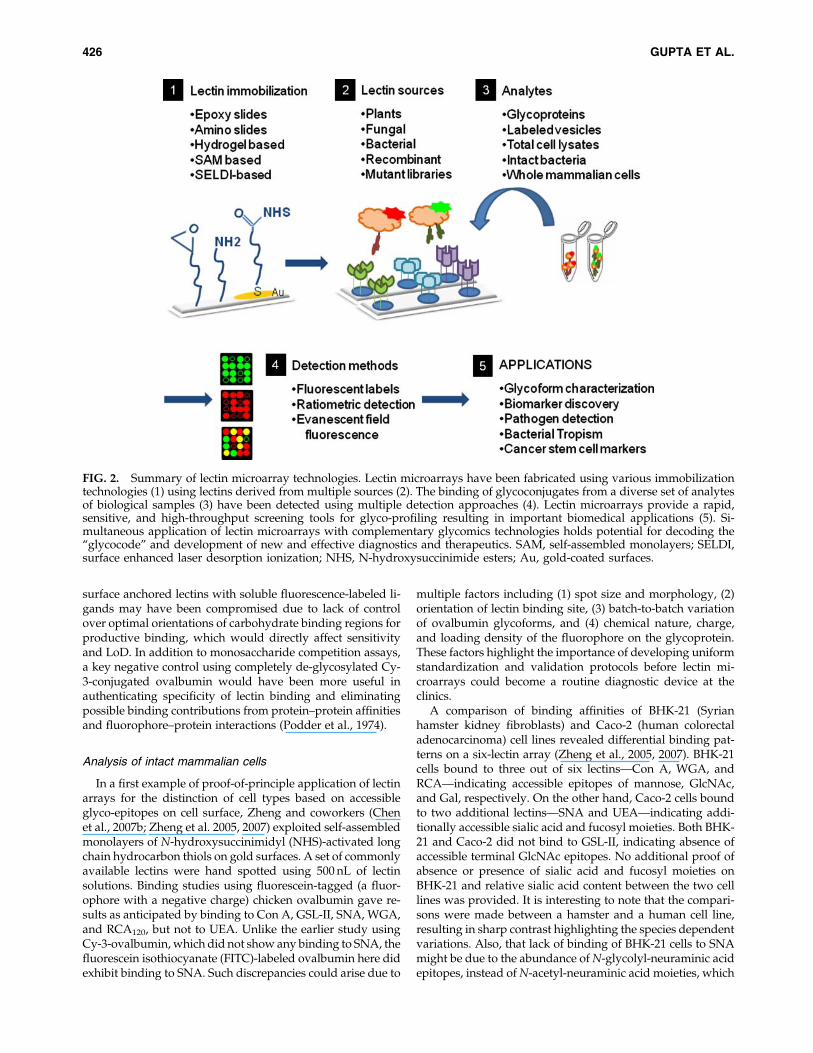
FIG. 2. Summary of lectin microarray technologies. Lectin microarrays have been fabricated using various immobilizationtechnologies (1) using lectins derived from multiple sources (2). The binding of glycoconjugates from a diverse set of analytesof biological samples (3) have been detected using multiple detection approaches (4). Lectin microarrays provide a rapid,sensitive, and high-throughput screening tools for glyco-profiling resulting in important biomedical applications (5). Si-multaneous application of lectin microarrays with complementary glycomics technologies holds potential for decoding the‘‘glycocode’’ and development of new and effective diagnostics and therapeutics. SAM, self-assembled monolayers; SELDI,surface enhanced laser desorption ionization; NHS, N-hydroxysuccinimide esters; Au, gold-coated surfaces.
426 GUPTA ET AL.

may not be bound as tightly by SNA (Knibbs et al., 1991;Taatjes et al., 1988).
Detection of pathogens
In an elegant application for the detection, differentialcharacterization of both pathogenic and nonpathogenic bac-teria (applicable for Gram-negative strains that express dis-tinct carbohydrate derived O-antigens attached to LPS ontheir surface) and monitoring of dynamic changes duringbacterial growth, Hsu and coworkers (2006) have designedmicroarrays with 21-lectins. Importantly, it was noted that useof minimum amount of glycerol and a 1.0-mM solution ofrespective ligand for each lectin significantly improved thespot morphology and accessibility of CRD of lectin (Hsu andMahal, 2006). Differential lectin binding patterns could beobtained for SYTO 85 fluorescent dye-labeled nonpathogenicEscherichia coli K12 strains JM101 and HB101, pathogenicE. coli RS218, and Salmonella typhimurium LT2, with RS218binding to maximum number of lectins. The lectin bindingcould be selectively abrogated by coincubation with com-petitive ligands such as lactose, and the intensity of fluores-cence was shown to decrease as the bacteria changed fromlogarithmic growth phase to stationary phase. Binding to si-alic acid and fucose specific lectins [Maackia amurensis agglu-tinin (MAA), SNA, and UEA-I] was observed for RS218,which is known to aid in evasion of host cell immune re-sponse. It would be interesting to apply this lectin microarrayapproach to other bacteria such as M. tuberculosis and para-sitic pathogens that cause tropical disease such as malaria andleishmaniasis. The studies on dynamic changes to their ac-cessible cell surface glycoforms could provide valuable in-sight in the mechanisms of immune evasion and switchingbetween active and dormant phase, particularly in the contextof latency of tuberculosis (Hirsch et al., 1997).
Analysis of glycosylation status of biopharmaceuticals
Soluble antibodies and proteins in body fluids play vitalroles in regulating homeostasis, development, and pathogendetection through immune surveillance. The changes in gly-coform expressed by antibodies are known to have differen-tial downstream effects. On the other hand, the recent clinicalsuccess of biopharmaceuticals consisting of glycoproteins,such as therapeutic monoclonal antibodies and erythropoietin(EPO), has raised many challenges for definition of homoge-neity, quality control, and batch-to-batch consistency (Sinclairand Elliott, 2005). Lectin microarrays provide a convenient,simple, and rapid means of comparing patterns generated byvarious glycoforms of a given protein produced under dif-ferent expression systems, fermentation, and mammalianbioreactor conditions. For this purpose, Rosenfeld and co-workers (2007) successfully applied a commercially availableglycoarrays kit called QproteomeTM consisting of 24 plant-derived lectins for the analysis of various glycoproteins in-cluding RNase B, prostate-specific antigen (PSA), porcinethyroglobulin, Tamm Horsfall glycoprotein, and recombinanthuman EPO. The analysis involved simplified algorithms thatcategorize binding according to the type of glycan present onthe analyte and a relative grading system. Such ready-to-usekits are valuable to nonspecialists as a rapid quality controlassay, and to specialists for preliminary screening of biologi-cal samples.
Detection in solution—evanescent field fluorescence
To generate unique patterns and to overcome promiscuityof lectin/carbohydrate interactions it was imperative thatrepertoire of lectins be increased on the microarray. Unlikeantigen–antibody interactions that exhibit dissociation constant(Kd) in the nano- to femto molar range, lectin–carbohydrateor lectin–glycan interactions exhibit only moderate bindingaffinities, with Kd in micromolar range. Considering thenumber of washing steps involved in microarray methodol-ogies and weak-to-moderate binding affinities, it is possiblethat majority of lectin interactions could be lost. To overcomethis drawback and to be able to study glycan binding to lectinarray in solution, Kuno and coworkers (2005) elegantly ap-plied the evanescent field fluorescence-based detection. Thismethodology, also known as total internal reflection fluores-cence (TIRF) microscopy, has been used to study interactionsof basal membrane of cells with substrates (extracellular ma-trix and biomaterials) (Fish, 2009). The application of eva-nescent field fluorescence-based detection eliminated theneed for washings of substrate solutions as only a very thinsurface (<100 nm) from the solid surface is scanned, and thus,only true equilibrium events are detected. Also, real-timemonitoring of binding events could be measured enabling thedetermination of optimal time of incubation for various lec-tins. Using a microarray containing 39 lectins, it was shownthat status of glycosylation and glycoforms of proteins such asRNase A (nonglycosylated control), RNase B, asialofetuin,fetuin, and bovine serum albumins could be analyzed. Dis-tinct patterns were obtained for glycoproteins such as asia-lofetuin, mouse laminin, bovine transferrin, and horse radishperoxidise (HRP) (a plant glycoprotein) reflecting variationsin their glycoforms in both N-linked and O-linked glycanstructures. The potential of microarray for fine glycan struc-ture determination was shown by enzymatic cleavage oftetramethylrhodamine (TMR)- labeled Asn-containing oligo-saccharide using a panel of enzymes specific for glycanlinkages. The evanescent field fluorescence detection is verypowerful, convenient, and capable of high throughput(Hirabayashi, 2008). However, for studies of mixture of gly-coproteins from biological samples, multiple real-time imagesshould be acquired and kinetics of equilibrium followed tofind optimal time points necessary to achieve differentialpatterns (Ebe et al., 2006).
Further improvement in sensitivity and limit of detectionhas been achieved by systematic optimization of critical pa-rameters such as lectin spotting, chemistry of immobilization,nature of the solid surface, temperature, humidity, blockingagent, and buffer conditions. Thus, Uchiyama and coworkerswere able to detect even monovalent oligosaccharides thatexhibit low affinity towards lectins (Uchiyama et al., 2008).The binding of asialofetuin and an N-glycan oligosaccharideprobe carrying asialo-biantennary glycans to RCA120 couldbe detected at concentrations as low as 100 pg/mL and100 pM, respectively. Also, with optimized spotting condi-tions the reproducibility was improved. Closely related gly-can structures for which no specific lectins exist, such as LewisA [Gal-b-(1? 3)-(Fuc-a-(1? 4)-GlcNAc] and Lewis X [Gal-b-(1? 4)-(Fuc-a-(1? 3))-GlcNAc] that are positional isomers,could be distinguished clearly based on the overall bindingpattern on the 43-lectin microarray. The lectins UEA-I andBauhinia purpuria alba lectin (BPL) showed opposite affinities
LECTIN MICROARRAYS FOR GLYCOMIC ANALYSIS 427

for the isomers and a ratio of intensities of BPL/UEA-1 wastaken to quantify binding. These results were in agreementwith quantitative sugar-protein interaction affinity valuesmeasured using frontal affinity chromatography (FAC) (Hir-abayashi, 2004; Tateno et al., 2007a).
Expanding the repertoire of lectin arrays
The ability of lectins to selectively recognize and bindcarbohydrate ligands of glycoconjugates and its potentialapplication to glycomic profiling in the form of lectin mi-croarray was soon realized. Many carbohydrate ligands arebound by several lectins with varying affinities. Conversely,several lectins are available that bind to the same carbohy-drate epitope. This results in overlapping affinities and rec-ognition for both lectins and ligands. Therefore, to achieveunique and specific patterns for a given glycoconjugate orcell surface glycome microarrays a larger set of lectins shouldbe more useful. Toward this end, Tateno and coworkers(2007b) developed a microarray containing 43 lectins, andshowed that it can be used to obtain unique and distinctsignature patterns for Chinese hamster ovary (CHO) cellsand its specific mutants Lec2 (deficient in CMP-sialic acidtransporter), Lec8 (deficient in UDP-Gal transporter), andLec1 [deficient in b-(1? 2)-N-acetylglucosaminyltransferaseI (GnT-1)], which are known to exhibit major changes in theircell surface glycoforms. The lectin binding data comparedmostly well with the literature data obtained by flow cy-tometry and other techniques and based on predicted defectsin glycosylation. However, anomalous binding of cells tounexpected lectins were found in some cases, such as Tri-chosanthes japonica lectin (TJA-I) [a-(2? 6)-NeuAc binder] forLec2 (defective in sialylation), SNA [a-(2? 6)-NeuAc bind-er] for Lec8 (defective in both sialylation and galactosyla-tion), and little differential binding to Con A (mannosebinder) for Lec1 (defective in GlcNAc-ylation). These dis-crepancies were explained by promiscuous binding to otherless preferred ligands and to the availability of uniquemultivalent formations available on intact mammalian cellsurface. This highlights an inherent disadvantage of lectinarray, that several lectins bind a given carbohydrate epitopeand that several carbohydrate epitopes bind a given lectinalbeit with varying quantitative affinities, which precludescreation of a mutually exclusive orthogonal set of lectin andligands. The 43-lectin array has been shown to generatedifferential patterns for murine splenocytes from WT andb-(1? 3)-N-acetylglucosaminyl transferase II (b3GnT2) (de-fective in biosynthesis of poly-N-acetyl lactosamine) knock-out mice consistent with the predicted changes inglycosylation. As an evidence to the long-believed hypoth-esis that cell surface glycoforms are characteristically dif-ferent before and after differentiation, K562 (humanerythroleukemia) cells were differentiated using sodiumbutyrate and studied by the 43-lectin array. As expected,major changes were found in lectins that recognize mucin-type oligosaccharides such as core 1, Tn (GalNAc) and sialomucin, which were consistent with reported literature. Ear-lier biochemical studies have shown enhanced O-glycosyl-ation of K562 cells upon differentiation (Gahmberg et al.,1984) as well as enhanced O-GalNAc expression on CD43(leukosialin) upon activation (Maemura and Fukuda, 1992;Piller et al., 1989). Additionally, the 43-lectin array provided
distinct and unique binding patterns for a panel of 10 celllines, including Jurkat, HEK293, and THP-1.
In an attempt to increase the repertoire of lectins on mi-croarray Tao et al. (2008) have reported a 94-lectin microarrayfor generation of signature patterns for 24 mammalian celllines including breast cancer cells, identification of changes inglycoforms between naive and activated B cells and T cellsand thymocytes, demonstration of carbohydrate-dependentdifferential bacterial tropism among related cell types, andidentification of potential biomarkers for cancer stem-likecells. Herein, a simplistic binary scoring approach (as inbound or nonbound) was used to generate the patterns. Basedon the results of cell binding, lectins were classified in to fourcategories namely, nonbinders, low, medium, and highbinders. Of the 94 lectins, 17 lectins did not bind any of 24 celltypes under study, which was attributed to low affinity andlow quality (crude preparations of lectins), and two lectinswere of unknown saccharide specificities. The binding of in-tact cells to remaining 75 lectins displayed distinct patterns.Some cell lines such as hESC, Caco-2, D407, and U937 werefound to bind only to 20 lectins on the array, whereas cell linessuch as Tag, 293, K1106, and MCF7 bound to more than 50lectins. Also, the clustering of similar cell types was unpre-dictable indicating that accessible glycoforms could be widelydifferent even among related cell types. Although the retina-derived cell-lines clustered tightly, the related breast cancercell lines exhibited wide pattern variations. The binding pat-tern found on the microarray was validated by flow cytom-etry using selected lectins. Significant changes were found inglycoforms of lymphocytes before and after activation in bothT and B cells. The array could distinguish the glycoforms in Tcells that were activated by two different agents [phorbolmyristyl acetate (PMA)/ionomycin and mixed lymphocytereaction (MLR)].
Pathogenic bacteria such as E. coli K12 exhibit cellular tro-pism, that is, differential affinity to related cell types, andinvade selected cell populations in a host through its fimbriaeprotein, which is a mannose binding lectin. Lectin array sig-nature patterns of three related breast cancer cell lines indi-cated that SkBr3 has higher cell surface mannose contentcompared to MCF7 and MDA-MB-231, and hence, could beused to predict bacterial tropism and pathogen-host cell in-teractions. Experiments using WT E. coli K12 and FimH mu-tant on the three cell lines confirmed the higher affinity ofE. coli to SkBr3 cell line, relative to MCF7 and MDA-MB-231.
To find unique biomarkers for identification and charac-terization of cancer stem cell-like cells, differential lectin sig-nature pattern was studied using MCF7 cells cultured eitheras a monolayer (normal culturing) or as spehroidal bodies(similar to embryonic stem cell culturing). Depending on thenature of culturing, the lectin patterns were different, partic-ularly for GlcNAc binding lectins such as Lycopersicon escu-lentum lectin (LEL), Aleuria aurantia lectin (AAL), and WGA.Based on this data, the MCF7 cells were depleted to removeLEL-binding breast cancer cells to obtain a side populationenriched in cancer stem-like cells. These cells when injected inmouse were found to result in higher tumor volume com-pared to the nonenriched whole cell populations (Tao et al.,2008). Thus, the changes in glycoforms, gleaned from lectinmicroarray analysis, could result in biomarkers that can eitheridentify side populations or can be used to separate majorpopulations.
428 GUPTA ET AL.

Lectin coupled to mass spectrometry
Recently, using a set of two selected lectins, namely, Jacalinand SNA, Ueda and coworkers (2009) have shown the ap-plication of ProteinChip arrays and SELDI-TOF for discoveryof glycosylation disorders and disease specific alterations inglycan structure in sera of lung cancer patients using a tar-geted serum glycoproteomics approach. The two lectins werecovalently immobilized on the commercially available Pro-teinChip array that carry carbonyldiimidazole moieties viafree amine groups present on lectins. The serum samples, afterdepletion of 14 most abundant proteins, were captured onlectins through their glycoforms and studies by mass spec-trometry. Clear-cut and significant differences were foundbetween serum samples of healthy volunteers and lung cancerpatients, captured by Jacalin and SNA. Through extensiveMS/MS analysis for protein identification, glycosylation sitemapping, and glycan structure analysis, apolipoprotein C-III(apoC-III) was found to be differentially glycosylated at Thr74between the two groups. A major feature is the higherabundance of linear sialyl core 1 structures [NeuAc-a-(2? 3)-Gal-b-(1? 3)-GalNAc; sialyl-T-antigen] in lung cancer pa-tients with a characteristic absence of the branching a-(2? 6)sialic acid on the core GalNAc [NeuAc-a-(2? 3)-Gal-b-(1? 3)-(NeuAc-a-(2? 6))-GalNAc; di-sialyl-T-antigen], whichwas found in higher abundance in healthy volunteers. Theabsence of di-sialyl moieties is attributed to epigenetic si-lencing of a-(2? 6)-sialyltransferase gene as corroborated byother studies. Clearly, inclusion of additional lectins, optimi-zation of immobilization on ProteinChip arrays, and holdingcapacities of lectins on SELDI-MS should be performed toverify these findings in a larger set of clinical sample (Chenet al., 2008).
Ratiometric detection
The first-generation lectin arrays used purified or enrichedglycoproteins samples with single-color fluorescent dye,which led to lack of consistency and reproducibility due tovariations in labeling conditions. Also, the glycolipids werenot studied as they were lost in the sample preparation pro-tocol and are difficult to label directly due lack of dye-reactivefunctional groups (e.g., free amine groups similar to lysineside chain of proteins). To develop semiquantitative and re-liable analysis using internal biological reference standardsthe dual-color ratiometric approach, extensively used in genemicroarrays, was successfully adapted to lectin microarraysalong with compatibility to standard image, statistical clus-tering, and ratiometric heat map analysis (Pilobello et al.,2007). Here, isolated micellae of mammalian cell membraneswere used, thus taking glycolipids into account, from twodifferent cell types with each cell type being differentially la-beled with yellow (Cy3) dye and red (Cy5) dye (Fig. 2). As aninternal control, the dyes were swapped between the samplesto nullify variations due to derivatization or dye-relatedbackground. Two well-characterized CHO cell lines (Pro-5,and a mutant Lec8 that is defective in UDP–Gal transport andresistant to agglutination by WGA) were used. The lectinbinding pattern obtained from Pro-5 and Lec8, using inde-pendent single-color lectin array, was almost similar, whichdid not reflect the true variations in the glycome structure.Whereas a mixture of dual-color labeled (and dye-swapped)membrane micellae of both Pro-5 and Lec8 analyzed on the
lectin array clearly highlighted the defects in glycan struc-tures. Stark differences were seen in the binding to MAA(preferably bound by Pro-5 which contains a-(2? 3) sialo-sides), Galanthus nivalis agglutinin (GNA) (preferably boundby Lec8, rich in N-linked mannose structures), Lens culinarislectin (LcH) (preferably bound by Lec8, rich in fucosylatedmannose core structures), and HPA and Artocarpus intergrifolialectin (AIA, Jacalin) (preferably bound by Lec8 with exposedand truncated core O-linked mucin-type GalNAc residues).The ratiometric technique was employed to differentiate theglycome profile of naive HL-60 (human promyelocytic leuke-mia) and its dimethyl sulfoxide induced differentiation intoneutrophils. HL-60 cells upon differentiation showed increasedlevels of N-linked high Man structures, increased b-(1? 6)-GalNAc residues, and reduced levels of a-(2? 6)-sialic acidepitopes. The binding data with SNA was ambiguous com-pared to Polyporus squamosus lectin (PSL) and Limulus polphe-mus agglutinin LPA, thus highlighting the need to have largernumber of lectins to compensate for overlapping ligand se-lectivities and to obtain unique patterns.
The versatility and sensitivity of the dual color ratiometricapproach was well illustrated by its application to the study ofvirions (virus particles) derived from HIV (human immuno-deficiency virus), SIV (simian immunodeficiency virus), andhost cell-derived microvescicles (Krishnamoorthy et al., 2009).Microvesicles are carved out of enriched raft-like micro-domains by exocytosis and are critical part of normal devel-opment and homeostasis. Comparative analysis of glycomeprofiling using a 68-lectin array (which included galectin-1 inaddition to plant-derived lectins) revealed that both HIV viri-ons and host-derived microvesicles have strikingly similarglycosylation patterns and hence the ability to evade immunedetection (Hirabayashi, 2009). Comparison of HIV and SIVindicated subtle differences in the levels of glycoforms of gp120,which was a major differential compared to host microvesicles.Novel anti-HIV therapy that seeks to exploit the binding oflectins, such as cyanovirin, to high-mannose structures of gp120may have to tread cautiously as they may obstruct the nativemicrovesicular pathway as well (Li et al., 2008).
Importance of multivalent interactions
Any technological advance in glycomics using lectins couldnot ignore two salient features of carbohydrate ligands,namely, multivalency and ligand-density effects (Brewer et al.,2002; Lee and Lee, 2000). A recent study highlights the im-portance of ligand density (available in natural form on thebiological samples, lysates, membrane preparations, or wholecells) on the ability of lectins or antibodies for recognition andbinding (Oyelaran et al., 2009). The antigen array used neo-glycoconjugate containing varying amount of ligands [car-bohydrate epitopes attached to bovine serum albumin (BSA)]immobilized on a solid support and probed with three lectins[Vicia villosa lectin B4 (VVL-B4), Helix pomatia agglutinin(HPA), Soy bean agglutinin (SBL)], three monoclonal anti-bodies and serum antibodies from patients. Study revealedthe necessity to include a given antigen or given carbohydrateligand in varying density both in terms of individual mole-cules and physical arraying in order to profile subpopulationsof antibodies with variable affinities. For example, the binding(apparent Kd) of VVL-B4 to a-GalNAc derivatized BSAchanged dramatically, by an order of magnitude, from
LECTIN MICROARRAYS FOR GLYCOMIC ANALYSIS 429

>1,430 nM for 9 ligands per BSA to 129 nM for 13 ligands perBSA. These values and relative affinities are consistent withoriginal studies establishing Tn-antigen (a-GalNAc-Ser) as aspecific recognition epitope for VVL-B4 (Puri et al., 1992).
Recombinant lectins
Owing to their easy isolation and commercial availability,plant-derived lectins were a straightforward choice for thedevelopment of lectin microarray for glycomic analysis.However, plant-derived lectins do suffer from multiple dis-advantages (Hsu et al., 2008). Excepting a few lectins such asCon A, WGA, and PNA, the majority of plant-derived lectinsare inherently glycosylated. They are obtained in multipleisoforms, called iso-lectins, in the isolation and purificationprocess due to genetic heterogeneity, variability in post-translational processing by proteases, and subunit structure(De Hoff et al., 2009). The inherent glycoprotein nature oflectins could potentially lead to false positive binding, par-ticularly with cell lysates and biological samples (e.g., bindingof mannose binding lectins such as DC-SIGN present inmammalian cell lysates to high-mannose glycans present onthe immobilized plant-derived lectins). There are multipleways in which this problem could be addressed such as (1)completely de-glycosylate plant-derived lectins and ensureintegrity of ligand selectivity and specificity, (2) to regenerateall existing plant lectins using bacterial recombinant technol-ogy, and (3) use a panel of bacterial lectins, most of whichlack glycosylation. In all the above cases carbohydrate ligandselectivity, specificity, and affinity must be obtained on a case-by-case basis and specific mutants using recombinant tech-nology that abrogate ligand binding could serve as valuablenegative controls. The feasibility and utility of recombinantbacterial lectins and their mutants for lectin microarray wasshown by Hsu and coworkers (2008) using a small set of sevenlectins. The lectins consisted of GafD (specific for b-GlcNAc),GafD-m (a Asp88Leu mutant which does not bind b-GlcNAc),PapGII (specific for globoside GbO4), and PapGIII (specificfor globoside GbO5) from E. coli and PA-IL (specific for Ga-lactose), and PA-IIL (binds both fucose and mannose withhigher affinity for fucose) from Pseudomonas aeruginosa, andRS-IIL (binds mannose and fucose with higher affinity formannose) from Ralstonia solanocearum. All the recombinantlectins were expressed with GST-His6 fusion tags at the N-terminus using pET-41 LIC/Ek vector and found not tocompromise carbohydrate–lectin interaction. The use of fu-sion-tags in recombinant lectins could aid greatly in con-trolled orientation of lectins during immobilization process,which might improve sensitivity. It was found that PA-IIL(fucose binder) bound strongly to gangliosides obtained fromporcine brain extract indicating the presence of fucosylatedgangliosides. Finally, the recombinant lectin array was usedto obtain differential patterns for micellae prepared from renalcarcinoma (ACHN and TK10) and melanoma (M14 and SK-MEL-5) cell lines. Although all four cell lines bound to PA-IIL(fucose/mannose binder), the binding to PA-IL (galactosebinder) and GafD (GlcNAc binder) was selective for renal celllines only.
Library of lectin mutants
At about the same time, Maenuma and coworkers (2008)reported the use of a library of 35 Maackia amurensis hemag-
glutinin (MAH) mutants with various point mutations at twoamino acids, 131Gly and 133Ser, which are present in the keyCRD of the lectin. The wild-type MAH is known to bindsialyl-T and disialyl-T antigens found on O-glycans and theMAH mutants were found to bind carbohydrates with widerspecificities when probed with polyacrylamide conjugatescontaining various glyco-epitopes. All the recombinant mu-tants were expressed with FLAG fusion tag, which was usedto capture on polystyrene surfaces coated with anti-FLAGantibody. This avoided the purification of bacterial lysatesand provided optimal orientation of lectins for interactionwith glycans on glycoproteins and mammalian cells. Thebound cells were labeled with crystal violet, lysed, andquantified by colorimetry. Study of binding of various cancercell lines using cluster analysis revealed that groups of cells ofsimilar origin such as colon carcinoma, melanoma, and my-eloid cell lines, tend to cluster together. The binding of in-tact cells exhibited wider differential patterns compared topolyacrylamide–glycoconjugates, which was attributed to ad-ditional cell surface glycan and mutant lectin protein backboneinteractions. Although not in a microarray format, this studyshowed the ability to generate significant number of lectinmutants and their potential for rapid glyco-profiling of cells.
Challenges remain in terms of the quantities of biologicalsamples needed for flooding the lectin microarray for gly-comic analysis. To improve the sensitivity and to utilize verylittle biological samples without pooling of multiple patientsamples, Nagaraj and coworkers (2008) have shown the utilityof piezoelectric printing for lectin immobilization, using sixcommon lectins, on nitrocellulose slides and piezoelectricliquid dispensing for analyte samples. This way glyco-profiling of pure glycoproteins such as asialofetuin (ASF) and6-sialofetuin (6-SF) could be accomplished using as little as1 mL of 1 nm or 15 fmol of ASF at 1 mg/mL, whereas a con-ventional lectin array would require about 450 mL for the sameanalysis. In another approach for label-free detection usingelectrochemical impedance spectroscopy (EIS), La Belle andcoworkers (2007) have shown that carbohydrate epitopefunctionalized gold-nanoparticles and glycoproteins could bedetected on Cu/Ni/Au printed circuit boards (PCBs) usingtwo lectins, PNA, and SNA. The sensitivity for asialofetuinwas found to be 10 pg/mL (150 fM), which was comparable toconventional fluorescence-based detection methods. Furtherdevelopment of such tools will involve expansion and mul-tiplexing to include a large number of lectins, miniaturization,application of nano-particle-based application (Dai et al.,2006), integration of antibody assisted detection (Kuno et al.,2009), and optimization of measurement conditions (Gemei-ner et al., 2008). An exhaustive list of lectins (including thosethat are currently not employed in microarrays) along withtheir source, ligand specificity, status of lectin glycosylationand oligomerization, and metal ion requirements for optimalactivity are provided in Supplementary Table 1 along withreferences (supplementary information).
Finally, a comment on lectin spot size for glycomic analysisof intact mammalian cells deserves mention. Depending onthe size and surface area of lectin spot and density of lectinavailable minor portions or side populations of mammaliancells could be selected, leading to batch to batch inconsistency.In other words, using simple calculations performed by us,when an asynchronous population of 5�105 cells are used inthe analyte, with a lectin array spot of 120 micron diameter,
430 GUPTA ET AL.

less than 10% of cells are captured leading to loss of 90% ofcells in the washing. The cells are perhaps lost due to lack ofavailability of lectins rather than lack of specific affinity for agiven lectin. Assuming a perfectly circular morphology, alectin spot of 120-mm diameter will provide a surface area of11,304 mm2. Taking Jurkat cells with a diameter of 10mm as amodel gives 78.5 mm2 for surface area of each cell. Taking aratio would result in a maximum of 144 Jurkat cells per lectinspot with a diameter of 120 mm. For a total of 282 spots(triplicate spots for 94 lectins) this would result in the captureof 40,608 cells, which is less than 10% of total number of cellsin the analyte. Mammalian cell populations, particularlycancer cell lines are known to exhibit side populations thathave distinct glycoform patterns and metabolic rates (Wuand Alman, 2008; Yarema et al., 2001). Therefore, multipletechnical replicates and standardization of lectin signa-ture pattern as a function of lectin spot diameter wouldbe required for analysis of whole mammalian cells. Ad-ditionally, the washed cells (* 90%) could be subject to re-peated analysis on the lectin array to confirm if identicalpatterns are obtained.
Overview and Outlook
Advances in research over years have made it clear thatwith limited number of genes in mammalian genomes, tran-scriptome, and translated proteome, posttranslational modi-fications in proteins are important players in regulating theirfunction. Glycosylation, the most abundant and complexform of posttranslational modification, is a key regulator ofthe properties of proteins and lipids that are present inabundance on the cell surface. Glycans at the extracellularinterface mediate several interactions between or within thecells, which in turn, control several important biologicalevents such as cell adhesion, cell signaling, cellular trafficking,and protein folding, stability, and transport. Moreover, gly-cosylation is important for regulation of many crucial cellularprocesses such as development, elicitation and homeostasisof activating and inhibitory immune responses. It is knownthat several diseases such as cancer exhibit improper glyco-sylation with striking differences from healthy tissues. Hun-dreds of glycogenes encode enzymes such as glycosyltransferases and sulfotransferases, which are essential forcarrying out glycan biosynthesis. An intricate network ofinteractions among these enzymes and sugar–nucleotidetransporters provides the cells with a rich and complex di-versity of glycans. Minute changes in the entire system—be itglycogene alteration, enzyme deficit, or disease-related alter-ation in expression, or deficiency in the availability of rawmaterials (activated glycosyl donor and their metabolic pre-cursors)—leads to modified glycan structures being presentedwithin the cell and on the cell surface (Schachter and Freeze,2009). A common example of such alterations can be observedin tumor cells that express an abnormally high degree of N-glycan branching, extension with polylactosamine chains,fucosylation, and sialylation, and altered degree of sulfation.Even the O-glycans found associated with tumors are alteredwith higher percentage of T antigens, sialyl-T antigens, andTn- and sialyl Tn-antigens with truncated glycoforms. Slightand subtle changes in glycan structures can initiate a cascadeof undesirable cellular events. Given equal levels of expres-sion a protein from a gene, the probability of aberrations are
much higher in the post translational glycosylation modifi-cations, which profoundly affect the concerned biologicalevents. Because aberrant glycosylation are frequently ob-served in disease conditions, they are well suited to be utilizedas biomarkers for those diseases (Hanash et al., 2008). Severaltechniques including lectin-microarrays are now availablethat can be used to identify these altered structures andtherefore put to diagnostic use.
Genome-wide studies were revolutionized with the adventof whole genome DNA microarray technology about a decadeago. Several transcription-level analyses have been success-fully carried out for over 20,000 human genes (Young, 2000).Gene expression profiling has since become a widespreadmethod for investigating biological and clinically relevantevents. However, a major problem initially was the validationof preliminary results obtained during gene expression pro-filing to prevent errors due to low quantities used in DNAmicroarray. Most statistical methods and programs used werelengthy and slow, and therefore diminished the total accom-plishments of the technique. With the introduction of real-time quantitative PCR (qPCR), which is highly sensitive andprecise, the validation of DNA microarray data has becomeeasy. It is now one of the most commonly used technique formicroarray data analysis. With lectin microarray as the newupcoming glycotechnology with ultrasensitive glycan profil-ing ability, it will be advantageous to apply qPCR arrayanalysis for expression profiling of glycogenes. The number ofglycan biosynthetic enzymes identified has risen significantlyand constantly since the identification of the first glycosyltransferase as early as in the 1980s. The human genome en-compasses a number of glycogenes, which encode the en-zymes involved in glycan biosynthesis (Narimatsu, 2006).Approximately 200 genes, about 1% of whole genome, areidentified to date that encode for glycosyltransferases. Use oflectin microarray in combination with qPCR data abolishesthe need of cumbersome protocols used for individual glycananalyses of glycoproteins and glycolipids from various sour-ces and replaces it with a high-throughput method based onmultiple lectin–sugar interactions (Narimatsu et al., 2009). Anumber of different protocols have been developed for de-tecting alterations to the glycosylation profile of cell surfaces.Recent advances using an ultrasensitive lectin microarrayfacilitates differential glycan analysis by targeting a restrictedregion of one-dot sections on a formalin-fixed tissue array(Matsuda et al., 2008).
Another method that has developed to be a powerful toolfor structural analysis of glycans is mass spectrometry (MS). Ithas proved to be a fast and high-throughput method forglycan structure analysis. However, it does have the disad-vantage of requiring extensive sample preparations prior toanalysis if a detailed characterization of glycan structure isneeded (Dell and Morris, 2001). Owing to immense hetero-geneity present in glycans isolated from any natural source,there is always a trade-off between sensitivity of character-ization and high-throughput results. Nuclear magnetic reso-nance (NMR) spectroscopy is a powerful technique fordetailed linkage analysis of glycans, but is limited by therequirement of substantial quantities of homogeneous sam-ples. On the other hand, high-performance liquid chroma-tography (HPLC) of fluorescently labeled glycans is anotheradvanced technique to obtain glycan profiles on the basis oftheir abundance in isolated mixtures (Domann et al., 2007).
LECTIN MICROARRAYS FOR GLYCOMIC ANALYSIS 431
Development of integrated user-friendly online databases forglycan structures analysis, lectin binding parameters, lectinfolds, and mass spectrometry reference data for fingerprintingare critically essential (Damodaran et al., 2008; Haslam et al.,2008; von der Lieth et al., 2006).
Each of the techniques, microarrays (lectins, synthetic andnatural carbohydrate ligands, neoglycoconjugates), qPCR,MS analyses, glyco-informatics, NMR and HPLC provideunique ability to identify novel alterations in glycosylationpatterns. Although each method allows for a specific advan-tage and improvement in detection of altered glycan profile, itis not easy to obtain detailed information of glycosylationpattern by using any one of them in isolation. By using acombination of several analytical methods, however, it shouldbe possible to determine the glycan structures and betterunderstand glyco-alterations that occur on cell surfaces and/or on proteins. In summary, lectin microarrays with theirsimplicity, capacity for rapid high-throughput analysis at the‘‘point-of-care,’’ availability of lectins, and affordable low-costequipment requirements are well positioned for wide spreadusage in biomedical research.
Expansion of existing lectin microarrays to include mam-malian, bacterial, and fungal lectins would add to its versa-tility and clinical value. The existence of isolectins, multipleglycoforms, heterogeneous preparations, and batch-to-batchvariations in plant-derived lectins are major challenges inobtaining consistent reproducible results. The problem ofglycosylation of lectins could be overcome by recombinantproduction of plant lectins in bacterial systems with con-comitant verification of carbohydrate binding specificitiesand affinities. Kulkarni and coworkers (2004; Shaaanan et al.,1991) have expressed recombinant EcorL and showed thatabsence of glycosylation on EcorL did not affect its carbohy-drate specificity and exhibited similar quaternary structure.However, glycosylation does seem to affect the folding pro-cess (Mitra et al., 2003). Recombinant lectins do seem to be farless practical compared to the ease and abundance of naturalplant lectins. It will be a demanding task to generate sufficientrepertoire of recombinant lectins with defined ligand specifi-cities to develop a microarray and for ready-to-use clinicalapplications. An ideal lectin microarray would consist of a setof lectins with mutually exclusive or orthogonal ligand spe-cificities, but the variety of glycoforms and their unpredictablethree-dimensional multivalent presentation on mammaliancell surface precludes the construction of such arrays (Gabius,2008; Gilleron et al., 1998).
Alternatively, a practical approach would be to undertakelarge scale explorations of biodiversity with a focus on dis-covery of new lectins and their biochemical characterizationand collect as many lectins as possible from lectin space orplant kingdom. With rapid advances and growing impor-tance of food security and plant biotechnology (Goldstein,2002; Pusztai et al., 2008; Vasil, 2008), it should be possible toidentify novel lectins with specificities for complex glyco-forms and their modifications such as phosphorylation, sul-fation, acetylation, methylation, and attachment of lactic acid.Additionally, novel modes of attachment to solid supportusing the glycan residues of the lectin are anticipated to pro-vide optimal orientation while hiding away the interferingglycan components.
In summary, the collective information obtained usingdiverse microarray technologies that employ immobilized
synthetic and natural carbohydrate ligands, neo-glycoconju-gates, antibodies, and lectins (natural and recombinant)would be valuable in developing glycomics-based diagnosticsand therapeutic targets (Yue and Haab, 2009). The applicationof metabolic glycan engineering (MGE) using variants ofnatural monosaccharide analogs as precursors and theirsubsequent detection via orthogonal chemical reactions invivo are opening up possibilities for glycan imaging (Laughlinand Bertozzi, 2009; Sletten and Bertozzi, 2009). Differentialglycan profiling of glycan engineered cells (expressing azide,ketone, and thiol functionalities) using lectin microarraysmight open up new possibilities for cell and tissue engineer-ing including stem cell differentiation (Du and Yarema;Sampathkumar et al., 2006).
In view of the various biological processes and diseases inwhich glycans play a crucial role the potential for develop-ment of glycotherapeutics is immense. Understanding the roleof glycan structure and function in fundamental biology andapplications of high-throughput tools such as lectin micro-array, reviewed here, will enable us to develop new-age di-agnostics and therapeutics with higher efficacy.
Acknowledgments
Gupta thanks the Council of Scientific and Industrial Re-search (CSIR), India for the award of Senior Research Fel-lowship. Surolia is a J.C. Bose fellow of the Department ofScience and Technology (DST) and thanks support for basicresearch by grants from Department of Biotechnology (DBT)and DST, Government of India. Sampathkumar is a Rama-lingaswami fellow of the DBT and is grateful for generousstart-up funding from NII and research grants from DST andDBT, Government of India.
Author Disclosure Statement
The authors declare that no conflicting financial interestsexist.
References
Angeloni, S., Ridet, J.L., Kusy, N., Gao, H., Crevoisier, F.,Guinchard, S., et al. (2005). Glycoprofiling with micro-arraysof glycoconjugates and lectins. Glycobiology 15, 31–41.
Baenziger, J.U., and Fiete, D. (1979a). Structural determinants ofconcanavalin A specificity for oligosaccharides. J Biol Chem254, 2400–2407.
Baenziger, J.U., and Fiete, D. (1979b). Structural determinants ofRicinus communis agglutinin and toxin specificity for oligo-saccharides. J Biol Chem 254, 9795–9799.
Baenziger, J.U., and Fiete, D. (1982). Photoactivatable glyco-peptide reagents for site-specific labeling of lectins. J BiolChem 257, 4421–4425.
Baum, L.G., and Crocker, P.R. (2009). Glycoimmunology: ignoreat your peril! Immunol Rev 230, 5–8.
Bertozzi, C.R., and Kiessling, L.L. (2001). Chemical glycobiology.Science 291, 2357–2364.
Bielik, A.M., and Zaia, J. (2010). Historical overview of glycoa-nalysis. Methods Mol Biol 600, 9–30.
Bishop, J.R., Schuksz, M., and Esko, J.D. (2007). Heparan sul-phate proteoglycans fine-tune mammalian physiology. Nature446, 1030–1037.
Blackstock, W.P., and Weir, M.P. (1999). Proteomics: quantita-tive and physical mapping of cellular proteins. Trends Bio-technol 17, 121–127.
432 GUPTA ET AL.

Brewer, C.F., Miceli, M.C., and Baum, L.G. (2002). Clusters,bundles, arrays and lattices: novel mechanisms for lectin–saccharide-mediated cellular interactions. Curr Opin StructBiol 12, 616–623.
Buskas, T., Ingale, S., and Boons, G.J. (2006). Glycopeptides asversatile tools for glycobiology. Glycobiology 16, 113R–136R.
Caramelo, J.J., and Parodi, A.J. (2007). How sugars convey in-formation on protein conformation in the endoplasmic retic-ulum. Semin Cell Dev Biol 18, 732–742.
Chen, P., Liu, Y., Kang, X., Sun, L., Yang, P., and Tang, Z. (2008).Identification of N-glycan of alpha-fetoprotein by lectin af-finity microarray. J Cancer Res Clin Oncol 134, 851–860.
Chen, S., LaRoche, T., Hamelinck, D., Bergsma, D., Brenner, D.,Simeone, D., et al. (2007a). Multiplexed analysis of glycanvariation on native proteins captured by antibody micro-arrays. Nat Methods 4, 437–444.
Chen, S., Zheng, T., Shortreed, M.R., Alexander, C., and Smith,L.M. (2007b). Analysis of cell surface carbohydrate expressionpatterns in normal and tumorigenic human breast cell linesusing lectin arrays. Anal Chem 79, 5698–5702.
Cooper, C.A., Gasteiger, E., and Packer, N.H. (2001). GlycoMod—a software tool for determining glycosylation compositionsfrom mass spectrometric data. Proteomics 1, 340–349.
Cummings, R.D. (2009). The repertoire of glycan determinants inthe human glycome. Mol Biosyst 5, 1087–1104.
Cummings, R.D., and Kornfeld, S. (1982). Fractionation of as-paragine-linked oligosaccharides by serial lectin–Agaroseaffinity chromatography. A rapid, sensitive, and specifictechnique. J Biol Chem 257, 11235–11240.
Dai, Z., Kawde, A.N., Xiang, Y., La Belle, J.T., Gerlach, J.,Bhavanandan, V.P., et al. (2006). Nanoparticle-based sensingof glycan-lectin interactions. J Am Chem Soc 128, 10018–10019.
Dam, T.K., and Brewer, C.F. (2002). Thermodynamic studies oflectin–carbohydrate interactions by isothermal titration calo-rimetry. Chem Rev 102, 387–429.
Dam, T.K., and Brewer, C.F. (2010). Lectins as pattern recogni-tion molecules: the effects of epitope density in innate im-munity. Glycobiology 20, 270–279.
Damodaran, D., Jeyakani, J., Chauhan, A., Kumar, N., Chandra,N.R., and Surolia, A. (2008). CancerLectinDB: a database oflectins relevant to cancer. Glycoconj J 25, 191–198.
De Hoff, P.L., Brill, L.M., and Hirsch, A.M. (2009). Plant lectins:the ties that bind in root symbiosis and plant defense. MolGenet Genomics 282, 1–15.
Debray, H., Decout, D., Strecker, G., Spik, G., and Montreuil, J.(1981). Specificity of twelve lectins towards oligosaccharidesand glycopeptides related to N-glycosylproteins. Eur J Bio-chem 117, 41–55.
Dell, A., and Morris, H.R. (2001). Glycoprotein structure deter-mination by mass spectrometry. Science 291, 2351–2356.
Ding, W., Nothaft, H., Szymanski, C.M., and Kelly, J. (2009).Identification and quantification of glycoproteins using ion-pairing normal-phase liquid chromatography and mass spec-trometry. Mol Cell Proteomics 8, 2170–2185.
Domann, P.J., Pardos-Pardos, A.C., Fernandes, D.L., Spencer,D.I., Radcliffe, C.M., Royle, L., et al. (2007). Separation-basedglycoprofiling approaches using fluorescent labels. Proteomics7(Suppl 1), 70–76.
Du, J., and Yarema, K.J. (2010). Carbohydrate engineered cellsfor regenerative medicine. Adv Drug Deliv Rev Feb 1 [Epubahead of print].
Ebe, Y., Kuno, A., Uchiyama, N., Koseki-Kuno, S., Yamada, M.,Sato, T., et al. (2006). Application of lectin microarray to crude
samples: differential glycan profiling of lec mutants. J Biochem139, 323–327.
Feizi, T., and Chai, W. (2004). Oligosaccharide microarrays todecipher the glyco code. Nat Rev Mol Cell Biol 5, 582–588.
Fish, K.N. (2009). Total internal reflection fluorescence (TIRF)microscopy. Curr Protoc Cytom Chapter 12, Unit12 18.
Gabius, H.J. (2000). Biological information transfer beyond thegenetic code: the sugar code. Naturwissenschaften 87, 108–121.
Gabius, H.J. (2008). Glycans: bioactive signals decoded by lec-tins. Biochem Soc Trans 36, 1491–1496.
Gabius, H.J., Andre, S., Kaltner, H., and Siebert, H.C. (2002). Thesugar code: functional lectinomics. Biochim Biophys Acta1572, 165–177.
Gabius, H.J., Siebert, H.C., Andre, S., Jimenez-Barbero, J., andRudiger, H. (2004). Chemical biology of the sugar code.Chembiochem 5, 740–764.
Gahmberg, C.G., Ekblom, M., and Andersson, L.C. (1984). Dif-ferentiation of human erythroid cells is associated with in-creased O-glycosylation of the major sialoglycoprotein,glycophorin A. Proc Natl Acad Sci USA 81, 6752–6756.
Gemeiner, P., Mislovicova, D., Tkac, J., Svitel, J., Patoprsty, V.,Hrabarova, E., et al. (2008). Lectinomics II. A highway tobiomedical/clinical diagnostics. Biotechnol Adv.
Gesner, B.M., and Ginsburg, V. (1964). Effect of glycosidases onthe fate of transfused lymphocytes. Proc Natl Acad Sci USA52, 750–755.
Ghesquiere, B., Van Damme, J., Martens, L., Vandekerckhove, J.,and Gevaert, K. (2006). Proteome-wide characterization of N-glycosylation events by diagonal chromatography. J ProteomeRes 5, 2438–2447.
Gilleron, M., Siebert, H.C., Kaltner, H., von der Lieth, C.W.,Kozar, T., Halkes, K.M., et al. (1998). Conformer selection anddifferential restriction of ligand mobility by a plant lectin—conformational behaviour of Galbeta1-3GlcNAcbeta1-R, Gal-beta1-3GalNAcbeta1-R and Galbeta1-2Galbeta1-Rprime; inthe free state and complexed with galactoside-specific mis-tletoe lectin as revealed by random-walk and conformational-clustering molecular-mechanics. Eur J Biochem 252, 416–427.
Goldberg, D., Sutton-Smith, M., Paulson, J., and Dell, A. (2005).Automatic annotation of matrix-assisted laser desorption/ionization N-glycan spectra. Proteomics 5, 865–875.
Goldstein, I.J. (2002). Lectin structure–activity: the story is neverover. J Agric Food Chem 50, 6583–6585.
Goldstein, I.J. (2009). My favorite enzyme: for love of lectins.IUBMB Life 62, 247–250.
Goldstein, I.J., Hughes, R.C., Monsigny, M., Osawa, T., andSharon, N. (1980). What should be called a lectin? Nature 285,66.
Hanash, S.M., Pitteri, S.J., and Faca, V.M. (2008). Mining theplasma proteome for cancer biomarkers. Nature 452, 571–579.
Haslam, S.M., Julien, S., Burchell, J.M., Monk, C.R., Ceroni, A.,Garden, O.A., et al. (2008). Characterizing the glycome of themammalian immune system. Immunol Cell Biol 86, 564–573.
Hirabayashi, J. (2003). Oligosaccharide microarrays for gly-comics. Trends Biotechnol 21, 141–143.
Hirabayashi, J. (2004). Lectin-based structural glycomics: glyco-proteomics and glycan profiling. Glycoconj J 21, 35–40.
Hirabayashi, J. (2008). Concept, strategy and realization of lec-tin-based glycan profiling. J Biochem 144, 139–147.
Hirabayashi, J. (2009). Glycome ‘‘fingerprints’’ provide definitiveclues to HIV origins. Nat Chem Biol 5, 198–199.
Hirabayashi, J., Arata, Y., and Kasai, K. (2001). Glycome project:concept, strategy and preliminary application to Caenorhabditiselegans. Proteomics 1, 295–303.
LECTIN MICROARRAYS FOR GLYCOMIC ANALYSIS 433

Hirsch, C.S., Ellner, J.J., Blinkhorn, R., and Toossi, Z. (1997). Invitro restoration of T cell responses in tuberculosis and aug-mentation of monocyte effector function against Mycobacter-ium tuberculosis by natural inhibitors of transforming growthfactor beta. Proc Natl Acad Sci USA 94, 3926–3931.
Horlacher, T., and Seeberger, P.H. (2006). The utility of carbo-hydrate microarrays in glycomics. Omics 10, 490–498.
Horlacher, T., and Seeberger, P.H. (2008). Carbohydrate arraysas tools for research and diagnostics. Chem Soc Rev 37, 1414–1422.
Hsu, K.L., Gildersleeve, J.C., and Mahal, L.K. (2008). A simplestrategy for the creation of a recombinant lectin microarray.Mol Biosyst 4, 654–662.
Hsu, K.L., and Mahal, L.K. (2006). A lectin microarray approachfor the rapid analysis of bacterial glycans. Nat Protoc 1, 543–549.
Hsu, K.L., and Mahal, L.K. (2009). Sweet tasting chips: micro-array-based analysis of glycans. Curr Opin Chem Biol 13, 427–432.
Hsu, K.L., Pilobello, K.T., and Mahal, L.K. (2006). Analyzing thedynamic bacterial glycome with a lectin microarray approach.Nat Chem Biol 2, 153–157.
Ito, H., Kuno, A., Sawaki, H., Sogabe, M., Ozaki, H., Tanaka, Y.,et al. (2009). Strategy for glycoproteomics: identification ofglyco-alteration using multiple glycan profiling tools. J Pro-teome Res 8, 1358–1367.
Knibbs, R.N., Goldstein, I.J., Ratcliffe, R.M., and Shibuya, N.(1991). Characterization of the carbohydrate binding specific-ity of the leukoagglutinating lectin from Maackia amurensis.Comparison with other sialic acid-specific lectins. J Biol Chem266, 83–88.
Krambeck, F.J., Bennun, S.V., Narang, S., Choi, S., Yarema, K.J.,and Betenbaugh, M.J. (2009). A mathematical model to deriveN-glycan structures and cellular enzyme activities from massspectrometric data. Glycobiology 19, 1163–1175.
Krishnamoorthy, L., and Mahal, L.K. (2009). Glycomic analysis:an array of technologies. ACS Chem Biol 4, 715–732.
Krishnamoorthy, L., Bess, J.W., Jr., Preston, A.B., Nagashima, K.,and Mahal, L.K. (2009). HIV-1 and microvesicles from T cellsshare a common glycome, arguing for a common origin. NatChem Biol 5, 244–250.
Kulkarni, K.A., Srivastava, A., Mitra, N., Sharon, N., Surolia, A.,Vijayan, M., et al. (2004). Effect of glycosylation on the struc-ture of Erythrina corallodendron lectin. Proteins 56, 821–827.
Kuno, A., Uchiyama, N., Koseki-Kuno, S., Ebe, Y., Takashima, S.,Yamada, M., et al. (2005). Evanescent-field fluorescence-assisted lectin microarray: a new strategy for glycan profiling.Nat Methods 2, 851–856.
Kuno, A., Kato, Y., Matsuda, A., Kaneko, M.K., Ito, H., Amano,K., et al. (2009). Focused differential glycan analysis with theplatform antibody-assisted lectin profiling for glycan-relatedbiomarker verification. Mol Cell Proteomics 8, 99–108.
La Belle, J.T., Gerlach, J.Q., Svarovsky, S., and Joshi, L. (2007).Label-free impedimetric detection of glycan-lectin interactions.Anal Chem 79, 6959–6964.
Laughlin, S.T., and Bertozzi, C.R. (2009). Imaging the glycome.Proc Natl Acad Sci USA 106, 12–17.
Lee, R.T., and Lee, Y.C. (2000). Affinity enhancement by multi-valent lectin-carbohydrate interaction. Glycoconj J 17, 543–551.
Lee, Y.C., and Lee, R.T. (1995). Carbohydrate–protein interac-tions: basis of glycobiology. Acc Chem Res 28, 321–327.
Li, Y., Zhang, X., Chen, G., Wei, D., and Chen, F. (2008). Algallectins for potential prevention of HIV transmission. Curr MedChem 15, 1096–1104.
Liang, P.H., Wu, C.Y., Greenberg, W.A., and Wong, C.H. (2008).Glycan arrays: biological and medical applications. Curr OpinChem Biol 12, 86–92.
Lis, H., and Sharon, N. (1986). Lectins as molecules and as tools.Annu Rev Biochem 55, 35–67.
Lundquist, J.J., and Toone, E.J. (2002). The cluster glycoside ef-fect. Chem Rev 102, 555–578.
Maemura, K., and Fukuda, M. (1992). Poly-N-acetyllactosaminylO-glycans attached to leukosialin. The presence of sialyl Le(x)structures in O-glycans. J Biol Chem 267, 24379–24386.
Maenuma, K., Yim, M., Komatsu, K., Hoshino, M., Takahashi,Y., Bovin, N., et al. (2008). Use of a library of mutated Maackiaamurensis hemagglutinin for profiling the cell lineage anddifferentiation. Proteomics 8, 3274–3283.
Manimala, J.C., Roach, T.A., Li, Z., and Gildersleeve, J.C. (2007).High-throughput carbohydrate microarray profiling of 27antibodies demonstrates widespread specificity problems.Glycobiology 17, 17C–23C.
Matsuda, A., Kuno, A., Ishida, H., Kawamoto, T., Shoda, J., andHirabayashi, J. (2008). Development of an all-in-one technol-ogy for glycan profiling targeting formalin-embedded tissuesections. Biochem Biophys Res Commun 370, 259–263.
McDonald, C.A., Yang, J.Y., Marathe, V., Yen, T.Y., and Macher,B.A. (2009). Combining results from lectin affinity chroma-tography and glycocapture approaches substantially improvesthe coverage of the glycoproteome. Mol Cell Proteomics 8,287–301.
Mitra, N., Sharon, N., and Surolia, A. (2003). Role of N-linkedglycan in the unfolding pathway of Erythrina corallodendronlectin. Biochemistry 42, 12208–12216.
Morgan, W.T. (1970). Carbohydrate structures responsible forantigenic specificity. Biochem Soc Symp 30, 99–115.
Morgan, W.T., and Watkins, W.M. (2000). Unravelling the bio-chemical basis of blood group ABO and Lewis antigenicspecificity. Glycoconj J 17, 501–530.
Nagaraj, V.J., Eaton, S., Thirstrup, D., and Wiktor, P. (2008).Piezoelectric printing and probing of Lectin NanoProbeArraysfor glycosylation analysis. Biochem Biophys Res Commun375, 526–530.
Narimatsu, H. (2006). Human glycogene cloning: focus on beta3-glycosyltransferase and beta 4-glycosyltransferase families.Curr Opin Struct Biol 16, 567–575.
Narimatsu, H., Sawaki, H., Kuno, A., Kaji, H., Ito, H., and Ike-hara, Y. (2009). A strategy for discovery of cancer glyco-bio-markers in serum using newly developed technologies forglycoproteomics. FEBS J 277, 95–105.
North, S.J., Hitchen, P.G., Haslam, S.M., and Dell, A. (2009).Mass spectrometry in the analysis of N-linked and O-linkedglycans. Curr Opin Struct Biol 19, 498–506.
Oyelaran, O., and Gildersleeve, J.C. (2009). Glycan arrays: recentadvances and future challenges. Curr Opin Chem Biol 13, 406–413.
Oyelaran, O., Li, Q., Farnsworth, D., and Gildersleeve, J.C.(2009). Microarrays with varying carbohydrate density revealdistinct subpopulations of serum antibodies. J Proteome Res 8,3529–3538.
Patwa, T.H., Qiu, Y., Zhao, J., Simeone, D.M., and Lubman, D.M.(2009). All-liquid separations, protein microarrays, and massspectrometry to interrogate serum proteomes: an applicationto serum glycoproteomics. Methods Mol Biol 520, 75–87.
Paulson, J.C., Blixt, O., and Collins, B.E. (2006). Sweet spots infunctional glycomics. Nat Chem Biol 2, 238–248.
Piller, V., Piller, F., Klier, F.G., and Fukuda, M. (1989). O-glycosylation of leukosialin in K562 cells. Evidence for
434 GUPTA ET AL.
initiation and elongation in early Golgi compartments. Eur JBiochem 183, 123–135.
Pilobello, K.T., Krishnamoorthy, L., Slawek, D., and Mahal, L.K.(2005). Development of a lectin microarray for the rapidanalysis of protein glycopatterns. Chembiochem 6, 985–989.
Pilobello, K.T., and Mahal, L.K. (2007). Deciphering the glyco-code: the complexity and analytical challenge of glycomics.Curr Opin Chem Biol 11, 300–305.
Pilobello, K.T., Slawek, D.E., and Mahal, L.K. (2007). A ratio-metric lectin microarray approach to analysis of the dynamicmammalian glycome. Proc Natl Acad Sci USA 104, 11534–11539.
Podder, S.K., Surolia, A., and Bachhawat, B.K. (1974). On thespecificity of carbohydrate-lectin recognition. The interactionof a lectin from Ricinus communis beans with simple saccha-rides and concanavalin A. Eur J Biochem 44, 151–160.
Prescher, J.A., and Bertozzi, C.R. (2006). Chemical technologiesfor probing glycans. Cell 126, 851–854.
Puri, K.D., Gopalakrishnan, B., and Surolia, A. (1992). Carbo-hydrate binding specificity of the Tn-antigen binding lectinfrom Vicia villosa seeds (VVLB4). FEBS Lett 312, 208–212.
Pusztai, A., Bardocz, S., and Ewen, S.W. (2008). Uses of plantlectins in bioscience and biomedicine. Front Biosci 13, 1130–1140.
Ramachandran, P., Boontheung, P., Xie, Y., Sondej, M., Wong,D.T., and Loo, J.A. (2006). Identification of N-linked glyco-proteins in human saliva by glycoprotein capture and massspectrometry. J Proteome Res 5, 1493–1503.
Rosen, S.D. (2004). Ligands for L-selectin: homing, inflamma-tion, and beyond. Annu Rev Immunol 22, 129–156.
Rosenfeld, R., Bangio, H., Gerwig, G.J., Rosenberg, R., Aloni, R.,Cohen, Y., et al. (2007). A lectin array-based methodology forthe analysis of protein glycosylation. J Biochem BiophysMethods 70, 415–426.
Rudiger, H., and Gabius, H. J. (2001). Plant lectins: occurrence,biochemistry, functions and applications. Glycoconj J 18, 589–613.
Sacchettini, J.C., Baum, L.G., and Brewer, C.F. (2001). Multi-valent protein–carbohydrate interactions. A new paradigm forsupermolecular assembly and signal transduction. Biochem-istry 40, 3009–3015.
Sampathkumar, S.G., Li, A.V., Jones, M.B., Sun, Z., and Yarema,K.J. (2006). metabolic installation of thiols into sialic acidmodulates adhesion and stem cell biology. Nat Chem Biol 2,149–152.
Sasisekharan, R., Raman, R., and Prabhakar, V. (2006). Gly-comics approach to structure–function relationships of gly-cosaminoglycans. Annu Rev Biomed Eng 8, 181–231.
Schachter, H., and Freeze, H.H. (2009). Glycosylation diseases:quo vadis? Biochim Biophys Acta 1792, 925–930.
Seeberger, P.H., and Werz, D.B. (2007). Synthesis and medicalapplications of oligosaccharides. Nature 446, 1046–1051.
Shaanan, B., Lis, H., and Sharon, N. (1991). Structure of a legumelectin with an ordered N-linked carbohydrate in complex withlactose. Science 254, 862–866.
Shanmugham, L.N., Castellani, M.L., Salini, V., Falasca, K.,Vecchiet, J., Conti, P., et al. (2006). Relevance of plant lectins inhuman cell biology and immunology. Riv Biol 99, 227–249.
Sharon, N. (1998). Lectins: from obscurity into the limelight.Protein Sci 7, 2042–2048.
Sharon, N. (2007). Lectins: carbohydrate-specific reagents andbiological recognition molecules. J Biol Chem 282, 2753–2764.
Sharon, N. (2008). Lectins: past, present and future. Biochem SocTrans 36, 1457–1460.
Sharon, N., and Lis, H. (1993). Carbohydrates in cell recognition.Sci Am 268, 82–89.
Sharon, N., and Lis, H. (2004). History of lectins: from hemag-glutinins to biological recognition molecules. Glycobiology 14,53R–62R.
Shibuya, N., Berry, J.E., and Goldstein, I.J. (1988). One-step pu-rification of murine IgM and human alpha 2-macroglobulin byaffinity chromatography on immobilized snowdrop bulb lec-tin. Arch Biochem Biophys 267, 676–680.
Sinclair, A.M., and Elliott, S. (2005). Glycoengineering: the effectof glycosylation on the properties of therapeutic proteins. JPharm Sci 94, 1626–1635.
Sletten, E.M., and Bertozzi, C.R. (2009). Bioorthogonal chemis-try: fishing for selectivity in a sea of functionality. AngewChem Int Ed Engl 48, 6974–6998.
Song, X., Xia, B., Stowell, S.R., Lasanajak, Y., Smith, D.F., andCummings, R.D. (2009). Novel fluorescent glycan microarraystrategy reveals ligands for galectins. Chem Biol 16, 36–47.
Srinivas, O., Mitra, N., Surolia, A., and Jayaraman, N. (2002).Photoswitchable multivalent sugar ligands: synthesis, isom-erization, and lectin binding studies of azobenzene–glycopyranoside derivatives. J Am Chem Soc 124, 2124–2125.
Srinivas, O., Mitra, N., Surolia, A., and Jayaraman, N. (2005).Photoswitchable cluster glycosides as tools to probe carbo-hydrate–protein interactions: synthesis and lectin-bindingstudies of azobenzene containing multivalent sugar ligands.Glycobiology 15, 861–873.
Surolia, A., Ahmad, A., and Bachhawat, B.K. (1974). Studies onthe interaction of immobilized lectin from Ricinus communiswith a simple sugar and a polysaccharide. Biochim BiophysActa 371, 491–500.
Surolia, A., Appukuttan, P.S., Pain, D., and Bachhawat, B.K.(1980). Monovalent lectin as a novel tool for the resolution ofmicroheterogeneity in glycoproteins. Anal Biochem 105, 436–440.
Taatjes, D.J., Roth, J., Peumans, W., and Goldstein, I.J. (1988).Elderberry bark lectin–gold techniques for the detection ofNeu5Ac (alpha 2,6) Gal/GalNAc sequences: applications andlimitations. Histochem J 20, 478–490.
Tao, S.C., Li, Y., Zhou, J., Qian, J., Schnaar, R.L., Zhang, Y., et al.(2008). Lectin microarrays identify cell-specific and function-ally significant cell surface glycan markers. Glycobiology 18,761–769.
Tateno, H., Nakamura-Tsuruta, S., and Hirabayashi, J. (2007a).Frontal affinity chromatography: sugar–protein interactions.Nat Protoc 2, 2529–2537.
Tateno, H., Uchiyama, N., Kuno, A., Togayachi, A., Sato, T.,Narimatsu, H., et al. (2007b). A novel strategy for mammaliancell surface glycome profiling using lectin microarray. Glyco-biology 17, 1138–1146.
Thobhani, S., Yuen, C.T., Bailey, M.J., and Jones, C. (2009).Identification and quantification of N-linked oligosaccharidesreleased from glycoproteins: an inter-laboratory study. Gly-cobiology 19, 210–211.
Turnbull, J., Powell, A., and Guimond, S. (2001). Heparan sul-fate: decoding a dynamic multifunctional cell regulator.Trends Cell Biol 11, 75–82.
Uchiyama, N., Kuno, A., Tateno, H., Kubo, Y., Mizuno, M.,Noguchi, M., et al. (2008). Optimization of evanescent-fieldfluorescence-assisted lectin microarray for high-sensitivitydetection of monovalent oligosaccharides and glycoproteins.Proteomics 8, 3042–3050.
Ueda, K., Fukase, Y., Katagiri, T., Ishikawa, N., Irie, S., Sato, T.A., et al. (2009). Targeted serum glycoproteomics for the
LECTIN MICROARRAYS FOR GLYCOMIC ANALYSIS 435
discovery of lung cancer-associated glycosylation disordersusing lectin-coupled ProteinChip arrays. Proteomics 9, 2182–2192.
Varki, A., Cummings, R.D., Esko, J.E., Freeze, H.H., Stanley, P.,Bertozzi, C.R., et al. (2008). Essentials of Glycobiology, 2nd ed.(Cold Spring Harbor Laboratory Press, Cold Spring Harbor, NY).
Vasil, I.K. (2008). A history of plant biotechnology: from the CellTheory of Schleiden and Schwann to biotech crops. Plant CellRep 27, 1423–1440.
von der Lieth, C.W., Lutteke, T., and Frank, M. (2006). The roleof informatics in glycobiology research with special emphasison automatic interpretation of MS spectra. Biochim BiophysActa 1760, 568–577.
Wada, Y., Azadi, P., Costello, C.E., Dell, A., Dwek, R.A., Geyer,H., et al. (2007). Comparison of the methods for profilingglycoprotein glycans—HUPO Human Disease Glycomics/Proteome Initiative multi-institutional study. Glycobiology 17,411–422.
Wang, C.C., Lee, J.C., Luo, S.Y., Kulkarni, S.S., Huang, Y.W.,Lee, C.C., et al. (2007). Regioselective one-pot protection ofcarbohydrates. Nature 446, 896–899.
Watkins, W.M., and Morgan, W.T. (1957). Specific inhibitionstudies relating to the Lewis blood-group system. Nature 180,1038–1040.
Werz, D.B., Ranzinger, R., Herget, S., Adibekian, A., von derLieth, C.W., and Seeberger, P.H. (2007). Exploring thestructural diversity of mammalian carbohydrates (‘‘glyco-space’’) by statistical databank analysis. ACS Chem Biol 2,685–691.
Wilson, I. B. (2002). Glycosylation of proteins in plants and in-vertebrates. Curr Opin Struct Biol 12, 569–577.
Wollscheid, B., Bausch-Fluck, D., Henderson, C., O’Brien, R.,Bibel, M., Schiess, R., et al. (2009). Mass-spectrometric identi-fication and relative quantification of N-linked cell surfaceglycoproteins. Nat Biotechnol 27, 378–386.
Wu, C., and Alman, B.A. (2008). Side population cells in humancancers. Cancer Lett 268, 1–9.
Yamaguchi, Y., Matsubara, Y., Ochi, T., Wakamiya, T., andYoshida, Z. (2008). How the pi conjugation length affects the
fluorescence emission efficiency. J Am Chem Soc 130, 13867–13869.
Yarema, K.J., Goon, S., and Bertozzi, C.R. (2001). Metabolic se-lection of glycosylation defects in human cells. Nat Biotechnol19, 553–558.
Young, R.A. (2000). Biomedical discovery with DNA arrays. Cell102, 9–15.
Yue, T., and Haab, B.B. (2009). Microarrays in glycoproteomicsresearch. Clin Lab Med 29, 15–29.
Zaia, J. (2008). Mass spectrometry and the emerging field ofglycomics. Chem Biol 15, 881–892.
Zhang, H. (2007). Glycoproteomics using chemical immobiliza-tion. Curr Protoc Protein Sci Chapter 24, Unit 24 23.
Zheng, T., Peelen, D., and Smith, L.M. (2005). Lectin arrays forprofiling cell surface carbohydrate expression. J Am Chem Soc127, 9982–9983.
Zheng, T., Yu, H., Alexander, C.M., Beebe, D.J., and Smith, L.M.(2007). Lectin-modified microchannels for mammalian cellcapture and purification. Biomed Microdevices 9, 611–617.
Address correspondence to:Dr. Srinivasa-Gopalan Sampathkumar
Laboratory of Chemical GlycobiologyNational Institute of Immunology
Aruna Asaf Ali MargNew Delhi 110067, India
E-mail: [email protected]
or
Prof. Avadhesha SuroliaNational Institute of Immunology
Molecular Sciences LaboratoryAruna Asaf Ali Marg
New Delhi 110067, India
E-mail: [email protected]
436 GUPTA ET AL.
Related Documents


![A Basic Lectin from Bulbs of Arisaema ringens · 2018. 1. 4. · Arisaema basic lectin lectin were different from the Arum lectin [18]. From the result of hemmaglutination inhibition,](https://static.cupdf.com/doc/110x72/6142723dd9e4dc11f47f0e76/a-basic-lectin-from-bulbs-of-arisaema-ringens-2018-1-4-arisaema-basic-lectin.jpg)