Skeletal Muscle Contraction-1 Date: 17 th January, 2012 Time : 9-00 AM – 10-00 AM Lec # 2

Welcome message from author
This document is posted to help you gain knowledge. Please leave a comment to let me know what you think about it! Share it to your friends and learn new things together.
Transcript

Skeletal Muscle Contraction-1
Date: 17th January, 2012
Time : 9-00 AM – 10-00 AM
Lec # 2
Objectives..
• Mechanism of muscle contraction & relaxation
• Characteristics of whole muscle contraction
• Remodelling of muscle to match function
1. explain the process of muscle contraction & relaxation
2. explain the characteristics of muscle contraction-muscle twitch, graded response, summation, clonus, tetanus & fatigue and associate them to real conditions in health & disease
3. explain the length- tension relationship in whole muscle
4. describe remodelling of muscle to
match function

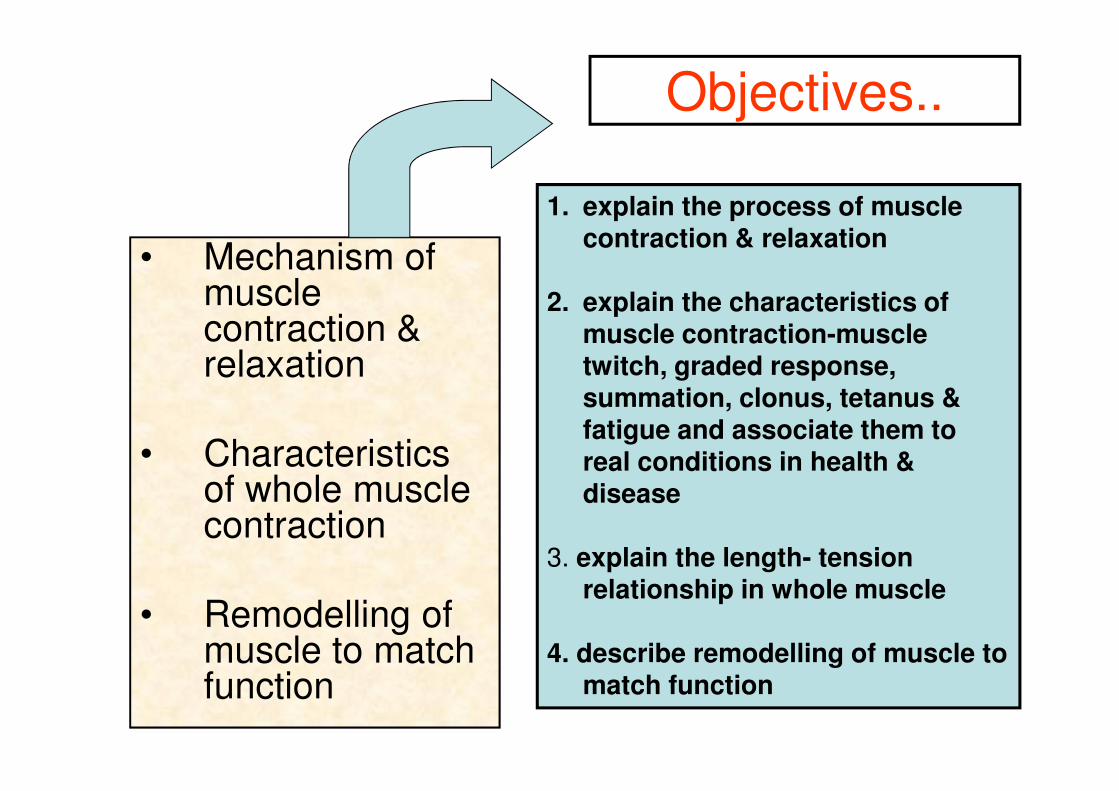
1. Muscle cells are excited by somatic efferent neurons.
2. Muscle cell excitation (the muscle cell action potential) triggers muscle cell activity (contraction).
3. Calcium (Ca++) is the second messenger that links excitation to contraction


Excitation-Contraction Coupling
A term coined in 1952 to describe the physiological process of converting an electrical
stimulus to a mechanical response
This process is fundamental to muscle physiology, whereby the electrical stimulus is
usually an action potential and the mechanical response is contraction.
EC coupling can be dysregulated in many disease conditions.

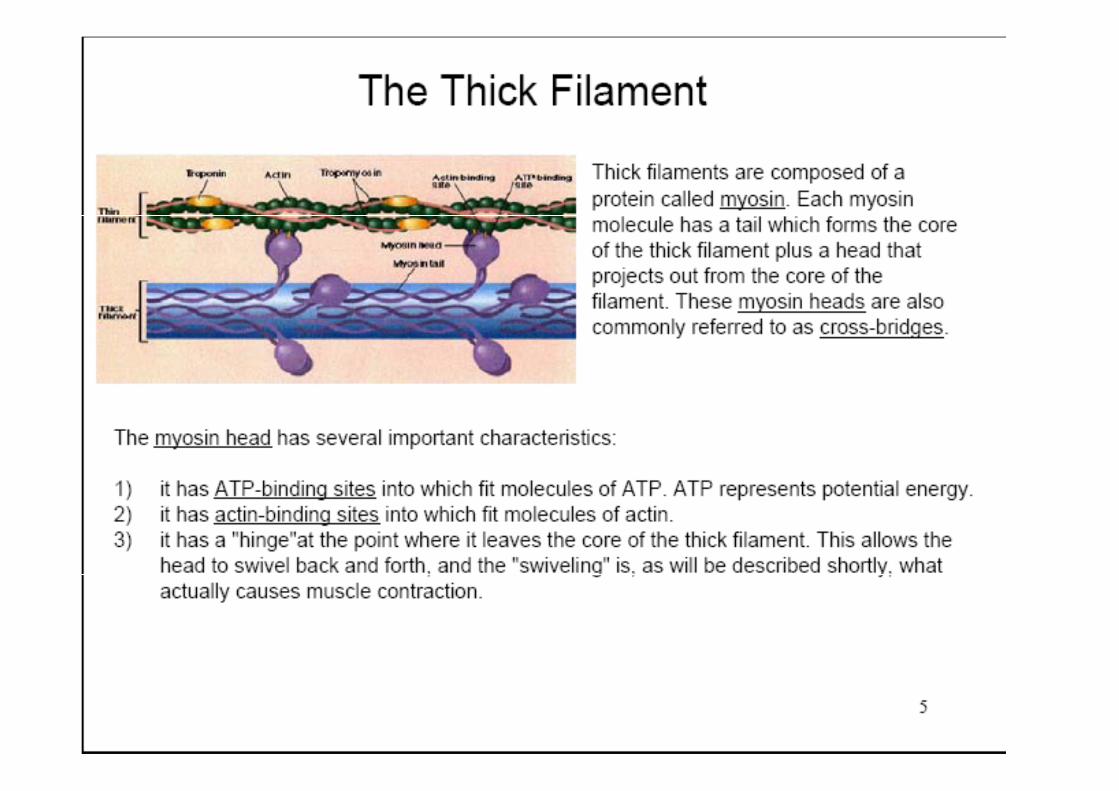
MYO FIBRIL
TERMINAL CISTERNAE
T- TUBULE
TRIAD MITOCHONDRIA
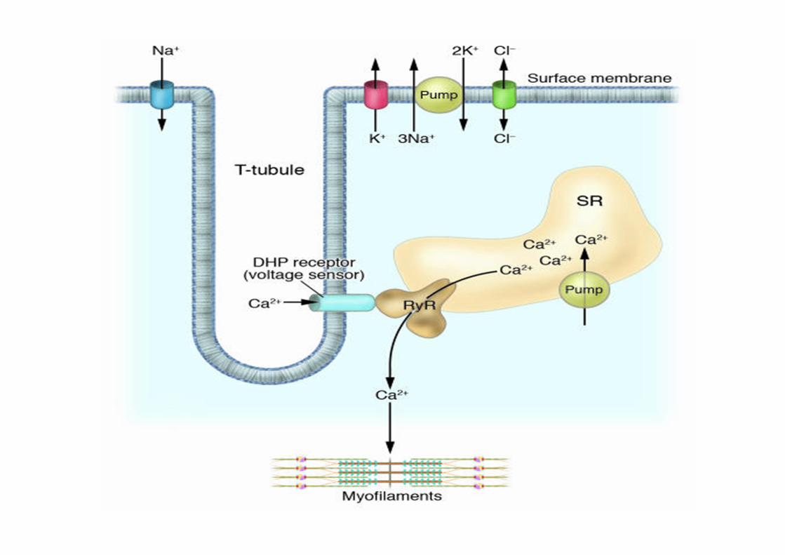
THIN MYOFILAMENT
THICK MYOFILAMENT
Z M Z

Neuromuscular
Transmission
Ca2+ released from the
sarcoplasmic reticulum binds
to Troponin C on actin filaments,
DIHYDROPYRIDINE RECEPTORS
Voltage dependent Ca+2 channels
T-tubules
RYANODINE RECEPTORS[RyR1]
Voltage dependent Ca+2 channels
SR
causes flow of Ca2+ from the sarcoplasmic reticulum, after its release from the Calsequestrin,
into the cytoplasm.
Opening
In humans, the gene encoding RyR1 is located on chromosome 19q13.2 and spans 104 exons.


• Mutations in the RYR1 gene underlie several debilitating and/or life-threatening muscle diseases including
– malignant hyperthermia (MH) ,
– heat/exercise induced exertional rhabdomyolysis ,
– atypical periodic paralyses (APP)

Proteins
• Structural proteins
• Regulatory proteins
• Contractile proteins
Dystrophin
Desmin Vimentin
Nebulin
Titin
Alpha- actinin

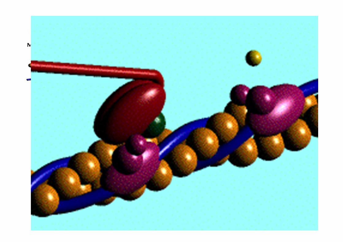

1 - Calcium released from sarcoplasmic reticulum
2 - Myosin head energized via myosin-ATPase activity which converts the bound ATP to ADP + Pi
3 - Calcium binds to troponin
4 - Tropomyosin translocates to uncover the cross-bridge binding sites
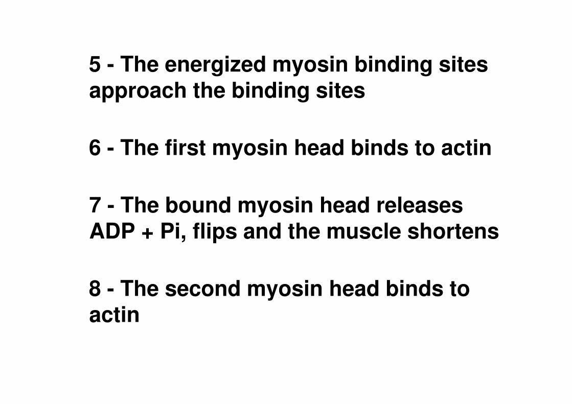
5 - The energized myosin binding sites approach the binding sites
6 - The first myosin head binds to actin
7 - The bound myosin head releases ADP + Pi, flips and the muscle shortens
8 - The second myosin head binds to actin
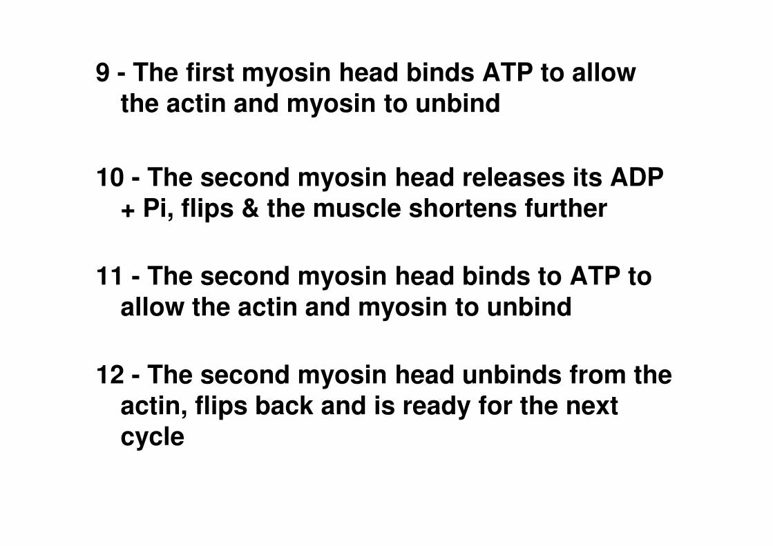
9 - The first myosin head binds ATP to allow the actin and myosin to unbind
10 - The second myosin head releases its ADP + Pi, flips & the muscle shortens further
11 - The second myosin head binds to ATP to allow the actin and myosin to unbind
12 - The second myosin head unbinds from the actin, flips back and is ready for the next cycle

13 - The cross-bridge cycle is terminated by the loss of calcium from the troponin
14 - Tropomyosin translocates to cover the cross-bridge binding sites
15 - The calcium returns to the sarcoplasmic reticulum, the muscle relaxes & returns to the resting state

As a muscle shortens, the following is observed:
a) sarcomeres
shorten; b) A band length
remains constant c) I band length
becomes shorten d) myofilament
lengths remain constant

SOME FACTS……….
• A single cycle of attachment, swivel, and detachment of the myosin head will produce a linear translation of the myofilaments of about 10 nm.
• If all cross-bridges in a myofibril cycle once synchronously, a relative movement equal to about 1% of the muscle length will occur, but obviously muscles shorten by more than 1%.
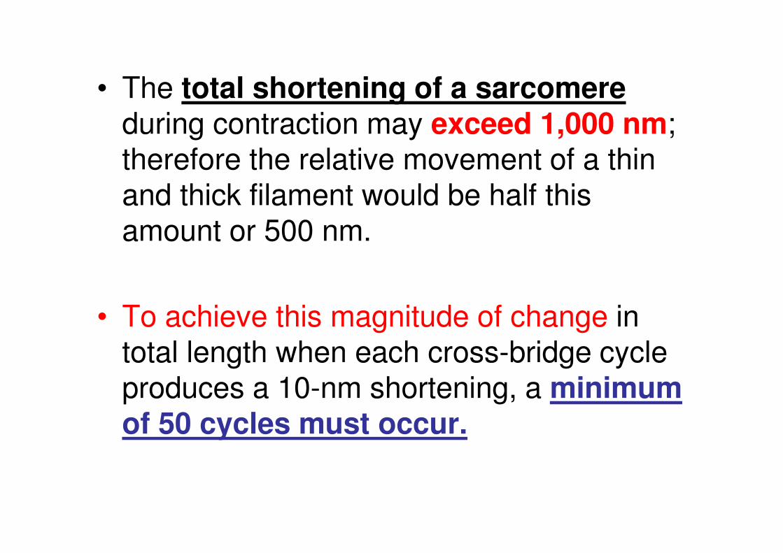
• The total shortening of a sarcomere during contraction may exceed 1,000 nm; therefore the relative movement of a thin and thick filament would be half this amount or 500 nm.
• To achieve this magnitude of change in total length when each cross-bridge cycle produces a 10-nm shortening, a minimum of 50 cycles must occur.
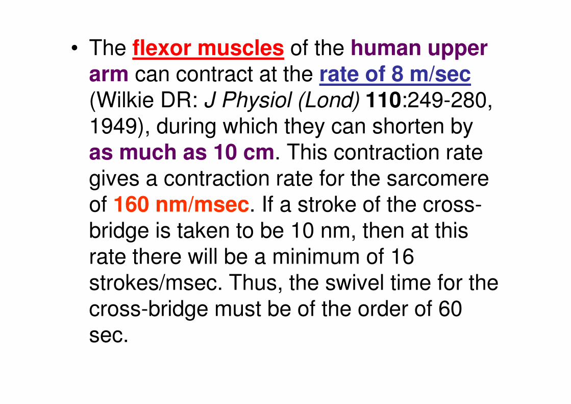
• The flexor muscles of the human upper arm can contract at the rate of 8 m/sec (Wilkie DR: J Physiol (Lond) 110:249-280, 1949), during which they can shorten by as much as 10 cm. This contraction rate gives a contraction rate for the sarcomere of 160 nm/msec. If a stroke of the cross-bridge is taken to be 10 nm, then at this rate there will be a minimum of 16 strokes/msec. Thus, the swivel time for the cross-bridge must be of the order of 60 sec.
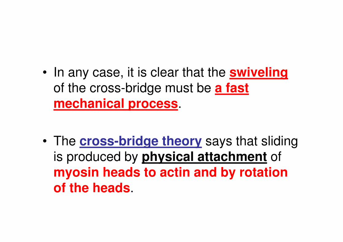
• In any case, it is clear that the swiveling of the cross-bridge must be a fast mechanical process.
• The cross-bridge theory says that sliding is produced by physical attachment of myosin heads to actin and by rotation of the heads.
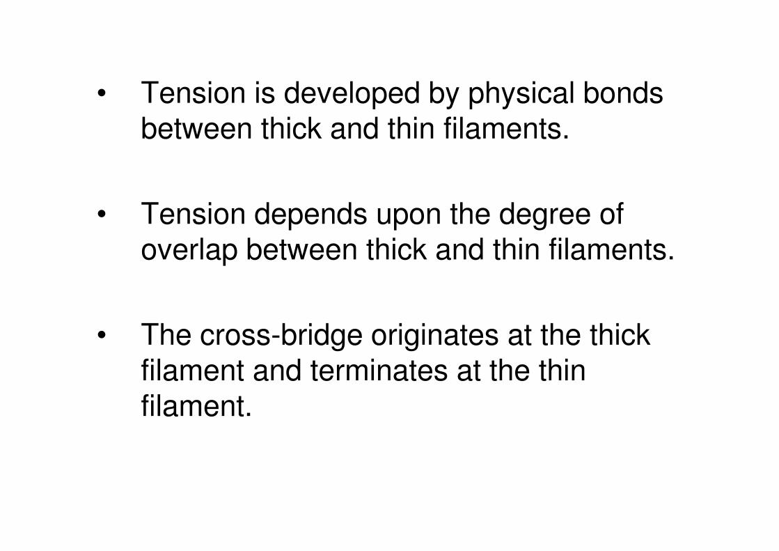
• Tension is developed by physical bonds between thick and thin filaments.
• Tension depends upon the degree of overlap between thick and thin filaments.
• The cross-bridge originates at the thick filament and terminates at the thin filament.

Malignant Hyperthermia
• MH is an autosomal dominant disease in which genetically susceptible individuals respond to inhalation anesthetics (e.g., halothane) and muscle relaxants (e.g., succinylcholine) with sustained muscle contractions.
• More than 150 different point mutations in the RYR1 gene have been identified and linked to MH .
• The majority of RyR1 mutations linked to MH cluster in the cytoplasmic domains of RyR1 (amino acids 35 to 614 and 2129 to 2458).
• Another cluster of mutations is found near the carboxyl terminus (4637 to 4973)
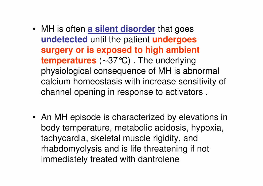
• MH is often a silent disorder that goes
undetected until the patient undergoes
surgery or is exposed to high ambient temperatures (∼37°C) . The underlying
physiological consequence of MH is abnormal
calcium homeostasis with increase sensitivity of
channel opening in response to activators .
• An MH episode is characterized by elevations in
body temperature, metabolic acidosis, hypoxia,
tachycardia, skeletal muscle rigidity, and
rhabdomyolysis and is life threatening if not
immediately treated with dantrolene
• HEAT RIGOR
• RIGOR MORTIS
http://cshperspectives.cshlp.org/content/2/11/a003996.full.pdf+html
Factors that Affect the Efficiency
of Muscle Contraction
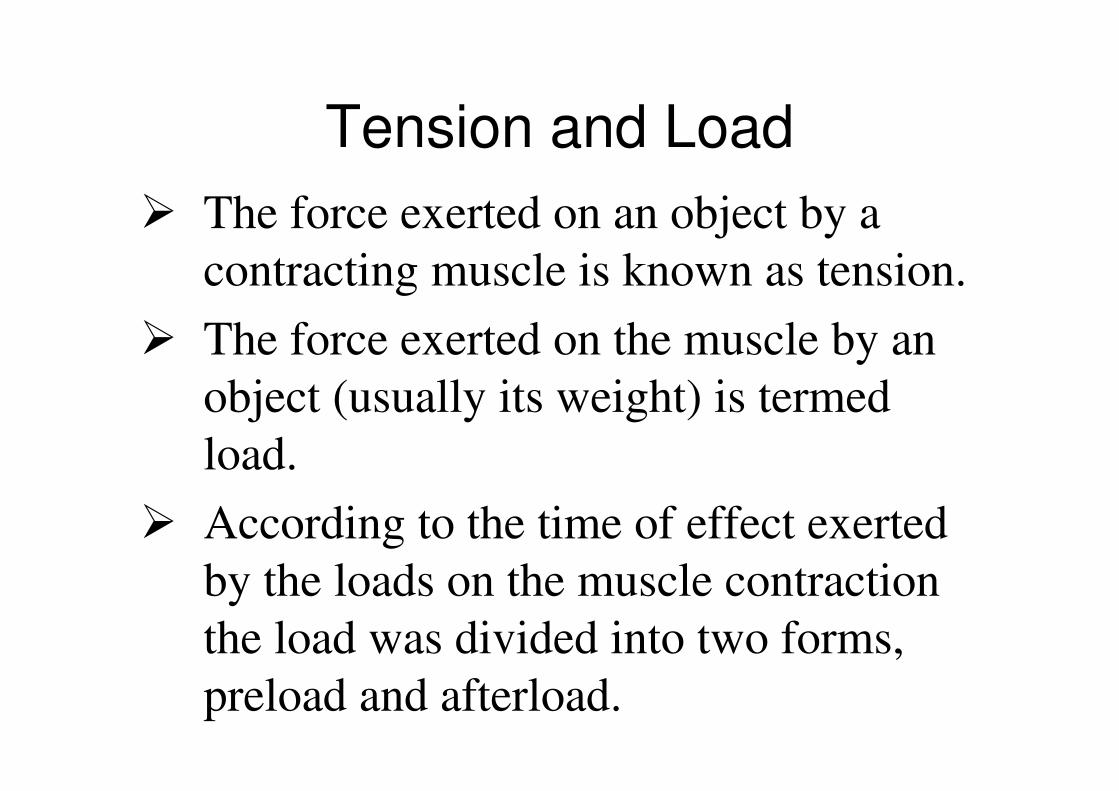
Tension and Load
� The force exerted on an object by a
contracting muscle is known as tension.
� The force exerted on the muscle by an
object (usually its weight) is termed
load.
� According to the time of effect exerted
by the loads on the muscle contraction
the load was divided into two forms,
preload and afterload.
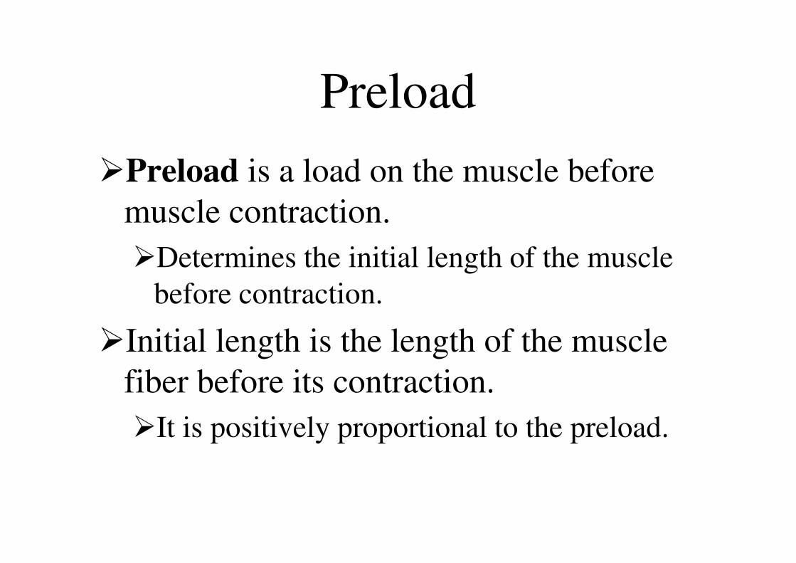
Preload
�Preload is a load on the muscle before
muscle contraction.
�Determines the initial length of the muscle
before contraction.
�Initial length is the length of the muscle
fiber before its contraction.
�It is positively proportional to the preload.

Afterload
� Afterload is a load on the muscle after the beginning of muscle contraction.
� The reverse force that oppose the contractile force caused by muscle contraction.
� The afterload does not change the initial length of the muscle,
� But it can prevent muscle from shortening because a part of force developed by contraction is used to overcome the afterload.
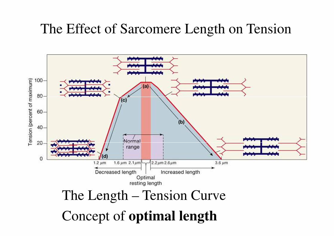
The Effect of Sarcomere Length on Tension
The Length – Tension Curve
Concept of optimal length
Types of Contractions I
� Twitch: a brief mechanical contraction of a single fiber produced by a single action potential at low frequency stimulation is known as single twitch.
� Tetanus: It means a summation of twitches that occurs at high frequency stimulation
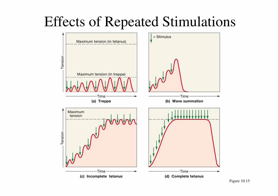
Effects of Repeated Stimulations
Figure 10.15
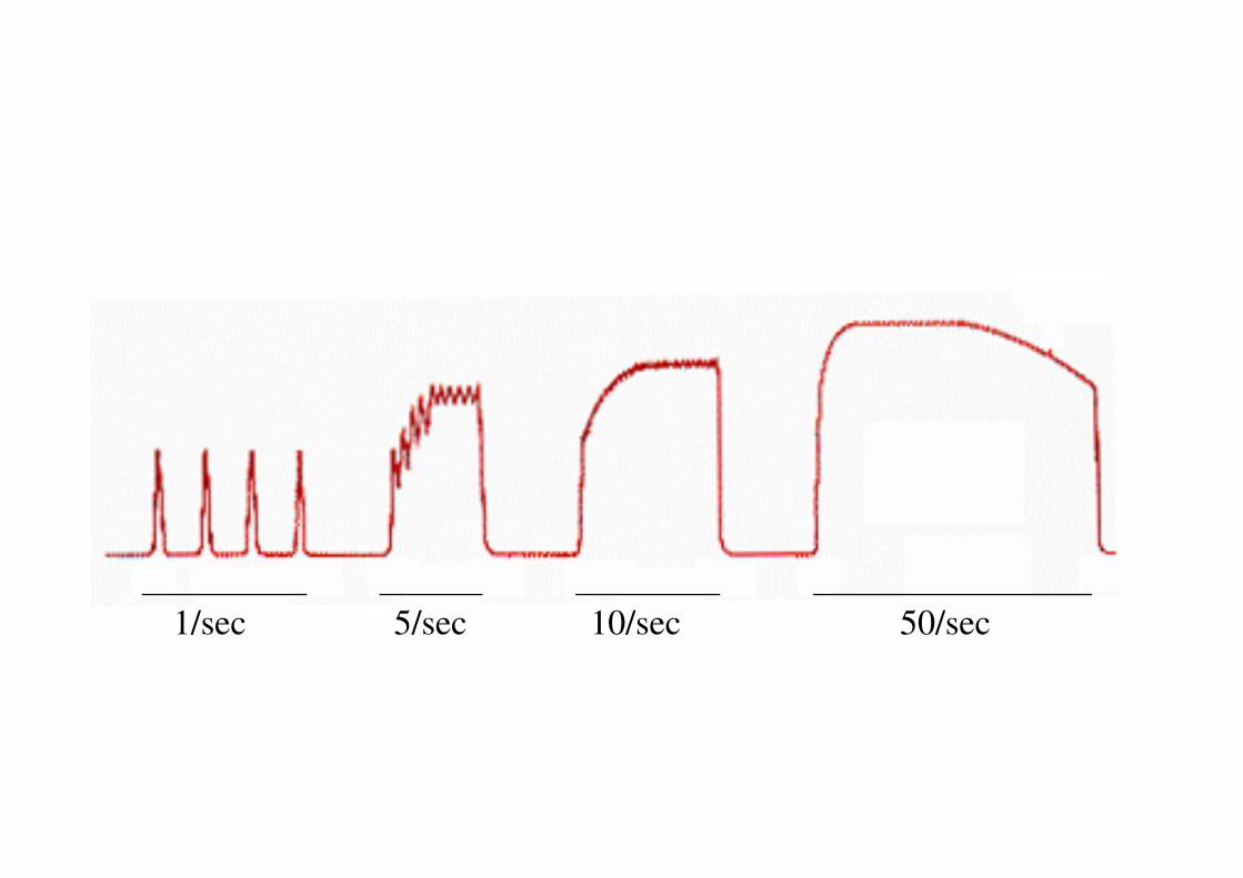
1/sec 5/sec 10/sec 50/sec
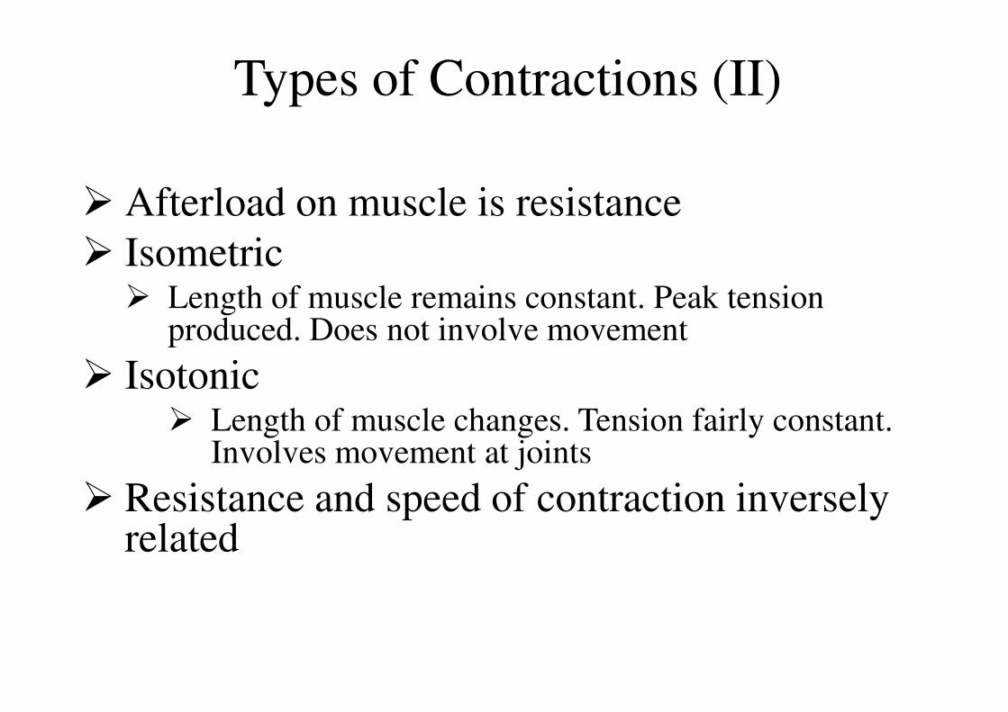
� Afterload on muscle is resistance
� Isometric � Length of muscle remains constant. Peak tension
produced. Does not involve movement
� Isotonic � Length of muscle changes. Tension fairly constant.
Involves movement at joints
� Resistance and speed of contraction inversely related
Types of Contractions (II)

Isotonic and Isometric Contractions
MUSCLE CONTRACTION-2
DATE: 17TH JANUARY,2012
Time : 10-30 AM to 11-30 AM
Venue- LT @ level 1
LECTURE# 3
Sarmishtha GhoshSarmishtha GhoshSarmishtha GhoshSarmishtha Ghosh
[email protected]@[email protected]@gmail.com
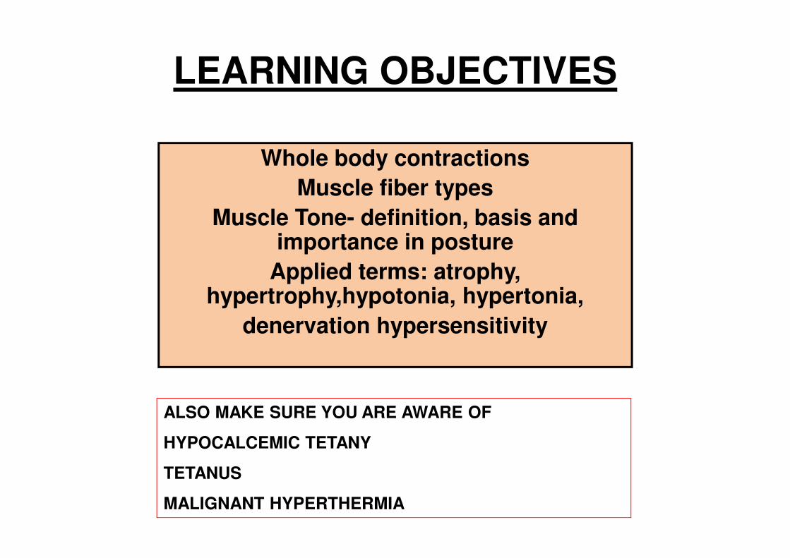
LEARNING OBJECTIVES
Whole body contractions
Muscle fiber types
Muscle Tone- definition, basis and importance in posture
Applied terms: atrophy, hypertrophy,hypotonia, hypertonia,
denervation hypersensitivity
ALSO MAKE SURE YOU ARE AWARE OF
HYPOCALCEMIC TETANY
TETANUS
MALIGNANT HYPERTHERMIA

Skeletal muscle
40% of the body weight
SINGLE
Large, elongated
cylinder shaped Dm= 10-100 µm
L=2.5 ft
Single muscle cell : multiple nuclei
Abundant mitochondria
Specialized contractile elements, 80% of the muscle fiber, Dm= 1µm ,L=2.5 ft Highly organized cytoskeletal elements

Muscle is a chemomechanical transducer.
It has the ability to convert chemical energy,
stored in the terminal phosphate group of ATP, into mechanical work.
• The myosin crossbridge, or myosin molecular motor, is the site for this energy conversion.
• Thus in addition to generating force and shortening, myosin is an enzyme that hydrolyzes ATP (i.e. ATPase).
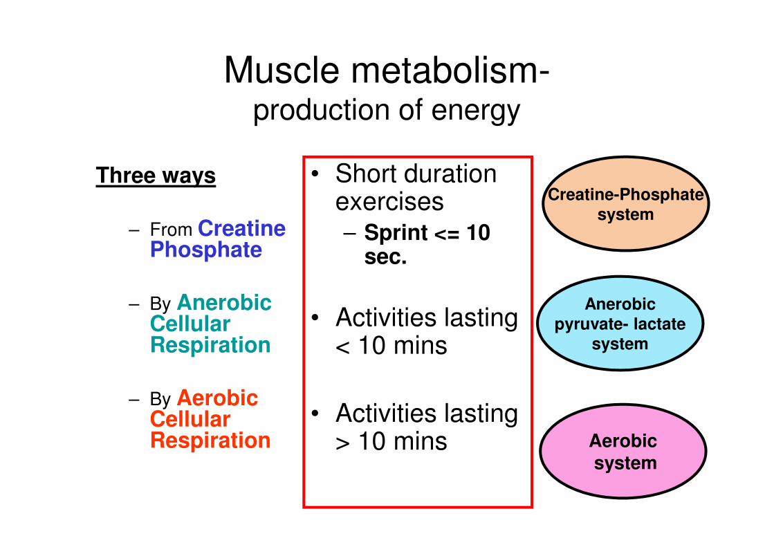
Muscle metabolism- production of energy
Three ways
– From Creatine Phosphate
– By Anerobic Cellular Respiration
– By Aerobic Cellular Respiration
• Short duration exercises – Sprint <= 10
sec.
• Activities lasting < 10 mins
• Activities lasting > 10 mins
Creatine-Phosphate system
Anerobic pyruvate- lactate
system
Aerobic system
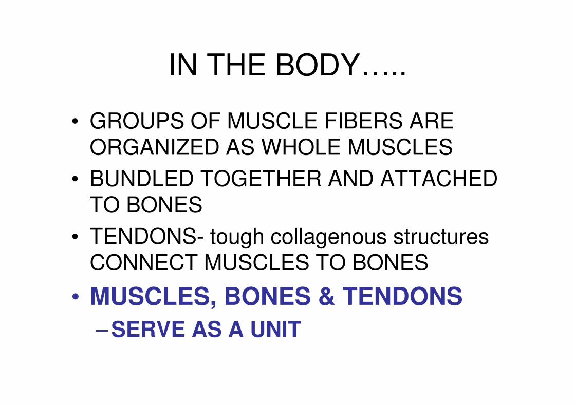
IN THE BODY…..
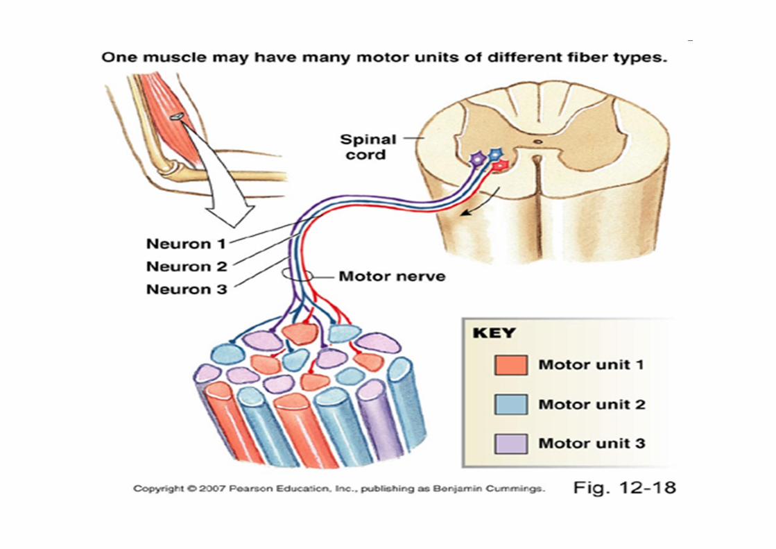
• GROUPS OF MUSCLE FIBERS ARE ORGANIZED AS WHOLE MUSCLES
• BUNDLED TOGETHER AND ATTACHED TO BONES
• TENDONS- tough collagenous structures CONNECT MUSCLES TO BONES
• MUSCLES, BONES & TENDONS
– SERVE AS A UNIT

SKELETAL MUSCLE MECHANICS

• Contraction of whole muscles can be of varying strength
– Number of muscle fibers contracting within a muscle
• Motor units and their recruitment
– Tension developed by each contracting fiber
• Frequency of stimulation
• Length of fiber at onset of contraction
• Extent of fatigue
• Thickness of fiber
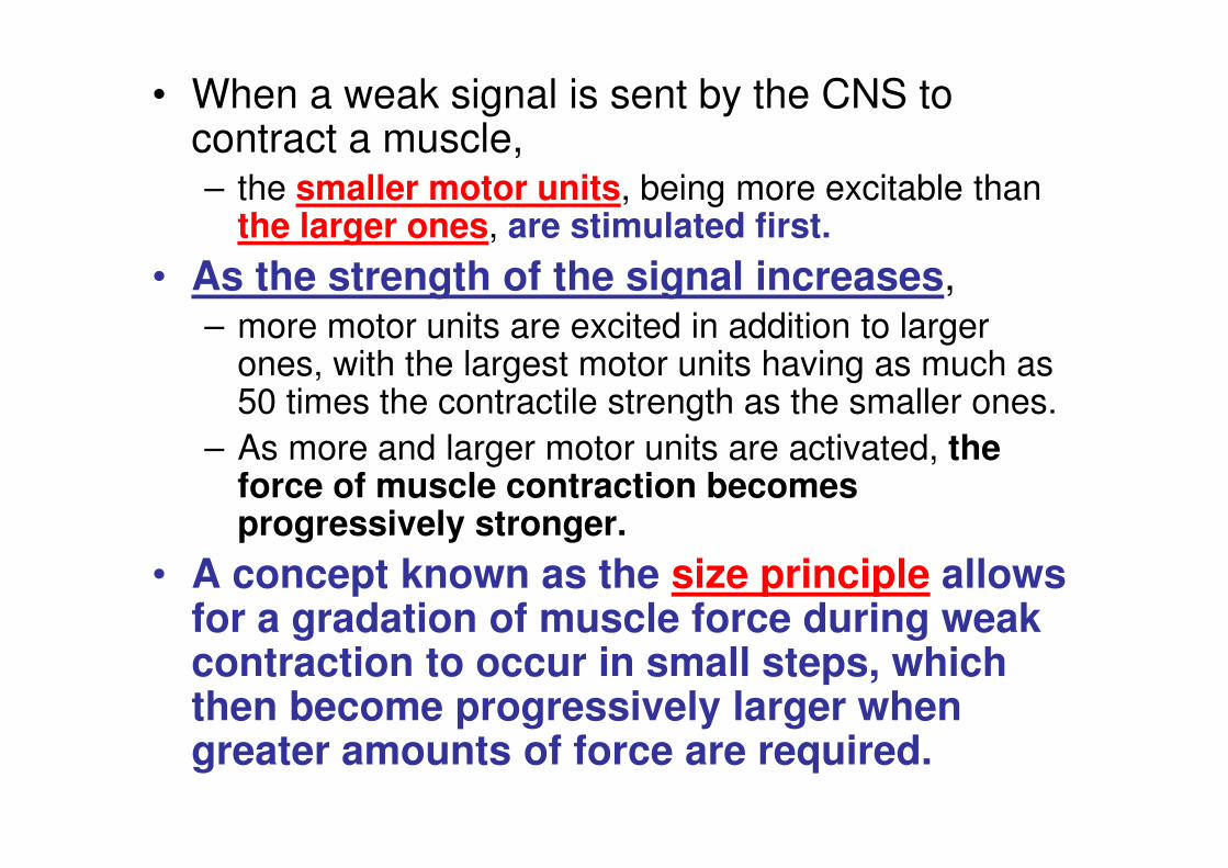
• When a weak signal is sent by the CNS to contract a muscle, – the smaller motor units, being more excitable than
the larger ones, are stimulated first.
• As the strength of the signal increases, – more motor units are excited in addition to larger
ones, with the largest motor units having as much as 50 times the contractile strength as the smaller ones.
– As more and larger motor units are activated, the force of muscle contraction becomes progressively stronger.
• A concept known as the size principle allows for a gradation of muscle force during weak contraction to occur in small steps, which then become progressively larger when greater amounts of force are required.
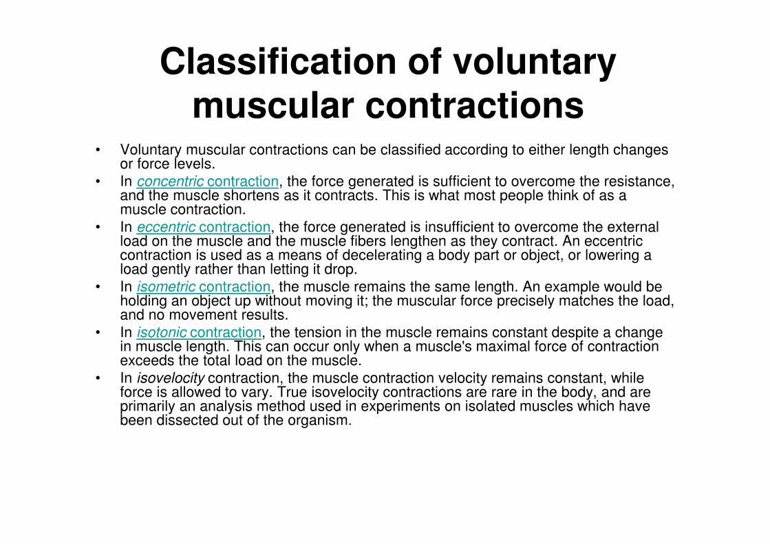
Classification of voluntary
muscular contractions • Voluntary muscular contractions can be classified according to either length changes
or force levels.
• In concentric contraction, the force generated is sufficient to overcome the resistance, and the muscle shortens as it contracts. This is what most people think of as a muscle contraction.
• In eccentric contraction, the force generated is insufficient to overcome the external load on the muscle and the muscle fibers lengthen as they contract. An eccentric contraction is used as a means of decelerating a body part or object, or lowering a load gently rather than letting it drop.
• In isometric contraction, the muscle remains the same length. An example would be holding an object up without moving it; the muscular force precisely matches the load, and no movement results.
• In isotonic contraction, the tension in the muscle remains constant despite a change in muscle length. This can occur only when a muscle's maximal force of contraction exceeds the total load on the muscle.
• In isovelocity contraction, the muscle contraction velocity remains constant, while force is allowed to vary. True isovelocity contractions are rare in the body, and are primarily an analysis method used in experiments on isolated muscles which have been dissected out of the organism.
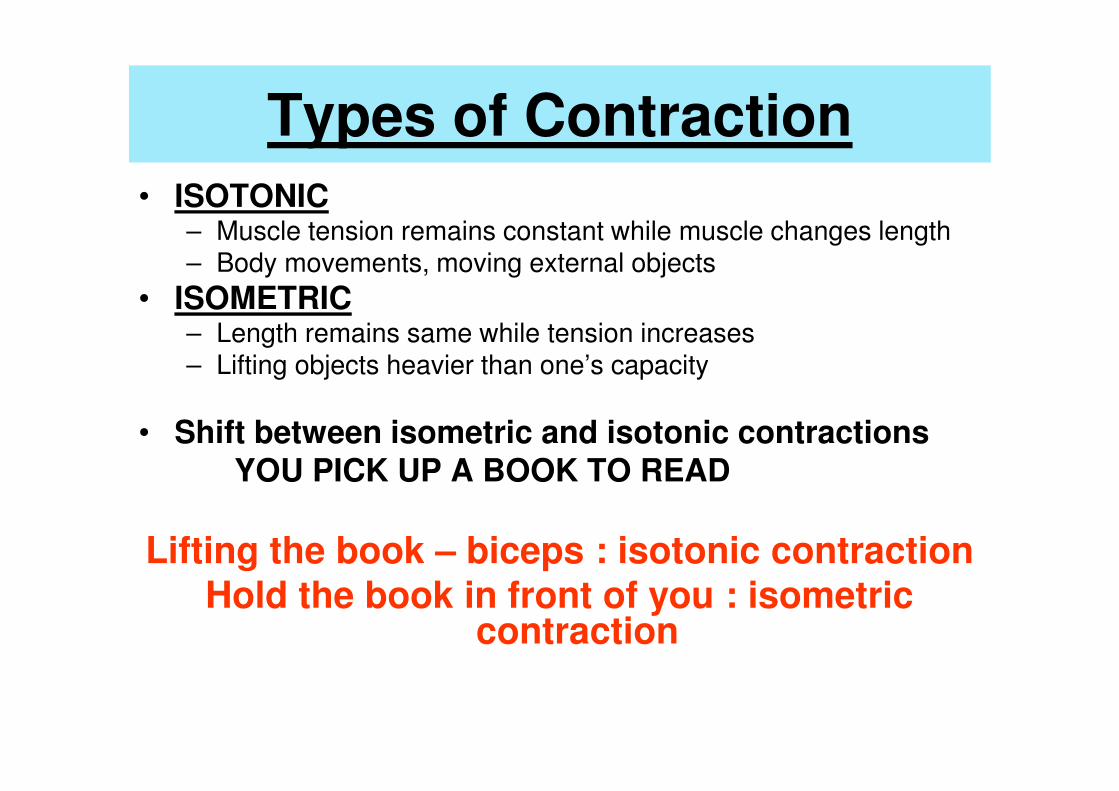
Types of Contraction
• ISOTONIC – Muscle tension remains constant while muscle changes length – Body movements, moving external objects
• ISOMETRIC – Length remains same while tension increases – Lifting objects heavier than one’s capacity
• Shift between isometric and isotonic contractions YOU PICK UP A BOOK TO READ
Lifting the book – biceps : isotonic contraction Hold the book in front of you : isometric
contraction
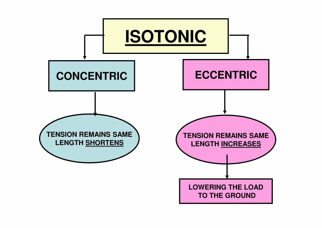
ISOTONIC
CONCENTRIC ECCENTRIC
TENSION REMAINS SAME LENGTH SHORTENS
TENSION REMAINS SAME LENGTH INCREASES
LOWERING THE LOAD TO THE GROUND
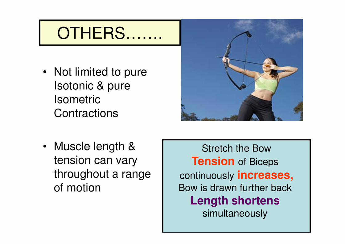
OTHERS…….
• Not limited to pure
Isotonic & pure
Isometric
Contractions
• Muscle length &
tension can vary
throughout a range
of motion
Stretch the Bow
Tension of Biceps
continuously increases, Bow is drawn further back
Length shortens
simultaneously

Still More…..
• Muscles of tongue – Not attached at the free end – Isotonic contractions :
facilitate speech and eating
• External eye muscles – Skull @ origin, eye @
insertion – Isotonic : Eye movements
• Sphincters : – Unattached to bone – Actually prevents movement – Prevents exit of urine and
feces by isotonic contraction
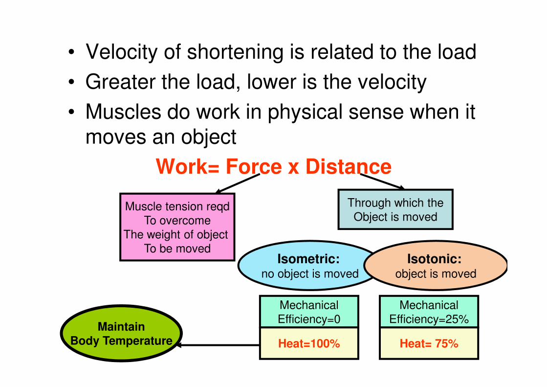
• Velocity of shortening is related to the load
• Greater the load, lower is the velocity
• Muscles do work in physical sense when it moves an object
Work= Force x Distance
Muscle tension reqd
To overcome The weight of object
To be moved
Through which the Object is moved
Isometric: no object is moved
Isotonic: object is moved
Mechanical Efficiency=0
Heat=100%
Mechanical Efficiency=25%
Heat= 75%
Maintain Body Temperature
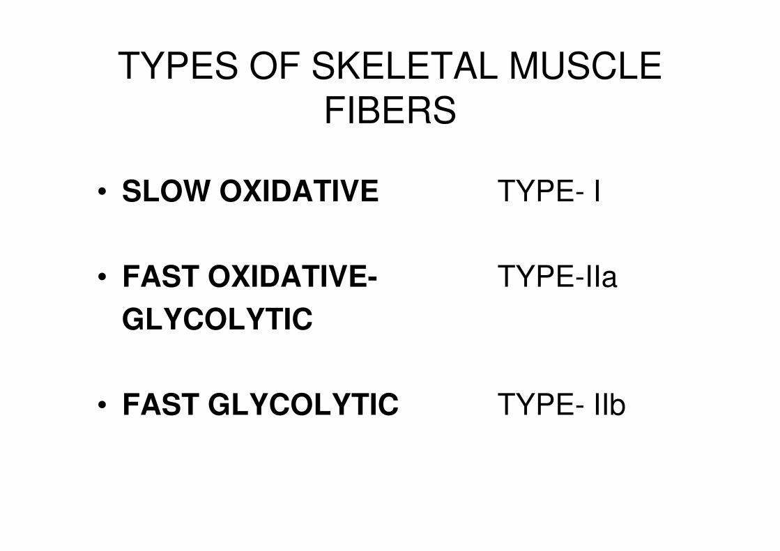
TYPES OF SKELETAL MUSCLE FIBERS
• SLOW OXIDATIVE TYPE- I
• FAST OXIDATIVE- TYPE-IIa
GLYCOLYTIC
• FAST GLYCOLYTIC TYPE- IIb
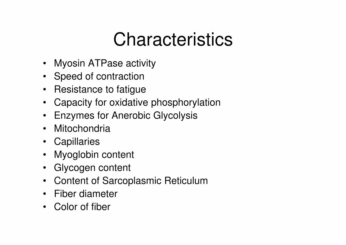
Characteristics • Myosin ATPase activity
• Speed of contraction
• Resistance to fatigue
• Capacity for oxidative phosphorylation
• Enzymes for Anerobic Glycolysis
• Mitochondria
• Capillaries
• Myoglobin content
• Glycogen content
• Content of Sarcoplasmic Reticulum
• Fiber diameter
• Color of fiber

Type I fibers -also called slow oxidative (SO) fibers
Postural muscles
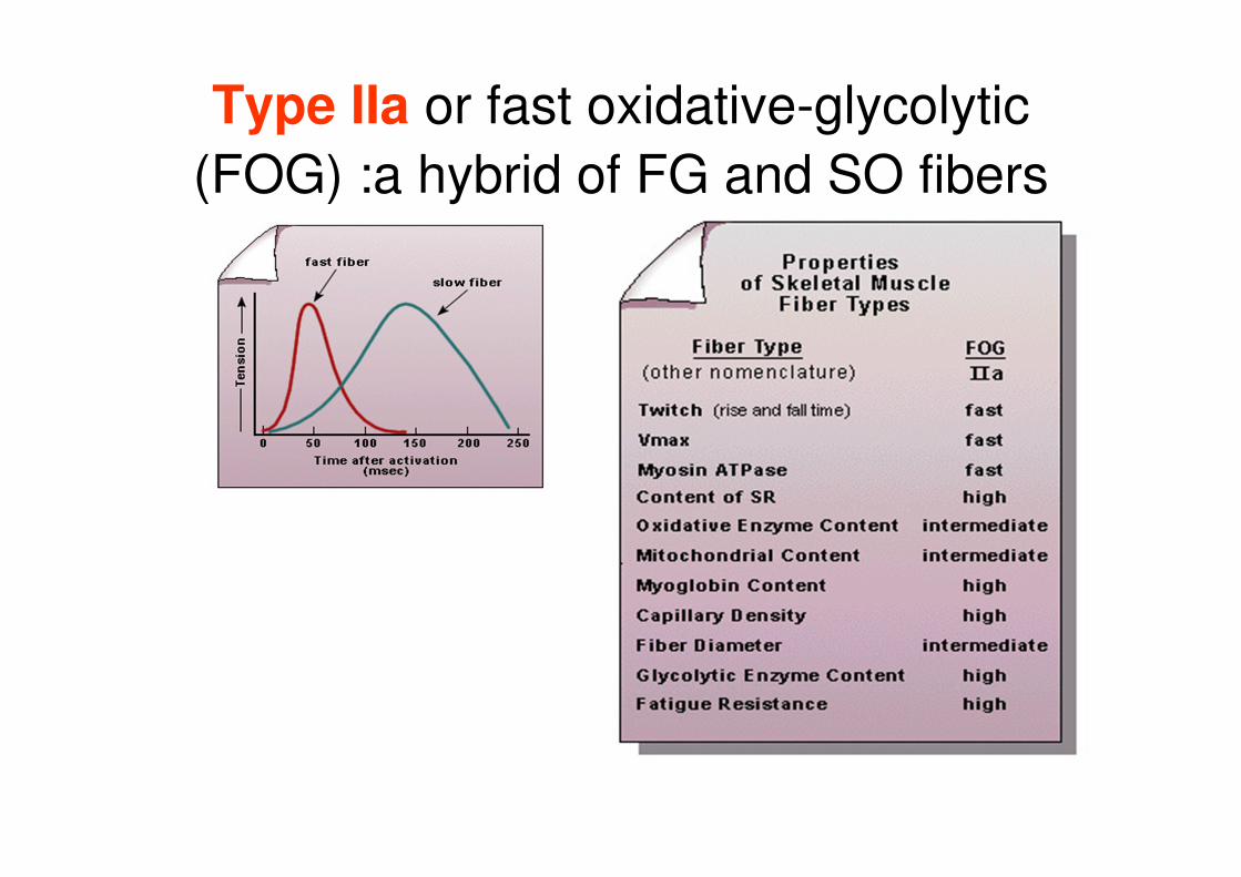
Type IIa or fast oxidative-glycolytic
(FOG) :a hybrid of FG and SO fibers
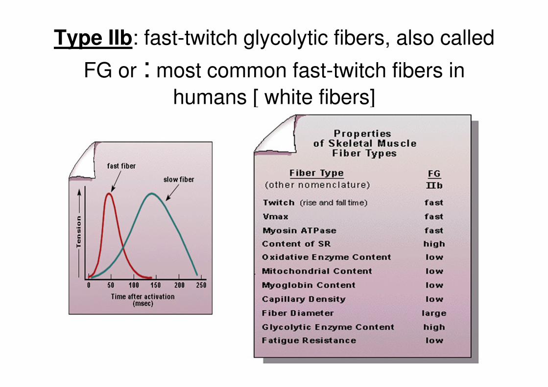
Type IIb: fast-twitch glycolytic fibers, also called
FG or : most common fast-twitch fibers in
humans [ white fibers]
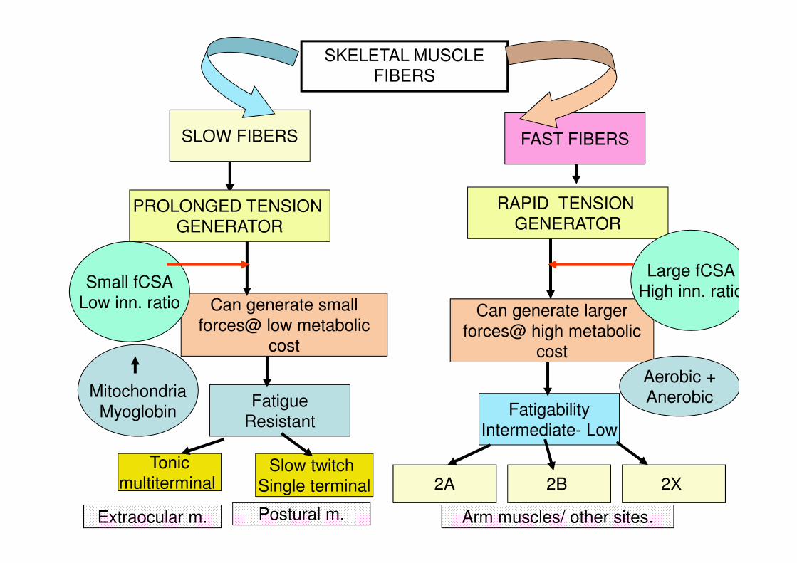
SKELETAL MUSCLE FIBERS
SLOW FIBERS FAST FIBERS
PROLONGED TENSION GENERATOR
RAPID TENSION GENERATOR
Can generate small forces@ low metabolic
cost
Can generate larger
forces@ high metabolic cost
Small fCSA Low inn. ratio
Large fCSA High inn. ratio
Mitochondria
Myoglobin
Aerobic + Anerobic Fatigue
Resistant Fatigability
Intermediate- Low
Tonic multiterminal
Slow twitch Single terminal 2A 2B 2X
Extraocular m. Postural m. Arm muscles/ other sites.
• Majority of muscles are of mixed fiber type composition being a combination of fast and slow fibers occurring in two arrangements
– 1) mosaic - fast and slow fibers uniformly distributed
– 2) compartmentalized - fiber types non-uniformly distributed into intramuscular compartments
• Some muscles which are used for repetitive or constant tasks (e.g., posture) can be comprised nearly entirely of slow fibers – - e.g., soleus

• Genetic Endowment of muscle fiber types
• Adaptation to demands placed on them
– Changes in their ATP synthesizing machinery
– Changes in their diameter

• Anerobic, short duration,high intensity resistance training – Weight lifting – Muscle enlargement
• Actual increase in diameter of fast glycolytic fibers • Increased synthesis of Actin and Myosin filaments
– Hypertrophy –
• Actions of Testosterone – the male sex hormone
• Interconversion between fast muscle fiber types – No conversion between fast and slow fibers
• Limited repair system available – No mitosis – Some myoblasts may fuse and cause a muscle fiber – Extensive injury- not adequate
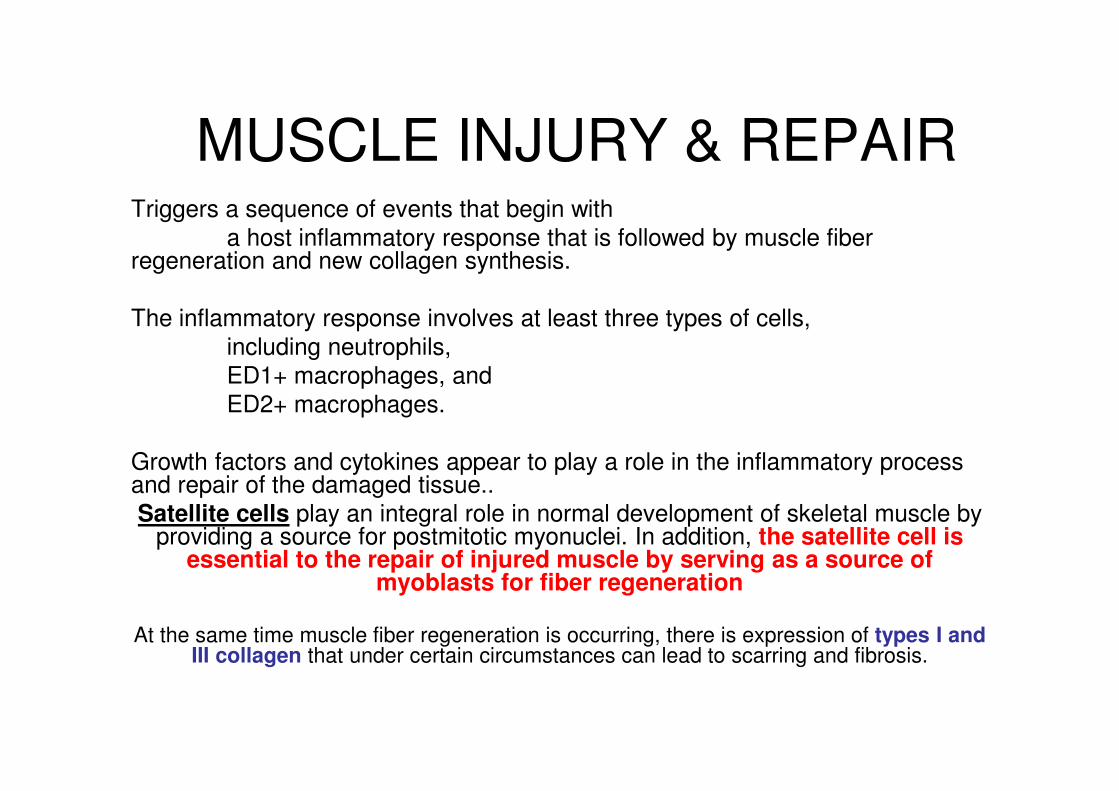
MUSCLE INJURY & REPAIR Triggers a sequence of events that begin with a host inflammatory response that is followed by muscle fiber regeneration and new collagen synthesis. The inflammatory response involves at least three types of cells, including neutrophils, ED1+ macrophages, and ED2+ macrophages. Growth factors and cytokines appear to play a role in the inflammatory process and repair of the damaged tissue.. Satellite cells play an integral role in normal development of skeletal muscle by
providing a source for postmitotic myonuclei. In addition, the satellite cell is essential to the repair of injured muscle by serving as a source of
myoblasts for fiber regeneration
At the same time muscle fiber regeneration is occurring, there is expression of types I and III collagen that under certain circumstances can lead to scarring and fibrosis.
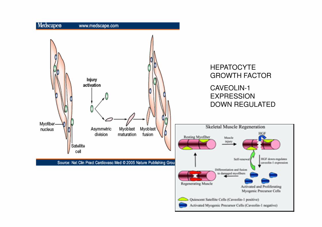
HEPATOCYTE GROWTH FACTOR
CAVEOLIN-1 EXPRESSION DOWN REGULATED
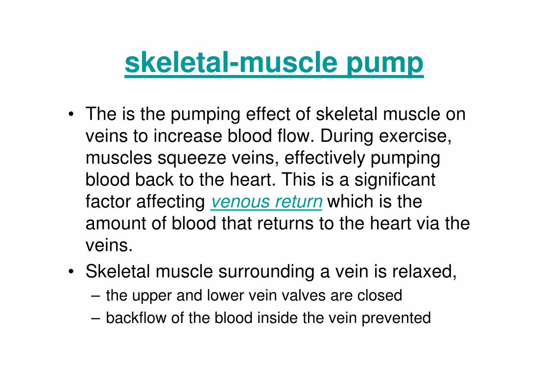
skeletal-muscle pump
• The is the pumping effect of skeletal muscle on
veins to increase blood flow. During exercise,
muscles squeeze veins, effectively pumping
blood back to the heart. This is a significant
factor affecting venous return which is the
amount of blood that returns to the heart via the
veins.
• Skeletal muscle surrounding a vein is relaxed,
– the upper and lower vein valves are closed
– backflow of the blood inside the vein prevented

MUSCLE TONE
• Residual muscle tension or tonus
• is the continuous and passive partial contraction of the muscles.
• helps maintain posture, and it declines during REM sleep.
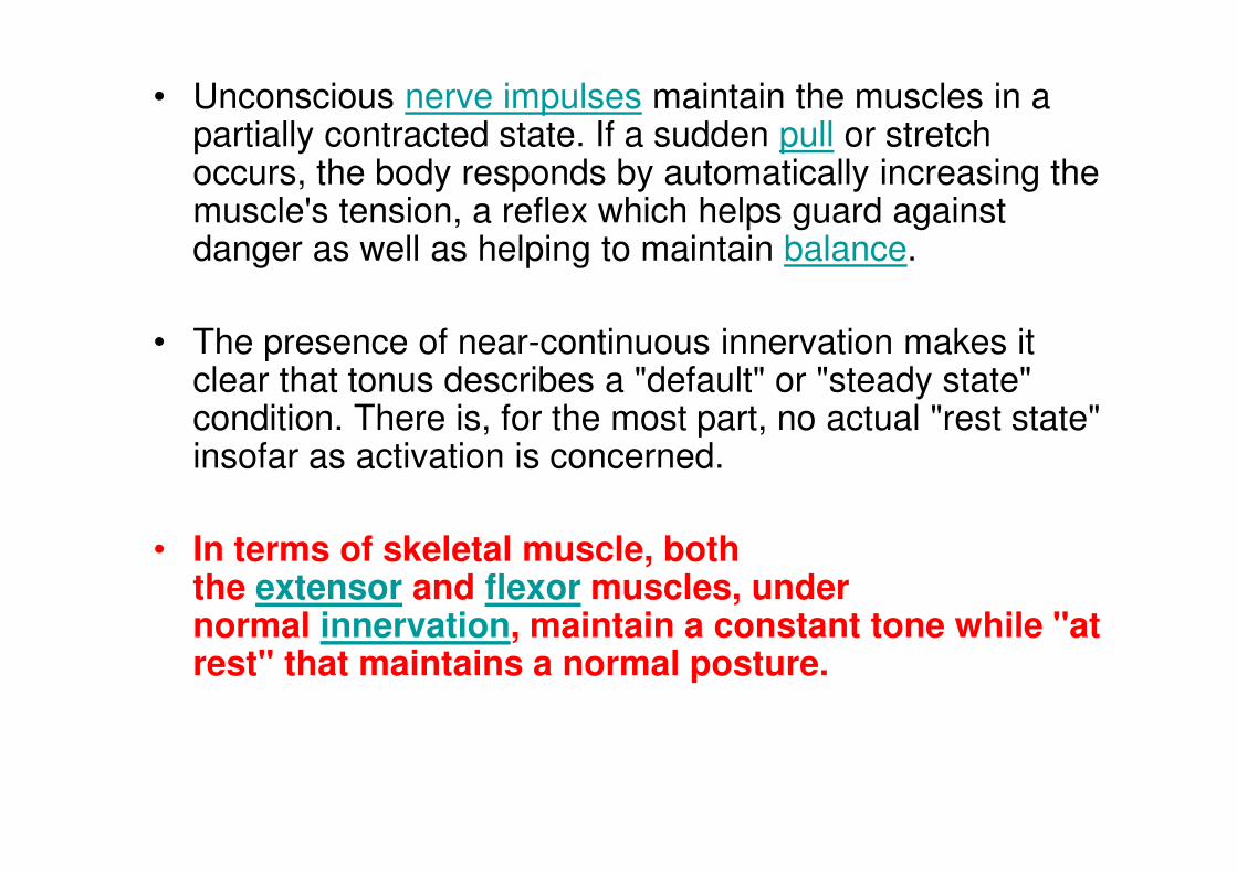
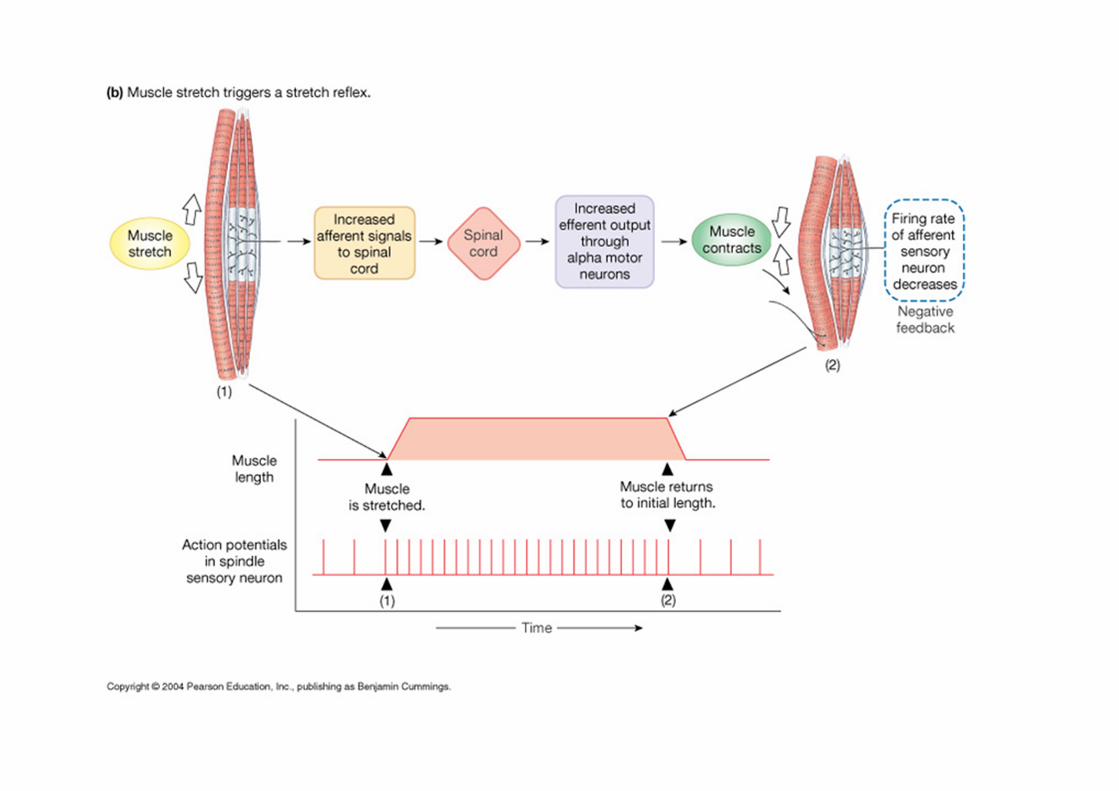
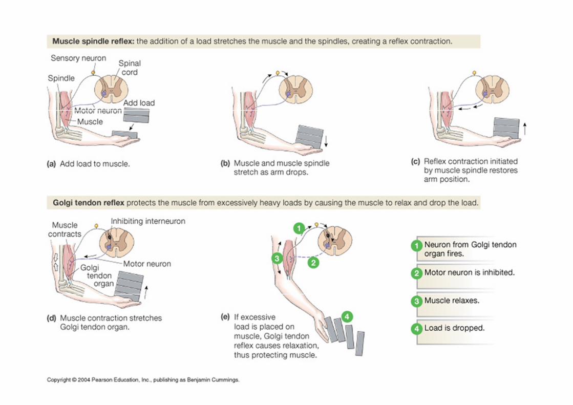
• Unconscious nerve impulses maintain the muscles in a partially contracted state. If a sudden pull or stretch occurs, the body responds by automatically increasing the muscle's tension, a reflex which helps guard against danger as well as helping to maintain balance.
• The presence of near-continuous innervation makes it clear that tonus describes a "default" or "steady state" condition. There is, for the most part, no actual "rest state" insofar as activation is concerned.
• In terms of skeletal muscle, both the extensor and flexor muscles, under normal innervation, maintain a constant tone while "at rest" that maintains a normal posture.

Pathological tonus • Physical disorders can result in
– abnormally low (hypotonia) or – high (hypertonia) muscle tone.
• Another form of hypertonia is paratonia, which is associated with dementia. Hypotonia is caused by lower motor neuron disease like poliomyelitis. Hypertonia is caused in upper motor neuron disease like lesion in pyramidal tract and extrapyramidal tract. Hypertonia can be of clasp knife variety, in which there is increased resistance only at the beginning or at the end of the movement, or lead pipe variety, in which there is resistance throughout to passive movement, or it may be of cog wheel type, in which the resistance to passive movement is in jerky manner.
•

http://faculty.pasadena.edu
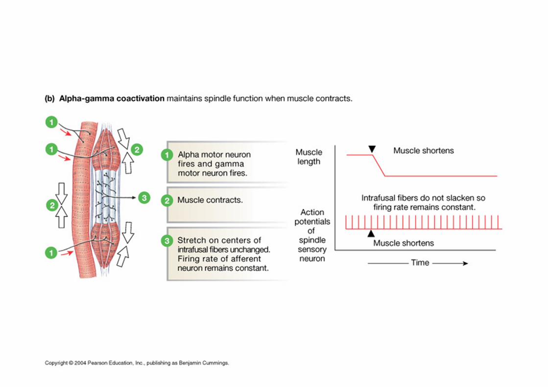

alpha-gamma coactivation;
when we actively contract extrafusal fibers (muscle), the contractile portion of intrafusal fibers contract as well; this stretches the spindles causing them to fire. Alpha motor neurons fire to contract extrafusal fibers and gamma motor neurons are COACTIVATED to fire with alpha's to contract intrafusal fibers simultaneousely


Stretching of muscles Stimulation of
MUSCLE SPINDLE
Activation of Sensory Neuron
Information Processing
At Motor neuron
Activation of Motor neuron
extrafusal fibers, rich in contractile proteins,
Contraction Of
Muscle
MUSCLE OPPOSES
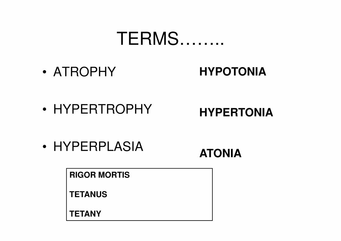
TERMS……..
• ATROPHY
• HYPERTROPHY
• HYPERPLASIA
HYPOTONIA
HYPERTONIA
ATONIA
RIGOR MORTIS TETANUS TETANY
DENERVATION HYPERSENSITIVITY
• Innervated adult skeletal muscle is sensitive to acetylcholine at the end-plate region only. After denervation the entire muscle membrane becomes chemosensitive..
• The motor nerve to skeletal muscle is cut and allowed to degenerate --------the muscle gradually becomes extremely sensitive to acetylcholine.
• This denervation hypersensitivity or supersensitivity is also seen in smooth muscle. Smooth muscle, unlike skeletal muscle, does not atrophy when denervated, but it becomes hyperresponsive to the chemical mediator that normally activates it
• It appears that the new receptors are released into the muscle surface from post-Golgi vesicles, giving rise to local ‘hot spots’ in A Ch sensitivity. Their generation appears to be triggered by the absence of a neural factor controlling genetic expression in the muscle cell — this factor may be A Ch itself.

ELECTROMYOGRAM
• Diagnose conditions that damage
muscle tissue, nerves, or the junctions between nerve and muscle (neuromuscular junctions), for example, a herniated disc.
• Evaluate the cause of weakness, paralysis, involuntary muscle twitching, or other symptoms.
• Problems in a muscle, the nerves supplying a muscle, the spinal cord, or the area of the brain that controls a muscle can all cause these kinds of symptoms.
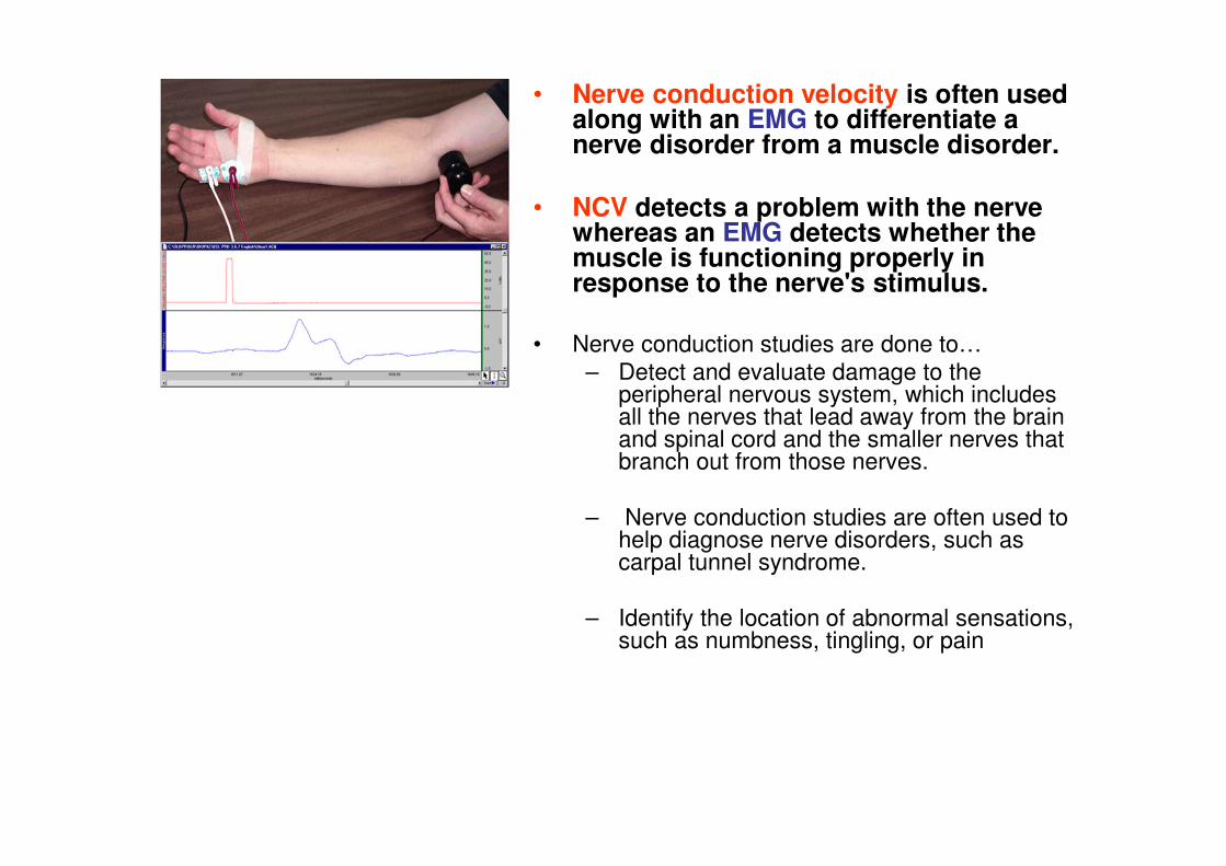
• Nerve conduction velocity is often used along with an EMG to differentiate a nerve disorder from a muscle disorder.
• NCV detects a problem with the nerve whereas an EMG detects whether the muscle is functioning properly in response to the nerve's stimulus.
• Nerve conduction studies are done to…
– Detect and evaluate damage to the peripheral nervous system, which includes all the nerves that lead away from the brain and spinal cord and the smaller nerves that branch out from those nerves.
– Nerve conduction studies are often used to help diagnose nerve disorders, such as carpal tunnel syndrome.
– Identify the location of abnormal sensations, such as numbness, tingling, or pain
Related Documents