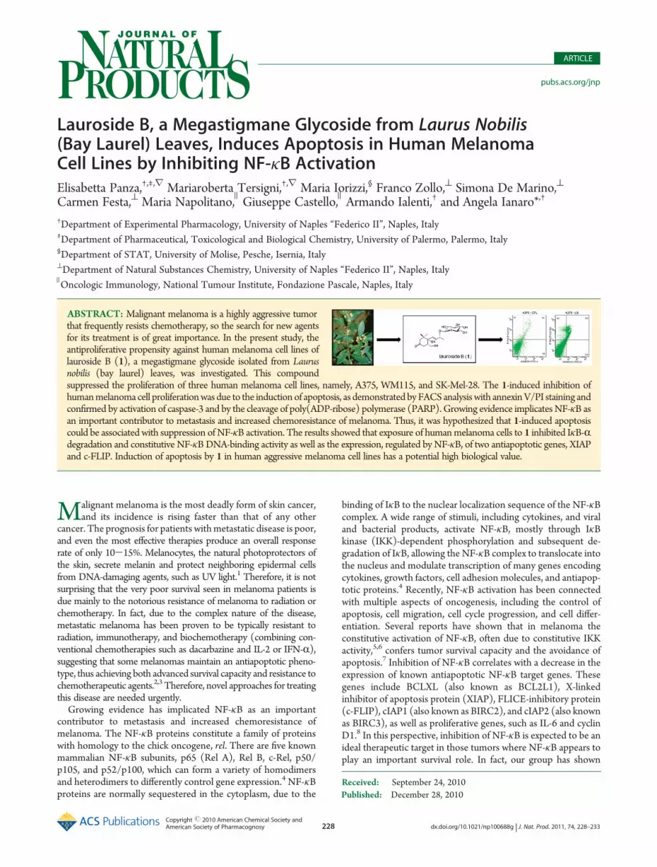
Published: December 28, 2010 Copyright r 2010 American Chemical Society and American Society of Pharmacognosy 228 dx.doi.org/10.1021/np100688g | J. Nat. Prod. 2011, 74, 228–233 ARTICLE pubs.acs.org/jnp Lauroside B, a Megastigmane Glycoside from Laurus Nobilis (Bay Laurel) Leaves, Induces Apoptosis in Human Melanoma Cell Lines by Inhibiting NF-κB Activation Elisabetta Panza, †,‡,r Mariaroberta Tersigni, †,r Maria Iorizzi, § Franco Zollo, ^ Simona De Marino, ^ Carmen Festa, ^ Maria Napolitano, ) Giuseppe Castello, ) Armando Ialenti, † and Angela Ianaro* ,† † Department of Experimental Pharmacology, University of Naples “Federico II”, Naples, Italy ‡ Department of Pharmaceutical, Toxicological and Biological Chemistry, University of Palermo, Palermo, Italy § Department of STAT, University of Molise, Pesche, Isernia, Italy ^ Department of Natural Substances Chemistry, University of Naples “Federico II”, Naples, Italy ) Oncologic Immunology, National Tumour Institute, Fondazione Pascale, Naples, Italy ABSTRACT: Malignant melanoma is a highly aggressive tumor that frequently resists chemotherapy, so the search for new agents for its treatment is of great importance. In the present study, the antiproliferative propensity against human melanoma cell lines of lauroside B (1), a megastigmane glycoside isolated from Laurus nobilis (bay laurel) leaves, was investigated. This compound suppressed the proliferation of three human melanoma cell lines, namely, A375, WM115, and SK-Mel-28. The 1-induced inhibition of human melanoma cell proliferation was due to the induction of apoptosis, as demonstrated by FACS analysis with annexin V/PI staining and confirmed by activation of caspase-3 and by the cleavage of poly(ADP-ribose) polymerase (PARP). Growing evidence implicates NF-κB as an important contributor to metastasis and increased chemoresistance of melanoma. Thus, it was hypothesized that 1-induced apoptosis could be associated with suppression of NF-κB activation. The results showed that exposure of human melanoma cells to 1 inhibited IκB-R degradation and constitutive NF-κB DNA-binding activity as well as the expression, regulated by NF- κB, of two antiapoptotic genes, XIAP and c-FLIP. Induction of apoptosis by 1 in human aggressive melanoma cell lines has a potential high biological value. M alignant melanoma is the most deadly form of skin cancer, and its incidence is rising faster than that of any other cancer. The prognosis for patients with metastatic disease is poor, and even the most effective therapies produce an overall response rate of only 10-15%. Melanocytes, the natural photoprotectors of the skin, secrete melanin and protect neighboring epidermal cells from DNA-damaging agents, such as UV light. 1 Therefore, it is not surprising that the very poor survival seen in melanoma patients is due mainly to the notorious resistance of melanoma to radiation or chemotherapy. In fact, due to the complex nature of the disease, metastatic melanoma has been proven to be typically resistant to radiation, immunotherapy, and biochemotherapy (combining con- ventional chemotherapies such as dacarbazine and IL-2 or IFN-R), suggesting that some melanomas maintain an antiapoptotic pheno- type, thus achieving both advanced survival capacity and resistance to chemotherapeutic agents. 2,3 Therefore, novel approaches for treating this disease are needed urgently. Growing evidence has implicated NF-κB as an important contributor to metastasis and increased chemoresistance of melanoma. The NF-κB proteins constitute a family of proteins with homology to the chick oncogene, rel. There are five known mammalian NF-κB subunits, p65 (Rel A), Rel B, c-Rel, p50/ p105, and p52/p100, which can form a variety of homodimers and heterodimers to differently control gene expression. 4 NF-κB proteins are normally sequestered in the cytoplasm, due to the binding of IκB to the nuclear localization sequence of the NF-κB complex. A wide range of stimuli, including cytokines, and viral and bacterial products, activate NF-κB, mostly through IκB kinase (IKK)-dependent phosphorylation and subsequent de- gradation of IκB, allowing the NF-κB complex to translocate into the nucleus and modulate transcription of many genes encoding cytokines, growth factors, cell adhesion molecules, and antiapop- totic proteins. 4 Recently, NF-κB activation has been connected with multiple aspects of oncogenesis, including the control of apoptosis, cell migration, cell cycle progression, and cell differ- entiation. Several reports have shown that in melanoma the constitutive activation of NF-κB, often due to constitutive IKK activity, 5,6 confers tumor survival capacity and the avoidance of apoptosis. 7 Inhibition of NF-κB correlates with a decrease in the expression of known antiapoptotic NF-κB target genes. These genes include BCLXL (also known as BCL2L1), X-linked inhibitor of apoptosis protein (XIAP), FLICE-inhibitory protein (c-FLIP), cIAP1 (also known as BIRC2), and cIAP2 (also known as BIRC3), as well as proliferative genes, such as IL-6 and cyclin D1. 8 In this perspective, inhibition of NF-κB is expected to be an ideal therapeutic target in those tumors where NF-κB appears to play an important survival role. In fact, our group has shown Received: September 24, 2010

Welcome message from author
This document is posted to help you gain knowledge. Please leave a comment to let me know what you think about it! Share it to your friends and learn new things together.
Transcript
Published: December 28, 2010
Copyright r 2010 American Chemical Society andAmerican Society of Pharmacognosy 228 dx.doi.org/10.1021/np100688g | J. Nat. Prod. 2011, 74, 228–233
ARTICLE
pubs.acs.org/jnp
Lauroside B, a Megastigmane Glycoside from Laurus Nobilis(Bay Laurel) Leaves, Induces Apoptosis in Human MelanomaCell Lines by Inhibiting NF-κB ActivationElisabetta Panza,†,‡,r Mariaroberta Tersigni,†,r Maria Iorizzi,§ Franco Zollo,^ Simona De Marino,^
Carmen Festa,^ Maria Napolitano, ) Giuseppe Castello, ) Armando Ialenti,† and Angela Ianaro*,†
†Department of Experimental Pharmacology, University of Naples “Federico II”, Naples, Italy‡Department of Pharmaceutical, Toxicological and Biological Chemistry, University of Palermo, Palermo, Italy§Department of STAT, University of Molise, Pesche, Isernia, Italy^Department of Natural Substances Chemistry, University of Naples “Federico II”, Naples, Italy
)Oncologic Immunology, National Tumour Institute, Fondazione Pascale, Naples, Italy
ABSTRACT: Malignant melanoma is a highly aggressive tumorthat frequently resists chemotherapy, so the search for new agentsfor its treatment is of great importance. In the present study, theantiproliferative propensity against human melanoma cell lines oflauroside B (1), a megastigmane glycoside isolated from Laurusnobilis (bay laurel) leaves, was investigated. This compoundsuppressed the proliferation of three human melanoma cell lines, namely, A375, WM115, and SK-Mel-28. The 1-induced inhibition ofhumanmelanoma cell proliferationwas due to the inductionof apoptosis, as demonstrated byFACS analysiswith annexinV/PI staining andconfirmed by activation of caspase-3 and by the cleavage of poly(ADP-ribose) polymerase (PARP). Growing evidence implicates NF-κB asan important contributor to metastasis and increased chemoresistance of melanoma. Thus, it was hypothesized that 1-induced apoptosiscould be associated with suppression of NF-κB activation. The results showed that exposure of humanmelanoma cells to 1 inhibited IκB-Rdegradation and constitutive NF-κBDNA-binding activity as well as the expression, regulated by NF-κB, of two antiapoptotic genes, XIAPand c-FLIP. Induction of apoptosis by 1 in human aggressive melanoma cell lines has a potential high biological value.
Malignant melanoma is the most deadly form of skin cancer,and its incidence is rising faster than that of any other
cancer. The prognosis for patients withmetastatic disease is poor,and even the most effective therapies produce an overall responserate of only 10-15%. Melanocytes, the natural photoprotectors ofthe skin, secrete melanin and protect neighboring epidermal cellsfrom DNA-damaging agents, such as UV light.1 Therefore, it is notsurprising that the very poor survival seen in melanoma patients isdue mainly to the notorious resistance of melanoma to radiation orchemotherapy. In fact, due to the complex nature of the disease,metastatic melanoma has been proven to be typically resistant toradiation, immunotherapy, and biochemotherapy (combining con-ventional chemotherapies such as dacarbazine and IL-2 or IFN-R),suggesting that some melanomas maintain an antiapoptotic pheno-type, thus achieving both advanced survival capacity and resistance tochemotherapeutic agents.2,3 Therefore, novel approaches for treatingthis disease are needed urgently.
Growing evidence has implicated NF-κB as an importantcontributor to metastasis and increased chemoresistance ofmelanoma. The NF-κB proteins constitute a family of proteinswith homology to the chick oncogene, rel. There are five knownmammalian NF-κB subunits, p65 (Rel A), Rel B, c-Rel, p50/p105, and p52/p100, which can form a variety of homodimersand heterodimers to differently control gene expression.4 NF-κBproteins are normally sequestered in the cytoplasm, due to the
binding of IκB to the nuclear localization sequence of the NF-κBcomplex. A wide range of stimuli, including cytokines, and viraland bacterial products, activate NF-κB, mostly through IκBkinase (IKK)-dependent phosphorylation and subsequent de-gradation of IκB, allowing the NF-κB complex to translocate intothe nucleus and modulate transcription of many genes encodingcytokines, growth factors, cell adhesion molecules, and antiapop-totic proteins.4 Recently, NF-κB activation has been connectedwith multiple aspects of oncogenesis, including the control ofapoptosis, cell migration, cell cycle progression, and cell differ-entiation. Several reports have shown that in melanoma theconstitutive activation of NF-κB, often due to constitutive IKKactivity,5,6 confers tumor survival capacity and the avoidance ofapoptosis.7 Inhibition of NF-κB correlates with a decrease in theexpression of known antiapoptotic NF-κB target genes. Thesegenes include BCLXL (also known as BCL2L1), X-linkedinhibitor of apoptosis protein (XIAP), FLICE-inhibitory protein(c-FLIP), cIAP1 (also known as BIRC2), and cIAP2 (also knownas BIRC3), as well as proliferative genes, such as IL-6 and cyclinD1.8 In this perspective, inhibition of NF-κB is expected to be anideal therapeutic target in those tumors where NF-κB appears toplay an important survival role. In fact, our group has shown
Received: September 24, 2010
229 dx.doi.org/10.1021/np100688g |J. Nat. Prod. 2011, 74, 228–233
Journal of Natural Products ARTICLE
recently that the NEMO-binding domain peptide inhibits pro-liferation of human melanoma cells by inhibiting NF-κBactivation.9
Natural products have proven to be a rich source of agents ofvalue to medicine. More than half of currently available drugs arenatural compounds or are related to them structurally, and, in thecase of cancer, this proportion surpasses 60%.10 Additionally,many new natural compounds of diverse structures, isolated fromplant and other sources, have been considered lead compounds,and their later structural modification has afforded substanceswith enhanced therapeutic possibilities.11
Moreover, it has been recently shown that impairing sodiumpump alpha-1 activity by means of selective ligands, which arecardiotonic steroids (cardenolides), markedly impairs cell migra-tion and kills apoptosis-resistant cancer cells, an effect probablyrelated to the inhibition of the NF-κB pathway.3,12
Recently, it has been shown that an extract from Laurus nobilis L.(Lauraceae) (bay laurel) inhibits proliferation of human mammaryand renal adenocarcinoma cells.13,14 While seldom used in pharma-ceutical applications, the bay leaf has utilization in digestive disordersand is used traditionally to treat symptoms such as epigastricbloating, impaired digestion, and flatulence.15,16 We have previouslyshown that megastigmane and phenolic components as well assesquiterpene lactones isolated from L. nobilis leaves inhibit NOproduction from murine macrophages J774 stimulated withlypopolysaccharide.17,18 The major aim of the current study was toinvestigate the potential anticancer effect of lauroside B (1), amegastigmane glycoside isolated from L. nobilis leaves, and to clarifythe mechanisms involved in its cytotoxic properties. Three differenthuman melanoma cell lines were used in this investigation, namely,the highly aggressive A375 cell line and the primary cutaneousmelanoma cell lines, WM115 and SK-Mel-28, all with constitutiveNF-κB activity. It was demonstrated that 1 suppressed the prolifera-tion of humanmelanoma cell lines by inducing apoptosis.Moreover,the results showed that apoptosis induced by 1 is associated with theinhibition of NF-κB activation.
’RESULTS AND DISCUSSION
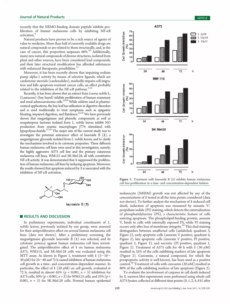
In preliminary experiments, individual constituents of L.nobilis leaves, previously isolated by our group, were assessedfor their antiproliferative effect on several human melanoma celllines (data not shown). After a preliminary screening, themegastigmane glycoside lauroside B (1) was selected, and itscytotoxic potency against human melanoma cell lines investi-gated. The antiproliferative effect of 1 on human melanomaA375, WM115, and SK-Mel-28 cells was evaluated using theMTT assay. As shown in Figure 1, treatment with 1 (3-10-30 μM) for 24-48 and 72 h caused inhibition of humanmelanomacell growth in a time- and concentration-dependent manner. Inparticular, the effect of 1 (30 μM) on cell growth, evaluated at72 h, resulted in almost 65% (p < 0.001, n = 3) inhibition forA375 cells, 50% (p < 0.001, n = 3) forWM115 cells, and 33% (p <0.001, n = 3) for SK-Mel-28 cells. Normal human epidermal
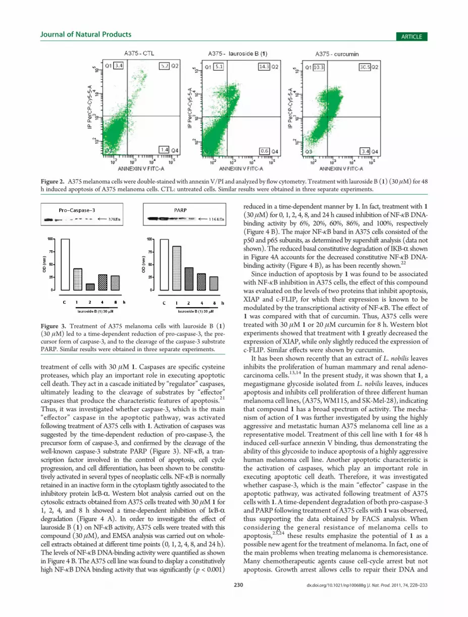
melanocyte (NHEM) growth was not affected by any of theconcentrations of 1 tested at all the time points considered (datanot shown). To further analyze the mechanism of 1-induced celldeath, induction of apoptosis was measured by annexin V/propidium iodide (PI) staining, which detects the externalizationof phosphatidylserine (PS), a characteristic feature of cellsentering apoptosis. The phospholipid-binding protein, annexinV, binds to cells with externally exposed PS, while PI stainingoccurs only after loss of membrane integrity.19 This dual stainingdistinguishes between unaffected cells (unlabeled; quadrant 3,Figure 2), early apoptotic cells (annexin V positive; quadrant 4,Figure 2), late apoptotic cells (annexin V positive, PI positive;quadrant 2, Figure 2), and necrotic (PI positive; quadrant 1,Figure 2). Treatment of A375 cells for 48 h with 1 (30 μM)resulted in 35% of the cells exhibiting markers of late apoptosis(Figure 2). Curcumin, a natural compound, for which theproapoptotic activity is well-known, has been used as a positivecontrol.20 Treatment of cells with curcumin (20 μM) resulted in80% of the cells exhibiting markers of late apoptosis (Figure 2).
To evaluate the involvement of caspases in cell death inducedby 1, western blot experiments were performed using whole-cellA375 lysates collected at different time points (0, 1, 2, 4, 8 h) after
Figure 1. Treatment with lauroside B (1) inhibits human melanomacell line proliferation in a time- and concentration-dependent fashion.
230 dx.doi.org/10.1021/np100688g |J. Nat. Prod. 2011, 74, 228–233
Journal of Natural Products ARTICLE
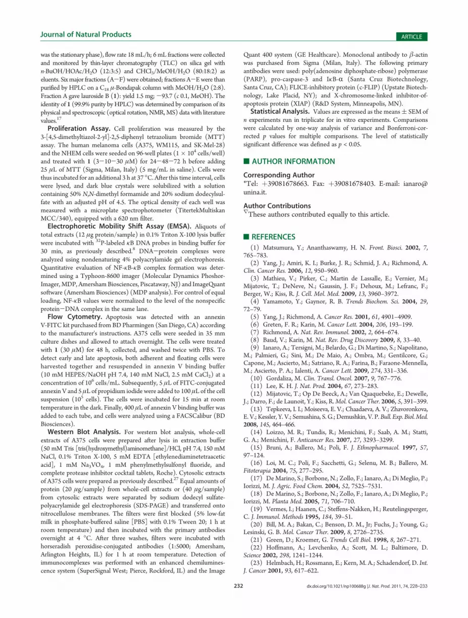
treatment of cells with 30 μM 1. Caspases are specific cysteineproteases, which play an important role in executing apoptoticcell death. They act in a cascade initiated by “regulator” caspases,ultimately leading to the cleavage of substrates by “effector”caspases that produce the characteristic features of apoptosis.21
Thus, it was investigated whether caspase-3, which is the main“effector” caspase in the apoptotic pathway, was activatedfollowing treatment of A375 cells with 1. Activation of caspases wassuggested by the time-dependent reduction of pro-caspase-3, theprecursor form of caspase-3, and confirmed by the cleavage of thewell-known caspase-3 substrate PARP (Figure 3). NF-κB, a tran-scription factor involved in the control of apoptosis, cell cycleprogression, and cell differentiation, has been shown to be constitu-tively activated in several types of neoplastic cells. NF-κB is normallyretained in an inactive form in the cytoplasm tightly associated to theinhibitory protein IκB-R. Western blot analysis carried out on thecytosolic extracts obtained from A375 cells treated with 30 μM 1 for1, 2, 4, and 8 h showed a time-dependent inhibition of IκB-Rdegradation (Figure 4 A). In order to investigate the effect oflauroside B (1) on NF-κB activity, A375 cells were treated with thiscompound (30 μM), and EMSA analysis was carried out on whole-cell extracts obtained at different time points (0, 1, 2, 4, 8, and 24 h).The levels of NF-κBDNA-binding activity were quantified as shownin Figure 4 B. The A375 cell line was found to display a constitutivelyhigh NF-κB DNA binding activity that was significantly (p < 0.001)
reduced in a time-dependent manner by 1. In fact, treatment with 1(30 μM) for 0, 1, 2, 4, 8, and 24 h caused inhibition of NF-κBDNA-binding activity by 6%, 20%, 60%, 86%, and 100%, respectively(Figure 4 B). The major NF-κB band in A375 cells consisted of thep50 and p65 subunits, as determined by supershift analysis (data notshown). The reduced basal constitutive degradation of IKB-R shownin Figure 4A accounts for the decreased constitutive NF-κB DNA-binding activity (Figure 4 B), as has been recently shown.22
Since induction of apoptosis by 1 was found to be associatedwith NF-κB inhibition in A375 cells, the effect of this compoundwas evaluated on the levels of two proteins that inhibit apoptosis,XIAP and c-FLIP, for which their expression is known to bemodulated by the transcriptional activity of NF-κB. The effect of1 was compared with that of curcumin. Thus, A375 cells weretreated with 30 μM 1 or 20 μM curcumin for 8 h. Western blotexperiments showed that treatment with 1 greatly decreased theexpression of XIAP, while only slightly reduced the expression ofc-FLIP. Similar effects were shown by curcumin.
It has been shown recently that an extract of L. nobilis leavesinhibits the proliferation of human mammary and renal adeno-carcinoma cells.13,14 In the present study, it was shown that 1, amegastigmane glycoside isolated from L. nobilis leaves, inducesapoptosis and inhibits cell proliferation of three different humanmelanoma cell lines, (A375,WM115, and SK-Mel-28), indicatingthat compound 1 has a broad spectrum of activity. The mecha-nism of action of 1 was further investigated by using the highlyaggressive and metastatic human A375 melanoma cell line as arepresentative model. Treatment of this cell line with 1 for 48 hinduced cell-surface annexin V binding, thus demonstrating theability of this glycoside to induce apoptosis of a highly aggressivehuman melanoma cell line. Another apoptotic characteristic isthe activation of caspases, which play an important role inexecuting apoptotic cell death. Therefore, it was investigatedwhether caspase-3, which is the main “effector” caspase in theapoptotic pathway, was activated following treatment of A375cells with 1. A time-dependent degradation of both pro-caspase-3and PARP following treatment of A375 cells with 1was observed,thus supporting the data obtained by FACS analysis. Whenconsidering the general resistance of melanoma cells toapoptosis,23,24 these results emphasize the potential of 1 as apossible new agent for the treatment of melanoma. In fact, one ofthe main problems when treating melanoma is chemoresistance.Many chemotherapeutic agents cause cell-cycle arrest but notapoptosis. Growth arrest allows cells to repair their DNA and
Figure 2. A375melanoma cells were double-stained with annexin V/PI and analyzed by flow cytometry. Treatment with lauroside B (1) (30 μM) for 48h induced apoptosis of A375 melanoma cells. CTL: untreated cells. Similar results were obtained in three separate experiments.
Figure 3. Treatment of A375 melanoma cells with lauroside B (1)(30 μM) led to a time-dependent reduction of pro-caspase-3, the pre-cursor form of caspase-3, and to the cleavage of the caspase-3 substratePARP. Similar results were obtained in three separate experiments.

231 dx.doi.org/10.1021/np100688g |J. Nat. Prod. 2011, 74, 228–233
Journal of Natural Products ARTICLE
limits the effectiveness of chemotherapy.25 Thus, agents that canoverride tumor resistance toward apoptosis are of great potentialvalue. Since 1 was able to induce apoptosis of a highly malignantmelanoma cell line such as A375, a better understanding of themechanism by which this critical effect is achieved was sought.The results obtained demonstrate that 1 is able to inhibit, in atime-dependent fashion, both IκB-R degradation and constitu-tive NF-κB DNA-binding activity, suggesting that the apoptoticeffect of 1 may be partly mediated via reduction of NF-κBsignaling.
In summary, the present study demonstrates, for the first time,the pro-apoptotic activity of lauroside B (1) against humanmelanoma cell lines and that this effect is associated with theinhibition of IκB-R degradation and constitutive NF-κB activa-tion. Further studies will focus on better elucidating the molec-ular mechanism of action of this molecule as well as addressing itseffects in animal models, in order to promote the potentialdevelopment of 1 as a possible chemotherapeutic agent to treathuman melanoma.
’EXPERIMENTAL SECTION
General Experimental Procedures. Electrospray ionizationmass spectrometry (ESI-MS) experiments were performed on anApplied Biosystem API 2000 triple-quadrupole mass spectrometer.Optical rotations were determined on a Jasco P-2000 polarimeter. 1Hand 13C NMR spectra were determined on a Varian Unity INOVAspectrometer at 500.13 and 125.77 MHz, respectively, equipped with an
indirect detection probe. 1H NMR were recorded relative to MeOH-d4(δ = 3.30 ppm), whereas 13C NMR spectra were recorded relative toMeOH-d4 (δ = 49.0 ppm) or XXXX. GC analyses were performed on aAgilent Technologies 6850 Series II gas chromatograph for the capillarycolumn (HP-5, 30 m� 0.25 mm, 180 �C; helium carrier flow 10mL/min)and a FID detector operated at 260 �C. TLC analysis was performed onprecoated silica gel plates (Kieselgel 60 F254, Merck). Spots were detectedunder UV light at λ254 and λ366 nm or by using ceric sulfate spray reagent.Droplet counter-current chromatography (DCCC) was performed ona DCC-A apparatus (Tokyo Rikakikai Co., Tokyo-Japan). HPLC wasperformed using a Waters 510 pump equipped with a Waters U6K injectorand aWaters 401 differential refractometer as detector, using a μ-BondapakC18 column (30 cm � 3.9 mm; Waters, Milford, MA, USA). All solvents(analytical, deuterated, and HPLC grade) were purchased from Carlo ErbaReagenti (Milano, Italy).
The human melanoma cell lines A375, WM115, and SK-Mel-28 andthe normal human epidermal melanocytes (NHEM) were purchasedfrom PromoCell (Heidelberg, D-69126, Germany). NHEMwere grownin melanocyte growth medium 2 (PromoCell). All the other cell lineswere grown in complete Dulbecco's modified Eagle's medium(DMEM), supplemented with 10% fetal bovine serum (FBS), 2 mMglutamine, 25 mM HEPES, 100 units/mL penicillin, and 100 μg/mLstreptomycin, at 37 �C in a humidified incubator under 5% CO2.Plant Material. Leaves of Laurus nobilis were collected in the hills
of Avellino (Italy) in August 2008 and identified by Prof. Paola Fortini(Dipartimento di Scienze e Tecnologie per l'Ambiente e il Territorio,DiSTAT, University of Molise). A voucher specimen is deposited underNo. LN80A-08 in the Herbarium of University of Molise (Pesche,Isernia, Italy). Leaves were kept frozen at -20 �C until analyzed.Extraction and Isolation. The leaves of L. nobilis (404 g) were
chopped and soaked in MeOH (3 � 4 L) for 6 h at room temperature.Evaporation of solvent afforded 32.5 g of a glassy material, which was thensubjected to a modified Kupchan's partitioning methodology26 as follows:the MeOH extract was dissolved in 10% aqueous methanol and partitionedagainst n-hexane (3� 400 mL), yielding 6.3 g of extract. The water content(% v/v) of theMeOH extract was adjusted to 20% and 40% and partitionedagainstCCl4 (3� 400mL) andCHCl3 (5� 400mL), respectively, yielding0.3 and 5.7 g of each extract; the aqueous residue was concentrated andpartitioned against n-BuOH (3� 500mL) to give 5.2 g of n-BuOH extract.The n-BuOH extract (5.2 g) was chromatographed by DCCC usingn-BuOH/Me2CO/H2O (3:1:5) in descending mode (the upper phase
Figure 4. Treatment of A375 human melanoma cells with lauroside B (1) (30 μM) inhibits IκB-R degradation (A) and NF-κB DNA-binding activity(B) in a time-dependent fashion. Similar results were obtained in three separate experiments.
Figure 5. Treatment of A375 human melanoma cells with lauroside B(1) (30 μM) inhibits XIAP and c-FLIP expression. Similar results wereobtained in three separate experiments.
232 dx.doi.org/10.1021/np100688g |J. Nat. Prod. 2011, 74, 228–233
Journal of Natural Products ARTICLE
was the stationary phase), flow rate 18 mL/h; 6 mL fractions were collectedand monitored by thin-layer chromatography (TLC) on silica gel withn-BuOH/HOAc/H2O (12:3:5) and CHCl3/MeOH/H2O (80:18:2) aseluents. Six major fractions (A-F) were obtained; fractions A-Ewere thanpurified by HPLC on a C18 μ-Bondapak column with MeOH/H2O (2:8).Fraction A gave lauroside B (1): yield 1.5 mg;-93.7 (c 0.1, MeOH). Theidentity of 1 (99.9% purity by HPLC) was determined by comparison of itsphysical and spectroscopic (optical rotation, NMR,MS) data with literaturevalues.17
Proliferation Assay. Cell proliferation was measured by the3-[4,5-dimethyltiazol-2-yl]-2,5-diphenyl tetrazolium bromide (MTT)assay. The human melanoma cells (A375, WM115, and SK-Mel-28)and the NHEM cells were seeded on 96-well plates (1� 104 cells/well)and treated with 1 (3-10-30 μM) for 24-48-72 h before adding25 μL of MTT (Sigma, Milan, Italy) (5 mg/mL in saline). Cells werethus incubated for an additional 3 h at 37 �C. After this time interval, cellswere lysed, and dark blue crystals were solubilized with a solutioncontaining 50% N,N-dimethyl formamide and 20% sodium dodecylsul-fate with an adjusted pH of 4.5. The optical density of each well wasmeasured with a microplate spectrophotometer (TitertekMultiskanMCC/340), equipped with a 620 nm filter.Electrophoretic Mobility Shift Assay (EMSA). Aliquots of
total extracts (12 μg protein/sample) in 0.1% Triton X-100 lysis bufferwere incubated with 32P-labeled κB DNA probes in binding buffer for30 min, as previously described.8 DNA-protein complexes wereanalyzed using nondenaturing 4% polyacrylamide gel electrophoresis.Quantitative evaluation of NF-κB-κB complex formation was deter-mined using a Typhoon-8600 imager (Molecular Dynamics Phoshor-Imager,MDP, AmershamBiosciences, Piscataway, NJ) and ImageQuantsoftware (Amersham Biosciences) (MDP analysis). For control of equalloading, NF-κB values were normalized to the level of the nonspecificprotein-DNA complex in the same lane.Flow Cytometry. Apoptosis was detected with an annexin
V-FITC kit purchased from BDPharmingen (San Diego, CA) accordingto the manufacturer's instructions. A375 cells were seeded in 35 mmculture dishes and allowed to attach overnight. The cells were treatedwith 1 (30 μM) for 48 h, collected, and washed twice with PBS. Todetect early and late apoptosis, both adherent and floating cells wereharvested together and resuspended in annexin V binding buffer(10 mM HEPES/NaOH pH 7.4, 140 mM NaCl, 2.5 mM CaCl2) at aconcentration of 106 cells/mL. Subsequently, 5 μL of FITC-conjugatedannexin V and 5 μL of propidium iodide were added to 100 μL of the cellsuspension (105 cells). The cells were incubated for 15 min at roomtemperature in the dark. Finally, 400 μL of annexin V binding buffer wasadded to each tube, and cells were analyzed using a FACSCalibur (BDBiosciences).Western Blot Analysis. For western blot analysis, whole-cell
extracts of A375 cells were prepared after lysis in extraction buffer(50 mM Tris [tris(hydroxymethyl)aminomethane]/HCl, pH 7.4, 150 mMNaCl, 0.1% Triton X-100, 5 mM EDTA [ethylenediaminetetraaceticacid], 1 mM Na3VO4, 1 mM phenylmethylsulfonyl fluoride, andcomplete protease inhibitor cocktail tablets, Roche). Cytosolic extractsof A375 cells were prepared as previously described.27 Equal amounts ofprotein (20 μg/sample) from whole-cell extracts or (40 μg/sample)from cytosolic extracts were separated by sodium dodecyl sulfate-polyacrylamide gel electrophoresis (SDS-PAGE) and transferred ontonitrocellulose membranes. The filters were first blocked (5% low-fatmilk in phosphate-buffered saline [PBS] with 0.1% Tween 20; 1 h atroom temperature) and then incubated with the primary antibodiesovernight at 4 �C. After three washes, filters were incubated withhorseradish peroxidise-conjugated antibodies (1:5000; Amersham,Arlington Heights, IL) for 1 h at room temperature. Detection ofimmunocomplexes was performed with an enhanced chemilumines-cence system (SuperSignal West; Pierce, Rockford, IL) and the Image
Quant 400 system (GE Healthcare). Monoclonal antibody to β-actinwas purchased from Sigma (Milan, Italy). The following primaryantibodies were used: poly(adenosine diphosphate-ribose) polymerase(PARP), pro-caspase-3 and IκB-R (Santa Cruz Biotechnology,Santa Cruz, CA); FLICE-inhibitory protein (c-FLIP) (Upstate Biotech-nology, Lake Placid, NY); and X-chromosome-linked inhibitor-of-apoptosis protein (XIAP) (R&D System, Minneapolis, MN).Statistical Analysis. Values are expressed as the means( SEM of
n experiments run in triplicate for in vitro experiments. Comparisonswere calculated by one-way analysis of variance and Bonferroni-cor-rected p values for multiple comparisons. The level of statisticallysignificant difference was defined as p < 0.05.
’AUTHOR INFORMATION
Corresponding Author*Tel: þ39081678663. Fax: þ39081678403. E-mail: [email protected].
Author ContributionsrThese authors contributed equally to this article.
’REFERENCES
(1) Matsumura, Y.; Ananthaswamy, H. N. Front. Biosci. 2002, 7,765–783.
(2) Yang, J.; Amiri, K. I.; Burke, J. R.; Schmid, J. A.; Richmond, A.Clin. Cancer Res. 2006, 12, 950–960.
(3) Mathieu, V.; Pirker, C.; Martin de Lassalle, E.; Vernier, M.;Mijatovic, T.; DeNeve, N.; Gaussin, J. F.; Dehoux, M.; Lefranc, F.;Berger, W.; Kiss, R. J. Cell. Mol. Med. 2009, 13, 3960–3972.
(4) Yamamoto, Y.; Gaynor, R. B. Trends Biochem. Sci. 2004, 29,72–79.
(5) Yang, J.; Richmond, A. Cancer Res. 2001, 61, 4901–4909.(6) Greten, F. R.; Karin, M. Cancer Lett. 2004, 206, 193–199.(7) Richmond, A. Nat. Rev. Immunol. 2002, 2, 664–674.(8) Baud, V.; Karin, M. Nat. Rev. Drug Discovery 2009, 8, 33–40.(9) Ianaro, A.; Tersigni, M.; Belardo, G.; Di Martino, S.; Napolitano,
M.; Palmieri, G.; Sini, M.; De Maio, A.; Ombra, M.; Gentilcore, G.;Capone, M.; Ascierto, M.; Satriano, R. A.; Farina, B.; Faraone-Mennella,M.; Ascierto, P. A.; Ialenti, A. Cancer Lett. 2009, 274, 331–336.
(10) Gordaliza, M. Clin. Transl. Oncol. 2007, 9, 767–776.(11) Lee, K. H. J. Nat. Prod. 2004, 67, 273–283.(12) Mijatovic, T.; Op De Beeck, A.; Van Quaquebeke, E.; Dewelle,
J.; Darro, F.; de Launoit, Y.; Kiss, R.Mol. Cancer Ther. 2006, 5, 391–399.(13) Tepkeeva, I. I.; Moiseeva, E. V.; Chaadaeva, A. V.; Zhavoronkova,
E. V.; Kessler, Y. V.; Semushina, S. G.; Demushkin, V. P.Bull. Exp. Biol. Med.2008, 145, 464–466.
(14) Loizzo, M. R.; Tundis, R.; Menichini, F.; Saab, A. M.; Statti,G. A.; Menichini, F. Anticancer Res. 2007, 27, 3293–3299.
(15) Bruni, A.; Ballero, M.; Poli, F. J. Ethnopharmacol. 1997, 57,97–124.
(16) Loi, M. C.; Poli, F.; Sacchetti, G.; Selenu, M. B.; Ballero, M.Fitoterapia 2004, 75, 277–295.
(17) DeMarino, S.; Borbone, N.; Zollo, F.; Ianaro, A.; Di Meglio, P.;Iorizzi, M. J. Agric. Food Chem. 2004, 52, 7525–7531.
(18) DeMarino, S.; Borbone, N.; Zollo, F.; Ianaro, A.; Di Meglio, P.;Iorizzi, M. Planta Med. 2005, 71, 706–710.
(19) Vermes, I.; Haanen, C.; Steffens-Nakken, H.; Reutelingsperger,C. J. Immunol. Methods 1995, 184, 39–51.
(20) Bill, M. A.; Bakan, C.; Benson, D. M., Jr; Fuchs, J.; Young, G.;Lesinski, G. B. Mol. Cancer Ther. 2009, 8, 2726–2735.
(21) Green, D.; Kroemer, G. Trends Cell Biol. 1998, 8, 267–271.(22) Hoffmann, A.; Levchenko, A.; Scott, M. L.; Baltimore, D.
Science 2002, 298, 1241–1244.(23) Helmbach, H.; Rossmann, E.; Kern, M. A.; Schadendorf, D. Int.
J. Cancer 2001, 93, 617–622.
233 dx.doi.org/10.1021/np100688g |J. Nat. Prod. 2011, 74, 228–233
Journal of Natural Products ARTICLE
(24) Soengas, M. S.; Capodieci, P.; Polsky, D.; Mora, J.; Esteller, M.;Opitz-Araya, X.; McCombie, R.; Herman, J. G.; Gerald, W. L.; Lazebnik,Y. A.; Cord�on-Card�o, C.; Lowe, S. W. Nature 2001, 409, 207–211.(25) Schwartz, G. K.; Shah, M. A. J. Clin. Oncol. 2005, 23,
9408–9421.(26) Kupchan, S. M.; Britton, R. W.; Ziegler, M. F.; Siegel, C. W.
J. Org. Chem. 1973, 38, 178–179.(27) Corea, G.; Fattorusso, E.; Lanzotti, V.; DiMeglio, P.; Maffia, P.;
Grassia, G.; Ialenti, A.; Ianaro, A. J. Med. Chem. 2005, 48, 7055–7062.
Related Documents