DR VINEET SAGGAR CONSULTANT DEPARTMENT OF NEUROSURGERY IVY HOSPITAL MOHALI LATE ONSET JUGULAR FORAMEN SYNDROME FOLLOWING HEAD TRAUMA

Late onset jugular foramen syndrome following head trauma
Jul 16, 2015
Welcome message from author
This document is posted to help you gain knowledge. Please leave a comment to let me know what you think about it! Share it to your friends and learn new things together.
Transcript

DR VINEET SAGGAR
CONSULTANT DEPARTMENT OF NEUROSURGERY
IVY HOSPITAL MOHALI
LATE ONSET JUGULAR FORAMEN
SYNDROME FOLLOWING HEAD TRAUMA

HISTORY
A 50 year old man presented in our OPD with sudden onset change of voice , nasal regurgitation of fluids, nausea and vomiting for past one day
Patient had suffered severe head injury few days back following RTA for which he was admitted in a hospital where he remained in unconsious state for few days and he was discharged in fully consious state after 10 days of admission in that hospital

CT scans of previous admission showed
bifrontal contusions with surrounding edema
which were managed conservatively
A week after his discharge patient suddenly
developed these problems.
There was no history of fever , ear discharge
or any previous history of coagulation
abnormalities

CLINICAL EXAMINATION
GPE of the patient was un remarkable
Gcs was 15/15
Examination of the cranial nerves revealed
- Absent gag reflex on the left side
- Lt pharyngeal arch was lower compared to rt. side
- Deviation of uvula on rt side
- Indirect laryngoscopy revealed vocal cord palsy on left side
- Shoulder shrugging was weak on left side
All these features suggested involving of 9th, 10th and 11th cranial nerves on left side.
Rest of the neurological examination was normal
RADIOLOGICAL INVESTIGATIOS
- MRI Brain revealed resolving bifrontalcontusions with absence of flow void in left transverse and sigmoid sinuses on t 2 weighted images suggestive of sinus thrombosis
- C T angio was normal in arterial phase but also revealed also revealed absence of flow in in sigmoid and transverse sinuses
- C T also revealed a fracture at the base of skull crossing jugular formamen on left side

Axial T2 flair image showing hyperintense thrombus in left sigmoid sinus

GRE IMAGES SHOWING THROMBUS IN LEFT SIGMOID SINUS
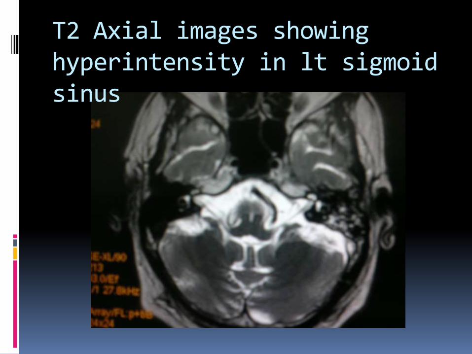
T2 Axial images showing hyperintensity in lt sigmoid sinus
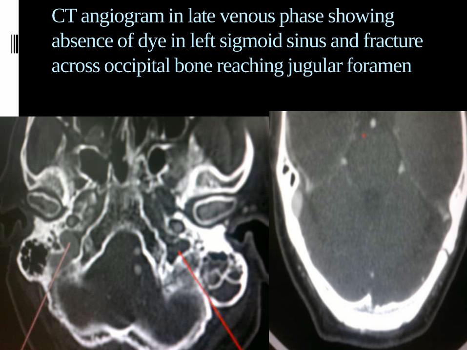
CT angiogram in late venous phase showing
absence of dye in left sigmoid sinus and fracture
across occipital bone reaching jugular foramen
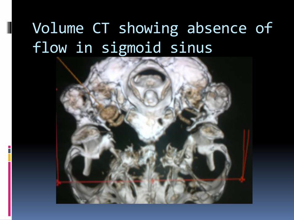
Volume CT showing absence of flow in sigmoid sinus

Patient was managed conservatively
There was no progression of symptoms over next few days
At one month follow up patient patients symptoms had resolved .
Hoarseness of voice had improved and he is able to swallow liquids normally

ANATOMY OF JUGULAR FORAMEN
It can be regarded as a hiatus between the temporal and the occipital bones
The foramen is situated so that its long axis is directed from posterolateral to anteromedialdirection.

The jugular foramen is divided into three compartments:
Two venous and a neural or intrajugularcompartment.
The venous compartments consist of a larger posterolateral venous channel, the sigmoid part, which receives the flow of the sigmoid sinus, and a smaller anteromedial venous channel,the petrosal part, which receives the drainage of the inferiorpetrosal sinus


The structures that traverse the jugular foramen are
- The sigmoid sinus and jugular bulb,
- The inferior petrosal sinus
- Meningeal branches of the ascending pharyngeal and occipital arteries,
- The glossopharyngeal, vagus, and accessory nerves with their ganglia, the tympanic branch of the glossopharyngeal nerve (Jacobson’s nerve), the auricular branch of the vagus nerve (Arnold’s nerve),
- The cochlear aqueduct.

Mechanism of thrombosis
Following head injury, skull fractures or intracranial hematomas can cause thrombosis either by direct compression of the sinus
OR
Endothelial damage within the sinus can cause the activation of the coagulation system resulting in sinus occlusion.
Uncommonly, sinus thrombosis can occur after mild closed head injury with sutural diastasis

Mechanism of cranial nerve palsies In the early literature, several cranial nerve
syndromes in CVT have been identified and attributed to extension of thrombosis into contiguous venous tributaries, presumably leading to direct pressure palsy of the nerves lying in proximity to the clot.
Recently that Kuehnen et al have suggested that local stasis in the cranial nerve veins draining into the transverse sinus might cause temporary nerve dysfunction.

DISCUSSION
The common causes of intracranial dural venous sinus thrombosis include
head and neck infections,
pregnancy and
puerperium, use of oral contraceptives,
dehydration
Rarely trauma

Although there are numerous case reports only a few case series of patients with dural venous sinus thrombosis (DVST) after blunt head trauma have been reported in the literature
Thus, the frequency and associated morbidity of DVST in patients with acute blunt head trauma are not well understood

Approximately 206 cases of traumatic DVST have been reported in the literature
For 131 of these cases, information regarding associated skull fractures has been provided
Skull fractures are present in 105 (80.2%) of the cases. In the remaining 26 cases, a diagnosis of DVST was rendered an average of 9 days (range, 1–42 days) after the trauma.

Most of these have been isolated case reports or case series mainly pretainnig to pediatric age group
Commonly, increased intracranial pressure signs such as nausea, vomiting and headache are present. The compensatory function of the venous collateral system is the main factor that determines the DST diagnosis and also affects prognosis.
When compared to adults, incomplete growth of venous collaterals in children explain higher frequency of these cases being symptomatic and reported in children
Largest study in adults to date is:
“ Prevalence of Traumatic Dural Venous Sinus Thrombosis in High-Risk Acute Blunt HeadTraumaPatients Evaluated with Multidetector CT Venography”
by DelgadoAlmandoz et al Radiology: Volume 255: Number 2—May 2010
INCLUSION CRITERIA Patients with acute blunt head trauma are
examined with multidetector CT venography for possible traumatic DVST if there was
(a) a skull fracture near a dural venous sinus or jugularbulb or
(b) a high index of clinical suspicion such as that in patients with persistent headache, vomiting, papilledema, seizures, or other signs of increased intracranial pressure.
In patients who are also deemed to be at risk for arterial injury, multidetector CT venography is performed as a delayed acquisition of the head after multidetector CT angiography of the cervical vasculature.

OBSERVATIONS
Traumatic occlusive DVST was found in 31 (19.5%) of the 159 patients with skull fractures .
The patients with skull fractures that extended to at least one dural venous sinus or jugular bulb had an overall risk of traumatic DVST of 40.7%
The risk of injury to the transverse sinuses, sigmoid sinuses, and jugular bulbs to be higher with fractures of the petrous temporal bone,
The risk of injury to the superior sagittalsinus to be higher with fractures of the occipital bone

Patients with traumatic occlusive DVST who experience neurologic deterioration, performing either MR imaging or follow-up nonenhanced CT may be useful in assessing for an associated hemorrhagic venous infarction
A small minority of patients with occlusive DVST were treated with anticoagulation. This is probably because of the high frequency of concurrent intracranial hemorrhage in this patient population and illustrates the inherent difficulty in managing their treatment
CONCLUSIONS
Traumatic DVST is a common finding complicating head injuries with fractures extending across venous sinuses
Multidetector CT angiography in venous phase is most useful in establishing the diagnosis
Treatment in most of the cases with mild neurological deficits is conservative
Treatment in patients with raised ICP may involve anticoagulation or thrombolysis but their respective roles require further studies to be fully established.

THANK YOU

Related Documents