LASIK following Small Incision Lenticule Extraction (SMILE) Lenticule Re-Implantation: A Feasibility Study of a Novel Method for Treatment of Presbyopia Chris H. L. Lim 1,2,3☯ , Andri K. Riau 2☯ , Nyein C. Lwin 2 , Shyam S. Chaurasia 2,4,5 , Donald T. Tan 2,3,4 , Jodhbir S. Mehta 2,3,6* 1 Faculty of Medicine, University of New South Wales, Sydney, New South Wales, Australia, 2 Tissue Engineering and Stem Cell Group, Singapore Eye Research Institute, Singapore, Singapore, 3 Singapore National Eye Centre, Singapore, Singapore, 4 Department of Ophthalmology, Yong Loo Lin School of Medicine, National University of Singapore, Singapore, Singapore, 5 SRP Neuroscience and Behavioral Disorders, Duke-NUS Graduate Medical School, Singapore, Singapore, 6 Department of Clinical Sciences, Duke-NUS Graduate Medical School, Singapore, Singapore Abstract Presbyopia remains a major visual impairment for patients, who have previously undergone laser refractive correction and enjoyed unaided distance vision prior to the onset of presbyopia. Corneal stromal volume restoration through small incision lenticule extraction (SMILE) lenticule re-implantation presents an opportunity for restoring the patients’ non-dominant eye to previous low myopia to achieve a monovision. In this study, we investigated the feasibility of performing LASIK after lenticule re-implantation as a method to create presbyopic monovision. A -6.00D SMILE correction was performed in 9 rabbit eyes. The lenticules were cryopreserved for 14 days and re-implanted. Five weeks later, 3 of these eyes underwent LASIK for -5.00D correction (RL group); 3 underwent LASIK flap creation, which was not lifted (RN); and no further procedures were performed on the remaining 3 eyes. These groups were compared with 3 eyes that underwent standard LASIK for a -5.00D correction (LO); 3 that underwent creation of non-lifted flap (LN); and 3 non-operated eyes. Rabbits were euthanized 1 day post-surgery. Tissue responses were analyzed by immunohistochemistry, slit lamp and in vivo confocal microscopy (IVCM). Intrastromal irregularities and elevated reflectivity levels of the excimer-ablated plane were observed on slit lamp and IVCM, respectively in the RL group. The results were comparable (P = 0.310) to IVCM findings in the LO group. RL and LO groups showed similar fibronectin expression levels, number of CD11b-positive cells (P = 0.304) and apoptotic cells (P = 0.198). There was no difference between the RN and LN groups in reflectivity levels (P = 0.627), fibronectin expression levels, CD11b-positive cells (P = 0.135) and apoptotic cells (P = 0.128). LASIK can be performed following lenticule re-implantation to create presbyopic monovision. The tissue responses elicited after performing LASIK on corneas that have undergone SMILE and subsequent lenticule re-implantation are similar to primary procedure. Citation: Lim CHL, Riau AK, Lwin NC, Chaurasia SS, Tan DT, et al. (2013) LASIK following Small Incision Lenticule Extraction (SMILE) Lenticule Re- Implantation: A Feasibility Study of a Novel Method for Treatment of Presbyopia. PLoS ONE 8(12): e83046. doi:10.1371/journal.pone.0083046 Editor: Sanjoy Bhattacharya, Bascom Palmer Eye Institute, University of Miami School of Medicine, United States of America Received September 3, 2013; Accepted November 7, 2013; Published December 11, 2013 Copyright: © 2013 Lim et al. This is an open-access article distributed under the terms of the Creative Commons Attribution License, which permits unrestricted use, distribution, and reproduction in any medium, provided the original author and source are credited. Funding: The work was supported by National Research Foundation of Singapore-Funded Translational and Clinical Research Programme Grant (NMRC/ TCR002-SERI/2008). The funders had no role in study design, data collection and analysis, decision to publish, or preparation of the manuscript. Competing interests: The authors have declared that no competing interests exist. * E-mail: [email protected] ☯ These authors contributed equally to this work. Introduction Laser in-situ keratomileusis (LASIK) is one of the most widely performed elective surgical procedure worldwide [1,2]. Its popularity stems from the short procedure time, excellent visual outcomes, pain-free and rapid post-operative visual recovery, as well as established safety profile [3,4]. This procedure first involves the creation of a corneal flap with either a mechanical microkeratome, or more recently, a femtosecond laser (FSL) [5-8]. The resulting flap is reflected and the exposed underlying stroma is then ablated with an excimer laser before the flap is repositioned. However, LASIK is not without its post-operative complications, which includes dry eye syndrome [9], transient light sensitivity [10], and keratectasia [11]. There has been a growing level of interest in recent years surrounding the application of FSL only surgery for myopic and astigmatic correction. FSLs emit ultra-short light pulses at high frequencies, permitting photodisruption of corneal tissue at lower energy levels [12,13]. This is accomplished by initiating PLOS ONE | www.plosone.org 1 December 2013 | Volume 8 | Issue 12 | e83046

Welcome message from author
This document is posted to help you gain knowledge. Please leave a comment to let me know what you think about it! Share it to your friends and learn new things together.
Transcript
LASIK following Small Incision Lenticule Extraction(SMILE) Lenticule Re-Implantation: A Feasibility Study ofa Novel Method for Treatment of PresbyopiaChris H. L. Lim1,2,3☯, Andri K. Riau2☯, Nyein C. Lwin2, Shyam S. Chaurasia2,4,5, Donald T. Tan2,3,4, Jodhbir S.Mehta2,3,6*
1 Faculty of Medicine, University of New South Wales, Sydney, New South Wales, Australia, 2 Tissue Engineering and Stem Cell Group, Singapore EyeResearch Institute, Singapore, Singapore, 3 Singapore National Eye Centre, Singapore, Singapore, 4 Department of Ophthalmology, Yong Loo Lin School ofMedicine, National University of Singapore, Singapore, Singapore, 5 SRP Neuroscience and Behavioral Disorders, Duke-NUS Graduate Medical School,Singapore, Singapore, 6 Department of Clinical Sciences, Duke-NUS Graduate Medical School, Singapore, Singapore
Abstract
Presbyopia remains a major visual impairment for patients, who have previously undergone laser refractivecorrection and enjoyed unaided distance vision prior to the onset of presbyopia. Corneal stromal volume restorationthrough small incision lenticule extraction (SMILE) lenticule re-implantation presents an opportunity for restoring thepatients’ non-dominant eye to previous low myopia to achieve a monovision. In this study, we investigated thefeasibility of performing LASIK after lenticule re-implantation as a method to create presbyopic monovision. A -6.00DSMILE correction was performed in 9 rabbit eyes. The lenticules were cryopreserved for 14 days and re-implanted.Five weeks later, 3 of these eyes underwent LASIK for -5.00D correction (RL group); 3 underwent LASIK flapcreation, which was not lifted (RN); and no further procedures were performed on the remaining 3 eyes. Thesegroups were compared with 3 eyes that underwent standard LASIK for a -5.00D correction (LO); 3 that underwentcreation of non-lifted flap (LN); and 3 non-operated eyes. Rabbits were euthanized 1 day post-surgery. Tissueresponses were analyzed by immunohistochemistry, slit lamp and in vivo confocal microscopy (IVCM). Intrastromalirregularities and elevated reflectivity levels of the excimer-ablated plane were observed on slit lamp and IVCM,respectively in the RL group. The results were comparable (P = 0.310) to IVCM findings in the LO group. RL and LOgroups showed similar fibronectin expression levels, number of CD11b-positive cells (P = 0.304) and apoptotic cells(P = 0.198). There was no difference between the RN and LN groups in reflectivity levels (P = 0.627), fibronectinexpression levels, CD11b-positive cells (P = 0.135) and apoptotic cells (P = 0.128). LASIK can be performedfollowing lenticule re-implantation to create presbyopic monovision. The tissue responses elicited after performingLASIK on corneas that have undergone SMILE and subsequent lenticule re-implantation are similar to primaryprocedure.
Citation: Lim CHL, Riau AK, Lwin NC, Chaurasia SS, Tan DT, et al. (2013) LASIK following Small Incision Lenticule Extraction (SMILE) Lenticule Re-Implantation: A Feasibility Study of a Novel Method for Treatment of Presbyopia. PLoS ONE 8(12): e83046. doi:10.1371/journal.pone.0083046
Editor: Sanjoy Bhattacharya, Bascom Palmer Eye Institute, University of Miami School of Medicine, United States of America
Received September 3, 2013; Accepted November 7, 2013; Published December 11, 2013
Copyright: © 2013 Lim et al. This is an open-access article distributed under the terms of the Creative Commons Attribution License, which permitsunrestricted use, distribution, and reproduction in any medium, provided the original author and source are credited.
Funding: The work was supported by National Research Foundation of Singapore-Funded Translational and Clinical Research Programme Grant (NMRC/TCR002-SERI/2008). The funders had no role in study design, data collection and analysis, decision to publish, or preparation of the manuscript.
Competing interests: The authors have declared that no competing interests exist.
* E-mail: [email protected]
☯ These authors contributed equally to this work.
Introduction
Laser in-situ keratomileusis (LASIK) is one of the mostwidely performed elective surgical procedure worldwide [1,2].Its popularity stems from the short procedure time, excellentvisual outcomes, pain-free and rapid post-operative visualrecovery, as well as established safety profile [3,4]. Thisprocedure first involves the creation of a corneal flap with eithera mechanical microkeratome, or more recently, a femtosecondlaser (FSL) [5-8]. The resulting flap is reflected and the
exposed underlying stroma is then ablated with an excimerlaser before the flap is repositioned. However, LASIK is notwithout its post-operative complications, which includes dry eyesyndrome [9], transient light sensitivity [10], and keratectasia[11].
There has been a growing level of interest in recent yearssurrounding the application of FSL only surgery for myopic andastigmatic correction. FSLs emit ultra-short light pulses at highfrequencies, permitting photodisruption of corneal tissue atlower energy levels [12,13]. This is accomplished by initiating
PLOS ONE | www.plosone.org 1 December 2013 | Volume 8 | Issue 12 | e83046
rapid gas expansion, which leads to cavitation bubblesformation, thereby creating an intrastromal incision plane withminimal heat development [13]. The photodisruption process,therefore, results in minimal collateral damage to adjacenttissues compared to the traditionally high-energy excimerlasers utilized in LASIK [14].
The use of FSL technology in refractive surgery has beenlargely restricted to the creation of corneal flaps, while theactual refractive correction remains the domain of the excimerlaser. With the advancement of the technology, FSL is nowcapable of correcting the refractive error by sculpting anintrastromal refractive lenticule, which is then removed by thesurgeon. The technique, coined as refractive lenticuleextraction (ReLEx), can be performed in 2 ways. The earliervariant of ReLEx, femtosecond lenticule extraction (FLEx),involves the creation of a flap akin to a LASIK flap [14-16]. Theflap is subsequently lifted, thereby exposing the refractivelenticule, which is then stripped away. The lenticule extractiontechnique has been further refined into a small incisiontechnique (SMILE, small incision lenticule extraction) [17],which involves the creation of an arcuate incision of ≤3mm inwidth, through which the refractive lenticule is extracted. Byobviating the creation of a flap, the SMILE procedure mayminimize disruption to the sub-basal nerve plexus, possiblyreducing post-operative dry eye symptoms [18], and alsoeliminating the risk of flap dislocation.
A refractive lenticule is the immediate by-product of ReLEx inall cases. The feasibility of cryopreservation and subsequentre-implantation of the extracted refractive lenticule to correctthe original myopic correction has been previously proposed[19-21]. This ability to restore corneal stromal volume bylenticule re-implantation and improve upon the post-refractivesurgical biomechanical strength of the cornea has numerouspossible applications; one of them being the unique opportunityto perform a secondary refractive surgical procedure, i.e.LASIK. This may be performed following refractive lenticule re-implantation, in order to achieve a monovision correction inpatients with presbyopia who have previously undergoneReLEx for myopic correction. A monovision is achieved whenthe dominant eye is corrected for emmetropia, while myopia iseither deliberately undercorrected or induced in the non-dominant eye [22]. Although patients will experience areduction in distance vision and stereopsis from the resultantanisometropia, ametropia of the non-dominant eye increasesthe functional zone of near and intermediate vision, as long asthe difference in refractive correction between both eyes is nottoo significant.
In this study, we demonstrate the feasibility of performingLASIK following reversal of myopic ReLEx through refractivelenticule re-implantation, as a novel method of inducingmonovision in a rabbit model of SMILE. In brief, the eyesunderwent a -6.00D spherical correction with SMILE andreversal of the myopic correction 14 days later. Once sufficientcorneal wound healing was allowed to take place over 5weeks, a LASIK flap was created anterior to the re-implantedlenticular anterior interface and a -5.00D spherical correctionperformed with an excimer laser to simulate monovisioncorrection by creating a 1.00D difference between both eyes.
Methods
AnimalsNine 12 to 15-week-old New Zealand White rabbits (3-4kg in
body weight) were procured from the National University ofSingapore. SMILE was performed to induce a refractivespherical correction of -6.00D in nine eyes. The extractedstromal lenticules were stored at -80°C for 14 days andautologous re-implantation was performed thereafter. Fiveweeks later, the nine eyes were further divided into threedifferent groups: three eyes had a corneal flap created, whichwas not lifted (this group is abbreviated as RN); while anotherthree eyes underwent LASIK for -5.00D spherical correction(RL). No further procedures were performed on the remainingthree eyes (RO). In the remaining 9 eyes that did not undergoSMILE, 3 eyes underwent LASIK for a -5.00D refractivespherical correction (LO), while another 3 eyes underwent thecreation of a non-lifted LASIK flap (LN). The last 3 non-operated eyes served as controls for this study. None of therabbits were subjected to binocular visual sensory deprivationat any point during the study. All rabbits were euthanized 1 daypost-operatively. The treatment allocation of rabbit eyes in thisstudy has been illustrated in Figure 1.
During the surgical procedures, and pre- and post-operativeexamination, the rabbits were anesthetized with xylazinehydrochloride (5 mg/kg intramuscularly; Troy Laboratories,Smithfield, Australia) and ketamine hydrochloride (50 mg/kgintramuscularly; Parnell Laboratories, Alexandria, Australia).Euthanasia was performed following the administration ofanaesthesia via an intracardiac bolus injection of sodiumpentobarbital (Jurox, Rutherford, Australia). All animals werehandled according to the guidelines outlined in the ARVOStatement for the Use of Animals in Ophthalmic and VisionResearch. Ethics approval for this study protocol was soughtand obtained from the Institutional Animal Care and UseCommittee of SingHealth (Singapore).
Small incision lenticule extraction (SMILE) procedureSMILE was performed using a VisuMax femtosecond laser
system (VisuMax, Carl Zeiss Meditec, Jena, Germany). A smallinterface cone was used in all of the procedures. Once suctionwas applied, laser incisions were made in the followingautomated sequence: a spiral in pattern for the posteriorsurface of the lenticule, spiral out pattern for the anteriorsurface of the lenticule [23], followed by a superiorly placed3mm wide incision. The diameter and depth of the cap were setat 7.5 mm and 130 μm, respectively. The diameter of thelenticule (equating to the optical zone) was 6.5 mm. Thefollowing FSL parameters were used: the power settings wereset to 200nJ in the creation of the lenticule, lenticule side, capand cap-side cuts. A side cut of 90° was used for this study.The spot distance and tracking spacing settings were set at 3μm/ 3 μm for lenticule, 2 μm/ 2 μm for lenticule side, 3 μm/ 3μm for cap, and 2 μm/ 2 μm for cap side.
After the laser sequence was completed, a Seibel spatula(Rhein Medical Inc., Petersburg, FL) was inserted into thesuperiorly placed 3mm incision to gain access to theintrastromal lenticule. Following which, a proprietary lamellar
LASIK Following SMILE Lenticule Re-Implantation
PLOS ONE | www.plosone.org 2 December 2013 | Volume 8 | Issue 12 | e83046
dissector (Asico, Westmont, IL), which we designed for SMILElamellar dissection, was introduced to dissect microadhesions;firstly on the anterior and then the posterior surfaces of thelenticule from the surrounding stroma. Once these planes wereseparated, the lenticule was extracted using a co-axial TanDSAEK forceps (Asico). The cornea stromal pocket was thenirrigated with balanced salt solution via a 24-gauge cannula.
Stromal lenticule storage and re-implantationThe extracted stromal lenticules were placed on rigid gas
permeable (RGP) contact lenses (Bausch and Lomb,Rochester, NY) and cryopreserved as previously described[19-22]. Due care was given to the anatomical orientation of thestromal lenticules while transferring these lenticules onto RGPlenses for cryopreservation. A marking was made on the RGPlenses at the 12 o'clock position to indicate the anatomicallyequivalent aspect of the stromal lenticule within the cornea.The RGP contact lenses with the refractive lenticules wereplaced in contact lens cases with the wells filled with a stockfreezing solution containing 10% FBS (fetal bovine serum;Sigma, St. Louis, MO) and 20% dimethyl sulfoxide (DMSO;Sigma). Freezing of the RGP contact lenses and contact lenscase containing the lenticule was carried out at a controlledcooling rate within a cryo-container (“Mr. Frosty”, ThermoFisher Scientific, Roskilde, Denmark) in a -80°C freezerovernight, and transferred into liquid nitrogen the following dayfor long-term storage.
Re-implantation of the refractive lenticule was performed 14days after the initial SMILE procedure. A day before theprocedure, the contact lens cases were first retrieved fromstorage and transferred to -80°C freezer overnight. Two hoursbefore re-implantation, the lenticules were retrieved from thefreezer and allowed to warm to room temperature. Therefractive lenticules were removed from the stock freezingsolution and rinsed thoroughly with balanced salt solution.Following this, the rabbits were anaesthetised and a Sinskeyhook (Rhein Medical Inc.) was used to open the previouslycreated 3-mm arcuate incision. A lamellar dissector (Asico)was then inserted through the small incision to gently releasethe cap-stromal bed adhesions. The refractive lenticule waspartially folded and reintroduced into the original site throughthe small incision using a pair of corneal forceps by carefullyobserving the 12 o’clock orientation of the lenticule on thestromal bed. Thereafter, the lamellar dissector was used tospread the refractive lenticule out within the stromal pocket. 1mL of gentamicin sulphate (40 mg/ml; Shin PoongPharmaceutical, Seoul, South Korea) and dexamethasonesodium phosphate (40 mg/ml; Shin Poong Pharmaceutical,Seoul, South Korea) were each administered via asubconjunctival injection before two interrupted sutures wereused to close the incision site. Prednisolone acetate (1%;Allergan, Irvine, CA) and tobramycin (0.3%; Alcon) drops werealso administered four times daily for the duration of one week.
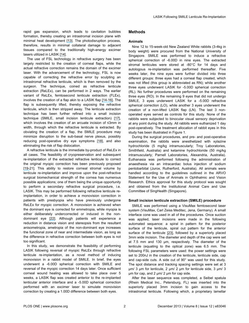
Figure 1. Treatment allocation of rabbits’ eyes in the study. On day 0, small incision lenticule extraction (SMILE) wasperformed to induce a refractive spherical correction (sph) of -6.00D in nine eyes. The extracted lenticules were cryopreserved for14 days and autologous re-implantation was performed thereafter. Five weeks later, the 9 eyes were divided into three differentgroups: three eyes had a corneal flap created, which was not lifted (this group is abbreviated as RN); while another three eyesunderwent LASIK for -5.00D spherical correction (RL); and no further procedures were performed on the remaining three eyes (RO).In the remaining 9 eyes that did not undergo SMILE on day 0, 3 eyes underwent LASIK for a -5.00D refractive spherical correction(LO); while another 3 eyes underwent the creation of a non-lifted LASIK flap (LN). The last 3 non-operated eyes served as controls.doi: 10.1371/journal.pone.0083046.g001
LASIK Following SMILE Lenticule Re-Implantation
PLOS ONE | www.plosone.org 3 December 2013 | Volume 8 | Issue 12 | e83046

Femtosecond laser-assisted LASIK procedureLASIK flaps were created with the VisuMax femtosecond
laser system (Carl Zeiss Meditec) as previously described [14].The following laser settings were used for rabbit eyes in the RNand RL groups: 80 µm flap thickness; 7.9 mm flap diameter;170 nJ power; and spot distance and tracking spacing of 4.8µm/4.8 µm for lamellar and 2 µm/2 µm for flap side cuts,respectively. All laser parameters were unchanged in the LOand LN groups, except for the flap thickness, which was set at130 µm. The corneal flap of rabbit eyes in the RL and LOgroups were lifted and a 6.5-mm optical zone ablation wasperformed using an excimer laser (Technolas; Bausch & Lomb)with the following parameters: spot size 2.0 mm diameter,fluence 120 mJ/cm2, and repetition rate 50 Hz. The flap wassubsequently repositioned and an interrupted suture used tohold the flap in place. A bandage contact lens was immediatelyapplied and temporary tarsorrhaphy was used to close theeyelids of eyes with the lifted flaps using a 6-0 silk suture.
Corneal imaging: Slit lamp photography and anteriorsegment-optical coherence tomography (AS-OCT)
Slit-lamp (Zoom Slit Lamp NS-2D; Righton, Tokyo, Japan)and AS-OCT (RTVue Fourier-Domain OCT; Optovue, Fremont,CA) photographs were captured at the following time intervals:before any surgical procedure throughout the study; 1 weekand 2 weeks after SMILE; 1 week, 2 weeks, 3 weeks and 5weeks post-re-implantation of the refractive lenticule; and 1 dayafter the secondary laser refractive surgery, LASIK, in the RLgroup. Images were also taken before surgery and 1 day afterRN, LO and LN procedures. For AS-OCT, the examineradjusted the system to position the vertex at the center of theimage and then slowly moved the AS-OCT away from thecornea until the vertical white reflection was barely visible.Measurements of central corneal thickness were obtained atthe center (0.0 mm) and at 1 mm either side of the centre (+1.0mm, -1.0 mm). The mean value of the three readings was thenreported.
In vivo confocal microscopyIn vivo confocal microscopy (HRT3; Heidelberg Engineering
GmbH, Heidelberg, Germany) was performed before anysurgical procedure throughout the study; at 1 week and 2weeks after SMILE; 1 week, 2 weeks, 3 weeks and 5 weeksafter re-implantation of the refractive lenticule; and 1 day afterthe secondary laser refractive surgery, LASIK, in the RL group.En-face images were also taken before surgery and 1 day afterRN, LO and LN procedures. A carbomer gel (Vidisic; MannPharma, Berlin, Germany) was used as the immersion fluid.The central aspects of the corneas were examined with aminimum of 3 z-axis scans, consisting of the entire cornealthickness. These micrographs were analysed and the lenticule-stromal interfaces identified. Semi-quantitative analysis of theflap interface reflectivity was performed using Image J(developed by Wayne Rasband, National Institutes of Health,Bethesda, MD; available at http://rsb.info.nih.gov/ij/index.html)by measuring the mean gray value of the reflective particles.
Tissue fixation and sectioningAfter the rabbits were euthanized, the eyes were enucleated
and the corneas excised. These corneas were embedded in anoptimum cutting temperature (OCT) cryo-compound (LeicaMicrosystems, Nussloch, Germany) and stored at -80°C untilsectioning. Using a cryostat (Microm HM550; Microm, Walldorf,Germany), 8 μm serial sagittal corneal sections were cut andplaced on polylysine-coated glass slides. These slides werestored at -80°C until immunofluorescent staining wasperformed.
Immunofluorescent stainingThe sections were air dried and fixed with 4%
paraformaldehyde (Sigma, St. Louis, MO). Following which,they were washed with 1X PBS, and incubated in 1X PBS,0.15% Triton X-100 (Sigma) to increase cellular permeability.These slides were then incubated in 4% bovine serum albumin(Sigma), a blocking reagent, and incubated overnight at 4°Cwith primary antibodies thereafter. The antibodies used wereeither mouse monoclonal antibody against cellular fibronectin(Millipore, Billerica, MA) with a dilution factor of 1:400, mousemonoclonal antibody against CD11b (BD Pharmingen, FranklinLakes, NJ) with a dilution factor of 1:100, or prediluted mousemonoclonal antibody (Invitrogen, Carlsbad, CA) against Ki-67.After washing the slides with 1X PBS the next day, the sectionswere incubated with a goat anti-mouse Alexa Fluor 488-conjugated secondary antibody (Invitrogen) for 1 hour. Theslides were then washed with 1X PBS before being mountedwith a medium containing DAPI (UltraCruz Mounting Medium;Santa Cruz Biotechnology, Santa Cruz, CA). The sections werevisualized and images captured using a fluorescencemicroscope (Zeiss AxioImager Z1; Carl Zeiss, Oberkochen,Germany).
TUNEL AssayA fluorescence-based TUNEL assay (In Situ Cell Death
Detection Kit; Roche Applied Science, Indianapolis, IN) wasused to detect apoptotic cells in the sections. This assay wasperformed according to the manufacturer’s instructions.
Statistical AnalysisData were expressed as mean ± standard deviation (SD).
The P value was determined using the Mann-Whitney U test. Avalue of P<0.05 was considered to be statistically significant.All statistical analysis was performed using SPSS software(version 21.0, SPSS Inc., Chicago, IL).
Results
Slit lamp photographyThe slit lamp photographs (Figure 2A-E) are representative
corneal images from one of the rabbits in the RL group. Retro-illumination photography demonstrated continual improvementin corneal clarity over time following re-implantation of therefractive lenticule (Figure 2A-D). No complications such asdiffuse lamellar keratitis were noted in all cases. Presence ofirregularities within the corneal stroma was noted in the first
LASIK Following SMILE Lenticule Re-Implantation
PLOS ONE | www.plosone.org 4 December 2013 | Volume 8 | Issue 12 | e83046

week following re-implantation (Figure 2A). These irregularitiesgradually reduced following suture removal, and disappearedby the second week (Figure 2B). By week 5, the corneas werecomparable to those in the control group in terms of clarity(Figure 2D and F). On examination of the corneas 1 dayfollowing LASIK, the presence of intrastromal irregularities inareas where excimer ablation was performed could beobserved (Figure 2E). The flap side cut was also visible at thistime point.
Cross-sectional visualization by AS-OCTThe AS-OCT images (Figure 3A-C) are representative
corneal cross-sectional images from one of the rabbits in theRL group. The corneal cap-stromal bed interface was barelyvisible (indicated by red arrowhead) in post-SMILE corneas(Figure 3A). However, the anterior (indicated by whitearrowhead) and posterior interfaces (indicated by white arrow)between the lenticule and stroma were clearly visible after
Figure 2. Slit lamp microscopy images of corneasfollowing refractive lenticule re-implantation andsubsequent LASIK to induce monovision. Intrastromalirregularities were evident on retro-illumination photographs onweek 1 post-re-implantation (A), but appeared to graduallyresolve from week 2 (B) to week 3 (C) and stabilized by week 5(D) post-re-implantation. (E) Slit lamp photograph of the corneaon day 1 post-LASIK demonstrating the presence ofintrastromal irregularities at the laser keratotomy site.Interrupted sutures used to hold the flap in place were visible inthe 10 o’clock position. (F) Image of a cornea from the controlgroup.doi: 10.1371/journal.pone.0083046.g002
lenticule re-implantation on AS-OCT imaging over 5-weekfollow-up period (Figure 3B). Thickening of the corneas relativeto post-SMILE corneal thickness following re-implantation wasnoted (Figure 3A, B and D), indicative of stromal volumerestoration. The corneas were relatively thicker 1-week post-re-implantation compared to subsequent follow-up examinations,attributable to the presence of corneal edema, which graduallyresolved over serial AS-OCT examinations. An increase incorneal thickness relative to post-re-implantation corneas wasnoted 1-day post-LASIK (Figure 3B, C and D). This, onceagain, most likely occurred as a result of the cornealinflammatory response and edema following LASIK.Additionally, the re-implanted lenticule also appeared thinnerafter excimer laser ablation of the anterior stroma (Figure 3C),indicated by the shorter caliper than that annotated in post-operative week 5 corneal AS-OCT image in Figure 3B.
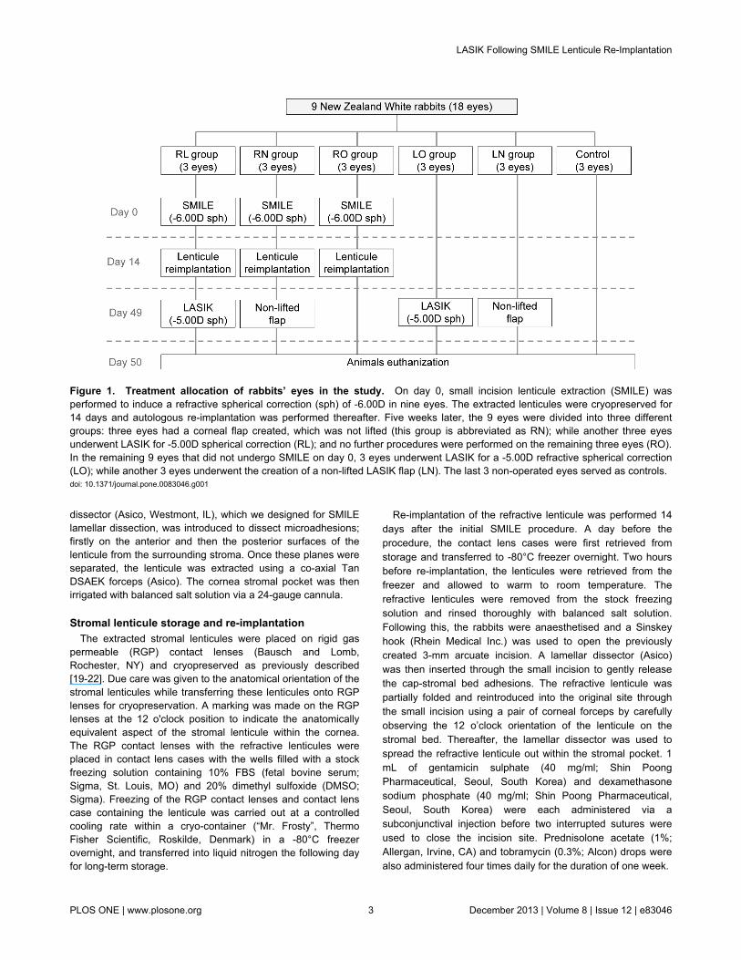
In vivo confocal microscopyThe in vivo confocal micrographs (Figure 4A) are
representative corneal en-face images from one of the rabbitsin RL group. We were able to visualize the anterior (top panel)and posterior (bottom panel) stromal-lenticular interfaces on invivo confocal microscopy (Figure 4A). Increased reflectanceand acellularity in both planes were observed 1 week after re-implantation. Small particles of varying sizes were also seen atthe anterior and posterior interfaces of the reimplantedlenticules, which could be attributed to inflammatory cells.However, these features gradually resolved over the follow-upperiod and keratocyte re-population could be seen as early as3 weeks post-re-implantation. Following LASIK, a highlyreflective and acellular layer with interspersed particles wasobserved at the anterior interface, which was the excimer laserablated stromal plane.
The relative reflectivity levels of the anterior reflectiveinterface obtained from all experimental groups were plotted ina bar graph (Figure 4B). No statistically significant differencesin the reflectivity level of interface were noted between post-RLand post-LO groups (P=0.310), and between post-RN andpost-LN groups (P=0.627).
Immunohistochemical analysisAbundant CD11b expressing cells were detected along the
excimer laser ablated plane in the central corneal stroma of theRL and LO groups (Figure 5A and B). Only a few CD11b-positive cells were seen along the non-lifted flap interface in theRN (1.5 ± 0.9) and LN (1.1 ± 0.8) groups (Figure 5C and D).CD11b staining was absent in the RO and control groups(Figure 5E and F). No differences were noted in the meanCD11b cell count between the RL and LO groups (P=0.304),and between the RN and LN groups (P=0.135). Statisticallysignificant differences were found between the RL and RNgroups (P<0.001), and between the LO and LN groups(P<0.001) (Figure 5G).
Fibronectin was expressed along the excimer laser ablatedplane in the central corneal stroma of the RL and LO groups(Figure 6A and B), with weaker expression along the non-liftedflap interface in the RN and LN groups (Figure 6C and D). Nofibronectin was present 5 weeks after lenticule re-implantation
LASIK Following SMILE Lenticule Re-Implantation
PLOS ONE | www.plosone.org 5 December 2013 | Volume 8 | Issue 12 | e83046

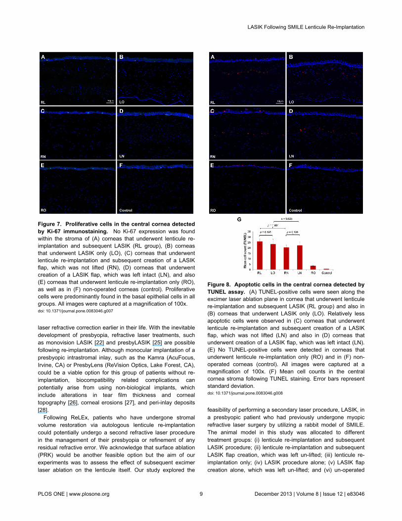
(Figure 6E) and in control corneas (Figure 6F). No Ki-67-positive cells were detected in the central corneal stroma of allexperimental groups and control corneas (Figure 7A-F). Theproliferative cells were predominantly seen in the basalepithelial cells.
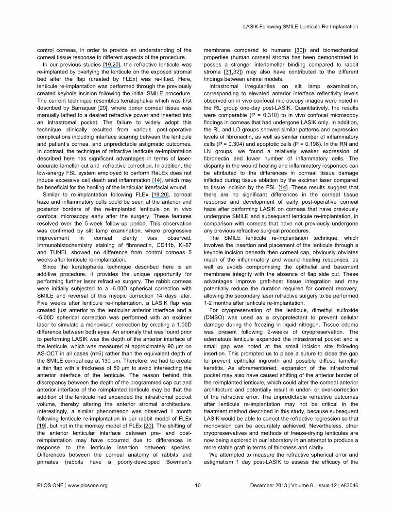
There were a significant number of TUNEL-positive cells inareas where laser ablation was performed in the RL and LOgroups (Figure 8A and B). Apoptotic cells were also seen alongthe non-lifted flap interface in the RN and LN groups (Figure 8Cand D). No cell death was observed within the corneal stroma
of rabbits in the RO and control groups (Figure 8E and F). Themean TUNEL-positive cell count was tallied (Figure 8G) and nostatistically significant differences were demonstrated betweenthe RL and LO groups (P=0.198), and between RN and LNgroups (P=0.128). No significant difference was noted betweenthe LO and LN groups (P=0.625). However, as expected, therewas a significant difference between the RL and RN groups(P=0.001).
Figure 3. Anterior segment-optical coherence tomography (AS-OCT) images of the central cornea at various study timepoints. The AS-OCT images (A-C) are representative corneal cross-sectional images from one of the rabbits that underwentlenticule re-implantation and subsequent LASIK to induce monovision. The reduction in corneal thickness following SMILE (A) wasevident, when compared to images of the cornea following refractive lenticule re-implantation (B). The bottom left panel (C) containsan image taken 1-day post-LASIK. (D) A graph depicting the corneal thickness measured on AS-OCT images at various time points.Height of error bars represents standard deviation. Red arrowheads indicate the SMILE cap-stromal bed interface. Whitearrowheads and arrows indicate the anterior and posterior planes of the re-implanted lenticule, respectively. Yellow arrowheadindicates the excimer laser ablated stromal plane and yellow arrow marks the posterior interface of the re-implanted lenticule. Acaliper in (B) and (C) is used to show the thinner lenticule within the cornea resulted from excimer laser ablation (C) compared tothe cornea before being subjected to LASIK (D).doi: 10.1371/journal.pone.0083046.g003
LASIK Following SMILE Lenticule Re-Implantation
PLOS ONE | www.plosone.org 6 December 2013 | Volume 8 | Issue 12 | e83046
Figure 4. In-vivo confocal micrographs depicting corneal stroma at various time points following refractive lenticule re-implantation and 1 day post-LASIK. The images (A) are representative corneal en-face images from one of the rabbits thatunderwent lenticule re-implantation and subsequent LASIK to induce monovision. (A) Top row shows images of the anterior borderof the reimplanted lenticule and excimer laser ablation plane in cornea that underwent LASIK following lenticule re-implantation.Bottom panel consists of micrographs of the posterior border of the reimplanted lenticule. Increased reflectance and acellularity inanterior and posterior planes were observed 1 week after re-implantation. Small particles of varying sizes were seen at bothinterfaces of the reimplanted lenticules, which could be attributed to inflammatory cells or surgical debris. However, the hazinessand particles gradually resolved over time and keratocyte re-population could be seen as early as 3 weeks post-re-implantation.Following LASIK, a highly reflective and acellular layer with interspersed particles was observed at the anterior interface, which wasnow the excimer laser ablated plane. (B) Bar graph depicting the relative reflectivity levels at the anterior reflective interfacefollowing various interventions and at various time points in the study. Error bars represent standard deviation. RN = corneas thatunderwent lenticule re-implantation and subsequent creation of a LASIK flap, which was not lifted. LO = corneas that underwentLASIK only. LN = corneas that underwent creation of a LASIK flap, which was not lifted.doi: 10.1371/journal.pone.0083046.g004
LASIK Following SMILE Lenticule Re-Implantation
PLOS ONE | www.plosone.org 7 December 2013 | Volume 8 | Issue 12 | e83046

Discussion
LASIK has undergone tremendous advancements to achieveits current well-established safety and efficacy profile [2,24].These include the integration of eye trackers, small spotscanning lasers, and wavefront-optimized/guided ablationtechnology [2]. Despite such improvements, LASIK, like allother previous forms of refractive laser correction, involves theirreversible removal of corneal tissue to achieve the requiredrefractive correction. However, the introduction of ReLEx has
Figure 5. Inflammatory cells in the central cornea detectedby CD11b immunostaining. (A) CD11b was expressed alongthe excimer laser ablation plane in corneas that underwentlenticule re-implantation and subsequent LASIK (RL group) andalso in (B) corneas that underwent LASIK only (LO). (C) NoCD11b-positive cells were observed in corneas that underwentlenticule re-implantation and subsequent creation of a LASIKflap, which was not lifted (RN), (D) corneas that underwentLASIK flap creation, which was left intact (LN), (E) corneas thatunderwent lenticule re-implantation only (RO), as well as (F)non-operated corneas (control). All images were captured at amagnification of 100x. (G) Mean cell counts in the centralcornea stroma following CD11b staining. Error bars representstandard deviation.doi: 10.1371/journal.pone.0083046.g005
possibly altered the refractive surgical landscape. ReLEx andits associated lenticule extraction techniques are the onlytheoretically reversible corneal refractive laser procedure todate [19,20]. The ability to reverse laser surgical procedureshas several clinical applications. For instance, corneal stromalvolume and biomechanical strength may be restored with eitherautologous or allogenic lenticule re-implantation in thetreatment of various forms of keratectasia. Corneal stromalrestoration also provides a unique opportunity to restore themyopic status in the non-dominant eye to previously lowmyopia, thereby resulting in presbyopic monovision. Analternative strategy for the management of presbyopia is thepossibility of re-implanting the lenticule, which has been re-shaped into a graft with a smaller diameter and positiverefractive power. In this scenario, the lenticule could act as apresbyopic intrastromal inlay.
The ability to achieve corneal volume restoration may alsoenable further refractive procedures to be performed. This is ofparticular significance in patients who have chosen to undergo
Figure 6. Wound healing reaction detected by fibronectinin the central cornea. (A) Fibronectin was expressed alongthe excimer laser ablation plane in corneas that underwentlenticule re-implantation and subsequent LASIK (RL group) andalso in (B) corneas that underwent LASIK only (LO). (C)Relatively weaker fibronectin expression was observed alongthe laser incision plane in corneas that underwent lenticule re-implantation and subsequent creation of a LASIK flap, whichwas not lifted (RN), and also in (D) corneas that underwentcreation of a LASIK flap, which was left intact (LN). (E)Fibronectin was absent in corneas that underwent lenticule re-implantation only (RO) and in (F) non-operated corneas(control). All images were captured at a magnification of 100x.doi: 10.1371/journal.pone.0083046.g006
LASIK Following SMILE Lenticule Re-Implantation
PLOS ONE | www.plosone.org 8 December 2013 | Volume 8 | Issue 12 | e83046
laser refractive correction earlier in their life. With the inevitabledevelopment of presbyopia, refractive laser treatments, suchas monovision LASIK [22] and presbyLASIK [25] are possiblefollowing re-implantation. Although monocular implantation of apresbyopic intrastromal inlay, such as the Kamra (AcuFocus,Irvine, CA) or PresbyLens (ReVision Optics, Lake Forest, CA),could be a viable option for this group of patients without re-implantation, biocompatibility related complications canpotentially arise from using non-biological implants, whichinclude alterations in tear film thickness and cornealtopography [26], corneal erosions [27], and peri-inlay deposits[28].
Following ReLEx, patients who have undergone stromalvolume restoration via autologous lenticule re-implantationcould potentially undergo a second refractive laser procedurein the management of their presbyopia or refinement of anyresidual refractive error. We acknowledge that surface ablation(PRK) would be another feasible option but the aim of ourexperiments was to assess the effect of subsequent excimerlaser ablation on the lenticule itself. Our study explored the
Figure 7. Proliferative cells in the central cornea detectedby Ki-67 immunostaining. No Ki-67 expression was foundwithin the stroma of (A) corneas that underwent lenticule re-implantation and subsequent LASIK (RL group), (B) corneasthat underwent LASIK only (LO), (C) corneas that underwentlenticule re-implantation and subsequent creation of a LASIKflap, which was not lifted (RN), (D) corneas that underwentcreation of a LASIK flap, which was left intact (LN), and also(E) corneas that underwent lenticule re-implantation only (RO),as well as in (F) non-operated corneas (control). Proliferativecells were predominantly found in the basal epithelial cells in allgroups. All images were captured at a magnification of 100x.doi: 10.1371/journal.pone.0083046.g007
feasibility of performing a secondary laser procedure, LASIK, ina presbyopic patient who had previously undergone myopicrefractive laser surgery by utilizing a rabbit model of SMILE.The animal model in this study was allocated to differenttreatment groups: (i) lenticule re-implantation and subsequentLASIK procedure; (ii) lenticule re-implantation and subsequentLASIK flap creation, which was left un-lifted; (iii) lenticule re-implantation only; (iv) LASIK procedure alone; (v) LASIK flapcreation alone, which was left un-lifted; and (vi) un-operated
Figure 8. Apoptotic cells in the central cornea detected byTUNEL assay. (A) TUNEL-positive cells were seen along theexcimer laser ablation plane in cornea that underwent lenticulere-implantation and subsequent LASIK (RL group) and also in(B) corneas that underwent LASIK only (LO). Relatively lessapoptotic cells were observed in (C) corneas that underwentlenticule re-implantation and subsequent creation of a LASIKflap, which was not lifted (LN) and also in (D) corneas thatunderwent creation of a LASIK flap, which was left intact (LN).(E) No TUNEL-positive cells were detected in corneas thatunderwent lenticule re-implantation only (RO) and in (F) non-operated corneas (control). All images were captured at amagnification of 100x. (F) Mean cell counts in the centralcornea stroma following TUNEL staining. Error bars representstandard deviation.doi: 10.1371/journal.pone.0083046.g008
LASIK Following SMILE Lenticule Re-Implantation
PLOS ONE | www.plosone.org 9 December 2013 | Volume 8 | Issue 12 | e83046
control corneas, in order to provide an understanding of thecorneal tissue response to different aspects of the procedure.
In our previous studies [19,20], the refractive lenticule wasre-implanted by overlying the lenticule on the exposed stromalbed after the flap (created by FLEx) was re-lifted. Here,lenticule re-implantation was performed through the previouslycreated keyhole incision following the initial SMILE procedure.The current technique resembles keratophakia which was firstdescribed by Barraquer [29], where donor corneal tissue wasmanually lathed to a desired refractive power and inserted intoan intrastromal pocket. The failure to widely adopt thistechnique clinically resulted from various post-operativecomplications including interface scarring between the lenticuleand patient’s cornea, and unpredictable astigmatic outcomes.In contrast, the technique of refractive lenticule re-implantationdescribed here has significant advantages in terms of laser-accurate-lamellar cut and -refractive correction. In addition, thelow-energy FSL system employed to perform ReLEx does notinduce excessive cell death and inflammation [14], which maybe beneficial for the healing of the lenticular interfacial wound.
Similar to re-implantation following FLEx [19,20], cornealhaze and inflammatory cells could be seen at the anterior andposterior borders of the re-implanted lenticule on in vivoconfocal microscopy early after the surgery. These featuresresolved over the 5-week follow-up period. This observationwas confirmed by slit lamp examination, where progressiveimprovement in corneal clarity was observed.Immunohistochemistry staining of fibronectin, CD11b, Ki-67and TUNEL showed no difference from control corneas 5weeks after lenticule re-implantation.
Since the keratophakia technique described here is anadditive procedure, it provides the unique opportunity forperforming further laser refractive surgery. The rabbit corneaswere initially subjected to a -6.00D spherical correction withSMILE and reversal of this myopic correction 14 days later.Five weeks after lenticule re-implantation, a LASIK flap wascreated just anterior to the lenticular anterior interface and a-5.00D spherical correction was performed with an excimerlaser to simulate a monovision correction by creating a 1.00Ddifference between both eyes. An anomaly that was found priorto performing LASIK was the depth of the anterior interface ofthe lenticule, which was measured at approximately 90 µm onAS-OCT in all cases (n=6) rather than the equivalent depth ofthe SMILE corneal cap at 130 µm. Therefore, we had to createa thin flap with a thickness of 80 µm to avoid intersecting theanterior interface of the lenticule. The reason behind thisdiscrepancy between the depth of the programmed cap cut andanterior interface of the reimplanted lenticule may be that theaddition of the lenticule had expanded the intrastromal pocketvolume, thereby altering the anterior stromal architecture.Interestingly, a similar phenomenon was observed 1 monthfollowing lenticule re-implantation in our rabbit model of FLEx[19], but not in the monkey model of FLEx [20]. The shifting ofthe anterior lenticular interface between pre- and post-reimplantation may have occurred due to differences inresponse to the lenticule insertion between species.Differences between the corneal anatomy of rabbits andprimates (rabbits have a poorly-developed Bowman’s
membrane compared to humans [30]) and biomechanicalproperties (human corneal stroma has been demonstrated toposses a stronger interlamellar binding compared to rabbitstroma [31,32]) may also have contributed to the differentfindings between animal models.
Intrastromal irregularities on slit lamp examination,corresponding to elevated anterior interface reflectivity levelsobserved on in vivo confocal microscopy images were noted inthe RL group one-day post-LASIK. Quantitatively, the resultswere comparable (P = 0.310) to in vivo confocal microscopyfindings in corneas that had undergone LASIK only. In addition,the RL and LO groups showed similar patterns and expressionlevels of fibronectin, as well as similar number of inflammatorycells (P = 0.304) and apoptotic cells (P = 0.198). In the RN andLN groups, we found a relatively weaker expression offibronectin and lower number of inflammatory cells. Thedisparity in the wound healing and inflammatory responses canbe attributed to the differences in corneal tissue damageinflicted during tissue ablation by the excimer laser comparedto tissue incision by the FSL [14]. These results suggest thatthere are no significant differences in the corneal tissueresponse and development of early post-operative cornealhaze after performing LASIK on corneas that have previouslyundergone SMILE and subsequent lenticule re-implantation, incomparison with corneas that have not previously undergoneany previous refractive surgical procedures.
The SMILE lenticule re-implantation technique, whichinvolves the insertion and placement of the lenticule through akeyhole incision beneath then corneal cap, obviously obviatesmuch of the inflammatory and wound healing responses, aswell as avoids compromising the epithelial and basementmembrane integrity with the absence of flap side cut. Theseadvantages improve graft-host tissue integration and maypotentially reduce the duration required for corneal recovery,allowing the secondary laser refractive surgery to be performed1-2 months after lenticule re-implantation.
For cryopreservation of the lenticule, dimethyl sulfoxide(DMSO) was used as a cryoprotectant to prevent cellulardamage during the freezing in liquid nitrogen. Tissue edemawas present following 2-weeks of cryopreservation. Theedematous lenticule expanded the intrastromal pocket and asmall gap was noted at the small incision site followinginsertion. This prompted us to place a suture to close the gapto prevent epithelial ingrowth and possible diffuse lamellarkeratitis. As aforementioned, expansion of the intrastromalpocket may also have caused shifting of the anterior border ofthe reimplanted lenticule, which could alter the corneal anteriorarchitecture and potentially result in under- or over-correctionof the refractive error. The unpredictable refractive outcomesafter lenticule re-implantation may not be critical in thetreatment method described in this study, because subsequentLASIK would be able to correct the refractive regression so thatmonovision can be accurately achieved. Nevertheless, othercryopreservatives and methods of freeze-drying lenticules arenow being explored in our laboratory in an attempt to produce amore stable graft in terms of thickness and clarity.
We attempted to measure the refractive spherical error andastigmatism 1 day post-LASIK to assess the efficacy of the
LASIK Following SMILE Lenticule Re-Implantation
PLOS ONE | www.plosone.org 10 December 2013 | Volume 8 | Issue 12 | e83046

technique described in the study, but the measurementsappeared inconsistent or inaccurate. This could be due to theearly post-surgical wound healing responses and changes incorneal biomechanics. A follow-up study is now under way inour lab to study the efficacy of the technique in a longer termusing an experimental primate model and the changes inrefractive state will then be addressed.
In conclusion, we have demonstrated that LASIK can beperformed following SMILE lenticule re-implantation, in creatingpresbyopic monovision in patients who have previouslyundergone SMILE for the correction of myopia. The cornealtissue responses elicited after performing LASIK on corneasthat have previously undergone SMILE and subsequentlenticule re-implantation are no different from the responsesevoked in corneas that have not previously undergone anyrefractive surgical procedures. Moreover, our study indicatesthat re-implantation of the lenticule in the patient’s cornea thathas previously undergone a laser refractive surgery canprovide a unique opportunity for the patient to undergo further
presbyopic corrective surgery, not limited to monovision LASIK,but also other commercially available presbyopic LASIKprogrammes, such as Supracor (Bausch + Lomb), PresbybondLaser Blended Vision (Carl Zeiss Meditec), PresbyMAX(Schwind, Kleinostheim, Germany) or PresbyLASIK (AMO,Santa Ana, CA).
Acknowledgements
The authors would like to thank Livia Yong and Jessica Yu(Carl Zeiss Meditec) for their technical assistance during theuse of the VisuMax femtosecond laser platform.
Author Contributions
Conceived and designed the experiments: DT JM. Performedthe experiments: CL AR NL. Analyzed the data: CL AR NL SCJM. Contributed reagents/materials/analysis tools: SC DT JM.Wrote the manuscript: CL AR.
References
1. American Academy of Ophthalmology (2008) Is LASIK for me? Apatient's guide to refractive surgery. Available: http://www.geteyesmart.org/eyesmart/glasses-contacts-lasik/upload/LASIK-patient-guide.pdf. Accessed 15 August 2013
2. Maldonado MJ, Nieto JC, PiñEro DP (2008) Advances in technologiesfor laser-assisted in situ keratomileusis (LASIK). Surgery - Expert RevMed Devices 5: 209-229. doi:10.1586/17434440.5.2.209.
3. Skevas C, Katz T, Wagenfeld L, Richard G, Linke S (2013) Subjectivepain, visual recovery and visual quality after LASIK, EpiLASIK (flap off)and APRK — a consecutive, non-randomized study. Graefes Arch ClinExp Ophthalmol 251: 1-9. PubMed: 22527326.
4. Fares U, Otri AM, Al-Aqaba MA, Faraj L, Dua HS (2012) Wavefront-optimized excimer laser in situ keratomileusis for myopia and myopicastigmatism: Refractive outcomes and corneal densitometry. J CataractRefract Surg 38: 2131-2138. doi:10.1016/j.jcrs.2012.07.041. PubMed:23084157.
5. Kezirian GM, Stonecipher KG (2004) Comparison of the IntraLasefemtosecond laser and mechanical keratomes for laser in situkeratomileusis. J Cataract Refract Surg 30: 804-811. doi:10.1016/j.jcrs.2003.10.026. PubMed: 15093642.
6. Nordan LT, Slade SG, Baker RN, Suarez C, Juhasz T et al. (2003)Femtosecond laser flap creation for laser in situ keratomileusis: six-month follow-up of initial U.S. clinical series. J Refract Surg 19: 8-14.PubMed: 12553599.
7. Montés-Micó R, Rodríguez-Galietero A, Alió JL (2007) Femtosecondlaser versus mechanical keratome LASIK for myopia. Ophthalmology114: 62-68. doi:10.1016/j.ophtha.2006.07.019. PubMed: 17070593.
8. Calvo R, McLaren JW, Hodge DO, Bourne WM, Patel SV (2010)Corneal aberrations and visual acuity after laser in situ keratomileusis:femtosecond laser versus mechanical microkeratome. Am JOphthalmol 149: 785-793. doi:10.1016/j.ajo.2009.12.023. PubMed:20227675.
9. Ang RT, Dartt DA, Tsubota K (2001) Dry eye after refractive surgery.Curr Opin Ophthalmol 12: 318-322. doi:10.1097/00055735-200108000-00013. PubMed: 11507347.
10. Stonecipher KG, Dishler JG, Ignacio TS, Binder PS (2006) Transientlight sensitivity after femtosecond laser flap creation: clinical findingsand management. J Cataract Refract Surg 32: 91-94. doi:10.1016/j.jcrs.2005.11.015. PubMed: 16516785.
11. Lifshitz T, Levy J, Klemperer I, Levinger S (2005) Late bilateralkeratectasia after LASIK in a low myopic patient. J Refract Surg 21:494-496. PubMed: 16209448.
12. Kullman G, Pineda R 2nd (2010) Alternative applications of thefemtosecond laser in ophthalmology. Semin Ophthalmol 25: 256-264.doi:10.3109/08820538.2010.518507. PubMed: 21091009.
13. Juhasz T, Kastis GA, Suárez C, Bor Z, Bron WE (1996) Time-resolvedobservations of shock waves and cavitation bubbles generated byfemtosecond laser pulses in corneal tissue and water. Lasers Surg Med19: 23-31. doi:10.1002/(SICI)1096-9101(1996)19:1. PubMed: 8836993.
14. Riau AK, Angunawela RI, Chaurasia SS, Lee WS, Tan DT et al. (2011)Early corneal wound healing and inflammatory responses afterrefractive lenticule extraction (ReLEx). Invest Ophthalmol Vis Sci 52:6213-6221. doi:10.1167/iovs.11-7439. PubMed: 21666235.
15. Ang M, Chaurasia SS, Angunawela RI, Poh R, Riau A et al. (2012)Femtosecond lenticule extraction (FLEx): clinical results, interfaceevaluation, and intraocular pressure variation. Invest Ophthalmol VisSci 53: 1414-1421. doi:10.1167/iovs.11-8808. PubMed: 22323464.
16. Blum M, Kunert K, Schröder M, Sekundo W (2010) Femtosecondlenticule extraction for the correction of myopia: preliminary 6-monthresults. Graefes Arch Clin Exp Ophthalmol 248: 1019-1027. doi:10.1007/s00417-009-1293-1. PubMed: 20130899.
17. Sekundo W, Kunert KS, Blum M (2011) Small incision cornealrefractive surgery using the small incision lenticule extraction (SMILE)procedure for the correction of myopia and myopic astigmatism: resultsof a 6 month prospective study. Br J Ophthalmol 95: 335-339. doi:10.1136/bjo.2009.174284. PubMed: 20601657.
18. Vestergaard AH, Grønbech KT, Grauslund J, Ivarsen AR, Hjortdal JO(2013) Subbasal nerve morphology, corneal sensation, and tear filmevaluation after refractive femtosecond laser lenticule extraction.Graefes Arch Clin Exp Ophthalmol (. (2013)) PubMed: 23793872.
19. Angunawela RI, Riau AK, Chaurasia SS, Tan DT, Mehta JS (2012)Refractive lenticule re-implantation after myopic ReLEx: a feasibilitystudy of stromal restoration after refractive surgery in a rabbit model.Invest Ophthalmol Vis Sci 53: 4975-4985. doi:10.1167/iovs.12-10170.PubMed: 22743323.
20. Riau AK, Angunawela RI, Chaurasia SS, Lee WS, Tan DT et al. (2013)Reversible femtosecond laser-assisted myopia correction: A non-human primate study of lenticule reimplantation after refractive lenticuleextraction. PLOS ONE 8: e67058. doi:10.1371/journal.pone.0067058.PubMed: 23826194.
21. Mohamed-Noriega K, Toh KP, Poh R, Balehosur D, Riau A et al. (2011)Cornea lenticule viability and structural integrity after refractive lenticuleextraction (ReLEx) and cryopreservation. Mol Vis 217: 3437-3449.PubMed: 22219639.
22. Goldberg DB (2001) Laser in situ keratomileusis monovision. JCataract Refract Surg 27: 1449-1455. doi:10.1016/S0886-3350(01)01001-X. PubMed: 11566531.
23. Riau AK, Angunawela RI, Chaurasia SS, Tan DT, Mehta JS (2012)Effect of different femtosecond laser-firing patterns on collagendisruption during refractive lenticule extraction. J Cataract Refract Surg38: 1467-1475. doi:10.1016/j.jcrs.2012.03.037. PubMed: 22814054.
24. Lawless M, Hodge C (2013) Lasik. Int Ophthalmol Clin 53: 111-128.doi:10.1097/IIO.0b013e318271346e. PubMed: 23221889.
25. Alió JL, Chaubard JJ, Caliz A, Sala E, Patel S (2006) Correction ofpresbyopia by technovision central multifocal LASIK (presbyLASIK). JRefract Surg 22: 453-460. PubMed: 16722483.
26. Dexl AK, Ruckhofer J, Riha W, Hohensinn M, Rueckl T et al. (2011)Central and peripheral corneal iron deposits after implantation of a
LASIK Following SMILE Lenticule Re-Implantation
PLOS ONE | www.plosone.org 11 December 2013 | Volume 8 | Issue 12 | e83046
small-aperture corneal inlay for correction of presbyopia. J Refract Surg27: 876-880. doi:10.3928/1081597X-20110802-02. PubMed:21815605.
27. Evans MD, Prakasam RK, Vaddavalli PK, Hughes TC, Knower W et al.(2011) A perfluoropolyether corneal inlay for the correction of refractiveerror. Biomaterials 32: 3158-3165. doi:10.1016/j.biomaterials.2011.01.047. PubMed: 21306775.
28. Mulet ME, Alio JL, Knorz MC (2009) Hydrogel intracorneal inlays for thecorrection of hyperopia: outcomes and complications after 5 years offollow-up. Ophthalmology 116: 1455-1460. doi:10.1016/j.ophtha.2009.05.019. PubMed: 19651310.
29. Barraquer JI (1972) Keratophakia. Trans Ophthalmol Soc U K 92:499-516. PubMed: 4515533.
30. Gipson IK, Spurr-Michaud SJ, Tisdale AS (1987) Anchoring fibrils forma complex network in human and rabbit cornea. Invest Ophthalmol VisSci 28: 212-220. PubMed: 8591898.
31. Maurice DM, Monroe F (1990) Cohesive strength of corneal lamellae.Exp Eye Res 50: 59-63. doi:10.1016/0014-4835(90)90011-I. PubMed:2307196.
32. Maurice DM (1988) Mechanics of the cornea. In: HD Cavanagh. TheCornea: Transactions of the World Congress on the Cornea III. NewYork. Raven Press. pp. 187-193.
LASIK Following SMILE Lenticule Re-Implantation
PLOS ONE | www.plosone.org 12 December 2013 | Volume 8 | Issue 12 | e83046
Related Documents


