Laser safety in fiber-optic monitoring of spinal cord hemodynamics: a preclinical evaluation David R. Busch James Davis Angela Kogler Robert M. Galler Ashwin B. Parthasarathy Arjun G. Yodh Thomas F. Floyd David R. Busch, James Davis, Angela Kogler, Robert M. Galler, Ashwin B. Parthasarathy, Arjun G. Yodh, Thomas F. Floyd, “Laser safety in fiber-optic monitoring of spinal cord hemodynamics: a preclinical evaluation, ” J. Biomed. Opt. 23(6), 065003 (2018), doi: 10.1117/1.JBO.23.6.065003. Downloaded From: https://www.spiedigitallibrary.org/journals/Journal-of-Biomedical-Optics on 6/20/2018 Terms of Use: https://www.spiedigitallibrary.org/terms-of-use

Welcome message from author
This document is posted to help you gain knowledge. Please leave a comment to let me know what you think about it! Share it to your friends and learn new things together.
Transcript
Laser safety in fiber-optic monitoringof spinal cord hemodynamics: apreclinical evaluation
David R. BuschJames DavisAngela KoglerRobert M. GallerAshwin B. ParthasarathyArjun G. YodhThomas F. Floyd
David R. Busch, James Davis, Angela Kogler, Robert M. Galler, Ashwin B. Parthasarathy, Arjun G. Yodh,Thomas F. Floyd, “Laser safety in fiber-optic monitoring of spinal cord hemodynamics: a preclinicalevaluation,” J. Biomed. Opt. 23(6), 065003 (2018), doi: 10.1117/1.JBO.23.6.065003.
Downloaded From: https://www.spiedigitallibrary.org/journals/Journal-of-Biomedical-Optics on 6/20/2018 Terms of Use: https://www.spiedigitallibrary.org/terms-of-use
Laser safety in fiber-optic monitoring of spinal cordhemodynamics: a preclinical evaluation
David R. Busch,a,b,c,* James Davis,d Angela Kogler,e,f Robert M. Galler,g Ashwin B. Parthasarathy,hArjun G. Yodh,c and Thomas F. Floyda,*aUniversity of Texas Southwestern, Department of Anesthesiology and Pain Management, Dallas Texas, United StatesbUniversity of Texas Southwestern, Department of Neurology and Neurotherapeutics, Dallas, Texas, United StatescUniversity of Pennsylvania, Department of Physics and Astronomy, Philadelphia, Pennsylvania, United StatesdStony Brook University Medical Center, Department of Pathology, Stony Brook, New York, United StateseStony Brook University Medical Center, Department of Anesthesiology, Stony Brook, New York, United StatesfStony Brook University, Department of Biomedical Engineering, Stony Brook, New York, United StatesgStony Brook University Medical Center, Department of Neurosurgery, Stony Brook, New York, United StateshUniversity of South Florida, Department of Electrical Engineering, Tampa, Florida, United States
Abstract. The prevention and treatment of spinal cord injury are focused upon the maintenance of spinal cordblood flow, yet no technology exists to monitor spinal cord ischemia. We recently demonstrated continuousmonitoring of spinal cord ischemia with diffuse correlation and optical spectroscopies using an opticalprobe. Prior to clinical translation of this technology, it is critically important to demonstrate the safety profileof spinal cord exposure to the required light. To our knowledge, this is the first report of in situ safety testingof such a monitor. We expose the spinal cord to laser light utilizing a custom fiber-optic epidural probe ina survival surgery model (11 adult Dorset sheep). We compare the tissue illumination from our instrumentwith the American National Standards Institute maximum permissible exposures. We experimentally evaluateneurological and pathological outcomes of the irradiated sheep associated with prolonged exposure to the lasersource and evaluate heating in ex vivo spinal cord samples. Spinal cord tissue was exposed to light levels at∼18× the maximum permissible exposure for the eye and ∼ð1∕3Þ× for the skin. Multidisciplinary testing revealedno functional neurological sequelae, histopathologic evidence of laser-related injury to the spinal cord, orsignificant temperature changes in ex vivo samples. Low tissue irradiance and the lack of neurological, patho-logical, and temperature changes upon prolonged exposure to the laser source offer evidence that spinal cordtissues can be monitored safely with near-infrared optical probes placed within the epidural space. © 2018 Society of
Photo-Optical Instrumentation Engineers (SPIE) [DOI: 10.1117/1.JBO.23.6.065003]
Keywords: spinal cord; ischemia; optical monitoring; hemodynamics; diffuse optics; intraoperative monitoring.
Paper 180118RR received Feb. 23, 2018; accepted for publication May 30, 2018; published online Jun. 19, 2018.
1 IntroductionSpinal cord injuries can occur as a result of disease (e.g.,tumors), trauma (e.g., accidents), or complications from sur-geries, such as scoliosis correction,1 spinal cord tumor resec-tion,2 and aortic aneurysm repair.3,4 The prevention andtreatment of spinal cord injury are focused upon the mainte-nance and/or restoration of spinal cord blood flow, yet no tech-nology exists to monitor spinal cord ischemia. This is a criticalgap in the field of neurocritical care monitoring. Recently, wedeveloped and demonstrated continuous monitoring for spinalcord ischemia using a thin, flexible fiber-optic probe employingnear-infrared (NIR) technology.5,6 Furthermore, testing isneeded prior to clinical adoption of this concept, includinga clear demonstration that deployment of the required laserlight in the proximity of the spinal cord is safe. This type ofsafety study is especially important because unlike many tissuesin the body, even minimal damage to spinal tissues, as mightarise in a “burn,” can lead to paralysis or paraparesis.
Current methods available for assessment of spinal cordischemia during surgery are indirect. Generally, they arebased upon neuro-electrophysiological principles, especiallysomatosensory and motor-evoked potentials (SSEP and MEP,respectively), which monitor the integrity of posterior spinalsensory pathways and the anterior/lateral spinal motor tracts,respectively. When combined, these modalities can help to iden-tify injury and can offer the surgeon insight into the impact ofinterventions and opportunities to limit or reverse injury.7,8
Interpretation of these data, however, requires the presence ofa neurologist skilled in neuro-electrophysiological monitoring.This monitoring, in turn, may be influenced by anesthetics,9,10
patient temperature,11 ischemia (cord and limb), and mechanicalmechanisms. “False negatives,” wherein patients awaken withimportant deficits in spite of “normal” evoked potentials, aswell as “false positives,” wherein patients awaken without def-icits in spite of loss or degradation of signal, have been reportedwith both SSEP12 and MEP13 monitoring, even when used ina complimentary fashion.14
Further, neuro-electrophysiological alerts may be temporallyinsensitive (delayed) relative to the inciting event,15,16 which
*Address all correspondence to: David R. Busch, E-mail: [email protected]; Thomas F. Floyd, E-mail: [email protected] 1083-3668/2018/$25.00 © 2018 SPIE
Journal of Biomedical Optics 065003-1 June 2018 • Vol. 23(6)
Journal of Biomedical Optics 23(6), 065003 (June 2018)
Downloaded From: https://www.spiedigitallibrary.org/journals/Journal-of-Biomedical-Optics on 6/20/2018 Terms of Use: https://www.spiedigitallibrary.org/terms-of-use
diminishes the chance for rescue of threatened tissue. To com-plicate the situation more, recovery of signals after rescueattempts is also markedly delayed, leaving the surgeon in aquandary per how to proceed. MEPs require application ofhigh currents to the scalp, causing significant pain, and canthus only be performed in heavily sedated patients. SSEPscan be performed in awaken patients, but the electrical stimu-lation is still painful and current spinal cord monitoring modal-ities cannot monitor patients continuously in ICU settings.Finally, these techniques are not specific. A 2012 evidence-based clinical guideline noted that only 16% to 40% patientswith significant intraoperative electrophysiological changesdeveloped postoperative-onset paraparesis, paraplegia, orquadriplegia.17 Nevertheless, despite the well-documented defi-ciencies of this methodology, MEP and SSEP are still consid-ered the “gold standard” for functional monitoring of the spinalcord for spine, spinal cord, and aortic surgery. A relatively newmonitor utilizes the perfusion pressure to the spinal column toidentify ischemic risk,18,19 yet this is still an indirect measure-ment of ischemia.
Recently, we demonstrated and validated direct monitoringof spinal cord hemodynamics in sheep using a custom fiber-optic probe and optical monitor based on diffuse optical spec-troscopy and diffuse correlation spectroscopy (DOS and DCS,respectively).5,6 DCS is a relatively new technology capable ofmeasuring microvascular blood flow. We have previously vali-dated it against fluorescent microsphere measurements of spinalcord blood flow in a sheep model.6 We, and others, have vali-dated this technology in brain and muscle against microspheres,MRI, Xe-CT, and Doppler ultrasound.20–26 DCS and DOSemploy NIR light to probe tissues up to a few centimetersbelow the surface with high temporal resolution (>0.5 Hz).These instruments are minimally invasive, portable,27–30
and they have been successfully employed to measure hemo-dynamics in a variety of tissues, including the brain,21,31–34
bone,35,36 cancer,37,38 and muscle,39,40 both at the bedside andintraoperatively.41–43 However, while optical techniques havebeen used for continuous long-term monitoring of tissues,22
such measurements typically employ low optical powers/flu-ence rates, and often, the input light travels through barrier tis-sues (e.g., scalp/skull), which effectively protect more sensitivetissues (e.g., cerebral cortex) from injury due to light radiationby reducing light energy deposition.
The importance of demonstrating the safety of optical mon-itoring techniques for the spinal cord cannot be overstated:injury to spinal cord tissues can result in life-changing paralysisor paraparesis. By comparison to other tissues, skin burns oreven localized retinal damage have relatively minor impacton patients’ quality of life. Spinal surgeries already carrysome degree of risk; a new monitoring device cannot addsignificantly to this burden. Despite this need and the rapidlyadvancing use of related optical tools in the clinic, we arenot aware of any published reports focused on optical radiationsafety in long-term monitoring of the spinal cord, and currentoptical safety standards do not address this application.44,45
A few papers have focused on therapeutic applications oflasers near the spinal cord, e.g., for laser disc decompression.These therapies frequently utilize longer wavelengths (e.g.,1064 nm) and higher power to deliberately heat or ablate tissuein a single brief session. A report of 2400 percutaneous laserdisc decompressions involving tissue ablation found no nerveor spinal cord damage,46 although damage to the nerve roots
was reported in another study.47 There have also been recentsuggestions that light of similar wavelengths may be utilizedto enhance healing of the spinal cord.48,49
Our previous spinal cord work5,6 focused on proof-of-con-cept demonstrations of optical monitoring of the spinal cordbut did not address the safety of these measurements as pertainsto optical spinal cord injury. Indeed, laser-related safety forlong-term monitoring with diffuse light has never been testedin the spinal cord and these safety questions should be consid-ered before moving these monitoring technologies to the spinalcords of human subjects. As discussed above, injury to the spi-nal cord is significantly more life-altering than damage e.g., tothe skin. Thus, the current American National StandardsInstitute (ANSI) standard,44 which is focused on skin and ocularexposure,45 lacks the specificity needed for the optical spinemonitoring problem and, thus, speaks directly to the gap inscientific knowledge in this niche and to the importance ofa safety study.
Given this knowledge gap and the serious consequences ofspinal cord damage, it is important to directly and explicitlydemonstrate the safety of optical monitoring of the spinalcord prior to undertaking human studies. In an animal model,the geometry and thermal dissipation are the key factors toreplicate, e.g., the spine of a small animal model would havea significantly different heat capacity. To this end, we explorethe safety issue using a large animal model (sheep), whichclosely approximates the geometry,50 thermal dissipation, andvulnerability to injury of the human spine and spinal cord.Specifically, we carry out monitoring measurements on sheepspinal cord with DCS and DOS laser illumination. We comparethe tissue illumination from our instrument with ANSI maxi-mum permissible exposures (MPE), and we evaluate neurologi-cal and pathological outcomes of the irradiated sheep withprolonged exposure to the laser source. Finally, tissue heatingis evaluated in ex vivo spinal cord samples.
2 Materials and MethodsExperiments were conducted in accordance with theInstitutional Animal Care and Use Committee (Stony BrookUniversity, Stony Brook, New York) guidelines.
2.1 Diffuse Optical Opto-Electronics
A “hybrid” optical system was utilized in this study, comprisingof a commercial DOS system (Imagent, ISS Inc., Urbana,Illinois) and a lab-built DCS system, which has been describedpreviously.6 Briefly, DOS relates the wavelength-dependent dif-ferential absorption of light transmitted through tissue to a quan-titative diffusion model of light propagation and estimates thebaseline/absolute concentrations of tissue chromophores, suchas oxy- and deoxy-hemoglobin.27 DCS measures the relativeblood flow in deep tissues by quantifying the temporal intensityfluctuations of the detected light, which occur mainly due tothe motion of red blood cells.29,51,52 Blood flow is estimatedfrom the decay rate of a temporal intensity autocorrelationfunction, which quantifies the light intensity fluctuations. Wedirect the interested reader to recent review articles27,53 formore details about these technologies, including their math-ematical underpinnings and an online summary of papers onDCS applications.26
The custom DCS module employs a long coherence length785-nm laser diode (CrystaLaser Inc., Nevada, DL785-100-SO) with nominal operating power of 100 mW. Light is detected
Journal of Biomedical Optics 065003-2 June 2018 • Vol. 23(6)
Busch et al.: Laser safety in fiber-optic monitoring of spinal cord hemodynamics. . .
Downloaded From: https://www.spiedigitallibrary.org/journals/Journal-of-Biomedical-Optics on 6/20/2018 Terms of Use: https://www.spiedigitallibrary.org/terms-of-use
using single-mode optical fibers coupled to avalanche photodiodedetectors (SPCM-AQ4C, Excelitas), and the photon correlationfunctions are computed using a customized (correlator.com)USB autocorrelator board.
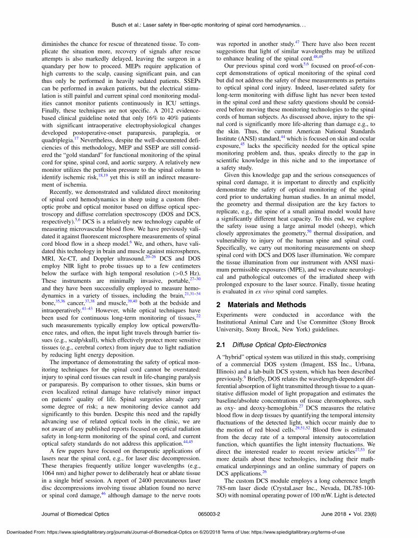
The commercial DOS system (Imagent, ISS, Illinois) utilizeslaser diodes at 686, 751, and 831 nm modulated at 110 MHz,with average operating powers of 2.7, 4, and 4 mW, respectively.This frequency-domain system quantifies tissue scattering andabsorption thereby facilitating the calculation of absolute con-centrations of oxygenated and deoxygenated hemoglobin. Noneof the wavelengths utilized have strong wavelength dependencyin standards for optical power limits; all lasers utilized have awavelength error and bandwidth of <2 nm. Note that the DCStechnique has an inherently low signal-to-noise ratio, requiringmuch higher input power (27 versus ≤4 mW) and integrationtime [2500 versus 30 ms, 35% versus 0.4% duty cycle,shown schematically in Fig. 1(d)]. In this configuration, theenergy deposited in tissue is, therefore, dominated by the DCScomponent. The ANSI standard also provides for considerationof energy deposition per pulse; only the DCS laser exposurecame even to a few percent of this limit.
2.2 Fiber-Optic Probe
Conventionally, illumination and detection of light are realizedusing flexible fiber-optic cables. For the minimally invasivemeasurements, our fiber-optic probe (Fiberoptic Systems Inc.,Simi Valley, California) is comprised of one laser sourcefiber (IR100/110/125P, 100-μm core, 0.22 NA), with DOS(IR100/110/125P, 100-μm core, 0.22 NA) and DCS (Cu8006/125/165, 0.13 NA, IVG Fiber, Toronto, Ontario, Canada)detector fibers located 2 cm axially from the source; all fibersare side-firing (see Fig. 1). These fibers were chosen to providethe best compromise between core size and total fiber size insidethe 0.9-mm-diameter probe.
2.3 Surgical, Anesthetic, and ExperimentalProcedures
Eleven adult Dorset sheep, approximately 2 years of age andweighing 60 to 70 kg, were used for the neurological and his-tological evaluations. Sheep are the preferred animal model forthese experiments as ovine spinal anatomy closely models thatof humans (e.g., spinal canal depth and width).50 Animals werepretreated with glycopyrrolate (0.02 mg∕kg, IM). Anesthesiawas induced with ketamine 10 to 20 mg∕kg IM, animalswere intubated, and ventilation controlled. Anesthesia was
maintained with isoflurane (1.5% to 3.0%). Blood pressure,electrocardiogram, ventilation rate, tidal volume, end-tidalCO2, and arterial oxygen saturation (pulse oximetry) werecontinuously monitored throughout the procedure.
After adequate general anesthesia was established, the sheepwas positioned prone. To test two commonly used clinical sur-gical procedures, the fiber-optic probe was placed via laminot-omy (N ¼ 3, “open”) and via percutaneous (N ¼ 8, “closed”)approaches into the epidural space. The percutaneous approachcarries a greater risk of spinal cord trauma related to the needleemployed, whereas the open approach virtually eliminates thisrisk of mechanical injury.
To create the laminotomy, a midline incision was made and asubperiosteal dissection was performed to expose the spinousprocesses, lamina, and medial facets. A Leksell rongeur wasthen used to remove the intraspinous ligaments as well as thespinous processes themselves. A thin-footed Kerrison rongeurwas used to carefully remove the lamina creating a trough thatwas sequentially widened until the dura of the spinal cord waswell seen. The probe was placed using loupe magnification and
(a)
(b) (c) (d)
Fig. 1 (a) Photograph of distal tip of probe (1-cm scale bar, white). (b) Diagram of distal tip of probe (not toscale) showing detector (DCS: blue and DOS: purple) and shared source (red) fibers in a protectiveTeflon sheath. (c) Photograph of probe in place and exposed spinal cord at the end of the experimentafter spinal resection (2-cm scale bar, white). (d) Timeline of optical data frame with relative laser power(to scale). Note that the irradiance from the DCS laser is much higher than the DOS lasers, and thus, theDCS laser dominates the laser safety profile of the hybrid device.
Table 1 Summary of the duration and technique of fiber-optic probeplacement, including the vertebral level at which probe was intro-duced and the final location of the probe tip on the spinal cord.
Sheep # Duration (h) TechniqueProbe entry
levelProbetip level
1 4 Laminotomy L4 L2
2 4 Laminotomy L5 L2
3 4 Laminotomy L3 L1
4 4 Percutaneous L1 T13
5 4 Percutaneous T14 T12
6 4 Percutaneous L6 L4
7 6 Percutaneous L6 L4
8 6 Percutaneous L6 L3
9 6 Percutaneous L6 L2
10 6 Percutaneous L7 L2
11 6 Percutaneous L8 L3
Journal of Biomedical Optics 065003-3 June 2018 • Vol. 23(6)
Busch et al.: Laser safety in fiber-optic monitoring of spinal cord hemodynamics. . .
Downloaded From: https://www.spiedigitallibrary.org/journals/Journal-of-Biomedical-Optics on 6/20/2018 Terms of Use: https://www.spiedigitallibrary.org/terms-of-use
was advanced superiorly or inferiorly under intact laminae tolie within the epidural space. Percutaneous placement of theprobe was achieved via 17 gauge Tuohy needle introduceda lumbar interspace, with the epidural space identified by lossof resistance to air or saline. The probe was then advanced tothe desired level under fluoroscopic guidance.
Table 1 summarizes the placement technique (laminectomyor percutaneous), vertebral level at which the probe was intro-duced, its final location on the spinal cord (confirmed fluoro-scopically for percutaneous approaches), and study duration.After introduction into the epidural space, signal was optimizedby rotation and axial adjustment along the spine. The probe wasthen left in place for 4 or 6 h, without being moved. No otherinterventions were performed during this period.
2.4 Measurement of Light Input to Tissue
In considerations of tissue safety, it is the light input to tissuethat is of concern rather than the rated power of the sources, i.e.,the distal fiber output after taking into account all coupling andtransmission losses. Table 2 summarizes the optical powers fromthe lasers at the distal end of the source fiber (the light source; alllasers were coupled into a single-source fiber). Also, the dura-tion of laser light exposure (texp) of each light source (laser) isindicated. Note that all of this light exposure is typically con-tained within a single data collection “frame” (a single DOSand DCS data point) of ∼7.25 s. Data might typically be col-lected in a clinical environment throughout a period of 4 to6 h, with a duty cycle between 5% and 35%. Here, the DCSlaser illuminated the tissue with the highest average power(27 versus ∼3 to 4 mW) and duty cycle (35% versus 0.4%);both parameters mimic our current experimental protocol.
2.5 Calculation of Maximum Permissible Exposures
The ANSI (Z136.1, 2014)45 defines a wavelength-dependentMPE. To apply this standard, we note that the laser wavelengthsof the DCS and DOS sources are in the range of 680 to 840 nm(Table 2). In this case, for a point source with wavelengthsbetween 500 and 1050 nm and exposure durations from 10to 30,000 s, the MPE (irradiance in mW∕cm2) for skin is
EQ-TARGET;temp:intralink-;sec2.5;63;307MPEskin ¼ 0.2CA:
The corresponding MPE for ocular exposure is
EQ-TARGET;temp:intralink-;sec2.5;63;266MPEeye ¼ 0.001CA;
where CA is a wavelength-dependent factor (mW∕cm2), i.e.,
EQ-TARGET;temp:intralink-;sec2.5;326;741CAð500 < λ < 700 nmÞ ¼ 1;
EQ-TARGET;temp:intralink-;sec2.5;326;711CAð700 ≤ λ < 1050 nmÞ ¼ 102λ−7001000 ;
where λ is the illumination wavelength in nm. For fiber-opticillumination with small cores (e.g., here, with ∼100-μm corediameters), ANSI Z136.1 establishes that illumination areas(Alim) be calculated based on a limiting circular aperture, corre-sponding to beam diameters of 0.7 and 0.35 cm (areas of 0.38and 0.096 cm2) for eye and skin, respectively. (Note, any smallphysiological motions, e.g., due to breathing, will have theeffect of shifting the illumination point slightly and randomlyso that the illuminated area is effectively larger than the spotsize calculated from the probe geometry and numerical aperture,here ∼0.325 mm or 0.083 mm2). In addition to heat conduction,these factors were taken into account in determining theseapertures.54 The exposure irradiance (Iexp) of the optical monitorfor each laser is hence calculated as
EQ-TARGET;temp:intralink-;sec2.5;326;530Iexp ¼PAlim
;
where P is the measured optical power at the tissue surface.In the NIR, the permitted illumination irradiance is ∼1
to 2 mW∕cm2 for the eye (point source) and 200 to375 mW∕cm2 for the skin over total exposure times of 10 to30,000 s. These total exposure time limits fall within therange of the experiments considered (i.e., with a maximumof 6 h or 21,600 s). The resulting maximal power per area(MPE) values are summarized in Table 3 for 100% and 35%duty cycles. Note that in the studies described here, as inmost clinical applications, the highest power laser (i.e., DCS)utilizes a duty cycle ≤35%. The DOS system is modulatedat RF frequencies (here, 110 MHz) and can be consideredeffectively continuous wave (CW) for the purposes of thiscalculation;45 the DCS laser is CW.
No standards are currently available for the spinal cord.Standards exist for the retina; however, although the retina stan-dards include allowance for the focusing optics in the eye. Theneural tissue of the retina is highly specialized for light detectionwith extremely sensitive photoreceptors, i.e., with structuresthat strongly absorb light. Since the spinal cord does notcontain such photoreceptors, we expect the spinal cord to beless vulnerable to optical damage than the retina. The DCSexposure irradiances we calculated for the present instrumenta-tion were less than the MPE for skin but higher than thatspecified for the retina, including the 35% duty cycle (e.g.,0.35 × Iskin ¼ 98 mW∕cm2 < 297 mW∕cm2); DOS irradianceswere well below the skin limits and only exceeded eye MPE at100% duty cycle (the 0.4% duty cycle used in this work is belowthe eye MPE).
2.6 Neurological Evaluation of Laser Safety
All animals were evaluated for neurological injury. After theillumination exposure experiment, the fiber-optic probes wereremoved, and the sheep were permitted to recover. Daily neu-rological evaluations were conducted on postoperative day 1 andday 2 using the modified Tarlov scale.55 The scoring system isdescribed in Table 4. At the end of the 48 h, the sheep were
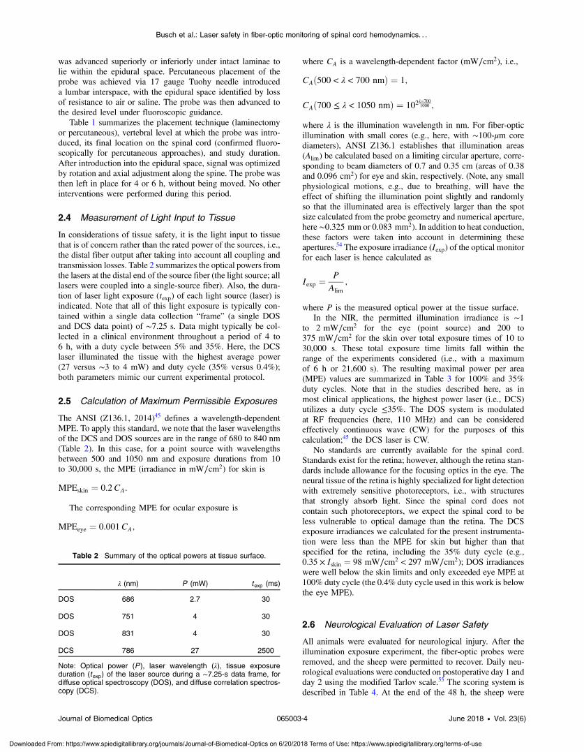
Table 2 Summary of the optical powers at tissue surface.
λ (nm) P (mW) texp (ms)
DOS 686 2.7 30
DOS 751 4 30
DOS 831 4 30
DCS 786 27 2500
Note: Optical power (P), laser wavelength (λ), tissue exposureduration (texp) of the laser source during a ∼7.25-s data frame, fordiffuse optical spectroscopy (DOS), and diffuse correlation spectros-copy (DCS).
Journal of Biomedical Optics 065003-4 June 2018 • Vol. 23(6)
Busch et al.: Laser safety in fiber-optic monitoring of spinal cord hemodynamics. . .
Downloaded From: https://www.spiedigitallibrary.org/journals/Journal-of-Biomedical-Optics on 6/20/2018 Terms of Use: https://www.spiedigitallibrary.org/terms-of-use
euthanized and the spinal cord was resected for histologicalanalysis as described in Sec. 2.6.
2.7 Histological Assessment
To determine histologically whether there was evidence of sur-face injury related to laser heating immediately followingeuthanasia, the spinal cords were excised and fixed in 10% buf-fered formalin followed by processing for paraffin embedment.Spinal cord levels (axial regions) excised included the follow-ing regions: (1) radiographically documented level of probetip, (2) one level (vertebra) above, and (3) one level belowfinal probe tip position. Paraffin-embedded tissue sectionscut at 5-μm thickness were stained with hematoxylin andeosin (H&E) with three sections produced per spinal cordlevel. A neuropathologist examined H&E slides under lightmicroscopy in a blinded fashion. Neuronal injury was evalu-ated using a semiquantitative scoring system for ischemic neu-ronal change in ventral gray motor neurons adapted from Celiket al.:56 no injury, mild injury (<5% motor neurons with ische-mic change), moderate injury (5% to 20% motor neurons withischemic change), and severe injury (>20% motor neuronswith ischemic change). H&E slides were also reviewed tonote presence or absence of changes from surface thermalinjury.
2.8 Ex vivo Evaluation of Tissue Heating
Employing a local thermometry approach, we performed ex vivoexperiments to calculate the tissue heating, if any, due to pro-longed laser exposure. Spinal cord sections from two sheep wereresected after euthanasia. In a model of the worst-case situation,we directly embedded our fiber-optic probe, along with a ther-mometry probe (Oxford Optronics, United Kingdom), coaxiallyinto the core of freshly resected, ∼3 cm, spinal cord sections.The spinal cord tissue, along with the two probes, was sus-pended in a normal saline bath maintained at 37°C to replicatein vivo conditions and the DOS/DCS sources illuminated at aduty cycle of 35% (i.e., the same settings as utilized for thein vivo measurements). The local tissue temperature was thenmonitored over 4 h, with recordings made every hour.
3 ResultsNo neurological damage to the spinal cord was observedin either daily neurological evaluation or in spinal cordhistopathology.
3.1 Neurological Evaluation of Laser Safety
Sequential neurological monitoring of 11 sheep exposed to thelaser source for 4 or 6 h revealed no signs of paresis or paralysis.The modified Tarlov score was 5 in all cases, which indicatedthat the sheep were normal and showed no signs of any neuro-logical sequelae.
3.2 Histological Assessment
Histological analysis was performed on 33 spinal cord tissuesamples resected from 11 sheep (3 slides per sheep representingthe levels above, below, and at the site where the tissue wasexposed to the laser source). We found no evidence of injuryin any of the 33 sections examined.
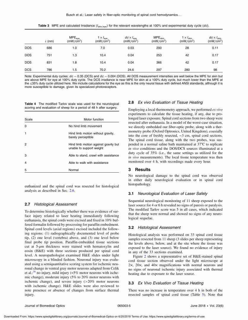
Figure 2 shows a representative set of H&E-stained spinalcord tissue section observed under the light microscope at2×, 20×, and 40× magnifications with normal neurons andno signs of neuronal ischemic injury associated with thermalheating due to exposure to the laser source.
3.3 Ex Vivo Evaluation of Tissue Heating
There was no increase in temperature over 4 h in both of theresected samples of spinal cord tissue (Table 5). Note that
Table 4 The modified Tarlov scale was used for the neurologicalscoring and evaluation of sheep for a period of 48 h after surgery.
Scale Motor function
0 No hind limb movement
1 Hind limb motion without gravity,barely perceptible
2 Hind limb motion against gravity butunable to support weight
3 Able to stand, crawl with assistance
4 Able to walk with assistance
5 Normal
Table 3 MPE and calculated Irradiance (Iskin∕eye) for the relevant wavelengths at 100% and experimental duty cycle (dc).
λ (nm)MPEeye
(mW∕cm2)1 × Ieye
(mW∕cm2)dc × Ieye(mW∕cm2)
MPEskin(mW∕cm2)
1 × Iskin(mW∕cm2)
dc × Iskin(mW∕cm2)
DOS 686 1.0 7.0 0.03 200 28 0.11
DOS 751 1.3 10.4 0.04 253 42 0.17
DOS 831 1.8 10.4 0.04 366 42 0.17
DCS 786 1.5 70.2 24.6 297 280 98
Note: Experimental duty cycles: dc ¼ 0.35 (DCS) and dc ¼ 0.004 (DOS). All DOS measurement intensities are well below the MPE for skin butare above MPE for eye at 100% duty cycle. The DCS irradiance is near MPE for skin at a 100% duty cycle, but much lower than the MPE atthe ≤35% duty cycle utilized here. We include calculations for the eye as this is the only neural tissue with defined ANSI standards, although it ismore susceptible to damage, given its specialized photoreceptors.
Journal of Biomedical Optics 065003-5 June 2018 • Vol. 23(6)
Busch et al.: Laser safety in fiber-optic monitoring of spinal cord hemodynamics. . .
Downloaded From: https://www.spiedigitallibrary.org/journals/Journal-of-Biomedical-Optics on 6/20/2018 Terms of Use: https://www.spiedigitallibrary.org/terms-of-use
this experiment was designed to mimic a “worst-case” scenario,with zero perfusion and primarily conductive heat transfer fromthe well-enclosed spinal cavity. The average and standard devi-ations of temperature measurements in the two sheep were36.9°C� 0.13°C and 37.0°C� 0.08°C.
4 DiscussionIn this study, we estimated and evaluated the safety of laser lightillumination for continuous diffuse optical hemodynamics mon-itoring of the spinal cord in sheep, utilizing a probe placedwithin the epidural space. First, we estimated the light irradianceon the spinal cord due to illumination from DOS/DCS lasers,operating at a specified duty cycle. At duty cycles that are typ-ical for long-term continuous monitoring of tissue, these tissueirradiances were less than the MPEs defined by ANSI (Z136.1)for skin (no such limits are given for spinal tissue). Furthermore,we found that exposure of the spinal cord to light did not causeobservable histological or functional neurological damage. Thefindings of this multifaceted assessment (histopathology, neuro-logical evaluations, and ex vivo measurements) demonstrate thesafety of the DOS/DCS technique for spinal cord monitoring,which may ultimately fill a critical gap in neurocritical caremonitoring.
For the wavelength range and continuous illumination (i.e.,lasers are not pulsed with high peak powers) considered in this
study, the risk to tissue from optical illumination is expected tobe exclusively thermal.45 Thermal risks include coagulation,protein denaturation, and cell death; indeed these are frequentlythe goal of photothermal therapies.57 However, these effects areall dependent upon input optical power and irradiance, as well asthe thermal conductivity of tissue.
The neural tissue of the spine is protected by the dura(∼0.3-mm thick)58—tissue that is somewhat analogous toskin in considerations of laser safety. At optical wavelengths,this dura layer scatters light, which has the effect of diffusinglight power over a broader area than the spot emanating fromthe optical fibers prior to illumination of the neural tissue.Recent computational work examined heating in the variouslayers of the head (also with a superficial skin layer) due to illu-mination at 800 nm. The computational results (10 mWat 100%duty cycle) that most closely resemble the work described(i.e., 27 mW at 35% duty cycle) suggest that laser illuminationcould potentially induce a highly localized 1.5°C increase intemperature.59 However, our experimental results using ther-mometry did not demonstrate such a change in the spinalcord with the illumination parameters described above, despiteour thermal experiment occurring in a worst-case scenario ofzero blood flow (excised tissue). We note that the catheterwall in our probe is ∼0.4-mm thick and the beam exiting thefiber-optic is diverging (0.22 NA), both of which serves to min-imize localized heating effects. In our ex vivo experiment,the introduction of the thermal probe could possibly distort/impact the distribution of light intensity in the spinal cordtissues. However, these light intensity distortions would morelikely cause an increase (e.g., due to absorption) in depositedradiant energy, thus enhancing, rather than reducing, anythermal effects. In vivo, the high blood flow (≈30 to60 ml∕100 g∕min)60,61 of the spinal cord would also help dis-perse heat. Furthermore, cerebrospinal fluid (CSF) has a higherheat conduction and capacity than air (to which skin is typicallyexposed), increasing thermal dissipation, and CSF flow likelyfurther reduces localized heating of the spinal cord.
We did not observe cell damage in histopathological analysisfollowing exposure of the spinal cord to 98 mW∕cm2 at a 35%duty cycle (i.e., ∼1∕3 of the skin MPE and ∼18× the ocularMPE). Furthermore, in a worst-case scenario of zero perfusionin an ex vivo spinal column in a thermal bath (mimicking theremainder of the body), local tissue temperature was constantwithin ¼°C. We note that power deposition is directly propor-tional to the optical duty cycle, which suggests that concernsabout the level of optical power deposition can be mitigatedby appropriately reducing the illumination duty cycle to the min-imum required to achieve the necessary temporal measurement
Fig. 2 Spinal cord tissue stained with H&E showing neurons with normal morphology. Representativeimages from a single sheep showing the whole cross section of the spinal cord (left, 2× magnification),close up showing normal spinal cord tissue surface (40× magnification), and an image showing normalneuronal morphology in the irradiated tissue (20× magnification).
Table 5 Temperature monitoring of resected spinal cord. No relevantchange in temperature of the two resected spinal cord tissue sampleswas observed after being directly exposed to the laser source ofthe fiber-optic probe for 4 h.
Temperatures (°C)
Time (h)
Sheep #
1 2
0 36.8 37.1
1 36.8 37.0
2 37.0 37.0
3 36.9 36.9
4 37.1 36.9
Mean ± SD 36.9 ± 0.1 37.0 ± 0.1
Journal of Biomedical Optics 065003-6 June 2018 • Vol. 23(6)
Busch et al.: Laser safety in fiber-optic monitoring of spinal cord hemodynamics. . .
Downloaded From: https://www.spiedigitallibrary.org/journals/Journal-of-Biomedical-Optics on 6/20/2018 Terms of Use: https://www.spiedigitallibrary.org/terms-of-use
resolution. Furthermore, these DCS data were not collected atthe minimum necessary signal-to-noise ratio for the DCS tech-nique, i.e., additional reductions in the deposited optical powerare possible simply by attenuating the lasers.
It is important to distinguish the work presented here fromprevious reports involving focused and pulsed lasers or high-speed scanning of the illumination point. Focused and pulsedlasers deliver much higher intensities at similar average powers(e.g., 25-s exposure, average power 13 mW, λ ¼ 704, 880 nm,2.5-ps pulses, and peak intensities ∼2 × 10¹¹ W∕cm2).62
Although the average power is roughly half of that describedin this work, the peak intensity is many orders of magnitudehigher, which may cause the formation of plasmas or cavitation;the unfocused, diverging, continuous illumination utilized hereis incapable of producing plasmas, etc. As per high-speed scan-ning, although there are numerous reports of scanning imagingtechniques utilized on the spine, the illumination at a singlepoint is brief. In these studies, the spine can be illuminatedat higher powers (e.g., during coherent anti-stokes Raman scat-tering microscopy), but the point of focus is scanned rapidly(<1 s). Thus, although the power densities are similar (withidentical limiting apertures, 50 to 100 mW versus 27 mW,Table 2), rapid scanning permits thermal diffusion and lessensthe probability for injury compared with the continuous station-ary illumination utilized in this paper.
Several potential limitations exist for this study. These lim-itations primarily arise from probe-tissue geometry and position-ing. A rotationally misaligned probe, or an off-center probe,could result in a lower power deposition than a probe that iswell centered with lasers directed perpendicular to the spinalcord; due to space constraints, we did not emplace continuousoptical dosimetry sensors. Similarly, the probe may be inadvert-ently placed below the dura into the subdural space instead ofthe epidural space; however, such an arrangement could onlyenhance the power deposition into the neuronal tissues. Itmay be useful to conduct future testing with purposeful place-ment of the probe below the dura to assure laser safety ina worst-case scenario. Additionally, measurements of highlylocalized (∼1 mm3) injury causing subtle changes in spinalfunction are beyond the scope of this work; this will requireprecise coregistration of illumination, histology, and functionaltesting. Modeling of light transport and simulations of heatdeposition in the spinal cord and skin will assist interpretationof future work. We have studied the effect of laser illuminationon a healthy, well-perfused spinal cord; an ischemic cord may bemore susceptible to injury. Potential future studies includeexamination of spinal cord susceptibility to laser damage inthe perfused and ischemic state and regional variation in suscep-tibility to damage. We note that we did not explicitly examinethe impact of long-term (days to weeks) exposure; long-termdamage is unlikely since we did not observe any physiologicalor structural changes due to light illumination, but subtle alter-ations in function cannot be ruled out. This lack of data on long-term chronic exposures is a well-known problem in the field, asnoted in international standards.54 Moreover, we did not attemptto define an upper limit of safe tissue exposure to laser illumi-nation. This more comprehensive study is beyond the currentlimitations of our instrumentation, i.e., our monitoring systemwas designed to be incapable of exceeding the ANSI skin limits,which have a significant safety margin prior to tissue injury. Wenote that, despite this restriction, the signal-to-noise ratio of ourcurrent system is quite high, permitting measurement of blood
flow in likely clinical scenarios and fiber geometries. Manyclinical scenarios require relatively low time resolution in meas-urement of blood flow (∼30 s), permitting significant reductionin the optical thermal input to tissue. Safety calculations for theeye must take into account the integral focusing optics; thepower density on the retina may be considerably greater thanthat on the cornea. The spinal cord does not have such focusingoptics, and thus, the direct comparison of these standards isproblematic. We include the calculations in this paper toplace the spinal cord data demonstrated here in context with theretinal limits as both a familiar value to many in the field and theonly neural tissue with established exposure limits. We note that,without establishing the upper limit for safe exposure, we cannotdetermine the ratio of our current exposure to the “true”MPE ofthe spinal cord. Finally, this study utilized a large animal model,closely mimicking human geometry. Like many large animalstudies, logistical constraints limited the total number of animalsstudied; additional work will need to follow this initial studyprior to translation into humans.
In conclusion, based on sheep studies, diffuse optical mea-surements provide a minimally invasive, nonionizing, bedsidemonitor of spinal cord hemodynamics at optical powers thatdo not cause observable neurological injury. These tools, there-fore, should be adequate for monitoring the spinal cord duringprocedures, which may cause ischemia (e.g., stent placement foraortic aneurysm), or even during spinal cord decompression.
DisclosuresDr. Floyd and Dr. Yodh’s conflicts include partial ownership ofpending patents directly related to this work: WO 2013/090658Al and PCT/US2012/069626. Dr. Yodh has partial ownership oftwo patents relevant to this work (United States patents8,082,015 and 6,076,010). Dr. Busch, Dr. Parthasarathy, andDr. Yodh have partial ownership of two pending patent appli-cations relevant to this work (PCT/US2015/017286 and PCT/US2015/017277). No author currently receives royalties or pay-ments from these patents. Dr. Floyd is the president and CEO ofNFOSYS, Inc., a startup company that may, in the future, beinvolved with producing and selling this technology.
AcknowledgmentsThis work was primarily funded by the Craig H. NeilsenFoundation Senior Research Grant (T.F.F., A.K., and A.G.Y.).The authors acknowledge contributing partial support fromthe National Institutes of Health (Nos. P41-EB015893 andR01-NS060653, A.G.Y. and A.B.P.), and we thank Dr. DavidSliney, Dr. Stefan Carp, and Valerie Perez for their valuablediscussions.
References1. D. L. Reames et al., “Complications in the surgical treatment of 19, 360
cases of pediatric scoliosis: a review of the Scoliosis Research SocietyMorbidity and Mortality Database,” Spine (Phila Pa 1976) 36(18),1484–1491 (2011).
2. C. G. Patil et al., “Complications and outcomes after spinal cord tumorresection in the United States from 1993 to 2002,” Spinal Cord 46(5),375–379 (2008).
3. D. A. Becker et al., “Predictors of outcome in patients with spinal cordischemia after open aortic repair,” Neurocrit. Care 18(1), 70–74 (2013).
4. M. F. Conrad et al., “Thoracoabdominal aneurysm repair: a 20-year per-spective,” Ann. Thorac. Surg. 83(2), S856–S861; discussion S890–S852 (2007).
Journal of Biomedical Optics 065003-7 June 2018 • Vol. 23(6)
Busch et al.: Laser safety in fiber-optic monitoring of spinal cord hemodynamics. . .
Downloaded From: https://www.spiedigitallibrary.org/journals/Journal-of-Biomedical-Optics on 6/20/2018 Terms of Use: https://www.spiedigitallibrary.org/terms-of-use
5. A. S. Kogler et al., “Fiber-optic monitoring of spinal cord hemodynam-ics in experimental aortic occlusion,” Anesthesiology 123(6), 1362–1373 (2015).
6. R. C. Mesquita et al., “Optical monitoring and detection of spinal cordischemia,” PLoS One 8(12), e83370 (2013).
7. P. Costa et al., “Somatosensory- and motor-evoked potential monitoringduring spine and spinal cord surgery,” Spinal Cord 45(1), 86–91 (2007).
8. V. Deletis and F. Sala, “Intraoperative neurophysiological monitoring ofthe spinal cord during spinal cord and spine surgery: a review focus onthe corticospinal tracts,” Clin. Neurophysiol. 119(2), 248–264 (2008).
9. S. Deiner, “Highlights of anesthetic considerations for intraoperativeneuromonitoring,” Semin. Cardiothorac. Vasc. Anesth. 14(1), 51–53(2010).
10. M. Mahmoud et al., “Susceptibility of transcranial electric motor-evoked potentials to varying targeted blood levels of dexmedetomidineduring spine surgery,” Anesthesiology 112(6), 1364–1373 (2010).
11. A. C. Wang et al., “Impact of anesthesia on transcranial electric motorevoked potential monitoring during spine surgery: a review of the liter-ature,” Neurosurg. Focus 27(4), E7 (2009).
12. R. P. Lesser et al., “Postoperative neurological deficits may occurdespite unchanged intraoperative somatosensory evoked potentials,”Ann. Neurol. 19(1), 22–25 (1986).
13. J. Y. Hong et al., “False negative and positive motor evoked potentials inone patient: is single motor evoked potential monitoring reliablemethod? A case report and literature review,” Spine (Phila Pa 1976)35(18), E912–E916 (2010).
14. C. ter Wolbeek et al., “Value and pitfalls of neurophysiological moni-toring in thoracic and thoracoabdominal aortic replacement and endo-vascular repair,” Thorac. Cardiovasc. Surg. 58(5), 260–264 (2010).
15. C. D. Etz et al., “Spinal cord blood flow and ischemic injury afterexperimental sacrifice of thoracic and abdominal segmental arteries,”Eur. J. Cardiothorac. Surg. 33(6), 1030–1038 (2008).
16. J. Lips et al., “Delayed detection of motor pathway dysfunction afterselective reduction of thoracic spinal cord blood flow in pigs,”J. Thorac. Cardiovasc. Surg. 123(3), 531–538 (2002).
17. M. R. Nuwer et al., “Evidence-based guideline update: intraoperativespinal monitoring with somatosensory and transcranial electricalmotor evoked potentials: report of the Therapeutics and TechnologyAssessment Subcommittee of the American Academy of Neurologyand the American Clinical Neurophysiology Society,” Neurology78(8), 585–589 (2012).
18. I. Phang et al., “Safety profile and probe placement accuracy of intra-spinal pressure monitoring for traumatic spinal cord injury: injured spi-nal cord pressure evaluation study,” J. Neurosurg. Spine 25(3), 398–405(2016).
19. J. W. Squair et al., “Spinal cord perfusion pressure predicts neurologicrecovery in acute spinal cord injury,” Neurology 89(16), 1660–1667(2017).
20. V. Jain et al., “Cerebral oxygen metabolism in neonates with congenitalheart disease quantified by MRI and optics,” J. Cereb. Blood FlowMetab. 34(3), 380–388 (2014).
21. S. A. Carp et al., “Validation of diffuse correlation spectroscopy mea-surements of rodent cerebral blood flow with simultaneous arterial spinlabeling MRI; towards MRI-optical continuous cerebral metabolic mon-itoring,” Biomed. Opt. Express 1(2), 553–565 (2010).
22. E. M. Buckley et al., “Validation of diffuse correlation spectroscopicmeasurement of cerebral blood flow using phase-encoded velocity map-ping magnetic resonance imaging,” J. Biomed. Opt. 17(3), 037007(2012).
23. G. Q. Yu et al., “Validation of diffuse correlation spectroscopy formuscle blood flow with concurrent arterial spin labeled perfusionMRI,” Opt. Express 15(3), 1064–1075 (2007).
24. T. Durduran et al., “Optical measurement of cerebral hemodynamicsand oxygen metabolism in neonates with congenital heart defects,”J. Biomed. Opt. 15(3), 037004 (2010).
25. M. N. Kim et al., “Validation of diffuse correlation spectroscopy againstxenon CTCBF in humans after traumatic brain injury or subarachnoidhemorrhage,” in Neurocritical Care Society Annual Meeting, Miami,Florida (2008).
26. D. R. Busch, “A collection of papers on applications of diffuse corre-lation spectroscopy (DCS),” 2017, https://www.physics.upenn.edu/yodhlab/dcs/ (01 August 2017).
27. T. Durduran et al., “Diffuse optics for tissue monitoring and tomogra-phy,” Rep. Prog. Phys. 73(7), 076701 (2010).
28. D. A. Boas and A. G. Yodh, “Spatially varying dynamical properties ofturbid media probed with diffusing temporal light correlation,” J. Opt.Soc. Am. A 14(1), 192–215 (1997).
29. E. M. Buckley et al., “Diffuse correlation spectroscopy for measurementof cerebral blood flow: future prospects,” Neurophotonics 1(1), 011009(2014).
30. J. Selb et al., “Sensitivity of near-infrared spectroscopy and diffuse cor-relation spectroscopy to brain hemodynamics: simulations and experi-mental findings during hypercapnia,” Neurophotonics 1(1), 015005(2014).
31. M. Diop et al., “Calibration of diffuse correlation spectroscopy with atime-resolved near-infrared technique to yield absolute cerebral bloodflow measurements,” Biomed. Opt. Express 2(7), 2068–2081 (2011).
32. E. M. Buckley et al., “Decreased microvascular cerebral blood flowassessed by diffuse correlation spectroscopy after repetitive concussionsin mice,” J. Cereb. Blood Flow Metab. 35(12), 1995–2000 (2015).
33. C. G. Favilla et al., “Optical bedside monitoring of cerebral blood flowin acute ischemic stroke patients during head-of-bed manipulation,”Stroke 45(5), 1269–1274 (2014).
34. D. R. Busch et al., “Cerebral blood flow response to hypercapnia inchildren with obstructive sleep Apnea syndrome,” Sleep 39(1), 209–216 (2016).
35. P. Farzam et al., “Noninvasive characterization of the healthy humanmanubrium using diffuse optical spectroscopies,” Physiol. Meas.35(7), 1469–1491 (2014).
36. S. Han et al., “Non-invasive monitoring of temporal and spatial bloodflow during bone graft healing using diffuse correlation spectroscopy,”PLoS One 10(12), e0143891 (2015).
37. R. Choe et al., “Optically measured microvascular blood flow contrastof malignant breast tumors,” PLoS One 9(6), e99683 (2014).
38. D. R. Busch et al., “Blood flow reduction in breast tissue due tomammographic compression,” Acad. Radiol. 21(2), 151–161 (2014).
39. G. Q. Yu et al., “Time-dependent blood flow and oxygenation in humanskeletal muscles measured with noninvasive near-infrared diffuse opti-cal spectroscopies,” J. Biomed. Opt. 10, 024027 (2005).
40. Y. Shang et al., “Noninvasive optical characterization of muscle bloodflow, oxygenation, and metabolism in women with fibromyalgia,”Arthritis Res. Ther. 14(6), R236 (2012).
41. C. Huang et al., “Noncontact diffuse optical assessment of blood flowchanges in head and neck free tissue transfer flaps,” J. Biomed. Opt.20(7), 075008 (2015).
42. Y. Shang et al., “Cerebral monitoring during carotid endarterectomyusing near-infrared diffuse optical spectroscopies and electroencephalo-gram,” Phys. Med. Biol. 56(10), 3015–3032 (2011).
43. D. R. Busch et al., “Continuous cerebral hemodynamic measurementduring deep hypothermic circulatory arrest,” Biomed. Opt. Express7(9), 3461–3470 (2016).
44. Laser Institute of America, “American National Standard for the safeuse of lasers: ANSI Z136.3-2011,” Laser Institute of America,Orlando, Florida (2011).
45. Laser Institute of America, “American National Standard for the safeuse of lasers: ANSI Z136.1-2014,” Laser Institute of America,Orlando, Florida (2014).
46. D. S. Choy, “Percutaneous laser disc decompression: a 17-year expe-rience,” Photomed. Laser Surg. 22(5), 407–410 (2004).
47. S. Kobayashi et al., “A case of nerve root heat injury induced by per-cutaneous laser disc decompression performed at an outside institution:technical case report,” Oper. Neurosurg. 60(Suppl. 2), ONS-E171–ONS-E172 (2007).
48. M. S. Moreira et al., “Effect of laser phototherapy on wound healingfollowing cerebral ischemia by cryogenic injury,” J. Photochem.Photobiol. B 105(3), 207–215 (2011).
49. S. Veronez et al., “Effects of different fluences of low-level laser therapyin an experimental model of spinal cord injury in rats,” Lasers Med. Sci.32(2), 343–349 (2017).
50. S. R. Sheng et al., “Anatomy of large animal spines and its comparison tothe human spine: a systematic review,” Eur. Spine J. 19(1), 46–56 (2010).
51. T. Durduran and A. G. Yodh, “Diffuse correlation spectroscopy fornon-invasive, micro-vascular cerebral blood flow measurement,”NeuroImage 85(Pt. 1), 51–63 (2014).
Journal of Biomedical Optics 065003-8 June 2018 • Vol. 23(6)
Busch et al.: Laser safety in fiber-optic monitoring of spinal cord hemodynamics. . .
Downloaded From: https://www.spiedigitallibrary.org/journals/Journal-of-Biomedical-Optics on 6/20/2018 Terms of Use: https://www.spiedigitallibrary.org/terms-of-use
52. D. A. Boas, L. E. Campbell, and A. G. Yodh, “Scattering and imagingwith diffusing temporal field correlations,” Phys. Rev. Lett. 75(9), 1855–1858 (1995).
53. R. C. Mesquita and A. Yodh, “Diffuse optics: fundamentals and tissueapplications,” in Proceedings of the International School of Physics“Enrico Fermi,” Nano Optics and Atomics: Transport of Light andMater Waves, R. Kaiser, D. S. Weirsma, and L. Fallini, Eds., IOSPress, Amsterdam (2011).
54. International Commission on Non-Ionizing Radiation Protection,“Protection ICoN-IR. ICNIRP guidelines on limits of exposure tolaser radiation of wavelengths between 180 nm and 1000 μm,”Health Phys. 105(3), 271–295 (2013).
55. P. G. Popovich et al., “A reassessment of a classic neuroprotective com-bination therapy for spinal cord injured rats: LPS/pregnenolone/indome-thacin,” Exp. Neurol. 233(2), 677–685 (2012).
56. M. Celik et al., “Erythropoietin prevents motor neuron apoptosis andneurologic disability in experimental spinal cord ischemic injury,”Proc. Natl. Acad. Sci. U. S. A. 99(4), 2258–2263 (2002).
57. S. Thomsen, “Pathologic analysis of photothermal and photomechanicaleffects of laser-tissue interactions,” Photochem. Photobiol. 53(6),825–835 (1991).
58. J. Y. Hong et al., “Analysis of dural sac thickness in human spine-cadaver study with confocal infrared laser microscope,” Spine J.11(12), 1121–1127 (2011).
59. M. Nourhashemi, M. Mahmoudzadeh, and F. Wallois, “Thermal impactof near-infrared laser in advanced noninvasive optical brain imaging,”Neurophotonics 3(1), 015001 (2016).
60. G. Zoccoli et al., “Spinal cord blood flow changes during the sleep-wake cycle in rat,” Neurosci. Lett. 163(2), 173–176 (1993).
61. S. H. Johnson, J. M. Kraimer, and G. M. Graeber, “Effects offlunarizine on neurological recovery and spinal cord blood flow in
experimental spinal cord ischemia in rabbits,” Stroke 24(10), 1547–1553 (1993).
62. Y. Fu et al., “Characterization of photodamage in coherent anti-Stokes Raman scattering microscopy,” Opt. Express 14(9), 3942–3951(2006).
David R. Busch is an assistant professor in the Department ofAnesthesiology and Pain Management and Department of Neurologyand Neurotherapeutics, the University of Texas SouthwesternMedical Center. His work focuses on developing noninvasive andminimally invasive optical tools to monitor deep tissues in healthand disease, as well as throughout therapy.
Ashwin B. Parthasarathy is an assistant professor of electricalengineering at the University of South Florida, Tampa, Florida. HisTranslational Optics Imaging and Spectroscopy Lab is focused onthe development and application of optical spectroscopy and micros-copy techniques for the bedside monitoring and imaging of tissuephysiology in human and animal models.
Arjun G. Yodh is a Skinner professor of science in the Department ofPhysics and Astronomy, the University of Pennsylvania. He alsodirects the Laboratory for Research on the structure of matter, aninterdisciplinary materials institute that hosts the Penn MRSEC. Hisbiomedical research is oriented toward diffuse optical imaging andmonitoring, with aims to demonstrate the potential of these tools forfunctional imaging/monitoring in brain, breast, and muscle, and formonitoring hemodynamic biomarkers during treatment.
Biographies for the other authors are not available.
Journal of Biomedical Optics 065003-9 June 2018 • Vol. 23(6)
Busch et al.: Laser safety in fiber-optic monitoring of spinal cord hemodynamics. . .
Downloaded From: https://www.spiedigitallibrary.org/journals/Journal-of-Biomedical-Optics on 6/20/2018 Terms of Use: https://www.spiedigitallibrary.org/terms-of-use
Related Documents