BioMed Central Page 1 of 12 (page number not for citation purposes) BMC Biology Open Access Research article Large-scale insertional mutagenesis of a coleopteran stored grain pest, the red flour beetle Tribolium castaneum, identifies embryonic lethal mutations and enhancer traps Jochen Trauner †1 , Johannes Schinko †2 , Marcé D Lorenzen †3 , Teresa D Shippy †4 , Ernst A Wimmer* 2 , Richard W Beeman 3 , Martin Klingler 1 , Gregor Bucher 2 and Susan J Brown 4 Address: 1 Department of Biology, Developmental Biology, Friedrich-Alexander-University Erlangen, Erlangen, Germany, 2 Department of Developmental Biology, Johann-Friedrich-Blumenbach-Institute of Zoology and Anthropology, Georg-August-University Göttingen, GZMB, Ernst-Caspari-Haus, Göttingen, Germany, 3 USDA-ARS-GMPRC, Manhattan, KS, USA and 4 Division of Biology, Ackert Hall, Kansas State University, Manhattan, KS, USA Email: Jochen Trauner - [email protected]; Johannes Schinko - [email protected]; Marcé D Lorenzen - [email protected]; Teresa D Shippy - [email protected]; Ernst A Wimmer* - [email protected]; Richard W Beeman - [email protected]; Martin Klingler - [email protected]; Gregor Bucher - [email protected]; Susan J Brown - [email protected] * Corresponding author †Equal contributors Abstract Background: Given its sequenced genome and efficient systemic RNA interference response, the red flour beetle Tribolium castaneum is a model organism well suited for reverse genetics. Even so, there is a pressing need for forward genetic analysis to escape the bias inherent in candidate gene approaches. Results: To produce easy-to-maintain insertional mutations and to obtain fluorescent marker lines to aid phenotypic analysis, we undertook a large-scale transposon mutagenesis screen. In this screen, we produced more than 6,500 new piggyBac insertions. Of these, 421 proved to be recessive lethal, 75 were semi-lethal, and eight indicated recessive sterility, while 505 showed new enhancer-trap patterns. Insertion junctions were determined for 403 lines and often appeared to be located within transcription units. Insertion sites appeared to be randomly distributed throughout the genome, with the exception of a preference for reinsertion near the donor site. Conclusion: A large collection of enhancer-trap and embryonic lethal beetle lines has been made available to the research community and will foster investigations into diverse fields of insect biology, pest control, and evolution. Because the genetic elements used in this screen are species- nonspecific, and because the crossing scheme does not depend on balancer chromosomes, the methods presented herein should be broadly applicable for many insect species. Published: 5 November 2009 BMC Biology 2009, 7:73 doi:10.1186/1741-7007-7-73 Received: 24 June 2009 Accepted: 5 November 2009 This article is available from: http://www.biomedcentral.com/1741-7007/7/73 © 2009 Trauner et al; licensee BioMed Central Ltd. This is an Open Access article distributed under the terms of the Creative Commons Attribution License (http://creativecommons.org/licenses/by/2.0 ), which permits unrestricted use, distribution, and reproduction in any medium, provided the original work is properly cited.

Welcome message from author
This document is posted to help you gain knowledge. Please leave a comment to let me know what you think about it! Share it to your friends and learn new things together.
Transcript
BioMed CentralBMC Biology
ss
Open AcceResearch articleLarge-scale insertional mutagenesis of a coleopteran stored grain pest, the red flour beetle Tribolium castaneum, identifies embryonic lethal mutations and enhancer trapsJochen Trauner†1, Johannes Schinko†2, Marcé D Lorenzen†3, Teresa D Shippy†4, Ernst A Wimmer*2, Richard W Beeman3, Martin Klingler1, Gregor Bucher2 and Susan J Brown4Address: 1Department of Biology, Developmental Biology, Friedrich-Alexander-University Erlangen, Erlangen, Germany, 2Department of Developmental Biology, Johann-Friedrich-Blumenbach-Institute of Zoology and Anthropology, Georg-August-University Göttingen, GZMB, Ernst-Caspari-Haus, Göttingen, Germany, 3USDA-ARS-GMPRC, Manhattan, KS, USA and 4Division of Biology, Ackert Hall, Kansas State University, Manhattan, KS, USA
Email: Jochen Trauner - [email protected]; Johannes Schinko - [email protected]; Marcé D Lorenzen - [email protected]; Teresa D Shippy - [email protected]; Ernst A Wimmer* - [email protected]; Richard W Beeman - [email protected]; Martin Klingler - [email protected]; Gregor Bucher - [email protected]; Susan J Brown - [email protected]
* Corresponding author †Equal contributors
AbstractBackground: Given its sequenced genome and efficient systemic RNA interference response, thered flour beetle Tribolium castaneum is a model organism well suited for reverse genetics. Even so,there is a pressing need for forward genetic analysis to escape the bias inherent in candidate geneapproaches.
Results: To produce easy-to-maintain insertional mutations and to obtain fluorescent marker linesto aid phenotypic analysis, we undertook a large-scale transposon mutagenesis screen. In thisscreen, we produced more than 6,500 new piggyBac insertions. Of these, 421 proved to berecessive lethal, 75 were semi-lethal, and eight indicated recessive sterility, while 505 showed newenhancer-trap patterns. Insertion junctions were determined for 403 lines and often appeared tobe located within transcription units. Insertion sites appeared to be randomly distributedthroughout the genome, with the exception of a preference for reinsertion near the donor site.
Conclusion: A large collection of enhancer-trap and embryonic lethal beetle lines has been madeavailable to the research community and will foster investigations into diverse fields of insectbiology, pest control, and evolution. Because the genetic elements used in this screen are species-nonspecific, and because the crossing scheme does not depend on balancer chromosomes, themethods presented herein should be broadly applicable for many insect species.
Published: 5 November 2009
BMC Biology 2009, 7:73 doi:10.1186/1741-7007-7-73
Received: 24 June 2009Accepted: 5 November 2009
This article is available from: http://www.biomedcentral.com/1741-7007/7/73
© 2009 Trauner et al; licensee BioMed Central Ltd. This is an Open Access article distributed under the terms of the Creative Commons Attribution License (http://creativecommons.org/licenses/by/2.0), which permits unrestricted use, distribution, and reproduction in any medium, provided the original work is properly cited.
Page 1 of 12(page number not for citation purposes)
BMC Biology 2009, 7:73 http://www.biomedcentral.com/1741-7007/7/73
BackgroundDuring the past few years, the red flour beetle Triboliumcastaneum has been developed into a powerful modelorganism suited for the study of short germ development,embryonic head and leg development, metamorphosis,cuticle metabolism, and other problems in insect biology.It is the first coleopteran species for which the genomesequence has become available [1]. In-depth functionalanalysis of molecularly identified genes is enabled by theavailability of germline transformation [2,3] and systemicRNA interference that is splice-variant-specific [4] and fea-sible at all life stages [5-7]. Furthermore, several tools andtechniques have been developed that facilitate insertionalmutagenesis in Tribolium castaneum [8-11]. Although can-didate gene approaches (reverse genetics) via RNA inter-ference work very well in Tribolium, they are biasedtowards previously recognized genes and mechanisms. Incontrast, forward genetic approaches offer the opportu-nity to detect new gene functions not yet described inother model systems. Small-scale chemical mutagenesisscreens have been performed in Tribolium [12,13], butstock-keeping of unmarked recessive mutants is difficultdue to the number of chromosomes (n = 10) and the lackof balancers (< 50% of the genome is covered) [14]. Incontrast, insertional mutagenesis screens using domi-nantly-marked donor transposons facilitate both stock-keeping and gene identification.
Several species-nonspecific transposons including Hermes,Minos, and piggyBac have been shown to function in Tribo-lium [2,10]. Berghammer et al. [2] introduced enhancedgreen fluorescent protein (EGFP) under the control of the3xP3 promoter as a universal, selectable marker for trans-genic insects. This promoter is also responsive to nearbychromosomal enhancers [3], allowing insertional muta-genesis to be combined with enhancer trapping [9]. In ourscheme, insertional mutagenesis is based on the control-led remobilization of a non-autonomous donor elementstably inserted in the genome. The transposase needed toremobilize the donor element is provided by a helper ele-ment (jumpstarter). Lorenzen et al. [11] created several
jumpstarter strains using a modified Minos transposon toprovide a source of piggyBac transposase [9].
Here we report the first large-scale insertional mutagenesisscreen conducted in an insect other than Drosophila. Wehave identified many insertion lines that are eitherhomozygous lethal, homozygous sterile and/or showenhancer-trap patterns at various developmental stages.The genomic locations, enhancer-trap patterns (ifpresent), recessive phenotypes, and genes affected bythese transposon insertions are documented in the GEKUdatabase (freely accessible at http://www.geku-base.uni-goettingen.de) and insertion lines are available uponrequest [GEKU: Göttingen, Erlangen, Kansas State Univer-sity (KSU), United States Department of Agriculture(USDA)].
Our screening procedure should be applicable to manyother insect species, because all genetic elements (trans-posons, promoters and marker genes) used in this screenare species-nonspecific [9]. It also renders unnecessary theuse of balancer chromosomes, which are not available forthe vast majority of insect species. Obvious limitationsmay be the ability to rear the insect species in the labora-tory, the feasibility of germline transformation to obtaindonor and helper strains, and the ability to perform sin-gle-pair matings with high efficiency.
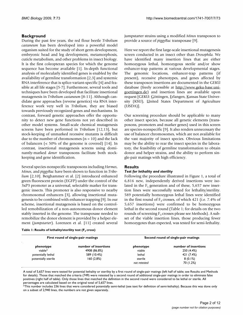
ResultsTest for lethality and sterilityFollowing the procedure illustrated in Figure 1, a total of6,816 new, independently derived insertions were iso-lated in the F1 generation and of these, 5,657 new inser-tion lines were successfully tested for lethality/sterility.589 potentially homozygous lethal lines were identifiedin the first round of F3 crosses, of which 421 (i.e. 7.4% of5,657 insertions) were confirmed to be homozygouslethal in the second round (Table 1; for details on the tworounds of screening F3 crosses please see Methods). A sub-set of the viable insertion lines, those producing fewerhomozygotes than expected, was tested for semi-lethality.
Table 1: Results of lethality/sterility test (F3 cross)
First round of single-pair matings Second round of single-pair matings
phenotype number of insertions phenotype number of insertionsviable* 4908 (86.8%) viable 250 (4.4%)
potentially lethal 589 (10.4%) lethal 421 (7.4%)potentially sterile 160 (2.8%) sterile 8 (0.1%)
not retested 70 (1.2%)
A total of 5,657 lines were tested for potential lethality or sterility by a first round of single-pair matings (left half of table; see Results and Methods for details). Those that matched the criteria (749) were retested by a second round of additional single-pair matings in order to eliminate false positives (right half of table). Only those lines that matched the definition in the second round were considered to be lethal or sterile. All percentages are calculated based on the original total of 5,657 lines.*This number includes 236 lines that were considered potentially semi-lethal (see text for definition of semi-lethality). Because this was done only on a subset of 2,940 lines, the numbers are not given separately.
Page 2 of 12(page number not for citation purposes)

BMC Biology 2009, 7:73 http://www.biomedcentral.com/1741-7007/7/73
Insertion lines were designated as potentially semi-lethalif homozygosity of a parent was indicated for no morethan one single-pair mating in the first round of F3 crosses,or less than four single-pair matings after the secondround. This was true for 236 insertions (out of the subsetof 2,940 insertions analyzed in Göttingen and Erlangen)after the first round, of which 75 remained in this categoryafter the second round. Hence, 2.5% (75/2,940) of allinsertions tested for semi-lethality met the criteria forsemi-lethality. This somewhat relaxed scoring criterion
reduced the likelihood of missing or overlooking lethal orsemi-lethal mutations.
Potentially homozygous sterile insertions lines were iden-tified by evaluating the single-pair matings: Whenever twoor more of the initial single-pair F3 self-crosses (roundone, Figure 1E) failed to produce offspring (although theparents were alive and healthy), the line was classified aspotentially sterile. This was the case for 160 insertions(Table 1). We used either of two methods to confirm orrefute a tentative diagnosis of recessive sterility. In the first
Screening procedure for the creation of new insertions and identification of lethal and sterile mutationsFigure 1Screening procedure for the creation of new insertions and identification of lethal and sterile mutations. (A) P1 cross: Mass-crosses were made between the donor (EGFP-marked eyes) and the helper strain (DsRed-marked eyes). The donor strain has an additional insertion site-dependent, muscle-specific enhancer-trap pattern. (B) P2 cross: Single hybrid females carrying both the donor and helper elements (simultaneous expression of EGFP and DsRed) were crossed to three pearl males. (C) F1 cross: A single individual carrying a stable new insertion was selected from the offspring of a P2 cross and crossed to several pearl mates. A remobilization event was evident in beetles that still showed EGFP-marked eyes, but had lost the muscle-specific enhancer-trap pattern. Note the altered enhancer-trap phenotype of the new insertion line in this example (EGFP expression in the wings). (D) F2 cross: All EGFP-marked offspring of the F1 cross were heterozygous for the insertion and were sibling-crossed. (E) F3 cross: Several single-pair matings were set up. (F) Test for lethality and sterility: Marker gene expression of the offspring of each single-pair mating was evaluated to determine whether their parents had been hetero- or homozygous for the piggyBac insertion (see Methods). Each single-pair mating was assigned to one of five classes (small black arrows; Table 4). The combined evaluation of all single-pair matings was used to define the phenotype of the insertion (see Table 5). (G) Summary of all identified insertions. For further details see methods part "Generating new piggyBac insertions", Table 1, and text.
Page 3 of 12(page number not for citation purposes)
BMC Biology 2009, 7:73 http://www.biomedcentral.com/1741-7007/7/73
method, we set up a second round of single-pair self-crosses bringing the total number of F3 crosses to 20. Thediagnosis was considered to be corroborated when thenumber of single-pair matings not producing any off-spring increased to four or more. Using this definition,124 potentially sterile lines were reduced to 21. However,further testing of these presumably sterile insertion linesshowed that this criterion was not always reliable (seebelow). In the second method we set up 10 male and 10female outcrosses. The diagnosis of recessive sterility wasconsidered to be corroborated if the crosses failed toreveal either a fertile, homozygous male or a fertile,homozygous female. Out of 36 potentially sterile linestested by the second method, only eight lines fulfilled thisdefinition of sterility. Since the second follow-up testappeared to be more rigorous than the first, we retested 11of the 21 apparently sterile lines from the former testusing the more rigorous criterion. All 11 lines proved to
be fertile in both sexes. It seems to be clear that most ster-ile lines found by using the first criterion are false-posi-tives. Hence, we suggest using the stricter test for recessivesterility, which has the added benefit of identifying theaffected sex.
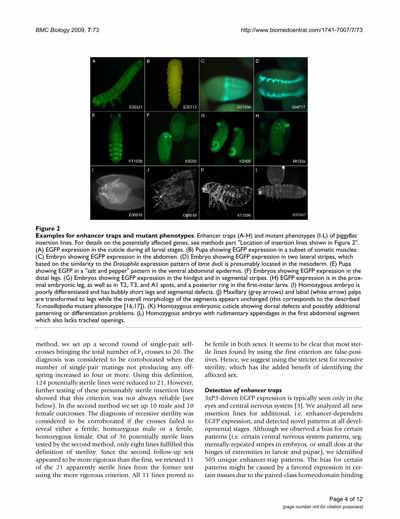
Detection of enhancer traps3xP3-driven EGFP expression is typically seen only in theeyes and central nervous system [3]. We analyzed all newinsertion lines for additional, i.e. enhancer-dependentEGFP expression, and detected novel patterns at all devel-opmental stages. Although we observed a bias for certainpatterns (i.e. certain central nervous system patterns, seg-mentally-repeated stripes in embryos, or small dots at thehinges of extremities in larvae and pupae), we identified505 unique enhancer-trap patterns. The bias for certainpatterns might be caused by a favored expression in cer-tain tissues due to the paired-class homeodomain binding
Examples for enhancer traps and mutant phenotypesFigure 2Examples for enhancer traps and mutant phenotypes. Enhancer traps (A-H) and mutant phenotypes (I-L) of piggyBac insertion lines. For details on the potentially affected genes, see methods part "Location of insertion lines shown in Figure 2". (A) EGFP expression in the cuticle during all larval stages. (B) Pupa showing EGFP expression in a subset of somatic muscles. (C) Embryo showing EGFP expression in the abdomen. (D) Embryo showing EGFP expression in two lateral stripes, which based on the similarity to the Drosophila expression pattern of lame duck is presumably located in the mesoderm. (E) Pupa showing EGFP in a "salt and pepper" pattern in the ventral abdominal epidermis. (F) Embryos showing EGFP expression in the distal legs. (G) Embryos showing EGFP expression in the hindgut and in segmental stripes. (H) EGFP expression is in the prox-imal embryonic leg, as well as in T2, T3, and A1 spots, and a posterior ring in the first-instar larva. (I) Homozygous embryo is poorly differentiated and has bubbly short legs and segmental defects. (J) Maxillary (grey arrows) and labial (white arrow) palps are transformed to legs while the overall morphology of the segments appears unchanged (this corresponds to the described Tc-maxillopedia mutant phenotype [16,17]). (K) Homozygous embryonic cuticle showing dorsal defects and possibly additional patterning or differentiation problems. (L) Homozygous embryo with rudimentary appendages in the first abdominal segment which also lacks tracheal openings.
Page 4 of 12(page number not for citation purposes)
BMC Biology 2009, 7:73 http://www.biomedcentral.com/1741-7007/7/73
sites in the 3xP3 element of the transformation marker[15]. For a random subset of about 200 of all newly-iden-tified insertions, we also dissected pupae and adults tolook for EGFP expression in internal organs that mightnot be visible without dissection. Such expression pat-terns (e.g. a spermatheca enhancer) were found onlyrarely. Examples of enhancer-trap lines are shown in Fig-ure 2A-H. Descriptions and/or photographs of allenhancer-trap lines together with information about theirchromosomal locations (when known) are available inGEKU-base (http://www.geku-base.uni-goettingen.de; seeMethods).
Analysis of lethal lines and developmental phenotypesWe analyzed the embryonic cuticle phenotypes of manylines identified as lethal and found a number of distinctcuticular abnormalities (Figure 2I-L). For example, lineG08519 displays a phenotype similar to the proboscipediaortholog maxillopedia in that maxillary (grey arrows) andlabial (white arrow) palps are transformed to legs (Figure2J); [16,17]. Indeed, this insertion is located in the firstintron of maxillopedia. In addition, many lethal linesshowed a high proportion of embryos that died prior tocuticularization, indicating early embryonic lethality.
To test whether the semi-lethal lines are false positives ortrue lethals with occasional escapers, we checked what por-tion of these lines (Göttingen subset) produce lethal L1cuticle phenotypes (at least two cuticles with similarstrong defects in one preparation when scoring at least 10individuals) and compared it to the percentage of cuticlephenotypes produced by the other classes. A total of25.8% (8/31) of a random selection of lines complyingwith the strict definition of lethality showed such pheno-types. Of lines with one or two single-pair matings (out of20) indicating homozygosity (semi-lethality), this por-tion was 16.6% in each case (5/30 and 3/18, respectively).
Lines with three single-pair matings indicatinghomozygous viability gave rise to cuticle phenotypes inonly 6.25% (1/16). Thus analyzing semi-lethal lines led tothe identification of additional cuticle phenotype-induc-ing mutations.
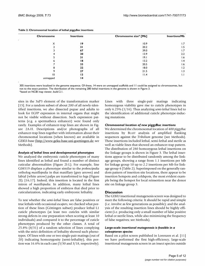
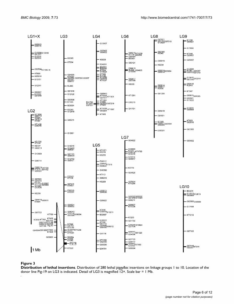
Chromosomal location of new piggyBac insertionsWe determined the chromosomal location of 400 piggyBacinsertions by BLAST analysis of amplified flankingsequences against the Tribolium genome (see Methods).These insertions included lethal, semi-lethal and sterile aswell as viable lines that showed an enhancer-trap pattern.The distribution of 280 homozygous lethal insertions onthe linkage groups is shown in Figure 3. The lethal inser-tions appear to be distributed randomly among the link-age groups, showing a range from 1.1 insertions per Mbfor linkage group 10 up to 2.2 insertions per Mb for link-age group 4 (Table 2). Superimposed on the generally ran-dom pattern of insertion site locations, there appear to beinsertion hotspots and coldspots, the most evident exam-ple being the hotspot for local reinsertion near the donorsite on linkage group 3.
DiscussionThe GEKU insertional mutagenesis screen was designed tomeet the following criteria: It should be rapid and simple(i.e. involve as few generations as possible); and the anal-ysis of the resulting insertion lines should be highly effi-cient (i.e. producing only a small number of false positivelethal or sterile lines, while also minimizing the frequencyof false negatives; see Methods).
Large-scale insertional mutagenesis is feasible in a coleopteran speciesBased on a pilot screen published in Lorenzen et al. [11]we have performed the first high-efficiency, large-scaleinsertional mutagenesis screen in an insect species outside
Table 2: Chromosomal location of lethal piggyBac insertions
Chromosome Insertions Chromosome size* [Mb] Insertions/Mb
X 19 10.9 1.72 31 20.2 1.53 67 39.0 1.74 30 13.9 2.25 31 19.1 1.66 18 13.2 1.47 33 20.5 1.68 22 18.0 1.29 27 21.5 1.3
10 13 11.4 1.1unmapped 14
305 insertions were localized in the genome sequence. Of these, 14 were on unmapped scaffolds and 11 could be assigned to chromosomes, but not to the exact position. The distribution of the remaining 280 lethal insertions in the genome is shown in Figure 3.*based on NCBI map viewer, build 2.1.
Page 5 of 12(page number not for citation purposes)
BMC Biology 2009, 7:73 http://www.biomedcentral.com/1741-7007/7/73
Page 6 of 12(page number not for citation purposes)
Distribution of lethal insertionsFigure 3Distribution of lethal insertions. Distribution of 280 lethal piggyBac insertions on linkage groups 1 to 10. Location of the donor line Pig-19 on LG3 is indicated. Detail of LG3 is magnified 12×. Scale bar = 1 Mb.
BMC Biology 2009, 7:73 http://www.biomedcentral.com/1741-7007/7/73
the genus Drosophila, and we have established a crossingscheme that circumvents the need for balancer chromo-somes or embryo injections. From our experience, we esti-mate that using the procedure presented here, one personshould be able to establish 150 lethal strains per year.While the GEKU screen has identified many interestingenhancer traps and lethal phenotypes, genome-wide satu-ration would be difficult to achieve at the current level ofefficiency. The most time-consuming step is setting upand evaluating 20 single-pair matings for each new inser-tion line to detect recessive lethality. For this reason we setup a small number of single-pair matings first, as mostviable insertions can be identified by evaluating just a fewcrosses from each subset. However, also for insertions rec-ognized as viable it was important to assess the fertility ofall remaining single-pair matings in order to ensure thatrecessive sterile insertions were detected.
Lethal insertions are readily detected while insertions causing sterility are difficult to detectWe found that lethal lines were readily detected by single-pair matings. Based on the frequency with which semi-lethal lines produced strong L1 larval cuticle phenotypes,we suggest defining lines as potentially lethal when onlyone or two out of 20 single-pair matings indicate homozy-gosity. However, our definition of sterility proved to betoo lax in the beginning, since most potentially sterilelines turned out to be false-positives in more detailedanalysis.
Comparing efficiencies with Drosophila melanogaster insertional mutagenesis and enhancer-trap screensThe efficiency of generating lethal mutations by piggyBac-based insertional mutagenesis in Tribolium (7.4%) is sim-ilar to equivalent screens in Drosophila based either on pig-gyBac [9,18] or P elements [19-21]. Whether the efficacy ofsuch screens can potentially be doubled by the inclusionof splice acceptor sites or insulator sequences within themutator element - as has been shown in Drosophila [22] -still has to be determined in Tribolium.
The enhancer detection rate within this large-scale inser-tional mutagenesis screen was also 7.4%. This is actuallyhigher than in a comparable Drosophila screen whereenhancer detection without a suitable amplification sys-tem was about 2% [9]. Only after including a GAL4-basedamplifier system could Drosophila enhancer detection beraised to 50% [9]. However, such directed expression sys-tems still need to be further developed and assayed in Tri-bolium before they can be used in insertional mutagenesisscreens.
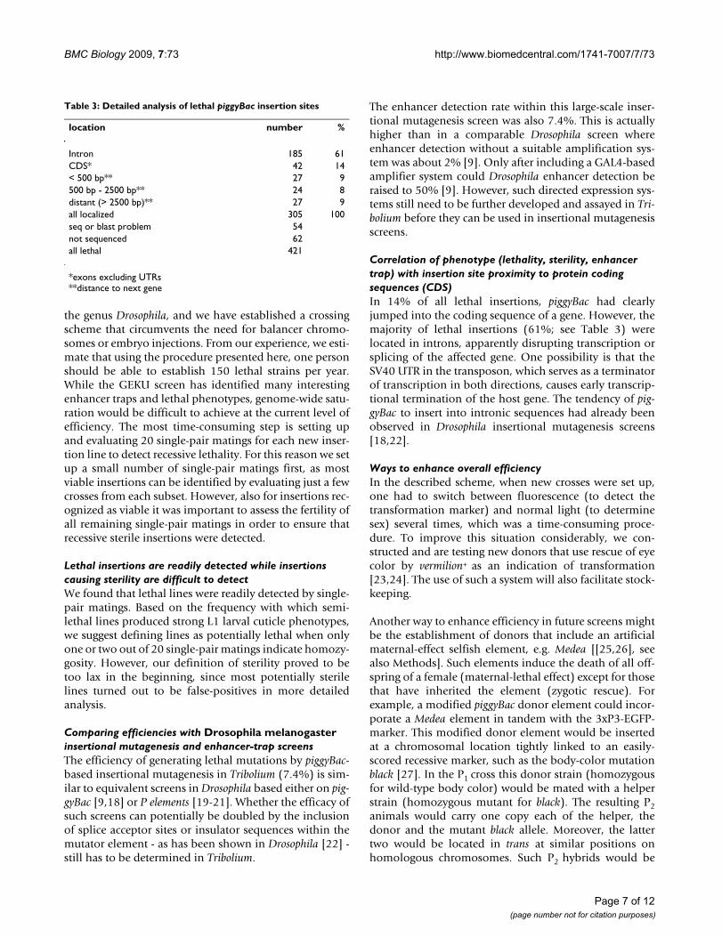
Correlation of phenotype (lethality, sterility, enhancer trap) with insertion site proximity to protein coding sequences (CDS)In 14% of all lethal insertions, piggyBac had clearlyjumped into the coding sequence of a gene. However, themajority of lethal insertions (61%; see Table 3) werelocated in introns, apparently disrupting transcription orsplicing of the affected gene. One possibility is that theSV40 UTR in the transposon, which serves as a terminatorof transcription in both directions, causes early transcrip-tional termination of the host gene. The tendency of pig-gyBac to insert into intronic sequences had already beenobserved in Drosophila insertional mutagenesis screens[18,22].
Ways to enhance overall efficiencyIn the described scheme, when new crosses were set up,one had to switch between fluorescence (to detect thetransformation marker) and normal light (to determinesex) several times, which was a time-consuming proce-dure. To improve this situation considerably, we con-structed and are testing new donors that use rescue of eyecolor by vermilion+ as an indication of transformation[23,24]. The use of such a system will also facilitate stock-keeping.
Another way to enhance efficiency in future screens mightbe the establishment of donors that include an artificialmaternal-effect selfish element, e.g. Medea [[25,26], seealso Methods]. Such elements induce the death of all off-spring of a female (maternal-lethal effect) except for thosethat have inherited the element (zygotic rescue). Forexample, a modified piggyBac donor element could incor-porate a Medea element in tandem with the 3xP3-EGFP-marker. This modified donor element would be insertedat a chromosomal location tightly linked to an easily-scored recessive marker, such as the body-color mutationblack [27]. In the P1 cross this donor strain (homozygousfor wild-type body color) would be mated with a helperstrain (homozygous mutant for black). The resulting P2animals would carry one copy each of the helper, thedonor and the mutant black allele. Moreover, the lattertwo would be located in trans at similar positions onhomologous chromosomes. Such P2 hybrids would be
Table 3: Detailed analysis of lethal piggyBac insertion sites
location number %
Intron 185 61CDS* 42 14< 500 bp** 27 9500 bp - 2500 bp** 24 8distant (> 2500 bp)** 27 9all localized 305 100seq or blast problem 54not sequenced 62all lethal 421
*exons excluding UTRs**distance to next gene
Page 7 of 12(page number not for citation purposes)
BMC Biology 2009, 7:73 http://www.biomedcentral.com/1741-7007/7/73
mated with beetles that were trans-heterozygous for blackand the Medea-containing donor element, or, if the P2hybrid is female, they could instead be mated tohomozygous black (non-helper, non-Medea) males. F1 off-spring with black body-color then would arise only if theyinherit the black chromosome from both parents. Becausethe Medea-tagged donor is arranged in trans with black,such black offspring do not carry a donor, and hence lackzygotic rescue activity of the Medea element. This leads totheir death by the maternal-lethal effect of the element.Only if the donor has been remobilized to anothergenomic location can offspring carry both black alleles aswell as the rescuing donor. Hence, black body-color in theoffspring indicates a remobilization event. This designwould be an elegant means to enhance the detection ofnew insertion lines by obviating the need for fluorescencedetection. It would also simplify the stock-keeping oflethal insertions, since a Medea element tightly linkedwith the lethal insertion would constitute a type of bal-anced lethal.
ConclusionWe have successfully established a method to conductlarge-scale insertional mutagenesis screens in the beetleTribolium castaneum. Using this method, we obtained sev-eral hundred lethal insertions as well as insertions pro-ducing enhancer-trap phenotypes. These lines have beenmade available to the research community.
MethodsStrains usedThe donor strain used in this screen, Pig-19, carries a 3xP3-EGFP marked piggyBac element, pBac [3xP3-EGFPaf], thatconfers both, insertion-site-independent eye-specificEGFP expression, and donor-site-dependent muscle-spe-cific EGFP expression [3]. We previously demonstratedthat remobilization of the Pig-19 insertion results in G1beetles lacking muscle-specific expression, but retainingeye-specific expression [3,11]. Thus, the loss of muscle-specific expression can be used to detect remobilizationevents. The jumpstarter/helper strain used in this screen,M26 [11], carries an X-chromosomal insertion of a 3xP3-DsRed marked Minos element (pMi [3xP3-DsRed;Dm'hsp70-pBac]) [9]. Both strains are in a white-eyed
pearl mutant background to facilitate detection of eye-spe-cific fluorescence.
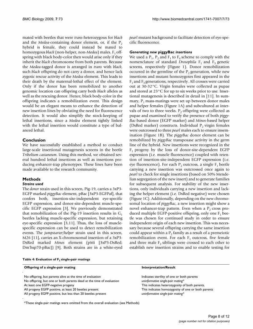
Generating new piggyBac insertionsWe used a P1, P2 and F1 to F4 scheme to comply with thenomenclature of standard Drosophila F1 and F3 geneticscreens, respectively (Figure 1). Donor remobilizationoccurred in the germline of the P2 generation, while newinsertions and mutant homozygotes first appeared in theF1 and F3 generations, respectively. All crosses were carriedout at 30-32°C. Virgin females were collected as pupaeand stored at 23°C for up to six weeks prior to use. Inser-tional mutagenesis is described in detail in [11]. In sum-mary, P1 mass-matings were set up between donor malesand helper females (Figure 1A) and subcultured at inter-vals of two to three weeks. P2 offspring were collected aspupae and examined to verify the presence of both piggy-Bac-based donor (EGFP marker) and Minos-based helper(DsRed marker) constructs. Individual P2 virgin femaleswere outcrossed to three pearl males each to ensure insem-ination (Figure 1B). The piggyBac donor element can beremobilized by piggyBac transposase activity in the germline of the hybrid. New insertions were recognized in theF1 progeny by the loss of donor-site-dependent EGFPexpression (i.e. muscle fluorescence) coupled with reten-tion of insertion-site-independent EGFP expression (i.e.eye fluorescence). For each P2 outcross, a single F1 beetlecarrying a new insertion was outcrossed once again topearl to check for single insertions (based on 50% Mende-lian segregation of the new insert) and to generate familiesfor subsequent analysis. For stability of the new inser-tions, only individuals carrying a new insertion and lack-ing the helper element (i.e. DsRed negative) were chosen(Figure 1C). Additionally, depending on the new chromo-somal location of piggyBac, a new insertion might show anovel enhancer-trap pattern. Even when a P2 cross pro-duced multiple EGFP-positive offspring, only one F1 bee-tle was chosen for continued study in order to ensureindependent origin of each new insertion. This was neces-sary because several offspring carrying the same insertioncould appear within a P2 family as a result of a premeioticremobilization event. For each F1 outcross, five femaleand three male F2 siblings were crossed to each other toestablish new insertion strains and to enable testing for
Table 4: Evaluation of F3 single-pair matings
Offspring of a single-pair mating Interpretation/Result
No offspring, but parents alive at the time of evaluation Indicates sterility of one or both parentsNo offspring, but one or both parents dead at the time of evaluation uninformative single-pair mating*At least one EGFP-negative progeny This indicates heterozygosity of both parents.All progeny EGFP positive, at least 20 beetles present This indicates homozygosity of one or both parentsAll progeny EGFP positive, but less than 20 beetles present uninformative single-pair mating*
*These single-pair matings were omitted from the overall evaluation (see Methods)
Page 8 of 12(page number not for citation purposes)
BMC Biology 2009, 7:73 http://www.biomedcentral.com/1741-7007/7/73
homozygous viability (F2 cross; Figure 1D). To accom-plish the latter, we performed a number of single-pair F3matings (Figure 1E) and analyzed their progeny for thepresence of the donor element (see below).
Statistical considerationsIf an insertion mutant were homozygous viable, then(after positive marker selection) the progeny of the F2cross would consist of a 1:2 ratio of homozygous to heter-ozygous beetles. Under the assumption of random sib-mating, 11.1% (1/3 × 1/3) of all F3 single-pair matingswould be between two homozygous beetles, 44.4% [2×(1/3 × 2/3)] between one homozygous and one hetero-zygous beetle, and 44.4% (2/3 × 2/3) between two heter-ozygous beetles. This implies that about 55.5% (11.1% +44.4%) of the single-pair matings (given a fully-viableinsertion) would produce only EGFP-positive progeny(because at least one parent would be homozygous for theinsertion). The remaining 44.4% would produce mixedprogeny (i.e. approximately 75% EGFP positive andapproximately 25% EGFP negative) because both parentswould be heterozygous for the insertion. In contrast, forrecessive lethal insertions, no homozygous beetles wouldbe present in the F3 generation so all F3 crosses would pro-duce mixed progeny. Thus, the presence of even a singleEGFP-negative beetle in the F4 generation indicates heter-ozygosity of both parents, and the complete absence ofEGFP-negative progeny indicates homozygosity of at leastone parent. Depending on the distribution of the above-mentioned phenotypes, each single-pair mating wasscored and assigned to one of five categories (see Figure 1Fand Table 4 for details).
Since more than 40% of all single-pair matings wereexpected to produce mixed progeny (even if the insertionwas fully viable) we analyzed a total of 20 single-pair mat-ings before concluding that an insertion was lethal. Onthe other hand, since viable insertions were usually iden-tified after evaluating just a few single-pair matings, wesplit the 20 crosses into two consecutive rounds to maxi-
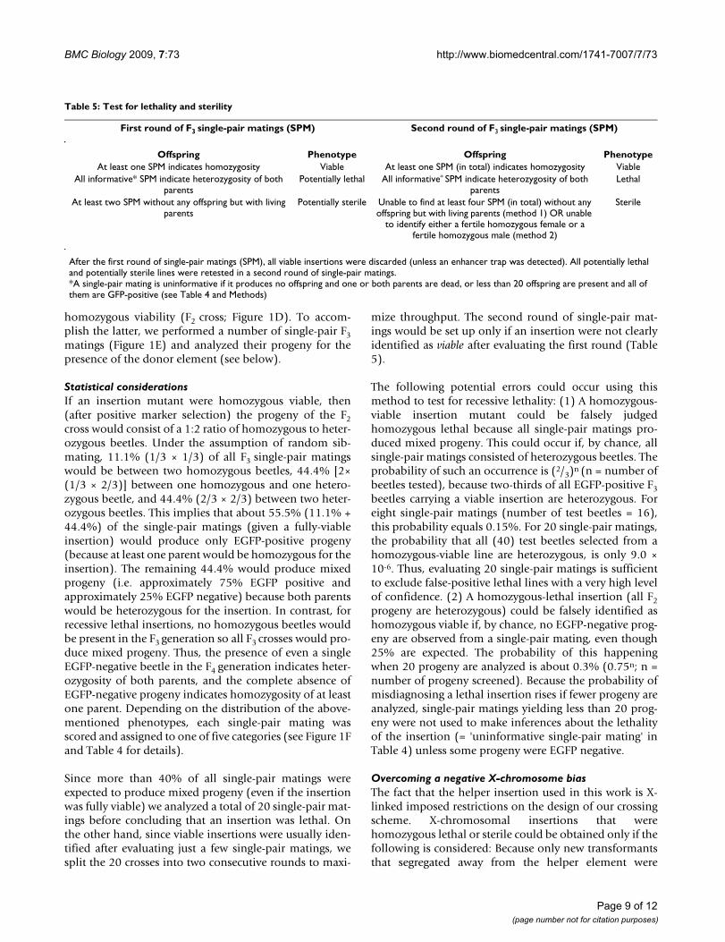
mize throughput. The second round of single-pair mat-ings would be set up only if an insertion were not clearlyidentified as viable after evaluating the first round (Table5).
The following potential errors could occur using thismethod to test for recessive lethality: (1) A homozygous-viable insertion mutant could be falsely judgedhomozygous lethal because all single-pair matings pro-duced mixed progeny. This could occur if, by chance, allsingle-pair matings consisted of heterozygous beetles. Theprobability of such an occurrence is (2/3)n (n = number ofbeetles tested), because two-thirds of all EGFP-positive F3beetles carrying a viable insertion are heterozygous. Foreight single-pair matings (number of test beetles = 16),this probability equals 0.15%. For 20 single-pair matings,the probability that all (40) test beetles selected from ahomozygous-viable line are heterozygous, is only 9.0 ×10⁻⁶. Thus, evaluating 20 single-pair matings is sufficientto exclude false-positive lethal lines with a very high levelof confidence. (2) A homozygous-lethal insertion (all F2progeny are heterozygous) could be falsely identified ashomozygous viable if, by chance, no EGFP-negative prog-eny are observed from a single-pair mating, even though25% are expected. The probability of this happeningwhen 20 progeny are analyzed is about 0.3% (0.75n; n =number of progeny screened). Because the probability ofmisdiagnosing a lethal insertion rises if fewer progeny areanalyzed, single-pair matings yielding less than 20 prog-eny were not used to make inferences about the lethalityof the insertion (= 'uninformative single-pair mating' inTable 4) unless some progeny were EGFP negative.
Overcoming a negative X-chromosome biasThe fact that the helper insertion used in this work is X-linked imposed restrictions on the design of our crossingscheme. X-chromosomal insertions that werehomozygous lethal or sterile could be obtained only if thefollowing is considered: Because only new transformantsthat segregated away from the helper element were
Table 5: Test for lethality and sterility
First round of F3 single-pair matings (SPM) Second round of F3 single-pair matings (SPM)
Offspring Phenotype Offspring PhenotypeAt least one SPM indicates homozygosity Viable At least one SPM (in total) indicates homozygosity Viable
All informative* SPM indicate heterozygosity of both parents
Potentially lethal All informative* SPM indicate heterozygosity of both parents
Lethal
At least two SPM without any offspring but with living parents
Potentially sterile Unable to find at least four SPM (in total) without any offspring but with living parents (method 1) OR unable
to identify either a fertile homozygous female or a fertile homozygous male (method 2)
Sterile
After the first round of single-pair matings (SPM), all viable insertions were discarded (unless an enhancer trap was detected). All potentially lethal and potentially sterile lines were retested in a second round of single-pair matings.*A single-pair mating is uninformative if it produces no offspring and one or both parents are dead, or less than 20 offspring are present and all of them are GFP-positive (see Table 4 and Methods)
Page 9 of 12(page number not for citation purposes)
BMC Biology 2009, 7:73 http://www.biomedcentral.com/1741-7007/7/73
selected, hybrid females had to be used to set up P2 crossesin order to avoid bias against new X-linked insertions.Additionally, males with a new hemizygous X-linkedlethal insertion would not survive and ones hemizygousfor a new X-linked sterile insertion would be useless forgenerating a new stock. Hence, one could obtain X-linkedlethal and sterile insertions only if female beetles carryingthe donor element were used to set up the P2 as well as theF1 crosses. Therefore, we selected only female hybrids andused females carrying new insertions whenever possible.
Efficiency of detecting new insertionsAt least one new insertion was detected in about 30% ofall P2 crosses when about 20 offspring were screened. Thepercentage of P2 crosses that yield new insertions can begreatly increased by screening a larger number of progenyper P2 cross. For a subset of P2 crosses we screened 100progeny per cross, and found at least one new insertion inevery case. In practice, about 10 - 30 P2 pupae were presentwhen the P2 progeny were screened for new insertions.The decision to discard the larval P2 offspring when a newinsertion could not be detected in the first attempt repre-sented a compromise between the need to find at leastone new insertion in each family and the aim to obtain alarge number of independent insertions with limitedresources in time and space.
Determination of insertion sitesThe genomic location of an insertion was determined bysequencing flanking DNA obtained by one of the follow-ing three polymerase chain reaction (PCR) -based meth-ods: inverse PCR [28], universal PCR [3,29], or vectorettePCR [30]. The procedure for inverse PCR including primerdesign was adapted from 'Inverse PCR and SequencingProtocol on 5 Fly Preps', Exelixis Pharmaceutical Corp(South San Francisco, California, USA) [22]. FollowingDNA isolation, approximately 1 μg of DNA was digestedwith Sau3A1, BfUC1, or Ase1 (for 5' iPCR) or HinP1 (for3' iPCR). Approximately 100 ng of digested DNA was thenself-ligated to obtain circular DNA fragments, followed bytwo rounds of nested PCR. DNA templates (PCR productsand/or cloned PCR products) were sequenced by Seqlab(Göttingen, Germany), Macrogen (Seoul, Korea), or usingan ABI 3730 DNA sequencer (Sequencing and Genotyp-ing Facility, Plant Pathology, Kansas State University,Manhattan, Kansas, USA). Data analysis was performedusing Vector NTI® software (Invitrogen, Carlsbad, Califor-nia, USA). After trimming vector sequences, flanking DNAsequences were then searched (BLASTN) against Triboliumcastaneum genome sequences at HGSC, Baylor College ofMedicine http://www.hgsc.bcm.tmc.edu/projects/tribolium/, NCBI http://www.ncbi.nlm.nih.gov/genome/seq/BlastGen/BlastGen.cgi?taxid=7070 or BeetleBase http://beetlebase.org/. If the insertion was in a predicted gene(GLEAN set), a transcription unit (EST or cDNA) or region
indicated by Drosophila BLAST or other gene predictionmethod as a potential gene, the predicted Tribolium genewas examined by BLAST analysis at FlyBase for the top Dro-sophila hit, and NCBI (nr database) to identify otherpotential orthologs. Insertion site sequences were depos-ited to NCBI (for accession numbers see Additional File 1)and also put - including the retrieved information - intoGEKU-base (see below).
Medea (maternal effect dominant embryonic arrest)When hybrid females and pearl males (P2 generation)were crossed severe segregation distortion was observed:98% of the progeny were EGFP positive, rather than theexpected 50%. The DsRed marker however showed theexpected 1:1 ratio (i.e. segregated independently of theEGFP marker). The unusual segregation of EGFP has beenshown [11] to be the result of close cis-linkage (approxi-mately 2 cM) of the maternally acting selfish gene Medea[25] with the Pig-19 donor insertion [3] on LG3. How-ever, the segregation ratios of new insertions were affectedonly when the piggyBac element reinserted near the origi-nal donor insertion (representing a local hop).
GEKU-baseAll available information about the analyzed insertionlines can be found at a web-based database called GEKU-base http://www.geku-base.uni-goettingen.de. Informa-tion provided includes (if available) photographs anddescriptions of enhancer traps and phenotypes, flankingsequences and chromosomal location, affected genes andtheir orthologs. GEKU-base also provides information onhow to obtain insertion lines.
EGFP and DsRed analysisMarker-gene fluorescence was detected using a Nikon flu-orescence stereomicroscope SMZ1500 (Nikon GmbH,Düsseldorf, Germany) at Göttingen and Erlangen, anOlympus SZX12 fluorescence stereomicroscope (Olym-pus Corporation, Tokyo, Japan), or a Leica MZ FLIII fluo-rescence stereomicroscope (Leica Microsystems Inc.,Wetzlar, Germany). The filter sets used for EGFP expres-sion were: [Göttingen: 470/40 nm excitation filter, 500nm LP emission filter, and 495 nm beamsplitter], [Erlan-gen: 480/40 nm excitation filter, 510 nm emission filter,and 505 nm beamsplitter], [KSU: 480/40 nm excitationfilter and 535/50 nm emission filter], [USDA: GFP Plusfilter set (excitation filter: 480/40 nm, barrier filter: 510nm)]. The filter sets used for DsRed expression were: [Göt-tingen: 546/12 nm excitation filter, 605/75 nm emissionfilter, and 560 nm beamsplitter], [Erlangen: 565/30 exci-tation filter, 620/60 nm emission filter, and 585 nmbeamsplitter], [KSU: 545/30 excitation filter and 620/60emission filter], [USDA: TXR TEXAS RED filter set (excita-tion filter: 560/40 nm, barrier filter: 610 nm)]. To detect
Page 10 of 12(page number not for citation purposes)
BMC Biology 2009, 7:73 http://www.biomedcentral.com/1741-7007/7/73
enhancer-trap patterns in embryos, we dechorionatedembryos derived from F3-crosses.
Location of insertion lines shown in Figure 2Gene names refer to respective Drosophila orthologs. Theline E00321 is homozygous lethal and carries an insertionin lethal (2) giant larvae (Figure 2A). The line E00713 ishomozygous viable and carries an insertion 149-bpupstream of the 5' end of GLEAN_03347, Glutatione Stransferase, (Figure 2B). The homozygous viable lineG01004 carries an insertion near Ultrabithorax (Figure2C). The homozygous viable line G04717 carries an inser-tion near lame duck (Figure 2D). The line KT1539 ishomozygous lethal and the insertion site is near the genepointed (Figure 2E). The homozygous lethal line KS030bears an insertion in an intron of lozenge (Figure 2F). TheKS406 line is homozygous viable and carries an insertionin an intron of GLEAN_00277 which shows similarity togenes encoding protein tyrosine phosphatases. Othergenes in the vicinity of this insertion are Fgf8 or Or48 (Fig-ure 2G). The homozygous viable line MH30a has an inser-tion near female sterile (2) Ketel (Figure 2H). The lineE00916 is homozygous lethal and carries an insertion inan exon of GLEAN_08270 (Drosophila ortholog: Cyclin D)(Figure 2I). The G08519 insertion is located in the firstintron of proboscipedia (Figure 2J). The KT1096 insertion isin an intron of the pecanex ortholog (Figure 2K). TheE03501 insertion is in an intron of the Tribolium orthologof Ftz-F1 (Figure 2L).
Abbreviationsbp: base pair; CDS: coding sequence; cM: centiMorgan;EGFP: enhanced green fluorescent protein; GEKU: Göttin-gen, Erlangen, KSU, USDA; KSU: Kansas State University;L1: first larval stage; LG: linkeage group; Mb: Megabase;MEDEA: maternal arrest dominant embryonic arrest;RNAi: RNA interference; SPM: single-pair mating; USDA:United States Department of Agriculture; UTR: untrans-lated region
Authors' contributionsJT, JS, MDL and TDS were involved in setting up and eval-uating the beetle crosses, screening for enhancer traps anddetermining the genomic location of insertion sites. JTdrafted the manuscript. EAW, RWB, MK, GB, MDL, TDSand SJB conceived of the study, participated in its designand coordination, and helped to draft the manuscript. Allauthors read and approved the final manuscript.
Additional material
AcknowledgementsWe are deeply indebted to Elke Küster, Susanne Fischer, Tatum Kimzey, Kathy Leonard and Jonna Voorhees for technical assistance in production and characterization of transgenic lines. All programs and services of the U.S. Department of Agriculture are offered on a nondiscriminatory basis, without regard to race, color, national origin, religion, sex, age, marital sta-tus or handicap. Mention of trade names or commercial products in this publication is solely for the purpose of providing specific information and does not imply recommendation or endorsement by the USDA. This work has been funded by the USDA within the CSREES_NRI program "Functional Genomics of Agriculturally Important Organisms - Insects and Mites" grant #2004-35604-14250 (EAW, RWB, MK, SJB).
References1. Tribolium Genome Sequencing Consortium, Richards S, Gibbs RA,
Weinstock GM, Brown SJ, Denell R, Beeman RW, Bucher G, Frie-drich M, Grimmelikhuijzen CJ, Klingler M, Lorenzen M, Roth S,Schröder R, Tautz D, Zdobnov EM, Muzny D, Attaway T, Bell S, BuhayCJ, Chandrabose MN, Chavez D, Clerk-Blankenburg KP, Cree A, DaoM, Davis C, Chacko J, Dinh H, Dugan-Rocha S, Fowler G, Garner TT,et al.: The genome of the model beetle and pest Tribolium cas-taneum. Nature 2008, 452:949-955.
2. Berghammer AJ, Klingler M, Wimmer EA: A universal marker fortransgenic insects. Nature 1999, 402:370-371.
3. Lorenzen MD, Berghammer AJ, Brown SJ, Denell RE, Klingler M, Bee-man RW: piggyBac-mediated germline transformation in thebeetle Tribolium castaneum. Insect Mol Biol 2003, 12:433-440.
4. Arakane Y, Muthukrishnan S, Kramer KJ, Specht CA, Tomoyasu Y,Lorenzen MD, Kanost M, Beeman RW: The Tribolium chitin syn-thase genes TcCHS1 and TcCHS2 are specialized for synthesisof epidermal cuticle and midgut peritrophic matrix. InsectMol Biol 2005, 14:453-463.
5. Brown SJ, Mahaffey JP, Lorenzen MD, Denell RE, Mahaffey JW: UsingRNAi to investigate orthologous homeotic gene functionduring development of distantly related insects. Evol Dev1999, 1:11-15.
6. Bucher G, Scholten J, Klingler M: Parental RNAi in Tribolium(Coleoptera). Curr Biol 2002, 12:R85-86.
7. Tomoyasu Y, Denell RE: Larval RNAi in Tribolium (Coleoptera)for analyzing adult development. Dev Genes Evol 2004,214:575-578.
8. Horn C, Schmid BGM, Pogoda FS, Wimmer EA: Fluorescent trans-formation markers for insect transgenesis. Insect Biochem MolBiol 2002, 32:1221-1235.
9. Horn C, Offen N, Nystedt S, Häcker U, Wimmer EA: piggyBac-based insertional mutagenesis and enhancer detection as atool for functional insect genomics. Genetics 2003, 163:647-661.
10. Pavlopoulos A, Berghammer AJ, Averof M, Klingler M: EfficientTransformation of the Beetle Tribolium castaneum Using theMinos Transposable Element: Quantitative and QualitativeAnalysis of Genomic Integration Events. Genetics 2004,167:737-746.
11. Lorenzen MD, Kimzey T, Shippy TD, Brown SJ, Denell RE, BeemanRW: piggyBac-based insertional mutagenesis in Tribolium cas-taneum using donor/helper hybrids. Insect Mol Biol 2007,16:265-275.
12. Sulston IA, Anderson KV: Embryonic patterning mutants of Tri-bolium castaneum. Development 1996, 122:805-814.
Additional file 1Gene bank accession numbers of integration site sequences.Click here for file[http://www.biomedcentral.com/content/supplementary/1741-7007-7-73-S1.pdf]
Page 11 of 12(page number not for citation purposes)
BMC Biology 2009, 7:73 http://www.biomedcentral.com/1741-7007/7/73
Publish with BioMed Central and every scientist can read your work free of charge
"BioMed Central will be the most significant development for disseminating the results of biomedical research in our lifetime."
Sir Paul Nurse, Cancer Research UK
Your research papers will be:
available free of charge to the entire biomedical community
peer reviewed and published immediately upon acceptance
cited in PubMed and archived on PubMed Central
yours — you keep the copyright
Submit your manuscript here:http://www.biomedcentral.com/info/publishing_adv.asp
BioMedcentral
13. Maderspacher F, Bucher G, Klingler M: Pair-rule and gap genemutants in the flour beetle Tribolium castaneum. Dev GenesEvol 1998, 208:558-568.
14. Berghammer AJ, Bucher G, Maderspacher F, Klingler M: A systemto efficiently maintain embryonic lethal mutations in theflour beetle Tribolium castaneum. Dev Genes Evol 1999,209:382-389.
15. Horn C, Jaunich B, Wimmer EA: Highly sensitive, fluorescenttransformation marker for Drosophila transgenesis. DevGenes Evol 2000, 210:623-629.
16. Shippy TD, Brown SJ, Denell RE: Maxillopedia is the Triboliumortholog of proboscipedia. Evol Dev 2000, 2:145-151.
17. Shippy TD, Guo J, Brown SJ, Beeman RW, Denell RE: Analysis ofmaxillopedia expression pattern and larval cuticular pheno-type in wild-type and mutant Tribolium. Genetics 2000,155:721-731.
18. Häcker U, Nystedt S, Barmchi MP, Horn C, Wimmer EA: piggyBac-based insertional mutagenesis in the presence of stably inte-grated P elements in Drosophila. Proc Natl Acad Sci USA 2003,100:7720-7725.
19. Cooley L, Kelley R, Spradling A: Insertional mutagenesis of theDrosophila genome with single P elements. Science 1988,239:1121-1128.
20. Bellen HJ: Ten years of enhancer detection: lessons from thefly. Plant Cell 1999, 11:2271-2281.
21. Peter A, Schöttler P, Werner M, Beinert N, Dowe G, Burkert P,Mourkioti F, Dentzer L, He Y, Deak P, Benos PV, Gatt MK, MurphyL, Harris D, Barrell B, Ferraz C, Vidal S, Brun C, Demaille J, Cadieu E,Dreano S, Gloux S, Lelaure V, Mottier S, Galibert F, Borkova D, Miña-na B, Kafatos FC, Bolshakov S, Sidén-Kiamos I, et al.: Mapping andidentification of essential gene functions on the X chromo-some of Drosophila. EMBO Rep 2002, 3:34-38.
22. Thibault ST, Singer MA, Miyazaki WY, Milash B, Dompe NA, SinghCM, Buchholz R, Demsky M, Fawcett R, Francis-Lang HL, Ryner L,Cheung LM, Chong A, Erickson C, Fisher WW, Greer K, HartouniSR, Howie E, Jakkula L, Joo D, Killpack K, Laufer A, Mazzotta J, SmithRD, Stevens LM, Stuber C, Tan LR, Ventura R, Woo A, Zakrajsek I,et al.: A complementary transposon tool kit for Drosophilamelanogaster unsing P and piggyBac. Nature Genetics 2004,36:283-287.
23. Lorenzen MD, Brown SJ, Denell RE, Beeman RW: Cloning andcharacterization of the Tribolium castaneum eye-color genesencoding tryptophan oxygenase and kynurenine 3-monoox-ygenase. Genetics 2002, 160:225-234.
24. Lorenzen MD, Brown SJ, Denell RE, Beeman RW: Transgeneexpression from the Tribolium castaneum Polyubiquitin pro-moter. Insect Mol Biol 2002, 11:399-407.
25. Beeman RW, Friesen KS, Denell RE: Maternal-effect selfish genesin flour beetles. Science 1992, 256:89-92.
26. Chen CH, Huang H, Ward CM, Su JT, Schaeffer LV, Guo M, Hay BA:A synthetic maternal-effect selfish genetic element drivespopulation replacement in Drosophila. Science 2007,316:597-599.
27. Sokoloff A, Slatis HM, Stanley J: The black mutation in Triboliumcastaneum. J Heredity 1960, 52:131-135.
28. Ochman H, Gerber AS, Hartl DL: Genetic applications of aninverse polymerase chain reaction. Genetics 1988, 120:621-623.
29. Beeman RW, Stauth DM: Rapid cloning of insect transposoninsertion junctions using 'universal' PCR. Insect Mol Biol 1997,1:83-88.
30. Arnold C, Hodgson I: Vectorette PCR: a novel approach togenomic walking. PCR Meth Appl 1991, 1:39-42.
Page 12 of 12(page number not for citation purposes)
http://www.ncbi.nlm.nih.gov/entrez/query.fcgi?cmd=Retrieve&db=PubMed&dopt=Abstract&list_uids=9811974
http://www.ncbi.nlm.nih.gov/entrez/query.fcgi?cmd=Retrieve&db=PubMed&dopt=Abstract&list_uids=2830671
http://www.ncbi.nlm.nih.gov/entrez/query.fcgi?cmd=Retrieve&db=PubMed&dopt=Abstract&list_uids=1566060
http://www.ncbi.nlm.nih.gov/entrez/query.fcgi?cmd=Retrieve&db=PubMed&dopt=Abstract&list_uids=1566060
http://www.ncbi.nlm.nih.gov/entrez/query.fcgi?cmd=Retrieve&db=PubMed&dopt=Abstract&list_uids=2852134
Related Documents


