Large Scale Immune Profiling of Infected Humans and Goats Reveals Differential Recognition of Brucella melitensis Antigens Li Liang 1 , Diana Leng 2 , Chad Burk 1 , Rie Nakajima-Sasaki 1 , Matthew A. Kayala 3,4 , Vidya L. Atluri 5 , Jozelyn Pablo 1 , Berkay Unal 1 , Thomas A. Ficht 6 , Eduardo Gotuzzo 2 , Mayuko Saito 7 , W. John W. Morrow 8 , Xiaowu Liang 8 , Pierre Baldi 3,4,9 , Robert H. Gilman 10 , Joseph M. Vinetz 11 *, Rene ´ e M. Tsolis 5 *, Philip L. Felgner 1,4,8 * 1 Division of Infectious Diseases, Department of Medicine, University of California Irvine, Irvine, California, United States of America, 2 Alexander von Humboldt Institute of Tropical Medicine, Universidad Peruana Cayetano Heredia, Lima, Peru, 3 Department of Computer Science, University of California Irvine, Irvine, California, United States of America, 4 Institute for Genomics and Bioinformatics, University of California Irvine, Irvine, California, United States of America, 5 Department of Medical Microbiology and Immunology, University of California Davis, Davis, California, United States of America, 6 Department of Veterinary Pathobiology, Texas A&M University and Texas Agricultural Experiment Station, College Station, Texas, United States of America, 7 Asociacion Benefica PRISMA, Lima, Peru, 8 Antigen Discovery, Inc., Irvine, California, United States of America, 9 Department of Biological Chemistry, University of California Irvine, Irvine, California, United States of America, 10 Department of International Health, Bloomberg School of Public Health, Johns Hopkins University, Baltimore, Maryland, United States of America, 11 Division of Infectious Diseases, University of California San Diego School of Medicine, La Jolla, California, United States of America Abstract Brucellosis is a widespread zoonotic disease that is also a potential agent of bioterrorism. Current serological assays to diagnose human brucellosis in clinical settings are based on detection of agglutinating anti-LPS antibodies. To better understand the universe of antibody responses that develop after B. melitensis infection, a protein microarray was fabricated containing 1,406 predicted B. melitensis proteins. The array was probed with sera from experimentally infected goats and naturally infected humans from an endemic region in Peru. The assay identified 18 antigens differentially recognized by infected and non-infected goats, and 13 serodiagnostic antigens that differentiate human patients proven to have acute brucellosis from syndromically similar patients. There were 31 cross-reactive antigens in healthy goats and 20 cross-reactive antigens in healthy humans. Only two of the serodiagnostic antigens and eight of the cross-reactive antigens overlap between humans and goats. Based on these results, a nitrocellulose line blot containing the human serodiagnostic antigens was fabricated and applied in a simple assay that validated the accuracy of the protein microarray results in the diagnosis of humans. These data demonstrate that an experimentally infected natural reservoir host produces a fundamentally different immune response than a naturally infected accidental human host. Citation: Liang L, Leng D, Burk C, Nakajima-Sasaki R, Kayala MA, et al. (2010) Large Scale Immune Profiling of Infected Humans and Goats Reveals Differential Recognition of Brucella melitensis Antigens. PLoS Negl Trop Dis 4(5): e673. doi:10.1371/journal.pntd.0000673 Editor: Jakob Zinsstag, Swiss Tropical Institute, Switzerland Received July 27, 2009; Accepted March 19, 2010; Published May 4, 2010 Copyright: ß 2010 Liang et al. This is an open-access article distributed under the terms of the Creative Commons Attribution License, which permits unrestricted use, distribution, and reproduction in any medium, provided the original author and source are credited. Funding: This work is supported by US National Institutes of Health grants U01AI078213, U54AI065359, and U01AI075420, and SBIR grant 1R43AI06816601A1. The funders had no role in study design, data collection and analysis, decision to publish, or preparation of the manuscript. Competing Interests: PLF is an inventor on patent applications pertaining to this work and owns stock in a company (Anitigen Discovery Inc.) that has licensed the technology. XL is an inventor on patent applications pertaining to this work and is employed at a company (Anitigen Discovery Inc.) that has licensed the technology. WJWM is employed at company (Anitigen Discovery Inc.) that has licensed technology pertaining to this work. * E-mail: [email protected] (JMV); [email protected] (RMT); [email protected] (PLF) Introduction Brucellosis is a zoonotic infectious disease endemic in regions around the world where agricultural, animal husbandry and vaccination practices have not controlled infection among livestock reservoirs [1–3]. The reservoirs of Brucella melitensis, the most virulent species affecting humans, include goats and sheep [4], especially in Peru and the Middle East [3]. Identification of goat, sheep and other animal sources of infection have long used agglutination tests, although newer tests are being developed and applied in the veterinary setting [5–7]. Commonly used screening tests do not necessarily differentiate between vaccination and infection in goats ([8]; summarized in [6]). By themselves, the Rose Bengal and other agglutination tests cannot be used exclusively to diagnose human brucellosis because while sensitive and specific for first episodes of brucellosis, these tests can be problematic in differentiating acute, chronic and relapsing forms of brucellosis in humans living in endemic regions [9–12], and typically require titration and differentiation of IgM from IgG antibodies either in solid phase formats or by use of the mercaptoethanol test [1,3,13–16]. The current knowledge of protein antigens recognized by humans and reservoir animals is limited to a relatively small number of immunogenic Brucella abortus proteins recognized by cattle, mice and sheep and limited studies on human and goat recognition of Brucella melitensis antigens [9–11,17–33]. No individual antigen has proven to be of sufficient diagnostic utility to replace the LPS-based tests. Indeed, antibodies to smooth LPS have been observed to arise sooner in the course of brucellosis compared to known antigens or groups of www.plosntds.org 1 May 2010 | Volume 4 | Issue 5 | e673

Welcome message from author
This document is posted to help you gain knowledge. Please leave a comment to let me know what you think about it! Share it to your friends and learn new things together.
Transcript

Large Scale Immune Profiling of Infected Humans andGoats Reveals Differential Recognition of Brucellamelitensis AntigensLi Liang1, Diana Leng2, Chad Burk1, Rie Nakajima-Sasaki1, Matthew A. Kayala3,4, Vidya L. Atluri5, Jozelyn
Pablo1, Berkay Unal1, Thomas A. Ficht6, Eduardo Gotuzzo2, Mayuko Saito7, W. John W. Morrow8, Xiaowu
Liang8, Pierre Baldi3,4,9, Robert H. Gilman10, Joseph M. Vinetz11*, Renee M. Tsolis5*, Philip L. Felgner1,4,8*
1 Division of Infectious Diseases, Department of Medicine, University of California Irvine, Irvine, California, United States of America, 2 Alexander von Humboldt Institute of
Tropical Medicine, Universidad Peruana Cayetano Heredia, Lima, Peru, 3 Department of Computer Science, University of California Irvine, Irvine, California, United States of
America, 4 Institute for Genomics and Bioinformatics, University of California Irvine, Irvine, California, United States of America, 5 Department of Medical Microbiology and
Immunology, University of California Davis, Davis, California, United States of America, 6 Department of Veterinary Pathobiology, Texas A&M University and Texas
Agricultural Experiment Station, College Station, Texas, United States of America, 7 Asociacion Benefica PRISMA, Lima, Peru, 8 Antigen Discovery, Inc., Irvine, California,
United States of America, 9 Department of Biological Chemistry, University of California Irvine, Irvine, California, United States of America, 10 Department of International
Health, Bloomberg School of Public Health, Johns Hopkins University, Baltimore, Maryland, United States of America, 11 Division of Infectious Diseases, University of
California San Diego School of Medicine, La Jolla, California, United States of America
Abstract
Brucellosis is a widespread zoonotic disease that is also a potential agent of bioterrorism. Current serological assays todiagnose human brucellosis in clinical settings are based on detection of agglutinating anti-LPS antibodies. To betterunderstand the universe of antibody responses that develop after B. melitensis infection, a protein microarray was fabricatedcontaining 1,406 predicted B. melitensis proteins. The array was probed with sera from experimentally infected goats andnaturally infected humans from an endemic region in Peru. The assay identified 18 antigens differentially recognized byinfected and non-infected goats, and 13 serodiagnostic antigens that differentiate human patients proven to have acutebrucellosis from syndromically similar patients. There were 31 cross-reactive antigens in healthy goats and 20 cross-reactiveantigens in healthy humans. Only two of the serodiagnostic antigens and eight of the cross-reactive antigens overlapbetween humans and goats. Based on these results, a nitrocellulose line blot containing the human serodiagnostic antigenswas fabricated and applied in a simple assay that validated the accuracy of the protein microarray results in the diagnosis ofhumans. These data demonstrate that an experimentally infected natural reservoir host produces a fundamentally differentimmune response than a naturally infected accidental human host.
Citation: Liang L, Leng D, Burk C, Nakajima-Sasaki R, Kayala MA, et al. (2010) Large Scale Immune Profiling of Infected Humans and Goats Reveals DifferentialRecognition of Brucella melitensis Antigens. PLoS Negl Trop Dis 4(5): e673. doi:10.1371/journal.pntd.0000673
Editor: Jakob Zinsstag, Swiss Tropical Institute, Switzerland
Received July 27, 2009; Accepted March 19, 2010; Published May 4, 2010
Copyright: � 2010 Liang et al. This is an open-access article distributed under the terms of the Creative Commons Attribution License, which permitsunrestricted use, distribution, and reproduction in any medium, provided the original author and source are credited.
Funding: This work is supported by US National Institutes of Health grants U01AI078213, U54AI065359, and U01AI075420, and SBIR grant 1R43AI06816601A1.The funders had no role in study design, data collection and analysis, decision to publish, or preparation of the manuscript.
Competing Interests: PLF is an inventor on patent applications pertaining to this work and owns stock in a company (Anitigen Discovery Inc.) that has licensedthe technology. XL is an inventor on patent applications pertaining to this work and is employed at a company (Anitigen Discovery Inc.) that has licensed thetechnology. WJWM is employed at company (Anitigen Discovery Inc.) that has licensed technology pertaining to this work.
* E-mail: [email protected] (JMV); [email protected] (RMT); [email protected] (PLF)
Introduction
Brucellosis is a zoonotic infectious disease endemic in regions
around the world where agricultural, animal husbandry and
vaccination practices have not controlled infection among
livestock reservoirs [1–3]. The reservoirs of Brucella melitensis, the
most virulent species affecting humans, include goats and sheep
[4], especially in Peru and the Middle East [3]. Identification of
goat, sheep and other animal sources of infection have long used
agglutination tests, although newer tests are being developed and
applied in the veterinary setting [5–7]. Commonly used screening
tests do not necessarily differentiate between vaccination and
infection in goats ([8]; summarized in [6]). By themselves, the
Rose Bengal and other agglutination tests cannot be used
exclusively to diagnose human brucellosis because while sensitive
and specific for first episodes of brucellosis, these tests can be
problematic in differentiating acute, chronic and relapsing forms
of brucellosis in humans living in endemic regions [9–12], and
typically require titration and differentiation of IgM from IgG
antibodies either in solid phase formats or by use of the
mercaptoethanol test [1,3,13–16].
The current knowledge of protein antigens recognized by
humans and reservoir animals is limited to a relatively small
number of immunogenic Brucella abortus proteins recognized by
cattle, mice and sheep and limited studies on human and goat
recognition of Brucella melitensis antigens [9–11,17–33]. No
individual antigen has proven to be of sufficient diagnostic
utility to replace the LPS-based tests. Indeed, antibodies to
smooth LPS have been observed to arise sooner in the course
of brucellosis compared to known antigens or groups of
www.plosntds.org 1 May 2010 | Volume 4 | Issue 5 | e673
uncharacterized cytoplasmic protein antigens [15,34–43], espe-
cially if treatment is initiated early after clinical presentation
[43]. We tested the hypothesis that the immune response to B.
melitensis infection of natural reservoir host (goat) and accidental
host (humans) is similar despite potentially different routes of
infection. For this we constructed a protein microarray
consisting of 1406 B. melitensis proteins and probed with a
collection of sera from naturally infected and control human
sera from Lima Peru, and goats experimentally infected with
virulent B. melitensis 16M.
Materials and Methods
Ethics statementHuman sera were obtained from patients enrolled in a
prospective clinical study of brucellosis in Lima, Peru. The human
subjects part of the study was approved by the Humans Research
Protections Committee of the University of California San Diego,
the Comite de Etica of Universidad Peruana Cayetano Heredia,
Lima, Peru and the Comite de Etica of Asociacion Benefica
PRISMA, Lima, Peru, all of whom have maintained federal wide
assurances with the United States Department of Health and
Human Services. All patients provided written informed consent
prior to enrollment in the study, and signed consent forms have
been stored in locked files in study offices at UPCH and AB
PRISMA, Lima, Peru.
Goat sera were obtained from previously stored samples from
experimentally infected goats under Institutional Animal Care and
Use protocols approved by Texas A&M University, College
Station, Texas, USA. Animals were housed in an outdoor,
restricted access, large-animal isolation facility operated under
guidelines approved by the United States Department of
Agriculture/Animal and Plant Health Inspection Service
(USDA/APHIS). At the termination of the experiments, adult
animals were euthanized by captive bolt. All animals were
disposed of by University approved protocols.
Gene amplification and cloningGenes were amplified and cloned using high-throughput PCR
and recombination method as described previously [44]. ORFs
from Brucella melitensis 16M genomic DNA were identified using
GenBank NC_003317 and NC_003318, amplified using gene
specific primers containing 33bp nucleotide extension comple-
mentary to ends of linearized pXT7 vector. Homologous
recombination takes place between the PCR product and pXT7
vector in competent DH5a cells. The recombinant plasmids were
isolated from this culture using QIAprep 96 Turbo kit (Qiagen).
Around one quarter of the cloned genes were sequenced and
verified that the correct sequence was inserted. The resulting
fusion proteins also harbor a hemagglutinin epitope at 39 end and
polyhistidine at the 59 end.
Microarray printing and stainingPlasmids were expressed at 30uC in 5 hour- in vitro transcrip-
tion/translation E. coli system (RTS 100 kits from Roche),
according to the manufacturer’s instructions. For microarrays,
15 ml of reaction was mixed with 5 ml 0.2% Tween 20 to give a
final concentration of 0.05% Tween 20, and 15-ml volumes were
transferred to 384-well plates and printed onto nitrocellulose
coated glass FAST slides (Whatman) using Omni Grid 100
microarray printer (Genomic Solutions). Protein expression and
printing was monitored by immunoprobing with anti-polyhistidine
(clone His-1, Sigma) and anti-hemagglutinin (clone 3F10, Roche).
For all array staining, sera samples were diluted to 1:200 in Protein
Array Blocking Buffer (Schleicher & Schuell). Slides were first
blocked for 30 min in protein array-blocking buffer before
incubation with primary antibody at 4uC overnight with agitation.
The slides are then washed extensively and incubated in biotin-
conjugated secondary antibody (Jackson Immuno Research)
diluted 1/200 in blocking buffer. After washing, bound antibodies
are detected by incubation with streptavidin-conjugated Sure-
LightH P-3 (Columbia Biosciences). The slides are washed and air
dried after brief centrifugation. Slides were scanned and analyzed
using a Perkin Elmer ScanArray Express HT microarray scanner.
Intensities are quantified using QuantArray software. All signal
intensities are corrected for spot-specific background.
Brucella melitensis serum samplesHuman sera tested in this study were obtained from the following
patient groups: patients confirmed (by positive blood culture) to have
acute brucellosis in Lima, Peru; from culture-negative, Rose Bengal-
positive patients presenting with brucellosis-compatible syndromes;
Rose Bengal-negative patients referred by their physicians for
possible brucellosis; and ambulatory, apparently healthy control
patients from the north Lima neighborhood of Puente Piedra where
brucellosis is known to occur with risk factors similar to those in the
rest of Lima. No patients in this study were known to be directly
exposed to goats; risk factors for all were reported to be ingestion of
unpasteurized goat’s milk products, the typical risk factor in Lima
for acquisition of brucellosis. All patients included in this study had
their first known episode of brucellosis, with clinical presentation
within 1–3 weeks of onset of symptoms. The patient samples were as
follows: 42 serum samples from B. melitenis culture-positive patients all
of whom were positive by the Rose Bengal screening test and had
tube agglutination tests . = 1/160; and 18 samples from culture
negative, Rose Bengal serology-positive patients. These latter 18
samples were from culture negative individuals diagnosed with
brucellosis and treated according to standard antibiotic therapy
within 2 days of serum sampling. Additional control patient samples
included 13 sera from Rose Bengal-negative patients, 44 samples
from ambulatory healthy controls from north Lima where
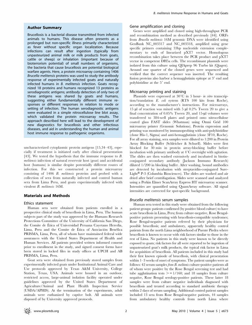
Author Summary
Brucellosis is a bacterial disease transmitted from infectedanimals to humans. This disease often presents as aprolonged but non-specific illness primarily characterizedas fever without specific organ localization. Becauseinfections can result after ingestion (typically fromunpasteurized animal milk or milk products from goats,cattle or sheep) or inhalation (important because ofbioterrorism potential) of small numbers of organisms,the bacteria that cause brucellosis are potential biologicalwarfare agents. Here, a protein microarray containing 1406Brucella melitensis proteins was used to study the antibodyresponse of experimentally infected goats and naturallyinfected humans in B. melitensis infection. Goats recog-nized 18 proteins and humans recognized 13 proteins asserodiagnostic antigens; antibody detection of only two ofthese antigens was shared by goats and humans,suggesting either fundamentally different immune re-sponses or different responses in relation to mode orsetting of infection. The human serodiagnostic antigenswere evaluated in a simple nitrocellulose line blot assay,which validated the protein microarray results. Theapproach described here will lead to the development ofnew diagnostics for brucellosis and other infectiousdiseases, and aid in understanding the human and animalhost immune response to pathogenic organisms.
B. melitensis Immune Response in Humans and Goats
www.plosntds.org 2 May 2010 | Volume 4 | Issue 5 | e673

brucellosis occasionally affects patients, and sera from humans in the
U.S. where brucellosis is not found.
Goat sera tested in this study were positive (B. melitensis 16M-
infected) and negative (uninfected) controls from a previously
conducted vaccine safety study [23] in which pregnant, card-test
negative angora goats were inoculated with B. melitensis. Goats
were experimentally infected with 16107 CFU of Brucella melitensis
strain 16M by bilateral conjunctival instillation at 110 days’
gestation, and sera were collected 8 weeks after infection. As an
additional negative control, 15 serum samples from a specific
pathogen-free goat flock were obtained (Capralogics, Inc, Hard-
wick, MA).
Immunostrip printing and probingThirteen plasmids of interest were expressed in five hour in vitro
transcription-translation reactions (RTS 100 E. coli HY Kit from
Roche) according to the manufacturer’s instructions. VIG was
obtained from ADi as a gift, and the concentration of VIG was
diluted to 0.05 mg/ml. Proteins were printed on Optitran BA-S 85
0.45 mm Nitrocellulose membrane (Whatman) using BioJet
dispenser (BioDot) at 1 ml/cm, and cut into 3 mm strips.
Individual strips were then blocked in 10% non fat dry milk
dissolved in 10 mM Tris (pH 8.0) and 150 mM NaCl containing
0.05% (v/v) Tween 20 buffer for 30 minutes. Prior to immuno-
strip probing, forty two culture positive and forty four Peruvian
naive sera were diluted to 1/250 in 10% non fat dry milk solution
containing 20% E. coli lysate (McLab) and incubated for
30 minutes with constant mixing at room temperature. Each strip
was then incubated with pretreated sera overnight at 4uC with
gentle mixing. Strips were then washed five times in Tris buffer
containing 0.05% (v/v) tween 20, and then incubated for 1 hour
at room temperature in alkaline phosphatase conjugated donkey
anti-human immunoglobulin (anti-IgG, Fcc fragment-specific,
Jackson ImmunoResearch), diluted to 1/5000 in tris buffer
containing 0.05% (v/v) tween 20. Strips were then washed
extensively and reactive bands were visualized by incubating with
1-step Nitro-Blue Tetrazolium Chloride/5-Bromo-4-Chloro-39-
Indolyphosphate p-Toluidine Salt (NBT/BCIP) developing buffer
(Thermo Fisher Scientific) for 1 minute at room temperature.
Strips were scanned with Hewlett-Packard scanner, and were
quantified using Image J software.
Data analysisAll analysis was performed using the R statistical environment
(http://www.r-project.org). It has been noted in the literature that
data derived from microarray platforms is heteroskedatic [45–48].
This mean-variance dependence has been observed in the arrays
presented in this manuscript [49,50]. In order to stabilize the
variance, the vsn method [51] implemented as part of the
Bioconductor suite (www.bioconductor.org) is applied to the
quantified array intensities. In addition to removing heteroskeda-
city, this procedure corrects for non-specific noise effects by
finding maximum likelihood shifting and scaling parameters for
each array such that control probe variance is minimized. This
calibration has been shown to be effective on a number of
platforms [52–54]. Normalized data is retransformed with the
‘sinh’ function to allow visualization and discussion at an
approximate raw scale.
Diagnostic biomarkers between groups were determined using a
Bayes regularized t-test adapted from Cyber-T for protein arrays
[47,48], which has been shown to be more effective than other
differential expression techniques [55]. To account for multiple
testing conditions, the Benjamini and Hochberg (BH) method was
used to control the false discovery rate [56]. Multiplex classifiers
were constructed using linear and non-linear Support Vector
Machines (SVMs) using the ‘‘e1071’’ R package. SVM is a
supervised learning method that has been successfully applied to
microarray data characterized by small samples sizes and a large
number of attributes [50,57]. The SVM approach, as any other
supervised classification approach, uses a training dataset to build
a classification model and a testing set to validate the model. To
generate unbiased training and testing sets, leave one out cross-
validation (LOOCV) was used. With this methodology, each data
point is tested with a classifier trained using all of the remaining
data points. Plots of receiver operating characteristic (ROC) curves
were made with the ‘ROCR’’ R package.
Results
Gene amplification, cloning and protein expressionA set of 1406 ORFs from Brucella melitensis 16M was selected for
this study. We picked 1009 antigens based on their Psort
information and B cell epitope prediction score, and 397 ORFs
were randomly selected. The ORFs were amplified from Brucella
melitensis 16M (Bm) genomic DNA and cloned using the high
Figure 1. Construction of a B. melitensis Protein Microarray.Arrays were printed containing 1406 B. melitensis proteins, positive andnegative control spots. Proteins were printed in duplicates. Each arraycontains positive control spots printed from 6 serial dilutions of humanand mouse IgG, 6 serial dilutions of EBNA1 protein, and ‘‘No DNA’’negative control spots. (A) The array was probed with anti-His antibodyas described in Materials and Methods, to confirm the expression andprinting of over 95% proteins. (B) Comparison of arrays probed withPeruvian naıve serum and Culture positive serum. The arrays were readin a laser confocal scanner, analyzed, and the data normalized asdescribed in Materials and Methods. The signal intensity of eachantigen is represented by rainbow palette of blue, green, red and whiteby increasing signal intensity.doi:10.1371/journal.pntd.0000673.g001
B. melitensis Immune Response in Humans and Goats
www.plosntds.org 3 May 2010 | Volume 4 | Issue 5 | e673

throughput recombination method previously described [44].
About one-fourth of the cloned genes were sequenced and
.99% of sequenced clones had the correct sequence in frame
with correct orientation. Bm ORFs cloned in pXT7 vector were
expressed under T7 promoter in the E. coli in vitro transcription/
translation system, and printed in duplicates on microarrays as
described in Methods and 97.4% of the proteins were positive for
the His tag (Fig. 1a), and 95.4% were positive for HA tags.
Immune screening with goat serum samplesBm protein arrays were probed with sera from experimentally
infected goats, naıve goats from the same pasture, and specific
Figure 2. Probing a collection of B. melitensis infected, uninfected, and SPF control goat sera and discovery of goat serodiagnosticantigens. Arrays containing 1406 B. melitensis proteins were probed with goat sera organized into 3 groups as described in the text. (A). Heatmapshowing normalized intensity with red strongest, bright green weakest and black in between. The antigens are in rows and are grouped toserodiagnostic and cross-reactive antigens. The goat samples are in columns and sorted left to right by increasing average intensity to serodiagnosticantigens. (B) The mean sera reactivity of the 1406 antigens was compared between the Infected and SPF Naive groups. Antigens with BenjaminiHochberg corrected p-value less than 0.05 are organized to the left and cross-reactive antigens to the right. The 18 most reactive serodiagnostic and31 of the most reactive cross-reactive antigens are shown.doi:10.1371/journal.pntd.0000673.g002
B. melitensis Immune Response in Humans and Goats
www.plosntds.org 4 May 2010 | Volume 4 | Issue 5 | e673

Figure 3. Probing a collection of B. melitensis human sera and discovery of human serodiagnostic antigens. Arrays were probed withhuman sera organized into 5 groups: Culture Positive, Culture Negative/Rose Bengal Positive, Rose Bengal Negative, USA Naıve, and Peruvian Naıve,as described in the text. (A). Heatmap showing normalized intensity with red strongest, bright green weakest and black in between. The antigens arein rows and are grouped to serodiagnostic and cross-reactive antigens. The human samples are in columns and sorted left to right by increasingaverage intensity to serodiagnostic antigens. (B) The mean sera reactivity of the 1406 antigens was compared between the Culture Positive andPeruvian Naive groups. Antigens with Benjamini Hochberg corrected p-value less than 0.05 are organized to the left and cross-reactive antigens tothe right. The 13 most reactive serodiagnostic and 31 of the most reactive cross-reactive antigens are shown. C2/RB+, Culture Positive and RoseBengal negative; RB2, Rose Bengal negative. Numbers in () are case numbers from each group.doi:10.1371/journal.pntd.0000673.g003
B. melitensis Immune Response in Humans and Goats
www.plosntds.org 5 May 2010 | Volume 4 | Issue 5 | e673

pathogen free (SPF) goats from a different location. Reactivity of
sera from the individual goats is shown as a heat map with samples
grouped according to their description (Fig. 2a). Data were
analyzed using methods described elsewhere [58]. Serodominant
antigens are defined as antigens with mean signal intensity greater
than the mean plus two standard deviations above the negative
controls. Serodiagnostic antigens are significantly differentially
reactive serodominant antigens with adjusted Cyber-T p-values
between infected and SPF goats ,0.05. All of the sera, whether
from infected, uninfected or naıve goats, reacted similarly to the
cross-reactive antigens (p-value .0.05). A set of 49 antigens were
identified to be serodominant among 1406 antigens tested. Of
these, 18 antigens were serodiagnostic, and reacted differentially
between infected goats and SPF goats (p-value ,0.05). The
remaining 31 serodominant antigens reacted similarly among all
goats (Fig. 2a, 2b).
Human antigenic profileBm protein arrays were also probed with sera from acute
brucellosis patients in Lima, Peru obtained within 1–3 weeks of
the onset of symptoms. All patients in this study, as is true of
virtually all patients from Lima [3,59–62], were infected with B..
melitensis biovar 1. Sera from Bm culture-positive humans (Fig. 1b)
showed pronounced reactivity against several antigens compared
to unexposed individuals. A set of 33 antigens was identified to be
serodominant among 1406 antigens tested (Fig. 3a, 3b). Of
these, 13 antigens were serodiagnostic, and reacted differentially
between naıve and culture positive patients from Peru (p-
value,.05). The same antigens also reacted robustly with
individuals diagnosed Rose Bengal positive but negative by blood
culture for B. melitensis. For some of these subjects, treatment with
antibiotics may have resulted in a negative blood culture for B.
melitensis. The elevated antibody response from a few individuals
in the Peruvian naıve group might be indicative of past exposure
to similar proteins in environmental bacteria, or to a past
subclinical Brucella infection. We also identified 20 cross-reactive
antigens that reacted similarly among all human samples,
whether from naıve individuals or individuals diagnosed to be
infected and use of these antigens in serodiagnostic tests can
therefore be selectively avoided.
Identification of serodiagnostic antigensTo establish a collection of antigens able to accurately
distinguish brucellosis cases from controls, leave one out cross-
validation (LOOCV) receiver operating characteristic (ROC)
curves were generated for individual serodiagnostic antigens to
assess the ability to separate the control and disease cases (Fig. 4).
The serodiagnostic antigens were ordered by decreasing single
antigen area under the curve (AUC). The top ten ORFs all have
an AUC greater than 0.734 (Table 1), with BP26 (BMEI0536;
AUC 0.983; Benjamini and Hochberg adjusted Cyber-T p-
value,10e-16) giving the best single antigen discrimination with
sensitivity and specificity 91% and 96% (Fig. 4), respectively. We
used kernel methods and support vector machines [47,63] to build
linear and nonlinear classifiers. As input to the classifier, we used
the highest-ranking 1, 2, 5, 10, 13 ORFs on the basis of single
antigen AUC. The results show that increasing the antigen
number from 2 to 5 produced an improvement in sensitivity and
specificity (Fig. 4). This classifier yielded a high sensitivity and
specificity rate of 95% and 96%, respectively.
Validation of serodiagnostic accuracy with immunostripsTo test the feasibility of using the serodiagnostic antigens in an
alternative analytical assay, thirteen serodiagnostic proteins were
printed onto Nitrocellulose membranes using a BioDot jet
dispenser. The paper was then cut into 3 mm strips (Fig. 5a).
The individual strips were probed with 42 different culture positive
sera and 44 Peruvian naive sera. Brucellosis patients reacted
strongly against the serodiagnostic antigens with variable signal
intensity among the patients. Naıve samples showed much lower
reactivity against these serodiagnostic antigens. To assess the
ability of antigens to separate disease and healthy controls, the
LOOCV ROC curve was also generated (Fig. 5b). The ROC
curve shows that this probing test yielded a high AUC of 0.962
with sensitivity rate of 94% and specificity rate of 89%. Thus,
thirteen differentially reactive serodiagnostic antigens identified by
microarray analysis in immunostrip format validated the list of
serodiagnostic antigens to correctly classify B. melitensis positive
sera.
The sensitivity and specificity of the top 5 serodiagnostic
antigens discovered using the protein microarrays had sensitivity
and specificity of 95% and 96%, better than that of the 13 antigens
on the immunostrips (94% and 89%).
Comparing antigenic proteins among humans and goatsBoth humans and animals can be infected by Bm. In the present
study, goats were infected by B. melitensis strain 16M which would
be expected to be virtually identical to the strains infected by
patients in Lima given the limited diversity of the strains found
there [3,59–62]. To better understand the differences in the
immune response to Bm infection between humans and goats, we
compared serodominant antigens for both humans and goats. In
the current study, two antigens are found to be serodiagnostic for
both humans and goats (Fig. 6, Table 1). The top antigen on the
list, BMEI0536 (Bp26 protein) is a 26KD periplasmic immuno-
genic protein which was simultaneously identified by three
nonrelated research groups as an immunodominant antigen in
infected cattle, sheep, goats, and humans [17,19,27,35]. Use of an
Figure 4. Multiple Antigen LOOCV ROC curves. The LOOCV ROCgraphs show classifiers with increasing number of human serodiagnos-tic antigens. Overall, the sensitivity and specificity for array test is over95%.doi:10.1371/journal.pntd.0000673.g004
B. melitensis Immune Response in Humans and Goats
www.plosntds.org 6 May 2010 | Volume 4 | Issue 5 | e673
indirect ELISA to detect antibodies in brucellosis patients (n = 20)
and uninfected controls (n = 35) yielded a sensitivity of 0.9 and
specificity of 0.91 (not shown). Another serodiagnostic protein for
both humans and goats was Protease DO, also designated as HtrA
[31]. Use of an indirect ELISA to BMEI1330 yielded a sensitivity
of 0.84 and a specificity of 0.99. Thus, the ELISA data were
consistent with values determined using immunostrips and with
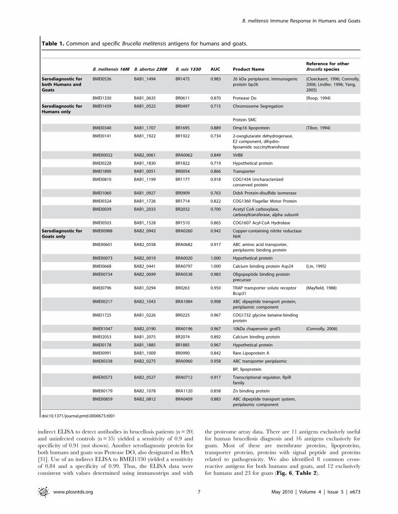
the proteome array data. There are 11 antigens exclusively useful
for human brucellosis diagnosis and 16 antigens exclusively for
goats. Most of these are membrane proteins, lipoproteins,
transporter proteins, proteins with signal peptide and proteins
related to pathogenicity. We also identified 8 common cross-
reactive antigens for both humans and goats, and 12 exclusively
for humans and 23 for goats (Fig. 6, Table 2).
Table 1. Common and specific Brucella melitensis antigens for humans and goats.
B. melitensis 16M B. abortus 2308 B. suis 1330 AUC Product NameReference for otherBrucella species
Serodiagnostic forboth Humans andGoats
BMEI0536 BAB1_1494 BR1475 0.983 26 kDa periplasmic immunogenicprotein bp26
{Cloeckaert, 1996; Connolly,2006; Lindler, 1996; Yang,2005}
BMEI1330 BAB1_0635 BR0611 0.870 Protease Do {Roop, 1994}
Serodiagnostic forHumans only
BMEI1439 BAB1_0522 BR0497 0.715 Chromosome Segregation
Protein SMC
BMEI0340 BAB1_1707 BR1695 0.889 Omp16 lipoprotein {Tibor, 1994}
BMEI0141 BAB1_1922 BR1922 0.734 2-oxoglutarate dehydrogenase,E2 component, dihydro-lipoamide succinyltransferase
BMEII0032 BAB2_0061 BRA0062 0.849 VirB8
BMEI0228 BAB1_1830 BR1822 0.719 Hypothetical protein
BMEI1890 BAB1_0051 BR0054 0.866 Transporter
BMEI0810 BAB1_1199 BR1177 0.918 COG1434 Uncharacterizedconserved protein
BMEI1060 BAB1_0927 BR0909 0.763 DsbA Protein-disulfide isomerase
BMEI0324 BAB1_1726 BR1714 0.822 COG1360 Flagellar Motor Protein
BMEI0039 BAB1_2033 BR2032 0.700 Acetyl CoA carboxylase,carboxyltransferase, alpha subunit
BMEI0503 BAB1_1528 BR1510 0.865 COG1607 Acyl-CoA Hydrolase
Serodiagnostic forGoats only
BMEII0988 BAB2_0943 BRA0260 0.942 Copper-containing nitrite reductaseNirK
BMEII0601 BAB2_0558 BRA0682 0.917 ABC amino acid transporter,periplasmic binding protein
BMEII0073 BAB2_0019 BRA0020 1.000 Hypothetical protein
BMEI0668 BAB2_0441 BRA0797 1.000 Calcium binding protein Asp24 {Lin, 1995}
BMEII0734 BAB2_0699 BRA0538 0.983 Oligopeptide binding proteinprecursor
BMEI0796 BAB1_0294 BR0263 0.950 TRAP transporter solute receptorBcsp31
{Mayfield, 1988}
BMEII0217 BAB2_1043 BRA1084 0.908 ABC dipeptide transport protein,periplasmic component
BMEI1725 BAB1_0226 BR0225 0.967 COG1732 glycine betaine-bindingprotein
BMEII1047 BAB2_0190 BRA0196 0.967 10kDa chaperonin groES {Connolly, 2006}
BMEI2053 BAB1_2075 BR2074 0.892 Calcium binding protein
BMEI0178 BAB1_1885 BR1885 0.967 Hypothetical protein
BMEI0991 BAB1_1009 BR0990 0.842 Rare Lipoprotein A
BMEII0338 BAB2_0275 BRA0960 0.958 ABC transporter periplasmic
BP, lipoprotein
BMEII0573 BAB2_0527 BRA0712 0.917 Transcriptional regulator, RpiRfamily
BMEII0179 BAB2_1078 BRA1120 0.858 Zn binding protein
BMEII0859 BAB2_0812 BRA0409 0.883 ABC dipeptide transport system,periplasmic component
doi:10.1371/journal.pntd.0000673.t001
B. melitensis Immune Response in Humans and Goats
www.plosntds.org 7 May 2010 | Volume 4 | Issue 5 | e673

Discussion
Here we report a large scale analysis showing that the humoral
immune responses against B. melitensis protein antigens differ
between humans naturally infected by consuming Brucella melitensis-
contaminated, unpasteurized goat’s milk products, and goats
experimentally infected with B. melitensis by conjunctival instilla-
tion. These observations show that a natural reservoir host and the
accidental human host have fundamentally different immune
responses against this zoonotic pathogen. These data have
implications for the practical development of diagnostics and
reflect basic differences in host pathogen interactions and disease
pathogenesis.
In addition, we demonstrate that a systematic, genome-wide
analysis proved to identify protein antigens recognized by humans
and animals not previously identified using Western blot or
genomic library immunoscreening. Further, by virtue of being
found to react with antibodies, the protein array technology is
able to provide strong evidence of the comprehensive set of
proteins expressed in vivo within a mammalian host by B. melitensis.
As with our published experience with viral, bacterial and pro-
tozoal genomes expressed using protein microarray technology
[44,49,50,58,64–66], conformation-dependent epitopes seem not
to present problems with data interpretation or comprehensiveness
of antigen discovery. This is likely because the polyclonal antibody
response to protein antigens after infection detects both linear and
3-dimensional epitopes. The B. melitensis proteins placed onto the
array, while expressed heterologously in a bacterial system, likely
reflect a mix of conformationally correct as well as misfolded
epitopes both of which are capable of binding specific antibodies.
Serological diagnosis of both human and animal brucellosis can
suffer from the inability to distinguish new from previous infection
(in the case of humans [1]) and differentiation of vaccination from
new infection (in the case of animals [8]). In the absence of known
exposure history in endemic regions, there is the possibility of
mistaken diagnosis and overtreatment [10]. Current assays are
based primarily on identification of antibodies to LPS in patient
serum. Since Brucella LPS is cross-reactive with several other
species, including E. coli O157:H7, Yersinia enterocolitica O9, and
Francisella tularensis (although the clinical presentations of infectious
caused by these agents are quite different), identification of
diagnostic protein antigens may facilitate the development of more
specific serodiagnostic assays [21,67,68]. The top 5 serodiagnostic
antigens discovered using the protein microarrays had sensitivity
and specificity of 95% and 96%, better than that of the 13 antigens
on the immunostrips (94% and 89%), which in turn was roughly
comparable to that of smooth LPS-based tests used in the Rose
Bengal, lateral flow, and ELISA formats. In the present study
however, the sensitivity of the serodiagnostic protein antigens
could not be compared to that of the Rose Bengal test because we
did not confirm any brucellosis cases among Rose Bengal negative
patients by culture.
One interesting finding of this study was the difference in
background reactivity to B. melitensis proteins in uninfected
individuals from endemic vs. non-endemic areas. In Peru, control
subjects tended to have higher background reactivity to Brucella
antigens, compared to US control subjects (Fig. 3a). Consideration
of these differences would be important for the development of
diagnostic assays intended for use in both endemic and non-
endemic regions of the world. The degree of variability between
subjects differs depending on the infection and the results for
Brucella reported here are similar to those that we obtained from
patients with melioidosis [64,65] and Lyme disease.
Figure 5. Immunostrips probing. (a)Thirteen serodiagnostic anti-gens were printed onto nitrocellulose paper in adjacent stripes using aBioDot jet dispenser as described in Materials and Methods. Strips wereprobed with Culture Positive or Peruvian naive sera diluted 1/200followed by alkaline phosphatase conjugated secondary antibody andenzyme substrate. Weak reactivity in the naıve healthy controls can bedistinguished from the strong reactivity in infected group. (b). TheLOOCV ROC curve was generated and sensitivity and specificity ofimmunostrips probing test is 94% and 89%, respectively.doi:10.1371/journal.pntd.0000673.g005
Figure 6. Serodiagnostic and cross-reactive antigens forhumans and goats.doi:10.1371/journal.pntd.0000673.g006
B. melitensis Immune Response in Humans and Goats
www.plosntds.org 8 May 2010 | Volume 4 | Issue 5 | e673
Our results with the B. melitensis proteome array represent the
first large-scale analysis of B. melitensis proteins that are
immunogenic in the context of naturally acquired human
infections. In the case of the present study, the identified risk
factor for human infection was ingestion of B. melitensis-
contaminated, unpasteurized goat’s milk products. In other
epidemiological contexts, B. melitensis can be contracted by direct
exposure to infected animals, not only goats, but also sheep and
cattle [1,3]. Further, we compared the set of proteins identified
using human patient sera with the set that was immunogenic in the
animal reservoir for zoonotic disease, the goat. Two proteins,
BMEI0536 (Bp26) and BMEI1330 (HtrA/DegP), were immuno-
genic in the context of both infections. These results are in good
agreement with previous reports on these antigens from other
Brucella spp. Roop et al. [31] showed that HtrA was recognized by
serum from goats, cattle and mice experimentally infected with B.
abortus and by serum of dogs infected with B. canis. HtrA/DegP is a
periplasmic serine protease that contributes to survival following
stresses including oxidative damage. Bp26 has been proposed as a
diagnostic antigen for detection of B. melitensis infection in sheep
and B. abortus in cattle [17,19]. The Omp16 lipoprotein
(BMEI0340), originally identified as an immunogenic protein of
B. abortus [32], was recognized by patient sera, but was found to be
reactive in both infected and uninfected goats. Our results differ
from those of Letesson et al., who reported no reactivity of
uninfected goats to Omp16 [25]. This difference may reflect
exposure of goats used in our study to other pathogens or to
environmental bacteria expressing a cross-reactive antigen. The
identification of known immunogenic Brucella proteins on the
proteome array provided confirmation that our approach could
identify both known and novel immunogenic proteins.
In addition to these well-characterized antigens, our study
identified several novel serodiagnostic antigens specific for human
B. melitensis patients (Table 1). These included BMEI1439 (SMC),
an ATPase shown in other bacterial species to be involved in
condensation and segregation of replicating chromosomes [29].
Further, the bacterial cell envelope proteins VirB8 (BMEII0032),
DsbA (BMEI1060), and an uncharacterized transporter
(BMEI1890), were immunogenic in patients. Three metabolic
enzymes, acetyl coA carboxylase, (BMEI0503), Acetyl CoA
carboxylase (BMEI0039 and 2-oxoglutarate dehydrogenase
(BMEI0141) also represent novel serodiagnostic antigens for
human brucellosis. The finding that these proteins are immuno-
genic suggests that they are expressed during B. melitensis infection
of humans.
A group of 16 antigens was found to be serodiagnostic for goats,
but not humans (Table 1). These included 7 predicted transport-
ers, as well as a zinc-binding protein and two binding proteins for
calcium: Asp24 (BMEI0668) [26], and a second, uncharacterized
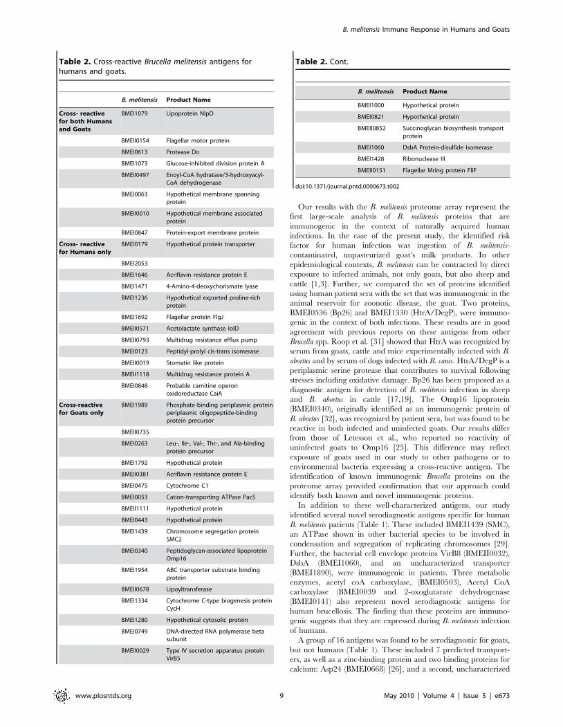
Table 2. Cross-reactive Brucella melitensis antigens forhumans and goats.
B. melitensis Product Name
Cross- reactivefor both Humansand Goats
BMEI1079 Lipoprotein NlpD
BMEII0154 Flagellar motor protein
BMEI0613 Protease Do
BMEI1073 Glucose-inhibited division protein A
BMEII0497 Enoyl-CoA hydratase/3-hydroxyacyl-CoA dehydrogenase
BMEI0063 Hypothetical membrane spanningprotein
BMEII0010 Hypothetical membrane associatedprotein
BMEI0847 Protein-export membrane protein
Cross- reactivefor Humans only
BMEI0179 Hypothetical protein transporter
BMEI2053
BMEI1646 Acriflavin resistance protein E
BMEI1471 4-Amino-4-deoxychorismate lyase
BMEI1236 Hypothetical exported proline-richprotein
BMEI1692 Flagellar protein FlgJ
BMEII0571 Acetolactate synthase IolD
BMEII0793 Multidrug resistance efflux pump
BMEI0123 Peptidyl-prolyl cis-trans isomerase
BMEII0019 Stomatin like protein
BMEII1118 Multidrug resistance protein A
BMEI0848 Probable carnitine operonoxidoreductase CaiA
Cross-reactivefor Goats only
BMEI1989 Phosphate-binding periplasmic proteinperiplasmic oligopeptide-bindingprotein precursor
BMEII0735
BMEI0263 Leu-, Ile-, Val-, Thr-, and Ala-bindingprotein precursor
BMEI1792 Hypothetical protein
BMEII0381 Acriflavin resistance protein E
BMEI0475 Cytochrome C1
BMEI0053 Cation-transporting ATPase PacS
BMEII1111 Hypothetical protein
BMEI0443 Hypothetical protein
BMEI1439 Chromosome segregation proteinSMC2
BMEI0340 Peptidoglycan-associated lipoproteinOmp16
BMEI1954 ABC transporter substrate bindingprotein
BMEII0678 Lipoyltransferase
BMEI1334 Cytochrome C-type biogenesis proteinCycH
BMEI1280 Hypothetical cytosolic protein
BMEI0749 DNA-directed RNA polymerase betasubunit
BMEII0029 Type IV secretion apparatus proteinVirB5
B. melitensis Product Name
BMEI1000 Hypothetical protein
BMEI0821 Hypothetical protein
BMEII0852 Succinoglycan biosynthesis transportprotein
BMEI1060 DsbA Protein-disulfide isomerase
BMEI1428 Ribonuclease III
BMEII0151 Flagellar Mring protein FliF
doi:10.1371/journal.pntd.0000673.t002
Table 2. Cont.
B. melitensis Immune Response in Humans and Goats
www.plosntds.org 9 May 2010 | Volume 4 | Issue 5 | e673
calcium-binding protein (BMEI2053). The chaperonin GroES,
shown to be seroreactive in a patient suffering from B. suis
infection [20], was serodiagnostic in goats, as well as Bscp31
(BMEI0796), a well-known seroreactive protein [28].
The differences in the patterns of goat and human seroreactivity
to B. melitensis proteins could be explained in several different ways.
First, the group of seroreactive proteins in goats and humans gives
some insight into the metabolic pathways expressed during
infection in these hosts. The large number of immunogenic
proteins with predicted function in nutrient uptake suggests that B.
melitensis utilizes peptides and amino acids for growth during
infection. Three serodiagnostic B. melitensis proteins (BMEI0340,
BMEI0141 and BMEI0178), have been found to be upregulated
during intracellular infection by B. suis or B. abortus [18,24],
suggesting that they may play a role in adaptation to intracellular
life in the host. Second, the immunogenetics of immune responses
to B. melitensis infection may be related to species differences
between humans and goats but further comment on this possibility
is limited by the lack of available data. Third, while humans in this
study were thought to have been infected by ingestion of infected
goat’s milk products containing a small inoculum, the goats were
infected with a high dose (107 CFU) of B. melitensis strain 16M via
the conjunctiva. Both dose and route of inoculum may have
contributed to the differential antigen recognition between goats
and humans, and, in fact, among humans as well. Finally, while it
seems unlikely that the different patterns of immune response
variation between goats and humans were due to protein
expression differences by different strains of B. melitensis, this
possibility must be considered as well.
The results presented here represent an analysis of 1406
proteins of 3198 predicted proteins in the B. melitensis 16M
genome. Of these 1406 proteins, we only observed less than two
fold enrichment of serodiagnostic antigens in the 1009 selected
versus randomly selected antigens (not shown). Completion of the
proteome array is currently underway, which will allow a more
complete genome-level analysis of all immunogenic B. melitensis
proteins. The subset of diagnostic antigens identified here
provided an initial estimated accuracy rate of 95% for diagnosis
of human cases and it is likely that this set of antigens will form the
basis of a new and accurate serodiagnostic assay for human
brucellosis. The clinical and veterinary utility of the protein
antigens discovered in this study for diagnosis of acute and chronic
brucellosis awaits validation in prospective studies in endemic
regions.
Acknowledgments
We thank Kalina Campos for outstanding microbiological work, and thank
Paula Maguina of the University of California San Diego who made
essential research contributions in terms of ethics management and
international coordination as well as logistical support for this project.
Author Contributions
Conceived and designed the experiments: LL CB WJWM XL JMV RMT
PLF. Performed the experiments: LL DL CB RNS VLA JP BU. Analyzed
the data: LL CB MAK PLF. Contributed reagents/materials/analysis
tools: DL MAK TAF EG MS PB RHG JMV RMT. Wrote the paper: LL
MAK JMV RMT PLF.
References
1. Pappas G, Akritidis N, Bosilkovski M, Tsianos E (2005) Brucellosis. N Engl J Med
352: 2325–2336.
2. Pappas G, Papadimitriou P, Akritidis N, Christou L, Tsianos EV (2006) The
new global map of human brucellosis. Lancet Infect Dis 6: 91–99.
3. Franco MP, Mulder M, Gilman RH, Smits HL (2007) Human brucellosis.
Lancet Infect Dis 7: 775–786.
4. Young EJ (1995) An overview of human brucellosis. Clin Infect Dis 21: 283–289;
quiz 290.
5. (2009) World Organisation for Animal Health Manual of Diagnostic Tests and
Vaccines for Terrestrial Animals.
6. Ramirez-Pfeiffer C, Diaz-Aparicio E, Gomez-Flores R, Rodriguez-Padilla C,
Morales-Loredo A, et al. (2008) Use of the Brucella melitensis native hapten to
diagnose brucellosis in goats by a rapid, simple, and specific fluorescence
polarization assay. Clin Vaccine Immunol 15: 911–915.
7. Ramirez-Pfeiffer C, Diaz-Aparicio E, Rodriguez-Padilla C, Morales-Loredo A,
Alvarez-Ojeda G, et al. (2008) Improved performance of Brucella melitensis
native hapten over Brucella abortus OPS tracer on goat antibody detection by
the fluorescence polarization assay. Vet Immunol Immunopathol 123: 223–229.
8. Diaz-Aparicio E, Marin C, Alonso-Urmeneta B, Aragon V, Perez-Ortiz S, et al.
(1994) Evaluation of serological tests for diagnosis of Brucella melitensis infection
of goats. J Clin Microbiol 32: 1159–1165.
9. Morgan WJ, MacKinnon DJ, Lawson JR, Cullen GA (1969) The rose bengal
plate agglutination test in the diagnosis of brucellosis. Vet Rec 85: 636–641.
10. Jennings GJ, Hajjeh RA, Girgis FY, Fadeel MA, Maksoud MA, et al. (2007)
Brucellosis as a cause of acute febrile illness in Egypt. Trans R Soc Trop Med
Hyg 101: 707–713.
11. Al Dahouk S, Tomaso H, Nockler K, Neubauer H, Frangoulidis D (2003)
Laboratory-based diagnosis of brucellosis–a review of the literature. Part II:
serological tests for brucellosis. Clin Lab 49: 577–589.
12. Gomez MC, Nieto JA, Rosa C, Geijo P, Escribano MA, et al. (2008) Evaluation
of seven tests for diagnosis of human brucellosis in an area where the disease is
endemic. Clin Vaccine Immunol 15: 1031–1033.
13. Orduna A, Almaraz A, Prado A, Gutierrez MP, Garcia-Pascual A, et al. (2000)
Evaluation of an immunocapture-agglutination test (Brucellacapt) for serodiag-
nosis of human brucellosis. J Clin Microbiol 38: 4000–4005.
14. Casao MA, Navarro E, Solera J (2004) Evaluation of Brucellacapt for the
diagnosis of human brucellosis. J Infect 49: 102–108.
15. Mantecon MA, Gutierrez P, del Pilar Zarzosa M, Duenas AI, Solera J, et al.
(2006) Utility of an immunocapture-agglutination test and an enzyme-linked
immunosorbent assay test against cytosolic proteins from Brucella melitensis
B115 in the diagnosis and follow-up of human acute brucellosis. Diagn
Microbiol Infect Dis 55: 27–35.
16. Bosilkovski M, Katerina S, Zaklina S, Ivan V (2009) The role of Brucellacapt test
for follow-up patients with brucellosis. Comp Immunol Microbiol Infect Dis.
17. Rossetti OL, Arese AI, Boschiroli ML, Cravero SL (1996) Cloning of Brucella
abortus gene and characterization of expressed 26-kilodalton periplasmic
protein: potential use for diagnosis. J Clin Microbiol 34: 165–169.
18. Al Dahouk S, Jubier-Maurin V, Scholz HC, Tomaso H, Karges W, et al. (2008)
Quantitative analysis of the intramacrophagic Brucella suis proteome reveals
metabolic adaptation to late stage of cellular infection. Proteomics 8: 3862–
3870.
19. Cloeckaert A, Debbarh HS, Vizcaino N, Saman E, Dubray G, et al. (1996)
Cloning, nucleotide sequence, and expression of the Brucella melitensis bp26
gene coding for a protein immunogenic in infected sheep. FEMS Microbiol Lett
140: 139–144.
20. Connolly JP, Comerci D, Alefantis TG, Walz A, Quan M, et al. (2006)
Proteomic analysis of Brucella abortus cell envelope and identification of
immunogenic candidate proteins for vaccine development. Proteomics 6:
3767–3780.
21. Corbel MJ, Stuart FA, Brewer RA (1984) Observations on serological cross-
reactions between smooth Brucella species and organisms of other genera. Dev
Biol Stand 56: 341–348.
22. Gor D, Mayfield JE (1992) Cloning and nucleotide sequence of the Brucella
abortus groE operon. Biochim Biophys Acta 1130: 120–122.
23. Kahl-McDonagh MM, Elzer PH, Hagius SD, Walker JV, Perry QL, et al. (2006)
Evaluation of novel Brucella melitensis unmarked deletion mutants for safety
and efficacy in the goat model of brucellosis. Vaccine 24: 5169–5177.
24. Lamontagne J, Forest A, Marazzo E, Denis F, Butler H, et al. (2009)
Intracellular adaptation of Brucella abortus. J Proteome Res 8: 1594–1609.
25. Letesson JJ, Tibor A, van Eynde G, Wansard V, Weynants V, et al. (1997)
Humoral immune responses of Brucella-infected cattle, sheep, and goats to eight
purified recombinant Brucella proteins in an indirect enzyme-linked immuno-
sorbent assay. Clin Diagn Lab Immunol 4: 556–564.
26. Lin J, Ficht TA (1995) Protein synthesis in Brucella abortus induced during
macrophage infection. Infect Immun 63: 1409–1414.
27. Lindler LE, Hadfield TL, Tall BD, Snellings NJ, Rubin FA, et al. (1996) Cloning
of a Brucella melitensis group 3 antigen gene encoding Omp28, a protein
recognized by the humoral immune response during human brucellosis. Infect
Immun 64: 2490–2499.
28. Mayfield JE, Bricker BJ, Godfrey H, Crosby RM, Knight DJ, et al. (1988) The
cloning, expression, and nucleotide sequence of a gene coding for an
immunogenic Brucella abortus protein. Gene 63: 1–9.
29. Pogliano K, Pogliano J, Becker E (2003) Chromosome segregation in
Eubacteria. Curr Opin Microbiol 6: 586–593.
B. melitensis Immune Response in Humans and Goats
www.plosntds.org 10 May 2010 | Volume 4 | Issue 5 | e673
30. Rolan HG, den Hartigh AB, Kahl-McDonagh M, Ficht T, Adams LG, et al.
(2008) VirB12 is a serological marker of Brucella infection in experimental and
natural hosts. Clin Vaccine Immunol 15: 208–214.
31. Roop RM, 2nd, Fletcher TW, Sriranganathan NM, Boyle SM, Schurig GG
(1994) Identification of an immunoreactive Brucella abortus HtrA stress response
protein homolog. Infect Immun 62: 1000–1007.
32. Tibor A, Weynants V, Denoel P, Lichtfouse B, De Bolle X, et al. (1994)
Molecular cloning, nucleotide sequence, and occurrence of a 16.5-kilodalton
outer membrane protein of Brucella abortus with similarity to pal lipoproteins.
Infect Immun 62: 3633–3639.
33. Yang X, Hudson M, Walters N, Bargatze RF, Pascual DW (2005) Selection of
protective epitopes for Brucella melitensis by DNA vaccination. Infect Immun
73: 7297–7303.
34. Baldi PC, Miguel SE, Fossati CA, Wallach JC (1996) Serological follow-up of
human brucellosis by measuring IgG antibodies to lipopolysaccharide and
cytoplasmic proteins of Brucella species. Clin Infect Dis 22: 446–455.
35. Salih-Alj Debbarh H, Cloeckaert A, Bezard G, Dubray G, Zygmunt MS (1996)
Enzyme-linked immunosorbent assay with partially purified cytosoluble 28-
kilodalton protein for serological differentiation between Brucella melitensis-
infected and B. melitensis Rev.1-vaccinated sheep. Clin Diagn Lab Immunol 3:
305–308.
36. Cassataro J, Delpino MV, Velikovsky CA, Bruno L, Fossati CA, et al. (2002)
Diagnostic usefulness of antibodies against ribosome recycling factor from
Brucella melitensis in human or canine brucellosis. Clin Diagn Lab Immunol 9:
366–369.
37. Cassataro J, Pasquevich K, Bruno L, Wallach JC, Fossati CA, et al. (2004)
Antibody reactivity to Omp31 from Brucella melitensis in human and animal
infections by smooth and rough Brucellae. Clin Diagn Lab Immunol 11:
111–114.
38. Contreras-Rodriguez A, Seleem MN, Schurig GG, Sriranganathan N,
Boyle SM, et al. (2006) Cloning, expression and characterization of
immunogenic aminopeptidase N from Brucella melitensis. FEMS Immunol
Med Microbiol 48: 252–256.
39. Delpino MV, Cassataro J, Fossati CA, Baldi PC (2003) Antibodies to the CP24
protein of Brucella melitensis lack diagnostic usefulness in ovine brucellosis. Vet
Microbiol 93: 101–107.
40. Estein SM, Baldi PC, Bowden RA (2002) Comparison of serological tests based
on outer membrane or internal antigens for detecting antibodies to Brucella ovis
in infected flocks. J Vet Diagn Invest 14: 407–411.
41. Goldbaum FA, Rubbi CP, Wallach JC, Miguel SE, Baldi PC, et al. (1992)
Differentiation between active and inactive human brucellosis by measuring
antiprotein humoral immune responses. J Clin Microbiol 30: 604–607.
42. Verstreate DR, Creasy MT, Caveney NT, Baldwin CL, Blab MW, et al. (1982)
Outer membrane proteins of Brucella abortus: isolation and characterization.
Infect Immun 35: 979–989.
43. Wallach JC, Miguel SE, Baldi PC, Guarnera E, Goldbaum FA, et al. (1994)
Urban outbreak of a Brucella melitensis infection in an Argentine family: clinical
and diagnostic aspects. FEMS Immunol Med Microbiol 8: 49–56.
44. Davies DH, Liang X, Hernandez JE, Randall A, Hirst S, et al. (2005) Profiling
the humoral immune response to infection by using proteome microarrays: high-
throughput vaccine and diagnostic antigen discovery. Proc Natl Acad Sci U S A
102: 547–552.
45. Durbin BP, Hardin JS, Hawkins DM, Rocke DM (2002) A variance-stabilizing
transformation for gene-expression microarray data. Bioinformatics 18 Suppl 1:
S105–110.
46. Ideker T, Thorsson V, Siegel AF, Hood LE (2000) Testing for differentially-
expressed genes by maximum-likelihood analysis of microarray data. J Comput
Biol 7: 805–817.
47. Baldi P, Brunak SR (2001) Bioinformatics: the machine learning approach.
CambridgeMA: MIT Press.
48. Baldi P, Long AD (2001) A Bayesian framework for the analysis of microarray
expression data: regularized t -test and statistical inferences of gene changes.Bioinformatics 17: 509–519.
49. Sundaresh S, Doolan DL, Hirst S, Mu Y, Unal B, et al. (2006) Identification of
humoral immune responses in protein microarrays using DNA microarray dataanalysis techniques. Bioinformatics 22: 1760–1766.
50. Sundaresh S, Randall A, Unal B, Petersen JM, Belisle JT, et al. (2007) Fromprotein microarrays to diagnostic antigen discovery: a study of the pathogen
Francisella tularensis. Bioinformatics 23: i508–518.
51. Huber W, von Heydebreck A, Sultmann H, Poustka A, Vingron M (2002)Variance stabilization applied to microarray data calibration and to the
quantification of differential expression. Bioinformatics 18 Suppl 1: S96–104.52. Kreil DP, Karp NA, Lilley KS (2004) DNA microarray normalization methods
can remove bias from differential protein expression analysis of 2D difference gelelectrophoresis results. Bioinformatics 20: 2026–2034.
53. Barbacioru CC, Wang Y, Canales RD, Sun YA, Keys DN, et al. (2006) Effect of
various normalization methods on Applied Biosystems expression array systemdata. BMC Bioinformatics 7: 533.
54. Sarkar D, Parkin R, Wyman S, Bendoraite A, Sather C, et al. (2009) Qualityassessment and data analysis for microRNA expression arrays. Nucleic Acids
Res 37: e17.
55. Choe SE, Boutros M, Michelson AM, Church GM, Halfon MS (2005) Preferredanalysis methods for Affymetrix GeneChips revealed by a wholly defined control
dataset. Genome Biol 6: R16.56. Hochberg Y, Benjamini Y (1990) More powerful procedures for multiple
significance testing. Stat Med 9: 811–818.57. Doran M, Raicu DS, Furst JD, Settimi R, Schipma M, et al. (2007)
Oligonucleotide microarray identification of Bacillus anthracis strains using
support vector machines. Bioinformatics 23: 487–492.58. Doolan DL, Mu Y, Unal B, Sundaresh S, Hirst S, et al. (2008) Profiling humoral
immune responses to P. falciparum infection with protein microarrays.Proteomics 8: 4680–4694.
59. Nockler K, Maves R, Cepeda D, Draeger A, Mayer-Scholl A, et al. (2009)
Molecular epidemiology of Brucella genotypes in patients at a major hospital incentral Peru. J Clin Microbiol 47: 3147–3155.
60. Espinosa BJ, Chacaltana J, Mulder M, Franco MP, Blazes DL, et al. (2009)Comparison of culture techniques at different stages of brucellosis. Am J Trop
Med Hyg 80: 625–627.61. Smits HL, Espinosa B, Castillo R, Hall E, Guillen A, et al. (2009) MLVA
genotyping of human Brucella isolates from Peru. Trans R Soc Trop Med Hyg
103: 399–402.62. Mendoza-Nunez M, Mulder M, Franco MP, Maas KS, Castaneda ML, et al.
(2008) Brucellosis in household members of Brucella patients residing in a largeurban setting in Peru. Am J Trop Med Hyg 78: 595–598.
63. Girosi F (1998) An Equivalence Between Sparse Approximation and Support
Vector Machines. Neural Comput 10: 1455–1480.64. Barbour AG, Jasinskas A, Kayala MA, Davies DH, Steere AC, et al. (2008) A
genome-wide proteome array reveals a limited set of immunogens in naturalinfections of humans and white-footed mice with Borrelia burgdorferi. Infect
Immun 76: 3374–3389.65. Felgner PL, Kayala MA, Vigil A, Burk C, Nakajima-Sasaki R, et al. (2009) A
Burkholderia pseudomallei protein microarray reveals serodiagnostic and cross-
reactive antigens. Proc Natl Acad Sci U S A 106: 13499–13504.66. Davies DH, Molina DM, Wrammert J, Miller J, Hirst S, et al. (2007) Proteome-
wide analysis of the serological response to vaccinia and smallpox. Proteomics 7:1678–1686.
67. Behan KA, Klein GC (1982) Reduction of Brucella species and Francisella
tularensis cross-reacting agglutinins by dithiothreitol. J Clin Microbiol 16:756–757.
68. Chart H, Okubadejo OA, Rowe B (1992) The serological relationship betweenEscherichia coli O157 and Yersinia enterocolitica O9 using sera from patients
with brucellosis. Epidemiol Infect 108: 77–85.
B. melitensis Immune Response in Humans and Goats
www.plosntds.org 11 May 2010 | Volume 4 | Issue 5 | e673
Related Documents