LANGUAGE DEVELOPMENT IN CHILDREN WITH UNILATERAL BRAIN INJURY Elizabeth Bates and Katherine Roe University of California, San Diego In C.A. Nelson & M. Luciana (Eds.), Handbook of developmental cognitive neuroscience. Cambridge, MA: MIT Press, 2001 Support for the research described here was provided by NIH/NINDS P50 NS22343 (“Center for the Study of the Neural Bases of Language and Learning”) and NIH/NIDCD P50 DC001289 (“Origins of Communicative Disorders”). Please address all correspondence to Elizabeth Bates, Center for Research in Language 0526, University of California at San Diego, La Jolla, CA 92093-0526 ([email protected]).

Welcome message from author
This document is posted to help you gain knowledge. Please leave a comment to let me know what you think about it! Share it to your friends and learn new things together.
Transcript
LANGUAGE DEVELOPMENT IN CHILDREN
WITH UNILATERAL BRAIN INJURY
Elizabeth Bates and Katherine Roe
University of California, San Diego
In C.A. Nelson & M. Luciana (Eds.), Handbook of developmental cognitive neuroscience. Cambridge, MA: MIT Press, 2001
Support for the research described here was provided by NIH/NINDS P50 NS22343
(“Center for the Study of the Neural Bases of Language and Learning”) and NIH/NIDCD P50
DC001289 (“Origins of Communicative Disorders”). Please address all correspondence to
Elizabeth Bates, Center for Research in Language 0526, University of California at San Diego,
La Jolla, CA 92093-0526 ([email protected]).
2
LANGUAGE DEVELOPMENT IN CHILDREN WITH UNILATERAL BRAIN INJURY
Elizabeth Bates and Katherine Roe
University of California, San Diego
AbstractAphasia (defined as the loss or impairment of language abilities following acquired brain injury) is strongly associated withdamage to the left hemisphere in adults. This well-known finding has led to the hypothesis that the left hemisphere is innatelyspecialized for language, and may be the site of a specific "language organ". However, for over a century we have known thatyoung children with left-hemisphere damage (LHD) do not suffer from aphasia, and in most studies do not differ significantlyfrom children with right-hemisphere damage (RHD). This result provides strong evidence for plasticity, i.e., brain reorganizationin response to experience, and constitutes a serious challenge to the language organ hypothesis. This chapter reviews the historyof research on language outcomes in children vs. adults with unilateral brain injury, addressing some discrepancies in the literatureto date, including methodological confounds that may be responsible for those discrepancies. It also reviews recent prospectivestudies of children with unilateral injury as they pass through the first stages of language development. Prospective studies havedemonstrated specific correlations between lesion site and profiles of language delay, but they look quite different from lesion-symptom correlations in adults, and gradually disappear across the course of language development. The classic pattern of brainorganization for language observed in normal adults may be the product rather than the cause of language learning, emerging out ofregional biases in information processing that are relevant for language, but only indirectly related to language itself. If thoseregions are damaged early in life, other parts of the brain can emerge to solve the language learning problem.
Aphasia, or the loss of language abilities following braininjury, has been studied systematically in adults for over acentury, and its existence has been docu-mented since the firstEgyptian surgical papyrus more than 4000 years ago(Goodglass, 1993; O'Neill, 1980). There is now a large bodyof research on adult aphasia, and although there is stillsubstantial controversy regarding its nature and causes,consensus has emerged on at least two points: injuries to theleft hemisphere are overwhelmingly more likely to causeaphasia than injuries to the right, which in turn suggests thatthe left hemisphere plays a privileged role in language pro-cessing by normal adults. The second conclusion has beenindependently confirmed in the 20th century by methodsranging from sodium amytal (WADA) tests and/or point-to-point electrical stimulation in adult candidates forneurosurgery (Ojemann, 1991), to neural imaging studies ofnormals, including positron emis-sion tomography (PET),functional magnetic resonance imaging (fMRI), magneto-encephalo-graphy (MEG) and event-related brain potentials(ERP) (for reviews, see Brown and Hagoort, 1999; Xiong etal., 1998).
The privileged status of the left hemisphere for languageprocessing is now beyond dispute (with esti-mates averagingfrom 95%-98% of normal adults, independent of handedness),but the origins and develop-ment of this specialization are stillpoorly understood. There must be something about the lefthemisphere that makes it especially suited for language -- butwhat is that “something”? Is it present at birth, or does itdevelop gradually? Is it possible to develop normal language inthe absence of a normal left hemisphere? And if an intact lefthemisphere is not required for language development, thenwhen, how, and why does it become necessary for languageuse in adults? Finally, if alternative forms of brain
organization for language can emerge in the presence of earlyleft-hemisphere damage, is there some critical period in whichthis must occur?
The sparse but growing body of evidence on languagedevelopment in children with left- vs. right-hemispheredamage is relevant to all these points, and it has yielded twovery puzzling results: (1) most children with early left-hemisphere damage go on to acquire language abilities withinthe normal range (although performance is often at the low endof the normal range), and (2) most studies fail to find anysignificant differences in language outcomes when direct com-parisons are made between children with left- vs. right-hemisphere damage. These unexpected findings in children arehard to reconcile with one of the most popular ideas inneuropsychology: that the left hemi-sphere of the humanbrain contains an innate and highly specialized organ forlanguage (e.g., Fodor, 1983; Gopnik, 1990; Gopnik and Crago,1991; Newmeyer, 1997; Pinker, 1994; Rice, 1996). Thelanguage-organ hypothesis is appealing on many grounds.Aside from its value in explaining left-hemispherespecialization, the existence of a specialized language organmight help to explain why all normal adults are virtuosi in thisdomain. For example, adult speakers of English produce anaverage of 150 words per minute, each rapidly select-ed froma pool of 20-40,000 lexical options. As quickly as these wordsare spoken (often blurred together, without well-markedboundaries), the average listener can parse these unbrokenstreams of sound into words and phrases, accessing themeaning of each word (from that same large pool), whilesimultaneously processing all the complex grammatical cuesnecessary for com-prehension. This is an ability no otherspecies on the planet appears to have, and one that today’slargest and fastest computers have yet to master.

3
Perhaps even more phenomenal than the speed and easewith which we produce and perceive speech is the speed andease with which we learn how to do it. Most 4-year-oldscannot tie their own shoes, but they can easily ask someoneelse to help them. In fact, most 4-year-olds have a vocabularyof 6000 words or more, and produce well-formed sentences asgrammatically com-plex as those observed in any adult (Bates,in press; Bates et al., in press; Fletcher and MacWhinney,1995). Children master their native language (or languages,for that matter) without formal instruction, without explicitcorrections, and, seemingly, without effort. Perhaps we arethe only animals on earth that can manage this feat because wehave an innate language organ. But the organ metaphor carriesa number of assumptions that are contradicted by research onlanguage development in children with early brain injury: (1)the brain in general and the left hemisphere in particular arespecialized for language at birth; (2) this specializationinvolves com-pact and well-defined regions of the lefthemisphere that are dedicated to language (and language alone);(3) this specialization is irreversible, so that normal levels oflanguage are precluded if the language organ is severelydamaged at birth; (4) even if some degree of language learningdoes take place (presumably through compen-satorymechanisms), children with early left-hemisphere injuriesshould display persistent deficits that are not observed withearly injuries to homologous areas on the right side of thebrain.
All of these assumptions are in peril. Although theseissues are not yet settled to everyone’s satis-faction, one factis clear: in the absence of other con-founding factors (e.g.,severe and intractable seizures), the language deficits observedin children with early left-hemisphere injury are (if they existat all) far less pronounced than the aphasic syndromes seen inadults (Bates, 1999; Bates, Vicari, and Trauner, 1999; Eiseleand Aram, 1995; Vargha-Khadem, Isaacs, and Muter, 1994;Vargha-Khadem, Isaacs, van der Werf, and Wil-son, 1992).Other conclusions are still controversial, regarding the timecourse of recovery, the nature of the mechanisms that supportit, and whether there are ultimately any significant differences(i.e., mild deficits) between children with left- vs. right-hemisphere dam-age.
Our ability to answer these questions is limited by anumber of factors. First, focal lesions in young children arevery rare, so that generalizations are sometimes based onsamples too small to support them. Second, results acrossstudies are often in direct conflict, due to methodologicalvariations including sample size, etiology (e.g., stroke, tumor,trauma, and conditions that might predispose children to anyof these injuries), age of lesion onset, age of testing, thedevelopmental sensitivity (or insensitivity) of the instrumentsused to evaluate language, and the kinds of statisticalcomparisons that were made (e.g., whether children with LHDand RHD are compared directly, vs. indirect comparisons inwhich each clinical group is evaluated against a separate set ofnormal controls).
Due in part to these troubling methodological factors,research on language outcomes following early brain injury has
swung back and forth between two extreme views:equipotentiality (site or side of injury do not matter at all inyoung children, because both sides of the brain are equivalentat birth) and irreversible determinism (the left hemisphereis innately and irrever-sibly specialized for language,precluding the possibility of complete and normal languagedevelopment if it is severely damaged). We will argue that thebulk of the evidence supports a compromise view betweenthese two extremes, in which the two hemispheres are char-acterized at birth by innate but “soft” biases in infor-mationprocessing that are relevant to language, but not specific tolanguage, permitting both neural and be-havioral reorganizationacross the course of language development (see also Satz,Strauss, and Whitaker, 1990). On this argument (which wewill call the emergentist view), we would expect to see left-/right-hemisphere differences early in life, but thesedifferences will decrease with time and may eventuallydisappear.
We will review the evidence in three partiallyoverlapping phases in the history of this field: anequipotentiality phase, an irreversible-determinism phase, and(after a brief stop to consider the contribution ofmethodological factors) the current move toward anemergentist view. A summary of evidence involving measuresof verbal and nonverbal IQ is presented in Table 1. Evidencebased on more specific measures of language is summarized inTable 2.
Phase I: EquipotentialityNot long after the first 19th-century studies link-ing
aphasia to left-hemisphere damage in adults, studies appearedsuggesting that children with the same kinds of damage havelittle or no difficulty with language (Clarus, 1974; Cotard,1868; both cited in Woods and Teuber, 1978), or that theyshow temporary deficits that quickly disappear (Bernhardt,1897).
In the 20th century, Basser (1962) reported on 34children with severe epilepsy who underwent a radicalprocess called hemispherectomy (removal of the damag-edside of the brain) to control intractable seizures. Results wereconsistent with those from the century before: all but one ofthese children developed speech abilities in the normal range(see also Rasmussen and Milner, 1977). It was Basser’sstudy that led Lenneberg (1967) to his controversial notionthat the brain is "equi-potential" at birth, with lateralizationdetermined gradually across the course of development. As acorollary, Lenneberg also argued that this period ofequipotentiality and plasticity is brought to an end atpuberty, providing the first systematic argument in favor of a“critical period” for language. Lenneberg’s views were quitecompatible with an earlier proposal by Lashley (1950), whointerpreted lesion studies of ani-mals to indicate that loss oflearning is predicted by the size of the lesion rather than itslocation (see also Irle, 1990). Lenneberg’s critical-periodproposal was com-patible not only with the evidence onrecovery from unilateral damage (i.e., the difference betweenchildren and adults with comparable injuries), but also with (a)the difficulty that adults display in acquiring a second

4
language without an accent, and (b) some influential “WildChild” studies, especially the famous case of Genie (Curtiss,1977), which seemed to suggest that acquisition of a firstlanguage is also precluded if normal input is delayed until latechildhood or puberty.
However, Lenneberg’s equipotentiality hypothesis didnot sit well with some of his contemporaries, who werepersuaded by the research of Sperry, Gazzaniga, Geschwindand others that the two hemispheres are too different tosupport a complete change of roles even early in life(Gazzaniga and Sperry, 1967; Geschwind and Kaplan, 1962;Levy, Nebes, and Sperry, 1971). Equipotentiality was alsodifficult to reconcile with Noam Chomsky’s theory ofgenerative grammar, with all its claims regarding theautonomy, innateness and “unlearnability” of language(Botha, 1989; Newmeyer, 1980). Another round of studies ofchildren with early brain injury rapidly ensued, leading to anentirely dif-ferent view.
Phase II: Irreversible Determinism.In response to Lashley’s and Lenneberg's contro-versial
ideas about equipotentiality, a number of studies appearedbetween 1960 and 1980 suggesting that early brain injury doeslead to subtle but persistent language impairments, deficitsthat are more likely following left-hemisphere damage (LHD)than right-hemisphere damage (RHD). For example, Woodsand colleagues (Woods, 1980; Woods and Carey, 1979;Woods and Teuber 1973, 1978) concluded that LHD inchildren does lead to speech and language problems, especiallyif lesion onset occurs after one year of age (see below for amore detailed discussion of age of lesion onset), and theyattribute earlier evidence for equipotentiality to limitations inmedical knowledge at that time (Woods and Teuber, 1978). Inthe same vein, Dennis and colleagues (Dennis, 1980; Dennisand Kohn, 1975; Dennis, Lovett, and Wiegel-Crump, 1981;Dennis and Whitaker, 1976, 1977) reported that left-hemispherec-tomized children are more likely to havephonological and grammatical problems than children withright hemispherectomies (although the reported deficits werequite subtle).
Although these studies were influential (and are cited inmany textbooks), most of them do not include directstatistical comparisons of children with LHD and childrenwith RHD (see Tables 1 and 2). Some looked exclusively atLHD children and controls, while others compared each groupto its own set of age-matched controls (a practice followed inmany of the studies reviewed below). The latter practice iscommon, but it is also problematic: authors infer that effectsof LHD are quantitatively and perhaps qualitatively differentthan the effects of RHD, but this supposed difference inpatterning assumes an untested statistical interaction (i.e., thatthe difference between LHD and their controls is statisticallygreater than the difference between RHD and their controls).As we shall see below, studies that have looked for suchstatistical interactions (or com-pared LHD and RHD directly)have generally failed to find the predicted effects.
As evidence accumulated, the picture became morecomplex, and more confusing. For example, Alajou-anine and
Lhermitte (1965) reported that children with LHD do haveinitial difficulty with some aspects of language, especiallyexpressive language, but these difficulties were far lesspronounced than those seen in adults, and disappeared withinsix months to two years after lesion onset. Note thatAlajounanine and Lher-mitte did not study right-hemisphere-damaged patients. Riva et al. (1986) found that while left-hemispherec-tomized children performed more poorly thanright-hemisphere children on some grammatical comprehen-sion tests, left- and right-hemisphere-damaged children wereequally impaired on measures of vocabulary production andcomprehension. Similar findings have been reported in a seriesof studies by Aram et al. (1985, 1986, 1990) and Eisele (Eiseleand Aram, 1993, 1994, 1995). While Aram et al (1985) andEisele and Aram (1993) found that on measures of lexical com-petence, RHD and LHD children were both impaired relativeto age-matched controls, it appeared that children with LHDperformed worse than their normal controls on a number ofother language measures, including tests of both grammaticalcomprehension and production, phonological discriminationtests, and tests of lexical fluency. By contrast, children withRHD showed no statistical difference from their own controlson nearly all such measures. However, later studies by thesame research team reached a different conclusion. Forexample, Eisele and Aram (1994) report no differ-encesbetween LHD and RHD on a test on syntax comprehension,although several children from both groups performed atchance. Based on a detailed qualitative examination of lesiondata (albeit without a statistical test), the authors concludethat subcortical involvement to either hemisphere may be themost important determiner of failure on this syntax task(Eisele and Aram, 1995).
A similar history can be traced in research by Vargha-Khadem and colleagues. For example, Vargha-Khadem,O'Gorman and Watters (1985) reported performance ongrammatical comprehension tests was more impaired inchildren with LHD. However, as they added more cases totheir sample, this difference disappeared (Vargha-Khadem etal. 1994). It now appears from studies by this research groupthat seizure history is the most important predictor oflanguage impairments in brain-injured children, regardless ofside or size of injury, or of the age at which the lesion wasacquired.
Variations in the tests used to assess language (seeTables 1-2) may be responsible for some of the dis-crepanciesseen between studies. However, even when standardized testsof IQ are used, studies differ in factors like age of onset,subcortical involvement, and presence or absence of seizures.When IQ scores are broken down into verbal and nonverbal(performance) quotients, adult LHD patients typically havehigher PIQ scores com-pared to their VIQ scores, whereasRHD patients typic-ally show the exact opposite pattern.The extent to which findings for children fit this pattern variesfrom study to study, due in part to methodological confounds.
In one study, Woods (1980) found that results for VIQand PIQ depended on both side of lesion and the age at whichthe lesion was acquired. He found that (1) children with LHD
5
scored significantly below normal on both VIQ and PIQ,regardless of the age at which the lesion was acquired; (2)children with RHD also scored below normal on bothsubscales, but only if their lesions were acquired before oneyear of age; (3) if children with RHD acquired their lesionsafter the first year, they scored in the normal range forlanguage but below normal on performance IQ. This complexnest of findings led Woods, Teuber and colleagues to proposethe “crowding hypothesis”: in an effort to salvage language inthe presence of LHD, language functions are moved to theright hemisphere, where they interfere with the spatial tasksnormally conducted in those areas of the brain.
Riva et al (1986) also report differential effects of age ofonset and lesion side, but their results were virtually theopposite of Woods (1980). Children with early LHD weresignificantly lower than controls on both VIQ and PIQ, butonly if their lesions occurred before one year of age; childrenwith later lesions did not differ significantly from normalcontrols on either subscale. Children with RHD scoredreliably below normal controls on PIQ, but not on VIQregardless of the age at which damage occurred. More recently,Ballantyne, Scarvie and Trauner (1994) found that brain-injured children as a group performed below controls on all IQsubscales; VIQ was no worse than PIQ for LHD children, butVIQ was better than PIQ for RHD children. Note that noneof these studies (Ballantyne et al., 1995; Riva et al., 1986;Woods, 1980) report a direct statistical comparison of LHDand RHD.
Nass, Peterson and Koch (1989) did conduct directcomparisons of children with congenital LHD and RHD, withsurprising results: children with LHD actually did better onVIQ than PIQ, and they also performed better than childrenwith RHD on the verbal scale. Eisele and Aram (1993) alsocompared groups of brain-injured children directly. Theyfound the adult pattern for PIQ (with RHD performing worsethan both LHD and controls), but there were no effects oflesion side on VIQ (where LHD and RHD were both indis-tinguishable from controls). Muter et al. (1997) and Vargha-Khadem et al. (1992) found no differences between RHD andLHD groups on either VIQ or PIQ, although children withseizures were more impaired on both scales than childrenwithout seizures.
As we move out of the 1990's and into the nextmillennium, some of the confusion that has charac-terizedresearch in this area has begun to lift. Most investigators nowembrace a "third view" midway between equipotentiality andirreversible determinism, a bidirectional relationship betweenbrain and behavioral development in which initial biases andsubsequent reorganization are both acknowledged. Thisconsensus is due in no small measure to methodologicalimprove-ments, including the availability of imagingtechniques to clarify the relationship between lesion type andlanguage outcomes. But improved neural imaging is not theonly relevant factor. Before reviewing a final set of studies insupport of this emergentist view, let us consider severalcrucial methodological factors and their theoreticalconsequences: timing of lesion onset, lesion type (both site
and size), lesion etiology, sample size, and the importance ofprospective studies that employ developmentally sensitivemeasures.
Intermezzo: Methodological ConfoundsTime of lesion onset and its implications for
plasticity. There is now a large body of evidencedemonstrating that the brains of young animals (es-peciallymammals) are quite plastic, and that many aspects of corticalspecialization are activity dependent. That is, corticalspecialization is determined not by endogenous growth plansunder direct genetic control, but by the input that corticalareas receive from the animal's own body (before and afterbirth) and from the outside world (for reviews, see chapters inthis volume by Kolb and by Elbert; Deacon, 1997; Elman etal., 1996; Johnson, 1997; Quartz and Sejnowski, 1994, 1997).For example, if the cortex of a fetal ferret is rewired so thatinput from the eye is fed to auditory cortex, it has been shownthat auditory cortex takes on retinotopic maps (Pallas and Sur,1993). And if slabs of fetal tissue are transplanted from visualto somato-sensory areas (and vice versa), the transplantedcortex takes on representations appropriate to the input re-ceived in its new home, as opposed to the represen-tationstypically seen in their regions of origin (O'Leary and Stanfield,1985, 1989; Stanfield and O'Leary, 1985). Lesion studies ofanimals also provide striking evidence for plasticreorganization. For example, Payne (1999) has shown thatcats with early bilateral removal of primary visual cortex arevirtually indistinguishable from normal on visual tasks; maturecats with the same operation are functionally blind. Webster,Bachevalier and Ungerleider (1995) have shown that infantmonkeys with bilateral removal of area TE (the ventraltemporal areas that are the final way station of the "what isit?" visual system in mature animals) perform only slightlybelow unoperated controls on a task that measures memoryfor new visual objects; mature animals with the same lesionsdisplay severe visual amnesia. The accumulated evidencestrongly suggests that cortical specialization is (at least inpart) driven by cortical input, and that new forms oforganization can emerge following early brain injury. Based onthis evidence, we should expect to find that early injuries inhumans are followed by substantial reorganization, forlanguage and for other cognitive functions (Stiles et al., 1998).
This well-attested finding leads to a prediction thatseems, at first glance, to be quite obvious: if plasticity isgreater in the young brain, then we ought to find a monotonicrelationship between cognitive outcomes and age of lesiononset. Although the shape of this function might vary in anumber of theoretically interesting ways (dropping sharply atsome point in a nonlinear pattern, or decreasing gradually frombirth to puberty), later lesions ought to produce worseoutcomes than early ones under any scenario. In fact, theshape of the function governing loss of plasticity in humans isstill entirely unknown, and it may not even be monotonic (i.e.,plasticity may fall, and then rise again). Many of the studiessummarized in Tables 1 and 2 have conflated cases ofcongenital injury with lesions that were ac-quired at pointslater in childhood. Other studies have divided age of lesion

6
onset into broad epochs, with mixed and often contradictoryresults. For example, Woods and Teuber (1978) conclude thatinjuries in the first year of life are actually more dangerousthan injuries acquired after age one, a finding that seems to flyin the face of accumulated evidence for early plasticity inanimal models.
Even more puzzling findings come from Goodman andYude (1996) and from unpublished data by Vargha-Khademand colleagues (personal communication, July 1996, cited inBates, Vicari, and Trauner, 1999). The latter two studiesemployed relatively large samples (by the standards of thisfield), and both revealed a result that would not be predictedeither by the theory of equipotentiality or the theory ofirreversible determin-ism: in the absence of severe seizures(which seem to preclude recovery to normal levels of languagein most cases), the best outcomes in both verbal and nonverbalIQ are seen either with congenital lesions (pre- or perinatal) orwith lesions that occur between 4-12 years of age! It is ofcourse possible that this U-shaped function is an artifact ofother methodological factors, including etiology (e.g., themedical conditions that lead to unilateral injury, includingstroke, may be quite different in infants, preschool childrenand children in the elementary school years) and thedevelopmental status of the child when testing occurs (e.g.,grade school children may have more sophisticated behavioralstrategies at their disposal, permitting them to perform betteron standardized tests in the short run, and to exploit theirresidual plasticity and recover to higher levels in the long run).It is also possible that this result would not replicate witheven larger samples (e.g., according to Vargha-Khadem,personal communi-cation June 1999, the significant U-shapedfunction reported for her unpublished data by Bates, Vicari,and Trauner, 1999, dropped below significance when the samewas expanded to include more than 300 cases). For presentpurposes, we can only conclude that the limits of plasticityand capacity for recovery in young children are still unknown,and that there is ample reason for families of children withunilateral injury to be hopeful about their children's chancesfor recovery.
Lesion type: site and size. Earlier studies (in-cludingmost of the studies reviewed in Tables 1 and 2) have beenrestricted to a global distinction between left- and right-hemisphere damage, often established via external neurologicalsigns like hemiparesis. More recent studies have takenadvantage of structural brain imaging, and have begun toqualify the crude distinction between LHD and RHD withfurther distinctions revolving around lesion size, thepresence/absence of subcortical damage, and the lobes of thedamaged hemisphere that are involved. Nevertheless, the term"focal brain injury" is still defined quite broadly in moststudies, referring to a single (contiguous) lesion restrict-ed toone half of the brain, of any size, cortical and/or subcortical.
Variations in lesion size merit consideration, althoughevidence on the contribution of lesion size to languageoutcomes is still mixed. Lashley's principle of mass action(the complement of equipotentiality) predicts that largerlesions will have greater behavioral repercussions, with the
less chance for functional recovery. His experiments withadult rats supported this idea. However, Irle (1990) carriedout a meta-analysis of over 200 lesion studies in monkeys,and found that while lesion size did affect skill reacquisition,the function was curvilinear; midsized lesions weresignificantly more likely to cause permanent damage thansmall lesions or large lesions, with the latter including lesionsof up to 60% of total brain tissue. At first glance this result iscounterintuitive, but Irle suggests a compelling explanationthat she calls “the fresh-start hypothesis”: small lesions havelittle effect because they are small; midsize lesions are largeenough to lead to permanent behavioral impairments, but notquite large enough to precipitate/cause the brain to reorganize;large lesions result in a better outcome, because the animalmakes a "fresh start," abandoning the inefficient strategies thatan animal with a midsized lesion still struggles to apply.Preliminary evidence by Thal et al. (discussed in more detailbelow) appeared to provide support for the fresh-starthypothesis, reflected in a significant U-shaped effect of lesionsize on early language outcomes (i.e., small lesions or verylarge lesions were both associated with better language abilitiesthan those observed in children with lesions in the middlerange). However, this U-shaped function dropped belowsignificance when the sample was doubled in size (Bates et al.,1997, discussed below), hence the fresh-start hypothesis stillawaits confirma-tion, and our understanding of the effects oflesion size on language outcomes is still very slim.
Lesion etiology and its neurological cor-relates.The prospective studies reviewed below have concentratedentirely on children with congenital in-juries (before sixmonths postnatal age) that are usually due to pre- or perinatalstroke (although it is not always possible to make a definitivediagnosis of the cause or timing of congenital injuries). Weshould not be surprised to find that these studies yielddifferent results from those that have included children withtrauma or tumor (Anderson et al., 1999). Results may alsodiffer from studies of children who suffered postnatal strokessecondary to cardiac catheterization (which is often associatedwith a lifetime of inadequate oxygen intake), and from studiesof outcomes following hemispherec-tomy in children whohave suffered for many years from intractable seizures. Infact, as Vargha-Khadem and her colleagues have recentlyreported (see also Ballantyne and Trauner, 1999), seizures arethe single greatest risk factor for language and cognitiveoutcomes in children with unilateral brain injury. We also needto consider when the seizure condition appeared and itssubsequent course. For example, no effects of seizure historywere found in prospective studies of early language develop-ment (Thal et al. and Bates et al., discussed below). However,such studies necessarily conflate relatively benign neonatalseizure conditions with more severe and persistent forms ofepilepsy that may not appear for months or even years afterbirth.
Sample size. Sample size is a banal but poten-tiallypowerful factor to consider when evaluating studies withdiscrepant results. There are massive individual differences inthe rate and nature of language development in perfectly

7
normal children (Bates, Dale, and Thal, 1995). Unilateralinjuries are superimposed upon this landscape of variation,which means that single-case studies or small-sample studiesmust be interpreted with caution. Consider a recent report byStark and McGregor (1997) on two cases of childhoodhemispherectomy, to the left and right hemispheresrespectively. These authors report an "adult-like" pattern:selectively greater deficits for language in the case of LHD,compared with a more even profile of delay in the case ofRHD, results interpreted to support a mild variant ofinnate/irreversible determinism. However, these two casescontrast sharply with Vargha-Khadem's case study of Alex, achild with severe LHD and intractable seizures who wasvirtually mute when he underwent hemispherectomy at 8years of age (Vargha-Khadem et al., 1997). After an initialdelay, Alex went on to attain fluent control over language(with no articulatory problems or specific delays in grammar),commensurate with his mental age. Although case studies canbe quite informative in showing us the range of outcomes thatare possible following various forms of unilateral injury, theyshould not be used as the basis for generalizations about thecorrelation between various forms of injury and their linguisticsequelae.
Developmental sensitivity and timing of languagetesting. There are two related factors at issue here. First, theamount of time that has elapsed since lesion onset mayinfluence how "recovered" a child appears during testing.That is, when children are tested in the middle school years orbeyond, those who suffered their lesions earlier in life havealso had more time to reorganize and recover. Second, theremay be specific effects of lesion type that are only evident inparticular phases of development, when children start to cometo terms with the demands of a new language task. For boththese reasons, studies that focus on the early stages oflanguage may yield qualitative informa-tion about the initialstate of the system, and about the processes involved inplastic reorganization of language and other cognitivefunctions.
Most of the studies summarized in Tables 1 and 2 havebeen retrospective in nature, testing children well after theperiod in which language is usually acquired and (we presume)after much of the recovery for which this population is sofamous has already occurred. For the remainder of thischapter, we will concentrate on developmental studies ofchildren with focal brain injury that take the children’s level ofdevelopment into account, tracking change over time usingage- and stage-appropriate language outcome measures. Inparticular, we will focus on prospective studies of childrenwith congenital injuries to one side of the brain, relyingprimarily on studies by the San Diego group and theircollaborators.
Phase III: The Emergentist ViewAll of the studies that we will consider here involve
children with congenital injuries (prior to six months ofpostnatal age), producing a single contiguous lesion (thoughoften very large) confined to one side of the brain. Theselesions are due primarily to pre- or peri-natal stroke, and in all
cases have been confirmed by CT or MRI. Children wereexcluded if the lesion was due to tumor, trauma or arterio-venal malformation, or any form of diffuse or multifocal braindamage, or if they suffered from any serious medicalconditions (other than seizures subsequent to the lesionitself). All children come from families in which thepredominant language is English, and although they representa broad socio-economic spectrum, children of middle-classparents tend to predominate (as they do in much of thebehavioral literature in developmental psychology).
The San Diego group and their collaborators haveconducted cross-sectional and longitudinal studies of thisclinical group for approximately 15 years, focusing on manyaspects of development including visual-spatial cognition,attention and hemispatial neglect, perception and productionof facial and vocal affect. We will concentrate here on studiesof speech and language. For reviews of development in otherdomains, see Stiles, 1995; Stiles, this volume; Stiles et al.,1998. For more detailed reviews of language development inthis popu-lation, see Bates et al., in press; Bates, Vicari, andTrauner, 1999; Broman and Fletcher, 1999; Elman et al., 1996.
We will start with results of cross-sectional studies thatfocus on development after 5 years of age, which largelyconfirm results of other large-sample studies of languageoutcomes in this population. Then we will end with studiesthat have examined the acquisition of language in thispopulation, starting in the first year of life. These studiesdemonstrate that side- and site-specific biases are presentearly in life; although the lesion-symptom correlationsobserved in these studies do not map directly onto thepatterns observed in adults, different lesions have differenteffects on early language learning that must be overcome. Thefact that they are overcome (disappearing entirely by 5-7years of age in the domain of language) provides powerfulevidence for the plastic and experience-dependent nature ofbrain and behavioral development. Furthermore, the evidencesug-gests that language learning itself is the catalyst for thisreorganization.
Starting with studies of language outcomes at later stagesof development, Bates, Vicari and Trauner (1999) summarizeperformance by 43 English-speaking child-ren from the SanDiego sample (28 LHD and 15 RHD) and 33 Italian-speakingchildren (18 LHD and 15 RHD) from Rome, tested cross-sectionally between 3 and 14 years of age. Mean full-ScaleIQs were in the low-normal range (94-97), although the rangewas quite broad (from 40 to 140). There were also more casesin the below-80 range (which some investigators use as a cut-off for mild mental retardation) than we would expect if wewere drawing randomly from the normal population.However, there were absolutely no differen-ces between LHDand RHD children in full-scale, verbal or nonverbal IQ. Forthe Italian sample, Bates et al. also summarize performance onseveral language tests, including lexical comprehension (anItalian version of the Peabody Picture Vocabulary Test),lexical pro-duction (an Italian adaptation of the BostonNaming Test), grammatical comprehension (the Token Testand an Italian version of the Test of Receptive Grammar), and

8
semantic category fluency. Again, although brain-injuredchildren performed significantly below normal controls on alllanguage measures except the TROG, there was no evidencewhatsoever for a difference between LHD and RHD on anymeasure. Furthermore, when mental age was controlled inanalyses of covari-ance, the difference between brain-injuredchildren and normal controls disappeared for every measureexcept the Boston Naming Test.
These cross-sectional results suggest that the plasticreorganization for which this population is known takes placeprior to 5-7 years of age. As a result, children with early focalbrain injury recover far better (relative to age-matchedcontrols) than adults with comparable injuries. Although thisconclusion has been around for quite a while, and there is alarge body of evidence on plasticity from animal research tosupport it, adults and children have rarely been compareddirectly, on a common set of measures. More directcomparisons would be helpful in assessing the nature andmagnitude of this presumed plasticity. We are aware of onlythree studies (all by the San Diego group and theircollaborators) that have compared school-age children andadults directly on the same measures (other than verbal andnonverbal IQ), using z-scores based on data from age-matchedcontrols.
The first study in this series, by Kempler et al. (1999),compared adults with RHD and LHD to a sample of 6-12-year-old children who had suffered comparable injuries (alsodue to cerebrovascular acci-dents or CVA) during the pre-/perinatal period. Child and adult patients with LHD vs. RHDwere compared directly in an age-by-side-of-lesion design,using age-based z-scores derived from relatively large samplesof age-matched controls on the van Lancker and KemplerFamiliar Phrases Test. As can be seen in Figure 1, RHD andLHD adult patients display a double dis-sociation on thistask. LHD patients have more difficulty on familiar phrases,whereas patients with RHD are significantly worse on idiomsor familiar phrases matched for length and complexity. AsFigure 1 also shows, child patients displayed absolutely noevidence for a double dissociation; children with LHD vs.RHD both performed significantly below normal controls as agroup, but did not differ significantly from each other. Evenmore important, the child patients performed within the low-normal range on both measures, while the adult patientsperformed many standard deviations below their age-matchedcontrols on their weakest measure (i.e., novel phrases forpatients with LHD; familiar phrases for patients with RHD).In other words, the children were not significantly impaired(i.e., their performance did not reach criteria required toestablish the existence of a language deficit) following eitherright- or left-hemisphere damage, and no selective effects oflesion side were detected.
The second study, by Dick et al. (1999), comparedperformance by children and adults with unilateral brain injuryand their age-matched controls in an on-line auditory sentencecomprehension test that contrasts syntactically simplesentences (active and subject clefts that follow canonical wordorder) with syntactically complex sentences (passives and
object clefts that violate canonical word order). All sentenceswere fully grammatical, and semantically reversible. Allgroups (including normal controls) displayed the same basicprofile of lower accuracy on noncanonical sentences (objectclefts and passives). Among the children, group by sentencetype interactions were obtained indicating that (1) theyoungest normal children were at a greater disadvantage thanolder children on the more difficult noncanonical sentencetypes, (2) as a group, brain-injured children showed a greaterdisadvantage on the difficult sentences than their age-matchedcontrols, (3) however, the brain-injured children were stillwithin the normal range for their age, and most important forour purposes here, (4) there were no significant differencesbetween children with LHD and children with RHD on any ofthe sentence types. In contrast with these findings forchildren, adults with unilateral brain injury were severelyimpaired, especially on the noncanonical sentences. Directcomparisons of adults and children with LHD clearlydemonstrate that LHD is associated with receptiveagrammatism in adults but not in children.
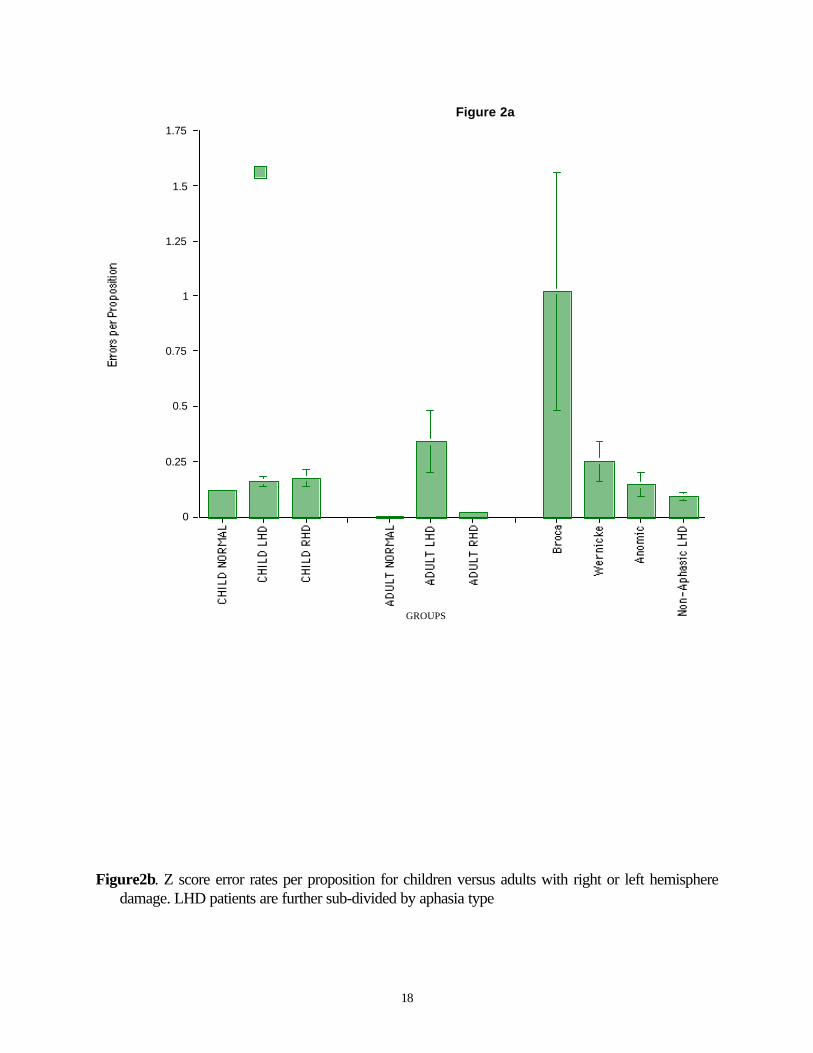
The third study in this series focused on languageproduction instead of comprehension, based on samples offree speech (Bates, Wulfeck, et al. 1999), collected within theframework of a biographical interview tailored to reflect thedifferent interests of children and adults. Participants included38 brain-injured children (24 LHD, 14 RHD) between 5-8years of age, 38 normal controls matched for age and gender,14 adults with LHD (including 3 Broca's aphasics, 3Wernicke's aphasics, 5 anomic aphasics, and 3 nonaphasic pa-tients), 7 adults with RHD, and 12 adult controls in the samerange of age and education. The structured interviews werevideotaped and transcribed following conventions of the ChildLanguage Data Exchange System, and coded into variouscategories assessing amount of speech (number of word types,word tokens, morphemes, and utterances), length (mean lengthof utterance in morphemes, or MLU), grammatical com-plexity (number of complex syntactic structures, in bothtypes and tokens), and errors (word omissions, morphologicalerrors, lexical errors). Although it was generally true thatchildren talk far less than adults (including adult aphasics),when proportion scores were used to correct for overallamount of output, results were exceedingly clear: (1) therewere absolutely no differences between children with LHD vs.RHD on any measure; (2) in this open-ended free-speech task,there were also very few differences between brain-injuredchildren (combining LHD and RHD) and their controls (theexceptions were small but significant disadvantages for FLchildren as a group in number of word omission errors and innumber of word types); (3) in striking contrast to the childdata, there were huge differences between adults with LHDvs. RHD on virtually every measure, in the predicteddirections; (4) LHD adults also showed qualitative variationsin their symptoms, reflect-ing different aphasia subtypes (e.g.,more morpho-logical and omission errors in Broca's aphasics,more lexical errors in Wernicke's aphasics). One smallillustration of these results can be seen in Figure 2a, whichplots the total number of errors per proposition in children vs.

9
adults within each lesion group, and Figure 2b, which plotsthe same data for LHD and RHD children and adults in z-scores based on performance by age-appropriate controls.Figure 2a shows that error rates are certainly higher forchildren than adults (as we have known for many years), butFigure 2b shows that LHD and RHD children are very closeto normal (with z-scores close to zero) while the worstaphasics produce error rates that are orders of magnitudehigher than normal controls (whose error rate is extremelysmall, leading to very small standard deviations). Althoughthese results are not surprising, in view of the accu-mulatedevidence for plasticity following early brain injury in humansand in other species, they document this phenomenon withexceptional clarity.
This brings us to a summary of evidence by the sameresearch group looking at the first stages of languagedevelopment, prior to 5-7 years of age.
In a study focusing on the earliest stages of languagedevelopment, Thal et al. (1991) describe results for 27congenitally brain-damaged infants be-tween 12 and 35months of age, using an early version of the MacArthurCommunicative Development Inven-tories, or CDI (Fenson etal., 1993), a parent-report instrument for the assessment ofearly lexical and grammatical development. Delays in wordcomprehen-sion in the very first stages of development wereactually more common in children with RHD. Delays in firstword production occurred for almost all the brain-injuredchildren, regardless of lesion side or site, but tended to bemore severe in children with left posterior damage -- anapparent reversal of the expected association betweencomprehension deficits and damage to Wernicke’s area.
Bates et al (1997) followed up on Thal et al. (1991) witha larger sample, using a combination of CDI data and freespeech to assess early language development in 53 childrenbetween 10 and 40 months of age (36 LHD, 17 RHD),including 18 of the 27 cases from Thal et al. The study wasdivided into three cross-sectional epochs (although many ofthe children participated in more than one): a period focusingon the dawn of word com-prehension, word production andgesture (26 children from 10-17 months), a second substudyfocusing on word production and the emergence of grammar(29 children from 19-31 months), and an analysis ofgrammatical development from free-speech samples (30children from 20 and 44 months). Performance at thesevarious stages of development was evaluated in comparisonsbased on lesion side, lesion size, and lesion site (i.e., whetheror not the frontal lobes or temporal lobes were involved).There were no effects of lesion size in any of these analyses(including a failure to replicate the U-shaped effect of lesionsize described by Thal et al., as we discussed earlier).Interesting effects of lesion side and intrahemispheric lesionsite did emerge, but in complex patterns that are surprisingfrom the point of view of the adult aphasia literature.
Between 10-17 months, delays in receptive languagewere particular evident in children with RHD (i.e., more RHDcases than we would expect by chance fell into bottom 10thpercentile for word comprehen-sion). By contrast, the LHD
children performed within the normal range on wordcomprehension, even if their lesions involved temporal lobe(the presumed site of Wernicke’s area, which is implicated inmoderate to severe forms of receptive aphasia in adults).However, there was no significant difference between LHDand RHD on direct statistical comparisons, so the RHDdisadvantage is not robust and should be investigated further.There was also a significant RHD disadvantage in thedevelopment of communicative and symbolic gesture, and thistime the RHD disadvantage did reach significance in a directLHD/RHD comparison. This result is also surprising, sincedeficits in the production of symbolic gestures are atypicallyassociated with left-hemisphere damage when they occur inadults (Goodglass, 1993). Finally, Bates et al. do report aselective delay in expressive vocabulary for children withLHD. However, in line with the earlier report by Thal et al.,this disadvantage was only evident in children whose lesionsinvolved the temporal lobe.
The second substudy followed children's languagedevelopment between 19-31 months, when the so-calledvocabulary burst is said to occur (e.g., an intense period ofdevelopment for vocabulary/lexical production), and whenchildren's comprehension is often so vast it is difficult tomeasure. This is also the period in which children typicallystart to combine words, followed by the emergence ofgrammatical inflections and function words. For the 29children whose scores on this scale were obtained, a selectivedisadvantage for children with LHD appeared both forexpressive vocabulary and the emergence of grammar (with noevidence whatsoever for a dissociation between grammaticaland lexical pro-duction). However, this LHD disadvantagewas due once again to children with left temporal involvement,in contrast with the typical adult pattern in which expressivedeficits (especially nonfluent aphasia) are usually associatedwith left frontal involvement (i.e., Broca’s area and adjacentcortical and subcortical regions). Similar delays in expressivevocabulary and grammar appeared when children with frontallobe involvement were compared with children whose lesionsspared the frontal lobe. However, in contrast with theasymmetrical left temporal disadvantage that we have justdiscussed, this frontal effect was perfectly sym-metrical:delays were equally severe with left frontal or right frontallesions.
Curiously, an abnormal proportion of the children withRHD were also producing a higher than normal number offunction words for their vocabulary size. As described insome detail by Bates, Bretherton and Snyder (1988) and byBates et al. (1994), such overuse of function words forchildren in the early stages of vocabulary development (i.e.,under 400 words) is definitely not a sign of precociousgrammar. In fact, children who overuse pronouns and otherfunction words in the early stages tend to be relatively slow ingrammatical development later on. For these children, functionwords tend to appear in frozen or rote expressions like “I wandat”, a style of early expressive language that has been called"pronominal style", or "holistic style." At the opposite end ofthe continuum are children who avoid function words in their
10
first word combinations, producing telegraphic utterances like“Adam truck” or “Mommy sock”. This style of earlyexpressive language has been referred to as “nominal style” or“analytic style”. Given the terms “holistic” and “analytic”,which are often attributed to right- vs. left-hemisphereprocessing, respectively, one might have predicted thatholistic style would be more common in children with LHD(who are presumably relying more on holistic right-hemisphere processes to acquire lan-guage). This prediction isroundly contradicted by the Bates et al. study, where holisticstyle was robustly associated with RHD (indicating that theoverproduction of function words in early speech reflectsreliance on the intact left hemisphere). Bates et al (1997)suggest that children with RHD are relying heavily on themore precise acoustic analysis and/or greater acoustic memoryavailable in the left hemisphere, storing up frozen expressionsthat they are unable to segment or understand beyond arelatively superficial level of analysis (rather like an Americanwho says “Gesund-heit” when someone sneezes, with no ideawhatsoever regarding the structure or meaning of that word inGerman). This would mean, in turn, that right-hemi-sphereprocesses are very important in the early stages of languagelearning for the breakdown of acoustic material and itsintegration into a larger cognitive system. However, once thematerial has been analyzed, understood and integrated into alarger framework, the contribution of the right hemispheremay be much less important, so that control may shift (in theundamaged brain) to rapid, automatic processes mediatedprimarily by the left hemisphere.
In the third and final subgroup of children, Bates et al.(1997) collected free-speech samples between 21 and 44months of age. As their CDI scores predicted, the MLUscores of children with damage that encompassed the lefttemporal region of their brain were significantly lower thannormal, and significantly lower than scores for brain-injuredchildren whose lesions spared this region (including allchildren with RHD and the subset of LHD children with notemporal involvement). Children with right or left frontaldamage also still looked delayed, but this difference was notstatistically significant in the 21-44-month subsample.
Vicari et al. (in press) attempted a partial repli-cation ofthe Bates et al. results for early lexical development,administering an Italian version of the MacArthur CDI to theparents of 43 children between 13 and 46 months of age. Theirstudy differed from the methods used by Bates et al. in twocrucial respects: children beyond the age range covered by theMacArthur CDI were included in the study (which means thatthey could not use age-based percentile scores), and parentswere given the Infant or the Toddler version of the MacArthurbased not on age but on their child's current level of linguisticability (children who were still in the one-word stage wereassigned the infant form, but children who were starting tocombine words were assigned the toddler form). For thesereasons, the studies are not entirely comparable, but resultsreplicate and extend the Bates et al. findings in someinteresting directions. First, Vicari et al. also report a massiveacross-the-board delay in early vocabulary development for
brain-injured children as a group. Hence, even though the long-term prospects for these children are relatively good, it isobviously hard to get language off the ground when significantdamage has occurred to either hemisphere. Second, Vicari et al.report a large and significant interaction between side of lesion(LHD vs. RHD) and stage of language development (singleword vs. multiword). Among children who were still in theone-word stage, LHD were significantly slower in vocabularydevelopment than RHD (since 10 out of 12 of the one-word-stage children with LHD had temporal lobe involvement, aspecific replication of Bates et al.'s left temporal findings wasnot possible). By contrast, among children who were now inthe multiword stage, the LHD disadvantage had disappearedentirely. In fact, LHD children in the multiword group had anumerical advantage over their RHD counterparts. Thisadvantage was not statistically significant, but it contributedto the robust interaction between language stage and lesionside. Vicari et al. suggest that recovery from this initial delaymay begin very early for some children, and may be forced inpart by the delay itself. That is, children who are particularlydisadvantaged in the first stages of language acquisition (e.g.,LHD cases) may be forced to abandon a failing strategy infavor of some alternative approach, leading to earlier and(ultimately) more suc-cessful language learning.
Reilly et al., 1998, conducted a cross-sectional study of15 RH- and 15 LH-damaged children, between 3 and 12 yearsif age, using a story-telling format (the well-known Frog Storynarratives -- Berman and Slo-bin, 1994) to assess lexical,grammatical and discourse development. For children between3 and 6 years of age, the now-familiar left temporaldisadvantage was ob-served in syntactic complexity and inpersistence of morphological errors. However, this effect oflesion site was not observed in children between 6-12 years ofage. Among the older children, there were still significantdifferences between focal lesion children (LHD and RHDcombined) and their age-matched controls on a number ofmeasures, but the focal lesion children were neverthelessperforming within the normal or low-normal range. Hence theReilly et al. results for grammar suggest a later variant of therecovery pattern that Vicari et al. observed within the lexicaldomain.
Because the Reilly et al. and Vicari et al. studies are bothcross-sectional, it would be very useful to replicate theseresults with longitudinal samples. Although their results arestill preliminary, based on a relatively small sample, Reillyand colleagues (Losh, Reilly, and Bates, 1996) have tested alongitudinal subgroup across the 5-7-year age range thatseemed to be a watershed in their cross-sectional study. Theyreport that children with left temporal involvement do indeedmove sharply upward in syntax and morphology across thisage range, scoring numerically above children with RHD at thelater time point. The general picture seems to be one in whichchildren with LHD display sharper or "steeper" growthfunctions, while children with RHD show a flatter profile ofgrowth in the language domain.
Summary and Conclusion

11
Putting these lines of evidence together, we may conclude(or perhaps hypothesize) that the infant brain contains strongbiases that, in the absence of early brain damage, guarantee theeventual emergence of left-hemisphere specialization forlanguage. Although (if anything) the right hemisphere seems toplay a more important (or at least equally important) role inthe emergence of word comprehension and communicativegesture, progress in expressive language (both lexical andgrammatical) seems to be delayed with frontal damage (toeither side of the brain) and with temporal damage (but, in thiscase, temporal damage restricted to the left hemisphere). Inother words, there is an early bias that predisposes the lefthemisphere to “take over” rapid and efficient production ofwords and sentences, a development which may also result inthe emergence of left-hemisphere specialization for manyaspects of receptive language as well. In the absence ofevidence to the contrary, one might have assumed that thisearly left-hemisphere advantage for speech/language produc-tion (but not reception) has a motor base. And yet severalstudies by the San Diego group suggest that the source of thisleft-hemisphere bias lies primarily within the temporal lobe, aregion that is supposed to be specialized for perception ratherthan production.
In this regard, Bates et al. note that some children withsevere otitis media (i.e., middle-ear infections) also showselective delays in the emergence of expressive (but notreceptive) language. Why would middle-ear impedance havegreater effects on language production than comprehension?The answer may lie in a simple fact: language learning is notthe same thing as fluent language use. When a child is trying tobreak into the language system for the first time, the amountof perceptual analysis required to produce her own versionsof a new word is greater than the amount of perceptualanalysis that she needs to recognize the word (especially ifshe is asked to recognize that word in a richly supportivesocial and physical context, which can be integrated with theacoustic signal to achieve compre-hension). If theseassumptions are correct, then we can put the story together asfollows: left temporal regions may be particularly well suited(perhaps at or before birth) for the extraction of perceptualdetail. Indeed, there is ample evidence from visual-spatialprocessing in adults to support this view, hence thehypothesized “perceptual detail advantage” would not bespecific to language, or even to audition. However, such a biaswould be particularly relevant in the first stages of languagelearning, leading (in the absence of injury) to the establishmentof left-hemisphere dominance. What these prospective studiesdo clearly show is that this bias is “soft”, and can beovercome. Indeed, by 5-7 years of age the initial disadvantagesassociated with left-hemisphere damage seem to havedisappeared, or at least, fallen below the levels that we areable to detect with the measures that we have developed sofar.
Finally, it appears from these studies that the emergenceof organization for language (in the un-damaged brain) andreorganization for language (in the damaged brain struggling toovercome initial biases) both occur within the period in which
language is acquired, i.e., somewhere between birth and 5years of age. We may speculate that this correlation betweenbrain and behavioral development is no accident. In fact, wepropose that learning itself plays a major role in organizing thebrain for efficient language use, as children struggle to find anoptimal solution to the challenges associated with languageand communica-tion.
REFERENCES
Alajouanine, T. and F. Lhermitte, 1965. Acquired aphasia inchildren. Brain, 88:653-662.
Anderson, S.W., A. Bechara, H. Damasio, D. Tranel, and A.R.Damasio, 1999. Impairment of social and moral behaviorrelated to early damage to human prefrontal cortex. Nat.Neurosci. 2:1032-1037.
Aram, D.M., B. Ekelman, D. Rose, and H. Whitaker, 1985.Verbal and cognitive sequelae following unilateral lesionsacquired in early childhood. J. Clin. Exp.Neuropsychol. 7:55-78.
Aram, D., B. Ekelman, and H. Whitaker, 1986. Spoken syntaxin children with acquired unilateral hemi-sphere lesions.Brain Lang. 27:75-100.
Aram, D.M., S.C. Meyers, and B.L. Ekelman, 1990. Fluencyof conversational speech in children with unilateral brainlesions. Brain Lang. 38:105-121.
Ballantyne, A.O., K.M. Scarvie, and D. Trauner, 1994. Verbaland performance IQ patterns in children after perinatalstroke. Dev. Psychol. 10:39-50.
Ballantyne, A.O. and D.A. Trauner, 1999. Neuro-psychological profiles of infants and children with earlyfocal brain damage. JINS 5:145.
Basser, L., 1962. Hemiplegia of early onset and the faculty ofspeech with special reference to the effects ofhemispherectomy. Brain 85:427-460.
Bates, E., in press. On the nature and nurture of language. InFrontiere della Biologia [Frontiers of Biology].The brain of homo sapiens, R. Levi-Montalcini, D.Baltimore, R. Dulbecco, and F. Jacob, series eds. and E.Bizzi, P. Calissano, and V. Volterra. vol. eds. Rome:Giovanni Trecanni.
Bates, E. (1999). Plasticity, localization and languagedevelopment. In The Changing Nervous System:Neurobehavioral Consequences of Early BrainDisorders, S. Broman and J.M. Fletcher, eds. NewYork: Oxford University Press, pp. 214-253.
Bates, E., I. Bretherton, and L. Snyder, 1988. From FirstWords to Grammar: Individual Differences andDissociable Mechanisms. New York: CambridgeUniversity Press, 1988. [Paperback edition issued 1991].
Bates, E., P.S. Dale, and D. Thal, 1995. Individual differencesand their implications for theories of languagedevelopment. In Handbook of Child Language, P.Fletcher and B. MacWhinney, eds. Oxford: BasilBlackwell, pp. 96-151.
Bates, E., V. Marchman, D. Thal, L. Fenson, P.S. Dale, J.S.Reznick, J. Reilly, and J. Hartung, 1994. Developmental

12
and stylistic variation in the composition of earlyvocabulary. J. Child Lang. 21:85-123.
Bates, E., D. Thal, B.L. Finlay, and B. Clancy, in press. Earlylanguage development and its neural correlates. InHandbook of Neuropsychology, Vol. 7: ChildNeurology (2nd ed.)., F. Boller and J. Grafman, serieseds. and I. Rapin and S. Segalowitz, vol. eds. Amsterdam:Elsevier.
Bates, E., D. Thal, D. Trauner, J. Fenson, D. Aram, J. Eisele,and R. Nass, 1997. From first words to grammar inchildren with focal brain injury. Dev. Neuropsychol.13:447-476.
Bates, E., S. Vicari, and D. Trauner, 1999. Neural mediation oflanguage development: Perspectives from lesion studiesof infants and children. In NeurodevelopmentalDisorders, H. Tager-Flusberg, ed. Cambridge, MA:MIT Press, pp. 533-581.
Bates, E., B. Wulfeck, M. Opie, J. Fenson, S. Kriz, J. Reilly,N. Dronkers, L. Miller, R. Jeffries, and K. Herbst, 1999.Comparing free speech in children and adults with left-vs right-hemisphere injury (Abstract). Brain Lang.69:377-379.
Berman, R.A. and D.I. Slobin, 1994. Relating Events inNarrative: A Cross-linguistic DevelopmentalStudy [in collaboration with Ayhan Aksu-Koc et al.].Hillsdale, NJ: Erlbaum.
Bernhardt, M., 1897. In Nothnagel. Spezielle Patho-logieund Therapie, 9 , Part 2. Vienna.
Botha, R.P., 1989. Challenging Chomsky: TheGenerative Garden Game . Oxford, UK; New York:Basil Blackwell.
Broman, S., and J.M. Fletcher, eds., 1999. The ChangingNervous System: Neurobehavioral Consequencesof Early Brain Disorders. New York: OxfordUniversity Press.
Brown, C.M. and P. Hagoort, eds. 1999. The Neuro-cognition of Language. Oxford University Press.
Clarus, A., 1874. Ueber Aphasie bei Kindern [Aphasia inchildren]. Jahresb.Kinderheilkd ..7: 369-400.
Cotard, J., 1868. Etude sur l'atophie partielle ducerveau. Thèse de Paris.
Curtiss, S., 1977. Genie: A Psycholinguistic Study of aModern-Day Wild Child. New York: Academic Press.
Deacon, T., 1997. The Symbolic Species: The Co-evolution of Language and the Brain. New York:Norton.
Dennis, M., 1980. Capacity and strategy for syntacticcomprehension after left or right hemidecortication.Brain Lang. 10:287-317.
Dennis, M. and B. Kohn, 1975. Comprehension of syntax ininfantile hemiplegics after cerebral hemidecortication.Brain Lang. 2:472-482.
Dennis, M., M. Lovett, and C.A. Wiegel-Crump, 1981.Written language acquisition after left or righthemidecortication in infancy. Brain Lang. 12:54-91.
Dennis, M. and H.A. Whitaker, 1976. Language acqui-sitionfollowing hemidecortication: Linguistic superiority of theleft over the right hemisphere. Brain Lang. 3:404-433.
Dennis, M. and H. Whitaker, 1977. Hemisphericequipotentiality and language acquisition. In LanguageDevelopment and Neurological theory, S.J.Segalowitz and F.A. Gruber, eds. New York: AcademicPress, pp. 93-106.
Dick, F., B. Wulfeck, E. Bates, D. Saltzman, N. Naucler, andN. Dronkers, 1999. Interpretation of complex syntax inaphasic adults and children with focal lesions or specificlanguage impairment (Abstract). Brain Lang. 69:335-336.
Eisele, J. and D. Aram, 1993. Differential effects of earlyhemisphere damage on lexical comprehension andproduction. Aphasiology 7:513-523.
Eisele, J. and D. Aram, 1994. Comprehension and imitation ofsyntax following early hemisphere damage. Brain Lang.46:212-231.
Eisele, J. and D. Aram, 1995. Lexical and grammaticaldevelopment in children with early hemisphere damage:A cross-sectional view from birth to adolescence. In TheHandbook of Child Language, P. Fletcher and B.MacWhinney, eds. Oxford: Basil Blackwell, pp. 664-689.
Elman, J.L., E. Bates, M. Johnson, A. Karmiloff-Smith, D.Parisi, and K. Plunkett, 1996. Rethink-ing Innateness:A Connectionist Perspective on Development.Cambridge, MA: MIT Press/Bradford Books [paperbackedition published 1998].
Fenson, L., P.S. Dale, J.S. Reznick, D. Thal, E. Bates, J.Hartung, S. Pethick, and J. Reilly, 1993. TheMacArthur Communicative Development Inven-tories: User's Guide and Technical Manual. SanDiego: Singular Publishing Group.
Fletcher, P. and B. MacWhinney, eds., 1995. Handbook ofChild Language. Oxford: Basil Blackwell.
Fodor, J.A., 1983. The Modularity of Mind: An Essay onFaculty Psychology. Cambridge, MA: MIT Press.
Gazzaniga, M.S. and R.W. Sperry, 1967. Language aftersection of the cerebral commissures. Brain 90:131-148.
Geschwind, N. and E. Kaplan, 1962. A human cerebraldeconnection syndrome. Neurology 12:675-685.
Goodglass, H. (1993). Understanding aphasia. SanDiego: Academic Press.
Goodman, R. and C. Yude, 1996. IQ and its predictors inchildhood hemiplegia. Dev. Med. Child Neurol.38:881-890.
Gopnik, M. (1990). Feature-blind grammar and dys-phasia.Nature 344(6268):715.
Gopnik, M. and M.B. Crago, 1991. Familial aggregation of adevelopmental language disorder. Cognition 39:1-50.
Irle, E., 1990. An analysis of the correlation of lesion size,localization and behavioral effects in 283 publishedstudies of cortical and subcortical lesions in old-worldmonkeys. Brain Res. Rev. 15:181-213.

13
Johnson, M.H., 1997. Developmental CognitiveNeuroscience: An Introduction. Cambridge, MA:Blackwell Publishers.
Kempler, D., D. van Lancker, V. Marchman, and E. Bates,1999. Idiom comprehension in children and adults withunilateral brain damage. Dev. Neuropsychol. 15:327-349.
Lashley, K.S. (1950). In search of the engram. In Symposiaof the Society for Experimental Biology, No. 4Physiological mechanisms and animal behaviour.New York: Academic Press, pp. 454-482.
Lenneberg, E.H., 1967. Biological Foundations ofLanguage. New York: Wiley.
Levine, S.C., P. Huttenlocher, M.T. Banich, and E. Duda,1987. Factors affecting cognitive functioning ofhemiplegic children. Dev. Med. Child Neurol. 29:27-35.
Levy, J., R.D. Nebes, and R.W. Sperry, 1971. Expressivelanguage in the surgically separated minor hemisphere.Cortex 7:49-58.
Losh, M., J.S. Reilly, and E. Bates, 1996, June. Languagedevelopment: Neural plasticity in children withearly focal brain injury. Poster session presented atthe 8th Annual Meeting of the American PsychologicalAssociation, San Fran-cisco.
Muter, V., S. Taylor, and F. Vargha-Khadem, 1997. Alongitudinal study of early intellectual development inhemiplegic children. Neuropsychologia 35:289-298.
Nass, R., H. Peterson, and D. Koch, 1989. Differential effectsof congenital left and right brain injury on intelligence.Brain Cogn. 9:258-266.
Newmeyer, F.J., 1980. Linguistic Theory in America. NewYork: Academic Press.
Newmeyer, F.J., 1997. Genetic dysphasia and linguistictheory. Journal of Neurolinguistics 10:47-73.
Ojemann, G.A., 1991. Cortical organization of language.J. Neurosci. 11:2281-2287.
O'Leary, D.D. and B.B. Stanfield, 1985. Occipital corticalneurons with transient pyramidal tract axons extend andmaintain collaterals to subcortical but not intracorticaltargets. Brain Res. 336:326-333.
O'Leary, D.D. and B.B. Stanfield, 1989. Selective eliminationof extended by developing cortical neurons is dependenton regional locale: Expe-riments utilizing fetal corticaltransplants. J. Neurosci. 9:2230-2246.
O'Neill, Y.V., 1980. Speech and Speech Disorders inWestern Thought Before 1600. Westport, CT:Greenwood Press.
Pallas, S.L. and M. Sur, 1993. Visual projections induced intothe auditory pathway of ferrets: II. Corticocorticalconnections of primary auditory cortex. J. Comp.Neurol. 337:317-333.
Payne, B.R., 1999. Immature visual cortex lesions: Globalrewiring, neural adaptations, and behavioral sparing. InThe Changing Nervous System: NeurobehavioralConsequences of Early Brain Disorders, S. Broman
and J.M. Fletcher, eds. New York: Oxford UniversityPress, pp. 114-145.
Pinker, S., 1994. The Language Instinct: How the MindCreates Language. New York: William Morrow.
Quartz, S.R. and T.J. Sejnowski, 1994. Beyond modularity:Neural evidence for constructivist principles indevelopment. Behav. Brain Sci. 17:725-726.
Quartz, S.R. and T.J. Sejnowski, 1997. The neural basis ofcognitive development: A constructivist manifesto.Behav. Brain Sci. 20:537.
Rasmussen, T. and B. Milner, 1977. The role of early leftbrain injury in determining lateralization of cerebralspeech functions. Ann. NY Acad. Sci. 229:355-369.
Reilly, J., E. Bates, and V. Marchman, 1998. Narrativediscourse in children with early focal brain injury. BrainLang. 61:335-375.
Rice, M., ed., 1996. Toward a Genetics of Language.Mahwah, NJ: Erlbaum.
Riva, D., L. Cazzaniga, C. Pantaleoni, N. Milani, and E.Fedrizzi, 1986. Acute hemiplegia in childhood: Theneuropsychological prognosis. Journal of PediatricNeurosciences 2:239-250.
Satz, P., E. Strauss, and H. Whitaker, 1990. The ontogeny ofhemispheric specialization: Some old hypothesesrevisited. Brain Lang. 38:596-614.
Stanfield, B.B. and D.D. O'Leary, 1985. Fetal occipitalcortical neurones transplanted to the rostral cortex canextend and maintain a pyramidal tract axon. Nature313(5998):135-137.
Stark, R.E. and K.K. McGregor, 1997. Follow-up study of aright- and left-hemispherectomized child: Implicationsfor localization and impairment of language in children.Brain Lang. 60: 222-242.
Stiles, J., 1995. Plasticity and development: Evidence fromchildren with early focal brain injury. In MaturationalWindows and Adult Cortical Plasticity: Is ThereReason for an Optimistic View? Proceedings ofthe Santa Fe Institute Studies in the Sciences ofComplexity, Vol. 23, B. Julesz and I. Kovacs, eds.Reading, MA: Addison-Wesley, pp. 217-237.
Stiles, J., this volume. Spatial cognitive development.Stiles, J., E. Bates, D. Thal, D. Trauner, and J. Reilly, 1998.
Linguistic, cognitive and affective development inchildren with pre- and perinatal focal brain injury: A ten-year overview from the San Diego Longitudinal Project.In Advances in Infancy Research. C. Rovee-Collier,L. Lipsitt, and H. Hayne, eds. Norwood, NJ: Ablex, pp.131-163.
Thal, D., V. Marchman, J. Stiles, D. Aram, D. Trauner, R.Nass, and E. Bates, 1991. Early lexical development inchildren with focal brain injury. Brain Lang. 40:491-527.
Vargha-Khadem, F., L. Carr, E. Isaacs, E. Brett, C. Adams,and M. Mishkin, 1997. Onset of speech after lefthemispherectomy in a nine-year-old boy. Brain 120:59-182.

14
Vargha-Khadem, F., E. Isaacs, S. van der Werf, S. Robb, and J.Wilson, 1992. Development of intelligence and memoryin children with hemi-plegic cerebral palsy: Thedeleterious consequences of early seizures. Brain, 115,315-329.
Vargha-Khadem, F., E. Isaacs, and V. Muter, 1994. A reviewof cognitive outcome after unilateral lesions sustainedduring childhood. Journal of Child Neurology,9(Suppl.), 2S67-2S73.
Vargha-Khadem, F., A. O’Gorman, and G. Watters, 1985.Aphasia and handedness in relation to hemispheric side,age at injury and severity of cerebral lesion duringchildhood. Brain 108:677-696.
Vicari, S., A. Albertoni, A. Chilosi, P. Cipriani, G. Cioni, andE. Bates, in press. Plasticity and reorganization duringearly language learning in children with congenital braininjury. Cortex.
Webster, M.J., J. Bachevalier, and L.G. Ungerleider, 1995.Development and plasticity of visual memory circuits. InMaturational Windows and Adult CorticalPlasticity: Is There Reason for an Optimistic View?Proceedings of the Santa Fe Institute Studies inthe Sciences of Complexity, Vol. 23. B. Julesz and I.Kovacs, eds. Reading, MA: Addison-Wesley, pp. 73-86.
Woods, B.T. 1980. The restricted effects of right-hemispherelesions after age one: Wechsler test data. Neuropsycho-logia 18:65-70.
Woods, B.T., 1983. Is the left hemisphere specialized forlanguage at birth? Trends Neurosci. 6:115-117.
Woods, B.T. and S. Carey, 1979. Language deficits afterapparent clinical recovery from childhood aphasia. Ann.Neurol. 6:405-409.

Woods, B.T. and H.L. Teuber, 1973. Early onset ofcomplementary specialisation of the cerebral hemispheresin man. Trans. Am. Neurol. Assoc. 98:113-117.Woods, B.T. and H.L. Teuber, 1978. Changing patterns of
childhood aphasia. Ann. Neurol. 3:272-280.
Xiong, J., S. Rao, J.-H. Gao, M.Woldorff, and others, 1998.Evaluation of hemispheric dominance for languageusing functional MRI: A comparison with positronemission tomography. Hum. Brain Mapp. 6:42-58.

16
Figures & Tables
Figure 1. Comprehension of novel and familiar phrases by children and adults with left vs. righthemisphere injury
-10
-9
-8
-7
-6
-5
-4
-3
-2
-1
0
1
2
LEFT RIGHT
Figure 1
Adult Novz
Adult Famz
-10
-9
-8
-7
-6
-5
-4
-3
-2
-1
0
1
2
LEFT RIGHT
Side of Injury
Novel Phrases
Familiar Phrases
Adults Children

17
Figure2a. The mean number of errors per proposition for normally developing children, adult controls,and children and adults with right or left hemisphere damage. LHD patients are further sub-divided byaphasia type.
18
0
0.25
0.5
0.75
1
1.25
1.5
1.75
GROUPS
Figure 2a
Figure2b. Z score error rates per proposition for children versus adults with right or left hemispheredamage. LHD patients are further sub-divided by aphasia type

19
0
25
50
75
100
125
150
175
200
225
250
275
300
325
GROUPZ
Figure 2b
Errors/Proposition Z

20

Table Key
RHD = right- hemisphere damage
LHD = left-hemisphere damage
PN = prenatal
+ S = patients suffering from seizures
–S = patients with no seizure activity
+Temp = injury includes damage to the temporal lobe
–Temp = injury spares the temporal lobe
+Frontal = injury includes damage to the frontal lobe
–Frontal = injury spares the frontal lobe
NR = not reported
NSR = no specifics reported
N/A = not applicable
XO = mean age of onset
XT = mean age at testing
Note that a "<" indicates worse performance regardless of the specific measure usedNote that if two groups are described as "=" , this indicates merely that they did not differ significantly.

TABLE 1: Summary of IQ data from studies of children with focal brain injuryPatients
AuthorsSite Neuro-
imaging?Age ofOnset
Age AtTesting
Seizures?
RHDvs.
LHD?
RESULTS?
Woods andTeuber (1973)
19 RHD17 LHD
NO PN - 15 years NR NR YES FIQ: LHD < controls RHD < controls LHD = RHDPIQ: LHD < controls RHD < controlsVIQ: LHD < controls RHD = controls
Woods (1980) 23 RHD27 LHD
NO “early”(<1yr) 1O RHD = RE, 11 LHD = LE
“late” (>1yr) 13 RHD = LR Xo = 6;4 6 LHD = LL Xo = 5;7
RE: XT = 14;01LE: XT = 17;02RL: XT = 10;03LL: XT = 8;06
RE: 5( –S) 5 (+S)
LE: 8 (–S) 3 (+S)
RL: 11 (–S) 2 (+S)
LL: 13 (–S) 3 (+S)
Comparisonsbetween +S and–S groups NOTmade
NOVIQ: RE < population mean & controls LE < population mean & controls RL = population mean & controls LL < population mean & controls
PIQ : RE < population mean & controls LE < population mean & controls RL < population mean & controls LL < population mean & controls

Vargha-Khademet al.
(1985)
28 LHD25 RHD
some 33 “Prenatal” (<2mo.)(16 RHD, 18 LHD)
11”early postnatal”(2 mo. - 5 yr.)(5 RHD, 6 LHD)
9 “late postnatal”( 5 yr. - 14 yr.)(5 RHD, 4 LHD)
NSR
At least > 2yrs postonset
NR NO prenatal RHD: VIQ = PIQprenatal LHD: VIQ < PIQ
early postnatal RHD: VIQ = PIQearly postnatal LHD: VIQ < PIQ
late postnatal RHD: VIQ = PIQlate postnatal RHD: VIQ = PIQ
Aram et al.
(1985)
8 RHD8 LHD
YES RHD: Xo = 1;4(2 mo. - 9;0)
LHD: Xo = 1;6(1 mo. - 6;2)
RHD: XT = 3;10(1;8 - 5;10)
LHD: XT =4.;6s(2mo. - 8;2)
none NO FIQ: LHD < controls RHD < controls
PIQ: LHD < controls RHD < controls
VIQ: LHD = controls RHD < controls
Riva andCazzaniga(1986)
26RHD22LHD
some “early”(<1yr) 13 RHD = RE, 10 LHD = LE
“late” (>1yr) 13 RHD = LR 12 LHD = LL XO = 6;6
early(<1yr)XT = 8;5
late(>1yr)XT = 11;8
none NO FIQ: LE = controls LL = controls RE < controls RL < controls
PIQ: LE < controls LL = controls RE < controls RL < controls
VIQ: LE < controls LL = controls RE = controls RL = controls
Riva et al.(1986)
8RHD8LHD
YES RHD: XO = 4;6(1;8 - 8;3)
LHD: XO = 6;4 (0;8 - 12;8)
RHD: XT = 10;7
LHD: XT = 11;1
5 RHD (-S)3 RHD (+S)
5 LHD (-S)3 LHD (+S)
Comparisonsbetween +S and–S groups NOTmade
NO PIQ: LHD < controls RHD < controls
VIQ: LHD < controls RHD = controls

Nass et al.(1989)
12 RHD14LHD
YES pre- or perinatal RHD: XT = 8;6
LHD: XT = 6;8
YES FIQ: LHD > RHD
PIQ: LHD = RHD
VIQ: LHD > RHD
LHD: PIQ < VIQRHD: PIQ = VIQ
Vargha-Khadem et al.(1992)
40 RHD42 LHD
YES pre- or perinatal RHD (–S): XT =11;3
RHD (+S): XT =11;6
LHD (-S): XT =12;9
LHD (+S): XT = 11;8
24 RHD (–S)16 RHD (+S)
28 LHD (–S)14 LHD (+S)
YES FIQ: RHD=LHD +S < controls -S < controls +S < -S
PIQ: RHD=LHD +S < controls -S < controls +S < -S
VIQ: RHD = LHD +S < controls -S = controls +S -S
Eisele andAram(1993)
12 RHD21 LHD
YES RHD: Xo = 1;06(PN-9;08)
LHD: Xo = 2:03 (PN-11;07)
RHD: XT = 5;08(2;06-14;07)
LHD: XT = 7:05 (2;0 7- 14;02)
none YES VIQ: LHD = RHD =controls
PIQ: LHD = controls RHD < LHD = controls RHD < controls
Ballantyne etal. (1994)
9 RHD8 LHD
YES pre- or perinatal RHD: XT= 11;2 (4;11- 20;1)
LHD: XT = 9(4;1 - 16;5)
NR NO FIQ: LHD < controls RHD < controls
PIQ: LHD < controls RHD < controls
VIQ: LHD < controls RHD < controls
LHD: PIQ = VIQRHD: PIQ < VIQ
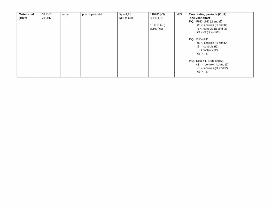
Muter et al.(1997)
18 RHD23 LHD
some pre- or perinatal XT = 4;11(3;6 to 6;8)
11RHD (–S)4RHD (+S)
15 LHD (–S)8LHD (+S)
YES Two testing periods (t1,t2) one year apartFIQ: RHD=LHD (t1 and t2) +S < controls (t1 and t2) -S < controls (t1 and t2) +S < -S (t1 and t2)
PIQ: RHD=LHD +S < controls (t1 and t2) -S = controls (t1) -S < controls (t2) +S < -S
VIQ: RHD = LHD (t1 and t2) +S < controls (t1 and t2) -S = controls (t1 and t2) +S < -S

15 RHD28 LHD
YES < 6 mo. 3 - 9 yr. 8RHD (–S)7RHD (+S)
16LHD (–S)11LHD (+S)
YES PIQ: LHD < controls RHD < controls LHD = RHDVIQ: LHD < controls RHD < controls LHD = RHD
Bates, Vicari,and Trauner(1999)
27 RHD46 LHD
YES < 6 mo. 3 - 14 yr. 20RHD (–S)7RHD (+S)
35LHD (–S)11LHD (+S)
YES FIQ: LHD < controls RHD < controls LHD = RHD

Table 2: Summary of data from studies of children with focal brain injury using various language assessment measures
PatientsAuthorsSite Neuro-
imaging??
Age of Onset Age AtTesting
Seizures?
RHDvs.LHD
?Tests Used Results
clinical/observational7 RH infantile with clinical dysphasia1 RH juvenile with clinical dysphasia4 LH infantile with clinical dysphasia4 LH juvenile with clinical dysphasia
McFie (1961) 24RHD14LHD
NO35 infantile(before 1yr.) (19 RHD,11 LHD)
8 juvenile(after 1yr.)(5 RH,3 LH) range of onset15 mo - 5 yr.
NR NR NR
Weigl Sorting testSentence learning testMemory for designs test
LHD: impaired on Weigl Sortingand Sentence Learning but NOTon memory for designs
RHD: impaired on Weigl Sortingand Sentence Learning but NOTon memory for designs
Basser
(1962)
48 LHD,54 RHD
No 72"pre-specch onset" (34 L,38 R)
30 "post-speech onset" (14 L,16 R )
NR NSR NOclinical/observational
none of the RHD or LHD had“dysphasia”
8/48 LHD had stuttering orarticulation problems8/48 LHD had reduced ornonexistent speech7/54 RHD had stuttering orarticulation problems11/54 RHD had reduced ornonexistent speech
34 pre-speech onset LHD haddelayed first word production38 pre-speech onset RHD haddelayed first word production
AlajouanineandLhermitte(1965)
32 LHDpatientsONLY
No from 6 - 15 years NR NR N/A clinical/observational 32/32 impaired expression(written, spoken, gestural)7/32 exhibited paraphasias10/32 impaired comprehension18/32 had alexia
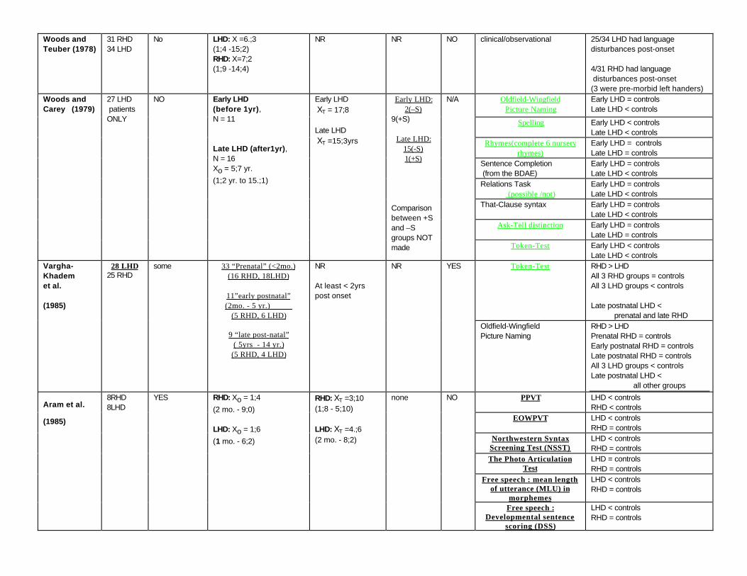
Woods andTeuber (1978)
31 RHD34 LHD
No LHD: X =6.;3(1;4 -15;2)RHD: X=7;2(1;9 -14;4)
NR NR NO clinical/observational 25/34 LHD had languagedisturbances post-onset
4/31 RHD had language disturbances post-onset(3 were pre-morbid left handers)
Oldfield-WingfieldPicture Naming
Early LHD = controlsLate LHD < controls
Spelling Early LHD < controlsLate LHD < controls
Rhymes(complete 6 nurseryrhymes)
Early LHD = controlsLate LHD = controls
Sentence Completion (from the BDAE)
Early LHD = controlsLate LHD < controls
Relations Task (possible /not)
Early LHD = controlsLate LHD < controls
That-Clause syntax Early LHD = controlsLate LHD < controls
Ask-Tell distinction Early LHD = controlsLate LHD = controls
Woods andCarey (1979)
27 LHD patientsONLY
NO Early LHD(before 1yr),N = 11
Late LHD (after1yr),N = 16Xo = 5;7 yr.(1;2 yr. to 15.;1)
Early LHD XT = 17;8
Late LHD XT =15;3yrs
Early LHD:2(–S)
9(+S)
Late LHD:15(-S)1(+S)
Comparisonbetween +Sand –Sgroups NOTmade
N/A
Token-Test Early LHD < controlsLate LHD < controls
Token-Test RHD > LHDAll 3 RHD groups = controlsAll 3 LHD groups < controls
Late postnatal LHD < prenatal and late RHD
Vargha-Khademet al.
(1985)
28 LHD25 RHD
some 33 “Prenatal” (<2mo.)(16 RHD, 18LHD)
11”early postnatal”(2mo. - 5 yr.)
(5 RHD, 6 LHD)
9 “late post-natal”( 5yrs - 14 yr.)
(5 RHD, 4 LHD)
NR
At least < 2yrspost onset
NR YES
Oldfield-WingfieldPicture Naming
RHD > LHDPrenatal RHD = controlsEarly postnatal RHD = controlsLate postnatal RHD = controlsAll 3 LHD groups < controlsLate postnatal LHD < all other groups
PPVT LHD < controlsRHD < controls
EOWPVT LHD < controlsRHD = controls
Northwestern SyntaxScreening Test (NSST)
LHD < controlsRHD = controls
The Photo ArticulationTest
LHD = controlsRHD = controls
Free speech : mean lengthof utterance (MLU) in
morphemes
LHD < controlsRHD = controls
Aram et al.
(1985)
8RHD8LHD
YES RHD: Xo = 1;4(2 mo. - 9;0)
LHD: Xo = 1;6(1 mo. - 6;2)
RHD: XT =3;10(1;8 - 5;10)
LHD: XT =4.;6(2 mo. - 8;2)
none NO
Free speech :Developmental sentence
scoring (DSS)
LHD < controlsRHD = controls

Related Documents