Antia Laboratories Inc. http://www.antialabs.com Landiolol has cardioprotective effects against reperfusion injury in the rat heart via the PKC1 signaling pathway YOSUKE TAKAHASHI, SHIGEKAZU TAKEMURA, YUKIKO MINAMIYAMA, TOHIHIKO SHIBATA, HIDEKAZU HIRAI, YASUYUKI SASAKI, MASANORI SAKAGUCHI, & SHIGEFUMI SUEHIRO Department of Cardiovascular Surgery, Osaka City University Graduate School of Medicine, Osaka, Japan Accepted by Professor G. Mann (Received 21 November 2006; in revised form 17 February 2007) Abstract Landiolol, a highly cardioselective b1-blocker, has cardioprotective effects against ischemia-reperfusion injury, although the precise mechanism is still unclear. The aim of this study was to clarify the cardioprotective mechanism of landiolol. Experiments were performed on Langendorff-perfused rat hearts undergoing 20 min stabilization, and 45 min of ischemia followed by 60 min of reperfusion. Various drugs with or without landiolol (100 mM) were administered before ischemia for 20 min. Preischemic administration of landiolol reduced cardiac cellular damage and improved the recovery of cardiac function by about 40%. The a1 blocker prazosin, the protein kinase C (PKC) inhibitor chelerythrine or the K ATP channel blocker glibenclamide, but not the selective mitochondrial K ATP channel blocker 5-hydroxydecanoate abrogated the cardioprotective effect induced by landiolol. Following landiolol pretreatment the activation of PKC1 and heat shock protein 27 were significantly higher than that in control. These data indicate that preischemic application of landiolol induces cardioprotective effects through PKC1-mediated pathway, similar to that afforded by ischemic preconditioning. Keywords: Protein kinase C, mitogen-activated protein kinase, heart rate Introduction Cardioprotective strategies for attenuating ischemia- reperfusion (I/R) injury have important clinical implications. Murry and colleagues first described that an inherent protective mechanism, designated ischemic preconditioning (IP), protects the heart against prolonged ischemic damage [1]. Cardiopro- tection induced by repeated short episodes of I/R prior to sustained ischemia is termed IP, while cardiopro- tection afforded by brief administration of substances prior to sustained ischemia is known as pharmaco- logical preconditioning. Many preconditioning agents have been described to date, including adrenergic receptor agonists, B2 bradykinin receptor agonists, A1 adenosine receptor agonists, opioid receptor agonists, protein kinase C (PKC) activators and mitochondrial ATP-sensitive potassium (K ATP ) channel openers. The beneficial effects of preconditioning are mediated by PKC, p38 mitogen-activated protein kinase (MAPK) and/or K ATP channels [2–4]. Furthermore, MAPK-activated protein kinase-2 located down- stream of p38 MAPK [5,6] is known to phosphorylate the 27-kDa small heat-shock protein (HSP27) [7 –9], which plays a protective role against ischemic or oxidative stress [10,11]. Landiolol hydrochloride is a highly cardioselective b1-blocker (b1/b2 ¼ 255) with little a-blocking action. It is nine times more potent in its b1-blocking activity and eight times more cardioselective than esmolol in vivo. In addition, its activity is ultra-short- acting and disappears after cessation of administration ISSN 1071-5762 print/ISSN 1029-2470 online q 2007 Informa UK Ltd. DOI: 10.1080/10715760701338810 Correspondence: Y. Takahashi, Department of Cardiovascular Surgery, Osaka City University Graduate School of Medicine, 1-4-3 Asahi-machi, Abeno-ku, Osaka 545-8585, Japan. Tel: 81 6 6645 3980. Fax: 81 6 6646 3071. E-mail: [email protected] Free Radical Research, July 2007; 41(7): 757–769

Welcome message from author
This document is posted to help you gain knowledge. Please leave a comment to let me know what you think about it! Share it to your friends and learn new things together.
Transcript

Antia L
abora
tories
Inc.
http:/
/www.an
tialab
s.com
Landiolol has cardioprotective effects against reperfusion injuryin the rat heart via the PKC1 signaling pathway
YOSUKE TAKAHASHI, SHIGEKAZU TAKEMURA, YUKIKO MINAMIYAMA,
TOHIHIKO SHIBATA, HIDEKAZU HIRAI, YASUYUKI SASAKI, MASANORI SAKAGUCHI,
& SHIGEFUMI SUEHIRO
Department of Cardiovascular Surgery, Osaka City University Graduate School of Medicine, Osaka, Japan
Accepted by Professor G. Mann
(Received 21 November 2006; in revised form 17 February 2007)
AbstractLandiolol, a highly cardioselective b1-blocker, has cardioprotective effects against ischemia-reperfusion injury, although theprecise mechanism is still unclear. The aim of this study was to clarify the cardioprotective mechanism of landiolol.Experiments were performed on Langendorff-perfused rat hearts undergoing 20 min stabilization, and 45 min of ischemiafollowed by 60 min of reperfusion. Various drugs with or without landiolol (100mM) were administered before ischemia for20 min. Preischemic administration of landiolol reduced cardiac cellular damage and improved the recovery of cardiacfunction by about 40%. The a1 blocker prazosin, the protein kinase C (PKC) inhibitor chelerythrine or the KATP channelblocker glibenclamide, but not the selective mitochondrial KATP channel blocker 5-hydroxydecanoate abrogated thecardioprotective effect induced by landiolol. Following landiolol pretreatment the activation of PKC1 and heat shock protein27 were significantly higher than that in control. These data indicate that preischemic application of landiolol inducescardioprotective effects through PKC1-mediated pathway, similar to that afforded by ischemic preconditioning.
Keywords: Protein kinase C, mitogen-activated protein kinase, heart rate
Introduction
Cardioprotective strategies for attenuating ischemia-
reperfusion (I/R) injury have important clinical
implications. Murry and colleagues first described
that an inherent protective mechanism, designated
ischemic preconditioning (IP), protects the heart
against prolonged ischemic damage [1]. Cardiopro-
tection induced by repeated short episodes of I/R prior
to sustained ischemia is termed IP, while cardiopro-
tection afforded by brief administration of substances
prior to sustained ischemia is known as pharmaco-
logical preconditioning. Many preconditioning agents
have been described to date, including adrenergic
receptor agonists, B2 bradykinin receptor agonists, A1
adenosine receptor agonists, opioid receptor agonists,
protein kinase C (PKC) activators and mitochondrial
ATP-sensitive potassium (KATP) channel openers.
The beneficial effects of preconditioning are mediated
by PKC, p38 mitogen-activated protein kinase
(MAPK) and/or KATP channels [2–4]. Furthermore,
MAPK-activated protein kinase-2 located down-
stream of p38 MAPK [5,6] is known to phosphorylate
the 27-kDa small heat-shock protein (HSP27) [7–9],
which plays a protective role against ischemic or
oxidative stress [10,11].
Landiolol hydrochloride is a highly cardioselective
b1-blocker (b1/b2 ¼ 255) with little a-blocking
action. It is nine times more potent in its b1-blocking
activity and eight times more cardioselective than
esmolol in vivo. In addition, its activity is ultra-short-
acting and disappears after cessation of administration
ISSN 1071-5762 print/ISSN 1029-2470 online q 2007 Informa UK Ltd.
DOI: 10.1080/10715760701338810
Correspondence: Y. Takahashi, Department of Cardiovascular Surgery, Osaka City University Graduate School of Medicine, 1-4-3 Asahi-machi,Abeno-ku, Osaka 545-8585, Japan. Tel: 81 6 6645 3980. Fax: 81 6 6646 3071. E-mail: [email protected]
Free Radical Research, July 2007; 41(7): 757–769

Antia L
abora
tories
Inc.
http:/
/www.an
tialab
s.com
in vivo. Landiolol has neither intrinsic sympathomi-
metic activity nor significant membrane-stabilizing
activity, and its cardiodepressive effect is lower than
those of other b-blockers including esmolol [12,13].
Landiolol has been reported to have cardioprotective
effects [14,15].
In the human myocardium, there are predominantly
three adrenergic receptors (a1, b1 and b2). The ratios
of these a1, b1 and b2 adrenergic receptors are about
1:8:2 [16,17]. The crosstalk between a1, b1 and b2
adrenergic receptors stimulation in the cardiac inotropic
response has been only partially understood. Schafer
et al. [18] demonstrated that b1 blocker augmented the
a1 adrenergic receptor induced activation of PKC.
Thus, we also considered that landiolol might enhance
PKC signaling in the myocardium.
Recently, we showed that administration of land-
iolol before, but not during, ischemia improved
postischemic cardiac function and reduced cardiac
cellular damage after reperfusion [19]. These results
suggested that landiolol may have cardioprotective
effects via enhancement of PKC signaling. Hence, the
aim of this study was to determine the optimal
concentration of landiolol for cardioprotection and
investigate the mechanism of landiolol against I/R
injury in the Langendorff-perfused heart
Materials and methods
Chemicals
Prazosin chloride, chelerythrine chloride, glibencla-
mide and 5-hydroxydecanoate (5-HD) were pur-
chased from Sigma (St Louis, MO). All other reagents
used were of analytical grade.
Isolated heart preparation and I/R protocol
Male Wistar rats weighing 270–320 g were purchased
from SLC Japan Inc. (Shizuoka, Japan). All animals
were housed and allowed free access to tap water and a
standard rodent diet. The animals were treated in
accordance with the Guide for the Care and Use of
Laboratory Animals approved by the Local Ethics
Committee of Osaka City University. Rats were
anesthetized with diethyl ether, followed by injection
of heparin (100 IU/kg) into the femoral vein. Diethyl
ether was chosen on the basis that it affected neither
rate-pressure product (RPP) nor post-ischemic cardiac
function. The heart was rapidly harvested and
immersed in ice-cold Krebs-Henseleit bicarbonate
buffer (KHBB) containing (in mM) 118.5 NaCl,
25.0 NaHCO3, 4.8 KCl, 1.2 KH2PO4, 1.2 MgSO4, 11
glucose and 2.5 CaCl2, adjusted to pH 7.4. The aorta
was rapidly attached to a stainless steel apparatus and
retrogradely perfused with KHBB equilibrated with
95% O2 and 5% CO2 at 378C at a constant perfusion
pressure of 100 cmH2O during the first 20 min of
stabilization. Through a left atrial incision, a latex
balloon connected to a pressure transducer was
inserted into the left ventricle (LV) for measurement
of the LV isovolumic pressure. The balloon was inflated
to obtain an LV end-diastolic pressure (LVEDP) of
4–8 mm Hg as previously described [20]. The LV
developed pressure (LVDP), maximum first derivative
of the LV pressure during systole (max dP/dt), LVEDP,
and heart rate (HR) were continuously monitored
using a polygraph with a computer analysis system
(LEG-1000; Nihon Kohden, Tokyo, Japan). All hearts
were spontaneously beating without pacing.
Experimental protocol
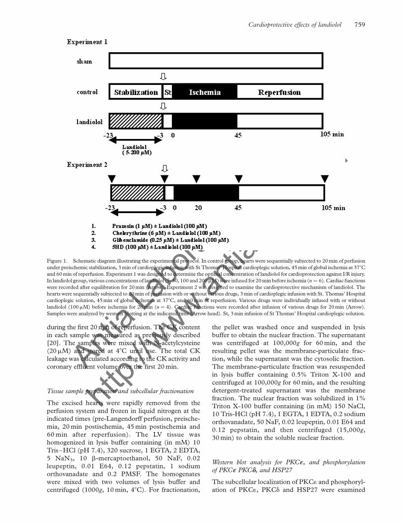
Experimental protocol was shown in Figure 1. Each
heart was allowed to stabilize during the initial 20 min.
Subsequently, cardiac arrest was achieved by clamping
the aortic cannula and injecting St Thomas’ Hospital
cardioplegic solution containing (in mM) 110 NaCl, 10
NaHCO3, 18 KCl, 1.2 MgCl2 and 1.2 CaCl2, adjusted
to pH 7.8. The cardioplegic solution was infused at
378C at a constant perfusion pressure of 60 cmH2O for
3 min. The hearts were subjected to global ischemia
for 45 min at 378C and then reperfused with KHBB for
60 min at a constant pressure of 100 cmH2O to obtain
about 50% recovery of cardiac function after 45 min
ischemia at 378C in control group.
Assessment of ventricular function
The LVDP, max dP/dt and LVEDP were measured
every 5 min before cardiac arrest and after reperfusion.
The postischemic recoveries of LVDP and max dP/dt
after 60 min of reperfusion were expressed as the
percentages of the respective preischemic values. The
RPP was expressed as HR £ LVDP. Experiment 1
was designed to determine the optimal concentration
of landiolol for cardioprotection against I/R injury.
Various concentrations of landiolol (0, 5, 50, 100 and
200mM) were infused for 20 min before ischemia in
each group (n ¼ 6). Experiment 2 was designed to
examine the cardioprotective mechanism of landiolol.
Prazosin (1mM; a selective a1-blocker), Chelerythr-
ine (6mM; a non-selective PKC inhibitor), glibencla-
mide (0.25 mM; a non-selective KATP channel
blocker) and 5-HD (100mM; a mitochondrial KATP
channel blocker) were individually infused with
landiolol (100mM) before ischemia for 20 min
(n ¼ 4). The concentrations of prazosin, chelerythrine
and 5-HD were selected by reference to previous
reports [21–26], while the concentration of gliben-
clamide (.0.25mM) decreased both the RPP and
post-ischemic cardiac function in Experiment 2.
Assessment of myocardial injury
Cellular damage was assessed by measuring creatine
kinase (CK) release into the coronary effluent collected
Y. Takahashi et al.758
Antia L
abora
tories
Inc.
http:/
/www.an
tialab
s.com
during the first 20 min of reperfusion. The CK content
in each sample was measured as previously described
[20]. The samples were mixed with N-acetylcysteine
(20mM) and stored at 48C until use. The total CK
leakage was calculated according to the CK activity and
coronary effluent volume over the first 20 min.
Tissue sample preparation and subcellular fractionation
The excised hearts were rapidly removed from the
perfusion system and frozen in liquid nitrogen at the
indicated times (pre-Langendorff perfusion, preische-
mia, 20 min postischemia, 45 min postischemia and
60 min after reperfusion). The LV tissue was
homogenized in lysis buffer containing (in mM) 10
Tris–HCl (pH 7.4), 320 sucrose, 1 EGTA, 2 EDTA,
5 NaN3, 10 b-mercaptoethanol, 50 NaF, 0.02
leupeptin, 0.01 E64, 0.12 pepstatin, 1 sodium
orthovanadate and 0.2 PMSF. The homogenates
were mixed with two volumes of lysis buffer and
centrifuged (1000g, 10 min, 48C). For fractionation,
the pellet was washed once and suspended in lysis
buffer to obtain the nuclear fraction. The supernatant
was centrifuged at 100,000g for 60 min, and the
resulting pellet was the membrane-particulate frac-
tion, while the supernatant was the cytosolic fraction.
The membrane-particulate fraction was resuspended
in lysis buffer containing 0.5% Triton X-100 and
centrifuged at 100,000g for 60 min, and the resulting
detergent-treated supernatant was the membrane
fraction. The nuclear fraction was solubilized in 1%
Triton X-100 buffer containing (in mM) 150 NaCl,
10 Tris-HCl (pH 7.4), 1 EGTA, 1 EDTA, 0.2 sodium
orthovanadate, 50 NaF, 0.02 leupeptin, 0.01 E64 and
0.12 pepstatin, and then centrifuged (15,000g,
30 min) to obtain the soluble nuclear fraction.
Western blot analysis for PKC1, and phosphorylation
of PKC1 PKCd, and HSP27
The subcellular localization of PKC1 and phosphoryl-
ation of PKC1, PKCd and HSP27 were examined
Figure 1. Schematic diagram illustrating the experimental protocol. In control group, hearts were sequentially subjected to 20 min of perfusion
under preischemic stabilization, 3 min of cardioplegic infusion with St Thomas’ Hospital cardioplegic solution, 45 min of global ischemia at 378C
and 60 min of reperfusion. Experiment 1 was designed to determine the optimal concentration of landiolol for cardioprotection against I/R injury.
In landiolol group, various concentrations of landiolol (5, 50, 100 and 200mM) were infused for 20 min before ischemia (n ¼ 6). Cardiac functions
were recorded after equilibration for 20 min (Arrow). Experiment 2 was designed to examine the cardioprotective mechanism of landiolol. The
hearts were sequentially subjected to 20 min of perfusion with or without various drugs, 3 min of cardioplegic infusion with St. Thomas’ Hospital
cardioplegic solution, 45 min of global ischemia at 378C, and 60 min of reperfusion. Various drugs were individually infused with or without
landiolol (100mM) before ischemia for 20 min (n ¼ 4). Cardiac functions were recorded after infusion of various drugs for 20 min (Arrow).
Samples were analyzed by western blotting at the indicated time (Arrow head). St, 3 min infusion of St Thomas’ Hospital cardioplegic solution.
Cardioprotective effects of landiolol 759

Antia L
abora
tories
Inc.
http:/
/www.an
tialab
s.com
by western blot analysis. Aliquots (20mg protein) of
the total cytosolic, membranous, and whole protein
extracts of each sample were separated by 12.5%
SDS-PAGE and transferred to nitrocellulose mem-
branes. The membranes were blocked with 3% nonfat
dry milk and incubated overnight with anti-PKC1
(mouse monoclonal antibody: Transduction Labora-
tories, Lexington, KY), anti-phospho-PKC1 (rabbit
polyclonal antibody: Upstate, Lake Placid, NY), anti-
phospho PKCd (rabbit polyclonal antibody: Abcam,
Tokyo, JPN), and anti-phospho HSP27 (rabbit
polyclonal antibody: R&D Systems, Inc. Minneapolis,
MN) primary antibodies at 48C. After washing, the
membranes were incubated with secondary antibodies
for 90 min and visualized using an enhanced
chemiluminescence ECL reagent (Amersham Bios-
ciences, Buckinghamshire, UK). The band intensities
were quantified using image analysis computer soft-
ware (Scion Image Beta 4.03).
Statistical analysis
All data are expressed as means ^ SD. Statistical
analyses were performed by ANOVA and Dunnett’s
test. Values of p , 0.05 were considered to indicate
statistical significance.
Results
Effects of landiolol on cardiac function in the preischemic
condition
The preischemic parameters of cardiac function are
summarized in Table I. None of the concentrations
(0–200mM) of landiolol affected any of the baseline
functional parameters.
Effects of landiolol on cardiac function and cellular injury
after reperfusion
The cardiac function and CK leakage after reperfusion
are shown in Figure 2. All the groups in Experiment 1
showed similarly marked reductions in LVDP
(Figure 2A) and max dP/dt after ischemia, while
LVEDP slowly increased. The percent recoveries of
LVDP (Figure 2B) and max dP/dt (Figure 2C) were
significantly decreased after 60 min of reperfusion.
These parameters were significantly improved by
pretreatment with 100mM landiolol (LVDP:
54.5 ^ 10.5% in the control group vs. 76.1 ^ 6.6%
in the landiolol group, p , 0.05; max dP/dt:
47.5 ^ 14.9% in the control group vs. 71.4 ^ 6.9%
in the landiolol group, p , 0.05). In the 100mM
landiolol group, the increase in LVEDP after reperfu-
sion was attenuated compared with that in control
hearts (48.0 ^ 10.8 mm Hg in the control group vs.
29.4 ^ 3.8 mm Hg in the landiolol group, p , 0.05)
(Figure 2D). CK leakage increased after reperfusion,
and administration of landiolol at more 50mM
significantly reduced this effect (Figure 2E).
Effects of various agents on cardiac function in the
preischemic condition
The preischemic parameters of cardiac function in the
prazosin, chelerythrine, glibenclamide and 5-HD
groups are summarized in Table II. No significant
differences were detected for any of the parameters of
cardiac function in any of the groups with or without
landiolol.
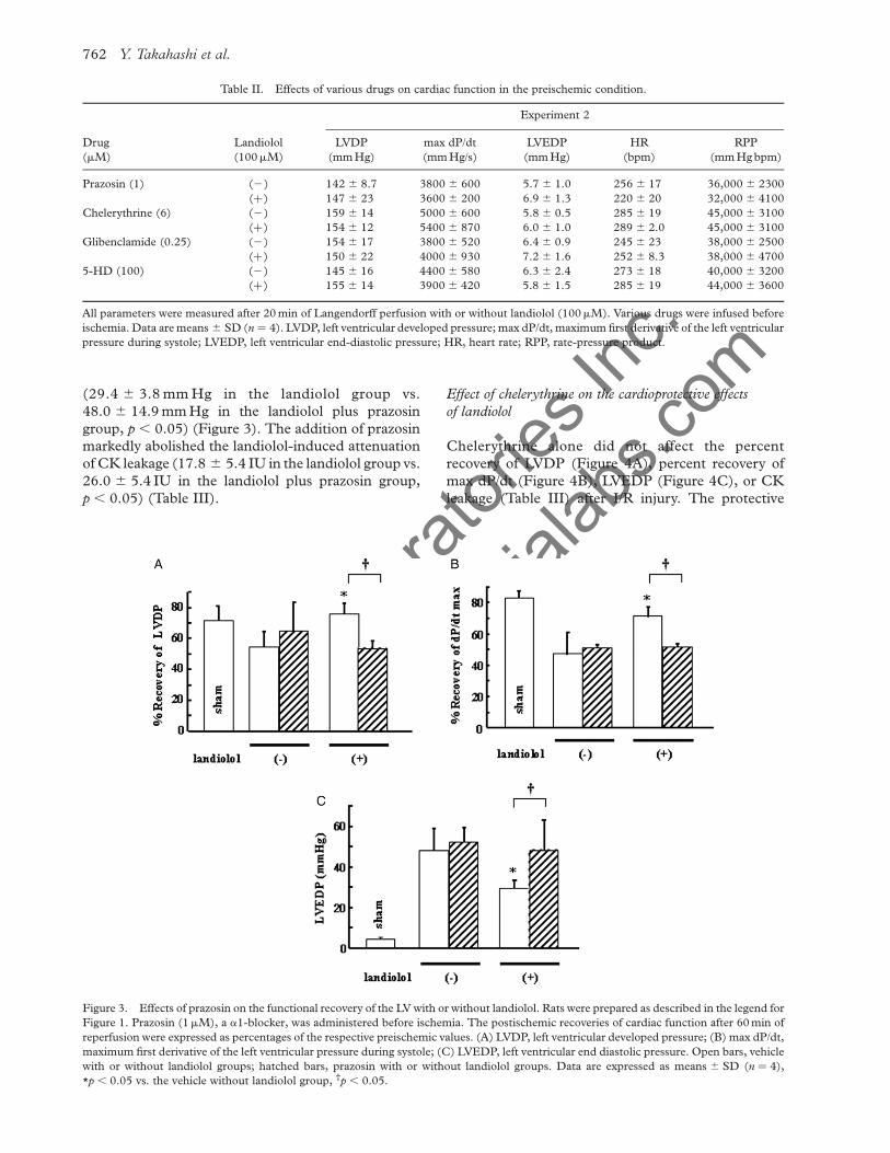
Effect of prazosin on the cardioprotective effects of landiolol
Prazosin alone did not affect the percent recovery of
LVDP (Figure 3A), percent recovery of max dP/dt
(Figure 3B), LVEDP (Figure 3C), or CK leakage
(Table III) after I/R injury. The protective effects of
landiolol on these parameters were abolished follow-
ing the addition of prazosin (LVDP: 76.1 ^ 6.6% in
the landiolol group vs. 53.5 ^ 5.2% in the landiolol
plus prazosin group, p , 0.05; max dP/dt:
71.4 ^ 6.9% in the landiolol group vs. 51.9 ^ 1.6%
in the landiolol plus prazosin group, p , 0.05)
(Figure 3). The addition of prazosin markedly
abolished the landiolol-induced reduction of LVEDP
Table I. Effects of landiolol on cardiac function in the preischemic condition.
Experiment 1
Landiolol (mM)
0 (control) 5 50 100 200
LVDP (mm Hg) 153 ^ 12 152 ^ 13 152 ^ 16 151 ^ 17 142 ^ 27
max dP/dt (mm Hg/s) 4500 ^ 320 4800 ^ 700 5100 ^ 920 5000 ^ 970 4600 ^ 750
LVEDP (mm Hg) 5.1 ^ 1.2 5.4 ^ 1.5 6.7 ^ 2.1 5.1 ^ 0.8 6.8 ^ 1.8
HR (bpm) 255 ^ 21 258 ^ 25 265 ^ 12 241 ^ 19 253 ^ 47
RPP (mm Hg bpm) 37800 ^ 5120 39200 ^ 6230 40220 ^ 3940 38700 ^ 5630 38900 ^ 2230
All parameters were measured after 20 min of Langendorff perfusion with or without landiolol (0–200mM). Data are expressed as the
means ^ SD (n ¼ 6). LVDP, left ventricular developed pressure; max dP/dt, maximum first derivative of the left ventricular pressure during
systole; LVEDP, left ventricular end-diastolic pressure; HR, heart rate; RPP, rate-pressure product.
Y. Takahashi et al.760

Antia L
abora
tories
Inc.
http:/
/www.an
tialab
s.com
Figure 2. Effects of landiolol on the functional recovery of the LV and CK leakage after reperfusion. The cardiac function was measured
every 5 min before cardiac arrest and after reperfusion. The postischemic recoveries of cardiac function after 60 min of reperfusion were
expressed as percentages of the respective preischemic values. CK leakage was assessed in coronary effluent samples collected during the first
20 min of reperfusion. (A) Left ventricular developed pressure (LVDP). Closed squares, sham; Open circles, control; closed triangles, 5mM
landiolol; open squares, 50mM landiolol; closed circles, 100mM landiolol; open triangles, 200mM landiolol. (B) Percentage of recovery of left
ventricular developed pressure (LVDP). (C) % recovery of maximum first derivative of the left ventricular pressure during systole (max dP/dt).
(D) Left ventricular end-diastolic pressure (LVEDP). (E) CK. In B–E: open bars, control (vehicle) group; dotted bars, landiolol groups
(0–200mM). Data are expressed as means ^ SD (n ¼ 6), *p , 0.05 vs. the control group.
Cardioprotective effects of landiolol 761
Antia L
abora
tories
Inc.
http:/
/www.an
tialab
s.com(29.4 ^ 3.8 mm Hg in the landiolol group vs.
48.0 ^ 14.9 mm Hg in the landiolol plus prazosin
group, p , 0.05) (Figure 3). The addition of prazosin
markedly abolished the landiolol-induced attenuation
of CK leakage (17.8 ^ 5.4 IU in the landiolol group vs.
26.0 ^ 5.4 IU in the landiolol plus prazosin group,
p , 0.05) (Table III).
Effect of chelerythrine on the cardioprotective effects
of landiolol
Chelerythrine alone did not affect the percent
recovery of LVDP (Figure 4A), percent recovery of
max dP/dt (Figure 4B), LVEDP (Figure 4C), or CK
leakage (Table III) after I/R injury. The protective
Table II. Effects of various drugs on cardiac function in the preischemic condition.
Experiment 2
Drug
(mM)
Landiolol
(100mM)
LVDP
(mm Hg)
max dP/dt
(mm Hg/s)
LVEDP
(mm Hg)
HR
(bpm)
RPP
(mm Hg bpm)
Prazosin (1) (2) 142 ^ 8.7 3800 ^ 600 5.7 ^ 1.0 256 ^ 17 36,000 ^ 2300
(þ) 147 ^ 23 3600 ^ 200 6.9 ^ 1.3 220 ^ 20 32,000 ^ 4100
Chelerythrine (6) (2) 159 ^ 14 5000 ^ 600 5.8 ^ 0.5 285 ^ 19 45,000 ^ 3100
(þ) 154 ^ 12 5400 ^ 870 6.0 ^ 1.0 289 ^ 2.0 45,000 ^ 3100
Glibenclamide (0.25) (2) 154 ^ 17 3800 ^ 520 6.4 ^ 0.9 245 ^ 23 38,000 ^ 2500
(þ) 150 ^ 22 4000 ^ 930 7.2 ^ 1.6 252 ^ 8.3 38,000 ^ 4700
5-HD (100) (2) 145 ^ 16 4400 ^ 580 6.3 ^ 2.4 273 ^ 18 40,000 ^ 3200
(þ) 155 ^ 14 3900 ^ 420 5.8 ^ 1.5 285 ^ 19 44,000 ^ 3600
All parameters were measured after 20 min of Langendorff perfusion with or without landiolol (100mM). Various drugs were infused before
ischemia. Data are means ^ SD (n ¼ 4). LVDP, left ventricular developed pressure; max dP/dt, maximum first derivative of the left ventricular
pressure during systole; LVEDP, left ventricular end-diastolic pressure; HR, heart rate; RPP, rate-pressure product.
Figure 3. Effects of prazosin on the functional recovery of the LV with or without landiolol. Rats were prepared as described in the legend for
Figure 1. Prazosin (1mM), a a1-blocker, was administered before ischemia. The postischemic recoveries of cardiac function after 60 min of
reperfusion were expressed as percentages of the respective preischemic values. (A) LVDP, left ventricular developed pressure; (B) max dP/dt,
maximum first derivative of the left ventricular pressure during systole; (C) LVEDP, left ventricular end diastolic pressure. Open bars, vehicle
with or without landiolol groups; hatched bars, prazosin with or without landiolol groups. Data are expressed as means ^ SD (n ¼ 4),
*p , 0.05 vs. the vehicle without landiolol group, †p , 0.05.
Y. Takahashi et al.762

Antia L
abora
tories
Inc.
http:/
/www.an
tialab
s.com
effects of landiolol on these parameters were abolished
following the addition of chelerythrine (LVDP:
76.1 ^ 6.6% in the landiolol group vs. 62.3 ^ 4.0%
in the landiolol plus chelerythrine group, p , 0.05;
max dP/dt: 71.4 ^ 6.9% in the landiolol group vs.
53.1 ^ 2.7% in the landiolol plus chelerythrine group,
p , 0.05) (Figure 4). The addition of chelerythrine
markedly abolished the landiolol-induced reduction of
LVEDP (29.4 ^ 3.8 mm Hg in the landiolol group vs.
41.3 ^ 3.2 mm Hg in the landiolol plus chelerythrine
group, p , 0.05) (Figure 4). The addition of
chelerythrine markedly abolished the landiolol-
induced attenuation of CK leakage (17.8 ^ 5.4 IU in
the landiolol group vs. 25.3 ^ 2.3 IU in the landiolol
plus chelerythrine group, p , 0.05) (Table III).
Effects of KATP channel blockers on the cardioprotective
effects of landiolol
To clarify the involvement of KATP channels in the
preconditioning effects, we used different types of
KATP channel blockers. Neither glibenclamide, a non-
selective KATP channel blocker, nor 5-HD, a selective
Table III. Effects of various drugs on CK leakage during the first
20 min of reperfusion.
CK (IU)
Landiolol (100mM)
Drug (mM) (2) (þ)
Vehicle 25.3 ^ 4.2 17.8 ^ 5.4*
##
Prazosin (1) 28.7 ^ 5.1 26.0 ^ 5.4
Chelerythrine (6) 23.7 ^ 2.2 25.3 ^ 2.3
Glibenclamide (0.25) 24.8 ^ 3.1 21.8 ^ 3.1
5-HD (100) 29.6 ^ 1.8 22.5 ^ 4.6
The CK content in the effluent was measured during the first 20 min
of reperfusion with or without landiolol (100mM) in each
group. Various drugs were infused before ischemia. Data are
means ^ SD (n ¼ 4). *p , 0.05 vs. the vehicle without landiolol
group #p , 0.05.
Figure 4. Effects of chelerythrine on the functional recovery of the LV with or without landiolol. Rats were prepared as described in the
legend for Figure 1. Chelerythrine (6mM), a non-selective PKC inhibitor, was administered before ischemia. The postischemic recoveries of
cardiac function after 60 min of reperfusion were expressed as percentages of the respective preischemic values. (A) LVDP, left ventricular
developed pressure; (B) max dP/dt, maximum first derivative of the left ventricular pressure during systole; (C) LVEDP, left ventricular end
diastolic pressure. Open bars, vehicle with or without landiolol groups; hatched bars, chelerythrine with or without landiolol groups. Data are
expressed as means ^ SD (n ¼ 4), *p , 0.05 vs. the vehicle without landiolol group, †p , 0.05.
Cardioprotective effects of landiolol 763

Antia L
abora
tories
Inc.
http:/
/www.an
tialab
s.com
mitochondrial KATP channel blocker, affected the
percent recoveries of LVDP (Figure 5A) and max
dP/dt (Figure 5B), and LVEDP (Figure 5C) after I/R
injury. Although 100mM landiolol improved the
postischemic LV functional recovery compared with
the control group, glibenclamide abolished the
protective effects of landiolol (LVDP: 76.1 ^ 6.6%
in the landiolol group vs. 60.2 ^ 6.4% in the landiolol
plus glibenclamide group, p , 0.05; max dP/dt:
71.4 ^ 6.9% in the landiolol group vs. 51.9 ^ 4.8%
in the landiolol plus glibenclamide group, p , 0.05;
LVEDP: 29.4 ^ 3.8 mm Hg in the landiolol group vs.
52.0 ^ 13.4 mm Hg in the landiolol plus glibencla-
mide group, p , 0.05) (Figure 5). On the other hand,
5-HD did not affect the beneficial effects of landiolol
on the postischemic LV function (Figure 5). Neither
glibenclamide nor 5-HD inhibited the protective
effect of landiolol on cellular damage (CK leakage:
17.8 ^ 5.4 IU in the landiolol group vs. 21.5 ^ 3.6 IU
and 22.5 ^ 4.6 IU in the landiolol plus glibenclamide
and landiolol plus 5-HD groups, respectively)
(Table III).
Subcellular distribution of PKC1
A western blot analysis was performed to investigate
the distribution of PKC1 (Figure 6). The ratio of
membranous PKC1 in the both sham and control
groups showed no remarkable change at every time
points. Continuous infusion of landiolol increased the
amount of membranous PKC1 and this increase
rapidly return to the basal level following discontinu-
ation of landiolol. The ratio of membranous PKC1 in
the landiolol group gradually increased after reperfu-
sion (Figure 6). The ratios of membranous PKC1
before ischemia and after 60 min of reperfusion in the
landiolol group were significantly higher than those in
the control group (Figure 6).
Phosphorylation of PKC1
The expression of phospho-PKC1 in the control
group increased before ischemia and decreased to the
basal level after 60 min of reperfusion in whole tissue
lysates (Figure 7A). Landiolol significantly increased
Figure 5. Effects of KATP channel blockers on the functional recovery of the LV with or without landiolol. 5-hydroxydecanoate (5-HD;
100mM), a mitochondrial KATP channel blocker, or glibenclamide (0.25mM), a non-selective KATP channel blocker, were administered
before ischemia. (A) LVDP, left ventricular developed pressure; (B) max dP/dt, maximum first derivative of the left ventricular pressure during
systole; (C) LVEDP, left ventricular end diastolic pressure. Open bars, vehicle with or without landiolol groups; dotted bars, glibenclamide
with or without landiolol groups; hatched bars, 5-HD with or without landiolol groups. Data are means ^ SD (n ¼ 4), *p , 0.05 vs. the
vehicle without landiolol group, †p , 0.05.
Y. Takahashi et al.764

Antia L
abora
tories
Inc.
http:/
/www.an
tialab
s.com
phospho-PKC1 expression before ischemia and pre-
served after reperfusion compared with the basal level.
Prazosin significantly decreased phospho-PKC1
expression before ischemia and returned to the basal
level after reperfusion (Figure 7A). In landiolol group,
phospho-PKC1 was significant higher than that of
control group before ischemia and after reperfusion.
Prazosin abolished the effect of landiolol before
ischemia and after reperfusion (Figure 7A).
Phosphorylation of PKCd
The expression of phospho-PKCd in whole tissue
lysates did not change at the indicated time in all
groups (Figure 7B).
HSP27 phosphorylation
The expression of nuclear phospho-HSP27 in the
control group increased after 60 min of reperfusion
(Figure 8). Landiolol significantly increased phosho-
HSP27 expression before ischemia and preserved after
reperfusion compared with the basal level (Figure 8).
In landiolol group, phospho-HSP27 was significantly
higher than that of control group before ischemia and
after reperfusion (Figure 8).
Discussion
In the present study, landiolol was found to improve
postischemic cardiac function and to reduce cellular
injury. The recovery of cardiac function by landiolol
was mediated by the a1 adrenoreceptor-induced PKC
signaling pathway, resulted in the opening of
sarcolemmal KATP channels. The attenuation of
cardiac cellular injury was mediated by a1 adrenor-
eceptor-induced PKC signaling pathway, rather than
by the opening of the KATP channels.
In the guinea pig Langendorff-perfused heart,
Kurosawa et al. [14] demonstrated that 20mM
landiolol had no cardioprotective effects, while
500mM landiolol showed cardioprotection with sig-
nificant myocardial depression before ischemia. From
these results, we chose to administer doses of landiolol
ranging from 5 to 200mM in the preischemic period. In
the present study, we found that the optimal dose of
landiolol for cardioprotection was 100mM without
myocardial depression. Although the cardiac cellular
injury was reduced by 200mM landiolol after reperfu-
sion, the cardiac function failed to improve, indicating
that landiolol has cardioprotective effect on cellular
injury dose-dependently and high doses of landiolol
may induce negative inotropic effects after reperfusion.
Since red blood cell’s esterase is not included in
Langendorff model, the biological half lives of landiolol
might be prolonged and negative inotropic effect of
landiolol might be persisting after reperfusion.
In general, PKC signal transduction has been
reported to be important for preconditioning.
Endogenous catecholamines cause a1 stimulation,
followed by PKC activation and opening of its
downstream targets, KATP channels, with a conse-
quent reduction in the intracellular Ca2þ overload
[27–29]. Sanada et al. [30] and Arnaud et al. [31]
reported that PKC activation during preconditioning
induces HSP27 expression, thereby contributing to the
Figure 6. Effects of landiolol on the expression of PKC1 in the cytosol and membrane. At the indicated time points, hearts were frozen,
homogenized and analyzed by western blotting to evaluate the translocation of PKC1 from the cytosol to the membrane. (A) Representative
western blotting image. (B) Densitometric evaluation of the ratios of membranous/cytosolic PKC1. Open circles, ratios of PKC1 in the control
(vehicle) group; closed circles, ratios of PKC1 in the landiolol group. M, membrane; C, cytosol; Pre, preischemia; Rep, reperfusion. Data are
means ^ SD (n ¼ 3), *p , 0.05 vs. the landiolol group at the basal level, #p , 0.05.
Cardioprotective effects of landiolol 765

Antia L
abora
tories
Inc.
http:/
/www.an
tialab
s.com
functional improvement after reperfusion by stabiliz-
ing the actin cytoskeleton and inducing antiapoptotic
effects. Recently, there have been a few reports
concerning preconditioning via the PKA pathway
[32]. Endogenous catecholamines cause cAMP
elevation and PKA activation, resulting in down-
regulation of the b-adrenergic signal transduction
pathway, thus contributing to the attenuation of
cAMP generation during sustained ischemia and
functional improvement during reperfusion [33].
In fact, prazosin (a1-blocker), chelerythrine (a non-
selective PKC inhibitor), and glibenclamide
(a non-selective KATP channel blocker) abolished the
cardioprotective effects of landiolol in the present
study. Therefore, we consider that the cardioprotec-
tive effects of landiolol may be concerned with
pharmacological preconditioning mainly mediated
via the a1-induced PKC pathway.
The ratio of membranous PKC1 in the landiolol
group was increased before ischemia and after
reperfusion compared to the control group. We found
that the former elevation of the membranous PKC1
ratio might be trigger of preconditioning by landiolol,
while the latter might be due to cardioprotection by
landiolol. As a consequence, the level of phospho-
HSP27 in the landiolol group might be higher levels
before ischemia and after reperfusion compared to the
control group. Furthermore, we showed that prazosin
Figure 7. Effects of landiolol on the phosphorylation of PKC1 and PKC1 Hearts were prepared as described in the legend for Figure 6. (A)
Representative western blotting image and densitometric evaluation of phospho PKC1 expression from western blots of the whole fraction.
(B) Representative western blotting image and densitometric evaluation of phospho PKC1 expression from western blots of the whole
fraction. Open bars, vehicle without landiolol groups; closed bars, vehicle with landiolol groups; hatched bars, prazosin with landiolol groups.
Data are means ^ SD (n ¼ 3), *p , 0.05 vs. the landiolol group at the basal level, #p , 0.05 vs. the control group at the basal level, †p , 0.05.
Y. Takahashi et al.766

Antia L
abora
tories
Inc.
http:/
/www.an
tialab
s.com
abolished the activation of PKC1 by landiolol,
indicating that activation of PKC1 was due to the
augmentation of a1 signaling.
Over recent years, it has been reported that one of
the mechanisms of IP was involved in adrenaline
induced PKC and PKA signaling. Therefore, the
effect of b-blocker on IP has been topic until today.
There are controversial studies about the effect of
selective b1-blocker against IP. Lange et al. [34]
reported that esmolol (30 mg/kg/h) attenuate IP-
induced cardioprotection in vivo rabbit heart, whereas
Iliodromitis et al. [35] (30 mg/kg/h þ 3 mg/kg/h)
reported that esmolol maintain the IP-induced
cardioprotection in vivo rabbit heart. Furthermore,
Mieno et al. [36] demonstrated that landiolol (3mM)
enhances IP-induced cardioprotection in Langendorff
rabbit heart. These conflicting results might be
explained by the difference in study protocols (the
frequency of IP), drugs concentration, and species.
Since the 1980s, the cardioprotective effects of
preischemic administration of long-acting b-blockers,
such as propranolol, metoprolol and atenolol, have
been known to prevent I/R injury. The mechanisms
underlying these cardioprotective effects involve
energy-sparing effects, antioxidation and preservation
of sarcoplasmic reticular function [37–40]. Recently,
several reports have shown that preischemic adminis-
tration of esmolol, an ultra-short-acting b1 selective-
blocker, had cardioprotective effects in the prevention
of I/R injury [15,41,42], although the cardioprotective
mechanism of esmolol has been not fully elucidated.
The present study has provided new insights into b1
adrenergic antagonists that induce cardioprotection via
PKC pathway similar to afforded by IP. To the best of
our knowledge, there have been no studies on
adrenergic antagonist-induced preconditioning effects.
Recently, postconditioning effect has been topic and
novel strategy to protect myocardium from I/R injury.
Similar to preconditioning, postconditioning has been
reported to involve PKC signaling pathway [43,44],
We preliminary demonstrated that post-ischemic
administration of 100mM landiolol tended to have
cardioprotective effects on cardiac functional recovery
and cellular injury after reperfusion (percentage of
recovery of LVDP: 54.5 ^ 10.5% in the control
group vs. 65.8 ^ 4.5% in the post-ischemic landiolol
administration). Therefore, landiolol may have possi-
bility to induce postconditioning effect.
We propose that the present study on the
cardioprotective activity of landiolol has the following
clinical significance. It is important that landiolol
shows low cardiodepression and is an ultra-short-
acting b-blocker. During cardiac surgery, adminis-
tration of long-acting b-blockers is not suitable due to
their prolonged negative inotropic effects. In this
study, the dose of 100mM landiolol was 10 , 30
folds higher than that used in clinical use. However, in
on-pump cardiac surgery, high dose of landiolol can
be available even if negative inotropic effect of b-
blocker is caused. In fact, significant high dose of
esmolol, that is about 15 , 100 times higher, has
been useful as cardioplegic arrest in cardiac surgery
today [45]. Thus, administration of high dose of
short-acting b-blockers, such as esmolol and landiolol,
before ischemia is preferable during cardiac surgery.
The limitation of this study was described below.
First, we did not study whether other b1-blockers have
the cardioprotective effect mediated by PKC pathway
as landiolol had. In fact, we consider that these b1-
blockers including esmolol may have so less b1
selectivity than landiolol as to induce a1 adrenergic
augmentation. Since we consider that high b1
selectivity of b blocker is essential to induce
cardioprotection, further studies are needed. Second,
we used chelerythrine that was a broad-spectrum
PKC inhibitor in this study. The amelioration of the
protective effects of landiolol following I/R by
chelerythrine may have been caused by antagonism
of the other PKC isozymes. Yabe et al. [46] reported
that pharmacological preconditioning by adrenergic
stimulation is mediated by activation of PKCd in the
rat heart. On the other hand, Arnaud et al. [31]
reported that heat stress-induced preconditioning is
mediated by PKC1 in the rat heart. Since the role of
PKC isoforms in the preconditioning has been
controversial, we should measure other phenotype of
PKC. Third, Landiolol activated HSP27 before
ischemia and after reperfusion in this study. However
we did not study whether HSP27 activation was
Figure 8. Effects of landiolol on the phosphorylation of HSP27 in
the nuclear fraction. Hearts were prepared as described in the legend
for Figure 6. Representative western blotting image and
densitometric evaluation of phospho-HSP27 expression from
western blots of the nuclear fraction. Open bars, vehicle without
landiolol groups; closed bars, vehicle with landiolol groups. Data are
means ^ SD (n ¼ 3), *p , 0.05 vs. the landiolol group at the basal
level, †p , 0.05.
Cardioprotective effects of landiolol 767
Antia L
abora
tories
Inc.
http:/
/www.an
tialab
s.com
abolished in association with PKC inhibition. Thus,
further studies are needed.
In conclusion, the present study has demonstrated
that the cardioselective b1-blocker landiolol has
cardioprotective effects against I/R injury. The
mechanism for this phenomenon was explained by
activation of the PKC1-mediated pathway. This
cardioprotective effect by landiolol may have impli-
cations for new therapies aimed at minimizing
reperfusion injury in cardiac surgery.
References
[1] Murry CE, Jennings RB, Reimer KA. Preconditioning with
ischemia: A delay of lethal cell injury in ischemic myocardium.
Circulation 1986;74:1124–1136.
[2] Loubani M, Galinanes M. Pharmacological and ischemic
preconditioning of the human myocardium: Mitok(ATP)
channels are upstream and p38MAPK is downstream of PKC.
BMC Physiol 2002;2:10.
[3] Armstrong SC, Liu GS, Downey JM, Ganote CE. Potassium
channels and preconditioning of isolated rabbit cardiomyo-
cytes: Effects of glyburide and pinacidil. J Mol Cell Cardiol
1995;27:1765–1774.
[4] Wang Y, Ashraf M. Role of protein kinase C in mitochondrial
KATP channel-mediated protection against Ca2 þ overload
injury in rat myocardium. Circ Res 1999;84:1156–1165.
[5] Nakano A, Baines CP, Kim SO, Pelech SL, Downey JM,
Cohen MV, Critz SD. Ischemic preconditioning activates
MAPKAPK2 in the isolated rabbit heart: Evidence for
involvement of p38 MAPK. Circ Res 2000;86:144–151.
[6] Nagarkatti DS, Sha’afi RI. Role of p38 MAP kinase in
myocardial stress. J Mol Cell Cardiol 1998;30:1651–1664.
[7] Hedges JC, Dechert MA, Yamboliev IA, Martin JL, Hickey E,
Weber LA, Gerthoffer WT. A role for p38(MAPK)/HSP27
pathway in smooth muscle cell migration. J Biol Chem 1999;
274:24211–24219.
[8] Martin JL, Hickey E, Weber LA, Dillmann WH, Mestril R.
Influence of phosphorylation and oligomerization on the
protective role of the small heat shock protein 27 in rat adult
cardiomyocytes. Gene Expr 1999;7:349–355.
[9] Sugden PH, Clerk A. “Stress-responsive” mitogen-activated
protein kinases (c-Jun N-terminal kinases and p38 mitogen-
activated protein kinases) in the myocardium. Circ Res 1998;
83:345–352.
[10] Martin JL, Mestril R, Hilal-Dandan R, Brunton LL, Dillmann
WH. Small heat shock proteins and protection against
ischemic injury in cardiac myocytes. Circulation 1997;96:
4343–4348.
[11] Clerk A, Michael A, Sugden PH. Stimulation of multiple
mitogen-activated protein kinase sub-families by oxidative
stress and phosphorylation of the small heat shock protein
HSP25/27, in neonatal ventricular myocytes. Biochem J 1998;
333(Pt 3):581–589.
[12] Iguchi S, Iwamura H, Nishizaki M, Hayashi A, Senokuchi K,
Kobayashi K, Sakaki K, Hachiya K, Ichioka Y, Kawamura M.
Development of a highly cardioselective ultra short-acting
beta-blocker ONO-1101. Chem Pharm Bull (Tokyo)
1992;40:1462–1469.
[13] Muraki K, Nakagawa H, Nagano N, Henmi S, Kawasumi H,
Nakanishi T, Imaizumi K, Tokuno T, Atsuki K, Imaizumi Y,
Watanabe M. Effects of ONO-1101, a novel beta-antagonist,
on action potential and membrane currents in cardiac muscle.
J Pharmacol Exp Ther 1996;278:555–563.
[14] Kurosawa S, Kanaya N, Niiyama Y, Nakayama M, Fujita S,
Namiki A. Landiolol, esmolol and propranolol protect from
ischemia/reperfusion injury in isolated guinea pig hearts. Can J
Anaesth 2003;50:489–494.
[15] Yasuda T, Kamiya H, Tanaka Y, Watanabe G. Ultra-short-
acting cardioselective beta-blockade attenuates postischemic
cardiac dysfunction in the isolated rat heart. Eur J
Cardiothorac Surg 2001;19:647–652.
[16] Rockman HA, Koch WJ, Lefkowitz RJ. Seven-transmem-
brane-spanning receptors and heart function. Nature 2002;
415:206–212.
[17] Port JD, Bristow MR. Altered beta-adrenergic receptor gene
regulation and signaling in chronic heart failure. J Mol Cell
Cardiol 2001;33:887–905.
[18] Schafer M, Ponicke K, Heinroth-Hoffmann I, Brodde OE,
Piper HM, Schluter KD. Beta-adrenoceptor stimulation
attenuates the hypertrophic effect of alpha-adrenoceptor
stimulation in adult rat ventricular cardiomyocytes. J Am
Coll Cardiol 2001;37:300–307.
[19] Takahashi YST, Hirai T, Takemura S, Minamiyama Y,
Sakaguchi M, Suehiro S. Pre-ischemic administration of
landiolol prevents ischemia-reperfusion injury in the rat heart.
Osaka City Journal 2007; in press.
[20] Masanori S, Toshihiko S, Koji H, Hidekazu H, Mitsuharu H,
Takanobu A, Takeshi I, Yasuyuki B, Shigefumi S. Orally
administered benidipine and manidipine prevent ischemia-
reperfusion injury in the rat heart. Circ J 2004;68:241–246.
[21] Hu K, Nattel S. Mechanisms of ischemic preconditioning in
rat hearts. Involvement of alpha 1B-adrenoceptors, pertussis
toxin-sensitive G proteins, and protein kinase C. Circulation
1995;92:2259–2265.
[22] Wang Y, Ashraf M. Activation of alpha1-adrenergic receptor
during Ca2 þ pre-conditioning elicits strong protection
against Ca2 þ overload injury via protein kinase C signaling
pathway. J Mol Cell Cardiol 1998;30:2423–2435.
[23] Ohnuma Y, Miura T, Miki T, Tanno M, Kuno A, Tsuchida A,
Shimamoto K. Opening of mitochondrial K(ATP) channel
occurs downstream of PKC-epsilon activation in the mechan-
ism of preconditioning. Am J Physiol Heart Circ Physiol
2002;283:H440–H447.
[24] Peart J, Willems L, Headrick JP. Receptor and non-receptor-
dependent mechanisms of cardioprotection with adenosine.
Am J Physiol Heart Circ Physiol 2003;284:H519–H527.
[25] Wang Y, Hirai K, Ashraf M. Activation of mitochondrial ATP-
sensitive K(þ) channel for cardiac protection against ischemic
injury is dependent on protein kinase C activity. Circ Res
1999;85:731–741.
[26] Kristiansen SB, Henning O, Kharbanda RK, Nielsen-Kudsk
JE, Schmidt MR, Redington AN, Nielsen TT, Botker HE.
Remote preconditioning reduces ischemic injury in the
explanted heart by a KATP channel-dependent mechanism.
Am J Physiol Heart Circ Physiol 2005;288:H1252–H1256.
[27] Inoue I, Nagase H, Kishi K, Higuti T. ATP-sensitive channel
in the mitochondrial inner membrane. Nature 1991;352:
244–247.
[28] Gross GJ, Fryer RM. Sarcolemmal versus mitochondrial ATP-
sensitive K þ channels and myocardial preconditioning. Circ
Res 1999;84:973–979.
[29] Gross GJ, Auchampach JA. Blockade of ATP-sensitive
potassium channels prevents myocardial preconditioning in
dogs. Circ Res 1992;70:223–233.
[30] Sanada S, Kitakaze M, Papst PJ, Hatanaka K, Asanuma H, Aki
T, Shinozaki Y, Ogita H, Node K, Takashima S, Asakura M,
Yamada J, Fukushima T, Ogai A, Kuzuya T, Mori H, Terada
N, Yoshida K, Hori M. Role of phasic dynamism of p38
mitogen-activated protein kinase activation in ischemic
preconditioning of the canine heart. Circ Res 2001;88:
175–180.
[31] Arnaud C, Joyeux-Faure M, Bottari S, Godin-Ribuot D,
Ribuot C. New insight into the signalling pathways of heat
stress-induced myocardial preconditioning: Protein kinase
Y. Takahashi et al.768
Antia L
abora
tories
Inc.
http:/
/www.an
tialab
s.com
Cepsilon translocation and heat shock protein 27 phosphoryl-
ation. Clin Exp Pharmacol Physiol 2004;31:129–133.
[32] Sanada S, Asanuma H, Tsukamoto O, Minamino T, Node K,
Takashima S, Fukushima T, Ogai A, Shinozaki Y, Fujita M,
Hirata A, Okuda H, Shimokawa H, Tomoike H, Hori M,
Kitakaze M. Protein kinase A as another mediator of ischemic
preconditioning independent of protein kinase C. Circulation
2004;110:51–57.
[33] Lochner A, Genade S, Tromp E, Podzuweit T, Moolman JA.
Ischemic preconditioning and the beta-adrenergic signal
transduction pathway. Circulation 1999;100:958–966.
[34] Lange M, Smul TM, Blomeyer CA, Redel A, Klotz KN,
Roewer N, Kehl F. Role of the beta1-adrenergic pathway in
anesthetic and ischemic preconditioning against myocardial
infarction in the rabbit heart in vivo. Anesthesiology 2006;105:
503–510.
[35] Iliodromitis EK, Tasouli A, Andreadou I, Bofilis E, Zoga A,
Cokkinos P, Kremastinos DT. Intravenous atenolol and
esmolol maintain the protective effect of ischemic precondi-
tioning in vivo. Eur J Pharmacol 2004;499:163–169.
[36] Mieno S, Horimoto H, Kishida K, Horimoto S, Sasaki S.
Landiolol enhances effect of ischemic preconditioning in
isolated rabbit hearts. Asian Cardiovasc Thorac Ann 2006;14:
239–243.
[37] Ichihara K, Abiko Y. Effects of diltiazem and propranolol on
irreversibility of ischemic cardiac function and metabolism in
the isolated perfused rat heart. J Cardiovasc Pharmacol 1983;
5:745–751.
[38] Krishnan SC, Antzelevitch C. Sodium channel block produces
opposite electrophysiological effects in canine ventricular
epicardium and endocardium. Circ Res 1991;69:277–291.
[39] Kramer JH, Mak IT, Freedman AM, Weglicki WB.
Propranolol reduces anoxia/reoxygenation-mediated injury
of adult myocytes through an anti-radical mechanism. J Mol
Cell Cardiol 1991;23:1231–1244.
[40] Temsah RM, Dyck C, Netticadan T, Chapman D, Elimban V,
Dhalla NS. Effect of beta-adrenoceptor blockers on sarco-
plasmic reticular function and gene expression in the ischemic-
reperfused heart. J Pharmacol Exp Ther 2000;293:15–23.
[41] Bessho R, Chambers DJ. Myocardial protection: The efficacy
of an ultra-short-acting beta-blocker, esmolol, as a cardiople-
gic agent. J Thorac Cardiovasc Surg 2001;122:993–1003.
[42] Mehlhorn U. Improved myocardial protection using continu-
ous coronary perfusion with normothermic blood and beta-
blockade with esmolol. Thorac Cardiovasc Surg 1997;45:
224–231.
[43] Zatta AJ, Kin H, Lee G, Wang N, Jiang R, Lust R, Reeves JG,
Mykytenko J, Guyton RA, Zhao ZQ, Vinten-Johansen J.
Infarct-sparing effect of myocardial postconditioning is
dependent on protein kinase C signalling. Cardiovasc Res
2006;70:315–324.
[44] Philipp S, Yang XM, Cui L, Davis AM, Downey JM, Cohen
MV. Postconditioning protects rabbit hearts through a protein
kinase C-adenosine A2b receptor cascade. Cardiovasc Res
2006;70:308–314.
[45] Mehlhorn U, Sauer H, Kuhn-Regnier F, Sudkamp M, Dhein
S, Eberhardt F, Grond S, Horst M, Hekmat K, Geissler HJ,
Warters RD, Allen SJ, Rainer de Vivie E. Myocardial beta-
blockade as an alternative to cardioplegic arrest during
coronary artery surgery. Cardiovasc Surg 1999;7:549–557.
[46] Yabe K, Ishishita H, Tanonaka K, Takeo S. Pharmacologic
preconditioning induced by beta-adrenergic stimulation is
mediated by activation of protein kinase C. J Cardiovasc
Pharmacol 1998;32:962–968.
Cardioprotective effects of landiolol 769
Related Documents