APPLIED AND ENVIRONMENTAL MICROBIOLOGY, May 1992, P. 1429-1434 0099-2240/92/051429-06$02.00/0 Copyright © 1992, American Society for Microbiology Lactococcus lactis Release from Calcium Alginate Beadst CLAUDE P. CHAMPAGNE,l* CHRISTOPHE GAUDY,2 DENIS PONCELET,3 AND RONALD J. NEUFELD3 Centre de Recherche et de Developpement sur les Aliments, Agriculture Canada, 3600, Boulevard Casavant Ouest, Saint-Hyacinthe, Quebec, Canada J2S 8E31; Ecole Nationale Superieure de Biologie Appliquee a la Nutrition et d l'Alimentation, Campus Universitaire Montmuzard, 2100 Dijon, France2; and Departement de Genie Chimique, Universite McGill, Montreal, Quebec, Canada H3A 2A 73 Received 12 November 1991/Accepted 10 February 1992 Cell release during milk fermentation by Lactococcus lactis immobilized in calcium alginate beads was examined. Numbers of free cells in the milk gradually increased from 1 x 10⁶ to 3 x 10⁷ CFU/ml upon successive reutilization of the beads. Rinsing the beads between fermentations did not influence the numbers of free cells in the milk. Cell release was not affected by initial cell density within the beads or by alginate concentration, although higher acidification rates were achieved with increased cell loading. Coating alginate beads with poly-L-lysine (PLL) did not significantly reduce the release of cells during five consecutive fermentations. A double coating of PLL and alginate reduced cell release by a factor of approximately 50. However, acidification of milk with beads having the PLL-alginate coating was slower than that with uncoated beads. Immersing the beads in ethanol to kill cells on the periphery reduced cell release, but acidification activity was maintained. Dipping the beads in aluminum nitrate or a hot CaCl2 solution was not as effective as dipping them in ethanol. Ethanol treatment or heating of the beads appears to be a promising method for maintaining acidification activity while minimizing viable cell release due to loosely entrapped cells near the surface of the alginate beads. Immobilized cell technology is increasingly being consid- ered for biotechnological processes. The advantages of using immobilized cells over the traditional free-cell systems in- clude continuous utilization, retention of plasmid-bearing cells, prevention of interfacial inactivation, stimulation of production, and excretion of secondary metabolites, protec- tion from a turbulent high-shear environment, and higher fermentation speeds (4, 21). This technology has thus inter- ested the dairy sector, and the use of immobilized lactic acid bacteria has been proposed for a variety of fermentations (7, 14). High bacterial densities (1011 CFU/g) can be reached in calcium alginate beads (A beads) (19), but cell release occurs (16). Up to 3% of the total bacterial population in a system can be comprised of free cells (19). In instances where the A beads are used to continuously inoculate yogurt (17) or milk used in cheesemaking (14, 16), cell release is desirable. However, in applications such as cream fermentation for the production of cultured butter, cottage-cheese dressings (6), or psychrotrophic bacterial inhibition in raw milk (3), the release of cells is undesirable. Cells are released into the surrounding medium once the matrix space in gel beads has been occupied (9). It has been shown that cell release can be influenced by bead load in the reactor (15) and by bead size (1). Limiting the release of yeast cells from A beads has been successfully reported for champagne production (8). However, there are no reports that this has been achieved with calcium-alginate-immobi- lized lactic acid bacteria. The aim of this work was to evaluate the effects of cell density, coating the A beads, and killing of cells on the periphery of the beads on the levels of cell release in subsequent milk fermentations. * Corresponding author. t Centre de Recherche et de Developpement sur les Aliments contribution no. 246. MATERIALS AND METHODS Biological material. Lactococcus lactis subsp. cremoris CRA-1 was maintained on 12% reconstituted nonfat dry milk. Two transfers per week were performed by inoculating milk at a 1% (vol/vol) concentration and incubating it at 23°C for 16 h. The pH after the incubation of the culture was 4.6. The culture was held at 4°C between transfers. Prior to bead formation, 200 ml of M17 broth (Difco, Detroit, Mich.) was inoculated with 0.2 ml of the milk-grown culture and incu- bated at 23°C for 22 h. Media and matrix. (i) Skim milk. Nonfat dry milk (Agro- pur, low heat type) was reconstituted at either 9 or 12% (wt/wt) solids and sterilized at 112°C for 10 min. The 9% milk was used for the fermentation processes, while the 12% milk served to transfer the mother culture. (ii) Alginate. A beads were formed with solutions of 2 or 5% (wt/vol) sodium alginate (BDH, Montreal, Quebec, Can- ada). Alginate coating was applied with a 0.17% sodium alginate solution. The alginate solutions were sterilized at 121°C for 15 min. (iii) PoIy-L-lysine. Three different molecular weights of poly-L-lysine (Sigma, St. Louis, Mo.) were studied, namely, 1,000 to 4,000, 15,000 to 30,000, and 150,000 to 300,000. The poly-L-lysine solutions were filter sterilized (0.22 ,um; Milli- pore, Montreal, Qu6bec, Canada). Cell immobilization. (i) Preparation of A beads. The M17- grown cells were recovered by centrifugation at 5,000 x g for 10 to 15 min. The pellet was resuspended in 5 ml of 0.1% sterile peptone solution, and this suspension was mixed with an alginate solution to yield a final alginate concentration of 1, 2.5, or 4% (Table 1). The alginate-cell suspensions were added dropwise in a 0.1 M CaCl2- 2H20 solution under 50-rpm agitation (magnetic bar). The drops solidified upon contact with the CaCl2 solution, thus entrapping the cells. After 30 min of incubation in the calcium chloride solution to permit hardening, the beads were recovered and washed 1429 Vol. 58, No. 5

Welcome message from author
This document is posted to help you gain knowledge. Please leave a comment to let me know what you think about it! Share it to your friends and learn new things together.
Transcript
APPLIED AND ENVIRONMENTAL MICROBIOLOGY, May 1992, P. 1429-14340099-2240/92/051429-06$02.00/0Copyright © 1992, American Society for Microbiology
Lactococcus lactis Release from Calcium Alginate BeadstCLAUDE P. CHAMPAGNE,l* CHRISTOPHE GAUDY,2 DENIS PONCELET,3 AND RONALD J. NEUFELD3
Centre de Recherche et de Developpement sur les Aliments, Agriculture Canada, 3600, Boulevard Casavant Ouest,Saint-Hyacinthe, Quebec, Canada J2S 8E31; Ecole Nationale Superieure de Biologie Appliquee a la
Nutrition et d l'Alimentation, Campus Universitaire Montmuzard, 2100 Dijon, France2;and Departement de Genie Chimique, Universite McGill,
Montreal, Quebec, Canada H3A 2A 73
Received 12 November 1991/Accepted 10 February 1992
Cell release during milk fermentation by Lactococcus lactis immobilized in calcium alginate beads was
examined. Numbers of free cells in the milk gradually increased from 1 x 10⁶ to 3 x 10⁷ CFU/ml upon
successive reutilization of the beads. Rinsing the beads between fermentations did not influence the numbers offree cells in the milk. Cell release was not affected by initial cell density within the beads or by alginateconcentration, although higher acidification rates were achieved with increased cell loading. Coating alginatebeads with poly-L-lysine (PLL) did not significantly reduce the release of cells during five consecutivefermentations. A double coating of PLL and alginate reduced cell release by a factor of approximately 50.However, acidification of milk with beads having the PLL-alginate coating was slower than that with uncoatedbeads. Immersing the beads in ethanol to kill cells on the periphery reduced cell release, but acidificationactivity was maintained. Dipping the beads in aluminum nitrate or a hot CaCl2 solution was not as effective as
dipping them in ethanol. Ethanol treatment or heating of the beads appears to be a promising method formaintaining acidification activity while minimizing viable cell release due to loosely entrapped cells near thesurface of the alginate beads.
Immobilized cell technology is increasingly being consid-ered for biotechnological processes. The advantages of usingimmobilized cells over the traditional free-cell systems in-clude continuous utilization, retention of plasmid-bearingcells, prevention of interfacial inactivation, stimulation ofproduction, and excretion of secondary metabolites, protec-tion from a turbulent high-shear environment, and higherfermentation speeds (4, 21). This technology has thus inter-ested the dairy sector, and the use of immobilized lactic acidbacteria has been proposed for a variety of fermentations (7,14). High bacterial densities (1011 CFU/g) can be reached incalcium alginate beads (A beads) (19), but cell release occurs(16). Up to 3% of the total bacterial population in a systemcan be comprised of free cells (19). In instances where the Abeads are used to continuously inoculate yogurt (17) or milkused in cheesemaking (14, 16), cell release is desirable.However, in applications such as cream fermentation for theproduction of cultured butter, cottage-cheese dressings (6),or psychrotrophic bacterial inhibition in raw milk (3), therelease of cells is undesirable.
Cells are released into the surrounding medium once thematrix space in gel beads has been occupied (9). It has beenshown that cell release can be influenced by bead load in thereactor (15) and by bead size (1). Limiting the release ofyeast cells from A beads has been successfully reported forchampagne production (8). However, there are no reportsthat this has been achieved with calcium-alginate-immobi-lized lactic acid bacteria.The aim of this work was to evaluate the effects of cell
density, coating the A beads, and killing of cells on theperiphery of the beads on the levels of cell release insubsequent milk fermentations.
* Corresponding author.t Centre de Recherche et de Developpement sur les Aliments
contribution no. 246.
MATERIALS AND METHODS
Biological material. Lactococcus lactis subsp. cremorisCRA-1 was maintained on 12% reconstituted nonfat drymilk. Two transfers per week were performed by inoculatingmilk at a 1% (vol/vol) concentration and incubating it at 23°Cfor 16 h. The pH after the incubation of the culture was 4.6.The culture was held at 4°C between transfers. Prior to beadformation, 200 ml of M17 broth (Difco, Detroit, Mich.) wasinoculated with 0.2 ml of the milk-grown culture and incu-bated at 23°C for 22 h.Media and matrix. (i) Skim milk. Nonfat dry milk (Agro-
pur, low heat type) was reconstituted at either 9 or 12%(wt/wt) solids and sterilized at 112°C for 10 min. The 9% milkwas used for the fermentation processes, while the 12% milkserved to transfer the mother culture.
(ii) Alginate. A beads were formed with solutions of 2 or
5% (wt/vol) sodium alginate (BDH, Montreal, Quebec, Can-ada). Alginate coating was applied with a 0.17% sodiumalginate solution. The alginate solutions were sterilized at121°C for 15 min.
(iii) PoIy-L-lysine. Three different molecular weights ofpoly-L-lysine (Sigma, St. Louis, Mo.) were studied, namely,1,000 to 4,000, 15,000 to 30,000, and 150,000 to 300,000. Thepoly-L-lysine solutions were filter sterilized (0.22 ,um; Milli-pore, Montreal, Qu6bec, Canada).
Cell immobilization. (i) Preparation of A beads. The M17-grown cells were recovered by centrifugation at 5,000 x gfor 10 to 15 min. The pellet was resuspended in 5 ml of 0.1%sterile peptone solution, and this suspension was mixed withan alginate solution to yield a final alginate concentration of1, 2.5, or 4% (Table 1). The alginate-cell suspensions were
added dropwise in a 0.1 M CaCl2- 2H20 solution under50-rpm agitation (magnetic bar). The drops solidified uponcontact with the CaCl2 solution, thus entrapping the cells.After 30 min of incubation in the calcium chloride solution topermit hardening, the beads were recovered and washed
1429
Vol. 58, No. 5
1430 CHAMPAGNE ET AL.
TABLE 1. Method of preparing calcium alginate beads
Vol of cell Sodium alginate Vol of CaCl2 Beads obtained:suspension solution (0.1 M)
(ml) Vol (ml) % (wt/vol) solution (ml) % Alginate Wet wt (g)
5 5 2 50 1.0 4.25 5 5 70 2.5 4.65 20 5 100 4.0 10.0
once with 0.1% peptone. The beads had an average size of 2mm. They were stored overnight at 4°C in a 0.1% peptonesolution to which 0.01 M CaCl2. 2H20 was added. Unlessotherwise stated, the beads having a 1% alginate concentra-tion were used.
(ii) Coating of the A beads. Two types of coating wereapplied to the A beads, poly-L-lysine (AP beads) or poly-L-lysine and alginate (APA beads).
Procedures for the preparation of AP and APA beads werebased on the methods of King et al. (13) and Sun and O'Shea(20). AP beads were prepared by suspending 3.4 g ofA beadsin 10 ml of a 0.05% poly-L-lysine solution (unless otherwisestated) and mixing at 50 rpm with a magnetic bar. After 20min of contact, the poly-L-lysine solution was discarded andthe resulting AP beads were rinsed with a 0.1% peptonesolution. For the preparation of APA beads, a second layerwas applied by adding 10 ml of 0.17% alginate to the APbeads. After 20 min at 50 rpm, the alginate solution wasdiscarded and the resulting APA beads were rinsed with thepeptone solution. This resulted in the preparation of a doublecoating around the original A bead, in which poly-L-lysinewas sandwiched between alginate.
(iii) Surface treatment of the beads. Three treatments wereapplied with the objective of killing the cells within the beadperiphery. The beads were dipped in either 60% ethanol for2.5 min, sterile Al(NO3)3 (0.1 M) for 1 min, or sterile CaCl2(0.1 M) at 55°C for 25 min. Beads were rinsed with 0.1%peptone after surface treatment. Treatment times or concen-trations varied for some experiments as noted below.
Fermentations. All of the fermentations were performed in100 ml of 9% milk at 30°C for 2 h, by using a Cellgro stirrersystem (Thermolyne) equipped with 125-ml Magnaflex jars(Wheaton) and adjusted to 30 rpm. Such 2-h fermentationscan serve as a model for the prefermentation of cream in themanufacture of cultured butter or cottage cheese (6).
After the 2-h incubation period, the fermented milk wasremoved by decantation and 100 ml of milk (30°C) was addedto the beads. The same beads could thus be used for as manyas five consecutive fermentations. After each 2-h fermenta-tion period, samples of the fermented milk or of the beadswere taken for pH determination and/or cell counts (CFU).In one series of assays, AP beads were rinsed twice with 100ml of peptone solution between fermentations.
Analyses. The pH measurements were taken at the end ofeach fermentation and were performed by use of Corningmodel 140 pH meter. In the studies on poly-L-lysine adsorp-tion to the alginate beads or hydrolysis by the lactococci,residual poly-L-lysine was estimated by using a Pierce(Rockford, Ill.) protein assay kit.
Results are the average of at least three independent trials.Statistical analyses were performed on SAS (Cary, N.C.)software by using Duncan's multiple-range variance test.
Bacterial counts of the fermented milk or of liquefied beadswere obtained by plating appropriate dilutions (0.1% peptone)on Elliker agar (Difco) and incubating them at 30°C for 48 h.
I
g
10I
10
10
10
10
lC
la
pH
12
(A9)
6.0 \
56--
6.4 f + --+ =
6.2--
5.0 (C)
4.860 1 2 8 4 5
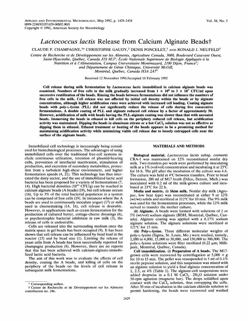
SUCCS1VI FINTLTONSFIG. 1. Effect of initial density of L. lactis in the alginate on
growth of the immobilized cells, cell release from the beads, andfermented milk acidity upon five successive fermentations with thesame beads. Data represent fermentations with L. lactis at an initialcount of 1 x 10⁹ (0) and then at 2 x 10¹⁰ (@) CFU/g of beads. Barsindicate standard deviation from the mean on the basis of threereplicate experiments.
Beads were liquefied by aseptically adding 1 g (wet weight) ofbeads to a sterile 1% sodium citrate solution (pH 6.0).
RESULTS
The addition of alginate-immobilized lactococci to milkresulted in acidification of the medium, with the initial pH of6.5 dropping to 4.2 after 6 h of incubation. The fermentationwas normally stopped by removing the beads when the milkpH dropped to 5.5, which occurred after approximately 2 h.This 2-h fermentation simulates cream fermentations re-quired in the manufacture of cultured butter or fermentedcottage-cheese dressings (6). The effect of multiple use of thesame lactococci-containing beads was examined, with theresults representing the experimental conditions at the endof each successive fermentation.
Initial cell density. A beads were prepared with bacterialpopulations of 1 x 10⁹ or 2 x 10¹⁰ CFU/g. Growth wasobserved in beads containing the lower initial population,while no change in cell density occurred with beads contain-ing 1010 CFU/g (Fig. 1A). The lower initial cell density
APPL. ENVIRON. MICROBIOL.

CELL RELEASE FROM ALGINATE BEADS 1431
MW 15-30000z0.06
0000
0.04
0.03
0.02.
-A~~~~
0.01
0.000 10 20 30 40
( )FIG. 2. Effect of poIY-L-lysine molecular weight (MW) on its
adsorption to A beads. Values are of residual levels of poIy-L-lysineafter immersion of the A beads in the poIY-L-lysine solutions. Thestandard deviation from the mean is based on at least three replicateexperiments.
resulted in slower acidification of milk during the first threefermentation periods (Fig. 1C). With repeated use of thebeads, the acidities of the milk samples after the 2-h incuba-tion period became similar, irrespective of initial bead celldensity.We observed an increase in free-cell density as the beads
were used for successive fermentations (Fig. 1A). Thenumbers of free cells in the fermented milks were initiallylower when low-cell-density beads were used (Fig. 1B), butthis difference gradually decreased as the beads were reuti-lized. After five fermentations, the total free-cell populationin the fermentor was 6 x 10⁹ CFU/100 ml while the totalimmobilized cell population reached 2.5 x 10¹¹ CFU/4.2 g.The free-cell population accounted for 2.4% of the total cellcount in the bioreactor.When the more concentrated cell suspension was used,
the pH values of fermented milk and the immobilized cellpopulation were stable over successive fermentations (Fig.1C); therefore, high-cell-density beads were used for most ofthe subsequent experiments.
Alginate concentration. The preparation of beads at vari-ous alginate concentrations did not significantly influence the
1.0-
'0 0.8A
0.6II
0.4--00.40-0 Abeads0.2- *-@ AP (MW 1-4000)0 & ~~~~~~~~-& AP (MW 15-30000)
0.0~~ ~~~~A AAP (MW 150-300000)0 1 2 3 4 5 6 7
TIME (minutes)FIG. 3. Bacterial release from A or AP beads of various molec-
ular weights (MW) when immersed in a 1% sodium citrate solution.
numbers of free cells found in the fermented milks. Milkacidification was greater (pH, 0.25 unit lower) with beadsprepared from 4% alginate cell suspensions. Although theinitial bacterial populations in the beads were identical for allassays, the amount of beads obtained from the 4% alginatepreparation was twice that of the other preparations (Table1). Since the preparation of 1% A beads was easier and sincefree-cell numbers were not influenced by alginate content,1% A beads were used for the subsequent assays.
Coating of the beads with poIy-L-lysine. Adsorption ofpoly-L-lysine to the surfaces of the A beads resulted indepletion of the poly-L-lysine from solution (Fig. 2). Lower-molecular-weight poly-L-lysine was bound to a greater ex-tent than the higher-molecular-weight form. The quantity ofpoly-L-lysine adsorbed was related to the initial concentra-tion (Fig. 2). Thus, when the initial concentration was0.05%, almost twice as much poly-L-lysine was adsorbed asthat observed when the initial concentration was 0.03%. Forthis reason, 0.05% solutions were used for coating of thebeads in subsequent experiments. The contact time waslimited to 20 min, since little adsorption occurred between 20and 40 min.The enumeration of the immobilized cells became more
difficult when the A beads were coated with poly-L-lysine(i.e., the AP beads). AP beads coated with the polymer of15,000 to 30,000 molecular weight did not completely dis-solve after 3 h of undisturbed incubation in the sodiumcitrate solution, and only 60% of the initial bacterial popu-lation could be recovered. In contrast, only 1 h was requiredto completely recover the immobilized cell population in theA beads. Slight agitation of the citrate-treated AP beadsruptured the fragile capsules, and "ghosts" of the poly-L-lysine alginate membranes were observed. It was thus nec-essary to use a blender to release the cells from the APbeads. All cells could be recovered after 4 min of blendingwhen the AP beads were coated with the poly-L-lysine of1,000 to 4,000 or 150,000 to 300,000 molecular weight (Fig.3). Cells entrapped in the AP beads coated with the poly-L-lysine of 15,000 to 30,000 molecular weight could not becompletely dispersed even with blender (Stomacher) homog-enization. When compared with a standard method (25stokes per 30-cm area per 7 s), such a homogenization wasfound not to influence viable counts from a CRA-1 culture.There were no significant differences (P > 0.05; average of
four trials) in acidification activity or cell release between thevarious types of poly-L-lysine. The use of AP beads did notinfluence the acidification of milk nor its number of released
VOL. 58, 1992
1432 CHAMPAGNE ET AL.
10-
(0) Kthanol 7O
(A) HeatingA3
2-o (O)0)AI(NO)0.1M
0 2 4 6 8 10
TIME (minutes)FIG. 4. Viability of L. lactis cells immobilized in A beads after
immersion in ethanol, Al(NO3)3, or hot 0.1 M CaC12 solutions.
cells. These results were not related to loss of the poly-L-lysine coating during the course of the fermentations. APbeads that had been incubated for 10 h under agitationdemonstrated the same properties as fresh AP beads inregards to stability to dissolution in citrate solutions. Thepopulation reached in the AP beads varied between 6.1 x10¹⁰ and 7.5 x 10¹⁰ CFU/g, after the fifth consecutivefermentation, and was thus not different from values ob-served with A beads (Fig. 1A).The effect of adding an alginate coat to the AP beads, thus
sandwiching the poly-L-lysine between alginate layers, wasevaluated. The free-cell count was 1 log lower when APAbeads were used. Less acidification (by 0.2 pH unit) was alsoobserved when APA beads were used to inoculate the milk.The acidification of milk with APA beads increased in thesuccessive fermentations. The pH after the third fermenta-tion with APA beads was similar to that found with A or APbeads at the end of the first fermentation. Increased cellrelease also occurred with repeated use of APA beads and,as was observed with acidification, the free-cell count at thethird APA fermentation was similar to that found after theinitial fermentation with A or AP beads.Complete removal of the fermented milk from the biore-
actor between the successive fermentations was difficult.Approximately 3% residual milk remained adsorbed to thebeads or reactor, which may contribute to the free-cellloading in subsequent fermentations. To estimate this effect,the A and AP beads were rinsed twice with 30 ml of peptonewater between each fermentation. Significant differencesbetween numbers of free cells in rinsed beads and numbersin unrinsed beads were not observed. These results suggestthat the main source of free cells in the bioreactor was cellrelease from the beads, with a possible second source beingsubsequent growth.Bead immersions. Immersing the beads in ethanol,
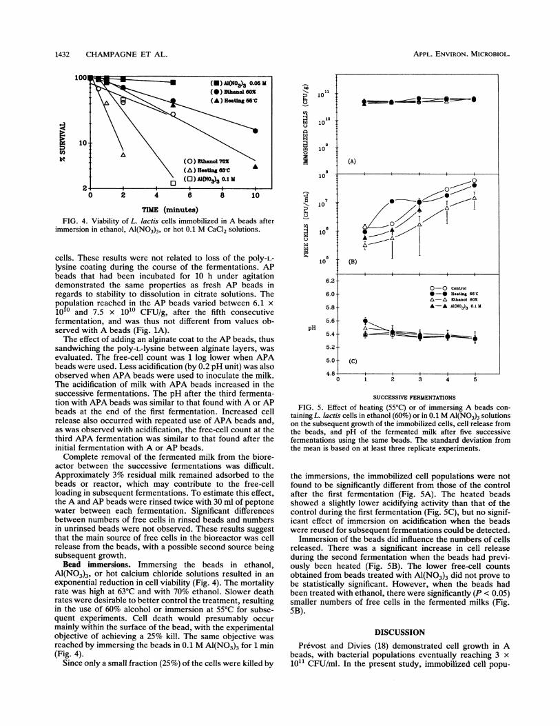
Al(NO3)3, or hot calcium chloride solutions resulted in anexponential reduction in cell viability (Fig. 4). The mortalityrate was high at 63°C and with 70% ethanol. Slower deathrates were desirable to better control the treatment, resultingin the use of 60% alcohol or immersion at 55°C for subse-quent experiments. Cell death would presumably occurmainly within the surface of the bead, with the experimentalobjective of achieving a 25% kill. The same objective wasreached by immersing the beads in 0.1 M Al(NO3)3 for 1 min(Fig. 4).
Since only a small fraction (25%) of the cells were killed by
C.)
C.)
N
0B9
10
10
10
10
la,
3
T.)
pH
10
1C
10
10
(A)
7 T
0~~~~~t~~~
(B)
6.2-0-0 Control
6.0- *-* Heating 655CA-L Ethanol 60Z
5 8 A-A AI(N03)3A .1
5.2-
5.0 (C)
4.80 1 2 3 4 5
SUCCESSIVE FERMENTATIONS
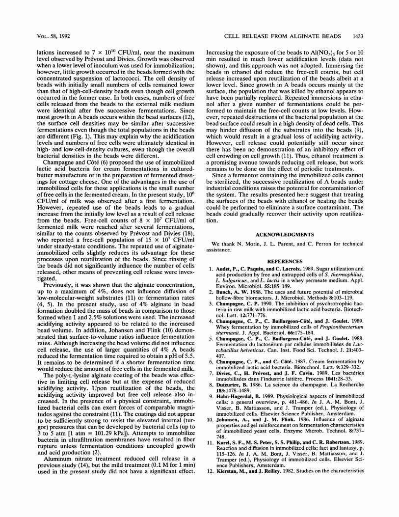
FIG. 5. Effect of heating (55°C) or of immersing A beads con-taining L. lactis cells in ethanol (60%) or in 0.1 M Al(N03)3 solutionson the subsequent growth of the immobilized cells, cell release fromthe beads, and pH of the fermented milk after five successivefermentations using the same beads. The standard deviation fromthe mean is based on at least three replicate experiments.
the immersions, the immobilized cell populations were notfound to be significantly different from those of the controlafter the first fermentation (Fig. SA). The heated beadsshowed a slightly lower acidifying activity than that of thecontrol during the first fermentation (Fig. SC), but no signif-icant effect of immersion on acidification when the beadswere reused for subsequent fermentations could be detected.
Immersion of the beads did influence the numbers of cellsreleased. There was a significant increase in cell releaseduring the second fermentation when the beads had previ-ously been heated (Fig. SB). The lower free-cell countsobtained from beads treated with Al(N03)3 did not prove tobe statistically significant. However, when the beads hadbeen treated with ethanol, there were significantly (P < 0.05)smaller numbers of free cells in the fermented milks (Fig.SB).
DISCUSSION
Prevost and Divies (18) demonstrated cell growth in Abeads, with bacterial populations eventually reaching 3 x10¹¹ CFU/ml. In the present study, immobilized cell popu-
APPL. ENVIRON. MICROBIOL.
CELL RELEASE FROM ALGINATE BEADS 1433
lations increased to 7 x 10¹⁰ CFU/ml, near the maximumlevel observed by Prevost and Divies. Growth was observedwhen a lower level of inoculum was used for immobilization;however, little growth occurred in the beads formed with theconcentrated suspension of lactococci. The cell density ofbeads with initially small numbers of cells remained lowerthan that of high-cell-density beads even though cell growthoccurred in the former case. In both cases, numbers of freecells released from the beads to the external milk mediumwere identical after five successive fermentations. Sincemost growth in A beads occurs within the bead surfaces (12),the surface cell densities may be similar after successivefermentations even though the total populations in the beadsare different (Fig. 1). This may explain why the acidificationlevels and numbers of free cells were ultimately identical inhigh- and low-cell-density cultures, even though the overallbacterial densities in the beads were different.Champagne and Cote (6) proposed the use of immobilized
lactic acid bacteria for cream fermentations in cultured-butter manufacture or in the preparation of fermented dress-ings for cottage cheese. One of the advantages in the use ofimmobilized cells for these applications is the small numberof free cells in the fermented cream. In the present study, 106CFU/ml of milk was observed after a first fermentation.However, repeated use of the beads leads to a gradualincrease from the initially low level as a result of cell releasefrom the beads. Free-cell counts of 8 x 10⁷ CFU/ml offermented milk were reached after several fermentations,similar to the counts observed by Pr6vost and Divies (18),who reported a free-cell population of 15 x 107 CFU/mlunder steady-state conditions. The repeated use of alginate-immobilized cells slightly reduces its advantage for theseprocesses upon reutilization of the beads. Since rinsing ofthe beads did not significantly influence the number of cellsreleased, other means of preventing cell release were inves-tigated.
Previously, it was shown that the alginate concentration,up to a maximum of 4%, does not influence diffusion oflow-molecular-weight substrates (11) or fermentation rates(4, 5). In the present study, use of 4% alginate in beadformation doubled the mass of beads in comparison to thoseformed when 1 and 2.5% solutions were used. The increasedacidifying activity appeared to be related to the increasedbead volume. In addition, Johansen and Flink (10) demon-strated that surface-to-volume ratios influence fermentationrates. Although increasing the bead volume did not influencecell release, the use of larger quantities of 4% A beadsreduced the fermentation time required to obtain a pH of 5.5.It remains to be determined if a shorter fermentation timewould reduce the amount of free cells in the fermented milk.The poly-L-lysine alginate coating of the beads was effec-
tive in limiting cell release but at the expense of reducedacidifying activity. Upon reutilization of the beads, theacidifying activity improved but free cell release also in-creased. In the presence of a physical constraint, immobi-lized bacterial cells can exert forces of comparable magni-tudes against the constraint (11). The coatings did not appearto be sufficiently strong to resist the elevated internal (tur-gor) pressures that can be developed by bacterial cells (up to3 to 5 atm [1 atm = 101.29 kPa]). Attempts to immobilizebacteria in ultrafiltration membranes have resulted in fiberrupture unless fermentation conditions uncoupled growthand acid production (2).Aluminum nitrate treatment reduced cell release in a
previous study (14), but the mild treatment (0.1 M for 1 min)used in the present study did not have a significant effect.
Increasing the exposure of the beads to Al(NO3)3 for 5 or 10min resulted in much lower acidification levels (data notshown), and this approach was not adopted. Immersing thebeads in ethanol did reduce the free-cell counts, but cellrelease increased upon reutilization of the beads albeit at alower level. Since growth in A beads occurs mainly at thesurface, the population that was killed by ethanol appears tohave been partially replaced. Repeated immersions in etha-nol after a given number of fermentations could be per-formed to maintain the free-cell counts at low levels. How-ever, repeated destructions of the bacterial population at thebead surface could result in a high density of dead cells. Thismay hinder diffusion of the substrates into the beads (9),which would result in a gradual loss of acidifying activity.However, cell release could potentially still occur sincethere has been no demonstration of an inhibitory effect ofcell crowding on cell growth (11). Thus, ethanol treatment isa promising avenue towards reducing cell release, but workremains to be done on the effect of periodic treatments.
Since a fermentor containing the immobilized cells cannotbe sterilized, the successive reutilization of A beads underindustrial conditions raises the potential for contamination ofthe system. The results presented here suggest that treatingthe surfaces of the beads with ethanol or heating the beadscould be performed to eliminate a surface contaminant. Thebeads could gradually recover their activity upon reutiliza-tion.
ACKNOWLEDGMENTS
We thank N. Morin, J. L. Parent, and C. Perron for technicalassistance.
REFERENCES1. Audet, P., C. Paquin, and C. Lacroix. 1989. Sugar utilization and
acid production by free and entrapped cells of S. thernophilus,L. bulganicus, and L. lactis in a whey permeate medium. Appl.Environ. Microbiol. 55:185-189.
2. Bunch, A. W. 1988. The uses and future potential of microbialhollow-fibre bioreactors. J. Microbiol. Methods 8:103-119.
3. Champagne, C. P. 1990. The inhibition of psychrotrophic bac-teria in raw milk with immobilized lactic acid bacteria. Biotech-nol. Lett. 12:771-776.
4. Champagne, C. P., C. Baillargeon-Cote, and J. Goulet. 1989.Whey fermentation by immobilized cells of Propionibacteriumshenrnandi. J. Appl. Bacteriol. 66:175-184.
5. Champagne, C. P., C. Baillargeon-C6te, and J. Goulet. 1988.Fermentation du lactoserum par cellules immobilisees de Lac-tobacillus helveticus. Can. Inst. Food Sci. Technol. J. 21:403-407.
6. Champagne, C. P., and C. C6te. 1987. Cream fermentation byimmobilized lactic acid bacteria. Biotechnol. Lett. 9:329-332.
7. Divies, C., H. Prevost, and J. F. Cavin. 1989. Les bacteriesimmobilis6es dans l'industrie laitiere. Process 1041:28-33.
8. Duteurtre, B. 1986. La science du champagne. La Recherche183:1478-1489.
9. Hahn-Hagerdal, B. 1989. Physiological aspects of immobilizedcells: a general overview, p. 481-486. In J. A. M. Bont, J.Visser, B. Mattiasson, and J. Tramper (ed.), Physiology ofimmobilized cells. Elsevier Science Publisher, Amsterdam.
10. Johansen, A., and J. M. Flink. 1986. Influence of alginateproperties and gel reinforcement on fermentation characteristicsof immobilized yeast cells. Enzyme Microb. Technol. 8:737-748.
11. Karel, S. F., M. S. Peter, S. S. Philip, and C. R. Robertson. 1989.Reaction and diffusion in immobilized cells: fact and fantasy, p.115-126. In J. A. M. Bont, J. Visser, B. Mattiasson, and J.Tramper (ed.), Physiology of immobilized cells. Elsevier Sci-ence Publishers, Amsterdam.
12. Kierstan, M., and J. Reilley. 1982. Studies on the characteristics
VOL. 58, 1992
1434 CHAMPAGNE ET AL.
of alginate gels in relation to their use in separation andimmobilization applications. Biotechnol. Bioeng. 24:1505-1517.
13. King, G. A., A. J. Daugulis, P. Faulkner, and M. F. A. Goosen.1987. Alginate-polylisine microcapsules of controlled membranemolecular weight cut off for mammalian cell culture engineering.Biotechnol. Process. 3:231-240.
14. Linko, P. 1985. Immobilized lactic acid bacteria, p. 25-36. InA. J. Laskin (ed.), Enzymes and immobilized cells in biotech-nology. Benjamin/Cummings Publishing Co., Menlo Park, Calif.
15. Nunez, M. J., R. Chamy, J. M. Lema, and A. Sanroman. 1989.Enhancement of cell retention for immobilized yeasts in K-car-rageenan, p. 283-288. In J. A. M. Bont, J. Visser, B. Mattias-son, and J. Tramper (ed.), Physiology of immobilized cells.Elsevier Science Publishers, Amsterdam.
16. Prevost, H., and C. Divies. 1988. Continuous pre-fermentation ofmilk by entrapped yoghurt bacteria. I. Development of theprocess. Milchwissenschaft 43:621-625.
17. Prevost, H., and C. Divies. 1988. Continuous pre-fermentation of
APPL. ENVIRON. MICROBIOL.
milk by entrapped yoghurt bacteria. II. Data for optimization ofthe process. Milchwissenschaft 43:716-719.
18. Prevost, H., and C. Divies. 1987. Fresh fermented cheeseproduction with continuous pre-fermented milk by a mixedculture of mesophilic lactic streptococci entrapped in Ca-algi-nate. Biotechnol. Lett. 9:789-794.
19. Prevost, H., C. Divies, and E. Rousseau. 1985. Continuousyoghurt production with Lactobacillus bulganicus and Strepto-coccus thermophilus entrapped in Ca-alginate. Biotechnol.Lett. 7:247-252.
20. Sun, A. M., and G. M. O'Shea. 1985. Microencapsulation ofliving cells-a long-term delivery system. J. Controlled Release2:137-141.
21. Tramper, J. 1989. Conversions by immobilized cells versustraditional fermentations, p. 115-126. In J. A. M. Bont, J.Visser, B. Mattiasson, and J. Tramper (ed.), Physiology ofimmobilized cells. Elsevier Science Publishers, Amsterdam.
Related Documents


