CELL BIOLOGY LABORATORY MANUAL DEPARTEMENT OF BIOLOGY VIRTUAL UNIVERSITY OF PAKISTAN BIO 201 1

Welcome message from author
This document is posted to help you gain knowledge. Please leave a comment to let me know what you think about it! Share it to your friends and learn new things together.
Transcript

CELL BIOLOGY LABORATORY MANUAL
DEPARTEMENT OF BIOLOGY
VIRTUAL UNIVERSITY OF PAKISTAN
BIO 201
1
Contents:
index Name of experiment Page#
1 Microscopy and staining techniques
3
2 Study of Prokaryotic, Eukaryotic,
Plant and Animal cells
8
3 Study of different types of plastids
12
4 Study of cellular reproduction
15
5 Study of Mitosis: Smear/Squash
preparation of Onion roots
18
6 References 24
2
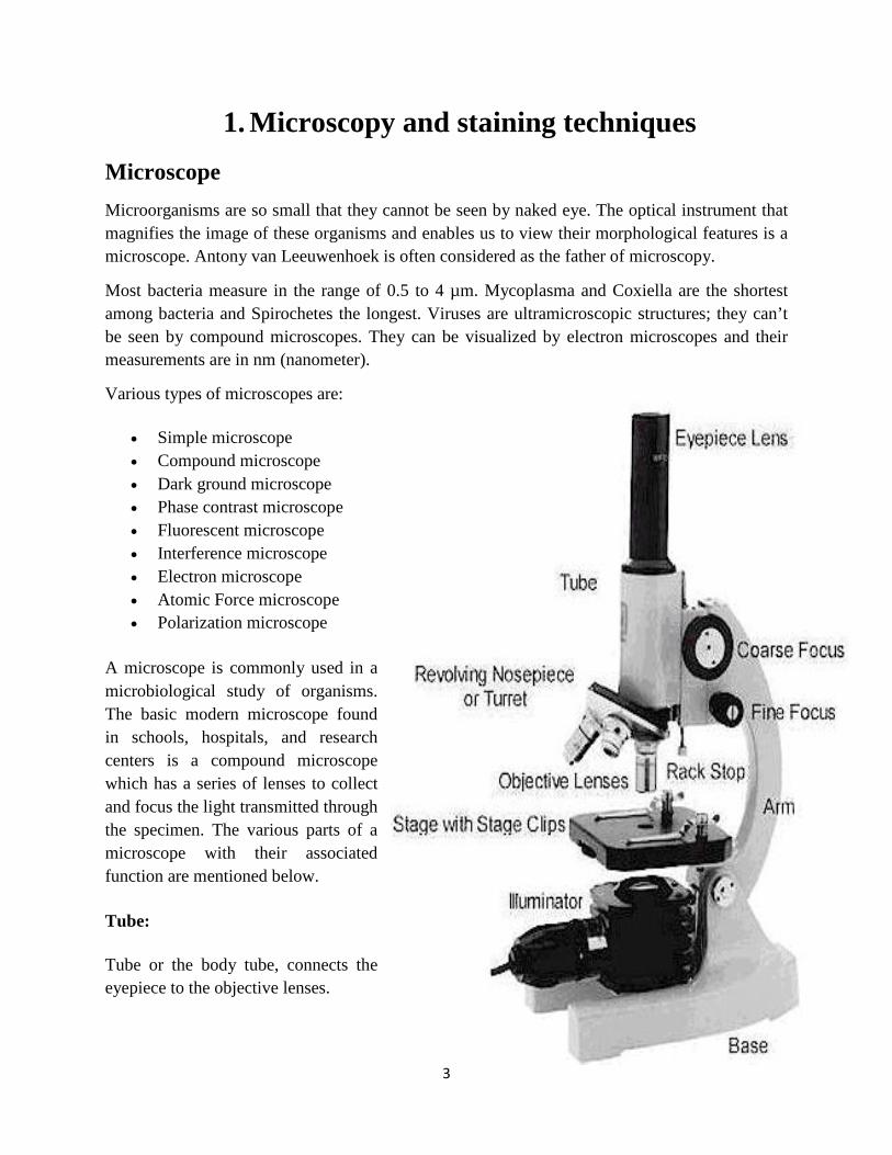
1. Microscopy and staining techniques Microscope Microorganisms are so small that they cannot be seen by naked eye. The optical instrument that magnifies the image of these organisms and enables us to view their morphological features is a microscope. Antony van Leeuwenhoek is often considered as the father of microscopy.
Most bacteria measure in the range of 0.5 to 4 µm. Mycoplasma and Coxiella are the shortest among bacteria and Spirochetes the longest. Viruses are ultramicroscopic structures; they can’t be seen by compound microscopes. They can be visualized by electron microscopes and their measurements are in nm (nanometer).
Various types of microscopes are:
• Simple microscope • Compound microscope • Dark ground microscope • Phase contrast microscope • Fluorescent microscope • Interference microscope • Electron microscope • Atomic Force microscope • Polarization microscope
A microscope is commonly used in a microbiological study of organisms. The basic modern microscope found in schools, hospitals, and research centers is a compound microscope which has a series of lenses to collect and focus the light transmitted through the specimen. The various parts of a microscope with their associated function are mentioned below.
Tube:
Tube or the body tube, connects the eyepiece to the objective lenses.
3
Eyepiece or ocular lens:
Eyepiece is the lens, present at the top and is used to see the objects under study. Eyepiece lens contains a magnification of 10X or 15X.
Resolving nosepiece:
It is also known as the Turret. Resolving nosepiece has holders for the different objective lenses. It allows the rotation of the lenses while viewing.
Objective lenses:
Generally, three or four objective lenses are found on a microscope, with ranges of 10X, 40X, 100X powers. Lenses are color coded, the shortest lens is of the lowest power, and the longest lens is high power lens.
Diaphragm:
Diaphragm helps in controlling the amount of light that is passing through the opening of the stage. It is helpful in the adjustment of the control of light that enters.
Coarse adjustment knob:
Used for focus on scanning. Usually the low power lens is used enabling the movement of the tube.
Fine adjustment knob:
Used for focus on oil. It moves the body tube for focusing the high power lens.
Arm:
It supports the tube of the microscope and connects to the base of the microscope.
Stage:
A flat platform used for placing the slides under observation.
Stage clip:
Stage clips hold the slides in proper place.
4

Condenser:
The main function of condenser lens is focusing the light on the specimen under observation. It gives a sharp image.
Base:
It provides basal support for the microscope.
Power switch:
It turns the illumination on or off.
Proper usage and handling the microscope:
Microscope is a delicate instrument that must be handled with care. All kinds of mechanical shocks must be avoided. The microscope must be lifted by holding its arm in one hard and supporting the base of the microscope with the palm of the other hand. The microscope must be kept in dust free environment. The oil must be wiped clean using a soft tissue paper. After usage the slide must be removed and cleaned before returning to its original place.
Staining
A technique that is used to define and examine different types of microbes.
Types of staining techniques
1. Simple stain techniques.
Simple staining is performed with basic dyes such as crystal violet or methylene blue.
2. Differential stain techniques.
Differential stain technique distinguishes two kinds of organisms. There are two types of this technique; one is Gram stain technique while other is acid fast technique.
Gram stain technique
A most commonly used technique for study of microbes like bacteria.
Principle
This technique separates bacteria into two groups, Gram‐positive bacteria and Gram‐negative bacteria.
5

Materials: Clean glass slides Inoculating loop Bunsen burner Bibulous paper Microscope Lens paper and lens cleaner Immersion oil Distilled water 18 to 24 hour cultures of organisms
Reagents: Primary Stain - Crystal Violet Mordant - Grams Iodine Decolourizer - Ethyl Alcohol Secondary Stain - Safranin Procedure:
• Smear preparation
A small sample of microorganisms is placed on a slide and permitted to air dry. The smear is heat fixed by quickly passing it over a flame. Heat fixing kills the organisms, makes them adhere to the slide, and permits them to accept.
• Primary stain
Apply crystal violet as primary stain
• Mordant
Use iodine as a mordant
• Decolourizer
Wash with ethyl alcohol that acts as a decolourizing agent. Gram‐positive bacteria retain the crystal‐violet iodine stain; however, the Gram‐negative bacteria lose the stain.
• Secondary stain
The Gram‐negative bacteria subsequently stain with the Safranin dye, the counterstain. These bacteria appear red under the oil‐immersion lens, while Gram‐positive bacteria appear blue or purple, reflecting the crystal violet retained during the washing step.
6
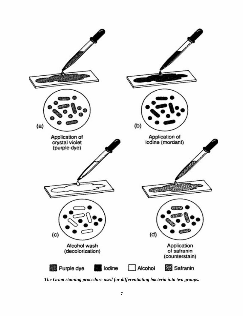
The Gram staining procedure used for differentiating bacteria into two groups.
7
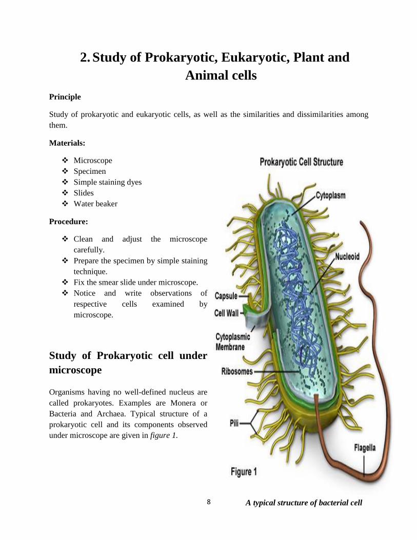
A typical structure of bacterial cell
2. Study of Prokaryotic, Eukaryotic, Plant and Animal cells
Principle
Study of prokaryotic and eukaryotic cells, as well as the similarities and dissimilarities among them.
Materials:
Microscope Specimen Simple staining dyes Slides Water beaker
Procedure:
Clean and adjust the microscope carefully.
Prepare the specimen by simple staining technique.
Fix the smear slide under microscope. Notice and write observations of
respective cells examined by microscope.
Study of Prokaryotic cell under microscope
Organisms having no well-defined nucleus are called prokaryotes. Examples are Monera or Bacteria and Archaea. Typical structure of a prokaryotic cell and its components observed under microscope are given in figure 1.
8
Cell Wall
Provide shape and protection to cell.
Pilli
Organ for the exchange of genetic material from one bacterium to another.
Capsule
Sticky projections that cover cell wall and provide protection from phagocytosis, chemicals and dehydration.
Flagellum
A “whip-like;” structure that helps in movement.
Plasma Membrane
A thin, flexible asymmetrical “sac” that holds the cytoplasm and serves as a passageway for anything that enters or leaves the cell such as nutrients and gases.
Cytoplasm
Contains organelles for different functions inside the cell.
Nucleoid or Nuclear Body
Area of the cytoplasm where the DNA strand is located
Plasmids
Extra chromosomal piece of DNA. Plasmids are often the site of genes that code for resistance to antibiotics.
Ribosomes
The function of prokaryotic ribosomes widely depends on the bacteria.
9
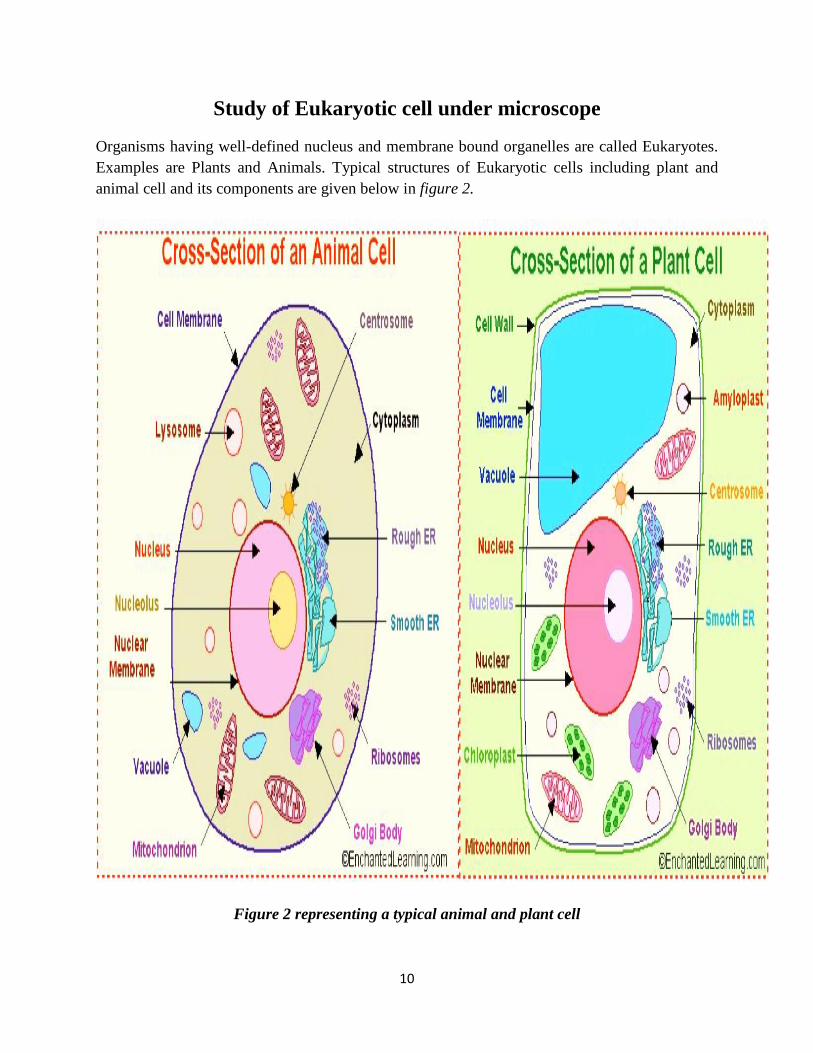
Study of Eukaryotic cell under microscope
Organisms having well-defined nucleus and membrane bound organelles are called Eukaryotes. Examples are Plants and Animals. Typical structures of Eukaryotic cells including plant and animal cell and its components are given below in figure 2.
Figure 2 representing a typical animal and plant cell
10
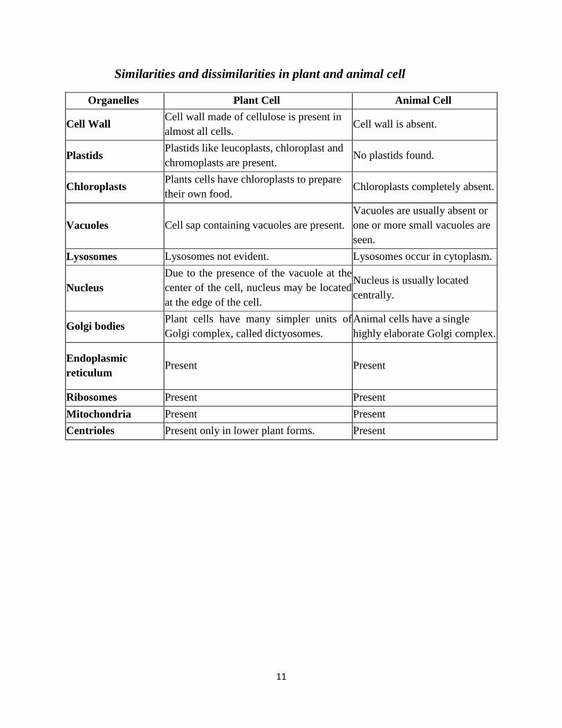
Similarities and dissimilarities in plant and animal cell
Organelles Plant Cell Animal Cell
Cell Wall Cell wall made of cellulose is present in almost all cells.
Cell wall is absent.
Plastids Plastids like leucoplasts, chloroplast and chromoplasts are present.
No plastids found.
Chloroplasts Plants cells have chloroplasts to prepare their own food.
Chloroplasts completely absent.
Vacuoles Cell sap containing vacuoles are present. Vacuoles are usually absent or one or more small vacuoles are seen.
Lysosomes Lysosomes not evident. Lysosomes occur in cytoplasm.
Nucleus Due to the presence of the vacuole at the center of the cell, nucleus may be located at the edge of the cell.
Nucleus is usually located centrally.
Golgi bodies Plant cells have many simpler units of Golgi complex, called dictyosomes.
Animal cells have a single highly elaborate Golgi complex.
Endoplasmic reticulum
Present Present
Ribosomes Present Present Mitochondria Present Present Centrioles Present only in lower plant forms. Present
11
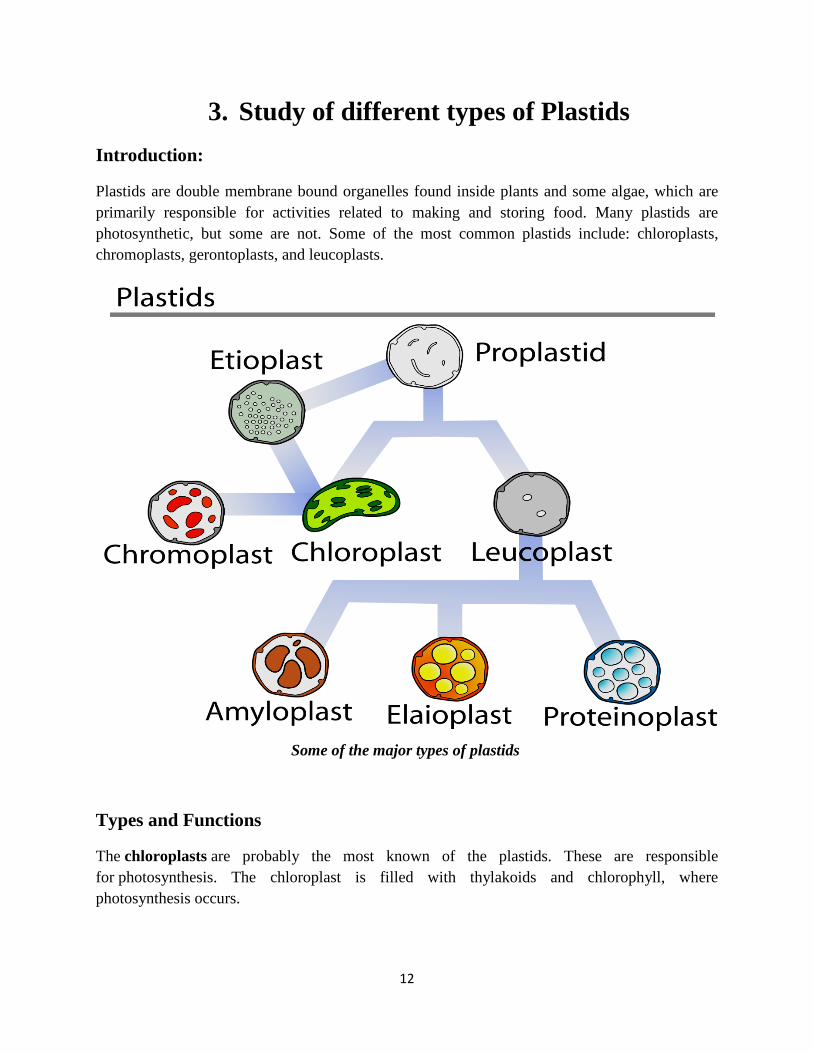
3. Study of different types of Plastids Introduction:
Plastids are double membrane bound organelles found inside plants and some algae, which are primarily responsible for activities related to making and storing food. Many plastids are photosynthetic, but some are not. Some of the most common plastids include: chloroplasts, chromoplasts, gerontoplasts, and leucoplasts.
Some of the major types of plastids
Types and Functions
The chloroplasts are probably the most known of the plastids. These are responsible for photosynthesis. The chloroplast is filled with thylakoids and chlorophyll, where photosynthesis occurs.
12

The basic structure of the chloroplast
Chromoplasts
These are found in flowering plants, fruits, and aging leaves. The chloroplasts actually convert over to chromoplasts. There are carotenoid pigments here that allow for the different colors you see in fruits and the fall leaves. One of the main reasons for these structures and the colors is to attract pollinators.
Gerontoplasts
They are basically chloroplasts that are going through the aging process. These are chloroplasts of the leaves that are beginning to convert into different organelles or are being repurposed since the leaf is no longer utilizing photosynthesis (such as in the fall months).
Leucoplasts
They are the non-pigmented organelles. Unlike the others we have talked about, leucoplasts have no colour at all. They are found in the non-photosynthetic parts of the plant, such as the roots. Depending on what the plant needs, they may become essentially just storage sheds for starches, lipids, and proteins. They are more readily used for synthesizing amino acids and fatty acids. Leucoplasts are further subdivided into three different plastids: amyloplasts, proteinoplasts, and elaioplasts. Amyloplasts are the largest of the three and are charged with storing starch. Then there are the proteinoplasts that help to store the proteins that a plant needs and are typically found in seeds. Finally, the elaioplasts are used to store fats and oils that are needed by the plant especially in seeds.
13
Objective:
Observation of amyloplast from potato tuber. Observation of chromoplast from rose petal. Observation of amyloplast from potato tuber Materials: Potato tuber Razor blade Iodine stain Slides with cover slips Microscope
Procedure: Make a thin section of potato tuber with the help of razor blade. Stain with iodine stain for a few seconds. Add cover slips on slides. Observe it under microscope The intensely stained structures in the cells are amyloplasts, a type of plastids that store
starch. Observation of chromoplast from rose petal Materials: Rose petal Razor blade Water beaker Slides with cover slips Microscope
Procedure: Make a thin section of red pepper with the help of razor blade. Place the section in a drop of water on glass slide and add a cover slip. Examine it under the microscope. The tiny pinpoint orange organelles are chromoplasts, a type of plastid containing
pigments other than chlorophyll.
14

Events of mitosis
4. Study of cellular reproduction Introduction:
Cellular reproduction is the process by which organisms are reproduce asexually by fission or spore formation and sexually by formation of gametes through cell division. Cell division is also a source of tissue growth and repair in multicellular organisms. Mitosis is responsible for reproducing somatic cells and meiosis is responsible for reproducing germ cells.
Mitosis
In single-cell organisms, mitosis is the only form of cellular reproduction. One round of mitosis yields two genetically identical cells. In bacteria, this process results in an entirely new, independent organism. This is classified as asexual reproduction because it does not require sex for the creation of new organisms. In multi-cellular organisms, mitosis only occurs in somatic cells, which comprise all cells in an organism excluding germ cells.
Cells that undergo mitosis duplicate their chromosomes, resulting in cells with two times their normal haploid or diploid numbers (4Nchromosomes). Newly-synthesized chromosomes remain closely associated with their like-chromosome. These two identical chromosomes are called sister chromatids. Once duplicated, sister chromatids separate such that one copy of each chromosome lines up on opposite ends of the cell. The cell then pinches in the center until it breaks into two different cells. A nucleus then forms around the chromosomes in each cell to yield two cells with the same original number of chromosomes as the preexisting cell.
15

Events of meiosis
Meiosis
There are two major differences between mitosis and meiosis. First, meiosis involves not one, but two cell divisions. Second, meiosis leads to the production of germ cells, which are cells that give rise to gametes. Germ cells are different from somatic cells in a critical way. Whereas somatic cells are diploid, meaning they have two copies of each chromosome, germ cells are haploid. The haploid nature of germ cells is vital to the process of sexual reproduction.
There are two different sex cells or gametes: sperm and eggs. Males produce sperm and females produce eggs. Because they are produced from germ cells, gametes are likewise haploid. In order to create a new individual via sexual reproduction, a sperm cell needs to activate an egg by joining it in a fertilization process. When these two haploid cells unite, a diploid cell results. This specialized cell can then develop into a new individual. The sexual reproductive process just described ensures that the resulting offspring will have an equal maternal and paternal genetic contribution.
As we mentioned earlier, higher-order cells contain homologous pairs of chromosomes--one from the father and the other from the mother. In meiosis, as in mitosis, the maternal and paternal homologues are replicated during DNA replication yielding two pairs of sister chromatids. After the first cell division, each of the resulting cells contains a pair of sister chromatids—-one maternal pair and the other paternal. Unlike mitosis, meiosis does not end after one division; it continues with a second cell division. In this division, the sister chromatids are separated yielding four total haploid cells.
16
Objective:
To understand the process of mitosis and different stages of mitosis. Observation of mitosis in onion root tips Roots and stems grow from their tips. Actively dividing tissues such as meristem are good sources of mitotically dividing cells. You will prepare squashes of onion root tip (Allium cepa) and observe cells in various stages of mitosis.
Materials: Onion plant with root Acetocarmine stain Scissors Forceps Razor blade Pasture pipette 1.5 ml Centrifuge tubes Dissection probe with wooden back Microscopic slides and cover slips Water bath Light Microscope
Procedure: Cut the tip 5 to 8 mm from the tip of the freshly sprouted root. Discard the rest of the
root. Place the cut tip on a clean microscope slide. Add 2-3 drops of Acetocarmine stain to the slide. Warm the slide gently over the alcohol lamp for about one minute. (Do not allow the
slide to get hot to the touch; you don't want to cook either your fingers or the root. Do not let the root dry out).
Cover the slide with a cover slip or lens paper. Squash the slide with your thumb using a firm and even pressure. (Avoid squashing with
such force that the cover slip breaks or slides). Observe it under a compound microscope in 10X objective. Scan and narrow down to a
region containing dividing cells and switch to 40X for a better view.
17
5. Study of Mitosis: Smear/Squash preparation of Onion roots
Introduction:
The genetic information of plants, animals and other eukaryotic organisms resides in several (or many) individual DNA molecules, or chromosomes. For example, each human cell possesses 46 chromosomes, while each cell of an onion possesses 8 chromosomes. All cells must replicate their DNA when dividing. During DNA replication, the two strands of the DNA double helix separate, and for each original strand a new complementary strand is produced, yielding two identical DNA molecules. DNA replication yields an identical pair of DNA molecules (called sister chromatids) attached at a region called the centromere.
DNA replication in eukaryotes is followed by the process called mitosis which assures that each daughter cell receives one copy of each of the replicated chromosomes. During the process of mitosis, the chromosomes pass through several stages known as prophase, metaphase, anaphase and telophase. The actual division of the cytoplasm is called cytokinesis and occurs during telophase. During each of the preceding stages, particular events occur that contribute to the orderly distribution of the replicated chromosomes prior to Cytokinesis.
The stages of Mitosis
Prophase:
During prophase, the chromosomes supercoil and the fibers of the spindle apparatus begin to form between Centrosome located at the pole of the cells. The nuclear membrane also disintegrates at this time, freeing the chromosomes into the surrounding cytoplasm.
Prometaphase:
During Prometaphase, some of the fibers attach to the centromere of each pair of sister chromatids and they begin to move toward the center of the cell.
Metaphase:
At metaphase the chromosomes have come to rest along the center plane of the cell.
Anaphase:
During anaphase, the centromeres split and the sister chromatids begin to migrate toward the opposite poles of the cell.
18
Telophase:
During telophase, the chromosomes at either end of the cell cluster begin to cluster together, which facilitates the formation of a new nuclear membrane. This also is when cytokinesis occurs, leading to two separate cells. One way to identify that telophase has begun is by looking for the formation of the cell plate, the new cell wall forming between the two cells.
Objective: Better understand the process and stages of mitosis.
Prepare your own specimens of onion root in which you can visualize all of the stages of mitosis.
Viewing mitosis in onion root tips
Why use onion roots for viewing mitosis?
• The roots are easy to grow in large numbers. • The cells at the tip of the roots are actively dividing, and thus many cells will be in stages
of mitosis. • The tips can be prepared in a way that allows them to be flattened on microscopes slide
(“squashed”) so that the chromosomes of individual cells can be observed. • The chromosomes can be stained to make them more easily observable.
Regions of Onion Root tips
There are three cellular regions near the tip of an onion root.
The root cap contains cells that cover and protect the underlying growth region as the root pushed through the soil.
The region of cell division (or meristem) is where cells are actively dividing but not increasing significantly in size.
In the region of cell elongation, cell are increasing in size, but not dividing.
Viewing Chromosomes
Chromosomes generally are not visible as distinct entities in non-dividing cells, since the DNA is uncoiled, but the process of mitosis is facilitated by supercoiling of the chromosomes into a highly compacted form. Supercoiled chromosomes can be visualized in cells, particularly if they are treated with a DNA-specific stain, such as the Feulgen stain.
19
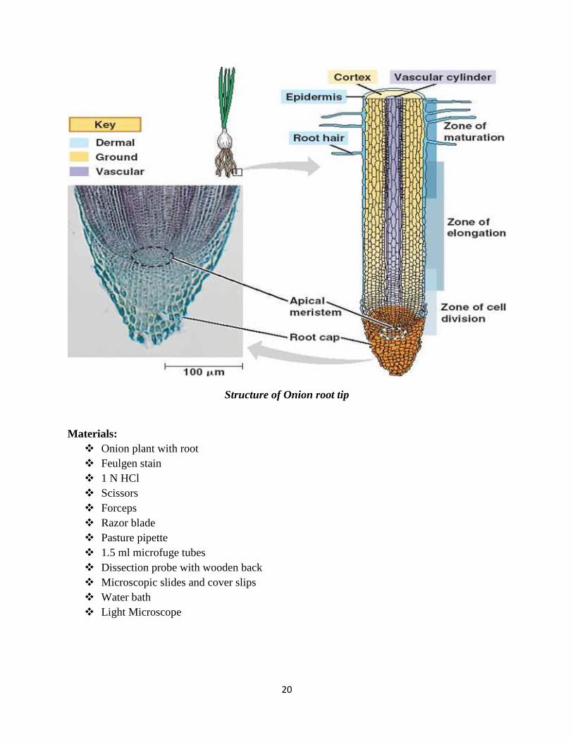
Structure of Onion root tip
Materials: Onion plant with root Feulgen stain 1 N HCl Scissors Forceps Razor blade Pasture pipette 1.5 ml microfuge tubes Dissection probe with wooden back Microscopic slides and cover slips Water bath Light Microscope
20
Procedure for preparing root tip squashes
While actively growing onions are present in the lab for you to observe, you will be provided with roots that have been previously harvested and treated with a fixative to stabilize the cells. You will work in groups of two for this lab exercise. The first step will be to ‘soften’ the roots so that they later can be spread on a microscope slide.
Using scissors, cut 2 roots tips about 1 cm long, and transfer them into a plastic micro-tube. (One of the rots will be an extra one.)
Fill the tube about 2/3 full with 1N HCl from a dropper bottle. *** Caution: Work with the HCl carefully, it is a strong acid. ***
Place the tube in a 60OC water bath, and allow the roots to incubate for 12 minutes. 4. After the 12 minute incubation period, remove the tube from the water bath.
Rinse the roots in H2O:
Using forceps carefully transfer the root tips to a small petri plate. Using a plastic ‘squeeze’ pipet, carefully remove the HCl from the micro-tube and
transfer it to the “discard flask”. Rinse the root tips 3 times with water from the dropper bottle, disposing of the rinses in
the discard flask.
Staining the chromosomes:
After removing the water from the third rinse, cover the root with the Feulgen stain. *** Caution: Although the Feulgen stain does not appear colored, it will strongly stain skin and clothing. ***
Incubate the roots in the stain for 12 minutes. During this time the very tip of the root will begin to turn red as the DNA stains the numerous small actively dividing cells at the tip.
Remove the stain and again rinse the roots:
Using a plastic ‘squeeze’ pipet, carefully remove the Feulgen stain and discard it in the discard flask. Again, rinse the root tips 3 times with water.
Preparing the root tip squash:
Transfer a root to the center of a clean microscope slide and add a drop of water. Using a razor blade cut off most of the unstained part of the root, and discards it. Cover the root tip with a cover slip, and then carefully push down on the cover slide with
the wooden end of a dissecting probe. Push hard, but do not twist or push the cover slide sideways. The root tip should spread out to a diameter about 0.5 – 1 cm.
21
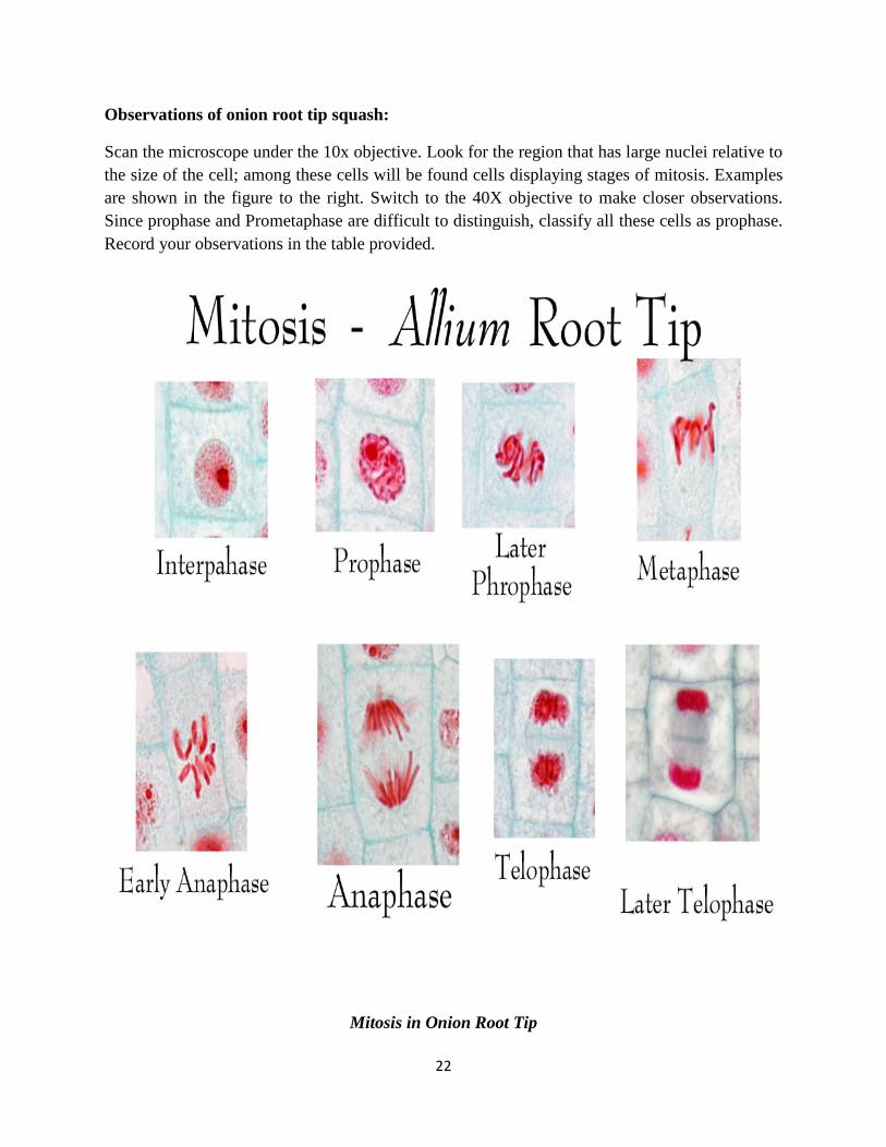
Observations of onion root tip squash:
Scan the microscope under the 10x objective. Look for the region that has large nuclei relative to the size of the cell; among these cells will be found cells displaying stages of mitosis. Examples are shown in the figure to the right. Switch to the 40X objective to make closer observations. Since prophase and Prometaphase are difficult to distinguish, classify all these cells as prophase. Record your observations in the table provided.
Mitosis in Onion Root Tip
22
Results Onion root tip squash
1. Find and draw a cell showing each stage of mitosis.
Prophase Metaphase Anaphase Telophase
2. What is a distinguishing visible feature of each stage of mitosis?
Prophase:
Metaphase:
Anaphase:
Telophase:
23
References: http://www.tutorvista.com/biology/microscope-parts-and-functions
http://www.microrao.com/simple_staining.htm
https://www.boundless.com/microbiology/textbooks/boundless-microbiology-textbook/microscopy-3/light-microscopy-29/general-staining-methods-243-4284/
http://www.cliffsnotes.com/study-guides/biology/microbiology/microscopy/staining-techniques
http://vlab.amrita.edu/?sub=3&brch=73&sim=208&cnt=2
http://www.microscopemaster.com/prokaryotes.html
http://biology.tutorvista.com/animal-and-plant-cells.html
http://www.cliffsnotes.com/study-guides/biology/biology/the-biology-of-cells/prokaryote-and-eukaryote-cell-structure
http://w3.marietta.edu/~biol/introlab/Onion%20root%20mitosis.pdf
Campbell, N.A. and J.B. Reece. 2005. Biology, Seventh Edition. Benjamin Cummings, San Francisco, CA.
http://www.biology4friends.org/cell-cycle-timing-lab1.html
http://vlab.amrita.edu/?sub=3&brch=188&sim=1102&cnt=2
http://sites.fas.harvard.edu/~bs50/Mit%20Mei%20Notebook.pdf
http://www.sparknotes.com/biology/cellreproduction/intro/section2.rhtml
http://vlab.amrita.edu/?sub=3&brch=188&sim=1102&cnt=2
http://study.com/academy/lesson/plastids-definition-structure-types-functions.html
http://faculty.valenciacollege.edu/tklenk/botany/labs/The%20Cell.htm
http://www.uq.edu.au/_School_Science_Lessons/UNBiol1.html#9.9.1
24
Related Documents