BIOTECHNOLOGY I – PROTEIN PURIFICATION BY ION EXCHANGE Written by Eilene Lyons Revised 2/25/10 4-1 LAB 4 PROTEIN PURIFICATION BY ION EXCHANGE STUDENT GUIDE GOAL The goal of this laboratory is to teach the basics of ion exchange chromatography, DNA restriction, and specific activity of enzymes. OBJECTIVES After this lab, the student will be able to 1. Describe the molecular action of Type II endonucleases. 2. Explain why cellular DNA is not degraded by the cell’s endonucleases. 3. Set up and use a chromatography column to purify a protein from cell extract. 4. Assay fractions from column chromatography of bacterial cells for DNA restriction enzyme concentration 5. Calculate the units of activity and the specific activity of an enzyme from experimental results. 6. Describe the difference between total activity and specific activity of an enzyme. 7. Apply knowledge of unit activity and specific activity when ordering enzymes. TIMELINE Day 1 – Prep, collect column fractions, cast agarose gel Day 2 – First Assay Day 3 – Second Assay and data analysis BACKGROUND Restriction Enzymes Bacteria use restriction enzymes to destroy invading virus DNA by cutting it into fragments along the sugar-phosphate backbone of the DNA. Immediately after DNA replication, bacteria protect their own DNA by adding methyl groups to restriction recognition sites recognized by their own restriction enzymes, preventing binding of these enzymes to the cellular DNA. Some restriction enzymes cut from a free end of the DNA (exonuclease), while others cut along the backbone (endonuclease). Since Type II endonucleases cut at sequence-specific sites and are the type of enzyme used most often in genetic engineering, they are referred to simply as restriction enzymes in the laboratory. Over 1500 different restriction enzymes have been found since the first was discovered by H. O. Smith and colleagues in 1968. Most restriction enzymes are either protein dimmers (two subunits of equal mass between 20,000 and 25,000 daltons) or single polypeptides of molecular weights from 30,000 to 35,000 daltons. Different restriction enzymes use different recognition sequences (usually palindromes) of DNA where they attach and cut. A palindrome is a word or phrase spelled exactly the same forward and backward. DNA palindromes involve both strands of the molecule, where the sequence on the bottom strand is the opposite of the complementary base pair sequence on the top. For example, GAATTC CTTAAG

Welcome message from author
This document is posted to help you gain knowledge. Please leave a comment to let me know what you think about it! Share it to your friends and learn new things together.
Transcript

BIOTECHNOLOGY I – PROTEIN PURIFICATION BY ION EXCHANGE
Written by Eilene Lyons Revised 2/25/10 4-1
LAB 4
PROTEIN PURIFICATION BY ION EXCHANGE
STUDENT GUIDE
GOAL
The goal of this laboratory is to teach the basics of ion exchange chromatography, DNA
restriction, and specific activity of enzymes.
OBJECTIVES
After this lab, the student will be able to
1. Describe the molecular action of Type II endonucleases.
2. Explain why cellular DNA is not degraded by the cell’s endonucleases.
3. Set up and use a chromatography column to purify a protein from cell extract.
4. Assay fractions from column chromatography of bacterial cells for DNA restriction enzyme
concentration
5. Calculate the units of activity and the specific activity of an enzyme from experimental
results.
6. Describe the difference between total activity and specific activity of an enzyme.
7. Apply knowledge of unit activity and specific activity when ordering enzymes.
TIMELINE
Day 1 – Prep, collect column fractions, cast agarose gel
Day 2 – First Assay
Day 3 – Second Assay and data analysis
BACKGROUND
Restriction Enzymes
Bacteria use restriction enzymes to destroy invading virus DNA by cutting it into fragments along
the sugar-phosphate backbone of the DNA. Immediately after DNA replication, bacteria protect
their own DNA by adding methyl groups to restriction recognition sites recognized by their own
restriction enzymes, preventing binding of these enzymes to the cellular DNA. Some restriction
enzymes cut from a free end of the DNA (exonuclease), while others cut along the backbone
(endonuclease). Since Type II endonucleases cut at sequence-specific sites and are the type of
enzyme used most often in genetic engineering, they are referred to simply as restriction
enzymes in the laboratory. Over 1500 different restriction enzymes have been found since the
first was discovered by H. O. Smith and colleagues in 1968. Most restriction enzymes are either
protein dimmers (two subunits of equal mass between 20,000 and 25,000 daltons) or single
polypeptides of molecular weights from 30,000 to 35,000 daltons. Different restriction enzymes
use different recognition sequences (usually palindromes) of DNA where they attach and cut. A
palindrome is a word or phrase spelled exactly the same forward and backward. DNA
palindromes involve both strands of the molecule, where the sequence on the bottom strand is the
opposite of the complementary base pair sequence on the top. For example,
GAATTC
CTTAAG

BIOTECHNOLOGY I – PROTEIN PURIFICATION BY ION EXCHANGE
Written by Eilene Lyons Revised 2/25/10 4-2
Different species have different restriction enzymes, so each has been named according
to the species where they were discovered. The Roman numeral denotes if the enzyme
was the first, second, third, etc., enzyme found in the species. Some restriction enzymes
recognize the same DNA sequence and are referred to as “isoschizomers.” The restriction
enzymes Cla I and Bsp 106 both attach to the DNA sequence ATCGAT and cut between
the T and C. Molecular biology product catalogues give isoschizomer lists and sequence
recognition sites for most enzymes used in the research laboratory. There are reports in
the scientific literature of restriction enzymes cutting at more than one recognition site
due to changes in the cation concentration, the pH of the solution, or the presence of
glycerol in the reaction mixture. This indiscriminate cutting is referred to as star activity,
and can also be caused when the restriction enzyme concentration is too high.
Some endonucleases, like EcoR V, cut leaving blunt ends. Others cut asymmetrically,
leaving cohesive or sticky ends – nucleotides that are not hydrogen bonded to other
complementary nucleotides. Once cut from within, DNA polymers separate as the
hydrogen bonds between the cuts break. See Table 1.
5’ CGAATG AATTCTACCCAAG 3’
3’ GCTTACTTAAG ATGGGTTC 5’
Table 1. Examples of Restriction Endonuclease Recognition And
Cutting Sites ( and spaces denote where the DNA backbone is cut.)
EcoR I: G AATTC G GATCC BamH I:
Escherichia coli, CTTAA G CCTAG G Bacillus amyloliquefaciens H
strain RY 13
Hind III: A AGCTT GAT ATC EcoR V (a blunt cutter):
Haemophilus TTCGA A CTA TAG Escherichia coli,
influenzae Rd strain RY 13
PRACTICE: Identify the restriction site in each DNA sequence below. Write the name of the
enzyme on the line beside each sequence. Mark the cutting sites with slashes. Draw a horizontal
line showing what hydrogen bonds will break, separating the two polymers of DNA.
A. B. TAGACTGAATTCAAGTCA ATACGTAGGATCCCTAAA
ATCTGACTTAAGTTCAGT ________ TATGCATCCTAGGGATTT ________
C. D.
TTACTGATATCCATATGC TCGTTAAGCTTATGCCAT AATGACTATAGGTATACG ________ AGCAATTCGAATACGGTA ________
Sticky
ends

BIOTECHNOLOGY I – PROTEIN PURIFICATION BY ION EXCHANGE
Written by Eilene Lyons Revised 2/25/10 4-3
Restriction of Lambda DNA by Eco RI
The most widely used method for determination of a restriction enzyme’s cutting ability
is to assay it using DNA from a small viral genome and running the resulting fragments
on an agarose gel for analysis. To assay for Eco RI activity in this lab, a sample of each
fraction collected by column chromatography will be incubated with chromosomal
Lambda viral DNA to see which of the fractions cuts the DNA. To determine cutting, the
DNA restrictions digestions will be applied to an agarose gel to determine if all the
known Eco RI sites are completely cut, giving an expected number of fragments on the
gel. There are 5 Eco RI sites in the Lambda chromosome, giving linear fragments of the
following sizes when cut: 21226, 7421, 5804, 5643, 4878, 3530 bp. The two fragments of
5804 and 5643 bp are so similar in size that there may not be separation to show two
distinct bands if the gel is not run long enough. Therefore, you may see either 5 or 6
bands on the gel, depending on how long it was electrophoresed and whether there was
enough Eco RI present to cut the DNA completely.
Ion Exchange Chromatography
Purification of restriction enzymes can be accomplished using Ion Exchange
Chromatography. In column chromatography, a resin or column packing compound, such
as polystyrene or a polysaccharide such as Sepharose, Sephadex, or Diethylaminoethyl-
Cellulose (DEAE-Cellulose), is “packed” into a column. The resin must be kept moist
with a buffer that covers it so that it does not dry out, preventing it from functioning
properly. The sample containing the protein of interest is applied to the top of the column
and allowed to enter the column as drops of the buffer are collected off the bottom (called
“fractions”). See Figure 1. The proteins in the sample bind to the charged ions of the
column, and the column is washed with buffer at a pH and salt concentration that
maintains the interaction of the proteins with the resin. The salt concentration of wash
buffer is gradually increased so that only the protein of interest binds tightly. As the salt
concentration is increased further, the protein of interest releases (elutes) from the resin
and will drip through the column. Measured fractions of wash and elution buffer are
collected and then tested for desired enzyme activity.
Figure 1. Steps of Ion Exchange Chromatography
Bound protein of interest
Other proteins

BIOTECHNOLOGY I – PROTEIN PURIFICATION BY ION EXCHANGE
Written by Eilene Lyons Revised 2/25/10 4-4
Calculating Total Activity The activity of an enzyme can be measure in two ways, as units of activity or as specific
activity. A Unit of activity is defined as the amount of enzyme that fully cuts a given
amount of substrate in a given amount of time under standard assay conditions. For
example, if the assay conditions are that 1 µg of DNA is used and that the reaction is to
be incubated for 1 hour, resulting in complete cutting of the DNA as determined by gel
electrophoresis, then the enzyme volume used is equivalent to 1 Unit of activity. See the
sample problem, below.
Calculating Specific Activity
Specific activity is the number of Units of activity per milligram of protein present in the
sample. Specific activity is a way of saying how much of the protein in the fraction is the
enzyme of interest, in this case, EcoR I. In industry, specific activity is a measure of
purity - how much enzyme there is per the total protein in the sample. This has a bearing
on how to select enzymes for purchase. You may find several forms of an enzyme from a
chemical supplier, some containing more Units of activity, while others show higher
specific activity. The amount of purity may be more important than the cost or more
important than how much you must use for your application, in which case, you would
opt to pay more and get the more highly purified enzyme rather than the cheaper, less
purified one. To calculate the specific activity of the enzyme from the sample problem,
above, you would first need to take a UV spectrophotometer absorbance reading to get
the milligrams of protein per milliliter. Suppose that the A280 was 0.234. Recall that the
spec reading at 280 nm is the milligrams of protein per milliliter that are present in a
sample. The calculation of specific activity would then be:
Sample Problem: By definition, one Unit of Eco R I enzyme activity is equal to the amount of enzyme that can digest 1 µg of genomic Lambda viral DNA in 60 minutes at 37°C. Units of activity are expressed as Units per milliliter of enzyme solution. A sample of the fraction is diluted 1 to 4. Ten microliters of the dilution showed complete
digestion of 1 g of Lambda DNA in 30 minutes. The amount of enzyme in that 10 µl of fraction (we don’t know its molar amount) is equivalent, therefore, to 1 Unit of activity. Multiply by the dilution factor (4) and by 2, for how much would be cut if you incubated for twice that long (since by definition 1 unit is how much enzyme it takes to cut the DNA in one hour).
(1U/10 l) x 4 x 2 = 8U/10 l = 0.8 U/l
To calculate how much is in 1 ml of the fraction:
(0.8 U/l) x 1000 l/ml = 800 U/ml
Specific Activity = (800 U/ml) ÷ 0.234 mg/ml = 3,418 U/ mg protein

BIOTECHNOLOGY I – PROTEIN PURIFICATION BY ION EXCHANGE
Written by Eilene Lyons Revised 2/25/10 4-5
Buying Enzymes for Research
Here are some points to consider before you buy an enzyme:
1. What is the application? Will the protocol make a difference?
2. How pure does the enzyme need to be, i.e., will other contaminants have an effect
on the experiment?
3. What is the size of the reaction for which the enzyme will be used? Is it better to
use a small amount rather than a large amount of enzyme in a single reaction in
order to get the substrate completely degraded?
4. How important is the cost? Is there a minimum amount you can spend or do you
want what is best for the purpose, regardless of the price?
5. How long does the enzyme keep? If it has a long shelf life, it may be better to buy
more so that you pay less per mg.
6. How many units will be needed based on previous tests?
LAB OVERVIEW In this lab, the restriction endonuclease Eco R I will be isolated from a bacterial cell
lysate by ion exchange chromatography. The fractions collected will be assayed by
incubation with Lambda chromosomal DNA. Units of activity and specific activity of the
isolated enzyme will then be calculated.
SAFETY GUIDELINES
Good Laboratory Practice requires wearing safety glasses and gloves. Use appropriate
safety precautions during gel electrophoresis as directed previously.
MATERIALS EQUIPMENT
Per class: Chromatography columns, one per Team
E. coli RY extract, lyophilized Ring stand with clamps, one per Team
DEAE-Cellulose Ten 13 x 100 mm test tubes per Team
10x Equilibration Buffer Gel electrophoresis units, one per Team
50% Glycerol 5 ml serological pipette and pump, one per Team
KCl Power supplies, one per two Teams
Eco RI Reaction Buffer Waterbath set at 37°C (for digestions); 65°C for
electrophoresis and 1.5 ml tube floats
Molecular grade water (Qualified water) UV Spectrophotometers, two
Lambda DNA Matched Quartz Cuvettes & cuvette rack
Lambda/Eco RI Marker Automatic micropipetters and tips
Eco RI Dilution Buffer Two microcentrifuges
10x Gel loading dye Microwave oven
50x TAE Electrophoresis Buffer Balances, spatula, weigh boats, cleaning brush
Agarose powder Ice buckets, ice
Ethidium Bromide [10 mg/ml] UV Transilluminator, camera, film
1 x TNE Buffer for UV analysis Dishes for transporting gels
Plastic wrap and parafilm
Lab diapers for staining station
5 ml serological pipette for rolling out bubbles
Sharpie markers
BIOTECHNOLOGY I – PROTEIN PURIFICATION BY ION EXCHANGE
Written by Eilene Lyons Revised 2/25/10 4-6
PROCEDURE
NOTE: The reagents you receive are for two assays. The kit contains enough
reagents for 6 Teams.
Part I. Prep
(Some of these steps may be performed by laboratory personnel.)
DEAE-Cellulose Matrix
Add 35 ml of 10x equilibration buffer (C) to the bottle of DEAE-Cellulose (B). Cap
tightly and place on a rocker or orbital shaker to hydrate for 30-60 minutes. Aliquot 6 ml
for each of the six Teams.
1x Equilibration Buffer
(NOTE: The 10x Equilibration buffer contains potassium phosphate, pH 7.4, EDTA, and
β-mercaptoethanol.) Mix the following and stir thoroughly:
350 ml dH2O
50 ml 10x Equilibration buffer (C)
100 ml 50% glycerol (D)
KCl Buffers
0.1 M KCl: use 0.75 g KCl and BTV of 100 ml with 1x Equilibration Buffer
0.2 M KCl: use 1.5 g KCl and BTV of 100 ml with 1x Equilibration Buffer
0.5 M KCl: use 3.75 g KCl and BTV of 100 ml with 1x Equilibration Buffer
E. coli Cell Extract containing Eco RI restriction enzyme 1. Re-hydrate the sample by adding 0.5 ml of ddH2O to tube component A and let sit for
5 minutes.
2. Mix by vortexing on high speed and transfer the entire contents to a 50 ml conical
tube. Rinse tube A six times – each time with 1 ml of 1x Equilibration Buffer and add
the rinse material to the 50 ml conical tube. Mix the contents well.
3. Label a tube for each of 6 lab Teams as “Cell extract” and add 1 ml of the re-hydrated
extract to each tube. Store this extract on ice.
Team 1
1. Obtain a test tube rack for each team. Label a 15 ml conical tube for each team as
“0.1 M KCl.” Dispense 6 ml of 0.1 M KCl into each tube and distribute to each
team’s test tube rack. Distribute racks.
2. Label a 1.5 ml tube for each team as “Rxn Buffer” and dispense 100 µl of Eco RI
Reaction Buffer (F) into each tube. Distribute to each team’s ice bucket.
Team 2
1. Set up an ice bucket for each team and turn on water bath – set at 37°C.
2. Label a 50 ml conical tube for each team as “1x Equilibration Buffer.” Dispense 35
ml of 1x Equilibration Buffer into each and distribute to each team’s ice bucket.

BIOTECHNOLOGY I – PROTEIN PURIFICATION BY ION EXCHANGE
Written by Eilene Lyons Revised 2/25/10 4-7
Team 3
1. Label a 1.5 ml microfuge tube for each team as “Lambda DNA” and dispense 100 µl
of Lambda DNA (H) into each tube. Distribute to each team’s ice bucket.
2. Label a 15 ml conical tube for each team as “0.2 M KCl.” Dispense 6 ml of 0.2 M
KCl into each tube and distribute to each team’s test tube rack (Team 2 may have the
racks at their bench).
Team 4
1. Label a 1.5 ml microfuge tube for each team as “10x Loading Dye” and dispense
100 µl of 10x Loading Dye into each tube. Distribute to each team.
2. Label a 15 ml conical tube for each team as “0.5 M KCl.” Dispense 6 ml of 0.5 M
KCl into each tube and distribute to each team’s test tube rack.
Team 5 (or Team 3, if there are only 4 Teams)
1. Label a 1.5 ml microfuge tube for each team as “-Eco RI Marker DNA” and
dispense 50 µl of Lambda/Eco RI Marker (I) into each tube. Distribute to each team.
(Note: is lab shorthand for Lambda.)
2. Label a 1.5 ml microfuge tube for each team as “Dilution Buffer” and dispense 250 µl
of Eco RI Dilution Buffer (J) into each tube. Place these tubes in the class freezer
box. They will not be needed until the second assay.
Each team should check to make sure they have the following at their bench:
Ice Bucket: On bench: In rack
*Q-Water 100 ul 10x Loading Dye 6 ml 0.1 M KCL
100 ul Rxn Buffer (F) 50 ul Eco RI Marker DNA 6 ml 0.2 M KCl
100 ul DNA (H)
[0.2 μg/μl]
1 chromatography column 6 ml 0.5 M KCl
1 ml Cell Extract (A) 1 ring stand with clamp 35 ml 1x Equilibration Buffer
250 ul dilution buffer (J) 9 glass test tubes (13 x 100 mm) 6 ml DEAE cellulose matrix
25 ul dilute DNA
[4 ng/μl] not required until first
assay
*Q-water = ddH2O = millipore H2O = molecular grade water
Automatic pipetters and tips
5 ml serological pipettes and pump
A supply of 1.5 ml microcentrifuge tubes
Beaker for used tips
REMEMBER to save all reagents for the next 2 labs when the assays will be conducted.
BIOTECHNOLOGY I – PROTEIN PURIFICATION BY ION EXCHANGE
Written by Eilene Lyons Revised 2/25/10 4-8
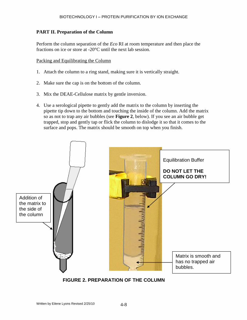
PART II. Preparation of the Column
Perform the column separation of the Eco RI at room temperature and then place the
fractions on ice or store at -20°C until the next lab session.
Packing and Equilibrating the Column
1. Attach the column to a ring stand, making sure it is vertically straight.
2. Make sure the cap is on the bottom of the column.
3. Mix the DEAE-Cellulose matrix by gentle inversion.
4. Use a serological pipette to gently add the matrix to the column by inserting the
pipette tip down to the bottom and touching the inside of the column. Add the matrix
so as not to trap any air bubbles (see Figure 2, below). If you see an air bubble get
trapped, stop and gently tap or flick the column to dislodge it so that it comes to the
surface and pops. The matrix should be smooth on top when you finish.
Matrix is smooth and has no trapped air bubbles.
Addition of the matrix to the side of the column
Equilibration Buffer DO NOT LET THE COLUMN GO DRY!
FIGURE 2. PREPARATION OF THE COLUMN

BIOTECHNOLOGY I – PROTEIN PURIFICATION BY ION EXCHANGE
Written by Eilene Lyons Revised 2/25/10 4-9
5. Place a small beaker underneath the column. This will be used to collect the wash
from the column as you drain it.
6. Remove the cap from the bottom of the column, allowing the DEAE-Cellulose
matrix to drain out, but DO NOT LET THE COLUMN GO DRY!"
7. After the column drains, add 25 ml of 1x equilibration buffer and let it drain again
until the meniscus is just above the surface of the matrix. Replace the end cap to stop
the draining.
8. Replace the cap so that a meniscus of the Equilibration Buffer remains on the top of
the matrix.
PART III. Collecting Fractions
1. Using a Sharpie marker, label eight 13 x 100 mm test tubes numbers 1 – 8. The chart
below indicates what each collected fraction in each tube will contain.
Tube Collected Fraction
1 Equilibration buffer
2 Equilibration buffer
3 0.1 M KCl
4 0.1 M KCl
5 0.2 M KCl
6 0.2 M KCl
7 0.5 M KCl
8 0.5 M KCl
2. Prepare a reference tube by adding 3 ml of water. Use this reference tube to mark the
3 ml point on each of the 8 collection tubes with a Sharpie marker. You will collect
eluent to this line in each of the tubes, one after another.
3. The meniscus should be just at the surface of the matrix. Use a P1000 automatic
micropipetter or a 1 ml serological pipette to slowly add 1 mL of the Eco RI cell
extract to the middle of the surface of the matrix so as not to disturb it. Remove the
cap and drain so that the buffer plus cell extract enters the surface of the matrix. See
photo, below.

BIOTECHNOLOGY I – PROTEIN PURIFICATION BY ION EXCHANGE
Written by Eilene Lyons Revised 2/25/10 4-10
4. Just as the surface of the liquid meets the surface of the matrix, slowly add 6 ml of
Equilibration Buffer to the inside of the column using a 5 ml serological pipette and
pump. Place tube number 1 under the column and collect 3 ml of the Equilibration
Buffer eluent. Switch to tube 2 and collect 3 mL more of eluent. Place these
collection tubes on ice and proceed directly to the next step without letting the
column drain out completely.
5. Add 6 mL of 0.1 M KCl to the top of the column and collect 3 mL fractions in tubes
3 and 4. Place on ice.
6. Add 6 mL of 0.2 M KCl to the top of the column and collect 3 mL fractions in tubes
5 and 6. Place on ice.
7. Add 6 mL of 0.5 M KCl to the top of the column and collect 3 mL fractions in tubes
7 and 8. Place on ice.
8. Wrap the top of each tube with Parafilm and place all into a test tube rack for storage
at -20°C until the next class session when the first assay will be performed.
9. Clean up your lab bench, dispose of the column, and proceed to casting your agarose
gel.
Add the cell extract just when the buffer reaches the top of the matrix.
BIOTECHNOLOGY I – PROTEIN PURIFICATION BY ION EXCHANGE
Written by Eilene Lyons Revised 2/25/10 4-11
PART IV. Casting an 0.8% Agarose Gel
Prepare an 0.8% agarose gel as done previously. Place it in a labeled zippered bag for
storage at 4°C until the next laboratory session.
PART V. First Assay of Eco RI Activity
Set up two water baths: one at 37°C and one at 65°C.
Note: Be careful not to get the Eco RI dilution buffer and Eco RI reaction buffer
confused. There are three different samples of DNA used in this lab: EcoR I markers,
chromosomal [0.2 ug/ul] Lambda DNA for the assays, and dilute Lambda DNA to run as
an uncut control on the gels.
1. Make sure the water baths are on and set and that your collected fractions from
the last lab are on ice and completely thawed.
2. Label nine 1.5 mL microcentrifuge tubes 1-9, Team number and date. To tube #9,
add the following:
Tube 9 – The Master Mix 270 µl molecular grade water
45 µl Eco RI Reaction Buffer
45 µl Lambda DNA [0.2 ug/ul]
360 µl total volume
3. Cap tube 9, flick to mix, and pulse spin (centrifuge briefly) to move liquid to the
bottom.
4. Transfer 40 µl of this master mix to each tube 1-8, leaving 40 µl in tube 9.
5. To tubes 1 – 8, add 10 µl of the corresponding Eco RI fraction to give a total
volume of 50 µl. Immediately pulse spin to mix and place into the 37°C water
bath for exactly 30 minutes. Tube 9 is the negative control and will have no
chromatography fraction added. Instead, add 10 µl of molecular grade water, cap
it and incubate with the rest of the test samples.
6. DO NOT DISCARD THE 9 TEST TUBES WITH THE CHROMATOGRAPHY
FRACTIONS. They should be returned to -20°C until the second assay during the
next laboratory session.
7. At the end of the 30 minute incubation period, remove the tubes from the 37°C
water bath and add 5 µl of 10x loading dye to all 9 tubes. The loading dye stops
the action of the restriction enzyme.
8. Set up the gel electrophoresis chamber using your 0.8% agarose gel and 1x TAE
Electrophoresis Buffer.

BIOTECHNOLOGY I – PROTEIN PURIFICATION BY ION EXCHANGE
Written by Eilene Lyons Revised 2/25/10 4-12
9. Heat the Lambda DNA standard markers and the Eco RI assays at 65°C for two
minutes. Pulse spin to move all liquid to the bottom of each tube. Place on ice and
load the gel in the following order:
Lane 1. 20 µl digestion #1 (Equilibration buffer fraction)
Lane 2. 20 µl digestion #2 (Equilibration buffer fraction)
Lane 3. 20 µl digestion #3 (0.1 M KCl fraction)
Lane 4. 20 µl digestion #4 (0.1 M KCl fraction)
Lane 5. 20 µl digestion #5 (0.2 M KCl fraction)
Lane 6. 20 µl digestion #6 (0.2 M KCl fraction)
Lane 7. 20 µl digestion #7 (0.5 M KCl fraction)
Lane 8. 20 µl digestion #8 (0.5 M KCl fraction)
Lane 9. 20 µl negative control #9 (no fraction added)
Lane 10. 20 µl DNA Eco RI Markers (+ control)
10. Add 25 μl of [10 mg/ml] Ethidium bromide to the buffer and run the gel at 90-95
volts until the dye has migrated at least 3 cm.
11. Photograph the gel as done previously. Give your gel photo to the instructor for
duplication for your lab partner(s) and for the instructor.
12. Analyze the results to determine which fractions contained enough Eco RI
restriction enzyme to cut the Lambda DNA.
PART VI. Second Assay of Eco RI Activity
Set up two water baths that will hold all group samples in each: one at 37°C and the other
at 65°C. Note: Be careful not to get the Eco RI dilution buffer and Eco RI reaction buffer
confused. There are three different samples of DNA used in this lab: EcoR I markers,
chromosomal [0.2 ug/ul] Lambda DNA for the assays, and dilute Lambda DNA to run as
an uncut control on the gels.
1. Make sure the water baths are on and up to temperature. Thaw your original
column chromatography fractions. Use the results from the first assay to
determine which fractions had cutting activity and pool these fractions. Pool
together only those fractions that gave significant cutting, not the ones that
showed only slight cutting as this will dilute those fractions with better activity.
2. Label two 1.5 µl microcentrifuge tubes as 1/2 dilution and as 1/3 dilution. Use the
Eco RI dilution buffer to mix the dilutions as followings:
1/2 dilution 1/3 dilution
Pooled fractions: 15 µl 10 µl
Eco RI dilution buffer 15 ul 20 ul
Total Volume 30 µl 30 µl

BIOTECHNOLOGY I – PROTEIN PURIFICATION BY ION EXCHANGE
Written by Eilene Lyons Revised 2/25/10 4-13
3. Label a fresh 1.5 ml microfuge tube as “Master Mix.” Prepare the master mix of
molecular grade water, Eco RI reaction buffer and Lambda DNA as follows:
“Master Mix” for digestion reactions:
Molecular grade water: 300 µl
Eco RI reaction buffer 50 µl
Lambda DNA [0.2 μg/μl] 50 µl
Total Volume: 400 µl
4. Label ten 1.5 ml microcentrifuge tubes as 1-10 with your Team number and date
on each. Use the following table to add components to each tube. One partner can
set up tubes 1 – 4, the other tubes 6 – 9. It is important to add the Eco R I to each
set of four tubes at about the same time so that some reactions do not have more
time to digest than others. Add master mix and water to tubes 5 and 10 last, just
in case there are slight pipetting errors in master mix aliquoting.
Last!
Digestion Tube: 1 2 3 4 5 6 7 8 9 10
Master Mix 40 ul 40 ul 40 ul 40 ul 40 ul 40 ul 40 ul 40 ul 40 ul 40 ul
Additional MG water ----- ----- ----- ----- 10 µl ----- ----- ----- ----- 10 µl
Eco RI 1/2 dilution 10 ul ----- ----- ----- ----- 10 ul ----- ----- ----- -----
Eco RI 1/3 dilution ----- 10 ul ----- ----- ----- ----- 10 ul ----- ----- -----
Undiluted Eco RI from
pool of fractions
----- ----- 10 ul 20 ul ----- ----- ----- 10 ul 20 ul -----
Total Volume 50 ul 50 ul 50 ul 60 ul 50 ul 50 ul 50 ul 50 ul 60 ul 50 ul
Time of Incubation at 37°C 30
min.
30
min.
30
min.
30
min.
30
min.
60
min.
60
min.
60
min.
60
min.
60
min.
5. Pulse spin all. Place all tubes in a 37°C water bath. Incubate tubes 1 – 5 for
exactly 30 minutes. Incubate tubes 6 – 10 for exactly 60 minutes.
6. While the samples are incubating or while the gel is running, determine the
amount of protein in your pooled fractions. Dilute 1 µl into each ml of 1x TNE
buffer in a quartz cuvette and take a UV spec reading at A280. A blank matching
quartz cuvette with 1x TNE buffer must be used. Take three readings of your
pooled sample and average the absorbance readings. An absorbance reading of 1
unit contains 1 mg of protein. Calculate the amount of protein per milliliter.
Record your readings and calculation in your lab notebook.
7. At the end of the incubation period, remove the tubes from the 37°C water bath
and add 5 µl of sample loading dye to each tube to stop the reaction.
8. Heat the Eco RI assays at 65°C for two minutes. Pulse spin to move all liquid to
the bottom of each tube. Place on ice until ready to perform gel electrophoresis
9. After all incubations are finished, heat the Lambda DNA markers and the Eco RI
assays at 65°C for two minutes. Pulse spin to move all liquid to the bottom of
each tube. Place on ice.
Last!

BIOTECHNOLOGY I – PROTEIN PURIFICATION BY ION EXCHANGE
Written by Eilene Lyons Revised 2/25/10 4-14
10. Insert the E-gel into the holder and run for 2 minutes with the comb still in the
wells. (NOTES: A steady, red light illuminates on the base if the gel is correctly
inserted. Press and hold either button until the red light turns to a flashing-green
light, indicating the start of the 2 minute pre-run. It will shut off automatically
after 2 minutes).
11. Remove the comb and load the samples in the following order:
Lane 1 20 µl Tube 1 – 1/2 dilution pooled fractions (30 min)
Lane 2 20 µl Tube 2 – 1/3 dilution pooled fractions (30 min)
Lane 3 20 µl Tube 3 – 10 µl undiluted pooled fractions (30 min)
Lane 4 20 µl Tube 4 – 20 µl undiluted pooled fractions (30 min)
Lane 5 20 µl Tube 5 – no Eco RI fraction (- control) (30 min)
Lane 6 20 µl Tube 6 – 1/2 dilution pooled fractions (60 min)
Lanes 7 20 µl Tube 7 – 1/3 dilution pooled fractions (60 min)
Lanes 8 20 µl Tube 8 – 10 µl undiluted pooled fractions (60 min)
Lane 9 20 µl Tube 9 – 20 µl undiluted pooled fractions (60 min)
Lane 10 20 µl Tube 10 – no Eco RI fraction (- control) (60 min)
Lane 11 20 µl Lambda – Eco RI MWt marker DNA (+ control) (60 min)
Lane 12 20 µl uncut Lambda DNA [4 ng/μL]
12. Run the gel for 30 minutes, view on the transilluminator, and document for your
lab notebook.
DATA ANALYSIS
Determine the units of activity per milliliter and the specific activity of the EcoR I
enzyme that you purified from the cell extract. The Lambda DNA added to the master
mix had a concentration of 0.2 μg/μl and 50 μl was added. Therefore, the total amount of
Lambda DNA in the master mix was 0.2 x 50 = 10 μg. Into each of the 10 assay tubes
was added 40 μl for a total of one microgram of Lambda DNA. This makes calculation of
the units of activity much easier.
10μg/400 μl = 1 μg/40 μl
In the second assay, the pooled fraction combo from the first assay was diluted to observe
the smallest dilution that gives complete cutting of 1 g of DNA during incubation. The
dilution you should use in your calculation for Specific Activity is the one that gave
complete cutting in the least amount of time with the least volume of pooled fraction
used.
BIOTECHNOLOGY I – PROTEIN PURIFICATION BY ION EXCHANGE
Written by Eilene Lyons Revised 2/25/10 4-15
QUESTIONS
1. If you do not see complete digestion in any of the second assays, can you
accurately calculate the total activity present? (HINT: Would you know how
much of the combined fraction is required to cut 1 g of the DNA?)
2. Explain how ion exchange chromatography works to isolate a particular cellular
product. Use lecture notes, books on reserve, Internet sources, etc., but document
the information with appropriate citations.
3. How is E. coli DNA protected from digestion by Eco RI while within the living
cell?
4. How many Eco RI sites are there in the Lambda genome? How many bands were
evident on the gel electrophoresis results? Explain any discrepancy.
5. Explain the difference between units of activity and specific activity in a sample.
6. If the specific activity of a restriction enzyme is 5U/mg, how much would be
required if you wanted to digest 1 μg of DNA? Show your work or explain your
reasoning.
7. Your lab is studying how to genetically engineer potatoes so that they contain
25% more starch and less water. This would be a financial benefit for the fast
food industry because if there is less water in potatoes, they absorb less oil as they
are French-fried, and can be advertised as lower in fat. The experiment will be an
assay to determine how much starch is present in potato cell extract. The
experiments will be carried out in volumes of no more than 3 ml with 1 gm of
ground potato added to each test. The product of the degradation of the starch will
be maltose, which will be tested for under standard conditions given by Sigma-
Aldrich. You will be in charge of preliminary testing on 1000 recombinant potato
plants that may be used to produce seed for growing commercial crops. It is an
important test and although costs must be kept to a minimum, good science is
what is most important. Either go online to the Sigma-Aldrich website or to their
printed catalogue (available in the lab prep area) and decide between products
A6814 and A6380 -amylase for use in this project. Remember to consider the
experimental application and the cost. Explain your choice.
8. Optional question: Do some research on your own and define Isoelectric Point
(PI). In ion exchange chromatography, how is the pH of the column packing
material you use related to the PI of the protein you are isolating?

BIOTECHNOLOGY I – PROTEIN PURIFICATION BY ION EXCHANGE
Written by Eilene Lyons Revised 2/25/10 4-16
Related Documents


