HAL Id: tel-03051788 https://tel.archives-ouvertes.fr/tel-03051788 Submitted on 10 Dec 2020 HAL is a multi-disciplinary open access archive for the deposit and dissemination of sci- entific research documents, whether they are pub- lished or not. The documents may come from teaching and research institutions in France or abroad, or from public or private research centers. L’archive ouverte pluridisciplinaire HAL, est destinée au dépôt et à la diffusion de documents scientifiques de niveau recherche, publiés ou non, émanant des établissements d’enseignement et de recherche français ou étrangers, des laboratoires publics ou privés. La protéine Prion cellulaire (PrPC) dans la mucoviscidose : Rôle dans le maintien de la barrière épithéliale bronchique Johanna Cormenier To cite this version: Johanna Cormenier. La protéine Prion cellulaire (PrPC) dans la mucoviscidose : Rôle dans le maintien de la barrière épithéliale bronchique. Biologie cellulaire. Université Grenoble Alpes, 2019. Français. NNT : 2019GREAV064. tel-03051788

Welcome message from author
This document is posted to help you gain knowledge. Please leave a comment to let me know what you think about it! Share it to your friends and learn new things together.
Transcript

HAL Id: tel-03051788https://tel.archives-ouvertes.fr/tel-03051788
Submitted on 10 Dec 2020
HAL is a multi-disciplinary open accessarchive for the deposit and dissemination of sci-entific research documents, whether they are pub-lished or not. The documents may come fromteaching and research institutions in France orabroad, or from public or private research centers.
L’archive ouverte pluridisciplinaire HAL, estdestinée au dépôt et à la diffusion de documentsscientifiques de niveau recherche, publiés ou non,émanant des établissements d’enseignement et derecherche français ou étrangers, des laboratoirespublics ou privés.
La protéine Prion cellulaire (PrPC) dans lamucoviscidose : Rôle dans le maintien de la barrière
épithéliale bronchiqueJohanna Cormenier
To cite this version:Johanna Cormenier. La protéine Prion cellulaire (PrPC) dans la mucoviscidose : Rôle dans le maintiende la barrière épithéliale bronchique. Biologie cellulaire. Université Grenoble Alpes, 2019. Français.�NNT : 2019GREAV064�. �tel-03051788�

THÈSE
Pour obtenir le grade de
DOCTEUR DE LA COMMUNAUTE UNIVERSITE GRENOBLE ALPES
Arrêté ministériel : 25 mai 2016
Présentée par
Johanna CORMENIER Thèse dirigée par le Dr Mohamed BENHAROUGA, MCU-HDR-UGA, Grenoble Préparée au sein du Laboratoire de Chimie et Biologie des Métaux Dans l'École Doctorale Chimie et Sciences du Vivant
La protéine Prion cellulaire (PrPC) dans la mucoviscidose : Rôle dans le maintien de la barrière épithéliale
bronchique
Thèse soutenue publiquement le 16 décembre 2019, devant le jury composé de :
Président : Dr. Jean-Jacque FEIGE, INSERM, CEA-Grenoble Rapporteur : Pr. Frédéric BECQ, CNRS, Université de Poitiers Rapporteur : Pr. Jean-Luc VILOTTE, INRA, Jouy-en-Josas Examinateur : Dr. Christelle CORAUX, INSERM, Reims Examinateur: Dr. Alexandre HINZPETER, INSERM, Paris Directeur de thèse : Dr. Mohamed BENHAROUGA, UGA, Grenoble


Remerciements
Je tiens à remercier chaleureusement Monsieur le Docteur Jean-Jacques Feige
d’avoir accepté de présider ma soutenance de thèse. Je remercie également
Messieurs les Professeurs Jean-Luc Vilotte et Frédéric Becq de m’avoir fait
l’honneur d’être les rapporteurs de mon manuscrit de thèse. Merci également à
Madame le Docteur Christelle Coraux et Monsieur le Docteur Alexandre
Hinzpeter de faire partie de mon jury de thèse en tant qu’examinateurs.
Je remercie profondément l’association "Vaincre La Mucoviscidose" ainsi que
tous ses membres et donateurs qui ont financé entièrement mes trois années de
thèse. Sans eux, cela n’aurait pas été possible !
Je voudrais remercier du fond du cœur ceux sans qui je ne serai pas là, mes
parents ! Merci Maman et merci Papa pour votre éducation, vos valeurs et tout ce
que vous m’avez transmis. Vous m’avez toujours soutenu dans mes choix qui,
parfois n’étaient pas les bons, mais qui m’ont permis de grandir et d’en arriver là
aujourd’hui ! Je n’oublie pas non plus mes sœurs. Certes, nous sommes toutes les
trois bien différentes mais c’est ça la famille, nan ?! En tout cas, merci à vous
quatre, je vous aime !
Merci au Docteur Mohamed Benharouga d’avoir dirigé mes travaux de thèse et de
m’avoir encadré pendant trois ans. Finalement, tu avais raison, c’est passé très vite
et j’ai l’impression d’être arrivé au labo hier ! Je pourrai même te raconter avec
précision ce lundi d’août 2016 quand je suis venu pour la première fois à Grenoble
pour mon entretien ! Je garde et je garderai un très bon souvenir de ma thèse et de
toute la gentillesse que tu as eu pour moi. J’en profite également pour remercier le
Docteur Nadia Alfaidy. Merci pour tes conseils, tes dépannages de kit qPCR, ton
aide à l’animalerie et tout ce que tu m’as apporté.
Je voudrais également remercier le Docteur et directeur du laboratoire Stéphane
Ménage de m’avoir accueilli dans ces locaux. Merci pour la bonne humeur
quotidienne que tu véhicules !
J’en viens maintenant à celles qui m’ont supportée chaque jour, pour le meilleur
et pour le pire ! Celle qui jette mes tubes d’ICP à la poubelle, celles à qui j’ai

régulièrement fais la manche pour un café car je n’ai jamais de monnaie, celles
qui m’ont fait mourir de rire lors de nos conversations "tupperware" dans le
poulailler, celle qui a enrichi ma collection de Monsieur Madame, celle qui … etc
etc je pourrai encore en écrire des pages entières … Bref, MERCI Peggy, Carole,
Mireille, Jennifer et Adeline ! Nos pauses café vont terriblement me manquer !
Je n’oublie pas non plus les "anciens" doctorants ou post-doctorants du labo,
Amel, Marianne, Jérôme, Xavier, Serge. Merci de m’avoir accueilli au tout début
et de m’avoir fait découvrir Grenoble ! Bon maintenant c’est sûr je ne me mettrai
jamais au ski, je ne pourrai jamais trahir ma côte Charentaise ! Serge, j’espère que
tu ne finiras pas congelé au fin fond du Canada et Marianne fais gaffe tu as bientôt
30 ans !!
Steve, ne t’inquiète pas, c’est ton tour ! Je ne te remercierai jamais assez pour ton
imagination ! Ton filleul Obama est aussi très reconnaissant ! Et merci pour tous
les bons moments au labo et en dehors !
Finalement, je voudrais remercier tous les membres du labo et cela comprend le
K et le K’ ! Oui oui vous avez bien lu ! J’ai franchis la frontière et j’en suis ressorti
vivante et surtout très contente d’avoir fait la connaissance de nombreuses belles
personnes ! Merci pour tous ces repas partagés et les discussions du midi parfois
houleuses, parfois complètement délirantes, mais toujours avec de la bonne
humeur ! Alors merci à tous les membres des équipes BioCat, BioCE, AFFOND,
SolHyCat, MCT et aussi et surtout BioMet, je ne vous oublierai pas et qui sait
j’espère qu’on se reverra !
Enfin pour finir, c’est toujours lorsque l’on ne s’y attend pas que les bonnes choses
arrivent. Tu es rentré dans ma vie au moment où j’en avais le plus besoin. C’est
simple, tu as mis des paillettes dans ma vie et sans t’en rendre compte tu as su
m’apporter tout le soutien dont j’avais besoin en fin de thèse. Alors merci Morgan
d’être là et d’être toi !

1
Table des matie res Liste des abréviations ...................................................................................................... 5
Table des figures .............................................................................................................. 9
Introduction ................................................................................................................... 15
Chapitre I : La mucoviscidose ..................................................................................... 17
I. Une maladie génétique des muqueuses ........................................................... 19
I.1. Histoire de la mucoviscidose .......................................................................... 19
I.2. Epidémiologie ................................................................................................. 20
II. Du gène à la protéine CFTR ............................................................................ 22
II.1. Le gène CFTR ................................................................................................. 22
II.2. Structure de la protéine CFTR ........................................................................ 22
II.3. Biosynthèse et maturation ............................................................................... 23
II.4. Dégradation ..................................................................................................... 24
III. Fonctions biologiques de CFTR ...................................................................... 25
III.1. Activité canal chlorure .................................................................................... 25
III.2. Activité régulatrice .......................................................................................... 25
Les canaux ORCC ............................................................................... 25
Les canaux ROMK .............................................................................. 26
Les canaux ENaC ................................................................................ 26
L’échangeur Cl-/HCO3- ....................................................................... 28
Les aquaporines ................................................................................... 29
III.3. Rôle dans le trafic intracellulaire .................................................................... 29
Le complexe SNARE .......................................................................... 29
La transcytose...................................................................................... 30
III.4. Implication dans la réponse inflammatoire ..................................................... 32
III.5. Implication dans les infections respiratoires bactériennes .............................. 34
III.6. Rôle dans les jonctions cellulaires .................................................................. 35
IV. Mutation du gène CFTR .................................................................................. 37
IV.1. Classification des mutations de CFTR .................................................... 37
Mutations de classe I ........................................................................... 37
Mutations de classe II .......................................................................... 38
Mutations de classe III ........................................................................ 39
Mutation de classe IV .......................................................................... 39

2
Mutations de classe V ......................................................................... 39
Mutations de classe VI ........................................................................ 40
IV.2. Modèles animaux pour l’étude de l’impact des mutations de CFTR ...... 40
IV.3. Thérapies moléculaires : Potentiateurs/correcteurs de CFTR ................. 43
IV.4. Description des atteintes cliniques .......................................................... 45
L’atteinte génitale ............................................................................... 46
L’atteinte pancréatique et biliaire........................................................ 47
L’atteinte métabolique ........................................................................ 47
L’atteinte pulmonaire .......................................................................... 48
Chapitre II : La barrière épithéliale bronchique ....................................................... 51
I. Les épithéliums au sein des poumons ............................................................. 53
II. Structure d’un épithélium bronchique sain ..................................................... 55
II.1. Les cellules ciliées .......................................................................................... 55
II.2. Les cellules sécrétrices .................................................................................... 55
II.3. Les cellules basales ......................................................................................... 56
III. Rôle de barrière protectrice ............................................................................. 56
III.1. Clairance mucociliaire .................................................................................... 56
III.2. Jonctions cellulaires ........................................................................................ 57
Les jonctions serrées ........................................................................... 57
Les jonctions adhérentes ..................................................................... 60
Les desmosomes ................................................................................. 61
IV. Cas de la mucoviscidose : perte de la barrière jonctionnelle bronchique ....... 62
IV.1. Augmentation de la viscosité du mucus, encombrement et perte de la
clairance mucociliaire ............................................................................................. 62
IV.2. Expression et activité des protéines jonctionnelles ................................. 63
Chapitre III : La protéine Prion cellulaire ................................................................. 67
I. Histoire de la protéine PrPC............................................................................. 69
I.1. Découverte des maladies à prions ................................................................... 69
I.2. Identification de l’agent infectieux responsable de la transmission et de la
propagation de la maladie : la protéine prion .......................................................... 70
II. Pathogénicité de PrPC associée aux Encéphalopathies Spongiformes
Transmissibles............................................................................................................. 72
II.1. Les différents types d’EST .............................................................................. 72
Les formes sporadiques ....................................................................... 72
Les formes génétiques ......................................................................... 73

3
Les formes acquises ou par transmission infectieuse .......................... 75
II.2. Les traitements et perspectives thérapeutiques ............................................... 76
III. Physiologie de PrPC ......................................................................................... 77
III.1. Le gène PRNP ................................................................................................. 77
III.2. Structure de la protéine PrPC ........................................................................... 78
III.3. Synthèse, maturation, recyclage et dégradation .............................................. 81
III.4. Expression et localisation ................................................................................ 84
III.5. Régulation ....................................................................................................... 84
IV. Fonctions de PrPC ............................................................................................ 87
IV.1. Rôle neuroprotecteur ............................................................................... 87
IV.2. Relation avec les métaux ......................................................................... 88
IV.3. Propriétés anti-oxydantes ........................................................................ 91
IV.4. Localisation aux jonctions cellulaires ..................................................... 91
IV.5. Implication dans l’inflammation ............................................................. 93
IV.6. Implication dans les infections ................................................................ 95
V. Modèles animaux pour l’étude de PrPC ........................................................... 96
V.1. Génération de modèles murins génétiquement modifiés pour le gène Prnp ... 97
V.2. Impact de l’extinction et de la surexpression du gène Prnp............................ 99
Objectifs ....................................................................................................................... 101
Résultats ....................................................................................................................... 107
I. Article 1 – Cellular prion protein (PrPC) is up regulated in cystic fibrosis :
Role of CFTR protein. .............................................................................................. 109
I.1. Contexte de l’étude ....................................................................................... 110
I.2. Résultats ........................................................................................................ 111
I.3. Conclusions ................................................................................................... 112
II. Article 2 – The cellular prion protein (PrPC) contributes to the acute
Pseudomonas aeruginosa lung infection in cystic fibrosis mice. ............................. 113
II.1. Contexte de l’étude ....................................................................................... 114
II.2. Résultats ........................................................................................................ 115
II.3. Conclusions ................................................................................................... 116
Conclusions générales ................................................................................................. 117
Perspectives de thèse ................................................................................................... 123
Bibliographie ............................................................................................................... 129
Annexes ........................................................................................................................ 155

4

5
Liste des abréviations
ABC ATP-Binding Cassette
ADN
AhR
Acide DésoxyriboNucléique
Aryl hydrocarbon Receptor
AMPA α-Amino-3-hydroxy-5-Méthylisoazol-4-Propionate
AMPc Adénosine Mono-Phosphate cyclique
Ap-1/2 Activator protein-1/2
AQ Aquaporine
Arg Arginine
ARM motif Armadillo
ARNm Acide RiboNucléique messager
ATP Adénosine Tri-Phosphate
Bax Bcl-2–associated X
Bcl-3 B-cell lymphoma protein 3
Bcl-6 B-cell lymphoma protein 6
BPCO Broncho-Pneumopathie Chronique Obstructive
CBAVD Congenital Bilateral Absence of the Vas Deferens
CF Cystic Fibrosis
CFTR Cystic Fibrosis Transmembrane conductance Regulator
Cl Chlorure
ClA
c-myc
Voie sécrétoire Alternative de Cl
cellular-myc proto-oncogene
COX-2 Cyclo-OXygénase-2
Cu Cuivre
deltaF508 Délétion de la phénylalanine en position 508
EGR4 Early Growth Response protein 4
ENaC Epithelial sodium Channel
ERAD Endoplasmic Reticulum-Associated protein Degradation
ERK Extracellular signal-Regulated Kinases
ESB Encéphalopathie Spongiforme Bovine
EST Encéphalopathie Spongiforme Transmissible
FI Filament intermédiaire
GDP Guanosine Di-Phosphate
GK Guanylate Kinase
Gly Glycine
GM-CSF Granulocyte-Macrophage Colony-Stimulating Factor
GMP Guanosine Mono-Phosphate
GPI Glycosyl-Phosphatidyl-Inositol
grm8 Glutamate Receptor Metabotropic 8

6
GSS syndrome de Gerstmann-Straussler-Scheinker
HCO3 Bicarbonate
Hdj Human DnaJ homologue
His Histidine
HRPT Hypoxanthine Phosphoribosyl-Transferase
HSE Heat Shock sequence Elements
Hsp Heat shock protein
ICAM-1 Intercellular Adhesion Molecule-1
IFF Insomnie Fatale Familiale
IFN Interféron
Ig Immunoglobuline
IκB Inhibitor of nuclear factor Kappa-B Kinase
IKK IκB Kinase
IL Interleukine
JAM Junctional Adhesion Molecule
JNK c-Jun N-terminal Kinases
loxP locus of crossing-over from Phage
LPS Lipopolysaccharide
MAGUK Membrane-Associated-GUnylate Kinases
MAPK Mitogen-Activated Protein Kinase
MBC Maladie de Bowel et de Crohn
MCJ Maladie de Creutzfeldt Jakob
MEF-2 Myocyte Enhancer Factor-2
Met Méthionine
MLS (HCCS) HoloCytochrome C Synthase
Mn Manganèse
MRE Metal Responsive Element
MyoD Myoblast Determination protein
MyT1 Myelin Transcription Factor 1
MZF-1 Myeloid Zinc Finger 1
Na Sodium
NBD Nucleotide Binding Domain
NEUROG1 Neurogenin-1
NFAT Nuclear Factor of Activated T-cells
NF-IL6 Nuclear Factor for IL-6 expression
NFκB Nuclear Factor kappa-light-chain-enhancer of activated B cells
NGF Nerve Growth Factor
NHERF Sodium-Hydrogen Exchanger Regulatory Factor
Ni Nickel
NLRP NOD-Like Receptor family Pyrin domain
NMDA N-Methyl-D-Aspartate

7
Oct-1/2 Organic cation transporter-1/2
ORCC Outwardly Rectifying Chloride Channel
ORF Open Reading Frame
p38 p38 mitogen-activated protein kinase
p53 tumor protein 53
p65 = RelA Transcription factor p65
PA Pseudomonas aeruginosa
PAMP Pathogen-Associated Molecular Pattern
PDZ Post synaptic density protein, Drosophila tumor suppressor,
Zonula occludens-1
PECAM-1 Platelet endothelial cell adhesion molecule-1
PI3K Phospho-Inositide-3 Kinase
PK A/C Protéine Kinase A/C
plcxd3 Phosphatidylinositol Specific Phospholipase C X Domain Containing 3
PMR Pattern Recognition Molecules
PNN PolyNucléaire Neutrophile
PrPC Protéine prion cellulaire
PrPSc Protéine prion Scrapie
RE Réticulum Endoplasmique
Rho/ROCK Rho-associated, Coiled-coil-containing protein Kinase
ROMK Renal Outer Medullary potassium channel
ROS Reactive Oxygen Species
RTE Résistance Trans-Epithéliale
SAF Scrapie-Associated-Fibrils
Sec61 Protein transport protein Sec61
SH3 Src Homology 3
SOD SuperOxyde Dismutase
Sp-1 Specificity protein-1
SPR Signal Particle Recognition
Src Proto-oncogene tyrosine-protein kinase Src
TcF/Lef Transcription Factor / Lymphoid enhancer-binding factor
tga20 transgene insertion a20
TJP Tight Junction Protein
TMD TransMembrane Domain
TNF-α Tumor Necrosis Factor alpha
Trp Tryptophane
UTP Uridine Tri-Phosphate
UV Ultra-Violet
vMCJ variante de la Maladie de Creutzfeldt Jakob
Wnt Wingless
wt wild-type

8
Zn Zinc
ZNF238 Zinc Finger protein 238
ZO Zonula Occludens

9
Table des figures
Figure 1 : Espérance de vie des patients atteints de mucoviscidose depuis 1930.
Graphique adapté à partir des données du registre de la fondation américaine pour
la mucoviscidose (CF foundation, https://www.cff.org/). ................................... 21
Figure 2 : Structure de la protéine CFTR. TMDs : TransMembrane Domain,
NBD : Nucleotide Binding Domain, Domaine R : Régulateur............................ 23
Figure 3 : Modèle proposé pour la transcytose de CFTR dans les épithéliums des
voies respiratoires. L’adressage basolatéral de CFTR se produit à partir
d'endosomes apicaux (a1) et le long de la voie de biosynthèse (a2). (b) La protéine
CFTR est endocytosé depuis la membrane basolatérale dans les endosomes de tri
basolatéral. (c) La protéine CFTR issue des BSE est conditionnée dans des
endosomes transcytotiques (TE) EEA1+ puis transportée au pôle apical par une
voie dépendante des microtubules (MT). (d) L'internalisation basolatérale, le
recyclage et la voie (e) transcytotique du récepteur de la transférrine (TfR) sont
indiqués par des lignes pointillées afin de comparer le trafic de CFTR et TfR. ASE:
endosome de tri apical; ARE: endosome de recyclage apical; BSE: endosome de
tri basolatéral; CRE: endosome de recyclage commun; EV: vésicules
endocytiques. D’après "Transcytosis maintains CFTR apical polarity in the face
of constitutive and mutation-induced basolateral missorting". Bidaud-Meynard A.
et al. J, Cell Science. 132-10. jcs226886 2019. ................................................... 32
Figure 4 : Classification des mutations de CFTR. Schéma adapté de "Progress in
therapies for cystic fibrosis. De Boeck K. et Amaral M.D. Lancet Respir. Med. 4-
8. p 662-674. 2016" et " Classification of CFTR mutation classes. Lima Marson
F.A. Lancet Respir. Med. 4. e37. 2016". ............................................................. 38
Figure 5 : Modèles murins existant pour l’étude de la protéine CFTR. Tableau
adapté de "Cystic fibrosis mouse models. Guilbault C. Am J Respir Cell Mol Biol.
36-1. p 1-7. 2007", "Pathophysiology of Gene-Targeted Mouse Models for cystic
fibrosis. Grubb B.R. and Boucher R.C. Physiological review. 79-1. p S193-S214.

10
1999" et "Un modèle murin mimant les manifestations pulmonaires de la
mucoviscidose vient d’être mis au point. Sauty A et Aubert J-D. Revue des
Maladies Respiratoires. 22. p 17-19. 2005". ........................................................ 41
Figure 6 : Résumé de résultats d'essais cliniques adaptés aux différentes mutations
de CFTR. VEMS : Volume Expiratoire Maximum par Seconde. ND : Non
Déterminé. Tableau adapté de "Cystic Fibrosis. Elborn J.S. Lancet. 388-10059. p
2519-2531. 2016". ................................................................................................ 43
Figure 7 : Apparition des symptômes cliniques de la mucoviscidose en fonction
de l’âge. ABPA : Aspergillose Broncho-Pulmonaire Allergique. Tableau adapté
de "Cystic Fibrosis. Elborn J.S. Lancet. 388-10059. p 2519-2531. 2016". ......... 46
Figure 8 : Comparaison entre une bronche saine et une bronche atteinte de
bronchectasie. Schéma adapté du "National Heart Lung and Blood Institute". .. 48
Figure 9 : Structure générale des poumons humains. .......................................... 53
Figure 10 : Transition cellulaire depuis la trachée jusqu’aux alvéoles. D’après "
Ganong's Review of medical physiology 25th Ed. Barret K.E. et al. 2015". ...... 54
Figure 11 : Localisation tissulaire des différentes cellules formant l’épithélium
bronchique humain............................................................................................... 56
Figure 12 : Schéma de l’organisation des jonctions cellulaires entre deux cellules
épithéliales. .......................................................................................................... 57
Figure 13 : Représentation schématique des jonctions serrées dans une cellule
épithéliale. D’après "Temporal and regional intestinal changes in permeability,
tight junction, and cytokine gene expression following ovariectomy‐induced
estrogen deficiency" Collins, F. L. et al. Physiological reports. 5-9. e13263. 2017.
.............................................................................................................................. 58

11
Figure 14 : Représentation schématique des jonctions adhérentes. D’après
"Régulation génétique de l’adhérence intercellulaire : Ou comment les cadhérines
sculptent la drosophile" Bécam, I. et Huynh, J.R. ipubli inserm MS num. 3, 2017.
............................................................................................................................. 60
Figure 15 : Représentation schématique des jonctions desmosomes. ................. 62
Figure 16 : Schéma de la barrière jonctionnelle bronchique dans le cas de la
mucoviscidose. PNN : Polynucléaire Neutrophile, ROS : Reactive Oxygen
Species. ................................................................................................................ 64
Figure 17 : Mutations du gène PRNP associées aux différentes EST génétiques.
GSS : Syndrome de Gerstmann-Straussler-Scheinker, IFF : Insomnie Fatale
Familiale, MCJ : Maladie de Creutzfeldt-Jakob. Schéma adapté de "Molecular
genetics of transmissible spongiform encephalopathies. Weissmann C.
J.Biol.Chem. 274-1, p 3-6. 1999". ....................................................................... 75
Figure 18 : (A) Schéma et (B) vue tridimensionnelle de PrPC. OR: Octarepeats
regions. D’après " Determinants of the Prion Protein N-Terminus and Its Adducts
with Copper Ions " Sánchez-López, C. et al. Structural. Int. J. Mol. Sci. 20-1. 2019.
............................................................................................................................. 79
Figure 19 : Mécanismes proposés pour le développement des EST acquises et
génétiques. SAF : Scrapie-Associated-Fibrils. Schéma adapté de "From prion
diseases to prion-like propagation mechanisms of neurodegenerative diseases.
Acquatella-Tran Van Ba I. et al. Int. J. Cell. Biol. 975832. 2013". ..................... 80
Figure 20 : Structure simplifiée de la protéine PrPC et des différents fragments
obtenus après clivage. Schéma adapté de "Cholesterol depletion and modification
of COOH-terminal targeting sequence of the prion protein inhibit formation of the
scrapie isoform. Taraboulos A. et al. J. cell biol. 129-1. p 121-132. 1995". ....... 83
12
Figure 21 : Positions des sites de régulation du gène PRNP (liste non exhaustive).
Schéma adapté de "Regulation of prion protein expression : a potential site for
therapeutic intervention in the transmissible spongiform encephalopathies. Haigh
C. L. et Brown D. R. IJBS 2-4. p 315-323. 2006". .............................................. 85
Figure 22 : Représentation de la coopération entre les protéines PrPC et AMPA
pour le transport intracérébral du zinc. Schéma adapté de "Neuronal zinc
regulation and the prion protein. Watt N.T. et al. Prion 7-3. p 203–208. 2013". 90
Figure 23 : Modèle proposé pour l’interaction de PrPC et de la kinase Src au sein
des desmosomes. PKG : Plakoglobine, PKP : Plakophiline, DP : Desmoplakine.
Schéma adapté de "Roles of the cellular prion protein in the regulation of cell-cell
junctions and barrier function. Petit C.S.V. et al. Tissue Barriers 1:2. e24377.
2013". ................................................................................................................... 92
Figure 24 : Stratégies génétiques utilisées pour invalider le gène Prnp par
recombinaison homologue dans des modèles murins. Rectangles rouges : cadres
de lecture ORF, rectangles blancs : régions non codantes, étiquettes jaunes ou
roses : séquences insérées, lignes pointillées : régions supprimées. Schéma adapté
de "Onset of ataxia and Purkinje cell loss in PrP null mice inversely correlated
with Dpl level in brain. Rossi D. et al. EMBO J. 20-4. p 694-702. 2001". ......... 97
Figure 25 : Caractéristiques et expression de PrPC dans les modèles murins de
surexpression de PRNP. ND : Non Déterminé. Tableau adapté de " Prion protein
(PrP) with amino‐proximal deletions restoring susceptibility of PrP knockout mice
to scrapie. Fischer M. et al. EMBO J. 15-6. p 1255-1264. 1996". ....................... 98
Figure 26 : Effets de l’extinction ou de la surexpression de la protéine PrPC dans
des modèles murins. ............................................................................................. 99
Figure 27 : Schéma récapitulatif du rôle cellulaire de la protéine PrPC dans la
mucoviscidose. ................................................................................................... 121

13
Figure 28 : Schéma récapitulatif illustrant les conséquences pulmonaires de la
dérégulation de PrPC dans la mucoviscidose ..................................................... 122

14

15
Introduction

16

17
Chapitre I : La mucoviscidose

18

Introduction : Chapitre I – La mucoviscidose
19
I. Une maladie génétique des muqueuses
I.1.Histoire de la mucoviscidose
Historiquement, les premiers symptômes de la mucoviscidose ont été rapportés au 15ème
siècle avec un proverbe irlandais qui disait "Malheur à cet enfant dont le baiser sur le front
a un goût salé, il est ensorcelé et va bientôt mourir". Les premières descriptions
pathologiques de la maladie ont été réalisées au cours du 16 et 17ème siècle où plusieurs
médecins, après avoir autopsiés les corps de jeunes enfants morts prématurément, décrivent
des atteintes pancréatiques ; notamment la présence d’un pancréas enflé, blanchâtre et
cirrhosé. Par la suite, le suisse Nils Rosen von Rosenstein (1706-1773) rapporte les cas de
douze enfants souffrant de diarrhées, de retards de croissance, d’asthénie et de gonflements
des membres pour lequels l’autopsie avait descellé un pancréas durci. Il qualifia alors cette
pathologie de maladie du flux cœliaque.
Au 19ème siècle, le pathologiste Carl von Rokitansky (1804-1878) ainsi que le pédiatre
Alois Bednar (1816-1888) décrivent le cas d’un fœtus de 7 mois présentant une perforation
de l’intestin grêle avec la présence de méconium dans le péritoine, puis le cas d’un nouveau-
né décédé d’obstruction intestinale 6 jours après sa naissance. L’autopsie montra que
l’intestin du nouveau-né était rempli de méconium visqueux et épais. Ce furent
probablement les deux premiers cas atteints d’iléus méconiale. Cependant, les médecins
s’interrogeaient sur le fait que certains enfants présentant les mêmes troubles intestinaux
ne mourraient pas si jeunes.
Il faudra attendre le début du 20ème siècle pour que le terme "fibrose kystique" (de l’anglais
cystic fibrosis) soit employé par le pédiatre Guido Fanconi von Grebe dans un article paru
en 1936. Il y décrit pour la première fois que l’insuffisance pancréatique et les problèmes
respiratoires chroniques observés chez certains enfants sont liés (1). En 1938, une autre
pédiatre, Dorothy Hansine Andersen, décrit les différences histologiques observées entre
un pancréas atteint de la maladie cœliaque et un pancréas atteint de fibrose kystique
pancréatique. Elle fait alors pour la première fois une distinction entre ces deux types
d’altération du pancréas qui présentent pourtant, des symptômes similaires (2). Quelques
années plus tard, le pédiatre pathologiste et père de l’oncologie pédiatrique Sidney Farber
invente le terme "mucoviscidose" car il pense que la fibrose kystique n’est pas seulement
un problème pancréatique mais qu’il s’agirait d’un trouble général de la production du

Introduction : Chapitre I – La mucoviscidose
20
mucus (3). Par la suite, D. H. Andersen réalisa une étude sur les familles des patients atteints
de mucoviscidose et elle en arriva à la conclusion qu’il s’agit d’une maladie génétique à
transmission autosomique récessive (4).
En 1948, suite à une canicule new-yorkaise, Paul di Sant’Agnese observe une forte
concentration en sels dans la sueur des patients atteints de mucoviscidose et publiera ensuite
un article démontrant les déséquilibres dans les concentrations en ions sodium et chlorure
dans cette maladie (5).
En 1981, le groupe de Michael Knowles montre des dysfonctionnements de l’épithélium
pulmonaire (6), ce qui conduit Paul Quinton, lui-même atteint de mucoviscidose, à
s’intéresser aux problèmes liés aux glandes sudoripares. Il constata alors que
l’imperméabilité des épithéliums aux ions chlorures (Cl-) signifiait que cet ion ne pouvait
pas être sécrété, pouvant ainsi expliquer l’augmentation de la viscosité du mucus au niveau
des différents épithéliums présents dans les organes touchés par la mucoviscidose (7).
Puis en 1989, le gène muté responsable de la mucoviscidose fut identifié et nommé CFTR
pour cystic fibrosis transmembrane conductance regulator (8) suivi par la découverte de la
première et principale mutation dans la mucoviscidose, la délétion de la phénylalanine en
position 508 (deltaF508).
En 1990, l’équipe de Michael J. Welsh a été la première à corriger le défaut de sécrétion
des ions chlorures par expression de la protéine CFTR sauvage dans les cellules épithéliales
atteintes de la mucoviscidose et à identifier l’existence d’une relation de cause à effet entre
les mutations du gène CFTR et les dysfonctionnements du transport des ions chlorures (9).
L’équipe de Riordan a par la suite apporté la preuve que CFTR est un canal Cl- activé par
l’AMPc (Adénosine Mono-Phosphate cyclique) et possède une conductance de 9
picosiemens (10).
I.2.Epidémiologie
La mucoviscidose est une maladie génétique rare (1/2500) à transmission autosomique et
récessive qui affecte principalement la population caucasienne (11),(12). Aux Etats-Unis,
environ 30 000 personnes sont affectées par la mucoviscidose, 4000 au Canada et en
Europe, un bébé sur 3000 naît avec la mucoviscidose. En France, le nombre de patients est
estimé à 7000 (13). Une personne sur 30 est porteur sain du gène CFTR muté et peut donc
transmette un allèle muté à sa descendance.

Introduction : Chapitre I – La mucoviscidose
21
La mucoviscidose affecte de manière égale les deux sexes et la population
mucoviscidosique est principalement jeune (14). Néanmoins, les progrès scientifiques de
ces dernières années en termes de diagnostic, de traitement, de prise en charge et de
recherche fondamentale ont permis d’améliorer considérablement la qualité de vie des
patients ainsi que leur espérance de vie. Cette dernière est passée de moins d’un an en 1938
à en moyenne 40 ans dans les années 2000 (15) (Figure 1). Ces chiffres sont en constante
évolution et montrent également que les enfants atteints de mucoviscidose nés après 2000
auront une espérance de vie de 50 à 60 ans.
Toutes ces données ont pu être obtenues grâce aux différents registres de patients mis en
place dans plusieurs états. En France, l’association Vaincre La Mucoviscidose a créé en
1992 l’observatoire national de la mucoviscidose, maintenant appelé registre français de la
mucoviscidose (RFM). Le développement de ces registres a permis une meilleure
connaissance des paramètres médico-sociaux des patients atteints de mucoviscidose, des
effets des traitements thérapeutiques ainsi que du coût engendré par les traitements.
Figure 1 : Espérance de vie des patients atteints de mucoviscidose
depuis 1930. Graphique adapté à partir des données du registre de la
fondation américaine pour la mucoviscidose (CF foundation,
https://www.cff.org/).

Introduction : Chapitre I – La mucoviscidose
22
II. Du gène à la protéine CFTR
II.1.Le gène CFTR
Le gène CFTR humain de 180kb se trouve sur le bras long du chromosome 7 en position
13 et est composé de 27 exons (Gene ID : 1080). Chez la souris, il est localisé sur le
chromosome 6 en position 6 et contient aussi 27 exons (Gene ID : 12638). L’expression de
CFTR est soumise à de fines régulations temporelles et tissulaires principalement
contrôlées par des réarrangements chromatiniens (16). En effet, des études ont permis de
mettre en évidence des fluctuations de l’expression de CFTR au cours de la vie fœtale et
également durant la vie adulte (17), avec notamment une nette diminution de l’abondance
pulmonaire post-natale (18).
II.2.Structure de la protéine CFTR
L’ARNm (Acide RiboNucléique messager) de 6,5 kb généré par le gène CFTR est traduit
en 1480 acides aminés qui constituent la protéine CFTR. En 2017, l’équipe de Jue Chen a
résolu la structure 3D de la protéine CFTR par cryomicroscopie électronique (19). CFTR
est principalement exprimée au pôle apical des cellules épithéliales et des glandes
exocrines. Les tissus épithéliaux, aussi appelés épithélium, tapissent de nombreux organes
comme les poumons (20),(21), les intestins (22), le système reproducteur (23) et sont
également présents au sein des glandes exocrines telles que le pancréas ou les glandes
salivaires (24). La localisation de CFTR au niveau neuronal a également été montrée (25).
La protéine CFTR appartient à la superfamille des transporteurs transmembranaires ABC
(ATP-Binding Cassette). Ce type de protéine est capable de fixer l’ATP (Adénosine Tri-
Phosphate) pour l’hydrolyser et utiliser l’énergie libérée afin de transporter
unidirectionnellement diverses substances de part et d’autre de la membrane plasmique.
Parmi les nombreuses protéines ABC, la protéine CFTR est la seule à fonctionner comme
un canal ionique (26). En effet, contrairement aux autres transporteurs de type ABC qui
tirent l’énergie de l’hydrolyse de l’ATP pour transporter les substrats contre leur gradient
chimique tel un transport actif primaire ligand-dépendant, CFTR conduit les anions dans
leur gradient électrochimique comme un transport actif secondaire grâce aux potentiels
électrochimiques de la membrane.
La protéine CFTR est constituée de deux domaines transmembranaires hydrophobes TMDs
(TransMembrane Domains) qui permettent la formation du canal au travers de la membrane
pour le passage des ions. Elle possède aussi deux domaines cytoplasmiques de liaison à

Introduction : Chapitre I – La mucoviscidose
23
l’ATP nommés NBD1 et NBD2 (Nucleotide Binding Domains). Ces domaines NBDs
partagent trois séquences nucléotidiques très conservées caractéristiques de la superfamille
des ABC: les motifs de Walker A et B (27) retrouvés chez de nombreuses protéines ayant
une activité ATPasique et qui permet la liaison de l’ATP, ainsi que la séquence C ou motif
signature des transporteurs ABC (LSGGQ) qui est très spécifique (Figure 2).
Cependant, la protéine CFTR possède un domaine globulaire cytoplasmique
supplémentaire, le domaine régulateur ou domaine R, qui comme son nom l’indique régule
l’activité de CFTR. Ce domaine R est codé par un exon unique, le treizième. Il contient des
sérines en position 660, 737, 795, et 813 qui doivent être phosphorylées par les protéines
kinases A et C (PKA et PKC) dépendantes de l’AMPc et qui induisent un changement de
conformation permettant l’ouverture du canal ionique CFTR (28).
II.3.Biosynthèse et maturation
La biosynthèse de CFTR, une glycoprotéine transmembranaire multi-domaines, s’effectue
au niveau du réticulum endoplasmique (RE) dans trois compartiments distincts : la
membrane du RE, la lumière du RE et le cytosol. En effet, la biogénèse de CFTR nécessite
Figure 2 : Structure de la protéine CFTR. TMDs : TransMembrane Domain,
NBD : Nucleotide Binding Domain, Domaine R : Régulateur.

Introduction : Chapitre I – La mucoviscidose
24
le repliement coordonné de ces différents domaines protéiques. La génération de la chaîne
polypeptidique de CFTR débute au niveau des ribosomes cytosoliques où elle est exportée
vers la membrane du RE via le complexe de translocation Sec61 (Protein transport protein
Sec61) grâce à sa séquence signale SPR (Signal Particle Recognition) et son récepteur SPR
(29),(30),(31). Parallèlement à son insertion dans la membrane du RE, des groupements N-
glycosylés sont ajoutés à la chaîne peptidique de CFTR, formant ainsi la forme core
glycosylé ou bande B sensible à l’enzyme de deglycosylation ; endo H (H). Par la suite, les
domaines TMDs et NBDs sont assemblés à la protéine CFTR naissante grâce à
l’intervention de nombreuses protéines chaperonnes cytosoliques et luminales telles que
Hsp40, Hsp70, Hsp90 (Heat Shock protein) ou encore la calnéxine (32),(31) assistées
également par les co-chaperonnes Hdj-1 et Hdj-2 (Human DnaJ homologue 1 et 2)
(33),(34). Enfin la protéine CFTR est exportée vers l’appareil de Golgi dans lequel sont
apportées des modifications post-traductionnelles comme la glycosylation du fragment
glycane qui génère la forme mature et entièrement glycosylée de CFTR appelée bande C
sensible à l’enzyme de deglycosylation endoF (F).
II.4.Dégradation
Lors de sa biosynthèse, si CFTR ne parvient pas à atteindre son repliement naturel et
correct, les chaperonnes telles que Hsp70 agissent pour recruter les enzymes ubiquitine
ligase E3 et/ou E4 qui vont finaliser le processus d’ubiquitination du CFTR qui sera pris en
charge par le système de dégradation cytoplasmique ubiquitine-dépendant, le protéasome
26S (35). Lorsque les protéines CFTR présentes à la membrane apicale sont recyclées, elles
sont alors endocytosées par les clathrines et transportées via des vésicules d’endocytose
(36). La détection de CFTR au niveau des cavéolines n’a jamais été observée et leur
inhibition ne modifie pas la quantité de CFTR à la membrane (37). Cependant, d’autres
travaux ont suggéré que l’internalisation de CFTR par les cavéolines serait indispensable
pour sa fonction de récepteur à Pseudomonas aeruginosa (PA). Il a été proposé que la
localisation de CFTR dans les domaines lipidiques raft de la membrane (membranaires
riches en cholestérol en glycosphingolipides, en protéines G et en cavéolines) était un point
essentiel pour la détection de PA et que le complexe CFTR-PA permettrait le
déclenchement de l’internalisation par les cavéolines (38),(39).

Introduction : Chapitre I – La mucoviscidose
25
III. Fonctions biologiques de CFTR
III.1.Activité canal chlorure
La protéine CFTR possède la fonction principale de canal anionique permettant le passage
de différents anions tels que les ions chlorures (Cl-) et les ions bicarbonates (HCO3-) au
travers de la membrane plasmique. Ces ions participent à l’équilibre hydrique et au
maintien d’un pH stable. Dès lors qu’il y a un déséquilibre ionique de part et d’autre de la
membrane, différents paramètres physico-chimiques de l’environnement cellulaire tels que
l’hydratation et la viscosité vont être modifiés.
Dans le cas de la mucoviscidose, les perturbations de l’activité canal chlorure vont conduire
à une déshydratation du mucus au sein des différentes muqueuses de l’organisme. Le mucus
devient visqueux, il ne circule pas ou peu et perd ses propriétés protectrices.
Cependant les dysfonctionnements de la protéine CFTR ne permettent pas d’expliquer
l’ensemble des désordres ioniques. Il a donc été suggéré un rôle régulateur à la protéine
CFTR permettant de contrôler l’activité d’autres canaux ioniques via notamment ses
domaines de liaisons PDZ (Post synaptic density protein, Drosophila tumor suppressor,
Zonula occludens-1) (40).
III.2.Activité régulatrice
Les canaux ORCC
Les canaux ORCC (Outwardly Rectifying Chloride Channel) possèdent une fonction de
canal chlorure permettant la sécrétion des ions Cl- et sont régulés par l’AMPc. Toutefois,
dans le contexte de la mucoviscidose, ces canaux sont inactifs et insensibles à la
phosphorylation par les protéines kinases A et C (41). Cette observation suggère que
l’activité de la protéine CFTR est nécessaire pour l’activation des canaux ORCC.
Des études plus approfondies ont confirmé la régulation d’ORCC par phosphorylation de
CFTR via un mécanisme autocrine/paracrine permettant le relargage d’ATP dans le milieu
extracellulaire. L’ATP ainsi libéré se fixe aux récepteurs purinergiques P2R et stimule
l’activité d’ORCC (42). Néanmoins, le mécanisme d’export de l’ATP par CFTR n’est pas
encore complétement établi.
Deux hypothèses ont été proposées selon lesquelles CFTR jouerait lui-même le rôle de
canal sécréteur d’ATP ou contrôlerait la formation de vésicules remplies d’ATP.

Introduction : Chapitre I – La mucoviscidose
26
D’un point de vue structural, il a été montré que les domaines NBD1 et R de CFTR sont
essentiels pour l’activité régulatrice des canaux ORCC (42).
Les canaux ROMK
Les canaux potassiques ROMK (Renal Outer Medullary Potassium Channel) sont localisés
au pôle apical des cellules rénales et participent au recyclage des ions K+. Des canaux
potassiques similaires aux ROMK sont également présents dans les voies aériennes (43).
Les canaux ROMK sont régulés notamment par le pH intracellulaire et l’ATP, qui sont
deux facteurs étroitement liés à CFTR.
Ainsi des études suggèrent que la protéine CFTR est capable de réguler négativement
l’activité des ROMK via le transport d’ATP intracellulaire. Cette inhibition est réalisée par
l’intermédiaire du complexe protéique régulateur NHERF1/NHERF2 (Sodium-Hydrogen
Exchanger Regulatory Factor 1/2) qui interagit avec les domaines PDZ de CFTR et ROMK
(44). Tout comme pour la régulation d’ORCC, le contrôle de ROMK requière les domaines
NBD1 et R de CFTR mais également le domaine TMD1 (45).
Les canaux ENaC
Dans la mucoviscidose, en absence de sécrétion d’ions Cl-, le canal ENaC (Epithelial
Sodium Channel) est hyperactivé par l’AMPc et entraîne l’absorption massive des ions Na+
au pôle apical des cellules épithéliales. En absence de CFTR, l’AMPc et la PKA activent
le canal ENaC. En revanche, lorsque les canaux CFTR et ENaC coexistent au sein d’une
cellule, l’effet de l’AMPc sur le canal ENaC s’inverse. En effet, l’AMPc stimule
préférentiellement le canal CFTR, qui lui-même inhibe l’activité d’ENaC. Ces phénomènes
de régulation entre différents canaux illustrent la dynamique d’un épithélium à interchanger
les mécanismes d’absorption et de sécrétion. Néanmoins, les bases moléculaires contrôlant
l’inhibition d’ENaC restent méconnues. Plusieurs travaux ont été menés et alimentent
diverses hypothèses :
Par homologie avec la régulation des canaux ioniques ORCC et ROMK, une étude a montré
que la libération d’ATP ou d’UTP (Uridine Tri-Phosphate) serait responsable de
l’inhibition du canal ENaC par activation des récepteurs purinergiques (46). Cependant, les
mécanismes moléculaires sont toujours méconnus et le transport d’ATP par CFTR reste
controversé. Ainsi, un an plus tard, les mêmes auteurs rapportent que leurs travaux ne
permettent pas de confirmer que l’inhibition du canal ENaC est le résultat de la libération
d'ATP induite par CFTR (47).

Introduction : Chapitre I – La mucoviscidose
27
Une interaction biochimique directe interviendrait entre CFTR et ENaC et contrôlerait
l’activité d’ENaC. En effet, les domaines NBD1 et R interagissent avec le domaine C-
terminal d’ENaC et la délétion du domaine NBD1 abolit la régulation négative d’ENaC
(48).
Une interaction indirecte liée au cytosquelette et au trafic intracellulaire interviendrait entre
CFTR et ENaC et permettrait à CFTR d’agir sur la régulation d’ENaC. Il est possible
d’envisager que l’actine pourrait être cet intermédiaire car il est connu que les canaux
CFTR et ENaC sont perturbés par les interactions avec les protéines du cytosquelette (49)
(50).
Cette dernière hypothèse semble plausible puisque des études réalisées sur l’épithélium du
canal sudoripare ont montré que dans ce contexte épithélial, l’effet de CFTR sur ENaC est
inversé et active la sécrétion de Na+ (51). De plus, ni la phosphorylation ni l’AMPc ne sont
nécessaires à l’activation d’ENaC. En revanche, un courant chlorure est essentiel pour cette
activation (52). L’ensemble de ces données laisse penser que l’adressage et le trafic des
protéines CFTR et ENaC via l’actine sont coordonnés, liés à la fonction canal Cl- et
dépendants du type de tissu épithélial.
D’autres études se sont intéressées aux gènes codant pour les trois sous-unités du canal
ENaC : α, β et γ. La sensibilité et l’efficacité de l’activité du canal ENaC requiert
l’ensemble de ces sous-unités (53).
Des mutations dans les gènes SCNN1A et B, qui codent les sous-unités α et β de la protéine
ENaC, respectivement, ont été associées aux maladies pulmonaires similaires à la
mucoviscidose en l'absence de mutations de CFTR (54). Lorsque SCNN1A est délété chez
la souris, le transport du sodium et la clairance mucociliaire sont abolis. La délétion de
SCNN1B ou SCNN1G (codant la sous-unité γ du canal ENaC), entraîne la mort par
hyperkaliémie (55), ce qui suggère que la sous-unité α du canal ENaC pourrait être plus
cruciale pour le bon fonctionnement du canal sodique dans les poumons.
A l’inverse, il a été montré que les mutations induisant une hypoactivité du canal ENaC
provoquent un pseudohypoaldostéronisme (56) (57), ce qui étend la profondeur de l'ASL
et accélère la vitesse de clairance mucociliaire.
Dès les années 2000, des chercheurs ont entrepris de comprendre le rôle de chacune des
sous-unités du canal ENaC. Pour cela, ils ont développé des oligonucléotides anti-sens et

Introduction : Chapitre I – La mucoviscidose
28
des ARN interférant (ARNi) ciblant SCNN1A, SCNN1B et SCNN1G (58) (59). Les
oligonucléotides anti-sens ciblant SCNN1A ont entraîné une diminution de la densité
membranaire apicale des canaux ENaC, et une réduction du courant et de la conductance.
Concernant les ARNi dirigés contre SCNN1B, ils ont entraîné une réduction de l’activité
de l’ENaC dans des cellules épithéliales bronchiques humaines, accompagné d’une
diminution du niveau d’ARNm de l’ENaC et du sodium intracellulaire. Plus récemment,
les travaux du groupe de Shuling Guo ont confirmé la diminution des sous-unités de
l’ENaC induites par les oligonucléotides anti-sens in vivo sur des modèles murins mimant
le phénotype de la mucoviscidose (60). Dans cette étude, les auteurs ont utilisé le modèle
transgenique de surexpression de la sous-unité β de l’ENaC qui entraine une
hyperabsorption de sodium ainsi qu’une déshydratation du mucus (61) et le modèle invalidé
pour le gène Nedd4-2 (Neural precursor cell expressed developmentally downregulated
gene 4-like) codant une ubiquitine ligase responsable de la régulation négative de l’ENaC
(62). Grâce à ces modèles, ils ont pu montrer que lorsque tous les niveaux des sous-unités
de l’ENaC sont diminués, la viscosité du mucus et le recrutement de neutrophiles sont
réduits et la fonction pulmonaire est améliorée.
L’échangeur Cl-/HCO3-
Les ions bicarbonates sont majoritairement présents dans les liquides de surface de
l’organisme où ils agissent comme des tampons pour maintenir le pH intracellulaire. La
protéine CFTR joue un rôle dans l’équilibre des ions bicarbonate en agissant comme un
canal sécréteur de HCO3- (63) ou en s’associant à un échangeur Cl-/HCO3
-. Dans la
mucoviscidose, lorsque CFTR est inactive ou non adressée à la membrane plasmique, sa
conductance pour les ions bicarbonates est inhibée ce qui engendre notamment des
dysfonctionnements de l’alcalinisation des sucs pancréatiques, à l’origine des insuffisances
pancréatiques observées chez les patients.
Pour pallier à ces perturbations de pH, il existe une régulation par des transporteurs ioniques
tels que CFTR et l’échangeur anionique AE2. La régulation de l’AE2 est induite par la
fixation du glucagon sur son récepteur entraînant une augmentation d’AMPc intracellulaire
puis l’activation de la PKA et la stimulation d’AE2. L’activation d’AE2 permet ainsi
l’entrée d’ions Cl- dans la cellule et la sécrétion d’ions HCO3-. Par la suite le Cl- importé
dans la cellule est pris en charge par CFTR qui l’excrète afin de maintenir un gradient de
Cl- et l’activation d’AE2 (64).

Introduction : Chapitre I – La mucoviscidose
29
Les aquaporines
Les aquaporines (AQs) sont des canaux transmembranaires permettant le passage
bidirectionnel de l’eau à travers la membrane plasmique. Lorsque la protéine CFTR est
mutée, les échanges d’eau effectués par les AQs entre le milieu extra- et intra-cellulaire
sont désordonnés, suggérant un lien entre CFTR et les AQs. CFTR co-localise notamment
avec les AQs 1 et 5 (65) où AQ1 co-localise également avec l’échangeur AE2. Il est
également connu que l’AMPc, activateur de CFTR, coordonne le trafic de vésicules vers la
membrane au sein desquelles se trouve CFTR, AQ1 et AE2 pour permettre la diffusion
d’eau (64).
III.3.Rôle dans le trafic intracellulaire
L’adressage de la protéine CFTR représente une étape cellulaire cruciale puisque l’absence
de CFTR à la membrane apicale implique un défaut de sécrétion de Cl-.
Le complexe SNARE
La régulation de l’adressage de CFTR implique les complexes multiprotéiques SNARE
(Soluble N-ethylmaleimide-sensitive factor Attachment protein REceptor) constitués
notamment par les protéines syntaxin, vtib (vesicle transport through interaction with t-
SNAREs homolog 1B) et VAMP (Vesicle-associated membrane protein). Les protéines
SNARE sont des composants clés dans le trafic intracellulaire des cellules eucaryotes et
sont impliquées dans les événements de fusion membranaire.
Au niveau neuronal, la syntaxine 1A est connue pour former un complexe avec les protéines
SNAP-25 et VAMP-2 régulant l’exocytose à la membrane plasmique présynaptique (66).
De manière intéressante, il a été montré que la syntaxine A1, accompagnée de SNAP-23,
un homologue non neuronal de SNAP-25, interagit également avec la partie N-terminale
de CFTR et inhibe le courant Cl- médié par CFTR (67) (68). En effet, la liaison de la
syntaxine A1 à l’extrémité N-terminal de CFTR empêche son interaction avec le domaine
R et/ou NBD1. Toutefois, le résidu essentiel à l’interaction spécifique entre CFTR et la
syntaxine A1 a pu être identifié, il s’agit d’un acide aminé hydrophile localisé dans le
domaine H3 (69).
Une autre protéine du complexe SNARE, la syntaxine 8 impliquée dans le trafic des
endosomes précoces aux endosomes tardifs, est connue pour interagir avec CFTR (70). La
surexpression de la syntaxine 8 inhibe l’activité canal Cl- de CFTR ainsi que son export
vers la membrane plasmique. De plus, les protéines syntaxine 8 et CFTR co-localisent dans

Introduction : Chapitre I – La mucoviscidose
30
des endosomes de recyclage Rab11 (Ras-related protein Rab-11), mais l’interaction entre
les deux protéines reste faible et suggère l’existence d’une protéine intermédiaire dans cette
interaction (71).
La protéine Csp (Cystein string protein) est connue pour interagir et réguler les complexes
SNARE comme la syntaxine 1A, 4 ou VAMP. De plus, les protéines Csp et CFTR
interagissent physiquement et fonctionnellement (72). Ainsi, Csp pourrait faire le lien entre
les protéines SNARE et CFTR et apporter un nouveau mécanisme de régulation en couplant
l’activation du canal CFTR avec son transport dans le trafic intracellulaire.
La transcytose
La transcytose est un type de transport transcellulaire dans lequel diverses molécules et
protéines subissent un transport intracellulaire. Les molécules transcytosées sont tout
d’abord endocytosées dans des vésicules depuis un pôle cellulaire, puis entraînées à travers
la cellule et exocytosées à un autre pôle de la cellule. Le phénomène de transcytose est
fréquent dans les cellules épithéliales.
Quatre voies de transport vésiculaires sont reconnues comme des évènements importants
dans l’expression basale de CFTR à la membrane plasmique : la sécrétion biosynthétique,
l’internalisation constitutive lente, le recyclage, ainsi que la dégradation qui comprend les
endolysosomes et les autophagosomes (73) (74) (75). Compte tenu de la longue demi-vie
cellulaire et membranaire du CFTR dans les épithéliums (76) (77), il est essentiel de
conserver un taux d’internalisation lent et un recyclage apical fidèle afin de maintenir la
fonction chlorure de CFTR en minimisant sa dégradation prématurée et son mauvais
adressage basolatéral.
Dans les épithéliums polarisés, les protéines membranaires apicales et basolatérales se
répartissent respectivement entre les endosomes de tri apical (ASE Apical Sorting
Endosome), de recyclage apical (ARE Apical Recycling Endosome), de tri basolatéral
(BSE Basolateral Sorting Endosome) et de recyclage commun (CRE Commun Recycling
Endosome) (78). Il a été montré que dans les cellules des voies respiratoires, le CFTR
internalisé apicalement, est capturé par les endosomes ASE-EEA1+ (Early Endosome-
Associated protein-1) et est recyclé via les endosomes ARE-Rab11+ (79) (80) de manière
dépendante de la PKA (81). Cependant, les mécanismes moléculaires de la dégradation
rapide de la protéine CFTR correctement conformée mais adressée de manière basolatérale
n'est pas connue. Toutefois, il a été montré que i) l’adressage forcé de CFTR vers la

Introduction : Chapitre I – La mucoviscidose
31
membrane plasmique basolatérale abolit sa fonction de sécrétion anionique transépithéliale
(82) et ii) en l'absence de transcytose apicale, l'accumulation basolatérale de CFTR réduit
l'entrée de Cl- basolatérale, provoquant une atténuation de la sécrétion d'eau et de la
clairance mucociliaire des épithéliums des voies respiratoires (83).
Très récemment, le groupe de Lukacs a montré que la transcytose apicale et basolatérale
représente des voies de trafic membranaire, auparavant non reconnues, contribuant
significativement au développement et au maintien de la polarité apicale de CFTR (84).
Les auteurs ont montré que la transcytose apicale du CFTR et l’internalisation basolatérale
jouent conjointement un rôle dans la neutralisation du mauvais adressage basolatéral
constitutif du canal CFTR. En effet, ces deux phénomènes maintiennent une quantité de
CFTR environ 30 fois plus élevée au niveau apical que basolatéral dans les cellules
épithéliales. Ils ont montré que la suppression des motifs PDZ n’a pas ou peu d’influence
sur l'expression apicale, l'activité de transport et la stabilité du CFTR. Néanmoins, la perte
du domaine PDZ induit une augmentation de l’internalisation basolatérale de CFTR et du
flux de transcytose apicale. Ainsi, ce groupe a proposé un modèle de trafic intracellulaire
physiologique de CFTR dans les cellules épithéliales des voies aériennes faisant intervenir
la transcytose apicale et basolatérale via les endosomes de tri apical (ASE) et basolatérale
(BSE), les endosomes de recyclage apical (ARE) et les microtubules (Figure 3).
Il est donc tout à fait possible d’imaginer que dans la mucoviscidose, les phénomènes de
transcytose de CFTR soient perturbés ou inhibés et ne parviennent pas à maintenir un taux
suffisant de CFTR fonctionnel à la membrane apicale. On peut aussi spéculer sur
l’existence de partenaires protéiques de CFTR pour son trafic intracellulaire.

Introduction : Chapitre I – La mucoviscidose
32
III.4.Implication dans la réponse inflammatoire
L’implication de CFTR dans la réponse inflammatoire dans le cas de la mucoviscidose est
toujours un sujet très controversé. En effet, l’inflammation observée chez les patients ne
serait que la conséquence des infections bactériennes persistantes. De plus, les thérapies
Figure 3 : Modèle proposé pour la transcytose de CFTR dans les épithéliums des
voies respiratoires. L’adressage basolatéral de CFTR se produit à partir
d'endosomes apicaux (a1) et le long de la voie de biosynthèse (a2). (b) La protéine
CFTR est endocytosé depuis la membrane basolatérale dans les endosomes de tri
basolatéral. (c) La protéine CFTR issue des BSE est conditionnée dans des
endosomes transcytotiques (TE) EEA1+ puis transportée au pôle apical par une voie
dépendante des microtubules (MT). (d) L'internalisation basolatérale, le recyclage
et la voie (e) transcytotique du récepteur de la transférrine (TfR) sont indiqués par
des lignes pointillées afin de comparer le trafic de CFTR et TfR. ASE: endosome de
tri apical; ARE: endosome de recyclage apical; BSE: endosome de tri basolatéral;
CRE: endosome de recyclage commun; EV: vésicules endocytiques. D’après
"Transcytosis maintains CFTR apical polarity in the face of constitutive and
mutation-induced basolateral missorting". Bidaud-Meynard A. et al. J, Cell
Science. 132-10. jcs226886 2019.

Introduction : Chapitre I – La mucoviscidose
33
existantes comme l’Orkambi© (prescrite chez les patients homozygotes pour la mutation
deltaF508) qui corrige et potentialise CFTR à la membrane plasmique n’ont pas d’effet sur
l’état inflammatoire des patients.
Néanmoins, les données actuelles issues de jeunes patients dépourvus de primo-infection
ainsi que celles obtenues sur des cultures cellulaires primaires stériles contredisent ce
dogme (85),(86) et suggèrent que l’origine de cette inflammation n’est pas infectieuse mais
moléculaire, en lien avec CFTR qui possède de nombreuses fonctions physiologiques.
A ce jour, les études portant sur l’origine de l’inflammation se sont surtout focalisées sur
la voie de signalisation NFκB (Nuclear Factor Kappa light chain enhancer of activated B
cells). En effet, l’activation de la voie NFκB induit une forte sécrétion d’interleukine 8 (IL-
8) qui elle-même entraîne l’afflux massif de polynucléaires neutrophiles par son effet
chimio-attractant : deux caractéristiques largement présentes chez les patients atteints de
mucoviscidose (87),(88).
Dans le cas de la mutation deltaF508, la voie NFκB est continuellement activée ce qui
conduit à un état inflammatoire chronique des voies respiratoires (89).
Une première hypothèse suggère que la protéine CFTR aurait un rôle de récepteur PMR
(Pattern Recognition Molecules) aux motifs spécifiques des pathogènes appelés PAMP
(Pathogen-Associated Molecular Pattern) comme le LPS (Lipopolysaccharide) des
bactéries Gram négatif. Les travaux de ce groupe ont montré que le LPS de PA est
spécifiquement reconnu et fixé par la protéine CFTR wild-type (wt) et que cette fixation
entraîne l’extraction du LPS à la surface de la bactérie. Puis le LPS est endocyté par les
cellules épithéliales qui vont rapidement induire la translocation et l’activation de la voie
NFκB (90). Cependant les résultats obtenus dans des cellules bronchiques humaines CFTR-
deltaF508 et des souris transgéniques CFTR-deltaF508 montrent une incapacité
d’internaliser le LPS de PA alors que la mutation deltaF508 induit une inflammation par le
NFκB. Cela suggère d’une part que l’endocytose du LPS nécessite un CFTR fonctionnel et
d’autre part que l’activation de la voie NFκB est régulée par d’autres mécanismes
cellulaires.
Une seconde hypothèse, à l’opposé de la première, suggère un rôle de CFTR dans la
régulation négative de la voie NFκB, via la phosphorylation de la protéine IκBα (Inhibitor
of kappa light polypeptide gene enhancer in B cells, alpha), un inhibiteur du NFκB (91).

Introduction : Chapitre I – La mucoviscidose
34
Dans cette étude, les auteurs ont co-exprimé in vitro de manière transitoire les promoteurs
de CFTR-wt et IL-8 ou CFTR-wt et NFκB couplés à la luciférase. Ils ont observé que
l'expression de CFTR-wt supprime les activités des promoteurs de l'IL-8 et du NFκB. Cette
observation a été confirmée dans les cellules épithéliale CFTR-deltaF508 transfectées
transitoirement avec le CFTR-wt et également in vivo chez des souris CFTR-/- après une
infection au LPS de PA (91). Ainsi ces auteurs ont suggéré que CFTR est un régulateur
négatif de la réponse immunitaire innée médiée par NFκB. Cependant, cette hypothèse
semble peu probable puisque la surexpression stable de CFTR-wt dans des cellules
épithéliales CFTR-deltaF508 ne permet pas de corriger l’hyperactivation de NFκB. Ceci
laisse penser que d’autres acteurs participent, en coopération ou non avec CFTR, à
l’activation de NFκB et que la correction par le CFTR-wt à elle seule ne suffit pas pour
réguler la voie NFκB.
III.5.Implication dans les infections respiratoires bactériennes
Les patients atteints de mucoviscidose présentent une sensibilité accrue aux infections
respiratoires et leurs voies aériennes sont chroniquement infectées. En effet, dans le liquide
broncho-alvéolaire de jeunes enfants, des pathogènes tels que Staphylococcus aureus ou
Haemophilus influenza sont détectés très rapidement. Puis dans des stades plus tardifs de
la maladie, les poumons des patients sont chroniquement colonisés par Pseudomonas
aeruginosa.
Les isolats de PA provenant de patients présentent fréquemment des mutations d’adaptation
à l'hôte, y compris dans le gène LasR. Le gène LasR est le principal régulateur
transcriptionnel du quorum sensing, un système de communication bactérien permettant
une expression génique coordonnée. L'exposition des voies aériennes à PA et/ou aux
exoproduits qu’elle sécrète a été associée à une perte d'intégrité épithéliale in vitro, à des
lésions tissulaires progressives et à une dégradation de la fonction pulmonaire in vivo (92)
(93) (94). Durant ces dernières années, le groupe d’Emmanuelle Brochiero a montré que
les exoproduits sous le contrôle de LasR altéraient la réparation épithéliale des voies
respiratoires après une blessure (95) et que l’inhibiteur de quorum sensing HDMF (4-
hydroxy-2,5-diméthyl-3 (2H) –furanone) supprime l'effet délétère de PA sur la réparation
des épithéliums respiratoires.
En plus des effets néfastes sur l’intégrité des voies respiratoires, les infections à PA ont
également une incidence sur l’expression et la fonction de CFTR. Plusieurs études ont

Introduction : Chapitre I – La mucoviscidose
35
montré que l'exposition à des souches ou à des exoproduits de PA réduisait l'expression de
CFTR-wt et la sécrétion de Cl- au niveau de la membrane apicale (96) (97) (98). Cependant,
les mécanismes par lesquels PA modifie CFTR semblent complexes et restent inconnus.
Certaines données indiquent que l'exposition aux exoproduits de PA réduit la synthèse de
la protéine CFTR et augmente sa dégradation (98). D’autres résultats montrent que
l'élastase contrôlée par le régulateur de quorum sensing LasB régule négativement les
niveaux d’expression de CFTR (99) et que l’ubiquitination et la dégradation accrue de
CFTR serait en partie dues au facteur Cif, un inhibiteur de CFTR présent dans les vésicules
de la membrane externe de PA (100). Il a été proposé que l'inhibition du recyclage
endocytaire de CFTR pourrait également être médiée par un mécanisme dépendant du
NHERF (97).
Enfin, il a été montré que la barrière épithéliale jonctionnelle bronchique est également
affectée dans la mucoviscidose et participe aux processus d’inflammation chronique. Dans
ce contexte inflammatoire, les cellules épithéliales bronchiques subissent un remodelage et
deviennent sécrétrices de médiateurs pro-inflammatoires. De plus, l’expression et l’activité
des protéines formant les jonctions cellulaires sont perturbées. Le domaine PDZ de la
protéine CFTR peut interagir avec certaines protéines des jonctions cellulaires et pourrait
via l’actine participer au maintien de la barrière jonctionnelle bronchique.
III.6.Rôle dans les jonctions cellulaires
Au sein de l’épithélium bronchique, la cohésion des cellules épithéliales est essentielle pour
garantir une barrière physique étanche et solide. Pour assurer cela, les cellules épithéliales
sont capables de former des complexes jonctionnels protéiques entres elles (ces complexes
jonctionnels protéiques sont décrit dans le chapitre II – La barrière épithéliale bronchique).
Dans le cas de la mucoviscidose, les mutations du gène CFTR entraînent des
dysfonctionnements de la barrière jonctionnelle bronchique qui n’assure plus ses fonctions
protectrices. Ce phénomène se caractérise par la perte de l’intégrité de l’épithélium
pulmonaire. Dans les tissus épithéliaux exprimant la protéine CFTR-deltaF508, la
Résistance Trans-Epithéliale (RTE) est drastiquement diminuée (101) tandis qu’une
augmentation de la perméabilité au mannitol est observée. De manière intéressante, lorsque
la mutation deltaF508 est corrigée in vitro par l’expression de la protéine CFTR sauvage,
la RTE est rétablie et la perméabilité au mannitol diminue. Ces phénomènes seraient liés à
la voie de signalisation Rho/ROCK (Rho-associated, Coiled-coil Containing Protein
Introduction : Chapitre I – La mucoviscidose
36
Kinase). En effet, l’activation de CFTR entraîne une dépolarisation de la membrane
plasmique. Celle-ci active la voie Rho/ROCK dans le but d’induire la phosphorylation des
chaînes de myosine et ainsi augmenter la perméabilité paracellulaire (102). Dans la
mucoviscidose où la protéine CFTR est absente, il a été proposé que l’hyperactivation du
canal ENaC est à l’origine de la dépolarisation de la membrane, qui devient perméable.
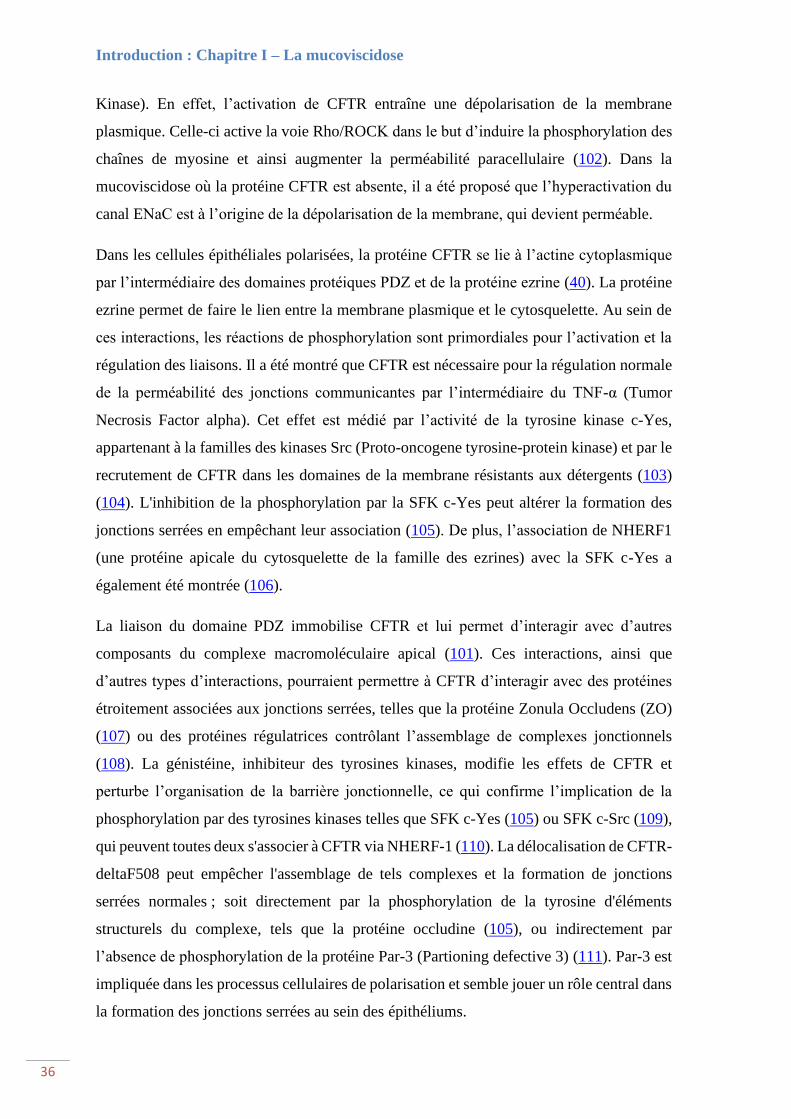
Dans les cellules épithéliales polarisées, la protéine CFTR se lie à l’actine cytoplasmique
par l’intermédiaire des domaines protéiques PDZ et de la protéine ezrine (40). La protéine
ezrine permet de faire le lien entre la membrane plasmique et le cytosquelette. Au sein de
ces interactions, les réactions de phosphorylation sont primordiales pour l’activation et la
régulation des liaisons. Il a été montré que CFTR est nécessaire pour la régulation normale
de la perméabilité des jonctions communicantes par l’intermédiaire du TNF-α (Tumor
Necrosis Factor alpha). Cet effet est médié par l’activité de la tyrosine kinase c-Yes,
appartenant à la familles des kinases Src (Proto-oncogene tyrosine-protein kinase) et par le
recrutement de CFTR dans les domaines de la membrane résistants aux détergents (103)
(104). L'inhibition de la phosphorylation par la SFK c-Yes peut altérer la formation des
jonctions serrées en empêchant leur association (105). De plus, l’association de NHERF1
(une protéine apicale du cytosquelette de la famille des ezrines) avec la SFK c-Yes a
également été montrée (106).
La liaison du domaine PDZ immobilise CFTR et lui permet d’interagir avec d’autres
composants du complexe macromoléculaire apical (101). Ces interactions, ainsi que
d’autres types d’interactions, pourraient permettre à CFTR d’interagir avec des protéines
étroitement associées aux jonctions serrées, telles que la protéine Zonula Occludens (ZO)
(107) ou des protéines régulatrices contrôlant l’assemblage de complexes jonctionnels
(108). La génistéine, inhibiteur des tyrosines kinases, modifie les effets de CFTR et
perturbe l’organisation de la barrière jonctionnelle, ce qui confirme l’implication de la
phosphorylation par des tyrosines kinases telles que SFK c-Yes (105) ou SFK c-Src (109),
qui peuvent toutes deux s'associer à CFTR via NHERF-1 (110). La délocalisation de CFTR-
deltaF508 peut empêcher l'assemblage de tels complexes et la formation de jonctions
serrées normales ; soit directement par la phosphorylation de la tyrosine d'éléments
structurels du complexe, tels que la protéine occludine (105), ou indirectement par
l’absence de phosphorylation de la protéine Par-3 (Partioning defective 3) (111). Par-3 est
impliquée dans les processus cellulaires de polarisation et semble jouer un rôle central dans
la formation des jonctions serrées au sein des épithéliums.

Introduction : Chapitre I – La mucoviscidose
37
Ces observations défendent l’implication de la protéine CFTR dans le contrôle et
l’organisation des complexes jonctionnels en association avec le cytosquelette des cellules
et les mécanismes de polarisation au sein des tissus épithéliaux. Néanmoins, les bases
moléculaires et les interactions précises permettant à la protéine CFTR d’exercer ses effets
restent inconnues.
IV. Mutation du gène CFTR
IV.1.Classification des mutations de CFTR
De nos jours, pas moins de 2000 mutations du gène CFTR ont été identifiées avec un degré
de sévérité variable. Ces mutations ont été classées en fonction du comportement
biologique de la protéique CFTR. La première classification des mutations de CFTR fut
proposée en 1993 par Welsh et Smith. Cette classification a tout d’abord permis de grouper
les mutations en quatre catégories selon le défaut de la protéine CFTR ; i) défauts de
synthèse, ii) défauts de maturation et de repliement, iii) défauts de régulation de l’ouverture
du canal CFTR et iv) défauts de conduction des ions Cl- (112).
La classification actuelle a été établie en se basant sur la première classification de Welsh
et Smith mais dénombre aujourd’hui six groupes de mutations (Figure 4) :
Mutations de classe I
Ces mutations conduisent à une absence partielle ou totale d’expression de la protéine
CFTR à la surface membranaire des cellules. Au niveau moléculaire, ces mutations sont
dues à la substitution d’un ou plusieurs nucléotides introduisant ainsi un codon stop
prématuré, des insertions ou des délétions. Ces mutations entraînent un décalage du cadre
de lecture ou des anomalies d’épissage aboutissant soit au saut complet d’un exon, soit à
une délétion importante du gène CFTR ou encore à un réarrangement génétique dans le
gène CFTR qui modifie la séquence d’un exon (113). Ces mutations sont retrouvées chez
5 à 10% des patients atteints de mucoviscidose dans le monde avec notamment les
mutations Trp1282X et Gly542X. Le X indique le codon stop qui remplace l’acide aminé.

Introduction : Chapitre I – La mucoviscidose
38
Mutations de classe II
Ces mutations sont à l’origine du trafic intracellulaire anormal de la protéine CFTR,
notamment en raison de repliement incorrect. Ces mutations, qui peuvent être retrouvées
Figure 4 : Classification des mutations de CFTR. Schéma adapté de "Progress
in therapies for cystic fibrosis. De Boeck K. et Amaral M.D. Lancet Respir.
Med. 4-8. p 662-674. 2016" et " Classification of CFTR mutation classes. Lima
Marson F.A. Lancet Respir. Med. 4. e37. 2016".
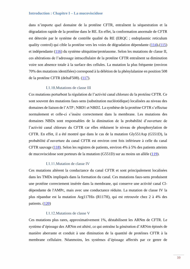
Introduction : Chapitre I – La mucoviscidose
39
dans n’importe quel domaine de la protéine CFTR, entraînent la séquestration et la
dégradation rapide de la protéine dans le RE. En effet, la conformation anormale de CFTR
est détectée par le système de contrôle qualité du RE (ERQC ; endoplasmic reticulum
quality control) qui cible la protéine vers les voies de dégradation dépendante (114),(115)
et indépendante (116) du système ubiquitine/protéasome. Selon les mutations de classe II,
ces altérations de l’adressage intracellulaire de la protéine CFTR entraînent sa diminution
voire son absence totale à la surface des cellules. La mutation la plus fréquente (environ
70% des mutations identifiées) correspond à la délétion de la phénylalanine en position 508
de la protéine CFTR (deltaF508). (117).
Mutations de classe III
Ces mutations perturbent la régulation de l’activité canal chlorure de la protéine CFTR. Ce
sont souvent des mutations faux-sens (substitution nucléotidique) localisées au niveau des
domaines de liaison de l’ATP ; NBD1 et NBD2. La synthèse de la protéine CFTR s’effectue
normalement et celle-ci s’insère correctement dans la membrane. Les mutations des
domaines NBDs sont responsables de la diminution de la probabilité d’ouverture de
l’activité canal chlorure du CFTR car elles réduisent le niveau de phosphorylation de
CFTR. En effet, il a été montré que dans le cas de la mutation Gly551Asp (G551D), la
probabilité d’ouverture du canal CFTR est environ cent fois inférieure à celle du canal
CFTR sauvage (118). Selon les registres de patients, environ 4% à 5% des patients atteints
de mucoviscidose sont porteurs de la mutation (G551D) sur au moins un allèle (119).
Mutation de classe IV
Ces mutations altèrent la conductance du canal CFTR et sont principalement localisées
dans les TMDs impliqués dans la formation du canal. Ces mutations faux-sens produisent
une protéine correctement insérée dans la membrane, qui conserve une activité canal Cl-
dépendante de l'AMPc, mais avec une conductance réduite. La mutation de classe IV la
plus répandue est la mutation Arg117His (R117H), qui est retrouvée chez 2 à 4% des
patients. (120)
Mutations de classe V
Ces mutations plus rares, approximativement 1%, déstabilisent les ARNm de CFTR. Le
système d’épissage des ARNm est altéré, ce qui entraîne la génération d’ARNm épissés de
manière aberrante et conduit à une diminution de la quantité de protéines CFTR à la
membrane cellulaire. Néanmoins, les systèmes d’épissage affectés par ce genre de

Introduction : Chapitre I – La mucoviscidose
40
mutations continuent de générer également un pool d’ARNm épissés correctement ce qui
permet d’éviter une absence totale de protéine CFTR fonctionnelle à la membrane
(121),(122).
Mutations de classe VI
Ces mutations sont souvent associées aux mutations de classe V puisqu’elles affectent
également la stabilité de CFTR mais au niveau protéique. Ces mutations provoquent une
troncation de l’extrémité C-terminale de la protéine CFTR, qui n’est cependant pas
indispensable pour la biosynthèse et la fonction de canal chlorure de CFTR. En revanche,
cette extrémité C-terminale semble jouer un rôle crucial dans la stabilité de la protéine
CFTR mature (123). De plus, il a été montré que l’absence de l’extrémité C-terminale
modifie les paramètres d’endocytose de CFTR qui de ce fait est dégradée prématurément.
En effet, l’augmentation du taux d’endocytose entraîne la réduction d’environ 50% de la
protéine CFTR dans la membrane plasmique (124).
De manière générale, les mutations de classe I, II et III sont associées à un phénotype sévère
de la maladie tandis que les mutations de classe IV, V et VI sont accompagnées de
symptômes moins sévères, avec un phénotype dit intermédiaire.
IV.2.Modèles animaux pour l’étude de l’impact des mutations de CFTR
Afin d’étudier les processus physiopathologiques de la mucoviscidose et de comprendre
les rôles multiples de CFTR, de nombreux modèles animaux ont été développés.
Cependant, le modèle murin reste le plus utilisé et a été largement développé puisque
l’homologie entre le gène CFTR humain et murin est de 78% et que la mutation deltaF508
se situe à la même position sur le gène murin (125).
Néanmoins, il est admis que ces modèles murins ne représentent pas fidèlement le
phénotype de la mucoviscidose car ils ne présentent pas ou peu de symptômes pulmonaires
de la mucoviscidose. En revanche, ces modèles permettent d’étudier les effets de
l’extinction ou des mutations de CFTR sur différents aspects moléculaires et dans un
environnement in vivo.
Plusieurs modèles murins ont été développés pour l’étude de CFTR (= souris CF pour
Cystic Fibrosis). Il est possible de distinguer deux types de modèles : les souris invalidées
pour le gène Cftr dites KO (Knockout) et les souris dans lesquelles une mutation du gène
Cftr a été introduite (Figure 5).

Introduction : Chapitre I – La mucoviscidose
41
De manière générale, la pathophysiologie intestinale de ces modèles murins est très
similaire à celle observée chez l’Homme. Ces souris présentent à la fois des défauts de
sécrétion macromoléculaire avec un épaississement du mucus intestinal et des défauts de
transport ionique avec une sécrétion de Cl- et HCO3- réduite ou absente et une absorption
accrue de Na+ (126). Ces modèles d’études sont généralement utilisés pour étudier la
physiologie fondamentale du transport des ions.
Phénotype
Souris Cible / mutation Resp. Dig. Fond
Génétique
Réf.
Kn
ock
ou
t
CFTRtm1Unc Codon stop dans
l’exon 10
+ +++ C57Bl/6-
129/Sv,
BALB/c-
129/Sv, B6D2-
129/Sv
(127)
CFTRtm1Hgu Insertion dans l’exon
10
- +
MF1-129/Sv
(128)
CFTRtm1Cam Insertion dans l’exon
10
- +++ MF1-129/Sv,
C57Bl/6-129/Sv
(129)
CFTRtm1Bay Duplication
insertionnelle dans
l’exon 3
- +++
C57Bl/6-129/Sv
(130)
CFTRtm3Bay Codon stop dans
l’exon 2
- +++ C57Bl/6-129/Sv (131)
CFTRtm1Hsc Insertion dans l’exon
1
- +++ CD1-129/Sv (132)
Mu
tati
on
s
CFTRtm1Kth Insertion de la
mutation F508del
dans l’exon 10
- +++
C57Bl/6-129/Sv
(133)
CFTRtm1Eur Insertion de la
mutation F508del
dans l’exon 10
- +
FVB-129/Sv
(134)
CFTRtm2Cam Insertion de la
mutation F508del
dans l’exon 10
- +++ C57Bl/6-129/Sv (135)
CFTRtm1G551D Insertion de la
mutation G551D dans
l’exon 11
- ++ CD1-129/Sv (136)
CFTRtm2Hgu Insertion de la
mutation G480C dans
l’exon 10
- +++ C57Bl/6-129/Sv (137)
Figure 5 : Modèles murins existant pour l’étude de la protéine CFTR. Tableau adapté
de "Cystic fibrosis mouse models. Guilbault C. Am J Respir Cell Mol Biol. 36-1. p 1-
7. 2007", "Pathophysiology of Gene-Targeted Mouse Models for cystic fibrosis.
Grubb B.R. and Boucher R.C. Physiological review. 79-1. p S193-S214. 1999" et "Un
modèle murin mimant les manifestations pulmonaires de la mucoviscidose vient
d’être mis au point. Sauty A et Aubert J-D. Revue des Maladies Respiratoires. 22. p
17-19. 2005".

Introduction : Chapitre I – La mucoviscidose
42
Au niveau pulmonaire, il a d’abord été considéré que les animaux examinés étaient trop
jeunes et élevés dans un environnement semi-stérile, pour développer des problèmes
respiratoires mais que si cette barrière semi-stérile était levée la pathologie des voies
aériennes se manifesterait (de la même manière que lorsque les nourrissons atteints de
mucoviscidose naissent et s’exposent aux pathogènes).
Cependant, cette hypothèse n’a pas pu être étayée puisque l'histologie des poumons de
souris CF est normale et ne présente pas de colmatage des bronches dû au mucus épais
(136),(128). En effet, les études avec des souris CFTRtm1Unc vieillissantes n’ont pas permis
de soutenir l'hypothèse selon laquelle des souris CF âgées et élevées dans un environnement
moins stérile pourraient présenter des atteintes respiratoires.
Néanmoins, il a été signalé que des souris CFTRtm1Hgu exposées longuement et de manière
répétée à des nébulisations de Staphylococcus aureus développaient des anomalies
pulmonaires. En l’occurrence ces souris présentaient une incidence significativement plus
élevée d'hyperplasie à cellules caliciformes, de rétention de mucus et de bronchiolite par
rapport aux individus non infectés (138). De plus, dans ces modèles exposés à des
pathogènes, une augmentation du nombre de colonies bactériennes a été observée,
suggérant un défaut de clairance pulmonaire.
D’autres travaux ont montré que les souris CF ne présentent aucune hyperabsorption de
Na+ et sécrètent significativement des ions Cl- en réponse à la forskoline (un activateur de
CFTR) (139). L’absence d’hyperabsorption du sodium peut être expliqué par une faible
expression de CFTR-wt dans les souris sauvages. Ainsi CFTR-wt n’aurait que peu
d’influence sur l’absorption du sodium et son invalidation dans les souris CF n’aurait pas
ou peu d’impact. La sécrétion normale d’ions Cl- chez les souris CF serait médiée par le
calcium intracellulaire indépendamment de CFTR. Cependant, la stimulation par des agents
augmentant le calcium intracellulaire (ATP, ionomycine) n’induit pas de sécrétion plus
importante de chlore (140) hormis pour les modèles CFTRtm2Cam et CFTRtm1G551D
(135),(136).
Il semblerait que dans la trachée murine, la voie sécrétoire alternative de Cl- (ClA) soit
beaucoup plus dominante que la voie CFTR médiée par l’AMPc et que, dans certains cas,
la voie ClA soit régulée positivement dans les tissus des voies respiratoires des souris CF.

Introduction : Chapitre I – La mucoviscidose
43
Enfin, il a été montré que les souris CFTRtm1Unc ne présentent aucun marqueur de
l’inflammation au niveau pulmonaire (127) hormis une augmentation du taux basal d’IL-8
dans le liquide de surface bronchique (141).
IV.3.Thérapies moléculaires : Potentiateurs/correcteurs de CFTR
Depuis l’identification du gène CFTR et des dysfonctions de la protéine CFTR, de
nombreuses études ont été menées dans le but de rechercher des molécules ayant un effet
sur la protéine CFTR. L’activité de la protéine CFTR est connue pour être régulée
principalement par phosphorylation/déphosphorylation. Ainsi, des composants naturels et
inducteurs de phosphorylation tels que les alkylxanthines (caféine, théophylline) ont été les
premières molécules modulatrices de CFTR identifiées (142).
D’autres molécules naturelles comme la curcumine, l'escine, extraite du marronnier, ou
encore le resveratrol ont été étudiées pour leurs propriétés inhibitrice de kinase, anti-
inflammatoire et anti-oxydante respectivement mais sans effet significatif in vivo. Il a aussi
été montré que le N-butyldeoxynojirimycin, appelé miglustat (Zavesca®) corrige le
transport des ions Cl- et Na+ en restaurant le trafic cellulaire de CFTR-deltaF508, mais ces
Classe
Molécule
Chlore
sudoral
Réponses cliniques
VEMS Exacerbation
pulmonaire
Gly551Asp III Ivacaftor Diminution
de 50%
Augmentation
de 10%
Diminution de
40%
Gly551Ser,
Gly178Arg,
Gly1244Glu,
Gly1349Asp,
Ser549Asn,
Ser549Arg,
Ser1251Asn,
Ser1255Pro
III
Ivacaftor
Diminution
de 50%
Augmentation
de 10%
ND
Arg117His IV Ivacaftor Diminution
de 25%
Augmentation
de 3%
ND
Gly542X I Ataluren = = Pas d’effet
Phe508del II Ivacaftor
+
Lumacaftor
Diminution
de 8%
Augmentation
de 3%
Diminution de
30%
Figure 6 : Résumé de résultats d'essais cliniques adaptés aux différentes mutations de
CFTR. VEMS : Volume Expiratoire Maximum par Seconde. ND : Non Déterminé.
Tableau adapté de "Cystic Fibrosis. Elborn J.S. Lancet. 388-10059. p 2519-2531.
2016".

Introduction : Chapitre I – La mucoviscidose
44
effets observés in vitro et lors des phases précliniques n’ont pas été confirmés lors des
essais cliniques de phase II (143).
Par la suite, le développement du screening à haut débit a permis d’identifier de nombreuses
molécules prometteuses qui ont été catégorisées en deux grandes familles de molécules :
les potentiateurs et les correcteurs de CFTR.
La société Vertex Pharmaceuticals (Boston, USA) a notamment développé deux molécules.
La première, VX-809, plus communément appelée Lumacaftor, est un correcteur de CFTR
qui permet de restaurer le trafic cellulaire de CFTR-deltaF508, de corriger son repliement
et de la stabiliser à la membrane (144). Les patients homozygotes pour deltaF508 ont
montré une diminution significative du chlore dans leur sueur après quatre semaines de
traitement, néanmoins aucune amélioration de la fonction respiratoire n’a pu être observée.
La deuxième molécule, le VX-770 ou Ivacaftor (Kalydéco®), est un potentiateur de CFTR
approuvé chez les patients porteurs de la mutation G551D (4-5% des patients) et qui permet
d’améliorer l’ouverture du canal CFTR-deltaF508 en combinaison avec un traitement au
VX-809. Ainsi l’association des deux molécules VX-809 et VX-770 semble intéressante et
a obtenu en 2015 une autorisation temporaire d’utilisation sous le nom d’Orkambi® pour
les patients homozygotes deltaF508 (Figure 6). Cependant, l’efficacité de ce traitement est
variable entre deux patients et ne semble pas avoir d’effet chez les patients hétérozygotes
pour deltaF508. L’entreprise Vertex a aussi développé le VX-661 ou Tézacaftor qui, en
association avec le VX-770, semble montrer une efficacité légèrement supérieure au VX-
809 (en 2018 la Food Drug Administration FDA a autorisé la combinaison du VX-770 et
du VX-661 sous le nom de Symdeko®).
D’autres molécules telles que le VX-152 et le VX-440 sont actuellement évaluées, tandis
que certaines industries pharmaceutiques telles que Proteostasis Therapeutics (Cambridge,
USA) et Flatley Discovery Lab (Charlestown, USA) développent eux aussi des correcteurs
agissant de manière différente du VX-809 et promettant une efficacité supérieure (145).
Enfin, la société ProQR Therapeutics NV (Leiden, Pays-Bas) s’oriente davantage sur le
développement d’ARN simple brin (QR-010). Ces ARN sont spécifiquement modifiés de
manière à corriger la mutation deltaF508 au niveau de l’ARNm afin de générer un ARNm
de CFTR sain. Les tests précliniques chez la souris ont montré une augmentation de
l’activité de CFTR et une régulation du taux de sécrétion dépendant de CFTR (146). Les

Introduction : Chapitre I – La mucoviscidose
45
phases cliniques suivantes sont actuellement en cours et permettront de valider la sécurité
de la molécule mais également la preuve de concept.
Malgré ces médicaments disponibles et/ou en cours de développement, la mucoviscidose
demeure une maladie incurable et souvent fatale sans transplantation pulmonaire. La
recherche de nouvelles thérapies reste donc une priorité qui s’oriente de plus en plus vers
une médicine personnalisée, notamment avec le développement et la culture d’organoïdes
intestinaux issus de patients. Cette nouvelle technique a pour but de tester des molécules
sur du tissu spécifique au patient et propre à sa mutation dans un environnement cellulaire
plus proche de la réalité en comparaison à la culture cellulaire primaire traditionnelle (147).
IV.4.Description des atteintes cliniques
La mucoviscidose est une maladie multi-viscérale qui affecte principalement les tissus
épithéliaux. Les organes majoritairement concernés sont l’appareil respiratoire, le système
digestif (comprenant les intestins et le pancréas), et le système uro-génital. La
mucoviscidose peut conduire à des phénotypes très diversifiés, ce qui explique les
nombreuses différences dans les symptômes observés entre deux patients, et cela même
lorsqu’ils possèdent une mutation identique. Néanmoins, la mucoviscidose reste une
maladie chronique qui s’exprime de manière progressive et généralement durant la petite
enfance voire même dès la naissance avec notamment la présence d’iléus méconial. Par la
suite, les différentes atteintes cliniques décrites ci-dessous apparaissent au cours de la vie
du patient (Figure 7) selon la ou les mutations qu’il porte :

Introduction : Chapitre I – La mucoviscidose
46
0-10 ans 10-20 ans 20-35 ans >35 ans
Voies aériennes
Epaississement
du mucus et
bronchectasie
précoces
Bronchectasie
établit
Bronchectasie
établit avec
hémoptysie et
pneumothorax
Insuffisance
respiratoire→
transplantation
pulmonaire
Principales
infections
S.aureus
Intermittence
S.aureus
/
P.aeruginosa
P.aeruginosa
et autres
bactéries
Gram- non
fermentatives,
ABPA
-
Pancréas
Insuffisance
pancréatique
exocrine
-
Diabète de
type I
-
Foie
Dysfonctions
hépatiques
Cirrhose Hypertension
de la veine
porte
Transplantation
hépatique
Intestins
Iléus méconial
-
Syndrome
d'obstruction
intestinale
distale
-
Appareil
reproducteur
Absence de canal
déférent
- - -
Autre
-
-
Arthropathie
et ostéoporose
-
Figure 7 : Apparition des symptômes cliniques de la mucoviscidose en fonction de
l’âge. ABPA : Aspergillose Broncho-Pulmonaire Allergique. Tableau adapté de
"Cystic Fibrosis. Elborn J.S. Lancet. 388-10059. p 2519-2531. 2016".
L’atteinte génitale
Plus de 95% des hommes sont stériles en raison d'une absence bilatérale congénitale du
canal déférent (CBAVD) et d'une azoospermie obstructive (148). La réduction de la fertilité
chez les femmes ne semble pas avoir une cause anatomique (149). Cette infertilité est
principalement due à l’épaississement du mucus cervical qui ne subit pas les changements
typiques au cours des menstruations et dont les concentrations en électrolytes sont
dérégulées. Ainsi le mucus cervical des femmes atteintes de mucoviscidose devient
imperméable au passage des spermatozoïdes (150),(151). Cependant, il a également été
proposé que les perturbations de la fertilité féminine dans la mucoviscidose soient liées à
un dysfonctionnement de l’ovulation et à une déficience en acide gras essentiels (152)

Introduction : Chapitre I – La mucoviscidose
47
L’atteinte pancréatique et biliaire
Les cellules épithéliales des canaux pancréatiques et biliaires sont également affectées par
les mutations de CFTR. En effet, CFTR est présent dans les canaux pancréatiques pour
permettre le transport des ions Cl- et HCO3- et obtenir un environnement alcalin. Les
cellules épithéliales pancréatiques absorbent le chlore et sécrètent le bicarbonate (153). Au
sein du mucus pancréatique, les ions bicarbonates neutralisent les acides gastriques et
procure un pH basique optimal pour le bon fonctionnement des enzymes pancréatiques
(153),(154). Dans le cas de la mucoviscidose, les dysfonctionnements des échanges
anioniques ont pour conséquence l’obstruction progressive des canaux pancréatiques, la
destruction des acini pancréatiques et dans les cas les plus grave aboutissent à une fibrose
du pancréas.
L’atteinte métabolique
Les progrès médicaux ont permis d’augmenter considérablement l’espérance de vie des
patients atteints de mucoviscidose mais à contrario, ils ont aussi permis le développement
d’autres pathologies en parallèle de la mucoviscidose.
Le diabète associé à la mucoviscidose est un problème croissant qui affecte environ 40%
de la population mucoviscidosique et qui est associé à un mauvais pronostic vital surtout
pour les femmes (155),(156). De nombreux patients souffrent également d’ostéopénie. En
effet, les retards de puberté causent une faible densité minérale osseuse et la présence d’une
réponse inflammatoire chronique favorise l’ostéopénie (157),(158). Enfin, les patients
présentent souvent des atteintes rénales en raison de l’administration répétée
d’antibiotiques tels que les aminoglycosides, comme la gentamicine, qui s’avèrent être
nephrotoxique (156).

Introduction : Chapitre I – La mucoviscidose
48
L’atteinte pulmonaire
Les atteintes respiratoires sont responsables de la morbidité et de la mortalité de la
mucoviscidose. Ce sont les symptômes les plus sévères et ils représentent un enjeu majeur
dans le développement de thérapies. La principale pathologie dans le poumon est
l’inflammation des bronches générée d’une part, par la diminution voire l’absence de
clairance muco-ciliaire qui ne permet pas d’éliminer les microorganismes présents dans le
mucus et d’autre part, par la génération d’un microenvironnement pro-inflammatoire et
toxique pour le tissu pulmonaire (159),(156). Ces différents problèmes apparaissent très tôt
dans la vie des patients (quelques mois pour certains) et se caractérisent par une
bronchectasie des voies respiratoires (160),(161). La bronchectasie (Figure 8) est
accompagnée d’une augmentation de l’élastase sécrétée par les neutrophiles qui conduit à
la perturbation des défenses immunitaires innées, à une augmentation de la production de
Figure 8 : Comparaison entre une bronche saine et une bronche atteinte de
bronchectasie. Schéma adapté du "National Heart Lung and Blood Institute".

Introduction : Chapitre I – La mucoviscidose
49
mucus, à une dégradation des peptides protecteurs du mucus et à la digestion de la matrice
extracellulaire (162),(163).
L’inflammation pulmonaire chronique est une caractéristique physiopathologique
inévitable pour les patients atteints de mucoviscidose. Cette inflammation chronique se
caractérise par un taux élevé de polynucléaires neutrophiles (PNN) avec une trans-
migration endothéliale accrue via l’expression exacerbée de la protéine ICAM-1
(Intercellular Adhesion Molecule-1) mais également une migration paracellulaire anormale
due à un relâchement des jonctions serrées. Ces nombreux PNN sécrètent de l’élastase en
très grande quantité et ne remplissent plus leur rôle de cellules suppressives (164). En effet,
les récepteurs des PNN sont altérés et ne permettent plus la phagocytose.
Les PNN affluent sous l’influence de médiateurs pro-inflammatoires qui sont dérégulés
dans la mucoviscidose. Les patients montrent une augmentation de la cytokine IL8 (165),
du leucotriène B4 (166) et du facteur de croissance GM-CSF (Granulocyte-Macrophage
Colony-Stimulating Factor) et une diminution de la cytokine anti-inflammatoire IL10.
Par la suite, ces PNN subissent un processus d’apoptose, ils libèrent alors les protéases et
autres ROS (Reactive Oxygen Species) qu’ils contiennent. Ces diverses molécules
présentent en quantité anormalement élevée deviennent néfastes et conduisent à la
destruction du tissu bronchique (167). A long terme, la destruction du tissu bronchique
provoque une insuffisance respiratoire et sans transplantation pulmonaire peut conduire au
décès du patient.
L’ensemble de ces données montrent que la protéine CFTR possède de très nombreuses
fonctions et participe à de multiples processus physiologiques mais dont les bases
moléculaires restent encore à éclaircir.
A ce stade de la recherche, nous savons que les atteintes pulmonaires induites par les
mutations de CFTR sont responsables de la morbidité et de la mortalité des patients atteints
de mucoviscidose. Dans ce contexte, la protéine CFTR participe à la régulation de canaux
ioniques, au trafic protéique intracellulaire, aux infections bactériennes, au processus
inflammatoire exacerbé et dans la perméabilité de la barrière épithéliale bronchique.
Cette barrière épithéliale bronchique est un élément clé dans la protection des voies
aériennes. Elle constitue une barrière physique entre la lumière des bronches et le
parenchyme pulmonaire et régule la perméabilité épithéliale. Dans la mucoviscidose, la

Introduction : Chapitre I – La mucoviscidose
50
barrière jonctionnelle bronchique est altérée et la perméabilité épithéliale est augmentée
suggérant un rôle de la barrière épithéliale dans les infections chroniques et l’inflammation.
Cette perméabilité accrue permettrait le passage des pathogènes vers le tissu pulmonaire et
la libération de cellules de l’inflammation dans la lumière des bronches. Cependant, ce
phénomène est controversé puisque certaines études suggèrent une migration trans-
cellulaire des bactéries au travers de l’épithélium, tandis que d’autres proposent un passage
para-cellulaire provoqué par un relâchement des jonctions cellulaires. De plus, les rôles de
CFTR au sein de la barrière épithéliale sont peu connus.
Ainsi pour mon projet de thèse, nous nous sommes focalisés sur l’étude de la barrière
épithéliale bronchique, des protéines jonctionnelles et du rôle de CFTR dans cette
organisation cellulaire.

51
Chapitre II : La barrière
épithéliale bronchique

52

Introduction : Chapitre II – La barrière épithéliale bronchique
53
I. Les épithéliums au sein des poumons
Le système respiratoire comprend les voies aériennes supérieures extra-thoraciques (nez,
fosses nasales, bouche, pharynx et larynx) et les voies aériennes inférieures intra-
thoraciques constituées par la trachée, les bronches et bronchioles et les alvéoles
pulmonaires. Les voies aériennes inférieures forment les poumons et doivent strictement
rester stériles pour leur bon fonctionnement. Les poumons humains sont divisés en deux
parties ; le poumon droit et le poumon gauche, eux-mêmes subdivisés en trois et deux lobes
respectivement (Figure 9). Les deux poumons sont séparés par le médiastin et sont
enveloppés par la plèvre, une membrane séreuse, au sein de la cage thoracique.
La structure des poumons peut varier entre différents mammifères, notamment entre
l’Homme et la souris. En effet, le poumon murin droit est divisé en quatre lobes tandis que
le poumon gauche n’en contient qu’un seul.
Les tissus épithéliaux respiratoires sont présents depuis la trachée jusqu’aux bronchioles et
exercent la fonction de barrière protectrice dynamique. Tout au long de l’arbre bronchique,
les cellules qui tapissent les parois de ces conduits suivent un gradient d’épaisseur et de
proportion pour former un tissu épithélial (Figure 10).
Figure 9 : Structure générale des poumons humains.

Introduction : Chapitre II – La barrière épithéliale bronchique
54
Les cellules formant les épithéliums sont solidairement juxtaposées les unes aux autres afin
de former une barrière physique. Cette cohésion cellulaire permet également de réguler la
perméabilité du tissu épithélial. Les cellules épithéliales ont la capacité de se polariser et
d’organiser spécifiquement la position de leurs organites. Le pôle apical des cellules est
orienté vers la lumière de l’organe tandis que le pôle basal est orienté vers le tissu conjonctif
sous-jacent intimement lié à la lame basale.
Au niveau de la trachée et des bronches, l’épithélium de revêtement est pseudo-stratifié et
cylindrique tandis qu’il devient monostratifié dans les bronchioles. Progressivement, les
cellules caliciformes pluristratifiées deviennent des cellules unistratifiées. De la trachée
jusqu’aux bronchioles, l’épithélium est cilié et recouvert de liquide de surface, le mucus.
Enfin au niveau alvéolaire, les cellules sont complètement différentes des cellules
épithéliales. Il s’agit des pneumocytes de type I et II qui possèdent chacun un rôle
fondamental. Les pnemocytes de type I sont responsables des échanges gazeux dans le
sang. En effet, la membrane basale des pneumocytes I est fusionnée à la membrane des
cellules endothéliales ce qui permet un lien direct avec les capillaires sanguins. Tandis que
les pneumocytes de type II sécrètent le surfactant, un liquide permettant de fluidifier le
mucus et de réduire la tension de surface exercée par le mucus sur les alvéoles.
Figure 10 : Transition cellulaire depuis la trachée jusqu’aux alvéoles. D’après "
Ganong's Review of medical physiology 25th Ed. Barret K.E. et al. 2015".

Introduction : Chapitre II – La barrière épithéliale bronchique
55
II. Structure d’un épithélium bronchique sain
Pour assurer ses multiples fonctions, l’épithélium bronchique est composé de plusieurs
types cellulaires sensiblement les mêmes depuis la trachée jusqu’aux bronchioles et qui ont
été divisés en trois catégories (Figure 11) :
II.1.Les cellules ciliées
Les cellules ciliées sont les principales cellules de l’épithélium pseudostratifié puisqu’elles
représentent plus de 50% des cellules totales (168). Ce sont des cellules issues de cellules
basales ou sécrétoires (169),(170), polarisées et qui une fois différenciées acquièrent des
cils à la surface membranaire apicale. Ces nombreux cils permettent aux cellules d’assurer
un mouvement mécanique qui joue un rôle crucial dans la clairance mucociliaire. Ce
mouvement ciliaire est possible grâce aux nombreuses mitochondries présentes à la surface
apicale des cellules ciliées et qui fournissent l’énergie nécessaire aux battements des cils
(171). Certaines études ont proposé un rôle des cellules ciliées dans le remodelage et la
régénération de l’épithélium bronchique (172),(173).
II.2.Les cellules sécrétrices
Parmi les cellules sécrétrices de l’épithélium bronchique, il est possible de distinguer deux
grands types cellulaires : les cellules caliciformes à mucus présentes depuis la trachée
jusqu’aux bronches et les cellules club qui remplacent progressivement les cellules
caliciformes dans les petites bronches et bronchioles.
Les cellules caliciformes à mucus sont caractérisées par la présence de granules de mucines
au sein de leur cytoplasme. Ces mucines sont sécrétées par la cellule dans la lumière de
l’épithélium afin de piéger les particules et pathogènes inhalés (174). Certaines pathologies
respiratoires telles que l’asthme, la BPCO (Bronchopneumopathie Chronique Obstructive)
ou encore la mucoviscidose entraînent des perturbations dans la production de mucus et
conduisent à des lésions pulmonaires. Enfin, il a été montré que les cellules caliciformes
sont capables de s’auto-renouveler et dans certains cas de se trans-différencier en cellules
ciliées, notamment lors de la régénération de l’épithélium (175).
Les cellules club, anciennement appelées cellules de Clara, sécrètent un liquide de surface
similaire au surfactant alvéolaire contenant notamment des glycosaminoglycanes ou des
lysozymes.

Introduction : Chapitre II – La barrière épithéliale bronchique
56
II.3.Les cellules basales
Les cellules basales s’organisent en monocouches de cellules pour former la structure
pseudostratifiée de l’épithélium (176). Les cellules basales sont directement connectées à
la membrane basale grâce aux hémi-desmosomes et représentent les fondations de
l’épithélium sur lesquelles viennent se fixer les cellules ciliées et sécrétrices (177),(178).
Ces cellules basales participent aux défenses anti-oxydantes, aux échanges d’eau
transépithéliaux ainsi qu’à la réponse inflammatoire (179). En effet, en réponse aux signaux
inflammatoires, les cellules basales sont capables de réguler positivement l’expression de
certains récepteurs de l’inflammation tels que la molécule d’adhésion ICAM-1 (180).
III. Rôle de barrière protectrice
Le tissu épithélial bronchique constitue une véritable barrière directement en contact avec
l’air inhalé. Ce tissu protecteur forme d’une part, une barrière physique par l’établissement
et le maintien de jonctions cellulaires protéiques entres les cellules épithéliales, et d’autre
part, une barrière chimique par la sécrétion de nombreuses molécules protectrices aux
activités antimicrobiennes, anti-oxydantes ou encore pro-inflammatoires.
III.1.Clairance mucociliaire
La clairance mucociliaire est l’action mécanique permise par le battement coordonné des
cils présents à la surface apicale des cellules ciliées. Elle consiste à éliminer les particules
et pathogènes pris au piège dans le mucus vers l’extérieur des bronches. Le mucus contient
des molécules antimicrobiennes comme la lactoferrine et le lysozyme (181) et des
Figure 11 : Localisation tissulaire des différentes cellules formant l’épithélium
bronchique humain.

Introduction : Chapitre II – La barrière épithéliale bronchique
57
molécules de défenses comme les protéases ou les défensines sécrétées par les cellules
épithéliales.
III.2.Jonctions cellulaires
Les jonctions cellulaires sont des protéines transmembranaires et cytosoliques organisées
spécifiquement au sein des épithéliums afin de maintenir les cellules épithéliales entres-
elles.
Les jonctions serrées sont organisées en couronne au pôle apical des cellules tandis que les
jonctions adhérentes et desmosomes ont une localisation basolatérale permettant
l’adhérence intercellulaire (Figure 11). Il existe également les jonctions hémi-desmosomes
qui assurent l’ancrage des cellules épithéliales à la lame basale et les jonctions
communicantes ou jonctions GAP qui ne participent pas au maintien physique de
l’épithélium mais qui permettent la communication intercellulaire, la transmission de
signaux cellulaires, la synchronisation cellulaire et le maintien de l’homéostasie tissulaire.
Les jonctions serrées
Les jonctions serrées représentent les protéines responsables de la perméabilité et de la
sélectivité de l’épithélium vis-à-vis des ions et de certaines molécules. Les jonctions serrées
régulent ainsi le passage paracellulaire de solutés et permettent également d’assurer
Figure 12 : Schéma de l’organisation des jonctions cellulaires entre deux cellules
épithéliales.

Introduction : Chapitre II – La barrière épithéliale bronchique
58
l’étanchéité tissulaire entre les différents compartiments (182). Ces protéines sont
également responsables de la frontière entre le pôle apical et le pôle basal des cellules et
maintiennent les cellules polarisées. Les protéines appartenant aux jonctions serrées se
divisent en trois grandes familles : Les claudines, les occludines et les JAM (Junctional
Adhesion Molecule) qui vont chacune s’associer à des protéines adaptatrices
intracellulaires de la membrane périphérique. Les protéines ZO interagissent directement
avec le cytosquelette d’actine (Figure 13).
L’occludine est une protéine constituée de quatre domaines transmembranaires et de deux
boucles extracellulaires. Cette protéine participe au maintien de la RTE et à la régulation
du flux paracellulaire. Néanmoins, cette protéine ne semble pas essentielle à l’établissement
des jonctions serrées puisque des cellules souches invalidées pour le gène de l’occludine
forment des jonctions serrées fonctionnelles (183). L’occludine établit des jonctions dans
une forme hyperphosphorylée en s’associant à la protéine ZO-1. Certains facteurs peuvent
perturber ces interactions et rompre l’intégrité épithéliale. C’est le cas de la cystéine
Figure 13 : Représentation schématique des jonctions serrées dans une cellule
épithéliale. D’après "Temporal and regional intestinal changes in permeability,
tight junction, and cytokine gene expression following ovariectomy‐induced
estrogen deficiency" Collins, F. L. et al. Physiological reports. 5-9. e13263. 2017.

Introduction : Chapitre II – La barrière épithéliale bronchique
59
protéase de l’allergène Der p1 qui clive la liaison occludine-ZO-1 et entraîne une
augmentation de la perméabilité paracellulaire (184).
La famille des protéines claudines dénombre 24 claudines chez l’Homme qui sont capables
d’interagir entres elles via des liaisons homo- ou hétérophiliques. Tout comme l’occludine,
les claudines comportent quatre domaines transmembranaires et deux boucles
extracellulaires cytoplasmiques capables d’interagir avec ZO-1 mais également ZO-2 et
ZO-3. Au sein de l’épithélium bronchique, les claudines 1, 2, 3, 4, 5 et 7 sont exprimées
(185),(186).
Il existe trois types de protéines JAM : A, B et C (187). Ce sont des glycoprotéines
appartenant à la famille des immunoglobulines (Ig) car elles possèdent un domaine
extracellulaire de type Ig et elles comportent également un motif de liaison à ZO-1 dans
leur partie C-terminale. En plus de participer à la régulation de la perméabilité
paracellulaire de l’épithélium, les protéines JAM sont capables d’interagir avec les
leucocytes en s’associant aux intégrines. Cette association facilite alors l’adhésion et la
migration trans-endothéliale des leucocytes (188).
Enfin, les protéines de la famille ZO sont considérées comme des jonctions serrées mais
leur localisation cellulaire ainsi que leur structure sont différentes de celles des claudines,
occludine et JAM. La protéine ZO ou Tight Junction Protein-1 (TJP-1), qui appartient à la
famille des protéines MAGUK (Membrane-Associated Guanylate Kinase), comporte un
domaine PDZ, un domaine SH3 (Src homology 3) et un domaine GK (Guanylate Kinase)
(189). Les domaines PDZ reconnaissent un court motif peptidique sur leurs cibles
protéiques et permettent la liaison de ZO aux autres protéines. Les domaines SH3 sont
retrouvés dans de nombreuses protéines de signalisation et du cytosquelette où ils
participent aux liaisons directes protéine-protéine, à la localisation subcellulaire et à la
formation de complexes multiprotéiques. Cependant, les ligands des domaines SH3 n’ont
pas été caractérisés. Le domaine GK est homologue à l’enzyme guanylate kinase qui
catalyse la conversion du GMP (Guanosine Mono-Phosphate) en GDP (Guanosine Di-
Phosphate) en présence d’ATP. Cependant, cette activité enzymatique n’a jamais été
décrite pour le domaine GK d’une protéine MAGUK (190). Les protéines ZO sont souvent
appelées protéines adaptatrices de par leur localisation à la face cytoplasmique de la
membrane plasmique. Elles lient les protéines des jonctions serrées par leur extrémité N-
terminale et les filaments d’actine par leur extrémité C-terminale afin de ceinturer la

Introduction : Chapitre II – La barrière épithéliale bronchique
60
structure et maintenir l’adhésion intercellulaire (191),(192). Les protéines ZO-1 et ZO-2
sont également connues pour interagir avec certaines protéines des jonctions adhérentes
lorsque les cellules ne synthétisent pas de jonctions serrées comme les fibroblastes (193) et
participent à la communication intercellulaire via des liaisons avec les connexines des
jonctions GAP (194). Enfin, les protéines ZO sont capables d’interagir aussi entre-elles via
leur domaine PDZ.
Les jonctions adhérentes
Les jonctions adhérentes sont des complexes protéiques adhésifs composés principalement
par des glycoprotéines transmembranaires de type cadhérine (comme la E-cadhérine) qui
s’associent avec des protéines cytoplasmiques de la famille des caténines pour former des
liaisons stables et solides entre les cellules épithéliales (195) (Figure 14). Outre l’initiation
et la stabilisation de l’adhésion cellulaire, les jonctions adhérentes sont susceptibles de
remplir d’autres fonctions. Elles participent à la régulation du cytosquelette d’actine, à la
signalisation intracellulaire et peuvent avoir un rôle dans la régulation de la transcription
(196).
Figure 14 : Représentation schématique des jonctions adhérentes. D’après
"Régulation génétique de l’adhérence intercellulaire : Ou comment les cadhérines
sculptent la drosophile" Bécam, I. et Huynh, J.R. ipubli inserm MS num. 3, 2017.

Introduction : Chapitre II – La barrière épithéliale bronchique
61
Les protéines E-cadhérine interagissent entre-elles via des liaisons homophiliques
dépendantes du calcium pour former des jonctions entre les cellules. Elles possèdent cinq
domaines extracellulaires, un domaine transmembranaire et un domaine cytoplasmique qui
lie les caténines, elles-mêmes liées au cytosquelette d’actine.
La famille des caténines inclue la β-caténine, α-caténine, γ-caténine et la caténine p120
mais seules la β-caténine et la γ-caténine (aussi appelée plakoglobine) sont en interaction
directe avec l’extrémité cytoplasmique de la E-cadhérine (197). En effet, les caténines β et
γ ont une structure très proche et se lient toutes deux à la E-cadhérine. En revanche la
protéine α-caténine ne sert que d’intermédiaire entre les caténines β/γ et les filaments
d’actine. La β-caténine possède également un rôle de régulation de la transcription. En
effet, lorsqu’elle n’est pas liée à la E-cadhérine, notamment lors des cycles d’endocytose,
la β-caténine peut se lier au facteur de transcription Tcf/Lef (Transcription Factor /
Lymphoid enhancer-binding factor) et activer la transcription de gènes impliqués dans la
prolifération cellulaire aboutissant à l’activation de la voie de signalisation Wnt (Wingless)
notamment impliquée dans l’embryogénèse (198),(199).
Les desmosomes
Les desmosomes sont classiquement considérés comme des sites de forte adhésion
intercellulaire qui confèrent une intégrité mécanique aux tissus par l’ancrage des filaments
intermédiaires (FI) du cytosquelette sur la membrane plasmique (Figure 15). Ces structures
hautement organisées sont difficiles à dissoudre et résistent aux pH extrêmes et à la plupart
des détergents (200). D’un point de vue ultrastructural, les desmosomes se présentent sous
la forme de disques denses aux électrons avec un diamètre d’environ 0,2 à 0,5 µm, qui
s’assemblent symétriquement à l’interface entre deux cellules (201). Les FI s'étendent
depuis la surface nucléaire et le cytoplasme vers la membrane plasmique, où ils se fixent
aux desmosomes en s’insérant dans la plaque cytoplasmique du complexe. Les plaques
desmosomales sont composées de différentes familles de protéines : les cadhérines
desmosomales transmembranaires (desmogléines et desmocollines), les protéines à motif
Armadillo ou ARM (plakoglobines et plakophilines) qui possèdent une structure constituée
d'une quarantaine de résidus d'acides aminés organisés en une séquence répétitive (202) et
les protéines plakiniques (desmoplakines) en contact direct avec le cytosquelette des
cellules. Les cadhérines desmosomales se lient entre-elles via leur domaine extracellulaire
et au sein du cytoplasme, leur extrémité C-terminale se lient aux protéines à motif ARM.
Ces liaisons permettent l’interaction indirecte entre les FI et les cadhérines desmosomales

Introduction : Chapitre II – La barrière épithéliale bronchique
62
via les plaques de desmoplakines. Tout comme les cadhérines adhérentes, les liaisons entre
les cadhérines desmosomales sont dépendantes du calcium. En revanche, les cadhérines
desmosomales ont la particularité de pouvoir réaliser des liaisons hétérophiliques avec
diverses cadhérines. La plakoglobine fait partie à la fois des jonctions adhérentes
lorsqu’elle interagit avec la E-cadhérine, et des desmosomes puisqu’elle comporte un motif
ARM spécifique et fait le lien entre les cadhérines desmosomales et la desmoplakine.
IV. Cas de la mucoviscidose : perte de la barrière jonctionnelle
bronchique
IV.1.Augmentation de la viscosité du mucus, encombrement et perte de la
clairance mucociliaire
L’augmentation de la viscosité du mucus entraînée par un défaut de sécrétion de chlore et
une réabsorption importante de sodium et d’eau n’est pas une cause directe de la perte des
jonctions bronchiques. En effet, les propriétés rhéologiques du mucus n’affectent pas
directement l’organisation des jonctions cellulaires de l’épithélium mais elles favorisent la
colonisation bactérienne et perturbent le battement des cils. En situation normale, le mucus
Figure 15 : Représentation schématique des jonctions desmosomes.

Introduction : Chapitre II – La barrière épithéliale bronchique
63
est un liquide de surface circulant, qui constitue la première barrière protectrice des
poumons (203). Ce liquide de surface des voies respiratoires se compose de deux phases
liquides. La première, nommée couche périciliaire, est adjacente aux cellules épithéliales
et entoure les cils. C’est la phase directement en contact avec le pôle apical des cellules. Le
mucus est la seconde phase au-dessus de la couche périciliaire qui subit le battement des
cils et les forces de cisaillement. Ce mucus capte les différentes particules et pathogènes
présents dans l’air inhalé afin de les évacuer du système respiratoire via la toux ou les
éternuements.
Lorsque le mucus est visqueux et épais, il va progressivement obstruer les bronches. En
effet, en raison de la réabsorption massive d’eau hors de la lumière des voies respiratoires,
le niveau de liquide périciliaire devient critique et le mucus entre en contact avec les
cellules épithéliales. Ainsi, des adhérences se développent entre les mucines de la couche
de mucus et les mucines à ancrage cellulaire. À mesure que ces liens adhésifs s’établissent,
le mucus devient plus résistant aux thérapies mucolytiques (204). Cependant, il va
continuer à piéger les pathogènes mais ne sera plus capable de les expulser. Ainsi les
bactéries piégées se retrouvent dans un environnement visqueux, contenant des niches
hypoxiques, à température optimale de croissance et peuvent proliférer aisément. La
prolifération bactérienne entraîne par la suite la sécrétion de facteurs de pathogénicité qui
activent la réponse inflammatoire de l’hôte. A long terme, la réponse inflammatoire du
patient qui est systématiquement activée devient exacerbée, détruit le tissu bronchique et
finit par entraîner des insuffisances pulmonaires sévères. Ce phénomène est d’autant plus
amplifié car les cils présents à la surface apicale des cellules n’exercent plus leurs
battements mécaniques (Figure 16).
IV.2.Expression et activité des protéines jonctionnelles
Différentes études se sont intéressées à l’expression des protéines de la famille des
claudines dans la mucoviscidose puisqu’elles représentent un élément important des
jonctions serrées.
Les claudines sont le principal déterminant de la perméabilité aux électrolytes par la voie
paracellulaire (205). Une étude transcriptomique a montré une diminution de l’expression
du gène codant pour la claudine 8 dans l’épithélium bronchique mucoviscidosique, tandis
qu’une augmentation de l’expression du gène codant pour la claudine 7 a été observée dans
l’épithélium nasal de ces patients (206). Aussi, il a été montré qu’une réorganisation néfaste

Introduction : Chapitre II – La barrière épithéliale bronchique
64
des claudines depuis la membrane latérale vers le cytoplasme existe au sein de l’épithélium
des villosités intestinales dans un modèle murin de mucoviscidose (207).
Ces changements de localisation pourraient expliquer l’augmentation de la perméabilité
aux macromolécules dans l’intestin grêle et la présence chronique du TNF-α dans la
lumière pourrait en être la cause. De manière intéressante, il a été montré qu’un
prétraitement de l'épithélium bronchique avec l'azithromycine, un antibiotique, atténue les
effets de PA sur les cellules épithéliales des voies aériennes et facilite la récupération de
l’activité antimicrobienne de l'épithélium (208). Le traitement à l’azithromycine aurait une
action protectrice sur les jonctions serrées puisqu’il augmente l'intégrité de la barrière
jonctionnelle et favorise l’assemblage des claudines 1 et 4, de l’occludine et de JAM-A.
Enfin, plusieurs groupes ont montré que l’inflammation chronique des bronches perturbe
l’organisation et le maintien des jonctions serrées (209),(210). En effet, l’exposition
chronique de TNF-α et d’IFN-α (Interféron alpha), un cocktail pro-inflammatoire mimant
Figure 16 : Schéma de la barrière jonctionnelle bronchique dans le cas de la
mucoviscidose. PNN : Polynucléaire Neutrophile, ROS : Reactive Oxygen Species.

Introduction : Chapitre II – La barrière épithéliale bronchique
65
les conditions de la mucoviscidose, sur des cultures primaires bronchiques perturbe
profondément la RTE et la perméabilité apparente des cellules probablement via la protéine
kinase C (211). Ces médiateurs pro-inflammatoires ont conduit à des modifications de
l’expression des protéines JAM, ZO-1 ainsi que de la protéine ICAM-1, ce qui expliquerait
l’augmentation importante de la trans-migration des neutrophiles dans les bronches.
Outre les infections bactériennes à répétition qui activent le processus inflammatoire dans
les bronches, le lien entre l’activité de CFTR et l’inflammation est un sujet très controversé
(212),(213). En effet, certains patients présentent une réponse inflammatoire très
importante en dehors des pics infectieux et conduisent à de nombreuses divergences dans
la communauté scientifique.
Cependant, l’inflammation pulmonaire chronique est une caractéristique
physiopathologique inévitable pour les patients atteints de mucoviscidose.
Dans un contexte sain, la réponse inflammatoire se compose d’un élément déclencheur,
d’un pic d’exacerbation et d’un retour à la normale. Dans le cas de la mucoviscidose, ce
schéma n’est plus respecté. La réponse inflammatoire ne retrouve pas un état initial et les
concentrations en cytokines pro-inflammatoires ne diminuent pas (214). Cependant les
mécanismes sous-jacents à cette hyper-activation restent inconnus.
Récemment, notre groupe a montré que la protéine prion cellulaire (PrPC) est localisée au
niveau des jonctions cellulaires et a caractérisé son expression au sein des cellules
épithéliales bronchiques humaines (215). Cette protéine est connue pour son implication
dans l’inflammation cérébrale dans le contexte pathologique des maladies à prion et dans
l’inflammation intestinale dans la maladie de Bowel. Ainsi PrPC pourrait également être
impliquée dans une maladie génétique inflammatoire telle que la mucoviscidose.

66

67
Chapitre III : La protéine Prion
cellulaire

68

Introduction : Chapitre III – La protéine Prion cellulaire
69
I. Histoire de la protéine PrPC
I.1.Découverte des maladies à prions
En 1732, les premiers cas de tremblante du mouton également appelée "scrapie"
du mouton (de l’anglais "tremblante") ont été décrits en Grande-Bretagne. Cette
maladie fut nommée ainsi car les moutons développaient des troubles du
comportement et notamment des tremblements en lien avec des atteintes du
système nerveux central.
Ce n’est que plus tard, au début du 20ème siècle, que des écrits ont été retrouvés
concernant cette pathologie. En 1920, le neurologue allemand Hans Gerhard
Creutzfeldt décrit pour la première fois des atteintes neurologiques similaires à la
tremblante du mouton chez l’Homme. C’est en examinant le corps d’une jeune
femme affectée par une maladie du système nerveux qu’il rédigea un article
nommé "à propos d’une singulière atteinte du système nerveux central, avec
formation de foyers de dégénérescence" (216). Parallèlement, à H. G. Creutzfeldt,
un autre neurologue allemand, Alfons Jakob, publie en 1921 un article rapportant
deux autres nouveaux cas de dégénérescence neuronale (217). Tous ces cas
présentaient une maigreur extrême, une atteinte grave de l’état général et des
troubles de coordination entraînant la mort. Les cerveaux des malades étaient tous
criblés de trous faisant penser à une éponge, d’où le nom d’encéphalopathie
spongiforme qui fut donné à cette maladie et qui plus tard prendra le nom des deux
neurologues l’ayant découverte : Maladie de Creutzfeldt-Jakob (MCJ).
Parmi les étapes importantes de la compréhension de ces pathologies humaines et
animales, les travaux des français Jean Cuillé et Paul Louis Chelle dans les années
1930 ont permis d’apporter les preuves de la transmission de la tremblante du
mouton par l’injection d’homogénats de cerveaux de moutons malades à des
moutons sains (218),(219). Le terme d’encéphalopathies spongiformes
transmissibles (EST) fut alors utilisé. En 1956, les américains Vincent Zigas et
Carleton Gajdusek décrivirent une maladie touchant un peuple aborigène des
Fores de Nouvelle-Guinée, le kuru ou "peur de trembler" dans leur langue locale.
Cette maladie se traduisait par une ataxie progressive évoluant vers un état

Introduction : Chapitre III – La protéine Prion cellulaire
70
grabataire en six mois et une mort inéluctable après un ou deux ans
(220),(221),(222).
Dans la même année, William J. Hadlow note de nombreuses similitudes anatomo-
pathologiques entre le kuru et la tremblante du mouton et suggère l’hypothèse que
le kuru serait également causé par un agent infectieux (223). Par la suite, les
recherches pour l’identification de cet agent infectieux se poursuivirent, le groupe
de Carleton Gajdusek démontra le caractère transmissible du kuru aux chimpanzés
(224). Puis, l’hypothèse d’un agent infectieux ou virus responsable de cette
maladie, toujours invisible au microscope électronique, fut remis en question
puisqu’en 1967 l’anglais Thykave Alper montre que l'irradiation par des rayons
ultraviolets, un procédé connu pour détruire les acides nucléiques et par
conséquent les virus, est totalement inefficace pour prévenir la transmission de la
maladie par des tissus cérébraux infectés (225). Par ailleurs, cet agent présentait
une étonnante résistance à de puissants antiseptiques, comme le formol, mais aussi
à la forte chaleur et à la dessiccation par lyophilisation. John Stanley Griffith
propose alors l’hypothèse d’une protéine unique (226), un nouveau type d’agent
infectieux qui remet en cause tous les dogmes de l’infectiologie à l’époque.
Ces résultats furent confirmés par le radiobiologiste français Raymond Latarjet en
1970 qui montre de nouveau la résistance de l'agent à des rayonnements ultra-
violets (UV) de 250nm, longueur d’onde caractéristique de la destruction des
acides nucléiques et qui met en évidence la sensibilité de l’agent aux rayonnements
UV de 280 nm, une longueur d’onde qui cette fois est caractéristique de la
dégradation des protéines (227).
I.2.Identification de l’agent infectieux responsable de la
transmission et de la propagation de la maladie : la protéine prion
Cet agent infectieux n’était donc ni un virus, ni une bactérie mais une protéine
infectieuse. C’est ce que le groupe de Stanley Ben Prusiner s’efforça de mettre en
évidence entre les années 1980 et 1990, puisqu’ils furent les premiers à purifier
l’agent de la tremblante et à le décrire comme une particule protéique infectieuse
d’où le terme de Prion pour "PROteinaceous INfectious particle" (228). Les
travaux de Prusiner et la théorie d’un agent infectieux de nature protéique capable
de se répliquer sans porter d’information génétique sont très vite décriés et la

Introduction : Chapitre III – La protéine Prion cellulaire
71
communauté scientifique reste sceptique, au point de refuser les publications
scientifiques du chercheur.
Parallèlement, Alan G. Dickinson un généticien, mène ses recherches sur l’agent
infectieux de la tremblante et propose quant à lui la théorie du virion où il suggère
la présence d’un minuscule acide nucléique (comme les viroïdes, des ARN
monocaténaires circulaires retrouvés chez les plantes) qui serait associé et protégé
par la protéine prion (229) . Cette deuxième théorie permet alors de concilier les
deux hypothèses de l’étiologie des maladies à prion. Plusieurs recherches sont
menées dans le but d’identifier des liens entre les données connues pointant la
nature protéique de l’agent infectieux avec les connaissances sur les agents
infectieux traditionnels (230),(231).
Dès les années 1980, des coupes de cerveaux de patients atteints d’EST sont
examinées au microscope électronique et les premières plaques amyloïdes
constituées de fibrilles sont observées. Ces plaques amyloïdes seront ensuite
décrites par P.A. Merz dans des cerveaux de souris inoculées avec l’agent de la
tremblante et il les qualifiera de "Scrapie-Associated-Fibrils" (SAF). Ce sont des
fibrilles constituées de deux ou quatre filaments et qui sont morphologiquement
différentes des fibrilles cérébrales normales, des microtubules, des neurofilaments
et des filaments gliaux (232). Prusiner montrera par la suite que ces fibrilles sont
composées de la protéine prion (233). Cette découverte suggère alors que la
présence de ces fibrilles dans lesquelles s’accumule la protéine prion pourrait être
à l’origine des maladies à prions et plus largement des lésions cérébrales.
La théorie de la protéine infectieuse fut alors de plus en plus acceptée, mais
cependant les origines d’un tel agent infectieux restaient totalement inconnues.
Néanmoins, en se basant sur les règles irréfutables de génétique, une protéine,
aussi atypique soit-elle est forcément codée par un gène, qu’elle qu’en soit son
origine.
Ainsi, le groupe de Prusiner entreprit de réaliser le séquençage de ce gène afin
d’en déduire la séquence codante de la protéine prion. Les résultats furent
stupéfiants puisque la séquence identifiée correspondait à celle d’un gène
cellulaire connu, codant une protéine de 253 acides aminés, très abondante dans
la membrane cytoplasmique des neurones (234),(235),(236). Ce gène fut nommé

Introduction : Chapitre III – La protéine Prion cellulaire
72
PRNP (prion protein). A partir de cerveaux de hamsters infectés, la protéine prion
fut pour la première fois observée par électrophorèse avec une bande protéique
entre 27 et 30 kDa, ce qui lui vaudra le nom de PrP 27-30 pendant de nombreuses
années.
Par la suite, des expériences de clonage ont permis de mettre en évidence la
présence de l’ARNm de la protéine prion au sein de cerveaux sains et infectés par
la tremblante. De plus, cette même séquence fut identifiée chez la souris et d’autres
espèces, suggérant un fort degré de conservation.
La théorie d’un agent infectieux "conventionnel" fut alors totalement écartée.
II. Pathogénicité de PrPC associée aux Encéphalopathies
Spongiformes Transmissibles
II.1.Les différents types d’EST
Les maladies à prions ou EST se différencient en deux grandes catégories, Les
EST animales et les EST humaines plus souvent appelées Maladie de Creutzfeldt
Jakob. Néanmoins, leurs caractéristiques générales sont comparables et la
dégénérescence neuronale causée par la maladie est observée dans les deux cas.
Parmi les EST humaines, il en existe différentes formes :
Les formes sporadiques
Ces formes représentent les maladies à prions les plus fréquentes mais restent
néanmoins rare. Le taux de mortalité est d’environ 1,5 cas par million d’individu
et par an dans les pays développés. Cependant, les causes de cette forme
sporadique de MCJ (MCJs) sont toujours inconnues.
Les différents paramètres épidémiologiques qui ont pu être rapportés au cours de
ces dernières années suggèrent fortement que la MCJs ne provient pas d’une
source infectieuse. L’origine de la MCJs proviendrait, selon l’hypothèse
privilégiée, d’une conversion aléatoire de la protéine PrPC en PrPSc (Protéine prion
Scrapie) infectieuse qui s’accumulerait par autocatalyse aboutissant à la mort
neuronale et qui permettrait le développement des différents symptômes cliniques
de la MCJ.

Introduction : Chapitre III – La protéine Prion cellulaire
73
Une étude britannique a montré que les MCJs étaient liées à l’âge puisqu’en
Grande-Bretagne elle touche principalement des individus âgés de 50 à 90 ans.
Cette observation concorde avec l’origine sporadique de la MCJs et la
dégénérescence neuronale liée à l’âge qui est un facteur de risque dans la majorité
des pathologies associées à une dégénérescence neuronale. Néanmoins de
nombreuses études ont tenté de montrer que tous les cas de MCJs avaient une
origine infectieuse iatrogénique ou zoonotique mais sans résultats. Aucun
nouveau facteur de risque n’a pu être mis en évidence hormis l’âge du patient.
Des études génétiques ont alors été menées dans le but d’expliquer l’apparition
des MCJs. C’est ainsi que des modifications du génotype du codon 129 du gène
PRNP ont été identifiées chez les sujets malades. Le codon ATG codant une
méthionine est substitué par le codon GTG codant une valine (237). Un génotype
homozygote pour la valine 129 conférerait une susceptibilité plus importante à
développer la maladie. Il a été montré une hétérogénéité des symptômes des MCJs
entre les patients qui s’explique notamment par le polymorphisme du codon 129
du gène PRNP et le type de dépôt de protéine prion dans le cerveau. Ces différentes
variations ont permis de diviser les MCJs en trois grandes classes et six sous-types.
Cependant, ce polymorphisme du codon 129 du gène PRNP ne permet pas
d’expliquer le déclenchement de la MCJs. Ainsi, une étude européenne a été
menée dans le but d’identifier d’autres facteurs génétiques en parallèle des
mutations du gène PRNP. Les résultats de cette étude ont montré des risques
génétiques au niveau des gènes PLCXD3 (phosphatidyl-inositol specific
phospholipase c x domain containing 3) et GRM8 (glutamate receptor
metabotropic 8), de plus, le polymorphisme de la région régulatrice du gène PRNP
semble être impliqué dans le développement de la MCJs. Cependant, les causes
précises de l’apparition de la MCJs restent méconnues et d’autres études sont en
cours.
Les formes génétiques
Ces formes sont plus rares et représentent seulement 10% des maladies à prions.
Elle sont causées par des modifications du gène PRNP qui correspondent à des
mutations ou des insertions nucléotidiques (238) (Figure 17). La transmission de
la maladie est autosomique et dominante avec une pénétrance variable (certain
individu possède le génotype à risque mais ne développe pas la maladie).

Introduction : Chapitre III – La protéine Prion cellulaire
74
Ces formes génétiques de la MCJ sont classées en trois sous-types selon le type
de l’anomalie génétique, des signes cliniques, de l’évolution des symptômes et de
la neuropathologie induite :
• Les MCJ génétiques ou MCJ familiale (MCJf)
• Le syndrome de Gerstmann-Straussler-Scheinker (GSS)
• L'insomnie fatale familiale (IFF)
L’un des problèmes majeurs dans les MCJ génétiques est l’histoire familiale, qui
dans 50% des cas n’est pas connue et complique le diagnostic. L’étude et la
recherche de mutations dans le gène PRNP, qui ne sont pas systématiquement
effectuées, prend alors toute son importance (The Euro CJD Group, 2001).
Les différentes formes de MCJ génétiques se distinguent par la localisation de la
mutation sur le gène PRNP. En effet, dans les MCJf les mutations se situent sur
les codons 200, 178, 180, 210, 232, 196, 203 ou 211 et les insertions de 1 à 9
nucléotides se trouvent entre les codons 51 et 91 dans la partie N-terminale de
PRNP (Figure 13). La mutation sur le codon 200 est la plus fréquente en France
et les symptômes induits par celle-ci sont comparables à ceux observés dans les
MCJs.
Le syndrome de GSS se caractérise par des mutations au niveau des codons 102
et 117 et ses particularités cliniques, évolutives et neuropathologiques.
Enfin, l’IFF est associée uniquement aux mutations sur le codon 178 additionné
du codon 129 codant pour une méthionine sur le même allèle. Cette forme
génétique de la MCJ se caractérise par une insomnie rebelle (rêves et
hallucinations), des troubles végétatifs (perte des rythmes circadiens, hyperactivité
sympathique), des difficultés motrices, une démence tardive et un
électroencéphalogramme de sommeil caractérisé par une disparition de l'activité
delta, des fuseaux de sommeil, des complexes K et des phases anormales de
sommeil paradoxal. De manière générale, les lésions neuropathologiques sont
localisées dans le thalamus (noyaux antérieur et dorsomédian).

Introduction : Chapitre III – La protéine Prion cellulaire
75
Les formes acquises ou par transmission infectieuse
Les formes acquises ou infectieuses de MCJ sont désignées lorsque le prion
infectieux ou PrP Scrapie a été transmis selon deux voies :
• Proximité cérébrale (lors de greffes)
• Périphérique (par voie orale ou par injection)
Le premier cas de MCJ acquise par proximité cérébrale fut identifié en 1974.
Environ 18 mois après une greffe de cornée la MCJ se développa, et plus tard le
greffon de cornée fut identifié comme porteur de la MCJ (239),(240). Par la suite
d’autres cas furent identifiés et notamment après des interventions avec des
instruments médicaux mal décontaminés ayant été utilisés sur des patients infectés
(241).
Figure 17 : Mutations du gène PRNP associées aux différentes EST
génétiques. GSS : Syndrome de Gerstmann-Straussler-Scheinker, IFF :
Insomnie Fatale Familiale, MCJ : Maladie de Creutzfeldt-Jakob. Schéma
adapté de "Molecular genetics of transmissible spongiform
encephalopathies. Weissmann C. J.Biol.Chem. 274-1, p 3-6. 1999".

Introduction : Chapitre III – La protéine Prion cellulaire
76
Enfin, la forme de MCJ acquise la plus connue est la variante de la MCJ (vMCJ)
ou familièrement appelée "maladie de la vache folle" qui s’est faite connaitre du
grand public dans les années 1990 avec de la viande bovine contaminée par l’ESB
(Encéphalopathie Spongiforme Bovine) importée d’Angleterre et qui avait été
consommée partout en Europe, entraînant la contamination et le décès de plusieurs
centaines de personnes. L’épidémie d’ESB est apparue en 1985 en Grande
Bretagne et son amplification s’est produite via le recyclage des produits
d’abattoirs en farines alimentaires destinées aux ruminants qui furent vendues
partout en Europe aux éleveurs. Le passage inter-espèce de l’agent infectieux a été
largement étudié et approuvé mais les mécanismes précis de transmissions restent
encore inconnus. Néanmoins, certaines études ont montré que la transmission de
la vMCJ était également possible par voie sanguine (242),(243). La durée
d’incubation étant très variable d’un individu à l’autre, il est difficile de définir un
temps d’incubation précis. Cependant, il semblerait que le profil génétique ait un
impact dans le développement des vMCJ par voie orale. En effet, le profil
génétique, en terme d’acide aminé, du codon 129 du gène PRNP analysé chez les
patients est homozygote pour la méthionine (Met/Met) alors que ce profil
génétique est retrouvé chez seulement 40% de la population générale
(244),(245),(246). Des recherches sont actuellement en cours afin de déterminer
si un profil génétique différent permettrait d’être protégé contre la propagation du
prion infectieux. Il existe cependant quelques cas recensés chaque année dans la
population qui s’expliquent par la longue période d’incubation durant laquelle se
développe la vMCJ.
II.2.Les traitements et perspectives thérapeutiques
Les maladies à prions sont actuellement incurables et il n’existe aucun traitement
médicamenteux efficace. Bien qu’étant une pathologie rare, l’issue est toujours
fatale. Il est alors primordial de rechercher des cibles thérapeutiques dans le but
de développer des traitements.
Concernant la recherche de traitements, de nombreuses études in vitro et in vivo
ont été menées dans le but de tester des molécules antivirales, antibiotiques,
antifongiques, immunomodulatrices, hormonales, antimitotiques et
antiparasitaires sur des cellules ou des animaux infectés. Certaines molécules,
telles que les polyanions (bloquants du système réticulo-endothélial),

Introduction : Chapitre III – La protéine Prion cellulaire
77
l’amphotéricine B (antifongique), la dapsone (antibiotique) ou les anthracyclines
(anticancéreux naturels), ont montré des effets dans le retardement de l’apparition
des signes cliniques chez l’animal mais sans avoir d’action sur le développement
de la maladie.
La quinacrine ou mépacrine (antiprotozoaire) s’était montrée efficace in vitro sur
des modèles cellulaires (247),(248) et a ainsi fait l’objet d’essais cliniques en
Grande-Bretagne et aux Etats-Unis. Malheureusement les résultats de ces tests en
double aveugle n’ont pas permis de conclure sur l’efficacité de cette molécule sur
des patients atteints de MCJ (249), probablement car la molécule est administrée
trop longtemps après l’initiation du développement de la maladie, en raison du
diagnostic actuel trop tardif.
D’autres études ont montré que les tétracyclines, et plus particulièrement la
doxycycline, auraient une activité anti-prion intéressante. La doxycycline serait
capable d’interagir avec la protéine PrPSc anormale issue de patient afin de rétablir
sa sensibilité aux protéases, de réduire les titres infectieux de PrPSc tissulaire et
d’augmenter la survie de rongeurs infectés expérimentalement par voie intra-
cérébrale ou périphérique. Ces différents résultats prometteurs ainsi que des
résultats rétrospectifs issus de traitements compassionnels à la doxycycline
montrant un allongement significatif de la survie des patients ont conduit une
équipe italo-française à lancer des tests cliniques en double aveugle sur 121
patients atteints de MCJ sporadique ou génétique avec une dose administrée
quotidiennement de 100mg de doxycycline (250). Malheureusement, cette étude
clinique n’a pas permis de révéler une efficacité significative de la molécule par
rapport au groupe de patients ayant reçu un placebo.
III. Physiologie de PrPC
III.1.Le gène PRNP
La protéine prion cellulaire ainsi que son isoforme pathologique PrPSc sont codées
par le gène PRNP d’environ 15kb localisé en position 12 sur le petit bras du
chromosome 20 humain (Gene ID : 5621). Chez la souris, le gène Prnp se situe
sur le chromosome 2 en position 6 (Gene ID : 19122). C’est un gène très conservé
avec un degré d’homologie d’environ 90% chez les mammifères, 70% chez les

Introduction : Chapitre III – La protéine Prion cellulaire
78
marsupiaux et 30% chez le poulet avec un cadre de lecture ORF (Open Reading
Frame) situé au sein d’un seul exon (236),(251),(252). Chez l’homme, la partie
codante du gène est constituée de deux exons de 760pb, tandis que chez la souris
la partie codante comporte quatre exons. Le promoteur du gène PRNP ne contient
pas de TATA box, en revanche il possède un motif de trois répétitions d’une
séquence riche en G-C : GCCCCGCCC (253) semblable aux sites de liaisons Sp1
(254) retrouvés dans les gènes de ménage. De plus, il a été montré que le gène
PRNP comporte une boîte CCAAT en amont du promoteur ainsi que d’autres sites
de liaison aux facteurs de transcriptions tels qu’Ap-1, Ap-2 (Activator protein-1/2)
et Sp-1 (Specificity protein-1) (255). Le gène PRNP contient également des sites
de fixation aux métaux MRE (Metal Responsive Element) en position 90, 1083 et
2537 (256),(257),(258).
III.2.Structure de la protéine PrPC
La protéine PrPC dont la structure tridimensionnelle a été déterminée par
résonnance magnétique nucléaire depuis 1996 (259),(260) est une
sialoglycoprotéine de 253 acides aminés avec un poids moléculaire d’environ 37
kDa. La protéine PrPC est composée de trois hélices α et deux feuillets β anti-
parallèles avec une partie N-terminale extracellulaire et un domaine C-terminal
membranaire. Ce domaine C-terminal COOH constitue l’ancre GPI (Glycosyl-
Phosphatidyl-Inositol) de la protéine PrPC et permet son insertion dans la
membrane. La conversion de PrPC en PrPSc est caractérisée par une diminution en
hélice α, une augmentation des feuillets β et la formation de plaques amyloïdes
résultants de l’accumulation de PrPSc. La structure N-terminale de PrPC (23-120)
devient flexible et la partie C-terminale de PrPC (121-231) forme un domaine
globulaire avec trois hélices α et un feuillet β (261) (Figure 18).
Parmi les éléments importants de la protéine PrPC, l’extrémité N-term contient un
octapeptide PHGGGWGQ répété cinq fois qui permet la liaison des ions Cu2+ avec
une grande affinité (262),(263) (Figure 18). Cette liaison induit alors un
changement de conformation de l’extrémité N-term (264), néanmoins les effets de
ce changement restent inconnus. L’extrémité C-term est constituée de deux sites
de N-glycosylation qui permettent différents niveaux de glycosylation de la
protéine avec une forme hyper-glycosylée, une forme intermédiaire et une forme
non glycosylée. Entre ces deux extrémités, une région hydrophobe TMD a été

Introduction : Chapitre III – La protéine Prion cellulaire
79
identifiée. Son rôle n’est pas encore connu mais cette région est très fortement
conservée. Il semblerait que cette séquence soit impliquée dans la conversion
pathologique de la forme PrPC en PrPSc (265).
Il a également été montré que la structure de l’ARNm de PrPC contient des
pseudonoeuds (266). Ces structures particulières de l’ARN se forment à partir de
structure en tige et boucle (267) et lorsqu’elles sont retrouvées dans des ARNm,
elles induisent un décalage programmé du cadre de lecture par le ribosome. Ces
mécanismes sont souvent utilisés par les virus pour leur permettre la traduction de
certaines protéines.
La structure de la protéine PrPC est un élément clé dans le déclenchement des EST.
En effet, un évènement encore inconnu à ce jour, entraîne la conversion post-
traductionnelle de la forme physiologique PrPC dans une isoforme pathologique
PrPSc qui se produit probablement au niveau de la membrane plasmique ou le long
Figure 18 : (A) Schéma et (B) vue tridimensionnelle de PrPC. OR:
Octarepeats regions. D’après " Determinants of the Prion Protein N-
Terminus and Its Adducts with Copper Ions " Sánchez-López, C. et al.
Structural. Int. J. Mol. Sci. 20-1. 2019.

Introduction : Chapitre III – La protéine Prion cellulaire
80
d'une voie endocytique menant aux lysosomes (268). Cette isoforme acquière
également des paramètres physico-chimiques propres : insoluble dans les
détergents et résistante aux protéases (269). La protéine PrPSc est le vecteur de
propagation des maladies à prion. En effet, l’hypothèse la plus pertinente de nos
jours suggère qu’une protéine PrPSc peut convertir d’autres protéines PrPC en PrPSc
par un mécanisme d’interaction et/ou autocatalytique (Figure 19). La conséquence
de cette conversion incontrôlée mène à une accumulation de la forme PrPSc dans
le système nerveux sous forme de plaques amyloïdes qui entraînent l’apoptose des
cellules neuronales et ainsi le développement de troubles cérébraux.
Il a aussi été montré qu’à pH acide, le domaine C-terminal de la protéine PrPC (le
seul segment de PrPC avec une structure tertiaire définie) peut adopter une autre
conformation avec des caractéristiques spectrales d’une protéine riche en feuillets
β. Ainsi, les différences structurelles entre les sous-unités de PrPC et PrPSc ne
peuvent être limitées qu’au segment 90–120 de la protéine et que le domaine C-
terminal de PrPC (121–231) peut également subir un changement de conformation
lors de la conversion de PrPC en PrPSc (269).
Figure 19 : Mécanismes proposés pour le développement des EST acquises et
génétiques. SAF : Scrapie-Associated-Fibrils. Schéma adapté de "From prion
diseases to prion-like propagation mechanisms of neurodegenerative
diseases. Acquatella-Tran Van Ba I. et al. Int. J. Cell. Biol. 975832. 2013".
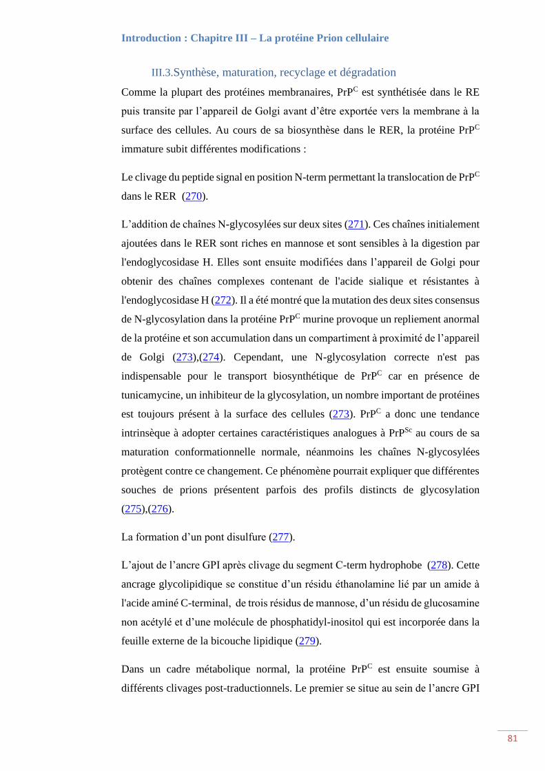
Introduction : Chapitre III – La protéine Prion cellulaire
81
III.3.Synthèse, maturation, recyclage et dégradation
Comme la plupart des protéines membranaires, PrPC est synthétisée dans le RE
puis transite par l’appareil de Golgi avant d’être exportée vers la membrane à la
surface des cellules. Au cours de sa biosynthèse dans le RER, la protéine PrPC
immature subit différentes modifications :
Le clivage du peptide signal en position N-term permettant la translocation de PrPC
dans le RER (270).
L’addition de chaînes N-glycosylées sur deux sites (271). Ces chaînes initialement
ajoutées dans le RER sont riches en mannose et sont sensibles à la digestion par
l'endoglycosidase H. Elles sont ensuite modifiées dans l’appareil de Golgi pour
obtenir des chaînes complexes contenant de l'acide sialique et résistantes à
l'endoglycosidase H (272). Il a été montré que la mutation des deux sites consensus
de N-glycosylation dans la protéine PrPC murine provoque un repliement anormal
de la protéine et son accumulation dans un compartiment à proximité de l’appareil
de Golgi (273),(274). Cependant, une N-glycosylation correcte n'est pas
indispensable pour le transport biosynthétique de PrPC car en présence de
tunicamycine, un inhibiteur de la glycosylation, un nombre important de protéines
est toujours présent à la surface des cellules (273). PrPC a donc une tendance
intrinsèque à adopter certaines caractéristiques analogues à PrPSc au cours de sa
maturation conformationnelle normale, néanmoins les chaînes N-glycosylées
protègent contre ce changement. Ce phénomène pourrait expliquer que différentes
souches de prions présentent parfois des profils distincts de glycosylation
(275),(276).
La formation d’un pont disulfure (277).
L’ajout de l’ancre GPI après clivage du segment C-term hydrophobe (278). Cette
ancrage glycolipidique se constitue d’un résidu éthanolamine lié par un amide à
l'acide aminé C-terminal, de trois résidus de mannose, d’un résidu de glucosamine
non acétylé et d’une molécule de phosphatidyl-inositol qui est incorporée dans la
feuille externe de la bicouche lipidique (279).
Dans un cadre métabolique normal, la protéine PrPC est ensuite soumise à
différents clivages post-traductionnels. Le premier se situe au sein de l’ancre GPI

Introduction : Chapitre III – La protéine Prion cellulaire
82
et libère la chaîne polypeptidique dans le milieu extracellulaire (280). Le rôle de
ce premier clivage n’est pas connu mais par homologie avec d’autres protéines à
ancre GPI qui subissent un clivage post-traductionnel, il semblerait qu’une
phospholipase membranaire soit responsable de cet évènement (281). Le second
clivage est un clivage protéolytique agissant au niveau du segment hydrophobe et
qui aboutit à la libération d’un peptide de 10 kDa. Ce peptide pourrait avoir un
rôle de ligand biologique, d’autant que ce clivage est très conservé chez différentes
espèces. Ce second clivage se produit au sein de vésicules d’endocytose (279) et
d’autres études suggèrent que les domaines riches en cholestérol de la membrane
cellulaire sont impliqués dans ce phénomène (282).
Cependant, ces clivages se produisent de manière très lente par rapport au temps
de demi-vie de la protéine PrPC et leurs rôles physiologiques respectifs n’ont pas
été identifiés.
D’autres clivages de la protéine PrPC, en condition normale, ont pu être mis en
évidence. Notamment les clivages alpha et bêta (283) dont les sites de clivage se
situent en position 111-112 et 96 respectivement. Ceci génère différents peptides
de PrPC appelés C1 et C2 pour les fragments résultant de la partie C-term et N1 et
N2 pour les fragments provenant de la partie N-term (Figure 20). Les rôles
physiologiques et/ou pathologiques de ces clivages sont encore méconnus mais
ces études ont permis de révéler un potentiel rôle de PrPC comme senseur de stress
et particulièrement du stress oxydatif associé au cuivre (284).

Introduction : Chapitre III – La protéine Prion cellulaire
83
Concernant, le trafic cellulaire et le recyclage de la protéine PrPC, dès les années
1990, le groupe de David A. Harris montre que lorsque la protéine PrPC est
exportée à la membrane des cellules, elle subit constitutivement des cycles entre
la membrane cellulaire et des vésicules d’endocytose (285) avec un temps de
transit d’environ 60 minutes. Cette transition s’effectue via des clathrines,
protéines membranaires permettant l’invagination de la membrane plasmique et la
formation de vésicules d’endocytoses intracellulaires (286). Cependant, ce
mécanisme d’endocytose est surprenant puisque les protéines endocytosées par les
clathrines possèdent souvent un motif tyrosine dans leur domaine cytoplasmique
(287) et la protéine PrPC ne contient pas de domaine cytoplasmique. Ainsi, il a été
mis en évidence que PrPC peut également être endocytosée par des cavéolines
membranaires (288). Près de 95% des protéines internalisées retournent à la
membrane sans modification, les autres sont protéolysées par le second clivage
post-traductionnel précédemment décrit ci-dessus dans une région fortement
conservée de l’extrémité N-term.
Figure 20 : Structure simplifiée de la protéine PrPC et des différents
fragments obtenus après clivage. Schéma adapté de "Cholesterol depletion
and modification of COOH-terminal targeting sequence of the prion protein
inhibit formation of the scrapie isoform. Taraboulos A. et al. J. cell biol. 129-
1. p 121-132. 1995".

Introduction : Chapitre III – La protéine Prion cellulaire
84
Enfin, lorsque des protéines PrPC ne passent pas le système de contrôle qualité
cellulaire du fait d’un repliement incorrect par exemple, elles s’accumulent dans
la lumière du RE et sont ensuite prises en charge par le système de dégradation
des protéines ERAD (Endoplasmic-Reticulum-Associated protein Degradation).
Le système ERAD permet ainsi la translocation des protéines vers le cytosol où
elles seront dégradées par le système ubiquitine/protéaosome/ (289),(290).
III.4.Expression et localisation
La protéine PrPC est une protéine cellulaire exprimée dans les neurones et les
cellules gliales du cerveau et de la moelle épinière et également dans de nombreux
tissus périphériques non neuronaux (291),(292). En effet, la protéine ainsi que
l’ARNm de PrPC ont été détectés dans la rate, les tissus lymphoïdes, les poumons,
le cœur, les reins, les muscles squelettiques, l’utérus, les intestins, les glandes
surrénales, les glandes parotides et les glandes mammaires et le placenta (293).
L’ARNm de PrPC est détectable très précocement au début de l’embryogénèse et
sa concentration augmente au fur et à mesure du développement (294),(295).
La protéine PrPC est principalement localisée à la surface des cellules, insérée dans
la membrane cellulaire grâce à son ancre GPI. Néanmoins, il a été démontré que
la forme infectieuse PrPSc est localisée dans le compartiment nucléaire (296). Par
la suite, une étude a montré que certains mutants de la protéine PrPC avaient une
localisation presque exclusivement nucléaire (297). Une localisation
mitochondriale a également été mise en évidence chez des souris transgéniques
surexprimant PrPC (298),(299).
III.5.Régulation
La transcription du gène PRNP est connue pour être modulée par la structure et la
conformation spatiale de la chromatine (300). De plus, le gène PRNP comporte
des sites de fixation potentiellement accessibles à de nombreux facteurs de
transcription indiquant ainsi que l’expression de PrPC est dépendante de facteurs
cellulaires. En plus des facteurs Ap1, Ap2 et Sp1 précédemment identifiés pour
réguler l’expression du gène PRNP, d’autres sites de fixation ont été identifiés
dans la région 5’ au sein du premier exon : MZF-1 (Myeloid Zinc Finger 1),
MEF2, MyT1 (Myelin Transcription Factor 1), NFAT(Nuclear Factor of
Activated T-cells), Bcl-6 (B-cell lymphoma protein 6), ZNF238 (Zinc

Introduction : Chapitre III – La protéine Prion cellulaire
85
Finger protein 238), NEUROG1 (Neurogenin-1), EGR4 (Early Growth
Response protein 4), Oct-1/Oct-2 (Organic cation transporter-1/2), NF-IL6
(Nuclear Factor for IL-6 expression), MyoD (Myoblast Determination protein),
p53 (tumor protein 53), HSE (Heat Shock sequence Elements), MRE et MLS
(HoloCytochrome C Synthase) (301),(302),(303),(304) (Figure 21).
La régulation de l’ARNm et de la protéine PrPC est étroitement liée au
développement. L’expression de l’ARNm murin de PrPC a été détectée entre le
8ème et le 9ème jour embryonnaire au moment de la transition du métabolisme
anaérobique vers un métabolisme aérobique (305) suggérant un rôle important de
PrPC dans le développement embryonnaire. Dans ce contexte, il a été montré que
l’injection du facteur de croissance NGF (Nerve Growth Factor) dans le cerveau
de nouveau-nés de hamsters induit une régulation positive de l’ARNm de PrPC
Figure 21 : Positions des sites de régulation du gène PRNP (liste non
exhaustive). Schéma adapté de "Regulation of prion protein expression : a
potential site for therapeutic intervention in the transmissible spongiform
encephalopathies. Haigh C. L. et Brown D. R. IJBS 2-4. p 315-323. 2006".
Introduction : Chapitre III – La protéine Prion cellulaire
86
ainsi qu’une augmentation de l’activité de la choline acétyltransférase de neurones
cholinergiques sensibles au NGF (306). Aussi, la différenciation cellulaire de
cellules tumorales de médullosurrénales induite par le facteur NGF ou l’IL-6
s’accompagne d’une augmentation de l’expression de l’ARNm de PrPC (307).
Toutefois, il a été démontré que l’induction de PrPC par le NGF dans des
fibroblastes sans différenciation dépend d’effets contraires aux voies des MAPK
(Mitogen-activated protein kinases) ou de la PI3K (Phospho-inositide 3-kinase) ce
qui indique que l’augmentation de PrPC n’est pas une conséquence de la
différentiation induite par le NGF (308).
De manière générale, il est admis que l’augmentation de l’expression de PrPC est
corrélée avec le développement cérébral et la différenciation neuronale mais ne
semble pas être corrélée avec la différenciation des cellules gliales.
Le stress cellulaire module également l’expression de PrPC. En effet, au sein de
cellules de neuroblastome, l’augmentation de la protéine Hsp70, une protéine
traduisant un stress cellulaire et particulièrement un choc thermique, induit une
régulation positive de l’expression de PrPC (309). L’oxygène hyperbare ainsi que
l’hypoglycémie sont des exemples de stress qui induisent la régulation positive
des gènes de PrPC, de Hsp70 et de la voie JNK (c-Jun N-terminal kinases). Dans
ce cas, l’activation du promoteur de PRNP dépend de la présence d’un ou plusieurs
éléments de choc thermique dans sa séquence (310),(311), de manière similaire à
ce qui est observé dans les cellules de neuroblastomes sujettes à des cycles
d’hypoxie et d’oxygénation (312).
Apparentée aux stress cellulaires, la réponse inflammatoire induit également des
régulations de l’expression de PrPC. Les premières observations ont été faites à
partir de peau et d’épithélium gastro-intestinaux enflammés où l’expression de
PrPC est régulée positivement (313),(314). De plus, une corrélation a été faite entre
un statut redox déstabilisé et la présence de PrPC (315). Néanmoins, la régulation
de l’expression de PrPC dans ces conditions reste peu connue. L’enjeu est de
déterminer si ces situations de stress entraînent une augmentation de l’expression
génique de PrPC ou préviennent la dégradation de la protéine PrPC.
La capacité de PrPC à fixer le cuivre via son extrémité N-term avec une forte
affinité est maintenant bien documentée (263),(316),(317) et il a été montré que

Introduction : Chapitre III – La protéine Prion cellulaire
87
l’accumulation d’une forte charge cuprique entraîne la régulation positive de la
protéine PrPC (318). Notre groupe a confirmé cette régulation positive de PrPC par
le cuivre sur des cellules épithéliales bronchiques mais également sur des cellules
placentaires et a montré que cette régulation n’était pas tissu-dépendante (215)
(319). De plus, nous avons observé que des traitements avec d’autres métaux
divalents capables de se lier à PrPC comme le fer ou le zinc induisent également
une régulation positive de PrPC (données non publiées). Enfin, il a été montré que
les réponses cellulaires de PrPC face au cuivre mais aussi au cadmium dépendent
de la présence de sites de fixation des métaux MRE dans la séquence de PRNP,
sans que le facteur de transcription MTF-1 (Metal regulatory transcription factor
1) soit impliqué (304).
IV. Fonctions de PrPC
Actuellement, les fonctions physiologiques précises de la protéine PrPC sont peu
décrites. Déterminer la physiologie de PrPC dans des conditions normales reste un
enjeu majeur puisqu’elle permettrait de comprendre comment survient la
conversion pathologique de PrPC en PrPSc et de lutter contre les EST, mais
également de comprendre d’autres situations physio-pathologiques dans
lesquelles PrPC semble avoir un rôle. Néanmoins, certaines propriétés de la
protéine ont pu être mises en évidence :
IV.1.Rôle neuroprotecteur
La protéine PrPC est connue pour jouer le rôle de récepteur et s’associer avec la
laminine via sa chaîne γ-1 (320). Les laminines sont des glycoprotéines
hétérotrimériques qui constituent majoritairement la lame basale avec le collagène
et connues pour avoir un rôle dans la prolifération, la différenciation, la migration
et la mort cellulaire neuronale (321). Ces différents effets sont médiés via
l’interaction de la laminine avec les intégrines. Cette interaction se caractérise par
la formation et le développement de neurites, la migration des neurones, la
régénération neuronale et axonale (322) et la prévention de la morte neuronale
induite après injection d’acide kaïnique, molécule analogue au glutamate,
convulsive et neurodégénérative (323). Il a été montré que le détachement des
neurones de leurs substrats constitués de laminine était l’événement déterminant
de la mort neuronale, caractéristique des crises de convulsion. Par la suite, une

Introduction : Chapitre III – La protéine Prion cellulaire
88
étude a montré que des souris invalidées pour PrPC étaient plus sensibles aux
convulsions causées par trois agents convulsifs, dont l'acide kaïnique, ce qui
suggère que l'absence de la protéine PrPC rend les animaux plus sensibles à la mort
neuronale en raison de la dégradation de la laminine (324). Cependant, l’absence
de protéine PrPC in vivo chez des souris transgéniques n’entraîne pas de
modification dans le développement et la structure du cerveau et ne perturbe pas
le comportement des souris ni leurs capacités cognitives (325).
Il a également été montré que la protéine PrPC joue un rôle cytoprotecteur contre
les stress endogènes et environnementaux qui activent les voies d’apoptoses
neuronales (326). Cette observation a été confirmée dans différents modèles : i)
dans des lignées cellulaires eucaryotes, PrPC a la capacité de protéger les neurones
de l’apoptose induite par le médiateur pro-apoptotique Bax (Bcl-2–associated X)
(327), (328), ii) chez la levure Saccharomyces cerevisiae, où l’expression
hétérologue du Bax eucaryotique est létale, l’expression de la protéine PrPC
permet de contrer efficacement l’apoptose induite par Bax (329) et iii) chez
différents modèles murins dont les séquences de Prnp ont été modifiées et/ou
tronquées, les souris développent progressivement une neurodégénérescence
fatale (330),(331), mais l’apport de protéines PrPC endogènes permet de
contrebalancer la neurodégénération et assure la survie des souris (332).
IV.2.Relation avec les métaux
Comme décrit précédemment, l’extrémité N-term de la protéine PrPC contient une
répétition de l’octapeptide PHGGSWGQ qui lui permet de fixer différents métaux
divalents comme le cuivre (Cu), le zinc (Zn), le manganèse (Mn) ou encore le
nickel (Ni) mais avec une plus grande affinité pour le cuivre (262),(263),(264).
L’interaction avec ces différents métaux entraîne des changements de
conformation de la structure N-term de PrPC (333),(334),(335),(336). De plus, il a
été montré que la séquence de l’octapeptide PHGGSWGQ est cruciale pour
l’endocytose de la protéine PrPC induite par les métaux. Cependant ces résultats
ont été obtenus sur des modèles cellulaires sur-exprimant la protéine PrPC. Dans
un modèle où la protéine est exprimée d’une manière endogène, notre groupe a
montré que le traitement par le cuivre augmente l’expression du PrPC par insertion
au niveau de la membrane plasmique (319) (215). Au niveau animal, l’invalidation

Introduction : Chapitre III – La protéine Prion cellulaire
89
du gène Prnp dans un modèle murin cause des altérations dans l’homéostasie des
métaux (337) (338).
Le cuivre est un métal essentiel pour le développement et la survie de l’organisme
(339),(340). Dans la nature, le cuivre se trouve sous deux états d’oxydation et de
par son statut redox, le cuivre assure la fonction de catalyseur pour différentes
enzymes (cytochrome c, Cu/Zn Superoxyde Dismutase (SOD) etc…) (341),(342)
ainsi que le transport des électrons dans différents systèmes biologiques
(342),(343). De plus, le cuivre est un élément crucial dans la formation de
l’hémoglobine ou encore pour le métabolisme des drogues. En conditions
normales, le cuivre n’est presque jamais libre au sein de l’organisme car sa
réactivité naturelle le rend sujet à des réactions toxiques via la réaction de Fenton
et Haber-Weiss (344). La perturbation de l’homéostasie du cuivre conduit à des
pathologies telles que les maladies de Wilson et Menkes (345),(346),(347),(348).
Il a été proposé que la surexpression de PrPC entraînerait une fixation plus élevée
du cuivre, suggérant ainsi que la protéine PrPC pourrait jouer le rôle d’un
transporteur membranaire du cuivre au sein de la cellule (349),(350). A l’inverse,
l’addition de cuivre induit une régulation positive de l’expression de PrPC (318).
D’autres études ont suggéré que le cuivre aurait un rôle dans le développement des
EST. En effet, dans certaines conditions, le cuivre a la capacité de convertir PrPC
dans une forme résistante à la protéinase K et insoluble dans les détergents (351)
et inhibe la formation de fibrilles, agrégats de PrPSc caractéristiques des EST (352).
Concernant le manganèse, c’est un des métaux les plus abondants sur Terre qui
existe sous onze états d’oxydation, ce qui le rend capable de participer à de
nombreux processus physiologiques tels que la coagulation ou la production
d’ATP (353). C’est également un élément clé pour la superoxide dismutase à
manganèse qui permet de détoxifier les ROS mitochondriaux (354). En revanche,
une accumulation de manganèse dans les mitochondries inhibe les complexes
mitochondriaux I et II et provoque un dysfonctionnement du transport des
électrons au sein de la chaîne mitochondriale (355),(356). L’arrêt de la chaîne
respiratoire conduit à l’augmentation de l’oxygène dans le cytoplasme, ce qui
génère la production de ROS et ainsi le stress oxydatif causé par les ROS entraîne
l’apoptose de nombreux types cellulaires (357),(358),(359). Plusieurs études ont

Introduction : Chapitre III – La protéine Prion cellulaire
90
montré que le stress oxydatif généré par le manganèse causait par la suite une
activation des caspases 3 et 9 ainsi que la fragmentation de l’ADN (Acide
Désoxyribonucléique) (360),(361). Dans ce contexte, des recherches ont permis
de mettre en évidence que PrPC peut lier le manganèse (362),(363) et que PrPC
jouerait un rôle protecteur contre le stress oxydatif induit par le manganèse (364).
Enfin, le zinc, un autre métal essentiel pour la vie et très abondant dans la nature,
a aussi été largement étudié en relation avec la protéine PrPC puisqu’au niveau
cérébrale, la concentration extracellulaire en zinc est très supérieure à celle du
cuivre. C’est l’oligoélément le plus abondant dans le cerveau qui permet
notamment la neurotransmission entre les synapses en maintenant la plasticité
synaptique et l’excitabilité neuronale. Pour assurer ses fonctions, le zinc doit être
Figure 22 : Représentation de la coopération entre les protéines PrPC et
AMPA pour le transport intracérébral du zinc. Schéma adapté de
"Neuronal zinc regulation and the prion protein. Watt N.T. et al. Prion 7-3.
p 203–208. 2013".

Introduction : Chapitre III – La protéine Prion cellulaire
91
finement régulé et des perturbations de l’homéostasie du zinc peuvent conduire à
des désordres psychologiques. Il a été montré que la protéine PrPC est capable de
fixer le zinc (362) et comme pour le cuivre, cette interaction PrPC-Zn entraîne
l’endocytose de PrPC (365). Il a également été montré que la protéine PrPC
participe au transport du zinc, comme un senseur, ainsi que par sa capacité à le
capturer au niveau des fentes synaptiques et à le transférer au récepteur AMPA (α-
amino-3-hydroxy-5-méthylisoazol-4-propionate) qui permettra son entrée dans la
cellule (366) (Figure 22).
IV.3.Propriétés anti-oxydantes
Les liens étroits entre la protéine PrPC et les métaux pouvant conduire à la
génération de stress oxydatif suggèrent un rôle de PrPC dans le stress oxydatif
cellulaire. Une première étude suggérait une activité SOD-like à la protéine PrPC
au niveau cérébrale et lui conférait ainsi des propriétés anti-oxydantes (367).
Cependant, cette activité qui avait été montré in vitro, n’a pas été confirmé in vivo
(368). Certaines études ont pourtant montré que des souris invalidées pour le gène
Prnp présentaient une diminution de l’activité de la Cu/Zn SOD et une
susceptibilité plus forte au stress oxydatif (369),(370) mais qui semble être
compensée par une augmentation de la Mn SOD. Ainsi, l’implication de la
protéine PrPC dans le système anti-oxydant de manière directe ou indirecte reste à
déterminer.
IV.4.Localisation aux jonctions cellulaires
Une localisation intercellulaire de la protéine PrPC en dehors du système nerveux
a été montrée pour la première fois en 2003 sur des cellules épithéliales intestinales
(371). En effet, plusieurs groupes se sont intéressés au système intestinal puisque
lors d’une contamination orale par PrPSc, la propagation de l’infection se fait des
intestins vers le système nerveux (372),(373). La protéine PrPC est localisée au
pôle apical des entérocytes (374) et également de manière latérale logée dans les
domaines rafts riches en cholestérol (371). Au sein des complexes jonctionnels,
PrPC co-localise et interagit avec certaines protéines des jonctions adhérentes, la
E-cadhérine et la kinase Src (Figure 23). Par la suite, les desmosomes ont aussi été
identifiés comme étant des partenaires de PrPC (375). Au sein des jonctions
cellulaires intestinales, PrPC présente deux types de distribution ; une localisation

Introduction : Chapitre III – La protéine Prion cellulaire
92
nucléaire dans les cellules en cours de division cellulaire et une localisation
intercellulaire dans les cellules polarisées et différentiées. L'inhibition de PrPC
dans les entérocytes humains entraîne une diminution de leurs propriétés de
barrière en modifiant la localisation et l'expression des protéines de jonction et
d'adhésion telles que l’E-cadhérine, la claudine-5 et l’occludine. Ainsi PrPC aurait
un rôle dans le mauvais adressage et la localisation des protéines jonctionnelles.
Notre groupe a montré que la protéine PrPC est exprimée au niveau apical et
latérale des cellules intestinales et rénales (374). Récemment, nous avons
également montré que la protéine PrPC est localisée au niveau des jonctions des
cellules épithéliales bronchiques où elle participe au maintien des complexes
jonctionnels et joue un rôle protecteur face au stress oxydant (215).
De plus, il a aussi été montré que cette protéine présente une localisation
intercellulaire au niveau du système endothéliale avec un rôle dans l’adhésion
cellulaire où elle interagit avec la protéine PECAM-1 (Platelet Endothelial Cell
Adhesion Molecule 1) pour permettre la migration trans-endothéliale des
monocytes (376).
Figure 23 : Modèle proposé pour l’interaction de PrPC et de la kinase Src au
sein des desmosomes. PKG : Plakoglobine, PKP : Plakophiline, DP :
Desmoplakine. Schéma adapté de "Roles of the cellular prion protein in the
regulation of cell-cell junctions and barrier function. Petit C.S.V. et al. Tissue
Barriers 1:2. e24377. 2013".

Introduction : Chapitre III – La protéine Prion cellulaire
93
En 2018, le groupe de Joan W. Berman s’est intéressé au rôle de PrPC dans les
jonctions cellulaires de cellules endothéliales microvasculaires du cerveau et à son
expression après une exposition à des médiateurs pro-inflammatoires. Ils ont
montré que des traitements au VEGF (Vascular Epithelial Growth Factor) ou au
TNF-α induisent une diminution d’expression de PrPC à la membrane plasmique,
associée à une perte d’intégrité de la barrière jonctionnelle (377). D’autant plus
que ces traitements sont également connus pour diminuer l’expression des
protéines des jonctions serrées (claudine-5 et occludine) (378). Dans cette étude,
les auteurs montrent que l’invalidation de PrPC augmente la perméabilité de la
monocouche endothéliale et diminue l’expression de la claudine-5 et de
l’occludine. Ils proposent que la régulation négative de PrPC induite par le VEGF
et le TNF-α contribuerait directement aux modifications d’expression et
d’adressage des protéines des jonctions serrées, affectant ainsi l'intégrité de la
barrière endothéliale. La protéine PrPC n'étant pas une protéine transmembranaire,
il est possible qu'elle provoque ces changements par couplage avec d'autres
protéines adaptatrices.
IV.5.Implication dans l’inflammation
L’implication de la protéine PrPC dans les réponses inflammatoires neuronales a
été démontrée. En effet, l’absence de PrPC dans le système nerveux central
exacerbe la réponse inflammatoire dans le cas d’encéphalomyélite auto-immune
induite expérimentalement (379), et également dans le cas de lésions cérébrales
(380), notamment lors de lésions ischémiques (381). De plus, une étude a montré
que PrPC est capable d’interagir avec le récepteur NMDA (N-méthyl-D-aspartate)
et d’inhiber son activité associée à la nociception (382) ce qui fait de PrPC un
important régulateur de la signalisation de la douleur.
Dans les tissus périphériques, la protéine PrPC semble également être impliquée
dans différents processus inflammatoires. Suite à l’administration systémique de
LPS bactériens induisant une réponse inflammatoire, PrPC a montré un lien dans
la régulation de cytokines pro et anti-inflammatoires (383),(384). Il a aussi été
observé que des souris qui surexpriment PrPC sont plus résistantes aux colites
induites expérimentalement que des souris invalidées pour le gène Prnp. De plus,
ces souris invalidées présentent un profil inflammatoire exacerbé avec notamment

Introduction : Chapitre III – La protéine Prion cellulaire
94
une augmentation des transcrits de cytokines pro-inflammatoires et une activation
des protéines pro-apoptotiques qui amplifient la colite (385).
Du fait de la localisation de PrPC au niveau des complexes jonctionnels des
entérocytes, une étude s’est focalisée sur les perturbations de l’épithélium
intestinal associées aux maladies inflammatoires des intestins telles que les
maladies de Bowel et de Crohn (MBC) en lien avec la protéine PrPC. Ces travaux
ont permis de montrer que l’absence de PrPC perturbe l’organisation des jonctions
cellulaires intestinales et s’accompagne d’une augmentation de la sécrétion de
cytokines pro-inflammatoires. Ces deux phénomènes participent ainsi à
l’exacerbation de l’inflammation chronique observée dans les MBC. Finalement,
cette étude suggère que le niveau d’expression de la protéine PrPC n’est pas
modifié dans les MBC, puisque le taux d’ARNm reste inchangé, mais qu’il y a
une augmentation du cycle de recyclage de PrPC induisant alors la déstabilisation
des jonctions cellulaires (386).
La protéine PrPC aurait également un rôle crucial dans l’activation de la voie pro-
inflammatoire du TNF-α. La voie NFκB comprend cinq facteurs de transcription
inductibles : NFκB-1 (p50), NFκB-2 (p52), RelA (p65 : Transcription factor 65),
RelB et c-Rel (387). Ces différentes protéines fonctionnent en dimères et régulent
principalement la production de cytokines et chimiokines pro-inflammatoires, le
recrutement des cellules immunitaires, l’expression de molécules d’adhésion et
certaines enzymes inductibles comme la COX-2 (Cyclo-oxygénase-2) (388). Ces
dimères sont maintenus inactifs par association avec des protéines kinases
inhibitrices appartenant à la famille IκB (Inhibitor of nuclear factor Kappa-B
Kinase), constituée de IκB-α, IκB-β, IκBε et Bcl-3 (B-cell lymphoma protein 3)
(389). Ainsi, en condition normale, les protéines IκB présentes dans le cytoplasme
inhibent NFκB (390). Lors d’une stimulation inflammatoire l'activation de NFκB
est médiée par la phosphorylation des protéines IκB qui sont ensuite
polyubiquitinées et dégradées par des protéases, libérant ainsi la protéine NFκB
active. La forme active de NFκB peut ensuite se transloquer et activer la
transcription de ses gènes cibles. Bien que la dégradation des protéines IκB reste
une étape fondamentale pour l'activation de NFκB, de nombreux autres
mécanismes tels que l'ubiquitination, la sumoylation ou la phosphorylation sont
impliqués dans la régulation de l'activité de transcription de NFκB. De plus,

Introduction : Chapitre III – La protéine Prion cellulaire
95
certaines enzymes semblent également contrôler ces processus. Ainsi, ces
multiples mécanismes qui régulent étroitement l'activité de NFκB permettent à
cette voie de signalisation de participer à de nombreuses fonctions cellulaires
vitales.
L’invalidation du gène Prnp entraîne des perturbations de l’expression des
différentes protéines intermédiaires de la voie du TNF-α (IKK α/β (IκB Kinase),
p65 et JNK) tandis que l’expression de PrPC améliore les réponses cellulaires au
TNF-α en favorisant la production de médiateurs pro-inflammatoires(391).
IV.6.Implication dans les infections
Du fait de la localisation de la protéine PrPC dans les domaines lipidiques rafts de
la membrane plasmique, des études ont été menées dans le but de déterminer une
éventuelle implication de PrPC dans les infections bactériennes et notamment dans
l’infiltration intracellulaire bactérienne. En effet, il a été montré que les domaines
raft présents dans les membranes cellulaires participent à l’entrée de nombreuses
espèces bactériennes dans la cellule (P. aeruginosa, E. coli, Brucella spp ou encore
Mycobactérium) via les cavéolines qui internalisent les bactéries au sein de
vésicules (392). Une première équipe a suggéré que la protéine PrPC pourrait
promouvoir les infections causées par la bactérie Brucella en améliorant
l’internalisation de Brucella au sein des macrophages, un lieu de réplication
stratégique pour cette bactérie (393). Par la suite, ce même groupe a suggéré une
interaction directe ou indirecte entre PrPC et Brucella via le récepteur membranaire
bactérien Hsp60, une protéine chaperonne de la famille des Growth E.coli Large
protein (GroEL) connue pour être activée lors d’invasions bactériennes (394).
Cependant, un autre groupe n’a pas confirmé ces résultats et ne considère pas un
rôle évident de PrPC dans les infections bactériennes causées par Brucella (395).
Il a été décrit que l’extrémité N-term de PrPC possède des propriétés
antimicrobiennes. En effet, l’extrémité non structurée N-terminale de PrPC est
capable d’agir comme certains peptides antimicrobiens connus, en formant des
pores dans la membrane bactérienne (396).
De plus, les fibrilles et agrégats de la protéine PrPC ont été identifiés comme
inducteur de la production de la cytokine pro-inflammatoire IL-1β au travers de
l’activation de NLRP3 (NOD-like receptor family, pyrin domain containing 3)

Introduction : Chapitre III – La protéine Prion cellulaire
96
(397). De manière très intéressante, NLRP3 qui est un membre de
l’inflammasome, ce complexe protéique oligomérique impliqué dans l’immunité
innée et la réponse inflammatoire (398), est activé dans les infections bactériennes
à PA via un facteur de virulence protéique, la piline (399). Lors d’infections
cérébrales avec la souche Mycobacterium bovis, l’expression du gène Prnp est
régulée à la hausse, ce qui conduirait à une diminution des facteurs pro-
inflammatoires, une augmentation de l’apoptose des cellules infectées et
permettrait ainsi à M. bovis d’échapper à la réponse immunitaire de l’hôte (400).
Enfin, un rôle protecteur durant certaines infections virales a été proposé pour PrPC
par différentes études. Après infection par le virus de l’encéphalomyocardite B
(inducteur d’encéphalites et de l’apoptose des cellules neuronales), les souris
sauvages (comparées aux souris invalidées pour PrPC) présentent une meilleure
infiltration des cellules inflammatoires accompagnée d’une augmentation de
l’activation des cellules microgliales (401), qui ont un rôle protecteur contre les
lésions cérébrales (402). Puis, une autre étude a suggéré un rôle protecteur de la
protéine PrPC lors d’infection avec le virus Influenza A (403). Ce rôle a été attribué
à l’octapeptide de PrPC permettant la fixation du cuivre, car des souris délétées
pour cette séquence peptidique infectées avec le virus Influenza A présentent un
fort taux de mortalité et des modifications pathologiques plus sévères que les
souris sauvages. Ces souris invalidées pour PrPC montrent notamment une
augmentation de la production des cytokines pro-inflammatoires et une charge
virale plus importante au niveau des poumons. Cette observation a pu être faite
car lorsque PrPC est réintroduite dans ces souris délétées, la mortalité est
drastiquement diminuée et le profil inflammatoire est similaire aux souris
sauvages infectées par le virus.
V. Modèles animaux pour l’étude de PrPC
Dès les années 1960, des expérimentations animales ont été menées sur des
hamsters, souris et chèvres, entres autres, dans le but d’étudier la transmission et
la propagation du prion infection. L’isoforme pathogène de PrPC étaient inoculées
aux animaux et les scientifiques étudiaient la période d’incubation, l’apparition
des symptômes et réalisaient des expériences post-mortem sur les échantillons
prélevés. Ces expérimentations ont notamment permis de découvrir la nature
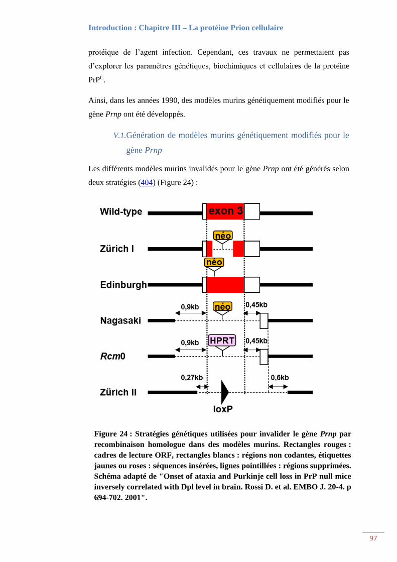
Introduction : Chapitre III – La protéine Prion cellulaire
97
protéique de l’agent infection. Cependant, ces travaux ne permettaient pas
d’explorer les paramètres génétiques, biochimiques et cellulaires de la protéine
PrPC.
Ainsi, dans les années 1990, des modèles murins génétiquement modifiés pour le
gène Prnp ont été développés.
V.1.Génération de modèles murins génétiquement modifiés pour le
gène Prnp
Les différents modèles murins invalidés pour le gène Prnp ont été générés selon
deux stratégies (404) (Figure 24) :
Figure 24 : Stratégies génétiques utilisées pour invalider le gène Prnp par
recombinaison homologue dans des modèles murins. Rectangles rouges :
cadres de lecture ORF, rectangles blancs : régions non codantes, étiquettes
jaunes ou roses : séquences insérées, lignes pointillées : régions supprimées.
Schéma adapté de "Onset of ataxia and Purkinje cell loss in PrP null mice
inversely correlated with Dpl level in brain. Rossi D. et al. EMBO J. 20-4. p
694-702. 2001".

Introduction : Chapitre III – La protéine Prion cellulaire
98
La stratégie "conservatrice" consiste à introduire des modifications génétiques
limitées seulement au cadre de lecture ORF de l’exon 3 (souris nommées Zurich I
et Edinburgh) (405).
La stratégie "radicale" dans laquelle la séquence ORF est entièrement supprimée
et les sites d’épissage du troisième exon sont modifiés (souris nommées Nagasaki,
Rcm0 et Zurich II).
Dans les deux cas, l’insertion d’une cassette néomycine, HRPT (Hypoxanthine
Phosphoribosyl-Transférase) ou loxP (locus of crossing-over from Phage) entraîne
la suppression de l’expression du gène Prnp.
Les souris de type Zurich I et Edinburgh se développent normalement et ne
présentent pas de pathologies particulières. Les souris de type Nagasaki, Rcm0 et
Zurich II se développent également normalement mais développent une ataxie
grave et une perte de cellules de Purkinje en vieillissant (406),(407). De manière
surprenante, les deux types de souris invalidés pour le gène Prnp sont résistants
aux maladies à prion et à la propagation de la protéine PrPSc (405).
Lignée Gène codant
pour PrPC
Nombre de
copie du
gène
Quantité
d’ARN de PrPC
Quantité de
protéine PrPC
Prnp +/+ Prnp 2 1 1
Prnp 0/+ Prnp 1 0,5 0,5
tga19/+ hgPrnp 30 ND 3-4
tga20/+ hgPrnp 30 1,2 6-7
tga20/tga20 hgPrnp 60 ND 10
tgc35/+ Prnp (cosmid) 40 ND 4-5
tgb24/+ PrPcDNA 150 <0,5 0
tge19h/+ PrPΔNco 150 1,8 3-4
tge19l/+ PrPΔNco 15 1 1,5-2,5
tgd11/+ PrPΔN-term 30 2,7 6
tgd12/+ PrPΔN-term 40 3,9 10-12
En parallèle des modèles d’extinction du gène Prnp, des modèles murins
transgéniques de surexpression ont été développés via l’introduction de transgènes
de Prnp (Figure 25) (408).
Figure 25 : Caractéristiques et expression de PrPC dans les modèles murins
de surexpression de PRNP. ND : Non Déterminé. Tableau adapté de " Prion
protein (PrP) with amino‐proximal deletions restoring susceptibility of PrP
knockout mice to scrapie. Fischer M. et al. EMBO J. 15-6. p 1255-1264.
1996".

Introduction : Chapitre III – La protéine Prion cellulaire
99
La surexpression du gène Prnp ne perturbe pas le développement des souris,
cependant il a été observé que la lignée tga20 (transgene insertion a20) présente
une période d’incubation du prion infectieux très courte comparée aux souris
sauvages (408).
V.2.Impact de l’extinction et de la surexpression du gène Prnp
A la suite de la génération de ces modèles murins, de nombreux paramètres de
PrPC ont été mis en évidence. Néanmoins, la plupart des projets étaient focalisés
sur l’étude de PrPC uniquement au sein du système nerveux. Les résultats de ces
travaux ont été résumés dans le tableau ci-dessous (Figure 26) :
Effets Références
Inval
idat
ion d
e P
rnp
Altération du rythme circadien et du sommeil
(409)
Diminution de la plasticité synaptique (410)
Altération du comportement, de la peur et de l’anxiété (411)
(412)
(413)
Dérégulation du métabolisme du fer (337)
Intolérance au glucose (414)
Activation de la prolifération des cellules précurseurs
d’oligodendrocytes lors du développement et chez l’adulte
(415)
Diminution de la concentration cuprique cérébrale (338)
Augmentation du nombre de mitochondries avec des
anomalies morphologiques
(416)
Augmentation de la phagocytose des macrophages (417)
Résistance aux infections à Brucella abortus (393)
Sure
xpre
ssio
n d
e P
rnp
Dysfonctions immunologiques (418)
Augmentation de la survie des neurones après une
axotomie
(419)
Amélioration de la plasticité synaptique (420)
Augmentation de la dégénération du système nerveux liée
à l’âge
(421)
Activation de la prolifération des cellules souches dérivées
de la zone sous-ventriculaire
(422)
Sensibilité aux infections du virus herpès simplex de type I (423)
Figure 26 : Effets de l’extinction ou de la surexpression de la protéine PrPC
dans des modèles murins.

Introduction : Chapitre III – La protéine Prion cellulaire
100
L’implication de PrPC dans les infections bactériennes respiratoires en lien avec
l’inflammation bronchique reste méconnue en raison d’un nombre limité d’études
menées sur cette thématique. Néanmoins, une étude de 2014 s’est intéressée au
rôle de PrPC après stimulation au LPS (383). Ainsi, les travaux de ce groupe ont
pu mettre en lumière différentes observations :
Durant la phase d’infection aigüe du LPS, le niveau d'ARNm des cytokines pro-
inflammatoires (TNF-α, IL-6 et IL-1β) et des facteurs de transcription (ERK
(Extracellular signal-Regulated Kinases), p38 (p38 mitogen-activated protein
kinase) et IκB) est plus élevé dans le cerveau et la rate des souris sauvages que
chez les souris PrP -/-.
Au cours de la phase tardive de l’infection, les souris PrP -/- conserve des niveaux
plus élevés de cytokines pro-inflammatoires et un faible niveau de cytokines anti-
inflammatoires (IL-10), tandis que chez les souris sauvages les taux sont inversés,
la concentration en cytokines pro-inflammatoires diminue, alors que le taux de
cytokines anti-inflammatoires augmente.
Les souris PrP -/- présentent une résorption plus lente des leucocytes périphériques
après leur recrutement lors de la réponse inflammatoire et une mortalité plus
élevée en réponse au choc septique induit par le LPS par rapport aux souris
sauvages.
L’ensemble des données présentées dans ces trois chapitres mettent en évidence
la complexité de la physiopathologie de la mucoviscidose et le rôle protecteur
primordial de la barrière épithéliale bronchique au sein des poumons. Dans ce
contexte, nous avons orienté mon projet de thèse vers la recherche de nouveaux
partenaires cellulaires de la protéine CFTR afin d’investiguer quant à l’origine du
relâchement des jonctions cellulaires, de la facilité des pathogènes à envahir le
parenchyme bronchique et de l’hyper-inflammation chronique pulmonaire.
Du fait de ses diverses implications cellulaires, notamment au sein des jonctions
cellulaires et dans l’inflammation nerveuse, la protéine PrPC s’est avérée être un
excellent candidat pour notre étude. De plus, les rôles exacts de cette protéine au
niveau pulmonaire sont encore inconnus et pour la première fois nous montrons
l’implication de la protéine PrPC dans la mucoviscidose.

101
Objectifs

102

Objectifs
103
Pourquoi une personne saine est-elle protégée contre les infections par des
pathogènes opportunistes, tandis que les personnes atteintes de mucoviscidose ou
qui présentent un épithélium pulmonaire endommagé sont particulièrement
sensibles à ces infections ? Pour répondre à cette question, il est essentiel de bien
comprendre l'interaction dynamique entre le microenvironnement hôte et ces
pathogènes. Plusieurs paramètres tissulaires et des facteurs physico-chimiques
peuvent individuellement ou collectivement favoriser l’infection aigue et
chronique par ces pathogènes. Parmi ces facteurs il y a la barrière épithéliale. Cette
dernière est le résultat d’un assemblage de cellules présentant une polarité
structurale et fonctionnelle assurée par des interactions entre protéines
transmembranaires et cytosoliques. Ces assemblages protéiques forment ainsi les
différentes catégories de jonctions intercellulaires; serrées, adhérentes,
desmosomes et communicantes.
La perte d’étanchéité au niveau de la barrière épithéliale pulmonaire est considérée
comme un facteur clé qui participe à l’exacerbation du processus d’inflammation
et d’infection en facilitant la transmigration tissulaire des cellules immunitaires et
des pathogènes comme Pseudomonas aeruginosa (PA).
Dans ces processus, la protéine CFTR jouerait un rôle très important, mais qui est
encore mal cerné et surtout très débattu.
La protéine CFTR a longtemps été considérée comme l’élément déterminant de la
physiopathologie de la mucoviscidose. Ainsi, son activité canal chlorure et ses
fonctions régulatrices, ont concentré la plus grande partie des thérapeutiques
développées et des résultats très encourageants ont été obtenus, surtout pour la
mutation G551D (~ 4 % des diagnostics). Néanmoins, ces résultats thérapeutiques
contrastent avec le bilan inflammatoire et infectieux qui reste encore inférieur aux
espérances attendues par les cliniciens.
Ces observations suggèrent surtout que d’autres facteurs comme ; les gènes
modificateurs (gènes modulant la sévérité des différentes atteintes pathologiques
des patients) ou les protéines dérégulées issues des dommages collatéraux de
CFTR mutée, peuvent participer à la perte de l’étanchéité épithéliale constatée
dans la mucoviscidose.
Récemment, nous avons montré au niveau des cellules bronchiques humaines
saines que la protéine prion cellulaire (PrPC) se lie aux protéines des jonctions
adhérentes et desmosomes. A travers cette liaison, PrPC protège ces protéines

Objectifs
104
contre le stress oxydatif et participe ainsi à la stabilité de ces jonctions. D’autres
études ont également montré, que PrPC serait impliqué dans la défaillance de la
barrière épithéliale intestinale observée dans la maladie inflammatoire de Bowel.
Il était donc logique d’entreprendre l’étude des rôles de PrPC dans la
mucoviscidose, maladie aussi caractérisée par l’inflammation et la perte de
l’étanchéité de l’épithélium pulmonaire.
Ainsi, pour mon projet de thèse, deux objectifs ont été fixés :
1. Etude in vitro du rôle de la protéine PrPC dans la barrière épithéliale
bronchique en lien avec la mucoviscidose.
Pour cette partie, nous avons utilisé des modèles de lignées cellulaires bronchiques
humaines pour étudier l’expression, la localisation, l’interaction avec les protéines
des jonctions et avec la protéine CFTR sauvage (wt) et mutée (deltaF508), la
résistance trans-épithéliale (RTE) et la perméabilité cellulaire ainsi que
l’évaluation de la fraction membranaire et de la stabilité de PrPC. Nous avons
aussi étudié l’impact de la surexpression de PrPC sur l’expression de CFTR et des
jonctions intercellulaires et analyser la distribution cellulaire de PrPC et des
protéines jonctionnelles associées au trafic et à l’adressage des protéines CFTR
sauvage et mutée.
2. Etude in vivo du rôle de la protéine PrPC dans l’infection aigue en lien avec
la protéine CFTR dans les modèles murins invalidés pour le gène Prnp ou le
gène Cftr.
Sur le plan animal, nos travaux ont porté sur l’implication de PrPC dans le passage
paracellulaire de PA vers le parenchyme pulmonaire et dans la réponse
inflammatoire.
Nous avons ainsi :
☞ Mesurer le passage paracellulaire de PA dans nos modèles cellulaires.
☞ Évaluer l’impact de PrPC sur la dissémination bactérienne et le recrutement des
cellules inflammatoires périphériques dans nos modèles murins.
☞ Comparer les structures pulmonaires de nos modèles murins par
immunohistochimie avant at après infection par PA.
☞ Analyser le profil d’expression des jonctions intercellulaires avant et après
l’infection aigue.

Objectifs
105
☞ La protéine PrPC est importante dans l’homéostasie du cuivre, ainsi nous avons
évalué la concentration du cuivre au niveau pulmonaire et hépatique (contrôle)
dans nos modèles murins avant et après infections aigue par PA.
L’ensemble des résultats seront présentés sous forme de deux publications,
préparées pour soumission:
➢ Johanna Cormenier, Mariam El Khatib, Amal Kouadri, Laurence Macari,
Isabelle Michaud-Soret, Boubou Camara, Christelle Coraux, Nadia
Alfaidy and Mohamed Benharouga.
"Cellular prion protein (PrPC) is up regulated in cystic fibrosis: Role of CFTR
protein."
➢ Johanna Cormenier, Marianne Marchioni, Laurence Macari, Boubou
Camara, Nicolas LeMaître, Frédéric Sergent, Pierre Richaud, Nadia
Alfaidy and Mohamed Benharouga.
"The cellular prion protein (PrPC) contribute to the acute Pseudomonas
aeruginosa lung infection in cystic fibrosis mice."

106

107
Résultats

108

Résultats : Article 1 – Cellular prion protein (PrPC) is up regulated in cystic
fibrosis: Role of CFTR protein.
109
I. Article 1 – Cellular prion protein (PrPC) is up
regulated in cystic fibrosis : Role of CFTR protein.
Cellular prion protein (PrPC) is up regulated in cystic fibrosis : Role of
CFTR protein.
Johanna Cormenier1, Mariam El Khatib1, Amal Kouadri1, Laurence Macari1,
Isabelle Michaud-Soret1, Boubou Camara4, Christelle Coraux3, Nadia Alfaidy2
and Mohamed Benharouga1.
1 Univ. Grenoble Alpes, CNRS, UMR 5249, CEA, IRIG, CBM, F-38000 Grenoble,
France.
2 Univ. Grenoble Alpes, INSERM U1036, CEA, IRIG, BCI, F-38000 Grenoble, France.
3 Institut National de la Santé et de la Recherche Médicale (INSERM), UMR-S 903,
Reims, France.
4 Centre de Ressources et de Compétences pour la Mucoviscidose, Clinique Universitaire
de Pneumologie - Pole Thorax et Vaisseaux, CHU de Grenoble, France.
Cet article sera soumis à la revue "Journal of Cell Science".

Résultats : Article 1 – Cellular prion protein (PrPC) is up regulated in cystic
fibrosis: Role of CFTR protein.
110
I.1.Contexte de l’étude
Dans la mucoviscidose, l’absence d’expression de CFTR (Cystic Fibrosis
Transmembrane conductance Regulator), une protéine canal chlorure activée par
l’AMPc au niveau de la membrane apicale des cellules de l’épithélium
bronchique, conduit au déséquilibre hydro-anionique, à l’augmentation de la
viscosité du mucus et à la perturbation de la clairance mucociliaire. Ces
changements vont induire une inflammation chronique des voies aériennes qui va
progressivement impacter la fonction respiratoire. L’origine de l’inflammation
reste cependant méconnue, en particulier l’inflammation précoce détectée en
absence de toute infection microbienne. Cette inflammation est exacerbée par la
transmigration des cellules de l’immunité et des pathogènes. La transmigration est
un processus qui va dépendre principalement de la barrière épithéliale pulmonaire
(BEP). Cette dernière est établie grâce aux jonctions intercellulaires, elles-mêmes
formées de protéines de jonctions. Plusieurs études ont démontré l’implication de
la protéine CFTR dans la diminution de l’étanchéité de la BEP. Toutefois, la
localisation apicale de CFTR, différente de la localisation latérale des jonctions
cellulaires suggère l’existence d’autres mécanismes régulant la BEP. Une des
hypothèses est que CFTR par son jeu de trafic intracellulaire à travers des cycles
de transcytoses, peut influencer l’adressage des protéines jonctionnelles. Tout
récemment, nous avons démontré que la protéine prion cellulaire (PrPC) participe
au maintien des jonctions adhérentes et desmosomes au niveau des cellules
épithéliales bronchiques humaines. Nous avons aussi démontré que cette protéine
subit une transcytose assurant son trafic de la membrane apicale vers la membrane
latérale où elle interagit, stabilise et protège les jonctions adhérentes et
desmosomes du stress oxydatif. Au niveau des tissus épithéliaux, la PrPC a été
incriminée dans l’effondrement de la barrière épithéliale intestinale,
caractéristique de la maladie inflammatoire de Bowel. Dans cette pathologie, la
PrPC semble perturber l’adressage des protéines jonctionnelles. Cependant, le rôle
de cette protéine dans la mucoviscidose, une pathologie inflammatoire également
caractérisée par une diminution de la barrière épithéliale bronchique n’est pas
encore connu.

Résultats : Article 1 – Cellular prion protein (PrPC) is up regulated in cystic
fibrosis: Role of CFTR protein.
111
I.2.Résultats
Nous avons mis en évidence pour la première fois une dérégulation de l’expression
de PrPC dans la mucoviscidose, au niveau ARNm et protéine. Nous avons
démontré une augmentation de l’expression de PrPC dans des échantillons de
bronches humaines CF et in vitro dans les cellules CFBE (dF508/dF508). Nous
avons observé que la localisation de la protéine PrPC à la membrane est plus
diffuse dans les cellules CFBE comparé aux cellules épithéliales bronchiques
saines (HBE), suggérant un défaut d’adressage de PrPC. Nous avons constaté que
la surexpression de la protéine CFTR sauvage (wt-CFTR) dans les cellules CFBE
réduit la localisation diffuse de PrPC à la membrane. Néanmoins, l’expression de
wt-CFTR ne diminue pas le niveau d’expression de PrPC. Par ailleurs, et de
manière intéressante, nous avons observé une augmentation de l’expression de la
forme hyperglycosylée (forme H) de PrPC. La régulation de PrPC serait liée à
l’expression et à la localisation cellulaire de CFTR, en lien avec d’autres
mécanismes cellulaires.
Nous avons mis en évidence que les protéines PrPC et CFTR n’interagissent pas
directement ensemble mais sont colocalisées à la membrane basolatérale. Aussi,
cette colocalisation disparaît quand le processus de polarisation s’initie, excepté
dans les cellules CFBE où l’on observe le défaut d’adressage de PrPC.
Dans les cellules CFBE, nous rapportons une diminution dans les niveaux
d’expression de protéines jonctionnelles clés telles que l’occludine, JAM-A, la
plakoglobine et la desmoplakine. Toutefois, l’expression de wt-CFTR n’a pas
permis de restaurer le niveau d’expression basal des protéines jonctionnelles.
Enfin, nous avons également démontré que la surexpression de la protéine PrPC
dans les cellules HBE perturbe l’expression des jonctions cellulaires, suggérant
que le niveau d’expression de PrPC est un paramètre important soumit à une
régulation très fine. De façon intéressante nous avons démontré que la
surexpression de PrPC entraîne une diminution de l’expression protéique de CFTR
sans affecter son niveau d’ARNm, renforçant notre hypothèse de l’existence d’une
relation d’expression protéique entre PrPC et CFTR.

Résultats : Article 1 – Cellular prion protein (PrPC) is up regulated in cystic
fibrosis: Role of CFTR protein.
112
I.3.Conclusions
En conclusion, l’ensemble de nos résultats démontrent l’existence d’une relation
entre les protéines CFTR et PrPC dans la régulation de l’expression et de la
localisation des protéines jonctionnelles. De plus, nos résultats concordent avec
ceux rapportés concernant le rôle de PrPC dans la protection et la stabilisation des
protéines jonctionnelles au niveau intestinal.
D’un point de vue clinique, nos résultats démontrent que la protéine CFTR mutée
affecte directement l’adressage de PrPC vers le pôle latéral des cellules épithéliales
bronchiques, où elle interagit et stabilise les protéines jonctionnelles. Ce défaut
d’adressage de la PrPC au pôle latéral expliquerait certaines caractéristiques de la
mucoviscidose, telle que le défaut de la perméabilité épithéliale. Toutefois, notre
étude a été principalement menée in vitro et des études supplémentaires in vivo
sont essentielles pour confirmer ces hypothèses.
Néanmoins, la compréhension du rôle de la PrPC dans la perméabilité épithéliale
et le maintien des jonctions cellulaires représente un enjeu majeur dans la
mucoviscidose, puisqu’à terme il pourrait contribuer au développement de
thérapies anti-inflammatoires et réduire l’atteinte pulmonaire.

1
Cellular prion protein (PrPC) is up regulated in cystic fibrosis: Role of
CFTR protein.
Johanna Cormenier1, Mariam El Khatib1, Amal Kouadri1, Laurence Macari1, Isabelle
Michaud-Soret1, Boubou Camara4, Christelle Coraux3, Nadia Alfaidy2 and Mohamed
Benharouga1.
1 Univ. Grenoble Alpes, CNRS, UMR 5249, CEA, IRIG, CBM, F-38000 Grenoble, France.
2 Univ. Grenoble Alpes, INSERM U1036, CEA, IRIG, BCI, F-38000 Grenoble, France.
3 Institut National de la Santé et de la Recherche Médicale (INSERM), UMR-S 903, Reims,
France.
4 Centre de Ressources et de Compétences pour la Mucoviscidose, Clinique Universitaire
de Pneumologie - Pole Thorax et Vaisseaux, CHU de Grenoble, France.
Running Title: CFTR controls PrPC trafficking
Keywords: CFTR, PrPC, cystic fibrosis, junctional proteins, epithelial barrier
Address correspondence to:
Dr. Mohamed Benharouga
LCBM-UMR5249
DRF-BIG, CEA-Grenoble
17 rue des Martyrs, F-38054, Grenoble cedex 09, France
Téléphone: (33)-4-38-78-44-51
Fax: (33)-4-38-78-54-87
E-mail: [email protected]

2
Abstract
In cystic fibrosis (CF), the loss of cystic fibrosis transmembrane conductance regulator
(CFTR)-associated cAMP-activated chloride channel has been proposed to affect the
bronchial paracellular permeability. The latter is highly dependent on the intercellular
junctional complexes, which consists of the tight (T), adherens (A), and desmosomes (D)
junctions (J). However, the molecular mechanisms involved in the increase in the bronchial
epithelial permeability (EP) in CF are unknown. Recently we demonstrated that the cellular
prion protein (PrPC) is important for the stability of A and D junctional proteins in normal
bronchial epithelial cells. Yet, in CF disease, the role of PrPC remains elusive. Herein, we
investigate the expression, localization, interaction, biochemical profiles and the roles in
EP of wild-type (wt)-, mutant (dF508)-CFTR, junctional, and PrPC proteins in human
healthy and CF conditions. Using normal (16HBE14o-), CF (CFBE41o-) and CF
overexpressing wt-CFTR (CFBE41o-wt) cells, we first demonstrated that the PrPC mRNA
and protein levels were highly expressed in CF compared to the control cells. Similar results
were found in CF versus healthy bronchial tissue samples. The PrPC increase was not
corrected in CFBE41o-wt cells. In CF cells, PrPC protein showed a diffuse expression and
increased residency time (RT) at the plasma membrane along with a mislocalization
profile. The latter, but not the RT was corrected by wt-CFTR overexpression in CF cells.
The upregulation and the mislocalization of PrPC protein did not affect its ability to interact
with junctional proteins. However, the overexpression of wt-CFTR or PrPC protein could
not correct the levels of expression of junctional proteins in the CF condition. No
interactions were detected between PrPC and wt- or dF508-CFTR proteins, however, wt-
CFTR and PrPC proteins showed a co-localized profiles that disappeared with the
polarization process, except in CF cells. Interestingly, we found also that the levels of
expression of wt-CFTR protein decreased in 16HBE14o- cells overexpressing PrPC protein.
Altogether, our results suggest that in bronchial epithelial cells CFTR trafficking is required
for the normal distribution and localization of PrPC protein, a key actor of the stability of
adherens and desmosome proteins and that the retention of dF508-CFTR at the ER affects
the normal PrPC transcytosis from the apical to the lateral membrane. These findings bring
new insights into the understanding of the CF-dysregulated EP.

3
Introduction
Cystic fibrosis (CF) is an inheritable autosomal-recessive disease characterized by a multi-
organ pathology affecting airways, as well as the intestinal and the reproductive tracts
(Cutting, 2015). More than 2000 mutations have been identified in the CF transmembrane
conductance regulator (CFTR) gene that codes for the CFTR protein; an epithelial apical
transmembrane, cAMP-activated Cl- channel and a regulatory protein (Cutting, 2015).
About 70% of CF mutations correspond to the deletion of a phenylalanine at position 508
(delF580) (Kerem et al., 1989). This mutation is responsible for the retention and rapid
degradation of CFTR in the endoplasmic reticulum (ER) (Benharouga, 2001).
The absence of a functional CFTR leads to unbalanced hydro-anionic efflux, reduced
airway volume and pH, and defective mucociliary clearance resulting in mucus
dehydration, bronchial inflammation and infection, which is the main cause of morbidity
and mortality in CF patients (Cutting, 2015). In CF, the major host factors that protect the
lung against infection are altered. Although an extensive work has been devoted to the study
of the CF gene and the multiple functions of CFTR, the origin of pulmonary superinfection
is still poorly understood.
Respiratory tissues are extensively colonized at the late stages of the disease by bacteria
such as Staphylococcus aureus, Haemophilus influenza and Pseudomonas aeruginosa (PA)
(Quinton, 1990).
Bacterial infection in the CF lung environment is multifactorial and is mainly related to the
loss of the apical function of the CFTR protein (Yang et al., 2011), (Pier, 2012). However,
recent work incriminates other factors such as the presence of an imbalance in the transport
of bicarbonates (Pier, 2012), (Quinton, 2007) and the loss of the cellular junctional barrier
(Ruan et al., 2014), (Fiorotto et al., 2016). Indeed, the disruption of the intercellular junction
of CF bronchial epithelium is associated with an increase in epithelial cell permeability and
an invasion by infectious agents (Bebok et al., 2005), (Vivekananda et al., 1994).
Expression of wild-type (wt) CFTR in delF508-CF cells (CFBE41o (-)) increased the trans-
epithelial resistance (RTE) and decreased the paracellular permeability of mannitol
(LeSimple et al., 2010). Altogether, these studies suggested that the trafficking and the
chloride channel activity of the CFTR protein might contribute to the assembly and function
of the airway epithelial barrier. However, CFTR protein is localized at the apical side
compared to junctional proteins that are restricted to the lateral membrane of polarized
cells, suggesting that other mechanisms could be involved in the control of the pulmonary
epithelial barrier and in the cross talk between the two cellular sites. Three main types of

4
intercellular junctional complexes are responsible for the sealing of the bronchial
epithelium: tight (T), adherens (A) and desmosomes (D) junctions (J) (Vasileva and Citi,
2018). Recently, we demonstrated that the cellular prion protein (PrPC) is expressed in the
lateral side of fully polarized bronchial epithelial cells (16HBE14o-) where it interacts and
protects adherens and desmosomes junction proteins against oxidative stress (Kouadri et
al., 2018).
PrPC is a glycosyl phosphatidyl inositol (GPI)-anchored glycoprotein known for its unique
ability to undergo structural conversion from a normal isoform (PrPC) into a pathogenic
conformer known as scrapie (PrPSc) (Prusiner, 1991). The physiological role of the cellular
PrPC has mainly been associated with stress-protection (Prusiner, 1991), (Rachidi et al.,
2003), copper homeostasis (Alfaidy, 2013) and neuronal excitability (Khosravani et al.,
2008). Other functions related to cellular processes and metabolism have also been reported
for this protein (Castle and Gill, 2017).
In the peripheral epithelial tissues, the pathophysiological role of PrPC has been
investigated in vitro and in vivo only in intestinal barrier in relation to the inflammatory
Bowel Disease (Petit et al., 2012). However, its role in the context of CF disease in relation
to the bronchial integrity is still unknown.
Given the role of PrPC protein in the control of cell-cell interaction and in the protection of
the junctional proteins against oxidative stress, we hypothesized that PrPC may also play a
role in the stability of the bronchial barrier in CF, an inflammatory disease characterized
by a loss of the junctional barrier.

5
Material and Methods
Human tissues collection:
Peripheral human lung tissues (containing non cartilaginous airways) were obtained from
Grenoble Hospital (Pulmonary department) at the time of lung transplantation from 10
nonsmoking CF adults, and from 10 nonsmoking controls undergoing lung resection upon
peripheral lung cancer (Grenoble Hospital). Clinical characteristics of these patients are
reported on Table 1. Collection and processing of human lungs were conformed to the
declaration of Helsinki and, to all the rules of the local Committees on Human Research.
Informed consent was obtained from each patient.
Lentiviral overexpression:
A lentiviral system was used to overexpress the prnp gene in 16HBE14o- cells (HBE-PrP).
The prnp sequence containing SPR signal and hemagglutinin (HA) tag was cloned in the
lentiviral vector pLVX-IRES-mcherry-C1 (Clontech, France), between EcoRI and BamHI
restriction sites. The resulting vector was then transfected into the HEK293T cells for
packaging and generation of lentiviral particles. Then, HBE cells were infected with the
lentiviral particles for 24h. The positive cells were selected using flow cytometry based on
the fluorescent protein mcherry and the overexpression was confirmed using real-time RT-
qPCR, immunoblotting and immunofluorescence assays.
Immunohistochemistry:
Ready 5-µm paraffin-embedded sections prepared from a human CF and non CF peripheral
lung tissues were used. Slides were generously obtained from Pr. Lantuejoul, (Dept. of
Pathology, DACP, Grenoble Hospital) and from Dr. Coraux (INSERM, UMR-S 903,
Reims, France). Immunohistochemistry was performed as described previously (De
Keukeleire et al., 2007). For antigen detection, sections were incubated with the anti‐PrP
antibody (SAF32, Bertin‐Pharma, France). Immunopositive staining was detected using a
Neostain ABC kit, HRP, mouse (NeoBiotech), with DAB (3, 3’‐diaminobenzidine) as the
chromogen (NeoBiotech). Slides were counterstained using hematoxylin (Sigma Aldrich).
Control sections were treated either with the secondary antibody alone or with the anti‐PrPC
antibody (SAF‐32), preabsorbed overnight at 4°C with the appropriate antigen peptide
(Covalab, Lyon, France).
Culture cells:
The following human bronchial epithelial cell lines were used: 16HBE14o- (HBE) (Cozens
et al., 1994), CFBE41o- (CFBE, homozygote for dF508 mutation), and its corresponding

6
plasmid corrected CFBE41o-cell line, overexpressing wt-CFTR (CFBE-wt) (Illek et al.,
2008).
All these cells are kindly gifted by Dr Dieter C. Gruenert, California Pacific Medical Center
Research Institute, San Francisco, CA, USA.
HBE-PrP, HBE, CFBE and CFBE-wt cells were grown in culture-adherent plates in
Minimal Essential Medium (MEM) with L-glutamine (Sigma-Aldrich, France)
supplemented with 10% fetal bovine serum (Eurobio, France). For the CFBE-wt, 2µg/mL
puromycine (Sigma-Aldrich) was added as selective agent. Cells were cultured at 37 °C in
a humidified atmosphere of 5% CO2/95 % air with culture medium change three times
weekly. Where indicated, cells were treated with vehicle or different concentrations of
copper (Cu) or cycloheximide 100µg/mL for the indicated times.
Immunofluorescence :
Expression and localization of PrPC, CFTR and junctional proteins were visualized by the
mouse monoclonal anti-PrP (SAF32, Bertin‐Pharma), the rabbit polyclonal anti-PrP
(ab703, Abcam), the mouse monoclonal anti-CFTR (R&D system), the rabbit polyclonal
anti-ZO-1 (Invitrogen), the mouse monoclonal anti-E-cadherin (Takara, France), the rabbit
polyclonal anti-palakoglobin (Abcam, France) and the mouse monoclonal anti-HA (Sigma-
Aldrich, France) antibodies. Rhodamine-and fluorescein-conjugated goat anti-mouse and
anti-rabbit (life technologies) were used as secondary antibodies. The X/Z projection and
the fluorescence micrographs were obtained with a Zeiss LSM880 Fast Airyscan laser
confocal fluorescence microscope. Confocal images were scanned using the pinhole 1UA,
and the multitrack scanning mode.
FITC-Dextran permeability:
Cells were seeded on 12mm polycarbonate membrane inserts with a pore of 0.4µm
(Transwell®, Corning®, Costar®). Fluorescein isothiocyanate (FITC)‐labeled dextran
4kDa (Sigma Aldrich) was dissolved in phenol red-free MEM at 1mg/mL. The solution
was added on the apical side of the cells. 5µL of the media were collected from apical and
basal side every 5 min during 45 min incubation at 37°C. The concentration of FITC‐
dextran in the collected medium was measured using a fluorescent plate reader Infinite
M200 (TECAN) at 495 nm excitation and 520 nm emission wavelength.
Proximity Ligation Assay (PLA):
Cells were seeded on glass coverslips for 8 days. The cells were washed in PBS buffer
added with Ca²+ and Mg²+, fixed in 10% formalin, permeabilized with 0.2% Triton X-100,
blocked in 0.5% BSA, and incubated for 1h with both anti-PrP (SAF32, Bertin‐Pharma,

7
France) and anti-CFTR (R&D system, France) primary antibodies and then with the
corresponding PLA secondary antibodies (Olink Bioscience, Uppsala, Sweden).
Following incubation with anti-mouse (-) and anti-rabbit (+) secondary probes, the cells
were incubated with free nucleotides for hybridization, DNA ligation and amplification as
described previously (Jarvius et al., 2007). The cellular fluorescence was examined with a
Zeiss Axiovert 200 M epifluorescence microscope under a 63x objective. Negligible
amounts of PLA signal were detected in the absence of the primary or secondary antibodies.
Trans-Epithelial Resistance (TER):
Cells were seeded at confluence (day 0) on polycarbonate membrane inserts (Transwell®,
Corning®, Costar®). TER was measured from day 1 to day 10 of cell culture using a Volt‐
Ohm Meter (Millipore, France).
Determination of cAMP-stimulated Iodide Conductance
The plasma membrane cAMP-dependent halide conductance of HBE, CFBE and CFBE-
wt cells were determined with iodide efflux as described (Sharma et al., 2001). In brief, the
chloride content was replaced with iodide by incubating the cells in loading buffer (136
mM NaI, 3 mM KNO3, 2 mMCa(NO3)2, 11 mM glucose, 20 mM HEPES, pH 7.4) for 60
min at room temperature. Iodide efflux was initiated by replacing the loading buffer with
efflux medium (composed of 136 mM nitrate in place of iodide). The extracellular medium
was replaced every minute with efflux buffer (1 ml). After a steady-state was reached, the
intracellular cAMP level was raised by agonists (10 μM forskolin, 0.2 mMCTP-cAMP, and
0.2 mM isobutylmethylxanthine) to achieve maximal phosphorylation of wt-CFTR, and
collection of the efflux medium resumed for an additional 6-9 min. The amount of iodide
in each sample was determined with an iodide-selective electrode (Orion).
Cell surface biotinylation:
To determine the cell surface PrPC protein expression, cells were biotinylated in the
presence of 0.5mg/ml of sulphosuccinimidyl-2-(biotinamido)ethyl-1,3-dithiopropionate
(EZ-Link sulfo-NHS-SS-biotin; Pierce Chemical Co.) in HEPES buffer at 4°C during
30min. Cells were washed with ice-cold PBS, supplemented with 0.1% BSA, 1mM MgCl2,
and 0.1mM CaCl2, and solubilized in RIPA buffer (150 mM NaCl, 20 mM Tris-HCl, 1%
Triton X-100, 0.1% SDS, and 0.5% sodium deoxycholate, pH 8.0) supplemented with
protease inhibitors (10 µg/ml leupeptin and pepstatin, 0.5 mM PMSF, and 10 mM
iodoacetamide). Biotinylated PrPC was isolated by immunoprecipitation on Streptavidin-
Sepharose (Sigma Aldrich) and visualized by immunoblotting using anti‐PrP antibody
(SAF32, Bertin‐Pharma).

8
mRNA extraction and RT-qPCR :
Total mRNA was extracted from bronchial tissues in the presence of 1mL of trizol, and
from cells with NucleoSpin® kit (Macherey-Nagel). 1μg of the mRNA was reverse
transcribed with iScript™ cDNA Synthesis kit (BioRad). The real time quantitative (q) RT-
qPCR was performed using the studied genes primers (Table 2) and GoTaq® qPCR Master
Mix provided by Promega. mRNA expression was quantified by RT‐qPCR using CFX96TM
Real‐Time detection system (BioRad) and normalized to housekeeping genes 18S and
GAPDH (Table 2).
Protein extraction:
Proteins were extracted at 4°C from bronchial tissue samples and cell lines as previously
reported (Kouadri et al., 2018) in the presence of RIPA buffer supplemented with protease
inhibitors.
For proteinase K (PK) treatment, cells were lysed in PBS containing 100 mM NaCl, 10
mM Tris, 10 mM EDTA, 0.5% NP‐40, 0.5% deoxycholic acid, pH 7.4. Protein
concentrations were measured using the Micro BCA protein assay kit (Thermo Scientific,
France).
Endoglycosidase and proteinase K (PK) digestion
To distinguish between high mannose and complex‐type N‐linked oligosaccharide
modification of PrPC, cell lysates were incubated with endoglycosidase H (H) and peptide
N‐glycosidase F (F) as previously reported (Micoud et al., 2015). The mobility shift of
deglycosylated PrPC was visualized by immunoblotting using an anti‐PrP mAb (SAF32).
For PK treatment, cell lysates were incubated with 20μg/ml PK for 3h at 37°C, and the
reaction was stopped by adding PMSF (3mM).
Immunoprecipitation and immunoblotting:
For immunoprecipitation assay, cells were lysed at 4°C for 20min in RIPA buffer added
with protease inhibitors. Protein extracts were incubated 2h with 1 μg/ml of anti‐PrP
antibody (SAF32, Bertin‐Pharma) at 4°C, and immunoprecipitated with protein G‐coupled
sepharose beads (Sigma Aldrich) for 2h hours at 4°C. Protein samples were eluted for
10min with Laemelli Sample Buffer (LBS) containing 10% of β-mercaptoethanol,
subjected to SDS‐PAGE, transferred into nitrocellulose membranes (Bio‐Rad) and probed
with corresponding primary; anti-ZO1 (Invitrogen) or anti-E-cadherin (Takara), or anti-
plakoglobin (Abcam), or anti-desmoplakin (Abcam), or anti-desmoglein (Covalab), or anti-
occludin (Sigma Aldrich) antibodies. The antigen‐antibody reaction was revealed using
IgG antibody coupled to HRP (Covalab) and developed with ECL (BioRad) using Fusion

9
FX7 (Vilbert Lourmat). Immunoblots, with multiple exposures, were quantified using
DuoScan transparency scanner and N.I.H. Image 6.1 software (developed by the National
Institutes of Health and available at their web site). β-actin protein and S-Ponceau staining
were used as loading control.
Statistical analysis:
Statistical comparisons were made using Student’s t‐test analysis. Calculations were
performed using SigmaStat (Jandel Scientific Software, SanRafael, CA).

10
Results
PrPC is highly expressed in CF
PrPC protein which is expressed in a wide range of tissues and cell types (Kouadri et al.,
2018), was shown to participate to the alteration of colonic epithelia cell-cell junctions and
barrier function in patients with Inflammatory Bowel Disease (IBD) (Petit et al., 2012),
suggesting its involvement in the control of the barrier function of epithelial tissues. In CF
disease also characterized by lung inflammation and junctional barrier disruption, the role
of PrPC remains elusive.
To shed light on the potential involvement of PrPC in CF, we first assessed its level of
expression and characterized its tissue distribution and localization in human bronchi of
healthy and CF patients. Using RT-qPCR, we demonstrated that PrPC is highly expressed
in CF samples compared to normal specimens (Fig. 1A). The mRNA results were
strengthened at the protein level using western blotting analysis and ImageJ quantification
(Fig. 1B and 1C). The anti-PrP antibodies recognized three different PrPC forms in protein
extracts from healthy and CF bronchi samples (Fig. 1B). The forms correspond to the
unglycosylated (U~24 kDa), the immature glycosylated (I~26-30 kDa) and the mature
highly glycosylated PrPC (H~31-37 kDa). To analyze the tissue distribution in human
bronchi, we performed immunohistochemical analyses using human healthy and CF lung
sections. PrPC protein was strongly expressed in CF lung sections compared to the healthy
samples (Fig. 1D (c,d)). PrPC protein was mainly observed in the cytoplasm and at lateral
and apical sides of cilia cells (Fig. 1D (c,d)). PrPC was also detected in blood vessels with
stronger expression in CF patients (Fig. 1D (c,d)). Incubation in the absence of the primary
antibody (Fig. 1D (a)), or in the presence of the primary antibody (SAF32) that has been
preincubated overnight with the immunogen peptide (Fig. 1D (b)) confirmed the specificity
of PrPC detection at the bronchial level. These results are in line with the recently reported
data regarding the localization of PrPC in healthy human bronchial tissues (Kouadri et al.,
2018). These data substantiate the findings at the mRNA and protein levels and suggest
that PrPC protein might participate into the pathophysiological features of CF disease.
PrPC is highly expressed in CF bronchial epithelial cells
Since the immunohistochemical analyses showed that PrPC is mainly expressed in
bronchial epithelial cells both in healthy and CF tissue sections, we used normal
(16HBE14o-; HBE), cystic fibrosis (CFBE41o-; CFBE) and corrected CFBE41o-

11
(CFBE41o- overexpressing wild type (wt)-CFTR; CFBE-wt) bronchial epithelial cell lines
to characterize the cellular expression and the localization profiles of PrPC protein.
Using RT-qPCR, we demonstrated that PrPC mRNA was highly expressed in CF tissues
but also in CFBE compared to HBE cells (Fig. 2A). Similar results were obtained using
immunoblot assay, where the three forms of PrPC protein, H, I and U were detected both in
HBE and CFBE (Fig. 2B). The quantification of independent experiments indicated that
the all three PrPC forms were significantly increased in CFBE compared to HBE cells (Fig.
2C). Finally, immunofluorescence of PrPC in highly polarized HBE and CFBE cells showed
a plasma membrane localization. PrPC expression was stronger and much diffused in CFBE
compared to HBE cells (Fig. 2D).
CFBE cells are originated from a CF patient carrying a homozygous dF508 mutation
(Kunzelmann et al., 1993). Hence, the observed deregulation in the expression of PrPC in
CF may be due to the physical absence of wt-CFTR from the plasma membrane, to the
absence of Cl- secretion, or to the retention of dF508-CFTR in the endoplasmic reticulum
(ER) compartment. To verify these hypotheses we used the CFBE-wt cell line that was
generated from the parental CFBE by overexpressing wt-CFTR (Bebok et al., 2005).
First, we confirmed that the overexpression of wt-CFTR in CFBE cells increased the copies
number of CFTR gene and the level of expression of the corresponding protein (Fig. S1-A-
B-C). The overexpression of wt-CFTR restored the cAMP-Cl- secretion in CFBE cells that
was measured using iodide efflux assays (Fig. S1-D).
Importantly, the restoration of wt-CFTR protein at the plasma membrane and the correction
of Cl- secretion increased the H form of PrPC (Fig. 2B-C). In addition, immunofluorescence
data showed that this correction reduced the diffused PrPC expression observed in the CFBE
cells (Fig. 2D). Altogether theses data suggest that CFTR protein may interfere with PrPC
trafficking and distribution in bronchial epithelial cells.
CFTR protein controls the stability of the PrPC highly-glycosylated (H) form.
The ER retention of dF508-CFTR or the overexpression of wt-CFTR mainly increased the
levels of expression of PrPC H-form (Fig. 2B). This might occur through the stabilization
of the H-form at the plasma membrane.
To verify this hypothesis, we first evaluated the level of the H-form expression in the
plasma membrane fraction using the cell surface biotinylation assays. Figures 3A and 3B
show that most of the H-form are associated with the plasma membrane of CFBE and
CFBE-wt cells, yet this association was more significant in CFBE-wt compared to CFBE
cells (Fig. 3A and 3B). Because, the stability of proteins is the major parameter that controls

12
their expression at the plasma membrane, we assessed the stability kinetic of PrPC protein
using the cycloheximide (CHX) assays (Fig. 3C). The quantification of three independent
experiments reported in figure 3D shows that the half-life (t1/2) of the H form is significantly
increased in CFBE (t1/2~ 1.5h) and in CFBE-wt (t1/2~ 3h) compared to HBE cells (t1/2~
0.75h).
These result was not the consequence of a change in the overall stability of the wt- and
dF508-CFTR, since both proteins exhibited a stability rate of ~12h and ~40 min (Fig. 3E),
respectively, comparable to what has previously been reported (Sharma et al., 2001). The
stability of I- and U-forms were also evaluated and the results demonstrate that their
stability was also increased in CFBE and CFBE-wt cells compared to HBE (Fig. S2 and
Table S1).
Importantly, the inhibition of CFTR associated Cl- channel activity, using the specific
inhibitor; CFTRinh172 did not affect the levels of expression of PrPC, both in HBE and in
CFBE-wt cells (data not shown). This data suggest that the regulation of PrPC expression
is not associated with the CFTR chloride channel activity, but rather with its expression
and cellular localization.
The biochemical structures of PrPC protein are preserved in CF
Because the abnormal stability of PrPC could also be due to a change in its tridimensional
structure, as was previously reported for the prion diseases (Peggion et al., 2017), we
evaluated three parameters that characterize PrPC structural changes; its pattern of
glycosylation, its resistance to proteinase K (PK) and its response to copper (Cu)
stimulation (Varela-Nallar et al., 2006). As previously demonstrated (Alfaidy, 2013), we
confirmed that the glycosylation of the H-form was sensitive to N-glycosidase F (F) and
insensitive to endoglycosidase H (H), indicating the expression of the mature PrPC form in
all tested cell lines (Fig. 4A). The PK test, commonly used to detect the Scrapie form of
PrP (PrPSC) showed that the PrPC was completely digested upon PK treatment, confirming
the absence of any switch of PrPC form towards PrPSC form (Peggion et al., 2017) (Fig.
4B). Finally, we demonstrated that the expression of the PrPC in the three cell lines was
stimulated by Cu treatment (Fig. 4C and 4D). Cu treatment during 24h increased PrPC
expression at the protein (Fig. 4C) and mRNA (Fig. 4D) levels in a concentration dependent
manner. In CFBE cells, the increase in PrPC expression was significant starting 50 µM of
Cu concentration. In HBE and CFBE-wt cells, this effect appears only at 100 µM of Cu,
suggesting that PrPC is more sensitive to Cu in CFBE cells (Fig. 4C). Similar results were

13
obtained in a time dependent manner under a Cu concentration of 100 µM [Fig. S3-A and
S3-B], with a significant starting 4 hours of treatment.
The protein data showed a specific and significant increase in the hyperglycosylated (H)
form of PrPC protein (Fig. 4C and S3-A). We further demonstrated that Cu increased the
PrPC membrane binding in all tested cell lines (HBE, CFBE and CFBE-wt), as the
biotinylated form of PrPC significantly increased after 14 hours of incubation in the
presence of 100 µM of Cu 100 µM, (Data not shown). These results were also confirmed
using confocal microscopy (Data not shown). The effect of Cu was not cell dependent since
similar treatments increased, in a time and concentration dependent manners, the level of
expression of PrPC in the bronchial epithelial cells; IB3-1 and in S9; an IB3-1 cells
corrected with wt-CFTR (Fig. S4).
Abnormal localization of PrPC protein in CF
We have shown that both wt- and dF508-CFTR conditions increased the membrane
stability of the PrPC H-form (Fig. 3), suggesting the existence of a potential interaction
between the two proteins. Hence, we investigated the cellular localization, the proximity
expression and the biochemical interactions of PrPC and CFTR proteins in HBE, CFBE,
and CFBE-wt cells.
For the cellular localization, we used immunofluorescence cell imaging and X/Z image
reconstitution to analyze the distribution of PrPC and CFTR at 3 and 10 days of cell culture.
These conditions were chosen because the polarization process is known to interfere with
PrPC and CFTR trafficking and localization (Kouadri et al., 2018). First, we demonstrated
that both proteins colocalize in CFBE compared to HBE and CFBE-wt cells (Fig. 5A and
B). The colocalization disappears with the polarization process except in CFBE cells (Fig.
5A and B).
Recently, we reported that in HBE cells PrPC protein exhibits an apical/lateral localization,
at three days of culture to an exclusively lateral localization at 10 days of cell culture
(Alfaidy, 2013), (Morel et al., 2008) (Fig. 5A and B). In CFBE cells, the PrPC trafficking
was disturbed (Fig. 5B). Interestingly, PrPC trafficking was corrected by the overexpression
of wt-CFTR, suggesting a potential involvement of the of CFTR protein in the targeting
and redistribution of PrPC in bronchial epithelial cells.
In HBE cells, most of the CFTR fluorescence was cytoplasmic with a weak apical staining
at 3 days that increased at 10 days of cell culture (Fig. 5A and B). Compared to CFBE,
CFTR was found at apical membrane and in the cytoplasm of the CFTR overexpressing
cells (Fig. 5A and B). The mistargeting of PrPC protein in CFBE cells was substantiated

14
using immuno-cell surface detection in non- and permeabilized conditions. As reported in
figure 5C, in non-permeabilized CFBE cells PrPC protein exhibited an apical and
basolateral localizations compared to HBE monolayers. The correction with wt-CFTR
modified the PrPC localization profile in CFBE-wt (Fig. 5C). While PrPC was apical, lateral
and basolateral in the CF context, it became apical when CFTR is overexpressed (Fig. 5C).
The CFTR-PrPC colocalization was further confirmed using proximity ligation assay
(PLA), a technique that allows in situ visualization and quantification of membrane proteins
that are within 10Å of each other (Micoud et al., 2015). CFTR and PrPC proteins were
detected using rabbit anti-PrP and mouse anti-CFTR primary antibodies. We observed an
increase in PLA signal in CFBE and CFBE-wt compared to HBE cells (Fig. 5D). The
fluorescently labeled bundle represent the formation of one molecular interaction that
corresponds to CFTR-PrPC complex in individual cells. Analysis using Blobfinder 3.1
software (Uppsala University) revealed that from 50 cells per experiment, a ~3 fold increase
in PLA signal/cell was detected in CFBE compared to HBE cells (Fig. 5E). The signals in
CFBE-wt (~1.5 fold) was significantly increased compared to HBE cells, but were less
abundant compared those detected in CFBE cells (Fig. 5E).
The colocalization and PLA results suggested that wt- and dF508-CFTR proteins might
interact with PrPC. To verify this hypothesis, we immunoprecipitated either CFTR or PrPC
proteins using anti-CFTR or anti-PrPC antibody, respectively. The targeted proteins were
detected using western-blot analysis. We demonstrated that CFTR and PrPC proteins do not
interact under biochemical conditions (Fig. S5-A and S5-B).
Trans-epithelial resistance (TER) and permeability are defective in CF
The up-regulation of PrPC and its targeting defect suggested that this protein might be
involved in the pathophysiology of CF. Recently, our group reported the importance of
PrPC in the stability of the junctional barrier in CF bronchial epithelial cells (Kouadri et al.,
2018). Hence, we investigated the role of PrPC in this process.
Airway epithelia are classified as moderately leaky structures because most trans-epithelial
diffusion of ions occurs via the paracellular pathway (Willumsen and Boucher, 1989),
(Tamada et al., 2001). An increase in the trans-epithelial resistance (TER) is likely to reflect
a decrease in the shunt conductance and junction complexes permeability.
The necessary time for the establishment of TER in all tested cells was determined as a
function of the number of days in culture. Under these conditions, the TER of CFBE
monolayers was significantly lower than HBE monolayers (450 vs 380 /cm2, P <0.005),

15
(Fig. 6A). When wt-CFTR was expressed in CFBE cells that already contained endogenous
dF508-CFTR we observed an increase in TER compared to HBE and CFBE (~750 /cm2,
P<0.005), (Fig. 6A).
To confirm that the overexpression of CFTR induced changes in TER were mediated by
the paracellular pathway; we measured the permeability of the monolayers to FITC-
dextran. The permeability of HBE monolayers to FITC-dextran was 400 F/cm2 /h, and
increased to 950 F/cm2/h in the CFBE monolayer (Fig. 6B). However, a significant
decrease was observed upon overexpression of wt-CFTR confirming the TER results (Fig.
6B). Thus, the decrease in TER and the enhanced permeability to dextran are strong
indicators of the loss of bronchial epithelial barrier function in CF.
We and other groups have demonstrated that PrPC interacts with proteins of the junctional
complexes (Kouadri et al., 2018), (Morel et al., 2008).
To verify whether PrPC protein retains its ability to interact with proteins involved in tight
(ZO1, Occludin (Occ)), JAM-A), adherens (E-cadherin (E-cad)) and desmosomes
(Plakoglobin (Plak), Desmoplakin (Desp), Desmoglein (Desg)) junctions, in the context of
CF, we performed immunoprecipitation analyses. As indicated in figures 6C and 6D, PrPC
interacts with all tested proteins except ZO1, confirming the previously reported data
(Kouadri et al., 2018). Moreover, the overexpression of wt-CFTR did not affect the
immunoprecipitation results (Fig. 6C, D). However, the degree of polarization increased
the amount of precipitated proteins as shown for occludin (Fig. 6D).
PrPC colocalization with junctional proteins
Since PrPC protein exhibited a mistargeting defect in CFBE cells that was associated with
the degree of cell polarization, we conducted immunofluorescence cell imaging analysis
(Fig. 7) and X/Z image reconstitution (Fig. 8) to investigate PrPC colocalization profile with
the junctional proteins at 3 and 10 days of cell culture. The merge results at 3 days
demonstrate that ZO1, E-cad and Plak colocalized with PrPC in HBE, CFBE and CFBE-wt
cells. The colocalization profile was more diffuse in CFBE compared to HBE and CFBE-
wt cells (Fig. 7A). However, at 10 days of culture, the merge signal disappears for ZO1,
except for E-cad and Plak proteins for which a weak signal persists (Fig. 7B). We have also
observed, mostly in CFBE cells, the appearance of a perinuclear structures that were not
localized in the nucleus. The colocalization with a specific marker of the nucleus, Ki67,
excluded this possibility (Fig. S7). These structures are probably the consequence of the

16
mistargeting of PrPC protein observed in CFBE cells, as they disappeared when wt-CFTR
was overexpressed in CFBE cells, (Fig. 7B).
The X/Z image reconstitution results also confirmed the presence of a targeting defect of
PrPC protein that became apparent at 10 days of polarization (Fig. 8A, B and C). In CFBE,
PrPC was mostly apical and basolateral as in HBE and CFBE-wt cells. However, once the
polarization process is launched, PrPC was founded in the lateral side of HBE but not in
CFBE cells. Overexpression of wt-CFTR partially corrected the PrPC targeting defect,
suggesting that other factors might also be involved in this process (Fig. 8A, B and C).
These results indicate that the overexpression of wt-CFTR induced an increase in PrPC
expression that was correctly trafficked to the lateral membrane of polarized CF cells.
In contrast, in CFBE cells, even if the expression of PrPC was significantly increased
compared to HBE, the retention of dF508-CFTR in the ER caused an abnormal trafficking
of PrPC and increased its residency at the plasma membrane. The overexpression of PrPC
might modulate the paracellular permeability, likely at the level of the tight junctions.
PrPC protein and the junctional complexes in the context of CF
The role of PrPC protein in the junctional barrier is established by its ability to colocalize,
interact, protect and stabilize the proteins of the junctional complexes (Kouadri et al.,
2018). Therefore, we investigated these roles in the context of CF, by evaluating the level
of expression of key proteins involved in tight, adherens and desmosomes junctions. The
evaluation was performed at 3 and 10 days of confluence using western blot analysis and
ImageJ quantification of three independent experiments. At day 3 of polarization, except
for ZO1, E-cad and Desg, all other tested proteins showed a decrease in their levels of
expression in CFBE compared to HBE cells (Fig. 9A and B). This decrease could not be
corrected in CFBE-wt (Fig. 9A and B). Following 10 days of polarization, similar results
has been obtained except for Plak, which expression levels became comparable to those
found in HBE cells (Fig. 9C and D).
Overexpression of PrPC protein in HBE cells affects the junctional proteins expression
To better understand the role of PrPC in the expression profile of junction proteins in CF,
we generated an HBE cell line overexpressing the hemagglutinin (HA)-tagged PrPC protein
using a lentivirus vehicle. Following stable infection in HBE cells (HBE-PrP), the
expression of HA-tagged PrPC protein was detected and evaluated using RT-qPCR, western
blotting, and immunofluorescence analyses. Compared to parental HBE cells, PrPC protein
was increased by ~1.8 and ~2 fold at the mRNA and protein levels, respectively (Fig. 10A
and B). Moreover, using anti-HA antibody, we confirmed the increase of PrPC expression

17
at the cellular level. PrPC was mostly detected at the plasma membrane (Fig. 10C). As
previously reported by our group (Alfaidy, 2013), the overexpression did not affect the
protein or the localization profile of PrPC.
We also performed biochemical characterization of the HA-PrPC and demonstrated that it
exhibits similar glycosylation, PK digestion, and Cu stimulation profiles to the endogenous
PrPC protein. (Data not shown). Analysis of the junctional proteins (ZO1, Occ, JAM-A, E-
cad, Plak, Desp and Desg) expression was performed using western blot. Except for Desg,
we demonstrated that the overexpression of PrPC downregulated JAM-A and Desp, and
upregulates ZO1, Occ, E-cad and Plak (Fig. 10D). The results of JAM-A, Desp and Desg
proteins are in the line with previous data in CFBE and CFBE-wt (Fig. 9), suggesting that
PrPC regulates the expression of these proteins. For ZO1, Occ, E-cad and Plak, we observed
opposite patterns of expression to those observed in CFBE and CFBE-wt, suggesting that
other mechanisms might be involved.
Wt-CFTR and PrPC proteins participates in the regulation of the airway epithelial barrier
using different mechanisms that are under investigation. Yet, the effect of PrPC
overexpression on CFTR expression is still unknown. Using HBE-PrP cells, we
demonstrated that the overexpression of PrPC decreased the level of CFTR protein without
any effect on its mRNA (Fig. 10E, F and G), confirming that both proteins can mutually
affect each other at the protein level. This was not an artifact of the use of virus strategy
because infection with empty lentivirus had no effect on the junctional proteins or the CFTR
expression (data not shown).
Such relationship is important for junctional proteins expression and therefore for the
airway epithelial protection.

18
Discussion
The airway epithelial barrier (AEB) is a structure that contributes to the exacerbation of the
disease-associated inflammatory and infection processes, through the increase of the para-
cellular permeability to bacteria and inflammatory cells (Wittekindt, 2017). Yet, AEB
homeostasis is poorly investigated in the context of infectious and inflammatory diseases
such as CF. This physical barrier relies on the cell polarization and interaction through
junctional complexes (Wittekindt, 2017). Recently, we showed that PrPC is a key protein
of the junctional complex of human bronchial cells, particularly at the level of adherens
junctions (Kouadri et al., 2018). However, the role of PrPC protein in CF, a disease
associated with a loss of epithelial barrier was still unknown.
This report is the first to demonstrate that in CF, PrPC protein is upregulated at the mRNA
and protein levels. This result is not specific to the PrPC, since other cytosolic and
membranous proteins have previously been reported to be deregulated in CF (Farinha et
al., 2018). The amount of PrPC protein expressed in CFBE was ~4 fold higher compared to
HBE cells. Interestingly, similar levels of expression of PrPC were observed in CFBE
corrected with wt-CFTR, excluding the role of CFTR in in PrPC biosynthesis. Importantly,
CFBE cells are to be ranked among cells that express PrPC at very high levels compared to
the human neuronal cells (SH-SY5Y), mice N2a cells and epithelial cells such as MDCK
and Caco-2 (Kouadri et al., 2018), (De Keukeleire et al., 2007), (Morel et al., 2004).
Our results demonstrate that the increased PrPC expression is associated with CF and is
independent of CFTR protein expression and activity, since CFTRinhi172 molecule, a
specific inhibitor of CFTR associated Cl- channel did not affect PrPC levels (data not
shown). Several studies have also shown that the upregulation of PrPC is associated with
several human pathologies such as the cerebral ischemia (Weise et al., 2004) and diseases-
associated with hypoxia (Liang et al., 2007). The later, has been documented in CF
(Legendre et al., 2011), suggesting potential upregulation of PrPC by hypoxia in CF.
The immunofluorescence analysis also confirmed the increase of PrPC expression and
showed a diffuse sub-membranous localization that has been corrected in the presence of
wt-CFTR. Since most PrPC proteins are localized at the cell surface (Westergard, 2007), it
was attempting to hypothesize that the diffuse localization of PrPC in CFBE cells was due
to its mistargeting and/or to the delay in its degradation from the plasma membrane. Both
process have been confirmed through our results. Cell surface biotinylation and
cycloheximide (CHX) studies demonstrated that PrPC is stabilized at the cell surface in
CFBE (t1/2~90 min) compared to HBE (t1/2~40 min) cells. Correction using wt-CFTR

19
protein doubled the PrPC half-life (t1/2~180 min) in CFBE-wt compared to the CFBE cells,
demonstrating that the sub-membrane localization corresponds to the PrPC H-form
accumulation at the plasma membrane. To date, only PrPC mutations and structural changes
have been reported to induce a cytoplasmic accumulation and stabilization of the PrPC H-
form, in the context of neurodegenerative diseases (Kupfer et al., 2009). In CF, based on
the deglycosylation profile, proteinase K digestion and Cu-stimulation results, no structural
changes have been identified, confirming that the accumulation of PrPC at the plasma
membrane is the consequence of a delay in the endocytosis process that may occur in the
absence of wt-CFTR. Similar role has been attributed to wt-CFTR in the control of the
trafficking and targeting of different proteins in airway and kidney epithelial cells (Saxena
et al., 2005), (Saxena et al., 2006), (Devuyst and Luciani, 2015).
However, even if both proteins colocalize and are present in a proximity domain, no
physical interaction could be demonstrated in our conditions. The X/Z reconstitution results
showed that the colocalization disappeared with the degree of polarization except in CFBE
cells where PrPC and CFTR proteins remained colocalized. While CFTR protein underwent
a transcytosis from lateral to apical membranes, as recently reported (Bidaud-Meynard et
al., 2019), in CFBE cells, where dF508-CFTR is trapped and degraded in the ER
compartment (Riordan, 1999), the PrPC protein has not been targeted to the lateral
membrane as observed in HBE cells at 10 days of culture. This defect was corrected by the
overexpression of wt-CFTR substantiating the role of CFTR in the PrPC targeting in
bronchial epithelial cells. Similar results have been reported for ENaC and ClC-5 channels
(Saxena et al., 2005), (Saxena et al., 2006), (Devuyst and Luciani, 2015).
Other evidences have been provided using colocalization, interactions and TER
measurements in relationship to the degree of cell polarization. Normal polarization and
differentiation of airway epithelial cells is dependent on the integrity of intercellular
junctions (Farquhar and Palade, 1963), (Wang and Margolis, 2007). Importantly, a link
between CFTR and epithelial cell polarization has been reported, although the relationship
is not well understood (Hollande et al., 1998).
In CF, the ability of PrPC protein to colocalize and interact physically with the proteins of
the adherens and desmosomes junctions such as E-cadherin, Plakoglobin and Desmoglein
was preserved, even if the level of expression of the junction proteins was decreased in
CFBE, excepted for ZO1, E-cadherin and Desmoglein. The wt-CFTR could not restore

20
their level of expression, demonstrating that CFTR protein participates in the control of the
junctional barrier, but without interfering with proteins junctions’ expression.
No interaction was observed between PrPC and ZO1, even in the presence of a strong
colocalization in CFBE, suggesting that both proteins moved to a domain where they can
colocalized without interaction. However, and as previously reported (Ruan et al., 2014),
we confirmed that CFTR interacts with ZO1 (data not showed). Such interaction has been
shown to occur through CFTR PDZ-binding domain (Ruan et al., 2014). Compared to HBE,
we observed a dot-shaped structure in CFBE cells that were not localized in the nucleus.
These structures disappeared following wt-CFTR expression, suggesting that PrPC and
CFTR protein are colocalized in the same vesicle that traffic from the lateral toward the
apical, for CFTR and from apical to the lateral side for PrPC protein. When dF508-CFTR
protein is blocked in the ER, the trafficking of the PrPC from apical membrane is perturbed
and the protein is trapped and not any more exhibiting a normal endocytosis. Such
transcytosis process has been proposed for PrPC in MDCK and HBE cells (Puig et al., 2011)
and recently for CFTR protein (Bidaud-Meynard et al., 2019).
Even if the level of the key junctional proteins such as Occludin, JAM-A, and Desmoplakin
in CFBE was not modified upon the overexpression of wt-CFTR in CFBE-wt cells, the
TER was increased to an even higher level than to the one measured in HBE cells,
confirming the role of CFTR in the control of the junctional barrier. This was previously
shown to be associated with the tight junctions (LeSimple et al., 2010) and for the first time
we showed that PrPC protein is also involved in this process. Recently, we showed that
knockdown of PrPC protein decreased the TER in HBE cells, confirming the involvement
of this protein in the control of the junctional barrier (Kouadri et al., 2018). Similar
conclusions were drawn from studies conducted on the intestine (Petit et al., 2012).
Our results also demonstrate the existence of a relationship between CFTR and PrPC
proteins in the regulation of junctional proteins. Interestingly, the overexpression of PrPC
decreased the expression of the CFTR and disrupted the expression profile of the junctional
proteins, even for ZO1. The increase of the level of ZO1 upon a decrease in CFTR
expression was previously documented (Ruan et al., 2014), suggesting that CFTR stabilized
ZO1 expression. Our results are in line with recent reported finding regarding the role of
PrPC in the protection and stabilization of the junctional proteins (Kouadri et al., 2018).
The clinical relevance of our finding reside on the demonstration that the mutated CFTR
protein directly affects the PrPC targeting to the lateral side of bronchial epithelial cells
where it interact and stabilizes the adherens and desmosomes junction proteins. The

21
absence of PrPC targeting to the lateral side might explain some of the hallmarks of the CF
diseases such as abnormal cell permeability (Colegio et al., 2002), (Hirsh, 2002). It is to be
noted that our study was mainly conducted in vitro and that further in vivo studies are
mandatory to confirm these assumption. Nevertheless, it is well known that pro-
inflammatory cytokines (Coyne et al., 2002) and bacterial toxins, for example
Pseudomonas aeruginosa (Vikström et al., 2006), that might affect PrPC expression and
membrane localization, increase the airway epithelial leakage (Elizur et al., 2008).
Acknowledgments
We thank the staff of the Department of Medicine, Respiratory Division, (Pr. C. Pison) at
the Hospital of Grenoble (CHU) for allowing access to human lung tissues.
We acknowledge the following sources of funding: CNRS (LCBM-UMR 5249), INSERM
(U1036), UJF, and CEA/DSV/BIG), ABCF2 and VLM (Vaincre la Mucoviscidose,
France). JC is fellowship from VLM (Vaincre la mucoviscidose, France); AK, is a
fellowship from AGIR program (University of Grenoble).
Author Disclosure Statement
There is no conflict of interest.

22
References
Alfaidy, N. C., S. Donadio-Andrei, S. Salomon, A. Saoudi, Y. Richaud, P. Aude-
Garcia, C. Hoffmann, P. Andrieux, A. Moulis, J. M. Feige, J. J. and Benharouga, M. (2013).
Prion Protein Expression and Functional Importance in Developmental Angiogenesis: Role
in Oxidative Stress and Copper Homeostasis. Antioxidants & Redox Signaling 18, 400-411.
Bebok, Z., Collawn, J. F., Wakefield, J., Parker, W., Li, Y., Varga, K., Sorscher, E. J.
and Clancy, J. (2005). Failure of cAMP agonists to activate rescued ΔF508 CFTR in
CFBE41o–airway epithelial monolayers. The Journal of Physiology 569, 601-615.
Benharouga, M. H., M. Kartner, N. and Lukacs, G. L. (2001). COOH-terminal
Truncations Promote Proteasome-dependent Degradation of Mature Cystic Fibrosis
Transmembrane Conductance Regulator from Post-Golgi Compartments. The journal of
cell biology 153, 957-970.
Bidaud-Meynard, A., Bossard, F., Schnúr, A., Fukuda, R., Veit, G., Xu, H. and
Lukacs, G. L. (2019). Transcytosis maintains CFTR apical polarity in the face of constitutive
and mutation-induced basolateral missorting. Journal of Cell Science 132, jcs226886.
Castle, A. R. and Gill, A. C. (2017). Physiological Functions of the Cellular Prion
Protein. Front Mol Biosci 4, 19.
Colegio, O. R., Van Itallie, C. M., McCrea, H. J., Rahner, C. and Anderson, J. M.
(2002). Claudins create charge-selective channels in the paracellular pathway between
epithelial cells. American Journal of Physiology-Cell Physiology 283, C142-C147.
Coyne, C. B., Vanhook, M. K., Gambling, T. M., Carson, J. L., Boucher, R. C. and
Johnson, L. G. (2002). Regulation of airway tight junctions by proinflammatory cytokines.
Mol Biol Cell 13, 3218-34.
Cozens, A. L., Yezzi, M. J., Kunzelmann, K., Ohrui, T., Chin, L., Eng, K., Finkbeiner,
W. E., Widdicombe, J. H. and Gruenert, D. C. (1994). CFTR expression and chloride
secretion in polarized immortal human bronchial epithelial cells. American Journal of
Respiratory Cell and Molecular Biology 10, 38-47.
Cutting, G. R. (2015). Cystic fibrosis genetics: from molecular understanding to
clinical application. Nature reviews. Genetics 16, 45-56.
De Keukeleire, B., Donadio, S., Micoud, J., Lechardeur, D. and Benharouga, M.
(2007). Human cellular prion protein hPrPC is sorted to the apical membrane of epithelial
cells. Biochem Biophys Res Commun 354, 949-54.

23
Devuyst, O. and Luciani, A. (2015). Chloride transporters and receptor‐mediated
endocytosis in the renal proximal tubule. The Journal of Physiology 593, 4151-4164.
Elizur, A., Cannon, C. L. and Ferkol, T. W. (2008). Airway inflammation in cystic
fibrosis. Chest 133, 489-495.
Farinha, C. M., Miller, E. and McCarty, N. (2018). Protein and lipid interactions –
Modulating CFTR trafficking and rescue. Journal of Cystic Fibrosis 17, S9-S13.
Farquhar, M. G. and Palade, G. E. (1963). Junctional complexes in various
epithelia. The journal of cell biology 17, 375-412.
Fiorotto, R., Villani, A., Kourtidis, A., Scirpo, R., Amenduni, M., Geibel, P. J.,
Cadamuro, M., Spirli, C., Anastasiadis, P. Z. and Strazzabosco, M. (2016). The cystic
fibrosis transmembrane conductance regulator controls biliary epithelial inflammation
and permeability by regulating Src tyrosine kinase activity. Hepatology 64, 2118-2134.
Hirsh, A. J. (2002). Altering airway surface liquid volume: inhalation therapy with
amiloride and hyperosmotic agents. Advanced Drug Delivery Reviews 54, 1445-1462.
Hollande, E., Fanjul, M., Chemin-Thomas, C., Devaux, C., Demolombe, S., Van
Rietschoten, J., Guy-Crotte, O. and Figarella, C. (1998). Targeting of CFTR protein is linked
to the polarization of human pancreatic duct cells in culture. European journal of cell
biology 76, 220-227.
Illek, B., Maurisse, R., Wahler, L., Kunzelmann, K., Fischer, H. and Gruenert, D.
(2008). Cl transport in complemented CF bronchial epithelial cells correlates with CFTR
mRNA expression levels. Cellular Physiology and Biochemistry 22, 057-068.
Jarvius, M., Paulsson, J., Weibrecht, I., Leuchowius, K.-J., Andersson, A.-C.,
Wählby, C., Gullberg, M., Botling, J., Sjöblom, T. and Markova, B. (2007). In situ detection
of phosphorylated platelet-derived growth factor receptor β using a generalized proximity
ligation method. Molecular & cellular proteomics 6, 1500-1509.
Kerem, B.-s., Rommens, J. M., Buchanan, J. A., Markiewicz, D., Cox, T. K.,
Chakravarti, A., Buchwald, M. and Tsui, L.-C. (1989). Identification of the cystic fibrosis
gene: genetic analysis. SCIENCE 245, 1073-1080.
Khosravani, H., Zhang, Y., Tsutsui, S., Hameed, S., Altier, C., Hamid, J., Chen, L.,
Villemaire, M., Ali, Z. and Jirik, F. R. (2008). Prion protein attenuates excitotoxicity by
inhibiting NMDA receptors. The journal of cell biology 181, 551-565.

24
Kouadri, A., El Khatib, M., Cormenier, J., Chauvet, S., Zeinyeh, W., El Khoury, M.,
Macari, L., Richaud, P., Coraux, C., Michaud-Soret, I. et al. (2018). Involvement of the
Prion Protein in the Protection of the Human Bronchial Epithelial Barrier Against Oxidative
Stress. Antioxidants & Redox Signaling.
Kunzelmann, K., Schwiebert, E., Zeitlin, P., Kuo, W., Stanton, B. and Gruenert, D.
(1993). An Immortalized Cystic Fibrosis Tracheal Epithelial Cell Line Homozygous for the
LW508 CFIR Mutation. Am. J. Respir. Cell Mol. BioI. Vol 8, 522-529.
Kupfer, L., Hinrichs, W. and Groschup, M. H. (2009). Prion protein misfolding.
Current molecular medicine 9, 826-835.
Legendre, C., Mooij, M. J., Adams, C. and O'Gara, F. (2011). Impaired expression
of hypoxia-inducible factor-1α in cystic fibrosis airway epithelial cells–A role for HIF-1 in
the pathophysiology of CF? Journal of Cystic Fibrosis 10, 286-290.
LeSimple, P., Liao, J., Robert, R., Gruenert, D. C. and Hanrahan, J. W. (2010). Cystic
fibrosis transmembrane conductance regulator trafficking modulates the barrier function
of airway epithelial cell monolayers. J Physiol 588, 1195-209.
Liang, J., Bai, F., Luo, G., Wang, J., Liu, J., Ge, F., Pan, Y., Yao, L., Du, R. and Li, X.
(2007). Hypoxia induced overexpression of PrPC in gastric cancer cell lines. Cancer biology
& therapy 6, 769-774.
Micoud, J., Chauvet, S., Scheckenbach, K. E., Alfaidy, N., Chanson, M. and
Benharouga, M. (2015). Involvement of the heterodimeric interface region of the
nucleotide binding domain-2 (NBD2) in the CFTR quaternary structure and membrane
stability. Biochim Biophys Acta 1853, 2420-31.
Morel, E., Fouquet, S., Chateau, D., Yvernault, L., Frobert, Y., Pincon-Raymond,
M., Chambaz, J., Pillot, T. and Rousset, M. (2004). The cellular prion protein PrPc is
expressed in human enterocytes in cell-cell junctional domains. J Biol Chem 279, 1499-
505.
Morel, E., Fouquet, S., Strup-Perrot, C., Pichol Thievend, C., Petit, C., Loew, D.,
Faussat, A. M., Yvernault, L., Pincon-Raymond, M., Chambaz, J. et al. (2008). The cellular
prion protein PrP(c) is involved in the proliferation of epithelial cells and in the distribution
of junction-associated proteins. PLoS One 3, e3000.

25
Peggion, C., Bertoli, A. and Sorgato, M. C. (2017). Almost a century of prion
protein(s): From pathology to physiology, and back to pathology. Biochem Biophys Res
Commun 483, 1148-1155.
Petit, C. S., Barreau, F., Besnier, L., Gandille, P., Riveau, B., Chateau, D., Roy, M.,
Berrebi, D., Svrcek, M., Cardot, P. et al. (2012). Requirement of cellular prion protein for
intestinal barrier function and mislocalization in patients with inflammatory bowel
disease. Gastroenterology 143, 122-32 e15.
Pier, G. B. (2012). The challenges and promises of new therapies for cystic fibrosis.
Journal of Experimental Medicine 209, 1235-1239.
Prusiner, S. B. (1991). Molecular biology of prion diseases. SCIENCE 252, 1515-
1522.
Puig, B., Altmeppen, H. C., Thurm, D., Geissen, M., Conrad, C., Braulke, T. and
Glatzel, M. (2011). N-Glycans and Glycosylphosphatidylinositol-Anchor Act on Polarized
Sorting of Mouse PrPC in Madin-Darby Canine Kidney Cells. PLoS One 6, e24624.
Quinton, P. M. (1990). Cystic fibrosis: a disease in electrolyte transport. The FASEB
Journal 4, 2709-2717.
Quinton, P. M. (2007). Cystic fibrosis: lessons from the sweat gland. Physiology 22,
212-225.
Rachidi, W., Vilette, D., Guiraud, P., Arlotto, M., Riondel, J., Laude, H., Lehmann,
S. and Favier, A. (2003). Expression of prion protein increases cellular copper binding and
antioxidant enzyme activities but not copper delivery. J Biol Chem 278, 9064-72.
Riordan, J. R. (1999). Cystic fibrosis as a disease of misprocessing of the cystic
fibrosis transmembrane conductance regulator glycoprotein. American journal of human
genetics 64, 1499-1504.
Ruan, Y. C., Wang, Y., Da Silva, N., Kim, B., Diao, R. Y., Hill, E., Brown, D., Chan, H.
C. and Breton, S. (2014). CFTR interacts with ZO-1 to regulate tight junction assembly and
epithelial differentiation through the ZONAB pathway. J Cell Sci 127, 4396-408.
Saxena, S., Singh, M., Engisch, K., Fukuda, M. and Kaur, S. (2005). Rab proteins
regulate epithelial sodium channel activity in colonic epithelial HT-29 cells. Biochemical
and Biophysical Research Communications 337, 1219-1223.

26
Saxena, S. K., Singh, M., Shibata, H., Kaur, S. and George, C. (2006). Rab4 GTP/GDP
modulates amiloride-sensitive sodium channel (ENaC) function in colonic epithelia.
Biochemical and Biophysical Research Communications 340, 726-733.
Sharma, M., Benharouga, M., Hu, W. and Lukacs, G. L. (2001). Conformational and
temperature-sensitive stability defects of the delta F508 cystic fibrosis transmembrane
conductance regulator in post-endoplasmic reticulum compartments. J Biol Chem 276,
8942-50.
Tamada, T., Hug, M. J., Frizzell, R. A. and Bridges, R. J. (2001). Microelectrode and
impedance analysis of anion secretion in Calu-3 cells. Jop 2, 219-228.
Varela-Nallar, L., Toledo, E. M., Larrondo, L. F., Cabral, A. L. B., Martins, V. R. and
Inestrosa, N. C. (2006). Induction of cellular prion protein gene expression by copper in
neurons. American Journal of Physiology-Cell Physiology 290, C271-C281.
Vasileva, E. and Citi, S. (2018). The role of microtubules in the regulation of
epithelial junctions. Tissue Barriers 6, 1539596.
Vikström, E., Tafazoli, F. and Magnusson, K.-E. (2006). Pseudomonas aeruginosa
quorum sensing molecule N‐(3 oxododecanoyl)‐l‐homoserine lactone disrupts epithelial
barrier integrity of Caco‐2 cells. FEBS letters 580, 6921-6928.
Vivekananda, J., Lin, A., Coalson, J. J. and King, R. J. (1994). Acute inflammatory
injury in the lung precipitated by oxidant stress induces fibroblasts to synthesize and
release transforming growth factor-alpha. Journal of Biological Chemistry 269, 25057-
25061.
Wang, Q. and Margolis, B. (2007). Apical junctional complexes and cell polarity.
Kidney International 72, 1448-1458.
Weise, J., Crome, O., Sandau, R., Schulz-Schaeffer, W., Bähr, M. and Zerr, I.
(2004). Upregulation of cellular prion protein (PrPc) after focal cerebral ischemia and
influence of lesion severity. Neuroscience Letters 372, 146-150.
Westergard, L. C., H. M. and Harris, D. A. (2007). The cellular prion protein (PrPC):
its physiological function and role in disease. Biochim Biophys Acta 1772, 629-644.
Willumsen, N. J. and Boucher, R. C. (1989). Shunt resistance and ion permeabilities
in normal and cystic fibrosis airway epithelia. American Journal of Physiology-Cell
Physiology 256, C1054-C1063.

27
Wittekindt, O. H. (2017). Tight junctions in pulmonary epithelia during lung
inflammation. Pflügers Archiv-European Journal of Physiology 469, 135-147.
Yang, H., Xu, L., Sui, Y., Liu, X., He, C., Fang, R., Liu, J., Hao, F. and Ma, T.-H. (2011).
Stimulation of airway and intestinal mucosal secretion by natural coumarin CFTR
activators. Frontiers in Pharmacology 2, 52.

28
Figures Legends
Figure 1: PrPC expression in human bronchial healthy and cystic fibrosis (CF)
samples.
(A) RT-qPCR of PrPC mRNA in human bronchial tissue. The expression levels of PrPC
mRNAs were quantified by RT-qPCR and normalized to 18S and GAPDH levels. A.U :
arbitrary unit. (B) Immunoblot detection of PrPC protein in extracts from human bronchial
tissues. Protein extracts were separated on 12% SDS-PAGE, transferred into nitrocellulose
membrane, probed with anti-PrP antibody (SAF32) and visualized by ECL. The Ponceau
red staining was used for standardization. Black, gray, and white arrowheads indicate the
high- (H; 31-37 kDa), intermediate- (I; 26-30 kDa), and unglycosylated (U; 24 kDa) forms,
respectively. (C) Quantification of the expression level of PrPC protein in healthy (Non CF)
and CF human bronchial tissues using NIH-Image J software (www.imagej.nih.gov). Data
are expressed as mean ± SE (n=4). Values overwritten with stars are significantly different
from the control (Non CF) (P < 0.05). (D) Immunolocalization of PrPC using mAb anti-PrP
(SAF32) in healthy (c) and CF (d) human bronchial tissue. (a) and (b) are Negative controls,
using the secondary antibody alone (a) or the SAF32 mAb preincubated (ImP) for 24h with
the corresponding antigenic peptide (b). Bl : Bronchial lumen, BEC : Bronchial Epithelial
Cells, Bv : Bronchial vessel. Scale bar=50µm.
Figure 2: PrPC expression in normal (HBE), CF (CFBE) and wt-CFTR CFBE-
corrected (CFBE-wt) human bronchial epithelial cells.
(A) Comparison of the PrPC mRNA levels in human bronchial epithelial cells. The levels
of expression of PrPC mRNAs were quantified by real-time RT-qPCR and normalized to
the 18S and the GAPDH levels. A.U : arbitrary unit. (B) Immunoblot detection of PrPC in
protein extracts prepared from the indicated cell lines. The black, gray and white arrows
indicate highly (H), intermediate (I), and unglycosylated (U) forms of PrPC protein,
respectively. (C) ImageJ quantification of the expression of the three forms of PrPC protein
standardized to β-actin expression in the indicated cell lines. A.U: arbitrary unit. Values are
the mean ± SEM. * P<0.05. (D) Immunofluorescence detection of PrPC in HBE, CFBE and
CFBE-wt cells. Cells were fixed, permeabilized, and immunostained for PrPC with mAb
anti-PrP (SAF32) and a fluorescein-conjugated secondary antibody.
Figure 3: PrPC membrane expression and stability in HBE, CFBE and CFBE-wt cells.
(A) Detection of PrPC protein in HBE, CFBE and CFBE-wt cells extracts before and after
cell surface biotinylation. PrPC was covalently labelled with NHS-SS-biotin at 4°C.

29
Biotinylated PrPC was affinity isolated on streptavidin beads and immunoblotted with anti-
PrP mAb (SAF32). Neither intermediate (I, white arrowheads) nor unglycosylated (U, gray
arrowheads) forms were susceptible to biotinylation. (B) ImageJ quantification of the total
and biotinylated PrPC Hyperglycosylated (H)-form. A.U: arbitrary unit. (C) Degradation
rate of PrPC protein in HBE, CFBE and CFBE-wt using cycloheximide (CHX) assays. For
the indicated times, cells were incubated in the presence of 100µg/mL of CHX. Protein
extracts were separated on 12% SDS-PAGE, transferred into nitrocellulose membrane,
probed with anti-PrPC antibody (SAF32) and visualized by ECL. (D) ImageJ quantification
of the remaining PrPC H-form in the presence of CHX. The results are expressed as a
percentage of the control (0h). The half-life of PrPC protein was determined for each tested
cell line. (E) Degradation profile of wt-and dF508-CFTR proteins evaluated in CFBE-wt
and CFBE cells, respectively, using cycloheximide (CHX) assays. For the indicated times,
cells were incubated in the presence of 100µg/mL of CHX. Protein extracts were separated
on 12% SDS-PAGE, transferred into nitrocellulose membrane, probed with anti-CFTR
antibody (24.1) and visualized by ECL. Black, gray and white arrowheads indicates
complex-, core- and un-glycosylated forms of CFTR protein, respectively
Figure 4: Characterization of PrPC biochemical profile in HBE, CFBE and CFBE-wt
cells.
(A) Endoglycosidase H (H) and N-glycosidase F (F) digestion profiles of PrPC protein. (B)
Protein digestion with proteinase K (PK, 20 μg/ml) for 1 hour at 37°C. (C) Cells were
treated with increasing concentrations of CuSO4 (50-150 µM) for 24 hours. For (A), (B)
and (C), cell lysates were separated on 12% SDS-PAGE, transferred into nitrocellulose
membrane, probed with anti-PrP mAb (SAF32) and visualized by ECL. The black, gray
and white arrows indicate highly (H), intermediate (I), and unglycosylated (U) forms of
PrPC protein, respectively. (D) Following copper treatment, PrPC mRNAs expression levels
were quantified by real-time RT-PCR, normalized to the 18S and the GAPDH levels and
plotted as function of Cu concentration (μM). A.U: arbitrary unit. Data are expressed as
mean ± SE (n=4). Values overwritten with stars are significantly different from the control
(0µM of Cu).Values are the mean ± SEM. * P<0.05.
Figure 5: Localization profiles of PrPC and CFTR proteins in HBE, CFBE and CFBE-
wt cells.
(A) Co-immunofluorescence labeling of PrPC, CFTR and nuclei (blue, Hoechst) was
performed in non-polarized (3 days) and polarized (10 days) HBE, CFBE and CFBE-wt
cells. (B) X/Z projections were reconstructed from horizontal optical section of labeled

30
cells at three and ten days of cell culture. ap: apical, bl: basolateral. (C) X/Z projections
from horizontal optical section of non-permeabilized and permeabilized cells labeled with
an anti-PrP antibody. (D) Proximity Ligation Assay of PrPC and CFTR protein using the
Duolink kit. The PLA probes generates an amplified signal only when the two proteins are
in close proximity (< 40 nm). The proximity was indicated by the yellow dots. The cellular
fluorescence was examined with a Zeiss laser confocal scanning microscopy under a 63×
oil objective. (E) Quantification of the fluorescently labeled bundle using Blob finder 3.1
software (Uppsala University). Values are the mean ± SEM. * P<0.05.
Figure 6: Characterization of PrPC roles in junctional barrier of HBE, CFBE and
CFBE-wt cells.
(A) Cells were seeded at confluency (day 0) on polycarbonate filters and the trans-epithelial
resistance (TER) was measured each day during twelve days. (B) The permeability of cell
monolayer was evaluated using the FITC-dextran efflux. The fluorescence results were
normalized to the filter area and to the time. (C) PrPC was immunoprecipitated (IP) using
mAb anti-PrPC (SAF32) and protein G coupled to sepharose. The eluted proteins were
subjected to immunoblots using anti-E-cad (E-cadherin), anti-ZO1, anti-Plak
(Plakoglobin), or anti-Desg (Desmoglein). INP: input (10% of the protein extract before
immunoprecipitation). (D) PrPC was immunoprecipitated (IP) following three and tend
days of cell culture using mAb anti-PrP (SAF32) and protein G coupled to sepharose. The
eluted proteins were subjected to immunoblots using anti-Occ (Occludin).
Figure 7: Co-localization of PrPC with junctional proteins.
(A) and (B) Immunofluorescence detection of PrPC and ZO1, E-cad or Plak proteins at three
(A) and ten (B) days of cell culture. Cells were fixed, permeabilized, and co-immunostained
for PrPC (fluorescein-conjugated secondary antibody) and ZO1, E-cad or Plak (rhodamine-
conjugated secondary antibody). The cellular fluorescence was examined with a Zeiss laser
confocal scanning microscopy under a 63× oil objective.
Figure 8: Plasma membrane targeting of PrPC protein in HBE, CFBE and CFBE-wt
cells.
Co-immunofluorescence labeling of PrPC and ZO1 (A), E-cad (B) or Plak (C) using
fluorescein or rhodamine-conjugated secondary antibody, respectively. X/Z projections
were reconstructed from horizontal optical section of labeled cells at three and ten days of
cell culture using laser confocal scanning microscopy (Carl Zeiss). ap: apical, bl:
basolateral.

31
Figure 9: Expression of junctional proteins in HBE, CFBE and CFBE-wt cells.
(A) and (C) Immunoblot detection of ZO1, Occluding (Occ), JAM-A, E-cad, Plak,
Desmoplakin (Desp) and Desg in protein extracts prepared from three or ten days cultured
cells. Proteins were separated on 12% SDS-PAGE, transferred into nitrocellulose
membrane, probed with the corresponding antibodies and visualized by ECL. The black
arrows indicate the molecular weight for the corresponding proteins. (B) and (D) ImageJ
quantification of the expression of the junctional proteins was standardized to β-actin
expression in the indicated cell lines. A.U: arbitrary unit. Values are the mean ± SEM. *
P<0.05.
Figure 10: The consequences of PrPC overexpression on CFTR and junctional
proteins in HBE cells.
(A) Comparison of PrPC mRNA in human bronchial epithelial cells that overexpress PrPC
protein (HBE-PrPC). The levels of expression of PrPC mRNAs were quantified by RT-
qPCR and normalized to the 18S and the GAPDH levels. A.U: arbitrary unit. (B)
Immunoblot detection of PrPC in protein extracts from the HBE-PrPC and the parental cells
HBE. (C) Immunofluorescence detection of PrPC in HBE and HBE-PrPC cells. Cells were
fixed, permeabilized and immunostained for PrPC with mAb anti-PrPC (SAF32) or anti-
Hemagglutinine tag (HA) and a fluorescein-conjugated secondary antibody. (D)
Immunoblot detection of ZO1, Occ, JAM-A, E-cad, Plak, Desp and Desg in protein extracts
from the indicated cell lines. (E) Comparison of CFTR mRNA in human bronchial
epithelial cells that overexpress PrPC protein (HBE-PrPC). The levels of expression of
CFTR mRNAs were quantified by RT-qPCR and normalized to the 18S and the GAPDH
levels. A.U: arbitrary unit. (F) Immunoblot detection of CFTR in protein extracts from the
HBE PrPC and the parental cells HBE. The grey arrow indicates the fully glycosylated
mature CFTR form (band C) and the white arrow indicates the core-glycosylated CFTR
(band B). (G) ImageJ quantification of the expression of the two forms of CFTR protein
standardized to β-actin expression in the indicated cell lines. A.U: arbitrary unit. Values are
the mean ± SEM. * P<0.05.
Legends of supplementary figures
Figure S1: Characterization of CFTR expression in HBE, CFBE and CFBE-wt cells.
(A) Comparison of CFTR mRNA expression in indicated cell lines. The levels of
expression of CFTR mRNAs were quantified by real-time RT-qPCR and normalized to the
18S and the GAPDH levels. A.U: arbitrary unit. (B) Immunoblot detection of CFTR in
protein extracts prepared from HBE, CFBE and CFBE-wt cells. Protein extracts were

32
separated on 12% SDS-PAGE, transferred into nitrocellulose membrane, probed with anti-
CFTR antibody (24.1) and visualized by ECL. Black, gray and white arrows indicate the
complex-, core- and un-glycosylate CFTR, respectively. (C) ImageJ quantification of the
expression of mature (C) and immature (B) forms of CFTR protein standardized to β-actin
expression in the indicated cell lines. A.U: arbitrary unit. (D) Detection of cAMP-activated
chloride channel using iodide efflux essays. The concentrations of iodide ions were
measured with an iodide specific electrode following incubation with cAMP and forskoline
cocktail. The stimulation Peak values were used to plot the results as nmoles/min.
Figure S2: PrPC protein stability in HBE, CFBE and CFBE-wt cells.
(A) and (B) Following ImageJ quantification of the remaining PrPC I- (A) and U-forms (B)
that were evaluated in HBE, CFBE and CFBE-wt cells in the presence of cycloheximide
(CHX), the results were presented in percentage compared to the control (0h) and the half-
life (T1/2) of PrPC protein was determined in each condition.
Figure S3: Regulation of PrPC mRNA and protein expression by copper in HBE,
CFBE and CFBE-wt.
(A) Cells were treated with CuSO4 (100µM) in a time dependent manner and the
corresponding protein extracts were subjected to immunoblots using mAb anti-PrP
(SAF32). (B) Following copper treatment, PrPC mRNAs expression levels were
quantified by RT-qPCR, normalized to the 18S and the GAPDH levels and plotted as
function of time. A.U: arbitrary unit. Values are presented as mean + SEM.
Figure S4: Copper treatment upregulated PrPC protein expression in IB3-1 and S9
cells.
(A) IB3 and S9 cells were treated with CuSO4 in a time and concentration (µM) dependent
manners. Cell lysates were subjected to immunoblots using mAb anti-PrP (SAF32).
Figure S5: PrPC and CFTR proteins immunoprecipitation.
(A) CFTR was immunoprecipitated (IP) using mAb anti-CFTR (24.1) and protein G
coupled to sepharose. The eluted proteins were subjected to immunoblots using anti-PrP
(SAF32) and anti-CFTR antibodies. Supernatant (Sp) was used as IP control. (B) PrPC was
immunoprecipitated (IP) using mAb anti-PrP (SAF32) and protein G coupled to sepharose.
The eluted proteins were subjected to immunoblots using anti-PrP (SAF32) and anti-CFTR
antibodies.

33
Figure S6: Co-localization of PrPC and junctional proteins in HBE, CFBE and CFBE-
wt cells.
(A) Immunofluorescence detection of PrPC and Occ, JAM-A, Desp and Desg proteins in
the indicated cell lines. At three days of confluence, the cells were fixed, permeabilized,
and co-immunostained for PrPC (fluorescein-conjugated secondary antibody) and Occ,
JAM-A, Desp or Desg (rhodamine-conjugated secondary antibody). (B) X/Z projections
were reconstructed from horizontal optical section of labeled cells at three days of
confluence using laser confocal scanning microscopy (Carl Zeiss). ap: apical, bl:
basolateral.
Figure S7: Co-immunofluorescence labeling of PrPC and Ki67 in CFBE cells.
Co-immunofluorescence labeling of PrPC, Ki67 protein and nuclei (blue, Hoechst) was
performed in CFBE cells as described in figure S6.
Figure S8: mRNA expression of junctional proteins in HBE, CFBE and CFBE-wt
cells.
(A) Quantification of ZO1, Occ, JAM-A, E-cad, Plak, Desp and Desg mRNA in human
bronchial epithelial cells at three (A) and ten (B) days of confluence, using RT-qPCR. The
levels of expression of ZO1, Occ, JAM-A, E-cad, Plak, Desp and Desg mRNAs were
quantified by real-time RT-qPCR and normalized to the 18S and the GAPDH levels. A.U:
arbitrary unit.
Table S1: Half-life of the three forms of the PrPC protein.
The half-life of PrPC forms were calculated from the three graphs reported in figures 3D,
S2A and S2B. H: hyper-, I: intermediate-, U: un-glycosylated forms.

34
Figure 1
A
B
Hu
ma
n b
ron
ch
ial P
rPC
mR
NA
exp
res
sio
n (
A.U
)
0.8
0.6
0.4
0.2
0
*
Non CF
CF
1.2
1
Non CF CF
α-PrPH
I
U 2426-30
31-37
kDa
S-Ponceau
PrP
Cp
rote
in
exp
res
sio
n (
A.U
)
Non CF CF
60
30
0
90
120
150
*
C
b
Bl
α-PrPC + ImP
BEC
α-PrPC
Non CF CF
c BEC
BvBl
Negative control
a
BEC
Bv
Bl Bvd
BEC
D
Figure 2A
B
C D
HBECFBE- wt
8
6
4
2
0
PrP
Cm
RN
Ae
xp
res
sio
n (
A.U
)
*
*HBE
wtCFBE
α-PrP
H
I
U 2426-30
31-37
kDa
α--actin
-
*
**
HBECFBE
- wt
80
40
0
PrP
Cp
rote
in
exp
res
sio
n (
A.U
)
120
160
200
HUI *
* *
HBE CFBE
DAPI/PrPC
CFBE-wt

35
Figure 3
A
B
C
D
CFBECFBE-wt
HBE
t1/2
60
40
20
0
80
100
0 0.5 1 2 4 8time (h)
110
Re
ma
inin
gP
rPC
Hy
pe
rgly
co
syla
ted
(H) fo
rm(%
)
α-CFTR
time (h)+ CHX
0 2 4 810.5
CFBE-wt
CFBE
E
HBE
-biotinylation
α-PrP
- -+ + +
CFBE- wt
PrP
CH
-fo
rm
ex
pre
ssio
n le
ve
l(A
.U)
200
150
100
50
250
300
HBE
biotinylation - - -+ + +
CFBE- wt
nsα-PrP
time (h)+ CHX
0 2 4 810.5
HBE
CFBE
CFBE-wt
ns
ns
C
D
A
PrP
mR
NA
ex
pre
ssio
n (A
.U)
HBE
- 100
treatment (h) 24
50Cu (µM) 150
α-PrPC
α--actin
CFBE-wt
- 100
24
50 150
CFBE
- 100
24
50 150
B Figure 4
-- -H H HF F F
HBE wt-
α-PrPHIU
CFBE
HBE wtCFBE
- + - + - +ProteinaseK
α-PrPHIU
-
CFBE-wt
0 10050 150200
*
*
CFBE
0 100
24
50 150200
*
***
2
1.5
1
0.5
2.5HBE
0 100treatment (h)
50Cu (µM) 150200
*
**

36
HBE CFBE
DAPI/PrPC/CFTRCFBE-wt
3
10
C
A Bc
on
flu
en
ce (d
ay
s) ap
blap
blap
bl
HBE
CFBE
CFBE-wt
3 10
confluence (days)
D
nu
mb
ero
f s
ign
als
pe
r c
ell
-HBE
wtCFBE
400
800
0
1200
2000
1600
*
*
Figure 5
CFBE-wt
HBE
CFBE
Permeabilization- +
DAPI/TexRed
HBE CFBE CFBE-wt
E
0
200
400
600
800
1000
1200
HBE CFBE- wt-
FIT
C-d
extr
an
eff
lux
(F.c
m-2
.h-1
)
*
*
CFBE-wt
HBE
CFBE
0
100
200
300
400
500
600
700
800
900
1 2 6 7 8 9 123 4 5
time (days)
TE
R (
.cm
2)
Figure 6
A B
INP
HBE- wtCFBE
α-Ecad
α-ZO1
α-Plak
IP:α-PrP
HBE- wtCFBE
140
194
85
α-Desg
C
D
150α-Occ
HBEwt
CFBE-
- + - + -+
- +-+-+
3 days
HBEwt
CFBE-
- + - + -+
- +-+-+
10 days
IP PrPC
Input (10%)
59

37
ap
bl
ap
blap
bl
HBE
CFBE
CFBE-wt
3 10
confluence (days)
ZO1
ap
blap
blap
bl
Ecad
HBE
CFBE
CFBE-wt
ap
blap
blap
bl
Plak
HBE
CFBE
CFBE-wt
A
B
c
Figure 8
Figure 7
Plak
ZO1
Ecad
HBE CFBE
DAPI/PrPC
CFBE-wt
confluence (10 days)
HBE CFBE
DAPI/PrPC
CFBE-wt
confluence (3 days)
ZO1
Plak
Ecad
A B

38
A Figure 9
BHB
E
wt-CFBE
α-Occ
α-JAM-A
α-β-actin
α-Desp
α-Plak
α-ZO1
α-Ecad
α-Desg
3 days
59
37
40
330
85
220
kDa
140
150
0
0.5
1
1.5
HBECFBE- wt
Pro
tein
ex
pre
ss
ion
(A
.U)
Occ JAM-A DespPlakZO1 Ecad Desg
- wt - wt - wt - wt - wt - wt
*
ns
*
nsns
**
ns*
*
HB
E
wt-CFBE
α-Occ
α-JAM-A
α-β-actin
α-Desp
α-Plak
α-ZO1
α-Ecad
α-Desg
10 days
59
37
40
330
85
220
kDa
140
150
C
D
0
0.5
1
1.5
HBECFBE- wt
Pro
tein
ex
pre
ss
ion
(A
.U)
Occ JAM-A DespPlakZO1 Ecad Desg
- wt - wt - wt - wt - wt - wt
*
ns
*
nsns
* *
**
*
*
PrPC
HBE- +
PrP
Cm
RN
Ae
xp
ressio
n
(A.U
) 2
1.5
1
0.5
2.5
3
*
A
B
C
α-HA
α-PrPC
HBE
D
Figure 10
α-ZO1PrPC +-
HBE
α-Occ
α-JAM-A
α-β-actin
α-Desp
α-Plak
α-Ecad
α-Desg
59
37
40
330
85
220
kDa
140
150
F
α-CFTR
PrPC +-HBE
α--actin
CF
TR
mR
NA
ex
pre
ssio
n (A
.U)
PrPC
HBE- +
2
1.5
1
0.5
ns
GE
PrPC +-HBE
0.5
1
1.5
CF
TR
pro
tein
ex
pre
ssio
n (A
.U)
form Cform B
**
PrPC +-HBE
α--actin
2426-30
31-37
kDa
α-PrP
H
I
U

39
Tables
Table 1: Clinical characteristics of Control and CF subjects.
Table 2: Primers used for real-time (RT) PCR. FW: Forward, BW: Backward (reverse
primer).
Gene Forward (5’ 3’) Reverse (5’ 3’)
prnp CAAGCCGAGTAAGCCAAAAACC CACTGCCGAAATGTATGATGGG
cftr AGCATTTGCTGATTGCACAG GAAAGAGCTTCACCCTGTCG
ZO1 TGACAAACAGCCCTACCCAT GTGTGTCATACCCAGGAGCT
Occ TCCAGGCCTCTTGAAAGTCC TGAGCATAGACAGGATCCGA
JAM-A TAGTGCCCGAAGTGAAGGAG ACACCAGGAATGACGAGGTC
E-cad TGGACAGGGAGGATTTTGAG ACCTGAGGCTTTGGATTCCT
Plak ACTCTCCAACCTGACATGCA GGTTGAGCAGCTTCACGATG
Desp AAGGGCCTTGTTGACAGGAA TGGTTAAATTCCTGACGCTGG
Desg GGTGTCTGGAGCTGGAGTC ACAGTGCTATGCTTGGTGAC
18S AAACGGCTACCACATCCAAG CCTCCAATGGATCCTCGTTA
GAPDH ATGGGGAAGGTGAAGGTCG GGGGTCATTGATGGCAACAATA
Control (n= 10) CF (n= 10)
Male sex, n 7 8
Female sex, n 3 2
Age, years 60-67 (10) 42 (1), 21 (6), 31 (3)
CFTR genotype, n
F508del/F508del
F508del/2184delA
delF508/R798
0
0
0
7
2
1
Bronchial colonization
P. aeruginosa
0
10

40
A
B
C
Figure S1
-HBE
wtCFBE
0
5
1520
30
35
10
25
CF
TR
mR
NA
ex
pre
ssio
n (A
.U)
*
ns
40
45
α--actin
-HBE wtCFBE
α-CFTRCBA
0
50
100
150
200
250
form Cform B
CF
TR
pro
tein
ex
pre
ssio
n (A
.U)
-HBE
wtCFBE
D
0
5
10
25
20
15
30
35
I e
fflu
x p
ea
k(n
mo
les
/min
)-
HBEwt
CFBE
*
*
Figure S2
CFBECFBE-wt
HBE
T1/2
60
40
20
0
80
100
0 0.5 1 2 4 8time (h)
110
Re
ma
inin
gP
rPC
Un
co
sy
late
d(U
) fo
rm(%
)
AB
CFBECFBE-wt
HBE
T1/2
60
40
20
0
80
100
0 0.5 1 2 4 8time (h)
110
Re
ma
inin
gP
rP
inte
rme
dia
te-g
lyco
syla
ted
(I) fo
rm(%
)

41
Figure S3
A
B
*
CFBE-wt
100
2
1.5
1
0.5
2.5
HBE
treatment (h)Cu (µM)
PrP
mR
NA
ex
pre
ssio
n (A
.U)
0 42 8 16 24
*
*
*
CFBE
42 8 16
*
24
*
42 8 16
*
**
24
*
0 0
0
100HBE
treatment (h) 4 16 24
Cu (µM)
α--actin
0
100CFBE-wt
4 16 240
100CFBE
4 16 24
2426-3031-37
kDa
α-PrP
HIU
Figure S4
A
B
temps (h)
Cu (µM) 10 50 100200
2 4 12 24
0
α-actin
α-PrP
010 50 100 200 10 50 100 200 10 50 100 200
IB3
temps (h)
Cu (µM) 10 50 100 200
2 4 12 24
0
α-actin
α-PrP
010 50 100200 10 50 100200 10 50 100 200
S9

42
Figure S5
AB
α-CFTR
sp IP
HBE- wt
CFBE
HIU
sp IP sp IP
IP CFTR
α-PrPC α-PrPC
HBE- wtCFBE
HIU
IP PrPC
α-CFTR
HBE- wtCFBE
input (10%)
Figure S6
HBE CFBEDAPI/PrPC
CFBE-wt
confluence (3 days)
Occ
JAM-A
Desp
Desg
ap
blHBE
3 days of cell culture
JAM-A Occ Desp
ap
blCFBE-wt
ap
blCFBE
Desg
A
B

43
Table S1: Half-life of PrPC protein forms calculated from the three graphs reported in
figures 3D, 2S-A and 2S-B. H; hyper-, I; Intermediate-, U; Un-glycosylated forms.
Cell lines
Half-life (t1/2)
H (min) I (min) U (in)
HBE 45 30 30
CFBE 90 40 40
CFBE-wt 90 80 80
CFBE-dF 180 100 100
Figure S8A
3 days of confluence
ns
*
HBECFBE
- wt
Desp
**
HBECFBE
- wt
Desg
*
*
HBECFBE
- wt
PlakEcad
**
HBECFBE
- wt
JAM-A
*
*
HBECFBE
- wt
*
*
HBECFBE
- wt
Occ
nsns
0
0.5
1
1.5
2
HBECFBE
- wt
mR
NA
ex
pre
ssio
n (A
.U)
ZO1
10 days of confluence
ZO1
ns
0
0.5
1
1.5
2
HBECFBE
- wt
mR
NA
ex
pre
ssio
n (A
.U)
ns
Occ
*
*
HBECFBE
- wt
JAM-A
**
HBECFBE
- wtHBE
***
***
CFBE
- wt
Ecad
*
*
HBECFBE
- wt
Plak
*
*
HBECFBE
- wt
Desg
**
HBECFBE
- wt
Desp
B
Ki67/DAPI Ki67/PrPC Ki67/PrPC/DAPI
Figure S7

Résultats : Article 2 – The cellular prion protein (PrPC) contributes to the
acute Pseudomonas aeruginosa lung infection in cystic fibrosis mice.
113
II. Article 2 – The cellular prion protein (PrPC)
contributes to the acute Pseudomonas aeruginosa
lung infection in cystic fibrosis mice.
The cellular prion protein (PrPC) contributes to the acute Pseudomonas
aeruginosa lung infection in cystic fibrosis mice.
Johanna Cormenier1, Marianne Marchioni1, Laurence Macari1, Nicholas
LeMaître3, Frederic Sergent3, Pierre Richaud4, Boubou Camara2, Nadia Alfaidy3
and Mohamed Benharouga1.
1 Univ. Grenoble Alpes, CNRS, UMR 5249, CEA, IRIG, CBM, F-38000 Grenoble,
France.
2 Centre de Ressources et de Compétences pour la Mucoviscidose, Clinique
Universitaire de Pneumologie - Pole Thorax et Vaisseaux, CHU de Grenoble,
France.
3 Univ. Grenoble Alpes, INSERM U1036, CEA, IRIG, BCI, F-38000 Grenoble, France.
4 Univ. Aix-Marseille, CNRS, CEA, Institut de Biosciences et Biotechnologies d’Aix-
Marseille (BIAM), UMR 7265, CEA Cadarache, Saint-Paul-lez Durance F-13108,
France.
Cet article sera soumis à la revue "Journal of Cystic Fibrosis".

Résultats : Article 2 – The cellular prion protein (PrPC) contributes to the
acute Pseudomonas aeruginosa lung infection in cystic fibrosis mice.
114
II.1.Contexte de l’étude
La mucoviscidose est une maladie génétique, récessive, et autosomale. Le gène
CFTR responsable de la pathologie code pour une protéine transmembranaire
appelée CFTR (Cystic Fibrosis Transmembrane Regulator). C’est une
glycoprotéine canal chlorure exprimée au niveau de la membrane apicale des
cellules épithéliales, où elle est aussi capable de réguler l’activité d’autres
protéines membranaires ou cytoplasmiques. La délétion de la phénylalanine à la
position 508 de CFTR (dF508) représente ~70% des mutations identifiées chez les
patients et est responsable du blocage et de la dégradation rapide de CFTR- dF508
au niveau du réticulum endoplasmique. La mucoviscidose est une maladie qui
atteint essentiellement les tissus épithéliaux exocrines. Les affections pulmonaires
associées à un processus inflammatoire chronique sont la principale cause de
mortalité chez les patients, dont les tissus respiratoires sont abondamment
colonisés par des bactéries comme Staphylococcus aureus, Haemophilus
influenzae et surtout Pseudomonas aeruginosa (P.A), dans les stades tardifs de
l’infection. Bien que de très nombreux travaux aient été consacrés à l’étude du
gène CFTR et aux fonctions multiples de la protéine CFTR, l’origine de la
surinfection pulmonaire surtout par P.A reste encore mal connue. Chez un individu
en bonne santé, P.A est éliminée rapidement par la clairance mucociliaire, par les
défenses immunitaires et par la présence d’une barrière épithéliale étanche dotée
d’une grande capacité de réparation et de remodelage nécessaire au contrôle de la
transmigration et de la dissémination des pathogènes. Dans la mucoviscidose, ces
principaux facteurs de défenses de l’appareil respiratoire sont modifiés et les
causes de dissémination restent encore très discutées. Dans un premier temps, la
protéine CFTR a été incriminée, puis d’autres études, contradictoires, ont impliqué
les protéines des jonctions intercellulaires. Même si aujourd’hui, il existe des liens
entre CFTR et les protéines des jonctions, les mécanismes moléculaires
responsables de la perte de l’étanchéité de la barrière épithéliale sont toujours
inconnus. Parmi les protéines décrites récemment comme partenaires des
protéines des jonctions adhérentes et desmosomes, il y’a la protéine prion
cellulaire (PrPC). Cependant, son rôle dans le maintien de la barrière épithéliale
pulmonaire et dans la transmigration et la dissémination de P.A dans le contexte
de la mucoviscidose reste encore inconnu.

Résultats : Article 2 – The cellular prion protein (PrPC) contributes to the
acute Pseudomonas aeruginosa lung infection in cystic fibrosis mice.
115
II.2.Résultats
Nous avons réalisé des études in vitro en utilisant des cellules normales ;
16HBE14o- (HBE) et des cellules homozygotes pour la mutation dF508 ;
CFBE41o- (CFBE), et des études in vivo sur des souris sauvages (wt) ou invalidées
pour les gènes Cftr (CFTR-/-) ou Prnp (PrP 0/0).
Le traitement des cellules avec la souche P.A-fluorescente et l’instillation nasale
de P.A dans les trois modèles de souris étudiées nous ont permis d’évaluer les
changements du poids des souris et des poumons, l’infiltration des cellules du
système immunitaire (macrophages et neutrophiles), la dissémination bactérienne
au niveau de la rate, les médiateurs inflammatoires systémiques, et les
concentrations de certains métaux essentiels comme le zinc, le fer, le manganèse
et le cuivre.
Les analyses par PCR quantitative, par immunoblot, par immunohistochimie, par
test ELISA, et par ICP-MS pour les métaux nous ont permis de montrer que
l’expression des protéines PrPC et CFTR augmente dans les poumons des souris
CFTR-/- et PrP0/0, respectivement. L’infection aigue avec P.A accentue cet effet et
entraîne, par rapport aux souris témoins, i) l’augmentation de l’infiltration de
cellules de l’inflammation dans les poumons, ii) l’augmentation de la
dissémination de P.A dans la rate, iii) l’augmentation des taux circulants d’IL-1,
IL-6 et TNF-α, et vi) la diminution des taux circulants d’IL-10. L’infection avec
P.A perturbe également l’homéostasie des métaux chez les souris PrP0/0 et CFTR-
/-. Au niveau cellulaire, l’infection par P.A augmente aussi l’expression des
protéines PrPC et CFTR. Les mesures de la transmigration du P.A-fluorescent
montrent une augmentation de la pénétration de ce pathogène dans les cellules
CFBE par rapport aux HBE. Ce résultat est en parfaite corrélation avec la
diminution de la résistance transépithéliale (RTE) dans les CFBE comparées aux
cellules HBE.

Résultats : Article 2 – The cellular prion protein (PrPC) contributes to the
acute Pseudomonas aeruginosa lung infection in cystic fibrosis mice.
116
II.3.Conclusions
Cette étude suggère que la protéine PrPC joue un rôle important dans la
transmigration et la dissémination de P.A dans les poumons au cours de la maladie
de la mucoviscidose. L'augmentation de l'expression de la protéine PrPC lors de
l'infection à P.A perturbe la stabilité des protéines des jonctions adhérentes et des
desmosomes ce qui impacte l’étanchéité de la barrière épithéliale pulmonaire.

1
The cellular prion protein (PrPC) contributes to the acute Pseudomonas
aeruginosa lung infection in cystic fibrosis mice.
Johanna Cormenier1, Marianne Marchioni1, Laurence Macari1, Nicolas LeMaître3, Frederic
Sergent3, Pierre Richaud4, Boubou Camara2, Nadia Alfaidy3 and Mohamed Benharouga1.
1 Univ. Grenoble Alpes, CNRS, UMR 5249, CEA, IRIG, CBM, F-38000 Grenoble, France.
2 Centre de Ressources et de Compétences pour la Mucoviscidose, Clinique Universitaire
de Pneumologie - Pôle Thorax et Vaisseaux, CHU de Grenoble, France.
3 Univ. Grenoble Alpes, INSERM U1036, CEA, IRIG, BCI, F-38000 Grenoble, France.
4 Univ. Aix-Marseille, CNRS, CEA, Institut de Biosciences et Biotechnologies d’Aix-
Marseille (BIAM), UMR 7265, CEA Cadarache, Saint-Paul-les Durance F-13108, France.
Running Title: PrPC in P.A infection
Keywords: CFTR, PrPC, cystic fibrosis, junctional proteins, Pseudomonas aeruginosa,
acute infection, inflammation, metal homeostasis
Address correspondence to:
Dr. Mohamed Benharouga
LCBM-UMR5249
DRF-BIG, CEA-Grenoble
17 rue des Martyrs, F-38054, Grenoble cedex 09, France
Téléphone: (33)-4-38-78-44-51
Fax: (33)-4-38-78-54-87
E-mail: [email protected]

2
Abstract
Background:
In cystic fibrosis (CF), the permeability of the bronchial epithelial barrier (BEB) is
abnormally increased, which contributes to Pseudomonas aeruginosa (P.A) transmigration,
thought to be associated with exacerbated inflammatory responses. Recently, we reported
the direct involvement of the cellular prion protein (PrPC) in the control of bronchial
epithelium permeability (BEP) through its interaction, stabilization and protection the
adherens and desmosomes junctional proteins against metal induced oxidative stress.
However, its role in the BEP in CF, particularly in the P.A transmigration and dissemination
is still unknown.
Methods:
We investigated the responses of wild type (wt) and invalidated mice for cftr (CFTR-/-) and
prnp (PrP0/0) genes to acute (24h) P.A infection, administered by nasal instillation. The host
responses were assessed through the measurement of the body and lung weight changes,
lung histology, spleen bacterial dissemination, systemic inflammatory mediators, and
metals (zinc, iron, manganese and copper) concentrations.
Results:
Immunoblot and immunostaining analyses demonstrated that PrPC and CFTR proteins were
increased in the lungs of CFTR-/- and PrP0/0 mice, respectively. P.A infection intensified
this increase and caused, compared to the control mice, the rise of lung’s inflammatory
cells infiltration and PA dissemination to the spleen along with an increase in the levels of
circulating IL-1, IL-6 and TNF-α and decreased in the systemic levels of IL-10. P.A
infection also caused the perturbation of metals homeostasis in PrP0/0 CFTR-/- mouse.
Conclusion:
This study demonstrated that PrPC protein plays an important role in the lung P.A
transmigration and dissemination in the CF disease. The increase in PrPC expression upon
P.A infection disturbed the stability of the junctional proteins, leading to increased BEP.
Further studies are need to emphasize the mechanism by which PrPC proteins controls P.A
dissemination through the deregulation of the BEP in CF.

3
Introduction
Cystic fibrosis (CF) is a fatal autosomal recessive disorder. The protein product that is
defective or absent in CF patients is a cyclic AMP-regulated chloride ion channel called
the cystic fibrosis transmembrane conductance regulator (CFTR) [1]. In patients with CF,
more than 2000 different mutations have been described in the CFTR gene database
(http://www.genet.sickkids.on.ca/cftr/Home.html), although the deletion of three bases in
exon 10 (dF508) is responsible for ~70 percent of all CF mutations [2]. The predominant
symptoms of CF are associated with abnormalities of epithelial surfaces in the respiratory,
digestive, and reproductive tracts [3]. In airway epithelial cells, the absence of Cl- secretion
results in chronic disease of the respiratory tract manifested by an airway obstruction,
excessive, predominately neutrophilic, inflammatory response and recurrent infections of
the lungs [4]. In CF, the lung is particularly susceptible to Pseudomonas aeruginosa (P.A),
the major contributor into the morbidity and mortality of at least 75% of patients [5].
However, it remains unclear whether CFTR dysfunction is the unique cause of the increased
lung predisposition to infection with P.A, an opportunistic pathogen capable of inducing
acute and chronic infections, or inappropriate inflammatory responses that generates a
favorable lung environment for subsequent infection with P.A.
The internalization of P.A by epithelial respiratory cells has been shown to be independent
of CFTR protein expression at the epithelial cell surface but rather depending on the cell
polarity and junctional complex’s (JC) integrity [6]. JC is tightly dependent on tight,
adherens and desmosomes junctions, that provide high resistance, maintain epithelial cell
polarity by separating cell’s apical and basolateral domains and form a barrier between
distinct biologic compartments. Infection of the respiratory tract by P.A was reported to
increase with the decrease in the efficiency of the epithelial barrier [7], suggesting that
epithelial junctions play an important role in the protection against P.A invasion [8].
The absence of cell polarization or the loss of epithelial barrier’s integrity have also been
reported to increase P.A invasion and its cytotoxic capacity [6] [9]. In addition, during the
inflammation processes the junctional complexes are disturbed facilitating the access of the
bacteria to the respiratory epithelium [10]. Other studies have also reported that P.A binds
and elicits cytotoxicity by interacting with the basolateral membranes of epithelial cells.
Fleiszig et al. [9] showed that all epithelial cell types were more susceptible to the bacteria
colonization upon tight junction’s disruption. In addition, both tissue injury and treatments
that disturb tight junctions have been reported to increase P.A binding compared to intact
epithelial cell layers [11] [12], supporting the hypothesis that epithelial junctional barrier

4
prevents airway disease by restricting access of bacteria to basolateral membranes. Lee et
al [13] also reported that the P.A binding to the basolateral membrane was tightly dependent
on the airway trans-epithelial resistance (TER). Altogether, these results emphasize the
importance of lung epithelial barrier in the protection against P.A invasion in the context
of CF. Yet, the molecular mechanisms underlying P.A transmigration through the lung
epithelial barrier remain elusive. The fact that P.A infection occurs in a beforehand
established pathological settings suggests that specific biological events were engaged to
facilitate bacterial dissemination. Recently, we demonstrated that the cellular prion protein
(PrPC) is expressed in the lateral membrane of human bronchial epithelial cells where it
interacts and protects adherens and desmosomes junction proteins against oxidative stress
[14]. We also demonstrated that PrPC is upregulated in CF as consequence of its CFTR-
associated mislocalization [15]. The PrPC upregulation downregulates CFTR expression
and participates in the loss of the lung epithelial barrier observed in CF [15]. PrPC is a
membrane glycoprotein that is expressed in all cell types [14]. The variety of functions that
have been proposed for PrPC include oxidative stress, immune modulation, differentiation,
translocation of metals, such as copper, alteration of copper, zinc and iron homeostasis in
the brain, cell adhesion and transmembrane signaling [16]. However, the role of PrPC
protein in the lung transmigration of P.A in normal and CF is still unknown.
To investigate the relationship between P.A transmigration across polarized epithelial
respiratory cells and CFTR and PrPC protein’s expression, we analyzed using wild type
(wt) and invalidated mice for PrPC (PrP-/-) or CFTR (Cftrtm1Unc) genes, the impact of lung
acute P.A infection on i) PrPC, CFTR, and junctional proteins expression, ii) P.A
dissemination, iii) tissue and systemic inflammation and vi) essential metal homeostasis
(copper, iron, manganese and iron).
We demonstrated that the invalidation of PrPC gene increased the expression of CFTR and
the lung sensitivity to P.A infection, causing an inflammatory response and the perturbation
of the metal homeostasis.

5
Materials and Methods
Mice
Eight- to ten-weeks-old male and female wild-type (wt) C57Bl/6, PrP-knockout (PrP0/0)
[17], and CFTR -/- (CFTRtm1Unc) [18] mice were obtained from the French National Center
of Scientific Research for animal Archive (Orleans, France), and maintained under specific
pathogen-free conditions in the animal facility of IRIG institute (CEA-Grenoble, France)
in accordance with institutional guidelines. PrPC and CFTR knockout mice were
backcrossed on C57BL/6 [17] [18] and genotypes were confirmed by polymerase chain
reaction amplification of tail snip DNA every 6 months in accordance with the full set of
recommendations from the Federation of European Laboratory Animal Science
Associations. Experiments were performed with 8- to 10-weeks-old mice. CFTR-/- mice
were supplied continuously in their drinking water with 6% of Movicol®.
Ethics Statement
All protocols in this study were conducted in strict accordance with the French guidelines
for the care and use of laboratory animals. The protocols for mouse infection were approved
by the animal research committee of the institute (CETEA, project number 17-067) and the
French Ministry of Research.
Experimental in vivo model of acute infection with Pseudomonas aeruginosa (PAO1F)
Bacteria from exponential growth (OD = 1.0) were centrifuged and resuspended in sterile
PBS at 1.7.108 per mL. Mice were anesthetized by intraperitoneal administration of a
mixture of xylazine (10mg/kg) and ketamine (50mg/kg) and were weighed before infection.
Then, 30μL of bacterial suspension (5.106 bacteria/mL) were dropped into the mouse
nostrils. Mice were euthanized by CO2 inhalation after 24h of infection and were weighed.
Blood was sampled from heart, spleen were isolated for bacterial dissemination, and lungs
were isolated for protein and RNA extraction and for the evaluation of metals
concentration. For histological analyses, mice were anesthetized with isoflurane, tracheal
injected with 4% paraformaldehyde (PFA) and the lungs isolated and placed in the same
solution for 24h.
Airway Epithelial Cell Culture
Transformed cell line 16HBE14o- (HBE) and the cystic fibrosis bronchial epithelial cells;
CFBE41o- (CFBE) [19], kindly provided by Dr. D. C. Gruenert (University of California,
CA), were cultured in MEM (Modified Eagle’s Medium) medium (Sigma Aldrich, France)
containing 10% fetal bovine serum (Eurobio, France). Cell cultures were performed at 37°C
in a 5% CO2/air atmosphere.

6
Transepithelial resistance (TER) measurements
HBE cells were grown on porous polycarbonate membranes (Transwell), and their TER
was measured using the Millicell-ERS Resistance system (Millipore, Bedford, MA). The
TER (ohms/cm2) was calculated from the following equation: (TERsample - TERblank) x
surface area.
Bacterial Internalization Assays
To evaluate the bacterial invasion on HBE and CFBE cells monolayer, the cells were
cultured on 0.4µm pore polycarbonate membrane. The cells were infected with 10 MOI
(Multiplicity Of Infection) of Pseudomonas aeruginosa (108 cfu/ml) strains O1 (PAO1)
constitutively expressing green fluorescent protein (PAO1-GFP). After 4h of incubation at
37°C, the cells were washed for 20 min with culture medium containing gentamycin
(300μg/ml) in order to kill the bacteria around the cells. The gentamycin-containing
medium was removed, the cells were washed with PBS and were fixed with 10% formalin
for 20min. The filters were cut, mounted on the coverslip, and cells were visualized using
fluorescence microscope. The level of bacterial invasion was quantified by counting the
number of fluorescent bacteria present in cells. This value was corrected by making the
ratio "number of bacteria / number of nuclei" and normalized in percentage to cell control
HBE.
Lung histopathology
Mouse lungs were fixed in 10% neutral buffered formalin by inflation‐immersion and
embedded in paraffin. 5μm paraffin-embedded sections were incubated with anti‐PrP
(SAF32, Bertin‐Pharma) or anti-CFTR (M3A7, Santa Cruz) antibodies. Immunopositive
staining was detected using a Neostain ABC kit and HRP-mouse using DAB (3, 3’‐
diaminobenzidine) as the chromogen (NeoBiotech). Slides were counterstained using
hematoxylin (Sigma Aldrich). Control sections were treated with the secondary antibody
alone.
Lungs protein extraction, electrophoresis and immunoblotting:
Mouse lungs were ground with ceramic beads and 200μL of 1X RIPA buffer added with
protease inhibitors (10 mM PMSF, 1μM leupeptin/pepstatin A and 2mg/ml of
iodoacetamide) 3 times 30sec at 5000U in a MagNA lyzer (Roche). Protein concentrations
were measured using the Micro BCA protein assay kit (Sigma Aldrich). Protein samples
were denaturated in 2X Laemelli Sample Buffer (LSB), subjected to a 10% SDS‐PAGE,
transferred onto nitrocellulose membranes (Bio‐Rad) and probed with corresponding
primary anti-PrP (SAF32, Bertin‐Pharma), anti-CFTR 24.1 (R&D system), anti-ZO1

7
(Invitrogen), anti-occludin (Sigma Aldrich), anti-E-cadherin (Takara), anti-β-catenin
(Sigma Aldrich), anti-plakoglobin (Abcam), anti-desmoplakin (Abcam), or anti-β-actin
(Sigma Aldrich) antibodies. The antigen‐antibody reaction was revealed using IgG
antibody coupled to HRP (Covalab) and developed with ECL (Bio‐Rad) using Fusion FX7
(Vilbert Lourmat).
Lungs RNA extraction and RT-qPCR
Mouse lungs were ground with ceramic beads and 1mL of trizol, 3 times 30sec at 5000U
in a MagNA lyzer (Roche). RNA were precipitated with chloroform and isopropanol and
were washed with ethanol. RNA concentration was measured using Nanodrop (Thermo
Fisher). 1μg of the RNA was reverse transcribed with iScript™ cDNA Synthesis kit
(BioRad). The qPCR was performed using the following prnp or cftr primers (Table 1) and
GoTaq® qPCR Master Mix provided by Promega. mRNA expression was quantified by
real‐time RT‐qPCR using CFX96TM Real‐Time detection system (Biorad) and normalized
to housekeeping genes 18S and actin.
Cytokine secretion by sandwich ELISA
Systemic levels of interleukins (IL)-1β, IL-6, IL-10 and tumor necrosis factor-alpha (TNF-
α) were assayed using a quantitative sandwich enzyme-linked immunosorbent assay
(ELISA) kit (Peprotech). The results were normalized per g of mouse body weight.
PMNs and macrophages quantification in mouse lungs
The degree of inflammation in the surface epithelium was evaluated by counting the
number of inflammatory cells in the epithelium. Hematoxylin and eosin staining were
performed on lung sections. PMNs and macrophages were quantified using X40
magnification. Cells were counted on ten fields per slide. For each condition, at least three
different slides were used and the mean ± SE was calculated.
Bacterial dissemination:
To study bacterial dissemination, spleens were isolated on euthanized mice. Spleens were
homogenized in PBS with a Polytron. Pseudomonas aeruginosa CFU were determined by
plating of serial dilutions and colony counting on pseudomonas aeruginosa isolation Agar
(Sigma Aldrich) plates. The CFU were calculated for the total organs and normalized per
g of mouse.
Metal concentrations in the mouse organs
To evaluate the copper, iron and zinc concentrations, lungs and liver were isolated from
mice. The organs were weighed before to dry it overnight at 50°C. Dried organs were
mineralized in 70% nitric acid before analysis with Inductively Coupled Plasma-Mass

8
Spectrometry (ICP-MS). The metal content (µg) was reported to the total mass of the dry
organ and was normalized per g of mouse.
Statistical analysis:
All experiments were repeated three times. Results are presented as means ± standard
deviation. A Student’s t test was used to compare means, and P < 0.05 was considered
significant.
Table 1: List of primers
gene Primer forward 5’3’ Primer reverse 5’3’
cftr AGCATTTGCTGATTGCACAG GAAAGAGCTTCACCCTGTCG
prnp CAAGCCGAGTAAGCCAAAAACC CACTGCCGAAATGTATGATGGG
18S AAACGGCTACCACATCCAAG CCTCCAATGGATCCTCGTTA
actin GAGAAGATGACCCAGATCATGTTTGAGACC TAGCACAGCCTGGATAGCACCGTA

9
Results
P.A Internalization is increased in CF bronchial epithelial Cells
We and other groups have demonstrated that the absence of CFTR at the plasma membrane,
but not its Cl- channel activity, affects the tightness of airway epithelial barrier [15] [20].
Also, CFBE cells have been reported to exhibit a decrease in the TER compared to the
control cells, HBE [21]. To confirm these results we measured the TER using 10 days
cultured CFBE and HBE cells. The results show a significant decrease in the CFBE TER
compared to HBE cells (Fig. 1A). We used the same conditions to evaluate the invasion of
P.A in CFBE and HBE cells grown on pre-coated polycarbonate filter. Cells were then
exposed to the PAO1F-GFP strain used at 108 CFU/ml. The bacteria were added to the
apical side for 4h at 37°C. After gentamicin wash, we observed more fluorescent P.A in
CFBE compared to the HBE cells (Fig. 1B). The quantification of fluorescent bacteria using
ImageJ indicated an increase of ~1.8 fold in CFBE compared to the HBE cells (Fig. 1C).
Using similar conditions, we assessed the relationship between PrPC and P.A infection.
Treatment with increasing volume (100, 250 and 500 µl) of PAO1F-GFP strain culture (108
CFU/ml) gradually increased the expression of the three specific forms (H, I and U) of PrPC
protein (Fig. 1D). P.A infection of mice was also performed to determine its effect on PrPC
expression. Nasal infection with PBS containing 5.105 cfu/ml of PAO1-GFP strain of
control mice (C57BL/6) for 24h also showed an increase in PrPC expression in bronchial
samples compared to the PBS-inhaled mice (Fig. 1E). These results demonstrate that in
both in vitro and in vivo studies, PrPC protein expression is increased in response to bacteria
lung infection.
PrP gene invalidation increases CFTR expression
Since PrPC expression is upregulated in CF and its overexpression decreases the expression
of wt-CFTR without physical interaction [15], we hypothesized that both proteins affect
their mutual expressions through an unknown mechanisms. To better characterize the
related regulations between PrPC and CFTR, we determined the levels of expression of the
CFTR protein in the PrP knockout mouse model (PrP0/0). Because sex differences have
been reported to influence protein expression, we first verified using RT-qPCR (Fig. 2A)
and genomic sequencing analyses (data not shown) that in our mice breeding conditions
the PrPC mRNA levels were not sex-dependent (Fig. 2A). Using the PrP0/0 mice model, we
demonstrated that the levels of CFTR mRNA did not change compared to the wild type
(wt) mice (Fig. 2B). However, using immunoblot analysis we demonstrated that the CFTR

10
expression increased significantly in the bronchial samples of the PrP0/0 mice compared to
the controls (Fig. 2C).
The levels of expression of CFTR were quantified using ImageJ and showed an increase of
~2.5 fold in PrP0/0 compared to the control mouse (Fig. 2D). The immunoblot results of
figure 2C also confirmed the biochemical expression profile of PrPC in wt-mouse, and
showed that only the unglycosylated (U) form of PrPC protein could be detected in the
bronchial samples of PrP0/0 (Fig. 2C). Finally, we confirmed that PrPC and CFTR proteins
expression are increased in the upper airway of wt-, PrP0/0, and CFTR-/- mouse models
following lung P.A acute infection (Fig. 2E). Additionally, we demonstrate that P.A
infection increase the expression of the tested junctional proteins, except for plakoglobin
which expression was increased (Fig. 2E).
Body and lung Weights before and after P.A infection
The mouse models used in the study were 8 weeks of age. At this age, wt and PrP0/0 mice
did not show any significant difference in their body weight (Fig. 4A). However, CFTR-/-
model showed lower (17g ± 0.5) body weight compared to wt mice (22g ± 0.3). Acute
infection using P.A did not affect the body in both groups. Surprisingly, PrP0/0 showed an
increase in the weight of their lungs (13.8g ± 0.8) compared to wt (10.1g ± 0.5) and CFTR-
/- mouse (10.9g ± 0.8) (Fig. 4B).
In situ expression of PrPC and CFTR proteins in the lungs of the CFTR-/- and PrP0/0
mice.
mRNA and protein analyses of lung tissues collected from CFTR0/0 and PrP0/0 mice
suggested that CFTR and PrPC proteins were inversely regulated. To verify whether these
results also apply in situ in the lung of CFTR-/- and PrP0/0 mice, we conducted
immunohistochemistry analyses to compare the expression levels of PrP and CFTR
proteins. Fig 3A.b shows that CFTR protein was highly expressed in the bronchial tissue
of PrP0/0 mice and that the expression of the PrPC protein was highly increased in the
bronchial tissue of CFTR0/0 mice (Fig 3A, f, a, d) 3Aa and Fig 3Ad report CFTR and PrPC
expression in wt lung mice. Fig 3A-c and Fig 3A-e report CFTR and PrPC expression in
CFTR-/- and PrP0/0 lung mice, respectively. Fig3B reports the same immunohistochemistry
tests in the lung of mice that were infected by P.A for 24h. The same increases in the
expression of CFTR and PrPC proteins were observed in the lungs of the PrP0/0 CFTR-/-
mice, respectively.

11
Pseudomonas aeruginosa dissemination
Previous studies have shown that P.A can disseminate into the mice spleen and liver
following lung infection [22]. Hence, we determined whether this process is also occurring
in our mouse models, following 24h lung infection with PAO1F strain. Bacterial
dissemination was evaluated by CFU counting (Fig. 4C). We observed significant increases
in numbers of disseminated populations of PAO1F in the spleen of CFTR-/- and PrP0/0 mice
compared to the wt-mice (Fig. 4C). PAO1F strain was hardly detected in the blood (data
not shown), suggesting that the bacteria accumulated in spleen rather than remaining in a
bactericidal environment. Moreover, the hematoxylin-eosin staining of mice lung sections
did not show any significant accumulation of P.A in the bronchial airways (Fig. 3B, 5A)
indicating that the bacteria crossed into the lung tissues.
Systemic inflammatory profiles of lung-infected mice
P.A pulmonary infections is characterized by a rapid recruitment of neutrophil and
macrophages into the lung tissue and by an increase in the local and systemic inflammatory
responses. Hence, we first evaluated the degree of airway’s macrophage and neutrophil
infiltration following 24h infection with PAO1F strain. Hematoxylin-eosin staining of the
lungs of the three mice models and analyses of the macrophages and neutrophil counts
showed that only PrP0/0 mice exhibited an increase in macrophage number (9 ± 0.03)
compared to wt (2 ± 0.01) and CFTR-/- (3 ± 0.04) (Fig. 5A and B). The number of neutrophil
increased both in PrP0/0 and CFTR-/- compared to wt-mice (Fig. 5A and B). These results
demonstrated that in CF the PrPC protein is necessary for lung neutrophil recruitment upon
P.A infection. Regarding the inflammatory response, we observed an elevation of the
proinflammatory mediators, IL-6, IL-1β, and TNF-α (p<0.05) in the sera of mice infected
with P.A compared to control mice. No difference could be observed in CFTR-/- (Fig.6A,
B, and C). Importantly, we observed that the anti-inflammatory cytokine IL-10, was
significantly decreased in wt, PrP0/0 and CFTR-/- mice infected with P.A, compared to non-
infected mice (Fig. 6D).
Acute P.A infection disturbed the lung metal homeostasis
Copper homeostasis has been reported to be finely regulated in bronchial cells [14]. PrPC
binds five copper ions with high affinity and participate in the regulation of its homeostasis
[23]. Disruption in copper physiological concentrations has also been reported to causes
perturbation in the homeostasis of other key metals such as manganese (Mn), iron (Fe) and
zinc (Zn) [24]. Because PrPC expression increased in CF disease [15] and in lung of the
CFTR-/- mice model, we evaluated Cu, Mn, Fe and Zn concentrations in the sera samples

12
collected from wt, PrP0/0, and CFTR-/- mice. Using ICP-MS analysis we demonstrated that
Cu concentration in wt-mice was (~12 µg/g of mice) and that this amount did not vary
significantly following P.A infection (~10.8 µg/g of mice) (Fig. 7A). However, both in
PrP0/0 and CFTR-/- models, P.A infection decreased the lung Cu content to 8 and 8.5 µg/g
of mice body weight, respectively (Fig. 7A). Similar results were obtained for Fe in the
lung of the PrP0/0 mice (Fig. 7B) and for Mn in the lung of all three tested mouse models
(Fig. 7C). No change was observed for Zn concentrations in all mouse models compared
to non-infected mice (Fig. 7D). Interestingly, compared to the wt-mice, Cu content
increased from ~12 to ~15 µg/g of mice body weight in the lung samples of CFTR-/- mice.
The invalidation of prnp gene also affected Mn concentration, that decreased from 1 to 0.75
µg/g of mice body weight (Fig. 7A and C).
Acute P.A infection disturbed the hepatic metal homeostasis
We showed that P.A infection during 24h caused bacteria dissemination to spleen. Other
groups have also reported that under the same conditions, P.A dissemination can reach the
liver [25]. Hence, we conducted similar analyses on livers collected from wt, PrP0/0 and
CFTR-/- mice models (Fig. 8). The hepatic Cu content was decreased in PrP0/0 mice (6 ±
0.4 µg/g of mice body weight) compared to wt and CFTR-/- mice (16 ± 1.3 µg/g of mice
body weight) (Fig. 8A). However, P.A infection significantly increased copper content to
reach 19 ± 1.1 µg/g of mice body weight (Fig. 8A). For hepatic iron, similar results as those
observed in lung PrP0/0 were obtained (Fig. 8B). The content of Fe was 450 ± 5 µg/g of
mice body weight before infection and decreased to 330 ± 2.5 µg/g of mice body weight
following 24h P.A infection (Fig. 8B). Regarding Mn, only CFTR-/- mice showed high
levels (5.2 ± 0.2 µg/g mice body weight) compared to wt- and PrP0/0 (3.8 ± 0.4 µg/g of mice
body weight) that decreased to 4.2 ± 0.1 µg/g mice body weight upon P.A infection (Fig.
8C). Finally, P.A infection significantly increased Zn content in hepatic samples as reported
in figure 8D. These results strongly suggest that P.A infection for 24h was able to perturb
the homeostasis of key metal elements not only at the lung level’s but also in the tissues
where P.A disseminates to, such as the liver (Fig. 8).

13
Discussion
In CF, the precise molecular mechanisms that allows the transmigration of P.A and
inflammatory cells through the bronchial epithelial barrier are still elusive. This barrier
relies on the establishment and maintenance of functional cell-cell junctions that depends
among other proteins on the normal function of the PrPC protein [14]. Here, we used a CF
cell line (CFBE) and PrP0/0 and CFTR-/- mice models to study the impact of the gain or loss
in the PrPC protein expression on P.A invasion and to determine the consequences on the
systemic inflammatory responses and the homeostasis of essential metals such as Zn, Fe,
Cu and Mn.
Using cultured CFBE cells we showed that despite increased expression of PrPC, the TER
was diminished and P.A invasion was increased. These results are in line with previous
reports demonstrating that PrPC protein is not correctly targeted to the lateral membrane in
CFBE cells, where disturbed expression of key junctional proteins was also observed [15].
These data substantiated the CFTR involvement in the normal distribution of junctional
complexes and support previous finding showing that P.A internalization by epithelial
respiratory cells depends on cell polarity and junctional complexes integrity [26].
The confirmation of these assumptions also came from the PrP0/0 and CFTR-/- mouse
models studies. Both models have extensively been used to investigate the
physiopathological functions of PrPC and CFTR, respectively [26] [27]. The present study
took advantages from the opposite expressions of PrPC and CFTR proteins in a way that
PrP0/0 expressed high levels of CFTR and CFTR-/- expressed high levels of PrPC (Fig. 2C
and 2E). Hence, each model served to determine the impact of the invalidated protein on
the overexpressed of the other one in relation to P.A invasion. Compared to the PrP0/0, the
CFTR-/- mouse model was broadly used to study P.A infection, although it lacks the
pulmonary pathophysiology, while developing severe gastrointestinal abnormalities [28].
We used the intranasal administration of P.A strain in mice to mimic an acute pneumonia
model. This method allows acute lung infection when rapid clearance of the bacteria occurs
or acute sepsis and death [28] [29]. Using P.A intranasal delivery method, we did not
observe any mortality after 24h, even in CFTR-/- mouse model known for its fragility due
to intestinal obstruction syndrome [30]. However, as reported previously [18], the body
weight of CFTR-/- mice at eight weeks was lower compared to wt and PrP0/0 mice, hence
this parameter was take into account for the data analysis. Interestingly, the weight of the
lungs was high in PrP0/0 compared to wt and CFTR-/- mice, suggesting a potential failure in
the respiratory system requiring an increase in the lung size. However, association between

14
PrPC expression and basal ventilation was reported in adult mice [31]. Mice lacking or
overexpressing PrPC did not show any modification in their resting ventilatory patterns or
in the chemosensory responses when awake and under urethane anesthesia [31].
Following P.A infection, we demonstrated that the levels of expression of PrPC and CFTR
proteins increased in HBE cells and in the lung samples prepared from the infected mice
models. The role of PrPC in relation to infection has been reported in the neuron system for
the viral and for the infectious form of PrPC, the PrP scrapie, (PrPSC) [32] [33]. The CFTR-
/- mice model has mainly been used to understand the mechanisms of lung infection by P.A.
Even if in this model, no CF symptoms were observed in the lungs compared to the ones
observed in CF patients [30]. In this mouse model, however, the abnormality reported for
the airways was confined to the upper airways and the findings were somewhat surprising
as the mice exhibited marked atrophy of the serous gland tissue in the dorsolateral sinus
[18]. Other studies have reported nasolacrimal gland distension in this CF mouse model
[30] or distension of the submucosal glands (upper trachea) [30], along with dilation of the
submucosal gland ducts but no acinar hyperplasia [18].
The results obtained using the mouse models strongly suggest that both proteins are
important for the transmigration of P.A. The P.A internalization results found in CFBE
cells that do not express CFTR protein at their apical membrane and in the CFTR-/- mice
model that showed a high level of bacteria dissemination after a short incubation period are
in contrast with previous reports [34] [35] but in total agreement with results reported in
other studies [20] [21] [36]. In the latter study, binding and internalization of P.A by
different cell lines carrying dF508 mutation were significantly higher compared to normal
non-CF cells, suggesting that neither adherence nor internalization of P.A depends on
bacterial interaction with apically expressed CFTR protein. In addition, non-polarized
epithelial cells do not express CFTR at their apical membranes but are highly susceptible
to P.A adherence, invasion, and survival [20].
The P.A infection of the lung was shown to be associated with increased inflammatory
reaction as early as 24 h [37]. Several line of evidences suggest that mice with defective
CFTR develop exaggerated inflammation compared with wt control animals in response to
P.A [38], and that repeated intranasal applications of B. cepacia have led to chronic lung
infection in CFTR-deficient mice, similar to the one observed in patients with CF [38].
In our study, similar results were found. In the PrP0/0 mouse model we observed an increase
in the circulating levels of IL-6, IL-1β, and TNF-α, indicating a generalized inflammatory
response upon P.A dissemination. In the CFTR-/- mouse model the degree of inflammation

15
was so high that P.A infection did not induce further increase. These results are in line with
previous reports [38].
Importantly, in the three tested mouse models, the circulating levels of IL-10 were
significantly decreased, particularly in the PrP0/0 and CFTR-/- mouse models, demonstrating
that P.A lung infection also affected the anti-inflammatory responses. IL-10 has also been
reported to regulate the inflammatory response to P.A upon endo-bronchial infection [39].
Recent data also showed that the PAOIF strain triggered higher IL-1β and TNF-α responses
[25].
The increased number of inflammatory cells, such as neutrophils found within the lung
section, confirmed the increased inflammatory response observed in the mice blood stream.
Bacterial infection and inflammatory responses have recently been reported to be affected
by copper homeostasis. Importantly, the PrPC N-terminal tail contains 5 octapeptide repeats
domains that can trap 4 to 5 copper (II) ions, which can alter the redox potential of copper
[40]. We found that P.A infection decreased Cu concentration in PrP0/0 and CFTR-/- mice
models and that this deregulation perturbed iron and manganese but not zinc homeostasis.
These results are the first to report metal deregulation in the CFTR-/- and PrP0/0 mouse
models following P.A acute infection. The loss of metal homeostasis and particularly that
of zinc in the liver of both mouse models substantiated the toxic consequences that might
have PA transmigration.
We previously identified PrPC as a partner of adherens and desmosomal proteins [14]. Here
we show for the first time that the PrPC protein plays a fundamental role in the maintenance
of the lung epithelial barrier. We propose to rank this protein among key actors that
contribute in an orchestrated manner with the CFTR protein in the control of the lung
barrier’s integrity and to its susceptibility to infections in CF patients.

16
Acknowledgments
We thank the staff of the Department of Medicine, Respiratory Division, (Pr. C. Pison) at
the Hospital of Grenoble (CHU) for allowing access to human lung tissues.
We acknowledge the following sources of funding: CNRS (LCBM-UMR 5249), INSERM
(U1036), UJF, and CEA/DSV/BIG), ABCF2 and VLM (Vaincre la Mucoviscidose,
France). JC is fellowship from VLM (Vaincre la mucoviscidose, France); AK, is a
fellowship from AGIR program (University of Grenoble).
Author Disclosure Statement
There is no conflict of interest.

17
References
[1] Riordan JR, Rommens JM, Kerem B, Alon N, Rozmahel R, Grzelczak Z, et al.
Identification of the cystic fibrosis gene: cloning and characterization of complementary
DNA. Science. 1989;245:1066.
[2] Bobadilla JL, Macek Jr M, Fine JP, Farrell PM. Cystic fibrosis: A worldwide analysis
of CFTR mutations—correlation with incidence data and application to screening. Human
Mutation. 2002;19:575-606.
[3] Snouwaert JN, Brigman KK, Latour AM, Malouf NN, Boucher RC, Smithies O, et al.
An Animal Model for Cystic Fibrosis Made by Gene Targeting. Science. 1992;257:1083.
[4] Konstan M, Berger M. Infection and inflammation in cystic fibrosis. Cysrtc Fibrosis PB
Davis, ed Marcel Dekker, New York. 1993:219.
[5] Di Sant'Agnese PA, Davis PB. Research in cystic fibrosis. New England Journal of
Medicine. 1976;295:597-602.
[6] Pereira SH, Cervante MP, de Bentzmann S, Plotkowski MC. Pseudomonas
aeruginosaentry into Caco-2 cells is enhanced in repairing wounded monolayers. Microbial
pathogenesis. 1997;23:249-55.
[7] Mougous JD, Cuff ME, Raunser S, Shen A, Zhou M, Gifford CA, et al. A virulence
locus of Pseudomonas aeruginosa encodes a protein secretion apparatus. Science.
2006;312:1526-30.
[8] Rodríguez-Rojas A, Oliver A, Blázquez J. Intrinsic and environmental mutagenesis
drive diversification and persistence of Pseudomonas aeruginosa in chronic lung infections.
Journal of Infectious Diseases. 2011;205:121-7.
[9] Fleiszig S, Evans DJ, Do N, Vallas V, Shin S, Mostov KE. Epithelial cell polarity affects
susceptibility to Pseudomonas aeruginosa invasion and cytotoxicity. Infection and
immunity. 1997;65:2861-7.
[10] Coyne CB, Vanhook MK, Gambling TM, Carson JL, Boucher RC, Johnson LG.
Regulation of airway tight junctions by proinflammatory cytokines. Mol Biol Cell.
2002;13:3218-34.
[11] Ramphal R, McNiece M, Polack F. Adherence of Pseudomonas aeruginosa to the
injured cornea: a step in the pathogenesis of corneal infections. Annals of ophthalmology.
1981;13:421-5.
[12] Sheth H, Lee K, Wong W, Srivastava G, Hindsgaul O, Hodges R, et al. The pili of
Pseudomonas aeruginosa strains PAK and PAO bind specifically to the carbohydrate

18
sequence βGalNAc (1–4) βGal found in glycosphingolipids asialo‐GM1 and asialo‐GM2.
Molecular microbiology. 1994;11:715-23.
[13] Lee A, Chow D, Haus B, Tseng W, Evans D, Fleiszig S, et al. Airway epithelial tight
junctions and binding and cytotoxicity of Pseudomonas aeruginosa. American Journal of
Physiology-Lung Cellular and Molecular Physiology. 1999;277:L204-L17.
[14] Kouadri A, El Khatib M, Cormenier J, Chauvet S, Zeinyeh W, El Khoury M, et al.
Involvement of the Prion Protein in the Protection of the Human Bronchial Epithelial
Barrier Against Oxidative Stress. Antioxidants & Redox Signaling. 2018.
[15] Cormenier J, El Khatib M, Kouadri A, Macari L, Michaud-Soret I, Camara B, et al.
Cellular prion protein (PrPC) is up regulated in cystic fibrosis: Role of CFTR protein.
Submitted manuscript in J of cell science. 2019.
[16] Sontag EM, Samant RS, Frydman J. Mechanisms and Functions of Spatial Protein
Quality Control. Annual Review of Biochemistry. 2017;86:97-122.
[17] Büeler H, Fischer M, Lang Y, Bluethmann H, Lipp H-P, DeArmond SJ, et al. Normal
development and behaviour of mice lacking the neuronal cell-surface PrP protein. Nature.
1992;356:577-82.
[18] Clarke LL, Grubb BR, Gabriel SE, Smithies O, Koller BH, Boucher RC. Defective
epithelial chloride transport in a gene-targeted mouse model of cystic fibrosis. Science.
1992;257:1125-8.
[19] Cozens AL, Yezzi MJ, Kunzelmann K, Ohrui T, Chin L, Eng K, et al. CFTR
expression and chloride secretion in polarized immortal human bronchial epithelial cells.
American Journal of Respiratory Cell and Molecular Biology. 1994;10:38-47.
[20] Plotkowski MC, Bentzmann Sd, Pereira SHM, Zahm J-M, Bajolet-Laudinat O, Roger
P, et al. Pseudomonas aeruginosa Internalization by Human Epithelial Respiratory Cells
Depends on Cell Differentiation, Polarity, and Junctional Complex Integrity. American
Journal of Respiratory Cell and Molecular Biology. 1999;20:880-90.
[21] LeSimple P, Liao J, Robert R, Gruenert DC, Hanrahan JW. Cystic fibrosis
transmembrane conductance regulator trafficking modulates the barrier function of airway
epithelial cell monolayers. J Physiol. 2010;588:1195-209.
[22] Elsen S, Huber P, Bouillot S, Couté Y, Fournier P, Dubois Y, et al. A Type III
Secretion Negative Clinical Strain of Pseudomonas aeruginosa Employs a Two-Partner
Secreted Exolysin to Induce Hemorrhagic Pneumonia. Cell Host & Microbe. 2014;15:164-
76.

19
[23] Brown DR, Qin K, Herms JW, Madlung A, Manson J, Strome R, et al. The cellular
prion protein binds copper in vivo. Nature. 1997;390:684.
[24] Xu J, Church SJ, Patassini S, Begley P, Waldvogel HJ, Curtis MA, et al. Evidence for
widespread, severe brain copper deficiency in Alzheimer's dementia. Metallomics.
2017;9:1106-19.
[25] Bouillot S, Munro P, Gallet B, Reboud E, Cretin F, Golovkine G, et al. Pseudomonas
aeruginosa Exolysin promotes bacterial growth in lungs, alveolar damage and bacterial
dissemination. Sci Rep. 2017;7:2120.
[26] Llorens F, Ferrer I, del Rio JA. Gene expression resulting from PrPC ablation and
PrPC overexpression in murine and cellular models. Mol Neurobiol. 2014;49:413-23.
[27] Lavelle GM, White MM, Browne N, McElvaney NG, Reeves EP. Animal Models of
Cystic Fibrosis Pathology: Phenotypic Parallels and Divergences. Biomed Res Int.
2016;2016:5258727.
[28] Jackson A, Southern PM, Pierce AK, Fallis BD, Sanford JP. Pulmonary clearance of
gram-negative bacilli. The Journal of laboratory and clinical medicine. 1967;69:833-41.
[29] Southern PM, Mays BB, Pierce AK, Sanford JP. Pulmonary clearance of Pseudomonas
aeruginosa. The Journal of laboratory and clinical medicine. 1970;76:548-59.
[30] Scholte BJ, Davidson DJ, Wilke M, De Jonge HR. Animal models of cystic fibrosis. J
Cyst Fibros. 2004;3 Suppl 2:183-90.
[31] Eliane Boudinot PT, Jean Champagnat, Arthur S. Foutz. Respiratory function in mice
lacking or overexpressing the prion protein. Neuroscience Letters. 2002;323:89-92.
[32] Sakaguchi S, Katamine S, Nishida N, Moriuchi R, Shigematsu K, Sugimoto T, et al.
Loss of cerebellar Purkinje cells in aged mice homozygous for a disrupted PrP gene. Nature.
1996;380:528-31.
[33] Curcio L, Sebastiani C, Di Lorenzo P, Lasagna E, Biagetti M. Review: A review on
classical and atypical scrapie in caprine: Prion protein gene polymorphisms and their role
in the disease. animal. 2016;10:1585-93.
[34] Pier GB, Grout M, Zaidi TS, Olsen JC, Johnson LG, Yankaskas JR, et al. Role of
Mutant CFTR in Hypersusceptibility of Cystic Fibrosis Patients to Lung Infections.
Science. 1996;271:64.
[35] Pier GB, Grout M, Zaidi TS. Cystic fibrosis transmembrane conductance regulator is
an epithelial cell receptor for clearance of Pseudomonas aeruginosa from the lung.
Proceedings of the National Academy of Sciences. 1997;94:12088-93.

20
[36] Ko YH, Delannoy M, Pedersen PL. Cystic fibrosis, lung infections, and a human
tracheal antimicrobial peptide (hTAP). FEBS Letters. 1997;405:200-8.
[37] Sagel SD, Gibson RL, Emerson J, McNamara S, Burns JL, Wagener JS, et al. Impact
of Pseudomonas and Staphylococcus infection on inflammation and clinical status in young
children with cystic fibrosis. J Pediatr. 2009;154:183-8.
[38] Darrah RJ, Mitchell AL, Campanaro CK, Barbato ES, Litman P, Sattar A, et al. Early
pulmonary disease manifestations in cystic fibrosis mice. J Cyst Fibros. 2016;15:736-44.
[39] Chmiel JF, Konstan MW, Knesebeck JE, Hilliard JB, Bonfield TL, Dawson DV, et al.
IL-10 attenuates excessive inflammation in chronic Pseudomonas infection in mice.
American journal of respiratory and critical care medicine. 1999;160:2040-7.
[40] Liu L, Jiang D, McDonald A, Hao Y, Millhauser GL, Zhou F. Copper redox cycling
in the prion protein depends critically on binding mode. Journal of the American Chemical
Society. 2011;133:12229-37.

21
Figure legends
Figure 1: Pseudomonas aeruginosa (P.A) transmigration and PrPC expression.
(A) Cells were seeded at confluence (day 0) on polycarbonate filters and the TER was
measured at day 10 of culture. (B) Determination of Pseudomonas aeruginosa (P.A)
invasion on epithelial cells monolayer. Cells were cultured on 0.4 µm pore polycarbonate
membrane and were infected with 10 MOI (Multiplicity Of Infection) of P.A constitutively
expressing GFP. The nuclei were visualized by Hoechst staining. (C) Quantification of P.A
invasion. The level of bacterial invasion was determined by counting the number of bacteria
present within the cells. This value corresponds to the ratio "number of bacteria / number
of nuclei" and normalized to the P.A number found in the control HBE cells. (D)
Immunoblot detection of PrPC protein after acute exposure to PAO1F. Cells were treated
with P.A in a dose dependent manner (0 to 10 MOI of P.A) for 24 hours. Protein extracts
were separated on 10% SDS-PAGE, transferred into nitrocellulose membrane, probed with
mAb anti-PrP, SAF32 (Bertin‐Pharma, France) and visualized by ECL. The black, gray and
white arrows indicate the highly (H), intermediate (I), and unglycosylated (U) forms,
respectively. (E) Immunoblot detection of PrPC and CFTR proteins after acute infection
with PAO1F. Mice were infected with 5.106 bacteria/mL for 24 hours and lung protein
extracts were subjected to immunoblots using mAb anti-PrP (SAF32) and anti-CFTR 24.1
(R&D system). The black arrow indicates the fully glycosylated mature CFTR form (band
C).
Figure 2: PrPC, CFTR and junctional proteins expression in the wt, PrP-/- and CFTR-
/- mice.
(A) and (B) RT-qPCR analyses of PrPC and CFTR mRNAs in wt and PrP-/- mice lungs. The
levels of expression of PrPC and CFTR mRNAs were quantified using RT-qPCR and
normalized to the 18S mRNA and the to the actin levels. A.U : arbitrary unit. The results
were presented according to the gender of the mouse. (C) Immunoblot detection of PrPC
and CFTR protein in lung protein extracts from wt and PrP-/- mice. For PrPC protein, the
black, gray and white arrows indicates the highly (H), intermediate (I), and unglycosylated
(U) forms, respectively. For the CFTR protein, the black arrow indicates the fully
glycosylated mature CFTR form (band C) and the gray arrow indicates the core-
glycosylated CFTR (band B). (D) ImageJ quantification of the CFTR complex glycosylated
form. The results were standardized to β-actin expression. The results are expressed as a
percentage of the levels quantified in the wt-mouse. (E) Immunoblot detection of CFTR,
PrPC, ZO1, Occludin (Occ), E-cadherin (E-cad), β-catenin (β-cat), Plakoglobin (Plak) and

22
Desmoplakin (Desp) in the protein extracted from the lung of the indicated mouse models,
at the steady state or after the P.A acute infection. The levels of the protein expression were
standardized to the β-actin expression.
Figure 3: PrPC and CFTR protein expression in the mouse bronchial tissues.
(A) and (B) Immunolocalization of PrPC and CFTR proteins using mAb anti-PrP (SAF32,
Bertin‐Pharma) or anti-CFTR (M3A7, Santa Cruz) antibodies, respectively, at the steady
state (A) and after pseudomonas aeruginosa acute infection (B) in wt, PrP-/- and CFTR-/-
mouse bronchial tissues. Slides were counterstained using hematoxylin (Sigma Aldrich)
and were examined under 40x objective. Br: Bronchiole, Al: Alveoli, Bv: Blodd vessels.
Scale bar=10µm.
Figure 4: Characterization of the Pseudomonas aeruginosa (P.A) dissemination in the
mouse models.
(A) Comparison of the mice weight before and after the acute infection with P.A (24h). (B)
Comparison of weights of the mice lungs after the P.A acute infection. (C) Comparison of
the P.A dissemination towards the spleens in the three mouse models after 24h of P.A acute
infection. The CFU were determined by plating of serial dilutions and colony counting in
the spleen of the three mouse models. The results were normalized to the body weight of
the mice (g).
Figure 5: Histological analyses of the lungs of the three mouse models upon the P.A
acute infection.
(A) Reports photographs of lung sections of the three mouse models that were infected or
not by P.A for 24h. Lung sections were stained with hematoxylin and eosin. The PMNs
and macrophages were visualized with 40x magnification. Scale bar=10 µm. (B)
Quantification of PMNs and macrophages in lung tissue after 24h of the P.A infection.
Cells were counted in ten fields per slide to generate a mean value. For each condition, at
least three different sections collected from three different mouse were used. The mean
values were compared to wt-mice. Br: Bronchiole, Al: Alveoli, Bv: Blodd vessels, IF:
inflammatory cells
Figure 6: Impact of P.A acute infection on the circulating cytokines in wt, PrP-/- and
CFTR-/- mice.
A, B, C and D graphs report comparisons of the circulating levels of IL-6, IL-1β, TNF-α
and IL-10, respectively in wt, PrP-/- and CFTR-/- mice sera. The levels of the cytokines were
assessed using the quantitative sandwich enzyme-linked immunoassay (ELISA) at the

23
steady state or after the P.A acute infection. The results were normalized to the body weight
of the mice (g) and compared to basal conditions.
Figure 7: Measurement of the metal concentrations in the lung of the three mouse
models in the absence or presence of the P.A acute infection.
ICP-MS determination of Cu (A), Fe (B), Mn (C) and Zn (D) contents in the lungs collected
from the indicated mouse models under basal conditions and following the P.A acute
infection. Lungs were dried and mineralized before the analysis. The results are expressed
as µg of metal per total mass of the dry organ and were normalized to the body weight of
the mice (g).
Figure 8: Measurement of the metal concentrations in the liver of the three mouse
models in the absence or presence of the P.A acute infection.
ICP-MS determination of Cu (A), Fe (B), Mn (C) and Zn (D) contents in the livers collected
from the indicated mouse models under basal conditions and following the P.A acute
infection. Livers were dried and mineralized before the analysis. The results are expressed
as µg of metal per total mass of the dry organ and were normalized to the body weight of
the mice (g).

24
C
A B
Figure 1
D
PA -HBE
α--actin
2426-3031-37
kDa
H
Uα-PrP I
α-CFTR
α--actin
PrPCmice
PA - +
2426-3031-37
kDa
H
Uα-PrP I
E
HBE CFBE
DAPI/PA
TE
R (Ω
.cm
2)
200
400
600
800
1000
*
HBE CFBE0
P. a
eru
gin
osa
inv
asio
n (%
)
25
50
75
100
125
150
175
200
*
HBE CFBE0
Figure 2A
PrPC
mice
+ KO
1
2
3
6
5
4
PrP
Cm
RN
A
ex
pre
ssio
n (A
.U)
7
ns
ns
*
PrPC
mice
+ KO
1
2
3
6
5
4
CF
TR
mR
NA
ex
pre
ssio
n (A
.U)
7
nsns
ns
B
C
D
PrPC - KO
α-actin
α-CFTR
α-PrPHIU 24
26-3031-37
kDa
PrPC
50
100
150
300
250
200
CF
TR
co
mp
lex
gly
co
syla
ted
form
(%)
*
- KO
PA
α-β-cat
α-CFTR
α-Plak
α-Occ
α-ZO1
α-actin
α-PrP
α-Desp
α-E-cad
wt
CF
TR
PrP
ko-
wt
CF
TR
PrP
ko+
E

25
Figure 3
α-CFTR
WT PrP0/0 CFTR -/-
α-PrP
+ PA
WT PrP0/0 CFTR -/-
A B
a
ed
b c
f
a
ed
b c
f
BrBv
Al
Br
Bv
Al
BrBv
Al
Br
Br
Bv
Al
Br
Bv
Br
AlBr
Bv
Al
Br
Bv
Al
Br
Bv
Al
Br
Al
Br
BvAl
Br
Br
Al
Figure 4
Lu
ng
we
igh
t
(mg
/g o
f m
ice
)
3
6
9
18
15
12
21ns
PrPCCFTRKO
wt0
PA
* *
PA
in
th
e s
ple
en
(CF
U/m
l/g
of
mic
e)
2
4
6
12
10
8
14
*
*
PrPCCFTRKO
wt0
A
BC
5
10
15
30
25
20
*
ns
wt
PrP-KOCFTR-KO
*
- +PA
ns
Mic
ew
eig
ht(g
)

26
Figure 5
PN
Ms
an
d m
ac
rop
hag
es
ce
ll n
um
be
r/lu
ng
se
cti
on
10
20
30
60
50
40
70
*
ns
*
80
wt
PrP-KOCFTR-KO
*
Macrop PMNs
PA
B
wt PrP-/- CFTR-/-
CT
RL
+ P
A
C57BL/6
A
b
BrBv
Al
a
Br
BvAl
Br
Bv
Al
Br
Bv
Al
BrBv
Al
Br
Bv
Al IF
IF
PrPC CFTRKO
50
100
150
250
200
sIL
-1
(pg
/ml/g
)
wt
ns
0
300
350control
+ PA
*
ns
PrPC CFTRKO
50
100
150
250
200
sIL
-10
(pg
/ml/g
)
wt0
300
350
control
+ PA *
*
*
PrPC CFTRKO
20
40
60
100
80
sIL
6 (p
g/m
l/g
)
wt0
120
140 control
+ PA
* ns
*
PrPC CFTRKO
25
50
75
125
100
sT
NFα
(pg
/ml/g
)
wt0
150
175 control
+ PA
*
ns
*
A B
CD
Figure 6

27
Lu
ng
Cu
co
nte
nt
(µg
/g o
f d
ry tis
su
e)
PrPC CFTRKO
2
4
6
12
8
wt0
14
16
control
+ PA*
ns*
A
Lu
ng
Fe
co
nte
nt
(µg
/g o
f d
ry tis
su
e)
PrPC CFTRKO
100
200
300
500
400
wt0
600
700
control
+ PA
* nsns
Lu
ng
Mn
co
nte
nt
(µg
/g o
f d
ry tis
su
e)
PrPC CFTRKO
0.5
1
wt0
1.5
control
+ PA
*
0.25
0.75
1.25
1.75
*
*
Lu
ng
Zn
co
nte
nt
(µg
/g o
f d
ry tis
su
e)
PrPC CFTRKO
40
80
wt0
120
control
+ PA
20
60
100
140
nsns ns
B
C D
Figure 7
Figure 8
He
pa
tic
Cu
co
nte
nt
(µg
/g o
f d
ry tis
su
e)
PrPC CFTRKO
8
12
20
16
wt0
24
28
control
+ PA
ns
4
ns* ns *
He
pa
tic
Fe
co
nte
nt
(µg
/g o
f d
ry tis
su
e)
PrPC CFTRKO
200
300
500
400
wt0
600
700
control
+ PA
100
ns
A B
ns
*
He
pa
tic
Mn
co
nte
nt
(µg
/g o
f d
ry tis
su
e)
PrPC CFTRKO
2
3
5
4
wt0
6
7
control
+ PA
1
ns
C D
**
He
pa
tic
Zn
co
nte
nt
(µg
/g o
f d
ry tis
su
e)
PrPC CFTRKO
50
75
125
100
wt0
150
175
control
+ PA
25
*

117
Conclusions générales

118

Conclusions générales
119
Que savons-nous sur l’adressage intracellulaire du CFTR et PrPC ?
Au début du processus de polarisation des cellules épithéliales intestinales et
bronchiques humaines, la protéine PrPC présente une localisation apicale et
latérale et la protéine CFTR est faiblement exprimée au niveau de la membrane
basolatérale. A la fin de ce processus, PrPC devient complétement latérale et la
protéine CFTR est exprimée exclusivement au niveau de la membrane apicale ;
c’est le processus de transcytose.
Dans la continuité de ce constat, nos travaux in vitro (cellules humaines
bronchiques) et in vivo (modèles de souris invalidées pour les gènes Cftr ou
Prnp) ont démontré que dans la maladie de la mucoviscidose :
→ L’expression de PrPC est augmentée au niveau ARNm et au niveau protéique.
→ La forme hyperglycosylée (forme H) de PrPC est augmentée au niveau de la
membrane apicale et latérale. Cette augmentation n’est pas associée à l’activité
canal chlorure du CFTR.
→ L’expression élevée de PrPC est due à la diminution de sa dégradation, associée
probablement à un défaut d’endocytose.
→ L’accumulation du PrPC au niveau de la membrane apicale est associée à la
rétention de CFTR-dF508 au niveau du réticulum endoplasmique (RE).
→ La surexpression de la protéine CFTR-wt corrige le défaut d’adressage de la
protéine PrPC sans modifier sa demi-vie.
→ Les protéines CFTR et PrPC n’interagissent pas, mais colocalisent au niveau du
RE et de la membrane. Cette colocalisation est dépendante du degré de
polarisation des cellules épithéliales.
→ L’absence d’une transcytose normale de PrPC n’influence pas sa capacité à
interagir avec les protéines des jonctions adhérentes et desmosomes, mais affecte
leur stabilité et leur niveau d’expression, ainsi que leur profil de colocalisation.
→ Le niveau d’expression des protéines des jonctions adhérentes et desmosomes
n’est pas corrigé par la surexpression de CFTR-wt.
→ La diminution de l’expression des protéines des jonctions adhérentes et
desmosomes est responsable de la perte d’étanchéité de la barrière épithéliale
bronchique (BEB).

120
→ La diminution de l’étanchéité de la BEB participe à la transmigration et à la
dissémination de Pseudomonas aeruginosa au niveau pulmonaire, qui a pour
conséquence l’exacerbation de la réponse inflammatoire et la dérégulation de
l’homéostasie des métaux essentiels comme le fer, le zinc, le manganèse et le
cuivre.
Pour résumer, la protéine CFTR participe à la transcytose de PrPC de la
membrane apicale vers la membrane latérale, au niveau de laquelle PrPC interagit,
stabilise et protège les protéines des jonctions adhérentes et desmosomes contre le
stress oxydative d’origine métallique. Dans la mucoviscidose, la protéine CFTR-
dF508 étant bloquée au niveau du RE, la protéine PrPC adressée dans un premier
temps vers la membrane apicale ne peut plus subir une transcytose normale et reste
par conséquent séquestrée au niveau de la membrane apicale. Ce phénomène
déstabilise les protéines des jonctions et affaiblit la barrière épithéliale bronchique,
la rendant franchissable par Pseudomonas aeruginosa.
Les schémas suivant (Figure 27 et 28) illustrent le mécanisme que nous proposons
et pour lequel plusieurs points restent à élucider.

Conclusions générales
121
Figure 27 : Schéma récapitulatif du rôle cellulaire de la protéine PrPC dans la mucoviscidose.

Conclusions générales
122
Figure 28 : Schéma récapitulatif illustrant les conséquences pulmonaires de la dérégulation de PrPC dans la mucoviscidose

123
Perspectives de thèse

124

Perspectives de thèse
125
Dans mon projet de thèse, nous avons répondu à plusieurs interrogations au sujet
de l’implication de la protéine PrPC dans le contrôle de la barrière épithéliale
bronchique. Néanmoins, il reste encore de nombreuses questions quant aux
mécanismes moléculaires responsables des phénomènes cellulaires précédemment
mis en lumière. Ainsi, mon projet ouvre de multiples perspectives:
֎ Vérifier par séquençage du gène PRNP (promoteur, exon et intron) si des
mutations sont présentes et seraient associées à la maladie de la mucoviscidose.
֎ Confirmer la présence d’un défaut d’endocytose du PrPC dans les CFBE par
rapport aux cellules HBE en mesurant la vitesse d’endocytose et de recyclage.
֎ PrPC est exprimée dans de nombreux tissus, il serait intéressant de
déterminer, par étude transcriptomique (en définissant un seuil de détection
optimal), si l’augmentation de l’expression de PrPC dans le contexte CF est e
spécifique du poumon ou si elle se retrouve également dans d’autres organes
particulièrement affectés par la maladie tels que les intestins, le pancréas ou
l’appareil reproducteur. Par ailleurs, notre groupe envisage de cartographier
l’expression de PrPC dans les différentes zones du poumon afin de préciser sa
localisation.
֎ Les mécanismes de régulations positives de PrPC dans la mucoviscidose sont
encore inconnus, néanmoins et étant donné que PrPC joue un rôle dans la
régulation de l’inflammation, il est probable que cette régulation soit médiée par
une voie de signalisation pro-inflammatoire telle que les voies NFκB, MAPK ou
JAK-STAT. Nous avons récemment initié cette étude et observé une augmentation
de l’expression de PrPC suite à un traitement au TNF-α, activateur de la voie
NFκB. De plus, nous avons identifié la séquence originale de fixation de NFκB
(ATTTGCAT) sur le promoteur du gène PRNP ainsi que le motif de fixation des
sous-unités p50-p65 de NFκB (GAGACC), suggérant que l’augmentation de
l’expression de PrPC pourrait être médiée par une régulation transcriptionnelle via
la voie NFκB. Afin de vérifier cette hypothèse, il serait pertinent de cloner le
promoteur du gène PRNP dans un système d’expression en association avec la
luciférase. D’une part, nous pourrons identifier les différentes séquences de

126
fixation des métaux et déterminer celles qui activent la transcription du gène
PRNP, d’autre part nous pourrons muter certains sites potentiellement impliqués
dans la régulation de l’expression de PrPC et caractériser leur rôle dans le contrôle
de processus en lien avec la perméabilité de la cellule bronchique.
֎ Au vue de son implication dans l’inflammation, dans la transmigration des
PMNs et de son expression élevée au niveau des cellules endothéliales, il serait
très intéressant de déterminer si PrPC joue un rôle le contrôle de la barrière
endothéliale et dans la transmigration des cellules de l’inflammation au travers
l’endothélium. Pour répondre à cette question, il serait intéressant de développer
un modèle de co-culture de cellules épithéliales et endothéliales bronchiques afin
d’y examiner l’expression de PrPC et l’effet de son invalidation sur la
transmigration endothéliale des neutrophiles. Ce lien étroit entre PrPC,
l’inflammation et le recrutement massif de PMNs laisse penser que PrPC pourrait
directement contrôler la libération et/ou l’activation des neutrophiles. Ainsi, il
serait pertinent de réaliser des tests d’activation de PMNs en présence de la
protéine PrPC, tronquée ou mutée. En effet, il a été démontré que la partie N-
terminale de PrPC peut être clivée et libérée dans le milieu extracellulaire.
Cependant, une fonction de ligand ou de messager de l’extrémité N-term de PrPC
n’a jamais été décrite. Cette notion de messager cellulaire représenterait également
un enjeu dans la compréhension des fonctions physiologiques de PrPC.
֎ Nos conclusions au sujet de l’existence d’un phénomène de transcytose
commun aux protéines PrPC et CFTR suggèrent l’existence de processus communs
dans la régulation du trafic intracellulaire de PrPC et celui de CFTR et ouvrent des
perspectives intéressantes à entreprendre. Pour confirmer ces hypothèses nous
envisageons d’étudier les différentes voies de transcytose ou encore tracer par
immunofluorescence le trafic intracellulaire de PrPC en absence ou en présence de
la protéine CFTR. En effet, en fonction du tissu et de l’environnement cellulaire
considéré, les voies de transcytoses incluant les protéines vésiculaires de la famille
Rab varient significativement. Il a été récemment décrit que la phosphorylation de
la cavéoline-1 induite par le peroxyde d'hydrogène est cruciale pour l'activation de
la transcytose dans le tissu vasculaire pulmonaire. Ainsi, nous pensons que la
régulation de la transcytose de PrPC et CFTR est soumise à la phosphorylation

Perspectives de thèse
127
préalable de certains récepteurs. Pour répondre à cette question, nous pourrons
dans un premier temps isoler les domaines cellulaires rafts (domaines radeaux
lipidiques) ou cavéolés (contenant des invaginations membranaires constituées de
cavéoles) afin de déterminer si les protéines PrPC et CFTR y coexistent. Aussi,
nous pourrons réaliser un fractionnement subcellulaire afin d’isoler les vésicules
de la membrane plasmique, de l’appareil de Golgi et du RE et identifier les
compartiments cellulaires dans lesquelles PrPC et CFTR sont retrouvées
conjointement.
֎ Les expérimentations réalisées in vitro dans mon projet de thèse sur les
cellules HBE et HBE-PrPC devront également être réalisées sur une lignée
cellulaire isogénique pour la lignée parental HBE et invalidée pour PrPC (HBE-
PrP-KO). Cette étude nous permettra de confirmer l’implication directe de PrPC
dans le contrôle de la perméabilité de la cellule bronchique normale et
mucoviscidosique. Nous avons tout récemment généré cette lignée cellulaire au
laboratoire par la technique du CRISPR-Cas9 et nous sommes en cours
d’investigation de l’impact cellulaire de l’extinction de PrPC sur les mêmes
processus étudiés dans le contexte de sa surexpression entrepris dans mon projet
de thèse. Dans le même sens, notre étude in vitro devra être complétée par des
expérimentations sur le modèle murin surexprimant la protéine PrPC (souris
tga20).
֎ Pour finir, l’étude par cryo-microscopie électronique de l’état de la barrière
épithéliale jonctionnelle dans des modèles surexprimants ou invalidés pour PrPC
serait un bon indicateur de l’intégrité de la barrière bronchique. En effet, cette
technique permettrait d’évaluer quantitativement l’impact de PrPC sur la perte de
l’étanchéité de la barrière et la formation de brèche en mesurant l’espace
paracellulaire entre deux cellules épithéliales. Par ailleurs, cette mesure pourrait
apporter la preuve que la taille d’une brèche dans la barrière épithéliale bronchique
serait suffisamment importante pour permettre le passage paracellulaire de
Pseudomonas aeruginosa, un sujet qui reste très débattu.

128

129
Bibliographie

130

Bibliographie
131
1.Fanconi G, Uehlinger E, Knauer C. Das Coeliakiesyndrom bei angeborener zystischer Pankreasfibromatose und Bronchiektasien. Wien Med Wochenschr. 1936;86:753-6.
2.Andersen DH. Cystic fibrosis of the pancreas and its relation to celiac disease: a clinical and pathologic study. American journal of Diseases of Children. 1938;56(2):344-99.
3.Farber S. Pancreatic insufficiency and the celiac syndrome. New England Journal of Medicine. 1943;229:653-87.
4.Andersen DH, Hodges RG. Celiac syndrome: V. genetics of cystic fibrosis of the pancreas with a consideration of etiology. American journal of diseases of children. 1946;72(1):62-80.
5.Di S, Agnese PA, Darling RC, Perera GA, Shea E. Abnormal electrolyte composition of sweat in Cystic Fibrosis of the pancreas. Pediatrics. 1953;12(5):549.
6.Knowles M, Gatzy J, Boucher R. Increased Bioelectric Potential Difference across Respiratory Epithelia in Cystic Fibrosis. New England Journal of Medicine. 1981;305(25):1489-95.
7.Quinton PM. Chloride impermeability in cystic fibrosis. Nature. 1983;301(5899):421-2.
8.Riordan JR, Rommens JM, Kerem B, Alon N, Rozmahel R, Grzelczak Z, et al. Identification of the cystic fibrosis gene: cloning and characterization of complementary DNA. Science. 1989;245(4922):1066.
9.Rich DP, Anderson MP, Gregory RJ, Cheng SH, Paul S, Jefferson DM, et al. Expression of cystic fibrosis transmembrane conductance regulator corrects defective chloride channel regulation in cystic fibrosis airway epithelial cells. Nature. 1990;347(6291):358-63.
10.Bear CE, Li C, Kartner N, Bridges RJ, Jensen TJ, Ramjeesingh M, et al. Purification and functional reconstitution of the cystic fibrosis transmembrane conductance regulator (CFTR). Cell. 1992;68(4):809-18.
11.Bobadilla JL, Macek Jr M, Fine JP, Farrell PM. Cystic fibrosis: A worldwide analysis of CFTR mutations—correlation with incidence data and application to screening. Human Mutation. 2002;19(6):575-606.
12.Strausbaugh SD, Davis PB. Cystic Fibrosis: A Review of Epidemiology and Pathobiology. Clinics in Chest Medicine. 2007;28(2):279-88.
13.Bellis GD, C. Lemonnier, L. Registre français de la Mucoviscidose - Bilan des données 2016. 2017.
14.Ravilly S, Le Roux E, Bellis G, Dufour F. Épidémiologie et physiopathologie de la mucoviscidose. Revue Francophone des Laboratoires. 2007;2007(397):25-36.
15.Dodge JA, Lewis PA, Stanton M, Wilsher J. Cystic fibrosis mortality and survival in the UK: 1947–2003. European Respiratory Journal. 2007;29(3):522.
16.Gosalia N, Harris A. Chromatin Dynamics in the Regulation of CFTR Expression. Genes. 2015;6(3).
17.Trezise AEO, Chambers JA, Wardle CJ, Gould S, Harris A. Expression of the cystic fibrosis gene in human foetal tissues. Human Molecular Genetics. 1993;2(3):213-8.
18.Engelhardt JF, Zepeda M, Cohn JA, Yankaskas JR, Wilson JM. Expression of the cystic fibrosis gene in adult human lung. The Journal of Clinical Investigation. 1994;93(2):737-49.
19.Liu F, Zhang Z, Csanády L, Gadsby DC, Chen J. Molecular Structure of the Human CFTR Ion Channel. Cell. 2017;169(1):85-95.e8.
20.Engelhardt JF, Yankaskas JR, Ernst SA, Yang Y, Marino CR, Boucher RC, et al. Submucosal glands are the predominant site of CFTR expression in the human bronchus. Nature Genetics. 1992;2(3):240-8.

Bibliographie
132
21.Cozens AL, Yezzi MJ, Kunzelmann K, Ohrui T, Chin L, Eng K, et al. CFTR expression and chloride secretion in polarized immortal human bronchial epithelial cells. American Journal of Respiratory Cell and Molecular Biology. 1994;10(1):38-47.
22.Trezíse AEO, Buchwald M. In vivo cell-specific expression of the cystic fibrosis transmembrane conductance regulator. Nature. 1991;353(6343):434-7.
23.Patrizio P, Salameh W. Expression of the cystic fibrosis transmembrane conductance regulator (CFTR) mRNA in normal and pathological adult human epididymis. Journal of reproduction and fertility Supplement. 1998;53:261-70.
24.Zeng W, Lee MG, Yan M, Diaz J, Benjamin I, Marino CR, et al. Immuno and functional characterization of CFTR in submandibular and pancreatic acinar and duct cells. American Journal of Physiology-Cell Physiology. 1997;273(2):C442-C55.
25.Guo Y, Su M, McNutt MA, Gu J. Expression and Distribution of Cystic Fibrosis Transmembrane Conductance Regulator in Neurons of the Human Brain. Journal of Histochemistry & Cytochemistry. 2009;57(12):1113-20.
26.Gadsby DC, Vergani P, Csanády L. The ABC protein turned chloride channel whose failure causes cystic fibrosis. Nature. 2006;440(7083):477-83.
27.Walker JE, Saraste M, Runswick MJ, Gay NJ. Distantly related sequences in the alpha- and beta-subunits of ATP synthase, myosin, kinases and other ATP-requiring enzymes and a common nucleotide binding fold. The EMBO journal. 1982;1(8):945-51.
28.Cheng SH, Rich DP, Marshall J, Gregory RJ, Welsh MJ, Smith AE. Phosphorylation of the R domain by cAMP-dependent protein kinase regulates the CFTR chloride channel. Cell. 1991;66(5):1027-36.
29.Lu Y, Xiong X, Helm A, Kimani K, Bragin A, Skach WR. Co- and Posttranslational Translocation Mechanisms Direct Cystic Fibrosis Transmembrane Conductance Regulator N Terminus Transmembrane Assembly. Journal of Biological Chemistry. 1998;273(1):568-76.
30.Skach WR. Defects in processing and trafficking of the cystic fibrosis transmembrane conductance regulator. Kidney International. 2000;57(3):825-31.
31.Amaral MD. CFTR and chaperones. Journal of Molecular Neuroscience. 2004;23(1):41-8.
32.Wang X, Venable J, LaPointe P, Hutt DM, Koulov AV, Coppinger J, et al. Hsp90 Cochaperone Aha1 Downregulation Rescues Misfolding of CFTR in Cystic Fibrosis. Cell. 2006;127(4):803-15.
33.Farinha CM, Nogueira P, Mendes F, Penque D, Amaral MD. The human DnaJ homologue (Hdj)-1/heat-shock protein (Hsp) 40 co-chaperone is required for the in vivo stabilization of the cystic fibrosis transmembrane conductance regulator by Hsp70. Biochemical Journal. 2002;366(3):797.
34.Geoffrey C. Meacham ZL, Scott King, Eric Sorscher, Albert Tousson, Douglas M. Cyr. The Hdj‐2/Hsc70 chaperone pair facilitates early steps in CFTR biogenesis. The EMBO Journal. 1999;18(6):1492–505.
35.Kim S, Skach W. Mechanisms of CFTR Folding at the Endoplasmic Reticulum. Frontiers in Pharmacology. 2012;3:201.
36.Lukacs LG, Segal G, Kartner N, Grinstein S, Zhang F. Constitutive internalization of cystic fibrosis transmembrane conductance regulator occurs via clathrin-dependent endocytosis and is regulated by protein phosphorylation. Biochemical Journal. 1997;328(2):353.
37.Bradbury NA, Clark JA, Watkins SC, Widnell CC, Smith HS, Bridges RJ. Characterization of the internalization pathways for the cystic fibrosis transmembrane conductance regulator. American Journal of Physiology-Lung Cellular and Molecular Physiology. 1999;276(4):L659-L68.
38.Kowalski MP, Pier GB. Localization of Cystic Fibrosis Transmembrane Conductance Regulator to Lipid Rafts of Epithelial Cells Is Required for Pseudomonas aeruginosa-Induced Cellular Activation. The Journal of Immunology. 2004;172(1):418.

Bibliographie
133
39.Pier GB, Grout M, Zaidi TS, Olsen JC, Johnson LG, Yankaskas JR, et al. Role of Mutant CFTR in Hypersusceptibility of Cystic Fibrosis Patients to Lung Infections. Science. 1996;271(5245):64.
40.Short DB, Trotter KW, Reczek D, Kreda SM, Bretscher A, Boucher RC, et al. An Apical PDZ Protein Anchors the Cystic Fibrosis Transmembrane Conductance Regulator to the Cytoskeleton. Journal of Biological Chemistry. 1998;273(31):19797-801.
41.Gabriel SE, Clarke LL, Boucher RC, Stutts MJ. CFTR and outward rectifying chloride channels are distinct proteins with a regulatory relationship. Nature. 1993;363(6426):263-6.
42.Schwiebert EM, Benos DJ, Egan ME, Stutts MJ, Guggino WB. CFTR Is a Conductance Regulator as well as a Chloride Channel. Physiological Reviews. 1999;79(1):S145-S66.
43.Loussouarn GD, S. Mohammad-Panah, R. Escande, D. Baro, I. Expression of CFTR controls cAMP-dependent activation of epithelial K+ currents. American Journal of Physiology-Cell Physiology. 1996;271(5):C1565-C73.
44.Yoo D, Flagg TP, Olsen O, Raghuram V, Foskett JK, Welling PA. Assembly and Trafficking of a Multiprotein ROMK (Kir 1.1) Channel Complex by PDZ Interactions. Journal of Biological Chemistry. 2004;279(8):6863-73.
45.McNicholas CMY, Y. Giebisch, G. and Hebert, S. C. Molecular site for nucleotide binding on an ATP-sensitive renal K+ channel (ROMK2). American Journal of Physiology-Renal Physiology. 1996;271(2):F275-F85.
46.Kunzelmann K, Schreiber R, Boucherot A. Mechanisms of the inhibition of epithelial Na+ channels by CFTR and purinergic stimulation. Kidney International. 2001;60(2):455-61.
47.König J, Schreiber R, Mall M, Kunzelmann K. No evidence for inhibition of ENaC through CFTR-mediated release of ATP. Biochimica et Biophysica Acta (BBA) - Biomembranes. 2002;1565(1):17-28.
48.Kunzelmann K, Kiser G, Schreiber R, Riordan J. Inhibition of epithelial Na+ currents by intracellular domains of the cystic fibrosis transmembrane conductance regulator. FEBS letters. 1997;400(3):341-4.
49.Cantiello HF. Role of the actin cytoskeleton in the regulation of the cystic fibrosis transmembrane conductance regulator. Experimental Physiology: Translation and Integration. 1996;81(3):505-14.
50.Berdiev BK, Prat AG, Cantiello HF, Ausiello DA, Fuller CM, Jovov B, et al. Regulation of epithelial sodium channels by short actin filaments. Journal of Biological Chemistry. 1996;271(30):17704-10.
51.Reddy M, Quinton P. Functional interaction of CFTR and ENaC in sweat glands. Pflügers Archiv. 2003;445(4):499-503.
52.Reddy M, Quinton P. Cytosolic potassium controls CFTR deactivation in human sweat duct. American Journal of Physiology-Cell Physiology. 2006;291(1):C122-C9.
53.Konstas A-A, Korbmacher C. The γ-subunit of ENaC is more important for channel surface expression than the β-subunit. American Journal of Physiology-Cell Physiology. 2003;284(2):C447-C56.
54.Hanukoglu I, Hanukoglu A. Epithelial sodium channel (ENaC) family: phylogeny, structure–function, tissue distribution, and associated inherited diseases. Gene. 2016;579(2):95-132.
55.Hummler E, Barker P, Gatzy J, Beermann F, Verdumo C, Schmidt A, et al. Early death due to defective neonatal lung liquid clearance in α ENaC-deficient mice. Nature genetics. 1996;12(3):325.
56.Kerem E, Bistritzer T, Hanukoglu A, Hofmann T, Zhou Z, Bennett W, et al. Pulmonary epithelial sodium-channel dysfunction and excess airway liquid in pseudohypoaldosteronism. New England Journal of Medicine. 1999;341(3):156-62.
57.Nur N, Lang C, Hodax JK, Quintos JB. Systemic pseudohypoaldosteronism type I: a case report and review of the literature. Case reports in pediatrics. 2017;2017.

Bibliographie
134
58.Caci E, Melani R, Pedemonte N, Yueksekdag G, Ravazzolo R, Rosenecker J, et al. Epithelial sodium channel inhibition in primary human bronchial epithelia by transfected siRNA. American journal of respiratory cell and molecular biology. 2009;40(2):211-6.
59.Sobczak K, Segal A, Bangel‐Ruland N, Semmler J, Van Driessche W, Lindemann H, et al. Specific inhibition of epithelial Na+ channels by antisense oligonucleotides for the treatment of Na+ hyperabsorption in cystic fibrosis. The Journal of Gene Medicine: A cross‐disciplinary journal for research on the science of gene transfer and its clinical applications. 2009;11(9):813-23.
60.Zhao C, Crosby J, Lv T, Bai D, Monia BP, Guo S. Antisense oligonucleotide targeting of mRNAs encoding ENaC subunits α, β, and γ improves cystic fibrosis-like disease in mice. Journal of Cystic Fibrosis. 2019;18(3):334-41.
61.Kimura T, Kawabe H, Jiang C, Zhang W, Xiang Y-Y, Lu C, et al. Deletion of the ubiquitin ligase Nedd4L in lung epithelia causes cystic fibrosis-like disease. Proceedings of the National Academy of Sciences. 2011;108(8):3216-21.
62.Mall M, Grubb BR, Harkema JR, O'Neal WK, Boucher RC. Increased airway epithelial Na+ absorption produces cystic fibrosis-like lung disease in mice. Nature medicine. 2004;10(5):487.
63.Seidler U, Blumenstein I, Kretz A, Viellard‐Baron D, Rossmann H, Colledge W, et al. A functional
CFTR protein is required for mouse intestinal cAMP‐, cGMP‐and Ca2+‐dependent HCO3− secretion. The Journal of Physiology. 1997;505(2):411-23.
64.Banales JM, Prieto J, Medina JF. Cholangiocyte anion exchange and biliary bicarbonate excretion. World journal of gastroenterology: WJG. 2006;12(22):3496.
65.Burghardt B, Elkjaer M, Kwon T, Racz G, Varga G, Steward M, et al. Distribution of aquaporin water channels AQP1 and AQP5 in the ductal system of the human pancreas. Gut. 2003;52(7):1008-16.
66.Söllner T, Bennett MK, Whiteheart SW, Scheller RH, Rothman JE. A protein assembly-disassembly pathway in vitro that may correspond to sequential steps of synaptic vesicle docking, activation, and fusion. Cell. 1993;75(3):409-18.
67.Naren AP, Quick MW, Collawn JF, Nelson DJ, Kirk KL. Syntaxin 1A inhibits CFTR chloride channels by means of domain-specific protein–protein interactions. Proceedings of the National Academy of Sciences. 1998;95(18):10972-7.
68.Cormet-Boyaka E, Di A, Chang SY, Naren AP, Tousson A, Nelson DJ, et al. CFTR chloride channels are regulated by a SNAP-23/syntaxin 1A complex. Proceedings of the National Academy of Sciences. 2002;99(19):12477-82.
69.Ganeshan R, Di A, Nelson DJ, Quick MW, Kirk KL. The interaction between syntaxin 1A and cystic fibrosis transmembrane conductance regulator Cl− channels is mechanistically distinct from syntaxin 1A-SNARE interactions. Journal of Biological Chemistry. 2003;278(5):2876-85.
70.Bilan F, Thoreau V, Nacfer M, Dérand R, Norez C, Cantereau A, et al. Syntaxin 8 impairs trafficking of cystic fibrosis transmembrane conductance regulator (CFTR) and inhibits its channel activity. Journal of cell science. 2004;117(10):1923-35.
71.Bilan F, Nacfer M, Fresquet F, Norez C, Melin P, Martin-Berge A, et al. Endosomal SNARE proteins regulate CFTR activity and trafficking in epithelial cells. Experimental cell research. 2008;314(11-12):2199-211.
72.Zhang H, Peters KW, Sun F, Marino CR, Lang J, Burgoyne RD, et al. Cysteine string protein interacts with and modulates the maturation of the cystic fibrosis transmembrane conductance regulator. Journal of Biological Chemistry. 2002;277(32):28948-58.
73.Gentzsch M, Chang X-B, Cui L, Wu Y, Ozols VV, Choudhury A, et al. Endocytic trafficking routes of wild type and ΔF508 cystic fibrosis transmembrane conductance regulator. Molecular biology of the cell. 2004;15(6):2684-96.

Bibliographie
135
74.Ameen N, Silvis M, Bradbury NA. Endocytic trafficking of CFTR in health and disease. Journal of Cystic Fibrosis. 2007;6(1):1-14.
75.Fu L, Sztul E. ER-associated complexes (ERACs) containing aggregated cystic fibrosis transmembrane conductance regulator (CFTR) are degraded by autophagy. European journal of cell biology. 2009;88(4):215-26.
76.Sharma M, Pampinella F, Nemes C, Benharouga M, So J, Du K, et al. Misfolding diverts CFTR from recycling to degradation: quality control at early endosomes. J Cell Biol. 2004;164(6):923-33.
77.Varga K, Goldstein RF, Jurkuvenaite A, Chen L, Matalon S, Sorscher EJ, et al. Enhanced cell-surface stability of rescued ΔF508 cystic fibrosis transmembrane conductance regulator (CFTR) by pharmacological chaperones. Biochemical Journal. 2008;410(3):555-64.
78.Stoops EH, Caplan MJ. Trafficking to the apical and basolateral membranes in polarized epithelial cells. Journal of the american society of nephrology. 2014;25(7):1375-86.
79.Cholon DM, O'Neal WK, Randell SH, Riordan JR, Gentzsch M. Modulation of endocytic trafficking and apical stability of CFTR in primary human airway epithelial cultures. American Journal of Physiology-Lung Cellular and Molecular Physiology. 2009;298(3):L304-L14.
80.Swiatecka-Urban A, Talebian L, Kanno E, Moreau-Marquis S, Coutermarsh B, Hansen K, et al. Myosin Vb is required for trafficking of the cystic fibrosis transmembrane conductance regulator in Rab11a-specific apical recycling endosomes in polarized human airway epithelial cells. Journal of Biological Chemistry. 2007;282(32):23725-36.
81.Holleran JP, Zeng J, Frizzell RA, Watkins SC. Regulated recycling of mutant CFTR is partially restored by pharmacological treatment. J Cell Sci. 2013;126(12):2692-703.
82.Farmen SL, Karp PH, Ng P, Palmer DJ, Koehler DR, Hu J, et al. Gene transfer of CFTR to airway epithelia: low levels of expression are sufficient to correct Cl− transport and overexpression can generate basolateral CFTR. American Journal of Physiology-Lung Cellular and Molecular Physiology. 2005;289(6):L1123-L30.
83.Ballard ST, Trout L, Mehta A, Inglis SK. Liquid secretion inhibitors reduce mucociliary transport in glandular airways. American Journal of Physiology-Lung Cellular and Molecular Physiology. 2002;283(2):L329-L35.
84.Bidaud-Meynard A, Bossard F, Schnúr A, Fukuda R, Veit G, Xu H, et al. Transcytosis maintains CFTR apical polarity in the face of constitutive and mutation-induced basolateral missorting. Journal of Cell Science. 2019;132(10):jcs226886.
85.Tirouvanziam R, de Bentzmann S, Hubeau C, Hinnrasky J, Jacquot J, Péault B, et al. Inflammation and Infection in Naive Human Cystic Fibrosis Airway Grafts. American Journal of Respiratory Cell and Molecular Biology. 2000;23(2):121-7.
86.Verhaeghe C, Delbecque K, de Leval L, Oury C, Bours V. Early inflammation in the airways of a cystic fibrosis foetus. Journal of Cystic Fibrosis. 2007;6(4):304-8.
87.Nakamura H, Yoshimura K, McElvaney NG, Crystal RG. Neutrophil elastase in respiratory epithelial lining fluid of individuals with cystic fibrosis induces interleukin-8 gene expression in a human bronchial epithelial cell line. The Journal of Clinical Investigation. 1992;89(5):1478-84.
88.Munir M. Zaman AG, Omer Junaidi, Meredith M. Regan, Michel Warny, Julie C. Shea, Ciaran Kelly, Brian P. O'Sullivan, Steven D. Freedman. Interleukin 8 Secretion from Monocytes of Subjects Heterozygous for the ΔF508 Cystic Fibrosis Transmembrane Conductance Regulator Gene Mutation Is Altered. American Society for Microbiology. 2004;11(5):819–24.
89.Bodas M, Vij N. The NFκB signaling in cystic fibrosis lung disease: pathophysiology and therapeutic potential. Discovery medicine. 2010;9(47):346.
90.Torsten H. Schroeder MML, Patrick W. Yacono, Carolyn L. Cannon, A. Alev Gerçeker, David E. Golan, and Gerald B. Pier. CFTR is a pattern recognition molecule that extracts Pseudomonas

Bibliographie
136
aeruginosa LPS from the outer membrane into epithelial cells and activates NF-κB translocation. PNAS. 2002;99(10):6907–12.
91.Vij N, Mazur S, Zeitlin PL. CFTR Is a Negative Regulator of NFκB Mediated Innate Immune Response. PLOS ONE. 2009;4(2):e4664.
92.Coraux C, Kileztky C, Polette M, Hinnrasky J, Zahm J-M, Devillier P, et al. Airway Epithelial Integrity Is Protected by a Long-Acting β2-Adrenergic Receptor Agonist. American Journal of Respiratory Cell and Molecular Biology. 2004;30(5):605-12.
93.Rejman J DGS, Bragonzi A, and Conese M. Pseudomonas aeruginosa Infection Destroys the Barrier Function of Lung Epithelium and Enhances Polyplex-Mediated Transfection. Human Gene Therapy. 2007;18(7):642-52.
94.Folkesson A, Jelsbak L, Yang L, Johansen HK, Ciofu O, Høiby N, et al. Adaptation of Pseudomonas aeruginosa to the cystic fibrosis airway: an evolutionary perspective. Nature Reviews Microbiology. 2012;10(12):841-51.
95.Ruffin M, Bilodeau C, Maillé É, LaFayette SL, McKay GA, Trinh NTN, et al. Quorum-sensing inhibition abrogates the deleterious impact of Pseudomonas aeruginosa on airway epithelial repair. The FASEB Journal. 2016;30(9):3011-25.
96.Swiatecka-Urban A, Moreau-Marquis S, MacEachran DP, Connolly JP, Stanton CR, Su JR, et al. Pseudomonas aeruginosa inhibits endocytic recycling of CFTR in polarized human airway epithelial cells. American Journal of Physiology-Cell Physiology. 2006;290(3):C862-C72.
97.Rubino R, Bezzerri V, Favia M, Facchini M, Tebon M, Singh AK, et al. Pseudomonas aeruginosa reduces the expression of CFTR via post-translational modification of NHERF1. Pflügers Archiv - European Journal of Physiology. 2014;466(12):2269-78.
98.Trinh NTN, Bilodeau C, Maillé É, Ruffin M, Quintal M-C, Desrosiers M-Y, et al. Deleterious impact of Pseudomonas aeruginosa on cystic fibrosis transmembrane conductance regulator function and rescue in airway epithelial cells. European Respiratory Journal. 2015;45(6):1590.
99.Saint-Criq V, Gray MA. Role of CFTR in epithelial physiology. Cellular and Molecular Life Sciences. 2017;74(1):93-115.
100.Bomberger JM, Ye S, MacEachran DP, Koeppen K, Barnaby RL, O'Toole GA, et al. A Pseudomonas aeruginosa Toxin that Hijacks the Host Ubiquitin Proteolytic System. PLOS Pathogens. 2011;7(3):e1001325.
101.LeSimple P, Liao J, Robert R, Gruenert DC, Hanrahan JW. Cystic fibrosis transmembrane conductance regulator trafficking modulates the barrier function of airway epithelial cell monolayers. J Physiol. 2010;588(Pt 8):1195-209.
102.Weiser N, Molenda N, Urbanova K, Bähler M, Pieper U, Oberleithner H, et al. Paracellular Permeability of Bronchial Epithelium is Controlled by CFTR. Cellular Physiology and Biochemistry. 2011;28(2):289-96.
103.Chanson M, Scerri I, Suter S. Defective regulation of gap junctional coupling in cystic fibrosis pancreatic duct cells. The Journal of Clinical Investigation. 1999;103(12):1677-84.
104.Dudez T, Borot F, Huang S, Kwak BR, Bacchetta M, Ollero M, et al. CFTR in a lipid raft-TNFR1 complex modulates gap junctional intercellular communication and IL-8 secretion. Biochimica et Biophysica Acta (BBA) - Molecular Cell Research. 2008;1783(5):779-88.
105.Chen Y-H, Lu Q, Goodenough DA, Jeansonne B. Nonreceptor Tyrosine Kinase c-Yes Interacts with Occludin during Tight Junction Formation in Canine Kidney Epithelial Cells. Molecular Biology of the Cell. 2002;13(4):1227-37.
106.Mohler PJ, Kreda SM, Boucher RC, Sudol M, Stutts MJ, Milgram SL. Yes-Associated Protein 65 Localizes P62c-Yes to the Apical Compartment of Airway Epithelia by Association with Ebp50. The Journal of Cell Biology. 1999;147(4):879.

Bibliographie
137
107.Wang S, Raab RW, Schatz PJ, Guggino WB, Li M. Peptide binding consensus of the NHE-RF-PDZ1 domain matches the C-terminal sequence of cystic fibrosis transmembrane conductance regulator (CFTR). FEBS Letters. 1998;427(1):103-8.
108.Zahraoui A. Les jonctions serrées : Plate-forme de régulation de la prolifération et de la polarité cellulaires. Med Sci (Paris). 2004;20(5):580-5.
109.Basuroy S, Sheth P, Kuppuswamy D, Balasubramanian S, Ray RM, Rao RK. Expression of Kinase-inactive c-Src Delays Oxidative Stress-induced Disassembly and Accelerates Calcium-mediated Reassembly of Tight Junctions in the Caco-2 Cell Monolayer. Journal of Biological Chemistry. 2003;278(14):11916-24.
110.Voltz JW, Weinman EJ, Shenolikar S. Expanding the role of NHERF, a PDZ-domain containing protein adapter, to growth regulation. Oncogene. 2001;20(44):6309-14.
111.Wang Y, Du D, Fang L, Yang G, Zhang C, Zeng R, et al. Tyrosine phosphorylated Par3 regulates epithelial tight junction assembly promoted by EGFR signaling. The EMBO Journal. 2006;25(21):5058-70.
112.Welsh MJ, Smith AE. Molecular mechanisms of CFTR chloride channel dysfunction in cystic fibrosis. Cell. 1993;73(7):1251-4.
113.Fanen P, Wohlhuter-Haddad A, Hinzpeter A. Genetics of cystic fibrosis: CFTR mutation classifications toward genotype-based CF therapies. The International Journal of Biochemistry & Cell Biology. 2014;52:94-102.
114.Jensen TJ, Loo MA, Pind S, Williams DB, Goldberg AL, Riordan JR. Multiple proteolytic systems, including the proteasome, contribute to CFTR processing. Cell. 1995;83(1):129-35.
115.Ward CL, Omura S, Kopito RR. Degradation of CFTR by the ubiquitin-proteasome pathway. Cell. 1995;83(1):121-7.
116.De Keukeleire B, Micoud J, Biard J, Benharouga M. Endoplasmic reticulum-associated degradation of mutant CFTR requires a guanine nucleotide-sensitive step. The International Journal of Biochemistry & Cell Biology. 2008;40(9):1729-42.
117.Lukacs GL, Chang XB, Bear C, Kartner N, Mohamed A, Riordan JR, et al. The delta F508 mutation decreases the stability of cystic fibrosis transmembrane conductance regulator in the plasma membrane. Determination of functional half-lives on transfected cells. Journal of Biological Chemistry. 1993;268(29):21592-8.
118.Anderson MP, Welsh MJ. Regulation by ATP and ADP of CFTR chloride channels that contain mutant nucleotide-binding domains. Science. 1992;257(5077):1701.
119.Foundation Cf. Cystic Fibrosis Foundation Patient Registry, 2017 Annual Data Report, Bethesda, Maryland. 2018.
120.Sheppard DN, Rich DP, Ostedgaard LS, Gregory RJ, Smith AE, Welsh MJ. Mutations in CFTR associated with mild-disease-form CI- channels with altered pore properties. Nature. 1993;362(6416):160-4.
121.Highsmith WE, Burch LH, Zhou Z, Olsen JC, Boat TE, Spock A, et al. A Novel Mutation in the Cystic Fibrosis Gene in Patients with Pulmonary Disease but Normal Sweat Chloride Concentrations. New England Journal of Medicine. 1994;331(15):974-80.
122.Zielenski J, Tsui L-C. Cystic Fibrosis : Genotypic and phenotypic variations. Annual Review of Genetics. 1995;29(1):777-807.
123.Haardt M, Benharouga M, Lechardeur D, Kartner N, Lukacs GL. C-terminal Truncations Destabilize the Cystic Fibrosis Transmembrane Conductance Regulator without Impairing Its Biogenesis: A NOVEL CLASS OF MUTATION. Journal of Biological Chemistry. 1999;274(31):21873-7.

Bibliographie
138
124.Silvis MR, Picciano JA, Bertrand C, Weixel K, Bridges RJ, Bradbury NA. A Mutation in the Cystic Fibrosis Transmembrane Conductance Regulator Generates a Novel Internalization Sequence and Enhances Endocytic Rates. Journal of Biological Chemistry. 2003;278(13):11554-60.
125.Tata F, Stanier P, Wicking C, Halford S, Kruyer H, Lench NJ, et al. Cloning the mouse homolog of the human cystic fibrosis transmembrane conductance regulator gene. Genomics. 1991;10(2):301-7.
126.Grubb BRaB, R. C. Pathophysiology of Gene-Targeted Mouse Models for cystic fibrosis. PHYSIOLOGICAL REVIEWS. 1999;79(1).
127.Snouwaert JN, Brigman KK, Latour AM, Malouf NN, Boucher RC, Smithies O, et al. An Animal Model for Cystic Fibrosis Made by Gene Targeting. Science. 1992;257(5073):1083.
128.Dorin JR, Dickinson P, Alton EWFW, Smith SN, Geddes DM, Stevenson BJ, et al. Cystic fibrosis in the mouse by targeted insertional mutagenesis. Nature. 1992;359(6392):211-5.
129.Ratcliff R, Evans MJ, Cuthbert AW, MacVinish LJ, Foster D, Anderson JR, et al. Production of a severe cystic fibrosis mutation in mice by gene targeting. Nature Genetics. 1993;4(1):35-41.
130.O'Neal WK, Hasty P, McCray Jr PB, Casey B, RiveraPérez J, Welsh MJ, et al. A severe phenotype in mice with a duplication of exon 3 in the cystic fibrosis locus. Human Molecular Genetics. 1993;2(10):1561-9.
131.Hasty P, O'Neal WK, Liu K-Q, Morris AP, Bebok Z, Shumyatsky GB, et al. Severe phenotype in mice with termination mutation in exon 2 of cystic fibrosis gene. Somatic Cell and Molecular Genetics. 1995;21(3):177-87.
132.Rozmahe R, Wilschanski M, Matin A, Plyte S, Oliver M, Auerbach W, et al. Modulation of disease severity in cystic fibrosis transmembrane conductance regulator deficient mice by a secondary genetic factor. Nature genetics. 1996;12(3):280.
133.Zeiher BG, Eichwald E, Zabner J, Smith JJ, Puga AP, McCray PB, et al. A mouse model for the delta F508 allele of cystic fibrosis. The Journal of clinical investigation. 1995;96(4):2051-64.
134.van Doorninck JH, French PJ, Verbeek E, Peters R, Morreau H, Bijman J, et al. A mouse model for the cystic fibrosis delta F508 mutation. The EMBO journal. 1995;14(18):4403-11.
135.Colledge WH, Abella BS, Southern KW, Ratcliff R, Jiang C, Cheng SH, et al. Generation and characterization of a ΔF508 cystic fibrosis mouse model. Nature Genetics. 1995;10(4):445-52.
136.Delaney SJ, Alton EW, Smith SN, Lunn DP, Farley R, Lovelock PK, et al. Cystic fibrosis mice carrying the missense mutation G551D replicate human genotype-phenotype correlations. The EMBO journal. 1996;15(5):955-63.
137.Dickinson P, Smith SN, Webb S, Kilanowski FM, Campbell IJ, Taylor MS, et al. The severe G480C cystic fibrosis mutation, when replicated in the mouse, demonstrates mistrafficking, normal survival and organ-specific bioelectrics. Human molecular genetics. 2002;11(3):243-51.
138.Davidson DJ, Dorin JR, McLachlan G, Ranaldi V, Lamb D, Doherty C, et al. Lung disease in the cystic fibrosis mouse exposed to bacterial pathogens. Nature genetics. 1995;9(4):351.
139.Grubb BR, Paradiso AM, Boucher RC. Anomalies in ion transport in CF mouse tracheal epithelium. American Journal of Physiology-Cell Physiology. 1994;267(1):C293-C300.
140.Clarke LL, Grubb BR, Yankaskas JR, Cotton CU, McKenzie A, Boucher RC. Relationship of a non-cystic fibrosis transmembrane conductance regulator-mediated chloride conductance to organ-level disease in CFTR (-/-) mice. Proceedings of the National Academy of Sciences. 1994;91(2):479-83.
141.Puchelle E. Inflammation bronchique précoce dans la mucoviscidose. Journal de la Société de Biologie. 2017;196(1):29-35.

Bibliographie
139
142.Dey I, Shah K, Bradbury NA. Natural Compounds as Therapeutic Agents in the Treatment Cystic Fibrosis. J Genet Syndr Gene Ther. 2016;7(1):284.
143.Leonard A, Lebecque P, Dingemanse J, Leal T. A randomized placebo-controlled trial of miglustat in cystic fibrosis based on nasal potential difference. Journal of Cystic Fibrosis. 2012;11(3):231-6.
144.Van Goor F, Hadida S, Grootenhuis PDJ, Burton B, Stack JH, Straley KS, et al. Correction of the F508del-CFTR protein processing defect in vitro by the investigational drug VX-809. Proceedings of the National Academy of Sciences of the United States of America. 2011;108(46):18843-8.
145.Schmidt BZ, Haaf JB, Leal T, Noel S. Cystic fibrosis transmembrane conductance regulator modulators in cystic fibrosis: current perspectives. Clin Pharmacol. 2016;8:127-40.
146.Henig N, Beumer W, Anthonijsz H, Beka M, Panin N, Leal T, et al. QR-010, an RNA Therapy, Restores CFTR Function in the Saliva Secretion Assay. A37 IT WON'T BE LONG: ADVANCES IN ADULT CYSTIC FIBROSIS. American Thoracic Society International Conference Abstracts: American Thoracic Society; 2015. p. A1449-A.
147.Schwank G, Koo BK, Sasselli V, Dekkers JF, Heo I, Demircan T, et al. Functional repair of CFTR by CRISPR/Cas9 in intestinal stem cell organoids of cystic fibrosis patients. Cell Stem Cell. 2013;13(6):653-8.
148.Wong PY. CFTR gene and male fertility. MHR: Basic science of reproductive medicine. 1998;4(2):107-10.
149.Chan HC, Ruan YC, He Q, Chen MH, Chen H, Xu WM, et al. The cystic fibrosis transmembrane conductance regulator in reproductive health and disease. The Journal of Physiology. 2009;587(10):2187-95.
150.Anthony E, Oppenheimer, Case AL, Esterly JR, Rothberg RM. Cervical mucus in cystic fibrosis: A possible cause of infertility. American Journal of Obstetrics and Gynecology. 1970;108(4):673-4.
151.Kopito LE, Kosasky HJ, Shwachman H. Water and Electrolytes in Cervical Mucus from Patients with Cystic Fibrosis. Fertility and Sterility. 1973;24(7):512-6.
152.Johannesson M, Landgren BM, Csemiczky G, Hjelte L, Gottlieb C. Female patients with cystic fibrosis suffer from reproductive endocrinological disorders despite good clinical status. Human Reproduction. 1998;13(8):2092-7.
153.Lee MG, Ohana E, Park HW, Yang D, Muallem S. Molecular Mechanism of Pancreatic and Salivary Gland Fluid and HCO3− Secretion. Physiological Reviews. 2012;92(1):39-74.
154.Kopelman H, Corey M, Gaskin K, Durie P, Weizman Z, Forstner G. Impaired chloride secretion, as well as bicarbonate secretion, underlies the fluid secretory defect in the cystic fibrosis pancreas. Gastroenterology. 1988;95(2):349-55.
155.Plant BJ, Goss CH, Plant WD, Bell SC. Management of comorbidities in older patients with cystic fibrosis. The Lancet Respiratory Medicine. 2013;1(2):164-74.
156.Elborn JS. Cystic fibrosis. The Lancet. 2016;388(10059):2519-31.
157.Stalvey MS, Clines GA. Cystic fibrosis-related bone disease: insights into a growing problem. Current opinion in endocrinology, diabetes, and obesity. 2013;20(6):547-52.
158.Sermet-Gaudelus I, Bianchi ML, Garabédian M, Aris RM, Morton A, Hardin DS, et al. European cystic fibrosis bone mineralisation guidelines. Journal of Cystic Fibrosis. 2011;10:S16-S23.
159.Cohen TS, Prince A. Cystic fibrosis: a mucosal immunodeficiency syndrome. Nat Med. 2012;18(4):509-19.
160.Collawn JF, Matalon S. CFTR and lung homeostasis. American Journal of Physiology-Lung Cellular and Molecular Physiology. 2014;307(12):L917-L23.

Bibliographie
140
161.Quinton PM. Role of epithelial HCO3− transport in mucin secretion: lessons from cystic fibrosis. American Journal of Physiology-Cell Physiology. 2010;299(6):C1222-C33.
162.Sly PD, Gangell CL, Chen L, Ware RS, Ranganathan S, Mott LS, et al. Risk Factors for Bronchiectasis in Children with Cystic Fibrosis. New England Journal of Medicine. 2013;368(21):1963-70.
163.Redondo M, Keyt H, Dhar R, Chalmers JD. Global impact of bronchiectasis and cystic fibrosis. Breathe. 2016;12(3):222.
164.Brennan S. Innate immune activation and cystic fibrosis. Paediatric Respiratory Reviews. 2008;9(4):271-80.
165.Dean TP, Dai Y, Shute JK, Church MK, Warner JO. Interleukin-8 Concentrations Are Elevated in Bronchoalveolar Lavage, Sputum, and Sera of Children with Cystic Fibrosis. Pediatric Research. 1993;34(2):159-61.
166.Santais MC, Callens E, Djebbar R, Ruff F. Intérêt potentiel des antileucotriènes dans le traitement de l'asthme et autres pathologies inflammatoires: à propos d'une nouvelle classe pharmacologique. La Revue de Médecine Interne. 1998;19(2):98-107.
167.Jacquot J, Tabary O, Le Rouzic P, Clement A. Airway epithelial cell inflammatory signalling in cystic fibrosis. The International Journal of Biochemistry & Cell Biology. 2008;40(9):1703-15.
168.Breeze RG, Wheeldon EB. The cells of the pulmonary airways. American Review of Respiratory Disease. 1977;116(4):705-77.
169.Inayama Y, Hook G, Brody A, Jetten A, Gray T, Mahler J, et al. In vitro and in vivo growth and differentiation of clones of tracheal basal cells. The American journal of pathology. 1989;134(3):539.
170.Ayers M, Jeffery P. Proliferation and differentiation in mammalian airway epithelium. European Respiratory Journal. 1988;1(1):58-80.
171.Harkema J, Mariassy A, George JS, Hyde D, Plopper C. Epithelial cells of the conducting airways: a species comparison. Lung biology in health and disease. 1991;55:3-39.
172.Lawson GW, Van Winkle LS, Toskala E, Senior RM, Parks WC, Plopper CG. Mouse strain modulates the role of the ciliated cell in acute tracheobronchial airway injury-distal airways. The American journal of pathology. 2002;160(1):315-27.
173.Park K-S, Wells JM, Zorn AM, Wert SE, Laubach VE, Fernandez LG, et al. Transdifferentiation of ciliated cells during repair of the respiratory epithelium. American journal of respiratory cell and molecular biology. 2006;34(2):151-7.
174.Jeffery PK. Morphologic features of airway surface epithelial cells and glands. American Review of Respiratory Disease. 1983;128(2P2):S14-S20.
175.Evans MJ, Plopper CG. The Role of Basal Cells in Adhesion of Columnar Epithelium to Airway Basement Membrane1-3. Am Rev Respir Dis. 1988;138:481-3.
176.Chang MM-J, Shih L, Wu R. Pulmonary epithelium: cell types and functions. The pulmonary epithelium in health and disease. 2008:2-26.
177.Rhodin J, Dalhamn T. Electron microscopy of the tracheal ciliated mucosa in rat. Zeitschrift für Zellforschung und mikroskopische Anatomie. 1956;44(4):345-412.
178.Frasca JM, Auerbach O, Parks VR, Jamieson JD. Electron microscopic observations of the bronchial epithelium of dogs: II. Smoking dogs. Experimental and molecular pathology. 1968;9(3):380-99.
179.Evans MJ, Van Winkle LS, Fanucchi MV, Plopper CG. Cellular and molecular characteristics of basal cells in airway epithelium. Experimental lung research. 2001;27(5):401-15.

Bibliographie
141
180.Zhu J, Rogers A, Burke-Gaffney A, Hellewell P, Jeffery P. Cytokine-induced airway epithelial ICAM-1 upregulation: quantification by high-resolution scanning and transmission electron microscopy. European Respiratory Journal. 1999;13(6):1318-28.
181.Travis SM, Conway B-AD, Zabner J, Smith JJ, Anderson NN, Singh PK, et al. Activity of abundant antimicrobials of the human airway. American journal of respiratory cell and molecular biology. 1999;20(5):872-9.
182.Krause G, Winkler L, Mueller SL, Haseloff RF, Piontek J, Blasig IE. Structure and function of claudins. Biochimica et Biophysica Acta (BBA)-Biomembranes. 2008;1778(3):631-45.
183.Saitou M, Fujimoto K, Doi Y, Itoh M, Fujimoto T, Furuse M, et al. Occludin-deficient embryonic stem cells can differentiate into polarized epithelial cells bearing tight junctions. The Journal of cell biology. 1998;141(2):397-408.
184.Wan H, Winton HL, Soeller C, Tovey ER, Gruenert DC, Thompson PJ, et al. Der p 1 facilitates transepithelial allergen delivery by disruption of tight junctions. The Journal of clinical investigation. 1999;104(1):123-33.
185.Coyne CB, Gambling TM, Boucher RC, Carson JL, Johnson LG. Role of claudin interactions in airway tight junctional permeability. American Journal of Physiology-Lung Cellular and Molecular Physiology. 2003;285(5):L1166-L78.
186.Kaarteenaho-Wiik R, Soini Y. Claudin-1,− 2,− 3,− 4,− 5, and− 7 in usual interstitial pneumonia and sarcoidosis. Journal of Histochemistry & Cytochemistry. 2009;57(3):187-95.
187.Luissint AC, Nusrat A, Parkos CA. JAM-related proteins in mucosal homeostasis and inflammation. Semin Immunopathol. 2014;36(2):211-26.
188.Ebnet K, Suzuki A, Ohno S, Vestweber D. Junctional adhesion molecules (JAMs): more molecules with dual functions? Journal of cell science. 2004;117(1):19-29.
189.Mitic L, Anderson J. Molecular architecture of tight junctions. Annual review of physiology. 1998;60(1):121-42.
190.Kistner U, Garner CC, Linial M. Nucleotide bindind by the synapse associated protein SAP90. FEBS letters. 1995;359(2-3):159-63.
191.Stevenson BR, Siliciano JD, Mooseker MS, Goodenough DA. Identification of ZO-1: a high molecular weight polypeptide associated with the tight junction (zonula occludens) in a variety of epithelia. The Journal of cell biology. 1986;103(3):755-66.
192.Gumbiner B, Lowenkopf T, Apatira D. Identification of a 160-kDa polypeptide that binds to the tight junction protein ZO-1. Proceedings of the National Academy of Sciences of the United States of America. 1991;88(8):3460-4.
193.Itoh M, Sasaki H, Furuse M, Ozaki H, Kita T, Tsukita S. Junctional adhesion molecule (JAM) binds to PAR-3: a possible mechanism for the recruitment of PAR-3 to tight junctions. The Journal of cell biology. 2001;154(3):491-7.
194.Giepmans BNG, Moolenaar WH. The gap junction protein connexin43 interacts with the second PDZ domain of the zona occludens-1 protein. Current Biology. 1998;8(16):931-4.
195.Yap AS, Brieher WM, Gumbiner BM. Molecular and functional analysis of cadherin-based adherens junctions. Annual Review of Cell and Developmental Biology. 1997;13(1):119-46.
196.Hartsock A, Nelson WJ. Adherens and tight junctions: Structure, function and connections to the actin cytoskeleton. Biochimica et Biophysica Acta (BBA) - Biomembranes. 2008;1778(3):660-9.
197.Ozawa M, Ringwald M, Kemler R. Uvomorulin-catenin complex formation is regulated by a specific domain in the cytoplasmic region of the cell adhesion molecule. Proceedings of the National Academy of Sciences of the United States of America. 1990;87(11):4246-50.

Bibliographie
142
198.Cadigan KM, Nusse R. Wnt signaling: a common theme in animal development. Genes & Development. 1997;11(24):3286-305.
199.Nelson WJ, Nusse R. Convergence of Wnt, beta-catenin, and cadherin pathways. Science (New York, NY). 2004;303(5663):1483-7.
200.Nekrasova O, Green KJ. Desmosome assembly and dynamics. Trends in Cell Biology. 2013;23(11):537-46.
201.Franke WW. Discovering the molecular components of intercellular junctions--a historical view. Cold Spring Harb Perspect Biol. 2009;1(3):a003061-a.
202.Peifer M, Berg S, Reynolds AB. A repeating amino acid motif shared by proteins with diverse cellular roles. Cell. 1994;76(5):789-91.
203.Antunes MB, Cohen NA. Mucociliary clearance – a critical upper airway host defense mechanism and methods of assessment. Current Opinion in Allergy and Clinical Immunology. 2007;7(1):5-10.
204.Treacy K, Tunney M, Elborn JS, Bradley JM. Mucociliary clearance in cystic fibrosis: physiology and pharmacological treatments. Paediatrics and Child Health. 2011;21(9):425-30.
205.Günzel D, Yu ASL. Claudins and the modulation of tight junction permeability. Physiological reviews. 2013;93(2):525-69.
206.Ogilvie V, Passmore M, Hyndman L, Jones L, Stevenson B, Wilson A, et al. Differential global gene expression in cystic fibrosis nasal and bronchial epithelium. Genomics. 2011;98(5):327-36.
207.De Lisle RC. Disrupted tight junctions in the small intestine of cystic fibrosis mice. Cell and tissue research. 2014;355(1):131-42.
208.Asgrimsson V, Gudjonsson T, Gudmundsson GH, Baldursson O. Novel effects of azithromycin on tight junction proteins in human airway epithelia. Antimicrob Agents Chemother. 2006;50(5):1805-12.
209.Boucher RC. Molecular insights into the physiology of the 'thin film' of airway surface liquid. The Journal of physiology. 1999;516 ( Pt 3)(Pt 3):631-8.
210.Loxham M, Davies DE, Blume C. Epithelial function and dysfunction in asthma. Clinical & Experimental Allergy. 2014;44(11):1299-313.
211.Coyne CB, Vanhook MK, Gambling TM, Carson JL, Boucher RC, Johnson LG. Regulation of airway tight junctions by proinflammatory cytokines. Mol Biol Cell. 2002;13(9):3218-34.
212.Bui S, Boisserie-Lacroix V, Ceccato F, Clouzeau H, Debeleix S, Fayon M. L'inflammation pulmonaire dans la mucoviscidose. Archives de Pédiatrie. 2012;19:S8-S12.
213.Armstrong DS, Grimwood K, Carlin JB, Carzino R, GutiÈRrez JP, Hull J, et al. Lower Airway Inflammation in Infants and Young Children with Cystic Fibrosis. American Journal of Respiratory and Critical Care Medicine. 1997;156(4):1197-204.
214.Nichols D, Chmiel J, Berger M. Chronic Inflammation in the Cystic Fibrosis Lung: Alterations in Inter- and Intracellular Signaling. Clinical Reviews in Allergy & Immunology. 2008;34(2):146-62.
215.Kouadri A, El Khatib M, Cormenier J, Chauvet S, Zeinyeh W, El Khoury M, et al. Involvement of the Prion Protein in the Protection of the Human Bronchial Epithelial Barrier Against Oxidative Stress. Antioxidants & Redox Signaling. 2018.
216.Creutzfeldt HG. Uber eine eigenartige herdförmige Erkrankung des Zentralnervensystems. Z ges Neurol Psychiat. 1920;4722(57):1-18.
217.Jakob A. Über eigenartige erkrankungen des zentralnervensystems mit bemerkenswertem anatomischen befunde. Zeitschrift für die gesamte Neurologie und Psychiatrie. 1921;64(1):147-228.

Bibliographie
143
218.Cuille J. Pathologie animal-la maladie dite tremblante du mouton est-elle inoculable? C R Acad Sci (Paris). 1936;203:1552-4.
219.Cuille J. La tremblante du mouton est bien inoculable. C R Acad Sci (Paris). 1938;206:78-9.
220.Zigas DCGaV. Degenerative disease of the central nervous system in the New Guinea, the endemic occurence of "Kuru" in the native population. The New England Journal Of Medecine. 1957;257(20):974-8.
221.Zigas V, Gajdusek DC. Kuru: Clinical Study of a New Syndrome resembling Paralysis Agitans in Natives of the Eastern Highlands of Australian New Guinea. Medical Journal of Australia. 1957;2(21):745-54.
222.Gajdusek DCaZ, V. Kuru. AMERICAN JOURNAL OF MEDIClNE. 1959:442-69.
223.Hadlow WJ. Scrapie and kuru. The Lancet. 1959:289-90.
224.D. C. Gajdusek DCG, C. J. JR. and Alpers, M. Experimental transmission of kuru-like syndrome to chimpanzees. Nature. 1966;209:794-6.
225.Alper T, Haig WACaDA, Clarke MC. Does the agent of scrapie replicate without nucleic acid. Nature. 1967;214:764-6.
226.Griffith JS. Self replication and scrapie. Nature. 1967;215:1043-4.
227.Latarjet RM, B. Haig, D. A. Clarke, M. C. Tikvah Alper. Inactivation of the Scrapie Agent by near monochromatic ultraviolet light. Nature. 1970;227:1341-3.
228.Prusiner SB. Novel Proteinaceous Infectious particles cause scrapie. Science. 1982;216:136-44.
229.Dickinson AG. The scrapie replication-site hypotheses and its implications for pathogenesis. Slow Transmissible Diseases of the Nervous System. 1979;2:13-31.
230.Kimberlin RH. Scrapie agent prions or virinos. Nature. 1982;297:107-8.
231.Weissmann C. A unified theory of prion propagation. Nature. 1991;352:679-83.
232.P. A. Merz RAS, H. M. Wisniewski, and K. Iqbal. Abnormal Fibrils from Scrapie infected Brain. Acta Neuropathol (Berl). 1981;54:63- 74.
233.Tetsuyuki Kitamoto JT, Takatoshi Tashima, Iwao Takeshita, Ronald A. Barry, Stephen J. DeArmond and Stanley B. Prusiner. Amyloid Plaques in Creutzfeldt-Jakob Disease Stain with Prion Protein Antibodies. Annals of Neurology. 1986;20(2):204-8.
234.Bruno Oesch DW, Monika Wälchli, Michael P McKinley,t Stephen B. H. Kent, Ruedi Aebersold, Ronald A. Barry, Paul Tempst, David B. Teplow,S Leroy E. Hood, Stanley B. Pruslner and Charles Welssmann. A Cellular Gene Encodes Scrapie PrP 27-30 Protein. Cell. 1985;40:735-46.
235.Bruce Chesebro RR, Kathy Wehrly, Jane Nishio, Marshall Bloom, David Lechner,, Sven Bergstromt KR, Leonard Mayert, Jerry M. Keith, Claude Garon & Ashley Haase. Identification of scrapie prion protein-specific mRNA in scrapie-infected and uninfected brain. Nature. 1985;315:331-3.
236.K. Basler BO, M. Scott,, D. Westaway MW, D. F. Groth, M. P McKinley, S. B. Prusiner and C. Weissmann. Scrapie and Cellular PrP lsoforms Are Encoded by the Same Chromosomal Gene. Cell. 1986;46:417-28.
237.Collinge J, Palmer MS, Dryden AJ. Genetic predisposition to iatrogenic Creutzfeldt-Jakob disease. The Lancet. 1991;337(8755):1441-2.
238.Weissmann C. Molecular Genetics of Transmissible Spongiform Encephalopathies. THE JOURNAL OF BIOLOGICAL CHEMISTRY. 1999;274(1):3-6.

Bibliographie
144
239.Duffy P. Possible person-to-person transmission of Creutzfeld-Jakob disease. N Engl J Med. 1974;290:692-3.
240.J G Heckmann CJGL, F Petruch, A Druschky, C Erb, P Brown, B Neundörfer. Transmission of Creutzfeldt Jakob disease via a corneal transplant. Journal of Neurology,Neurosurgery, and Psychiatry. 1997;63:388-90.
241.Bernoulli CS, J. Baumgartner, G. Regli, F. Rabinowicz, T. Gajdusek, D. C. Gibbs, C. J. JR. Danger of accidental person to person transmission of Creutzfeldt-Jakob disease by surgery. The Lancet. 1977:478-9.
242.Hewitt PE, Llewelyn CA, Mackenzie J, Will RG. Creutzfeldt-Jakob disease and blood transfusion: results of the UK Transfusion Medicine Epidemiological Review study. Vox Sang. 2006;91(3):221-30.
243.Wroe SJ, Pal S, Siddique D, Hyare H, Macfarlane R, Joiner S, et al. Clinical presentation and pre-mortem diagnosis of variant Creutzfeldt-Jakob disease associated with blood transfusion: a case report. The Lancet. 2006;368(9552):2061-7.
244.Lewis PA, Tattum MH, Jones S, Bhelt D, Batchelor M, Clarke AR, et al. Codon 129 polymorphism of the human prion protein influences the kinetics of amyloid formation. J Gen Virol. 2006;87(Pt 8):2443-9.
245.E. Mitrova´ VM, V. Jovankovicˇova´ , D. Slivarichova´ and L. Wso´lov. Creutzfeldt–Jakob disease risk and PRNP codon 129 polymorphism necessity to revalue current data. European Journal of Neurology. 2005;12:998-1001.
246.Jolanta Bratosiewicz PPL, Jerzy Kulczycki and Radzislaw Kordek. Codon 129 polymorphisme of the PRNP gene in normal Polish population and in Creutzfeldt-Jakob Disease, and the search for new mutations in PRNP gene. Acta Neurobiol Exp. 2001;61:151-6.
247.Korth C MB, Cohen FE, Prusiner SB. Acridine and phenothiazine derivatives as pharmacotherapeutics for prion disease. Proc Natl Acad Sci USA. 2001;98(17):9836-41.
248.Doh-Ura KI, T. and Caughey, B. Lysosomotropic agents and cysteine protease inhibitors inhibit scrapie-associated prion protein accumulation. JOURNAL OF VIROLOGY. 2000;74(10):4894-7.
249.Michael D. Geschwind ALK, Katherine S. Wong, Aissa Haman, Gillian Devereux, Benjamin J. Raudabaugh, David Y. Johnson, Charles C. Torres-Chae, Ron Finley, Paul Garcia, Julie N. Thai, Hugo Q. Cheng, John M. Neuhaus, Sven A. Forner, Jacque L. Duncan, Katherine L. Possin, Stephen J. DeArmond, Stanley B. Prusiner, Bruce L. Miller. Quinacrine treatment trial for sporadic Creutzfeldt-Jakob disease. Neurology. 2013;81:2015-23.
250.Haïk S, Marcon G, Mallet A, Tettamanti M, Welaratne A, Giaccone G, et al. Doxycycline in Creutzfeldt-Jakob disease: a phase 2, randomised, double-blind, placebo-controlled trial. The Lancet Neurology. 2014;13(2):150-8.
251.Westaway D, Goodman PA, Mirenda CA, McKinley MP, Carlson GA, Prusiner SB. Distinct prion proteins in short and long scrapie incubation period mice. Cell. 1987;51(4):651-62.
252.Hsiao K, Baker HF, Crow TJ, Poulter M, Owen F, Terwilliger JD, et al. Linkage of a prion protein missense variant to Gerstmann–Sträussler syndrome. Nature. 1989;338(6213):342.
253.Inyoul Y. Lee DW, Arian F.A. Smit, Kai Wang,, Jason Seto LC, Chetana Acharya, Mike Ankener, Dale Baskin, Carol Cooper, Hong Yao, Stanley B. Prusiner and Leroy E. Hood. Complete Genomic Sequence and Analysis of the Prion Protein Gene Region from Three Mammalian Species. Genome Res. 1998;8(1022-1037).
254.McKnight S, Tjian R. Transcriptional selectivity of viral genes in mammalian cells. Cell. 1986;46(6):795-805.
255.Sukhvir P. Mahala EAA, Michael Antonioub, John Collingea. Isolation and functional characterisation of the promoter region of the human prion protein gene. Gene. 2001;268:105-14.

Bibliographie
145
256.Durnam D, Palmiter R. Transcriptional regulation of the mouse metallothionein-I gene by heavy metals. Journal of Biological Chemistry. 1981;256(11):5712-6.
257.Murata M, Gong P, Suzuki K, Koizumi S. Differential metal response and regulation of human
heavy metal‐inducible genes. Journal of cellular physiology. 1999;180(1):105-13.
258.Varela-Nallar L, Toledo EM, Chacon MA, Inestrosa NC. The functional links between prion protein and copper. Biological research. 2006;39(1):39-44.
259.Roland Riek SH, Gerhard Wider, Martin Billeter, Rudi Glockshuber and Kurt Wüthrich. NMR structure of the mouse prion protein PrP (121-131). Nature. 1996;382:180-2.
260.Ralph Zahn AL, Thorsten Lührs, Roland Riek, Christine von Schroetter, Francisco Lopez Garcia, Martin Billeter,, Luigi Calzolai GW, and Kurt Wüthrich. NMR solution structure of the human prion protein. PNAS. 2000;97(1):145-50.
261.Hornemann S, Glockshuber R. Autonomous and reversible folding of a soluble amino-terminally truncated segment of the mouse prion protein. Elsevier; 1996.
262.Hornshaw MP, McDermott J, Candy J, Lakey J. Copper binding to the N-terminal tandem repeat region of mammalian and avian prion protein: structural studies using synthetic peptides. Biochemical and biophysical research communications. 1995;214(3):993-9.
263.Hornshaw M, McDermott J, Candy J. Copper binding to the N-terminal tandem repeat regions of mammalian and avian prion protein. Biochemical and biophysical research communications. 1995;207(2):621-9.
264.Viles JH, Cohen FE, Prusiner SB, Goodin DB, Wright PE, Dyson HJ. Copper binding to the prion protein: structural implications of four identical cooperative binding sites. Proceedings of the National Academy of Sciences. 1999;96(5):2042-7.
265.Harrison CF, Lawson VA, Coleman BM, Kim YS, Masters CL, Cappai R, et al. Conservation of a glycine-rich region in the prion protein is required for uptake of prion infectivity. J Biol Chem. 2010;285(26):20213-23.
266.Irene J. Barrette GP, Patrick Gendron, F. Major. Pseudoknots in prion protein mRNAs confirmed by comparative sequence analysis and pattern searching. Irene J Barrette, Guylaine Poisson, Patrick Gendron, F Major. 2001;29(3):753-8.
267.Staple DW, Butcher SE. Pseudoknots: RNA structures with diverse functions. PLoS Biol. 2005;3(6):e213.
268.Caughey B, Raymond GJ. The scrapie-associated form of PrP is made from a cell surface precursor that is both protease-and phospholipase-sensitive. Journal of Biological Chemistry. 1991;266(27):18217-23.
269.Hornemann SaG, R. A scrapie-like unfolding intermediate of the prion protein domain PrP(121–231) induced by acidic pH. Proc Natl Acad Sci USA. 1998;95:6010-4.
270.Caughey B. In vitro expression and biosynthesis of prion protein. Transmissible Spongiform Encephalopathies:: Springer; 1991. p. 93-107.
271.Haraguchi T, Fisher S, Olofsson S, Endo T, Groth D, Tarentino A, et al. Asparagine-linked glycosylation of the scrapie and cellular prion proteins. Archives of biochemistry and biophysics. 1989;274(1):1-13.
272.Caughey BR, R. E. Ernst, D., Buchmeier MJC, B. Prion protein biosynthesis in scrapie-infected and uninfected neuroblastoma cells. Journal of virology. 1989;63(1):175-81.
273.Lehmann S, Harris DA. Blockade of glycosylation promotes acquistion of scrapie-like properties by the prion protein in cultured cells. Journal of Biological Chemistry. 1997;272(34):21479-87.

Bibliographie
146
274.Rogers M, Taraboulos A, Scott M, Groth D, Prusiner SB. Intracellular accumulation of the cellular prion protein after mutagenesis of its Asnlinked glycosylation sites. Glycobiology. 1990;1(1):101-9.
275.Kascsak RJ, Rubenstein R, Merz PA, Carp RI, Robakis NK, Wisniewski HM, et al. Immunological comparison of scrapie-associated fibrils isolated from animals infected with four different scrapie strains. Journal of Virology. 1986;59(3):676.
276.Monari L, Chen SG, Brown P, Parchi P, Petersen RB, Mikol J, et al. Fatal familial insomnia and familial Creutzfeldt-Jakob disease: different prion proteins determined by a DNA polymorphism. Proceedings of the National Academy of Sciences. 1994;91(7):2839.
277.Turk E, Teplow DB, Hood LE, Prusiner SB. Purification and properties of the cellular and scrapie hamster prion proteins. European Journal of Biochemistry. 1988;176(1):21-30.
278.Stahl N, Borchelt DR, Hsiao K, Prusiner SB. Scrapie prion protein contains a phosphatidylinositol glycolipid. Cell. 1987;51(2):229-40.
279.Harris DA. Cellular Biology of Prion Diseases. CLINICAL MICROBIOLOGY REVIEWS. 1999:429-44.
280.Borchelt DR, Telling G, Rogers M, Stahl N, Prusiner SB. Release of the cellular prion protein from cultured cells after loss of its glycoinositol phospholipid anchor. Glycobiology. 1993;3(4):319-29.
281.Low MG. The glycosyl-phosphatidylinositol anchor of membrane proteins. Biochimica et Biophysica Acta (BBA) - Reviews on Biomembranes. 1989;988(3):427-54.
282.Taraboulos A, Scott M, Semenov A, Avrahami D, Laszlo L, Prusiner SB. Cholesterol depletion and modification of COOH-terminal targeting sequence of the prion protein inhibit formation of the scrapie isoform. The Journal of cell biology. 1995;129(1):121-32.
283.Mange A, Beranger F, Peoc'h K, Onodera T, Frobert Y, Lehmann S. Alpha- and beta- cleavages of the amino-terminus of the cellular prion protein. Biol Cell. 2004;96(2):125-32.
284.McMahon HE, Mange A, Nishida N, Creminon C, Casanova D, Lehmann S. Cleavage of the amino terminus of the prion protein by reactive oxygen species. J Biol Chem. 2001;276(3):2286-91.
285.Show-Ling Shyng MTH, and David A. Harris. A Prion Protein Cycles between the Cell Surface and an Endocytic Compartment in Cultured Neuroblastoma Cell. THE JOURNAL OF BIOLOGICAL CHEMISTRY. 1993;268(21):15922-8.
286.Show-Ling Shyng JEH, and David A. Harris. A Glycolipid-anchored Prion Protein Is Endocytosed via Clathrin-coated Pits. The Journal of Cell Biology. 1994;125(6).
287.Schmid SL. Clathrin-coated vesicle formation and protein sorting : An integrated process. Annual Review of Biochemistry. 1997;66(1):511-48.
288.Peters PJ, Mironov A, Jr., Peretz D, van Donselaar E, Leclerc E, Erpel S, et al. Trafficking of prion proteins through a caveolae-mediated endosomal pathway. The Journal of cell biology. 2003;162(4):703-17.
289.Yifat Yedidia LH, Salit Tzaban, Anat Yanai and Albert Taraboulos. Proteasome and ubiquitin are involved in the turnover of wild-type prion protein. The EMBO Journal. 2001;20(19):5383-91.
290.Lindquist JMaS. Wild-type PrP and a mutant associated with prion disease are subject to retrograde transport and proteasome degradation. PNAS. 2001;98(26):14955-60.
291.Robakis NKS, P. R. Wolfe, G. C. Rubenstein, R. Carp, R. I. and Innis, M. A. Isolation of a cDNA clone encoding the leader peptide of prion protein and expression of the homologous gene in various tissues. Proc Natl Acad Sci USA. 1986;83:6377-81.
292.Bendheim PE, Brown HR, Rudelli RD, Scala LJ, Goller NL, Wen GY, et al. Nearly ubiquitous tissue distribution of the scrapie agent precursor protein. Neurology. 1992;42(1):149.

Bibliographie
147
293.Motohiro Horiuchi NY, Tetsuya Ikeda, Naotaka Ishiguro and Morikazu Shinagawa. A cellular form of prion protein (PrP c) exists in many non-neuronal tissues of sheep. Journal of General Virology. 1995;76:2583-7.
294.Harris DA, Lele P, Snider WD. Localization of the mRNA for a chicken prion protein by in situ hybridization. Proceedings of the National Academy of Sciences. 1993;90(9):4309.
295.Manson JW, J. D. Thomson, V. McBride, P. Kaufman, M. H. and Hope, J. The prion protein gene: a role in mouse embryogenesis? Development. 1992;115:117-22.
296.Jaegly A, Mouthon F, Peyrin J-M, Camugli B, Deslys J-P, Dormont D. Search for a Nuclear Localization Signal in the Prion Protein. Molecular and Cellular Neuroscience. 1998;11(3):127-33.
297.Lorenz H, Windl O, Kretzschmar HA. Cellular phenotyping of secretory and nuclear prion proteins associated with inherited prion diseases. J Biol Chem. 2002;277(10):8508-16.
298.Hachiya NS, Yamada M, Watanabe K, Jozuka A, Ohkubo T, Sano K, et al. Mitochondrial localization of cellular prion protein (PrPC) invokes neuronal apoptosis in aged transgenic mice overexpressing PrPC. Neuroscience letters. 2005;374(2):98-103.
299.Faris R, Moore RA, Ward A, Race B, Dorward DW, Hollister JR, et al. Cellular prion protein is present in mitochondria of healthy mice. Sci Rep. 2017;7:41556.
300.Cabral ALB, Lee KS, Martins VR. Regulation of the Cellular Prion Protein Gene Expression Depends on Chromatin Conformation. Journal of Biological Chemistry. 2002;277(7):5675-82.
301.Westaway D, Cooper C, Turner S, Da Costa M, Carlson GA, Prusiner SB. Structure and polymorphism of the mouse prion protein gene. Proceedings of the National Academy of Sciences of the United States of America. 1994;91(14):6418-22.
302.Premzl M, Delbridge M, Gready JE, Wilson P, Johnson M, Davis J, et al. The prion protein gene: identifying regulatory signals using marsupial sequence. Gene. 2005;349:121-34.
303.Shyu W-C, Harn H-J, Saeki K, Kubosaki A, Matsumoto Y, Onodera T, et al. Molecular modulation of expression of prion protein by heat shock. Molecular Neurobiology. 2002;26(1):1-12.
304.Varela-Nallar L, Toledo EM, Larrondo LF, Cabral ALB, Martins VR, Inestrosa NC. Induction of cellular prion protein gene expression by copper in neurons. American Journal of Physiology-Cell Physiology. 2006;290(1):C271-C81.
305.Miele G, Alejo Blanco AR, Baybutt H, Horvat S, Manson J, Clinton M. Embryonic activation and developmental expression of the murine prion protein gene. Gene expression. 2003;11(1):1-12.
306.Mobley WC, Neve RL, Prusiner SB, McKinley MP. Nerve growth factor increases mRNA levels for the prion protein and the beta-amyloid protein precursor in developing hamster brain. Proceedings of the National Academy of Sciences of the United States of America. 1988;85(24):9811-5.
307.Lazarini F, Castelnau P, Chermann J-F, Deslys J-P, Dormont D. Modulation of prion protein gene expression by growth factors in cultured mouse astrocytes and PC-12 cells. Molecular Brain Research. 1994;22(1):268-74.
308.Zawlik I, Witusik M, Hulas-Bigoszewska K, Piaskowski S, Szybka M, Golanska E, et al. Regulation of PrPC expression: Nerve growth factor (NGF) activates the prion gene promoter through the MEK1 pathway in PC12 cells. Neuroscience Letters. 2006;400(1):58-62.
309.Shyu W-C, Kao M-C, Chou W-Y, Hsu Y-D, Soong B-W. Heat shock modulates prion protein expression in human NT-2 cells. NeuroReport. 2000;11(4):771-4.
310.Shyu WC, Chen CP, Saeki K, Kubosaki A, Matusmoto Y, Onodera T, et al. Hypoglycemia enhances the expression of prion protein and heat-shock protein 70 in a mouse neuroblastoma cell line. Journal of Neuroscience Research. 2005;80(6):887-94.

Bibliographie
148
311.Shyu W-C, Lin S-Z, Saeki K, Kubosaki A, Matsumoto Y, Onodera T, et al. Hyperbaric Oxygen Enhances the Expression of Prion Protein and Heat Shock Protein 70 in a Mouse Neuroblastoma Cell Line. Cellular and Molecular Neurobiology. 2004;24(2):257-68.
312.Shyu WC, Lin SZ, Chiang MF, Ding DC, Li KW, Chen SF, et al. Overexpression of PrPC by adenovirus-mediated gene targeting reduces ischemic injury in a stroke rat model. J Neurosci. 2005;25(39):8967-77.
313.Pammer J, Cross HS, Frobert Y, Tschachler E, Oberhuber G. The pattern of prion-related protein expression in the gastrointestinal tract. Virchows Archiv. 2000;436(5):466-72.
314.Johannes Pammer WWa, Tschachler E. Human Keratinocytes Express Cellular Prion-Related Protein in Vitro and during inflammatory skin disease. American Journal of Pathology. 1998;153(5):1353-8.
315.Sauer H, Dagdanova A, Hescheler J, Wartenberg M. Redox-regulation of intrinsic prion expression in multicellular prostate tumor spheroids. Free Radical Biology and Medicine. 1999;27(11):1276-83.
316.Stöckel J, Safar J, Wallace AC, Cohen FE, Prusiner SB. Prion Protein Selectively Binds Copper(II) Ions. Biochemistry. 1998;37(20):7185-93.
317.Garnett AP, Viles JH. Copper binding to the octarepeats of the prion protein. Affinity, specificity, folding, and cooperativity: insights from circular dichroism. J Biol Chem. 2003;278(9):6795-802.
318.Armendariz AD, Gonzalez M, Loguinov AV, Vulpe CD. Gene expression profiling in chronic copper overload reveals upregulation of PRNP and App. Physiol Genomics. 2004;20(1):45-54.
319.Nadia Alfaidy SC, Sandrine Donadio-Andrei, Aude Salomon, Yasmina Saoudi, Pierre Richaud, Catherine Aude-Garcia, Pascale Hoffmann, Annie Andrieux, Jean-Marc Moulis, Jean-Jacques Feige, and Mohamed Benharouga. Prion Protein Expression and Functional Importance in Developmental Angiogenesis: Role in Oxidative Stress and Copper Homeostasis. Antioxidants & Redox Signaling. 2013;18(4):400-11.
320.Graner E, Mercadante AF, Zanata SM, Forlenza OV, Cabral ALB, Veiga SS, et al. Cellular prion protein binds laminin and mediates neuritogenesis. Molecular Brain Research. 2000;76(1):85-92.
321.Timpl R, Brown JC. The laminins. Matrix Biology. 1994;14(4):275-81.
322.Luckenbill-Edds L. Laminin and the mechanism of neuronal outgrowth. Brain Research Reviews. 1997;23(1):1-27.
323.Chen Z-L, Strickland S. Neuronal Death in the Hippocampus Is Promoted by Plasmin-Catalyzed Degradation of Laminin. Cell. 1997;91(7):917-25.
324.Walz R, Amaral OB, Rockenbach IC, Roesler R, Izquierdo I, Cavalheiro EA, et al. Increased Sensitivity to Seizures in Mice Lacking Cellular Prion Protein. Epilepsia. 1999;40(12):1679-82.
325.Hansruedi Büeler MF, Yolande Lang, Horst Bluethmann, Hans-Peter Lipp, Stephen J. DeArmond, Stanley B. Prusiner, Michel Aguet & Charles Weissmann. Normal development and behaviour of mice lacking the neuronal cell-surface PrP protein. Nature. 1992;356:577-82.
326.Roucou X, Gains M, LeBlanc AC. Neuroprotective functions of prion protein. Journal of Neuroscience Research. 2004;75(2):153-61.
327.van Delft MF, Huang DCS. How the Bcl-2 family of proteins interact to regulate apoptosis. Cell Research. 2006;16:203.
328.Yuan J, Yankner BA. Apoptosis in the nervous system. Nature. 2000;407:802.
329.Li A, Harris DA. Mammalian Prion Protein Suppresses Bax-induced Cell Death in Yeast. Journal of Biological Chemistry. 2005;280(17):17430-4.

Bibliographie
149
330.Shmerling D, Hegyi I, Fischer M, Blättler T, Brandner S, Götz J, et al. Expression of Amino-Terminally Truncated PrP in the Mouse Leading to Ataxia and Specific Cerebellar Lesions. Cell. 1998;93(2):203-14.
331.Li A, Christensen HM, Stewart LR, Roth KA, Chiesa R, Harris DA. Neonatal lethality in transgenic mice expressing prion protein with a deletion of residues 105–125. The EMBO Journal. 2007;26(2):548.
332.Nishida N, Tremblay P, Sugimoto T, Shigematsu K, Shirabe S, Petromilli C, et al. A mouse prion protein transgene rescues mice deficient for the prion protein gene from purkinje cell degeneration and demyelination. Laboratory investigation; a journal of technical methods and pathology. 1999;79(6):689-97.
333.Shyng S-L, Lehmann S, Moulder KL, Harris DA. Sulfated glycans stimulate endocytosis of the cellular isoform of the prion protein, PrPC, in cultured cells. Journal of Biological Chemistry. 1995;270(50):30221-9.
334.Donne DG, Viles JH, Groth D, Mehlhorn I, James TL, Cohen FE, et al. Structure of the recombinant full-length hamster prion protein PrP (29–231): the N terminus is highly flexible. Proceedings of the National Academy of Sciences. 1997;94(25):13452-7.
335.González-Iglesias R, Pajares MaA, Ocal C, Carlos Espinosa J, Oesch B, Gasset Ma. Prion Protein Interaction with Glycosaminoglycan Occurs with the Formation of Oligomeric Complexes Stabilized by Cu(II) Bridges. Journal of Molecular Biology. 2002;319(2):527-40.
336.Leclerc E, Serban H, Prusiner S, Burton D, Williamson R. Copper induces conformational changes in the N-terminal part of cell-surface PrP C. Archives of virology. 2006;151(11):2103-9.
337.Singh A, Mohan ML, Isaac AO, Luo X, Petrak J, Vyoral D, et al. Prion protein modulates cellular iron uptake: a novel function with implications for prion disease pathogenesis. PloS one. 2009;4(2):e4468.
338.Brown DR, Qin K, Herms JW, Madlung A, Manson J, Strome R, et al. The cellular prion protein binds copper in vivo. Nature. 1997;390(6661):684.
339.Wysokinska Z. Metal trace elements and their role in the animal organism. Postepy higieny i medycyny doswiadczalnej. 1964;18:593-611.
340.Elbowicz-Waniewska Z. Biological role of copper compounds. Polski tygodnik lekarski (Warsaw, Poland: 1960). 1968;23(23):882-4.
341.O'Dell BL. Biochemistry of copper. Med Clin North Am. 1976;60(4):687-703.
342.Solomons NW. Biochemical, metabolic, and clinical role of copper in human nutrition. Journal of the American College of Nutrition. 1985;4(1):83-105.
343.Camakaris J, Voskoboinik I, Mercer JF. Molecular Mechanisms of Copper Homeostasis. Biochemical and Biophysical Research Communications. 1999;261(2):225-32.
344.Aust SD, Morehouse LA, Thomas CE. Role of metals in oxygen radical reactions. Journal of Free Radicals in Biology & Medicine. 1985;1(1):3-25.
345.Goka TJ, Stevenson RE, Hefferan PM, Howell RR. Menkes disease: a biochemical abnormality in cultured human fibroblasts. Proceedings of the National Academy of Sciences. 1976;73(2):604.
346.Herron BE. Wilson's disease (hepatolenticular degeneration). Ophthalmic Semin. 1976;1(1):63-9.
347.Hunt D. Copper and Neurological Function. Ciba Foundation Symposium 79 ‐ Biological Roles of Copper2008.
348.Madsen E, Gitlin JD. Copper deficiency. Current Opinion in Gastroenterology. 2007;23(2):187-92.

Bibliographie
150
349.Herms J, Tings T, Gall S, Madlung A, Giese A, Siebert H, et al. Evidence of Presynaptic Location and Function of the Prion Protein. The Journal of Neuroscience. 1999;19(20):8866.
350.Stuermer CAO, Plattner H. The "lipid raft" microdomain proteins reggie-1 and reggie-2 (flotillins) are scaffolds for protein interaction and signalling. Biochemical Society Symposium. 2005;72:109.
351.Quaglio E, Chiesa R, Harris DA. Copper Converts the Cellular Prion Protein into a Protease-resistant Species That Is Distinct from the Scrapie Isoform. Journal of Biological Chemistry. 2001;276(14):11432-8.
352.Olga V. Bocharova LB, Vadim V. Salnikov, and Ilia V. Baskakov. Copper(II) Inhibits in Vitro Conversion of Prion Protein into Amyloid Fibrils. Biochemistry. 2005;44:6776-87.
353.Keen CL, Ensunsa JL, Watson MH, Baly DL, Donovan SM, Monaco MH, et al. Nutritional aspects of manganese from experimental studies. Neurotoxicology. 1999;20(2-3):213-23.
354.Aschner JL, Aschner M. Nutritional aspects of manganese homeostasis. Molecular Aspects of Medicine. 2005;26(4):353-62.
355.Galvani P, Fumagalli P, Santagostino A. Vulnerability of mitochondrial complex I in PC12 cells exposed to manganese. European Journal of Pharmacology: Environmental Toxicology and Pharmacology. 1995;293(4):377-83.
356.Gunter TE, Gavin CE, Aschner M, Gunter KK. Speciation of manganese in cells and mitochondria: A search for the proximal cause of manganese neurotoxicity. Neurotoxicology. 2006;27(5):765-76.
357.Castilho RF, Ward MW, Nicholls DG. Oxidative Stress, Mitochondrial Function, and Acute Glutamate Excitotoxicity in Cultured Cerebellar Granule Cells. Journal of Neurochemistry. 1999;72(4):1394-401.
358.Hirata Y. Manganese-induced apoptosis in PC12 cells. Neurotoxicology and Teratology. 2002;24(5):639-53.
359.Stredrick DL, Stokes AH, Worst TJ, Freeman WM, Johnson EA, Lash LH, et al. Manganese-Induced Cytotoxicity in Dopamine-Producing Cells. Neurotoxicology. 2004;25(4):543-53.
360.Vellareddy Anantharam MK, Jarrad Wagner, Siddharth Kaul and Anumantha G. Kanthasamy. Caspase-3-Dependent Proteolytic Cleavage of Protein Kinase Cδ Is Essential for Oxidative Stress-Mediated Dopaminergic Cell Death after Exposure to Methylcyclopentadienyl Manganese Tricarbonyl. Journal of Neuroscience. 2002;22(5):1738-51.
361.Kaul S, Kanthasamy A, Kitazawa M, Anantharam V, Kanthasamy AG. Caspase-3 dependent proteolytic activation of protein kinase Cδ mediates and regulates 1-methyl-4-phenylpyridinium (MPP+)-induced apoptotic cell death in dopaminergic cells: relevance to oxidative stress in dopaminergic degeneration. European Journal of Neuroscience. 2003;18(6):1387-401.
362.Jackson GS, Murray I, Hosszu LLP, Gibbs N, Waltho JP, Clarke AR, et al. Location and properties of metal-binding sites on the human prion protein. Proceedings of the National Academy of Sciences. 2001;98(15):8531.
363.Tsenkova RN, Iordanova IK, Toyoda K, Brown DR. Prion protein fate governed by metal binding. Biochem Biophys Res Commun. 2004;325(3):1005-12.
364.Choi CJ, Anantharam V, Saetveit NJ, Houk RS, Kanthasamy A, Kanthasamy AG. Normal Cellular Prion Protein Protects against Manganese-Induced Oxidative Stress and Apoptotic Cell Death. Toxicological Sciences. 2007;98(2):495-509.
365.Hooper WSSPaNM. Ablation of the metal ion-induced endocytosis of the prion protein by disease-associated mutation of the octarepeat region. Current Biology. 2001;11:519-23.
366.Watt NT, Taylor DR, Kerrigan TL, Griffiths HH, Rushworth JV, Whitehouse IJ, et al. Prion protein facilitates uptake of zinc into neuronal cells. Nature Communications. 2012;3:1134.

Bibliographie
151
367.Brown DRW, B. S. Hafiz, F. Clive, C. Haswell, S. J. and Jones, I. M. Normal prion protein has an activity like that of superoxide dismutase. Biochem J. 1999;344:1-5.
368.Hutter G, Heppner FL, Aguzzi A. No Superoxide Dismutase Activity of Cellular Prion Protein in vivo. Biological Chemistry2003. p. 1279.
369.Brown DR, Besinger A. Prion protein expression and superoxide dismutase activity. Biochemical Journal. 1998;334(2):423.
370.Brown DR, Schulz-Schaeffer WJ, Schmidt B, Kretzschmar HA. Prion Protein-Deficient Cells Show Altered Response to Oxidative Stress Due to Decreased SOD-1 Activity. Experimental Neurology. 1997;146(1):104-12.
371.Morel E, Fouquet S, Chateau D, Yvernault L, Frobert Y, Pincon-Raymond M, et al. The cellular prion protein PrPc is expressed in human enterocytes in cell-cell junctional domains. J Biol Chem. 2004;279(2):1499-505.
372.Heppner FL, Christ AD, Klein MA, Prinz M, Fried M, Kraehenbuhl J-P, et al. Transepithelial prion transport by M cells. Nature Medicine. 2001;7:976.
373.Okamoto M, Furuoka H, Horiuchi M, Noguchi T, Hagiwara K, Muramatsu Y, et al. Experimental Transmission of Abnormal Prion Protein (PrPsc) in the Small Intestinal Epithelial Cells of Neonatal Mice. Veterinary Pathology. 2003;40(6):723-7.
374.De Keukeleire B, Donadio S, Micoud J, Lechardeur D, Benharouga M. Human cellular prion protein hPrPC is sorted to the apical membrane of epithelial cells. Biochem Biophys Res Commun. 2007;354(4):949-54.
375.Morel E, Fouquet S, Strup-Perrot C, Pichol Thievend C, Petit C, Loew D, et al. The cellular prion protein PrP(c) is involved in the proliferation of epithelial cells and in the distribution of junction-associated proteins. PLoS One. 2008;3(8):e3000.
376.Viegas P, Chaverot N, Enslen H, Perrière N, Couraud P-O, Cazaubon S. Junctional expression of the prion protein PrPC by brain endothelial cells: a role in trans-endothelial migration of human monocytes. Journal of Cell Science. 2006;119(22):4634.
377.Megra BW, Eugenin EA, Berman JW. Inflammatory mediators reduce surface PrP(c) on human BMVEC resulting in decreased barrier integrity. Laboratory investigation; a journal of technical methods and pathology. 2018;98(10):1347-59.
378.Aveleira CA, Lin C-M, Abcouwer SF, Ambrósio AF, Antonetti DA. TNF-α signals through PKCζ/NF-κB to alter the tight junction complex and increase retinal endothelial cell permeability. Diabetes. 2010;59(11):2872-82.
379.Gourdain P, Ballerini C, Nicot AB, Carnaud C. Exacerbation of experimental autoimmune encephalomyelitis in prion protein (PrPc)-null mice: evidence for a critical role of the central nervous system. Journal of Neuroinflammation. 2012;9(1):25.
380.Hoshino S, Inoue K, Yokoyama T, Kobayashi S, Asakura T, Teramoto A, et al., editors. Prions prevent brain damage after experimental brain injury: a preliminary report. Brain Edema XII; 2003 2003//; Vienna: Springer Vienna.
381.Spudich A, Frigg R, Kilic E, Kilic Ü, Oesch B, Raeber A, et al. Aggravation of ischemic brain injury by prion protein deficiency: Role of ERK-1/-2 and STAT-1. Neurobiology of Disease. 2005;20(2):442-9.
382.Gadotti VM, Zamponi GW. Cellular prion protein protects from inflammatory and neuropathic pain. Molecular Pain. 2011;7(1):59.
383.Liu J, Zhao D, Liu C, Ding T, Yang L, Yin X, et al. Prion protein participates in the protection of mice from lipopolysaccharide infection by regulating the inflammatory process. Journal of Molecular Neuroscience. 2015;55(1):279-87.

Bibliographie
152
384.Salvesen Ø, Reiten MR, Espenes A, Bakkebø MK, Tranulis MA, Ersdal C. LPS-induced systemic inflammation reveals an immunomodulatory role for the prion protein at the blood-brain interface. Journal of Neuroinflammation. 2017;14(1):106.
385.Martin GR, Keenan CM, Sharkey KA, Jirik FR. Endogenous Prion Protein Attenuates Experimentally Induced Colitis. The American Journal of Pathology. 2011;179(5):2290-301.
386.Petit CS, Barreau F, Besnier L, Gandille P, Riveau B, Chateau D, et al. Requirement of cellular prion protein for intestinal barrier function and mislocalization in patients with inflammatory bowel disease. Gastroenterology. 2012;143(1):122-32 e15.
387.Ghosh S, May MJ, Kopp EB. NF-κB and Rel proteins: evolutionarily conserved mediators of immune responses. Annual review of immunology. 1998;16(1):225-60.
388.Moynagh PN. The NF-κB pathway. Journal of cell science. 2005;118(20):4589-92.
389.Pasparakis M, Luedde T, Schmidt-Supprian M. Dissection of the NF-κB signalling cascade in transgenic and knockout mice. Cell death and differentiation. 2006;13(5):861.
390.Kadhim H, Tabarki B, Verellen G, De Prez C, Rona A-M, Sébire G. Inflammatory cytokines in the pathogenesis of periventricular leukomalacia. Neurology. 2001;56(10):1278-84.
391.Wu G-R, Mu T-C, Gao Z-X, Wang J, Sy M-S, Li C-Y. Prion protein is required for tumor necrosis factor α (TNFα)-triggered nuclear factor κB (NF-κB) signaling and cytokine production. Journal of Biological Chemistry. 2017;292(46):18747-59.
392.Zaas DW, Duncan M, Rae Wright J, Abraham SN. The role of lipid rafts in the pathogenesis of bacterial infections. Biochimica et Biophysica Acta (BBA) - Molecular Cell Research. 2005;1746(3):305-13.
393.Watarai M, Kim S, Erdenebaatar J, Makino S-i, Horiuchi M, Shirahata T, et al. Cellular Prion Protein Promotes Brucella Infection into Macrophages. The Journal of Experimental Medicine. 2003;198(1):5.
394.Watarai M. Interaction between Brucella abortus and cellular prion protein in lipid raft microdomains. Microbes and Infection. 2004;6(1):93-100.
395.Fontes P, Alvarez-Martinez M-T, Gross A, Carnaud C, Köhler S, Liautard J-P. Absence of Evidence for the Participation of the Macrophage Cellular Prion Protein in Infection with Brucella suis. Infection and Immunity. 2005;73(10):6229.
396.Pasupuleti M, Roupe M, Rydengård V, Surewicz K, Surewicz WK, Chalupka A, et al. Antimicrobial Activity of Human Prion Protein Is Mediated by Its N-Terminal Region. PLOS ONE. 2009;4(10):e7358.
397.Hafner-Bratkovič I, Benčina M, Fitzgerald KA, Golenbock D, Jerala R. NLRP3 inflammasome activation in macrophage cell lines by prion protein fibrils as the source of IL-1β and neuronal toxicity. Cellular and Molecular Life Sciences. 2012;69(24):4215-28.
398.Martinon F, Burns K, Tschopp J. The Inflammasome: A Molecular Platform Triggering Activation of Inflammatory Caspases and Processing of proIL-β. Molecular Cell. 2002;10(2):417-26.
399.Lindestam Arlehamn CS, Evans TJ. Pseudomonas aeruginosa pilin activates the inflammasome. Cellular Microbiology. 2011;13(3):388-401.
400.Ding T, Zhou X, Kouadir M, Shi F, Yang Y, Liu J, et al. Cellular Prion Protein Participates in the Regulation of Inflammatory Response and Apoptosis in BV2 Microglia During Infection with Mycobacterium bovis. Journal of Molecular Neuroscience. 2013;51(1):118-26.
401.Nasu-Nishimura Y, Taniuchi Y, Nishimura T, Sakudo A, Nakajima K, Ano Y, et al. Cellular prion protein prevents brain damage after encephalomyocarditis virus infection in mice. Archives of Virology. 2008;153(6):1007-12.
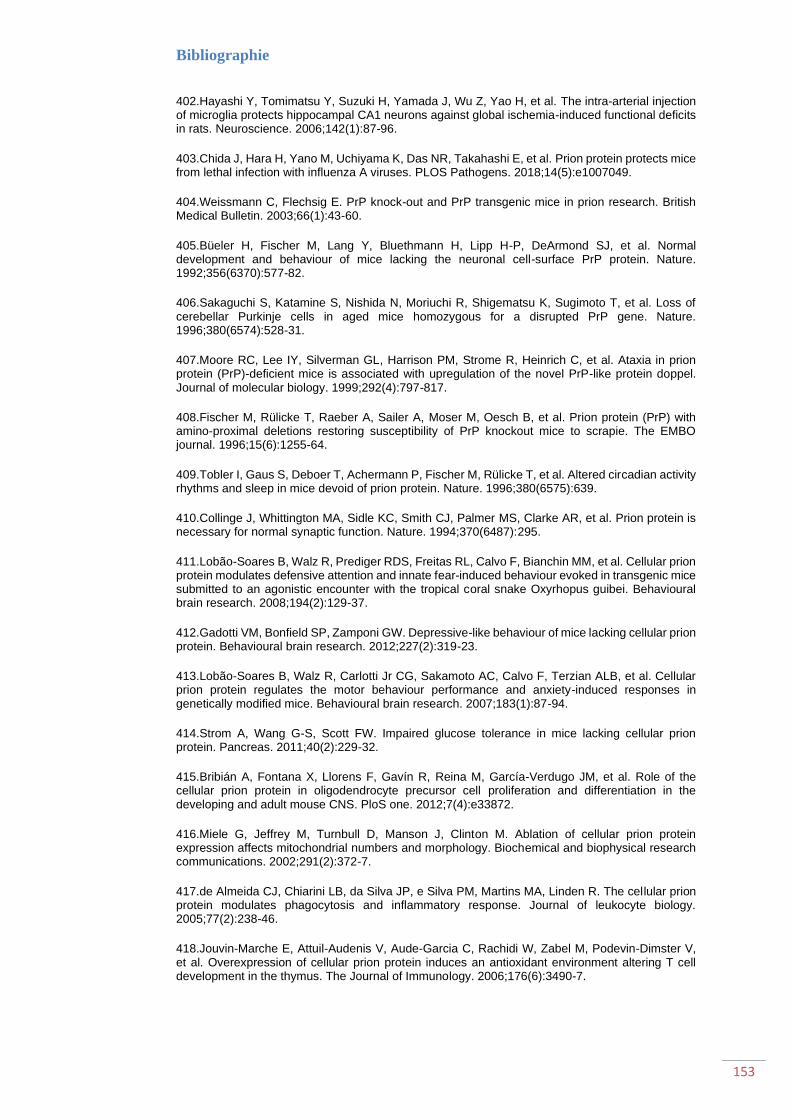
Bibliographie
153
402.Hayashi Y, Tomimatsu Y, Suzuki H, Yamada J, Wu Z, Yao H, et al. The intra-arterial injection of microglia protects hippocampal CA1 neurons against global ischemia-induced functional deficits in rats. Neuroscience. 2006;142(1):87-96.
403.Chida J, Hara H, Yano M, Uchiyama K, Das NR, Takahashi E, et al. Prion protein protects mice from lethal infection with influenza A viruses. PLOS Pathogens. 2018;14(5):e1007049.
404.Weissmann C, Flechsig E. PrP knock-out and PrP transgenic mice in prion research. British Medical Bulletin. 2003;66(1):43-60.
405.Büeler H, Fischer M, Lang Y, Bluethmann H, Lipp H-P, DeArmond SJ, et al. Normal development and behaviour of mice lacking the neuronal cell-surface PrP protein. Nature. 1992;356(6370):577-82.
406.Sakaguchi S, Katamine S, Nishida N, Moriuchi R, Shigematsu K, Sugimoto T, et al. Loss of cerebellar Purkinje cells in aged mice homozygous for a disrupted PrP gene. Nature. 1996;380(6574):528-31.
407.Moore RC, Lee IY, Silverman GL, Harrison PM, Strome R, Heinrich C, et al. Ataxia in prion protein (PrP)-deficient mice is associated with upregulation of the novel PrP-like protein doppel. Journal of molecular biology. 1999;292(4):797-817.
408.Fischer M, Rülicke T, Raeber A, Sailer A, Moser M, Oesch B, et al. Prion protein (PrP) with amino-proximal deletions restoring susceptibility of PrP knockout mice to scrapie. The EMBO journal. 1996;15(6):1255-64.
409.Tobler I, Gaus S, Deboer T, Achermann P, Fischer M, Rülicke T, et al. Altered circadian activity rhythms and sleep in mice devoid of prion protein. Nature. 1996;380(6575):639.
410.Collinge J, Whittington MA, Sidle KC, Smith CJ, Palmer MS, Clarke AR, et al. Prion protein is necessary for normal synaptic function. Nature. 1994;370(6487):295.
411.Lobão-Soares B, Walz R, Prediger RDS, Freitas RL, Calvo F, Bianchin MM, et al. Cellular prion protein modulates defensive attention and innate fear-induced behaviour evoked in transgenic mice submitted to an agonistic encounter with the tropical coral snake Oxyrhopus guibei. Behavioural brain research. 2008;194(2):129-37.
412.Gadotti VM, Bonfield SP, Zamponi GW. Depressive-like behaviour of mice lacking cellular prion protein. Behavioural brain research. 2012;227(2):319-23.
413.Lobão-Soares B, Walz R, Carlotti Jr CG, Sakamoto AC, Calvo F, Terzian ALB, et al. Cellular prion protein regulates the motor behaviour performance and anxiety-induced responses in genetically modified mice. Behavioural brain research. 2007;183(1):87-94.
414.Strom A, Wang G-S, Scott FW. Impaired glucose tolerance in mice lacking cellular prion protein. Pancreas. 2011;40(2):229-32.
415.Bribián A, Fontana X, Llorens F, Gavín R, Reina M, García-Verdugo JM, et al. Role of the cellular prion protein in oligodendrocyte precursor cell proliferation and differentiation in the developing and adult mouse CNS. PloS one. 2012;7(4):e33872.
416.Miele G, Jeffrey M, Turnbull D, Manson J, Clinton M. Ablation of cellular prion protein expression affects mitochondrial numbers and morphology. Biochemical and biophysical research communications. 2002;291(2):372-7.
417.de Almeida CJ, Chiarini LB, da Silva JP, e Silva PM, Martins MA, Linden R. The cellular prion protein modulates phagocytosis and inflammatory response. Journal of leukocyte biology. 2005;77(2):238-46.
418.Jouvin-Marche E, Attuil-Audenis V, Aude-Garcia C, Rachidi W, Zabel M, Podevin-Dimster V, et al. Overexpression of cellular prion protein induces an antioxidant environment altering T cell development in the thymus. The Journal of Immunology. 2006;176(6):3490-7.

Bibliographie
154
419.Coulpier M, Messiaen S, Boucreaux D, Eloit M. Axotomy-induced motoneuron death is delayed in mice overexpressing PrPc. Neuroscience. 2006;141(4):1827-34.
420.Rangel Rincones AH, Madroñal N, Gruart i Massó A, Gavín Marín R, Llorens Torres F, Sumoy L, et al. Regulation of GABA (A) and glutamate receptor expression, synaptic facilitation and long-term potentiation in the hippocampus of prion mutant mice. PLoS ONE, 2009, 4 (10): e7592. 2009.
421.Westaway D, DeArmond SJ, Cayetano-Canlas J, Groth D, Foster D, Yang S-L, et al. Degeneration of skeletal muscle, peripheral nerves, and the central nervous system in transgenic mice overexpressing wild-type prion proteins. Cell. 1994;76(1):117-29.
422.Steele AD, Emsley JG, Özdinler PH, Lindquist S, Macklis JD. Prion protein (PrPc) positively regulates neural precursor proliferation during developmental and adult mammalian neurogenesis. Proceedings of the National Academy of Sciences. 2006;103(9):3416-21.
423.Thackray AM, Bujdoso R. PrPc expression influences the establishment of herpes simplex virus type 1 latency. Journal of virology. 2002;76(5):2498-509.

155
Annexes
A – Publications
✓ Involvement of the prion protein in the protection of the human
bronchial epithelial barrier against oxidative stress.
Antioxidants and Redox Signaling (2019).
DOI: 10.1089/ars.2018.7500.
✓ Copper Homeostasis and Oxidative Stress in Polarized Cystic Fibrosis
Human Bronchial Epithelia: Role in Inflammation Responses.
Soumit dans la revue “Journal of inflammation”
B – Curriculum Vitae

Page 1 of 46 Antioxidants and Redox Signaling
© Mary Ann Liebert, Inc.
DOI: 10.1089/ars.2018.7500
1Antioxidan
ts and Red
ox Sign
aling
Invo
lvemen
t of the Prion Protein in
the Protection of the Human
Bronchial Epithelial B
arrier against Oxidative Stress (DOI: 10.1089/ars.2018.7500)
This pap
er has bee
n pee
r‐review
ed and accep
ted for publication, b
ut has yet to undergo
copyediting an
d proof correction. The final published
version m
ay differ from this proof.
Original Research Communication
Involvement of the Prion Protein in the Protection of the Human
Bronchial Epithelial Barrier against Oxidative Stress
Amal Kouadri1, Mariam El Khatib1, Johanna Cormenier1, Sylvain Chauvet1, Wael Zeinyeh1,
Micheline El Khoury1, Laurence Macari1, Pierre Richaud3, Christelle Coraux4, Isabelle
Michaud‐Soret1, Nadia Alfaidy2 and Mohamed Benharouga1
1 Univ. Grenoble Alpes, CNRS, UMR 5249, CEA, BIG, CBM, F‐38000 Grenoble, France.
2 Univ. Grenoble Alpes, INSERM U1036, CEA, BIG, BCI, F‐38000 Grenoble, France.
3 Univ. Aix‐Marseille, CNRS, CEA, Institut de Biosciences et Biotechnologies d’Aix‐Marseille
(BIAM), UMR 7265, CEA Cadarache, Saint‐Paul‐lez Durance F‐13108, France.
4 Institut National de la Santé et de la Recherche Médicale (INSERM), UMR‐S 903, Reims,
France.
Running Title: Role of PrPC protein in the lung epithelial barrier.
Keywords: Prion protein, Bronchial epithelium, Oxidative stress, Copper, Junctional
proteins
Address correspondence to:
Dr. Mohamed Benharouga
LCBM‐UMR5249
DRF‐BIG, CEA‐Grenoble
17 rue des Martyrs, F‐38054, Grenoble cedex 09, France
Téléphone: (33)‐4‐38‐78‐44‐51
Fax: (33)‐4‐38‐78‐54‐87
E‐mail: [email protected]
Word count: 6106
Reference numbers: 73
Greyscale illustrations: 7
Color illustrations: 3 (online 3)
Dow
nloa
ded
by Y
OR
K U
NIV
ER
SIT
Y L
IBR
AR
IES
from
ww
w.li
eber
tpub
.com
at 1
2/21
/18.
For
per
sona
l use
onl
y.

Page 2 of 46
2
Antioxidan
ts and Red
ox Sign
aling
Invo
lvem
ent of the Prion Protein in
the Protection of the Human
Bronchial Epithelial B
arrier against Oxidative Stress (DOI: 10.1089/ars.2018.7500)
This pap
er has bee
n pee
r‐review
ed and accep
ted for publication, b
ut has yet to undergo
copyediting an
d proof correction. The final published
version m
ay differ from this proof.
Abstract
Aim: Bronchial epithelium acts as a defensive barrier against inhaled pollutants and
microorganisms. This barrier is often compromised in inflammatory airway diseases that
are characterized by excessive oxidative stress responses, leading to bronchial epithelial
shedding, barrier failure, and increased bronchial epithelium permeability. Among proteins
expressed in the junctional barrier and participating to the regulation of the response to
oxidative and to environmental stresses is the cellular prion protein (PrPC). However, the
role of PrPC is still unknown in the bronchial epithelium. Herein, we investigated the
cellular mechanisms by which PrPC protein participates into the junctional complexes
formation, regulation, and oxidative protection in human bronchial epithelium.
Results: Both PrPC mRNA and mature protein were expressed in human epithelial
bronchial cells. PrPC was localized in the apical domain and became lateral, at high degree
of cell polarization, where it co‐localized and interacted with adherens (E‐cadherin/‐
catenin) and desmosomal (desmoglein/desmoplakin) junctional proteins. No interaction
was detected with tight junction proteins. Disruption of such interactions induced the loss
of the epithelial barrier. Moreover, we demonstrated that PrPC protection against copper
associated oxidative stress was involved in multiple processes, including the stability of
adherens and desmosomal junctional proteins.
Innovation: PrPC is a pivotal protein in the protection against oxidative stress that is
associated with the degradation of adherens and desmosomal junctional proteins.
Conclusion: Altogether, these results demonstrate that the loss of the integrity of the
epithelial barrier by oxidative stress is attenuated by the activation of PrPC expression,
which deregulation might be associated to respiratory diseases.
Dow
nloa
ded
by Y
OR
K U
NIV
ER
SIT
Y L
IBR
AR
IES
from
ww
w.li
eber
tpub
.com
at 1
2/21
/18.
For
per
sona
l use
onl
y.

Page 3 of 46
3
Antioxidan
ts and Red
ox Sign
aling
Invo
lvem
ent of the Prion Protein in
the Protection of the Human
Bronchial Epithelial B
arrier against Oxidative Stress (DOI: 10.1089/ars.2018.7500)
This pap
er has bee
n pee
r‐review
ed and accep
ted for publication, b
ut has yet to undergo
copyediting an
d proof correction. The final published
version m
ay differ from this proof.
Introduction
The bronchial epithelium forms a critical barrier that is continually exposed to a variety of
environmental agents such as pollutants, allergens, particles and microorganisms that are
capable of causing tissue injury. These injuries are normally countered by the
establishment of an effective line of defense and repair mechanisms (51,65). The
periciliary surface fluid and mucus form the first line of defense of the airways, including
the production of abundant mediators and innate airway immune responses (31). The
second line is physical and consists of three main types of intercellular junctional
complexes responsible for the sealing of the epithelium: tight junctions, adherens
junctions and desmosomes (25,31,34). The disruption of the intercellular junction of
bronchial epithelium is associated with an increase in epithelial cell permeability and an
invasion by infectious agents (37,42,69). This phenomenon constitutes a hallmark of
inflammatory and chronic infectious diseases, such as cystic fibrosis (CF) (10). One of the
parameters that was reported to contribute to the airway epithelial barrier’s injury and
dysfunction is the oxidative stress status, induced by endogenous productions of reactive
oxygen species (ROS) (26,39,47). To protect and repair the injured epithelial barrier
following exposure to an oxidative stress, the cells setup an anti‐oxidative system that
involves molecules such as vitamins E and C, with the ability to scavenger intracellular free
radicals; peptides such as glutathione that chelate free copper; and enzymes such as
catalase and superoxide dismutase (SOD) (45,68).
The cellular prion protein (PrPC) is one of the proteins that have been reported to be
involved both in antioxidant protection (1,33) and in maintaining the integrity of the
intestinal epithelial barrier (49,54). PrPC is a glycosyl phosphatidyl inositol (GPI)‐anchored
glycoprotein known for its unique ability to undergo structural conversion from a normal
isoform (PrPC) into a pathogenic conformer known as scrapie (PrPSc) (57). The
accumulation of PrPSc aggregates in the brain is a distinctive feature of transmissible
spongiform encephalopathies (TSE), a group of animal and human lethal
neurodegenerative diseases (14). The physiological role of the cellular PrP has mainly been
associated with stress‐protection (60,73), copper homeostasis (2) and neuronal excitability
(38). Other functions related to cellular processes and metabolism have also been
reported for this protein (11). In epithelial cells, PrPC localization is still a matter of
Dow
nloa
ded
by Y
OR
K U
NIV
ER
SIT
Y L
IBR
AR
IES
from
ww
w.li
eber
tpub
.com
at 1
2/21
/18.
For
per
sona
l use
onl
y.

Page 4 of 46
4
Antioxidan
ts and Red
ox Sign
aling
Invo
lvem
ent of the Prion Protein in
the Protection of the Human
Bronchial Epithelial B
arrier against Oxidative Stress (DOI: 10.1089/ars.2018.7500)
This pap
er has bee
n pee
r‐review
ed and accep
ted for publication, b
ut has yet to undergo
copyediting an
d proof correction. The final published
version m
ay differ from this proof.
discussion and the majority of data were obtained in enterocytes and MDCK cells
(18,44,49,50,52,62,67). In these cells, PrPC was reported to be directed to the apical and/or
basolateral membranes (18,44,49,50,52,62,67).
Recently, in a fully polarized MDCK cells, a basolateral to apical transcytosis was also
reported for the full‐length PrPC and its fragments of cleavage (3). However, in highly
polarized enterocytes PrPC was found to be targeted to the lateral junctional complexes of
adjacent cells and to interact with junctional proteins (3,62).
To date, the role of the PrPC in the oxidative stress protection and in copper homeostasis
in epithelial cells, including bronchial epithelial cells, is still poorly documented and the link
between its protective function and its localization in the apical and/or basolateral
domains of bronchial epithelial cells is still unknown.
We used the human bronchial epithelial cells (16HBE14o‐) as a model (21) to investigate
the expression, localization, regulation and function of PrPC in relation to oxidative stress
and copper status. We demonstrated that PrPC protein i) is expressed at the apical and
basolateral sides ii) undergoes apical‐to‐basolateral transcytosis in fully polarized
16HBE14o‐ cells, iii) localizes and interacts with adherens junction proteins, vi) impacts
cell‐cell adhesion and cell junctions’ integrity. More importantly, we demonstrated that
PrPC is involved in copper homeostasis and in the protection against the collapse of the
bronchial epithelial barrier caused by oxidative stress.
Altogether, this study highlights, for the first time, the role of PrPC protein in the
protection of the lung barrier against oxidative stress by increasing its proper expression
and by stabilizing key junctional proteins. These findings will improve our understanding of
the oxidative stress‐associated with the loss of the integrity of the epithelial barrier in
inflammatory and chronic infectious lung diseases.
Results
PrPC expression and characterization in human bronchial epithelial cells
The expression of PrPC protein was examined in human pulmonary epithelial cells. We used
a well‐established transformed bronchial epithelial cell line 16HBE‐14o‐ (HBE) (16) that was
kindly provided by Dr. D.C. Gruenert (University of California, CA) and A549, a human lung
adenocarcinoma epithelial cell line. These cells are widely used as in vitro models to study
type II pulmonary epithelial cells (ATCC, (23)). Both cell lines are able to grow as adherent
Dow
nloa
ded
by Y
OR
K U
NIV
ER
SIT
Y L
IBR
AR
IES
from
ww
w.li
eber
tpub
.com
at 1
2/21
/18.
For
per
sona
l use
onl
y.

Page 5 of 46
5
Antioxidan
ts and Red
ox Sign
aling
Invo
lvem
ent of the Prion Protein in
the Protection of the Human
Bronchial Epithelial B
arrier against Oxidative Stress (DOI: 10.1089/ars.2018.7500)
This pap
er has bee
n pee
r‐review
ed and accep
ted for publication, b
ut has yet to undergo
copyediting an
d proof correction. The final published
version m
ay differ from this proof.
monolayer cells by developing cell‐cell interactions through junctional complexes.
However, only HBE cells are able to develop a progressive and high trans‐epithelial
resistance (TER). In our conditions, A549 reached a maximum TER‐value of approximately
18 Ω/cm2 after 9 days of culture, while HBE cells developed a TER of ~650 Ω/cm2 (Fig. S1‐
A). Comparable TER‐values have been reported previously (22). These findings confirm that
despite normal distribution of junctional barrier proteins in the both cell types, A549 cells
are not able to develop comparable tightness to HBE cells. This makes HBE cells a good
model to study the role of proteins that are involved in the development and the
maintenance of intercellular junctions.
Using RT‐PCR, we demonstrated that PrPC gene is expressed in HBE and A549 cells (Fig.
1A). As control, we used N2a, mouse neuroblastoma cells commonly used to study PrPC
(24,41,56), and HTR‐8/SVneo cells, a human anchoring trophoblast cell line, in which PrPC
expression has previously been characterized (2). PrPC mRNA was highly expressed in HBE
compared to A549 and N2a cells (Fig.1B). These results were confirmed by immunoblot
assay (Fig. 1C). The anti‐PrP antibodies recognized three different PrPC forms in HTR, N2a,
A549 and HBE cells. These forms correspond to the unglycosylated (U~24 kDa), the
immature glycosylated (I~26‐30 kDa), and the mature highly glycosylated PrPC (H~31‐37
kDa) (Fig. 1C). The level of expression was evaluated using Na+/K+‐ATPase α‐subunit as
loading control. Similar results were obtained using a second loading control, the β‐actin
(Fig. S1‐B).
At the cellular level, PrPC maturation is characterized by its degree of glycosylation and by
its expression at the plasma membrane (30). As previously described (18), the glycosylation
of mature PrPC was sensitive to N‐glycosidase F (F) and insensitive to endoglycosidase H
(H), indicating the expression of mature PrPC in A549 and HBE cells (Fig.1D).
To further characterize PrPC expression, we analyzed its digestion profile using the
proteinase K (PK) enzyme. It is well established that only PrPSC is resistant to PK, while PrPC
is completely digested (9). Our results showed that the PrPC protein expressed in HBE and
A549 cells were totally digested by PK (Fig. 1E), indicating that the PrPC protein detected in
lung epithelial cells exhibits a normal conformation.
Dow
nloa
ded
by Y
OR
K U
NIV
ER
SIT
Y L
IBR
AR
IES
from
ww
w.li
eber
tpub
.com
at 1
2/21
/18.
For
per
sona
l use
onl
y.

Page 6 of 46
6
Antioxidan
ts and Red
ox Sign
aling
Invo
lvem
ent of the Prion Protein in
the Protection of the Human
Bronchial Epithelial B
arrier against Oxidative Stress (DOI: 10.1089/ars.2018.7500)
This pap
er has bee
n pee
r‐review
ed and accep
ted for publication, b
ut has yet to undergo
copyediting an
d proof correction. The final published
version m
ay differ from this proof.
Human tissue and cellular localization of PrPC
To characterize the localization of PrPC protein in human bronchial epithelial cells and to
analyze the tissue distribution in human bronchi, we performed both
immunohistochemical and immunofluorescence analyses using human healthy lung
sections (Fig. 2A) and fixed A549 and HBE cells (Fig. 2B‐E).
At the tissue level, PrPC protein was mainly detected in cilia cells in which the localization
appeared to be cytoplasmic and at the plasma membrane, mostly at the lateral and apical
sides (Fig. 2A (c)). Low expression was observed in the connective tissue (Fig. 2A (c)).
Incubation in the absence of the primary antibody (Fig. 2A (a)), or in the presence of the
primary antibody (SAF32) that has been preincubated overnight with the immunogen
peptide (Fig. 2A (b)) confirmed the specificity of PrPC detection at the bronchial level.
For the cellular localization, we used immunofluorescence cell imaging and X/Z image
reconstitution to analyze the distribution of PrPC in exponentially growing (3 days of
culture) or in polarized (10 days of culture) A549 and HBE cells. When A549 and HBE cells
were used before the establishment of a cell‐cell junction (3 days), PrPC protein was
detected at the apical, lateral and in the cytoplasmic domains (Fig. 2B and 2C).
Interestingly, in confluent and polarized cells (10 days), only HBE cells that developed a
high TER (Fig. S1‐A) exhibited a unique lateral localization of PrPC (Fig. 2D and 2E), while
A549 cells, even after 10 days of culture, did not display any change in the PrPC
distribution, compared to those observed after 3 days of culture (Fig. 2B and 2C).
Altogether these results reveal that PrPC distribution in HBE cells is dependent upon the
cell polarization, highlighting its potential role in the maintenance and/or control of the
cell’s junction repertoire.
PrPC colocalizes with key junctional proteins
Since the immunolocalization results showed that PrPC was localized to the lateral
membrane of polarized HBE cells (Fig. 2), we investigated the co‐localization profiles of
PrPC with different junctional resident proteins using immunofluorescence imaging cells,
combined with X/Z image reconstitution. PrPC was found to co‐localize with ‐catenin and
E‐cadherin, two representative proteins of the adherens junctions (Fig. 3A and 3B). PrPC
also co‐localized with desmoplakin and desmoglein, two proteins of the desmosome
junctions (Fig. 3A and 3B).
Dow
nloa
ded
by Y
OR
K U
NIV
ER
SIT
Y L
IBR
AR
IES
from
ww
w.li
eber
tpub
.com
at 1
2/21
/18.
For
per
sona
l use
onl
y.

Page 7 of 46
7
Antioxidan
ts and Red
ox Sign
aling
Invo
lvem
ent of the Prion Protein in
the Protection of the Human
Bronchial Epithelial B
arrier against Oxidative Stress (DOI: 10.1089/ars.2018.7500)
This pap
er has bee
n pee
r‐review
ed and accep
ted for publication, b
ut has yet to undergo
copyediting an
d proof correction. The final published
version m
ay differ from this proof.
However, no co‐localization was found with ZO‐1 protein, a member of the tight junctions
(Fig. 3A and 3B). In A549 cells, PrPC protein also co‐localized with E‐cadherin, ‐catenin,
and desmoplakin (Fig. S1‐C and S1‐D).
Because it was reported that PrPC protein could also be localized into the nucleus of
intestinal cells (7), we searched for such localization in the HBE cells. Very light staining
was observed in the nucleus of HBE cells, suggesting a possible nuclear localization of PrPC
protein in this cell type. To verify the existence of such localization, we performed a
nucleus staining using DAPI (4',6‐diamidino‐2‐phenylindole), a fluorescent stain that binds
strongly to DNA; anti‐Ki‐67, a nucleus resident protein that is associated with cell
proliferation; and an anti‐PrPC antibody. Figure 3C shows that Ki‐67 and DAPI co‐localized
in the nucleus. However, no staining and/or colocalization was found with the PrPC protein
(Fig. 3C), suggesting that the localization and the role of PrPC in lung bronchial epithelial
cells are restricted to the plasma membrane.
PrPC interacts with junction‐associated proteins and is involved in the maintenance of the
junctional barrier
The co‐localization results suggested that PrPC protein might interact with junction‐
associated proteins. To verify this hypothesis, we immunoprecipitated PrPC protein using
an anti‐PrPC specific antibody (SAF‐32). The targeted proteins were detected using
western‐blot analysis. We demonstrated that PrPC does not interact with ZO‐1 (Fig. 4A).
However, PrPC interacts with ‐catenin, E‐cadherin, desmoplakin and desmoglein (Fig. 4A).
To confirm the specificity of the antibody‐antigen interaction, we used a non‐specific
antibody (mAb0) instead of the SAF‐32 antibody to immunoprecipitate the PrPC protein.
The results reported in figure S2‐A confirm the specificity of SAF‐32 for the PrPC protein.
These results suggest the involvement of PrPC in the establishment of the junctional
complexes in the bronchial epithelial tissue.
To test this hypothesis, we knocked down PrPC expression using siRNA strategy, and
measured the TER and fluorescein‐5‐isothiocyanate (FITC)‐inulin transport. To invalidate
the PrPC expression in HBE cells, we tested two different siRNA labelled #1 and #2. Both
siRNA knocked down PrPC expression at mRNA and protein levels, as reported in figure S2‐
B‐D. For the subsequent experiments using siRNA strategy, only siRNA#1 was used.
Dow
nloa
ded
by Y
OR
K U
NIV
ER
SIT
Y L
IBR
AR
IES
from
ww
w.li
eber
tpub
.com
at 1
2/21
/18.
For
per
sona
l use
onl
y.

Page 8 of 46
8
Antioxidan
ts and Red
ox Sign
aling
Invo
lvem
ent of the Prion Protein in
the Protection of the Human
Bronchial Epithelial B
arrier against Oxidative Stress (DOI: 10.1089/ars.2018.7500)
This pap
er has bee
n pee
r‐review
ed and accep
ted for publication, b
ut has yet to undergo
copyediting an
d proof correction. The final published
version m
ay differ from this proof.
The TER was assessed during 8 days of culture to determine junction formation and
strength (Fig. 4B). TER levels at days 8 of culture were plotted in figure 4C. As expected,
TER increased in a time‐dependent manner and reached a plateau after 8 days of culture
(~620 Ω.cm2), which reflects the formation of cell‐cell junctions (Fig. 4B and 4C).
Interestingly, in PrPC‐knockdown HBE cells the TER levels were lower (~50%, ~300 Ω.cm2)
compared to the control condition (Fig. 4B and 4C). In addition to TER results, the
evaluation of the transport of FITC‐inulin demonstrate the increase of the paracellular
permeability upon PrPC knock down in HBE cells (Fig. 4D). Thus, the decrease in TER
associated with enhanced permeability to inulin is an indicator of loss of bronchial
epithelial barrier function.
Altogether, these results demonstrate for the first time, the direct involvement of PrPC
protein in the junction formation of bronchial epithelial cells.
Cu upregulates PrPC expression in human bronchial epithelial cells and in mice bronchial
epithelial tissue
Since PrPC protein binds with high affinity 5 copper ions (8), we first analyzed the dose and
time‐dependent effects of Cu on PrPC expression in HBE cells. After Cu stimulation, PrPC
expression gradually increased in a concentration‐ (Fig. 5A and 5B) and time‐dependent
manners (Fig. 5D).
PrPC protein levels were quantified and normalized based on the levels of the alpha
subunit of Na+/K+‐ATPase (Fig. 5B). As shown in Fig. 5A, PrPC was detected in untreated
HBE cells as U‐, I‐ and H‐glycosylated forms. Following Cu stimulation, the three
glycosylated forms gradually increased after 24h of treatment (Fig. 5A and 5B). Similar to
PrPC protein expression, the PrPC mRNA levels were also significantly increased in response
to Cu treatment (Fig. 5C and 5E), suggesting a transcriptional effect. Using a non‐epithelial
N2a cells, we observed that copper treatment also increased the expression of the three
forms of PrPC protein (Fig S3‐A and B).
The high level of the H‐glycosylated form of PrPC observed following Cu treatment
suggested an increase of PrPC insertion into the plasma membrane. This hypothesis was
confirmed by the cell surface biotinylation assay (Fig. 6A). Image analysis revealed that,
compared to the control condition (absence of Cu), the abundance of biotinylated PrPC
Dow
nloa
ded
by Y
OR
K U
NIV
ER
SIT
Y L
IBR
AR
IES
from
ww
w.li
eber
tpub
.com
at 1
2/21
/18.
For
per
sona
l use
onl
y.

Page 9 of 46
9
Antioxidan
ts and Red
ox Sign
aling
Invo
lvem
ent of the Prion Protein in
the Protection of the Human
Bronchial Epithelial B
arrier against Oxidative Stress (DOI: 10.1089/ars.2018.7500)
This pap
er has bee
n pee
r‐review
ed and accep
ted for publication, b
ut has yet to undergo
copyediting an
d proof correction. The final published
version m
ay differ from this proof.
was increased ~25% following 24h of Cu‐treatment (100µM), suggesting that the H‐
glycosylated form is almost totally inserted into the plasma membrane (Fig. 6B).
Cu increases PrPC expression, specifically at the junctional side of HBE cells
To determine the HBE cells membrane domain (apical, lateral and/or basolateral) in which
the H‐form of PrPC protein was expressed following Cu treatment (Fig. 6A and B), we used
immunofluorescent assays (Fig. 6C and 6D). Following Cu treatment, we observed an
increase in PrPC expression in non‐ and in permeabilized HBE cells (Fig. 6C, X/Y). The X/Z
image reconstitution (Fig. 6C, X/Z) demonstrates that the plasma membrane fluorescence
increased specifically in the lateral side. The quantification of the intensity of the lateral
fluorescence indicated an increase of ~65% and ~40% in non‐ and in permeabilized HBE
cells, respectively (Fig. 6D), suggesting that PrPC insertion at the lateral side might play an
important role in the protection of the junctional barrier against the effects of oxidative
stress in bronchial epithelial cells.
To get more insights into the effect of copper on PrPC expression in an in vivo system, we
evaluated the effect of copper treatment on PrPC expression in bronchial epithelial tissue,
by watering 8‐10 weeks C57BL/6 mice with aquatic solution containing copper. The
efficiency of Cu treatment was determined by the measurement of blood Cu
concentrations as previously reported by our group (2).
Immunohistochemistry analysis of lung tissues collected from control and copper treated
mice showed that PrPC was expressed in ciliated and basal cells in control mice (Fig. S3‐B
(b) and (c)). Following copper treatment, the PrPC expression increased in bronchial tissues
(Fig. S‐B (c)). Altogether, these results suggest that there might be a strong relationship
between the concentration of extracellular copper and the levels of PrPC expression in the
bronchial epithelial tissue.
Elevated PrPC levels protect against Cu accumulation, Cu‐induced oxidative stress
production, and bronchial epithelial cell death
Copper homeostasis is finely regulated and any disruption of its physiological
concentrations causes an increase in the intracellular oxidative stress. The increase in PrPC
expression upon Cu treatment may reveal a stress protective response within the cell. To
test the role of elevated PrPC expression in preventing the Cu‐induced cytotoxicity, we first
measured the HBE cells viability using the MTT assay. After 24 h of incubation with the
Dow
nloa
ded
by Y
OR
K U
NIV
ER
SIT
Y L
IBR
AR
IES
from
ww
w.li
eber
tpub
.com
at 1
2/21
/18.
For
per
sona
l use
onl
y.

Page 10 of 46
10
Antioxidan
ts and Red
ox Sign
aling
Invo
lvem
ent of the Prion Protein in
the Protection of the Human
Bronchial Epithelial B
arrier against Oxidative Stress (DOI: 10.1089/ars.2018.7500)
This pap
er has bee
n pee
r‐review
ed and accep
ted for publication, b
ut has yet to undergo
copyediting an
d proof correction. The final published
version m
ay differ from this proof.
indicated Cu concentrations, HBE cells exhibited a significant cell death of ~30% at 100 µM
(Fig. 7A). Similar degree of cell death was observed at 200 µM (Fig. 7A).
Based on the high proportion of dead cells, we hypothesized that this might be the
consequence of an oxidative stress induced by the increase in the intracellular copper
concentrations. Using ICP‐AES assay, we confirmed that the intracellular concentrations of
Cu increased in a dose‐dependent manner (Fig. 7B). To confirm the relationship between
copper and the levels of PrPC expression, we knocked down the cellular prion protein using
siRNA strategy. Knockdown of PrPC had no significant effect on cell survival (Fig. 7C). A
significant increase in cell death was however observed in PrP‐depleted HBE cells (~80% at
24h) following incubation with 100 µM of Cu (Fig. 7C).
Then we monitored the oxidative‐stress production and intracellular Cu content following
Cu treatment in the absence or presence of PrPC. After exposure of HBE cells to 100 µM of
Cu, the DCF fluorescence, indicative of the presence of an oxidative stress, increased
compared to untreated HBE cells (Fig. 7D). In the PrPC‐silenced HBE cells, the level of DCF
fluorescence was similar compared to the control HBE (Fig. 7D). The treatment of PrP‐
knockdown HBE with 100 µM of Cu increased the oxidative stress accumulation by ~2‐fold
(Fig. 7D).
Finally, using PrP‐knockdown HBE cells we confirmed that PrPC participates into the
regulation of the intracellular Cu concentration (Fig. 7E). Using ICP‐AES we found that Cu
concentration in normal HBE was ~0.02 µg/106 cells (Fig. 7E). A slightly elevated copper
levels (~0.03 µg/106 cell) were observed in PrPC‐silenced HBE cells (Fig. 7E). When Cu was
added during 24h, the intracellular copper concentration was increased to ~0.8 µg/106 cell
in wild type and to ~1.3 µg/106 cell in PrPC‐depleted HBE, respectively (Fig. 7E).
ROS‐mediated increase of PrPC in HBE cells
Based on the results reported in figures 5A and 7D, 24h of treatment with 100 µM of
copper significantly increased PrPC expression and the production of pro‐oxidants
molecules in HBE cells. These data suggested that PrPC increased expression might in part
be due to a primary increase in pro‐oxidants molecules, such as hydrogen peroxide (H2O2).
This hypothesis was tested by treating HBE cells for 24h with 100 µM of H2O2 in the
absence or in the presence of 100 µM of Trolox, a hydrophilic analogue of vitamin E (Vit E)
that acts as a free radicals scavenger (63).
Dow
nloa
ded
by Y
OR
K U
NIV
ER
SIT
Y L
IBR
AR
IES
from
ww
w.li
eber
tpub
.com
at 1
2/21
/18.
For
per
sona
l use
onl
y.
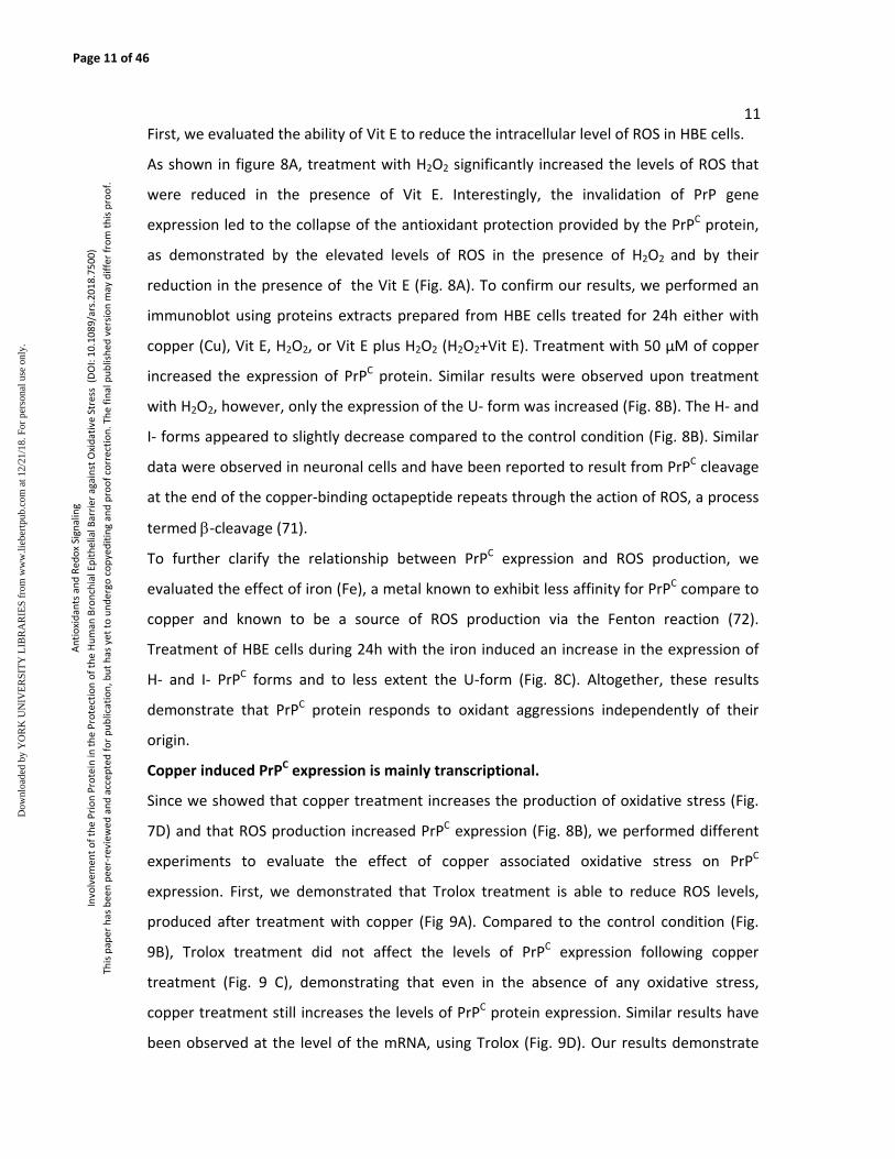
Page 11 of 46
11
Antioxidan
ts and Red
ox Sign
aling
Invo
lvem
ent of the Prion Protein in
the Protection of the Human
Bronchial Epithelial B
arrier against Oxidative Stress (DOI: 10.1089/ars.2018.7500)
This pap
er has bee
n pee
r‐review
ed and accep
ted for publication, b
ut has yet to undergo
copyediting an
d proof correction. The final published
version m
ay differ from this proof.
First, we evaluated the ability of Vit E to reduce the intracellular level of ROS in HBE cells.
As shown in figure 8A, treatment with H2O2 significantly increased the levels of ROS that
were reduced in the presence of Vit E. Interestingly, the invalidation of PrP gene
expression led to the collapse of the antioxidant protection provided by the PrPC protein,
as demonstrated by the elevated levels of ROS in the presence of H2O2 and by their
reduction in the presence of the Vit E (Fig. 8A). To confirm our results, we performed an
immunoblot using proteins extracts prepared from HBE cells treated for 24h either with
copper (Cu), Vit E, H2O2, or Vit E plus H2O2 (H2O2+Vit E). Treatment with 50 µM of copper
increased the expression of PrPC protein. Similar results were observed upon treatment
with H2O2, however, only the expression of the U‐ form was increased (Fig. 8B). The H‐ and
I‐ forms appeared to slightly decrease compared to the control condition (Fig. 8B). Similar
data were observed in neuronal cells and have been reported to result from PrPC cleavage
at the end of the copper‐binding octapeptide repeats through the action of ROS, a process
termed ‐cleavage (71).
To further clarify the relationship between PrPC expression and ROS production, we
evaluated the effect of iron (Fe), a metal known to exhibit less affinity for PrPC compare to
copper and known to be a source of ROS production via the Fenton reaction (72).
Treatment of HBE cells during 24h with the iron induced an increase in the expression of
H‐ and I‐ PrPC forms and to less extent the U‐form (Fig. 8C). Altogether, these results
demonstrate that PrPC protein responds to oxidant aggressions independently of their
origin.
Copper induced PrPC expression is mainly transcriptional.
Since we showed that copper treatment increases the production of oxidative stress (Fig.
7D) and that ROS production increased PrPC expression (Fig. 8B), we performed different
experiments to evaluate the effect of copper associated oxidative stress on PrPC
expression. First, we demonstrated that Trolox treatment is able to reduce ROS levels,
produced after treatment with copper (Fig 9A). Compared to the control condition (Fig.
9B), Trolox treatment did not affect the levels of PrPC expression following copper
treatment (Fig. 9 C), demonstrating that even in the absence of any oxidative stress,
copper treatment still increases the levels of PrPC protein expression. Similar results have
been observed at the level of the mRNA, using Trolox (Fig. 9D). Our results demonstrate
Dow
nloa
ded
by Y
OR
K U
NIV
ER
SIT
Y L
IBR
AR
IES
from
ww
w.li
eber
tpub
.com
at 1
2/21
/18.
For
per
sona
l use
onl
y.

Page 12 of 46
12
Antioxidan
ts and Red
ox Sign
aling
Invo
lvem
ent of the Prion Protein in
the Protection of the Human
Bronchial Epithelial B
arrier against Oxidative Stress (DOI: 10.1089/ars.2018.7500)
This pap
er has bee
n pee
r‐review
ed and accep
ted for publication, b
ut has yet to undergo
copyediting an
d proof correction. The final published
version m
ay differ from this proof.
that copper increases the expression of PrPC through the Cu‐associated oxidative stress
but also through a transcriptional pathway. However, the latter is more predominant as
previously reported in placental cells (2).
PrPC protein protects the junctional proteins against Cu oxidative effects
The functional effects of Cu on the permeability of the junctional barrier in polarized HBE
cells were monitored by measuring TER values upon their treatment with 100 µM of Cu,
from the apical side, for the indicated times. As shown in Fig. 10A, copper ions decreased
TER values in a time‐dependent manner, as evidenced by the decrease in TER values
(~20%) (Fig. 10B). The levels of TER measured in the HBE cells treated with copper were
compared to those obtained in PrPC‐knockdown HBE cells (Fig. 10B), at day 4 of
confluence.
The results indicated that in HBE cells the decrease in PrPC expression (siPrPC) increased
cell permeability (~50%) compared to Cu treatment (~18%). Our results suggest that the
increase in PrPC expression in the lateral side of bronchial epithelial cells (Fig. 6) protects
the junctional barrier against the Cu‐inducing oxidative stress that might affect the levels
of expression of junctional protein, as was previously reported (20).
To test our hypothesis, we first evaluated the effect of copper treatment on the steady
state expression of tights (ZO‐1, occluding (Occ)), adherents (‐catenin (‐Cat) and E‐
cadherin (E‐Cad)) and desmosome (desmoplakin (Dep)) junctions. The results showed that
in the presence of copper only ZO‐1 expression decreased in a time‐dependent manner
(Fig. 10C). Neither ‐Cat/E‐Cad nor desmoplakin proteins were affected by the copper
treatment (Fig. 10C).
Subsequently, we assessed the stability of ZO‐1 and occludin proteins using cycloheximide
(CHX) assays in the absence or in the presence of copper. As reported in figure 10D the
degradation of ZO‐1 and occludin (Occ) were accelerated in the presence of copper.
Using PrPC‐knockdown HBE cells, we evaluated the role of PrPC protein using
cycloheximide (CHX) assays in the absence or in the presence of copper (Fig. 10E).
Since there was no co‐localization, or interaction between PrPC and ZO‐1 protein (Fig. 3
and 4), we were not surprised to observe that the degradation rate of ZO‐1 was not
accelerated in PrPC‐knockdown HBE cells, in the absence or in the presence of copper (Fig.
10E). Unexpectedly, occludin degradation was accelerated under the same conditions,
Dow
nloa
ded
by Y
OR
K U
NIV
ER
SIT
Y L
IBR
AR
IES
from
ww
w.li
eber
tpub
.com
at 1
2/21
/18.
For
per
sona
l use
onl
y.

Page 13 of 46
13
Antioxidan
ts and Red
ox Sign
aling
Invo
lvem
ent of the Prion Protein in
the Protection of the Human
Bronchial Epithelial B
arrier against Oxidative Stress (DOI: 10.1089/ars.2018.7500)
This pap
er has bee
n pee
r‐review
ed and accep
ted for publication, b
ut has yet to undergo
copyediting an
d proof correction. The final published
version m
ay differ from this proof.
suggesting that ZO‐1 and occludin are differentially regulated by PrPC and copper, although
they belong to the same family of tight junctions. The degradation rate of desmoplakin
(Dep) and desmoglein (Deg), proteins that interact with PrPC, was significantly increased in
PrPC‐knockdown HBE cells, particularly in the presence of copper (Fig. 10E). Similar results
were obtained using a second siRNA (siPrP (#2)) (Fig. S4‐B) compared to the control, using
scrambled siRNA (Fig. S4‐A).
These results demonstrate that by interacting and stabilizing the junctional proteins
(adherens and desmosomes), PrPC participates in the oxidative stress protection and in the
maintenance of the bronchial epithelial junctional barrier.
Discussion
The present study is the first to demonstrate that PrPC is expressed in the human bronchial
epithelial cells, which form a physical barrier to inhaled toxic particles, bacteria and viruses
(58). Using multiple approaches, we provide strong evidences for a novel role of the PrPC
protein in the protection of the bronchial epithelial barrier against potentially generated
oxidative stress. The in vivo studies performed both in mice and human demonstrated that
the PrPC protein is highly expressed in the bronchial epithelial cells, comforting the search
of its role in this epithelium.
Importantly, we demonstrate that PrPC expression, glycosylation and digestion profiles
detected in A549 and HBE cell lines were similar to those reported in intestinal epithelial
cells (49), (50) and placental trophoblast cells (2). Moreover, the amount of PrPC protein
was 4 and 5 times higher in human HBE cells compared to human (SH‐SY5Y) and mice
(N2a) neuronal cells (data not shown), suggesting an important role for PrPC in the
bronchial epithelium function.
Compared to renal and intestinal MDCK and Caco‐2 epithelial cells (18,49), HBE cells
display also a high number of PrP gene copies and a high levels of PrPC protein.
HTR, A549 and HBE cell lines mostly expressed PrPC forms of lower electrophoretic
mobility with apparent molecular weights between 30 and 37 kDa, compared to N2a cells
that exhibit 26‐30 kDa molecular weights for the PrPC. Similar results were previously
reported in N2a cells when compared to the rat B104 neuroblastoma, the mouse
hypothalamic GT1‐7, and the rat adrenal pheochromocytoma PC‐12 cell lines (48). Monnet
Dow
nloa
ded
by Y
OR
K U
NIV
ER
SIT
Y L
IBR
AR
IES
from
ww
w.li
eber
tpub
.com
at 1
2/21
/18.
For
per
sona
l use
onl
y.

Page 14 of 46
14
Antioxidan
ts and Red
ox Sign
aling
Invo
lvem
ent of the Prion Protein in
the Protection of the Human
Bronchial Epithelial B
arrier against Oxidative Stress (DOI: 10.1089/ars.2018.7500)
This pap
er has bee
n pee
r‐review
ed and accep
ted for publication, b
ut has yet to undergo
copyediting an
d proof correction. The final published
version m
ay differ from this proof.
et al (48) reported that in N2a cells the threshold of detection of the PrPC was too low to
evaluate the distribution of its multiple forms.
Although the molecular difference between the glycosylated forms in N2a and the other
cells types is still to be elucidated, the deglycosylation results clearly showed that the 30‐
37 kDa PrPC forms detected in HTR, A549 and HBE cells correspond to the PrPC forms
observed in N2a cells. The N‐glycosylated forms of PrPC might exhibit different glycan
chains or derivatizations depending on the cell type. PrPC deglycosylation in the different
cell extracts including N2a cells (Fig. S4‐C) indicated that the glycosylation processes were
similar for all samples. The cellular prion protein glycosylation and expression levels may
vary depending on the cell’s density and/or state of differentiation, as previously reported
for neuroblastoma B104 cells (48). Other possibility is that PrPC protein may undergo
proteolytic processing resulting in the production of different fragments as previously
reported (40). In our study, copper‐dependent ‐cleavage is certainly taking place. The β‐
cleavage performed by radicals in a Fenton type reaction has been proposed to take place
around the amino acid position, 90 of the PrPC protein (40). In consequence of this
cleavage, an N‐terminal fragment of ~9 kDa (termed N2) is released whereas a
corresponding C2 fragment of 18–20 kDa (depending on the glycosylation status) remains
bound to the cellular membrane. To avoid the interference of this parameter with the
interpretation of our results regarding the expression of the full length PrPC protein under
its different glycosylation states, we used the SAF‐32 mAb that recognizes the whole
octarepeated region of PrPC spanning residues 51‐98 (28).
To date, there is no consensus regarding the precise localization of the PrPC in epithelia.
Several groups, including ours, have proposed an apical localization of PrPC in MDCK cells
(13,18,59), which diverge from other studies showing a baso‐lateral localization (62,67).
These findings suggest that PrPC localization is either controlled by the degree of epithelial
cell polarization or that the PrPC protein contributes to epithelial cell fusion and
polarization. Such transcytosis trafficking has been recently observed in polarized
epithelial MDCK cells (3).
PrPC was detected in junctional complexes of the lateral membranes of adjacent polarized
HBE cells where it was found to co‐localize and interact with E‐cadherin and desmoglein,
and with their cytoplasmic partner, ‐catenin (plakoglobin) and desmoplakin. Interestingly,
Dow
nloa
ded
by Y
OR
K U
NIV
ER
SIT
Y L
IBR
AR
IES
from
ww
w.li
eber
tpub
.com
at 1
2/21
/18.
For
per
sona
l use
onl
y.

Page 15 of 46
15
Antioxidan
ts and Red
ox Sign
aling
Invo
lvem
ent of the Prion Protein in
the Protection of the Human
Bronchial Epithelial B
arrier against Oxidative Stress (DOI: 10.1089/ars.2018.7500)
This pap
er has bee
n pee
r‐review
ed and accep
ted for publication, b
ut has yet to undergo
copyediting an
d proof correction. The final published
version m
ay differ from this proof.
no co‐localization or interaction of PrPC was reported with tight junction proteins. Similar
results were described in enterocytes, where PrPC was reported to be targeted to cell–cell
junctional zone and to interact with several desmosomal (desmoglein‐2, desmoplakin, γ‐
catenin), but not with adherens proteins (49). The absence of interaction with E‐cadherin
in enterocytes cells, even if a cellular co‐localization was observed (49), suggested that in
the bronchial epithelium such interactions might be important for the stability and the
function of the junctional barrier.
In relation to the intracellular localization of PrPC, it was reported that this protein could
also be localized into the nucleus of intestinal cells (7). Based on the primary structure of
the PrP protein and the site of its biosynthesis and maturation (64), the corresponding
sialoglycoprotein is normally addressed to the plasma membrane where it can be released
by phospholipase or protease treatments (64). Importantly, in the mouse scrapie‐infected
neuroblastoma cell line (ScN2a), immunocytochemical studies revealed that PrPSc and/or
PrPC are present, not only at the plasma membrane or in cytoplasmic compartments
(12,43), but also in the nuclear compartment, particularly into the nucleolus (55).
According to its size (24 to 37 kDa), PrPC is able to enter the nucleus either in an energy‐
dependent pathway if it possesses an NLS or by passive diffusion. Indeed, the analysis of
the primary structure of the mature protein have revealed the existence of a nuclear
localization sequence in N‐terminal extremity (KKRPKP noted Pnls) (6,27). Through our
experiments, we cannot conclude that PrPC protein is localized into the nucleus of HBE
cells. This statement is based on the co‐immunofluorescence experiments that did not
show any co‐localisation between the PrPC and Ki‐67. Interestingly, as reported for
trophoblast cells (2), the incubation of HBE cells with Cu increased PrPC plasma membrane
insertion, providing evidences for a potential role of PrPC as a Cu sensor and/or chelator at
the cell surface to protect these cells from Cu excess. However, in neurons, it has been
reported that treatment with Cu increased PrPC endocytosis (53), suggesting that the role
of PrPC protein in copper homeostasis is cell‐dependent. Importantly, our in vivo approach
substantiated the increase of PrPC expression at the bronchial level, upon Cu intake by the
mice. Because of the immediate endocytosis of PrPC protein upon copper binding, one can
speculate that prion protein could constitute a real copper transporter. In fact, once PrPC
binds extracellular copper on the cell surface and, when endocytosed and exposed to
Dow
nloa
ded
by Y
OR
K U
NIV
ER
SIT
Y L
IBR
AR
IES
from
ww
w.li
eber
tpub
.com
at 1
2/21
/18.
For
per
sona
l use
onl
y.

Page 16 of 46
16
Antioxidan
ts and Red
ox Sign
aling
Invo
lvem
ent of the Prion Protein in
the Protection of the Human
Bronchial Epithelial B
arrier against Oxidative Stress (DOI: 10.1089/ars.2018.7500)
This pap
er has bee
n pee
r‐review
ed and accep
ted for publication, b
ut has yet to undergo
copyediting an
d proof correction. The final published
version m
ay differ from this proof.
lower pH of intracellular organelles, releases the metal ions. This hypothesis is very
tempting, however multiple biochemical studies rather support the sensor role of PrPC for
copper and for other metals such as Zn2+ and Mn2+ (35,46,70).
The increase in PrPC expression following Cu treatment caused a significant increase in the
intracellular Cu concentration, along with an increase in ROS production. In addition, the
loss of the expression of PrPC protein, in the presence of Cu, caused dramatic effects on
HBE viability and increased Cu content and ROS production. These finding highlights the
stress protective role of PrPC protein in bronchial epithelial cells and substantiate recent
publish data reporting a stress protective role of PrPC protein (60,73). We also confirm that
PrPC protein respond to other type of stress such as H2O2 and iron. Regarding iron, recent
reports also indicated that PrPC promotes cellular iron uptake by functioning as a
ferrireductase (29). However, the metal transporter(s) involved in PrPC‐associated iron
uptake and transport are not known. Human neuroblastoma cells (M17) that over‐express
PrPC show resistance to higher amounts of ferric ammonium citrate (FAC) relative to non‐
transfected cells, suggesting a dose‐dependent protection against redox‐iron. One likely
explanation for these observations is that interaction of redox‐iron with copper and iron
bound PrP initiates the Fenton reaction, resulting in denaturation and aggregation of PrP
at the cell surface (17).
In addition, we demonstrated that even if PrPC expression is increased during oxidative
stress, under copper treatment, its levels of expression are mainly controlled through a
transcriptional pathway.
However, the link between the role of PrPC in bronchial epithelial barrier and its associated
role against the oxidative stress is still to be demonstrate.
One of the consequences of chronic oxidative stress in the lung is the bronchial epithelial
shedding and the breakdown of epithelium cohesion (4,5,36). Several reports in the
literature have shown that damages caused by free radicals in epithelial cells are
associated with an increase in the tight junction‘s permeability (4,5,61). We confirmed this
finding in HBE cells and further demonstrate that these effects were ZO‐1 and occluding
dependent. Similar results have been reported in Caco‐2 cells (19,20). The PrP gene
invalidation in HBE cells caused a decrease in TER along with an increase in the instability
Dow
nloa
ded
by Y
OR
K U
NIV
ER
SIT
Y L
IBR
AR
IES
from
ww
w.li
eber
tpub
.com
at 1
2/21
/18.
For
per
sona
l use
onl
y.

Page 17 of 46
17
Antioxidan
ts and Red
ox Sign
aling
Invo
lvem
ent of the Prion Protein in
the Protection of the Human
Bronchial Epithelial B
arrier against Oxidative Stress (DOI: 10.1089/ars.2018.7500)
This pap
er has bee
n pee
r‐review
ed and accep
ted for publication, b
ut has yet to undergo
copyediting an
d proof correction. The final published
version m
ay differ from this proof.
of tight and desomosomal protein junctions, highlighting the importance of PrPC protein in
oxidative protection of junctional proteins.
Through its interactions, PrPC protein might ensure the stability and an oxidative stress
protection for E‐cadherin, ‐catenin, desmoplakin and desmoglein. The absence of such
interactions might lead to the degradation of key junctional proteins, as was observed for
ZO‐1 and occludin. Previous studies, reported that in Caco‐2 cells copper induced a
concentration‐ and time‐dependent decreases in the expression of tight junction proteins
along with an increase in their permeability (19,20). These effects might be explained by
the absence of interactions between PrPC and E‐cadherin, as previously reported in these
cells (49).
Altogether our results demonstrate that PrPC protein by forming complexes with E‐
cadherin/γ‐catenin and desmoglein/plakoglobin proteins participates in the regulation, the
stability and the oxidative protection of adherens and desmosomal junctions in the
bronchial cell barrier. Yet, it is still to be demonstrated whether the induction of PrPC
expression and its lateral localization are protective mechanisms during chronic airway
diseases.
Innovation:
Chronic bronchial infection, inflammation and oxidative stress in pulmonary diseases such
as cystic fibrosis lead to epithelial cell disorganization along with increased permeability.
Yet, the mechanism by which this occurs is still to be determined. Here we brought
evidences that the cellular prion is involved in the protection of the bronchial epithelial
barrier upon oxidative stress occurrence. PrPC protection of the lung epithelial barrier was
exhibited through its capacity to interact and to stabilize adherens and desmosomal
protein.
Experimental Procedures
Human tissues collection
Peripheral human lung tissue (containing non cartilaginous airways) was obtained at the
time of lung transplantation from non‐smoking controls undergoing lung resection upon
peripheral lung cancer (Grenoble Hospital). Lung tissue samples were fixed in 10% neutral
buffered formalin by inflation‐immersion and embedded in paraffin. Collection and
Dow
nloa
ded
by Y
OR
K U
NIV
ER
SIT
Y L
IBR
AR
IES
from
ww
w.li
eber
tpub
.com
at 1
2/21
/18.
For
per
sona
l use
onl
y.

Page 18 of 46
18
Antioxidan
ts and Red
ox Sign
aling
Invo
lvem
ent of the Prion Protein in
the Protection of the Human
Bronchial Epithelial B
arrier against Oxidative Stress (DOI: 10.1089/ars.2018.7500)
This pap
er has bee
n pee
r‐review
ed and accep
ted for publication, b
ut has yet to undergo
copyediting an
d proof correction. The final published
version m
ay differ from this proof.
processing of human lungs were conformed to the declaration of Helsinki and to all the
rules of the local Committees on Human Research. Informed consent was obtained from
each patient.
Mice tissues collection
For the in vivo experiments, mice (8–10‐weeks old, 20–25 g) of C57BL/6 strains were used.
Mice were obtained from the animal facility of CNRS (Orléans, France). The mice were
randomly assigned to receive either distilled water, containing 50 ppm sucrose, or 250
ppm copper (copper sulfate (CuSO4)) and 50 ppm sucrose.
All procedures involving animals and their care were approved by the local institutional
Ethics Committee. Mice were sacrificed using cervical dislocation after chloral hydrate
anesthesia. Fresh lungs were collected from each animal. Shortly after collection, tissues
were fixed in 4% paraformaldehyde at room temperature.
Immunohistochemistry
5 µm paraffin‐embedded sections were prepared from human non‐smoking peripheral
lung tissues. Immunohistochemistry was performed as described previously (2). Sections
were incubated with anti‐PrP antibody (SAF32, Bertin‐Pharma, France). Immunopositive
staining was detected using a Vectastain ABC kit (Vector Labs) using DAB (3, 3’‐
diaminobenzidine) as the chromagen (Vector Labs). Slides were counterstained using
hematoxylin (Sigma Aldrich, France). Control sections were treated with either the
secondary antibody alone or with the anti‐PrPC antibody (SAF‐32) that has been
preabsorbed overnight at 4°C with the appropriate antigen peptide (Covalab company,
Lyon, France).
Immunohistochemistry was also performed on lung sections collected form C57 mice as
described previously (2).
Cell culture
All culture media were purchased from Life‐Technologies (France). The mouse
neuroblastoma (N2a) and adenocarcinoma human alveolar basal epithelial (A549) cell lines
were cultured in high glucose DMEM‐Glutamax (Dulbecco’s Modified Eagle’s Medium)
medium, whereas HBE cells were cultured in normal MEM (Modified Eagle’s Medium)
medium.
Dow
nloa
ded
by Y
OR
K U
NIV
ER
SIT
Y L
IBR
AR
IES
from
ww
w.li
eber
tpub
.com
at 1
2/21
/18.
For
per
sona
l use
onl
y.

Page 19 of 46
19
Antioxidan
ts and Red
ox Sign
aling
Invo
lvem
ent of the Prion Protein in
the Protection of the Human
Bronchial Epithelial B
arrier against Oxidative Stress (DOI: 10.1089/ars.2018.7500)
This pap
er has bee
n pee
r‐review
ed and accep
ted for publication, b
ut has yet to undergo
copyediting an
d proof correction. The final published
version m
ay differ from this proof.
All media were supplemented with 10% fetal Bovine serum (Biowest, France) and 1%
antibiotics mix (100 IU/ml of penicillin and 10 mg/ml streptomycin, Sigma). Cell cultures
were performed at 37°C in a 5% CO2/air atmosphere. Where indicated, cells were treated
with vehicle or different concentrations of copper (Cu) for the indicated times.
siRNA knockdown of PrPC and Transfection
The silencing PrPC expression essay was performed following the protocol described in a
previous report (32). The experiments where the expression of PrP gene was invalidated
using siRNA strategy were performed in parallel of controlled experiments using scrambled
siRNA. The presence of the transfectant reagent (Lipofectamine® 2000, France) and siRNA
scramble did not affect the cell viability compared to the control (untreated) condition.
The results are reported as supplementary data (Fig. S4‐D).
Cells lysates
For the steady state expression and endoglycosidase experiments, cells were washed twice
with ice‐cold phosphate buffer saline (PBS) and lysed at 4°C for 20 min in PBS containing
1% NP40, 0.5% deoxycholic acid, and 0.1% SDS added with protease inhibitors (10 mM
PMSF, 1 µM leupeptin/pepstatin A and 1mg/ml of iodoacetamide). For proteinase K (PK)
treatment, cells were lysed in PBS containing 100 mM NaCl, 10 mM Tris, 10 mM EDTA,
0.5% NP‐40, 0.5% deoxycholic acid, pH 7.4. Protein concentrations were measured using
the Micro BCA protein assay kit (Thermo Scientific, France).
Endoglycosidase and proteinase K (PK) digestion
To distinguish between high mannose and complex‐type N‐linked oligosaccharide
modification of PrPC, cell lysates were incubated with endoglycosidase H (H) and peptide
N‐glycosidase F (F) as previously reported (2). The mobility shift of deglycosylated PrPC was
visualized by immunoblotting using an anti‐PrP mAb (SAF32). For PK treatment, cells
lysates were incubated with 20μg/ml PK for 3h at 37°C, and the reaction was stopped by
adding PMSF (3mM).
Immunoprecipitation, electrophoresis and immunoblotting
For immunoprecipitation assay, cells were lysed at 4°C for 30 min in PBS lysis solution
containing 20 mM Tris HCl pH 8, 137 mM NaCl, 10% glycerol and 1% Nonidet P‐40 (NP‐40).
Protein extracts were incubated 2 h with 1 μg/ml of anti‐PrPC antibody (SAF32) at 4°C, and
immunoprecipitated with protein G‐coupled sepharose beads (Sigma) for 2 h hours at 4°C.
Dow
nloa
ded
by Y
OR
K U
NIV
ER
SIT
Y L
IBR
AR
IES
from
ww
w.li
eber
tpub
.com
at 1
2/21
/18.
For
per
sona
l use
onl
y.

Page 20 of 46
20
Antioxidan
ts and Red
ox Sign
aling
Invo
lvem
ent of the Prion Protein in
the Protection of the Human
Bronchial Epithelial B
arrier against Oxidative Stress (DOI: 10.1089/ars.2018.7500)
This pap
er has bee
n pee
r‐review
ed and accep
ted for publication, b
ut has yet to undergo
copyediting an
d proof correction. The final published
version m
ay differ from this proof.
All protein samples were denatured for 5 min at 90°C in 2.5% SDS final concentration of
Laemelli Sample Buffer (LBS), subjected to an SDS‐PAGE (12%), transferred onto
nitrocellulose membranes (Bio‐Rad) and probed with corresponding primary antibodies.
The following antibodies were used: mouse anti‐E‐cadherin (Ecad, Ozyme, France), anti‐
desmoglein (Deg, Covalab, France), anti‐Ki‐67 (DAKO, France ), anti‐Na+/K+‐ATPase
(Hybridoma Bank, University of Iowa, Iowa), and rabbit anti‐ZO‐1 (Fisher Sc., France), anti‐
γ‐catenin (‐cat, Abcam), anti‐occludin (Occ, Sigma, France), anti‐desmoplakin (Dep,
Covalab, France). The antigen‐antibody reaction was revealed using IgG antibody coupled
to HRP (Covalab, France) and developed with ECL (Bio‐Rad, France) using Fusion FX7
(Vilbert Lourmat).
Cell surface biotinylation
To determine the cell surface expression of PrPC protein, HBE cells were selectively
biotinylated as described previously (18). Biotinylated PrPC protein was isolated by
immunoprecipitation on Streptavidin‐Sepharose (Sigma‐Aldrich) and visualized by
immunobloting using anti‐PrP mAb. The amount of biotinylated PrPC relative to the control
(time (t) = 0) was quantified by scanning the gel lanes followed by analysis with the Scion
Image program (http://www. scioncorp.com/pages).
Immunofluorescence microscopy
The indirect immunofluorescence has been done as described previously (18).
RNA isolation, RT, and real‐time qPCR analysis
Total mRNA was extracted from cells and 1 µg of the mRNA was reverse transcribed under
conditions recommended by the manufacturer (Agilent technologies). The PCR was
performed using the mouse (forward: 5’GTGTACTACAGGAAAGTGGATC3’; backward:
5’ACGACTGCGTCAATATCACCAT3’) and human (forward:
5’CAAGCCGAGTAAGCCAAAAACC3’; backward: 5’CCCATCATACATTTCGGCAGTG) 3’ primers
and GoTaq® qPCR Master Mix provided by Promega. The primers were designed to amplify
a 131 pb fragment of the human PrP gene in HTR, A549 and HBE cells and a 69 pb
fragment of the mouse PrP gene in the N2a cells.
Prion mRNA expression was quantified by real‐time RT‐qPCR using CFX96TM Real‐Time
detection system (Biorad) as previously described (66).
Dow
nloa
ded
by Y
OR
K U
NIV
ER
SIT
Y L
IBR
AR
IES
from
ww
w.li
eber
tpub
.com
at 1
2/21
/18.
For
per
sona
l use
onl
y.

Page 21 of 46
21
Antioxidan
ts and Red
ox Sign
aling
Invo
lvem
ent of the Prion Protein in
the Protection of the Human
Bronchial Epithelial B
arrier against Oxidative Stress (DOI: 10.1089/ars.2018.7500)
This pap
er has bee
n pee
r‐review
ed and accep
ted for publication, b
ut has yet to undergo
copyediting an
d proof correction. The final published
version m
ay differ from this proof.
Cell viability using MTT test
Cells were cultured in 96 wells plate upon confluency. Depending on experiment, 0‐200
μM CuSO4 was added for 0‐24 hours and followed by 4 h incubation in phenol red free
medium containing 10% of MTT ([4,5‐dimethylthiazol‐2‐yl]‐2,5‐diphenyl tetrazolium
bromide, Sigma). The MTT solution was replaced by MTT lysis solution (10% Triton X‐100
and 0.1 N HCl in anhydrous isopropanol), and the resulting optical density (DO) was
determined after measurement of the difference between 570 nm and 690 nm
absorbance.
Trans‐epithelial resistance (TER) assay
Cells were seeded at confluency (day 0) on collagen‐coated polycarbonate filters
(Transwell‐COL, Costar). TER was measured from day 1 to day 8 of cell culture using a Volt‐
Ohm Meter (Millipore, France).
Paracellular Permeability measurement
The rate of inulin diffusion across the HBE cell layer was measured 48h after transfection
with siRNA‐PrPC or scrambled (control). Fluorescein isothiocyanate (FITC)‐labeled inulin
(Sigma, France) was dissolved in HBE cells medium at 100 μg/ml and was introduced to the
apical side of the cells. After 24 h incubation at 37°C, the media on the apical and
basolateral side of the cells were collected. The concentration of FITC‐inulin in the
collected medium was measured using a fluorescent plate reader (TECAN, France) with
475 nm excitation and 500–550 nm emission. The apparent permeability was calculated as
previously reported (15).
Intracellular copper determination
To evaluate the intracellular copper concentration, control and PrPC‐knockdown HBE cells
were collected and stored at RT. Samples were vacuum‐dried and mineralized in 70% nitric
acid before analysis with Inductively Coupled Plasma‐Atomic Emission Spectrometry (ICP‐
AES) with a Varian, Vista MPX instrument. The copper content (µg) was reported relatively
to the number of cells (106).
ROS Measurements
Normal and PrPC siRNA knockdown HBE cells (106 cells/well) were seeded in a 24‐well
plate and cultured for 24 h. The cells were washed and changed to serum‐free media and
incubated with 50 μM of 5‐(and 6)‐chloromethyl‐2,7‐dichlorodihydrofluorescein
Dow
nloa
ded
by Y
OR
K U
NIV
ER
SIT
Y L
IBR
AR
IES
from
ww
w.li
eber
tpub
.com
at 1
2/21
/18.
For
per
sona
l use
onl
y.

Page 22 of 46
22
Antioxidan
ts and Red
ox Sign
aling
Invo
lvem
ent of the Prion Protein in
the Protection of the Human
Bronchial Epithelial B
arrier against Oxidative Stress (DOI: 10.1089/ars.2018.7500)
This pap
er has bee
n pee
r‐review
ed and accep
ted for publication, b
ut has yet to undergo
copyediting an
d proof correction. The final published
version m
ay differ from this proof.
diacetate acetyl ester (H2DCFDA, Invitrogen, France) for 45 min. The conversion of
H2DCFDA to fluorescent DCF was measured using a plate reader Infinite M200 (TECAN,
France).
Statistical analysis
Statistical comparisons were made using Student’s t‐test analysis. Calculations were
performed using SigmaStat (Jandel Scientific Software, SanRafael, CA).
Acknowledgments
We thank the staff of the Department of Medicine, Respiratory Division, (Pr. C. Pison) at
the Hospital of Grenoble (CHU) for allowing access to human lung tissues.
We acknowledge the following sources of funding: CNRS (LCBM‐UMR 5249), INSERM
(U1036), UJF, and CEA/DSV/BIG), and VLM (Vaincre la Mucoviscidose, France). AK, is a
fellowship from AGIR program (University of Grenoble). SC and JC are fellowships from
VLM (Vaincre la mucoviscidose, France)
Author Disclosure Statement
There is no conflict of interest.
Dow
nloa
ded
by Y
OR
K U
NIV
ER
SIT
Y L
IBR
AR
IES
from
ww
w.li
eber
tpub
.com
at 1
2/21
/18.
For
per
sona
l use
onl
y.

Page 23 of 46
23
Antioxidan
ts and Red
ox Sign
aling
Invo
lvem
ent of the Prion Protein in
the Protection of the Human
Bronchial Epithelial B
arrier against Oxidative Stress (DOI: 10.1089/ars.2018.7500)
This pap
er has bee
n pee
r‐review
ed and accep
ted for publication, b
ut has yet to undergo
copyediting an
d proof correction. The final published
version m
ay differ from this proof.
References
1. Aguzzi A, Baumann F, Bremer J. The prion's elusive reason for being. Annu Rev
Neurosci 31: 439‐77, 2008.
2. Alfaidy N, Chauvet S, Donadio‐Andrei S, Salomon A, Saoudi Y, Richaud P, Aude‐
Garcia C, Hoffmann P, Andrieux A, Moulis JM, Feige JJ, Benharouga M. Prion protein
expression and functional importance in developmental angiogenesis: role in oxidative
stress and copper homeostasis. Antioxid Redox Signal 18: 400‐11, 2013.
3. Arkhipenko A, Syan S, Victoria GS, Lebreton S, Zurzolo C. PrPC Undergoes Basal to
Apical Transcytosis in Polarized Epithelial MDCK Cells. PLoS One 11: e0157991, 2016.
4. Baker RD, Baker SS, LaRosa K. Polarized Caco‐2 cells. Effect of reactive oxygen
metabolites on enterocyte barrier function. Dig Dis Sci 40: 510‐8, 1995.
5. Banan A, Fields JZ, Zhang Y, Keshavarzian A. iNOS upregulation mediates oxidant‐
induced disruption of F‐actin and barrier of intestinal monolayers. Am J Physiol
Gastrointest Liver Physiol 280: G1234‐46, 2001.
6. Basler K, Oesch B, Scott M, Westaway D, Walchli M, Groth DF, McKinley MP,
Prusiner SB, Weissmann C. Scrapie and cellular PrP isoforms are encoded by the same
chromosomal gene. Cell 46: 417‐28, 1986.
7. Besnier LS, Cardot P, Da Rocha B, Simon A, Loew D, Klein C, Riveau B, Lacasa M,
Clair C, Rousset M, Thenet S. The cellular prion protein PrPc is a partner of the Wnt
pathway in intestinal epithelial cells. Mol Biol Cell 26: 3313‐28, 2015.
8. Brown DR, Qin K, Herms JW, Madlung A, Manson J, Strome R, Fraser PE, Kruck T,
von Bohlen A, Schulz‐Schaeffer W, Giese A, Westaway D, Kretzschmar H. The cellular prion
protein binds copper in vivo. Nature 390: 684‐7, 1997.
9. Buschmann A, Kuczius T, Bodemer W, Groschup MH. Cellular prion proteins of
mammalian species display an intrinsic partial proteinase K resistance. Biochem Biophys
Res Commun 253: 693‐702, 1998.
10. Cantin AM, Hartl D, Konstan MW, Chmiel JF. Inflammation in cystic fibrosis lung
disease: Pathogenesis and therapy. J Cyst Fibros 14: 419‐30, 2015.
11. Castle AR, Gill AC. Physiological Functions of the Cellular Prion Protein. Front Mol
Biosci 4: 19, 2017.
Dow
nloa
ded
by Y
OR
K U
NIV
ER
SIT
Y L
IBR
AR
IES
from
ww
w.li
eber
tpub
.com
at 1
2/21
/18.
For
per
sona
l use
onl
y.

Page 24 of 46
24
Antioxidan
ts and Red
ox Sign
aling
Invo
lvem
ent of the Prion Protein in
the Protection of the Human
Bronchial Epithelial B
arrier against Oxidative Stress (DOI: 10.1089/ars.2018.7500)
This pap
er has bee
n pee
r‐review
ed and accep
ted for publication, b
ut has yet to undergo
copyediting an
d proof correction. The final published
version m
ay differ from this proof.
12. Caughey B, Neary K, Buller R, Ernst D, Perry LL, Chesebro B, Race RE. Normal and
scrapie‐associated forms of prion protein differ in their sensitivities to phospholipase and
proteases in intact neuroblastoma cells. J Virol 64: 1093‐101, 1990.
13. Christensen HM, Harris DA. A deleted prion protein that is neurotoxic in vivo is
localized normally in cultured cells. J Neurochem 108: 44‐56, 2009.
14. Collinge J. Human prion diseases and bovine spongiform encephalopathy (BSE).
Human Molecular Genetics 6: 1699‐1705, 1997.
15. Coyne CB, Vanhook MK, Gambling TM, Carson JL, Boucher RC, Johnson LG.
Regulation of airway tight junctions by proinflammatory cytokines. Mol Biol Cell 13: 3218‐
34, 2002.
16. Cozens AL, Yezzi MJ, Yamaya M, Steiger D, Wagner JA, Garber SS, Chin L, Simon EM,
Cutting GR, Gardner P. A transformed human epithelial cell line that retains tight junctions
post crisis. In Vitro Cellular & Developmental Biology: Journal of the Tissue Culture
Association 28A: 735‐744, 1992.
17. Das D, Luo X, Singh A, Gu Y, Ghosh S, Mukhopadhyay CK, Chen SG, Sy MS, Kong Q,
Singh N. Paradoxical role of prion protein aggregates in redox‐iron induced toxicity. PLoS
One 5: e11420, 2010.
18. De Keukeleire B, Donadio S, Micoud J, Lechardeur D, Benharouga M. Human
cellular prion protein hPrPC is sorted to the apical membrane of epithelial cells.
Biochemical and Biophysical Research Communications 354: 949‐954, 2007.
19. Ferruzza S, Scacchi M, Scarino ML, Sambuy Y. Iron and copper alter tight junction
permeability in human intestinal Caco‐2 cells by distinct mechanisms. Toxicol In Vitro 16:
399‐404, 2002.
20. Ferruzza S, Scarino ML, Rotilio G, Ciriolo MR, Santaroni P, Muda AO, Sambuy Y.
Copper treatment alters the permeability of tight junctions in cultured human intestinal
Caco‐2 cells. Am J Physiol 277: G1138‐48, 1999.
21. Forbes B, Shah A, Martin GP, Lansley AB. The human bronchial epithelial cell line
16HBE14o‐as a model system of the airways for studying drug transport. International
Journal of Pharmaceutics 257: 161‐167, 2003.
Dow
nloa
ded
by Y
OR
K U
NIV
ER
SIT
Y L
IBR
AR
IES
from
ww
w.li
eber
tpub
.com
at 1
2/21
/18.
For
per
sona
l use
onl
y.

Page 25 of 46
25
Antioxidan
ts and Red
ox Sign
aling
Invo
lvem
ent of the Prion Protein in
the Protection of the Human
Bronchial Epithelial B
arrier against Oxidative Stress (DOI: 10.1089/ars.2018.7500)
This pap
er has bee
n pee
r‐review
ed and accep
ted for publication, b
ut has yet to undergo
copyediting an
d proof correction. The final published
version m
ay differ from this proof.
22. Geys J, Coenegrachts L, Vercammen J, Engelborghs Y, Nemmar A, Nemery B, Hoet
PH. In vitro study of the pulmonary translocation of nanoparticles: a preliminary study.
Toxicol Lett 160: 218‐26, 2006.
23. Giard DJ, Aaronson SA, Todaro GJ, Arnstein P, Kersey JH, Dosik H, Parks WP. In vitro
cultivation of human tumors: establishment of cell lines derived from a series of solid
tumors. J Natl Cancer Inst 51: 1417‐23, 1973.
24. Gomes MP, Millen TA, Ferreira PS, e Silva NL, Vieira TC, Almeida MS, Silva JL,
Cordeiro Y. Prion protein complexed to N2a cellular RNAs through its N‐terminal domain
forms aggregates and is toxic to murine neuroblastoma cells. J Biol Chem 283: 19616‐25,
2008.
25. Gonzalez‐Mariscal L, Namorado MC, Martin D, Luna J, Alarcon L, Islas S, Valencia L,
Muriel P, Ponce L, Reyes JL. Tight junction proteins ZO‐1, ZO‐2, and occludin along isolated
renal tubules. Kidney Int 57: 2386‐402, 2000.
26. Grumbach Y, Quynh NV, Chiron R, Urbach V. LXA4 stimulates ZO‐1 expression and
transepithelial electrical resistance in human airway epithelial (16HBE14o‐) cells. Am J
Physiol Lung Cell Mol Physiol 296: L101‐8, 2009.
27. Gu Y, Hinnerwisch J, Fredricks R, Kalepu S, Mishra RS, Singh N. Identification of
cryptic nuclear localization signals in the prion protein. Neurobiol Dis 12: 133‐49, 2003.
28. Haeberle AM, Ribaut‐Barassin C, Bombarde G, Mariani J, Hunsmann G, Grassi J,
Bailly Y. Synaptic prion protein immuno‐reactivity in the rodent cerebellum. Microsc Res
Tech 50: 66‐75, 2000.
29. Haldar S, Tripathi A, Qian J, Beserra A, Suda S, McElwee M, Turner J, Hopfer U,
Singh N. Prion protein promotes kidney iron uptake via its ferrireductase activity. J Biol
Chem 290: 5512‐22, 2015.
30. Harris DA. Cellular biology of prion diseases. Clin Microbiol Rev 12: 429‐44, 1999.
31. Hiemstra PS, McCray PB, Jr., Bals R. The innate immune function of airway
epithelial cells in inflammatory lung disease. Eur Respir J 45: 1150‐62, 2015.
32. Hoffmann P, Saoudi Y, Benharouga M, Graham CH, Schaal J‐P, Mazouni C, Feige J‐J,
Alfaidy N. Role of EG‐VEGF in human placentation: Physiological and pathological
implications. Journal of Cellular and Molecular Medicine 13: 2224‐2235, 2009.
Dow
nloa
ded
by Y
OR
K U
NIV
ER
SIT
Y L
IBR
AR
IES
from
ww
w.li
eber
tpub
.com
at 1
2/21
/18.
For
per
sona
l use
onl
y.

Page 26 of 46
26
Antioxidan
ts and Red
ox Sign
aling
Invo
lvem
ent of the Prion Protein in
the Protection of the Human
Bronchial Epithelial B
arrier against Oxidative Stress (DOI: 10.1089/ars.2018.7500)
This pap
er has bee
n pee
r‐review
ed and accep
ted for publication, b
ut has yet to undergo
copyediting an
d proof correction. The final published
version m
ay differ from this proof.
33. Hu W, Kieseier B, Frohman E, Eagar TN, Rosenberg RN, Hartung HP, Stuve O. Prion
proteins: physiological functions and role in neurological disorders. J Neurol Sci 264: 1‐8,
2008.
34. Ikenouchi J, Sasaki H, Tsukita S, Furuse M, Tsukita S. Loss of occludin affects
tricellular localization of tricellulin. Mol Biol Cell 19: 4687‐93, 2008.
35. Jackson GS, Murray I, Hosszu LL, Gibbs N, Waltho JP, Clarke AR, Collinge J. Location
and properties of metal‐binding sites on the human prion protein. Proc Natl Acad Sci U S A
98: 8531‐5, 2001.
36. John LJ, Fromm M, Schulzke JD. Epithelial barriers in intestinal inflammation.
Antioxid Redox Signal 15: 1255‐70, 2011.
37. Johnson LN, Koval M. Cross‐talk between pulmonary injury, oxidant stress, and gap
junctional communication. Antioxid Redox Signal 11: 355‐67, 2009.
38. Khosravani H, Zhang Y, Tsutsui S, Hameed S, Altier C, Hamid J, Chen L, Villemaire M,
Ali Z, Jirik FR, Zamponi GW. Prion protein attenuates excitotoxicity by inhibiting NMDA
receptors. J Cell Biol 181: 551‐65, 2008.
39. Kohler K, Louvard D, Zahraoui A. Rab13 regulates PKA signaling during tight
junction assembly. J Cell Biol 165: 175‐80, 2004.
40. Linsenmeier L, Altmeppen HC, Wetzel S, Mohammadi B, Saftig P, Glatzel M. Diverse
functions of the prion protein ‐ Does proteolytic processing hold the key? Biochim Biophys
Acta 1864: 2128‐2137, 2017.
41. Llorens F, Ansoleaga B, Garcia‐Esparcia P, Zafar S, Grau‐Rivera O, Lopez‐Gonzalez I,
Blanco R, Carmona M, Yague J, Nos C, Del Rio JA, Gelpi E, Zerr I, Ferrer I. PrP mRNA and
protein expression in brain and PrP(c) in CSF in Creutzfeldt‐Jakob disease MM1 and VV2.
Prion 7: 383‐93, 2013.
42. MacNee W, Rahman I. Oxidants/antioxidants in idiopathic pulmonary fibrosis.
Thorax 50 Suppl 1: S53‐8, 1995.
43. McKinley MP, Taraboulos A, Kenaga L, Serban D, Stieber A, DeArmond SJ, Prusiner
SB, Gonatas N. Ultrastructural localization of scrapie prion proteins in cytoplasmic vesicles
of infected cultured cells. Lab Invest 65: 622‐30, 1991.
Dow
nloa
ded
by Y
OR
K U
NIV
ER
SIT
Y L
IBR
AR
IES
from
ww
w.li
eber
tpub
.com
at 1
2/21
/18.
For
per
sona
l use
onl
y.

Page 27 of 46
27
Antioxidan
ts and Red
ox Sign
aling
Invo
lvem
ent of the Prion Protein in
the Protection of the Human
Bronchial Epithelial B
arrier against Oxidative Stress (DOI: 10.1089/ars.2018.7500)
This pap
er has bee
n pee
r‐review
ed and accep
ted for publication, b
ut has yet to undergo
copyediting an
d proof correction. The final published
version m
ay differ from this proof.
44. Mishra RS, Basu S, Gu Y, Luo X, Zou W‐Q, Mishra R, Li R, Chen SG, Gambetti P,
Fujioka H, Singh N. Protease‐resistant human prion protein and ferritin are cotransported
across Caco‐2 epithelial cells: implications for species barrier in prion uptake from the
intestine. The Journal of Neuroscience: The Official Journal of the Society for Neuroscience
24: 11280‐11290, 2004.
45. Mittal M, Thiruvenkatachari B, Sandler PJ, Benson PE. A three‐dimensional
comparison of torque achieved with a preadjusted edgewise appliance using a Roth or
MBT prescription. Angle Orthod 85: 292‐7, 2015.
46. Miura T, Hori‐i A, Mototani H, Takeuchi H. Raman spectroscopic study on the
copper(II) binding mode of prion octapeptide and its pH dependence. Biochemistry 38:
11560‐9, 1999.
47. Mo GW, Cai SX, Zhao HJ, Li WJ, Tong WC, Liu LY. [Effect of toluene diisocyanate on
reactive oxygen species production and permeability of human bronchial epithelial cells in
vitro]. Nan Fang Yi Ke Da Xue Xue Bao 31: 239‐43, 2011.
48. Monnet C, Marthiens V, Enslen H, Frobert Y, Sobel A, Mege RM. Heterogeneity and
regulation of cellular prion protein glycoforms in neuronal cell lines. Eur J Neurosci 18:
542‐8, 2003.
49. Morel E, Fouquet S, Chateau D, Yvernault L, Frobert Y, Pincon‐Raymond M,
Chambaz J, Pillot T, Rousset M. The cellular prion protein PrPc is expressed in human
enterocytes in cell‐cell junctional domains. The Journal of Biological Chemistry 279: 1499‐
1505, 2004.
50. Morel E, Fouquet S, Strup‐Perrot C, Thievend CP, Petit C, Loew D, Faussat A‐M,
Yvernault L, Pinçon‐Raymond M, Chambaz J, Rousset M, Thenet S, Clair C. The Cellular
Prion Protein PrPc Is Involved in the Proliferation of Epithelial Cells and in the Distribution
of Junction‐Associated Proteins. PLoS ONE 3: e3000, 2008.
51. Nawijn MC, Hackett TL, Postma DS, van Oosterhout AJ, Heijink IH. E‐cadherin:
gatekeeper of airway mucosa and allergic sensitization. Trends Immunol 32: 248‐55, 2011.
52. Paquet S, Sabuncu E, Delaunay J‐L, Laude H, Vilette D. Prion Infection of Epithelial
Rov Cells Is a Polarized Event. Journal of Virology 78: 7148‐7152, 2004.
53. Pauly PC, Harris DA. Copper stimulates endocytosis of the prion protein. J Biol
Chem 273: 33107‐10, 1998.
Dow
nloa
ded
by Y
OR
K U
NIV
ER
SIT
Y L
IBR
AR
IES
from
ww
w.li
eber
tpub
.com
at 1
2/21
/18.
For
per
sona
l use
onl
y.

Page 28 of 46
28
Antioxidan
ts and Red
ox Sign
aling
Invo
lvem
ent of the Prion Protein in
the Protection of the Human
Bronchial Epithelial B
arrier against Oxidative Stress (DOI: 10.1089/ars.2018.7500)
This pap
er has bee
n pee
r‐review
ed and accep
ted for publication, b
ut has yet to undergo
copyediting an
d proof correction. The final published
version m
ay differ from this proof.
54. Petit CS, Besnier L, Morel E, Rousset M, Thenet S. Roles of the cellular prion protein
in the regulation of cell‐cell junctions and barrier function. Tissue Barriers 1: e24377, 2013.
55. Pfeifer K, Bachmann M, Schroder HC, Forrest J, Muller WE. Kinetics of expression of
prion protein in uninfected and scrapie‐infected N2a mouse neuroblastoma cells. Cell
Biochem Funct 11: 1‐11, 1993.
56. Provansal M, Roche S, Pastore M, Casanova D, Belondrade M, Alais S, Leblanc P,
Windl O, Lehmann S. Proteomic consequences of expression and pathological conversion
of the prion protein in inducible neuroblastoma N2a cells. Prion 4: 292‐301, 2010.
57. Prusiner SB. Molecular biology of prion diseases. Science 252: 1515‐22, 1991.
58. Puchelle E, Zahm JM, Tournier JM, Coraux C. Airway epithelial repair, regeneration,
and remodeling after injury in chronic obstructive pulmonary disease. Proc Am Thorac Soc
3: 726‐33, 2006.
59. Puig B, Altmeppen HC, Thurm D, Geissen M, Conrad C, Braulke T, Glatzel M. N‐
glycans and glycosylphosphatidylinositol‐anchor act on polarized sorting of mouse PrP(C)
in Madin‐Darby canine kidney cells. PLoS One 6: e24624, 2011.
60. Rachidi W, Vilette D, Guiraud P, Arlotto M, Riondel J, Laude H, Lehmann S, Favier A.
Expression of prion protein increases cellular copper binding and antioxidant enzyme
activities but not copper delivery. J Biol Chem 278: 9064‐72, 2003.
61. Rao RK, Baker RD, Baker SS, Gupta A, Holycross M. Oxidant‐induced disruption of
intestinal epithelial barrier function: role of protein tyrosine phosphorylation. Am J Physiol
273: G812‐23, 1997.
62. Sarnataro D, Paladino S, Campana V, Grassi J, Nitsch L, Zurzolo C. PrPC is sorted to
the basolateral membrane of epithelial cells independently of its association with rafts.
Traffic 3: 810‐21, 2002.
63. Sauer H, Wefer K, Vetrugno V, Pocchiari M, Gissel C, Sachinidis A, Hescheler J,
Wartenberg M. Regulation of intrinsic prion protein by growth factors and TNF‐alpha: the
role of intracellular reactive oxygen species. Free Radic Biol Med 35: 586‐94, 2003.
64. Stahl N, Borchelt DR, Prusiner SB. Differential release of cellular and scrapie prion
proteins from cellular membranes by phosphatidylinositol‐specific phospholipase C.
Biochemistry 29: 5405‐12, 1990.
Dow
nloa
ded
by Y
OR
K U
NIV
ER
SIT
Y L
IBR
AR
IES
from
ww
w.li
eber
tpub
.com
at 1
2/21
/18.
For
per
sona
l use
onl
y.

Page 29 of 46
29
Antioxidan
ts and Red
ox Sign
aling
Invo
lvem
ent of the Prion Protein in
the Protection of the Human
Bronchial Epithelial B
arrier against Oxidative Stress (DOI: 10.1089/ars.2018.7500)
This pap
er has bee
n pee
r‐review
ed and accep
ted for publication, b
ut has yet to undergo
copyediting an
d proof correction. The final published
version m
ay differ from this proof.
65. Sussan TE, Gajghate S, Chatterjee S, Mandke P, McCormick S, Sudini K, Kumar S,
Breysse PN, Diette GB, Sidhaye VK, Biswal S. Nrf2 reduces allergic asthma in mice through
enhanced airway epithelial cytoprotective function. Am J Physiol Lung Cell Mol Physiol
309: L27‐36, 2015.
66. Traboulsi W, Sergent F, Boufettal H, Brouillet S, Slim R, Hoffmann P, Benlahfid M,
Zhou QY, Balboni G, Onnis V, Bolze PA, Salomon A, Sauthier P, Mallet F, Aboussaouira T,
Feige JJ, Benharouga M, Alfaidy N. Antagonism of EG‐VEGF Receptors as Targeted Therapy
for Choriocarcinoma Progression In Vitro and In Vivo. Clin Cancer Res 23: 7130‐7140, 2017.
67. Uelhoff A, Tatzelt J, Aguzzi A, Winklhofer KF, Haass C. A pathogenic PrP mutation
and doppel interfere with polarized sorting of the prion protein. J Biol Chem 280: 5137‐40,
2005.
68. Villegas L, Stidham T, Nozik‐Grayck E. Oxidative Stress and Therapeutic
Development in Lung Diseases. J Pulm Respir Med 4, 2014.
69. Vivekananda J, Lin A, Coalson JJ, King RJ. Acute inflammatory injury in the lung
precipitated by oxidant stress induces fibroblasts to synthesize and release transforming
growth factor‐alpha. J Biol Chem 269: 25057‐61, 1994.
70. Waggoner DJ, Drisaldi B, Bartnikas TB, Casareno RL, Prohaska JR, Gitlin JD, Harris
DA. Brain copper content and cuproenzyme activity do not vary with prion protein
expression level. J Biol Chem 275: 7455‐8, 2000.
71. Watt NT, Taylor DR, Gillott A, Thomas DA, Perera WS, Hooper NM. Reactive oxygen
species‐mediated beta‐cleavage of the prion protein in the cellular response to oxidative
stress. J Biol Chem 280: 35914‐21, 2005.
72. Winterbourn CC. Toxicity of iron and hydrogen peroxide: the Fenton reaction.
Toxicol Lett 82‐83: 969‐74, 1995.
73. Zeng F, Watt NT, Walmsley AR, Hooper NM. Tethering the N‐terminus of the prion
protein compromises the cellular response to oxidative stress. J Neurochem 84: 480‐90,
2003.
Dow
nloa
ded
by Y
OR
K U
NIV
ER
SIT
Y L
IBR
AR
IES
from
ww
w.li
eber
tpub
.com
at 1
2/21
/18.
For
per
sona
l use
onl
y.

Page 30 of 46
30
Antioxidan
ts and Red
ox Sign
aling
Invo
lvem
ent of the Prion Protein in
the Protection of the Human
Bronchial Epithelial B
arrier against Oxidative Stress (DOI: 10.1089/ars.2018.7500)
This pap
er has bee
n pee
r‐review
ed and accep
ted for publication, b
ut has yet to undergo
copyediting an
d proof correction. The final published
version m
ay differ from this proof.
List of Abbreviations
Cu: Copper
DCF: dichlorofluorescein
16HBE14o‐ (HBE): Human Bronchial Epithelial cells
GPI: Glycosyl Phosphatidyl Inositol
CF: Cystic fibrosis
PrPC: Cellular Prion Protein
ROS: Reactive Oxygen Species
Dow
nloa
ded
by Y
OR
K U
NIV
ER
SIT
Y L
IBR
AR
IES
from
ww
w.li
eber
tpub
.com
at 1
2/21
/18.
For
per
sona
l use
onl
y.

Page 31 of 46
31
Antioxidan
ts and Red
ox Sign
aling
Invo
lvem
ent of the Prion Protein in
the Protection of the Human
Bronchial Epithelial B
arrier against Oxidative Stress (DOI: 10.1089/ars.2018.7500)
This pap
er has bee
n pee
r‐reviewed and accep
ted for publication, b
ut has yet to undergo
copyediting an
d proof correction. The final published
version m
ay differ from this proof.
Figure legends
Figure 1: Steady state expression and characterization of PrPC protein in human bronchial
epithelial cells (HBE). (A) RT‐qPCR of PrPC mRNA in HTR, N2a, A549 and HBE cells. (B) The
expression levels of PrPC mRNAs were quantified by real‐time RT‐qPCR, normalized to the
18S levels. Data are expressed as mean ± SE (n=4). (C) Immunoblot detection of PrPC in
protein extracts from N2a, HTR, A549 and HBE cells. (D) Endoglycosidase H (H) and N‐
glycosidase F (F) digestion profiles of PrPC protein. (E) PrPC digestion with 20 µg/ml of
proteinase K (PK) for 1 hour at 37°C. (C, D, E) Equal amounts of protein extracts from the
indicated cell lines were separated on 12% SDS‐PAGE, transferred to nitrocellulose, and
immunoblotted using the monoclonal anti‐PrP antibody (SAF‐32). The high‐ (H),
intermediate‐ (I), and unglycosylated (U) forms are indicated, respectively, by black, gray,
and white arrowheads.
Dow
nloa
ded
by Y
OR
K U
NIV
ER
SIT
Y L
IBR
AR
IES
from
ww
w.li
eber
tpub
.com
at 1
2/21
/18.
For
per
sona
l use
onl
y.

Page 32 of 46
32
Antioxidan
ts and Red
ox Sign
aling
Invo
lvem
ent of the Prion Protein in
the Protection of the Human
Bronchial Epithelial B
arrier against Oxidative Stress (DOI: 10.1089/ars.2018.7500)
This pap
er has bee
n pee
r‐reviewed and accep
ted for publication, b
ut has yet to undergo
copyediting an
d proof correction. The final published
version m
ay differ from this proof.
Figure 2: Localization of PrPC protein in human bronchial epithelial tissue and cells. (A)
Immunolocalization of PrPC using mAb anti‐PrP (SAF‐32) in human bronchial tissue (c). (a)
and (b) correspond to the negative controls using the secondary antibody alone (a) or the
SAF‐32 mAb preincubated (ImP) for 24h at 4°C with the corresponding antigenic peptide
(b). Asterisks in panels (b) show the strongest sites of PrPC immunoreactivity. Bl, bronchial
lumen, BEC, bronchial epithelial cells. (B‐E) Immunofluorescence labeling of PrPC (green,
MAb anti‐PrP) and nuclei (bleu, Hoechst) was performed after 3 or 10 days of HBE culture,
(C and E) X/Z projections were reconstructed from horizontal optical sections of labeled
cells at 3 and 10 days of confluency. ap, apical, bl, basolateral (n=4).
“To see this illustration in color, the reader is referred to the online version of this
article at www.liebertpub.com/ars"
Dow
nloa
ded
by Y
OR
K U
NIV
ER
SIT
Y L
IBR
AR
IES
from
ww
w.li
eber
tpub
.com
at 1
2/21
/18.
For
per
sona
l use
onl
y.

Page 33 of 46
33
Antioxidan
ts and Red
ox Sign
aling
Invo
lvem
ent of the Prion Protein in
the Protection of the Human
Bronchial Epithelial B
arrier against Oxidative Stress (DOI: 10.1089/ars.2018.7500)
This pap
er has bee
n pee
r‐reviewed and accep
ted for publication, b
ut has yet to undergo
copyediting an
d proof correction. The final published
version m
ay differ from this proof.
Figure 3: Colocalization of PrPC protein with different proteins of the junctional barrier in
polarized HBE cells. (A) Co‐immunofluorescence labeling of PrPC, zona occludens‐1 (ZO‐1),
γ‐catenin (‐Cat), E‐cadherin (Eca), desmoplakin (Dep), desmoglein (Des) and nuclei (blue,
Hoechst) was performed in polarized HBE cells at day 10 of culture. (B) X/Z projections
were reconstructed from horizontal optical section of labeled HBE cells at 10 days
confluency. ap, apical, bl, basolateral. (C) Co‐immunofluorescence labeling of PrPC, Ki‐67
protein and nuclei (blue, Hoechst) was performed in polarized HBE cells at day 10 of
culture (n=4).
“To see this illustration in color, the reader is referred to the online version of this article
at www.liebertpub.com/ars"
Dow
nloa
ded
by Y
OR
K U
NIV
ER
SIT
Y L
IBR
AR
IES
from
ww
w.li
eber
tpub
.com
at 1
2/21
/18.
For
per
sona
l use
onl
y.

Page 34 of 46
34
Antioxidan
ts and Red
ox Sign
aling
Invo
lvem
ent of the Prion Protein in
the Protection of the Human
Bronchial Epithelial B
arrier against Oxidative Stress (DOI: 10.1089/ars.2018.7500)
This pap
er has bee
n pee
r‐reviewed and accep
ted for publication, b
ut has yet to undergo
copyediting an
d proof correction. The final published
version m
ay differ from this proof.
Figure 4: Interaction profiles and role of PrPC protein in bronchial epithelial barrier. (A)
PrPC was immunoprecipitated (IP) using anti‐PrP mAb (SAF32) and protein G coupled to
sepharose. The eluted proteins were subjected to immunoblots (IB) using anti‐PrP, anti‐
ZO‐1, anti‐γ‐Cat (γ‐Catenin) anti‐Ecad (E‐cadherin), anti‐desmoplakin (Dsp) or anti‐
desmoglein (Deg). IN: input (10% of the protein extract before immunoprecipitation). The
high‐ (H), intermediate‐ (I), and unglycosylated (U) forms are indicated, respectively, by
black, gray, and white arrowheads. (B) Trans‐epithelial resistance (TER) measurement in
control (si‐Scramble ) and PrPC‐knockdown HBE cells (). Cells were seeded at confluency
(day 0) on collagen‐coated polycarbonate filters and TER was assessed daily for 8 days. (C)
TER values measured after 8 days of culture were plotted for the control (‐) and PrPC‐
knockdown (+) HBE cells. Values overwritten with stars are significantly different from the
control (p < 0.05, n=4). (D) Paracellular permeability of HBE cells. FITC labeled‐inulin was
added to the apical chamber of si‐Scramble (‐) or PrPC‐knockdown HBE cells. Following 24h
incubation at 37°C, the basolateral medium was removed for fluorescence measurement,
and Papp (permeability apparent) was calculated. Results represent the mean of four
independent experiments carried out in triplicate; bars represent the mean ± SE (P < 0.05).
Dow
nloa
ded
by Y
OR
K U
NIV
ER
SIT
Y L
IBR
AR
IES
from
ww
w.li
eber
tpub
.com
at 1
2/21
/18.
For
per
sona
l use
onl
y.

Page 35 of 46
35
Antioxidan
ts and Red
ox Sign
aling
Invo
lvem
ent of the Prion Protein in
the Protection of the Human
Bronchial Epithelial B
arrier against Oxidative Stress (DOI: 10.1089/ars.2018.7500)
This pap
er has bee
n pee
r‐reviewed and accep
ted for publication, b
ut has yet to undergo
copyediting an
d proof correction. The final published
version m
ay differ from this proof.
Figure 5: Copper treatment increases PrPC expression in HBE cells. (A, D) HBE cells were
treated with copper sulfate in a dose (µM) and time (h) dependent manner and cell lysates
were subjected to immunoblots using anti‐PrP (SAF‐32) or anti‐Na+/K+‐ATPase antibody.
The high‐ (H), intermediate‐ (I), and unglycosylated (U) forms are indicated, respectively,
by black, white, and gray arrowheads. (B) ImageJ quantification of the expression of the
three glycosylation forms of PrPC protein standardized to Na+/K+‐ ATPase expression in HBE
cells. (C, E) Following copper treatment, PrPC mRNAs expression levels were quantified by
real‐time RT‐PCR, normalized to the 18S levels and plotted as function of incubation time
(h) and concentration (µM). A.U: arbitrary unit. Data are expressed as mean ± SE (n=6).
Values overwritten with stars are significantly different from the control (0 h) (P < 0.05).
Dow
nloa
ded
by Y
OR
K U
NIV
ER
SIT
Y L
IBR
AR
IES
from
ww
w.li
eber
tpub
.com
at 1
2/21
/18.
For
per
sona
l use
onl
y.

Page 36 of 46
36
Antioxidan
ts and Red
ox Sign
aling
Invo
lvem
ent of the Prion Protein in
the Protection of the Human
Bronchial Epithelial B
arrier against Oxidative Stress (DOI: 10.1089/ars.2018.7500)
This pap
er has bee
n pee
r‐reviewed and accep
ted for publication, b
ut has yet to undergo
copyediting an
d proof correction. The final published
version m
ay differ from this proof.
Figure 6: Copper treatment stimulates lateral membrane insertion of PrPC protein.
(A) Detection of biotinylated PrPC protein in HBE cells extract before (lysaste, lys) and after
biotinylation (biot) was achieved after 24h stimulation (+) of HBE cells with 100 µM of
copper. PrPC was covalently labelled with NHS‐SS‐biotin at 4°C. Biotinylated (biot) PrPC was
affinity–isolated on streptavidin (strep) beads and immunoblotted with anti‐PrP mAb
(SAF32). Neither intermediate‐ (I, white arrowheads) nor unglycosylated (U, gray
arrowheads) forms were susceptible to biotinylation. (B) Biotinylated high‐glycosylated
form of PrPC quantified using Image J was plotted as function of copper stimulation. Data
are expressed as mean ± SE (n=3). Values with asterisk are significantly different from the
control (0 h) (P < 0.05). (C) Immunofluorescence detection of PrPC (green, MAb anti‐PrP) in
polarized HBE cells (10 days of culture) was performed following 24 h copper stimulation
under non‐ and permeabilized conditions. ap, apical, bl, basolateral. X/Y and X/Z
projections were reconstructed from horizontal optical sections. After 10 days of culture,
nuclei were stained using Hoechst. (D) Lateral fluorescence labeling of HBE X/Z projection
detected after 24 h copper treatment in non‐ and in permeabilized HBE cells (X/Z
projection) was quantified using Image J and plotted as function of copper stimulation.
Data are expressed as mean ± SE (n=3). Values with asterisk are significantly different from
the control (‐) (P < 0.05).
“To see this illustration in color, the reader is referred to the online version of this article
at www.liebertpub.com/ars"
Dow
nloa
ded
by Y
OR
K U
NIV
ER
SIT
Y L
IBR
AR
IES
from
ww
w.li
eber
tpub
.com
at 1
2/21
/18.
For
per
sona
l use
onl
y.

Page 37 of 46
37
Antioxidan
ts and Red
ox Sign
aling
Invo
lvem
ent of the Prion Protein in
the Protection of the Human
Bronchial Epithelial B
arrier against Oxidative Stress (DOI: 10.1089/ars.2018.7500)
This pap
er has bee
n pee
r‐reviewed and accep
ted for publication, b
ut has yet to undergo
copyediting an
d proof correction. The final published
version m
ay differ from this proof.
Figure 7: Depletion of PrPC in HBE cells enhances Cu‐induced pro‐oxidants accumulation,
increases cell Cu content and causes acute cell death. (A, C) Control (si‐Scramble) or PrP‐
knockdown HBE cells (siPrPC) were incubated in the absence or the presence of Cu for the
indicated times. Cells were trypsinized and harvested. Dead cells were assessed by trypan
blue staining and MTT assays. For Fig. 7C, 100 µM of Cu was used. (B, E) ICP‐AES
determination of Cu content in HBE cells under basal and following 24 h stimulation with
Cu of wt and PrPC depleted HBE cells (siPrPC). For Fig. 7E, 100 µM of copper was used. (D)
HBE or PrP‐knockdown HBE cells (siPrPC) were incubated with 50 µM DCFH‐DA for 45 min.
Cells were then incubated with or without 100 µM of Cu at 37°C for 2 h. DCF fluorescence
was determined at an excitation wavelength of 485 nm and emission wavelength of 538
nm by a microplate reader. Data are expressed as mean ± SE (n=6). Values with an asterisk
are significantly different from the corresponding control (P < 0.05).
Dow
nloa
ded
by Y
OR
K U
NIV
ER
SIT
Y L
IBR
AR
IES
from
ww
w.li
eber
tpub
.com
at 1
2/21
/18.
For
per
sona
l use
onl
y.

Page 38 of 46
38
Antioxidan
ts and Red
ox Sign
aling
Invo
lvem
ent of the Prion Protein in
the Protection of the Human
Bronchial Epithelial B
arrier against Oxidative Stress (DOI: 10.1089/ars.2018.7500)
This pap
er has bee
n pee
r‐reviewed and accep
ted for publication, b
ut has yet to undergo
copyediting an
d proof correction. The final published
version m
ay differ from this proof.
Figure 8: PrPC protein responds to other pro‐oxidants stimuli.(A) Control, si‐Scramble (Scr),
or PrP‐knockdown HBE cells (siPrPC) were cultured for 24h in the absence or in the
presence of 100 µM of Trolox (vitamin E; Vit E), 100 µM of H2O2, or in the presence of both
molecules. Oxidative stress evaluation was achieved by incubating the cells with 50 µM of
DCFH‐DA for 45 min. DCF fluorescence was determined at an excitation wavelength of 485
nm and emission wavelength of 538 nm by a microplate reader. Data are expressed as
mean ± SE (n=4). Values with an asterisk are significantly different from the corresponding
control (P < 0.05). (B) Detection of PrPC protein in HBE cells extract following 24h
treatment with 50 µM of copper (Cu), 100 µM of Vit E, 100 µM of H2O2 or a combination of
Vit E and H2O2. PrPC was immunoblotted with anti‐PrP mAb (SAF‐32). (C) HBE cells were
treated with iron sulfate in a dose (µM) dependent manner and cell lysates were subjected
to immunoblots using anti‐PrP (SAF‐32) or anti‐Na+/K+‐ATPase antibody. The high‐ (H),
intermediate‐ (I), and unglycosylated (U) forms are indicated, respectively, by black, white,
and gray arrowheads.
Dow
nloa
ded
by Y
OR
K U
NIV
ER
SIT
Y L
IBR
AR
IES
from
ww
w.li
eber
tpub
.com
at 1
2/21
/18.
For
per
sona
l use
onl
y.

Page 39 of 46
39
Antioxidan
ts and Red
ox Sign
aling
Invo
lvem
ent of the Prion Protein in
the Protection of the Human
Bronchial Epithelial B
arrier against Oxidative Stress (DOI: 10.1089/ars.2018.7500)
This pap
er has bee
n pee
r‐reviewed and accep
ted for publication, b
ut has yet to undergo
copyediting an
d proof correction. The final published
version m
ay differ from this proof.
Figure 9: Copper increases PrPC protein expression via a transcriptional pathway.
(A) HBE cells were cultured for 24h in the absence or in the presence of 100 µM of Trolox
(vitamin E; Vit E), 100 µM of Cu, or in the presence of both molecules. Oxidative stress
evaluation was achieved as described in figure 8A. Data are expressed as mean ± SE (n=4).
Values with an asterisk are significantly different from their corresponding controls (P <
0.05). (B and C) report the detection of PrPC protein in HBE cells extracts following 24h
treatment with different concentrations of copper; Cu alone (B) or in combination with
100 µM of Trolox (C). (D) reports PrPC mRNAs expression levels following 24h treatment
with different concentrations of copper (Cu) alone or with 100 µM of Trolox. PrPC mRNAs
expression were quantified by real‐time RT‐PCR, normalized to the 18S levels and plotted
as function of concentration of copper (µM). A.U: arbitrary unit. Data are expressed as
mean ± SE (n=6). Values overwritten with stars are significantly different from the control
(0 h) (P < 0.05).
Dow
nloa
ded
by Y
OR
K U
NIV
ER
SIT
Y L
IBR
AR
IES
from
ww
w.li
eber
tpub
.com
at 1
2/21
/18.
For
per
sona
l use
onl
y.

Page 40 of 46
40
Antioxidan
ts and Red
ox Sign
aling
Invo
lvem
ent of the Prion Protein in
the Protection of the Human
Bronchial Epithelial B
arrier against Oxidative Stress (DOI: 10.1089/ars.2018.7500)
This pap
er has bee
n pee
r‐reviewed and accep
ted for publication, b
ut has yet to undergo
copyediting an
d proof correction. The final published
version m
ay differ from this proof.
Figure 10: PrPC protects adherent and desmosome proteins from oxidative‐associated
degradation.
(A) Trans‐epithelial resistance (TER) measurement in control () and in the presence of 100
µM of copper ( ) for the indicated time. Cells were seeded at confluency (day 0) on
collagen‐coated polycarbonate filters and TER was assessed daily for 5 days. (B) TER values
measured after 4 days of culture were plotted for the control (si‐Scramble) and PrPC‐
knockdown (+) HBE cells. Values overwritten with stars are significantly different from the
control (p < 0.05).
(C) Steady state expression of tight (ZO‐1 and occludin (Occ)), adherent (γ‐catenin (γ‐Cat)
and E‐cadherin (E‐Cad)) and desmosome (desmoplakin (Dep)) junctional proteins following
100 µM copper treatment for the indicated times. Proteins were detected using the
indicated antibodies. Anti‐Na/K‐ATPase was used as a loading control. The high‐ (H),
intermediate‐ (I), and unglycosylated (U) forms of PrPC protein are indicated, respectively,
by black, gray, and white arrowheads.
(D) Degradation rate of ZO‐1 and occludin (Occ) using cycloheximide (CHX) assays. For the
indicated times, HBE cells were incubated in the presence of 100 µg/ml of CHX and in the
absence (‐) or presence (+) of 100 µM of copper. Protein extracts were separated on SDS‐
PAGE gels and proteins of interest were detected using anti‐ZO‐1 and anti‐occludin
antibodies.
(E) Degradation rate of PrPC, ZO‐1, occludin (Occ), desmoplakin (Dep) and desmoglein
(Deg) in PrPC‐knockdown HBE cells (si‐PrP) using cycloheximide (CHX) assays. For the
Dow
nloa
ded
by Y
OR
K U
NIV
ER
SIT
Y L
IBR
AR
IES
from
ww
w.li
eber
tpub
.com
at 1
2/21
/18.
For
per
sona
l use
onl
y.

Page 41 of 46
41
Antioxidan
ts and Red
ox Sign
aling
Invo
lvem
ent of the Prion Protein in
the Protection of the Human
Bronchial Epithelial B
arrier against Oxidative Stress (DOI: 10.1089/ars.2018.7500)
This pap
er has bee
n pee
r‐review
ed and accep
ted for publication, b
ut has yet to undergo
copyediting an
d proof correction. The final published
version m
ay differ from this proof.
indicated times, si‐PrP HBE cells were incubated in the presence of 100 µg/ml of CHX and
in the absence (‐) or presence (+) of 100 µM of copper. Proteins extract were separated on
SDS‐PAGE gels and proteins of interest were detected using anti‐ZO‐1, anti‐occludin, and‐
desmoplakin and anti‐desmoglein antibodies.
Dow
nloa
ded
by Y
OR
K U
NIV
ER
SIT
Y L
IBR
AR
IES
from
ww
w.li
eber
tpub
.com
at 1
2/21
/18.
For
per
sona
l use
onl
y.

Page 42 of 46
42
Antioxidan
ts and Red
ox Sign
aling
Invo
lvem
ent of the Prion Protein in
the Protection of the Human
Bronchial Epithelial B
arrier against Oxidative Stress (DOI: 10.1089/ars.2018.7500)
This pap
er has bee
n pee
r‐reviewed and accep
ted for publication, b
ut has yet to undergo
copyediting an
d proof correction. The final published
version m
ay differ from this proof.
Figures Legends of Supplementary figures
Figure S1: Trans‐epithelial resistance (TER), expression, and localization of PrPC and
junctional proteins in A549 cells.
(A) TER measurement in HBE and A549 cells. HBE and A549 cells were seeded at a density
of 20,000 cells/cm2 on HTS 24‐Transwell® filter plates. The changes in the TER were
measured every day for 10 days, and the reported results corresponded to the values
obtained at days 10. Each data point represents the mean SD (n=12 for A549 and HBE).
(B) Immunoblot detection of PrPC protein in HTR, N2a, A549 and HBE cells. Equal amounts
of protein extracts (20 µg) from the indicated cell lines were separated on 12% SDS‐PAGE,
transferred into nitrocellulose, and immunoblotted using the monoclonal anti‐PrP
antibody (SAF‐32). The high‐ (H), intermediate‐ (I), and unglycosylated (U) forms are
indicated, respectively by black, gray, and white arrowheads. ‐actin (40 kDa) was
detected as an internal loading control. (C) PrPC colocalization with E‐cadherin (E‐cad), γ‐
catenin (γ‐Cat) and desmoplakin (Dep) in A549 cells. (D) X/Z projections were
reconstructed from horizontal optical section of labelled A549 cells at 10 days confluency.
Dow
nloa
ded
by Y
OR
K U
NIV
ER
SIT
Y L
IBR
AR
IES
from
ww
w.li
eber
tpub
.com
at 1
2/21
/18.
For
per
sona
l use
onl
y.

Page 43 of 46
43
Antioxidan
ts and Red
ox Sign
aling
Invo
lvem
ent of the Prion Protein in
the Protection of the Human
Bronchial Epithelial B
arrier against Oxidative Stress (DOI: 10.1089/ars.2018.7500)
This pap
er has bee
n pee
r‐reviewed and accep
ted for publication, b
ut has yet to undergo
copyediting an
d proof correction. The final published
version m
ay differ from this proof.
Figure S2: PrPC immunoprecipitation control and PrPC knockdown in HBE cells.
(B‐C) PrPC knockdown using two specific siRNA (#1 and #2). 48 h after transfection. mRNA
and proteins were isolated and quantified by RT‐qPCR (B) and by western blot using SAF‐
32 mAb (C) and image J analysis (D). Data are expressed as mean ± SE (n=4). Values
overwritten with stars are significantly different from the control (0 h) (P < 0.05).
Dow
nloa
ded
by Y
OR
K U
NIV
ER
SIT
Y L
IBR
AR
IES
from
ww
w.li
eber
tpub
.com
at 1
2/21
/18.
For
per
sona
l use
onl
y.

Page 44 of 46
44
Antioxidan
ts and Red
ox Sign
aling
Invo
lvem
ent of the Prion Protein in
the Protection of the Human
Bronchial Epithelial B
arrier against Oxidative Stress (DOI: 10.1089/ars.2018.7500)
This pap
er has bee
n pee
r‐reviewed and accep
ted for publication, b
ut has yet to undergo
copyediting an
d proof correction. The final published
version m
ay differ from this proof.
Figure S3: Copper stimulation increased PrPC protein expression in mice bronchial
epithelial tissue and in N2a neuroblasma cells.
(A) Immunolocalization of PrPC using mAb anti‐PrP (SAF32) in mice bronchial tissue in the
absence (b) or in the presence of copper treatment (c). (a) Negative controls.
(B) N2a cells were treated with copper sulfate in a dose (µM) dependent manner and cell
lysates were subjected to immunoblots using anti‐PrP (SAF‐32) or anti‐‐actin antibody.
The high‐ (H), intermediate‐ (I), and unglycosylated (U) forms are indicated, respectively,
by black, white, and gray arrowheads. (C) ImageJ quantification of the expression of the
three glycosylation forms of PrPC protein standardized to Na+/K+‐ ATPase expression in HBE
cells. Data are expressed as mean ± SE (n=4). Values overwritten with stars are significantly
different from the control (0 h) (P < 0.05).
Dow
nloa
ded
by Y
OR
K U
NIV
ER
SIT
Y L
IBR
AR
IES
from
ww
w.li
eber
tpub
.com
at 1
2/21
/18.
For
per
sona
l use
onl
y.

Page 45 of 46
45
Antioxidan
ts and Red
ox Sign
aling
Invo
lvem
ent of the Prion Protein in
the Protection of the Human
Bronchial Epithelial B
arrier against Oxidative Stress (DOI: 10.1089/ars.2018.7500)
This pap
er has bee
n pee
r‐reviewed and accep
ted for publication, b
ut has yet to undergo
copyediting an
d proof correction. The final published
version m
ay differ from this proof.
Figure S4:
(A‐B) Degradation rate of PrPC, ZO‐1, occludin (Occ), desmoplakin (Dep), and desmoglein
(Deg) in si‐Scramble (Scr) (A) or PrPC‐knockdown or HBE cells (si‐PrP (43)) (B) using
cycloheximide (CHX) assays. For the indicated times, si‐PrP HBE cells were incubated in the
presence of 100 µg/ml of CHX and in the absence (‐) or presence (+) of 100 µM of copper.
Proteins extract were separated on SDS‐PAGE gels and proteins of interest were detected
using anti‐ZO‐1, anti‐occludin, anti‐desmoplakin and anti‐desmoglein antibodies.
(C) Endoglycosidase H (H) and N‐glycosidase F (F) digestion profiles of PrPC protein in N2a
proteins extracts. (D) Control and (Scr) HBE cells were were trypsinized and harvested 48
h after transfection and the dead cells were assessed by MTT assays. Data are expressed as
mean ± SE (n=4). Values overwritten with stars are significantly different from the control
(0 h) (P < 0.05).
Dow
nloa
ded
by Y
OR
K U
NIV
ER
SIT
Y L
IBR
AR
IES
from
ww
w.li
eber
tpub
.com
at 1
2/21
/18.
For
per
sona
l use
onl
y.

Page 46 of 46
46
Antioxidan
ts and Red
ox Sign
aling
Invo
lvem
ent of the Prion Protein in
the Protection of the Human
Bronchial Epithelial B
arrier against Oxidative Stress (DOI: 10.1089/ars.2018.7500)
This pap
er has bee
n pee
r‐reviewed and accep
ted for publication, b
ut has yet to undergo
copyediting an
d proof correction. The final published
version m
ay differ from this proof.
Dow
nloa
ded
by Y
OR
K U
NIV
ER
SIT
Y L
IBR
AR
IES
from
ww
w.li
eber
tpub
.com
at 1
2/21
/18.
For
per
sona
l use
onl
y.

1
Copper Homeostasis and Oxidative Stress in Polarized Cystic
Fibrosis Human Bronchial Epithelia: Role in Inflammation
Responses
Johanna Cormenier1,2,3, Amal Kouadri1,2,3, Sylvain Chauvet1,2,3, Laurence
Macari1,2,3, Peggy Charbonnier1,2,3, Pierre Richaud5, Isabelle Michaud-Soret1,2,3,
Nadia Alfaidy2,3,4, and Mohamed Benharouga1,2,3.
1Centre National de la Recherche Scientifique (CNRS), LCBM-UMR 5249,
Grenoble, France.
2Commissariat à l’Energie Atomique (CEA), DSV-iRTSV, Grenoble, France.
3Université Joseph Fourrier (UJF), Grenoble 1, France.
4Institut National de la Santé et de la Recherche Médicale (INSERM), U1036,
Grenoble, France.
5Univ. Aix-Marseille, CNRS, CEA, Institut de Biosciences et Biotechnologies
d’Aix-Marseille (BIAM), UMR 7265, CEA Cadarache, Saint-Paul-les Durance F-
13108, France.
Running Title: Copper in cystic fibrosis human bronchial epithelia.
Address correspondence to:
Dr. Mohamed Benharouga
LCBM-UMR5249
DRF-BIG, CEA-Grenoble
17 rue des Martyrs, F-38054, Grenoble cedex 09, France
Téléphone: (33)-4-38-78-44-51
Fax: (33)-4-38-78-54-87
E-mail: [email protected]

2
Abstract
Cystic Fibrosis (CF) is a frequent and lethal autosomal recessive disease caused
by mutations in the gene encoding the Cystic Fibrosis Transmembrane
Conductance Regulator (CFTR), an apical transmebrane chloride channel.
In CF, initial inflammatory responses have been postulated as key steps in the
pathogenesis of the lung disease. Previous studies have also indicated that the CF
airway epithelium contributes to the hyper-inflammatory process. However, the
causes of such inflammation, particularly bronchial epithelial inflammation, in the
absence of any bacterial and/or pro- inflammatory stimulus is not well known. In
addition, most of the results regarding epithelial inflammatory responses were
obtained using different cells lines, leading to conflicting conclusions, particularly
regarding the involvement of CFTR protein in this process. Alterations in the CF
lung oxidant/antioxidant status has been proposed as a candidate for triggering
early inflammation via the activation of the NFκB system. However, direct
evidences are still missing. Using healthy (16HBE14o-; HBE), CF (CFBE14o-;
CFBE), and isogenic corrected-wild type CFTR CF (CFBE-wt) cells, we
characterized the inflammation and oxidative profiles in relation to the CFTR
function and expression. Measurement of the secretion of, IL-1β, IL-6, IL-8, IL-
10, IL-17 (A, E, F) and TNF-α showed that CF bronchial cells present an intrinsic
inflammation that was not corrected upon a rescue of CFTR function or expression
(CFBE-wt). Compared to HBE, CFBE cells showed an alteration in the oxidant
(mitochondria and unfolded protein response; UPR) and antioxidant system
(Cu/Zn-SOD and Mn-SOD). Only UPR activation was corrected upon CFTR
function and expression rescues in CFBE cells. The most interesting results were
the detection of an intracellular stress associated to a significant decrease in copper
(Cu) concentrations in CFBE cells. Finally, we showed that oxidative stress and
inflammatory responses were tightly associated with copper concentrations in
HBE cells. Altogether, these data highlight new therapeutic tracks in targeting
anti-oxidant pathways to reduce oxidative stress and inflammation in CF cells.

3
Introduction
Cystic Fibrosis (CF) is an autosomal recessive disease characterized by viscous
mucus 1 and abnormal ion transport across the apical plasma membrane (PM) of
the gastrointestinal and the pulmonary epithelia2. CF is caused by mutations in the
CFTR gene that codes for CFTR (Cystic Fibrosis Transmembrane Conductance
Regulator) protein, an apical transmembrane cAMP-regulated chloride (Cl-)
channel 3,4. Currently, there are 2023 mutations identified in the CFTR gene and
classified according to their consequences on the CFTR activity, expression or
localization [http://www.genet.sickkids.on.ca/cftr/].
The most common CF mutation corresponds to a deletion of a phenylalanine (F)
at position 508 of the CFTR protein (F508del-CFTR). This mutation causes an
abnormal conformation to F508del- CFTR leading to its recognition, retention and
degradation at the endoplasmic reticulum (ER) compartment 5. Even if the gating
of the channel is affected by the F508 deletion, the mutated protein retains some
functionality as a Cl- channel 6. The absence of a functional CFTR at the plasma
membrane dysregulates ions and water flux, leading to dehydrated secretion, thick
mucus and reduced clearance of inhaled particles, including bacteria. These
dysregulations are associated with persistent infection and chronic inflammation,
two major causes of morbimortality in CF population 1,7. The CF airway contains
large concentrations of several pro-inflammatory mediators including tumor
necrosis- α (TNF-α), interleukin (IL)-1β, IL-6, IL-8, and IL-17 8–10. The airway
epithelial cells are known to release various inflammatory mediators, which
appear to be sensitive to alterations in CFTR function. An activation of the pro-
inflammatory nuclear factor-κB (NF-κB) has also been reported to be increased in
response to CFTR dysfunction 11–14. The CF airway epithelium is also catheterized
by a decrease in the secretion of anti-inflammatory mediators 15–18. Importantly,
even in the absence of bacterial or viral pathogens, exacerbated inflammation has
been reported in the respiratory tract of CF infants 19–21. The inflammatory process
characterized by the presence of a high density of acute inflammatory cells, such
as neutrophils and macrophages 22,23 and by an excessive CF cells production and
secretion of proinflammatory cytokines, such as IL-8 and IL-6 24–26. The origin of
inflammation in CF has been a matter of debate and appears to be the consequence
of a hyperactivation of NF-κB transcription factor and ER retention of CFTR-
F508del 27. However, CFTR correction in epithelial cells did not allow a return to

4
normal levels of cytokine’s secretion, revealing an activated inflammatory
intrinsic pathway in CF epithelial cells 28–30, and rising the question on how a
single mutation (delF508) might impact epithelial innate inflammatory response.
The dysregulated inflammatory response in CF is complex, as it involves a
multitude of stimuli, including oxidative stress (OxS). Evidences supporting the
occurrence of OxS in cystic fibrosis (CF) is extensive 31,32. OxS is defined as a
disequilibrium in the pro- and antioxidative balance 33. The prominent of the pro-
oxidants responses manifests by an increase in the levels of the reactive oxygen
species (ROS) that are generated by oxygen during oxidation reactions 34. In CF,
ROS levels are increased 35,36 and participate in the injury of the lung by
overwhelming endogenous antioxidant defenses. This causes the release of
inflammatory mediators that damage the epithelial cell surface, along with an
impairment of bacterial clearance 37,38.
The link between CF-associated OxS and CFTR defects has previously been
reported 39. ER retention of misfolded CFTR-delF508 protein has been associated
with the activation of the unfolded protein response (UPR), and ER calcium
expansion 39. Importantly, CFTR dysfunction has been associated with an innate
defect in the metabolism of glutathione (GSH), characterized by a low GSH levels
in the bronchial epithelial liquid 40. Moreover, low activity of copper (Cu) and zinc
(Zn) dependent superoxide dismutatse (Cu/Zn-SOD), and mitochondrial
dysfunctions have been observed in cells with abnormal CFTR expression 37,41–43.
Altogether, these results indicate that in CF, the bronchial epithelial antioxidant
defense system is failing, especially at the enzymatic (Cu/Zn-SOD) and redox
system levels (GSH). Importantly, these systems are sensitive to copper
homeostasis. This metal acts as an activator for Cu/Zn-SOD and as a natural
substrate for the chelation by GSH 44. Copper is also an important trace element
for cellular function. Nevertheless, it becomes toxic when its cellular homeostasis
is disrupted. Copper-induced oxidative stress has been documented in vitro and in
vivo 44, yet the in vitro effects of metal-induced oxidative stress have not been
studied in bronchial epithelial cells. The role of copper homeostasis in redox

5
imbalance and inflammatory processes in lung epithelial cells in relationship to
CFTR expression and activity is still unknown.
Hence, in the present study, we investigated three major processes in relation to
the intracellular redox balance and inflammation in CF bronchial cells, namely i)
oxidant and antioxidant statuses; ii) alteration in the inflammation profile, iii) the
role of CFTR and copper in redox balance and inflammation. We demonstrated
that CF bronchial epithelial cells exhibit, i) significantly higher levels of ROS and
catalase activity, ii) decreased Cu/Zn- and Mn-SOD activity, iii) decreased copper,
iron and zinc concentration. Furthermore, we characterized the relationship
between inflammation and the OxS, and revealed the role of copper in CF-
associated inflammatory processes.

6
Materiel and Methods
Cell culture
The experiments were performed using human bronchiolar epithelial cell lines:
16HBE14o- (abbreviated as HBE), expressing wild-type CFTR; CFBE41o-
derived from a CF patient (abbreviated as CFVE)), homozygous for the dF508
mutation (dF508/dF508); CFBE-wtCFTR (abbreviated as CFBE-wt), CFBE41o-
cells stably transfected with wt-CFTR protein (a generous gift from Dieter
Gruenert, University of California at San Francisco, CA). The cells were grown in
Eagle’s minimal essential medium (MEM) (Thermofisher, France) supplemented
with 10% fetal bovine serum (Biowest, France) at 37°C under 5% CO2. The cells
were grown in plastic dishes coated with an extracellular matrix containing
fibronectin, collagen and bovine serum albumin. Where indicated, cells were
treated with vehicle, with different concentrations of copper (Cu), of
bathocuproine disulphonate (BCS), a copper chelator, or of CFTR chloride
channel inhibitor; CFTRinh-172 45, for the indicated times.
Measurement of the cAMP-stimulated Iodide Conductance of the Plasma
Membrane
The plasma membrane cAMP-dependent halide conductance of BHK cells
expressing wt- and mutants CFTR was determined using iodide efflux technique,
as previously described 46. Iodide efflux was initiated by replacing the loading
buffer with efflux medium (composed of 136 mM nitrate). The extracellular
medium was replaced every minute with the efflux buffer (1 ml). After a steady
state was reached, the intracellular cAMP level was raised by agonists (10 µM
forskolin, 0.2 mM CTP-cAMP, and 0.2 mM isobutyl-methyl xanthane) to achieve
maximal phosphorylation of the CFTR protein. The collection of the efflux
medium resumed for an additional 6–9 min. The amount of iodide in each sample
was determined with an iodide-selective electrode (Orion).
Extraction of total RNA and reverse transcription
Total RNA was extracted from the cells according to the manufacturer’s protocol
(RNAgents; Promega, France). 1µg of total RNA was reverse transcribed under
conditions recommended by the manufacturer (Agilent technologies).
Quantitative polymerase chain reaction
The level of mRNA of UPR target genes; IRE1, ATF6, PERK and XBP-1 has
been assessed using real-time RT-PCR (Biorad, France). The PCR was performed

7
using the primers shown in Table 1 and SYBR green PCR core reagents according
to the manufacturer’s instructions (Biorad, France). PCR conditions were as
described by Alfaidy et al. 2013. The results were normalized to 18S rRNA
expression levels. Relative expression was evaluated with ΔΔCT method.
Gene Primer Sequence 5’- 3’ Size
PERK FW : TCTGTTCAGCTCTGGGTTGT 158 bp
BW : CCGAAGTTCAAAGTGGCCAA
XBP-1 FW: TGTCACCCCTCCAGAACATC 196 bp
BW: AAGGGAGGCTGGTAAGGAAC
IRE1 FW: AGCAAGAGGACAGGCTCAAT 205 pb
BW: CATCTGAACTTCGGCATGGG
ATF6 FW: GTGTCAGAGAACCAGAGGCT 166 bp
BW: GGTGCCTCCTTTGATTTGCA
Table 1: Primers used for real-time RT-PCR. FW: Forward, BW: Backward
(reverse primer)
Cells lysates
For immunoblot and ELISA assays, cells were washed twice with ice-cold
phosphate buffer saline (PBS) and lysed at 4°C for 20 min in PBS containing 1%
NP40, 0.5% deoxycholic acid, and 0.1% SDS added with protease inhibitors (10
mM PMSF, 1 µM leupetin/pepstatin A and 1mg/ml of iodoacetamide). Protein
concentrations were measured using the Micro BCA protein assay kit (Thermo
scientific, France).
Electrophoresis and Immunoblotting
Total cell extracts were prepared as described 47. Protein samples were separated
by SDS-PAGE and transferred to nitrocellulose membranes. The blots were
probed using monoclonal anti-CFTR antibodies; L12B4 (Millipore, France).
Primary antibodies were visualized by horseradish peroxidase-conjugated sheep
anti-mouse IgG and ECL detection Kit (Covalab, France). Immunoblotting of
Na+/K+-ATPase α1-subunit was performed with the mouse monoclonal (α6F,
DSHB, University of Iowa) antibodies.
Cell viability using MTT test
Cells were cultured in 96 wells plate upon confluency. Following the treatments,
cells were incubated 4h in phenol red free medium containing 10% of MTT ([4,5-
dimethylthiazol-2-yl]-2,5-diphenyl tetrazolium bromide; Sigma). The MTT
solution was finally replaced by MTT lysis solution (10% Triton X-100 and 0.1 N

8
HCl in anhydrous isopropanol), and the resulting optical density (DO) was
determined after measurement of the difference between 570 nm and 690 nm
absorbance.
Intracellular copper determination
To evaluate the intracellular copper concentration, cells were collected and stored
at room temperature (RT). Samples were vacuum-dried and mineralized in 70%
nitric acid before analysis with Inductively Coupled Plasma-Atomic Emission
Spectrome-try (ICP-AES) with a Varian, Vista MPX instrument. The copper (Cu),
iron (Fe) and zinc (Zn) content were reported relatively to the cells number
(nM/106 cells).
ROS Measurements
Cells (106 cells/well) were seeded in a 24-well plate and cultured for 24 h. The
cells were washed and changed to serum-free media and incubated with 50 μM 5-
(and 6)-chloromethyl-2’,7’-dichlorodihydrofluorescein diacetate acetyl ester
(H2DCFDA; Thermofisher, France) for 45 min. The conversion of H2DCFDA to
fluorescent DCF was measured using a plate reader Infinte M200 (TECAN,
France). Cell lysate was measured at λex / λem of 495/527 nm using a Berthold.
Catalase enzyme activity assays
The catalase activity was determined spectrophotometrically at 240 nm by
measuring the decomposition of hydrogen peroxide (H2O2) using Beers and Sizer
method 48. Specific enzyme activity (S.A) was expressed as units per mg of protein
(abbreviated as S.A).
Cu/Zn- and Mn-Superoxide Dismutase (SOD) Activities
The total SOD activities, including Cu/Zn- and Mn-SOD, was measured according
to the method of S. Marklund and G. Marklund based on the inhibition of
pyrogallol (1,2,3-trihydroxybenzene, C6H6O3) autoxidation 49. The absorbance
was measured at 420 nm for 5 min, and one unit of SOD activity is defined as the
amount of enzyme that inhibits the rate of pyrogallol autoxidation by 50%. To
determine only the activity of Mn-SOD, 10 μL of 54 mM potassium of cyanide
(KCN) is added to the reaction.
Glutathione peroxidase (GPx) avtivity
The GPx activity was determined according to the method of Flohe and Gunzler
50. The reaction is based on the reduction of organic hydroperoxides to alcohols
by GPx, along with the oxidation of reduced glutathione (GSH) to oxidized

9
glutathione (GSSG). GSSG is then reduced by glutathione reductase (GR) in the
presence of NADPH. The GPx activity in the samples is determined by following
the decrease in NADPH absorption at 340 nm for 3 min.
Mitochondrial Isolation
Isolation of mitochondria from cell pellets was achieved using a differential
centrifugation procedure 51. The pelleted bronchial cells were resuspended in 250
µl of mitochondrial isolation buffer (210 mM mannitol, 70 mM sucrose, 5 mM
Tris-HCl, 1 mM EDTA, pH 7.5) and homogenized with 15 strokes in a Dounce
homogenizer (Fisher Scientific, France) on ice. To obtain mitochondria, the
homogenate was subjected to the differential centrifugation procedure described
previously 51. Mitochondrial purity and isolation efficiency were assessed by
determination of lactate dehydrogenase (LDH) and glutamate dehydrogenase
(GDH) activities.
GDH and LDH Activities
To assess mitochondrial fraction contamination by cytosolic components we
measured LDH and GDH activities in the mitochondrial and cytosolic fractions 52.
LDH and GDH are enzymes exclusively found in the cytosol and mitochondria,
respectively. Before LDH and GDH analyses, the mitochondrial pellets were
resuspended in PBS and lysed by the addition of lauryl dimethylamine N-oxide to
a final concentration of 0.3% (vol/vol). LDH activity in both fractions was
determined kinetically by monitoring the loss of NADH at 340 nm for 10 min.
GDH activity was determined as previously reported 53. The assay quantified GDH
activity based on the consumption of NADH in the transamination of α-
ketoglutarate (oxoglutarate) monitored at 340 nm. Purity and isolation efficiency
were expressed as percent of the total activity of LDH and GDH in the samples.
Aconitase and Fumarase Activity Assays
Immediately before aconitase activities were determined, freshly isolated
mitochondria were suspended in 0.5 ml of buffer containing 50 mM Tris-HCl (pH
7.4) and 0.6 mM MnCl2 and sonicated for 2 s. Aconitase activity was measured
spectrophotometrically by monitoring the formation of cis-aconitate from added
iso-citrate (20 mM) at 240 nm and 25°C. Fumarase activity was determined by
measuring the increase in absorbance at 240 nm at 25°C in the reaction mixture to

10
which 30 mM potassium phosphate (pH 7.4), and 0.1 mM L-malate were added.
Aconitase and fumarase activities were expressed as units per µg of protein.
Cytokine secretion by sandwich ELISA
Interleukins (IL) 1β, IL6, IL8, IL10, IL17 (A, E, F) and tumor necrosis factor-
alpha (TNF-α) released into the culture media and present in proteins extract were
assayed using a quantitative sandwich enzyme-linked immunoassay kit [R&D
Systems]. According to the manufacturer, the sensitivity of this assay system is
less than 10 pg/ml.
Statistical analysis
Differences between mean values were compared by Student's unpaired two-tailed
t-tests using SigmaStat (Jandel Scientific Software, SanRafael, CA). Data are
expressed as mean ± S.E.M, unless otherwise indicated. Significance was set at a
two-tailed P value of 0.05.

11
Results
Bronchial epithelial ROS production is independent of CFTR expression and
function.
CF airway epithelial cells exhibit constitutive oxygen-derived reactive oxygen
species (ROS) generation that lead with ROS-derived neutrophils to irreversible
lung damage 54,55. However, the role of CFTR in bronchial epithelial ROS
generation is still not clear. Using the well-characterized transformed healthy
(HBE) and CF (CFBE) bronchial epithelial cells, we undertook the evaluation of
the role of CFTR in ROS production.
First, we characterized CFTR expression and function in our bronchial epithelial
cells model. Using immunoblot assays and anti-CFTR antibodies, we detected
CFTR protein in HBE, CFBE and CFBE-wt protein extracts (Fig. 1A). The
molecular weight analysis showed the presence of the complex- (black arrow) and
the core- (white arrow) glycosylated forms. Our results also confirm the presence
of the complex-glycosylated CFTR in CFBE cells resulting from the stably
expression of wildtype (wt) CFTR (CFBE-wt) (Fig. 1A). Functional assays
confirmed the presence of CFTR at the plasma membrane of HBE and CFBE-wt
cells (Fig. B). CFTR Cl-activity, measured using cAMP-activated I efflux was
inhibited in both cells lines, in the presence of CFTR-inh-172, a potent and very
specific inhibitor of CFTR 45 (Fig. 1B).
To further analyze the relationship between ROS production, CFTR expression,
and CFTR Cl-function, the intracellular ROS variations were monitored. We
observed that the intracellular level of ROS was higher (~ two fold) in CFBE cells
compared to HBE (Fig.1C). Correction upon expression of wt-CFTR (CFBE-wt)
did not significantly attenuate the level of ROS in CFBE cells (Fig. 1C). Also,
CFTR Cl- activity inhibition using CFTRinh-172 did not impact ROS levels,
neither in HBE nor in CFBE-wt cells, suggesting that CFTR channel’s activity is
not involved in ROS production (Fig.1D).
Our results showed that ROS levels in CFBE cells is not influenced by the
expression or by the activation of CFTR Cl- channel.
These observations also suggest that other sources, independent of CFTR, might
participate in the generation of ROS, thus promoting the development of the CF-
associated oxidative stress (OxS). To better evaluate the OxS in CFBE compared
to HBE and CFBE-wt cells, we assessed various causes of the OxS that have been
12
reported in CF; these included the stress of the ER, the innate defect in the
metabolism of mitochondria, the abnormalities in antioxidant enzyme, and
inflammation 37,41,43.
Antioxidants enzymes activities in the bronchial epithelial cells
First, we measured the activities of the cytosolic superoxide dismutases (SOD)
Cu/Zn form (Cu/ZnSOD), and the mitochondrial SOD manganese form
(MnSOD). Both enzymes are the first line of defense against the deleterious effects
of reactive oxygen species 56. Our results demonstrate that at the cellular level,
both activities are decreased in CFBE compared to HBE cells (Fig. 2 A and 2B).
Interestingly, the stably expression of wt-CFTR in CFBE cells did not correct this
defect, suggesting that both activities are independent of the CFTR expression.
The superoxide dismutase (SOD) enzymatically scavenges the superoxide anions
(O2-.), converting them to hydrogen peroxide (H2O2). The H2O2 so produced is
removed by the enzymes catalase and glutathione peroxidase (GPx). Thus, we
evaluated the activity of catalase and GPx in healthy and CF bronchial epithelial
cells. As reported in figures 2C, the catalase activity is almost two fold higher in
CFBE compared to HBE cells, confirming the over-production of CF-associated
ROS. However, the catalase activity was not corrected by overexpressing wt-
CFTR protein (Fig. 2C). Finally, we did not observe any changes in GPx activity
in CFBE cells compared to HBE and CFBE-wt, suggesting a limited role of GPx
in CF-associated OxS (Fig. 2D).
In vitro markers of Mitochondria Oxidative Stress
Mitochondria is the primary site of H2O2 generation by aerobic metabolism 56,57.
At the mitochondrial level, the MnSOD is responsible for eliminating H2O2
generated by the electron transport chain, therefore protecting the mitochondria
from oxidative damage. As the activity of MnSOD is reduced in CFBE cells (Fig.
2B), we assessed the mitochondrial function in relation to CF disease and CFTR
protein. First, we determined the purity of the mitochondrial fraction prepared
from HBE, CFBE and CFBE-wt cells, by measuring LDH and GDH activities,
indicators of the cytosol contamination and mitochondrial enrichment 43,
respectively. In this study, we could recover ~ 85.7 ± 2.5% of the total GDH
activity detected in HBE, CFBE and CFBE-wt homogenates (Fig. S-1A), the
remaining activities were present in the cytosolic fraction. For LDH, only ~ 5.7 ±
0.8% of the total activity were detected in the mitochondrial fractions (Fig. S-1B).

13
These results indicate that the mitochondrial fractions are relatively free of
cytosolic contamination.
To assess the presence of oxidative stress in the mitochondria we measured the
activities of aconitase and fumarase. The activity of aconitase was inactivated by
the oxidants. In contrast, fumarase activity was unaffected by such stimuli 56.
Our results showed that mitochondria from CFBE cells displayed 80% lower
aconitase activity compared to HBE cells (Fig. 2E). The loss of aconitase activity
in CFBE cells was independent of CFTR expression, since similar loss in CFBE-
wt cells was observed (Fig. 2E). Mitochondrial fumarase activities were however
comparable between the CFBE, CFBE-wt and control HBE cells (Figure 2F).
These results support the conclusion that CF bronchial epithelial cells have an
intrinsic OxS that appears to be independent of CFTR expression and function.
In vitro indicators of endoplasmic reticulum (ER) stress
ER stress and UPR (Unfolded Protein Response) activation have been a focus of
interest of several studies, particularly for the calcium homeostasis dysregulation
and inflammation pathways activation 58. Yet the relationship with CFTR
expression and ROS production is still unclear. UPR is mediated by the activation
of three ER transmembrane stress sensors: IRE1 (inositol-requiring
transmembrane kinase/endonuclease-1); PERK (PKR-like ER kinase); and ATF6
(activating transcription factor) 58–60. In our study, activation of the UPR was
monitored by quantitative PCR (qPCR) analysis. Compared to HBE cells, the
levels of the three UPR-associated activation were significantly higher in CFBE
cells (Fig. 3A-3D). The levels of expression of PERK (Fig. 3A), XBP-1 (Fig. 3B),
IRE1 (Fig. 3C) and ATF6 (Fig. 3D) were ~2.5, ~2.3, ~2 and ~0.5 fold higher in
CFBE cells.
These results confirmed the presence of an intrinsic UPR activation in CFBE cells
that seems to be independent of exogenous stimuli, such as infection or
inflammation. Interestingly, restoring plasma membrane CFTR expression and Cl-
secretion corrected the activation defects of UPR, suggesting a relationship
between CFTR and UPR function in bronchial epithelial cells.

14
Inflammatory profile of healthy and cystic fibrosis bronchial epithelial cells
Exaggerated ROS availability detected in CFBE cells (Fig. 1C and 1D) might
activate various signaling pathways involved in the excessive production of pro-
inflammatory cytokines in the absence of bacterial infection.
Hence, we analyzed inflammatory profiles of healthy (HBE) and CF (CFBE)
bronchial epithelial cells. Using ELISA assays, we evaluated both the production
and secretion of different cytokines; IL-1β, IL-8, IL-6, IL-10, IL-17 (A,F,E) and
TNF-α. All these cytokines were reported to be dysregulated in CF, particularly in
bronco-alveolar lavage (BAL) fluid from CF patients 16. For HBE cells, except IL-
6 and IL-8 cytokines for which the levels of secretion were significantly higher
than the production (Fig. 1B and 1C), the others cytokines, IL-1β, TNF-α, IL-17A
and IL-17F showed opposite profiles (Fig. 4A, E, F and G). For these cytokines,
the levels of secretion were significantly reduced compared to their production
(Fig. 4A, E, F and G). These results suggest that in normal bronchial epithelial
cells all tested cytokines are constitutively produced. However, the secretion
appears to be constitutive for IL-6 and IL-8, and needs induction for IL-1β, TNF-
α, IL-17A and IL-17F. For IL-17E, no differences were observed between
production and secretion (Fig. 4H). IL-10, a potent regulatory cytokine that
decreases inflammatory responses and T-cell stimulation, was not detected in
medium, while its production was close to ~63 pg/mg of protein (Fig. 1D).
Compared to HBE, the CF bronchial epithelial cells (CFBE) showed an intrinsic
inflammation even in the absence of bacteria, viruses, and fungus infections (Fig.
4A-4H). All tested cytokines showed a significant increase in their level of
production (Fig. 4A-4H); except TNF-α and IL-10 which showed a significant
decrease compared to HBE cells (Fig. 4D, 4E). The levels of secretion were also
significantly increased compared to HBE cells for IL-1β, IL-6, IL-17A, IL17F and
IL-17E (Fig. 4A, 4B, 4F, 4G and 4H). Interestingly, despite an increase in the
production of IL-8 in CFBE cells, its secretion was significantly decreased (Fig.
4C). For TNF-α, a slight decrease in its secretion was observed (Fig. 4E).
IL-8 secretion is associated with CFTR function
The data regarding the involvement of CFTR protein in the control of the
inflammatory profile in CF lung epithelial cells is very conflicting and depends on
the protocols, conditions of culture and cell type of cells used. We used HBE cells
known for their ability to develop a highly polarized cell layer. To evaluate the

15
effect of CFTR inhibition on pro-inflammatory cytokines secretion, HBE cells
were treated with CFTRinh-172. First, we tested the effect of this drug on the cell
viability since CFTRinh-172 was reported to disturb the mitochondrial function in
HELA cells. A dose and time dependent treatment have been evaluated and the
summary of these results is reported on figure 5A. Compared to untreated HBE
cells, treatment with concentration as high as 100 µM did not affect cell viability,
excluding any apoptotic effect of CFTRinhi-172 (Fig. 5A).
The inflammatory profile was therefore evaluated in the absence and in the
presence of CFTR-inh-172 (100 µM) of. Treatment for 24h did not affect the
secretion levels of IL-1β, IL-6, IL-17F and TNF-α (Fig. 5B, 5C, 5E and 5F).
However, the secretion of IL-8 was significantly increased from 9.7 ± 0.2 (control)
to 13.3 ± 0.4 pg/mg (100 µM) (Fig. 5D).
Homeostasis of Bio-active trace metals (biometals) are dysregulated in CF
bronchial epithelial cells
In addition to UPR and mitochondria dysfunction, intracellular biometals such as
zinc (Zn), copper (Cu) and iron (Fe) might participate into ROS production.
Reduced metal might undergo a Fenton reaction and form highly toxic hydroxyl
radicals (•OH), that induce an OxS 61. To evaluate the intracellular concentration
of Cu, Zn and Fe, in HBE, CFBE and CFBE-wt cells we used inductively coupled
plasma mass- spectrometry (ICP-MS) analysis. Hepatocyte cell line; HpeG2 was
used as a control to validate the measurement. These cells are known to participate
in metal detoxification. Compared to HBE cells, the intracellular concentrations
of Cu (Fig. 6A), Fe (Fig. 6B), and Zn (Fig. 6C) were significantly decreased. The
introduction of wt-CFTR did not correct the deficiency of these biometals (Fig.
6A-6C), suggesting that other mechanisms and/or other proteins are responsible
for the observed CF bronchial epithelial cells dyshomeostasis.
Effect of copper treatment on healthy and CF inflammatory responses
The results reported in figures 2A, 2B and 2E showed a decrease in Cu/Zn- and
MnSOD activities and the presence of a mitochondrial dysregulation in CF
bronchial epithelial cells. Moreover, previous data showed a decrease of
cytoplasmic and mitochondrial concentration of glutathione, a potent Cu
chelatore, in CF lung epithelial cells 37. Based on these results, we hypothesized

16
that copper is a biometal with a critical importance in CF, particularly in
relationship to OxS and inflammation.
To mimic the decrease in the intracellular copper concentrations observed in
CFBE cells (Fig. 6 A), we used the specific copper chelator bathocuproine
sulphonate (BCS). HBE cells were treated with different BCS concentrations and
the levels of intracellular copper concentrations; ROS production, cytokines
secretion, and cell viability were evaluated. The results reported in figures S-2 and
S-3 did not show any differences compared to the control condition (non-treated
HBE cells), suggesting that the chelation of the extracellular copper using BCS
did not affect the intracellular copper concentrations, most of which is in a
bounded form. The remaining free copper was not sufficient to be detected using
the copper probe.
However, the treatment of HBE cells using different copper concentrations did
increase the intracellular copper concentration and ROS production (Fig. S-3),
indicating that extracellular copper elevation, as was reported in BAL of CF
patients 62, induced OxS that might be the cause of the generated inflammation.
To test this hypothesis, we evaluated the inflammatory responses of HBE cells at
the secretory level, in the absence or presence of copper.
For IL-1β and 1L-17 (A, F, E), we did not observe any significant effect upon
copper treatment (Fig. 7A, 7E, 7F, 7G). However, significant increase of IL-6 and
IL-8 secretion was found following copper treatment (Fig. 7B, 7C), indicating a
relationship between inflammatory processes and copper-associated OxS
generation in bronchial epithelial cells. Copper did not modify the secretion levels
of IL-10, but did increase its production (Fig. 7D). Among well characterized pro-
inflammatory cytokines, TNF-α exhibited a significant decreased secretion (Fig.
7H).

17
Discussion
In this study, we have shown that bronchial epithelial cells homozygous for the
delF508 mutation exhibited a constitutive secretion of pro-inflammatory cytokines
and an intrinsic oxidative stress response. These two phenomenon were evaluated
in the absence of any bacterial infection, a condition that represents the earliest
step of cystic fibrosis (CF) development 37,63. The cellular model that we used is
representative of the bronchial tissue, the first line of defense against the external
environment and infections 64. Unlike other bronchial cell lines, these cells are
characterized by their ability to develop a highly polarized cell layer 65. Despite
the fact that the cells used do not originate from primary cultures, they have the
advantage to be isogenic for CFBE and CFBE-wt, validating the corrective effect
of CFTR-wt. Also, we did not use the Air Liquide Interface (ALI) culture model.
This system does not allow collecting media samples from the apical side to assess
the levels of secreted cytokines.
In CFBE cells, we confirmed the presence of an oxidative stress by measuring the
production of ROS. These results are in agreement with previous reports using
different cellular models 66. In our conditions, neither the expression nor the
inhibition of the CFTR activity did affect the levels of ROS production. The
statements that CFTR was involved in triggering an oxidative stress were
supported by the measurement of ROS in the broncho-alveolar fluid (BAL) of
cystic fibrosis patients 67. These measurements were carried out in a fluid rich in
neutrophils, the first producer of ROS, and in virulent bacteria 67. At the cellular
level, the link between oxidative stress and CFTR activity was made indirectly by
measuring the glutathione concentration (GSH) and the activity of antioxidant
enzymes, such as Cu/Zn-SOD 37. Moreover, our results confirmed a decrease in
the antioxidant response that was not corrected by the CFTR-wt protein. We have,
for the first time, shown that the activity of Mn-SOD, an important enzyme for
mitochondrial function, was decreased in CF bronchial cells. This result is in the
line with observed decreases in the aconitase activity, an enzyme associated with
mitochondrial activity, showing that local production of H2O2 is not eliminated by
Mn-SOD; this might contribute in a long term in the overproduction of ROS. The
correction with the CFTR-wt protein did not modify this activity confirming that
the production of ROS by CFBE cells occurs independently of the CFTR protein.
Another parameter involved in the production of ROS is the UPR system. The

18
latter has been well described in CF, particularly in relation to calcium
homeostasis 68. However, the link with the CFTR protein was still under debate.
Using an isogenic model, we showed that the expression of the CFTR protein
(CFBE-wt) corrected this defect and reduced the expression of the UPR actors;
PERK, IRE1, XBP1 and ATF6, in CFBE cells. These results are in line with those
published previously, showing that correction of CFTR-delF508 trafficking
decreased UPR activation 69.
The results of the inflammatory responses showed that despite the absence of
infection, CFBE cells secreted pro-inflammatory cytokines. Our results showed
that these cells were able to produce IL-8, but could not secrete this cytokine. This
lack of secretion has also been observed in other studies using primary culture
models, validating the cell model used in this study 18. The most intriguing result
was the loss of the production and secretion of IL-10 by CFBE cells, suggesting
that in CF other mechanisms are involved in controlling the inflammatory
response. Interestingly, inhibition of CFTR activity in HBE cells did not modify
secretion levels of the cytokines. Only IL-8 was increased upon the inhibition of
CFTR. This result may be explained by the indirect effect of the inh-172 molecule
on the mitochondrial activity observed in Hela and IB3 cells. Altogether, these
results validate the cellular model we used and demonstrate that the generated
oxidative stress and inflammation are independent of the CFTR expression. To
further characterize the oxidative stress, we evaluated the concentration of three
transition metals, copper (Cu), zinc (Zn) and iron (Fe). It is well established that
the homeostasis of transition metals is finely regulated and their deregulations is
associated with human genetic diseases 70.
Using ICPM-MS, we showed strong deregulations in the homeostasis of Cu, Zn
and Fe. The decrease in their intracellular concentrations in CFBE compared to
HBE cells was not corrected by CFTR-wt. The existence of such deregulations is
well documented in the BAL of CF patients 71. However, the direct effect of these
metals on the function of bronchial cells remains to be investigated. The evaluation
of the effect of copper on the inflammatory response of HBE cells was further
investigated due to its association to several CF-cellular abnormalities 72,73. In
addition to elevated Cu in BAL of CF patients, we and other reported a decrease
in Cu/Zn-SOD activity (Fig. 2 A) 71. We have also shown that the mitochondrial
activity, for which copper is the first electron donor, was reduced in CFBE cells.

19
In addition, it is well established that the homeostasis of glutathione, an important
cell copper chelator, is deregulated in CF 37. In copper-treated HBE cells, we
observed that only the secretion of IL-6 and IL-8 were increased. However, the
secretion of TNF-α, a potent pro-inflammatory cytokine, was reduced, following
copper treatment. For IL-10, an anti-inflammatory cytokine, copper treatment did
not affect its secreted levels but it increased its production.
Altogether these results show that cytokines that respond to infectious stimuli are
not affected by copper variations, while IL-6 and IL-8, known to respond to
oxidative stress 37 are stimulated by this metal. Hence, one can speculate that
copper deregulations in CF patients might be causative of the oxidative stress often
observed in this pathology. Nevertheless, it would be interesting to establish a link
between NFκB activation and the cupric status in CFBE cells. Indeed, NFκB along
with AP1 were shown to regulate the transcription of several CF-associated pro-
inflammatory cytokines 37. The correlations between copper and inflammation, as
well as the correlations between oxidative stress and ROS production, and the
impairment of antioxidant defenses, provide evidences that the production of ROS
by the CFBE cells might strongly contribute to the activation of the inflammatory
process. Better characterization of the importance of the bio-metals identified in
CFBE cells, and the demonstration of their relation to lung diseases, might
advance our understanding of the lung micro-environment, especially in lung
diseases associated to chronic inflammation.
This work provides new insights into the understanding of the intrinsic
inflammatory and oxidative stress responses that are activated at the onset of the
CF disease, in the absence of infection and/or pro-inflammatory stimuli, such as
TNF-α.

20
References
1. Boucher, R. C. An overview of the pathogenesis of cystic fibrosis lung
disease. Adv. Drug Deliv. Rev. 54, 1359–1371 (2002).
2. Riordan, J. R. The Cystic Fibrosis Transmembrane Conductance
Regulator. Annu. Rev. Physiol. 55, 609–630 (1993).
3. Riordan, J. R. & Chang, X. B. CFTR, a channel with the structure of a
transporter. Biochim. Biophys. Acta 1101, 221–222 (1992).
4. Akabas, M. H. Cystic fibrosis transmembrane conductance regulator.
Structure and function of an epithelial chloride channel. J. Biol. Chem. 275, 3729–
3732 (2000).
5. Lukacs, G. L. et al. Conformational maturation of CFTR but not its mutant
counterpart (delta F508) occurs in the endoplasmic reticulum and requires ATP.
EMBO J. 13, 6076– 6086 (1994).
6. Dalemans, W. et al. Altered chloride ion channel kinetics associated with
the ΔF508 cystic fibrosis mutation. Nature 354, 526–528 (1991).
7. Heijerman, H. Infection and inflammation in cystic fibrosis: A short
review. J. Cyst. Fibros. 4, 3–5 (2005).
8. Bonfield, T. L. et al. Normal bronchial epithelial cells constitutively
produce the anti-inflammatory cytokine interleukin-10, which is downregulated in
cystic fibrosis. Am. J. Respir. Cell Mol. Biol. 13, 257–261 (1995).
9. Hostoffer, R. W., Krukovets, I. & Berger, M. Enhancement by tumor
necrosis factor-alpha of Fc alpha receptor expression and IgA-mediated
superoxide generation and killing of Pseudomonas aeruginosa by
polymorphonuclear leukocytes. J. Infect. Dis. 170, 82–87 (1994).
10. Klebanoff, S. J. et al. Stimulation of neutrophils by tumor necrosis factor.
J. Immunol. Baltim. Md 1950 136, 4220–4225 (1986).
11. Kube, D., Sontich, U., Fletcher, D. & Davis, P. B. Proinflammatory
cytokine responses to P. aeruginosa infection in human airway epithelial cell lines.
Am. J. Physiol. Lung Cell. Mol. Physiol. 280, L493-502 (2001).

21
12. Weber, A. J., Soong, G., Bryan, R., Saba, S. & Prince, A. Activation of
NF-kappaB in airway epithelial cells is dependent on CFTR trafficking and Cl-
channel function. Am. J. Physiol. Lung Cell. Mol. Physiol. 281, L71-78 (2001).
13. Saadane, A., Soltys, J. & Berger, M. Role of IL-10 deficiency in excessive
nuclear factor-κB activation and lung inflammation in cystic fibrosis
transmembrane conductance regulator knockout mice. J. Allergy Clin. Immunol.
115, 405–411 (2005).
14. Perez, A. et al. CFTR inhibition mimics the cystic fibrosis inflammatory
profile. AJP Lung Cell. Mol. Physiol. 292, L383–L395 (2006).
15. Tabary, O. et al. High susceptibility for cystic fibrosis human airway gland
cells to produce IL-8 through the I kappa B kinase alpha pathway in response to
extracellular NaCl content. J. Immunol. Baltim. Md 1950 164, 3377–3384 (2000).
16. Bonfield, T. L., Konstan, M. W. & Berger, M. Altered respiratory
epithelial cell cytokine production in cystic fibrosis. J. Allergy Clin. Immunol.
104, 72–78 (1999).
17. Kammouni, W., Figarella, C., Baeza, N., Marchand, S. & Merten, M. D.
Pseudomonas aeruginosaLipopolysaccharide Induces CF-like Alteration of
Protein Secretion by Human Tracheal Gland Cells. Biochem. Biophys. Res.
Commun. 241, 305–311 (1997).
18. Tabary, O. et al. Selective Up-Regulation of Chemokine IL-8 Expression
in Cystic Fibrosis Bronchial Gland Cells in Vivo and in Vitro. Am. J. Pathol. 153,
921–930 (1998).
19. Dakin, C. J. et al. Inflammation, Infection, and Pulmonary Function in
Infants and Young Children with Cystic Fibrosis. Am. J. Respir. Crit. Care Med.
165, 904–910 (2002).
20. Khan, T. Z. et al. Early pulmonary inflammation in infants with cystic
fibrosis. Am. J. Respir. Crit. Care Med. 151, 1075–1082 (1995).
21. Muhlebach, M. S., Stewart, P. W., Leigh, M. W. & Noah, T. L.
Quantitation of Inflammatory Responses to Bacteria in Young Cystic Fibrosis and
Control Patients. Am. J. Respir. Crit. Care Med. 160, 186–191 (1999).

22
22. Bergoin, C. et al. Cell and cytokine profile in nasal secretions in cystic
fibrosis. J. Cyst. Fibros. Off. J. Eur. Cyst. Fibros. Soc. 1, 110–115 (2002).
23. Roum, J. H., Buhl, R., McElvaney, N. G., Borok, Z. & Crystal, R. G.
Systemic deficiency of glutathione in cystic fibrosis. J. Appl. Physiol. Bethesda
Md 1985 75, 2419–2424 (1993).
24. Escotte, S. et al. Fluticasone reduces IL-6 and IL-8 production of cystic
fibrosis bronchial epithelial cells via IKK-beta kinase pathway. Eur. Respir. J. 21,
574–581 (2003).
25. Rottner, M., Kunzelmann, C., Mergey, M., Freyssinet, J.-M. & Martinez,
M. C. Exaggerated apoptosis and NF- B activation in pancreatic and tracheal cystic
fibrosis cells. FASEB J. 21, 2939–2948 (2007).
26. Venkatakrishnan, A. et al. Exaggerated Activation of Nuclear Factor- κ B
and Altered I κB- β Processing in Cystic Fibrosis Bronchial Epithelial Cells. Am.
J. Respir. Cell Mol. Biol. 23, 396–403 (2000).
27. Rottner, M., Freyssinet, J.-M. & Martínez, M. C. Mechanisms of the
noxious inflammatory cycle in cystic fibrosis. Respir. Res. 10, 23 (2009).
28. Tabary, O. et al. Calcium-dependent regulation of NF-κB activation in
cystic fibrosis airway epithelial cells. Cell. Signal. 18, 652–660 (2006).
29. Courtney, J. M., Ennis, M. & Elborn, J. S. Cytokines and inflammatory
mediators in cystic fibrosis. J. Cyst. Fibros. 3, 223–231 (2004).
30. Virella-Lowell, I. et al. Effects of CFTR, interleukin-10, and Pseudomonas
aeruginosa on gene expression profiles in a CF bronchial epithelial cell Line. Mol.
Ther. 10, 562–573 (2004).
31. van der Vliet, A., Eiserich, J. P., Marelich, G. P., Halliwell, B. & Cross, C.
E. Oxidative stress in cystic fibrosis: does it occur and does it matter? Adv.
Pharmacol. San Diego Calif 38, 491–513 (1997).
32. Brown, R. K. & Kelly, F. J. Evidence for Increased oxidative Damage in
Patients with Cystic Fibrosis. Pediatr. Res. 36, 487–492 (1994).
33. Haleng, J., Pincemail, J., Defraigne, J. O., Charlier, C. & Chapelle, J. P.
[Oxidative stress]. Rev. Med. Liege 62, 628–638 (2007).

23
34. Aprioku, J. S. Pharmacology of free radicals and the impact of reactive
oxygen species on the testis. J. Reprod. Infertil. 14, 158–172 (2013).
35. Sadowska-Woda, I., Rachel, M., Pazdan, J., Bieszczad-Bedrejczuk, E. &
Pawliszak, K. Nutritional supplement attenuates selected oxidative stress markers
in pediatric patients with cystic fibrosis. Nutr. Res. 31, 509–518 (2011).
36. Laguna, T. A. et al. Decreased Total Serum Coenzyme-Q10
Concentrations: A Longitudinal Study in Children with Cystic Fibrosis. J. Pediatr.
153, 402–407.e1 (2008).
37. Galli, F. et al. Oxidative stress and antioxidant therapy in cystic fibrosis.
Biochim. Biophys. Acta BBA - Mol. Basis Dis. 1822, 690–713 (2012).
38. Cantin, A. M. et al. Antioxidants in cystic fibrosis☆Conclusions from the
CF Antioxidant Workshop, Bethesda, Maryland, November 11-12, 2003. Free
Radic. Biol. Med. 42, 15– 31 (2007).
39. Ribeiro, C. & Lubamba, B. Role of IRE1α/XBP-1 in Cystic Fibrosis
Airway Inflammation. Int. J. Mol. Sci. 18, 118 (2017).
40. Day, B. J., van Heeckeren, A. M., Min, E. & Velsor, L. W. Role for cystic
fibrosis transmembrane conductance regulator protein in a glutathione response to
bronchopulmonary pseudomonas infection. Infect. Immun. 72, 2045–2051 (2004).
41. Back, E. I. et al. Antioxidant deficiency in cystic fibrosis: when is the right
time to take action? Am. J. Clin. Nutr. 80, 374–384 (2004).
42. Tirouvanziam, R. et al. High-dose oral N-acetylcysteine, a glutathione
prodrug, modulates inflammation in cystic fibrosis. Proc. Natl. Acad. Sci. 103,
4628–4633 (2006).
43. Velsor, L. W., Kariya, C., Kachadourian, R. & Day, B. J. Mitochondrial
oxidative stress in the lungs of cystic fibrosis transmembrane conductance
regulator protein mutant mice. Am. J. Respir. Cell Mol. Biol. 35, 579–586 (2006).
44. Valko, M., Jomova, K., Rhodes, C. J., Kuča, K. & Musílek, K. Redox- and
non-redox-metal-induced formation of free radicals and their role in human
disease. Arch. Toxicol. 90, 1–37 (2016).

24
45. Ma, T. et al. Thiazolidinone CFTR inhibitor identified by high-throughput
screening blocks cholera toxin–induced intestinal fluid secretion. J. Clin. Invest.
110, 1651–1658 (2002).
46. Sharma, M., Benharouga, M., Hu, W. & Lukacs, G. L. Conformational and
Temperature-sensitive Stability Defects of the ΔF508 Cystic Fibrosis
Transmembrane Conductance Regulator in Post-endoplasmic Reticulum
Compartments. J. Biol. Chem. 276, 8942–8950 (2001).
47. Benharouga, M., Haardt, M., Kartner, N. & Lukacs, G. L. COOH-terminal
truncations promote proteasome-dependent degradation of mature cystic fibrosis
transmembrane conductance regulator from post-Golgi compartments. J. Cell
Biol. 153, 957–970 (2001).
48. Beers, R. F. & Sizer, I. W. A spectrophotometric method for measuring the
breakdown of hydrogen peroxide by catalase. J. Biol. Chem. 195, 133–140 (1952).
49. Marklund, S. & Marklund, G. Involvement of the superoxide anion radical
in the autoxidation of pyrogallol and a convenient assay for superoxide dismutase.
Eur. J. Biochem. 47, 469–474 (1974).
50. Flohé, L. & Günzler, W. A. Assays of glutathione peroxidase. Methods
Enzymol. 105, 114–121 (1984).
51. Fernández-Vizarra, E. Isolation of biogenetically competent mitochondria
from mammalian tissues and cultured cells. Methods 26, 292–297 (2002).
52. Andersson, B. S. & Jones, D. P. Use of digitonin fractionation to determine
mitochondrial transmembrane ion distribution in cells during anoxia. Anal.
Biochem. 146, 164–172 (1985).
53. Schmidt, E. & Schmidt, F. W. [Methods and value of determination of
glutamic acid dehydrogenase activity in the serum. A contribution to the
importance of examination of enzyme relations in the serum]. Klin. Wochenschr.
40, 962–969 (1962).
54. Moskwa, P. et al. A novel host defense system of airways is defective in
cystic fibrosis. Am. J. Respir. Crit. Care Med. 175, 174–183 (2007).

25
55. Boncoeur, E. et al. Oxidative stress induces extracellular signal-regulated
kinase 1/2 mitogen-activated protein kinase in cystic fibrosis lung epithelial cells:
Potential mechanism for excessive IL-8 expression. Int. J. Biochem. Cell Biol. 40,
432–446 (2008).
56. Ambrosio, G. et al. Evidence that mitochondrial respiration is a source of
potentially toxic oxygen free radicals in intact rabbit hearts subjected to ischemia
and reflow. J. Biol. Chem. 268, 18532–18541 (1993).
57. Guidot, D. M., McCord, J. M., Wright, R. M. & Repine, J. E. Absence of
electron transport (Rho 0 state) restores growth of a manganese-superoxide
dismutase-deficient Saccharomyces cerevisiae in hyperoxia. Evidence for electron
transport as a major source of superoxide generation in vivo. J. Biol. Chem. 268,
26699–26703 (1993).
58. Martino, M. E. B. et al. Airway Epithelial Inflammation-induced
Endoplasmic Reticulum Ca 2+ Store Expansion Is Mediated by X-box Binding
Protein-1. J. Biol. Chem. 284, 14904–14913 (2009).
59. Martino, M. B. et al. The ER stress transducer IRE1β is required for airway
epithelial mucin production. Mucosal Immunol. 6, 639–654 (2013).
60. Walter, P. & Ron, D. The Unfolded Protein Response: From Stress
Pathway to Homeostatic Regulation. Science 334, 1081–1086 (2011).
61. Jozefczak, M., Remans, T., Vangronsveld, J. & Cuypers, A. Glutathione Is
a Key Player in Metal-Induced Oxidative Stress Defenses. Int. J. Mol. Sci. 13,
3145–3175 (2012).
62. Smith, D. J., Anderson, G. J., Bell, S. C. & Reid, D. W. Elevated metal
concentrations in the CF airway correlate with cellular injury and disease severity.
J. Cyst. Fibros. 13, 289– 295 (2014).
63. Cantin, A. M., Hartl, D., Konstan, M. W. & Chmiel, J. F. Inflammation in
cystic fibrosis lung disease: Pathogenesis and therapy. J. Cyst. Fibros. Off. J. Eur.
Cyst. Fibros. Soc. 14, 419–430 (2015).
64. Eddens, T. & Kolls, J. K. Host defenses against bacterial lower respiratory
tract infection. Curr. Opin. Immunol. 24, 424–430 (2012).

26
65. Monterisi, S. et al. CFTR regulation in human airway epithelial cells
requires integrity of the actin cytoskeleton and compartmentalized cAMP and
PKA activity. J. Cell Sci. 125, 1106–1117 (2012).
66. Voisin, G. et al. Oxidative stress modulates the expression of genes
involved in cell survival in F508 cystic fibrosis airway epithelial cells. Physiol.
Genomics 46, 634–646 (2014).
67. Starosta, V., Rietschel, E., Paul, K., Baumann, U. & Griese, M. Oxidative
Changes of Bronchoalveolar Proteins in Cystic Fibrosis. Chest 129, 431–437
(2006).
68. Ribeiro, C. M. P. & Boucher, R. C. Role of Endoplasmic Reticulum Stress
in Cystic Fibrosis-Related Airway Inflammatory Responses. Proc. Am. Thorac.
Soc. 7, 387–394 (2010).
69. Bartoszewski, R. et al. Activation of the Unfolded Protein Response by
ΔF508 CFTR. Am. J. Respir. Cell Mol. Biol. 39, 448–457 (2008).
70. Skjørringe, T., Møller, L. B. & Moos, T. Impairment of Interrelated Iron-
and Copper Homeostatic Mechanisms in Brain Contributes to the Pathogenesis of
Neurodegenerative Disorders. Front. Pharmacol. 3, (2012).
71. Bargagli, E. et al. Trace metals in fluids lining the respiratory system of
patients with idiopathic pulmonary fibrosis and diffuse lung diseases. J. Trace
Elem. Med. Biol. 42, 39– 44 (2017).
72. Percival, S. S., Kauwell, G. P., Bowser, E. & Wagner, M. Altered copper
status in adult men with cystic fibrosis. J. Am. Coll. Nutr. 18, 614–619 (1999).
73. Best, K., McCoy, K., Gemma, S. & Disilvestro, R. A. Copper enzyme
activities in cystic fibrosis before and after copper supplementation plus or minus
zinc. Metabolism. 53, 37– 41 (2004).

27
Legend figures
Figure 1: Characterization of CFTR expression and ROS production.
(A) Expression of CFTR protein normalized to Na+/K+-ATPase in normal (HBE)
and cystic fibrosis homozygote for F508del (CFBE) human bronchial epithelial
cells. Black arrow correspond to fully glycosylated mature CFTR form (band C).
White arrow correspond to core-glycosylated CFTR (band B). (B) Iodide efflux
used as a functional assay to determine the activity of CFTR chloride channel in
HBE and CFBE cells, in the absence or presence of 10 µM of inh-172. (C)
Measurement of ROS concentration in HBE, CFBE and CFBE cells stably
transfected with wt-CFTR protein (CFBE-wt) using DCF fluorescent probe. Cells
were incubated with 50 μM H2DCFDA for 45min. The conversion of H2DCFDA
to fluorescent DCF was measured at λex / λem of 495/527 nm. (D) Measurement
of ROS concentration in HBE and CFBE-wt in the absence or presence of inh-172
using the DCF fluorescent probe. Values with an asterisk are significantly
different from their corresponding controls (P < 0.05). Data are expressed as mean
± SE (n=3).
Figure 2: Characterization of the oxidant/antioxidant system in CF bronchial
cells.
Catalase, Cu/Zn-SOD, Mn-SOD, glutathione superoxydase (GPx), aconitase and
fumarase activities were measured in HBE, CFBE and CFBE-wt cells as described
in materiel and methods. (A-B) One unit of the total SOD activity was defined as
the amount of enzyme that inhibits the rate of pyrogallol autoxidation by 50%.
The absorbance was measured at 420 nm for 5 min. (C) Catalase activity was
determined at 240 nm by measuring the decomposition of hydrogen peroxide
(H2O2). (D) GPx activity in the samples was determined by monitoring the
decrease in NADPH absorption at 340 nm for 3 min. (E) Aconitase activity was
measured by monitoring the formation of cis-aconitate upon addition of iso-citrate
(20 mM), at 240 nm and 25°C. (F) Fumarase activity was determined by
measuring the increase in the absorbance at 240 nm at 25°C. Values with an
asterisk are significantly different from their corresponding controls (P < 0.05).
Data are expressed as mean ± SE (n=3)

28
Figure 3: UPR characterization in healthy and CF bronchial cells.
Relative mRNAs expression of PERK (A), XBP1 (B), IRE1 (C) and ATF6 (D) in
HBE, CFBE and CFBE-wt cells using real-time RT-qPCR. 18S rRNA expression
was used as internal control. Values overwritten with stars are significantly
different from each other (P < 0.05).
Figure 4: Evaluation of inflammatory profile in healthy and CF bronchial cells.
Measurements of IL-1β (A), IL-6 (B), IL-8 (C), IL-10 (D), TNF-α (E), IL-17A
(F), IL-17F (G) and IL-17E (H) released in the culture media (grey charts), or
present in protein extracts (black charts). The cytokines were assayed using a
quantitative sandwich enzyme-linked immunoassay in HBE and CFBE.
Concentrations are in pg/mg of extracted proteins. Values overwritten with
different letters (a, b and c) are different from each other (P < 0.05). Data are
expressed as mean ± SE (n=6)
Figure 5: Effect of CFTR inhibition on cytokines secretion in HBE cells.
(A) Evaluation of HBE viability using MTT assays following CFTR inhibition
using 100µM during 24h. For MTT assays, cells were first incubated 4h in phenol
red free medium containing 10% of MTT and then the medium was replaced by
MTT lysis solution. Cell viability was determined after measurement of the
difference between 570 nm and 690 nm absorbance. Measurement of IL-1β (B),
IL-6 (C), IL-8 (D), IL-17F (E) and TNF-α (F) released into the culture media were
assayed using a quantitative sandwich enzyme-linked immunoassay in HBE
following treatment with CFTRinh-172. Values overwritten with stars are
significantly different from the control (P < 0.05). Data are expressed as mean ±
SE (n=6)
Figure 6: ICPM-MS determination of biometals concentrations in healthy and
CF bronchial cells.
(A) Copper (Cu), (B) Iron (Fe) and (C) Zinc (Zn) content was assessed in HBE,
CFBE, and CFBE-wt cells. For the control, HpeG2, a hepatocyte cell line was
used. The content was determined using the inductively coupled plasma mass-
spectrometry (ICP-MS) technique. Values overwritten with stars are significantly
different from the control (P < 0.05). Data are expressed as mean ± SE (n=3)

29
Figure 7: Effect of copper treatment on the inflammation profile of healthy and
CF bronchial cells.
Measurement of IL-1β (A), IL-6 (B), IL-8 (C), IL-17A (E), IL-17F (F), IL-17E
(G) and TNF-α (H), released into the culture media (grey chart) and IL-10 (D),
present in the protein extracts (black chart) of HBE after treatment with 100µM of
Cu, during 24h. Cytokine levels were assayed using a quantitative sandwich
enzyme-linked immunoassay in HBE and CFBE. Concentration are in pg/mg of
protein extracts. Values overwritten with stars are significantly different from the
control (P < 0.05). Data are expressed as mean ± SE (n=6)
Figure S-1: Determination of LDH and GDH activities in HBE, CFBE and
CFBE-wt cells.
(A) GDH activity was determined by monitored at 340 nm the decrease of NADH
concentration during transamination of the α-ketoglutarate (oxoglutarate). (B)
LDH activity was determined by monitoring the loss of NADH at 340 nm for 10
min. Data are expressed in % of the control as mean ± SE (n=6).
Figure S-2: Effect of BCS treatment on IL-1β, IL-8, IL-17F and TNF-α
secretion by HBE cells.
Measurements of IL-1β (A), IL-8 (B), TNFα (C) and IL-17F (D) released into the
culture media (grey chart) of HBE after treatment with different concentration of
BCS, a copper chelator, during 24h. Cytokines were assayed using a quantitative
sandwich enzyme-linked immunoassay. Data are expressed as mean ± SE (n=3).
Figure S-3: Evaluation of BCS effect on HBE cells viability, copper content and
ROS production.
Determination of cell viability (A), copper content (B) and DCF fluorescence (C)
in HBE cells after their treatment with 50µM of BCS for 24h. Data are expressed
as mean ± SE (n=3).

30

31

32

33

34

v
Johanna CORMENIER26 ans Française
145 cours de la Libération
38100 Grenoble
06 68 26 41 31
Thèse de biologie cellulaire, CEA Grenoble (UMR5249)
Projet de thèse : La protéine prion cellulaire (PrPC) dans la mucoviscidose : rôle dans le maintien de
la barrière épithéliale bronchique
Stage de fin d’étude de Master 2, IBL Pasteur Lille (UMR8161)
Projet de recherche : Rôle du facteur de transcription ATF6α dans l’établissement de la sénescence
chez des fibroblastes normaux humains du derme
Stage de Master 1, MFP, Bordeaux (UMR5234)
Stage de Licence 3, LIENSs, La Rochelle (UMR7266)
Stage de validation de DUT, Université de Montréal (Canada)
Expériences Professionnelles
Formations et diplômes
Doctorat de Biologie Cellulaire, spécialité chimie et sciences du vivant. Université Grenoble-Alpes
Certificat d'expérimentation animale niveau 1 et 2 (obtenu en juin 2017)
Master Biotechnologies, parcours Biochimie. Université La Rochelle
Licence Sciences du vivant, parcours Biochimie. Université La Rochelle
DUT Génie Biologique, analyses biologiques et biochimiques. IUT La Rochelle
Docteur en Biologie Cellulaire
Recherche contrat post-doctoral
Domaines de compétences
➢ Travail en équipe
➢ Rédaction de rapport d’activité
➢ Communication orale en conférence
➢ Encadrement d’étudiants en stage
➢ Gestion des consommables, des équipements et des
commandes
➢ Maitrise des logiciels : Pack Office, Zen (Carl
Zeiss), ImageJ, CFX Manager (BioRad), Endnote
➢ Langues : Français maternel, Anglais scientifique
➢ Conception, organisation et planification d’un
projet de recherche et des expérimentations
associées
➢ Culture cellulaire
➢ Cytométrie en flux
➢ ELISA
➢ Western Blot, Dot-Blot, immunoprécipitation
➢ Clonage moléculaire, transformation bactérienne,
extraction ARN, RT-qPCR.
➢ Microscopie optique et confocale (lumière
blanche, fluorescence)
➢ Histologie
➢ Expérimentation animale sur modèles murins
Techniques Scientifiques
Centres d’intérêt
Références
Dr Mohamed Benharouga, MCU UGA
Laboratoire de chimie et biologie des métaux (UMR5249)
[email protected] 04-38-78-44-51
Dr Olivier Pluquet, MCU Université Lille
Laboratoire M3T (UMR8161)
[email protected] 03-20-87-10-26
Décoration d’intérieur
Athlétisme
Cuisine
2016-2019
Janvier-Juin 2016
Mai-Juin 2015
Avril-Juin 2014
Avril-Juin 2013
2019
2016
2014 .
2013
Contributions scientifiques
• Cormenier J, et al. (2019) Cellular prion protein (PrPC) is up regulated in cystic fibrosis: Role of CFTR protein. Soumit dans
“Journal of cell science”.
• Cormenier J, et al. (2019) The cellular prion protein (PrPC) contribute to the acute Pseudomonas aeruginosa lung infection in
cystic fibrosis mice. Soumit dans “Journal of Cystic Fibrosis”.
• Cormenier J, et al. (2019) Secreted cellular prion protein (PrPC) participates in the cystic fibrosis inflammatory response:
consequences on the bronchial epithelia barriers. Journal of Cystic Fibrosis 18(S1):S1–S38.
• Kouadri A, El Khatib M, Cormenier J, Chauvet S, Zeinyeh W, El Khoury M, Macari L, Richaud P, Coraux C, Michaud-Soret
I, Alfaidy N, Benharouga M. (2019) Involvement of the Prion Protein in the Protection of the Human Bronchial Epithelial
Barrier Against Oxidative Stress. Antioxid Redox Signal. 31(1):59-74.
• Cormenier J, et al. (2018) The Activating Transcription Factor 6a (ATF6a) mediates replicative senescence in human
fibroblasts through a prostaglandin E2 pathway. Mech Ageing Dev. 170 :82-91
• Druelle C, Drullion C, Deslé J, Martin N, Saas L, Cormenier J, Malaquin N, Huot L, Slomianny C, Bouali F, Vercamer C, Hot
D, Pourtier A, Chevet E, Abbadie C and Pluquet O. (2016) ATF6a regulates morphological changes associated with senescence
in human fibroblasts. Oncotarget. 7:67699-715.

Résumé
Une des caractéristiques physiopathologiques de la mucoviscidose (MV) est la perte de
l’étanchéité de la barrière épithéliale (BE) intestinale et bronchique. Ce processus participe à la
transmigration des cellules de l’inflammation et des pathogènes comme Pseudomonas
aeruginosa (P.A). Cependant, les mécanismes moléculaires qui régissent ce phénomène sont
toujours à déterminer. L’implication de la protéine CFTR a été proposée dans ce processus,
cependant sa localisation apicale par rapport à la localisation latérale des jonctions
intercellulaires (JI) suggère la participation d’autres protéines dans le maintien de la BE.
Récemment, nous avons identifié la protéine prion cellulaire (PrPC) comme partenaire des
protéines des jonctions adhérentes (JA) et desmosomes (JD) dans les cellules bronchiques
humaines. Dans ces cellules, PrPC est d’abord adressée vers la membrane apicale avant d’être
redirigée vers la membrane latérale où elle interagit, stabilise et protège les protéines des JA et
JD contre le stress oxydatif. Toutefois, le rôle de la protéine PrPC dans la MV, particulièrement
dans le défaut de la BE, reste inconnu.
Ainsi, mon projet de thèse avait pour objectif la caractérisation du rôle de PrPC dans le contrôle
de la BE, in vitro (lignées cellulaires) et in vivo (modèles animaux), en relation avec la MV.
J’ai ainsi i) étudié l’expression, la localisation, et le profil biochimique de PrPC, ii) caractérisé
l’expression des protéines des jonctions et leurs interactions avec PrPC, et évalué l’impact de la
protéine CFTR sauvage et mutée (dF508) sur le trafic et la stabilité membranaire de PrPC, et
iii) démontré l’implication de PrPC dans la transmigration des neutrophiles et de P.A au niveau
des voies aériennes.
L’ensemble de mes résultats ont démontré pour la première fois que dans la MV, la protéine
PrPC est mal adressée vers la membrane latérale pour stabiliser les protéines des jonctions avec
lesquelles elle interagit. Ce défaut d’adressage serait une des conséquences de la mutation
dF508. Le défaut d’adressage de la protéine PrPC serait directement associé à la permissivité de
la BE aux neutrophiles et aux pathogènes comme P.A dans la MV.
Abstract
One of the pathophysiological features of cystic fibrosis (CF) disease is the loss of the tightness
of the intestinal and bronchial epithelial barrier (EB). This process participates in the
transmigration of inflammatory cells as well as pathogens, such as Pseudomonas aeruginosa
(P.A). However, the underlying mechanisms are still poorly understood. The involvement of
the CFTR protein has recently been proposed in this phenomenon, yet its apical and not lateral
localization suggests the involvement of other factors in the maintenance of EB. Recently, we
identified the cellular prion protein (PrPC) as a partner of adherens (AJ) and desmosome
junctions (DJ) proteins in human bronchial cells. In these cells, PrPC is first addressed to the
apical membrane before being redirected to the lateral membrane where it interacts, stabilizes
and protects the AJ and DJ proteins against oxidative stress. Nevertheless, the role of the PrPC
protein in CF, particularly in the control of the EB, remains unknown.
Thus, the objectives of my thesis were to characterize in vitro (cell lines) and in vivo (animal
models), in the context of CF, i) the expression, localization and biochemical profile of the PrPC
protein, ii) the expression of junctional proteins and their interactions with PrPC protein and the
impact of wild type and mutated CFTR (dF508) on the trafficking and membrane stability of
PrPC protein, and finally iii) the involvement of PrPC protein in neutrophils and P.A
transmigration in the CF airways.
Altogether, the results showed for the first time that in CF the PrPC protein is not correctly
addressed to the lateral membrane where it interacts and stabilizes the junctional proteins. The
lack of PrPC trafficking from the apical to the lateral membrane appears to be linked to the
CFTR mutation. As a result, the EB loses its effectiveness and becomes permissive to
neutrophils and pathogens like P.A.
Related Documents