l-Glutamine supplementation during the lactation period facilitates cortical spreading depression in well-nourished and early-malnourished rats Denise Sandrelly Cavalcanti de Lima, Luciana Maria Silva de Seixas Maia, E'lida de Andrade Barboza, Raísa de Almeida Duarte, Laís Santos de Souza, Rubem Carlos Araújo Guedes ⁎ Dept. of Nutrition, Universidade Federal de Pernambuco, 50670901 Recife, PE, Brazil abstract article info Article history: Received 17 November 2008 Accepted 28 May 2009 Keywords: Brain development Cortical spreading depression Glutamine Malnutrition Aims: Glutamine (Gln) participates in the so-called “brain glutamine–glutamate cycle” and therefore it is likely to influence brain excitability. Here we investigated, in weaned well-nourished and early-malnourished rats, the effects of previous Gln oral supplementation, during the brain development period, on cortical spreading depression (CSD), an excitability-related brain phenomenon. Main methods: Male Wistar (W) suckling rat pups, well-nourished (litters with 6 pups) and malnourished (M) during lactation (by increasing the litters to 12 pups), received Gln (500 mg/kg/day) by gavage during postnatal days 7 to 27. At 30–40 days of life, they were submitted to a cortical spreading depression (CSD) recording session during 4 h, on 2 cortical parietal points of the right hemisphere. CSD velocity propagation was calculated from the time required for a CSD wave to cross the inter-electrode distance. Key findings: In both nutritional condition, Gln rats presented higher (p b 0.05) CSD propagation velocities (W-Gln, 4.22±0.23; M-Gln, 4.51±0.27 mm/min), as compared to water-treated controls (W-Wa, 3.77±0.21; M-Wa, 4.15 ± 0.18 mm/min). This water control group did not differ from a naïve control group that was not submitted to the gavage procedure. A fourth group, treated with a “placebo amino acid” (glycine), also displayed CSD velocities in the control range. Significance: The results indicate that Gln supplementation during brain development facilitates cortical spreading depression propagation, as judged by the higher CSD velocities, and this effect is not abolished by malnutrition. Data support the idea of Gln-related changes in brain excitability, during neural development. © 2009 Elsevier Inc. All rights reserved. Introduction Glutamine (Gln) is the most abundant amino acid in the extracellular space of the organism (Ennis et al. 1998). This includes the cerebrospinal fluid and the brain tissue, where concentrations are at least one order of magnitude higher than those of any other amino acid (Albrectch et al. 2007). Gln has been classified as a “conditionally essential amino acid” (Lacey and Wilmore 1990), because under certain conditions, such as major surgery, extensive burns, sepsis and inflammation, the metabolic demand may exceed the capacity of synthesis, and thus Gln must be supplemented. In hospitalized newborns, enteral Gln has been associated to reduction of infectious morbidity, growth improvement and reduction of hospital costs (Van den Berg et al. 2007; Korkmaz et al. 2007; Dallas et al. 1998). However, the effects of this supplementation on neural development and excitability modulation have not been object of much investigation. In the “brain glutamate–glutamine cycle”, astrocytes convert glutamate released by neurons to glutamine and release it into the extracellular space. The extracellular glutamine is then taken up by neurons and is converted back into glutamate or into gamma- aminobutyric acid (GABA; Bröer and Brookes 2001; Patel et al. 2001; Albrectch et al. 2007). This cycle can in all probability be influenced by exogenous Gln administration, which could modulate the neuronal excitability (Tani et al. 2007). In order to investigate the possibility of neuronal excitability modulation by Gln, we analyzed, in the rat, the electrophysiological effects of Gln enteral supplementation on the propagation of the phenomenon known as cortical spreading depression (CSD). CSD is a fully reversible, excitability-related neural response first described in the rabbit cortex as a slowly propagating wave of depression of spontaneous neuronal activity produced by electrical, mechanical or chemical stimulation of one point on brain tissue, from which it spreads concentrically to remote cortical regions (Leão 1944). CSD has already been demonstrated in the human brain (Dohmen et al. 2008). The recovery process is completed 5–10 min thereafter, rendering again the brain tissue prone to another CSD. Measuring CSD velocity of propagation along the cortical tissue is a reasonable and easy way of estimating the brain CSD susceptibility. This has been experimentally characterized in our laboratory under conditions of environmental, pharmacological, and nutritional Life Sciences 85 (2009) 241–247 ⁎ Corresponding author. Tel.: +55 81 21268936; fax: +55 81 21268473. E-mail address: [email protected] (R.C.A. Guedes). 0024-3205/$ – see front matter © 2009 Elsevier Inc. All rights reserved. doi:10.1016/j.lfs.2009.05.017 Contents lists available at ScienceDirect Life Sciences journal homepage: www.elsevier.com/locate/lifescie

Welcome message from author
This document is posted to help you gain knowledge. Please leave a comment to let me know what you think about it! Share it to your friends and learn new things together.
Transcript
Life Sciences 85 (2009) 241–247
Contents lists available at ScienceDirect
Life Sciences
j ourna l homepage: www.e lsev ie r.com/ locate / l i fesc ie
l-Glutamine supplementation during the lactation period facilitates corticalspreading depression in well-nourished and early-malnourished rats
Denise Sandrelly Cavalcanti de Lima, Luciana Maria Silva de Seixas Maia, E'lida de Andrade Barboza,Raísa de Almeida Duarte, Laís Santos de Souza, Rubem Carlos Araújo Guedes ⁎Dept. of Nutrition, Universidade Federal de Pernambuco, 50670901 Recife, PE, Brazil
⁎ Corresponding author. Tel.: +55 81 21268936; fax:E-mail address: [email protected] (R.C.A. Gued
0024-3205/$ – see front matter © 2009 Elsevier Inc. Adoi:10.1016/j.lfs.2009.05.017
a b s t r a c t
a r t i c l e i n f oArticle history:
Received 17 November 2008Accepted 28 May 2009Keywords:Brain developmentCortical spreading depressionGlutamineMalnutrition
Aims: Glutamine (Gln) participates in the so-called “brain glutamine–glutamate cycle” and therefore it is likely toinfluence brain excitability. Here we investigated, in weaned well-nourished and early-malnourished rats, theeffects of previous Gln oral supplementation, during the brain development period, on cortical spreadingdepression (CSD), an excitability-related brain phenomenon.Main methods: Male Wistar (W) suckling rat pups, well-nourished (litters with 6 pups) and malnourished (M)during lactation (by increasing the litters to 12 pups), received Gln (500mg/kg/day) by gavage during postnataldays 7 to 27. At 30–40days of life, theywere submitted to a cortical spreading depression (CSD) recording sessionduring 4 h, on 2 cortical parietal points of the right hemisphere. CSDvelocity propagationwas calculated from the
time required for a CSD wave to cross the inter-electrode distance.Keyfindings: In both nutritional condition,Gln rats presentedhigher (pb0.05) CSDpropagationvelocities (W-Gln,4.22±0.23; M-Gln, 4.51±0.27 mm/min), as compared to water-treated controls (W-Wa, 3.77±0.21; M-Wa,4.15±0.18mm/min). Thiswater control groupdid not differ fromanaïve control group thatwasnot submitted tothe gavage procedure. A fourth group, treatedwith a “placebo amino acid” (glycine), also displayed CSDvelocitiesin the control range.Significance: The results indicate that Gln supplementation during brain development facilitates corticalspreading depression propagation, as judged by the higher CSD velocities, and this effect is not abolished bymalnutrition. Data support the idea of Gln-related changes in brain excitability, during neural development.© 2009 Elsevier Inc. All rights reserved.
Introduction
Glutamine (Gln) is the most abundant amino acid in theextracellular space of the organism (Ennis et al. 1998). This includesthe cerebrospinal fluid and the brain tissue, where concentrations areat least one order of magnitude higher than those of any other aminoacid (Albrectch et al. 2007). Gln has been classified as a “conditionallyessential amino acid” (Lacey and Wilmore 1990), because undercertain conditions, such as major surgery, extensive burns, sepsis andinflammation, the metabolic demand may exceed the capacity ofsynthesis, and thus Gln must be supplemented.
In hospitalized newborns, enteral Gln has been associated toreduction of infectiousmorbidity, growth improvement and reductionof hospital costs (Van den Berg et al. 2007; Korkmaz et al. 2007; Dallaset al. 1998). However, the effects of this supplementation on neuraldevelopment and excitability modulation have not been object ofmuch investigation.
In the “brain glutamate–glutamine cycle”, astrocytes convertglutamate released by neurons to glutamine and release it into the
+55 81 21268473.es).
ll rights reserved.
extracellular space. The extracellular glutamine is then taken up byneurons and is converted back into glutamate or into gamma-aminobutyric acid (GABA; Bröer and Brookes 2001; Patel et al. 2001;Albrectch et al. 2007). This cycle can in all probability be influenced byexogenous Gln administration, which could modulate the neuronalexcitability (Tani et al. 2007).
In order to investigate the possibility of neuronal excitabilitymodulation by Gln, we analyzed, in the rat, the electrophysiologicaleffects of Gln enteral supplementation on the propagation of thephenomenon known as cortical spreading depression (CSD). CSD is afully reversible, excitability-related neural response first described inthe rabbit cortex as a slowly propagating wave of depression ofspontaneous neuronal activity produced by electrical, mechanical orchemical stimulation of one point on brain tissue, from which itspreads concentrically to remote cortical regions (Leão 1944). CSD hasalready been demonstrated in the human brain (Dohmen et al. 2008).The recovery process is completed 5–10 min thereafter, renderingagain the brain tissue prone to another CSD.
Measuring CSD velocity of propagation along the cortical tissue is areasonable and easy way of estimating the brain CSD susceptibility.This has been experimentally characterized in our laboratory underconditions of environmental, pharmacological, and nutritional
242 D.S.C. de Lima et al. / Life Sciences 85 (2009) 241–247
manipulations (Abadie-Guedes et al. 2008; Fregni et al. 2007;Amâncio-dos-Santos et al. 2006; Costa-Cruz et al. 2006). Concerningthe nutritional factors, it has beenwell established that conditions likeearly malnutrition (De Luca et al. 1977; Rocha-de-Melo et al. 2006), aswell as enteral administration of the amino acid L-arginine (Frazãoet al. 2008) increase CSD propagation, but no information is availableregarding systemic Gln effects on CSD in vivo.
By using electrophysiological recording of CSD, two questions inthe brain of weaned young rats, subjected to malnutrition duringlactation followed by nutritional recovery, have been presentlyaddressed: (1) How does daily enteral administration of Gln duringthe brain development affect CSD propagation, and (2) if so, howwould this effect be influenced by the previous brain nutritionalcondition.
Materials and methods
Animals
The Wistar rat pups of this study (n=81; males only) werehandled in accordancewith the “Principles of Laboratory Animal Care”(National Institutes of Health, USA) and with the norms of the EthicsCommittee for Animal Research of the Universidade Federal dePernambuco. They were maintained in polyethylene cages(51 cm×35.5 cm×18.5 cm) in a room maintained at 21±1 °C witha 12:12 h light:dark cycle (lights on at 7:00 a.m.). These pups weredivided in two groups, according to the nutritional status consequentto the lactation conditions: well-nourished and malnourished(respectively W- and M-groups). The W-group originated from litterswith six pups whereas in the M-condition the litters were larger,formed by twelve pups during the entire lactation (0–25 days of life),as described previously (Rocha-de-Melo et al. 2006). Under thiscondition of increased demand for the dam's milk, the pups suffer amoderate degree of malnutrition during the lactation period. After
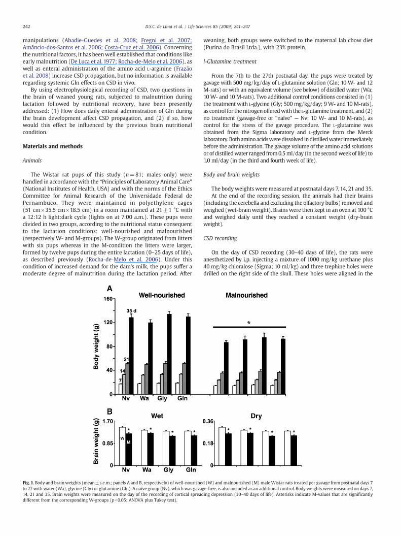
Fig. 1. Body and brainweights (mean±s.e.m.; panels A and B, respectively) of well-nourisheto 27 with water (Wa), glycine (Gly) or glutamine (Gln). A naïve group (Nv), which was gava14, 21 and 35. Brain weights were measured on the day of the recording of cortical spreaddifferent from the corresponding W-groups (pb0.05; ANOVA plus Tukey test).
weaning, both groups were switched to the maternal lab chow diet(Purina do Brasil Ltda.), with 23% protein.
l-Glutamine treatment
From the 7th to the 27th postnatal day, the pups were treated bygavage with 500 mg/kg/day of L-glutamine solution (Gln; 10 W- and 12M-rats) or with an equivalent volume (see below) of distilled water (Wa;10W- and 10 M-rats). Two additional control conditions consisted in (1)the treatment with L-glycine (Gly; 500 mg/kg/day; 9W- and 10M-rats),as control for the nitrogenofferedwith the L-glutamine treatment, and (2)no treatment (gavage-free or “naïve” — Nv; 10 W- and 10 M-rats), ascontrol for the stress of the gavage procedure. The L-glutamine wasobtained from the Sigma laboratory and L-glycine from the Mercklaboratory.Bothaminoacidsweredissolved indistilledwater immediatelybefore the administration. The gavage volume of the amino acid solutionsor of distilledwater ranged from0.5ml/day (in the secondweekof life) to1.0 ml/day (in the third and fourth week of life).
Body and brain weights
The bodyweights weremeasured at postnatal days 7,14, 21 and 35.At the end of the recording session, the animals had their brains
(including the cerebella and excluding the olfactory bulbs) removed andweighed (wet-brainweight). Brainswere then kept in an oven at 100 °Cand weighed daily until they reached a constant weight (dry-brainweight).
CSD recording
On the day of CSD recording (30–40 days of life), the rats wereanesthetized by i.p. injecting a mixture of 1000 mg/kg urethane plus40 mg/kg chloralose (Sigma; 10 ml/kg) and three trephine holes weredrilled on the right side of the skull. These holes were aligned in the
d (W) and malnourished (M) male Wistar rats treated per gavage from postnatal days 7ge-free, is also included as an additional control. Body weights were measured on days 7,ing depression (30–40 days of life). Asterisks indicate M-values that are significantly
243D.S.C. de Lima et al. / Life Sciences 85 (2009) 241–247
fronto-occipital direction and parallel to themidline. CSDwas elicited at20 min intervals by 1 min application of a cotton ball (1–2 mmdiameter), soaked in 2% KCl solution, to the anterior hole (2 mm indiameter) drilled at the frontal region. The two other holes (2–3 mm indiameter) on the parieto-occipital region served as recording places.Both the cortical spontaneous electrical activity (electrocorticogram;ECoG) and the slow potential change accompanying CSD werecontinuously recorded for 4 h, by means of two Ag–AgCl agar-Ringerelectrodes (one in each hole), against a common reference electrode ofthe same type, placed on thenasal bone. The CSDvelocityof propagationwas calculated from the time required for a CSD wave to pass thedistance between the two cortical recording points. The number of CSDselicited by each KCl application, amplitudes of the CSD-related slowpotential shifts, rise and recovery times and the durations of the CSDswere also evaluated.
During the recording period, rectal temperature was maintained at37±1 °C by means of a heating blanket. The CSD was recorded by apolygraph MODEL 7D (Grass Medical Instruments). At the end of therecording session, the still anesthetized animals were subjected toeuthanasia by bulbar injury (provoked by introducing a sharp needleinto the cisterna magna), with subsequent cardio-respiratory arrest.
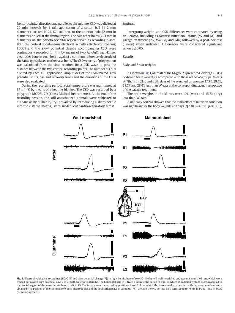
Fig. 2. Electrophysiological recordings (ECoG [E] and slow potential change [P]) in right hemtreated per gavage from postnatal days 7 to 27 with water or glutamine. The horizontal barsthe frontal region of the same hemisphere, to elicit SD. The inset shows the recording posobtained. The position of the common reference electrode (R) and the application place of s(negative upwards).
Statistics
Intergroup weight- and CSD-differences were compared by usingan ANOVA, including as factors: nutritional status (W and M), andgavage treatment (Nv, Wa, Gly and Gln) followed by a post-hoc test(Tukey) when indicated. Differences were considered significantwhen p≤0.05.
Results
Body and brain weights
As shown in Fig.1, animals of theM-groupspresented lower (pb0.05)bodyandbrainweights, as comparedwith those of theW-groups.M-ratsat 7th, 14th, 21st and 35th days of life weighed on average 17.3%, 28.4%,28.7% and 28.4% less thanW-rats at the corresponding ages, irrespectiveof the gavage treatment.
The brain weights in the M-rats were 10% (wet) and 15.7% (dry)less than W-rats.
A one-way ANOVA showed that the main effect of nutrition conditionwas significant for the bodyweights at 7 days (F[7, 81]=6.351;pb0.001),
isphere of two 30–40 day-old well-nourished and two malnourished rats, which werein P-trace 1 indicate the period (1 min) in which stimulation with 2% KCl was applied toitions 1 and 2, from which the traces marked at center with the same numbers weretimulus (KCl) are also shown. Vertical bars correspond to 10 mV in P and 1 mV in ECoG
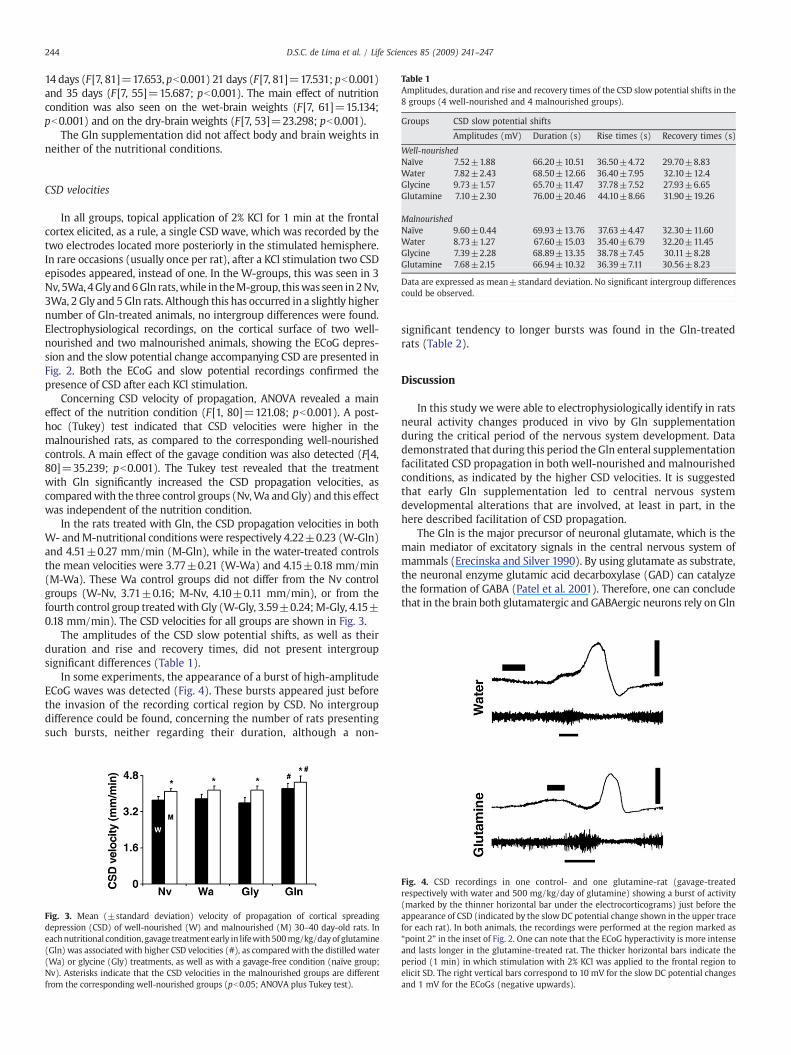
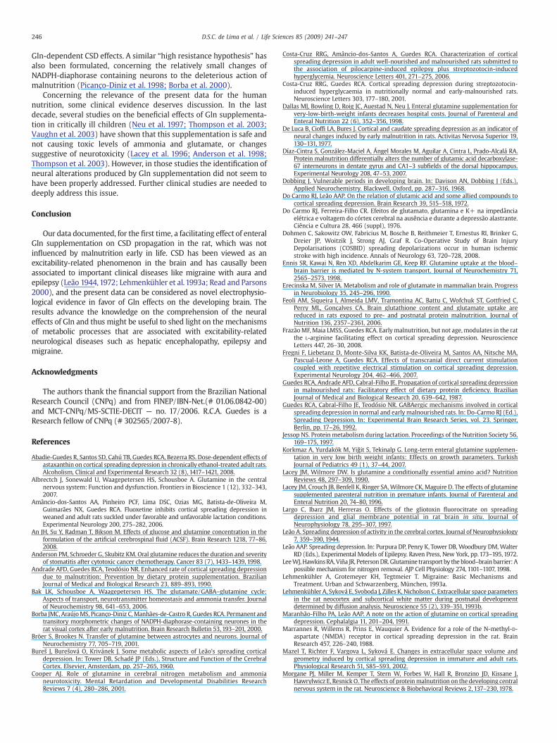
Table 1Amplitudes, duration and rise and recovery times of the CSD slow potential shifts in the8 groups (4 well-nourished and 4 malnourished groups).
Groups CSD slow potential shifts
Amplitudes (mV) Duration (s) Rise times (s) Recovery times (s)
Well-nourished
244 D.S.C. de Lima et al. / Life Sciences 85 (2009) 241–247
14 days (F[7, 81]=17.653, pb0.001) 21 days (F[7, 81]=17.531; pb0.001)and 35 days (F[7, 55]=15.687; pb0.001). The main effect of nutritioncondition was also seen on the wet-brain weights (F[7, 61]=15.134;pb0.001) and on the dry-brain weights (F[7, 53]=23.298; pb0.001).
The Gln supplementation did not affect body and brain weights inneither of the nutritional conditions.
Naïve 7.52±1.88 66.20±10.51 36.50±4.72 29.70±8.83Water 7.82±2.43 68.50±12.66 36.40±7.95 32.10±12.4Glycine 9.73±1.57 65.70±11.47 37.78±7.52 27.93±6.65Glutamine 7.10±2.30 76.00±20.46 44.10±8.66 31.90±19.26
MalnourishedNaïve 9.60±0.44 69.93±13.76 37.63±4.47 32.30±11.60Water 8.73±1.27 67.60±15.03 35.40±6.79 32.20±11.45Glycine 7.39±2.28 68.89±13.35 38.78±7.45 30.11±8.28Glutamine 7.68±2.15 66.94±10.32 36.39±7.11 30.56±8.23
Data are expressed as mean±standard deviation. No significant intergroup differencescould be observed.
CSD velocities
In all groups, topical application of 2% KCl for 1 min at the frontalcortex elicited, as a rule, a single CSD wave, which was recorded by thetwo electrodes located more posteriorly in the stimulated hemisphere.In rare occasions (usually once per rat), after a KCl stimulation two CSDepisodes appeared, instead of one. In the W-groups, this was seen in 3Nv, 5Wa,4Glyand6Gln rats,while in theM-group, thiswas seen in2Nv,3Wa, 2 Gly and 5Gln rats. Although this has occurred in a slightly highernumber of Gln-treated animals, no intergroup differences were found.Electrophysiological recordings, on the cortical surface of two well-nourished and two malnourished animals, showing the ECoG depres-sion and the slow potential change accompanying CSD are presented inFig. 2. Both the ECoG and slow potential recordings confirmed thepresence of CSD after each KCl stimulation.
Concerning CSD velocity of propagation, ANOVA revealed a maineffect of the nutrition condition (F [1, 80]=121.08; pb0.001). A post-hoc (Tukey) test indicated that CSD velocities were higher in themalnourished rats, as compared to the corresponding well-nourishedcontrols. A main effect of the gavage condition was also detected (F[4,80]=35.239; pb0.001). The Tukey test revealed that the treatmentwith Gln significantly increased the CSD propagation velocities, ascomparedwith the three control groups (Nv,Wa andGly) and this effectwas independent of the nutrition condition.
In the rats treated with Gln, the CSD propagation velocities in bothW- andM-nutritional conditions were respectively 4.22±0.23 (W-Gln)and 4.51±0.27 mm/min (M-Gln), while in the water-treated controlsthe mean velocities were 3.77±0.21 (W-Wa) and 4.15±0.18 mm/min(M-Wa). These Wa control groups did not differ from the Nv controlgroups (W-Nv, 3.71±0.16; M-Nv, 4.10±0.11 mm/min), or from thefourth control group treatedwith Gly (W-Gly, 3.59±0.24;M-Gly, 4.15±0.18 mm/min). The CSD velocities for all groups are shown in Fig. 3.
The amplitudes of the CSD slow potential shifts, as well as theirduration and rise and recovery times, did not present intergroupsignificant differences (Table 1).
In some experiments, the appearance of a burst of high-amplitudeECoG waves was detected (Fig. 4). These bursts appeared just beforethe invasion of the recording cortical region by CSD. No intergroupdifference could be found, concerning the number of rats presentingsuch bursts, neither regarding their duration, although a non-
Fig. 3. Mean (±standard deviation) velocity of propagation of cortical spreadingdepression (CSD) of well-nourished (W) and malnourished (M) 30–40 day-old rats. Ineachnutritional condition, gavage treatment early in lifewith500mg/kg/dayof glutamine(Gln) was associated with higher CSD velocities (#), as compared with the distilled water(Wa) or glycine (Gly) treatments, as well as with a gavage-free condition (naïve group;Nv). Asterisks indicate that the CSD velocities in the malnourished groups are differentfrom the corresponding well-nourished groups (pb0.05; ANOVA plus Tukey test).
significant tendency to longer bursts was found in the Gln-treatedrats (Table 2).
Discussion
In this study we were able to electrophysiologically identify in ratsneural activity changes produced in vivo by Gln supplementationduring the critical period of the nervous system development. Datademonstrated that during this period the Gln enteral supplementationfacilitated CSD propagation in both well-nourished and malnourishedconditions, as indicated by the higher CSD velocities. It is suggestedthat early Gln supplementation led to central nervous systemdevelopmental alterations that are involved, at least in part, in thehere described facilitation of CSD propagation.
The Gln is the major precursor of neuronal glutamate, which is themain mediator of excitatory signals in the central nervous system ofmammals (Erecinska and Silver 1990). By using glutamate as substrate,the neuronal enzyme glutamic acid decarboxylase (GAD) can catalyzethe formation of GABA (Patel et al. 2001). Therefore, one can concludethat in the brain both glutamatergic and GABAergic neurons rely on Gln
Fig. 4. CSD recordings in one control- and one glutamine-rat (gavage-treatedrespectively with water and 500 mg/kg/day of glutamine) showing a burst of activity(marked by the thinner horizontal bar under the electrocorticograms) just before theappearance of CSD (indicated by the slowDC potential change shown in the upper tracefor each rat). In both animals, the recordings were performed at the region marked as“point 2” in the inset of Fig. 2. One can note that the ECoG hyperactivity is more intenseand lasts longer in the glutamine-treated rat. The thicker horizontal bars indicate theperiod (1 min) in which stimulation with 2% KCl was applied to the frontal region toelicit SD. The right vertical bars correspond to 10 mV for the slow DC potential changesand 1 mV for the ECoGs (negative upwards).
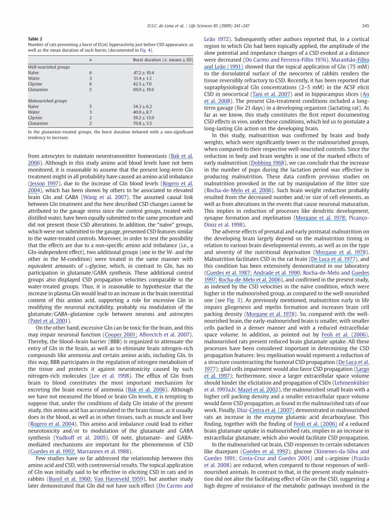
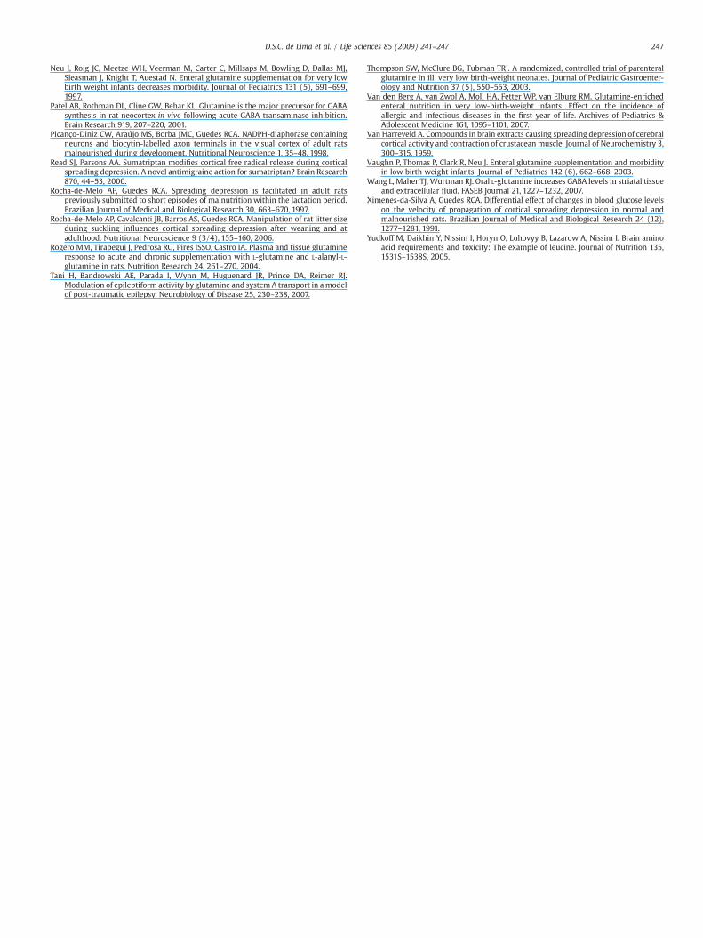
Table 2Number of rats presenting a burst of ECoG hyperactivity just before CSD appearance, aswell as the mean duration of such bursts (documented in Fig. 4).
n Burst duration (s; means±SD)
Well-nourished groupsNaïve 6 47.2±10.4Water 3 51.4±1.3Glycine 6 42.5±7.0Glutamine 5 69.0±19.6
Malnourished groupsNaïve 5 34.3±6.2Water 3 40.0±8.7Glycine 2 39.2±13.0Glutamine 2 70.8±3.5
In the glutamine-treated groups, the burst duration behaved with a non-significanttendency to increase.
245D.S.C. de Lima et al. / Life Sciences 85 (2009) 241–247
from astrocytes to maintain neurotransmitter homeostasis (Bak et al.2006). Although in this study amino acid blood levels have not beenmonitored, it is reasonable to assume that the present long-term Glntreatmentmight in all probability have caused an amino acid imbalance(Jessop 1997), due to the increase of Gln blood levels (Rogero et al.2004), which has been shown by others to be associated to elevatedbrain Gln and GABA (Wang et al. 2007). The assumed causal linkbetween Gln treatment and the here described CSD changes cannot beattributed to the gavage stress since the control groups, treated withdistilled water, have been equally submitted to the same procedure anddid not present those CSD alterations. In addition, the “naïve” groups,whichwere not submitted to the gavage, presented CSD features similarto the water-treated controls. Moreover, in order to test the possibilitythat the effects are due to a non-specific amino acid imbalance (i.e., aGln-independent effect), two additional groups (one in the W- and theother in the M-condition) were treated in the same manner withequivalent amounts of L-glycine, which, in contrast to Gln, has noparticipation in glutamate/GABA synthesis. These additional controlgroups also displayed CSD propagation velocities comparable to thewater-treated groups. Thus, it is reasonable to hypothesize that theincrease in plasma Glnwould lead to an increase in the brain interstitialcontent of this amino acid, supporting a role for excessive Gln inmodifying the neuronal excitability, probably via modulation of theglutamate/GABA–glutamine cycle between neurons and astrocytes(Patel et al. 2001).
On the other hand, excessive Gln can be toxic for the brain, and thismay impair neuronal function (Cooper 2001; Albrectch et al. 2007).Thereby, the blood–brain barrier (BBB) is organized to attenuate theentry of Gln in the brain, as well as to eliminate brain nitrogen-richcompounds like ammonia and certain amino acids, including Gln. Inthis way, BBB participates in the regulation of nitrogen metabolism ofthe tissue and protects it against neurotoxicity caused by suchnitrogen-rich molecules (Lee et al. 1998). The efflux of Gln frombrain to blood constitutes the most important mechanism forexcreting the brain excess of ammonia (Bak et al. 2006). Althoughwe have not measured the blood or brain Gln levels, it is tempting tosuppose that, under the conditions of daily Gln intake of the presentstudy, this amino acid has accumulated in the brain tissue, as it usuallydoes in the blood, as well as in other tissues, such as muscle and liver(Rogero et al. 2004). This amino acid imbalance could lead to eitherneurotoxicity and/or to modulation of the glutamate and GABAsynthesis (Yudkoff et al. 2005). Of note, glutamate- and GABA-mediated mechanisms are important for the phenomenon of CSD(Guedes et al. 1992; Marrannes et al. 1988).
Few studies have so far addressed the relationship between thisamino acid and CSD, with controversial results. The topical applicationof Gln was initially said to be effective in eliciting CSD in rats and inrabbits (Bureš et al. 1960; Van Harreveld 1959), but another studylater demonstrated that Gln did not have such effect (Do Carmo and
Leão 1972). Subsequently other authors reported that, in a corticalregion to which Gln had been topically applied, the amplitude of theslow potential and impedance changes of a CSD evoked at a distancewere decreased (Do Carmo and Ferreira-Filho 1976). Maranhão-Filhoand Leão (1991) showed that the topical application of Gln (75 mM)to the dorsolateral surface of the neocortex of rabbits renders thetissue reversibly refractory to CSD. Recently, it has been reported thatsupraphysiological Gln concentrations (2–5 mM) in the ACSF elicitCSD in neocortical (Tani et al. 2007) and in hippocampus slices (Anet al. 2008). The present Gln-treatment conditions included a long-term gavage (for 21 days) in a developing organism (lactating rat). Asfar as we know, this study constitutes the first report documentingCSD effects in vivo, under these conditions, which led us to postulate along-lasting Gln action on the developing brain.
In this study, malnutrition was confirmed by brain and bodyweights, which were significantly lower in the malnourished groups,when compared to their respective well-nourished controls. Since thereduction in body and brain weights is one of the marked effects ofearly malnutrition (Dobbing 1968), we can conclude that the increasein the number of pups during the lactation period was effective inproducing malnutrition. These data confirm previous studies onmalnutrition provoked in the rat by manipulation of the litter size(Rocha-de-Melo et al. 2006). Such brain weight reduction probablyresulted from the decreased number and/or size of cell elements, aswell as from alterations in the events that cause neuronal maturation.This implies in reduction of processes like dendritic development,synapse formation and myelination (Morgane et al. 1978; Picanço-Diniz et al. 1998).
The adverse effects of prenatal and early postnatal malnutrition onthe developing brain largely depend on the malnutrition timing inrelation to various brain developmental events, as well as on the typeand severity of the nutritional deprivation (Morgane et al. 1978).Malnutrition facilitates CSD in the rat brain (De Luca et al. 1977), andthis condition has been extensively demonstrated in our laboratory(Guedes et al. 1987; Andrade et al. 1990; Rocha-de-Melo and Guedes1997; Rocha-de-Melo et al. 2006), and confirmed in the present study,as indexed by the CSD velocities in the naïve condition, which werehigher in the malnourished group, as compared to the well-nourishedone (see Fig. 3). As previously mentioned, malnutrition early in lifeimpairs gliogenesis and myelin formation and increases brain cellpacking density (Morgane et al. 1978). So, compared with the well-nourished brain, the early-malnourished brain is smaller, with smallercells packed in a denser manner and with a reduced extracellularspace volume. In addition, as pointed out by Feoli et al. (2006),malnourished rats present reduced brain glutamate uptake. All theseprocesses have been considered important in determining the CSDpropagation features: less myelinationwould represent a reduction ofa structure counteracting the humoral CSD propagation (De Luca et al.1977); glial cells impairment would also favor CSD propagation (Largoet al. 1997); furthermore, since a larger extracellular space volumeshould hinder the elicitation and propagation of CSDs (Lehmenkühleret al. 1993a,b; Mazel et al. 2002), the malnourished small brain with ahigher cell packing density and a smaller extracellular space volumewould favor CSD propagation, as found in themalnourished rats of ourwork. Finally, Díaz-Cintra et al. (2007) demonstrated in malnourishedrats an increase in the enzyme glutamic acid decarboxylase. Thisfinding, together with the finding of Feoli et al. (2006) of a reducedbrain glutamate uptake in malnourished rats, implies in an increase inextracellular glutamate, which also would facilitate CSD propagation.
In the malnourished rat brain, CSD responses to certain substanceslike diazepam (Guedes et al. 1992), glucose (Ximenes-da-Silva andGuedes 1991; Costa-Cruz and Guedes 2001) and L-arginine (Frazãoet al. 2008) are reduced, when compared to those responses of well-nourished animals. In contrast to that, in the present study malnutri-tion did not alter the facilitating effect of Gln on the CSD, suggesting ahigh degree of resistance of the metabolic pathways involved in the
246 D.S.C. de Lima et al. / Life Sciences 85 (2009) 241–247
Gln-dependent CSD effects. A similar “high resistance hypothesis” hasalso been formulated, concerning the relatively small changes ofNADPH-diaphorase containing neurons to the deleterious action ofmalnutrition (Picanço-Diniz et al. 1998; Borba et al. 2000).
Concerning the relevance of the present data for the humannutrition, some clinical evidence deserves discussion. In the lastdecade, several studies on the beneficial effects of Gln supplementa-tion in critically ill children (Neu et al. 1997; Thompson et al. 2003;Vaughn et al. 2003) have shown that this supplementation is safe andnot causing toxic levels of ammonia and glutamate, or changessuggestive of neurotoxicity (Lacey et al. 1996; Anderson et al. 1998;Thompson et al. 2003). However, in those studies the identification ofneural alterations produced by Gln supplementation did not seem tohave been properly addressed. Further clinical studies are needed todeeply address this issue.
Conclusion
Our data documented, for the first time, a facilitating effect of enteralGln supplementation on CSD propagation in the rat, which was notinfluenced by malnutrition early in life. CSD has been viewed as anexcitability-related phenomenon in the brain and has causally beenassociated to important clinical diseases like migraine with aura andepilepsy (Leão 1944,1972; Lehmenkühler et al. 1993a; Read and Parsons2000), and the present data can be considered as novel electrophysio-logical evidence in favor of Gln effects on the developing brain. Theresults advance the knowledge on the comprehension of the neuraleffects of Gln and thus might be useful to shed light on the mechanismsof metabolic processes that are associated with excitability-relatedneurological diseases such as hepatic encephalopathy, epilepsy andmigraine.
Acknowledgments
The authors thank the financial support from the Brazilian NationalResearch Council (CNPq) and from FINEP/IBN-Net.(# 01.06.0842-00)and MCT-CNPq/MS-SCTIE-DECIT — no. 17/2006. R.C.A. Guedes is aResearch fellow of CNPq (# 302565/2007-8).
References
Abadie-Guedes R, Santos SD, Cahú TB, Guedes RCA, Bezerra RS. Dose-dependent effects ofastaxanthin on cortical spreading depression in chronically ethanol-treated adult rats.Alcoholism, Clinical and Experimental Research 32 (8), 1417–1421, 2008.
Albrectch J, Sonewald U, Waagepetersen HS, Schousboe A. Glutamine in the centralnervous system: Function and dysfunction. Frontiers in Bioscience 1 (12), 332–343,2007.
Amâncio-dos-Santos AA, Pinheiro PCF, Lima DSC, Ozias MG, Batista-de-Oliveira M,Guimarães NX, Guedes RCA. Fluoxetine inhibits cortical spreading depression inweaned and adult rats suckled under favorable and unfavorable lactation conditions.Experimental Neurology 200, 275–282, 2006.
An JH, Su Y, Radman T, Bikson M. Effects of glucose and glutamine concentration in theformulation of the artificial cerebrospinal fluid (ACSF). Brain Research 1218, 77–86,2008.
Anderson PM, Schroeder G, Skubitz KM. Oral glutamine reduces the duration and severityof stomatitis after cytotoxic cancer chemotherapy. Cancer 83 (7), 1433–1439, 1998.
Andrade AFD, Guedes RCA, Teodósio NR. Enhanced rate of cortical spreading depressiondue to malnutrition: Prevention by dietary protein supplementation. BrazilianJournal of Medical and Biological Research 23, 889–893, 1990.
Bak LK, Schousboe A, Waagepetersen HS. The glutamate/GABA–glutamine cycle:Aspects of transport, neurotransmitter homeostasis and ammonia transfer. Journalof Neurochemistry 98, 641–653, 2006.
Borba JMC, AraújoMS, Picanço-Diniz C, Manhães-de-Castro R, Guedes RCA. Permanent andtransitory morphometric changes of NADPH-diaphorase-containing neurones in therat visual cortex after early malnutrition. Brain Research Bulletin 53, 193–201, 2000.
Bröer S, Brookes N. Transfer of glutamine between astrocytes and neurons. Journal ofNeurochemistry 77, 705–719, 2001.
Bureš J, Burešová O, Krivánek J. Some metabolic aspects of Leão's spreading corticaldepression. In: Tower DB, Schadé JP (Eds.), Structure and Function of the CerebralCortex. Elsevier, Amsterdam, pp. 257–265, 1960.
Cooper AJ. Role of glutamine in cerebral nitrogen metabolism and ammonianeurotoxicity. Mental Retardation and Developmental Disabilities ResearchReviews 7 (4), 280–286, 2001.
Costa-Cruz RRG, Amâncio-dos-Santos A, Guedes RCA. Characterization of corticalspreading depression in adult well-nourished and malnourished rats submitted tothe association of pilocarpine-induced epilepsy plus streptozotocin-inducedhyperglycemia. Neuroscience Letters 401, 271–275, 2006.
Costa-Cruz RRG, Guedes RCA. Cortical spreading depression during streptozotocin-induced hyperglycaemia in nutritionally normal and early-malnourished rats.Neuroscience Letters 303, 177–180, 2001.
Dallas MJ, Bowling D, Roig JC, Auestad N, Neu J. Enteral glutamine supplementation forvery-low-birth-weight infants decreases hospital costs. Journal of Parenteral andEnteral Nutrition 22 (6), 352–356, 1998.
De Luca B, Cioffi LA, Bures J. Cortical and caudate spreading depression as an indicator ofneural changes induced by early malnutrition in rats. Activitas Nervosa Superior 19,130–131, 1977.
Díaz-Cintra S, González-Maciel A, Ángel Morales M, Aguilar A, Cintra L, Prado-Alcalá RA.Protein malnutrition differentially alters the number of glutamic acid decarboxylase-67 interneurons in dentate gyrus and CA1–3 subfields of the dorsal hippocampus.Experimental Neurology 208, 47–53, 2007.
Dobbing J. Vulnerable periods in developing brain. In: Davison AN, Dobbing J (Eds.),Applied Neurochemistry. Blackwell, Oxford, pp. 287–316, 1968.
Do Carmo RJ, Leão AAP. On the relation of glutamic acid and some allied compounds tocortical spreading depression. Brain Research 39, 515–518, 1972.
Do Carmo RJ, Ferreira-Filho CR. Efeitos de glutamato, glutamina e K+ na impedânciaelétrica e voltagem do córtex cerebral na ausência e durante a depressão alastrante.Ciência e Cultura 28, 466 (suppl), 1976.
Dohmen C, Sakowitz OW, Fabricius M, Bosche B, Reithmeier T, Ernestus RI, Brinker G,Dreier JP, Woitzik J, Strong AJ, Graf R. Co-Operative Study of Brain InjuryDepolarisations (COSBID) spreading depolarizations occur in human ischemicstroke with high incidence. Annals of Neurology 63, 720–728, 2008.
Ennis SR, Kawai N, Ren XD, Abdelkarim GE, Keep RF. Glutamine uptake at the blood–brain barrier is mediated by N-system transport. Journal of Neurochemistry 71,2565–2573, 1998.
Erecinska M, Silver IA. Metabolism and role of glutamate in mammalian brain. Progressin Neurobiology 35, 245–296, 1990.
Feoli AM, Siqueira I, Almeida LMV, Tramontina AC, Battu C, Wofchuk ST, Gottfried C,Perry ML, Gonçalves CA. Brain glutathione content and glutamate uptake arereduced in rats exposed to pre- and postnatal protein malnutrition. Journal ofNutrition 136, 2357–2361, 2006.
FrazãoMF,Maia LMSS, Guedes RCA. Early malnutrition, but not age,modulates in the ratthe L-arginine facilitating effect on cortical spreading depression. NeuroscienceLetters 447, 26–30, 2008.
Fregni F, Liebetanz D, Monte-Silva KK, Batista-de-Oliveira M, Santos AA, Nitsche MA,Pascual-Leone A, Guedes RCA. Effects of transcranial direct current stimulationcoupled with repetitive electrical stimulation on cortical spreading depression.Experimental Neurology 204, 462–466, 2007.
Guedes RCA, Andrade AFD, Cabral-Filho JE. Propagation of cortical spreading depressionin malnourished rats: Facilitatory effect of dietary protein deficiency. BrazilianJournal of Medical and Biological Research 20, 639–642, 1987.
Guedes RCA, Cabral-Filho JE, Teodósio NR. GABAergic mechanisms involved in corticalspreading depression in normal and earlymalnourished rats. In: Do-Carmo RJ (Ed.),Spreading Depression. In: Experimental Brain Research Series, vol. 23. Springer,Berlin, pp. 17–26, 1992.
Jessop NS. Protein metabolism during lactation. Proceedings of the Nutrition Society 56,169–175, 1997.
Korkmaz A, Yurdakök M, Yiğit S, Tekinalp G. Long-term enteral glutamine supplemen-tation in very low birth weight infants: Effects on growth parameters. TurkishJournal of Pediatrics 49 (1), 37–44, 2007.
Lacey JM, Wilmore DW. Is glutamine a conditionally essential amino acid? NutritionReviews 48, 297–309, 1990.
Lacey JM, Crouch JB, Benfell K, Ringer SA,Wilmore CK, Maguire D. The effects of glutaminesupplemented parenteral nutrition in premature infants. Journal of Parenteral andEnteral Nutrition 20, 74–80, 1996.
Largo C, Ibarz JM, Herreras O. Effects of the gliotoxin fluorocitrate on spreadingdepression and glial membrane potential in rat brain in situ. Journal ofNeurophysiology 78, 295–307, 1997.
Leão A. Spreading depression of activity in the cerebral cortex. Journal of Neurophysiology7, 359–390, 1944.
Leão AAP. Spreading depression. In: Purpura DP, Penry K, Tower DB, Woodbury DM,WalterRD (Eds.), Experimental Models of Epilepsy. Raven Press, New York, pp.173–195, 1972.
LeeWJ,HawkinsRA,Viña JR, PetersonDR.Glutamine transport by the blood–brainbarrier:Apossible mechanism for nitrogen removal. AJP Cell Physiology 274, 1101–1107, 1998.
Lehmenkühler A, Grotemeyer KH, Tegtmeier T. Migraine: Basic Mechanisms andTreatment. Urban and Schwarzenberg, München, 1993a.
Lehmenkühler A, Syková E, Svoboda J, Zilles K, Nicholson C. Extracellular space parametersin the rat neocortex and subcortical white matter during postnatal developmentdetermined by diffusion analysis. Neuroscience 55 (2), 339–351, 1993b.
Maranhão-Filho PA, Leão AAP. A note on the action of glutamine on cortical spreadingdepression. Cephalalgia 11, 201–204, 1991.
Marrannes R, Willems R, Prins E, Wauquier A. Evidence for a role of the N-methyl-D-aspartate (NMDA) receptor in cortical spreading depression in the rat. BrainResearch 457, 226–240, 1988.
Mazel T, Richter F, Vargova L, Syková E. Changes in extracellular space volume andgeometry induced by cortical spreading depression in immature and adult rats.Physiological Research 51, S85–S93, 2002.
Morgane PJ, Miller M, Kemper T, Stern W, Forbes W, Hall R, Bronzino JD, Kissane J,Hawrylwicz E, Resnick O. The effects of proteinmalnutrition on the developing centralnervous system in the rat. Neuroscience & Biobehavioral Reviews 2, 137–230, 1978.
247D.S.C. de Lima et al. / Life Sciences 85 (2009) 241–247
Neu J, Roig JC, Meetze WH, Veerman M, Carter C, Millsaps M, Bowling D, Dallas MJ,Sleasman J, Knight T, Auestad N. Enteral glutamine supplementation for very lowbirth weight infants decreases morbidity. Journal of Pediatrics 131 (5), 691–699,1997.
Patel AB, Rothman DL, Cline GW, Behar KL. Glutamine is the major precursor for GABAsynthesis in rat neocortex in vivo following acute GABA-transaminase inhibition.Brain Research 919, 207–220, 2001.
Picanço-Diniz CW, Araújo MS, Borba JMC, Guedes RCA. NADPH-diaphorase containingneurons and biocytin-labelled axon terminals in the visual cortex of adult ratsmalnourished during development. Nutritional Neuroscience 1, 35–48, 1998.
Read SJ, Parsons AA. Sumatriptan modifies cortical free radical release during corticalspreading depression. A novel antimigraine action for sumatriptan? Brain Research870, 44–53, 2000.
Rocha-de-Melo AP, Guedes RCA. Spreading depression is facilitated in adult ratspreviously submitted to short episodes of malnutrition within the lactation period.Brazilian Journal of Medical and Biological Research 30, 663–670, 1997.
Rocha-de-Melo AP, Cavalcanti JB, Barros AS, Guedes RCA. Manipulation of rat litter sizeduring suckling influences cortical spreading depression after weaning and atadulthood. Nutritional Neuroscience 9 (3/4), 155–160, 2006.
Rogero MM, Tirapegui J, Pedrosa RG, Pires ISSO, Castro IA. Plasma and tissue glutamineresponse to acute and chronic supplementation with L-glutamine and L-alanyl-L-glutamine in rats. Nutrition Research 24, 261–270, 2004.
Tani H, Bandrowski AE, Parada I, Wynn M, Huguenard JR, Prince DA, Reimer RJ.Modulation of epileptiform activity by glutamine and systemA transport in amodelof post-traumatic epilepsy. Neurobiology of Disease 25, 230–238, 2007.
Thompson SW, McClure BG, Tubman TRJ. A randomized, controlled trial of parenteralglutamine in ill, very low birth-weight neonates. Journal of Pediatric Gastroenter-ology and Nutrition 37 (5), 550–553, 2003.
Van den Berg A, van Zwol A, Moll HA, Fetter WP, van Elburg RM. Glutamine-enrichedenteral nutrition in very low-birth-weight infants: Effect on the incidence ofallergic and infectious diseases in the first year of life. Archives of Pediatrics &Adolescent Medicine 161, 1095–1101, 2007.
Van Harreveld A. Compounds in brain extracts causing spreading depression of cerebralcortical activity and contraction of crustacean muscle. Journal of Neurochemistry 3,300–315, 1959.
Vaughn P, Thomas P, Clark R, Neu J. Enteral glutamine supplementation and morbidityin low birth weight infants. Journal of Pediatrics 142 (6), 662–668, 2003.
Wang L, Maher TJ, Wurtman RJ. Oral L-glutamine increases GABA levels in striatal tissueand extracellular fluid. FASEB Journal 21, 1227–1232, 2007.
Ximenes-da-Silva A, Guedes RCA. Differential effect of changes in blood glucose levelson the velocity of propagation of cortical spreading depression in normal andmalnourished rats. Brazilian Journal of Medical and Biological Research 24 (12),1277–1281, 1991.
Yudkoff M, Daikhin Y, Nissim I, Horyn O, Luhovyy B, Lazarow A, Nissim I. Brain aminoacid requirements and toxicity: The example of leucine. Journal of Nutrition 135,1531S–1538S, 2005.
Related Documents