KULIAH BEDAH DASAR KULIAH BEDAH DASAR MUCCULOSKELETAL MUCCULOSKELETAL

Welcome message from author
This document is posted to help you gain knowledge. Please leave a comment to let me know what you think about it! Share it to your friends and learn new things together.
Transcript
KULIAH BEDAH DASARKULIAH BEDAH DASARMUCCULOSKELETALMUCCULOSKELETAL

ANATOMY AND PHYSIOLOGYANATOMY AND PHYSIOLOGY
• Structure and Function of bone
• The skeletal system consist of : bone, cartilage and joint
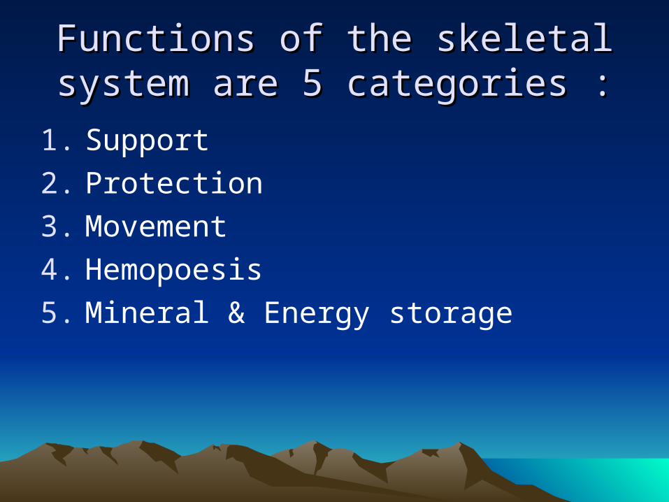
Functions of the skeletal system are 5 Functions of the skeletal system are 5 categories :categories :
1. Support
2. Protection
3. Movement
4. Hemopoesis
5. Mineral & Energy storage

• Support : – The skeleton forms a rigid framework to which
are attached the softer tissue and organs of the body
• Protections – The skull, vertebral column, rib cage and
pelvic girdle enclose & protect vital organs – Sites for blood cell production are protected
with in the hollow center of certain bone
• Movement – Bone act as levers when attached muscle
contact, causing movement about joints

• Hemopoiesis- Red bone marrow of an adult produces white
& red blood cell & platelets
• Mineral & Energy storage
Matrix of bone is composed as :1. Calcium, phosphorus
2. Magnesium, natrium, (Lesser amounts)
3. Lipids stored in adipose cell of yellow bone marrow store energy

4 Categories of bones 4 Categories of bones
1. Long bones : longer than wide appendages
2. Short bones : More/less cubical confined spaces
3. Flat bones : Protection skull, ribs
4. Irregular bones : odd shaped vertabrae & skull bones

LONG BONES LONG BONES • Diaphysis medullary cavity endosteum• Epiphysis
– Medullary cavity contains fetty yellow bone marrow – Epiphysis consist
• Spongy bone surrounded by compact bone • Red bone marrow in the pores of spongy bone • Epiphyseal plate linier bone growth (elongation) • Epiphyseals Line replaces the plate when bone
growth is completed• Periosteum : is a dense regular connective tissue
covers the bone & the site of tendon muscle attachment• Periosteum Diametric bone growth (widening)

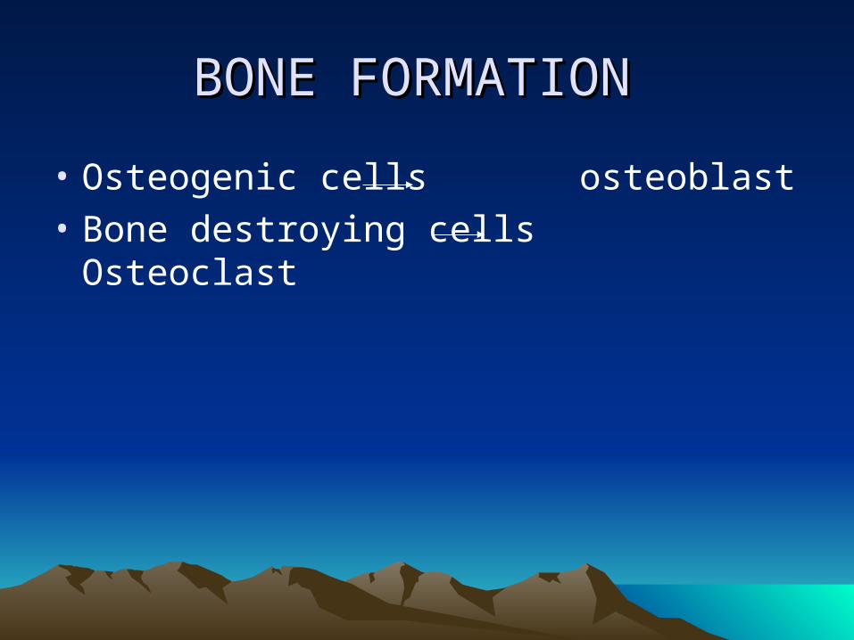
BONE FORMATION BONE FORMATION
• Osteogenic cells osteoblast
• Bone destroying cells Osteoclast

Osteoblast Osteoblast
• Bone building cells synthesis the collagenous fibers & bone matrix.
• Promote mineralisation during ossification
• Progenator cells give rise all bone cells

Osteoclasts Osteoclasts
• Bone destroying cells countain :– Lysosomes– Phagositic vacuoles demineralize bone
tissue

Ossification (Bone Formation)Ossification (Bone Formation)
Begins during 4th week of prenatal developmentBone Development
Endochondral ossificationIntramembranous ossification

Endochondral ossification Endochondral ossification
• Long bone : begins in a primary center in the shaft of cartilage model with hypotrophy of chondrocytes (cartilage cells) and calcifications of the cartilage matrix
• Osteoblast bone matrix around the calcareous spicula

Primary centers ossification Occurs before birth
Secondary centers ossification (Epiphyses)Occurs during the first 5 years
Most of the bones of the skeleton form through endochondral ossification

Intra Membranous OssificationIntra Membranous Ossification• Occurs during fetal development & Infancy• Membranous bone : - (Top & side cranium Fibrous
suture, six large fontanels).- permit undergo
change in shape during birth.- permit rapid
growth the brain during infancy.
• Ossification of the fontanels is complete by 20-24 month of age.
• Most of the cranial bones & clavicle form intramembranous ossification.

1. BONES OF THE AXIAL SKELETON1. BONES OF THE AXIAL SKELETON 2. BONES OF THE APPENDICULLAR 2. BONES OF THE APPENDICULLAR SKELETON SKELETON
BONES of the Axial skeleton
Bone form axis of the body Support & protect the organs of = head, neck & trunkInclude : skull, vertebra column, rib cage,auditory ossicles & hyoid bone Skull include : - 8 cranial bones - 14 facial bones


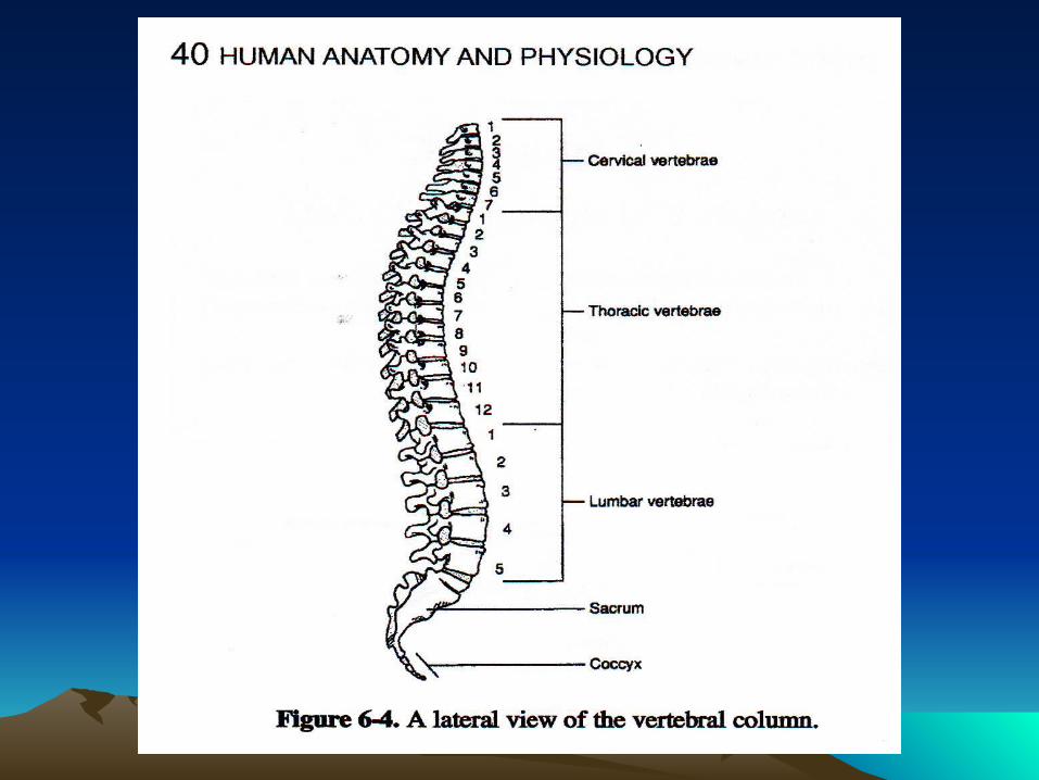
The Vertebral ColumnThe Vertebral Column
• 7 Cervical
• 12 Thoracic
• 5 Lumbar
• 4 or 5-fused Sacral
• 4 or 5-fused caccygeal
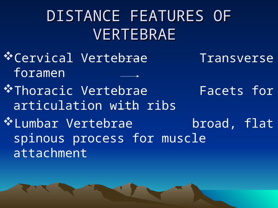
DISTANCE FEATURES OF DISTANCE FEATURES OF VERTEBRAE VERTEBRAE
Cervical Vertebrae Transverse foramenThoracic Vertebrae Facets for articulation
with ribs Lumbar Vertebrae broad, flat spinous
process for muscle attachment


Rib Cage – ComposedRib Cage – Composed
SternumCostal CartilageRibs Thoracic Vertebra


SternumSternum
• Manubrium
• Body
• Xiphoid Process

Ribs Ribs
• 7 True ribs
• 3 False ribs
• 2 Floating ribs
• 10 ribs have a head & Tubercle for articulation with a vertebra
• Ribs 11,12 have a head, but no tubercle
• All ribs have a neck, angle & shaft ( body )
BONES OF THE APPENDICULAR SKELETONBONES OF THE APPENDICULAR SKELETON
Pectoral and pelvic girdles (Korset)Upper extremity Lower extremity

Pectoral girdles 2 scapulae 2 clavicles

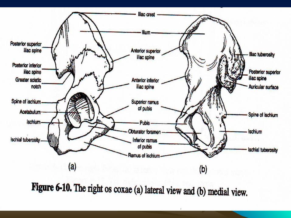
Pelvic girdles Pelvic girdles
• 2 ossa coxae united by symphysis pubis
• Each coxae - an ileum
- an ichium
- a pubis

Upper extremity divided in to Upper extremity divided in to
brachium – humerus antebrachium – radius, ulna hand - 8 carpal bones
- 5 metacarpal bones
- 14 phalanges


Lower extremity divided in to Lower extremity divided in to
• Femur
• Patella (sesamoid bone) = kneecap
• Tibia
• Fibula
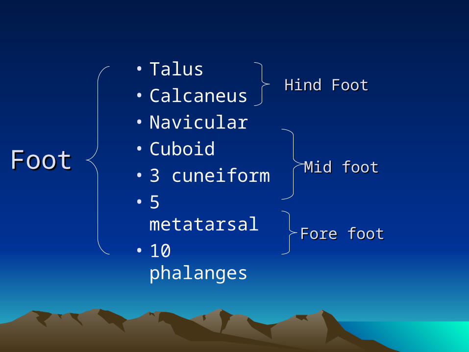
• Talus
• Calcaneus
• Navicular
• Cuboid
• 3 cuneiform
• 5 metatarsal
• 10 phalanges
Foot Foot
Hind FootHind Foot
Mid foot Mid foot
Fore foot Fore foot

ARTICULATIONS (Joints)ARTICULATIONS (Joints)Classified according to structure or functionClassified according to structure or function


PATHOLOGY SYSTEM PATHOLOGY SYSTEM
MUSKULOSKELETALMUSKULOSKELETAL
Disease Pathology Clinical features
Nutritional deficiency rickets
Deficit vit D & calcium
Genu varum or valgus kiphosis Scoliosis Bowing of bones enlarged ends of bone
Bone Healing / Fracture Healing Bone Healing / Fracture Healing
• Long bone / cortical bone healing
• Concellous bone healing

Long bone / cortical bone healing Long bone / cortical bone healing
1. Stage of impact
2. Stage of inflamation (1-3 days)a. Haemorrhage.
b. Hematoma = PMNs, macrophages, mast cell, fibrin clot.
c. Bone necrosis at ends of fracture site
d. Granulation tissue replaces hematoma with invading
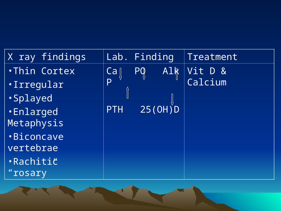
X ray findings Lab. Finding Treatment
•Thin Cortex•Irregular •Splayed •Enlarged Metaphysis •Biconcave vertebrae •Rachitic “rosary”
Ca PO Alk P
PTH 25(OH)D
Vit D & Calcium

Cappilaries, and FibroblastsCappilaries, and Fibroblasts
Osteoclast remove dead bone
e. Induction (stage of sub periosteal and endosteal cellular proliferation) soft tissue defferentiated in to osteoblast and chondroblasts.

3. Stage of soft callus (3 weeks)Primary callus & External bridging callus
a. Fibrocartilagenous tissue (High Proteoglycan)
b. Progressive calcification1. Mineral deposition on collagen fibrils
2. Matrix vesicles in fracture callus
3. This fails occur non union
c. Vascular invasion and vascularity peaks at 2 weeks

4. Stage of hard callus (3-4 Months)=late medullary callus.
a. Ossification is an aerrobic processb. Clinical healing when no gross movement
and no tendernessc. Radiologic healing follow later
5. Stage of remodeling (wolff’low) lamelar structure

# CANCELLOUS BONE HEALING ## CANCELLOUS BONE HEALING #
I. Inflamation Stage Hematoma Granulation tissue Induction
II. Callus stage Osteoblast lay down matrix (heaven bone)
III. Remodelling stage Trabecular bone in lamellar structure

Type of callus Type of callus
1. Primary callus response = fracture that are not rigidly fixed
2. External bridging callus = if bony ends are not in continuity
3. Late medullury callus = osteogenic cell from ends of bones
4. Primary cortical healing = rigit fixation and perfect apposition

Vascularity Vascularity
1. The presence of periosteal vessels alone is sufficient for fracture healing.
2. Nutrient or endosteal vessels constitute 2/3 of cortical circulation important as vascular supply.
3. Interosseus vein reconstituted about 12 weeks after fracture.

Type of non Union Type of non Union
A. Hypertropic 1) Viable or biologic activity on both sides of fracture
site but no bridging due to motion
2) Union can be obtained with reduction and campression stabilization alone (Rigid fixation)

B. Atropic 1) Non viable or avascular fracture site.
2) No change over time in callus formation.
3) Stimulus for farther healing is necessary, such as = electricity as bone graft.
C. Synovial pseudoarthrosis – Separated by synovia-like joint due to
excess motion.– Pseudo joint.– More common in humerus, clavicle and
femur.– Excision of this membrane is necessary in
addition to rigid fixation.

FACTORS ASSOCIATED WITH NON UNIONFACTORS ASSOCIATED WITH NON UNION A. Local
1. Devascularization
2. Discontinuity = distracting force and fibrous tissue
3. Inadequate immobilization (Cyclic tonsile loading)
4. Infection
B. Systemic 1. Nutrition
2. Steroids
3. Anticoagulants
4. Radiation ‘
5. Hormones etc

BIOMECHANIC OF FRACTUREBIOMECHANIC OF FRACTURE
• Bone is composite of collagen & hydroxy patite
• Collagen - good tensile strength
- poor compressive strength• Calcium apatite is stiff & Brittle material
good in compressive strength• Bone is combination an isotropic material
that resist many forces

Bone - strongest in compression forces
- weakest in shear forces
- intermediate in tension forces
Cortical bone excellent in resisting torque
Cancellouis bone good in resisting compression & shear
Bone dynamic material able to self repair (healing)

FRACTURE Forces > BONE Resistance
FRACTURE is base of mechanism of injury
1) Tension transverse – perpendicular to the load & bone axis
2) Compression axial loading crush type of fracture

3. Shear force parallel to bone surface (around joints) fracture parallel to the load
4. Bending eccentric loading or direct blow
oblique fracture
butterfly fracture
5. Torsion spiral fracture

Thank youThank you
Related Documents