ARTICLE Kufs Disease, the Major Adult Form of Neuronal Ceroid Lipofuscinosis, Caused by Mutations in CLN6 Todor Arsov, 1 Katherine R. Smith, 2 John Damiano, 1 Silvana Franceschetti, 3 Laura Canafoglia, 3 Catherine J. Bromhead, 2 Eva Andermann, 4 Danya F. Vears, 1 Patrick Cossette, 5 Sulekha Rajagopalan, 6 Alan McDougall, 7 Vito Sofia, 8 Michael Farrell, 9 Umberto Aguglia, 10 Andrea Zini, 11 Stefano Meletti, 11 Michela Morbin, 12 Saul Mullen, 1 Frederick Andermann, 13 Sara E. Mole, 14 Melanie Bahlo, 2,15, * and Samuel F. Berkovic 1, * The molecular basis of Kufs disease is unknown, whereas a series of genes accounting for most of the childhood-onset forms of neuronal ceroid lipofuscinosis (NCL) have been identified. Diagnosis of Kufs disease is difficult because the characteristic lipopigment is largely confined to neurons and can require a brain biopsy or autopsy for final diagnosis. We mapped four families with Kufs disease for whom there was good evidence of autosomal-recessive inheritance and found two peaks on chromosome 15. Three of the families were affected by Kufs type A disease and presented with progressive myoclonus epilepsy, and one was affected by type B (presenting with dementia and motor system dysfunction). Sequencing of a candidate gene in one peak shared by all four families identified no mutations, but sequencing of CLN6, found in the second peak and shared by only the three families affected by Kufs type A disease, revealed pathogenic mutations in all three families. We subsequently sequenced CLN6 in eight other families, three of which were affected by recessive Kufs type A disease. Mutations in both CLN6 alleles were found in the three type A cases and in one family affected by unclassified Kufs disease. Mutations in CLN6 are the major cause of recessive Kufs type A disease. The phenotypic differences between variant late-infantile NCL, previously found to be caused by CLN6, and Kufs type A disease are striking; there is a much later age at onset and lack of visual involvement in the latter. Sequencing of CLN6 will provide a simple diagnostic strategy in this disorder, in which defin- itive identification usually requires invasive biopsy. Introduction The neuronal ceroid lipofuscinoses (NCLs) are a family of inherited, neurodegenerative disorders that are character- ized by lysosomal lipopigment storage in neurons, and usually the eye, and cause progressive neurological impair- ment, motor and intellectual deterioration, seizures, visual failure, and early death. 1,2 In the past, the NCLs have been classified according to the age at onset as infantile (INCL, Santavuori-Haltia), late-infantile (LINCL, Jansky- Bielschowsky), juvenile (JNCL, Batten disease, Spielmeyer- Vogt), and adult (Kufs disease). 1–3 Mutations causing the childhood NCL forms have been reported in eight genes: PPT1 (CLN1 [MIM 256730]), TPP1 (CLN2 [MIM 204500]), CLN3 (MIM 204200), CLN5 (MIM 256731), CLN6 (MIM 601780), MFSD8 (CLN7 [MIM 610951]), CLN8 (MIM 600143), and CTSD (CLN10 [MIM 610127]). 1 Although the traditional classification of age at onset is pragmatically useful, genotype-phenotype correlations have shown heterogeneity. 1,3,4 For example, allelic variants of PPT1, the gene underlying most cases of the infantile form, can present in later childhood or even early-adult life with neurological deterioration and visual failure; 1,5,6 a missense mutation in CLN8 causes Northern epilepsy syndrome and other mutations cause a more typical variant late- infantile NCL. 3 Kufs disease is rare and differs from most other forms of NCL because the retina is not involved, vision is preserved, and onset is in adulthood. 7,8 The Kufs disease locus was designated CLN4 in the 1990s but was never specifically mapped and has remained enigmatic and unsolved. The clinical presentation has been divided into two overlap- ping types. Type A presents with progressive myoclonus epilepsy, whereas type B presents with dementia and a variety of motor-system signs. Typically, the patients present around the age of 30, but onset has been described in patients ranging from teenagers to persons more than 50 years of age. 1,8,9 In contrast to other NCL forms, which are always auto- somal recessive, both recessive and dominant forms of 1 Epilepsy Research Center, Department of Medicine, University of Melbourne, Austin Health, Heidelberg, Victoria 3084, Australia; 2 Bioinformatics Divi- sion, The Walter and Eliza Hall Institute of Medical Research, 1G Royal Parade, Parkville, Victoria 3052, Australia; 3 Unit of Neurophysiopathology, IRCCS Foundation, C. Besta Neurological Institute, 20133 Milan, Italy; 4 Departments of Neurology and Neurosurgery and Human Genetics, Montreal Neurolog- ical Institute and Hospital, McGill University, Montreal, Quebec H3A 2B4, Canada; 5 De ´partement de Me ´decine, Universite ´ de Montre ´al, CHUM-Ho ˆpital Notre-Dame, Montre ´al, Que ´bec H2L 4M1, Canada; 6 Department of Clinical Genetics, Liverpool Hospital, Liverpool, New South Wales 1871, Australia; 7 Department of Neurology, Liverpool Hospital, Liverpool, New South Wales 1871, Australia; 8 Department of Neuroscience, University of Catania, 95123 Catania, Italy; 9 Department of Neuropathology, Beaumont Hospital, Dublin 9, Ireland; 10 Institute of Neurology, University Magna Græcia, Viale Europa, 88100 Catanzaro, Italy; 11 Department of Neuroscience, University of Modena and Reggio Emilia, Nuovo Ospedale Civile, 41100 Modena, Italy; 12 Neuropathology-Neurology 5, IRCCS Foundation, C. Besta Neurological Institute, 20133 Milan, Italy; 13 Departments of Neurology and Neurosurgery and Pediatrics, Montreal Neurological Institute and Hospital, McGill University, Montreal, Quebec H3A 2B4, Canada; 14 Medical Research Council Labora- tory for Molecular Cell Biology, Molecular Medicine Unit, Institute of Child Health and Department of Genetics, Evolution and Environment, University College London, London WC1E 6BT, UK; 15 Department of Mathematics and Statistics, University of Melbourne, Victoria 3010, Australia *Correspondence: [email protected] (M.B.), [email protected] (S.F.B.) DOI 10.1016/j.ajhg.2011.04.004. Ó2011 by The American Society of Human Genetics. All rights reserved. 566 The American Journal of Human Genetics 88, 566–573, May 13, 2011

Welcome message from author
This document is posted to help you gain knowledge. Please leave a comment to let me know what you think about it! Share it to your friends and learn new things together.
Transcript
ARTICLE
Kufs Disease, the Major Adult Form of Neuronal CeroidLipofuscinosis, Caused by Mutations in CLN6
Todor Arsov,1 Katherine R. Smith,2 John Damiano,1 Silvana Franceschetti,3 Laura Canafoglia,3
Catherine J. Bromhead,2 Eva Andermann,4 Danya F. Vears,1 Patrick Cossette,5 Sulekha Rajagopalan,6
Alan McDougall,7 Vito Sofia,8 Michael Farrell,9 Umberto Aguglia,10 Andrea Zini,11 Stefano Meletti,11
Michela Morbin,12 Saul Mullen,1 Frederick Andermann,13 Sara E. Mole,14 Melanie Bahlo,2,15,*and Samuel F. Berkovic1,*
The molecular basis of Kufs disease is unknown, whereas a series of genes accounting for most of the childhood-onset forms of neuronal
ceroid lipofuscinosis (NCL) have been identified. Diagnosis of Kufs disease is difficult because the characteristic lipopigment is largely
confined to neurons and can require a brain biopsy or autopsy for final diagnosis. We mapped four families with Kufs disease for
whom there was good evidence of autosomal-recessive inheritance and found two peaks on chromosome 15. Three of the families
were affected by Kufs type A disease and presented with progressive myoclonus epilepsy, and one was affected by type B (presenting
with dementia and motor system dysfunction). Sequencing of a candidate gene in one peak shared by all four families identified no
mutations, but sequencing of CLN6, found in the second peak and shared by only the three families affected by Kufs type A disease,
revealed pathogenic mutations in all three families. We subsequently sequenced CLN6 in eight other families, three of which were
affected by recessive Kufs type A disease. Mutations in both CLN6 alleles were found in the three type A cases and in one family affected
by unclassified Kufs disease. Mutations in CLN6 are themajor cause of recessive Kufs type A disease. The phenotypic differences between
variant late-infantile NCL, previously found to be caused by CLN6, and Kufs type A disease are striking; there is a much later age at onset
and lack of visual involvement in the latter. Sequencing ofCLN6will provide a simple diagnostic strategy in this disorder, in which defin-
itive identification usually requires invasive biopsy.
Introduction
The neuronal ceroid lipofuscinoses (NCLs) are a family of
inherited, neurodegenerative disorders that are character-
ized by lysosomal lipopigment storage in neurons, and
usually the eye, and cause progressive neurological impair-
ment, motor and intellectual deterioration, seizures, visual
failure, and early death.1,2 In the past, the NCLs have
been classified according to the age at onset as infantile
(INCL, Santavuori-Haltia), late-infantile (LINCL, Jansky-
Bielschowsky), juvenile (JNCL, Batten disease, Spielmeyer-
Vogt), and adult (Kufs disease).1–3 Mutations causing the
childhood NCL forms have been reported in eight genes:
PPT1 (CLN1 [MIM 256730]), TPP1 (CLN2 [MIM 204500]),
CLN3 (MIM 204200), CLN5 (MIM 256731), CLN6 (MIM
601780), MFSD8 (CLN7 [MIM 610951]), CLN8 (MIM
600143), and CTSD (CLN10 [MIM 610127]).1 Although
the traditional classification of age at onset is pragmatically
useful, genotype-phenotype correlations have shown
heterogeneity.1,3,4 For example, allelic variants of PPT1,
1Epilepsy Research Center, Department of Medicine, University of Melbourne
sion, The Walter and Eliza Hall Institute of Medical Research, 1G Royal Parade
Foundation, C. Besta Neurological Institute, 20133 Milan, Italy; 4Departments
ical Institute and Hospital, McGill University, Montreal, Quebec H3A 2B4, Ca
Notre-Dame, Montreal, Quebec H2L 4M1, Canada; 6Department of Clinical7Department of Neurology, Liverpool Hospital, Liverpool, New South Wale
95123 Catania, Italy; 9Department of Neuropathology, Beaumont Hospital, D
Europa, 88100 Catanzaro, Italy; 11Department of Neuroscience, University of12Neuropathology-Neurology 5, IRCCS Foundation, C. Besta Neurological Ins
and Pediatrics, Montreal Neurological Institute and Hospital, McGill Universit
tory for Molecular Cell Biology, Molecular Medicine Unit, Institute of Child H
College London, London WC1E 6BT, UK; 15Department of Mathematics and
*Correspondence: [email protected] (M.B.), [email protected] (S.F.B
DOI 10.1016/j.ajhg.2011.04.004. �2011 by The American Society of Human
566 The American Journal of Human Genetics 88, 566–573, May 13,
the gene underlying most cases of the infantile form, can
present in later childhood or even early-adult life with
neurological deterioration and visual failure;1,5,6 amissense
mutation in CLN8 causes Northern epilepsy syndrome
and other mutations cause a more typical variant late-
infantile NCL.3
Kufs disease is rare and differs from most other forms of
NCL because the retina is not involved, vision is preserved,
and onset is in adulthood.7,8 The Kufs disease locus was
designated CLN4 in the 1990s but was never specifically
mapped and has remained enigmatic and unsolved. The
clinical presentation has been divided into two overlap-
ping types. Type A presents with progressive myoclonus
epilepsy, whereas type B presents with dementia and
a variety of motor-system signs. Typically, the patients
present around the age of 30, but onset has been described
in patients ranging from teenagers to persons more than
50 years of age.1,8,9
In contrast to other NCL forms, which are always auto-
somal recessive, both recessive and dominant forms of
, Austin Health, Heidelberg, Victoria 3084, Australia; 2Bioinformatics Divi-
, Parkville, Victoria 3052, Australia; 3Unit of Neurophysiopathology, IRCCS
of Neurology and Neurosurgery and Human Genetics, Montreal Neurolog-
nada; 5Departement de Medecine, Universite de Montreal, CHUM-Hopital
Genetics, Liverpool Hospital, Liverpool, New South Wales 1871, Australia;
s 1871, Australia; 8Department of Neuroscience, University of Catania,
ublin 9, Ireland; 10Institute of Neurology, University Magna Græcia, Viale
Modena and Reggio Emilia, Nuovo Ospedale Civile, 41100 Modena, Italy;
titute, 20133 Milan, Italy; 13Departments of Neurology and Neurosurgery
y, Montreal, Quebec H3A 2B4, Canada; 14Medical Research Council Labora-
ealth and Department of Genetics, Evolution and Environment, University
Statistics, University of Melbourne, Victoria 3010, Australia
.)
Genetics. All rights reserved.
2011

Kufs disease have been described, and this suggests genetic
heterogeneity.8,10,11 Screening genes in which mutations
are known to cause other forms of NCL has generally
been unrewarding.12 Determining the molecular basis of
Kufs disease is complicated not only by the rarity of the
disease but alsoby challenges indiagnosis. In the childhood
forms of NCL, the characteristic lipopigment is relatively
easily identified in peripheral tissues such as skin,
muscle, and (sometimes) lymphocytes.13 However, in
Kufs disease the distribution of the pigment is much more
restricted.8,14,15 Diagnosis is most reliably made by brain
biopsy, although sometimes the characteristic abnormali-
ties can be found in neurons of the rectal mucosa or other
peripheral tissues.15,16 Moreover, normal lipofuscin accu-
mulates with age, and strict pathological criteria must be
used to avoid overdiagnosis. As previously reviewed, some
published cases probably do not have the disorder.8,14,16
Thus, diagnosis in life remains a challenge, and a simple
molecular test is urgently required. The aim of this study
was to identify the molecular basis of Kufs disease.
Subjects and Methods
SubjectsFour Kufs disease pedigrees with evidence for autosomal-recessive
inheritance suggested by either consanguinity or multiple affected
siblings were used for initial linkage mapping (the mapping set).
After gene identification, samples from eight additional unrelated
subjects or families diagnosed with Kufs disease were used for
mutational testing (the validation set). All Kufs disease cases
included in this study were verified pathologically by electron
microscopic demonstration of fingerprint profiles or granular os-
miophilic deposits in pathological material (a brain biopsy, an
autopsy, or a rectal or muscle biopsy) from the subjects or from
an affected first-degree relative. High-molecular-weight DNA was
extracted from peripheral blood cells, skin fibroblasts, or stored
autopsy material and used for mapping and sequencing analysis.
The study was approved by the Human Research Ethics
Committee of Austin Health, Melbourne, Australia. Informed
consent was obtained from living subjects or their relatives.
Some subjects had died as much as 20 years earlier, and autopsy
material was collected and stored under the appropriate regula-
tions of other participating hospitals or universities.
Linkage MappingIn view of the apparent absence in typical Kufs disease12 of muta-
tions in the genes that are mutated in other NCLs, we undertook
a hypothesis-free approach to map the disorder. The mapping
set comprised four unrelated pedigrees of Italian ancestry; three
had Kufs type A (Ku1, Ku2, and Ku3), and one had type B (Ku4,
Figure 1). We genotyped six affected and eight unaffected subjects
from these families by using Illumina Infinium HumanHap610W-
Quad BeadChip genotyping arrays at the Australian Genome
Research Facility in Melbourne, Australia.
The parents of the affected individual in Ku2 are known to be
first cousins. The other pedigrees had no known consanguinity,
but the parents of affected individuals came from the same small
towns (in the case of Ku1 and Ku4) or from adjacent small towns
(Ku3) in various parts of Italy. We estimated the inbreeding coeffi-
The Ame
cient (F) of all genotyped individuals by using FEstim.17,18We veri-
fied the relationships between genotyped individuals by using
PLINK to estimate identity-by-descent (IBD) allele sharing.19
We performed multipoint parametric linkage analysis by using
MERLIN20 with a fully penetrant recessive model and a disease
allele frequency of 0.0001. We selected a subset of 12,323 SNP
markers that have high heterozygosity and are in approximate
linkage equilibrium (spaced at least 0.3 cM apart). Marker selection
was performed with the Perl script linkdatagen_illumina.pl,21
which discards uninformative markers and those containing
detectable Mendelian errors and generates MERLIN-style input
files. Allele frequencies from the Centre d’Etude du Polymor-
phisme Humain (CEPH; Utah residents with ancestry from
northern and western Europe) HapMap population were used.
We used MERLIN to detect and remove unlikely genotypes on
the basis of unlikely double recombination events and to estimate
the proportion of families linked to each marker and calculate
a heterogeneity LOD (hLOD) score. Finally, we used MERLIN to
infer haplotypes, which were visualized with HaploPainter.22
SequencingAll of the exons, exon-intron boundaries, and untranslated
regions of ADAM10 (MIM 602192; NM_001110.2) and CLN6
(NM_017882.2) were amplified by PCR, and the PCR products
were sequenced bidirectionally in triplicate with standard BigDye
chemistry sequencing protocols and an ABI 3730XL sequencing
platform (Life Technologies Corporation, Carlsbad, CA). ADAM10
primersweredesignedwithPrimer3webapplication.CLN6primers
were as described previously with slight modifications.23 PCR
conditions andprimer sequences are available on request. Sequence
analysis and alignment to a reference were done with the Codon-
Code Aligner software (CodonCode Corporation, Dedham, MA).
Variantswere checked in aminimumof 360 control chromosomes.
Results
FEstim analysis validated the known first-cousin relation-
ship of the parents in Ku2 (an estimated F ¼ 0.067 and
F ¼ 0.093 for the two children compared to an expected
value of 0.0625). The affected individual in Ku3 had an
estimated F ¼ 0.018, close to the expected value for the
offspring of second cousins (0.016); we added an appro-
priate inbreeding loop into her family’s pedigree. F for
members of Ku1 and Ku4 was estimated to be 0, indicating
no evidence of recent inbreeding. IBD estimation with
PLINK verified all known relationships and indicated no
recent relatedness between the four families.
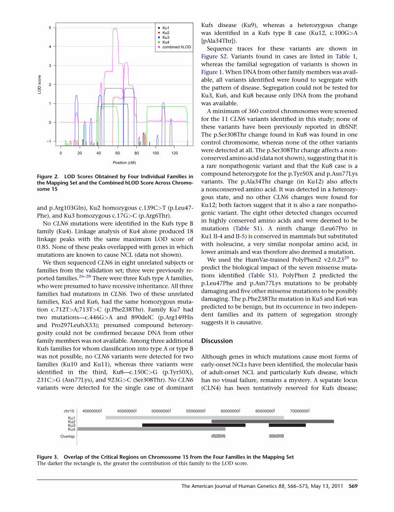
The multipoint linkage analysis achieved a maximum
hLOD score of 4.96 at 58.37 cM on chromosome 15
(Figure 2; see also Figure S1, available online). An adjacent
linkage peak achieved the second highest hLOD of 3.16 at
70.67 cM. The estimated proportions of families showing
linkage to these peaks were 1 and 0.74, respectively,
meaning that all four families showed linkage to the high-
est peak, whereas only three families showed linkage to the
second highest peak (the exception being Ku4, the Kufs
type B family).
Inspection of the inferred haplotypes (not shown)
revealed that the affected individuals from Ku2 and Ku3
rican Journal of Human Genetics 88, 566–573, May 13, 2011 567

Figure 1. Kufs Disease Pedigrees and CLN6 Mutational Status(A) Mapping the set of four families with the mutant allele described with the symbol ‘‘m’’ and with different mutations indicated byvarying superscripts.(B) Four pedigrees from the validation set with themutations shown. The p.Ser308Thr variant in Ku8 (also found in one control) and thep.Ala34Thr variant in Ku12 are not shown.
were homozygous by descent for a haplotype but not for
the same haplotype. Affected individuals from Ku1 and
Ku4 each carry two different haplotypes; no haplotype
appears in more than one family. The breakpoint that
marks the start of the first peak is provided by Ku2, whereas
Ku1 provides the breakpoint that marks the end of the
second peak. Ku3 has an internal double recombination
event that causes the drop in hLOD score between the
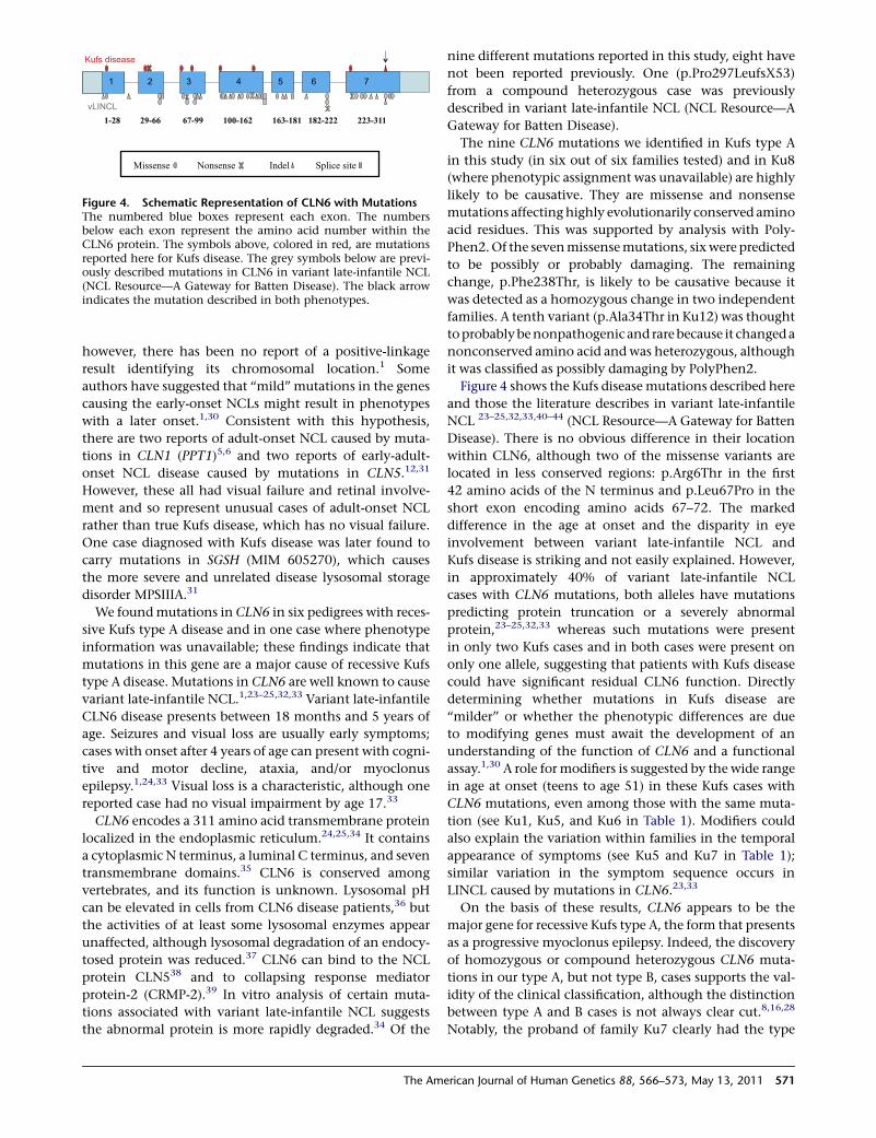
two peaks. The overlap of critical regions for the four fami-
lies is shown in Figure 3.
Inspection of genotypes for all markers on the SNP
genotyping arrays (excluding uninformative SNPs or those
containing Mendelian errors) revealed that the two
affected individuals from Ku2 and Ku3 are both homozy-
gous by state for 1143 consecutive markers in a 4.87 Mb
region that overlaps the first linkage peak and for
another 948 consecutive markers in a 5.29 Mb region
overlapping the second linkage peak. This matches the
expected homozygous linkage results from the linkage
analysis and the inbreeding estimates. This information
allowed the first linkage peak to be refined to 55.37–
61.07 cM (rs873393–rs12907068, 2.58 Mb, 5.7 cM) and
568 The American Journal of Human Genetics 88, 566–573, May 13,
the second linkage peak to be refined to 67.27–71.27 cM
(rs1477799–rs1838544, 2.37 Mb, 4 cM). We identified
candidate genes in these two regions by using the NCBI
MapViewer and UCSC Human Genome databases. The
first peak contained nine genes, including two pseudo-
genes, whereas the second contained 14 genes, including
one pseudogene.
On the basis of the mapping results, we initially focused
on the interval from the mapping set linked to all four
families (Ku1, Ku2, Ku3, and Ku4) and identified
ADAM10, encoding an endopeptidase expressed in the
brain, as a candidate gene. Extensive sequencing of this
gene in six cases (Ku1 II-4 and II-5, Ku2 IV-1, Ku3 II-2,
and Ku4 II-2 and II-3) did not reveal plausible mutations.
Subsequently, we considered the second interval, which
was linked only to the three Kufs type A families (Ku1,
Ku2, and Ku3) but not the type B family (Ku4). An obvious
candidate in this region was CLN6 (ENSG00000128973);
mutations in this gene cause variant late-infantile
NCL.1,24,25 Mutational analysis identified plausible muta-
tions in CLN6 in all three Kufs type A families: Ku1 com-
poundheterozygous c.200T>Cand c.308G>A (p.Leu67Pro
2011
0 20 40 60 80 100 120
−1
0
1
2
3
4
5
Position (cM)
LOD
sco
re
●
●
●
●
●
Ku1Ku2Ku3Ku4combined hLOD
Figure 2. LOD Scores Obtained by Four Individual Families inthe Mapping Set and the Combined hLOD Score Across Chromo-some 15
and p.Arg103Gln), Ku2 homozygous c.139C>T (p.Leu47-
Phe), and Ku3 homozygous c.17G>C (p.Arg6Thr).
No CLN6 mutations were identified in the Kufs type B
family (Ku4). Linkage analysis of Ku4 alone produced 18
linkage peaks with the same maximum LOD score of
0.85. None of these peaks overlapped with genes in which
mutations are known to cause NCL (data not shown).
We then sequenced CLN6 in eight unrelated subjects or
families from the validation set; three were previously re-
ported families.26–28 There were three Kufs type A families,
whowere presumed to have recessive inheritance. All three
families had mutations in CLN6. Two of these unrelated
families, Ku5 and Ku6, had the same homozygous muta-
tion c.712T>A;713T>C (p.Phe238Thr). Family Ku7 had
two mutations—c.446G>A and 890delC (p.Arg149His
and Pro297LeufsX53); presumed compound heterozy-
gosity could not be confirmed because DNA from other
familymembers was not available. Among three additional
Kufs families for whom classification into type A or type B
was not possible, no CLN6 variants were detected for two
families (Ku10 and Ku11), whereas three variants were
identified in the third, Ku8—c.150C>G (p.Tyr50X),
231C>G (Asn77Lys), and 923G>C (Ser308Thr). No CLN6
variants were detected for the single case of dominant
chr15
Ku1Ku2Ku3Ku4
Overlap
40000000 45000000 50000000 550000
Figure 3. Overlap of the Critical Regions on Chromosome 15 fromThe darker the rectangle is, the greater the contribution of this fami
The Ame
Kufs disease (Ku9), whereas a heterozygous change
was identified in a Kufs type B case (Ku12, c.100G>A
[pAla34Thr]).
Sequence traces for these variants are shown in
Figure S2. Variants found in cases are listed in Table 1,
whereas the familial segregation of variants is shown in
Figure 1.WhenDNA from other familymembers was avail-
able, all variants identified were found to segregate with
the pattern of disease. Segregation could not be tested for
Ku3, Ku6, and Ku8 because only DNA from the proband
was available.
A minimum of 360 control chromosomes were screened
for the 11 CLN6 variants identified in this study; none of
these variants have been previously reported in dbSNP.
The p.Ser308Thr change found in Ku8 was found in one
control chromosome, whereas none of the other variants
were detected at all. The p.Ser308Thr change affects a non-
conserved amino acid (datanot shown), suggesting that it is
a rare nonpathogenic variant and that the Ku8 case is a
compound heterozygote for the p.Tyr50X and p.Asn77Lys
variants. The p.Ala34Thr change (in Ku12) also affects
a nonconserved amino acid. It was detected in a heterozy-
gous state, and no other CLN6 changes were found for
Ku12; both factors suggest that it is also a rare nonpatho-
genic variant. The eight other detected changes occurred
in highly conserved amino acids and were deemed to be
mutations (Table S1). A ninth change (Leu67Pro in
Ku1 II-4 and II-5) is conserved in mammals but substituted
with isoleucine, a very similar nonpolar amino acid, in
lower animals and was therefore also deemed a mutation.
We used the HumVar-trained PolyPhen2 v2.0.2329 to
predict the biological impact of the seven missense muta-
tions identified (Table S1). PolyPhen 2 predicted the
p.Leu47Phe and p.Asn77Lys mutations to be probably
damaging and five other missensemutations to be possibly
damaging. The p.Phe238Thr mutation in Ku5 and Ku6 was
predicted to be benign, but its occurrence in two indepen-
dent families and its pattern of segregation strongly
suggests it is causative.
Discussion
Although genes in which mutations cause most forms of
early-onset NCLs have been identified, the molecular basis
of adult-onset NCL and particularly Kufs disease, which
has no visual failure, remains a mystery. A separate locus
(CLN4) has been tentatively reserved for Kufs disease;
00 60000000 65000000 70000000
the Four Families in the Mapping Setly to the LOD score.
rican Journal of Human Genetics 88, 566–573, May 13, 2011 569
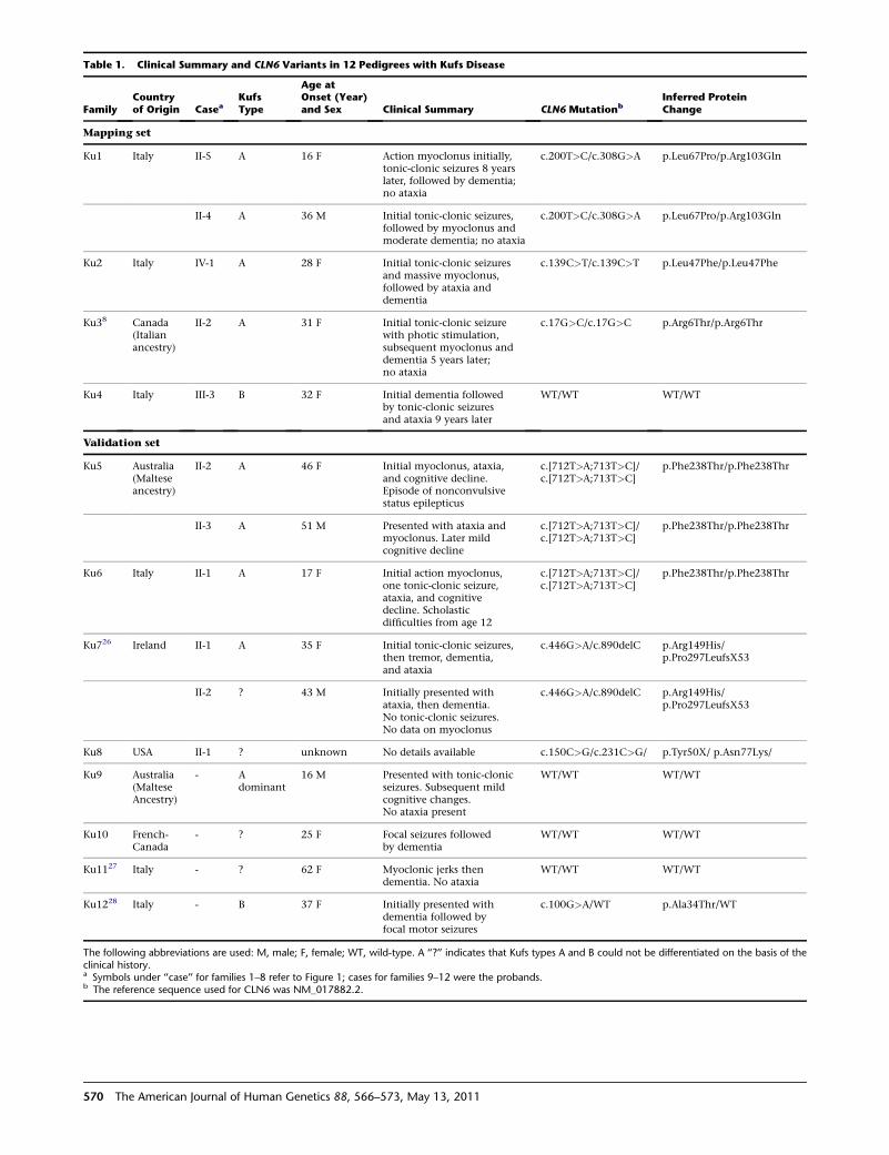
Table 1. Clinical Summary and CLN6 Variants in 12 Pedigrees with Kufs Disease
FamilyCountryof Origin Casea
KufsType
Age atOnset (Year)and Sex Clinical Summary CLN6 Mutationb
Inferred ProteinChange
Mapping set
Ku1 Italy II-5 A 16 F Action myoclonus initially,tonic-clonic seizures 8 yearslater, followed by dementia;no ataxia
c.200T>C/c.308G>A p.Leu67Pro/p.Arg103Gln
II-4 A 36 M Initial tonic-clonic seizures,followed by myoclonus andmoderate dementia; no ataxia
c.200T>C/c.308G>A p.Leu67Pro/p.Arg103Gln
Ku2 Italy IV-1 A 28 F Initial tonic-clonic seizuresand massive myoclonus,followed by ataxia anddementia
c.139C>T/c.139C>T p.Leu47Phe/p.Leu47Phe
Ku38 Canada(Italianancestry)
II-2 A 31 F Initial tonic-clonic seizurewith photic stimulation,subsequent myoclonus anddementia 5 years later;no ataxia
c.17G>C/c.17G>C p.Arg6Thr/p.Arg6Thr
Ku4 Italy III-3 B 32 F Initial dementia followedby tonic-clonic seizuresand ataxia 9 years later
WT/WT WT/WT
Validation set
Ku5 Australia(Malteseancestry)
II-2 A 46 F Initial myoclonus, ataxia,and cognitive decline.Episode of nonconvulsivestatus epilepticus
c.[712T>A;713T>C]/c.[712T>A;713T>C]
p.Phe238Thr/p.Phe238Thr
II-3 A 51 M Presented with ataxia andmyoclonus. Later mildcognitive decline
c.[712T>A;713T>C]/c.[712T>A;713T>C]
p.Phe238Thr/p.Phe238Thr
Ku6 Italy II-1 A 17 F Initial action myoclonus,one tonic-clonic seizure,ataxia, and cognitivedecline. Scholasticdifficulties from age 12
c.[712T>A;713T>C]/c.[712T>A;713T>C]
p.Phe238Thr/p.Phe238Thr
Ku726 Ireland II-1 A 35 F Initial tonic-clonic seizures,then tremor, dementia,and ataxia
c.446G>A/c.890delC p.Arg149His/p.Pro297LeufsX53
II-2 ? 43 M Initially presented withataxia, then dementia.No tonic-clonic seizures.No data on myoclonus
c.446G>A/c.890delC p.Arg149His/p.Pro297LeufsX53
Ku8 USA II-1 ? unknown No details available c.150C>G/c.231C>G/ p.Tyr50X/ p.Asn77Lys/
Ku9 Australia(MalteseAncestry)
- Adominant
16 M Presented with tonic-clonicseizures. Subsequent mildcognitive changes.No ataxia present
WT/WT WT/WT
Ku10 French-Canada
- ? 25 F Focal seizures followedby dementia
WT/WT WT/WT
Ku1127 Italy - ? 62 F Myoclonic jerks thendementia. No ataxia
WT/WT WT/WT
Ku1228 Italy - B 37 F Initially presented withdementia followed byfocal motor seizures
c.100G>A/WT p.Ala34Thr/WT
The following abbreviations are used: M, male; F, female; WT, wild-type. A ‘‘?’’ indicates that Kufs types A and B could not be differentiated on the basis of theclinical history.a Symbols under ‘‘case’’ for families 1–8 refer to Figure 1; cases for families 9–12 were the probands.b The reference sequence used for CLN6 was NM_017882.2.
570 The American Journal of Human Genetics 88, 566–573, May 13, 2011
1 2 3 4 5 6 7
Missense Nonsense Indel Splice site
1-28 29-66 67-99 100-162 163-181 182-222 223-311
Kufs disease
vLINCL
Figure 4. Schematic Representation of CLN6 with MutationsThe numbered blue boxes represent each exon. The numbersbelow each exon represent the amino acid number within theCLN6 protein. The symbols above, colored in red, are mutationsreported here for Kufs disease. The grey symbols below are previ-ously described mutations in CLN6 in variant late-infantile NCL(NCL Resource—A Gateway for Batten Disease). The black arrowindicates the mutation described in both phenotypes.
however, there has been no report of a positive-linkage
result identifying its chromosomal location.1 Some
authors have suggested that ‘‘mild’’ mutations in the genes
causing the early-onset NCLs might result in phenotypes
with a later onset.1,30 Consistent with this hypothesis,
there are two reports of adult-onset NCL caused by muta-
tions in CLN1 (PPT1)5,6 and two reports of early-adult-
onset NCL disease caused by mutations in CLN5.12,31
However, these all had visual failure and retinal involve-
ment and so represent unusual cases of adult-onset NCL
rather than true Kufs disease, which has no visual failure.
One case diagnosed with Kufs disease was later found to
carry mutations in SGSH (MIM 605270), which causes
the more severe and unrelated disease lysosomal storage
disorder MPSIIIA.31
We foundmutations in CLN6 in six pedigrees with reces-
sive Kufs type A disease and in one case where phenotype
information was unavailable; these findings indicate that
mutations in this gene are a major cause of recessive Kufs
type A disease. Mutations in CLN6 are well known to cause
variant late-infantile NCL.1,23–25,32,33 Variant late-infantile
CLN6 disease presents between 18 months and 5 years of
age. Seizures and visual loss are usually early symptoms;
cases with onset after 4 years of age can present with cogni-
tive and motor decline, ataxia, and/or myoclonus
epilepsy.1,24,33 Visual loss is a characteristic, although one
reported case had no visual impairment by age 17.33
CLN6 encodes a 311 amino acid transmembrane protein
localized in the endoplasmic reticulum.24,25,34 It contains
a cytoplasmic N terminus, a luminal C terminus, and seven
transmembrane domains.35 CLN6 is conserved among
vertebrates, and its function is unknown. Lysosomal pH
can be elevated in cells from CLN6 disease patients,36 but
the activities of at least some lysosomal enzymes appear
unaffected, although lysosomal degradation of an endocy-
tosed protein was reduced.37 CLN6 can bind to the NCL
protein CLN538 and to collapsing response mediator
protein-2 (CRMP-2).39 In vitro analysis of certain muta-
tions associated with variant late-infantile NCL suggests
the abnormal protein is more rapidly degraded.34 Of the
The Ame
nine different mutations reported in this study, eight have
not been reported previously. One (p.Pro297LeufsX53)
from a compound heterozygous case was previously
described in variant late-infantile NCL (NCL Resource—A
Gateway for Batten Disease).
The nine CLN6 mutations we identified in Kufs type A
in this study (in six out of six families tested) and in Ku8
(where phenotypic assignment was unavailable) are highly
likely to be causative. They are missense and nonsense
mutations affectinghighly evolutionarily conserved amino
acid residues. This was supported by analysis with Poly-
Phen2.Of the sevenmissensemutations, sixwere predicted
to be possibly or probably damaging. The remaining
change, p.Phe238Thr, is likely to be causative because it
was detected as a homozygous change in two independent
families. A tenth variant (p.Ala34Thr in Ku12) was thought
toprobablybenonpathogenic and rare because it changeda
nonconserved amino acid and was heterozygous, although
it was classified as possibly damaging by PolyPhen2.
Figure 4 shows the Kufs disease mutations described here
and those the literature describes in variant late-infantile
NCL 23–25,32,33,40–44 (NCL Resource—A Gateway for Batten
Disease). There is no obvious difference in their location
within CLN6, although two of the missense variants are
located in less conserved regions: p.Arg6Thr in the first
42 amino acids of the N terminus and p.Leu67Pro in the
short exon encoding amino acids 67–72. The marked
difference in the age at onset and the disparity in eye
involvement between variant late-infantile NCL and
Kufs disease is striking and not easily explained. However,
in approximately 40% of variant late-infantile NCL
cases with CLN6 mutations, both alleles have mutations
predicting protein truncation or a severely abnormal
protein,23–25,32,33 whereas such mutations were present
in only two Kufs cases and in both cases were present on
only one allele, suggesting that patients with Kufs disease
could have significant residual CLN6 function. Directly
determining whether mutations in Kufs disease are
‘‘milder’’ or whether the phenotypic differences are due
to modifying genes must await the development of an
understanding of the function of CLN6 and a functional
assay.1,30 A role formodifiers is suggested by the wide range
in age at onset (teens to age 51) in these Kufs cases with
CLN6 mutations, even among those with the same muta-
tion (see Ku1, Ku5, and Ku6 in Table 1). Modifiers could
also explain the variation within families in the temporal
appearance of symptoms (see Ku5 and Ku7 in Table 1);
similar variation in the symptom sequence occurs in
LINCL caused by mutations in CLN6.23,33
On the basis of these results, CLN6 appears to be the
major gene for recessive Kufs type A, the form that presents
as a progressive myoclonus epilepsy. Indeed, the discovery
of homozygous or compound heterozygous CLN6 muta-
tions in our type A, but not type B, cases supports the val-
idity of the clinical classification, although the distinction
between type A and B cases is not always clear cut.8,16,28
Notably, the proband of family Ku7 clearly had the type
rican Journal of Human Genetics 88, 566–573, May 13, 2011 571
A phenotype, whereas her sibling could have been type B,
and unfortunately the phenotypic assignment of Ku8 as
type A or B could not be made because of a lack of data
(see Table 1). Furthermore, the role of the heterozygous
CLN6 variant in Ku12 could not be determined, and it is
possible that this variant influences the disease arising
from mutations in another locus. The major gene or genes
for Kufs type B, as well as for the dominant form of adult
NCL, await discovery. However, linkage mapping of Ku4
suggests that Kufs type B is not caused by a mutation in
the same genes mutated in other forms of NCL.
Diagnosis of Kufs disease is challenging and often
requires invasive biopsy. A relatively inexpensive CLN6
mutation screening should now be considered as an initial
diagnostic step in suspected Kufs disease cases, especially
when progressive myoclonus epilepsy is the presenting
feature.
Supplemental Data
Supplemental Data include two figures and one table and can be
found with this article online at http://www.cell.com/AJHG/.
Acknowledgments
S.F.B. was supported by an NHMRC Australia Fellowship and an
NHMRC Program Grant. M.B. was funded by an NHMRC Career
Development Award and an NHMRC Program Grant. We thank
the Batten Disease Support and Research Association for addi-
tional financial support (to S.M.) and the families themselves.
Received: February 18, 2011
Revised: April 7, 2011
Accepted: April 8, 2011
Published online: May 5, 2011
Web Resources
The URLs for data presented herein are as follows:
dbSNP, http://www.ncbi.nlm.nih.gov/projects/SNP/
Online Mendelian Inheritance in Man, http://www.omim.org
NCBIMapViewer,http://www.ncbi.nlm.nih.gov/projects/mapview/
NCL Resource—A Gateway for Batten Disease, http://www.ucl.ac.
uk/ncl/cln6.shtml
Primer 3, http://frodo.wi.mit.edu/primer3
UCSC Genome Browser, http://www.genome.ucsc.edu
References
1. Mole, S.E., and Williams, R.E. (2010). Neuronal Ceroid-
Lipofuscinoses. Gene Reviews, http://www.ncbi.nlm.nih.
gov/books/NBK1428/
2. Santavuori, P. (1988). Neuronal ceroid-lipofuscinoses in child-
hood. Brain Dev. 10, 80–83.
3. Mole, S.E., Williams, R.E., and Goebel, H.H. (2005). Correla-
tions between genotype, ultrastructural morphology and
clinical phenotype in the neuronal ceroid lipofuscinoses.
Neurogenetics 6, 107–126.
572 The American Journal of Human Genetics 88, 566–573, May 13,
4. Wisniewski, K.E., Zhong, N., and Philippart, M. (2001).
Pheno/genotypic correlations of neuronal ceroid lipofuscino-
ses. Neurology 57, 576–581.
5. van Diggelen, O.P., Thobois, S., Tilikete, C., Zabot, M.T., Keule-
mans, J.L., vanBunderen,P.A., Taschner, P.E., Losekoot,M., and
Voznyi, Y.V. (2001). Adult neuronal ceroid lipofuscinosis with
palmitoyl-protein thioesterase deficiency: first adult-onset
patients of a childhood disease. Ann. Neurol. 50, 269–272.
6. Ramadan, H., Al-Din, A.S., Ismail, A., Balen, F., Varma, A.,
Twomey, A., Watts, R., Jackson, M., Anderson, G., Green, E.,
and Mole, S.E. (2007). Adult neuronal ceroid lipofuscinosis
caused by deficiency in palmitoyl protein thioesterase 1.
Neurology 68, 387–388.
7. Kufs, H. (1925). Uber eine Spatform der amaurotischen Idiotie
und ihre heredofamiliaren Grundlagen. Z ges Neurol Psychiat
95, 169–188.
8. Berkovic, S.F., Carpenter, S., Andermann, F., Andermann, E.,
and Wolfe, L.S. (1988). Kufs’ disease: a critical reappraisal.
Brain 111, 27–62.
9. Constantinidis, J., Wisniewski, K.E., and Wisniewski, T.M.
(1992). The adult and a new late adult forms of neuronal
ceroid lipofuscinosis. Acta Neuropathol. 83, 461–468.
10. Boehme, D.H., Cottrell, J.C., Leonberg, S.C., and Zeman, W.
(1971). A dominant form of neuronal ceroid-lipofuscinosis.
Brain 94, 745–760.
11. Nijssen, P.C., Ceuterick, C., van Diggelen, O.P., Elleder, M.,
Martin, J.J., Teepen, J.L., Tyynela, J., and Roos, R.A. (2003).
Autosomal dominant adult neuronal ceroid lipofuscinosis:
a novel form of NCL with granular osmiophilic deposits
without palmitoyl protein thioesterase 1 deficiency. Brain
Pathol. 13, 574–581.
12. Xin, W., Mullen, T.E., Kiely, R., Min, J., Feng, X., Cao, Y.,
O’Malley, L., Shen, Y., Chu-Shore, C., Mole, S.E., et al. (2010).
CLN5 mutations are frequent in juvenile and late-onset
non-Finnish patients with NCL. Neurology 74, 565–571.
13. Carpenter, S., Karpati, G., Andermann, F., Jacob, J.C., and
Andermann, E. (1977). The ultrastructural characteristics of
the abnormal cytosomes in Batten-Kufs’ disease. Brain 100,
137–156.
14. Goebel, H.H., and Braak, H. (1989). Adult neuronal ceroid-
lipofuscinosis. Clin. Neuropathol. 8, 109–119.
15. Pasquinelli, G., Cenacchi, G., Piane, E.L., Russo, C., and
Aguglia, U. (2004). The problematic issue of Kufs disease
diagnosis as performed on rectal biopsies: a case report. Ultra-
struct. Pathol. 28, 43–48.
16. Sadzot, B., Reznik, M., Arrese-Estrada, J.E., and Franck, G.
(2000). Familial Kufs’ disease presenting as a progressive
myoclonic epilepsy. J. Neurol. 247, 447–454.
17. Leutenegger, A.L., Prum, B., Genin, E., Verny, C., Lemainque,
A., Clerget-Darpoux, F., and Thompson, E.A. (2003). Estima-
tion of the inbreeding coefficient through use of genomic
data. Am. J. Hum. Genet. 73, 516–523.
18. Leutenegger, A.L., Labalme, A., Genin, E., Toutain, A., Stei-
chen, E., Clerget-Darpoux, F., and Edery, P. (2006). Using
genomic inbreeding coefficient estimates for homozygosity
mapping of rare recessive traits: Application to Taybi-Linder
syndrome. Am. J. Hum. Genet. 79, 62–66.
19. Purcell, S., Neale, B., Todd-Brown, K., Thomas, L., Ferreira,
M.A., Bender, D., Maller, J., Sklar, P., de Bakker, P.I., Daly,
M.J., and Sham, P.C. (2007). PLINK: a tool set for whole-
genome association and population-based linkage analyses.
Am. J. Hum. Genet. 81, 559–575.
2011

20. Abecasis, G.R., Cherny, S.S., Cookson, W.O., and Cardon, L.R.
(2002). Merlin—rapid analysis of dense genetic maps using
sparse gene flow trees. Nat. Genet. 30, 97–101.
21. Bahlo, M., and Bromhead, C.J. (2009). Generating linkage
mapping files from Affymetrix SNP chip data. Bioinformatics
25, 1961–1962.
22. Thiele, H., and Nurnberg, P. (2005). HaploPainter: a tool for
drawing pedigrees with complex haplotypes. Bioinformatics
21, 1730–1732.
23. Sharp, J.D., Wheeler, R.B., Parker, K.A., Gardiner, R.M.,
Williams, R.E., and Mole, S.E. (2003). Spectrum of CLN6
mutations in variant late infantile neuronal ceroid lipofusci-
nosis. Hum. Mutat. 22, 35–42.
24. Gao, H., Boustany, R.M., Espinola, J.A., Cotman, S.L., Srinidhi,
L., Antonellis, K.A., Gillis, T., Qin, X., Liu, S., Donahue, L.R.,
et al. (2002). Mutations in a novel CLN6-encoded transmem-
brane protein cause variant neuronal ceroid lipofuscinosis in
man and mouse. Am. J. Hum. Genet. 70, 324–335.
25. Wheeler, R.B., Sharp, J.D., Schultz, R.A., Joslin, J.M., Williams,
R.E., and Mole, S.E. (2002). The gene mutated in variant late-
infantile neuronal ceroid lipofuscinosis (CLN6) and in nclf
mutant mice encodes a novel predicted transmembrane
protein. Am. J. Hum. Genet. 70, 537–542.
26. Callagy, C., O’Neill, G., Murphy, S.F., and Farrell, M.A. (2000).
Adult neuronal ceroid lipofuscinosis (Kufs’ disease) in two
siblings of an Irish family. Clin. Neuropathol. 19, 109–118.
27. Gambardella, A., Pasquinelli, G., Cittadella, R., Bono, F.,
Oliveri, R.L., Valentino, P., Zappia, M., Quattrone, A., and
Aguglia, U. (1998). Kufs’ disease presenting as late-onset
epilepsia partialis continua. Neurology 51, 1180–1182.
28. Zini, A., Cenacchi, G., Nichelli, P., Zunarelli, E., Todeschini, A.,
and Meletti, S. (2008). Early-onset dementia with prolonged
occipital seizures: an atypical case of Kufs disease. Neurology
71, 1709–1712.
29. Adzhubei, I.A., Schmidt, S., Peshkin, L., Ramensky, V.E.,
Gerasimova, A., Bork, P., Kondrashov, A.S., and Sunyaev, S.R.
(2010). A method and server for predicting damaging
missense mutations. Nat. Methods 7, 248–249.
30. Lauronen, L., Munroe, P.B., Jarvela, I., Autti, T., Mitchison,
H.M., O’Rawe, A.M., Gardiner, R.M., Mole, S.E., Puranen, J.,
Hakkinen, A.M., et al. (1999). Delayed classic and protracted
phenotypes of compound heterozygous juvenile neuronal
ceroid lipofuscinosis. Neurology 52, 360–365.
31. Sleat, D.E., Ding, L., Wang, S., Zhao, C., Wang, Y., Xin, W.,
Zheng, H., Moore, D.F., Sims, K.B., and Lobel, P. (2009).
Mass spectrometry-based protein profiling to determine the
cause of lysosomal storage diseases of unknown etiology.
Mol. Cell. Proteomics 8, 1708–1718.
32. Moore, S.J., Buckley, D.J., MacMillan, A., Marshall, H.D.,
Steele, L., Ray, P.N., Nawaz, Z., Baskin, B., Frecker, M., Carr,
S.M., et al. (2008). The clinical and genetic epidemiology of
neuronal ceroid lipofuscinosis in Newfoundland. Clin. Genet.
74, 213–222.
The Ame
33. Cannelli, N., Garavaglia, B., Simonati, A., Aiello, C., Barzaghi,
C., Pezzini, F., Cilio, M.R., Biancheri, R., Morbin, M., Dalla
Bernardina, B., et al. (2009). Variant late infantile ceroid
lipofuscinoses associated with novel mutations in CLN6.
Biochem. Biophys. Res. Commun. 379, 892–897.
34. Kurze, A.K., Galliciotti, G., Heine, C., Mole, S.E., Quitsch, A.,
and Braulke, T. (2010). Pathogenic mutations cause rapid
degradation of lysosomal storage disease-related membrane
protein CLN6. Hum. Mutat. 31, E1163–E1174.
35. Heine, C., Quitsch, A., Storch, S., Martin, Y., Lonka, L.,
Lehesjoki, A.E., Mole, S.E., and Braulke, T. (2007). Topology
and endoplasmic reticulum retention signals of the lysosomal
storage disease-relatedmembrane protein CLN6.Mol. Membr.
Biol. 24, 74–87.
36. Holopainen, J.M., Saarikoski, J., Kinnunen, P.K., and Jarvela, I.
(2001). Elevated lysosomal pH in neuronal ceroid lipofuscino-
ses (NCLs). Eur. J. Biochem. 268, 5851–5856.
37. Heine, C., Koch, B., Storch, S., Kohlschutter, A., Palmer, D.N.,
and Braulke, T. (2004). Defective endoplasmic reticulum-
resident membrane protein CLN6 affects lysosomal degrada-
tion of endocytosed arylsulfatase A. J. Biol. Chem. 279,
22347–22352.
38. Lyly, A., von Schantz, C., Heine, C., Schmiedt, M.L., Sipila, T.,
Jalanko, A., and Kyttala, A. (2009). Novel interactions of CLN5
support molecular networking between Neuronal Ceroid
Lipofuscinosis proteins. BMC Cell Biol. 10, 83.
39. Benedict, J.W., Getty, A.L., Wishart, T.M., Gillingwater, T.H.,
and Pearce, D.A. (2009). Protein product of CLN6 gene
responsible for variant late-onset infantile neuronal ceroid
lipofuscinosis interacts with CRMP-2. J. Neurosci. Res. 87,
2157–2166.
40. Al-Muhaizea, M.A., Al-Hassnan, Z.N., and Chedrawi, A.
(2009). Variant late infantile neuronal ceroid lipofuscinosis
(CLN6 gene) in Saudi Arabia. Pediatr. Neurol. 41, 74–76.
41. Cismondi, I.A., Kohan, R., Ghio, A., Ramirez, A.M., and Halac,
I.N. (2008). Gene symbol: CLN6. Disease: Neuronal ceroid
lipofuscinosis, late infantile. Hum. Genet. 124, 323–324.
42. Kousi, M., Siintola, E., Dvorakova, L., Vlaskova, H., Turnbull,
J., Topcu, M., Yuksel, D., Gokben, S., Minassian, B.A., Elleder,
M., et al. (2009). Mutations in CLN7/MFSD8 are a common
cause of variant late-infantile neuronal ceroid lipofuscinosis.
Brain 132, 810–819.
43. Siintola, E., Topcu, M., Kohlschutter, A., Salonen, T., Joensuu,
T., Anttonen, A.K., and Lehesjoki, A.E. (2005). Two novel
CLN6 mutations in variant late-infantile neuronal ceroid
lipofuscinosis patients of Turkish origin. Clin. Genet. 68,
167–173.
44. Teixeira, C.A., Espinola, J., Huo, L., Kohlschutter, J., Persaud
Sawin, D.A., Minassian, B., Bessa, C.J., Guimaraes, A.,
Stephan, D.A., Sa Miranda, M.C., et al. (2003). Novel muta-
tions in the CLN6 gene causing a variant late infantile
neuronal ceroid lipofuscinosis. Hum. Mutat. 21, 502–508.
rican Journal of Human Genetics 88, 566–573, May 13, 2011 573
Related Documents