Knockdown of NPY expression in the dorsomedial hypothalamus promotes development of brown adipocytes and prevents diet-induced obesity Pei-Ting Chao 1,4 , Liang Yang 1,3,4 , Susan Aja 2 , Timothy H. Moran 1 , and Sheng Bi 1 1 Department of Psychiatry and Behavioral Sciences, Johns Hopkins University School of Medicine, Baltimore, Maryland, 21205, USA 2 Center for Metabolism and Obesity Research, Johns Hopkins University School of Medicine, Baltimore, Maryland, 21205, USA SUMMARY Hypothalamic neuropeptide Y (NPY) has been implicated in control of energy balance, but the physiological importance of NPY in the dorsomedial hypothalamus (DMH) remains unclear. Here we report that knockdown of NPY expression in the DMH by adeno-associated virus-mediated RNAi reduced fat depots in rats fed regular chow and ameliorated high-fat diet-induced hyperphagia and obesity. DMH NPY knockdown resulted in development of brown adipocytes in inguinal white adipose tissue through the sympathetic nervous system. This knockdown increased uncoupling protein 1 expression in both inguinal fat and interscapular brown adipose tissue (BAT). Consistent with the activation of BAT, DMH NPY knockdown increased energy expenditure and enhanced the thermogenic response to a cold environment. This knockdown also increased locomotor activity, improved glucose homeostasis and enhanced insulin sensitivity. Together, these results demonstrate critical roles of DMH NPY in body weight regulation through affecting food intake, body adiposity, thermogenesis, energy expenditure and physical activity. INTRODUCTION The hypothalamus plays a central role in maintaining energy homeostasis (Cone, 2006; Elmquist et al., 1999; Schwartz et al., 2000; Spiegelman and Flier, 2001). Various hypothalamic nuclei, including the arcuate nucleus (ARC), dorsomedial hypothalamus (DMH), paraventricular nucleus (PVN), lateral hypothalamic area (LH), and ventromedial hypothalamus (VMH) have been demonstrated to have specific functions in the regulation of energy balance. Although the observations that DMH lesions result in hypophagia and decreased body weight indicate the importance of the DMH in maintaining energy homeostasis (Bellinger and Bernardis, 2002), the neural mechanisms through which the DMH acts to affect energy balance remain unclear. © 2011 Elsevier Inc. All rights reserved. Correspondence should be addressed to: Sheng Bi, Department of Psychiatry and Behavioral Sciences, Johns Hopkins University School of Medicine, 720 Rutland Ave., Ross 618, Baltimore, Maryland 21205. Telephone: (410) 502-4789; Fax: (410) 502-3769; [email protected]. 3 Present address: Harvard-MIT Division of Health Sciences & Technology, Massachusetts Institute of Technology, Cambridge, MA 02139, USA 4 These authors contributed equally to this work. Publisher's Disclaimer: This is a PDF file of an unedited manuscript that has been accepted for publication. As a service to our customers we are providing this early version of the manuscript. The manuscript will undergo copyediting, typesetting, and review of the resulting proof before it is published in its final citable form. Please note that during the production process errors may be discovered which could affect the content, and all legal disclaimers that apply to the journal pertain. NIH Public Access Author Manuscript Cell Metab. Author manuscript; available in PMC 2012 May 4. Published in final edited form as: Cell Metab. 2011 May 4; 13(5): 573–583. doi:10.1016/j.cmet.2011.02.019. NIH-PA Author Manuscript NIH-PA Author Manuscript NIH-PA Author Manuscript

Welcome message from author
This document is posted to help you gain knowledge. Please leave a comment to let me know what you think about it! Share it to your friends and learn new things together.
Transcript
Knockdown of NPY expression in the dorsomedialhypothalamus promotes development of brown adipocytes andprevents diet-induced obesity
Pei-Ting Chao1,4, Liang Yang1,3,4, Susan Aja2, Timothy H. Moran1, and Sheng Bi11 Department of Psychiatry and Behavioral Sciences, Johns Hopkins University School ofMedicine, Baltimore, Maryland, 21205, USA2 Center for Metabolism and Obesity Research, Johns Hopkins University School of Medicine,Baltimore, Maryland, 21205, USA
SUMMARYHypothalamic neuropeptide Y (NPY) has been implicated in control of energy balance, but thephysiological importance of NPY in the dorsomedial hypothalamus (DMH) remains unclear. Herewe report that knockdown of NPY expression in the DMH by adeno-associated virus-mediatedRNAi reduced fat depots in rats fed regular chow and ameliorated high-fat diet-inducedhyperphagia and obesity. DMH NPY knockdown resulted in development of brown adipocytes ininguinal white adipose tissue through the sympathetic nervous system. This knockdown increaseduncoupling protein 1 expression in both inguinal fat and interscapular brown adipose tissue(BAT). Consistent with the activation of BAT, DMH NPY knockdown increased energyexpenditure and enhanced the thermogenic response to a cold environment. This knockdown alsoincreased locomotor activity, improved glucose homeostasis and enhanced insulin sensitivity.Together, these results demonstrate critical roles of DMH NPY in body weight regulation throughaffecting food intake, body adiposity, thermogenesis, energy expenditure and physical activity.
INTRODUCTIONThe hypothalamus plays a central role in maintaining energy homeostasis (Cone, 2006;Elmquist et al., 1999; Schwartz et al., 2000; Spiegelman and Flier, 2001). Varioushypothalamic nuclei, including the arcuate nucleus (ARC), dorsomedial hypothalamus(DMH), paraventricular nucleus (PVN), lateral hypothalamic area (LH), and ventromedialhypothalamus (VMH) have been demonstrated to have specific functions in the regulation ofenergy balance. Although the observations that DMH lesions result in hypophagia anddecreased body weight indicate the importance of the DMH in maintaining energyhomeostasis (Bellinger and Bernardis, 2002), the neural mechanisms through which theDMH acts to affect energy balance remain unclear.
© 2011 Elsevier Inc. All rights reserved.Correspondence should be addressed to: Sheng Bi, Department of Psychiatry and Behavioral Sciences, Johns Hopkins UniversitySchool of Medicine, 720 Rutland Ave., Ross 618, Baltimore, Maryland 21205. Telephone: (410) 502-4789; Fax: (410) 502-3769;[email protected] address: Harvard-MIT Division of Health Sciences & Technology, Massachusetts Institute of Technology, Cambridge, MA02139, USA4These authors contributed equally to this work.Publisher's Disclaimer: This is a PDF file of an unedited manuscript that has been accepted for publication. As a service to ourcustomers we are providing this early version of the manuscript. The manuscript will undergo copyediting, typesetting, and review ofthe resulting proof before it is published in its final citable form. Please note that during the production process errors may bediscovered which could affect the content, and all legal disclaimers that apply to the journal pertain.
NIH Public AccessAuthor ManuscriptCell Metab. Author manuscript; available in PMC 2012 May 4.
Published in final edited form as:Cell Metab. 2011 May 4; 13(5): 573–583. doi:10.1016/j.cmet.2011.02.019.
NIH
-PA Author Manuscript
NIH
-PA Author Manuscript
NIH
-PA Author Manuscript
Neuropeptide Y (NPY) is an important hypothalamic orexigenic peptide (Clark et al., 1984;Levine and Morley, 1984; Stanley and Leibowitz, 1984). Numerous reports havedemonstrated the actions of NPY in the ARC in the control of energy balance. The ARCcontains two distinct populations of neurons: orexigenic neuropeptide NPY/agouti-relatedprotein (AgRP) neurons and anorexigenic proopiomelanocortin (POMC) neurons. These twoneural systems integrate hormonal (such as leptin and insulin) and nutrient signals tomodulate food intake and body weight (Cone, 2006; Elmquist et al., 1999; Schwartz et al.,2000; Spiegelman and Flier, 2001). Modulation of ARC NPY signaling in adult animalssignificantly impacts energy balance. Genetic ablation of neurons expressing NPY/AgRP inadult mice results in a lean and hypophagic phenotype (Bewick et al., 2005; Gropp et al.,2005). Adeno-associated virus (AAV)-mediated expression of antisense Npy cRNA in theARC of adult rats decreased NPY expression and resulted in decreased food intake and bodyweight (Gardiner et al., 2005). We have shown that knockdown of NPY expression in theARC via AAV-mediated RNA interference (RNAi) attenuated the feeding response to fooddeprivation (Yang et al., 2009).
NPY is also expressed by neurons within the DMH, but the function of NPY in this nucleushas yet to be fully determined. Recent reports imply that NPY in the DMH may serve as animportant neuromodulator to influence energy balance. We and other investigators havereported that Npy gene expression was elevated in the DMH in specific animal models withincreased energy demands (Bi et al., 2003; Kawaguchi et al., 2005; Smith, 1993). Inductionor overexpression of Npy in the DMH has also been found in several rodent models ofobesity (Bi et al., 2001; Guan et al., 1998; Kesterson et al., 1997; Tritos et al., 1998).Moreover, whereas NPY in the ARC is under the control of leptin, its regulation in theDMH is leptin-independent (Bi et al., 2003). Overall, these observations suggest that NPY inthe DMH is important for maintaining energy homeostasis, but the mechanism of action ofDMH NPY in controlling energy balance differs from that of ARC NPY.
We have manipulated DMH NPY signaling using viral-mediated alterations of NPYexpression in the DMH for examination of the role of DMH NPY in controlling energybalance. We found that AAV-mediated overexpression of NPY in the DMH of lean ratsincreased food intake and body weight, and exacerbated diet-induced obesity (Yang et al.,2009). Knockdown of NPY expression in the DMH via AAV-mediated RNAi amelioratedthe hyperphagia, obesity and diabetes of Otsuka Long-Evans Tokushima Fatty (OLETF) rats(Yang et al., 2009) in which DMH NPY overexpression has been proposed to play anetiological role (Bi et al., 2001). Thus, these results implicate DMH NPY in modulatingfood intake and energy balance. It is unclear, however, whether NPY in the DMH isphysiologically important in maintaining energy homeostasis and/or whether DMH NPYaffects other aspects of energy balance including energy expenditure, thermoregulation,adipogenesis and physical activity. Here, we sought to ascertain such roles of DMH NPY inbody weight regulation by using AAV-mediated RNAi for specifically knocking down NPYexpression in the DMH of rats.
RESULTSAAV-mediated knockdown of NPY expression in the DMH
We generated a recombinant vector of AAV-mediated RNAi with NPY-specific shorthairpin RNA (AAVshNPY) containing humanized Renilla green fluorescent protein(hrGFP) marker as previously reported (Yang et al., 2009). To test the idea that DMH NPYmay be an important neuromodulator of energy balance under normal conditions, we firstdetermined the effect of AAV-mediated RNAi on Npy gene expression in Sprague-Dawleyrats by injecting this vector bilaterally into the DMH (Fig. 1A). We established that the viralvectors infected neurons within the DMH as early as 1 week after viral injection, led to a
Chao et al. Page 2
Cell Metab. Author manuscript; available in PMC 2012 May 4.
NIH
-PA Author Manuscript
NIH
-PA Author Manuscript
NIH
-PA Author Manuscript
robust infection within 2 weeks (Fig. 1B, hrGFP-positive neurons), and produced significantknockdown of Npy mRNA expression in the DMH (Fig. 1C) by 28%, 47% and 49% at 1, 2and 4 weeks post-viral injections, respectively, compared to rats receiving control vectorinjections (AAVshCTL, Fig. 1D). This knockdown effect was site-specific since no hrGFP-positive neurons were detected in the ARC (Fig. 1B) and Npy mRNA levels were unalteredin the ARC (Fig. 1E). Consistent with our previous report (Yang et al., 2009), the effects ofAAV-mediated RNAi on Npy mRNA expression in the DMH were long lasting; 16 weekspost-viral injection, Npy mRNA levels remained reduced by 36% (data not shown).
Effects of DMH NPY knockdown on regulation of body weightFollowing determination of viral-mediated knockdown of NPY expression in the DMH, weexamined whether this knockdown affects body weight regulation. We found that DMHNPY knockdown resulted in a small but significant decrease in body weight gain over thefirst 5 weeks post-viral injection when rats were maintained on regular chow (RC, P =0.035,Fig. 2A). The weight gain of NPY knockdown rats was reduced by about 9%. Since high-fatdiet (HF) increases body weight and induces obesity, we next assessed the effect of DMHNPY knockdown on HF-induced weight gain. Half the NPY knockdown and control ratswere challenged with HF at 5 weeks post-viral injection. We found that NPY knockdownsignificantly reduced HF-induced increases in weight gain (P=0.023). Control rats fed HFgained significantly more weight by 2 week (P =0.026) and had gained 35% more weight by11 weeks compared to control rats on RC. In contrast, NPY knockdown rats fed HF gainedbody weight more slowly, only achieving significantly increased body weight by 4 weeks (P=0.021) and having only 26% more weight by 11 weeks compared to those on RC (Fig. 2A).As a result, the body weight gain of NPY knockdown rats remained relatively normal until 7weeks on HF compared to control rats on RC and was significantly less than control rats in11 weeks on HF (Fig. 2A).
Since we have demonstrated the effects of DMH NPY on food intake and meal patterns inboth OLETF and intact rats (Yang et al., 2009), we examined whether DMH NPYknockdown altered daily food intake in the present study. Although daily energy intake didnot differ between the two groups of rats over 16 weeks on RC, DMH NPY knockdownsignificantly reduced HF-induced hyperphagia (Fig. 2B). Both groups of rats increased dailyintake dramatically upon initial access to HF, but the degree of increase and its durationwere significantly reduced in NPY knockdown rats (Fig. 2B). While control rats on HFremained hyperphagic, NPY knockdown rats normalized energy intake in 4 weeks on HF(Fig. 2B).
DMH NPY knockdown improves glucose homeostasisWe next tested the effects of DMH NPY knockdown on glucose homeostasis. Although oralglucose administration resulted in similar patterns of glucose clearance in NPY knockdownand control rats on RC (Fig. 2C), NPY knockdown rats required less insulin secretion toclear the glucose as indicated by a reduction in the area under the response curve of insulinin NPY knockdown rats (Fig. 2D), suggesting that down-regulation of DMH NPYexpression enhances insulin sensitivity. HF access caused hyperinsulinemia and impairedglucose clearance in control rats as determined by high fasting insulin levels and elevatedblood glucose and plasma insulin levels in response to oral glucose (Fig. 2C and D). DMHNPY knockdown significantly ameliorated these changes. NPY knockdown rats on HF hadnormal glucose response to an oral glucose load (Fig. 2C) and normal fasting insulin levels(Fig. 2D) relative to control rats on RC. Although the area under the response curve ofinsulin in NPY knockdown rats on HF was higher than that of control rats on RC, the levelswere significantly reduced compared to control rats on HF (Fig. 2D).
Chao et al. Page 3
Cell Metab. Author manuscript; available in PMC 2012 May 4.
NIH
-PA Author Manuscript
NIH
-PA Author Manuscript
NIH
-PA Author Manuscript
DMH NPY knockdown promotes development of brown adipocytes in white adipose tissueExamination of body fat mass revealed a site-specific effect of DMH NPY knockdown onadiposity. We found that subcutaneous inguinal fat mass was significantly decreased in NPYknockdown rats on RC compared to control rats (Fig. 3A) and observed that the color of theinguinal fat appeared significantly darker (brownish) in NPY knockdown rats than that ofcontrol rats (Fig. 3B). This color change was also found in the subcutaneous axillary whitefat areas, but not in other subcutaneous, epididymal and visceral white fat depots (includingmesenteric, retroperitoneal and perirenal fat) in NPY knockdown rats (data not shown).Moreover, while high-fat diet resulted in significant increases in fat accumulation in inguinaland epididymal white and interscapular brown fat in control rats, all these increases weresignificantly decreased in NPY knockdown rats on HF (Fig. 3A). Although NPYknockdown rats on HF accumulated more inguinal fat than those on RC (Fig. 3A), the fatstill appeared more brown (Fig. 3B).
We next characterized inguinal adipose tissue in NPY knockdown rats. Hematoxylin andeosin (H&E) staining revealed that inguinal adipocytes in control rats contained unilocularadipocytes, i.e., showing typical white adipocytes (Fig. 3C, upper left), whereas both thesize and number of white adipocytes were reduced in inguinal adipose tissue of NPYknockdown rats. In addition, the cells formed new large clusters that contained multilocularadipocytes (brown-like adipocytes) and were surrounded by white adipocytes (Fig. 3C,upper middle). In support of brown adipocyte formation, these cells showed robustimmunostaining (green) for mitochondrial uncoupling protein 1 (UCP1, a marker of brownadipose tissue, BAT, Fig. 3C, lower middle). UCP1 immunostaining (green) was alsodetected in a number of unilocular adipocytes in inguinal adipose tissue of NPY knockdownrats (Fig. 3C, lower right), but undetectable in those of control rats under basal conditions(Fig. 3C, lower left). Quantitative real-time RT-PCR (reverse transcriptase-polymerasechain reaction) and Western blot analyses further confirmed UCP1 expression in theinguinal fat of NPY knockdown rats (Fig. 3D and E). Levels of Ucp1 mRNA expressionwere significantly elevated in NPY knockdown rats relative to their controls (Fig. 3F). Bycontrast, Ucp1 mRNA expression was undetectable (or unchanged) in other white fat depotsincluding epididymal, mesenteric, retroperitoneal and perirenal fat in NPY knockdown rats(data not shown). We also examined another BAT-select gene, peroxisome proliferator-activated receptor-γ (PPAR-γ) coactivator-1a (PGC-1a, Handschin and Spiegelman, 2006)and found that Pgc-la was also highly expressed in the inguinal fat of NPY knockdown rats(Fig. 3F). Together, these results provide clear evidence that DMH NPY knockdownpromotes development of brown adipocytes in inguinal white adipose tissue (WAT), orcauses inguinal WAT into BAT transformation.
To test the possiblity of effects of DMH NPY knockdown on adipogenesis and fatmetabolism in the inguinal adipose tissue, we examined gene expression for PPAR-γ, fattyacid synthase (FAS) and carnitine palmitoyltransferase la (CPTla). PPAR-γ is an importanttranscription factor in the development of both white and brown fat cells (Rosen et al.,1999). Compared to control rats, Ppar-γ mRNA levels were significantly increased in NPYknockdown rats, and while high-fat diet increased Ppar-γ mRNA levels in control rats, thisincrease was significantly reduced in NPY knockdown rats (Fig. 3F), suggesting that DMHNPY knockdown may contribute to brown adipogenesis in the inguinal fat and also limitsHF-induced white fat adipogenesis. DMH NPY knockdown also affected metabolism in theinguinal fat. FAS plays a key role in fatty acid synthesis, whereas CPTla is the rate-limitingenzyme controlling fatty acid oxidation. Compared to control rats, Cpt1a gene expressionwas significantly increased in the inguinal fat of NPY knockdown rats with a trend toward adecrease in Fas gene expression, indicating a shift from lipogenesis to fatty acid oxidation inthis tissue (Fig. 3F). High-fat diet induced more fatty acid synthesis in control rats, with
Chao et al. Page 4
Cell Metab. Author manuscript; available in PMC 2012 May 4.
NIH
-PA Author Manuscript
NIH
-PA Author Manuscript
NIH
-PA Author Manuscript
increased Fas gene expression and decreased Cptla gene expression, whereas DMH NPYknockdown reversed these alterations (Fig. 3F).
DMH NPY knockdown increases BAT activationBrown fat is mainly deposited in the interscapular area of rats where it plays a primary rolein nonshivering thermogenesis through activation of UCPl (Cannon and Nedergaard, 2004).We next examined whether DMH NPY knockdown affected activity of interscapular BAT.We found that Ucpl gene expression was significantly increased in interscapular BAT ofNPY knockdown rats on RC compared to control rats (Fig. 3G), suggesting that DMH NPYknockdown results in increased BAT activity. When rats were fed HF, Ucpl gene expressionwas significantly elevated in interscapular BAT in both groups (Fig. 3G), implying that HFinduces thermogenesis in these two groups. Although Ucpl gene expression was slightlyhigher in NPY knockdown rats than in control rats on HF, the difference was not statisticallysignificant (Fig. 3G).
Sympathetic mediation of development of brown adipocytes in white adipose tissueTo test whether the sympathetic nervous system (SNS) mediates the development of brownadipocytes in inguinal WAT, we examined whether sympathetic denervation altered brownadipocyte formation by injecting the neurotoxin 6-hydroxydopamine (6-OHDA) unilaterallyinto the inguinal fat area two weeks prior to bilateral DMH injections of the vectorAAVshNPY (Fig. 4A). At sacrifice, examination of the inguinal fat pads revealed that whilethe inguinal adipose tissue became dark brown on the side of saline injection, the fat tissueremained relatively white (or significantly less brown) in the side of 6-OHDA injection (Fig.4B). DMH NPY knockdown resulted in significant increases in the level of norepinephrine(NE) within the saline-treated inguinal fat pads as compared to control rats (Fig. 4C). 6-OHDA treatment prevented this increase (Fig. 4C). Fat NE levels were significantlydecreased in the side of 6-OHDA treatment relative to the control side in both groups of ratsand the levels of NE within the 6-OHDA-treated inguinal fat pads did not differ between thetwo groups of rats (Fig. 4C). Consistent with the change of fat color, numerous clusters ofbrown-like adipocytes (multilocular adipocytes) were found in the side of saline-treatedinguinal adipose tissue of NPY knockdown rats, whereas brown-like adipocytes weredramatically reduced by 6-OHDA treatment (Fig. 4D). Determination of UCPl expressionconfirmed that 6-OHDA treatment prevented UCPl expression at both the protein andmRNA levels (Fig. 4E and F). Thus, sympathetic denervation prevented development ofbrown adipocytes in inguinal WAT.
DMH NPY knockdown increases energy expenditureWe next examined whether DMH NPY knockdown affected energy expenditure in anothercohort of NPY knockdown and control rats receiving bilateral DMH viral injections. Wefound that NPY knockdown rats increased locomotor activity, particularly during the darkperiod (Fig. 5A). Moreover, indirect calorimetry revealed that energy expenditure wassignificantly increased during both dark and light phases of the circadian cycle in NPYknockdown rats (Fig. 5B). Since NPY knockdown rats showed brown adipocytes in inguinalfat and increased UCPl expression in this inguinal and the interscapular BAT, we testedwhether DMH NPY knockdown affected thermogenesis. Although core body temperaturedid not differ between NPY knockdown and control rats at room temperature (24°C), NPYknockdown rats had a greater increase in thermogenic response to 6 h of cold exposure(6°C) compared to their control counterparts (Fig. 5C).
Chao et al. Page 5
Cell Metab. Author manuscript; available in PMC 2012 May 4.
NIH
-PA Author Manuscript
NIH
-PA Author Manuscript
NIH
-PA Author Manuscript
DISCUSSIONThe DMH plays an important role in maintaining energy homeostasis. Lesions of the DMHresulted in hypophagia and reduced body weight (Bellinger and Bernardis, 2002).Disinhibition of neurons in the DMH provoked nonshivering thermogenesis and elevatedcore body temperature (Zaretskaia et al., 2002). Despite these observations, the neuralmechanisms underlying these actions of the DMH remain undetermined. Here we establish acritical role for NPY in the DMH in regulating energy homeostasis by using AAV-mediatedRNAi to knock down NPY expression in the DMH of intact rats.
We first assessed the effect of DMH NPY knockdown on regulation of body weight.Consistent with the orexigenic effect of DMH NPY (Yang et al., 2009), we found that DMHNPY knockdown significantly decreased diet-induced hyperphagia, resulted in slowerweight gain on both RC and HF diets and reduced body fat mass. In addition, we notedselective effects of DMH NPY on inguinal adiposity and BAT thermogenesis. DMH NPYknockdown resulted in development of brown adipocytes in inguinal WAT, increased UCP1expression in the inguinal and interscapular BAT and increased energy expenditure andcold-induced thermogenesis. DMH NPY knockdown promoted inguinal lipid mobilizationand decreased diet-induced fat accumulation. DMH NPY knockdown also resulted inincreased locomotor activity. Together, our results demonstrated that DMH NPY affectsmultiple aspects of energy homeostasis including food intake, body adiposity,thermogenesis, energy expenditure and physical activity.
Two types of fat, WAT and BAT, exist in mammals including in adult humans (Cypess etal., 2009; van Marken Lichtenbelt et al., 2009; Virtanen et al., 2009). While WAT storesexcess calories, BAT burns fat to produce heat via nonshivering thermogenesis as a defenseagainst cold. Both types of fat are innervated by the SNS (Bartness and Bamshad, 1998;Cannon and Nedergaard, 2004). Activation of the sympathetic innervation induces lipolysisin WAT (Fredholm and Karlsson, 1970; Weiss and Maickel, 1968), and producesthermogenesis through mitochondrial UCP1 in BAT (Cannon and Nedergaard, 2004).Sympathetic activation via treatment of β-adrenergic agonist or cold stress has also beendemonstrated to cause development of brown adipocytes in white fat pads (Himms-Hagen etal., 1994; Jimenez et al., 2003; Nagase et al., 1996). In contrast, intracerebroventricularadministration of NPY increases WAT lipoprotein lipase activity (suggesting increased lipidstorage) and decreases BAT GDP binding activity (indicating decreased thermogenicactivity) in addition to its orexigenic effect (Billington et al., 1991) and centraladministration of NPY also suppresses sympathetic activity in interscapular BAT in rats(Egawa et al., 1991). These observations imply that central NPY may serve as aneuromodulator of the SNS controlling both WAT lipogenesis and BAT thermogenesis. Ourcurrent findings provide support for this view and further identify DMH NPY as animportant contributing factor to these effects. We found that DMH NPY knockdownresulted in development of brown adipocytes (or white into brown adipocyte transformation)in inguinal WAT and reduced inguinal fat accumulation and that sympathetic denervationprevented this brown adipocyte formation. DMH NPY knockdown also resulted in increasedUCP1 expression in the interscapular BAT. These results indicate that DMH NPY normallymodulates SNS signaling to influence adiposity and energy homeostasis and that knockdownof NPY expression in the DMH results in increases in peripheral sympathetic toneselectively in the inguinal fat and interscapular brown fat. As a result, Ucp1 gene expressionwas up-regulated in the inguinal fat and interscapular BAT of NPY knockdown rats, leadingto increased thermogenesis and overall increased energy expenditure; and increases in Cpt1agene expression with a trend for a decrease in Fas gene expression in the inguinal fat ofNPY knockdown rats appear to cause increased fatty acid oxidation in adipose tissue(increased lipid mobilization), and overall reduce body adiposity.
Chao et al. Page 6
Cell Metab. Author manuscript; available in PMC 2012 May 4.
NIH
-PA Author Manuscript
NIH
-PA Author Manuscript
NIH
-PA Author Manuscript
Bamshad and colleagues (1998) have investigated the central nervous system origins of SNSoutflow to WAT. By using viral transsynaptic retrograde tracer, they found that viral tracerwas less detected in the DMH in animals receiving epididymal viral injection than thosereceiving inguinal injection (Bamshad et al., 1998), implying that the central nervous controlof inguinal WAT is more DMH-related than that of epididymal WAT. In support of thisview, we found that DMH NPY knockdown specifically affected lipid mobilization andbrown adipocyte formation in inguinal WAT through the SNS. This suggests that DMHNPY is an important factor influencing sympathetic innervation in inguinal WAT, but notepididymal WAT. Overall, in combination with the evidence that the DMH is involved inthermoregulation (Dimicco and Zaretsky, 2007), our results suggest that NPY in the DMHmay serve to modulate actions of both inguinal WAT and interscapular BAT in maintainingenergy homeostasis.
WAT contains mature adipocytes for storage of lipids and other types of cells includingpreadipocytes, fibroblasts, pericytes, endothelial cells, and various blood cells in thestromal-vascular fraction (SVF) (Ailhaud et al., 1992). Although white fat progenitor cellshave been demonstrated to reside in the adipose SVF (Tang et al., 2008), types of brown fatprecursor cells in WAT or whether precursor cells in the SVF of WAT possess the ability todevelop into both white and brown adipocytes is unclear. Reversible physiologicaltransdifferentiation between WAT and BAT implies that white and brown adipocytes aremixed in most fat dopets in rodents (including inguinal WAT, Cinti, 2009). Barbatelli et al.(2010) further reported that the emergence of cold-induced brown adipocytes in mousewhite fat depots (including inguinal WAT) is determined predominantly by white to brownadipocyte transdifferentiation. This trandifferentiation is thought to be directly derived frommature white adipocytes as determined by adipocytes with intermediate features betweenwhite and brown adipocytes (referred as transdifferentiating paucilocular adipocytes,Barbatelli et al., 2010). The present study did not find clear UCP1 immunoreactivepaucilocular cells in inguinal fat of NPY knockdown rats as proposed above. In fact, wefound numerous clusters of brown-like adipocytes surround by white adipocytes as well asvarious UCP1 immunoreactive unilocular adipocytes in inguinal adipose tissue of NPYknockdown rats. We further found a significant elevation of Ppar-γ expression, an essentialfactor for adipogenesis, in this inguinal fat tissue. Therefore, although there is still thepossibility of white into brown adipocyte transdifferentiation in this rat model, our resultsimply that development of brown adipocytes in inguinal WAT resulting from DMH NPYknockdown may be directly derived from brown fat-like precursor cells in the SVF. To thisend, the identification of such brown fat-like precursor cells or determination of thisdevelomental origin merits further investigation.
In addition, we demonstrated a role for DMH NPY in regulation of spontaneous physicalactivity. We found that knockdown of NPY expression in the DMH resulted in increasedlocomotor activity. Based on the evidence that nonexercise activity thermogenesis fromspontaneous physical activity may play a pivotal role in protection against fat gain (Levineet al., 1999), our results suggest that the effect of DMH NPY on physical activity may alsocontribute to its influence on body weight control. Moreover, consistent with the previousreport of the nocturnal effect of DMH NPY on feeding behavior (Yang et al., 2009), DMHNPY knockdown produced a dark phase-specific effect on locomotor activity. These resultsprovide additional evidence indicating a potential role for DMH NPY in regulation of day-night rhythms. Previous studies have suggested a role for the DMH in food-entrainablecircadian behavior. Although there is some controversy over the effect of DMH lesions onfood anticipatory activity (Gooley et al., 2006; Landry et al., 2006), a robust oscillation ofPer1 and Per2 expression has been found in the DMH under restricted feeding (Mieda et al.,2006). Whether NPY in the DMH is involved in this circadian regulation remains to bedetermined.
Chao et al. Page 7
Cell Metab. Author manuscript; available in PMC 2012 May 4.
NIH
-PA Author Manuscript
NIH
-PA Author Manuscript
NIH
-PA Author Manuscript
The finding of a role for DMH NPY in glucose homeostasis is also intriguing. Previousstudies have shown that the DMH contains both glucoreceptive and glucose-sensitiveneurons and lesions of the DMH alter feeding response to exogenous glucose and insulin(Bellinger and Bernardis, 2002), implicating this region in the regulation of glucosehomeostasis. We found that DMH NPY knockdown enhanced insulin sensitivity, improvedglucose tolerance and prevented diet-induced hyperglycemia and hyperinsulinemia. Theseresults indicate an important role of NPY in the DMH in the regulation of glucosehomeostasis. Whether this is a direct result of reduced NPY expression in the DMH or aconsequence of the demonstrated brown adipocytes in inguinal fat, activation ofinterscapular BAT, and resulting in increased thermogenesis and subsequent leanphenotypes is unclear. Nevertheless, we demonstrate that alterations in DMH NPY signalinginfluence insulin sensitivity and glucose homeostasis, but the mechanisms through whichDMH NPY acts to affect insulin action and regulate glucose levels merit furtherinvestigation.
In summary, we demonstrate the physiological importance of DMH NPY in energyhomeostasis. DMH NPY affects food intake, body adiposity, thermogenesis, energyexpenditure and physical activity to regulate body weight. These results indicate thatorexigenic NPY in the DMH normally serves as a key factor in maintaining energyhomeostasis and also point to the DMH as a potential target site for therapies aimed atcombating obesity and/or diabetes.
EXPERIMENTAL PROCEDURESAnimals
Male Sprague-Dawley rats were purchased from Charles River Laboratories, Inc., and wereindividually housed on a 12:12 h light-dark cycle (lights on at 0600h) in a temperature-controlled colony room (22–24°C) with ad libitum access to tap water and standardlaboratory rodent chow, except where noted. All procedures were approved by theInstitutional Animal Care and Use Committee at the Johns Hopkins University.
AAV-mediated RNAi vectorAs described previously (Yang et al., 2009), the plasmids (pAAVshNPY or pAAVshCTL)containing the two cassettes of CMV (cytomegalovirus) promoter-driven hrGFP marker andmouse U6 promoter- driven shRNA (shNPY or shCTL), flanked by AAV2 inverted terminalrepeats (ITR), were constructed using the pAAV-hrGFP plasmid (Stratagene). AAV-293cells (Stratagene) cultured in DMEM growth medium (containing 4.5 g/L glucose, 110 mg/Lsodium pyruvate, and 4 mM L-glutamine, Invitrogen, Carlsbad, CA) supplemented with10% (v/v) heat-inactivated fetal bovine serum were used for viral packaging. Three plasmidsof pAAVshNPY (or pAAVshCTL), pHelper (carrying adenovirus-derived genes) andpAAV-RC (carrying AAV-2 replication and capsid genes) were co-transfected intoAAV-293 cells according to the manufacturer’s protocol (Stratagene). Three days aftertransfection, cells were harvested, and the recombinant viral vector AAVshNPY (orAAVshCTL) was purified using the AAV purification kit (Virapur, LLC) and concentratedusing Centricon YM-100 (Millipore) according to the manufacturers’ protocols. Virus titerswere determined using quantitative PCR and ~1 × 10⁹ particles/site were used for each virusinjection.
AAV-mediated knockdown of NPY expression in the DMHFor determining the effects of the vector AAVshNPY on Npy gene expression in the DMH,15 rats weighing 270–300 g received bilateral DMH injections of AAVshNPY and wereeuthanized (n =5) at 1, 2 and 4 weeks post-viral injection. Five control rats received control
Chao et al. Page 8
Cell Metab. Author manuscript; available in PMC 2012 May 4.
NIH
-PA Author Manuscript
NIH
-PA Author Manuscript
NIH
-PA Author Manuscript
vector AAVshCTL injections and were euthanized at 4 weeks post-viral injection. DMHviral injection was made as previously described (Yang et al., 2009). Briefly, 0.5 μl/site (~1×10⁹ particles/site) of recombinant AAV vectors were injected into the DMH withcoordinates: 3.1 mm caudal to bregma, 0.4 mm lateral to midline and 8.6 mm ventral toskull surface at a rate of 0.1 μl/min for 5 min and the injector remained in place foradditional 5 min before removal. After euthanization, coronal sections (14 μm) through thehypothalamus were prepared, and the sections containing hrGFP expression were examinedon a Zeiss Axio Imager (Carl Zeiss MicroImaging, Inc.). Levels of Npy mRNA expression atareas of the DMH and the ARC (3.0–3.5 mm posterior to bregma (Paxinos and Watson,2005)) were examined using in situ hybridization with 35S-labeled antisense riboprobes ofNPY as previously described (Yang et al., 2009).
Effects of DMH NPY knockdown on food intake and body weightFollowing determination of the effects of AAVshNPY on NPY expression, 24 rats weighing130–150 g were randomly assigned to either bilateral DMH injections of AAVshNPY orAAVshCTL (n =12/group) as described above with coordinates: 2.3 mm caudal to bregma,0.4 mm lateral to midline and 7.6 mm ventral to skull surface. Rats had ad libitum access toregular chow (15.8% fat, 65.6% carbohydrate, and 18.6% protein in kcal%; 3.37 kcal/g; PMINutrition International, LLC). Five weeks post-viral injection, half the rats from each groupwere switched to ad libitum access to a high-fat diet (60% fat, 20% carbohydrate, and 20%protein in kcal%; 5.2 kcal/g; Research Diets). Food intake was measured weekly and bodyweight was determined daily. Glucose tolerance tests were conducted 12 weeks post-viralinjection. At 16 weeks post-viral injection, rats were euthanized and the adipose tissues werecollected and analyzed.
Glucose tolerance testFollowing an overnight fast, rats were administered oral glucose (2 g/kg) by gavage. Tailblood was sampled before and 15, 30, 45, 60 and 120 min after giving glucose formeasurements of blood glucose and plasma insulin concentrations. Blood glucose levelswere determined with a FreeStyle glucometer (TheraSense). Plasma insulin concentrationswere determined by a rat insulin radioimmunoassay kit (Linco Research).
H&E stain and immunostainingFollowing 4% paraformaldehyde fixation and paraffin embedding, 5μm sections of inguinaladipose tissue were cut via a cryostat. The sections were stained with H&E, and examinedon Zeiss Axio Imager. For UCP1 immunostaining, the sections were incubated with goatanti-UCP1 antibody (Santa Cruz Biotechnology, Inc.) at 4°C overnight. After three washes,UCP1 signals were stained with Cy2-conjugated donkey anti-goat secondary antibody(Jackson ImmunoResearch) at room temperature for 60 min. After final washes, the sectionswere counterstained with DAPI (4′6 diamidi- no-2-phenylindole, for nuclei staining),coverslipped, and examined on Zeiss Axio Imager.
Quantitative real-time RT-PCRTotal RNA was extracted from each sample by using Trizol reagent (Invitrogen) and theremaining organic phase was saved for subsequent protein extraction according to themanufacturer’s protocols. Two-step quantitative real time RT-PCR was performed for geneexpression determination. 1 μg of total RNA was reverse-transcribed into first strand cDNAusing the RevertAid™ First Strand cDNA Synthesis Kits (FERMENTAS INC.), and theresulting cDNA product was then quantified using iQ SYBR Green Supermix kit (Bio-RadLaboratories) on iQ5 Multicolor Real-Time PCR Detection System (Bio-Rad Laboratories).β-actin was used as an internal control for quantification of individual mRNA. A list of
Chao et al. Page 9
Cell Metab. Author manuscript; available in PMC 2012 May 4.
NIH
-PA Author Manuscript
NIH
-PA Author Manuscript
NIH
-PA Author Manuscript
primer sets included: UCP1, Forward Primer: 5′-cgttccaggatccgagtcgcaga -3′ and ReversePrimer: 5′-tcagctcttgtcgccgggttttg -3′; PGC1a, Forward Primer: 5′-aatgcagcggtcttagcact -3′and Reverse Primer: 5′-gtgtgaggagggtcatcgtt -3′; PPAR, Forward Primer: 5′-gcctgcggaagccctttggtgac -3′ and Reverse Primer: 5′-ttggcgaacagctgggaggactc -3′; FAS,Forward Primer: 5′-tcgagacacatcgtttgagc -3′ and Reverse Primer: 5′-tcaaaaagtgcatccagcag-3′; CPT1a, Forward Primer: 5′-ggcagaagagatggcggtcgatg -3′ and Reverse Primer: 5′-ccccaagtcaacggcagagcaga -3′.
Western blotProteins were separated by using 4–12% SDS-PAGE (sodium dodecyl sulfatepolyacrylamide gel electrophoresis), and transferred to an Immun-Blot PVDF membrane.The membrane was then incubated with goat anti-UCP1 antibody (1:200 dilution, SantaCruz Biotechnology), followed by incubation with horseradish peroxidase-labeled donkeyanti-goat antibody (Santa Cruz Biotechnology), and detected by using Super Signal WestPico Chemiluminescent Substrate Kit (Thermo Scientific).
Sympathetic denervationAs previously described (Rooks et al., 2005), fifteen rats weighing 100–120 g received 20microinjections of 6-hydroxydopamine (6-OHDA, 1 μl per injection, 9 mg/ml in 0.15 MNaCl containing 1% ascorbic acid, Sigma Chemical) throughout the left inguinal fat pads.Right pads received an equal volume of vehicle injections and served as within-animalcontrols. Two weeks after 6-OHDA injections, 10 rats received bilateral DMH injections ofAAVshNPY and 5 rats received bilateral DMH injections of AAVshCTL as describedabove. Twelve weeks after 6-OHDA injections, inguinal adipose tissue was evaluated.
Norepinephrine (NE) measurementsNE concentrations in the inguinal fat were determined using HPLC with electrochemicaldetection as previously described (Pletnikov et al., 2000; Rooks et al., 2005) with somemodifications. Briefly, fat tissue was homogenized on ice by sonication in 0.1 M perchloricacid solution containing dihydroxybenzylamine as an internal standard. The extracts werecentrifuged at 7,000 rpm for 15 min at 4°C and filtrated. After filtration, 15 μl of the clearhomogenate were injected into the chromatographic column, the peak of NE inchromatograms of samples was identified by its retention time, and NE content wascalculated and expressed as nanograms of NE per fat depot.
Locomotor activityFifteen rats weighing 130–150 g received bilateral DMH injections of AAVshNPY (n=8) orAAVshCTL (n=7) for examining locomotor activity, energy expenditure and thermogenicresponse to cold environment. Four weeks post-viral injection, locomotor activity wasexamined in 40×40×30 cm Plexiglas test chambers with a row of infrared monitoringsensors and a Digiscan computer for data collection and analysis (Accuscan Instruments) aspreviously described (Aja et al., 2006). Animals were placed into individual chambers 2 hprior to lights out and activity was monitored in 2-h intervals for 24 h with access to foodand water ad libitum. The first 2-h period was considered a habituation period. Data onhorizontal activity (the number of beam interruptions in 2-h intervals) during the next 22-hperiod were collected and analyzed.
Indirect calorimetryFive weeks post-viral injection, rats were placed into individual Oxymax chambers attachedto an Oxymax Equal Flow indirect calorimetric system (Columbus Instruments). After 5–7days of habituation, calorimetric oxygen consumption and carbon dioxide production, daily
Chao et al. Page 10
Cell Metab. Author manuscript; available in PMC 2012 May 4.
NIH
-PA Author Manuscript
NIH
-PA Author Manuscript
NIH
-PA Author Manuscript
body weight and food intake were measured on 3 consecutive days, and daily energyexpenditure was analyzed.
Cold exposureEight weeks post-viral injection, rats were initially habituated by measurement of core bodytemperature using a rodent rectal probe (OAKTON Instruments) for three to five days. Afterhabituation, rats were exposed to cold environment (6°C) for 6 h during the light period(0900–1500h). Core body temperature was measured at 0, 2, 4 and 6 h of cold exposureusing a rodent rectal probe.
Statistical analysisAll values are presented as means ± SEM. Data were analyzed by StatSoft Statistica-7software. Data for Npy mRNA expression were analyzed using one-way ANOVA. Data forbody weight and food intake were analyzed using three-way ANOVA with one repeatedfactor. Data for blood glucose, plasma insulin, fat mass, mRNA levels and fat NEconcentrations were analyzed using two-way ANOVA. Data for UCP1 mRNA levels fromsympathetic denervation experiment, locomotor activity and energy expenditure wereanalyzed using Student’s t test (two-tailed). Data for body temperature were analyzed usingtwo-way repeated measures ANOVA. All ANOVA’s were followed by pairwise multipleFisher’s LSD comparisons. P < 0.05 was considered as a statistically significant difference.
AcknowledgmentsThis work was supported by US National Institute of Diabetes and Digestive and Kidney Diseases GrantDK074269 to S.B.
ReferencesAilhaud G, Grimaldi P, Negrel R. Cellular and molecular aspects of adipose tissue development. Annu
Rev Nutr. 1992; 12:207–233. [PubMed: 1503804]Aja S, Bi S, Knipp SB, McFadden JM, Ronnett GV, Kuhajda FP, Moran TH. Intracerebroventricular
C75 decreases meal frequency and reduces AgRP gene expression in rats. Am J Physiol RegulIntegr Comp Physiol. 2006; 291:R148–154. [PubMed: 16484442]
Bamshad M, Aoki VT, Adkison MG, Warren WS, Bartness TJ. Central nervous system origins of thesympathetic nervous system outflow to white adipose tissue. Am J Physiol. 1998; 275:R291–299.[PubMed: 9688991]
Barbatelli G, Murano I, Madsen L, Hao Q, Jimenez M, Kristiansen K, Giacobino JP, De Matteis R,Cinti S. The emergence of cold-induced brown adipocytes in mouse white fat depots is determinedpredominantly by white to brown adipocyte transdifferentiation. Am J Physiol Endocrinol Metab.2010; 298:E1244–1253. [PubMed: 20354155]
Bartness TJ, Bamshad M. Innervation of mammalian white adipose tissue: implications for theregulation of total body fat. Am J Physiol. 1998; 275:R1399–1411. [PubMed: 9791054]
Bellinger LL, Bernardis LL. The dorsomedial hypothalamic nucleus and its role in ingestive behaviorand body weight regulation: lessons learned from lesioning studies. Physiol Behav. 2002; 76:431–442. [PubMed: 12117580]
Bewick GA, Gardiner JV, Dhillo WS, Kent AS, White NE, Webster Z, Ghatei MA, Bloom SR. Post-embryonic ablation of AgRP neurons in mice leads to a lean, hypophagic phenotype. Faseb J. 2005;19:1680–1682. [PubMed: 16099943]
Bi S, Ladenheim EE, Schwartz GJ, Moran TH. A role for NPY overexpression in the dorsomedialhypothalamus in hyperphagia and obesity of OLETF rats. Am J Physiol Regul Integr Comp Physiol.2001; 281:R254–260. [PubMed: 11404301]
Chao et al. Page 11
Cell Metab. Author manuscript; available in PMC 2012 May 4.
NIH
-PA Author Manuscript
NIH
-PA Author Manuscript
NIH
-PA Author Manuscript
Bi S, Robinson BM, Moran TH. Acute food deprivation and chronic food restriction differentiallyaffect hypothalamic NPY mRNA expression. Am J Physiol Regul Integr Comp Physiol. 2003;285:R1030–1036. [PubMed: 12842868]
Billington CJ, Briggs JE, Grace M, Levine AS. Effects of intracerebroventricular injection ofneuropeptide Y on energy metabolism. Am J Physiol. 1991; 260:R321–327. [PubMed: 1996719]
Cannon B, Nedergaard J. Brown adipose tissue: function and physiological significance. Physiol Rev.2004; 84:277–359. [PubMed: 14715917]
Cinti S. Transdifferentiation properties of adipocytes in the Adipose Organ. Am J Physiol EndocrinolMetab. 2009
Clark JT, Kalra PS, Crowley WR, Kalra SP. Neuropeptide Y and human pancreatic polypeptidestimulate feeding behavior in rats. Endocrinology. 1984; 115:427–429. [PubMed: 6547387]
Cone RD. Studies on the physiological functions of the melanocortin system. Endocr Rev. 2006;27:736–749. [PubMed: 17077189]
Cypess AM, Lehman S, Williams G, Tal I, Rodman D, Goldfine AB, Kuo FC, Palmer EL, Tseng YH,Doria A, et al. Identification and importance of brown adipose tissue in adult humans. N Engl JMed. 2009; 360:1509–1517. [PubMed: 19357406]
Dimicco JA, Zaretsky DV. The dorsomedial hypothalamus: a new player in thermoregulation. Am JPhysiol Regul Integr Comp Physiol. 2007; 292:R47–63. [PubMed: 16959861]
Egawa M, Yoshimatsu H, Bray GA. Neuropeptide Y suppresses sympathetic activity to interscapularbrown adipose tissue in rats. Am J Physiol. 1991; 260:R328–334. [PubMed: 1996720]
Elmquist JK, Elias CF, Saper CB. From lesions to leptin: hypothalamic control of food intake andbody weight. Neuron. 1999; 22:221–232. [PubMed: 10069329]
Fredholm BB, Karlsson J. Metabolic effects of prolonged sympathetic nerve stimulation in caninesubcutaneous adipose tissue. Acta Physiol Scand. 1970; 80:567–576. [PubMed: 4395438]
Gardiner JV, Kong WM, Ward H, Murphy KG, Dhillo WS, Bloom SR. AAV mediated expression ofanti-sense neuropeptide Y cRNA in the arcuate nucleus of rats results in decreased weight gainand food intake. Biochem Biophys Res Commun. 2005; 327:1088–1093. [PubMed: 15652508]
Gooley JJ, Schomer A, Saper CB. The dorsomedial hypothalamic nucleus is critical for the expressionof food-entrainable circadian rhythms. Nat Neurosci. 2006; 9:398–407. [PubMed: 16491082]
Gropp E, Shanabrough M, Borok E, Xu AW, Janoschek R, Buch T, Plum L, Balthasar N, Hampel B,Waisman A, et al. Agouti-related peptide-expressing neurons are mandatory for feeding. NatNeurosci. 2005; 8:1289–1291. [PubMed: 16158063]
Guan XM, Yu H, Van der Ploeg LH. Evidence of altered hypothalamic pro-opiomelanocortin/neuropeptide Y mRNA expression in tubby mice. Brain Res Mol Brain Res. 1998; 59:273–279.[PubMed: 9729427]
Handschin C, Spiegelman BM. Peroxisome proliferator-activated receptor gamma coactivator 1coactivators, energy homeostasis, and metabolism. Endocr Rev. 2006; 27:728–735. [PubMed:17018837]
Himms-Hagen J, Cui J, Danforth E Jr, Taatjes DJ, Lang SS, Waters BL, Claus TH. Effect ofCL-316,243, a thermogenic beta 3-agonist, on energy balance and brown and white adipose tissuesin rats. Am J Physiol. 1994; 266:R1371–1382. [PubMed: 7910436]
Jimenez M, Barbatelli G, Allevi R, Cinti S, Seydoux J, Giacobino JP, Muzzin P, Preitner F. Beta 3-adrenoceptor knockout in C57BL/6J mice depresses the occurrence of brown adipocytes in whitefat. Eur J Biochem. 2003; 270:699–705. [PubMed: 12581209]
Kawaguchi M, Scott KA, Moran TH, Bi S. Dorsomedial hypothalamic corticotropin-releasing factormediation of exercise-induced anorexia. Am J Physiol Regul Integr Comp Physiol. 2005;288:R1800–1805. [PubMed: 15677523]
Kesterson RA, Huszar D, Lynch CA, Simerly RB, Cone RD. Induction of neuropeptide Y geneexpression in the dorsal medial hypothalamic nucleus in two models of the agouti obesitysyndrome. Mol Endocrinol. 1997; 11:630–637. [PubMed: 9139806]
Landry GJ, Simon MM, Webb IC, Mistlberger RE. Persistence of a behavioral food-anticipatorycircadian rhythm following dorsomedial hypothalamic ablation in rats. Am J Physiol Regul IntegrComp Physiol. 2006; 290:R1527–1534. [PubMed: 16424080]
Chao et al. Page 12
Cell Metab. Author manuscript; available in PMC 2012 May 4.
NIH
-PA Author Manuscript
NIH
-PA Author Manuscript
NIH
-PA Author Manuscript
Levine AS, Morley JE. Neuropeptide Y: a potent inducer of consummatory behavior in rats. Peptides.1984; 5:1025–1029. [PubMed: 6549409]
Levine JA, Eberhardt NL, Jensen MD. Role of nonexercise activity thermogenesis in resistance to fatgain in humans. Science. 1999; 283:212–214. [PubMed: 9880251]
Mieda M, Williams SC, Richardson JA, Tanaka K, Yanagisawa M. The dorsomedial hypothalamicnucleus as a putative food-entrainable circadian pacemaker. Proc Natl Acad Sci U S A. 2006;103:12150–12155. [PubMed: 16880388]
Nagase I, Yoshida T, Kumamoto K, Umekawa T, Sakane N, Nikami H, Kawada T, Saito M.Expression of uncoupling protein in skeletal muscle and white fat of obese mice treated withthermogenic beta 3-adrenergic agonist. J Clin Invest. 1996; 97:2898–2904. [PubMed: 8675704]
Paxinos, G.; Watson, C. The Rat Brain in Stereotaxic Coordinates. 5. Elsevier Academic Press; SanDiego, California: 2005.
Pletnikov MV, Rubin SA, Schwartz GJ, Carbone KM, Moran TH. Effects of neonatal rat Bornadisease virus (BDV) infection on the postnatal development of the brain monoaminergic systems.Brain Res Dev Brain Res. 2000; 119:179–185.
Rooks CR, Penn DM, Kelso E, Bowers RR, Bartness TJ, Harris RB. Sympathetic denervation does notprevent a reduction in fat pad size of rats or mice treated with peripherally administered leptin. AmJ Physiol Regul Integr Comp Physiol. 2005; 289:R92–102. [PubMed: 15731403]
Rosen ED, Sarraf P, Troy AE, Bradwin G, Moore K, Milstone DS, Spiegelman BM, Mortensen RM.PPAR gamma is required for the differentiation of adipose tissue in vivo and in vitro. Mol Cell.1999; 4:611–617. [PubMed: 10549292]
Schwartz MW, Woods SC, Porte D Jr, Seeley RJ, Baskin DG. Central nervous system control of foodintake. Nature. 2000; 404:661–671. [PubMed: 10766253]
Smith MS. Lactation alters neuropeptide-Y and proopiomelanocortin gene expression in the arcuatenucleus of the rat. Endocrinology. 1993; 133:1258–1265. [PubMed: 8365368]
Spiegelman BM, Flier JS. Obesity and the regulation of energy balance. Cell. 2001; 104:531–543.[PubMed: 11239410]
Stanley BG, Leibowitz SF. Neuropeptide Y: stimulation of feeding and drinking by injection into theparaventricular nucleus. Life Sci. 1984; 35:2635–2642. [PubMed: 6549039]
Tang W, Zeve D, Suh JM, Bosnakovski D, Kyba M, Hammer RE, Tallquist MD, Graff JM. White fatprogenitor cells reside in the adipose vasculature. Science. 2008; 322:583–586. [PubMed:18801968]
Tritos NA, Elmquist JK, Mastaitis JW, Flier JS, Maratos-Flier E. Characterization of expression ofhypothalamic appetite-regulating peptides in obese hyperleptinemic brown adipose tissue-deficient(uncoupling protein-promoter-driven diphtheria toxin A) mice. Endocrinology. 1998; 139:4634–4641. [PubMed: 9794475]
van Marken Lichtenbelt WD, Vanhommerig JW, Smulders NM, Drossaerts JM, Kemerink GJ, BouvyND, Schrauwen P, Teule GJ. Cold-activated brown adipose tissue in healthy men. N Engl J Med.2009; 360:1500–1508. [PubMed: 19357405]
Virtanen KA, Lidell ME, Orava J, Heglind M, Westergren R, Niemi T, Taittonen M, Laine J, SavistoNJ, Enerback S, Nuutila P. Functional brown adipose tissue in healthy adults. N Engl J Med. 2009;360:1518–1525. [PubMed: 19357407]
Weiss B, Maickel RP. Sympathetic nervous control of adipose tissue lipolysis. Int J Neuropharmacol.1968; 7:395–403. [PubMed: 4388579]
Yang L, Scott KA, Hyun J, Tamashiro KL, Tray N, Moran TH, Bi S. Role of dorsomedialhypothalamic neuropeptide Y in modulating food intake and energy balance. J Neurosci. 2009;29:179–190. [PubMed: 19129396]
Zaretskaia MV, Zaretsky DV, Shekhar A, DiMicco JA. Chemical stimulation of the dorsomedialhypothalamus evokes non-shivering thermogenesis in anesthetized rats. Brain Res. 2002; 928:113–125. [PubMed: 11844478]
Chao et al. Page 13
Cell Metab. Author manuscript; available in PMC 2012 May 4.
NIH
-PA Author Manuscript
NIH
-PA Author Manuscript
NIH
-PA Author Manuscript
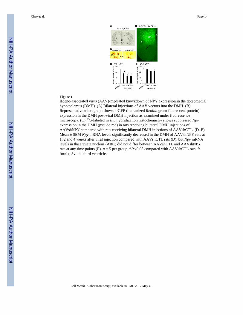
Figure 1.Adeno-associated virus (AAV)-mediated knockdown of NPY expression in the dorsomedialhypothalamus (DMH). (A) Bilateral injections of AAV vectors into the DMH. (B)Representative micrograph shows hrGFP (humanized Renilla green fluorescent protein)expression in the DMH post-viral DMH injection as examined under fluorescencemicroscopy. (C) 35S-labeled in situ hybridization histochemistry shows suppressed Npyexpression in the DMH (pseudo red) in rats receiving bilateral DMH injections ofAAVshNPY compared with rats receiving bilateral DMH injections of AAVshCTL. (D–E)Mean ± SEM Npy mRNA levels significantly decreased in the DMH of AAVshNPY rats at1, 2 and 4 weeks after viral injection compared with AAVshCTL rats (D), but Npy mRNAlevels in the arcuate nucleus (ARC) did not differ between AAVshCTL and AAVshNPYrats at any time points (E). n = 5 per group. *P<0.05 compared with AAVshCTL rats. f:fornix; 3v: the third ventricle.
Chao et al. Page 14
Cell Metab. Author manuscript; available in PMC 2012 May 4.
NIH
-PA Author Manuscript
NIH
-PA Author Manuscript
NIH
-PA Author Manuscript
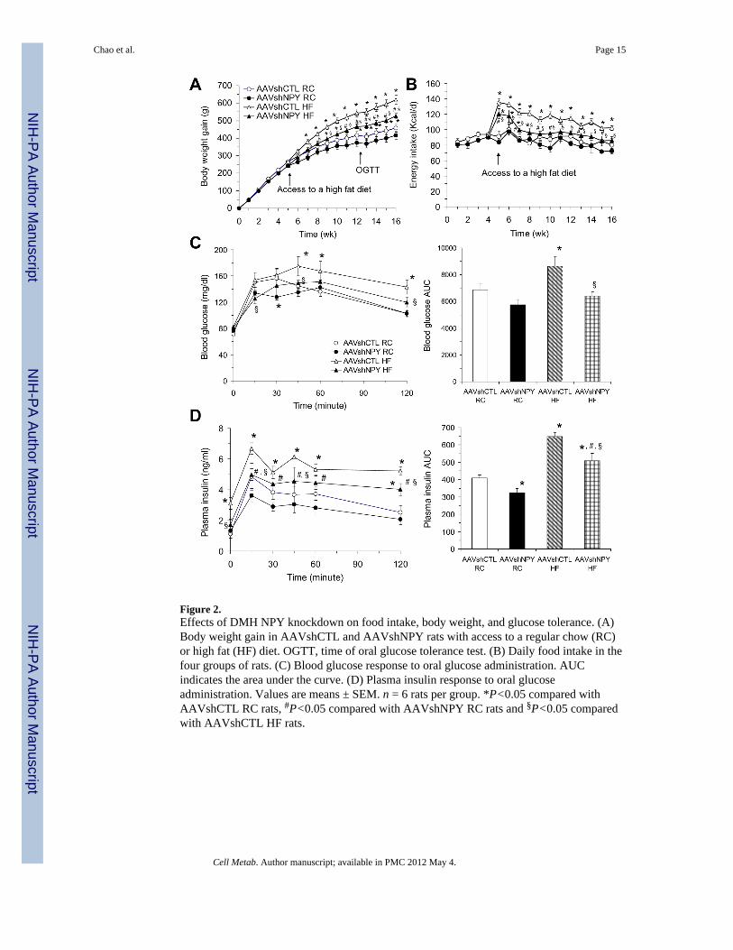
Figure 2.Effects of DMH NPY knockdown on food intake, body weight, and glucose tolerance. (A)Body weight gain in AAVshCTL and AAVshNPY rats with access to a regular chow (RC)or high fat (HF) diet. OGTT, time of oral glucose tolerance test. (B) Daily food intake in thefour groups of rats. (C) Blood glucose response to oral glucose administration. AUCindicates the area under the curve. (D) Plasma insulin response to oral glucoseadministration. Values are means ± SEM. n = 6 rats per group. *P<0.05 compared withAAVshCTL RC rats, #P<0.05 compared with AAVshNPY RC rats and §P<0.05 comparedwith AAVshCTL HF rats.
Chao et al. Page 15
Cell Metab. Author manuscript; available in PMC 2012 May 4.
NIH
-PA Author Manuscript
NIH
-PA Author Manuscript
NIH
-PA Author Manuscript
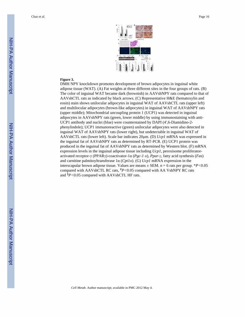
Figure 3.DMH NPY knockdown promotes development of brown adipocytes in inguinal whiteadipose tissue (WAT). (A) Fat weights at three different sites in the four groups of rats. (B)The color of inguinal WAT became dark (brownish) in AAVshNPY rats compared to that ofAAVshCTL rats as indicated by black arrows. (C) Representative H&E (hematoxylin andeosin) stain shows unilocular adipocytes in inguinal WAT of AAVshCTL rats (upper left)and multilocular adipocytes (brown-like adipocytes) in inguinal WAT of AAVshNPY rats(upper middle); Mitochondrial uncoupling protein 1 (UCP1) was detected in inguinaladipocytes in AAVshNPY rats (green, lower middle) by using immunostaining with anti-UCP1 antibody and nuclei (blue) were counterstained by DAPI (4′,6-Diamidino-2-phenylindole); UCP1 immunoreactive (green) unilocular adipocytes were also detected ininguinal WAT of AAVshNPY rats (lower right), but undetectable in inguinal WAT ofAAVshCTL rats (lower left). Scale bar indicates 20μm. (D) Ucp1 mRNA was expressed inthe inguinal fat of AAVshNPY rats as determined by RT-PCR. (E) UCP1 protein wasproduced in the inguinal fat of AAVshNPY rats as determined by Western blot. (F) mRNAexpression levels in the inguinal adipose tissue including Ucp1, peroxisome proliferator-activated receptor-γ (PPARγ)-coactivator-1α (Pgc-1 α), Ppar-γ, fatty acid synthesis (Fas)and carnitine palmitoyltransferase 1α (Cpt1α). (G) Ucp1 mRNA expression in theinterscapular brown adipose tissue. Values are means ± SEM. n = 6 rats per group. *P<0.05compared with AAVshCTL RC rats, #P<0.05 compared with AA VshNPY RC ratsand §P<0.05 compared with AAVshCTL HF rats.
Chao et al. Page 16
Cell Metab. Author manuscript; available in PMC 2012 May 4.
NIH
-PA Author Manuscript
NIH
-PA Author Manuscript
NIH
-PA Author Manuscript
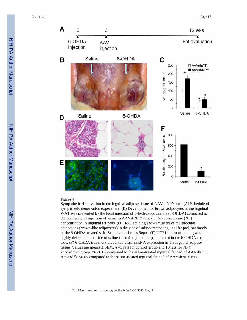
Figure 4.Sympathetic denervation in the inguinal adipose tissue of AAVshNPY rats. (A) Schedule ofsympathetic denervation experiment. (B) Development of brown adipocytes in the inguinalWAT was prevented by the local injection of 6-hydroxydopamine (6-OHDA) compared tothe contralateral injection of saline in AAVshNPY rats. (C) Norepinephrine (NE)concentration in inguinal fat pads. (D) H&E staining shows clusters of multilocularadipocytes (brown-like adipocytes) in the side of saline-treated inguinal fat pad, but barelyin the 6-OHDA-treated side. Scale bar indicates 50μm. (E) UCP1 immunostaining washighly detected in the side of saline-treated inguinal fat pad, but not in the 6-OHDA-treatedside. (F) 6-OHDA treatment prevented Ucp1 mRNA expression in the inguinal adiposetissue. Values are means ± SEM. n =5 rats for control group and 10 rats for NPYknockdown group. *P<0.05 compared to the saline-treated inguinal fat pad of AAVshCTLrats and #P<0.05 compared to the saline-treated inguinal fat pad of AAVshNPY rats.
Chao et al. Page 17
Cell Metab. Author manuscript; available in PMC 2012 May 4.
NIH
-PA Author Manuscript
NIH
-PA Author Manuscript
NIH
-PA Author Manuscript
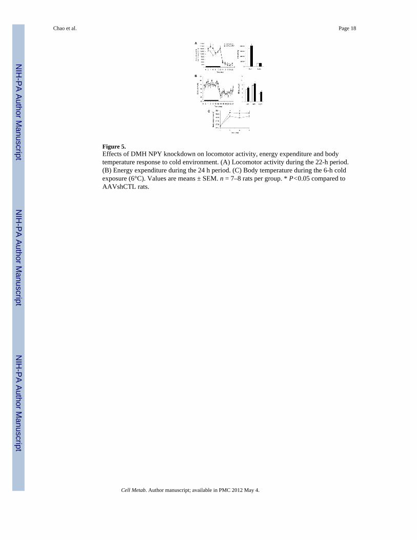
Figure 5.Effects of DMH NPY knockdown on locomotor activity, energy expenditure and bodytemperature response to cold environment. (A) Locomotor activity during the 22-h period.(B) Energy expenditure during the 24 h period. (C) Body temperature during the 6-h coldexposure (6°C). Values are means ± SEM. n = 7–8 rats per group. * P<0.05 compared toAAVshCTL rats.
Chao et al. Page 18
Cell Metab. Author manuscript; available in PMC 2012 May 4.
NIH
-PA Author Manuscript
NIH
-PA Author Manuscript
NIH
-PA Author Manuscript
Related Documents


