Knee disarticulation versus above-knee amputation* R. F. BAUMGARTNER Balgrist Orthopaedic Hospital, University of Zurich. *Based on a paper presented at the ISPO Internation- al Course on Above-knee Prosthetics, Rungsted, November, 1978. Abstract If below-knee amputation is impossible, knee disarticulation should be considered before above-knee amputation, regardless of age and etiology. Knee disarticulation which leaves the femur and patella untouched offers many advantages. The surgical technique is simple and non-traumatic since no bone or muscle tissue is to be dissected. The thigh muscles are completely preserved and thus there is no muscular imbalance. The stump permits total end bearing and its bulbous shape permits easy and firm attachment of the prosthesis. A specially designed double-wall socket and various types of knee joints are presented. Modern prostheses are superior to above-knee prostheses with regard to function, comfort and cosmesis. Results of 72 patients of all age groups are presented and discussed. Introduction If it is impossible to obtain a short below- knee amputation, knee disarticulation should be considered before above-knee amputation regardless of etiology and patient's age. There is no level of amputation between the ankle and the hip joint where one can perform an amputation which does not require separation of bone or muscle tissues. At knee disarticu- lation level the surface of the wound is the smallest possible which minimizes the danger of haemorrhage or infection. In children, the distal epiphyseal line of growth is fully pre- served. In contrast to above-knee amputation the stump resulting from knee disarticulation permits full endbearing. All the muscles of the thigh are entirely preserved. Despite all these advantages, knee disartic- ulation is still unpopular compared with above- knee amputation. The main objection comes from the prosthetists who complain that there is not enough space for the prosthetic knee joint and have difficulties in fitting the bulbous shape of the stump into a regular socket. For this reason many surgical techniques of through-knee amputation indicate the removal of parts or all of the femoral condyles and the patella to facilitate prosthetic fitting (Fig. 1). However, knee disarticulation leaving the femur and patella untouched offers many advantages, particularly with regard to surgical technique, stump qualities and finally even prosthetic fitting. The following technique (Kjolbye, 1970; Vitali et al, 1978) is recom- mended. Operative technique It is preferable to have the patient in a supine position which gives easy access to the knee joint and also permits an above-knee amputation to be done if necessary, without changing the patient's position. Furthermore, the supine position does not give any particular problems to the anaesthetist which is of great importance in poor risk cases, such as geriatric patients. A tourniquet may be used except for vascular patients. However, as there are only a few easily identified vessels a tourniquet is not an absolute necessity. Fig. 1. Some of the most common through-knes amputations. Except for knee disarticulation they involve resection of part or all of the femoral condyles and the patella.

Welcome message from author
This document is posted to help you gain knowledge. Please leave a comment to let me know what you think about it! Share it to your friends and learn new things together.
Transcript

Knee disarticulation versus above-knee amputation* R. F . B A U M G A R T N E R
Balgrist Orthopaedic Hospital, University of Zurich.
*Based on a paper presented at the ISPO International Course on Above-knee Prosthetics, Rungsted, November, 1978.
Abstract If below-knee amputa t ion is impossible, knee disarticulation should be considered before above-knee amputat ion, regardless of age and etiology. Knee disarticulation which leaves the femur a n d patella untouched offers many advantages. The surgical technique is simple and non-t raumatic since n o bone or muscle tissue is to be dissected. The thigh muscles are completely preserved and thus there is n o muscular imbalance. The s tump permits total end bearing and its bulbous shape permits easy and firm at tachment of the prosthesis. A specially designed double-wall socket and various types of knee joints are presented. Modern prostheses are superior to above-knee prostheses with regard to function, comfort and cosmesis. Results of 72 patients of all age groups are presented and discussed.
Introduction If it is impossible to obtain a short below-
knee amputat ion, knee disarticulation should be considered before above-knee amputa t ion regardless of etiology and patient 's age. There is no level of amputa t ion between the ankle and the hip joint where one can perform an amputa t ion which does not require separation of bone or muscle tissues. At knee disarticulation level the surface of the wound is the smallest possible which minimizes the danger of haemorrhage or infection. In children, the distal epiphyseal line of growth is fully preserved. In contrast t o above-knee amputa t ion the s tump resulting from knee disarticulation permits full endbearing. All the muscles of the thigh are entirely preserved.
Despite all these advantages, knee disarticulation is still unpopular compared with above-knee amputa t ion . The main objection comes from the prosthetists who complain that there is not enough space for the prosthetic knee
joint and have difficulties in fitting the bulbous shape of the s tump into a regular socket. F o r this reason many surgical techniques of through-knee amputa t ion indicate the removal of par ts or all of the femoral condyles and the patella to facilitate prosthetic fitting (Fig. 1).
However, knee disarticulation leaving the femur and patella untouched offers many advantages, particularly with regard to surgical technique, s tump qualities and finally even prosthetic fitting. The following technique (Kjolbye, 1970; Vitali et al , 1978) is recommended.
Operative technique It is preferable to have the patient in a
supine posit ion which gives easy access to the knee joint and also permits an above-knee amputa t ion to be done if necessary, without changing the patient 's position. Fur thermore, the supine position does not give any particular problems to the anaesthetist which is of great importance in p o o r risk cases, such as geriatric patients. A tourniquet may be used except for vascular patients. However, as there are only a few easily identified vessels a tourniquet is not an absolute necessity.
Fig. 1. Some of the most common through-knes amputations. Except for knee disarticulation they involve resection of part or all of the femoral
condyles and the patella.
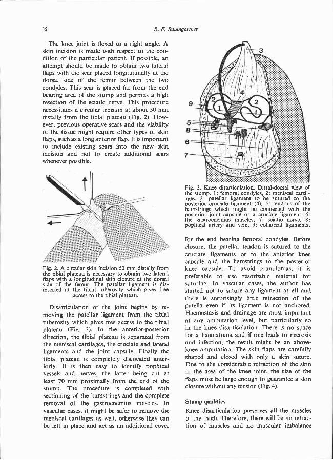
The knee joint is flexed to a right angle. A skin incision is made with respect to the condition of the particular patient. If possible, an a t tempt should be made to obtain two lateral flaps with the scar placed longitudinally at the dorsal side of the femur between the two condyles. This scar is placed far from the end bearing area of the s tump and permits a high resection of the sciatic nerve. This procedure necessitates a circular incision at about 50 m m distally from the tibial plateau (Fig. 2). However, previous operative scars a n d the viability of the tissue might require other types of skin flaps, such as a long anterior flap. It is important t o include existing scars into the new skin incision and not to create additional scars whenever possible.
Disarticulation of the joint begins by removing the patellar ligament from the tibial tuberosity which gives free access to the tibial plateau (Fig. 3). In the anterior-posterior direction, the tibial plateau is separated from the meniscal cartilages, the cruciate and lateral l igaments and the joint capsule. Finally the tibial plateau is completely dislocated anteriorly. It is then easy to identify popliteal vessels and nerves, the latter being cut at least 70 m m proximally from the end of the s tump. The procedure is completed with sectioning of the hamstrings and the complete removal of the gastrocnemius muscles. In vascular cases, it might be safer to remove the meniscal cartilages as well, otherwise they can be left in place and act as an additional cover
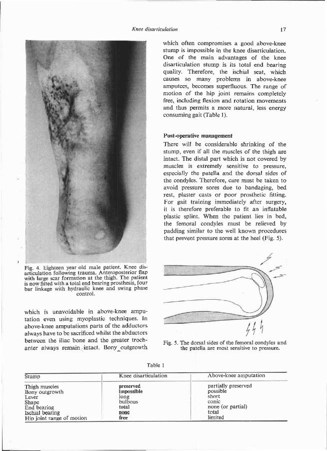
for the end bearing femoral condyles. Before closure, the patellar tendon is sutured to the cruciate ligaments or to the anterior knee capsule and the hamstrings to the posterior knee capsule. T o avoid granulomas, it is preferable to use resorbable material for suturing. In vascular cases, the au thor has started not to suture any ligament at all and there is surprisingly little retraction of the patella even if its ligament is not anchored. Haemostasis and drainage are most important at any amputa t ion level, but particularly so in the knee disarticulation. There is n o space for a haematoma and if one leads to necrosis and infection, the result might be an above-knee amputat ion. The skin flaps are carefully shaped and closed with only a skin suture. D u e to the considerable retraction of the skin in the area of the knee joint, the size of the flaps must be large enough to guarantee a skin closure without any tension (Fig. 4).
Stump qualities
Knee disarticulation preserves all the muscles of the thigh. Therefore, there will be n o retraction of muscles and n o muscular imbalance
Fig. 2. A circular skin incision 50 mm distally from the tibial plateau is necessary to obtain two lateral flaps with a longitudinal skin closure at the dorsal side of the femur. The patellar ligament is dis-inserted at the tibial tuberosity which gives free
access to the tibial plateau.
Fig. 3. Knee disarticulation. Distal-dorsal view of the stump. 1: femoral condyles, 2: meniscal cartilages, 3: patellar ligament to be sutured to the posterior cruciate ligament (4), 5: tendons of the hamstrings which might be connected with the posterior joint capsule or a cruciate ligament, 6: the gastrocnemius muscles, 7: sciatic nerve, 8: popliteal artery and vein, 9: collateral ligaments.
which is unavoidable in above-knee amputation even using myoplastic techniques. In above-knee amputat ions parts of the adductors always have to be sacrificed whilst the abductors between the iliac bone and the greater t rochanter always remain intact. Bony outgrowth
which often compromises a good above-knee s tump is impossible in the knee disarticulation. One of the main advantages of the knee disarticulation s tump is its total end bearing quality. Therefore, the ischial seat, which causes so many problems in above-knee amputees, becomes superfluous. The range of mot ion of the hip joint remains completely free, including flexion and rotat ion movements and thus permits a more natural , less energy consuming gait (Table 1).
Post-operative management There will be considerable shrinking of the s tump, even if all the muscles of the thigh are intact. The distal par t which is not covered by muscles is extremely sensitive to pressure, especially the patella and the dorsal sides of the condyles. Therefore, care must be taken to avoid pressure sores due to bandaging, bed rest, plaster casts or poor prosthetic fitting. Fo r gait training immediately after surgery, it is therefore preferable to fit an inflatable plastic splint. When the patient lies in bed, the femoral condyles must be relieved by padding similar to the well known procedures that prevent pressure sores at the heel (Fig. 5).
Fig. 4. Eighteen year old male patient. Knee disarticulation following trauma. Anteroposterior flap with large scar formation at the thigh. The patient is now fitted with a total end bearing prosthesis, four bar linkage with hydraulic knee and swing phase
control.
Fig. 5. The dorsal sides of the femoral condyles and the patella are most sensitive to pressure.
Table 1
Post-operative complications As at any other amputa t ion level, problems
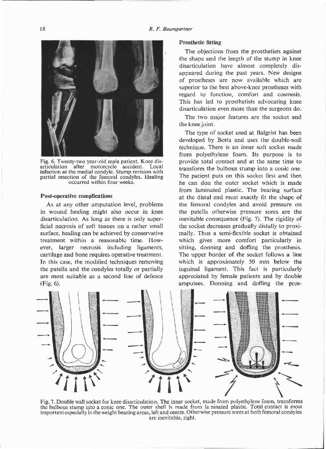
in w o u n d healing might also occur in knee disarticulation. As long as there is only superficial necrosis of soft tissues on a rather small surface, healing can be achieved by conservative treatment within a reasonable time. H o w ever, larger necrosis including ligaments, cartilage and bone requires operative treatment. In this case, the modified techniques removing the patella and the condyles totally or partially are most suitable as a second line of defence (Fig. 6).
Prosthetic fitting
The objections from the prosthetists against the shape and the length of the s tump in knee disarticulation have almost completely disappeared during the past years. New designs of prostheses are now available which are superior to the best above-knee prostheses with regard t o function, comfort and cosmesis. This has led to prosthetists advocating knee disarticulation even more than the surgeons do .
The two major features are the socket and. the knee joint.
The type of socket used at Balgrist has been developed by Bot ta and uses the double-wall technique. There is an inner soft socket made from polyethylene foam. Its purpose is to provide total contact and at the same time to transform the bulbous s tump into a conic one. The patient puts on this socket first and then he can don the outer socket which is made from laminated plastic. The bearing surface at the distal end must exactly fit the shape of the femoral condyles and avoid pressure on the patella otherwise pressure sores are the inevitable consequence (Fig. 7). The rigidity of the socket decreases gradually distally to proxi-mally. Thus a semi-flexible socket is obtained which gives more comfort particularly in sitting, donning and doffing the prosthesis. The upper border of the socket follows a line which is approximately 50 m m below the inguinal ligament. This fact is particularly appreciated by female patients and by double amputees. Donning and doffing the pros-
Fig. 6. Twenty-two year-old male patient. Knee disarticulation after motorcycle accident. Local infection at the medial condyle. Stump revision with partial resection of the femoral condyles. Healing
occurred within four weeks.
Fig. 7. Double wall socket for knee disarticulation. The inner socket, made from polyethylene foam, transforms the bulbous stump into a conic one. The outer shell is made from laminated plastic. Total contact is most important especially in the weight bearing areas, left and centre. Otherwise pressure sores at both femoral condyles
are inevitable, right.

thesis is very simple since it does not require any particular effort or skill which again is
especially appreciated by double amputees and by geriatric patients (Fig. 8).
Knee joints as they are commonly used in above-knee prostheses are of n o use with knee disarticulations. Joints especially designed for knee disarticulation are now available, the first one being developed by Lyquist at the Orthopaedic Hospital in Copenhagen. Other types of knee joints have since been developed, most of them using four bar linkage mechanisms and permitt ing knee flexion far in excess of 90°. They are available with or without swing phase control . Fur ther research is being done with regard to improving the solidity of the knee mechanism. Prostheses for knee disarticulation and above-knee amputa t ion are comp a r e d in Table 2.
Results
F r o m 1968—1978, the au thor has performed 72 knee disarticulations, mainly for peripheral vascular occlusion. F o u r patients presenting congential deformities corresponding t o knee disarticulation also have been fitted with the new type of prosthesis. Re-amputat ion at an above-knee level was necessary in 7 cases, all of them being vascular patients. They all had to be amputa ted at a high above-knee level due t o advanced tissue necrosis. Local complications in wound healing occured in 16 cases.
FURTHER READING BAUMGARTNER, R. (1973). Beinamputationen und
Prothesenversorgung bei arteriellen Durchblutungsstörungen, 56-64 and 114-116, Ferdinand Enke, Stuttgart.
BAUMGARTNER, R. (1977). Amputation und Prothesenversorgung beim Kind. Ed. Baumgartner R. 18 and 108-110, Ferdinand Enke, Stuttgart.
BURGESS, E. M. (1977). Disarticulation of knee. Arch Surg., 1 1 2 1250-1255.
Fig. 8. Donning and doffing the prosthesis presents no problems even to geriatric patients.
Table 2
REFERENCES KJØLBYE, J. ( 1 9 7 0 ) . The surgery of the through-knee
amputation 2 5 5 - 2 5 8 . Prosthetic and Orthotic Practice. Ed. Murdoch, G . , 2 5 5 - 2 5 7 , Edward Arnold, London.
VITALI, M,, ROBINSON, K . P., ANDREWS, B. G . and HARRIS, E. E. (1978) . Amputations and prostheses, 1 5 8 - 1 6 4 , Bailliere Tindall, London.
Related Documents