REVIEW ARTICLE published: 08 July 2014 doi: 10.3389/fncel.2014.00186 Kir3 channel signaling complexes: focus on opioid receptor signaling Karim Nagi 1,2 and Graciela Pineyro 1,2,3 * 1 Département de Pharmacologie, Faculté de Médecine, Université de Montréal, Montreal, QC, Canada 2 Centre de Recherche du CHU Sainte-Justine, Montréal, QC, Canada 3 Département de Psychiatrie, Faculté de Médecine, Université de Montréal, Montréal, QC, Canada Edited by: Leigh Anne Swayne, University of Victoria, Canada Reviewed by: Lin Tian, University of California, Davis, USA Nathan Dascal, Tel Aviv University, Israel *Correspondence: Graciela Pineyro, Department of Psychiatry, Centre de Recherche du CHU Ste-Justine, University of Montreal, 3175, Côte Ste-Catherine, Bureau 2722, Montréal, QC H3T 1C5, Canada e-mail: graciela.pineyro.filpo@ umontreal.ca Opioids are among the most effective drugs to treat severe pain. They produce their analgesic actions by specifically activating opioid receptors located along the pain perception pathway where they inhibit the flow of nociceptive information. This inhibition is partly accomplished by activation of hyperpolarizing G protein-coupled inwardly-rectifying potassium (GIRK or Kir3) channels. Kir3 channels control cellular excitability in the central nervous system and in the heart and, because of their ubiquitous distribution, they mediate the effects of a large range of hormones and neurotransmitters which, upon activation of corresponding G protein-coupled receptors (GPCRs) lead to channel opening. Here we analyze GPCR signaling via these effectors in reference to precoupling and collision models. Existing knowledge on signaling bias is discussed in relation to these models as a means of developing strategies to produce novel opioid analgesics with an improved side effects profile. Keywords: GIRK, opioid, signaling, analgesia, bias, signaling complexes, functional selectivity, allosteric modulation Opioids produce their actions via the activation of μ (MOR), δ (DOR), and κ (KOR) receptors (Pert and Snyder, 1973; Simon et al., 1973) all of which couple to heterotrimeric Gi/o proteins. As such they share adenylyl cyclase, Phospholipase Cβ [PLCβ, N-type Ca 2+ (Cav2.2)] channels and G-protein coupled inward rectifier K + channels (GIRKs or Kir3 channels) as canonical Gα and Gβγ effectors. The two channel effectors prominently medi- ate analgesic effects of opioids. For example, studies in rodents have shown that activation of MORs on small, unmyelinated nociceptors (Scherrer et al., 2009; Heinke et al., 2011; Bardoni et al., 2014) contribute to the analgesic effects of morphine (Wei et al., 1996; Andrade et al., 2010), and this is done via inhibition of N-type Ca+2 channels that control neurotransmitter release from these primary afferents (Glaum et al., 1994; Wrigley et al., 2010). Kir3 channel contribution to opioid analgesia has also been demonstrated in animal models (Ikeda et al., 2000; Mitrovic et al., 2003; Marker et al., 2005), and their relevance in the clin- ical response to opioid analgesics has been further confirmed by Abbreviations: GIRK, G protein activated inward rectifier K + ; GPCRs, G protein-coupled receptors; DORs, Delta opioid receptors; MORs, Mu opi- oid receptors; KORs, Kappa opioid receptors; PLC, Phospholipase C; GABA, Gamma-aminobutyric acid; DRG, dorsal root ganglion; DAMGO, [D-Ala(2),N- Me-Phe(4),Gly(5)-ol]-enkephalin; KCNJ6, Inwardly-rectifying potassium channel, subfamily J, member 6; KCNJ3, Inwardly-rectifying potassium channel, subfam- ily J, member 3; 5-HT, 5-hydroxytryptamine; MAPK, Mitogen-activated protein kinases; βarr, βarrestin; cAMP, Cyclic adenosine monophosphate; VTA, Ventral tegmental area; PIP2, Phosphatidylinositol 4,5-bisphosphate; GDP, Guanosine diphosphate; GTP, Guanosine triphosphate; PSD95, Postsynaptic density protein 95; SAP97, Synapse-associated protein 97; FRET, Fluorescence resonance energy transfer; BRET, Bioluminescence resonance energy transfer; PC12, Neuron-like pheochromocytoma. gene linkage analyses (Nishizawa et al., 2009; Lotsch et al., 2010; Bruehl et al., 2013). In addition, the use of genetically modified mice lacking different channel subunits has further established activation of Kir3 channels as a pervasive analgesic mechanism which, in addition to opioids, also mediates pain modulation by α2 adrenergic, muscarinic, GABA B and cannabinoid receptor ligands (Blednov et al., 2003; Mitrovic et al., 2003). From this per- spective the development of direct Kir3 channel activators may be considered a promising strategy for the development of novel analgesics. However, enthusiasm for this particular approach has remained limited given the possibility of widespread side-effects associated with administration of direct channel openers (Lujan et al., 2014). Indeed, these would result from the great variety of physiological responses mediated by Kir3 channels, includ- ing heart frequency control (Wickman et al., 1998; Bettahi et al., 2002), memory (Wickman et al., 2000), learning (Wickman et al., 2000; Cooper et al., 2012), as well as their participation in dif- ferent pathological conditions including development of seizures (Signorini et al., 1997), generation of anxiety states (Blednov et al., 2001; Pravetoni and Wickman, 2008) and contribution to abnormal plasticity by drugs of abuse (Padgett et al., 2012; Hearing et al., 2013). Consequently, the prevailing strategy for engaging Kir3-mediated analgesia has remained the activation of pain-modulating GPCRs (McAllister et al., 1999; Lujan et al., 2014). Among them MORs, DORs and KORs all three mediate effective analgesia but display different side effects that distinc- tively limit their therapeutic use (Kieffer and Gaveriaux-Ruff, 2002; Bruchas and Chavkin, 2010; Gaveriaux-Ruff and Kieffer, 2011). Here we will summarize Kir3 channel contribution to desired and undesired responses of opioid analgesics and address Frontiers in Cellular Neuroscience www.frontiersin.org July 2014 | Volume 8 | Article 186 | 1 CELLULAR NEUROSCIENCE

Welcome message from author
This document is posted to help you gain knowledge. Please leave a comment to let me know what you think about it! Share it to your friends and learn new things together.
Transcript

REVIEW ARTICLEpublished: 08 July 2014
doi: 10.3389/fncel.2014.00186
Kir3 channel signaling complexes: focus on opioid receptorsignalingKarim Nagi1,2 and Graciela Pineyro1,2,3*
1 Département de Pharmacologie, Faculté de Médecine, Université de Montréal, Montreal, QC, Canada2 Centre de Recherche du CHU Sainte-Justine, Montréal, QC, Canada3 Département de Psychiatrie, Faculté de Médecine, Université de Montréal, Montréal, QC, Canada
Edited by:
Leigh Anne Swayne, University ofVictoria, Canada
Reviewed by:
Lin Tian, University of California,Davis, USANathan Dascal, Tel Aviv University,Israel
*Correspondence:
Graciela Pineyro, Department ofPsychiatry, Centre de Recherche duCHU Ste-Justine, University ofMontreal, 3175, Côte Ste-Catherine,Bureau 2722, Montréal,QC H3T 1C5, Canadae-mail: [email protected]
Opioids are among the most effective drugs to treat severe pain. They producetheir analgesic actions by specifically activating opioid receptors located along thepain perception pathway where they inhibit the flow of nociceptive information. Thisinhibition is partly accomplished by activation of hyperpolarizing G protein-coupledinwardly-rectifying potassium (GIRK or Kir3) channels. Kir3 channels control cellularexcitability in the central nervous system and in the heart and, because of their ubiquitousdistribution, they mediate the effects of a large range of hormones and neurotransmitterswhich, upon activation of corresponding G protein-coupled receptors (GPCRs) lead tochannel opening. Here we analyze GPCR signaling via these effectors in reference toprecoupling and collision models. Existing knowledge on signaling bias is discussed inrelation to these models as a means of developing strategies to produce novel opioidanalgesics with an improved side effects profile.
Keywords: GIRK, opioid, signaling, analgesia, bias, signaling complexes, functional selectivity, allosteric
modulation
Opioids produce their actions via the activation of μ (MOR), δ
(DOR), and κ (KOR) receptors (Pert and Snyder, 1973; Simonet al., 1973) all of which couple to heterotrimeric Gi/o proteins.As such they share adenylyl cyclase, Phospholipase Cβ [PLCβ,N-type Ca2+ (Cav2.2)] channels and G-protein coupled inwardrectifier K+ channels (GIRKs or Kir3 channels) as canonical Gα
and Gβγ effectors. The two channel effectors prominently medi-ate analgesic effects of opioids. For example, studies in rodentshave shown that activation of MORs on small, unmyelinatednociceptors (Scherrer et al., 2009; Heinke et al., 2011; Bardoniet al., 2014) contribute to the analgesic effects of morphine (Weiet al., 1996; Andrade et al., 2010), and this is done via inhibitionof N-type Ca+2 channels that control neurotransmitter releasefrom these primary afferents (Glaum et al., 1994; Wrigley et al.,2010). Kir3 channel contribution to opioid analgesia has alsobeen demonstrated in animal models (Ikeda et al., 2000; Mitrovicet al., 2003; Marker et al., 2005), and their relevance in the clin-ical response to opioid analgesics has been further confirmed by
Abbreviations: GIRK, G protein activated inward rectifier K+; GPCRs, Gprotein-coupled receptors; DORs, Delta opioid receptors; MORs, Mu opi-oid receptors; KORs, Kappa opioid receptors; PLC, Phospholipase C; GABA,Gamma-aminobutyric acid; DRG, dorsal root ganglion; DAMGO, [D-Ala(2),N-Me-Phe(4),Gly(5)-ol]-enkephalin; KCNJ6, Inwardly-rectifying potassium channel,subfamily J, member 6; KCNJ3, Inwardly-rectifying potassium channel, subfam-ily J, member 3; 5-HT, 5-hydroxytryptamine; MAPK, Mitogen-activated proteinkinases; βarr, βarrestin; cAMP, Cyclic adenosine monophosphate; VTA, Ventraltegmental area; PIP2, Phosphatidylinositol 4,5-bisphosphate; GDP, Guanosinediphosphate; GTP, Guanosine triphosphate; PSD95, Postsynaptic density protein95; SAP97, Synapse-associated protein 97; FRET, Fluorescence resonance energytransfer; BRET, Bioluminescence resonance energy transfer; PC12, Neuron-likepheochromocytoma.
gene linkage analyses (Nishizawa et al., 2009; Lotsch et al., 2010;Bruehl et al., 2013). In addition, the use of genetically modifiedmice lacking different channel subunits has further establishedactivation of Kir3 channels as a pervasive analgesic mechanismwhich, in addition to opioids, also mediates pain modulationby α2 adrenergic, muscarinic, GABAB and cannabinoid receptorligands (Blednov et al., 2003; Mitrovic et al., 2003). From this per-spective the development of direct Kir3 channel activators maybe considered a promising strategy for the development of novelanalgesics. However, enthusiasm for this particular approach hasremained limited given the possibility of widespread side-effectsassociated with administration of direct channel openers (Lujanet al., 2014). Indeed, these would result from the great varietyof physiological responses mediated by Kir3 channels, includ-ing heart frequency control (Wickman et al., 1998; Bettahi et al.,2002), memory (Wickman et al., 2000), learning (Wickman et al.,2000; Cooper et al., 2012), as well as their participation in dif-ferent pathological conditions including development of seizures(Signorini et al., 1997), generation of anxiety states (Blednovet al., 2001; Pravetoni and Wickman, 2008) and contributionto abnormal plasticity by drugs of abuse (Padgett et al., 2012;Hearing et al., 2013). Consequently, the prevailing strategy forengaging Kir3-mediated analgesia has remained the activationof pain-modulating GPCRs (McAllister et al., 1999; Lujan et al.,2014). Among them MORs, DORs and KORs all three mediateeffective analgesia but display different side effects that distinc-tively limit their therapeutic use (Kieffer and Gaveriaux-Ruff,2002; Bruchas and Chavkin, 2010; Gaveriaux-Ruff and Kieffer,2011). Here we will summarize Kir3 channel contribution todesired and undesired responses of opioid analgesics and address
Frontiers in Cellular Neuroscience www.frontiersin.org July 2014 | Volume 8 | Article 186 | 1
CELLULAR NEUROSCIENCE

Nagi and Pineyro Kir3 channels in opioid analgesic
the question of whether biasing opioid signaling toward theseeffectors could help develop better opioid analgesics with areduced side effects profile.
Kir3 CHANNEL SIGNALING AND OPIOID-DEPENDENTANALGESIAKir3 channels are formed by tetrameric association of four differ-ent subunits (Kir3.1-3.4). Although all four of them are expressedin peripheral (Gao et al., 2007) and central nervous system(Wickman et al., 2000; Saenz Del Burgo et al., 2008; Fernandez-Alacid et al., 2011), neural channels are most frequently formedby association of Kir3.1, 3.2 and 3.3 subunits (Liao et al., 1996;Hibino et al., 2010; Luscher and Slesinger, 2010). The subunitcombination that forms the channel will depend on the neu-ronal population involved (Inanobe et al., 1999; Cruz et al., 2004)and the cellular compartment (Koyrakh et al., 2005; Fernandez-Alacid et al., 2009) in which channels are found. Nonetheless,Kir3.1/3.2 heterotetramers are considered prototypical neuronalchannels.
Kir3 channel contribution to opioid analgesia was initially sug-gested by the fact that mice carrying a missense mutation whichrenders Kir3.2 subunits insensitive to G protein activation (Patilet al., 1995; Navarro et al., 1996) displayed reduced analgesicresponses to morphine (Ikeda et al., 2000, 2002). These observa-tions were subsequently confirmed using null mice for differentsubunits (Mitrovic et al., 2003; Marker et al., 2005), which allowedto also implicate Kir3.1 (Marker et al., 2004) and Kir3.3 (Markeret al., 2002) in pain modulation. Knock-out of Kir3.2 and 3.3was shown to interfere with morphine’s ability to prolong avoid-ance behavior in the hot plate test. Since this response involvessupraspinal integration, it can be concluded that both subunitscontribute to mechanisms of opioid analgesia at this level (Markeret al., 2002; Mitrovic et al., 2003). Possible sites of supraspinalKir3-mediated analgesia may include thalamus and limbic cor-tex, both of which express Kir3.1, 3.2 and 3.3 subunits (SaenzDel Burgo et al., 2008; Fernandez-Alacid et al., 2011) as well asopioid receptors (Le Merrer et al., 2009). The midbrain periaque-ductal gray seems less likely since in this nucleus opioid actionsare primarily presynaptic reducing neurotransmitter release via amechanism that involves phospholipase A2, arachidonic acid and12-lipoxygenase, which leads to modulation of voltage-dependentpotassium channels (Vaughan and Christie, 1997; Vaughan et al.,1997).
Genetically engineered mice lacking Kir3.1 or Ki3.2 subunitsalso display reduced responses to intrathecal administration ofmorphine in the tail flick test (Marker et al., 2004, 2005) impli-cating both subunits in spinal mechanisms of opioid analgesia.This interpretation is supported by reports locating Kir3.1 andKir3.2 subunits to lamina II interneurons that co-express μ opioidreceptors (Marker et al., 2006). Functional studies also indi-cated that silencing of Kir3.1 or Kir3.2 subunits, or intrathecalinfusion of Kir3 channel blocker tertiapin-Q interfered with theanalgesic response by MOR and DOR agonists administered bythe same route (Marker et al., 2005). In contrast, Kir3.3 abla-tion was without effect on the analgesic response elicited byintrathecal morphine (Marker et al., 2004), arguing against its sig-nificant contribution at this level. The latter observation is also in
keeping with immunohistological studies which reported absenceof Kir3.3 labeling in the dorsal horn (Marker et al., 2004).
In addition to their participation in acute opioid analge-sia, spinal Kir3 channels seem to mediate neuroadaptations thatmodulate responsiveness to opioids in conditions such as inflam-matory or cancer-related pain. This possibility is particularlysuggested by reports indicating that carrageenan-inflammation(Gonzalez-Rodriguez et al., 2010) and bone cancer (Gonzalez-Rodriguez et al., 2012) enhance the inhibitory effect of Kir3channel blocker tertiapin-Q on analgesia induced by intrathecaladministration of morphine.
Apart from brain and spinal cord, opioid receptors are alsopresent in sensory neurons of the dorsal root ganglion (DRG)(Li et al., 1998; Gendron et al., 2006; Wu et al., 2008; Wanget al., 2010) and are transported to peripheral terminals wherethey mediate analgesic actions of peripherally injected opioids(Hassan et al., 1993; Obara et al., 2009; Vadivelu et al., 2011).Transcripts for Kir3.1/3.2 subunits have been detected in humanDRGs and in rat nociceptors, but not in mice (Gao et al., 2007;Nockemann et al., 2013). This distribution parallels differencesin species sensitivity to peripheral administration of opioids sug-gesting that when expressed, DRG Kir3 subunits actively partic-ipate in opioid analgesia. Indeed, intraplantar injection of MORagonists does not produce analgesia in mice (Nockemann et al.,2013) but effectively mitigates pain in inflammatory or neuro-pathic rat models (Stein et al., 1989; Obara et al., 2009; Chunget al., 2013; Nockemann et al., 2013) as well as postoperative andarthritic pain in humans (Kalso et al., 2004; Vadivelu et al., 2011).Moreover, the active contribution of Kir3.2 channels to peripheralopioid analgesia has now been experimentally established usingtransgenic mice genetically engineered to express these subunitsin sensory neurons. Unlike their wild-type counterparts, the lat-ter display an analgesic response to plantar application of MORagonist DAMGO (Nockemann et al., 2013).
It is important to bear in mind that most of the evidence ana-lyzed thus far links Kir3 channel function to opioid analgesia inanimal models. However, the latter have shown somewhat limitedsuccess in identifying and validating analgesic targets of clinicalrelevance (Mogil, 2009). Hence, from this perspective, any evi-dence linking Kir3 channels to therapeutic response in humansis of specific interest. At least three studies have now shown thatvariations in the gene coding for Kir3.2 subunits (KCNJ6) influ-ence opioid dose requirements for both acute management ofpostoperative pain (Nishizawa et al., 2009; Bruehl et al., 2013) andfor pain control in chronic patients (Lotsch et al., 2010). Similarassessment of Kir3.1 (KCNJ3) variations showed no effect (Bruehlet al., 2013).
OPIOID SIDE EFFECTS AND Kir3 CHANNEL SIGNALINGIn spite of the established analgesic efficacy of MOR, DOR, andKOR agonists, activation of the different receptor subtypes resultsin a distinct set of side effects which limit therapeutic applicationof their agonists. Constipation, nausea, respiratory depression,tolerance, dependence, and abuse are among the most commonundesired effects of clinically available MOR agonists (Ballantyneand Shin, 2008; Morgan and Christie, 2011). Many of these unde-sired actions, particularly respiratory depression (Cheng et al.,
Frontiers in Cellular Neuroscience www.frontiersin.org July 2014 | Volume 8 | Article 186 | 2

Nagi and Pineyro Kir3 channels in opioid analgesic
1993; Gallantine and Meert, 2005), gastrointestinal side effects(Tavani et al., 1990; Gallantine and Meert, 2005; Feng et al., 2006)and physical dependence (Cowan et al., 1988; Codd et al., 2009)are less severe or absent with DOR agonists. Moreover, althoughDORs and MORs are both involved in reward response to phys-iological stimuli (Charbogne et al., 2014), DORs are not directlyinvolved in assigning reward value to stimuli but instead facilitatepredictive learning and influence the choice of action in face ofcontingencies that involve reward (Laurent et al., 2012). In keep-ing with this functional profile, DOR agonists do not facilitateintracranial self-stimulation (Do Carmo et al., 2009), are not dis-criminated as morphine substitutes (Gallantine and Meert, 2005)and do not display reinforcing properties in non-human primates(Banks et al., 2011). Nonetheless, despite these advantages, poten-tial for tolerance (Pradhan et al., 2010; Audet et al., 2012) remainsa limitation for long-term use of DOR agonists, as does theirpropensity to produce hippocampal hyperexcitability that maylead to seizures. The latter are produced both by DOR (De Sarroet al., 1992; Broom et al., 2002; Jutkiewicz et al., 2005) and MORagonists (Drake et al., 2007). In the specific case of DOR ago-nists, hyperexcitability is brief and non-lethal. Moreover, seizuresare rare at analgesic doses (Jutkiewicz et al., 2006), they do notappear with all ligands (Le Bourdonnec et al., 2008; Saitoh et al.,2011) and can be suppressed by controlling the rate of agonistadministration (Jutkiewicz et al., 2005). Thus, overall, side effectsof DORs agonists are considerably milder as compared to thoseof MORs, and their proven efficacy in chronic pain managementmake DORs especially attractive as targets for the development ofnovel analgesics (Gaveriaux-Ruff and Kieffer, 2011). This notionis further reinforced by the fact that DOR agonists display antide-pressant properties (Chu Sin Chung and Kieffer, 2013) whichcould be of additional benefit in controlling negative affect, fre-quently associated with prolonged pain syndromes (Goldenberg,2010). Unlike DOR agonists, KOR-mediated analgesia is typicallyassociated with stress, depression, and dysphoria (Bruchas andChavkin, 2010; Van’t Veer and Carlezon, 2013) which, togetherwith their tendency to induce tolerance (McLaughlin et al., 2004;Xu et al., 2004), would make these ligands less attractive for thetreatment of chronic pain.
Kir3 channel function has been associated with some of theundesired effects of opioid analgesics. For example, reducedGABAergic activity in hippocampal dentate gyrus increases theexcitability of glutamatergic granule cells and may reduce thethreshold for seizures (Drake et al., 2007). MORs and DORsagonists silence hippocampal GABAergic interneurons by amechanism that involves both Kir3 (Luscher et al., 1997) andvoltage-dependent K+ channels (Wimpey and Chavkin, 1991;Moore et al., 1994), explaining their documented tendency toproduce seizures (Drake et al., 2007). MORs agonists also enhanceexcitability and firing activity of dopaminergic neurons of theventral tegmental area (Gysling and Wang, 1983), and do soby hyperpolarization of local interneurons (Johnson and North,1992; Bonci and Williams, 1997). Like in hippocampus, disinhibi-tion is mediated through Kir3 channel activation (Luscher et al.,1997) and its consequence is the enhanced release of dopaminein corticolimbic areas which is thought to facilitate compulsivebehaviors characteristic of addiction (Luscher and Ungless, 2006).
Finally, Kir3 channel activation by KORs modulates firing activ-ity of dorsal raphe 5-HT neurons (Bruchas et al., 2007; Lemoset al., 2012). Dynorphin release during repeated stress exposureproduces sustained activation of these receptors driving p38α-MAPK activity and subsequent Kir3.1 subunit phosphorylation(Lemos et al., 2012). As a consequence 5-HT neuron firing activitybecomes deregulated, possibly contributing to dysphoric effectsof uncontrollable stress and to aversive actions of KOR agonists(Bruchas et al., 2007).
Additional undesired actions of opioids analgesics involvesignaling effectors other than Kir3 channels. For example, con-stipation and respiratory depression have been associated withβarrestin (βarr)-mediated signaling by MOR agonists (Raehalet al., 2005; Dewire et al., 2013). The use of βarr-knockout micehas also suggested that DOR agonist tendency to induce seizuresmay involve this regulatory protein, and biased agonists that fail torecruit βarrs are being developed as a means of further improvingthe side effects profile of analgesics acting at this receptor subtype.Kir3-independent side effects also include compensatory changesin the cyclase pathway (cyclase superactivation). These are trig-gered by sustained inhibition of cAMP production (Christie,2008) and have been shown to contribute to physical dependence(Han et al., 2006; Cao et al., 2010; Yang et al., 2014) and analgesictolerance of opioid agonists (Javed et al., 2004; He and Whistler,2007; Bobeck et al., 2014).
Data summarized thus far indicate that undesired actionsof opioid ligands segregate according to receptor subtype, andwithin each subtype, desired and unwanted effects are not all nec-essarily mediated by the same effectors. Thus, undesired actionsof DOR and KOR-activating analgesics are less than those ofMOR ligands, and among the former, the presence of antide-pressant properties and lack of dysphoric actions makes DORspecifically interesting as putative targets for the managementof chronic pain syndromes (Gaveriaux-Ruff and Kieffer, 2011;Pradhan et al., 2011). Tolerance however, remains a drawbackthat limits further development of DOR-acting analgesics. Thislimitation could be, at least, partly addressed by taking advantageof biased signaling since effectors that mediate cellular toleranceand analgesia seem partly segregated. Indeed, as detailed above,while analgesic actions are generally mediated via modulationof channel effectors, adaptations of the cAMP cascade seem toaccount for at least some of the manifestations of tolerance (Javedet al., 2004; He and Whistler, 2007; Bobeck et al., 2014). Basedon these observations, opioid ligands that specifically bias theirpharmacological stimulus toward channel effectors could con-ceivably conserve analgesic properties while displaying reducedpotential for tolerance, particularly the component that dependson cyclase adaptations. In the following sections, we will considerpossible strategies to direct opioid signaling toward Kir3 channels.However, before doing so it is worth revising the events that leadto channel activation.
Kir3 CHANNEL ACTIVATION VIA GPCRSIt is now well established that Kir3 channels open via directinteraction with Gβγ dimers that are released from pertussistoxin sensitive heterotrimeric Gi/o proteins upon receptor acti-vation (Logothetis et al., 1987; Wickman et al., 1994; Raveh
Frontiers in Cellular Neuroscience www.frontiersin.org July 2014 | Volume 8 | Article 186 | 3

Nagi and Pineyro Kir3 channels in opioid analgesic
et al., 2009). Gβγ activates the channel as long as the surfacethat contacts Kir3 subunits does not re-associate with Gαi/o-GDP,which results in signal termination (Schreibmayer et al., 1996).Biochemical, mutational and nuclear magnetic resonance studieshave mapped several interaction sites for Gβγ on all four chan-nel subunits. These sites are summarized in Table 1 and thosein the C-terminal cytosolic domain have been largely confirmed
in recent crystallization of a complex formed by Gβ1γ2 and theKir3.2 homotetramer (Whorton and MacKinnon, 2013). Crystalsof the complex revealed that the contact area between Gβ and thechannel is approximately 700A◦2. The contact zone on channelsubunits corresponds to the interface of two contiguous cytoso-lic domains encompassing β sheets/loops βK, βL, βM, and βNon one subunit and βD-βE elements from the adjacent one. The
Table 1 | Gβγ interaction sites on different Kir3 subunits.
Interaction sites Methodology References
Kir3.1
Kir3.2
Kir3.3
Kir3.4
N-TERMINAL
N-terminal hydrophobic domain Coaffinity precipitation Huang et al., 1995
Amino acids M1-N83 GST pull-down Kunkel and Peralta, 1995
Amino acids Q34-I86 Coprecipitation Huang et al., 1997
Gβ1γ2 binds N-terminal domain IP* and GST pull-down Kawano et al., 2007
Amino acids R45, F46, V47, N50, G51, N54 NMR* Yokogawa et al., 2011
C-TERMINAL
Amino acids V273-P462 Coaffinity precipitation Huang et al., 1995
Amino acids T290-Y356 GST pull-down Huang et al., 1995
Amino acids E318-P374 and D390-P462 Coprecipitation Huang et al., 1997
Amino acids M364-R383 Gβγ binding assay Krapivinsky et al., 1998
Amino acids F181-G254 and to a lesser extent G254-P370 GST pull-down Ivanina et al., 2003
Gβ1γ2 binds C-terminal domain IP* and GST pull-down Kawano et al., 2007
Cytoplasmic pore with a binding site composed by amino acids NMR* Yokogawa et al., 2011
A226, S235, R236, Q237, T238, E240, G241, E242, F243, L244, V253, G254,F255, S256, A259, D260, Q261, S278, T290, G307, M308, T317, E318, D319,E320, L333, E334, G336, F337, F338, K339, D341, Y342, S343, Q344, A347,T348, E350, and V358
N-TERMINAL
N-terminal domain Coprecipitation Huang et al., 1997
Amino acids I46-L96 GST pull-down Ivanina et al., 2003
Gβ1γ2 binds amino acids T2-L96 with W91-L96 as crucialresidues for Gβ1γ2binding
IP* and GST pull-down Kawano et al., 2007
C-TERMINAL
C-terminal domain Coprecipitation Huang et al., 1997
Amino acids L310-E380 GST pull-down Ivanina et al., 2003
Amino acid L344 (βL-βM loop) Coaffinity precipitation Finley et al., 2004
Gβ1γ2 binds C-terminal domain IP* and GST pull-down Kawano et al., 2007
N-TERMINAL
N-terminal domain Coprecipitation Huang et al., 1997
C-TERMINAL
C-terminal domain Coprecipitation Huang et al., 1997
N-TERMINAL
N-terminal domain Coprecipitation Huang et al., 1997
Amino acids R41-V92 with H64 as a crucial residue in Gβγ binding GST pull-down He et al., 2002
Gβ1γ2 binds N-terminal domain IP* and GST pull-down Kawano et al., 2007
C-TERMINAL
C-terminal domain Coprecipitation Huang et al., 1997
Amino acids S209-R225 and N226-K245 with C216 as crucial residue for Gβγ
bindingCo-IP* Krapivinsky et al., 1998
C-terminal domain with L339 as crucial residue for Gβγ binding GST pull-down He et al., 1999
Amino acids N253- Y348 with L268 as a crucial residue for Gβγ binding GST pull-down He et al., 2002
Gβ1γ2 binds C-terminal domain IP* and GST pull-down Kawano et al., 2007
IP, Immunoprecipitation; NMR, Nuclear magnetic resonance spectroscopy.
Frontiers in Cellular Neuroscience www.frontiersin.org July 2014 | Volume 8 | Article 186 | 4

Nagi and Pineyro Kir3 channels in opioid analgesic
channel’s interaction surface on Gβ overlaps the Gα binding site(Ford et al., 1998; Whorton and MacKinnon, 2013). The arrange-ment of Kir3.2 and Gβγ subunits within the crystal correspondsto a membrane-delimited signaling complex consisting of onechannel tetramer, four Gβγ subunits, four phosphatidylinositol4,5-bisphosphate (PIP2) molecules and four Na+ ions boundto corresponding regulatory sites on the channel (Whorton andMacKinnon, 2011). This organization is compatible with Gβγ
binding to the channel to induce an intermediate active state thatis stabilized into the full open conformation by PIP2 and Na+ions (Whorton and MacKinnon, 2011, 2013). Interaction sites forGαi/o have also been mapped to all four channel subunits, bothin GDP- and GTP-bound states and these sites as summarized inTable 2.
Although structural determinants of channel activation arematter of considerable consensus, the dynamics that govern theprocess remain subject of active investigation. Originally, GPCRs,G proteins and channels were conceived as isolated membraneentities capable of conveying pharmacological stimuli from recep-tors to effectors through a series of collisions which included the Gprotein as intermediary (Orly and Schramm, 1976; Tolkovsky andLevitzki, 1978). However, the perception that this original formu-lation of the collision coupling model failed to account for thespecificity and temporal resolution of Kir3 channel signaling hasled to the proposal of alternative paradigms (Neubig, 1994). One
of such alternatives postulates that receptors and their signalingpartners may be precoupled in the absence of agonist (Wreggettand De Lean, 1984; Tian et al., 1994) while another proposes thatreceptors, their G proteins and effectors are compartmentalizedwithin microdomains (Neubig, 1994; Neer, 1995) where signalingpartners are present in high enough concentrations to allow rapidinteractions by collision (Gross and Lohse, 1991). In the case ofneurons, postsynaptic densities are typically specialized domainswhere anchoring and scaffolding proteins control signaling part-ners present within dendritic spines (Romero et al., 2011; Fourieet al., 2014). Kir3.2c subunits directly interact with post synap-tic density protein 95 (PSD95) and synapse-associated protein 97(SAP97), two such proteins which not only regulate local concen-tration of channel subunits but their responsiveness to G proteinsas well (Inanobe et al., 1999; Hibino et al., 2000; Nassirpour et al.,2010).
The idea that receptors and channels may associate to forma complex is supported by evidence obtained in native andheterologous systems. Thus, in co-immunoprecipitations frombrain samples Kir3 subunits can be recovered with dopamineD2 (Lavine et al., 2002) or GABAB receptors (Ciruela et al.,2010). Functional evidence is also consistent with the notion thatnative receptors and channels may associate. For example, immo-bilization of MORs receptors expressed in cerebellar granulecells does not alter the rate of Kir3 channel activation, implying
Table 2 | Gα interaction sites on different Kir3 subunits.
Interaction sites Methodology References
Kir3.1
Kir3.4
Kir3.2
N-TERMINAL
Gα-GDP binds N-terminal domain Coaffinity precipitation Huang et al., 1995
Gαi1-GDP binds N-terminal domain GST pull-down Ivanina et al., 2004
Gαq-GDP binds N-terminal domain IP* and GST pull-down Kawano et al., 2007
Gαi3-GDP and Gαi3-GTP bind N-terminal domain GST pull-down Berlin et al., 2010
C-TERMINAL
Gαi3-GDP binds amino acids F181-G254, G254-D319, and E320-P370 GST pull-down Ivanina et al., 2004
Gαi1-GDP binds amino acids G254-D319
Gαi3-GDP and Gαi3-GTP bind amino acids M184-E362 GST pull-down Berlin et al., 2010
Gαi3-GTP binds amino acids E242, V358, L365, L366, M367, S368, S369, L371,I372, and A373
NMR* Mase et al., 2012
N-TERMINAL
Gαi3-GDP binds amino acids I46-L96 GST pull-down Ivanina et al., 2004
Gαo-GDP and Gαq-GDP bind to N-terminal domain Co-IP* and GST pull-down Clancy et al., 2005
Gαq-GDP binds amino acids T51-K90 with D81-K90 as crucial residues forGαq-GDP binding
IP* and GST pull-down Kawano et al., 2007
C-TERMINAL
Gαi3-GDP binds amino acids L310-E380 GST pull-down Ivanina et al., 2004
Gαi1-GDP binds amino acids M191-G414 GST pull-down Clancy et al., 2005
Gαo-GDP binds amino acids E314-S330 with G318, C321, A323, I328, T329, andS330 as critical residues for Gαo-GDP binding
N-TERMINAL
Gαq-GDP binds N-terminal domain IP* and GST pull-down Kawano et al., 2007
C-TERMINAL
Gαi-GDP and Gαq-GDP bind C-terminal domain GST pull-down Rusinova et al., 2007
IP, Immunoprecipitation; NMR, Nuclear magnetic resonance spectroscopy.
Frontiers in Cellular Neuroscience www.frontiersin.org July 2014 | Volume 8 | Article 186 | 5
Nagi and Pineyro Kir3 channels in opioid analgesic
that neither species freely diffuses within the membrane duringsignaling (Lober et al., 2006). More at a systems level, neuroadap-tive changes induced by psychostimulants also suggests physicalassociation between receptors and Kir3 channels. In particular,heterologous regulation of GABAB receptors causes them to co-internalize with Kir3 subunits in the ventral tegmental area (VTA)(Padgett et al., 2012) and in pyramidal neurons of the prelimbiccortex (Hearing et al., 2013) following administration of cocaineor amphetamines. Similar conclusions were drawn from studyingreceptor trafficking in PC12 neurons where homologous desensi-tization of native muscarinic M2 receptors drives internalizationand intracellular accumulation of Kir3 subunits (Clancy et al.,2007). However, a limitation with this group of observations isthat native systems not always allow to rule out receptor activationby endogenous ligands, making it difficult to ascertain whethersignaling complexes are spontaneously formed or if they resultfrom receptor activation.
Spectroscopic studies in overexpression systems have provenvaluable in assessing spontaneous interactions and in provid-ing detailed kinetics of the steps involved in Kir3 activation.In heterologous systems, fluorescence resonance energy transfer(FRET) and bioluminescence resonance energy transfer (BRET)-based approaches have both revealed spontaneous energy transferbetween channel subunits and G proteins (Riven et al., 2006;Robitaille et al., 2009; Berlin et al., 2010, 2011) and between thelatter and receptors (Rebois et al., 2006; Audet et al., 2008). Foropioid receptors in particular, DORs were shown to organize intomultimeric arrays that also contain GαoAβ1γ2 and Kir3.1/Kir3.2subunits (Richard-Lalonde et al., 2013). However, some of theseconstitutive associations have not been consistently observed. Forexample, BRET studies show that α2A-adrenergic receptors pre-couple to Gαi1 (Gales et al., 2006) but FRET data indicate thatprecoupling occurs with Gαo (Nobles et al., 2005) but not Gαi1(Hein et al., 2005). These discrepancies have been explained bydifferent arguments: (a) differences in sensitivity between the twotechniques would allow BRET to detect lower basal levels of inter-action than FRET; (b) receptors do not display the same affinityfor different Gα subunits, and (c) receptors with different levels ofconstitutive activity have different levels of G protein precouplingas reviewed in Lohse et al. (2012).
Kinetic approaches have also addressed the question ofwhether receptors and G protein precouple in the absence of lig-and. For example, FRET assays have established that the timecourse of conformational changes undergone by the receptor’sthird intracellular loop upon its activation (50 ms –1 s) (Vilardagaet al., 2003) may be undistinguishable from the kinetics of recep-tor conformational rearrangements with respect to the G protein(Hein et al., 2005; Jensen et al., 2009). Although these findingsargue in favor of precoupling, other observations can be taken asevidence of collision, particularly the fact that the speed of FRETchanges that were observed at the interface of the receptor withthe G protein varies with the concentration of agonist used toactivate the receptor (Hein et al., 2005) and with the amount of Gproteins expressed (Hein et al., 2005; Falkenburger et al., 2010).Indeed, both findings are consistent with the essence of the col-lision model, namely that activated receptors have free access toa common pool of G-proteins. Nonetheless these observations
can also be accommodated by a precoupling model if one con-ceives receptor signaling in terms of conformational ensembles(Kenakin and Onaran, 2002). According to the latter model, aFRET value can be considered representative of a macroscopicstate which arises from an ensemble of different receptor states.Within this context, FRET changes corresponding to “receptoractivation” represent a multiplicity of states undergoing somedegree of conformational change which involves the displacementof the third intracellular loop, not all of which necessarily achievethe full conformational alteration that leads to G protein acti-vation. As agonist concentrations increase and more receptorsbecome permanently occupied by the ligand, the ensemble is pro-gressively constrained so that all states achieve the conformationalchange that effectively modifies energy transfer between recep-tors and downstream signaling partners. The increased efficiency,with which higher concentrations of agonist allow the ensembleto attain conformational changes that fully modify the receptorG protein interface, may translate as an increase in the speedwith which the two signaling partners reach maximal FRET. Thisenhanced efficiency to attain full activation may take place in areceptor ensemble that is precoupled to the G protein. Conversereasoning may be applied to explain how an increase in G pro-tein concentrations may enhance the speed of FRET changes at itsinterface with the receptor in the context of a precoupling model.Indeed, when G proteins are a limiting factor, some receptors areprecoupled and others not. As recently demonstrated in crystallo-graphic studies, agonist-occupied receptors will not become fullyactivated unless coupled to a G protein (Rasmussen et al., 2011).In such case enhanced precoupling that takes place upon higheravailability of the G protein will increase the probability of thereceptor ensemble of achieving a full active state which evokes aneffective conformational rearrangement vis a vis the G protein. Asabove, the ensemble’s improved efficiency to achieve this activa-tion state can be perceived as an increase in the speed with whichenergy is transferred between activated receptors and G proteins.
FRET technology has also been used in combination withtotal internal reflection to demonstrate that G protein subunitsand Kir3 channels may organize into a membrane-delimited pre-formed complex (Riven et al., 2006). Constitutive associationbetween Kir3 and Gβγ subunits has also been observed by meansof BRET (Rebois et al., 2006; Robitaille et al., 2009; Richard-Lalonde et al., 2013). Moreover, the fact that kinetics of Kir3channel currents are concordant with conformational changesundergone within the Gαβγ trimer upon activation (Bunemannet al., 2003), has been taken as an additional argument favoringpre-association between G proteins and channel effectors (Lohseet al., 2012).
An aspect upon which BRET and FRET data consistently agree,is the fact that G proteins remain associated with the recep-tor at least during initial stages of signaling (Gales et al., 2005,2006; Hein et al., 2005, 2006) implying that at one point intime the receptor, the G protein and the effector are all part ofthe same complex. This reasoning is also in line with evidencesummarized in the previous paragraph, which would place thetransducer in direct contact with the effector even before activa-tion. This kind of “triple multimeric array” has been describedfor DORs, GαoAβ1γ2 and Kir3.1/Kir3.2 subunits using BRET
Frontiers in Cellular Neuroscience www.frontiersin.org July 2014 | Volume 8 | Article 186 | 6

Nagi and Pineyro Kir3 channels in opioid analgesic
FIGURE 1 | Schematic representation of putative spatial organization of
DORs, Kir3.2 and the Gαβγ subunits within a signaling complex
containing all signaling partners. The complex was constructed fromdiagrams based on the published crystal structures of the Kir3.2 tetramer inassociation with corresponding Gβ1γ2 subunits (PDB: 4KFM) (A) and that ofthe nucleotide-free Gαsβ1γ2 trimer in association with the active β2adrenergic receptor (PDB: 3sn6) where the latter was replaced by thetopography corresponding to the DOR crystal (PDB: 4EJ4). Note that in theactive receptor-G protein complex only the Ras-GTPase domain of Gα is fullyvisible (B). To construct the multimeric array, the DOR-Gαβγ complex shownin B was associated to the Kir3.2 channel shown in (A) by superimposingboth Gβγ dimers and then removing the one corresponding to the channel.DOR and Kir3.2 subunits were both aligned with respect to the plain of the
membrane. By completing this operation transmembrane domains 5 and 6 ofthe receptor came in close proximity of the outer helix of the channelsubunit. The complex is shown in its inactive state where the helical andGTPase domains of Gα are visible and in contact with Gβγ. The inset showstopography of a single channel subunit with its corresponding Gαβγ
heterotrimer and DOR seen from above (C). In the active complex theagonist (violet) is bound to the receptor, the third intracellular loop isdisplaced toward the channel, the C-terminal end of Gα insinuates betweenintracellular loops 2 and 3. For the proposed multimeric organization to befunctional the complex must allow the displacement of the helical domain ofGα upon nucleotide exchange; this is indeed the case since its helical domainmoves laterally, from its initial position in the lower part of the inactivecomplex. Inset shows topography from above (D).
Frontiers in Cellular Neuroscience www.frontiersin.org July 2014 | Volume 8 | Article 186 | 7

Nagi and Pineyro Kir3 channels in opioid analgesic
assays and co-immunoprecipitation in overexpression systems.Moreover, BRET changes that were observed among differentinteraction partners revealed that the conformational informa-tion that is codified by agonist binding to the receptor is relayedto the channel via the G protein (Richard-Lalonde et al., 2013).Figure 1 shows a schematic representation of how DORs, Kir3.2and Gαβγ subunits could organize within a multimeric array withthe receptor in its inactive (Figure 1C) or its active (Figure 1D)states. The proposed complex is based on published structuresfor the Gβγ-bound semi-open Kir3.2 channel (1A) (Whortonand MacKinnon, 2013) and an activated receptor-G protein com-plex (1B) (Rasmussen et al., 2011). In this putative array receptorand channel were allowed to maintain their positioning withrespect to the plain of the membrane while receptor and G proteinmaintained their relative orientation with respect to one another,as described for the crystallized receptor-G protein complex(Rasmussen et al., 2011). Interestingly, if the diagram had beenproduced maintaining Gβγ’s inclination with respect to the chan-nel (Whorton and MacKinnon, 2013), the latter would have col-lided with the receptor. This suggest that, in order to organize intoa complex, the different signaling partners most likely influencetheir mutual positions. If each of the four Gβγ subunits, that asso-ciate to Kir3.2 subunits in the crystal, interacts with one Gαi/o,and these in turn couple to a corresponding GPCR, it is conceiv-able that one receptor-G protein complex could occupy one ofthe grooves that correspond to the site of interaction between twoadjacent Kir3 subunits (Figures 1C,D). A supramolecular orga-nization which involves simultaneous association of all signaling
partners is compatible with the notion that ligand-specific con-formational changes undergone by the receptor can translateinto ligand-specific patterns of channel activation. Moreover,allosteric interactions within the array could allow a precou-pling model to explain additional observations that are usuallyattributed to a collision model. For example, the fact that it ispossible to attain maximal Kir3 channel currents at concentra-tions that produce submaximal conformational rearrangement ofreceptor-G protein interface (Hein et al., 2005) can be explainedby positive cooperativity among channel subunits and with theactivated Gβγ dimers, even if not all receptors have been occu-pied and undergone conformational changes associated withactivation.
IS IT POSSIBLE TO BIAS PHARMACOLOGICAL STIMULITOWARD Kir3 CHANNEL ACTIVATION?Although DORs agonists effectively alleviate chronic pain andhave milder side effects than ligands acting at other opioidreceptors (Gallantine and Meert, 2005; Feng et al., 2006; Coddet al., 2009), their potential for tolerance (Pradhan et al., 2010;Audet et al., 2012) limits their possible application as therapeu-tic agents. Given the contribution of cyclase pathway adaptationsto the development of this side effect (Javed et al., 2004; Heand Whistler, 2007; Bobeck et al., 2014), biasing pharmacolog-ical stimulus toward Kir3 channel activation and/or away fromcyclase modulation was proposed as a rational means of reducingtolerance. Importantly, together with voltage-gated K+ channels(Wimpey and Chavkin, 1991; Moore et al., 1994) and βarrs, Kir3
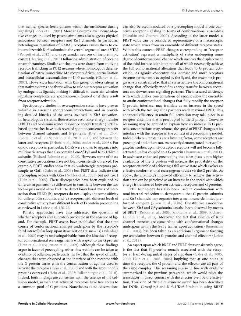
FIGURE 2 | Functional selectivity due to biased signaling of orthosteric
receptor agonists. Within the context of a precoupling model, an agonist thatpreferentially recognizes the receptor conformation stabilized by signalingpartners in the channel complex over the receptor state stabilized within thecyclase complex would display bias toward channel signaling (A). In a restricted
collision model G proteins and effectors coexist as a preformed complex thatis activated through collision with the agonist-bound receptor. In this contextbias toward Kir3 signaling can take place if the agonist-bound receptordisplays higher affinity for Gαβγ-Kir3 than Gαβγ-cyclase complexes (B). To aidvisualization, Kir3 channel is shown in blue and adenylyl cyclase in red.
Frontiers in Cellular Neuroscience www.frontiersin.org July 2014 | Volume 8 | Article 186 | 8

Nagi and Pineyro Kir3 channels in opioid analgesic
channels may also participate in hippocampal hyperexcitability(Luscher et al., 1997) induced by certain DOR ligands (Broomet al., 2002; Jutkiewicz et al., 2005). Depending on the degree ofKir3 involvement in this side effect, the incidence of seizures couldincrease for DOR ligands with Kir3 signaling bias. If this werethe case, the alternative strategy based on obliteration of cyclasemodulation would be of choice.
Biased agonism refers to the ability of orthosteric receptorligands to selectively engage the activity of a distinct set of sig-naling partners over another (Urban et al., 2007), a type ofselectivity that ensues from the stabilization of receptor confor-mations which activate a specific effector(s) while sparing the rest(Kenakin and Miller, 2010). Functional selectivity may also beindirect, driven by allosteric ligands (Leach et al., 2007; Kenakin,2008). Sodium is an allosteric modulator of DORs and manip-ulation of its binding site provides an example of how allostericinfluences may direct pharmacological stimuli toward differenteffectors (Fenalti et al., 2014). In particular, mutation of oneof the residues (Asn131) in the first coordination shell of theNa+ ion produced an “efficacy switch” that changed DOR signal-ing from the canonical Gαi/o pathway toward βarr recruitment.Furthermore, this effect was ligand sensitive since agonists lostGαi signaling while the antagonist naltrindole gained the abilityto recruit βarr (Fenalti et al., 2014). Hence, by stabilizing receptorconformations that differentially favor one orthosteric responseover another, allosteric modulation of receptor conformations offer
a great potential for directing pharmacological stimuli toward adesired response.
Let us first consider signaling bias within the context of thetraditional shuttling-collision model. According to this paradigmthe agonist interacts with a receptor which then travels within themembrane to interact and activate a G protein whose Gα andGβγ subunits subsequently dissociate from the ligand-receptorcomplex to find and activate an effector (Orly and Schramm,1976; Tolkovsky and Levitzki, 1978; Gilman, 1987; Bourne, 1997).Because in this model receptor and effector do not simultane-ously interact with the G protein, the paradigm does not providefor a “memory” that would allow transferring conformationalinformation codified by the agonist-bound receptor beyond thetransducer stage. However, more recent spectroscopic studies ofreceptor-G protein-effector interaction point to greater restric-tion in mobility where G protein and effectors would be spon-taneously coupled (Lohse et al., 2012). Moreover, independent ofwhether receptors form part of this constitutive complex or not,evidence analyzed in the previous section indicated that all threespecies may persistently associate during signaling. This associa-tion provides the basis for a “conformational memory” and thepossibility of exploring novel bias strategies to specifically directthe pharmacological stimulus of a given receptor (in this caseDORs) to a desired G protein coupled effector (Kir3 channels).
Being allosteric proteins (Kenakin and Miller, 2010), the con-formation adopted by the receptor will not be solely determined
FIGURE 3 | Mechanisms of indirect bias involving allosteric modulators
that specifically recognize a complex of desired composition. In aprecoupling model or in a restricted collision model receptors, G proteins andeffectors may all persistently associate during signaling. Within this context(only precoupling model represented in figure), small positive allostericmodulators (PAM ) that specifically recognize the interface between DORs
and Kir3 channels (in blue) may specifically bias signaling of DOR orthostericagonists toward this effector by stabilizing the complex and/or favoringchannel opening (A). A negative allosteric modulator (NAM) that recognizesthe DOR-adenylyl cyclase (in red) interface could distinctively block cAMPinhibition by the activated DOR, also allowing for bias in favor of channeleffectors (B).
Frontiers in Cellular Neuroscience www.frontiersin.org July 2014 | Volume 8 | Article 186 | 9

Nagi and Pineyro Kir3 channels in opioid analgesic
by orthosteric agonist binding, but also by its interaction with itscytosolic (G protein subunits) and membrane (Kir3 or cyclase)signaling partners, which therefore function as natural allostericmodulators. In cases where signaling complexes exist before acti-vation, selectivity favoring Kir3 vs. cyclase signaling could beachieved by designing orthosteric ligands that display higheraffinity for the conformation adopted by the receptor when con-tained within a Kir3 signaling complex than the one inducedby its inclusion into cyclase multimers (Figure 2A). The ideathat orthosteric agonists can indeed be tailored to specificallyrecognize receptors in association with distinct signaling part-ners is supported by studies indicating that the pharmacologicalproperties of receptors contained in homodimers are differentfrom those displayed by the same receptor when it is part ofan heterodimer (Jordan and Devi, 1999; Waldhoer et al., 2005).Alternatively, if the complex is formed during signaling, biastoward Kir3 channel effectors would depend on the agonist’s abil-ity to stabilize a receptor conformation whose affinity for the Gprotein/Kir3 complex is higher than the one displayed for the Gprotein/cyclase complex (Figure 2B).
In addition, if at rest and/or during signaling receptors, Gproteins and effectors associate through a distinct network ofconformational influences, it is also conceivable that the phar-macological stimulus that is produced by an orthosteric ligandcould be influenced by small allosteric modulators that specifi-cally recognize the complex with the desired combination of DORs,
Gαβγ subunits and Kir3 channels. For example, small moleculesthat could bind the interface of DORs and Kir3 subunits tostabilize the complex and/or favor channel opening (positiveallosteric modulators), would be of particular interest since theycould selectively enhance Kir3 signaling by DORs and no otherreceptors that modulate this effector (Figure 3A). Alternatively,a negative allosteric modulator that recognizes the DOR/adenylylcyclase interface could distinctively block cAMP inhibition by thisreceptor, resulting in another desired type of bias (Figure 3B).Finally, a variation of this strategy would be to design complex-selective allosteric agonists which are able to initiate signaling,independent of whether the orthosteric ligand is present or not(Figure 4A), or only when it is present for the case of restrictedcollision (Figure 4B). Such type of ligand could putatively recog-nize and stabilize the interface formed between the C-terminusof the activated Gαi/o subunit, the channel N-terminus and thethird intracellular loop of the receptor. Admittedly, the structuralinformation required for designing these compounds is not yetavailable but should become available once receptor/G proteinscomplexes are co-crystallized with their effectors.
In summary, we have analyzed evidence indicating that Kir3channels are mediators of opioid analgesia. While they play aconsiderable role in undesired effects of MOR agonists, theircontribution to those of DOR ligands is limited. Biasing DORresponses in favor of Kir3 channels and away from cyclase inhi-bition was suggested as a means of controlling analgesic tolerance
FIGURE 4 | Mechanisms of indirect bias involving allosteric agonists that
specifically recognize a complex of desired composition. Acomplex-specific allosteric agonist (AA) may distinctively recognize aninterface that is unique to the complex of interest. In the context of the
precoupling model AA may initiate signaling independent of whether theorthosteric agonist is present or not (A). In the restricted collision model thepresence of the orthosteric agonist is necessary for the complex to beformed and provide the binding site for AA (B).
Frontiers in Cellular Neuroscience www.frontiersin.org July 2014 | Volume 8 | Article 186 | 10

Nagi and Pineyro Kir3 channels in opioid analgesic
of DOR agonists, a side effect that limits their potential therapeu-tic application. Different modalities of GPCR association with Gproteins and effectors were discussed, and putative bias strategiesto ensure specific activation of a desired combination of receptors(DORs) and effectors (Kir3 channels) were provided.
ACKNOWLEDGMENTSThis work was supported by grants from the Natural Sciences andEngineering Research Council of Canada (NSERC) to GracielaPineyro [311997]. Karim Nagi holds a studentship from Ste-Justine Hospital Research Center and the Faculty of Graduate andPostdoctoral Studies, University of Montreal.
REFERENCESAndrade, A., Denome, S., Jiang, Y. Q., Marangoudakis, S., and Lipscombe,
D. (2010). Opioid inhibition of N-type Ca2+ channels and spinal analge-sia couple to alternative splicing. Nat. Neurosci. 13, 1249–1256. doi: 10.1038/nn.2643
Audet, N., Charfi, I., Mnie-Filali, O., Amraei, M., Chabot-Dore, A. J., Millecamps,M., et al. (2012). Differential association of receptor-Gbetagamma com-plexes with beta-arrestin2 determines recycling bias and potential for tol-erance of delta opioid receptor agonists. J. Neurosci. 32, 4827–4840. doi:10.1523/JNEUROSCI.3734-11.2012
Audet, N., Gales, C., Archer-Lahlou, E., Vallieres, M., Schiller, P. W., Bouvier,M., et al. (2008). Bioluminescence resonance energy transfer assays revealligand-specific conformational changes within preformed signaling complexescontaining delta-opioid receptors and heterotrimeric G proteins. J. Biol. Chem.283, 15078–15088. doi: 10.1074/jbc.M707941200
Ballantyne, J. C., and Shin, N. S. (2008). Efficacy of opioids for chronic pain: areview of the evidence. Clin. J. Pain 24, 469–478. doi: 10.1097/AJP.0b013e31816b2f26
Banks, M. L., Roma, P. G., Folk, J. E., Rice, K. C., and Negus, S. S. (2011). Effectsof the delta-opioid agonist SNC80 on the abuse liability of methadone in rhe-sus monkeys: a behavioral economic analysis. Psychopharmacology (Berl) 216,431–439. doi: 10.1007/s00213-011-2235-2
Bardoni, R., Tawfik, V. L., Wang, D., Francois, A., Solorzano, C., Shuster, S. A., et al.(2014). Delta opioid receptors presynaptically regulate cutaneous mechanosen-sory neuron input to the spinal cord dorsal horn. Neuron 81, 1312–1327. doi:10.1016/j.neuron.2014.01.044
Berlin, S., Keren-Raifman, T., Castel, R., Rubinstein, M., Dessauer, C. W., Ivanina,T., et al. (2010). G alpha(i) and G betagamma jointly regulate the con-formations of a G betagamma effector, the neuronal G protein-activatedK+ channel (GIRK). J. Biol. Chem. 285, 6179–6185. doi: 10.1074/jbc.M109.085944
Berlin, S., Tsemakhovich, V. A., Castel, R., Ivanina, T., Dessauer, C. W., Keren-Raifman, T., et al. (2011). Two distinct aspects of coupling between Galpha(i)protein and G protein-activated K+ channel (GIRK) revealed by fluorescentlylabeled Galpha(i3) protein subunits. J. Biol. Chem. 286, 33223–33235. doi:10.1074/jbc.M111.271056
Bettahi, I., Marker, C. L., Roman, M. I., and Wickman, K. (2002). Contributionof the Kir3.1 subunit to the muscarinic-gated atrial potassium channel IKACh.J. Biol. Chem. 277, 48282–48288. doi: 10.1074/jbc.M209599200
Blednov, Y. A., Stoffel, M., Alva, H., and Harris, R. A. (2003). A pervasive mecha-nism for analgesia: activation of GIRK2 channels. Proc. Natl. Acad. Sci. U.S.A.100, 277–282. doi: 10.1073/pnas.012682399
Blednov, Y. A., Stoffel, M., Chang, S. R., and Harris, R. A. (2001). GIRK2 defi-cient mice. Evidence for hyperactivity and reduced anxiety. Physiol. Behav. 74,109–117. doi: 10.1016/S0031-9384(01)00555-8
Bobeck, E. N., Chen, Q., Morgan, M. M., and Ingram, S. L. (2014). Contributionof adenylyl cyclase modulation of pre- and postsynaptic GABA neurotrans-mission to morphine antinociception and tolerance. Neuropsychopharmacology.doi: 10.1038/npp.2014.62. [Epub ahead of print].
Bonci, A., and Williams, J. T. (1997). Increased probability of GABA release duringwithdrawal from morphine. J. Neurosci. 17, 796–803.
Bourne, H. R. (1997). How receptors talk to trimeric G proteins. Curr. Opin. CellBiol. 9, 134–142. doi: 10.1016/S0955-0674(97)80054-3
Broom, D. C., Nitsche, J. F., Pintar, J. E., Rice, K. C., Woods, J. H., and Traynor,J. R. (2002). Comparison of receptor mechanisms and efficacy requirementsfor delta-agonist-induced convulsive activity and antinociception in mice.J. Pharmacol. Exp. Ther. 303, 723–729. doi: 10.1124/jpet.102.036525
Bruchas, M. R., and Chavkin, C. (2010). Kinase cascades and ligand-directed sig-naling at the kappa opioid receptor. Psychopharmacology (Berl) 210, 137–147.doi: 10.1007/s00213-010-1806-y
Bruchas, M. R., Land, B. B., Aita, M., Xu, M., Barot, S. K., Li, S., et al.(2007). Stress-induced p38 mitogen-activated protein kinase activation medi-ates kappa-opioid-dependent dysphoria. J. Neurosci. 27, 11614–11623. doi:10.1523/JNEUROSCI.3769-07.2007
Bruehl, S., Denton, J. S., Lonergan, D., Koran, M. E., Chont, M., Sobey, C., et al.(2013). Associations between KCNJ6 (GIRK2) gene polymorphisms and pain-related phenotypes. Pain 154, 2853–2859. doi: 10.1016/j.pain.2013.08.026
Bunemann, M., Frank, M., and Lohse, M. J. (2003). Gi protein activation in intactcells involves subunit rearrangement rather than dissociation. Proc. Natl. Acad.Sci. U.S.A. 100, 16077–16082. doi: 10.1073/pnas.2536719100
Cao, J. L., Vialou, V. F., Lobo, M. K., Robison, A. J., Neve, R. L., Cooper, D. C., et al.(2010). Essential role of the cAMP-cAMP response-element binding proteinpathway in opiate-induced homeostatic adaptations of locus coeruleus neu-rons. Proc. Natl. Acad. Sci. U.S.A. 107, 17011–17016. doi: 10.1073/pnas.1010077107
Charbogne, P., Kieffer, B. L., and Befort, K. (2014). 15 years of genetic approachesin vivo for addiction research: opioid receptor and peptide gene knockoutin mouse models of drug abuse. Neuropharmacology 76(Pt B), 204–217. doi:10.1016/j.neuropharm.2013.08.028
Cheng, P. Y., Wu, D., Soong, Y., McCabe, S., Decena, J. A., and Szeto, H. H. (1993).Role of mu 1- and delta-opioid receptors in modulation of fetal EEG andrespiratory activity. Am. J. Physiol. 265, R433–R438.
Christie, M. J. (2008). Cellular neuroadaptations to chronic opioids: tol-erance, withdrawal and addiction. Br. J. Pharmacol. 154, 384–396. doi:10.1038/bjp.2008.100
Chung, M. K., Cho, Y. S., Bae, Y. C., Lee, J., Zhang, X., and Ro, J. Y. (2013).Peripheral G protein-coupled inwardly rectifying potassium channels areinvolved in delta-opioid receptor-mediated anti-hyperalgesia in rat massetermuscle. Eur. J. Pain 18, 29–38. doi: 10.1002/j.1532-2149.2013.00343.x
Chu Sin Chung, P., and Kieffer, B. L. (2013). Delta opioid receptors in brain func-tion and diseases. Pharmacol. Ther. 140, 112–120. doi: 10.1016/j.pharmthera.2013.06.003
Ciruela, F., Fernandez-Duenas, V., Sahlholm, K., Fernandez-Alacid, L., Nicolau, J.C., Watanabe, M., et al. (2010). Evidence for oligomerization between GABABreceptors and GIRK channels containing the GIRK1 and GIRK3 subunits. Eur.J. Neurosci. 32, 1265–1277. doi: 10.1111/j.1460-9568.2010.07356.x
Clancy, S. M., Boyer, S. B., and Slesinger, P. A. (2007). Coregulation ofnatively expressed pertussis toxin-sensitive muscarinic receptors with G-protein-activated potassium channels. J. Neurosci. 27, 6388–6399. doi: 10.1523/JNEUROSCI.1190-07.2007
Clancy, S. M., Fowler, C. E., Finley, M., Suen, K. F., Arrabit, C., Berton,F., et al. (2005). Pertussis-toxin-sensitive Galpha subunits selectively bindto C-terminal domain of neuronal GIRK channels: evidence for a het-erotrimeric G-protein-channel complex. Mol. Cell. Neurosci. 28, 375–389. doi:10.1016/j.mcn.2004.10.009
Codd, E. E., Carson, J. R., Colburn, R. W., Stone, D. J., Van Besien, C.R., Zhang, S. P., et al. (2009). JNJ-20788560 [9-(8-azabicyclo[3.2.1]oct-3-ylidene)-9H-xanthene-3-carboxylic acid diethylamide], a selective delta opi-oid receptor agonist, is a potent and efficacious antihyperalgesic agent thatdoes not produce respiratory depression, pharmacologic tolerance, or physi-cal dependence. J. Pharmacol. Exp. Ther. 329, 241–251. doi: 10.1124/jpet.108.146969
Cooper, A., Grigoryan, G., Guy-David, L., Tsoory, M. M., Chen, A., and Reuveny,E. (2012). Trisomy of the G protein-coupled K+ channel gene, Kcnj6, affectsreward mechanisms, cognitive functions, and synaptic plasticity in mice. Proc.Natl. Acad. Sci. U.S.A. 109, 2642–2647. doi: 10.1073/pnas.1109099109
Cowan, A., Zhu, X. Z., Mosberg, H. I., Omnaas, J. R., and Porreca, F. (1988). Directdependence studies in rats with agents selective for different types of opioidreceptor. J. Pharmacol. Exp. Ther. 246, 950–955.
Cruz, H. G., Ivanova, T., Lunn, M. L., Stoffel, M., Slesinger, P. A., and Luscher, C.(2004). Bi-directional effects of GABA(B) receptor agonists on the mesolimbicdopamine system. Nat. Neurosci. 7, 153–159. doi: 10.1038/nn1181
Frontiers in Cellular Neuroscience www.frontiersin.org July 2014 | Volume 8 | Article 186 | 11

Nagi and Pineyro Kir3 channels in opioid analgesic
De Sarro, G. B., Marra, R., Spagnolo, C., and Nistico, G. (1992). Delta opioid recep-tors mediate seizures produced by intrahippocampal injection of ala-deltorphinin rats. Funct. Neurol. 7, 235–238.
Dewire, S. M., Yamashita, D. S., Rominger, D. H., Liu, G., Cowan, C. L., Graczyk,T. M., et al. (2013). A G protein-biased ligand at the mu-opioid receptoris potently analgesic with reduced gastrointestinal and respiratory dysfunc-tion compared with morphine. J. Pharmacol. Exp. Ther. 344, 708–717. doi:10.1124/jpet.112.201616
Do Carmo, G. P., Folk, J. E., Rice, K. C., Chartoff, E., Carlezon, W. A. Jr., and Negus,S. S. (2009). The selective non-peptidic delta opioid agonist SNC80 does notfacilitate intracranial self-stimulation in rats. Eur. J. Pharmacol. 604, 58–65. doi:10.1016/j.ejphar.2008.12.021
Drake, C. T., Chavkin, C., and Milner, T. A. (2007). Opioid systems in the dentategyrus. Prog. Brain Res. 163, 245–263. doi: 10.1016/S0079-6123(07)63015-5
Falkenburger, B. H., Jensen, J. B., and Hille, B. (2010). Kinetics of M1 muscarinicreceptor and G protein signaling to phospholipase C in living cells. J. Gen.Physiol. 135, 81–97. doi: 10.1085/jgp.200910344
Fenalti, G., Giguere, P. M., Katritch, V., Huang, X. P., Thompson, A. A., Cherezov,V., et al. (2014). Molecular control of delta-opioid receptor signalling. Nature506, 191–196. doi: 10.1038/nature12944
Feng, P., Rahim, R. T., Cowan, A., Liu-Chen, L. Y., Peng, X., Gaughan, J., et al.(2006). Effects of mu, kappa or delta opioids administered by pellet or pumpon oral Salmonella infection and gastrointestinal transit. Eur. J. Pharmacol. 534,250–257. doi: 10.1016/j.ejphar.2006.01.048
Fernandez-Alacid, L., Aguado, C., Ciruela, F., Martin, R., Colon, J., Cabanero, M. J.,et al. (2009). Subcellular compartment-specific molecular diversity of pre- andpost-synaptic GABA-activated GIRK channels in Purkinje cells. J. Neurochem.110, 1363–1376. doi: 10.1111/j.1471-4159.2009.06229.x
Fernandez-Alacid, L., Watanabe, M., Molnar, E., Wickman, K., and Lujan, R.(2011). Developmental regulation of G protein-gated inwardly-rectifying K+(GIRK/Kir3) channel subunits in the brain. Eur. J. Neurosci. 34, 1724–1736. doi:10.1111/j.1460-9568.2011.07886.x
Finley, M., Arrabit, C., Fowler, C., Suen, K. F., and Slesinger, P. A. (2004). betaL-betaM loop in the C-terminal domain of G protein-activated inwardly rectifyingK(+) channels is important for G(betagamma) subunit activation. J. Physiol.555, 643–657. doi: 10.1113/jphysiol.2003.056101
Ford, C. E., Skiba, N. P., Bae, H., Daaka, Y., Reuveny, E., Shekter, L. R.,et al. (1998). Molecular basis for interactions of G protein betagammasubunits with effectors. Science 280, 1271–1274. doi: 10.1126/science.280.5367.1271
Fourie, C., Li, D., and Montgomery, J. M. (2014). The anchoring protein SAP97influences the trafficking and localisation of multiple membrane channels.Biochim. Biophys. Acta 1838, 589–594. doi: 10.1016/j.bbamem.2013.03.015
Gales, C., Rebois, R. V., Hogue, M., Trieu, P., Breit, A., Hebert, T. E., et al. (2005).Real-time monitoring of receptor and G-protein interactions in living cells. Nat.Methods 2, 177–184. doi: 10.1038/nmeth743
Gales, C., Van Durm, J. J., Schaak, S., Pontier, S., Percherancier, Y., Audet, M.,et al. (2006). Probing the activation-promoted structural rearrangements inpreassembled receptor-G protein complexes. Nat. Struct. Mol. Biol. 13, 778–786.doi: 10.1038/nsmb1134
Gallantine, E. L., and Meert, T. F. (2005). A comparison of the antinociceptiveand adverse effects of the mu-opioid agonist morphine and the delta-opioidagonist SNC80. Basic Clin. Pharmacol. Toxicol. 97, 39–51. doi: 10.1111/j.1742-7843.2005.pto_97107.x
Gao, X. F., Zhang, H. L., You, Z. D., Lu, C. L., and He, C. (2007). G protein-coupledinwardly rectifying potassium channels in dorsal root ganglion neurons. ActaPharmacol. Sin. 28, 185–190. doi: 10.1111/j.1745-7254.2007.00478.x
Gaveriaux-Ruff, C., and Kieffer, B. L. (2011). Delta opioid receptor analgesia:recent contributions from pharmacology and molecular approaches. Behav.Pharmacol. 22, 405–414. doi: 10.1097/FBP.0b013e32834a1f2c
Gendron, L., Lucido, A. L., Mennicken, F., O’Donnell, D., Vincent, J. P., Stroh, T.,et al. (2006). Morphine and pain-related stimuli enhance cell surface availabil-ity of somatic delta-opioid receptors in rat dorsal root ganglia. J. Neurosci. 26,953–962. doi: 10.1523/JNEUROSCI.3598-05.2006
Gilman, A. G. (1987). G proteins: transducers of receptor-generated signals. Annu.Rev. Biochem. 56, 615–649. doi: 10.1146/annurev.bi.56.070187.003151
Glaum, S. R., Miller, R. J., and Hammond, D. L. (1994). Inhibitory actions ofdelta 1-, delta 2-, and mu-opioid receptor agonists on excitatory transmissionin lamina II neurons of adult rat spinal cord. J. Neurosci. 14, 4965–4971.
Goldenberg, D. L. (2010). Pain/Depression dyad: a key to a better understandingand treatment of functional somatic syndromes. Am. J. Med. 123, 675–682. doi:10.1016/j.amjmed.2010.01.014
Gonzalez-Rodriguez, S., Hidalgo, A., Baamonde, A., and Menendez, L.(2010). Involvement of Gi/o proteins and GIRK channels in the poten-tiation of morphine-induced spinal analgesia in acutely inflamed mice.Naunyn Schmiedeberg’s Arch. Pharmacol. 381, 59–71. doi: 10.1007/s00210-009-0471-3
Gonzalez-Rodriguez, S., Llames, S., Hidalgo, A., Baamonde, A., and Menendez, L.(2012). Potentiation of acute morphine-induced analgesia measured by a ther-mal test in bone cancer-bearing mice. Fundam. Clin. Pharmacol. 26, 363–372.doi: 10.1111/j.1472-8206.2010.00921.x
Gross, W., and Lohse, M. J. (1991). Mechanism of activation of A2 adenosine recep-tors. II. A restricted collision-coupling model of receptor-effector interaction.Mol. Pharmacol. 39, 524–530.
Gysling, K., and Wang, R. Y. (1983). Morphine-induced activation of A10dopamine neurons in the rat. Brain Res. 277, 119–127. doi: 10.1016/0006-8993(83)90913-7
Han, M. H., Bolanos, C. A., Green, T. A., Olson, V. G., Neve, R. L., Liu, R.J., et al. (2006). Role of cAMP response element-binding protein in the ratlocus ceruleus: regulation of neuronal activity and opiate withdrawal behaviors.J. Neurosci. 26, 4624–4629. doi: 10.1523/JNEUROSCI.4701-05.2006
Hassan, A. H., Ableitner, A., Stein, C., and Herz, A. (1993). Inflammation of therat paw enhances axonal transport of opioid receptors in the sciatic nerve andincreases their density in the inflamed tissue. Neuroscience 55, 185–195. doi:10.1016/0306-4522(93)90465-R
He, C., Yan, X., Zhang, H., Mirshahi, T., Jin, T., Huang, A., et al. (2002).Identification of critical residues controlling G protein-gated inwardly rectify-ing K(+) channel activity through interactions with the beta gamma subunitsof G proteins. J. Biol. Chem. 277, 6088–6096. doi: 10.1074/jbc.M104851200
He, C., Zhang, H., Mirshahi, T., and Logothetis, D. E. (1999). Identification of apotassium channel site that interacts with G protein betagamma subunits tomediate agonist-induced signaling. J. Biol. Chem. 274, 12517–12524.
He, L., and Whistler, J. L. (2007). The biochemical analysis of methadone mod-ulation on morphine-induced tolerance and dependence in the rat brain.Pharmacology 79, 193–202. doi: 10.1159/000100893
Hearing, M., Kotecki, L., Marron Fernandez De Velasco, E., Fajardo-Serrano, A.,Chung, H. J., Lujan, R., et al. (2013). Repeated cocaine weakens GABA(B)-Girksignaling in layer 5/6 pyramidal neurons in the prelimbic cortex. Neuron 80,159–170. doi: 10.1016/j.neuron.2013.07.019
Hein, P., Frank, M., Hoffmann, C., Lohse, M. J., and Bunemann, M. (2005).Dynamics of receptor/G protein coupling in living cells. EMBO J. 24,4106–4114. doi: 10.1038/sj.emboj.7600870
Hein, P., Rochais, F., Hoffmann, C., Dorsch, S., Nikolaev, V. O., Engelhardt, S., et al.(2006). Gs activation is time-limiting in initiating receptor-mediated signaling.J. Biol. Chem. 281, 33345–33351. doi: 10.1074/jbc.M606713200
Heinke, B., Gingl, E., and Sandkuhler, J. (2011). Multiple targets of mu-opioidreceptor-mediated presynaptic inhibition at primary afferent Adelta- and C-fibers. J. Neurosci. 31, 1313–1322. doi: 10.1523/JNEUROSCI.4060-10.2011
Hibino, H., Inanobe, A., Furutani, K., Murakami, S., Findlay, I., and Kurachi, Y.(2010). Inwardly rectifying potassium channels: their structure, function, andphysiological roles. Physiol. Rev. 90, 291–366. doi: 10.1152/physrev.00021.2009
Hibino, H., Inanobe, A., Tanemoto, M., Fujita, A., Doi, K., Kubo, T., et al.(2000). Anchoring proteins confer G protein sensitivity to an inward-rectifier K(+) channel through the GK domain. EMBO J. 19, 78–83. doi:10.1093/emboj/19.1.78
Huang, C. L., Jan, Y. N., and Jan, L. Y. (1997). Binding of the G protein betagammasubunit to multiple regions of G protein-gated inward-rectifying K+ channels.FEBS Lett. 405, 291–298. doi: 10.1016/S0014-5793(97)00197-X
Huang, C. L., Slesinger, P. A., Casey, P. J., Jan, Y. N., and Jan, L. Y. (1995).Evidence that direct binding of G beta gamma to the GIRK1 G protein-gatedinwardly rectifying K+ channel is important for channel activation. Neuron 15,1133–1143.
Ikeda, K., Kobayashi, T., Kumanishi, T., Niki, H., and Yano, R. (2000).Involvement of G-protein-activated inwardly rectifying K (GIRK) channelsin opioid-induced analgesia. Neurosci. Res. 38, 113–116. doi: 10.1016/S0168-0102(00)00144-9
Ikeda, K., Kobayashi, T., Kumanishi, T., Yano, R., Sora, I., and Niki, H. (2002).Molecular mechanisms of analgesia induced by opioids and ethanol: is the
Frontiers in Cellular Neuroscience www.frontiersin.org July 2014 | Volume 8 | Article 186 | 12

Nagi and Pineyro Kir3 channels in opioid analgesic
GIRK channel one of the keys? Neurosci. Res. 44, 121–131. doi: 10.1016/S0168-0102(02)00094-9
Inanobe, A., Yoshimoto, Y., Horio, Y., Morishige, K. I., Hibino, H., Matsumoto,S., et al. (1999). Characterization of G-protein-gated K+ channels composed ofKir3.2 subunits in dopaminergic neurons of the substantia nigra. J. Neurosci. 19,1006–1017.
Ivanina, T., Rishal, I., Varon, D., Mullner, C., Frohnwieser-Steinecke, B.,Schreibmayer, W., et al. (2003). Mapping the Gbetagamma-binding sites inGIRK1 and GIRK2 subunits of the G protein-activated K+ channel. J. Biol.Chem. 278, 29174–29183. doi: 10.1074/jbc.M304518200
Ivanina, T., Varon, D., Peleg, S., Rishal, I., Porozov, Y., Dessauer, C. W., et al.(2004). Galphai1 and Galphai3 differentially interact with, and regulate,the G protein-activated K+ channel. J. Biol. Chem. 279, 17260–17268. doi:10.1074/jbc.M313425200
Javed, R. R., Dewey, W. L., Smith, P. A., and Smith, F. L. (2004). PKCand PKA inhibitors reverse tolerance to morphine-induced hypothermiaand supraspinal analgesia in mice. Eur. J. Pharmacol. 492, 149–157. doi:10.1016/j.ejphar.2004.03.061
Jensen, J. B., Lyssand, J. S., Hague, C., and Hille, B. (2009). Fluorescence changesreveal kinetic steps of muscarinic receptor-mediated modulation of phos-phoinositides and Kv7.2/7.3 K+ channels. J. Gen. Physiol. 133, 347–359. doi:10.1085/jgp.200810075
Johnson, S. W., and North, R. A. (1992). Opioids excite dopamine neurons byhyperpolarization of local interneurons. J. Neurosci. 12, 483–488.
Jordan, B. A., and Devi, L. A. (1999). G-protein-coupled receptor heterodimeriza-tion modulates receptor function. Nature 399, 697–700.
Jutkiewicz, E. M., Baladi, M. G., Folk, J. E., Rice, K. C., and Woods, J. H. (2006).The convulsive and electroencephalographic changes produced by nonpeptidicdelta-opioid agonists in rats: comparison with pentylenetetrazol. J. Pharmacol.Exp. Ther. 317, 1337–1348. doi: 10.1124/jpet.105.095810
Jutkiewicz, E. M., Rice, K. C., Traynor, J. R., and Woods, J. H. (2005). Separationof the convulsions and antidepressant-like effects produced by the delta-opioid agonist SNC80 in rats. Psychopharmacology (Berl) 182, 588–596. doi:10.1007/s00213-005-0138-9
Kalso, E., Edwards, J. E., Moore, R. A., and McQuay, H. J. (2004). Opioids in chronicnon-cancer pain: systematic review of efficacy and safety. Pain 112, 372–380.doi: 10.1016/j.pain.2004.09.019
Kawano, T., Zhao, P., Floreani, C. V., Nakajima, Y., Kozasa, T., and Nakajima,S. (2007). Interaction of Galphaq and Kir3, G protein-coupled inwardlyrectifying potassium channels. Mol. Pharmacol. 71, 1179–1184. doi:10.1124/mol.106.032508
Kenakin, T. (2008). Functional selectivity in GPCR modulator screening.Comb. Chem. High Throughput Screen. 11, 337–343. doi: 10.2174/138620708784534824
Kenakin, T., and Miller, L. J. (2010). Seven transmembrane receptors as shapeshift-ing proteins: the impact of allosteric modulation and functional selectivity onnew drug discovery. Pharmacol. Rev. 62, 265–304. doi: 10.1124/pr.108.000992
Kenakin, T., and Onaran, O. (2002). The ligand paradox between affinity and effi-cacy: can you be there and not make a difference? Trends Pharmacol. Sci. 23,275–280. doi: 10.1016/S0165-6147(02)02036-9
Kieffer, B. L., and Gaveriaux-Ruff, C. (2002). Exploring the opioid system bygene knockout. Prog. Neurobiol. 66, 285–306. doi: 10.1016/S0301-0082(02)00008-4
Koyrakh, L., Lujan, R., Colon, J., Karschin, C., Kurachi, Y., Karschin, A., et al.(2005). Molecular and cellular diversity of neuronal G-protein-gated potassiumchannels. J. Neurosci. 25, 11468–11478. doi: 10.1523/JNEUROSCI.3484-05.2005
Krapivinsky, G., Kennedy, M. E., Nemec, J., Medina, I., Krapivinsky, L., andClapham, D. E. (1998). Gbeta binding to GIRK4 subunit is critical for Gprotein-gated K+ channel activation. J. Biol. Chem. 273, 16946–16952. doi:10.1074/jbc.273.27.16946
Kunkel, M. T., and Peralta, E. G. (1995). Identification of domains conferring Gprotein regulation on inward rectifier potassium channels. Cell 83, 443–449. doi:10.1016/0092-8674(95)90122-1
Laurent, V., Leung, B., Maidment, N., and Balleine, B. W. (2012). mu- anddelta-opioid-related processes in the accumbens core and shell differentiallymediate the influence of reward-guided and stimulus-guided decisions onchoice. J. Neurosci. 32, 1875–1883. doi: 10.1523/JNEUROSCI.4688-11.2012
Lavine, N., Ethier, N., Oak, J. N., Pei, L., Liu, F., Trieu, P., et al. (2002). Gprotein-coupled receptors form stable complexes with inwardly rectifying
potassium channels and adenylyl cyclase. J. Biol. Chem. 277, 46010–46019. doi:10.1074/jbc.M205035200
Leach, K., Sexton, P. M., and Christopoulos, A. (2007). Allosteric GPCR modula-tors: taking advantage of permissive receptor pharmacology. Trends Pharmacol.Sci. 28, 382–389. doi: 10.1016/j.tips.2007.06.004
Le Bourdonnec, B., Windh, R. T., Ajello, C. W., Leister, L. K., Gu, M., Chu, G.H., et al. (2008). Potent, orally bioavailable delta opioid receptor agonists forthe treatment of pain: discovery of N,N-diethyl-4-(5-hydroxyspiro[chromene-2,4’-piperidine]-4-yl)benzamide (ADL5859). J. Med. Chem. 51, 5893–5896. doi:10.1021/jm8008986
Le Merrer, J., Becker, J. A., Befort, K., and Kieffer, B. L. (2009). Reward pro-cessing by the opioid system in the brain. Physiol. Rev. 89, 1379–1412. doi:10.1152/physrev.00005.2009
Lemos, J. C., Roth, C. A., Messinger, D. I., Gill, H. K., Phillips, P. E., and Chavkin,C. (2012). Repeated stress dysregulates kappa-opioid receptor signaling in thedorsal raphe through a p38alpha MAPK-dependent mechanism. J. Neurosci. 32,12325–12336. doi: 10.1523/JNEUROSCI.2053-12.2012
Li, J. L., Ding, Y. Q., Li, Y. Q., Li, J. S., Nomura, S., Kaneko, T., et al. (1998).Immunocytochemical localization of mu-opioid receptor in primary affer-ent neurons containing substance P or calcitonin gene-related peptide. Alight and electron microscope study in the rat. Brain Res. 794, 347–352. doi:10.1016/S0006-8993(98)00332-1
Liao, Y. J., Jan, Y. N., and Jan, L. Y. (1996). Heteromultimerization of G-protein-gated inwardly rectifying K+ channel proteins GIRK1 and GIRK2 and theiraltered expression in weaver brain. J. Neurosci. 16, 7137–7150.
Lober, R. M., Pereira, M. A., and Lambert, N. A. (2006). Rapid activationof inwardly rectifying potassium channels by immobile G-protein-coupledreceptors. J. Neurosci. 26, 12602–12608. doi: 10.1523/JNEUROSCI.4020-06.2006
Logothetis, D. E., Kurachi, Y., Galper, J., Neer, E. J., and Clapham, D. E. (1987).The beta gamma subunits of GTP-binding proteins activate the muscarinic K+channel in heart. Nature 325, 321–326. doi: 10.1038/325321a0
Lohse, M. J., Nuber, S., and Hoffmann, C. (2012). Fluorescence/bioluminescenceresonance energy transfer techniques to study G-protein-coupled receptor acti-vation and signaling. Pharmacol. Rev. 64, 299–336. doi: 10.1124/pr.110.004309
Lotsch, J., Pruss, H., Veh, R. W., and Doehring, A. (2010). A KCNJ6 (Kir3.2,GIRK2) gene polymorphism modulates opioid effects on analgesia and addic-tion but not on pupil size. Pharmacogenet. Genomics 20, 291–297. doi:10.1097/FPC.0b013e3283386bda
Lujan, R., Marron Fernandez De Velasco, E., Aguado, C., and Wickman, K. (2014).New insights into the therapeutic potential of Girk channels. Trends Neurosci.37, 20–29. doi: 10.1016/j.tins.2013.10.006
Luscher, C., Jan, L. Y., Stoffel, M., Malenka, R. C., and Nicoll, R. A. (1997). Gprotein-coupled inwardly rectifying K+ channels (GIRKs) mediate postsynap-tic but not presynaptic transmitter actions in hippocampal neurons. Neuron 19,687–695. doi: 10.1016/S0896-6273(00)80381-5
Luscher, C., and Slesinger, P. A. (2010). Emerging roles for G protein-gated inwardlyrectifying potassium (GIRK) channels in health and disease. Nat. Rev. Neurosci.11, 301–315. doi: 10.1038/nrn2834
Luscher, C., and Ungless, M. A. (2006). The mechanistic classification of addictivedrugs. PLoS Med. 3:e437. doi: 10.1371/journal.pmed.0030437
Marker, C. L., Cintora, S. C., Roman, M. I., Stoffel, M., and Wickman, K.(2002). Hyperalgesia and blunted morphine analgesia in G protein-gatedpotassium channel subunit knockout mice. Neuroreport 13, 2509–2513. doi:10.1097/00001756-200212200-00026
Marker, C. L., Lujan, R., Colon, J., and Wickman, K. (2006). Distinct popula-tions of spinal cord lamina II interneurons expressing G-protein-gated potas-sium channels. J. Neurosci. 26, 12251–12259. doi: 10.1523/JNEUROSCI.3693-06.2006
Marker, C. L., Lujan, R., Loh, H. H., and Wickman, K. (2005). Spinal G-protein-gated potassium channels contribute in a dose-dependent manner to the anal-gesic effect of mu- and delta- but not kappa-opioids. J. Neurosci. 25, 3551–3559.doi: 10.1523/JNEUROSCI.4899-04.2005
Marker, C. L., Stoffel, M., and Wickman, K. (2004). Spinal G-protein-gated K+channels formed by GIRK1 and GIRK2 subunits modulate thermal nocicep-tion and contribute to morphine analgesia. J. Neurosci. 24, 2806–2812. doi:10.1523/JNEUROSCI.5251-03.2004
Mase, Y., Yokogawa, M., Osawa, M., and Shimada, I. (2012). Structural basis formodulation of gating property of G protein-gated inwardly rectifying potassium
Frontiers in Cellular Neuroscience www.frontiersin.org July 2014 | Volume 8 | Article 186 | 13

Nagi and Pineyro Kir3 channels in opioid analgesic
ion channel (GIRK) by i/o-family G protein alpha subunit (Galphai/o). J. Biol.Chem. 287, 19537–19549. doi: 10.1074/jbc.M112.353888
McAllister, S. D., Griffin, G., Satin, L. S., and Abood, M. E. (1999). Cannabinoidreceptors can activate and inhibit G protein-coupled inwardly rectifying potas-sium channels in a xenopus oocyte expression system. J. Pharmacol. Exp. Ther.291, 618–626.
McLaughlin, J. P., Myers, L. C., Zarek, P. E., Caron, M. G., Lefkowitz, R. J., Czyzyk, T.A., et al. (2004). Prolonged kappa opioid receptor phosphorylation mediated byG-protein receptor kinase underlies sustained analgesic tolerance. J. Biol. Chem.279, 1810–1818. doi: 10.1074/jbc.M305796200
Mitrovic, I., Margeta-Mitrovic, M., Bader, S., Stoffel, M., Jan, L. Y., and Basbaum,A. I. (2003). Contribution of GIRK2-mediated postsynaptic signaling to opiateand alpha 2-adrenergic analgesia and analgesic sex differences. Proc. Natl. Acad.Sci. U.S.A. 100, 271–276. doi: 10.1073/pnas.0136822100
Mogil, J. S. (2009). Are we getting anywhere in human pain genetics? Pain 146,231–232. doi: 10.1016/j.pain.2009.07.023
Moore, S. D., Madamba, S. G., Schweitzer, P., and Siggins, G. R. (1994). Voltage-dependent effects of opioid peptides on hippocampal CA3 pyramidal neuronsin vitro. J. Neurosci. 14, 809–820.
Morgan, M. M., and Christie, M. J. (2011). Analysis of opioid efficacy, tolerance,addiction and dependence from cell culture to human. Br. J. Pharmacol. 164,1322–1334. doi: 10.1111/j.1476-5381.2011.01335.x
Nassirpour, R., Bahima, L., Lalive, A. L., Luscher, C., Lujan, R., and Slesinger, P.A. (2010). Morphine- and CaMKII-dependent enhancement of GIRK chan-nel signaling in hippocampal neurons. J. Neurosci. 30, 13419–13430. doi:10.1523/JNEUROSCI.2966-10.2010
Navarro, B., Kennedy, M. E., Velimirovic, B., Bhat, D., Peterson, A. S., and Clapham,D. E. (1996). Nonselective and G betagamma-insensitive weaver K+ channels.Science 272, 1950–1953. doi: 10.1126/science.272.5270.1950
Neer, E. J. (1995). Heterotrimeric G proteins: organizers of transmembrane signals.Cell 80, 249–257. doi: 10.1016/0092-8674(95)90407-7
Neubig, R. R. (1994). Membrane organization in G-protein mechanisms. FASEB J.8, 939–946.
Nishizawa, D., Nagashima, M., Katoh, R., Satoh, Y., Tagami, M., Kasai, S., et al.(2009). Association between KCNJ6 (GIRK2) gene polymorphisms and post-operative analgesic requirements after major abdominal surgery. PLoS ONE4:e7060. doi: 10.1371/journal.pone.0007060
Nobles, M., Benians, A., and Tinker, A. (2005). Heterotrimeric G proteins precou-ple with G protein-coupled receptors in living cells. Proc. Natl. Acad. Sci. U.S.A.102, 18706–18711. doi: 10.1073/pnas.0504778102
Nockemann, D., Rouault, M., Labuz, D., Hublitz, P., McKnelly, K., Reis, F. C.,et al. (2013). The K(+) channel GIRK2 is both necessary and sufficient forperipheral opioid-mediated analgesia. EMBO Mol. Med. 5, 1263–1277. doi:10.1002/emmm.201201980
Obara, I., Parkitna, J. R., Korostynski, M., Makuch, W., Kaminska, D., Przewlocka,B., et al. (2009). Local peripheral opioid effects and expression of opioid genes inthe spinal cord and dorsal root ganglia in neuropathic and inflammatory pain.Pain 141, 283–291. doi: 10.1016/j.pain.2008.12.006
Orly, J., and Schramm, M. (1976). Coupling of catecholamine receptor from onecell with adenylate cyclase from another cell by cell fusion. Proc. Natl. Acad. Sci.U.S.A. 73, 4410–4414. doi: 10.1073/pnas.73.12.4410
Padgett, C. L., Lalive, A. L., Tan, K. R., Terunuma, M., Munoz, M. B., Pangalos,M. N., et al. (2012). Methamphetamine-evoked depression of GABA(B)receptor signaling in GABA neurons of the VTA. Neuron 73, 978–989. doi:10.1016/j.neuron.2011.12.031
Patil, N., Cox, D. R., Bhat, D., Faham, M., Myers, R. M., and Peterson, A. S.(1995). A potassium channel mutation in weaver mice implicates membraneexcitability in granule cell differentiation. Nat. Genet. 11, 126–129. doi: 10.1038/ng1095-126
Pert, C. B., and Snyder, S. H. (1973). Opiate receptor: demonstration in nervoustissue. Science 179, 1011–1014. doi: 10.1126/science.179.4077.1011
Pradhan, A. A., Befort, K., Nozaki, C., Gaveriaux-Ruff, C., and Kieffer, B. L.(2011). The delta opioid receptor: an evolving target for the treatment of braindisorders. Trends Pharmacol. Sci. 32, 581–590. doi: 10.1016/j.tips.2011.06.008
Pradhan, A. A., Walwyn, W., Nozaki, C., Filliol, D., Erbs, E., Matifas, A.,et al. (2010). Ligand-directed trafficking of the delta-opioid receptor in vivo:two paths toward analgesic tolerance. J. Neurosci. 30, 16459–16468. doi:10.1523/JNEUROSCI.3748-10.2010
Pravetoni, M., and Wickman, K. (2008). Behavioral characterization of micelacking GIRK/Kir3 channel subunits. Genes Brain Behav. 7, 523–531. doi:10.1111/j.1601-183X.2008.00388.x
Raehal, K. M., Walker, J. K., and Bohn, L. M. (2005). Morphine side effects inbeta-arrestin 2 knockout mice. J. Pharmacol. Exp. Ther. 314, 1195–1201. doi:10.1124/jpet.105.087254
Rasmussen, S. G., Devree, B. T., Zou, Y., Kruse, A. C., Chung, K. Y., Kobilka, T.S., et al. (2011). Crystal structure of the beta2 adrenergic receptor-Gs proteincomplex. Nature 477, 549–555. doi: 10.1038/nature10361
Raveh, A., Riven, I., and Reuveny, E. (2009). Elucidation of the gating of theGIRK channel using a spectroscopic approach. J. Physiol. 587, 5331–5335. doi:10.1113/jphysiol.2009.180158
Rebois, R. V., Robitaille, M., Gales, C., Dupre, D. J., Baragli, A., Trieu, P.,et al. (2006). Heterotrimeric G proteins form stable complexes with adeny-lyl cyclase and Kir3.1 channels in living cells. J. Cell Sci. 119, 2807–2818. doi:10.1242/jcs.03021
Richard-Lalonde, M., Nagi, K., Audet, N., Sleno, R., Amraei, M., Hogue, M., et al.(2013). Conformational dynamics of Kir3.1/Kir3.2 channel activation via delta-opioid receptors. Mol. Pharmacol. 83, 416–428. doi: 10.1124/mol.112.081950
Riven, I., Iwanir, S., and Reuveny, E. (2006). GIRK channel activation involves alocal rearrangement of a preformed G protein channel complex. Neuron 51,561–573. doi: 10.1016/j.neuron.2006.08.017
Robitaille, M., Ramakrishnan, N., Baragli, A., and Hebert, T. E. (2009). Intracellulartrafficking and assembly of specific Kir3 channel/G protein complexes. Cell.Signal. 21, 488–501. doi: 10.1016/j.cellsig.2008.11.011
Romero, G., Von Zastrow, M., and Friedman, P. A. (2011). Role of PDZ proteinsin regulating trafficking, signaling, and function of GPCRs: means, motif, andopportunity. Adv. Pharmacol. 62, 279–314. doi: 10.1016/B978-0-12-385952-5.00003-8
Rusinova, R., Mirshahi, T., and Logothetis, D. E. (2007). Specificity of Gbetagammasignaling to Kir3 channels depends on the helical domain of pertus-sis toxin-sensitive Galpha subunits. J. Biol. Chem. 282, 34019–34030. doi:10.1074/jbc.M704928200
Saenz Del Burgo, L., Cortes, R., Mengod, G., Zarate, J., Echevarria, E., and Salles, J.(2008). Distribution and neurochemical characterization of neurons express-ing GIRK channels in the rat brain. J. Comp. Neurol. 510, 581–606. doi:10.1002/cne.21810
Saitoh, A., Sugiyama, A., Nemoto, T., Fujii, H., Wada, K., Oka, J., et al. (2011). Thenovel delta opioid receptor agonist KNT-127 produces antidepressant-like andantinociceptive effects in mice without producing convulsions. Behav. Brain Res.223, 271–279. doi: 10.1016/j.bbr.2011.04.041
Scherrer, G., Imamachi, N., Cao, Y. Q., Contet, C., Mennicken, F., O’Donnell,D., et al. (2009). Dissociation of the opioid receptor mechanisms that con-trol mechanical and heat pain. Cell 137, 1148–1159. doi: 10.1016/j.cell.2009.04.019
Schreibmayer, W., Dessauer, C. W., Vorobiov, D., Gilman, A. G., Lester, H. A.,Davidson, N., et al. (1996). Inhibition of an inwardly rectifying K+ channel byG-protein alpha-subunits. Nature 380, 624–627. doi: 10.1038/380624a0
Signorini, S., Liao, Y. J., Duncan, S. A., Jan, L. Y., and Stoffel, M. (1997). Normalcerebellar development but susceptibility to seizures in mice lacking G protein-coupled, inwardly rectifying K+ channel GIRK2. Proc. Natl. Acad. Sci. U.S.A. 94,923–927. doi: 10.1073/pnas.94.3.923
Simon, E. J., Hiller, J. M., and Edelman, I. (1973). Stereospecific binding of thepotent narcotic analgesic (3H) Etorphine to rat-brain homogenate. Proc. Natl.Acad. Sci. U.S.A. 70, 1947–1949. doi: 10.1073/pnas.70.7.1947
Stein, C., Millan, M. J., Shippenberg, T. S., Peter, K., and Herz, A. (1989). Peripheralopioid receptors mediating antinociception in inflammation. Evidence forinvolvement of mu, delta and kappa receptors. J. Pharmacol. Exp. Ther. 248,1269–1275.
Tavani, A., Petrillo, P., La Regina, A., and Sbacchi, M. (1990). Role of periph-eral mu, delta and kappa opioid receptors in opioid-induced inhibition ofgastrointestinal transit in rats. J. Pharmacol. Exp. Ther. 254, 91–97.
Tian, W. N., Duzic, E., Lanier, S. M., and Deth, R. C. (1994). Determinants ofalpha 2-adrenergic receptor activation of G proteins: evidence for a precoupledreceptor/G protein state. Mol. Pharmacol. 45, 524–531.
Tolkovsky, A. M., and Levitzki, A. (1978). Mode of coupling between the beta-adrenergic receptor and adenylate cyclase in turkey erythrocytes. Biochemistry17, 3795. doi: 10.1021/bi00611a020
Frontiers in Cellular Neuroscience www.frontiersin.org July 2014 | Volume 8 | Article 186 | 14

Nagi and Pineyro Kir3 channels in opioid analgesic
Urban, J. D., Clarke, W. P., Von Zastrow, M., Nichols, D. E., Kobilka, B., Weinstein,H., et al. (2007). Functional selectivity and classical concepts of quantitativepharmacology. J. Pharmacol. Exp. Ther. 320, 1–13. doi: 10.1124/jpet.106.104463
Vadivelu, N., Mitra, S., and Hines, R. L. (2011). Peripheral opioid receptor ago-nists for analgesia: a comprehensive review. J. Opioid Manag. 7, 55–68. doi:10.5055/jom.2011.0049
Van’t Veer, A., and Carlezon, W. A. Jr. (2013). Role of kappa-opioid receptors instress and anxiety-related behavior. Psychopharmacology (Berl.) 229, 435–452.doi: 10.1007/s00213-013-3195-5
Vaughan, C. W., and Christie, M. J. (1997). Presynaptic inhibitory action of opi-oids on synaptic transmission in the rat periaqueductal grey in vitro. J. Physiol.498(Pt 2), 463–472.
Vaughan, C. W., Ingram, S. L., Connor, M. A., and Christie, M. J. (1997). Howopioids inhibit GABA-mediated neurotransmission. Nature 390, 611–614. doi:10.1038/37610
Vilardaga, J. P., Bunemann, M., Krasel, C., Castro, M., and Lohse, M. J. (2003).Measurement of the millisecond activation switch of G protein-coupled recep-tors in living cells. Nat. Biotechnol. 21, 807–812. doi: 10.1038/nbt838
Waldhoer, M., Fong, J., Jones, R. M., Lunzer, M. M., Sharma, S. K., Kostenis,E., et al. (2005). A heterodimer-selective agonist shows in vivo relevance of Gprotein-coupled receptor dimers. Proc. Natl. Acad. Sci. U.S.A. 102, 9050–9055.doi: 10.1073/pnas.0501112102
Wang, H. B., Zhao, B., Zhong, Y. Q., Li, K. C., Li, Z. Y., Wang, Q., et al. (2010).Coexpression of delta- and mu-opioid receptors in nociceptive sensory neurons.Proc. Natl. Acad. Sci. U.S.A. 107, 13117–13122. doi: 10.1073/pnas.1008382107
Wei, Z. Y., Karim, F., and Roerig, S. C. (1996). Spinal morphine/clonidine antinoci-ceptive synergism: involvement of G proteins and N-type voltage-dependentcalcium channels. J. Pharmacol. Exp. Ther. 278, 1392–1407.
Whorton, M. R., and MacKinnon, R. (2011). Crystal structure of the mammalianGIRK2 K+ channel and gating regulation by G proteins, PIP2, and sodium. Cell147, 199–208. doi: 10.1016/j.cell.2011.07.046
Whorton, M. R., and MacKinnon, R. (2013). X-ray structure of the mam-malian GIRK2-betagamma G-protein complex. Nature 498, 190–197. doi:10.1038/nature12241
Wickman, K., Karschin, C., Karschin, A., Picciotto, M. R., and Clapham, D. E.(2000). Brain localization and behavioral impact of the G-protein-gated K+channel subunit GIRK4. J. Neurosci. 20, 5608–5615.
Wickman, K., Nemec, J., Gendler, S. J., and Clapham, D. E. (1998). Abnormalheart rate regulation in GIRK4 knockout mice. Neuron 20, 103–114. doi:10.1016/S0896-6273(00)80438-9
Wickman, K. D., Iniguez-Lluhl, J. A., Davenport, P. A., Taussig, R., Krapivinsky, G.B., Linder, M. E., et al. (1994). Recombinant G-protein beta gamma-subunits
activate the muscarinic-gated atrial potassium channel. Nature 368, 255–257.doi: 10.1038/368255a0
Wimpey, T. L., and Chavkin, C. (1991). Opioids activate both an inwardrectifier and a novel voltage-gated potassium conductance in the hip-pocampal formation. Neuron 6, 281–289. doi: 10.1016/0896-6273(91)90363-5
Wreggett, K. A., and De Lean, A. (1984). The ternary complex model. Its propertiesand application to ligand interactions with the D2-dopamine receptor of theanterior pituitary gland. Mol. Pharmacol. 26, 214–227.
Wrigley, P. J., Jeong, H. J., and Vaughan, C. W. (2010). Dissociation of mu- anddelta-opioid inhibition of glutamatergic synaptic transmission in superficialdorsal horn. Mol. Pain 6:71. doi: 10.1186/1744-8069-6-71
Wu, Z. Z., Chen, S. R., and Pan, H. L. (2008). Distinct inhibition of voltage-activated Ca2+ channels by delta-opioid agonists in dorsal root ganglion neu-rons devoid of functional T-type Ca2+ currents. Neuroscience 153, 1256–1267.doi: 10.1016/j.neuroscience.2008.03.031
Xu, M., Petraschka, M., McLaughlin, J. P., Westenbroek, R. E., Caron, M. G.,Lefkowitz, R. J., et al. (2004). Neuropathic pain activates the endogenous kappaopioid system in mouse spinal cord and induces opioid receptor tolerance.J. Neurosci. 24, 4576–4584. doi: 10.1523/JNEUROSCI.5552-03.2004
Yang, H. Y., Wu, Z. Y., Wood, M., Whiteman, M., and Bian, J. S. (2014). Hydrogensulfide attenuates opioid dependence by suppression of adenylate cyclase/cAMPpathway. Antioxid. Redox Signal. 20, 31–41. doi: 10.1089/ars.2012.5119
Yokogawa, M., Osawa, M., Takeuchi, K., Mase, Y., and Shimada, I. (2011). NMRanalyses of the Gbetagamma binding and conformational rearrangements of thecytoplasmic pore of G protein-activated inwardly rectifying potassium channel1 (GIRK1). J. Biol. Chem. 286, 2215–2223. doi: 10.1074/jbc.M110.160754
Conflict of Interest Statement: The authors declare that the research was con-ducted in the absence of any commercial or financial relationships that could beconstrued as a potential conflict of interest.
Received: 16 March 2014; accepted: 18 June 2014; published online: 08 July 2014.Citation: Nagi K and Pineyro G (2014) Kir3 channel signaling complexes: focus onopioid receptor signaling. Front. Cell. Neurosci. 8:186. doi: 10.3389/fncel.2014.00186This article was submitted to the journal Frontiers in Cellular Neuroscience.Copyright © 2014 Nagi and Pineyro. This is an open-access article distributed underthe terms of the Creative Commons Attribution License (CC BY). The use, distribu-tion or reproduction in other forums is permitted, provided the original author(s)or licensor are credited and that the original publication in this journal is cited, inaccordance with accepted academic practice. No use, distribution or reproduction ispermitted which does not comply with these terms.
Frontiers in Cellular Neuroscience www.frontiersin.org July 2014 | Volume 8 | Article 186 | 15
Related Documents