King’s Research Portal DOI: 10.1093/cvr/cvv281 Document Version Peer reviewed version Link to publication record in King's Research Portal Citation for published version (APA): Testai, L., Barrese, V., Soldovieri, M. V., Ambrosino, P., Martelli, A., Vinciguerra, I., ... Taglialatela, M. (2016). Expression and function of Kv7.4 channels in rat cardiac mitochondria: Possible targets for cardioprotection. Cardiovascular Research, 110(1), 40-50. DOI: 10.1093/cvr/cvv281 Citing this paper Please note that where the full-text provided on King's Research Portal is the Author Accepted Manuscript or Post-Print version this may differ from the final Published version. If citing, it is advised that you check and use the publisher's definitive version for pagination, volume/issue, and date of publication details. And where the final published version is provided on the Research Portal, if citing you are again advised to check the publisher's website for any subsequent corrections. General rights Copyright and moral rights for the publications made accessible in the Research Portal are retained by the authors and/or other copyright owners and it is a condition of accessing publications that users recognize and abide by the legal requirements associated with these rights. •Users may download and print one copy of any publication from the Research Portal for the purpose of private study or research. •You may not further distribute the material or use it for any profit-making activity or commercial gain •You may freely distribute the URL identifying the publication in the Research Portal Take down policy If you believe that this document breaches copyright please contact [email protected] providing details, and we will remove access to the work immediately and investigate your claim. Download date: 20. Jun. 2018

Welcome message from author
This document is posted to help you gain knowledge. Please leave a comment to let me know what you think about it! Share it to your friends and learn new things together.
Transcript
King’s Research Portal
DOI:10.1093/cvr/cvv281
Document VersionPeer reviewed version
Link to publication record in King's Research Portal
Citation for published version (APA):Testai, L., Barrese, V., Soldovieri, M. V., Ambrosino, P., Martelli, A., Vinciguerra, I., ... Taglialatela, M. (2016).Expression and function of Kv7.4 channels in rat cardiac mitochondria: Possible targets for cardioprotection.Cardiovascular Research, 110(1), 40-50. DOI: 10.1093/cvr/cvv281
Citing this paperPlease note that where the full-text provided on King's Research Portal is the Author Accepted Manuscript or Post-Print version this maydiffer from the final Published version. If citing, it is advised that you check and use the publisher's definitive version for pagination,volume/issue, and date of publication details. And where the final published version is provided on the Research Portal, if citing you areagain advised to check the publisher's website for any subsequent corrections.
General rightsCopyright and moral rights for the publications made accessible in the Research Portal are retained by the authors and/or other copyrightowners and it is a condition of accessing publications that users recognize and abide by the legal requirements associated with these rights.
•Users may download and print one copy of any publication from the Research Portal for the purpose of private study or research.•You may not further distribute the material or use it for any profit-making activity or commercial gain•You may freely distribute the URL identifying the publication in the Research Portal
Take down policyIf you believe that this document breaches copyright please contact [email protected] providing details, and we will remove access tothe work immediately and investigate your claim.
Download date: 20. Jun. 2018

Testai et al. CVR-2015-217
1
EXPRESSION AND FUNCTION OF Kv7.4 CHANNELS IN RAT CARDIAC
MITOCHONDRIA: POSSIBLE TARGETS FOR CARDIOPROTECTION
1Lara Testai*, 2,3Vincenzo Barrese*, 4Maria Virginia Soldovieri, 4Paolo Ambrosino, 1Alma Martelli,
4Iolanda Vinciguerra, 2Francesco Miceli, 3Iain Greenwood, 5Michael J. Curtis,
1Maria Cristina Breschi, 2Maria Jose Sisalli, 2Antonella Scorziello, 5Miren Josune Canduela,
5Pedro Grandes, 1Vincenzo Calderone #, and 2,4Maurizio Taglialatela #
1Department of Pharmacy, University of Pisa (Italy) 2Department of Neuroscience, University of Naples “Federico II”, Naples (Italy) 3Vascular Biology Research Centre, Institute of Cardiovascular and Cell Sciences, St George's, University of London, London (UK) 4Department of Medicine and Health Science, University of Molise, Campobasso (Italy) 5Cardiovascular Division, Faculty of Life Sciences & Medicine, King’s College London, London (UK) 6Department of Neurosciences, University of the Basque Country, Leioa (Spain)
*These Authors contributed equally #These Authors contributed equally Corresponding Author: Maurizio Taglialatela, MD PhD Department of Medicine and Health Sciences University of Molise Via De Sanctis, 86100 – Campobasso, ITALY Tel. (+39) 0874-404851; Fax: (+39) 0874-404778 Email: [email protected] Total number of words in the text : 5733
Testai et al. CVR-2015-217
2
EXPRESSION AND FUNCTION OF Kv7.4 CHANNELS IN RAT CARDIAC MITOCHONDRIA: POSSIBLE TARGETS
FOR CARDIOPROTECTION
Lara Testai, Vincenzo Barrese, Maria Virginia Soldovieri, Paolo Ambrosino, Alma Martelli, Iolanda
Vinciguerra, Francesco Miceli, Iain Greenwood, Michael J. Curtis, Maria Cristina Breschi, Maria Jose
Sisalli, Antonella Scorziello, Miren Josune Canduela, Pedro Grandes, Vincenzo Calderone,
and Maurizio Taglialatela
ABSTRACT
Aims. Plasmalemmal Kv7.1 (KCNQ1) channels are critical players in cardiac excitability; however, little is
known on the functional role of additional Kv7 family members (Kv7.2-5) in cardiac cells. In this work, the
expression, function, cellular and subcellular localization, and potential cardioprotective role against
anoxic-ischemic cardiac injury of Kv7.4 channels have been investigated.
Methods and Results. Expression of Kv7.1 and Kv7.4 transcripts was found in rat heart tissue by qPCR.
Western-blots detected Kv7.4 subunits in mitochondria from Kv7.4-transfected cells, H9c2 cardiomyoblasts,
freshly-isolated adult cardiomyocytes, and whole hearts. Immunofluorescence experiments revealed that
Kv7.4 subunits co-localized with mitochondrial markers in cardiac cells, with about 30-40% of cardiac
mitochondria being labelled by Kv7.4 antibodies, a result also confirmed by immuno-gold electron
microscopy experiments. In isolated cardiac (but not liver) mitochondria, retigabine (1-30 M) and
flupirtine (30 M), two selective Kv7 activators, increased Tl+ influx, depolarized the membrane potential,
and inhibited calcium uptake; all these effects were antagonized by the Kv7 blocker XE991. In intact H9c2
cells, reducing Kv7.4 expression by RNA interference blunted retigabine-induced mitochondrial membrane
depolarization; in these cells, retigabine decreased mitochondrial Ca2+ levels and increased radical oxygen
species production, both effects prevented by XE991. Finally, retigabine reduced cellular damage in H9c2
cells exposed to anoxia/reoxygenation, and largely prevented the functional and morphological changes
triggered by global ischemia/reperfusion (I/R) in Langendorff-perfused rat hearts.
Conclusions. Kv7.4 channels are present and functional in cardiac mitochondria; their activation exerts a
significant cardioprotective role, making them potential therapeutic targets against I/R-induced cardiac
injury.
Testai et al. CVR-2015-217
3
1. INTRODUCTION
Activation of potassium (K+) fluxes across the inner mitochondrial membrane (IMM) is a key
mechanism for cardiac ischemic pre-conditioning.1 In physiological conditions, the IMM is poorly permeable
to K+ ions as mitochondrial K+ (mitoK) channels are mostly closed. However, during ischemia, different
triggers activate mitoK channels, leading to significant influx of K+ ions, accompanied by water and anions,
resulting in matrix swelling and depolarization of the membrane potential. As such, the activation of mitoK
channels controls the matrix volume, preserving a narrow intermembrane space, necessary for an effective
oxidative phosphorylation, and opposes Ca2+-overload and subsequent opening of the mitochondrial
permeability transition pore, a powerful trigger of apoptosis.1 In cardiac cells, pharmacological activation of
mitochondrial ATP-sensitive (mitoKATP) and large-conductance Ca2+-activated (mitoBKCa) K+ channels2-4
triggers cardioprotective responses in different models of ischemia/reperfusion (I/R) injury.3,5-7 Other K+
channel subtypes have been described in the IMM of mostly non cardiac cells.8 Despite these studies,
molecular identification of cardiac mitoK channels heterogeneity is far from being complete.
Voltage-gated K+ channels encoded by the Kv7 gene family (Kv7.1-7.5, also known as KCNQ1-5)
have well defined expression pattern and functional roles in heart, neurons, epithelia and vascular and non-
vascular smooth muscle.9 In brain and sensory neurons, Kv7.2/7.3 or 7.3/7.5 heteromers contribute to a
sub-threshold current called M-current, whereas Kv7.4 and/or Kv7.5 channels, together with Kv7.1, appear
to be major determinants of cellular excitability in vascular and non-vascular smooth muscle cells.10 In
cardiac cells, Kv7.1 encodes for subunits contributing to the slowly repolarizing current IKs;11 however,
significant levels of Kv7.4 transcripts have also been reported in the heart12, but no function ascribed.
To date all functional roles of Kv7 channels have been attributed to their plasma membrane
location and regulations of cellular membrane potential, while their possible impact on mitochondrial
physiology has not been determined. In this study, we report biochemical and morphological evidence for
the presence of Kv7.4 subunits in cardiac mitochondria, also defining their contribution to mitochondrial
function. In addition, we show that the pharmacological activation of these channels exerts significant
cytoprotective effects in mitochondrial-dependent in-vitro and ex-vivo models of cardiac I/R damage,
suggesting that targeting mitoKv7.4 channels might be an effective strategy for cardioprotection against I/R
cardiac injury.

Testai et al. CVR-2015-217
4
2. METHODS
Only a general overview of key methods is provided here; please refer to the Online supplement for further
experimental details.
1. Animals
Male rats of 2-3 months of age were used. Experimental procedures were carried out following the
guidelines of the Directive 2010/63/EU of the European Parliament, and have been approved by the
Committee for animal experimentation of the Institutions where experiments were carried out.
2. RNA extraction and quantitative real-time PCR
Total RNA from rat brains, livers, and hearts was isolated and reverse transcribed to cDNA; qPCR was
carried out in a real-time PCR system using the SYBR Green detection technique and specific primers
(Supplementary Table 1).
3. Isolation of rat primary cardiomyocytes
Dissected rat hearts were perfused in the Langendorff mode with collagenase. Isolated cells were
resuspended in Ca2+-Tyrode solution, spread on laminin-coated coverslips and allowed to adhere for 8
hours before further processing.
4. Cell cultures and transfection
H9c2 cells from embryonic rat ventricular myocytes, and Chinese Hamster Ovary (CHO) cells were used in
the present experiments; cells were transfected using Lipofectamine.
5. Immunofluorescence
H9c2 cells or primary cardiomyocytes were incubated with Mitotracker Red, fixed with
paraformaldehyde, and then incubated with primary antibodies. Rat cardiac slices were fixed in
paraformaldehyde, and incubated in sucrose for cryopreservation at −80°C; frozen sections (20 μm) were
cut and stored at −20°C until further processing. Following incubation with primary and secondary
antibodies, cells and cardiac slices were counterstained with Hoechst 33258 to visualize cells nuclei, and
analyzed using a Zeiss LSM 510 Meta argon/krypton laser scanning confocal microscope.
6 Pre-embedding immunogold electron microscopy
Rat heart coronal vibrosections were incubated with the primary Kv7.4 antibody, and then with a 1.4 nm
gold-labeled antibody directed toward mouse IgG. Ultrathin sections containing silver-intensified gold

Testai et al. CVR-2015-217
5
particles were osmicated, dehydrated, embedded in Epon resin 812, and examined in a PHILIPS EM208S
electron microscope.
7. Mitochondria isolation
Rat cardiac and hepatic mitochondria were isolated by differential centrifugation;13 mitochondria from
H9c2 cells and primary adult rat cardiomyocytes were obtained using a commercially-available kit.
Mitoplasts were prepared using the detergent digitonin.
8. Western blots
Protein samples for Western-blot experiments were loaded on 8% or 8-15% SDS–PAGE, and then
transferred to a polyvinylidene fluoride membrane. Membranes were incubated with primary and
secondary antibodies, and reactive bands detected by chemiluminescence.
9. Thallium fluxes, membrane potential, and Ca2+ measurements in isolated mitochondria from H2c9 cells
A fluorescent Tl+-sensitive probe (benzothiazole coumarinacetoxy methyl ester; ex = 488 nm, em = 525
nm) was used to evaluate fluxes of the K+-mimetic cation thallium (Tl+) in isolated mitochondria14 using in a
multiplate reader. Mitochondrial membrane potential (Δψ) was measured potentiometrically13 with
tetraphenylphosphonium chloride-sensitive mini-electrodes. Mitochondrial Ca2+-uptake13 was evaluated by
measuring the changes of the extra-mitochondrial Ca2+ concentration using a Ca2+ selective mini-electrode.
10. Membrane potential, calcium concentration ([Ca2+]m), and ROS production monitoring in H9c2 cells
mitochondria
membrane potential, calcium concentration ([Ca2+]m), and ROS production in living H9c2 cells mitochondria
were assessed using the fluorescent dyes tetramethyl rhodamine ethyl ester (TMRE), X-Rhod, and MitoSOX
Red, respectively. Images from TMRE and X-Rhod experiments were recorded by a conventional
immunofluorescence imaging system; those from MitoSOX red, were acquired using confocal microscopy.
11. Silencing Kv7.4 expression by shRNA in H9c2 cells
H9c2 cells were transiently transfected with a plasmid encoding for a short hairpin RNA (shRNA) directed
against Kv7.4 mRNA (pLKO.1-shKv7.4), or with a control plasmid carrying a nonsense sequence (pLKO.1-
scramble). The procedure has been previously used in our lab to selectively suppress Kv7.4 expression in
C2C12 rat skeletal myoblasts; Kv7.4 silencing was assessed by western-blotting experiments.15

Testai et al. CVR-2015-217
6
16. Anoxia/reoxygenation in H9c2 cells
To simulate anoxia in H9c2 cells, plated cells were sealed for 16 hours in airtight containers saturated with
95% N2 and 5% CO2 (37°C), while twin plates were placed in 95% air and 5% CO2 (normoxic conditions).
Then, cells were subjected to reoxygenation for 2 hours in an atmosphere containing 95 % air and 5% CO2
(37°C). After reoxygenation, cell viability was assessed.
17. Langendorff-perfused rat hearts
Rat hearts were quickly removed, mounted on a Langendorff apparatus, and perfused with oxygenated
solution at 37 °C and constant pressure (70-80 mmHg).16 A water-filled latex balloon connected to a
pressure transducer was introduced into the left ventricle to achieve a stable left ventricular end-diastolic
pressure of 5-10 mmHg. The functional parameters of heart rate (HR), left ventricular developed pressure
(LVDP) and the rate of rise of the left ventricular pressure (dP/dt) were monitored continuously. Rate
pressure product (RPP) was calculated as HR x LVDP. Coronary flow (CF) was also estimated. After 20 min of
equilibration, 30 min of global ischemia (no flow) followed. At the end of the ischemic period, the hearts
were reperfused for 2 hours; following reperfusion, hearts were removed from the Langendorff apparatus,
and 2mm large slices cut from the left ventricle were immersed in a solution containing 2,3,5-
triphenyltetrazolium chloride to evaluate the extension of the damage.
18. Drugs and antibodies
Retigabine and flupirtine were from Valeant (Laval, Canada); XE991, olygomicin, 2,4-dinitrophenol,
valinomycin, and FCCP were from Sigma-Aldrich (Milan, Italy). The primary antibodies used were: i) mouse
monoclonal Kv7.4 (UC Davis/NIH NeuroMab Facility, USA; clone N43/6.1; dilution: 1:100 for
immunofluorescence and electron microscopy; 1:500/1000 for western blot); ii) rabbit polyclonal
Sphingosine 1-phosphate receptor 1 (S1PR1, 1:500; Abcam, Cambridge, UK); iii) mouse monoclonal Kv7.1
(1:500; NeuroMab, clone N37A/10.1, used for western blot); iv) rabbit Kv7.1 (1:100; Millipore, Temecula,
USA, used for immunocytochemistry); v) mouse VDAC1 (TC supernatant, 1:5, NeuroMab); vi) mouse α-
tubulin antibody (1:5000; Sigma-Aldrich); vi) mouse GAPDH (1:5000; Sigma-Aldrich); viii) rabbit COX-IV
(Abcam, Cambridge, UK). Secondary antibodies for immunofluorescence (Jackson ImmunoResearch,
Newmarket, UK) were: i) anti-mouse conjugated to Alexa Fluor 488; ii) anti-rabbit conjugated to Alexa Fluor
488; iii) anti-rabbit conjugated to Alexa Fluor 568. Anti-mouse or anti-rabbit conjugated to HRP secondary
antibodies (Jackson ImmunoResearch, Newmarket, UK) were used for Western blot experiment, at 1:5000
dilution.
Testai et al. CVR-2015-217
7
19. Statistics
Data, reported as Mean±SEM, were statistically analysed by ANOVA followed (if F reached significance) by
the Tukey test (true or not, Vincenzo? Or did you use another one?), or by the Student’s t test (software:
GraphPadPrism 4.0). P values < 0.05 were considered as indicative of significant differences.
Testai et al. CVR-2015-217
8
3. RESULTS
3.1 Expression of Kv7.4 mRNA and protein in cardiac tissue and cardiomyocytes
Quantitative PCR experiments using specific primers (Supplementary Table 1) were performed on
mRNA extracted from rat brain, heart and liver. In the brain, Kv7.2-5 transcripts were readily identified,
whereas smaller but detectable amounts of Kv7.1 transcripts were also found (Fig. 1A). By contrast, mRNAs
encoding for Kv7.2-5 were expressed at rather marginal levels in the liver, whereas Kv7.1 transcripts were
clearly detected. In cardiac tissue, Kv7.1 showed the highest expression level among Kv7 transcripts, but
Kv7.4 transcripts were also found,12 with expression levels comparable to the brain.
Western blots experiments with anti-Kv7.4 antibodies in total lysates from rat heart lysates
revealed a band of ~77kDa; this band was enriched in voltage-dependent anion-selective (VDAC) channel-
enriched mitochondrial fractions, which tested negative for cytosolic markers such as -tubulin and
glyceraldehyde-3-phosphate dehydrogenase (GAPDH) (Fig. 1B). Kv7.4 signals were also readily detected in
rat heart mitoplasts, in which the outer mitochondrial membrane (OMM) VDAC signal was markedly
decreased (Fig. 1C). A band of similar molecular mass was also observed in Kv7.4-transfected CHO cells, but
not in CHO cells transfected with Kv7.1, Kv7.2, Kv7.3 or Kv7.5 cDNAs, or in non-transfected CHO cells
(Suppl. Fig. 1A), confirming antibody specificity. Also in Kv7.4-transfected CHO cells, this band was highly
enriched in mitochondrial fractions (Suppl. Fig. 1).
In H9c2 cardiomyoblasts, Kv7.4 antibodies identified a 77kDa band in mitochondria (positive to
cytochrome c oxidase subunit IV, COX-IV), but not in cytoplasmic (tubulin-positive) or microsomal (tubulin-
negative) fractions (Fig. 1D); a similar subcellular pattern of expression of Kv7.4 subunits was also observed
in freshly isolated rat cardiac myocytes, where the Kv7.4-reactive band was mainly found in COX-IV-
positive, but GADPH- and PDI-negative fractions corresponding to mitochondria. By contrast, in cardiac
myocytes, a 71 kDa band corresponding to Kv7.1 subunits could be readily detected in the cytosolic
fraction, but was absent from the microsomal and mitochondrial fractions (Fig. 1E). A band of similar
molecular mass was revealed in total lysates from Kv7.1-transfected (but not Kv7.2-, Kv7.3-, Kv7.4-, or
Kv7.5-transfected) CHO cells (Suppl. Fig. 1B); this band was not detected in mitochondrial preparations
from Kv7.1-transfected cells (Suppl. Fig. 1C).
3.2 Subcellular localization of Kv7.4 in H9c2 cells, isolated primary rat cardiomyocytes, and adult rat
heart
In H9c2 rat embryonic cardiomyocytes, immunofluorescence experiments revealed that Kv7.4
antibodies stained a network of filamentous structures surrounding the nucleus and spreading throughout
the cytosol (Fig. 2A, panels a and d); the same subcellular structures were also labelled by the
mitochondria-specific marker Mitotracker (Fig. 2A, panel b; merge image in Fig. 2A, panel c). Better
resolution was obtained in higher magnification images (Fig. 2A, panels d-f). By contrast, H2c9 cell staining

Testai et al. CVR-2015-217
9
with antibodies directed against type-1 Sphingosine-1-phosphate receptor (S1PR1)17 clearly labelled the
plasma membrane (indicated by the arrow heads) and the cytoplasm (Fig. 2AB, panel h), and did not co-
localize with that of Mitotracker (data not shown) or of Kv7.4 antibodies (Fig. 2A, panel g). Notably, no
Kv7.4-specific signal was detected in Mitotracker-labelled mitochondria of non-transfected CHO cells
(Suppl. Fig. 2), arguing against an unspecific targeting of anti-Kv7.4 antibodies to mitochondria.
Acutely-isolated rat adult primary cardiomyocytes displayed typical rod-like shape and rectangular
ends (Figs. 2B, panels a-l).18 In these cells, Mitotracker (red pseudocolor) stained longitudinal arrays of
mitochondria running in parallel with the sarcoplasmic reticulum19 and clustering around the cell nuclei16
(arrows in Figs. 2B, panels a, d, g, and j). The same structures were also labelled by Kv7.4 antibodies (Figs.
2B, panels b and e), as shown in the merge panels (c and f, at lower and higher magnification, respectively);
the Pearson's coefficient for Kv7.4 and mitotracker co-localization was 0.33±0.01 (n=???), suggesting that
not all mitochondria express a detectable amount of Kv7.4 subunits. Conversely, staining produced by
Kv7.1 antibodies was transversely-oriented (Figs. 2B, panels h and k) and did not colocalize with
Mitotracker (Fig 2B, panels g and j; merge panels in I and l); the Pearson's coefficient for Kv7.1 and
mitotracker co-localization was 0.02±0.02 (n=???).
In rat heart slices, Kv7.4 antibody staining displayed the same longitudinal pattern observed in
isolated cardiomyocytes, with positively-labeled structures running along the major axis of cardiac fibers
and surrounding the contractile myofilaments (Fig. 2B, panel n). The expression pattern of Kv7.4 subunits
overlapped considerably with that of mitochondrial COX-IV (Fig2B, panels m and o).
3.2 Subcellular localization of Kv7.4 subunits in adult rat heart detected by immunogold electron
microscopy
To confirm the localization of Kv7.4 to mitochondria, pre-embedding immunogold electron
microscopy experiments were performed in rat adult heart tissue using Kv7.4 antibodies. The upper part of
Fig. 3 shows four representative images from 3 different hearts where gold labeled particles were detected
in the mitochondria, as defined by their distinctive cristae. In 138 images analyzed, 41.2% of the
mitochondria were labeled by gold particles (Fig. 3, lower left panel), a value consistent with
immunofluorescence analysis results. In most cases (66.7%, corresponding to 112 mitochondria), labeling
was associated to mitochondrial membranes (periphery or cristae), whereas in 22.6% (38 mitochondria)
was predominantly located in the mitochondrial matrix; 10.7% (18 mitochondria) were labelled both at the
membrane and intramitochondrial level (Fig. 3, lower right panel).

Testai et al. CVR-2015-217
10
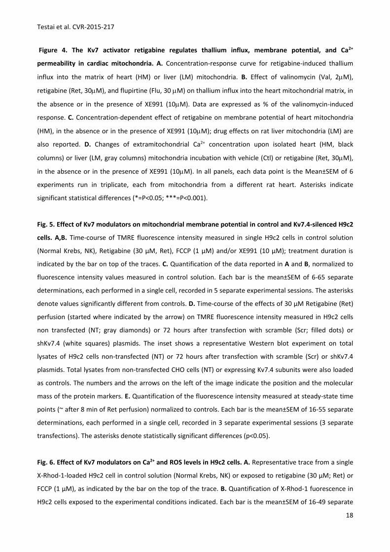
3.3 Effects of Kv7 activators on Tl+ fluxes, membrane potential and Ca2+ uptake in isolated rat heart
mitochondria
Isolated rat heart mitochondria exposed to the K+ ionophore valinomycin (2 M) showed a marked
increase in their trans-membrane permeability to Tl+. Increasing concentrations of the Kv7.2-7.5-activators
retigabine (1-30 M) and flupirtine (30 M) also promoted Tl+ influx; the pEC50 for retigabine was 5.59±0.05
(Fig. 4A). The initial rate of fluorescence increase induced by valinomycin was 336±70 F/sec; those for 1, 3,
10 and 30µM retigabine were 19±37, 134±33, 151±43, and 211±41 F/sec, respectively. In heart
mitochondria, the effects of retigabine and flupirtine were abrogated by the selective Kv7-blocker XE991
(10 M), which had no effect on valinomycin-induced Tl+ influx (Fig. 5B). Instead, in mitochondria isolated
from rat adult hepatic tissue (where Kv7.4 mRNA levels are virtually undetectable; Fig. 1A), retigabine was
ineffective in triggering Tl+ influx, whereas valinomycin was still effective (Fig. 4A).
In cardiac, but not in liver, mitochondria retigabine also produced an XE991 (10 M)-sensitive
concentration-dependent depolarization of the mitochondrial membrane, with a pEC50 of 5.19±0.08 (Fig.
4C).
Incubation of isolated heart and liver mitochondria in a Ca2+-rich solution (100 M) caused a rapid
and almost complete uptake of the cation into the mitochondrial matrix, reducing its extracellular free
concentration. In heart mitochondria, this effect was significantly reduced by retigabine (30 M); XE991 (10
M) did not influence resting mitochondrial Ca2+ uptake, but completely antagonized the effects of
retigabine. In contrast, retigabine did not influence Ca2+ uptake in liver mitochondria (Fig. 4D).
3.4 Effects of retigabine on mitochondrial membrane potential, Ca2+ levels, and ROS production in H9c2
cardiomyoblasts
To confirm that Kv7.4 activation could influence mitochondrial function in intact cardiac cells,
mitochondrial membrane potential, Ca2+ levels, and radical oxygen species (ROS) production were
measured in intact H9c2 rat cardiomyoblasts. Exposure of H9c2 cells to 30 M retigabine irreversibly
decreased TMRE fluorescence intensity, indicative of mitochondrial depolarization (Figs. 5A and 5C), with
an efficacy about 20% of that of the mitochondrial uncoupler FCCP (1 M). Retigabine IC50 was 13.1±1.1 µM
(n=6-11). XE991 (10 µM) did not modify TMRE fluorescence, but largely abolished RET-induced inhibition of
TMRE fluorescence (Figs. 5B and 5C).
To assess the specific contribution of mitoKv7.4 channels in retigabine-induced effects on
mitochondrial membrane potential, H9c2 cells were transfected with a short hairpin RNA targeted against
Kv7.4 mRNA (sh-Kv7.4).15 Western blots experiments confirmed that Kv7.4 expression was reduced to
51.0+6.0% (n=5) in total lysated from shKv7.4-transfected cells (inset in panel 5D); a smaller reduction of
the Kv7.4 signal was instead observed (74.3+3.3%; n=5) upon transfection with a control plasmid (scr). In

Testai et al. CVR-2015-217
11
shKv7.4-trasfected (but not in src-transfected) H9c2 cells, retigabine (30 M)-induced inhibition of TMRE
fluorescence intensity was decreased when compared to un-transfected H9c2 cells (Figs. 5D and 5E).
Exposure of H9c2 cells to 30 µM retigabine reversibly decreased X-RHOD-1 fluorescence intensity,
suggesting a decrease in mitochondrial Ca2+ levels (Figs. 6A and 6B); an opposite effect was instead
promoted by the mitochondrial uncoupler FCCP (1 M). XE991 (10 µM) prevented retigabine-induced
decrease in X-RHOD-1 fluorescence (Fig. 6B). Retigabine (30 µM) also increased mitochondrial ROS
production in H9c2 cells; XE991 (10 µM) largely prevented retigabine-induced ROS increase (Fig. 6C).
3.5 Effects of retigabine on H9c2 cardiomyoblasts survival following anoxia/reoxygenation (A/R)
H9c2 cardiomyoblasts exposed to 16 hours of anoxia, followed by 2 hours of reoxygenation,
exhibited a significant decrease in viability. Preincubation with retigabine (100M; applied one hour before
and throughout the anoxic period, but not in the reoxygenation phase), failed to modify cell viability in
normoxic conditions, but significantly increased H9c2 cells survival after the A/R injury. The Kv7 blocker
XE991 did not influence the cell viability in both normoxic and A/R conditions, but fully antagonized
retigabine-induced protection of cardiomyoblasts during A/R (Fig. 7A). To investigate whether retigabine-
induced ROS formation contributed to retigabine-induced cardioprotection, the effect of vitamin E (vitE)
was also evaluated. VitE (50 M) failed to affect cell vitality in normoxic conditions, fully prevented A/R-
induced H9c2 cell death, but did not prevent retigabine (100 M)-induced cytoprotection when incubated 1
hour before and together with the Kv7.4 activator (Supplementary Fig. 3).
3.6 Cardioprotective effects exerted by Kv7 channel activation in Langendorff-perfused adult rat hearts
During reperfusion following 30 min of global ischemia, vehicle-treated hearts exhibited a reduction
of the inotropic functional parameters. In particular, the rate pressure product (RPP) and the maximal rate
of rise of the left ventricular pressure (dP/dt) always remained lower than the corresponding pre-ischemic
values (Figs. 7B and 7C, respectively). In vehicle treated hearts, the coronary flow (CF) recorded during the
reperfusion time following the ischemic episode was significantly reduced as compared to the pre-ischemic
phase (Fig. 7D). Retigabine exposure (100M, perfused during the pre-ischemic phase only), led to an
almost complete recovery of the RPP, dP/dt, and CF during reperfusion (Figs. 7B-D). By contrast, no
retigabine-induced cardioprotection was observed when the drug was administered upon reperfusion,
after the ischemia/reoxygenation (I/R) cycle (what parameter was measured? Functional or morphological?
data not shown? Could we add some data in the text?). Morphometric analysis revealed an almost 50%
decrease in tissue vitality in the left ventricles from I/R-treated hearts (expressed as Ai/Alv); treatment with
retigabine (100M) during the pre-ischemic phase led to a significant reduction of the tissue injury (Figs. 7E
and 7F). Noteworthy, retigabine (100M)-induced beneficial effects on functional (Figs. 7B-D) and
morphometric (Fig. 7F) parameters was largely abolished by XE991 (10M) pretreatment.

Testai et al. CVR-2015-217
12
4. DISCUSSION
The fine tuning of the mitochondrial membrane potential is a critical factor in controlling cell fate
during physiological or pathological states, such as myocardial I/R injury, and the pharmacological
modulation of mitochondrial ion channels appears as an innovative cardioprotective strategy. In this study,
we provide the first evidence that K+ channels of the Kv7.4 subclass localize to mitochondria in cardiac
myocytes, and that their pharmacological activation depolarizes the mitochondrial membrane potential,
reduces mitochondrial Ca2+ uptake, and attenuates damage following I/R.
Quantitative PCR experiments revealed that the rat heart expressed Kv7.1 transcripts at high levels,
a result consistent with the well described contribution of Kv7.1 subunits to IKs, the late repolarizing current
of the cardiac action potential.11 In addition, as previously suggested in mouse12 and zebrafish,21 moderate
levels of Kv7.4 transcripts were also observed in the heart, whereas expression levels of Kv7.2, Kv7.3, and
Kv7.5 genes were negligible, suggesting that, in addition to their roles in vascular and non-vascular smooth
muscles10 and in the auditory system,22 Kv7.4 channels may also play a critical role in cardiac physiology.
Western blot experiments confirmed the abundant expression of Kv7.4 subunits in rat cardiac
tissue, and revealed that rat heart subcellular fractions highly enriched in mitochondria were intensively
positive for Kv7.4 subunits, suggesting their preferential location in mitochondria; experiments in
mitoplasts confirmed Kv7.4 subunit expression on the IMM. Rat heart samples used in these experiments
likely contain a substantial proportion of vascular tissue, where Kv7.4 channels are known to be
expressed;23,234 therefore, similar experiments were also carried out in H9c2 rat cardiomyoblasts,25 and in
freshly-isolated adult cardiomyocytes. In both cell types, biochemical experiments revealed Kv7.4 subunits
mainly in VDAC- or COX-IV-positive mitochondrial fractions.
Immunofluorescence analysis in H9c2 cells confirmed that Kv7.4 expression pattern overlapped
that of the mitochondrial marker Mitotracker, and was clearly distinct from a plasma membrane GPCR such
as type-1 Sphingosine-1-phosphate receptor (S1PR1).26 In freshly-isolated cardiomyocytes, Kv7.4 antibodies
also labelled Mitotracker-positive longitudinal structures corresponding to mitochondria running in parallel
to their major axis. Consistent with previous work,27 Kv7.1 antibody staining was predominantly
transversely-oriented, with striations resembling those of the transverse component of the T-tubular
system of adult ventricular myocytes.28 Similarly, immunohistochemical experiments in adult cardiac slices
revealed that Kv7.4 displayed a longitudinally-oriented punctuate staining pattern likely corresponding to
single, dot-like mitochondria,19 similar to that of the mitochondrial marker COX-IV. In the same preparation,
electron microscopy confirmed labeling of Kv7.4 subunits in about 40% of the mitochondria, with a
preferential location on internal (cristae) or peripheral membranes.
To investigate the functional significance of cardiac mitoKv7.4 channels, Kv7 activators (retigabine
and flupirtine) and blockers (XE991) were used. Retigabine and flupirtine act on channels formed by all Kv7
subunits, except Kv7.1.29,30 In rat heart mitochondria, both retigabine and flupirtine increased Tl+ influx,

Testai et al. CVR-2015-217
13
with potency values consistent with their ability to enhance Kv7.4 currents in electrophysiological
experiments.31 The effects of Kv7 activators on Tl+ fluxes across heart mitochondrial membranes closely
resemble those of the mitoKATP-opener diazoxide,14,32 and of naringenin, a mitoBKCa-opener.16 Noteworthy,
XE991 antagonized Tl+ influx triggered by retigabine and flupirtine, but not by valinomycin, confirming a
specific involvement of Kv7 channels. Retigabine also evoked concentration-dependent and XE991-sensitive
mitochondrial depolarization, a result consistent with the recognized effect of an increased IMM K+
permeability on mitochondrial membrane potential.4 The extent of retigabine-induced mitochondrial
depolarization is similar to that shown by activators of KATP channels, such as diazoxide, pinacidil33 and
benzopyrane-derived selective mitoKATP-openers, as well as by BKCa-openers.13 Retigabine was also effective
in depolarizing the mitochondrial membrane potential in intact H9c2 cells; this effect was blocked by XE991
as well as by reducing Kv7.4 expression with shRNAs, providing genetic evidence for a specific role for Kv7.4
channels in the pharmacological effects herein described.
Mitochondria avidly accumulate Ca2+ ions into the matrix, thus buffering excessive increases in free
cytosolic Ca2+.34 Both in isolated mitochondria and intact H9c2 cells, retigabine decreased mitochondrial
Ca2+ uptake in an XE991-sensitive manner, suggesting that even relatively small positive shifts of the
mitochondrial potential substantially reduce Ca2+ uptake.13 In mitochondria isolated from hepatic tissue,
where Kv7.4 mRNA levels were almost undetectable, retigabine failed to affect Tl+ fluxes, mitochondrial
membrane potential, and Ca2+ uptake, suggesting that retigabine-evoked effects in cardiac mitochondria
are selectively mediated by Kv7.4 channels.
Activation of mitochondrial K+ channels such as mitoKATP, mitoSKCa and mitoBKCa promotes
protective effects against cardiac ischemic injury.1 In cultured H9c2 cells exposed to A/R, retigabine
attenuated cell injury, and XE-991 antagonized the protective effects of the Kv7 activator. Noteworthy,
neither retigabine nor XE991 influenced the viability of H9c2 cells exposed to normoxic environment, thus,
suggesting specific anti-ischemic mechanisms of protection involving the activation of Kv7 channels. In
H9c2 cells, retigabine increased ROS formation, a result lending support to the hypothesis that an initial K+
entry via mitoKv7.4, by promoting a mild oxidative stress, would prevent opening of the mitochondrial
permeability transition pore and decrease anoxic cell death.35 However, vitamin E did not prevent
retigabine-induced H9c2 cardioprotective effects; although this result seems to suggest that retigabine-
induced cytoprotection is not directly caused by an increased ROS production, vitamin E likely targets a
myriad of molecular steps triggered by anoxia-reoxygenation, hampering a potential inhibition of
retigabine-induced cytoprotection.
In Langendorff-perfused rat hearts submitted to I/R, retigabine added during the pre-ischemic
phase improved all the functional and morphological parameters of post-ischemic recovery; also these
effects were fully abolished by XE991. However, these results need to be interpreted with caution, since
sarcolemmal Kv7.4 channels identified in the vascular smooth muscle of rat coronary arteries mediate

Testai et al. CVR-2015-217
14
significant vasorelaxing actions36 which may participate in the observed cardioprotective effects. However,
retigabine-induced functional and structural protection was assessed after 2 hours of drug-free post-
ischemic recovery, suggesting a major contribution of retigabine-sensitive cardiac mitoKv7.4 channels in
cardioprotection against I/R. This view seems to be confirmed by the observation that retigabine was
ineffective when administered only upon reperfusion, after the I/R cycle; however, further experiments are
needed to dissect the relative contribution of Kv7.4 channels in cardiomyocytes and/or vascular smooth
muscle cells in cardioprotection triggered by the Kv7.4 activator. The fact that retigabine-induced beneficial
effects required drug concentrations higher than those affecting mitochondrial function may reflect a
limited drug delivery to the mitochondrial target across the plasma membrane.
Overall, the results obtained demonstrate that rat cardiomyocytes express mitochondrial Kv7.4
channels which, by regulating membrane potential, influence mitochondrial Ca2+ permeability. The
pharmacological activation of myocardial mitoKv7.4 channel promotes structural and functional recovery
following I/R injury, highlighting new and appealing therapeutic strategies for cardioprotection.
FUNDING
Supported by grants from the Telethon Foundation (grant GGP15113) to MT; and the “Regional Health
Research Program 2009” of Regione Toscana, Italy to VC. IAG receives funding from the MRC (UK) and
British Heart Foundation. FM and MVS postdoctoral fellows from the Fondazione Umberto Veronesi and
the Italian Society for Pharmacology, respectively.
ACKNOWLEDGEMENTS
We are indebted to Dr. Thomas J. Jentsch, Department of Physiology and Pathology of Ion Transport,
Leibniz-Institut für Molekulare Pharmakologie (FMP), Berlin (Germany) for sharing Kv7.4 cDNA.

Testai et al. CVR-2015-217
15
REFERENCES
1. Testai L, Rapposelli S, Martelli A, Breschi MC, Calderone V. Mitochondrial Potassium Channels as Pharmacological Target for Cardioprotective Drugs. Med Res Rev. 2015; 35:520-53..
2. Inoue I, Nagase H, Kishi K, Higuti T. ATP-sensitive K+ channel in the mitochondrial inner membrane. Nature. 1991; 352:244-247.
3. Xu WH, Liu YG, Wang S, McDonald T, VanEyk JE, Sidor A, O’Rourke B. Cytoprotective role of Ca++-activated K+ channels in cardiac inner mitochondrial membrane. Science. 2002;298:1029–1033.
4. Szabò I, Leanza L, Gulbins E, Zoratti M. Physiology of potassium channels in the inner membrane of mitochondria. Pflugers Arch. 2012; 463:231-246.
5. Garlid KD, Dos Santos P, Xie ZJ, Costa AD, Paucek P. Mitochondrial potassium transport: the role of the mitochondrial ATP-sensitive K(+) channel in cardiac function and cardioprotection. Biochim Biophys Acta. 2003; 1606:1-21.
6. Murata M, Akao M, O'Rourke B, Marbán E. Mitochondrial ATP-sensitive potassium channels attenuate matrix Ca(2+) overload during simulated ischemia and reperfusion: possible mechanism of cardioprotection. Circ Res. 2001; 89:891-898.
7. Bentzen BH, Osadchii O, Jespersen T, Hansen RS, Olesen SP, Grunnet M. Activation of big conductance Ca(2+)-activated K (+) channels (BK) protects the heart against ischemia-reperfusion injury. Pflugers Arch. 2009; 457:979-988.
8. Szabò I, Zoratti M. Mitochondrial channels: ion fluxes and more. Physiol Rev. 2014; 94:519-608. 9. Soldovieri MV, Miceli F, Taglialatela M. Driving with no brakes: molecular pathophysiology of Kv7
potassium channels. Physiology (Bethesda). 2011; 26:365-376. 10. Stott JB, Jepps TA, Greenwood IA. K(V)7 potassium channels: a new therapeutic target in smooth
muscle disorders. Drug Discov Today. 2014; 19:413-424. 11. Sanguinetti MC, Curran ME, Zou A, Shen J, Spector PS, Atkinson DL, Keating MT. Coassembly of
K(V)LQT1 and minK (IsK) proteins to form cardiac I(Ks) potassium channel. Nature. 1996; 384:80-83. 12. Beisel KW, Rocha-Sanchez SM, Morris KA, Nie L, Feng F, Kachar B, Yamoah EN, Fritzsch B.
Differential expression of KCNQ4 in inner hair cells and sensory neurons is the basis of progressive high-frequency hearing loss. J Neurosci. 2005; 25:9285-92893.
13. Calderone V, Testai L, Martelli A, Rapposelli S, Digiacomo M, Balsamo A, Breschi MC. Anti-ischemic properties of a new spiro-cyclic benzopyran activator of the cardiac mito-KATP channel. Biochem Pharmacol. 2010; 79:39-47.
14. Wojtovich AP, Williams DM, Karcz MK, Lopes CM, Gray DA, Nehrke KW, Brookes PS. A novel mitochondrial K(ATP) channel assay. Circ Res. 2010; 106:1190-1196.
15. Iannotti FA, Barrese V, Formisano L, Miceli F, Taglialatela M. Specification of skeletal muscle differentiation by repressor element-1 silencing transcription factor (REST)-regulated Kv7.4 potassium channels. Mol Biol Cell. 2013; 24:274-284.
16. Testai L, Martelli A, Marino A, D'Antongiovanni V, Ciregia F, Giusti L, Lucacchini A, Chericoni S, Breschi MC, Calderone V. The activation of mitochondrial BK potassium channels contributes to the protective effects of naringenin against myocardial ischemia/reperfusion injury. Biochem Pharmacol. 2013; 85:1634-1643.
17. Zhang J, Honbo N, Goetzl EJ, Chatterjee K, Karliner JS, Gray MO. Signals from type 1 sphingosine 1-phosphate receptors enhance adult mouse cardiac myocyte survival during hypoxia. Am J Physiol Heart Circ Physiol. 2007; 293:H3150–H3158.
18. Mitcheson JS, Hancox JC, Levi AJ. Cultured adult cardiac myocytes: future applications, culture methods, morphological and electrophysiological properties. Cardiovasc Res. 1998; 39:280-300.
19. Driesen RB, Verheyen FK, Schaart G, de Mazière A, Viebahn C, Prinzen FW, Lenders MH, Debie W, Totzeck A, Borgers M, Ramaekers FC. Cardiotin localization in mitochondria of cardiomyocytes in vivo and in vitro and its down-regulation during dedifferentiation. Cardiovasc Pathol. 2009; 18:19-27.

Testai et al. CVR-2015-217
16
20. Dzeja PP, Bortolon R, Perez-Terzic C, Holmuhamedov EL, Terzic A. Energetic communication between mitochondria and nucleus directed by catalyzed phosphotransfer. Proc Natl Acad Sci U S A. 2002; 99:10156-10161.
21. Wu C, Sharma K, Laster K, Hersi M, Torres C, Lukas TJ, Moore EJ. Kcnq1-5 (Kv7.1-5) potassium channel expression in the adult zebrafish. BMC Physiol. 2014; 14:1.
22. Kubisch C, Schroeder BC, Friedrich T, Lütjohann B, El-Amraoui A, Marlin S, Petit C, Jentsch TJ. KCNQ4, a novel potassium channel expressed in sensory outer hair cells, is mutated in dominant deafness. Cell. 1999; 96:437-446.
23. Mackie AR, Byron KL. Cardiovascular KCNQ (Kv7) potassium channels: physiological regulators and new targets for therapeutic intervention. Mol Pharmacol. 2008 ; 74:1171-1179.
24. Ng FL, Davis AJ, Jepps TA, Harhun MI, Yeung SY, Wan A, Reddy M, Melville D, Nardi A, Khong TK, Greenwood IA. Expression and function of the K+ channel KCNQ genes in human arteries. Br J Pharmacol. 2011; 162:42-53.
25. Hescheler J, Meyer R, Plant S, Krautwurst D, Rosenthal W, Schultz G. Morphological, biochemical, and electrophysiological characterization of a clonal cell (H9c2) line from rat heart. Circ Res. 1991; 69:1476-1486.
26. Means CK, Brown JH. Sphingosine-1-phosphate receptor signalling in the heart. Cardiovasc Res. 2009; 82:193-200.
27. Rasmussen HB, Møller M, Knaus HG, Jensen BS, Olesen SP, Jørgensen NK. Subcellular localization of the delayed rectifier K(+) channels KCNQ1 and ERG1 in the rat heart. Am J Physiol Heart Circ Physiol. 2004; 286:H1300-9.
28. Brette F, Orchard C. T-Tubule Function in Mammalian Cardiac Myocytes. Circ Res. 2003; 92:1182-1192
29. Yeung S, Schwake M, Pucovský V, Greenwood I. Bimodal effects of the Kv7 channel activator retigabine on vascular K+ currents. Br J Pharmacol. 2008; 155:62-72.
30. Miceli F, Soldovieri MV, Martire M, Taglialatela M. Molecular pharmacology and therapeutic potential of neuronal Kv7-modulating drugs. Curr Opin Pharmacol. 2008;8):65-74.
31. Søgaard R, Ljungstrøm T, Pedersen K A, Olesen S P, Jensen B S. KCNQ4 channels expressed in mammalian cells: functional characteristics and pharmacology. Am J Physiol Cell Physiol. 2001; 280;C859-C866.
32. Foster DB, Ho AS, Rucker J, Garlid AO, Chen L, Sidor A, Garlid KD, O'Rourke B. Mitochondrial ROMK channel is a molecular component of mitoK(ATP). Circ Res. 2012; 111:446-454.
33. Holmuhamedov EL, Wang L, Terzic A. ATP-sensitive potassium channel openers prevent calcium overload in rat cardiac mitochondria. J Physiol. 1999; 519:347-360.
34. Dedkova EN, Blatter LA. Mitochondrial Ca2+ and the heart. Cell Calcium. 2008; 44:77-91. 35. Costa AD, Jakob R, Costa CL, Andrukhiv K, West IC, Garlid KD. The mechanism by which the
mitochondrial ATP-sensitive K+ channel opening and H2O2 inhibit the mitochondrial permeability transition. J Biol Chem. 2006;281:20801-20808.
36. Khanamiri S, Soltysinska E, Jepps TA, Bentzen BH, Chadha PS, Schmitt N, Greenwood IA, Olesen SP. Contribution of Kv7 channels to basal coronary flow and active response to ischemia. Hypertension. 2013; 62:1090-1097.

Testai et al. CVR-2015-217
17
FIGURE LEGENDS
Fig 1. Kv7.4 expression in rat heart. A. Quantitative PCR showing Kv7 mRNA levels in rat brain, heart and
liver. Cycle threshold (Ct) are normalized to the housekeeping gene GAPDH, using the 2-ΔCt formula. Data
are from 3 separate experiments. B. Western blot for Kv7.4, α-tubulin, GAPDH and VDAC in rat heart
mitochondria (mito) and whole homogenate (total). C. Western blot for Kv7.4 in rat cardiac mitochondria
before (Mito) and after their exposure to digitonin (1X for 15 min, 2X for 15 min, 1X for 45 min). Successful
mitoplasts isolation was indicated by the preservation of the IMM marker COX IV and the disappearance of
the OMM marker VDAC. D. Western blot for Kv7.4, α-tubulin, and COX IV in H9c2 cells mitochondrial
(Mito), cytosolic (Cyto) and microsomal (Micr) fractions; molecular weights are on the left. E. Western blot
for Kv7.4, Kv7.1, GADPH, PDI, and COX IV in mitochondrial (Mito), cytosolic (Cyto) and microsomal (Micr)
fractions from freshly-isolated adult rat cardiomyocytes. In all panels, data are representative of 4
experiments and markers molecular weights are shown on the left.
Fig 2. Subcellular localization of Kv7.4 in rat H9c2 cardiomyoblasts and in primary rat cardiomyocytes. A.
Immunofluorescence in H9c2 cells. Mitochondria labelled with Mitotracker are in red in panels b and e;
Kv7.4 antibody labelling is in green (panels a, d, and g); labelling with S1PR1 antibodies is in green (panel h);
nuclear Hoechst staining is in blue. Arrows: mitochondria; arrow-heads: plasma membrane. Merged images
are in panels c, f, and i. Panel d-e are enlargements of the region boxed in red in panel c. Scale bar, 5μm. B.
Panels a-l: immunofluorescence in acutely-isolated adult rat cardiomyocytes. Mitochondria labelled with
Mitotracker (red in panels a, d, g, j), were then stained for Kv7.4 (green, panels b and e) or Kv7.1 (green,
panels h and k); nuclei (blue) were counterstained with Hoechst. Merged images are in panels c, f, i, and I.
The second and the fourth row set of panels are higher magnifications of images shown in the first and
third row, respectively. Scale bar is 10μm (panels c, i) or 5μm (panels f, l). Arrows: mitochondria. Panels m-
o: immunofluorescence in rat cardiac slices incubated with anti COX-IV (red) and Kv7.4- antibody (green);
nuclei were counterstained with Hoechst (blue). Scale bar: 10μm. Experiments was repeated 3 times, with
similar results.
Figure 3. Electron immunogold detection of Kv7.4 subunits in mouse cardiomyocytes mitochondria. Four
representative images (A-D), each from different sections, are shown. Scale bar: 500 nm. Black arrows
indicate mitochondria. The lower panels show the quantification of gold particles distribution in 408
mitochondria (138 images analyzed; sections from 3 separate animals). The left panel shows the
percentage of gold-labelled ad un-labelled mitochondria; for gold-labelled mitochondria, the right panel
reports the particles distribution in the membranes (periphery or cristae), intramitochondrially (inside), or
at both locations. Data are expressed as Mean±S.E.M.
Testai et al. CVR-2015-217
18
Figure 4. The Kv7 activator retigabine regulates thallium influx, membrane potential, and Ca2+
permeability in cardiac mitochondria. A. Concentration-response curve for retigabine-induced thallium
influx into the matrix of heart (HM) or liver (LM) mitochondria. B. Effect of valinomycin (Val, 2M),
retigabine (Ret, 30M), and flupirtine (Flu, 30 M) on thallium influx into the heart mitochondrial matrix, in
the absence or in the presence of XE991 (10M). Data are expressed as % of the valinomycin-induced
response. C. Concentration-dependent effect of retigabine on membrane potential of heart mitochondria
(HM), in the absence or in the presence of XE991 (10M); drug effects on rat liver mitochondria (LM) are
also reported. D. Changes of extramitochondrial Ca2+ concentration upon isolated heart (HM, black
columns) or liver (LM, gray columns) mitochondria incubation with vehicle (Ctl) or retigabine (Ret, 30M),
in the absence or in the presence of XE991 (10M). In all panels, each data point is the Mean±SEM of 6
experiments run in triplicate, each from mitochondria from a different rat heart. Asterisks indicate
significant statistical differences (*=P<0.05; ***=P<0.001).
Fig. 5. Effect of Kv7 modulators on mitochondrial membrane potential in control and Kv7.4-silenced H9c2
cells. A,B. Time-course of TMRE fluorescence intensity measured in single H9c2 cells in control solution
(Normal Krebs, NK), Retigabine (30 µM, Ret), FCCP (1 µM) and/or XE991 (10 µM); treatment duration is
indicated by the bar on top of the traces. C. Quantification of the data reported in A and B, normalized to
fluorescence intensity values measured in control solution. Each bar is the mean±SEM of 6-65 separate
determinations, each performed in a single cell, recorded in 5 separate experimental sessions. The asterisks
denote values significantly different from controls. D. Time-course of the effects of 30 µM Retigabine (Ret)
perfusion (started where indicated by the arrow) on TMRE fluorescence intensity measured in H9c2 cells
non transfected (NT; gray diamonds) or 72 hours after transfection with scramble (Scr; filled dots) or
shKv7.4 (white squares) plasmids. The inset shows a representative Western blot experiment on total
lysates of H9c2 cells non-transfected (NT) or 72 hours after transfection with scramble (Scr) or shKv7.4
plasmids. Total lysates from non-transfected CHO cells (NT) or expressing Kv7.4 subunits were also loaded
as controls. The numbers and the arrows on the left of the image indicate the position and the molecular
mass of the protein markers. E. Quantification of the fluorescence intensity measured at steady-state time
points (~ after 8 min of Ret perfusion) normalized to controls. Each bar is the mean±SEM of 16-55 separate
determinations, each performed in a single cell, recorded in 3 separate experimental sessions (3 separate
transfections). The asterisks denote statistically significant differences (p<0.05).
Fig. 6. Effect of Kv7 modulators on Ca2+ and ROS levels in H9c2 cells. A. Representative trace from a single
X-Rhod-1-loaded H9c2 cell in control solution (Normal Krebs, NK) or exposed to retigabine (30 µM; Ret) or
FCCP (1 µM), as indicated by the bar on the top of the trace. B. Quantification of X-Rhod-1 fuorescence in
H9c2 cells exposed to the experimental conditions indicated. Each bar is the mean±SEM of 16-49 separate

Testai et al. CVR-2015-217
19
determinations, each performed in a single cell, recorded in 3 separate experimental sessions. The asterisks
indicate values significantly different from controls (p<0.05). C. Effect of Retigabine (30 µM; Ret) on
MitoSOX fluorescence intensity in H9c2 cells. Each bar is the mean±SEM of 36-59 separate determinations,
each performed in a single cell, recorded in 3 separate experimental sessions. The asterisks indicate
statistically significant differences (p<0.05).
Figure 7. Cardioprotection by Kv7 modulators in in vitro anoxia and ex-vivo ischemia models. A. Viability
of H9c2 cardiomyoblasts exposed to normoxic conditions (black columns) or to anoxia/reoxygenation
(white columns) treated with: vehicle (Ctl), retigabine (Ret, 100M), XE991 (XE991, 10M) and retigabine
100M plus XE991 10M (Ret+XE991), expressed as % of vehicle-treated cells exposed to normoxic
conditions. Asterisks indicate significant statistical differences, analysed by one-way ANOVA (*=P<0.05;
***=P<0.001). Data are from 6 experiments each performed in triplicate. Time-course of RPP (Rate x
Pressure Product; B), dP/dt (C), and coronary flow (D) in Langendorff-perfused hearts treated with vehicle,
retigabine (100 M) or retigabine (100 M) plus XE991 (10 M). Data are expressed as % of the respective
values recorded in the pre-ischemic phase. In B, C, and D, two-way ANOVA analysis indicated that the
vehicle and retigabine curves exhibit highly significant (***=P<0.001) differences. E. Representative images
of left ventricle slices from vehicle- or retigabine-treated (100 M) hearts after ischemia/reperfusion. F.
Quantification of the extension of the ischemic damage (white/pale regions), expressed as % of the left
ventricle slice area (Ai/Alv) in the indicated groups. The asterisk indicates a statistically-significant
difference (*=P<0.05). Data are from 6-8 experiments each performed in different animals.
Related Documents