King’s Research Portal DOI: 10.1371/journal.pone.0152628 Document Version Publisher's PDF, also known as Version of record Link to publication record in King's Research Portal Citation for published version (APA): Taneike, M., Nishida, K., Omiya, S., Zarrinpashneh, E., Misaka, T., Kitazume-Taneike, R., ... Otsu, K. (2016). MTOR hyperactivation by ablation of tuberous sclerosis complex 2 in the mouse heart induces cardiac dysfunction with the increased number of small mitochondria mediated through the down-regulation of autophagy. PL o S One , 11(3), [e0152628]. 10.1371/journal.pone.0152628 Citing this paper Please note that where the full-text provided on King's Research Portal is the Author Accepted Manuscript or Post-Print version this may differ from the final Published version. If citing, it is advised that you check and use the publisher's definitive version for pagination, volume/issue, and date of publication details. And where the final published version is provided on the Research Portal, if citing you are again advised to check the publisher's website for any subsequent corrections. General rights Copyright and moral rights for the publications made accessible in the Research Portal are retained by the authors and/or other copyright owners and it is a condition of accessing publications that users recognize and abide by the legal requirements associated with these rights. •Users may download and print one copy of any publication from the Research Portal for the purpose of private study or research. •You may not further distribute the material or use it for any profit-making activity or commercial gain •You may freely distribute the URL identifying the publication in the Research Portal Take down policy If you believe that this document breaches copyright please contact [email protected] providing details, and we will remove access to the work immediately and investigate your claim. Download date: 18. Feb. 2017

King s Research Portal - core.ac.uk · If you believe that this document breaches copyright please contact [email protected] providing details, ... *[email protected] Abstract
May 25, 2019
Welcome message from author
This document is posted to help you gain knowledge. Please leave a comment to let me know what you think about it! Share it to your friends and learn new things together.
Transcript
King’s Research Portal
DOI:10.1371/journal.pone.0152628
Document VersionPublisher's PDF, also known as Version of record
Link to publication record in King's Research Portal
Citation for published version (APA):Taneike, M., Nishida, K., Omiya, S., Zarrinpashneh, E., Misaka, T., Kitazume-Taneike, R., ... Otsu, K. (2016).MTOR hyperactivation by ablation of tuberous sclerosis complex 2 in the mouse heart induces cardiacdysfunction with the increased number of small mitochondria mediated through the down-regulation ofautophagy. PL o S One , 11(3), [e0152628]. 10.1371/journal.pone.0152628
Citing this paperPlease note that where the full-text provided on King's Research Portal is the Author Accepted Manuscript or Post-Print version this maydiffer from the final Published version. If citing, it is advised that you check and use the publisher's definitive version for pagination,volume/issue, and date of publication details. And where the final published version is provided on the Research Portal, if citing you areagain advised to check the publisher's website for any subsequent corrections.
General rightsCopyright and moral rights for the publications made accessible in the Research Portal are retained by the authors and/or other copyrightowners and it is a condition of accessing publications that users recognize and abide by the legal requirements associated with these rights.
•Users may download and print one copy of any publication from the Research Portal for the purpose of private study or research.•You may not further distribute the material or use it for any profit-making activity or commercial gain•You may freely distribute the URL identifying the publication in the Research Portal
Take down policyIf you believe that this document breaches copyright please contact [email protected] providing details, and we will remove access tothe work immediately and investigate your claim.
Download date: 18. Feb. 2017
RESEARCH ARTICLE
mTOR Hyperactivation by Ablation ofTuberous Sclerosis Complex 2 in the MouseHeart Induces Cardiac Dysfunction with theIncreased Number of Small MitochondriaMediated through the Down-Regulation ofAutophagyManabu Taneike1, Kazuhiko Nishida1, Shigemiki Omiya1, Elham Zarrinpashneh1,Tomofumi Misaka1, Rika Kitazume-Taneike1, Ruth Austin1, Minoru Takaoka1,Osamu Yamaguchi2, Michael J. Gambello3, Ajay M. Shah1, Kinya Otsu1*
1 Cardiovascular Division, King’s College London British Heart Foundation Centre of Excellence, London,United Kingdom, 2 Department of Cardiovascular Medicine, Graduate School of Medicine, Osaka University,Suita, Osaka, Japan, 3 Division of Medical Genetics, Emory University School of Medicine, Atlanta, Georgia,United States of America
AbstractMammalian target of rapamycin complex 1 (mTORC1) is a key regulator of cell growth, prolifer-
ation andmetabolism. mTORC1 regulates protein synthesis positively and autophagy nega-
tively. Autophagy is a major system to manage bulk degradation and recycling of cytoplasmic
components and organelles. Tuberous sclerosis complex (TSC) 1 and 2 form a heterodimeric
complex and inactivate Ras homolog enriched in brain, resulting in inhibition of mTORC1. Here,
we investigated the effects of hyperactivation of mTORC1 on cardiac function and structure
using cardiac-specific TSC2-deficient (TSC2-/-) mice. TSC2-/- mice were born normally at the
expectedMendelian ratio. However, the median life span of TSC2-/- mice was approximately 10
months and significantly shorter than that of control mice. TSC2-/- mice showed cardiac dysfunc-
tion and cardiomyocyte hypertrophy without considerable fibrosis, cell infiltration or apoptotic
cardiomyocyte death. Ultrastructural analysis of TSC2-/- hearts revealed misalignment, aggre-
gation and a decrease in the size and an increase in the number of mitochondria, but the mito-
chondrial function wasmaintained. Autophagic flux was inhibited, while the phosphorylation
level of S6 or eukaryotic initiation factor 4E -binding protein 1, downstream of mTORC1, was
increased. The upregulation of autophagic flux by trehalose treatment attenuated the cardiac
phenotypes such as cardiac dysfunction and structural abnormalities of mitochondria in TSC2-/-
hearts. The results suggest that autophagy via the TSC2-mTORC1 signaling pathway plays an
important role in maintenance of cardiac function andmitochondrial quantity and size in the
heart and could be a therapeutic target to maintain mitochondrial homeostasis in failing hearts.
PLOS ONE | DOI:10.1371/journal.pone.0152628 March 29, 2016 1 / 18
OPEN ACCESS
Citation: Taneike M, Nishida K, Omiya S,Zarrinpashneh E, Misaka T, Kitazume-Taneike R, etal. (2016) mTOR Hyperactivation by Ablation ofTuberous Sclerosis Complex 2 in the Mouse HeartInduces Cardiac Dysfunction with the IncreasedNumber of Small Mitochondria Mediated through theDown-Regulation of Autophagy. PLoS ONE 11(3):e0152628. doi:10.1371/journal.pone.0152628
Editor: Martin Young, University of Alabama atBirmingham, UNITED STATES
Received: August 28, 2015
Accepted: March 10, 2016
Published: March 29, 2016
Copyright: © 2016 Taneike et al. This is an openaccess article distributed under the terms of theCreative Commons Attribution License, which permitsunrestricted use, distribution, and reproduction in anymedium, provided the original author and source arecredited.
Data Availability Statement: All relevant data arewithin the paper and its Supporting Information files.
Funding: This work was supported by MedicalResearch Council (MR/K019031/1), http://www.mrc.ac.uk/, KO; British Heart Foundation (CH/11/3/29051,RG/11/12/29052), https://www.bhf.org.uk/, KO. Thefunders had no role in study design, data collectionand analysis, decision to publish, or preparation ofthe manuscript.
IntroductionMammalian target of rapamycin complex 1 (mTORC1) plays a critical role in the regulation ofcell growth, proliferation and metabolism [1]. mTORC1 positively controls protein synthesisby phosphorylating downstream substrates, namely eukaryotic initiation factor 4E (eIF4E)-binding protein 1 (4E-BP1) and p70 ribosomal S6 kinase 1 (S6K1). The phosphorylation of4E-BP1 prevents its binding to eIF4E, enabling eIF4E to promote cap-dependent translation[2]. Active S6K1 phosphorylates several substrates, including S6, that function in translationinitiation as well as other steps that drive protein production. In addition to protein synthesis,mTORC1 positively controls mitochondrial function through a yin yang 1-peroxisome prolif-erator activated receptor coactivator 1alpha (PGC-1alpha) transcriptional complex [3].
On the other hand, mTORC1 also plays an important role in protein degradation. In mam-malian cells, there are two major systems of protein degradation, namely the ubiquitin/protea-some system and autophagy. mTORC1 inhibits autophagy [4] by phosphorylating unc-51 likeautophagy activating kinase 1 (ULK1), a mammalian ortholog of Atg1 [5]. In autophagy, anisolation membrane sequesters a part of the cytoplasm or organelles including mitochondria toform an autophagosome [6]. The autophagosome fuses with a lysosome and becomes an auto-lysosome. Then, the materials contained in the autolysosome are degraded by lysosomalenzymes. Autophagy contributes to macromolecule synthesis and energy production as well asquality control of cytoplasmic proteins and organelles. We have previously reported that con-stitutive autophagy in the heart is a homeostatic mechanism for maintaining cardiomyocytesize, global cardiac structure and function and the quality of mitochondria [7].
Mitochondria are important organelles for ATP production and dynamic organelles. Theycontinuously undergo fusion and fission during cell life, exhibiting short round-shaped orelongated morphology. Autophagy will be preceded by mitochondrial fission, which divideselongated mitochondria into pieces of manageable size for engulfment by isolation membrane.When autophagy is induced in the cells by nutrient depletion, mitochondria elongate [8]. Elon-gated mitochondria are spared from autophagy and maintain ATP production uponstarvation.
mTORC1 is positively regulated by Ras homolog enriched in brain (Rheb). Rheb is a smallGTPase, that exists either in an active GTP-bound or an inactive GDP-bound state [9]. In thesignaling pathway towards mTORC1, tuberous sclerosis complex (TSC) 1 and 2 exist upstreamof Rheb. Mutations in either the TSC1 or TSC2 gene are the principal cause of TSC, which is anautosomal dominant multisystemic disorder characterized by the development of numerousbenign tumours (e.g. hamartomas) in many organs, such as brain, kidneys, skin, heart andlungs [10]. There is no homology between the 140 kDa TSC1 and the 200 kDa TSC2 proteins.TSC1 and TSC2 associate with each other to form a heterodimeric complex as a regulatory unitand a catalytic unit, respectively. The TSC1/2 complex is a GTPase-activating protein thatcatalyses the conversion of Rheb-GTP to Rheb-GDP [11] and inactivates Rheb, resulting ininhibition of mTORC1 signaling [12, 13]. Akt-mediated phosphorylation of TSC2 inhibits thefunction of the TSC1/2 complex [14], resulting in activation of mTORC1. However, the exis-tence of an mTORC1-dependent feedback mechanism blocks growth-factor-stimulated phos-phorylation of Akt [15].
Studies using Drosophila revealed that loss-of-function mutations of TSC1, TSC2 and bothgenes combined lead to organ overgrowth via increased cell proliferation and cell-autonomousincrease in size [16, 17]. In mice, the deficiency of mTORC1 leads to embryonic lethality [18,19]. Cardiac-specific mTOR-deficient mice are also embryonic lethal [20]. Furthermore, abla-tion ofMtor in the adult mouse heart results in a fatal, dilated cardiomyopathy, which is mainly
Mitochondrial Quantity in TSC2-Deficient Hearts
PLOS ONE | DOI:10.1371/journal.pone.0152628 March 29, 2016 2 / 18
Competing Interests: The authors have declaredthat no competing interests exist.
caused by enhanced dephosphorylation of 4E-BP1 [21]. These data suggest that mTOR has acritical role in protein synthesis to maintain cardiac function.
To investigate the role of the mTORC1 signaling pathway in the heart, mouse models whereits activity is altered by modulation of upstream signaling molecules will be more physiologi-cally relevant thanMtor ablation model. We previously investigated the role of mTORC1 inthe heart using cardiac-specific Rheb-deficient mice, where mTORC1 was downregulated [22].We found that mTORC1 activity through Rheb is essential for normal cardiac hypertrophicgrowth during the postnatal period, although autophagy was not related to the phenotypesobserved in Rheb-deficient hearts. Since conventional TSC2-deficient mice are embryoniclethal [23, 24], we used cardiac-specific TSC2-deficient mice to examine the effect of mTORC1hyperactivation on protein degradation and mitochondrial dynamics as well as protein synthe-sis in cardiomyocytes in this study. The ablation of TSC2 in mouse hearts leads to Rheb activa-tion, mTORC1 activation, ULK1 phosphorylation and then finally inhibition of autophagy.Our results suggest that autophagy via the TSC-mTORC1 signaling pathway plays an impor-tant role in determining the size and number of mitochondria to maintain cardiac function.
Materials and Methods
AnimalsAll in vivo procedures in this study were carried out in accordance with the Guidance on theOperation of the Animals (Scientific Procedures) Act, 1986 (UK Home Office) or the Guide-lines for Animal Experiments of Osaka University and the Japanese Act on Welfare and Man-agement of Animals (No. 105). King’s College London Ethical Review Process Committee andUK Home Office (Project Licence No. PPL70/7260) or Osaka University Animal Experimenta-tion Committee approved this study. Mortality was described as a possibility in the approvedstudy protocols. Mice were given food and water ad libitum. Mice were monitored daily duringthe animal study including the survival study to minimize suffering. Animals that showed signsof significant clinical cardio-respiratory distress due to the development of heart failure (e.g.,increased respiratory rates, reduced activity, piloerection, hunched posture) were immediatelyeuthanized by CO2 exposure. In a small pilot study approved by Osaka University AnimalExperimentation Committee and performed under the Guidelines for Animal Experiments ofOsaka University and the Japanese Act onWelfare and Management of Animals (No. 105), 4mice showed unexpected death (S1 Protocol). Autopsy of all the dead mice showed pleuraleffusion and increased lung weight, suggesting the cause of death was heart failure. Thus,euthanized mice were considered as deaths. All the mice were harvested in the early morning.We didn’t detect any difference in food consumption and body weight gaining among groupsused in this study.
Generation of cardiac-specific TSC2-deficient mice andechocardiographyThe generation of mice bearing a TSC2flox allele, in which exon 2–4 of the TSC2 gene is flankedby two loxP sequences, has been previously reported [25]. Mice bearing the TSC2flox allele werecrossed with transgenic mice expressing Cre recombinase under the control of alpha-myosinheavy chain promoter (alpha-MyHC-Cre) [26]. The genetic backgrounds of the TSC2flox/flox
and alpha-MyHC-Cremice are 129/SvJ x C57B/6J and C57B/6J, respectively. All mice whichwere used in this study were male. Vevo 2100 system with a 22–55-MHz linear transducer(Visual Sonics) was used to perform echocardiography on awake or anaesthetized mice. Theechocardiography was performed on awake mice, those were trained prior to the actual
Mitochondrial Quantity in TSC2-Deficient Hearts
PLOS ONE | DOI:10.1371/journal.pone.0152628 March 29, 2016 3 / 18
measurement. Isoflurane was used for echocardiography on anesthetized mice. The sonogra-pher blinded to the groups. Trans-thoracic M-mode images were acquired in parasternal shortaxis view. Fractional shortening (FS) and end-diastolic left ventricular (LV) mass were calcu-lated as 100 x (end-diastolic LV internal dimension (LVIDd)—end-systolic LV internal dimen-sion (LVIDs))/LVIDd and 1.05 x [(LVIDd + end-diastolic interventricular septal thickness(IVSd) + LV posterior wall thickness (LVPWd))3 - (LVIDd)3], respectively.
AntibodiesSpecific antibodies targeted to the following proteins were used for the Western blot analysis in1,000 times dilution (except for the one to alpha-tubulin (3,000)) and according to manufactur-ers’ instructions: TSC1 (rabbit monoclonal, Cell Signaling Technology (CST), #6935), TSC2(rabbit monoclonal, CST, #4308), phosphorylated Akt (rabbit monoclonal, CST, #4058), Akt(rabbit polyclonal, CST, #9272), phosphorylated AMP-activated protein kinase (AMPK) (rab-bit monoclonal, CST, #2535), AMPK (rabbit polyclonal, CST, #2532), phosphorylated S6 (rab-bit, polyclonal, CST, #2215), S6 (rabbit monoclonal, CST, #2217), ubiquitin (rabbit polyclonal,CST, #3933), microtubule-associated protein 1 light chain 3 (LC3) B (rabbit polyclonal, CST,#2775), alpha-tubulin (mouse monoclonal, CST, #3873), voltage-dependent anion channel(VDAC) (rabbit polyclonal, CST, #4866), Parkin (rabbit polyclonal, CST, #2132), 4E-BP1 (rab-bit polyclonal, Abcam, ab2606), PINK1 (rabbit polyclonal, Abcam, ab23707), KDEL proteins(mouse monoclonal, Enzo Life Sciences, ADI-SPA-827), sequestosome 1/p62 (p62) (C-termi-nal specific) (guinea pig polyclonal, Progen, GP62-C), translocase of inner mitochondrialmembrane 23 homolog (Timm23) (rabbit polyclonal, Proteintech, 11123-1-AP), 4-hydroxy-2-nonenal michael adducts (HNE) (rabbit polyclonal, Calbiochem, 393207). Tissues were lysedin homogenization buffer (50 mM Tris-HCl, pH 7.4, 150 mMNaCl, 1 mM EDTA, 1 mMEGTA, 2.5 mM Na-orthovanadate, 2.5 mM Na-pyrophosphate, 1 mM β-glycerophosphate, 1%Triton X-100) with protease inhibitors (PMSF or protease inhibitor cocktail (Sigma)). Westernblots were incubated with the secondary antibodies, followed by developing with an infraredimaging system, ODYSSEY CLx (LI-COR). NIH Image J software (version 1.46r) or Image Stu-dio software (LI-COR) was used to perform densitometric analyses.
Histological analysisThe heart was excised and immediately fixed in buffered 4% paraformaldehyde in PBS, embed-ded in paraffin, and sectioned to a thickness of 6 micrometers. Haematoxylin and eosin (H&E)staining was performed on serial sections. For wheat germ agglutinin (WGA) staining, heartsamples were mounted and frozen in O.C.T. compound (Thermo Scientific), cryo-sectioned toa thickness of 6 micrometers and stained with FITC-conjugated lectin (Sigma) to measurecross sectional area of cardiomyocytes. To determine the number of cells undergoing nuclearfragmentation, TdT-mediated dUTP-biotin nick end labeling (TUNEL) assay was performedon paraffin-embedded heart sections, using In situ Apoptosis Detection Kit (Takara Bio Inc.).For electron microscopy, the heart was perfused and then fixed with 2% formaldehyde and 2%glutaraldehyde. LV tissues were processed for transmission electron microscopy. We measuredthe area of mitochondria or counted the number of mitochondria in micrographs taken at10,000-fold magnification, using NIH Image J software (version 1.46r) for 4 fields per mouse.
Quantitative real-time RT-PCRWe isolated total RNA from the LV using the TRIzol reagent (Life Technologies). We deter-mined mRNA expression levels for atrial natriuretic factor (Nppa), beta-myosin heavy chain(Myh7), collagen type 1 (Col1a2), Pgc-1a and Gapdh by quantitative RT-PCR. For reverse
Mitochondrial Quantity in TSC2-Deficient Hearts
PLOS ONE | DOI:10.1371/journal.pone.0152628 March 29, 2016 4 / 18
transcription, we used SuperScript II Reverse Transcriptase (Life Technologies). Real-timePCR reaction was performed using Power SYBR Green Master Mix (Life Technologies) andPCR primers designed as follows: forward 5’-TCGTCTTGGCCTTTTGGCT-3’ and reverse5’-TCCAGGTGGTCTAGCAGGTTCT-3’ for Nppa, forward 5’-ATGTGCCGGACCTTGGAAG-3’ and reverse 5’-CCTCGGGTTAGCTGAGAGATCA-3’ forMyh7, forward 5’-ACGCGGACTCTGTTGCTGCT-3’ and reverse 5’-GCGGGACCCCTTTGTCCACG-3’ for Col1a2, for-ward 5’-AATCAGACCTGACACAACGC-3’ and reverse 5’-GCATTCCTCAATTTCACCAA-3’ for Pgc-1a and forward 5’-ATGACAACTTTGTCAAGCTCATTT-3’ and reverse5’-GGTCCACCACCCTGTTGCT-3’ for Gapdh. We constructed real-time PCR standardcurves using the corresponding complementary DNA. All data were normalized to Gapdhcontent and are expressed as fold increase over the control group.
Mitochondrial enzyme activitiesThe activities of NADH cytochrome-c oxidoreductase (complex I + III) and succinate cyto-chrome-c oxidoreductase (complex II + III) were determined in mitochondrial fractions freshlyisolated from hearts using previously described spectrophotometric methods [27]. Results areshown as nmol/min/mg protein.
Tissue ATP concentration and the ratio of ADP to ATPThe heart was excised and immediately frozen in liquid nitrogen. The samples were groundusing a pestle and mortar chilled with liquid nitrogen. ATP concentration and the ratio ofADP to ATP were measured using EnzyLight ADP/ATP Ratio Assay Kit (BioAssay Systems).
Trehalose treatmentTo examine the role of autophagy in the development of heart failure in TSC2-/- mice, the micewere treated with 1% trehalose in drinking water for 10 weeks starting from 6 weeks after birth.The water solutions were changed twice weekly.
Statistical analysisResults are shown as the mean ± S.E.M. Paired data were evaluated using a Student’s t-test. TheKaplan-Meier method with Logrank test was used for survival analysis. Distribution of mito-chondrial size was analysed using Chi-square or Mann-Whitney U test. A value of P< 0.05was considered statistically significant.
Results
Generation and characterization of mice lacking TSC2 in the heartTo investigate the effect of the upregulated mTORC1 signaling pathway in the heart, we gener-ated cardiac-specific TSC2-deficient mice. TSC2flox/flox mice were crossed with mice expressingalpha-myosin heavy chain promoter driven Cre recombinase (alpha-MyHC-Cre) to obtainTSC2flox/flox;alpha-MyHC-Cre+ (TSC2-/-) mice. TSC2flox/flox;alpha-MyHC-Cre- (TSC2+/+) litter-mates were used as controls. The alpha-MyHC-Cre+ mice exhibited similar cardiac function totheir control non-transgenic mice up to 12 months of age [28] and had normal life span. West-ern blot analysis revealed a 95% reduction of TSC2 protein in cardiomyocytes isolated from8-week-old TSC2-/- hearts compared with those from TSC2+/+ hearts (Fig 1A). There was sig-nificant reduction in the expression level of TSC1 protein in TSC2-/- hearts. TSC1 and TSC2associate with each other to form a heterodimer [29, 30]. Thus, there is a possibility that thestability of TSC1 is reduced by absence of TSC2. TSC2-/- mice were born normally at the
Mitochondrial Quantity in TSC2-Deficient Hearts
PLOS ONE | DOI:10.1371/journal.pone.0152628 March 29, 2016 5 / 18
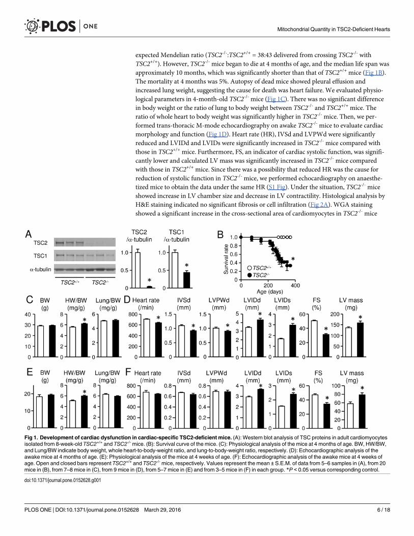
expected Mendelian ratio (TSC2-/-:TSC2+/+ = 38:43 delivered from crossing TSC2-/- withTSC2+/+). However, TSC2-/- mice began to die at 4 months of age, and the median life span wasapproximately 10 months, which was significantly shorter than that of TSC2+/+ mice (Fig 1B).The mortality at 4 months was 5%. Autopsy of dead mice showed pleural effusion andincreased lung weight, suggesting the cause for death was heart failure. We evaluated physio-logical parameters in 4-month-old TSC2-/- mice (Fig 1C). There was no significant differencein body weight or the ratio of lung to body weight between TSC2-/- and TSC2+/+ mice. Theratio of whole heart to body weight was significantly higher in TSC2-/- mice. Then, we per-formed trans-thoracic M-mode echocardiography on awake TSC2-/- mice to evaluate cardiacmorphology and function (Fig 1D). Heart rate (HR), IVSd and LVPWd were significantlyreduced and LVIDd and LVIDs were significantly increased in TSC2-/- mice compared withthose in TSC2+/+ mice. Furthermore, FS, an indicator of cardiac systolic function, was signifi-cantly lower and calculated LV mass was significantly increased in TSC2-/- mice comparedwith those in TSC2+/+ mice. Since there was a possibility that reduced HR was the cause forreduction of systolic function in TSC2-/- mice, we performed echocardiography on anaesthe-tized mice to obtain the data under the same HR (S1 Fig). Under the situation, TSC2-/- miceshowed increase in LV chamber size and decrease in LV contractility. Histological analysis byH&E staining indicated no significant fibrosis or cell infiltration (Fig 2A). WGA stainingshowed a significant increase in the cross-sectional area of cardiomyocytes in TSC2-/- mice
Fig 1. Development of cardiac dysfunction in cardiac-specific TSC2-deficient mice. (A): Western blot analysis of TSC proteins in adult cardiomyocytesisolated from 8-week-old TSC2+/+ and TSC2-/- mice. (B): Survival curve of the mice. (C): Physiological analysis of the mice at 4 months of age. BW, HW/BW,and Lung/BW indicate body weight, whole heart-to-body-weight ratio, and lung-to-body-weight ratio, respectively. (D): Echocardiographic analysis of theawake mice at 4 months of age. (E): Physiological analysis of the mice at 4 weeks of age. (F): Echocardiographic analysis of the awake mice at 4 weeks ofage. Open and closed bars represent TSC2+/+ and TSC2-/- mice, respectively. Values represent the mean ± S.E.M. of data from 5–6 samples in (A), from 20mice in (B), from 7–8 mice in (C), from 9 mice in (D), from 5–7 mice in (E) and from 3–5 mice in (F) in each group. *P < 0.05 versus corresponding control.
doi:10.1371/journal.pone.0152628.g001
Mitochondrial Quantity in TSC2-Deficient Hearts
PLOS ONE | DOI:10.1371/journal.pone.0152628 March 29, 2016 6 / 18
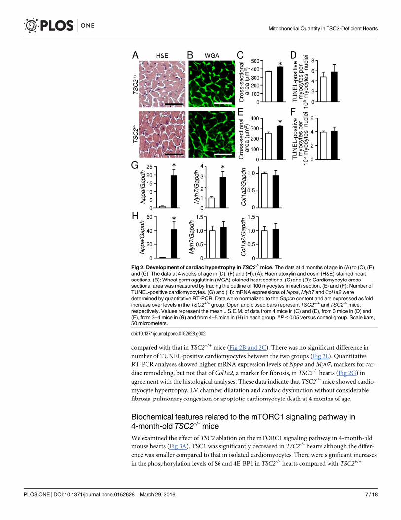
compared with that in TSC2+/+ mice (Fig 2B and 2C). There was no significant difference innumber of TUNEL-positive cardiomyocytes between the two groups (Fig 2E). QuantitativeRT-PCR analyses showed higher mRNA expression levels of Nppa andMyh7, markers for car-diac remodeling, but not that of Col1a2, a marker for fibrosis, in TSC2-/- hearts (Fig 2G) inagreement with the histological analyses. These data indicate that TSC2-/- mice showed cardio-myocyte hypertrophy, LV chamber dilatation and cardiac dysfunction without considerablefibrosis, pulmonary congestion or apoptotic cardiomyocyte death at 4 months of age.
Biochemical features related to the mTORC1 signaling pathway in4-month-old TSC2-/- miceWe examined the effect of TSC2 ablation on the mTORC1 signaling pathway in 4-month-oldmouse hearts (Fig 3A). TSC1 was significantly decreased in TSC2-/- hearts although the differ-ence was smaller compared to that in isolated cardiomyocytes. There were significant increasesin the phosphorylation levels of S6 and 4E-BP1 in TSC2-/- hearts compared with TSC2+/+
Fig 2. Development of cardiac hypertrophy in TSC2-/- mice. The data at 4 months of age in (A) to (C), (E)and (G). The data at 4 weeks of age in (D), (F) and (H). (A): Haematoxylin and eosin (H&E)-stained heartsections. (B): Wheat germ agglutinin (WGA)-stained heart sections. (C) and (D): Cardiomyocyte cross-sectional area was measured by tracing the outline of 100 myocytes in each section. (E) and (F): Number ofTUNEL-positive cardiomyocytes. (G) and (H): mRNA expressions of Nppa,Myh7 andCol1a2weredetermined by quantitative RT-PCR. Data were normalized to theGapdh content and are expressed as foldincrease over levels in the TSC2+/+ group. Open and closed bars represent TSC2+/+ and TSC2-/- mice,respectively. Values represent the mean ± S.E.M. of data from 4 mice in (C) and (E), from 3 mice in (D) and(F), from 3–4 mice in (G) and from 4–5 mice in (H) in each group. *P < 0.05 versus control group. Scale bars,50 micrometers.
doi:10.1371/journal.pone.0152628.g002
Mitochondrial Quantity in TSC2-Deficient Hearts
PLOS ONE | DOI:10.1371/journal.pone.0152628 March 29, 2016 7 / 18
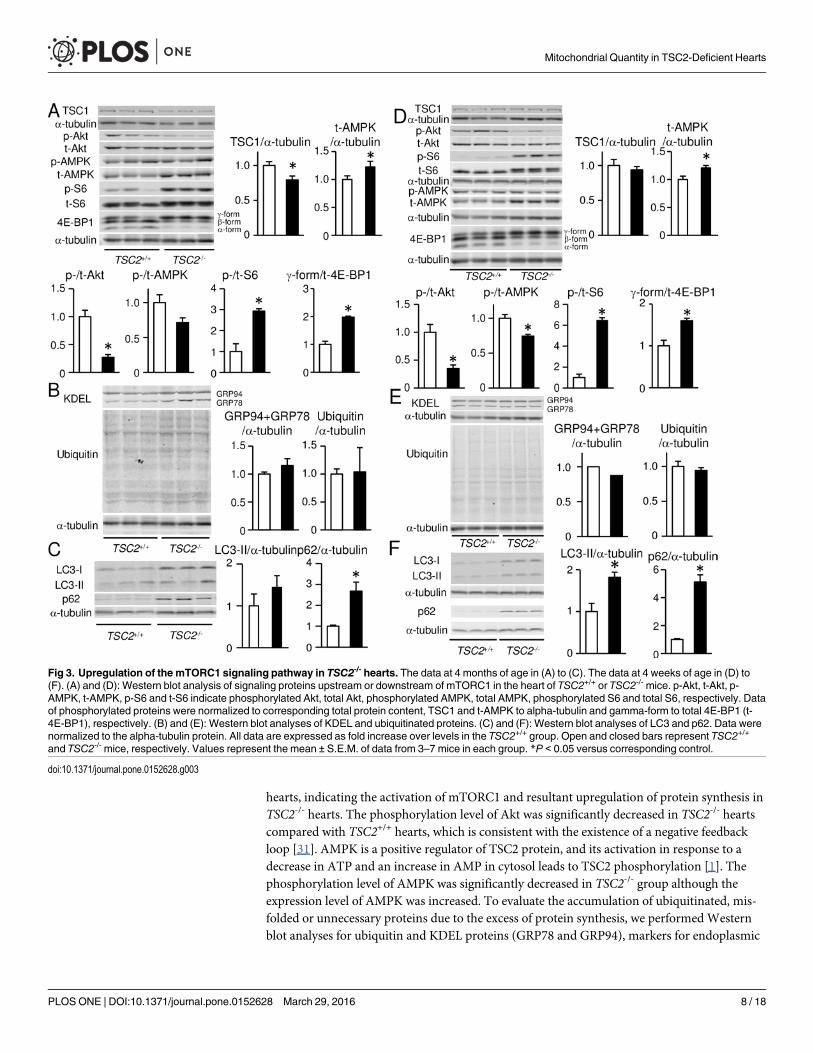
hearts, indicating the activation of mTORC1 and resultant upregulation of protein synthesis inTSC2-/- hearts. The phosphorylation level of Akt was significantly decreased in TSC2-/- heartscompared with TSC2+/+ hearts, which is consistent with the existence of a negative feedbackloop [31]. AMPK is a positive regulator of TSC2 protein, and its activation in response to adecrease in ATP and an increase in AMP in cytosol leads to TSC2 phosphorylation [1]. Thephosphorylation level of AMPK was significantly decreased in TSC2-/- group although theexpression level of AMPK was increased. To evaluate the accumulation of ubiquitinated, mis-folded or unnecessary proteins due to the excess of protein synthesis, we performedWesternblot analyses for ubiquitin and KDEL proteins (GRP78 and GRP94), markers for endoplasmic
Fig 3. Upregulation of the mTORC1 signaling pathway in TSC2-/- hearts. The data at 4 months of age in (A) to (C). The data at 4 weeks of age in (D) to(F). (A) and (D): Western blot analysis of signaling proteins upstream or downstream of mTORC1 in the heart of TSC2+/+ or TSC2-/- mice. p-Akt, t-Akt, p-AMPK, t-AMPK, p-S6 and t-S6 indicate phosphorylated Akt, total Akt, phosphorylated AMPK, total AMPK, phosphorylated S6 and total S6, respectively. Dataof phosphorylated proteins were normalized to corresponding total protein content, TSC1 and t-AMPK to alpha-tubulin and gamma-form to total 4E-BP1 (t-4E-BP1), respectively. (B) and (E): Western blot analyses of KDEL and ubiquitinated proteins. (C) and (F): Western blot analyses of LC3 and p62. Data werenormalized to the alpha-tubulin protein. All data are expressed as fold increase over levels in the TSC2+/+ group. Open and closed bars represent TSC2+/+
and TSC2-/- mice, respectively. Values represent the mean ± S.E.M. of data from 3–7 mice in each group. *P < 0.05 versus corresponding control.
doi:10.1371/journal.pone.0152628.g003
Mitochondrial Quantity in TSC2-Deficient Hearts
PLOS ONE | DOI:10.1371/journal.pone.0152628 March 29, 2016 8 / 18
reticulum stress (Fig 3B). However, we detected no significant differences in the expression lev-els of the proteins between the two groups. Since mTORC1 is known to be a negative regulatorof autophagy in mammalian cells [4], we estimated the autophagic activity in TSC2-/- hearts(Fig 3C) [32]. The conversion of LC3-I to LC3-II is an essential step for autophagosome forma-tion. The expression level of LC3-II was not significantly different between the two groups.However, p62, a marker for autophagic flux, was significantly accumulated in TSC2-/- heartscompared to TSC2+/+ hearts.
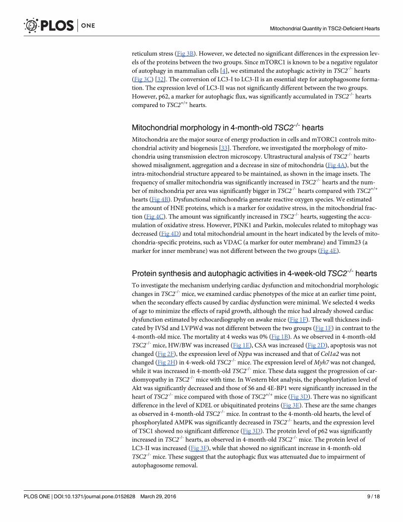
Mitochondrial morphology in 4-month-old TSC2-/- heartsMitochondria are the major source of energy production in cells and mTORC1 controls mito-chondrial activity and biogenesis [33]. Therefore, we investigated the morphology of mito-chondria using transmission electron microscopy. Ultrastructural analysis of TSC2-/- heartsshowed misalignment, aggregation and a decrease in size of mitochondria (Fig 4A), but theintra-mitochondrial structure appeared to be maintained, as shown in the image insets. Thefrequency of smaller mitochondria was significantly increased in TSC2-/- hearts and the num-ber of mitochondria per area was significantly bigger in TSC2-/- hearts compared with TSC2+/+
hearts (Fig 4B). Dysfunctional mitochondria generate reactive oxygen species. We estimatedthe amount of HNE proteins, which is a marker for oxidative stress, in the mitochondrial frac-tion (Fig 4C). The amount was significantly increased in TSC2-/- hearts, suggesting the accu-mulation of oxidative stress. However, PINK1 and Parkin, molecules related to mitophagy wasdecreased (Fig 4D) and total mitochondrial amount in the heart indicated by the levels of mito-chondria-specific proteins, such as VDAC (a marker for outer membrane) and Timm23 (amarker for inner membrane) was not different between the two groups (Fig 4E).
Protein synthesis and autophagic activities in 4-week-old TSC2-/- heartsTo investigate the mechanism underlying cardiac dysfunction and mitochondrial morphologicchanges in TSC2-/- mice, we examined cardiac phenotypes of the mice at an earlier time point,when the secondary effects caused by cardiac dysfunction were minimal. We selected 4 weeksof age to minimize the effects of rapid growth, although the mice had already showed cardiacdysfunction estimated by echocardiography on awake mice (Fig 1F). The wall thickness indi-cated by IVSd and LVPWd was not different between the two groups (Fig 1F) in contrast to the4-month-old mice. The mortality at 4 weeks was 0% (Fig 1B). As we observed in 4-month-oldTSC2-/- mice, HW/BW was increased (Fig 1E), CSA was increased (Fig 2D), apoptosis was notchanged (Fig 2F), the expression level of Nppa was increased and that of Col1a2 was notchanged (Fig 2H) in 4-week-old TSC2-/- mice. The expression level ofMyh7 was not changed,while it was increased in 4-month-old TSC2-/- mice. These data suggest the progression of car-diomyopathy in TSC2-/- mice with time. In Western blot analysis, the phosphorylation level ofAkt was significantly decreased and those of S6 and 4E-BP1 were significantly increased in theheart of TSC2-/- mice compared with those of TSC2+/+ mice (Fig 3D). There was no significantdifference in the level of KDEL or ubiquitinated proteins (Fig 3E). These are the same changesas observed in 4-month-old TSC2-/- mice. In contrast to the 4-month-old hearts, the level ofphosphorylated AMPK was significantly decreased in TSC2-/- hearts, and the expression levelof TSC1 showed no significant difference (Fig 3D). The protein level of p62 was significantlyincreased in TSC2-/- hearts, as observed in 4-month-old TSC2-/- mice. The protein level ofLC3-II was increased (Fig 3F), while that showed no significant increase in 4-month-oldTSC2-/- mice. These suggest that the autophagic flux was attenuated due to impairment ofautophagosome removal.
Mitochondrial Quantity in TSC2-Deficient Hearts
PLOS ONE | DOI:10.1371/journal.pone.0152628 March 29, 2016 9 / 18
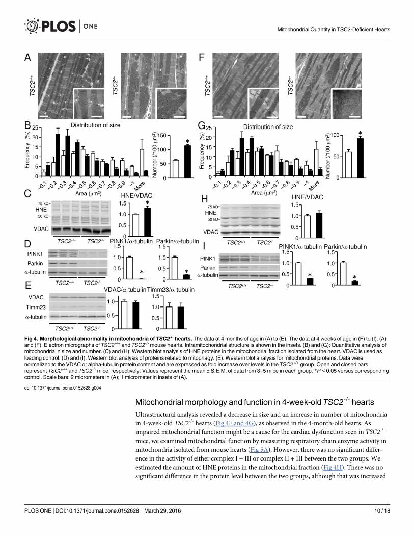
Mitochondrial morphology and function in 4-week-old TSC2-/- heartsUltrastructural analysis revealed a decrease in size and an increase in number of mitochondriain 4-week-old TSC2-/- hearts (Fig 4F and 4G), as observed in the 4-month-old hearts. Asimpaired mitochondrial function might be a cause for the cardiac dysfunction seen in TSC2-/-
mice, we examined mitochondrial function by measuring respiratory chain enzyme activity inmitochondria isolated from mouse hearts (Fig 5A). However, there was no significant differ-ence in the activity of either complex I + III or complex II + III between the two groups. Weestimated the amount of HNE proteins in the mitochondrial fraction (Fig 4H). There was nosignificant difference in the protein level between the two groups, although that was increased
Fig 4. Morphological abnormality in mitochondria of TSC2-/- hearts. The data at 4 months of age in (A) to (E). The data at 4 weeks of age in (F) to (I). (A)and (F): Electron micrographs of TSC2+/+ and TSC2-/- mouse hearts. Intramitochondrial structure is shown in the insets. (B) and (G): Quantitative analysis ofmitochondria in size and number. (C) and (H): Western blot analysis of HNE proteins in the mitochondrial fraction isolated from the heart. VDAC is used asloading control. (D) and (I): Western blot analysis of proteins related to mitophagy. (E): Western blot analysis for mitochondrial proteins. Data werenormalized to the VDAC or alpha-tubulin protein content and are expressed as fold increase over levels in the TSC2+/+ group. Open and closed barsrepresent TSC2+/+ and TSC2-/- mice, respectively. Values represent the mean ± S.E.M. of data from 3–5 mice in each group. *P < 0.05 versus correspondingcontrol. Scale bars: 2 micrometers in (A); 1 micrometer in insets of (A).
doi:10.1371/journal.pone.0152628.g004
Mitochondrial Quantity in TSC2-Deficient Hearts
PLOS ONE | DOI:10.1371/journal.pone.0152628 March 29, 2016 10 / 18
in 4-month-old TSC2-/- mice. The expression levels of PINK1 and Parkin were decreased (Fig4I) as observed in 4-month-old TSC2-/- mice. Furthermore, we examined ATP concentrationand the ratio of ADP to ATP concentration (ADP/ATP) in the heart tissue (Fig 5B). ATP con-centration was found to be significantly increased in TSC2-/- hearts compared with TSC2+/+
hearts, and a significant decrease in ADP/ATP was detected. The mRNA expression of Pgc-1ameasured by quantitative RT-PCR was preserved in TSC2-/- hearts (Fig 5C). These data indi-cate that the mitochondrial function was maintained in TSC2-/- hearts, although mitochondrialmorphology was changed. Thus, the main cause of the cardiac phenotype seen in TSC2-/- heartsis not mitochondrial dysfunction.
Attenuation of the cardiac phenotypes in TSC2-deficient hearts byupregulation of autophagic fluxWe hypothesized that reduced autophagic flux is one of the mechanisms for the cardiac pheno-types seen in TSC2-/- mice. To investigate the involvement of autophagy in cardiac phenotypesin TSC2-/- hearts, we treated the TSC2-/- mice with trehalose. Trehalose is a non-reducing disac-charide found in a wide variety of organisms, including bacteria, yeast, invertebrates, andplants and functions to protect cells against various environmental stresses [34]. Recently, it isreported that trehalose acts as an mTOR-independent autophagy activator and can reduce pro-tein aggregates in some mouse disease models [35–37]. First, we treated TSC2+/+ mice with tre-halose for 10 weeks starting from 6 weeks after birth to confirm upregulation of autophagicactivity. Western blot analysis for LC3 indicated that autophagy was upregulated (Fig 6A).Thus, we treated TSC2-/- mice with trehalose for 10 weeks starting from 6 weeks after birth.After the treatment, cardiac function indicated by echocardiographic fractional shortening wassignificantly improved in the trehalose-treated group compared with control non-treatedgroup (Fig 6B). The heart weight and the ratio of heart to body weight were not differentbetween the two groups (Fig 6C). The accumulation of p62 was significantly reduced in treha-lose-treated TSC2-/- hearts compared with control TSC2-/- hearts, although the protein level of
Fig 5. Biochemical analyses of mitochondrial function in TSC2-/- hearts at 4 weeks of age. (A): Theactivities of complex I + III and complex II + III in mitochondria isolated from the hearts. (B): The ATP contentexpressed as fold increase over levels in the TSC2+/+ group and ADP to ATP ratio (ADP/ATP) in the hearttissues. (C): mRNA expression of Pgc-1awas determined by quantitative RT-PCR. Data were normalized totheGapdh content and are expressed as fold increase over levels in the TSC2+/+ group. Values represent themean ± S.E.M. of data from 3–4 mice in each group. Open and closed bars represent TSC2+/+ and TSC2-/-
mice, respectively. *P < 0.05 versus corresponding control.
doi:10.1371/journal.pone.0152628.g005
Mitochondrial Quantity in TSC2-Deficient Hearts
PLOS ONE | DOI:10.1371/journal.pone.0152628 March 29, 2016 11 / 18

LC3-II was not different (Fig 6D). This supports that trehalose increased autophagic flux inTSC2-/- mice. Ultrastructural analysis showed that the size of mitochondria was larger and thenumber was smaller in trehalose-treated group than those in control group (Fig 6E and 6F).
DiscussionIn this study, we showed that the TSC-mTORC1 signaling pathway plays an important role tomaintain cardiac function and to regulate mitochondrial size and number. Its homeostaticfunction is mediated through autophagy. Since mitochondria were not damaged in TSC2-/-
hearts, autophagy is the main player in this system rather than mitochondria-specific autop-hagy, mitophagy.
It has been reported that cardiac-specific TSC1-deficient mice, which were generated bycrossing floxed TSC1mice with myosin light chain 2v-promotor driven Cre recombinaseknock-in mice, had a median survival of 6 months and developed dilated cardiomyopathy [38].Since TSC1 is required to stabilize TSC2 and prevents its ubiquitin-mediated degradation [29,30], ablation of TSC1 would lead to downregulation of TSC2. It is reasonable that TSC1-defi-cient mice developed similar cardiac phenotypes to TSC2-/- mice. Thus, the TSC1/2 complex isessential for maintenance of cardiac function after birth, but not for heart development or
Fig 6. Effects of trehalose treatment on cardiac phenotypes in TSC2-/- mice. (A): Western blot analysis for LC3 in trehalose-treated TSC2+/+ group. Datawere normalized to alpha-tubulin protein content. White and light gray bars represent control TSC2+/+ mice and trehalose-treated TSC2+/+ mice, respectively.(B): Echocardiographic parameters of the awake TSC2-/- mice. (C): Physiological parameters of the TSC2-/- mice. BW, HW and HW/BW indicate body weight,whole heart weight and whole heart-to-body-weight ratio, respectively. (D): Western blot analysis for LC3 and p62. Data were normalized to alpha-tubulinprotein content. All data are expressed as fold increase over levels in control TSC2-/- group. (E and F): Electron micrographs of the TSC2-/- hearts (E) andquantitative analyses of mitochondrial size and number (F). In (B) to (F), black and dark gray bars represent control TSC2-/- mice and trehalose-treatedTSC2-/- mice, respectively. Values represent the mean ± S.E.M. of data from 3 to 5 mice in each group. *P < 0.05 versus control group. Scale bars: 2micrometers.
doi:10.1371/journal.pone.0152628.g006
Mitochondrial Quantity in TSC2-Deficient Hearts
PLOS ONE | DOI:10.1371/journal.pone.0152628 March 29, 2016 12 / 18

survival during embryonic stage. Although the molecular mechanisms underlying the develop-ment of dilated cardiomyopathy in the TSC1-deficient mice have not been investigated, histo-logical analysis of the mice showed the occurrence of scattered foci of enlarged cardiomyocytesaccumulating glycogen without any evidence of proliferation in the lesion [38]. However, wedid not observe such histological changes nor tumorigenesis in TSC2-/- hearts macroscopicallyand by echocardiography. Although the molecular mechanism underlying the difference in thehistological findings between cardiac-specific TSC1 and TSC2-deficient hearts is unknown, itmay be due to the timing of gene knockdown or distinct functions of the products of thesegenes.
TSC2 ablation in the heart led to not only cardiac dysfunction, but also a decrease in heartrate. ATP is released from cardiomyocytes [39, 40] and immediately metabolized to adenosineby ATPases that have their catalytic domain on the outer side of plasma membrane. Sinceadenosine is known to have a negative effect on heart rate [41], increased cytosolic ATP levelmay result in a decrease in heart rate in TSC2-/- mice. Other possibilities are that histologicalabnormalities including misalignment of mitochondria or direct effects of the TSC2 signalingpathway impair the function of the conduction system in the heart.
Autophagic flux indicated by protein level of LC3-II and p62 was decreased in TSC2-/-
hearts at 4 weeks of age. Co-existence of increased LC3-II with increased p62 protein levels isconsistent with impaired autophagosome removal rather than decreased formation. mTORC1has been reported to regulate autophagy by signalling to ULK1 and lysosome homeostasis [5,42].
We observed cardiomyocyte hypertrophy indicated by increases in heart-to-body weightratio, LV mass and cross-sectional area of cardiomyocytes in TSC2-/- mice. Absence of fibrosisand cardiomyocyte death indicates that cardiac remodeling observed in TSC2-/- mice is not typ-ical for general dilated cardiomyopathy. The hypertrophic responses can be induced by activa-tion of protein synthesis. We previously reported that inhibition of autophagy incardiomyocytes leads to cardiac hypertrophy [7]. In this study, as the treatment with trehalosehad no effect on cardiac hypertrophy, the results suggest that inhibition of autophagy is notinvolved in the development of cardiac hypertrophy in TSC2-/- mice.
Our previous results showed that inhibition of autophagy in the heart by ablation of Atg5induced misalignment and heterogeneous size of mitochondria and damage of intramitochon-drial structure, impaired mitochondrial function and increased oxidative stress [7, 28]. If weconsider the importance of autophagy for maintenance of normal functional mitochondria, itis surprising that TSC2-/- hearts showed normal mitochondrial respiratory function even withalteration in mitochondrial number and size. The normal mitochondrial function was con-firmed by the results that ATP content was not decreased and oxidative stress was notincreased in TSC2-/- hearts. This is in agreement with the previously reported in vitro studiesthat mitochondrial activity is stimulated and ATP levels are increased under the condition ofconstitutive mTORC1 activation in TSC2-deficient or knockdown cells [33, 43, 44]. The TSC2/mTORC1-dependent autophagy pathway may not be required for the removal of damagedmitochondria at least in the absence of TSC2. However, the mitochondrial function was mea-sured using isolated mitochondria in this study and the assay has experimental limitation. Ithas been reported that isolation of mitochondria causes the difference in functional character-istics compared to intact mitochondria in permeabilized miofibers [45]. mTORC1 is reportedto control mitochondrial ATP production capacity by selectively promoting translation ofnucleus-encoded mitochondria-related mRNA [33]. Hyperactivation of mTORC1 may protectmitochondria against stress. We observed an increase in number and a decrease in size of mito-chondria in TSC2-/- hearts. Consistent with our findings, the number of mitochondria isincreased in pancreatic beta-cells lacking TSC2 [44]. The quantity, quality and size of
Mitochondrial Quantity in TSC2-Deficient Hearts
PLOS ONE | DOI:10.1371/journal.pone.0152628 March 29, 2016 13 / 18

mitochondria are determined by fusion and fission, followed by autophagy [46]. It is possiblethat the TSC2/mTORC1-dependent signaling pathway may be directly or indirectly involvedin mitochondrial dynamics.
The activation of AMPK leads to TSC2 phosphorylation, resulting in restoration of ATPthrough regulation of metabolism and inhibition of growth via the mTORC1 pathway. AMPKphosphorylation was impaired in our TSC2-/- hearts, conversely the level was reported to beincreased in tumor-derived cells or neurons lacking TSC2 [47, 48]. The phosphorylation levelof AMPK in TSC2-/- hearts may be affected by the increase in intracellular ATP concentration.AMPK activates autophagy not only by inhibition of mTORC1 but also by direct phosphoryla-tion of ULK1, which is required for autophagy induction [5, 48, 49]. In addition to activationof mTORC1 activity, inhibition of AMPK activity may supress autophagic activity in TSC2-/-
hearts. Although the decreased Akt phosphorylation level in TSC2-/- hearts can inhibitmTORC1 via proline-rich Akt substrate of 40 kDa (PRAS40), an endogenous mTORC1 inhibi-tor, the negative feedback mechanism would not be enough for the normalization of themTORC1 signaling pathway.
mTORC1 positively regulates cell growth and proliferation by promoting biosynthesis ofproteins and lipids and mitochondrial metabolism and biogenesis and also by limiting autop-hagy [1]. Over-activating these functions of mTORC1 could be related to the observed pheno-types in TSC2-/- hearts. mTOR controls mitochondrial oxidative function through a yin yang1‒PGC-1alpha transcriptional complex [3]. PGC-1alpha is known to promote mitochondrialbiogenesis and function. In TSC2-/- hearts, mitochondrial function was maintained, ATP con-centration was increased and the expression of PGC-1alpha was preserved. Therefore, we canexclude the possibility that impairment of mitochondrial metabolism and biogenesis is a causefor cardiac dysfunction in TSC2-/- mice.
We previously reported that inhibition of autophagy by cardiac-specific Atg5-deficient miceinduces age-related cardiomyopathy [28] and it was found that the accumulation and aggrega-tion of damaged or unnecessary proteins could cause the malfunction of biological processesand play an important role in aging. As signaling pathways towards protein synthesis were sig-nificantly accelerated in TSC2-/- hearts, there is a possibility that unnecessary or misfolded pro-teins were accumulated in cardiomyocytes to induce cardiac dysfunction. However, weobserved no increase in ubiquitinated or KDEL proteins in TSC2-/- hearts. Some compensatorymechanisms, such as TSC2/mTORC1-independent autophagy, chaperone-mediated autop-hagy and proteasome system, could be involved in the maintenance of protein turnover. It isalso possible that the ability of mTORC1-dependent autophagy still might be enough torespond to the increased protein load.
PINK1 and Parkin are known to be key molecules for damaged mitochondria clearance viaubiquitination and mitophagy in neuron [50]. Surprisingly, the expression level of PINK1 andParkin was reduced in TSC2-/- hearts, suggesting PINK1 and Parkin-related mitophagy isdown-regulated. Although it was reported that suppression of mTOR pathway can increase theexpression level of PINK1 and Parkin [51], it is unclear why PINK1 and Parkin are reduced inthis model. Further study is requested to clarify the effect of the decreases in PINK1/Parkin lev-els on the cardiac phenotype and mitochondrial morphology observed in TSC2-/- mice.
The protein level of p62 was significantly decreased by treatment of trehalose for 10 weeks,suggesting that autophagic flux was improved by trehalose treatment. However, the proteinlevel of LC3-II was not different between control and trehalose-treated TSC2-/- groups. Thismight be due to the balance between the upregulation of autophagic induction and autolysoso-mal removal. The protective effect of trehalose on cardiac dysfunction in TSC2-/- mice indicatesthe existence of an mTOR-independent pathway to enhance autophagic flux. Upregulation ofautophagic activity by trehalose treatment improved the cardiac function, indicating that
Mitochondrial Quantity in TSC2-Deficient Hearts
PLOS ONE | DOI:10.1371/journal.pone.0152628 March 29, 2016 14 / 18
downregulation of autophagy could be a significant cause for cardiac phenotypes in TSC2-/-
mice. Since the extent of rescue of the cardiac phenotypes was partial, we could not excludepossibilities that mechanisms other than autophagy are involved in the development of cardiacphenotypes in TSC2-/- mice. Trehalose is not a selective activator of autophagy [34]. Further-more, trehalose does not appear to be absorbed and is rather broken down to glucose in thegastrointestinal tract of mammals [52]. Thus, we cannot exclude a possibility that improve-ment in cardiac function is due to the other potential “off-target” effects of trehalose. Molecularmechanisms how downregulation of autophagic flux in TSC2-/- mice resulted in theimpairment of cardiac function remain to be elucidated. Cytoarchitectural disorganizationwould lead to cardiac dysfunction [53]. Ultrastructural images of TSC2-/- hearts showed mis-alignment of mitochondria and looked as if many of mitochondria could not interact withmyofibril closely, suggesting that energy transfer between mitochondria and myosin-ATPasecould be restricted. Activated autophagy can remove not only small mitochondria but alsoother cytoplasmic components and improve the interaction and energy transfer between mito-chondria and myofibril. It is also possible that the misaligned mitochondria simply disturb sar-comere contraction.
In conclusion, we demonstrated that constitutive stimulation of mTORC1 signaling byTSC2 ablation in the heart leads to cardiac chamber dilatation and dysfunction and that upre-gulation of autophagic activity attenuates the cardiac dysfunction. Autophagy regulated byTSC-mTORC1 signaling in the heart is essential for the control of mitochondrial quantity. It isreported that mTORC1 inhibitors such as rapamycin can be pharmacological treatments ofTSC disease [54, 55]. Trehalose could be a novel therapeutic for TSC patients. However, furtherinvestigation is required to reveal the involvement of autophagy in the pathogenesis of the dis-ease with benign tumours.
Supporting InformationS1 Fig. Echocardiographic analysis of the mice at 4 months of age under anaesthesia.(PDF)
S2 Fig. The original blots shown in Figs 1, 3, 4 and 6.(PDF)
S1 Protocol. Methods for the pilot study.(DOCX)
AcknowledgmentsWe thank Dr. Gema Vizcay-Barrena, Centre for Ultrastructural Imaging at King’s College Lon-don for processing the samples for transmission electron microscopy.
Author ContributionsConceived and designed the experiments: KO. Performed the experiments: M. Taneike KN SOEZ TM RKT RAM. Takaoka. Analyzed the data: M. Taneike KN SO EZ TM RKT RAM.Takaoka OY. Contributed reagents/materials/analysis tools: MJG. Wrote the paper: M. TaneikeKNMJG AMS KO.
References1. Laplante M, Sabatini DM. mTOR signaling at a glance. J Cell Sci. 2009; 122(Pt 20):3589–94. doi: 10.
1242/jcs.051011 PMID: 19812304
Mitochondrial Quantity in TSC2-Deficient Hearts
PLOS ONE | DOI:10.1371/journal.pone.0152628 March 29, 2016 15 / 18
2. Richter JD, Sonenberg N. Regulation of cap-dependent translation by eIF4E inhibitory proteins. Nature.2005; 433(7025):477–80. PMID: 15690031
3. Cunningham JT, Rodgers JT, Arlow DH, Vazquez F, Mootha VK, Puigserver P. mTOR controls mito-chondrial oxidative function through a YY1-PGC-1alpha transcriptional complex. Nature. 2007; 450(7170):736–40. PMID: 18046414
4. Thoreen CC, Kang SA, Chang JW, Liu Q, Zhang J, Gao Y, et al. An ATP-competitive mammalian targetof rapamycin inhibitor reveals rapamycin-resistant functions of mTORC1. J Biol Chem. 2009; 284(12):8023–32. doi: 10.1074/jbc.M900301200 PMID: 19150980
5. Kim J, Kundu M, Viollet B, Guan KL. AMPK and mTOR regulate autophagy through direct phosphoryla-tion of Ulk1. Nat Cell Biol. 2011; 13(2):132–41. doi: 10.1038/ncb2152 PMID: 21258367
6. Mizushima N, Komatsu M. Autophagy: renovation of cells and tissues. Cell. 2011; 147(4):728–41. doi:10.1016/j.cell.2011.10.026 PMID: 22078875
7. Nakai A, Yamaguchi O, Takeda T, Higuchi Y, Hikoso S, Taniike M, et al. The role of autophagy in cardi-omyocytes in the basal state and in response to hemodynamic stress. Nat Med. 2007; 13(5):619–24.PMID: 17450150
8. Gomes LC, Di Benedetto G, Scorrano L. During autophagy mitochondria elongate, are spared fromdegradation and sustain cell viability. Nat Cell Biol. 2011; 13(5):589–98. doi: 10.1038/ncb2220 PMID:21478857
9. Yamagata K, Sanders LK, KaufmannWE, YeeW, Barnes CA, Nathans D, et al. rheb, a growth factor-and synaptic activity-regulated gene, encodes a novel Ras-related protein. J Biol Chem. 1994; 269(23):16333–9. PMID: 8206940
10. Huang J, Manning BD. The TSC1-TSC2 complex: a molecular switchboard controlling cell growth. Bio-chem J. 2008; 412(2):179–90. doi: 10.1042/BJ20080281 PMID: 18466115
11. Inoki K, Li Y, Xu T, Guan KL. Rheb GTPase is a direct target of TSC2 GAP activity and regulatesmTOR signaling. Genes Dev. 2003; 17(15):1829–34. PMID: 12869586
12. Tabancay AP Jr., Gau CL, Machado IM, Uhlmann EJ, Gutmann DH, Guo L, et al. Identification of domi-nant negative mutants of Rheb GTPase and their use to implicate the involvement of human Rheb inthe activation of p70S6K. J Biol Chem. 2003; 278(41):39921–30. PMID: 12869548
13. Tee AR, Blenis J, Proud CG. Analysis of mTOR signaling by the small G-proteins, Rheb and RhebL1.FEBS Lett. 2005; 579(21):4763–8. PMID: 16098514
14. Inoki K, Li Y, Zhu T, Wu J, Guan KL. TSC2 is phosphorylated and inhibited by Akt and suppressesmTOR signalling. Nat Cell Biol. 2002; 4(9):648–57. PMID: 12172553
15. Manning BD. Balancing Akt with S6K: implications for both metabolic diseases and tumorigenesis. JCell Biol. 2004; 167(3):399–403. PMID: 15533996
16. Tapon N, Ito N, Dickson BJ, Treisman JE, Hariharan IK. The Drosophila tuberous sclerosis complexgene homologs restrict cell growth and cell proliferation. Cell. 2001; 105(3):345–55. PMID: 11348591
17. Potter CJ, Huang H, Xu T. Drosophila Tsc1 functions with Tsc2 to antagonize insulin signaling in regu-lating cell growth, cell proliferation, and organ size. Cell. 2001; 105(3):357–68. PMID: 11348592
18. Gangloff YG, Mueller M, Dann SG, Svoboda P, Sticker M, Spetz JF, et al. Disruption of the mousemTOR gene leads to early postimplantation lethality and prohibits embryonic stem cell development.Mol Cell Biol. 2004; 24(21):9508–16. PMID: 15485918
19. Murakami M, Ichisaka T, Maeda M, Oshiro N, Hara K, Edenhofer F, et al. mTOR is essential for growthand proliferation in early mouse embryos and embryonic stem cells. Mol Cell Biol. 2004; 24(15):6710–8. PMID: 15254238
20. Zhu Y, Pires KM, Whitehead KJ, Olsen CD, Wayment B, Zhang YC, et al. Mechanistic target of rapamy-cin (Mtor) is essential for murine embryonic heart development and growth. PLoS One. 2013; 8(1):e54221. doi: 10.1371/journal.pone.0054221 PMID: 23342106
21. Zhang D, Contu R, Latronico MV, Zhang J, Rizzi R, Catalucci D, et al. MTORC1 regulates cardiac func-tion and myocyte survival through 4E-BP1 inhibition in mice. J Clin Invest. 2010; 120(8):2805–16. doi:10.1172/JCI43008 PMID: 20644257
22. Tamai T, Yamaguchi O, Hikoso S, Takeda T, Taneike M, Oka T, et al. Rheb (Ras homologue enrichedin brain)-dependent mammalian target of rapamycin complex 1 (mTORC1) activation becomes indis-pensable for cardiac hypertrophic growth after early postnatal period. J Biol Chem. 2013; 288(14):10176–87. doi: 10.1074/jbc.M112.423640 PMID: 23426372
23. Kobayashi T, Minowa O, Kuno J, Mitani H, Hino O, Noda T. Renal carcinogenesis, hepatic hemangio-matosis, and embryonic lethality caused by a germ-line Tsc2 mutation in mice. Cancer Res. 1999; 59(6):1206–11. PMID: 10096549
Mitochondrial Quantity in TSC2-Deficient Hearts
PLOS ONE | DOI:10.1371/journal.pone.0152628 March 29, 2016 16 / 18
24. Onda H, Lueck A, Marks PW, Warren HB, Kwiatkowski DJ. Tsc2(+/-) mice develop tumors in multiplesites that express gelsolin and are influenced by genetic background. J Clin Invest. 1999; 104(6):687–95. PMID: 10491404
25. Hernandez O, Way S, McKenna J 3rd, Gambello MJ. Generation of a conditional disruption of the Tsc2gene. Genesis. 2007; 45(2):101–6. PMID: 17245776
26. Yamaguchi O, Watanabe T, Nishida K, Kashiwase K, Higuchi Y, Takeda T, et al. Cardiac-specific dis-ruption of the c-raf-1 gene induces cardiac dysfunction and apoptosis. J Clin Invest. 2004; 114(7):937–43. PMID: 15467832
27. Trounce IA, Kim YL, Jun AS, Wallace DC. Assessment of mitochondrial oxidative phosphorylation inpatient muscle biopsies, lymphoblasts, and transmitochondrial cell lines. Methods Enzymol. 1996;264:484–509. PMID: 8965721
28. Taneike M, Yamaguchi O, Nakai A, Hikoso S, Takeda T, Mizote I, et al. Inhibition of autophagy in theheart induces age-related cardiomyopathy. Autophagy. 2010; 6(5):600–6. doi: 10.4161/auto.6.5.11947PMID: 20431347
29. Benvenuto G, Li S, Brown SJ, Braverman R, VassWC, Cheadle JP, et al. The tuberous sclerosis-1(TSC1) gene product hamartin suppresses cell growth and augments the expression of the TSC2 prod-uct tuberin by inhibiting its ubiquitination. Oncogene. 2000; 19(54):6306–16. PMID: 11175345
30. Chong-Kopera H, Inoki K, Li Y, Zhu T, Garcia-Gonzalo FR, Rosa JL, et al. TSC1 stabilizes TSC2 byinhibiting the interaction between TSC2 and the HERC1 ubiquitin ligase. J Biol Chem. 2006; 281(13):8313–6. PMID: 16464865
31. Zhang H, Cicchetti G, Onda H, Koon HB, Asrican K, Bajraszewski N, et al. Loss of Tsc1/Tsc2 activatesmTOR and disrupts PI3K-Akt signaling through downregulation of PDGFR. J Clin Invest. 2003; 112(8):1223–33. PMID: 14561707
32. Klionsky DJ, Abdalla FC, Abeliovich H, Abraham RT, Acevedo-Arozena A, Adeli K, et al. Guidelines forthe use and interpretation of assays for monitoring autophagy. Autophagy. 2012; 8(4):445–544. PMID:22966490
33. Morita M, Gravel SP, Chenard V, Sikstrom K, Zheng L, Alain T, et al. mTORC1 controls mitochondrialactivity and biogenesis through 4E-BP-dependent translational regulation. Cell Metab. 2013; 18(5):698–711. doi: 10.1016/j.cmet.2013.10.001 PMID: 24206664
34. Chen Q, Haddad GG. Role of trehalose phosphate synthase and trehalose during hypoxia: from flies tomammals. J Exp Biol. 2004; 207(Pt 18):3125–9. PMID: 15299033
35. Davies JE, Sarkar S, Rubinsztein DC. Trehalose reduces aggregate formation and delays pathology ina transgenic mouse model of oculopharyngeal muscular dystrophy. HumMol Genet. 2006; 15(1):23–31. PMID: 16311254
36. Rodriguez-Navarro JA, Rodriguez L, Casarejos MJ, Solano RM, Gomez A, Perucho J, et al. Trehaloseameliorates dopaminergic and tau pathology in parkin deleted/tau overexpressing mice through autop-hagy activation. Neurobiol Dis. 2010; 39(3):423–38. doi: 10.1016/j.nbd.2010.05.014 PMID: 20546895
37. Schaeffer V, Lavenir I, Ozcelik S, Tolnay M, Winkler DT, Goedert M. Stimulation of autophagy reducesneurodegeneration in a mouse model of human tauopathy. Brain. 2012; 135(Pt 7):2169–77. doi: 10.1093/brain/aws143 PMID: 22689910
38. Meikle L, McMullen JR, Sherwood MC, Lader AS, Walker V, Chan JA, et al. A mouse model of cardiacrhabdomyoma generated by loss of Tsc1 in ventricular myocytes. HumMol Genet. 2005; 14(3):429–35. PMID: 15601645
39. Kunugi S, Iwabuchi S, Matsuyama D, Okajima T, Kawahara K. Negative-feedback regulation of ATPrelease: ATP release from cardiomyocytes is strictly regulated during ischemia. Biochem Biophys ResCommun. 2011; 416(3–4):409–15. doi: 10.1016/j.bbrc.2011.11.068 PMID: 22133679
40. Dutta AK, Sabirov RZ, Uramoto H, Okada Y. Role of ATP-conductive anion channel in ATP releasefrom neonatal rat cardiomyocytes in ischaemic or hypoxic conditions. J Physiol. 2004; 559(Pt 3):799–812. PMID: 15272030
41. Froldi G, Belardinelli L. Species-dependent effects of adenosine on heart rate and atrioventricularnodal conduction. Mechanism and physiological implications. Circ Res. 1990; 67(4):960–78. PMID:2208618
42. Roczniak-Ferguson A, Petit CS, Froehlich F, Qian S, Ky J, Angarola B, et al. The transcription factorTFEB links mTORC1 signaling to transcriptional control of lysosome homeostasis. Sci Signal. 2012; 5(228):ra42. doi: 10.1126/scisignal.2002790 PMID: 22692423
43. Schieke SM, Phillips D, McCoy JP Jr., Aponte AM, Shen RF, Balaban RS, et al. The mammalian targetof rapamycin (mTOR) pathway regulates mitochondrial oxygen consumption and oxidative capacity. JBiol Chem. 2006; 281(37):27643–52. PMID: 16847060
Mitochondrial Quantity in TSC2-Deficient Hearts
PLOS ONE | DOI:10.1371/journal.pone.0152628 March 29, 2016 17 / 18
44. Koyanagi M, Asahara S, Matsuda T, Hashimoto N, Shigeyama Y, Shibutani Y, et al. Ablation of TSC2enhances insulin secretion by increasing the number of mitochondria through activation of mTORC1.PLoS One. 2011; 6(8):e23238. doi: 10.1371/journal.pone.0023238 PMID: 21886784
45. Picard M, Taivassalo T, Ritchie D, Wright KJ, Thomas MM, Romestaing C, et al. Mitochondrial structureand function are disrupted by standard isolation methods. PLoS One. 2011; 6(3):e18317. doi: 10.1371/journal.pone.0018317 PMID: 21512578
46. Youle RJ, van der Bliek AM. Mitochondrial fission, fusion, and stress. Science. 2012; 337(6098):1062–5. doi: 10.1126/science.1219855 PMID: 22936770
47. Lacher MD, Pincheira R, Zhu Z, Camoretti-Mercado B, Matli M, Warren RS, et al. Rheb activatesAMPK and reduces p27Kip1 levels in Tsc2-null cells via mTORC1-independent mechanisms: implica-tions for cell proliferation and tumorigenesis. Oncogene. 2010; 29(50):6543–56. doi: 10.1038/onc.2010.393 PMID: 20818424
48. Di Nardo A, Wertz MH, Kwiatkowski E, Tsai PT, Leech JD, Greene-Colozzi E, et al. Neuronal Tsc1/2complex controls autophagy through AMPK-dependent regulation of ULK1. HumMol Genet. 2014; 23(14):3865–74. doi: 10.1093/hmg/ddu101 PMID: 24599401
49. Egan DF, Shackelford DB, Mihaylova MM, Gelino S, Kohnz RA, Mair W, et al. Phosphorylation of ULK1(hATG1) by AMP-activated protein kinase connects energy sensing to mitophagy. Science. 2011; 331(6016):456–61. doi: 10.1126/science.1196371 PMID: 21205641
50. Youle RJ, Narendra DP. Mechanisms of mitophagy. Nat Rev Mol Cell Biol. 2011; 12(1):9–14. doi: 10.1038/nrm3028 PMID: 21179058
51. Klinkenberg M, Gispert S, Dominguez-Bautista JA, Braun I, Auburger G, Jendrach M. Restriction of tro-phic factors and nutrients induces PARKIN expression. Neurogenetics. 2012; 13(1):9–21. doi: 10.1007/s10048-011-0303-8 PMID: 22028146
52. Ishihara R, Taketani S, Sasai-Takedatsu M, Kino M, Tokunaga R, Kobayashi Y. Molecular cloning,sequencing and expression of cDNA encoding human trehalase. Gene. 1997; 202(1–2):69–74. PMID:9427547
53. Joubert F, Wilding JR, Fortin D, Domergue-Dupont V, Novotova M, Ventura-Clapier R, et al. Local ener-getic regulation of sarcoplasmic and myosin ATPase is differently impaired in rats with heart failure. JPhysiol. 2008; 586(Pt 21):5181–92.
54. Kenerson H, Dundon TA, Yeung RS. Effects of rapamycin in the Eker rat model of tuberous sclerosiscomplex. Pediatr Res. 2005; 57(1):67–75. PMID: 15557109
55. Sato A, Kasai S, Kobayashi T, Takamatsu Y, Hino O, Ikeda K, et al. Rapamycin reverses impairedsocial interaction in mouse models of tuberous sclerosis complex. Nat Commun. 2012; 3:1292. doi: 10.1038/ncomms2295 PMID: 23250422
Mitochondrial Quantity in TSC2-Deficient Hearts
PLOS ONE | DOI:10.1371/journal.pone.0152628 March 29, 2016 18 / 18
Related Documents


