Kinetics and thermodynamics of metal-binding to histone deacetylase 8 Byungchul Kim, 1 Amit S. Pithadia, 2 and Carol A. Fierke 1,2,3 * 1 Chemical Biology Program, University of Michigan, Ann Arbor, Michigan 48109-2216 2 Department of Chemistry, University of Michigan, Ann Arbor, Michigan 48109-1055 3 Department of Biological Chemistry, University of Michigan Medical School, Ann Arbor, Michigan 48109-0600 Received 5 November 2014; Accepted 8 December 2014 DOI: 10.1002/pro.2623 Published online 16 December 2014 proteinscience.org Abstract: Histone deacetylase 8 (HDAC8) was originally classified as a Zn(II)-dependent deacetylase on the basis of Zn(II)-dependent HDAC8 activity in vitro and illumination of a Zn(II) bound to the active site. However, in vitro measurements demonstrated that HDAC8 has higher activity with a bound Fe(II) than Zn(II), although Fe(II)-HDAC8 rapidly loses activity under aerobic conditions. These data suggest that in the cell HDAC8 could be activated by either Zn(II) or Fe(II). Here we detail the kinetics, thermodynamics, and selectivity of Zn(II) and Fe(II) binding to HDAC8. To this end, we have developed a fluorescence anisotropy assay using fluorescein-labeled suberoylanilide hydroxamic acid (fl- SAHA). fl-SAHA binds specifically to metal-bound HDAC8 with affinities comparable to SAHA. To measure the metal affinity of HDAC, metal binding was coupled to fl-SAHA and assayed from the observed change in anisotropy. The metal K D values for HDAC8 are significantly different, ranging from picomolar to micromolar for Zn(II) and Fe(II), respectively. Unexpectedly, the Fe(II) and Zn(II) dis- sociation rate constants from HDAC8 are comparable, k off ~0.0006 s 21 , suggesting that the apparent association rate constant for Fe(II) is slow (~3 3 10 3 M 21 s 21 ). Furthermore, monovalent cations (K 1 or Na 1 ) that bind to HDAC8 decrease the dissociation rate constant of Zn(II) by 100-fold for K 1 and 10-fold for Na 1 , suggesting a possible mechanism for regulating metal exchange in vivo. The HDAC8 metal affinities are comparable to the readily exchangeable Zn(II) and Fe(II) concentrations in cells, consistent with either or both metal cofactors activating HDAC8. Keywords: histone deacetylase 8; fl-SAHA; metal-binding mechanism; monovalent cations Introduction Misregulation of posttranslational modifications are implicated in many human diseases, including can- cer and neurodegenerative diseases. 1 Post- translational acetylation of lysine side chains has been observed at more than 3600 sites in proteins, including histones, 2,3 and this modification regulates the biological activity of many of these proteins. For example, acetylation and deacetylation correlate with the activation and deactivation, respectively, of transcriptional gene expression. The acetylation sta- tus of lysine residues is reversibly regulated by his- tone acetyltransferase (HAT) and histone deacetylase (HDAC) activities. HDACs are a family of 18 enzymes grouped into four classes. Class I, II, and IV are metalloenzymes with a largely conserved catalytic core, consistent with a common catalytic Abbreviations: c-SAHA, coumarin-suberoylanilide hydroxamic acid; CAII, carbonic anhydrase II; DVC, divalent cation; EDTA, ethylenediaminetetraacetic acid; FdL, fluor de Lys; fl- SAHA, fluorescein-labeled suberoylanilide hydroxamic acid; FP, fluorescence polarization; HAT, histone acetyltransferase; HDAC, histone deacetylase; HEPES, 4-(2-hydroxyethyl)-1- piperazineethanesulfonic acid; ICP-MS, inductively coupled plasma mass spectrometry; LpxC, UDP-3-O-[(R)-3-hydroxy- myristoyl]-N-acetylglucosamine deacetylase; LuxS, S-ribosylho- mocysteine lyase; MOPS, 3-(N-morpholino)-propanesulfonic acid; MVC, monovalent cation; NTA, nitrilotriacetic acid; PDF, peptide deformylase. Grant sponsor: National Institutes of Health (NIH); Grant num- ber: GM40602 (to C.A.F.). *Correspondence to: Carol Fierke, Department of Chemistry, University of Michigan, 930 N University, Ann Arbor, MI 48109. E-mail: [email protected] 354 PROTEIN SCIENCE 2015 VOL 24:354—365 Published by Wiley-Blackwell. V C 2014 The Protein Society

Welcome message from author
This document is posted to help you gain knowledge. Please leave a comment to let me know what you think about it! Share it to your friends and learn new things together.
Transcript
Kinetics and thermodynamicsof metal-binding to histone deacetylase 8
Byungchul Kim,1 Amit S. Pithadia,2 and Carol A. Fierke1,2,3*
1Chemical Biology Program, University of Michigan, Ann Arbor, Michigan 48109-22162Department of Chemistry, University of Michigan, Ann Arbor, Michigan 48109-10553Department of Biological Chemistry, University of Michigan Medical School, Ann Arbor, Michigan 48109-0600
Received 5 November 2014; Accepted 8 December 2014
DOI: 10.1002/pro.2623Published online 16 December 2014 proteinscience.org
Abstract: Histone deacetylase 8 (HDAC8) was originally classified as a Zn(II)-dependent deacetylase
on the basis of Zn(II)-dependent HDAC8 activity in vitro and illumination of a Zn(II) bound to the activesite. However, in vitro measurements demonstrated that HDAC8 has higher activity with a bound
Fe(II) than Zn(II), although Fe(II)-HDAC8 rapidly loses activity under aerobic conditions. These data
suggest that in the cell HDAC8 could be activated by either Zn(II) or Fe(II). Here we detail the kinetics,thermodynamics, and selectivity of Zn(II) and Fe(II) binding to HDAC8. To this end, we have developed
a fluorescence anisotropy assay using fluorescein-labeled suberoylanilide hydroxamic acid (fl-
SAHA). fl-SAHA binds specifically to metal-bound HDAC8 with affinities comparable to SAHA. Tomeasure the metal affinity of HDAC, metal binding was coupled to fl-SAHA and assayed from the
observed change in anisotropy. The metal KD values for HDAC8 are significantly different, ranging
from picomolar to micromolar for Zn(II) and Fe(II), respectively. Unexpectedly, the Fe(II) and Zn(II) dis-sociation rate constants from HDAC8 are comparable, koff ~0.0006 s21, suggesting that the apparent
association rate constant for Fe(II) is slow (~3 3 103 M21 s21). Furthermore, monovalent cations (K1
or Na1) that bind to HDAC8 decrease the dissociation rate constant of Zn(II) by �100-fold for K1 and�10-fold for Na1, suggesting a possible mechanism for regulating metal exchange in vivo. The
HDAC8 metal affinities are comparable to the readily exchangeable Zn(II) and Fe(II) concentrations in
cells, consistent with either or both metal cofactors activating HDAC8.
Keywords: histone deacetylase 8; fl-SAHA; metal-binding mechanism; monovalent cations
IntroductionMisregulation of posttranslational modifications are
implicated in many human diseases, including can-
cer and neurodegenerative diseases.1 Post-
translational acetylation of lysine side chains has
been observed at more than 3600 sites in proteins,
including histones,2,3 and this modification regulates
the biological activity of many of these proteins. For
example, acetylation and deacetylation correlate
with the activation and deactivation, respectively, of
transcriptional gene expression. The acetylation sta-
tus of lysine residues is reversibly regulated by his-
tone acetyltransferase (HAT) and histone
deacetylase (HDAC) activities. HDACs are a family
of 18 enzymes grouped into four classes. Class I, II,
and IV are metalloenzymes with a largely conserved
catalytic core, consistent with a common catalytic
Abbreviations: c-SAHA, coumarin-suberoylanilide hydroxamicacid; CAII, carbonic anhydrase II; DVC, divalent cation;EDTA, ethylenediaminetetraacetic acid; FdL, fluor de Lys; fl-SAHA, fluorescein-labeled suberoylanilide hydroxamic acid;FP, fluorescence polarization; HAT, histone acetyltransferase;HDAC, histone deacetylase; HEPES, 4-(2-hydroxyethyl)-1-piperazineethanesulfonic acid; ICP-MS, inductively coupledplasma mass spectrometry; LpxC, UDP-3-O-[(R)-3-hydroxy-myristoyl]-N-acetylglucosamine deacetylase; LuxS, S-ribosylho-mocysteine lyase; MOPS, 3-(N-morpholino)-propanesulfonicacid; MVC, monovalent cation; NTA, nitrilotriacetic acid; PDF,peptide deformylase.
Grant sponsor: National Institutes of Health (NIH); Grant num-ber: GM40602 (to C.A.F.).
*Correspondence to: Carol Fierke, Department of Chemistry,University of Michigan, 930 N University, Ann Arbor, MI 48109.E-mail: [email protected]
354 PROTEIN SCIENCE 2015 VOL 24:354—365 Published by Wiley-Blackwell. VC 2014 The Protein Society

mechanism.4 Class I comprises four HDAC family
members, HDAC 1, 2, 3, and 8, which are expressed
ubiquitously and generally display deacetylase activ-
ity toward histone substrates.5 Class II includes
HDAC 4, 7, 9, and 10 which generally have lower
catalytic activity. HDAC11, the sole member of class
IV, lies at the boundary between the other two
HDAC classes. All three classes are distinct from
the sirtuin-family of enzymes (class III) in the cata-
lytic domain sequence, three-dimensional structure,
and catalytic mechanism.6
High-resolution crystal structures of the histone
deacetylase-like protein from Aquifex aeolicus,
human HDAC8, and Schistosoma mansoni HDAC8
have been solved illustrating a single a/b domain
with a core eight-stranded b-sheet surrounded by 11
a-helices.7–11 The substrate binding surface is com-
posed of nine loops and an 11 A tunnel leading to
the active site that includes a HisAsp2 divalent
metal binding site [Fig. 1(a)]. These crystal struc-
tures were solved primarily with zinc(II) as the
active site metal ion, although little change in the
inhibitor-bound structure of HDAC8 was observed
with other metals (Co(II), Fe(II), and Mn(II)) bound
in the active site.9 Nonetheless, the enzymatic activ-
ity of HDAC8 varies with the active site metal ion,
Co(II)>Fe(II)>Zn(II)>Ni(II), using the commercial
HDAC8 substrate.13
Previous research has demonstrated that
HDAC8 is activated by either Fe(II) or Zn(II) in vitro
and, possibly, in vivo. The kcat/KM value for deacety-
lation of a Fluor de Lys peptide catalyzed by HDAC8
stoichiometrically substituted with metals is 3-fold
greater for Fe(II) compared with Zn(II). Further-
more, for recombinant expression of HDAC8 in E.
coli, the HDAC8 activity in cell-free lysates is
approximately fourfold higher under anaerobic con-
ditions than under aerobic conditions and both Zn
and Fe co-purify with the protein.13 Finally, the
active site metal ligands of HDAC8 (HisAsp2) are
unusual for a zinc-dependent hydrolase.9,14–16 These
findings suggest that HDACs could potentially be
activated by Fe(II) rather than Zn(II) in vivo [Fig.
1(b)]. There have been several examples of Zn(II)-
dependent hydrolases that have been reclassified as
“Fe(II)-dependent” enzymes, including peptide defor-
mylase (PDF), S-ribosylhomocysteine lyase (LuxS),
and UDP-3-O-[(R)23-hydroxymyristoyl]-N-acetylglu-
cosamine deacetylase (LpxC).17–19 In particular,
pulldown and metal analysis experiments of the
metal-dependent deacetylase LpxC demonstrated
that this protein binds mainly Fe(II) in Escherichia
coli cells, despite the weaker affinity for Fe(II) com-
pared with Zn(II) (KD for Fe(II) and Zn(II) is 0.1 lM
and 60 pM, respectively).19,20 The data thus far sug-
gest that HDAC8 may fall into this category as well.
A detailed understanding of the metal binding
kinetics and thermodynamics will provide insight
into the native cofactor.
To date, a number of HDAC activity assays
have been developed to measure peptide reactivity
(kcat/KM) and inhibitor potency (Ki).21,22 Recently, a
fluorescence-based binding assay has been developed
using coumarin-suberoylanilide hydroxamic acid (c-
SAHA) as the fluorescent probe. However, the fluo-
rescence of this ligand is only quenched 50% upon
binding to HDAC823 and the coumarin fluorophore
(kex 5 325 nm, kex 5 400 nm) is unsuitable for fluo-
rescence polarization (FP) measurements due to a
short lifetime.24 To develop an anisotropy (or polar-
ization)-based assay, we synthesized fluorescein-
labeled SAHA (fl-SAHA) as a small molecule probe.
Fluorescein as the fluorophore has high quantum
yield, good photostability, a large Stokes shift, and a
Figure 1. Structure of HDAC8. Catalytic activity of HDAC8 is activated by either Zn(II) or Fe(II)11. (A) A close-up of HDAC8
active site showing the HisAsp2 metal coordination sphere with bound SAHA (PDB ID: 1T69). (B) Crystal structure of HDAC8
displaying divalent cation (black sphere) and two monovalent cation (blue spheres) binding sites (PDB ID: 2V5W).12 The active
site metal could either be Zn(II) (PDB: 3EW8) or Fe(II) (PDB ID: 3MZ6)10 and selectivity is governed by kinetic and thermody-
namic values.
Kim et al. PROTEIN SCIENCE VOL 24:354—365 355

lifetime in the correct range for measuring binding
to proteins using changes in anisotropy or polariza-
tion.25–27 Furthermore, coupling of the binding of
fl-SAHA to metal binding to HDAC8 allows mea-
surement of metal affinity from changes in fluores-
cence polarization in a 96-well format with ease and
precision since it is a ratiometric measurement. Fur-
thermore, in a competition format this assay is also
applicable for high-throughput screening for inhibi-
tors against HDAC8.
Herein the affinity of HDAC8 for Zn(II) is deter-
mined to be much higher (5 pM) than the Fe(II)
affinity (0.2 lM), as predicted based on the Lewis
acidity of Zn(II) compared with Fe(II) and in accord-
ance with the Irving-Williams series of stability con-
stants.28 Although, the total cellular concentrations
of zinc and iron are comparable at 0.1 to 0.2 mM in
eukaryotes and E. coli,29,30 the readily exchangeable
(RE) metal ion concentrations differ significantly;
[Zn(II)]RE has been measured in the picomolar range
(10–400 pM)31–33 in both eukaryotes and prokar-
yotes,30 while the best estimate for [Fe(II)]RE is
micromolar (0.2–6 lM) in eukaryotic cells.34–37
Therefore, based on thermodynamic considerations,
it is possible that HDACs could be activated by
Fe(II) rather than Zn(II) in eukaryotic cells.
We also demonstrate that monovalent cations
bound to HDAC8 play a role in regulating metal
equilibrium kinetics. Finally, the metal dissociation
rate constant is comparable for zinc and iron, sug-
gesting a two-step metal association mechanism.
These studies measuring the metal kinetics and
thermodynamics of HDAC8 provide insight into the
feasibility and biological relevance of the regulation
of HDAC8 activity by mono- and divalent cations.
Results
Design of a Fluorescein-SAHA Probe for HDAC8Recently, the pan-HDAC inhibitor, SAHA, was deriv-
atized with a coumarin fluorophore to form
coumarin-suberoylanilide hydroxamic acid (c-
SAHA).23 The fluorescence of this molecule is
quenched 50% upon binding to HDAC8 and this
property has been used to measure the binding
kinetics for HDAC inhibitors.38 However, the modest
change in the fluorescence intensity limits the utility
of this approach for measuring the affinity of HDAC
ligands. Therefore, we developed a fluorescence
polarization probe by coupling a fluorescein deriva-
tive (6-aminofluorescein) to SAHA to form fl-SAHA
(Fig. 2). We have analyzed the affinity of this inhibi-
tor for HDAC8 and used this probe to measure the
metal binding affinity of HDAC8 for Zn(II) and
Fe(II).
Binding affinity of apo and metal-bound HDAC8for fluorescein-SAHA
To probe the affinity of fl-SAHA for metal-bound
HDAC8, we measured the fluorescence anisotropy of
fl-SAHA upon titration with bovine serum albumin
(BSA), apo or metal-bound HDAC8 under equilib-
rium conditions in assay buffer (20 mM HEPES, pH
8, 137 mM NaCl, and 3 mM KCl, Fig. 3). Upon addi-
tion of metal-bound HDAC8, the fluorescence anisot-
ropy increased with a hyperbolic dependence on the
HDAC8 concentration, as predicted for binding of a
small molecule to the larger protein (MW 5 42.5
kDa). A quadratic fit to these data allows calculation
of a dissociation constant of fl-SAHA (KD) from Zn-
bound and Fe-bound HDAC8 of 0.4 6 0.1 lM and
Figure 2. Synthetic scheme of fluorescein-SAHA (fl-SAHA).
356 PROTEINSCIENCE.ORG Metal-Binding Properties of Histone Deacetylase 8

0.10 6 0.08 lM, respectively (Table I). Little or no
change in fluorescence anisotropy was observed
upon titration of fl-SAHA with BSA or apo-HDAC8;
the affinity for apo-HDAC8 for fl-SAHA can be esti-
mated (KD> 20 lM) assuming a constant anisotropy
endpoint. This result indicates that the affinity of
HDAC8 for fl-SAHA is enhanced >10-fold by inter-
action with the bound divalent metal ion. Therefore,
fl-SAHA can be used as a probe to visualize metal-
bound HDAC8 and to interrogate metal selectivity
and affinity.
Rate constant for dissociation of fl-SAHA com-
plexed with HDAC8To measure the dissociation rate constant for fl-
SAHA, the HDAC8•fl-SAHA complex was mixed
with excess unlabeled SAHA to rapidly bind to and
trap HDAC8 upon dissociation of fl-SAHA. The
replacement of SAHA for fl-SAHA bound to HDAC8
leads to both a decrease in the fluorescence anisot-
ropy and an increase in the fluorescent intensity
(kex 5 495 nm, kem> 500 nm), allowing measurement
of the fl-SAHA dissociation rate constant. The time-
dependent increase in fluorescence observed after
mixing the metal-bound HDAC8•fl-SAHA complex
with 20 mM SAHA in the stopped-flow fluorometer
(Fig. 4) is well-described by a single exponential
with fl-SAHA dissociation rate constants of
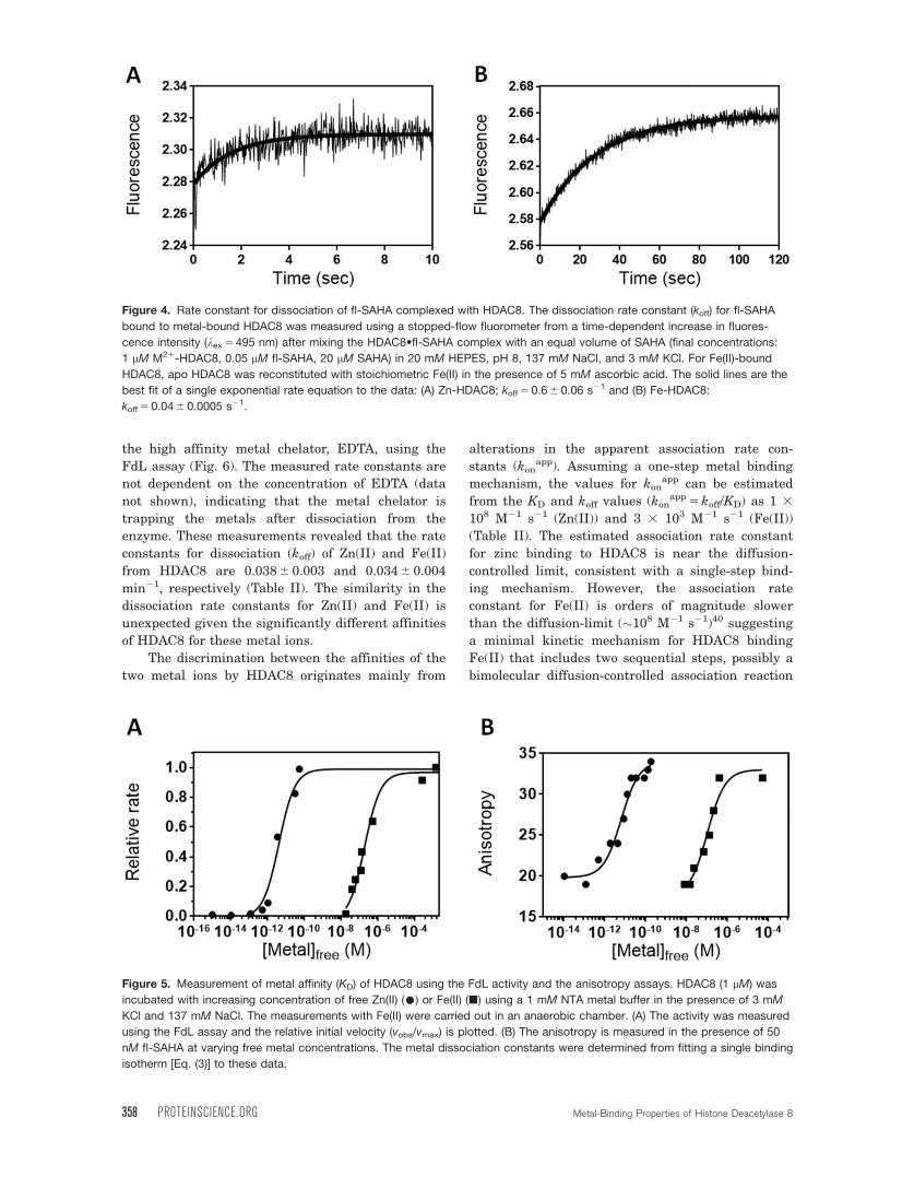
0.62 6 0.06 and 0.036 6 0.001 s21 for Zn(II)-bound
and Fe(II)-bound HDAC8, respectively (Table I).
These rate constants are unchanged when the con-
centration of SAHA is increased by twofold (data not
shown), demonstrating that trapping of HDAC8 by
SAHA is rapid and therefore the measured rate con-
stant reflects dissociation of fl-SAHA.
Metal binding properties of HDAC8
Zn(II) and Fe(II) are the most likely native cofactors
that activate HDAC8 in vivo since they exist in high
concentrations in the cell.29 The binding affinities
(KD) of HDAC8 for Zn(II) and Fe(II) were measured
from the metal-dependent activation of catalytic
activity using the FdL assay as a function of free
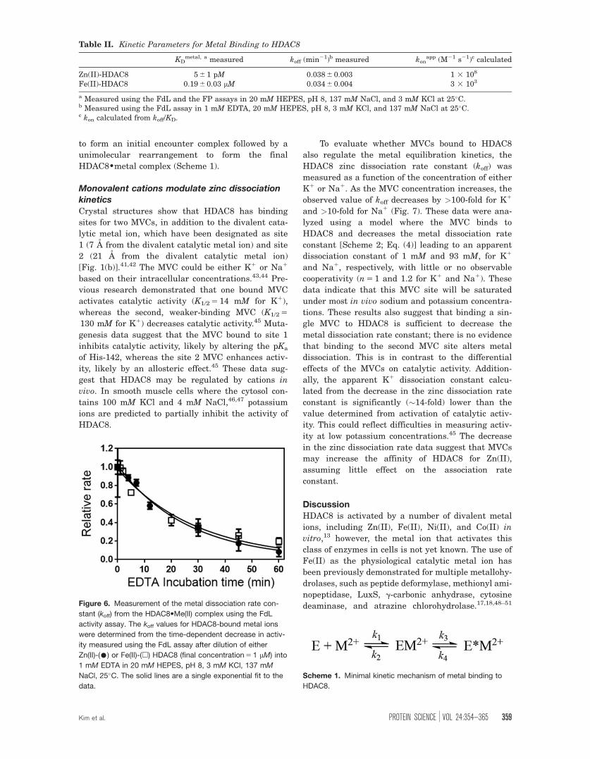
metal concentration [Fig. 5(a) and Table II,
KD,Zn 5 5 6 1 pM and KD,Fe 5 0.19 6 0.03 lM]. These
values are slightly lower than previously measured
metal affinities for HDAC8.9 This method requires
that the metal complex has significant catalytic
activity. In contrast, metal affinity could be meas-
ured for inactive complexes by coupling metal bind-
ing to fl-SAHA binding. To validate this method, the
fluorescence anisotropy of fl-SAHA in the presence
of HDAC8 was measured as a function of free metal
concentration in nitrilotriacetate (NTA)-buffered sol-
utions under equilibrium conditions. The fluores-
cence anisotropy increases with a hyperbolic
dependence on the free metal concentration [Fig.
5(b)] while the total fluorescence intensity decreases
less than twofold (data not shown). Dissociation con-
stants (KD) were obtained from fitting a single bind-
ing isotherm to these data (KD,Zn 5 6 6 1 pM and
KD,Fe 5 0.2 6 0.1 lM), in agreement with the affinity
measurements using the FdL assay. These data con-
firm that HDAC8 has significantly higher affinity
for Zn(II) compared with Fe(II), consistent with the
higher Lewis acidity of Zn(II).39
To examine the metal binding kinetics, the rate
constants for dissociation (koff) of the HDAC8-bound
metal ion were determined from the time-dependent
decrease in activity upon dilution of the enzyme into
Figure 3. Metal dependence of HDAC8 affinity for fl-SAHA.
Measurement of the binding affinity (KD) of apo-(D), Zn(II)-
bound (•), or Fe(II)-bound (�) HDAC8 for fl-SAHA from
changes in fluorescence anisotropy (kex 5485 nm,
kem 5 535 nm) upon addition of protein to 50 nM fl-SAHA in
20 mM HEPES, pH 8, 137 mM NaCl, and 3 mM KCl, at 25�C.
As a control, BSA (r) was titrated into fl-SAHA. The anisot-
ropy values are adjusted for a modest decrease in the total
fluorescence. The KD of Zn-HDAC8 and Fe-HDAC8 for fl-
SAHA are calculated as 0.4 6 0.1 lM and 0.1 6 0.08 lM,
respectively, from a fit of Eq. (1) to the data (solid lines). Little
change in anisotropy is observed for addition of either apo-
HDAC8 or BSA.
Table I. Kinetic Parameters of fl-SAHA Binding to Zn(II)- or Fe(II)-HDAC8
KDfl-SAHA (lM)a measured koff (s21)b measured kon (M21 s21)c calculated
Zn(II)-HDAC8 0.4 6 0.1 0.62 6 0.06 4 3 105
Fe(II)-HDAC8 0.10 6 0.08 0.036 6 0.001 1 3105
a Measured using the FP assay in 20 mM HEPES, pH 8, 137 mM NaCl, and 3 mM KCl, at 25�C.b Measured using stopped-flow fluorometry using the same conditions as in a.c kon calculated from koff/KD.
Kim et al. PROTEIN SCIENCE VOL 24:354—365 357
the high affinity metal chelator, EDTA, using the
FdL assay (Fig. 6). The measured rate constants are
not dependent on the concentration of EDTA (data
not shown), indicating that the metal chelator is
trapping the metals after dissociation from the
enzyme. These measurements revealed that the rate
constants for dissociation (koff) of Zn(II) and Fe(II)
from HDAC8 are 0.038 6 0.003 and 0.034 6 0.004
min21, respectively (Table II). The similarity in the
dissociation rate constants for Zn(II) and Fe(II) is
unexpected given the significantly different affinities
of HDAC8 for these metal ions.
The discrimination between the affinities of the
two metal ions by HDAC8 originates mainly from
alterations in the apparent association rate con-
stants (konapp). Assuming a one-step metal binding
mechanism, the values for konapp can be estimated
from the KD and koff values (konapp 5 koff/KD) as 1 3
108 M21 s21 (Zn(II)) and 3 3 103 M21 s21 (Fe(II))
(Table II). The estimated association rate constant
for zinc binding to HDAC8 is near the diffusion-
controlled limit, consistent with a single-step bind-
ing mechanism. However, the association rate
constant for Fe(II) is orders of magnitude slower
than the diffusion-limit (�108 M21 s21)40 suggesting
a minimal kinetic mechanism for HDAC8 binding
Fe(II) that includes two sequential steps, possibly a
bimolecular diffusion-controlled association reaction
Figure 4. Rate constant for dissociation of fl-SAHA complexed with HDAC8. The dissociation rate constant (koff) for fl-SAHA
bound to metal-bound HDAC8 was measured using a stopped-flow fluorometer from a time-dependent increase in fluores-
cence intensity (kex 5 495 nm) after mixing the HDAC8•fl-SAHA complex with an equal volume of SAHA (final concentrations:
1 lM M21-HDAC8, 0.05 lM fl-SAHA, 20 lM SAHA) in 20 mM HEPES, pH 8, 137 mM NaCl, and 3 mM KCl. For Fe(II)-bound
HDAC8, apo HDAC8 was reconstituted with stoichiometric Fe(II) in the presence of 5 mM ascorbic acid. The solid lines are the
best fit of a single exponential rate equation to the data: (A) Zn-HDAC8; koff 5 0.6 6 0.06 s21 and (B) Fe-HDAC8:
koff 5 0.04 6 0.0005 s21.
Figure 5. Measurement of metal affinity (KD) of HDAC8 using the FdL activity and the anisotropy assays. HDAC8 (1 lM) was
incubated with increasing concentration of free Zn(II) (•) or Fe(II) (�) using a 1 mM NTA metal buffer in the presence of 3 mM
KCl and 137 mM NaCl. The measurements with Fe(II) were carried out in an anaerobic chamber. (A) The activity was measured
using the FdL assay and the relative initial velocity (vobs/vmax) is plotted. (B) The anisotropy is measured in the presence of 50
nM fl-SAHA at varying free metal concentrations. The metal dissociation constants were determined from fitting a single binding
isotherm [Eq. (3)] to these data.
358 PROTEINSCIENCE.ORG Metal-Binding Properties of Histone Deacetylase 8
to form an initial encounter complex followed by a
unimolecular rearrangement to form the final
HDAC8•metal complex (Scheme 1).
Monovalent cations modulate zinc dissociation
kinetics
Crystal structures show that HDAC8 has binding
sites for two MVCs, in addition to the divalent cata-
lytic metal ion, which have been designated as site
1 (7 A from the divalent catalytic metal ion) and site
2 (21 A from the divalent catalytic metal ion)
[Fig. 1(b)].41,42 The MVC could be either K1 or Na1
based on their intracellular concentrations.43,44 Pre-
vious research demonstrated that one bound MVC
activates catalytic activity (K1/2 5 14 mM for K1),
whereas the second, weaker-binding MVC (K1/2 5
130 mM for K1) decreases catalytic activity.45 Muta-
genesis data suggest that the MVC bound to site 1
inhibits catalytic activity, likely by altering the pKa
of His-142, whereas the site 2 MVC enhances activ-
ity, likely by an allosteric effect.45 These data sug-
gest that HDAC8 may be regulated by cations in
vivo. In smooth muscle cells where the cytosol con-
tains 100 mM KCl and 4 mM NaCl,46,47 potassium
ions are predicted to partially inhibit the activity of
HDAC8.
To evaluate whether MVCs bound to HDAC8
also regulate the metal equilibration kinetics, the
HDAC8 zinc dissociation rate constant (koff) was
measured as a function of the concentration of either
K1 or Na1. As the MVC concentration increases, the
observed value of koff decreases by >100-fold for K1
and >10-fold for Na1 (Fig. 7). These data were ana-
lyzed using a model where the MVC binds to
HDAC8 and decreases the metal dissociation rate
constant [Scheme 2; Eq. (4)] leading to an apparent
dissociation constant of 1 mM and 93 mM, for K1
and Na1, respectively, with little or no observable
cooperativity (n 5 1 and 1.2 for K1 and Na1). These
data indicate that this MVC site will be saturated
under most in vivo sodium and potassium concentra-
tions. These results also suggest that binding a sin-
gle MVC to HDAC8 is sufficient to decrease the
metal dissociation rate constant; there is no evidence
that binding to the second MVC site alters metal
dissociation. This is in contrast to the differential
effects of the MVCs on catalytic activity. Addition-
ally, the apparent K1 dissociation constant calcu-
lated from the decrease in the zinc dissociation rate
constant is significantly (�14-fold) lower than the
value determined from activation of catalytic activ-
ity. This could reflect difficulties in measuring activ-
ity at low potassium concentrations.45 The decrease
in the zinc dissociation rate data suggest that MVCs
may increase the affinity of HDAC8 for Zn(II),
assuming little effect on the association rate
constant.
DiscussionHDAC8 is activated by a number of divalent metal
ions, including Zn(II), Fe(II), Ni(II), and Co(II) in
vitro,13 however, the metal ion that activates this
class of enzymes in cells is not yet known. The use of
Fe(II) as the physiological catalytic metal ion has
been previously demonstrated for multiple metallohy-
drolases, such as peptide deformylase, methionyl ami-
nopeptidase, LuxS, g-carbonic anhydrase, cytosine
deaminase, and atrazine chlorohydrolase.17,18,48–51
Table II. Kinetic Parameters for Metal Binding to HDAC8
KDmetal, a measured koff (min21)b measured kon
app (M21 s21)c calculated
Zn(II)-HDAC8 5 6 1 pM 0.038 6 0.003 1 3 108
Fe(II)-HDAC8 0.19 6 0.03 lM 0.034 6 0.004 3 3 103
a Measured using the FdL and the FP assays in 20 mM HEPES, pH 8, 137 mM NaCl, and 3 mM KCl at 25�C.b Measured using the FdL assay in 1 mM EDTA, 20 mM HEPES, pH 8, 3 mM KCl, and 137 mM NaCl at 25�C.c kon calculated from koff/KD.
Figure 6. Measurement of the metal dissociation rate con-
stant (koff) from the HDAC8•Me(II) complex using the FdL
activity assay. The koff values for HDAC8-bound metal ions
were determined from the time-dependent decrease in activ-
ity measured using the FdL assay after dilution of either
Zn(II)-(•) or Fe(II)-(w) HDAC8 (final concentration 5 1 lM) into
1 mM EDTA in 20 mM HEPES, pH 8, 3 mM KCl, 137 mM
NaCl, 25�C. The solid lines are a single exponential fit to the
data.
Scheme 1. Minimal kinetic mechanism of metal binding to
HDAC8.
Kim et al. PROTEIN SCIENCE VOL 24:354—365 359

These enzymes were originally misidentified as
Zn(II)-metalloenzymes due to purification under aero-
bic conditions resulting in replacement of the oxygen-
sensitive Fe(II) with the higher affinity Zn(II) cofac-
tor. The oxygen sensitivity of HDAC8 activity in E.
coli cell lysates originally suggested that HDAC8 may
be another non-heme Fe(II) hydrolase that was previ-
ously identified as a zinc metalloenzyme.13 Similarly,
the deacetylase activity of endogenously expressed
HDAC8 isolated from HeLa cells by immunoprecipita-
tion decreased upon exposure to oxygen (approxi-
mately twofold), suggesting that HDAC8 is partially
Fe-bound.52 These data are consistent with activation
of HDAC8 by a Fe(II) metal cofactor under many
physiological conditions.
An interesting possibility is that the metal ion
bound to HDAC8 may depend on the readily
exchangeable concentrations of Fe(II) and Zn(II); the
bound Fe(II) metal ion may exchange with zinc
under conditions where the cellular zinc concentra-
tion increases significantly, such as oxidative
stress,32 or due to the activity of metallochaper-
ones.32,53–55 While metallochaperones for Zn have
not yet been identified, the Philpott lab has demon-
strated that poly r(C)-binding protein 1 (PCBP1)
functions as a cytosolic iron chaperone in Saccharo-
myces cerevisiae to deliver iron to ferritin, a cellular
iron storage protein, as well as several hydroxy-
lases.56–58 A recent mass spectrometric analysis
evaluating HDAC8 protein binding partners sug-
gested interactions with the PCBP family of iron-
metallochaperones.59
To further explore metal activation of HDAC8
we have measured the metal binding kinetics and
thermodynamics of HDAC8 using the FdL activity
assay and an FP assay using a new fluorescent
probe (fl-SAHA). Notably, this FP assay can be used
to measure the metal binding kinetics and thermo-
dynamics of HDAC8 in the absence of catalytic
activity, allowing analysis of metal affinity in a wide
variety of conditions and with mutant enzymes. This
assay could also be developed as a high-throughput
screen for HDAC inhibitors and, potentially, for
imaging HDAC8 in cells since anisotropy measure-
ments are ratiometric.60–63
fl-SAHA binds to metal-bound HDAC8 in the
submicromolar range (0.4 lM for Zn(II)-bound and
0.1 lM for Fe(II)-bound), which is comparable to
both SAHA (KI 5 0.3 lM for Zn(II) and 0.1 lM for
Fe(II)-bound HDAC813) and coumarin-conjugated
SAHA (0.2 lM for HDAC8).23 This is reasonable
since both SAHA analogs contain the same aliphatic
hydroxamate moiety that chelates the active site
metal ion and can be accommodated in the hydro-
phobic tunnel of HDAC8.41 Additionally, fl-SAHA
displays similar KD values for the Zn(II)- and Fe(II)-
bound HDAC8 forms, as previously observed for
SAHA.13 Dissociation rate constants (koff) of fl-SAHA
from metal-bound HDAC8 were determined using
stopped-flow spectrometry as 0.6 s21 and 0.04 s21
for Zn(II)- and Fe(II)-HDAC8, respectively. Apparent
association rate constants of fl-SAHA to metal-
bound HDAC8 were calculated as 4 3 105 M21 s21
and 1 3 105 M21 s21 for Zn(II)- and Fe(II)-HDAC8,
respectively, assuming it is a single-binding step
mechanism (Table I). The differential effects of
metal-substitution on the association rate constants
may suggest a multistep binding mechanism.
The affinity of HDAC8 for Zn(II) is 105-fold
higher than Fe(II), as measured by either the
fl-SAHA FP assay or metal-dependent activation of
catalytic activity (KD,Zn 5 5 6 1 pM and KD,Fe 5
0.19 6 0.03 lM).9 However, the picomolar and micro-
molar affinities of HDAC8 for Zn(II) and Fe(II),
respectively, are in the range of estimated concentra-
tions of readily exchangeable metal ions in
cells.9,31,32,37 This similarity suggests that HDAC8
might be poised to switch between Fe(II)- and
Zn(II)-bound states in cells. Unexpectedly, the disso-
ciation rate constants (koff) of both metals
(0.0006 s21) are similar, despite the difference in the
binding affinities. This result indicates that metal
selectivity primarily resides in the apparent associa-
tion rate constant, estimated as 108 and 103 M21 s21
for Zn(II) and Fe(II), respectively, assuming a single
association step (Table II). The low value for the
apparent association rate constant for Fe(II)
Figure 7. Dependence of metal dissociation rate constant
(koff) from the HDAC8•Zn(II) complex on the concentration of
monovalent cations. The rate constants were determined as
described in the legend of Figure 6. The values for koff
decrease at increasing concentrations of KCl (•) or NaCl
(~). The solid line is a fit of Eq. (4) to the data.
Scheme 2. Coupled binding of MVC and DVC to HDAC8.
360 PROTEINSCIENCE.ORG Metal-Binding Properties of Histone Deacetylase 8
suggests that metal association is a two-step mecha-
nism (Scheme 1) where an initial encounter complex
forms followed by a unimolecular rearrangement to
form the final Me(II)-metal complex. This two-step
mechanism may describe both Zn(II) and Fe(II)
binding to HDAC8 with different rate-limiting steps;
formation of the encounter complex may be the slow
step for Zn(II) binding (k1) while the rearrangement
step may be rate-limiting for Fe(II) binding (k1k3/k2).
Metal dissociation could be limited in both cases by
the reverse rearrangement step (k4).
Previously, the metal association kinetics of
human carbonic anhydrase II (CAII) were deter-
mined with estimated association rate constants for
Zn(II) and Cu(II) of 104 to 105 and 109 M21 s21,
respectively.64 These data along with analysis of the
zinc binding kinetics of CAII mutants suggest a two-
step binding mechanism.64,65 In the first step, zinc
binds to CAII at near diffusion-controlled rates to
form an initial complex with two protein ligands fol-
lowed by a second slower step that includes
exchange of inner-sphere water molecules with the
third protein ligand.64 This may be a paradigm for
two-step metal binding to proteins, including
HDAC8. Metal binding kinetics similar to HDAC8
have been measured for the bacterial deacetylase,
LpxC, where the apparent association rate constants
for Zn(II) and Fe(II) are estimated as 106 and 104
M21 s21, respectively, also suggesting a two-step
binding mechanism for metal association.19 In this
case, the physiologically relevant metal of LpxC in
E. coli is Fe(II) under most conditions, although
LpxC binds Zn(II) in vivo when Fe(II) concentrations
are very low. In LpxC and CAII, the physiological
metal ions (Fe(II) and Zn(II), respectively) have a
slow dissociation rate constant (LpxC for Fe(II)
koff 5 0.067 6 0.004 min21 and CAII for Zn(II) koff 5
8.5 6 0.3 3 1025 min21), leading to an apparent asso-
ciation rate constant that is slower than the
diffusion-controlled limit.19,65 The varied dissociation
rate constants may be important for metal selectiv-
ity in vivo, decreasing kinetic traps for the higher
affinity, but incorrect metal ion, such as Zn(II) for
LpxC and possibly HDAC8 or Cu(II) for CAII.19,66
Furthermore, the similarities between the in vitro
metal binding kinetics and thermodynamics of LpxC
and HDAC8 fuel the hypothesis that HDAC8 might
be activated by Fe(II) under most conditions but
switches to the Zn(II) bound enzyme at high levels
of cellular zinc.19 This model suggests the possibility
of regulation of the activity of HDAC8 by metal
switching in response to conditions that alter the
cellular concentration of readily exchangeable metal
concentrations, such as redox stress.
The crystal structure of HDAC8 also displays
two monovalent metal binding sites that are con-
served in class I and II human HDACs.41,42 Site 1
and 2 are located 7 A and 21 A, respectively, from
the divalent metal ion binding site and could be
occupied by either K1 or Na1. Previous studies have
revealed that MVCs bound to site 1 inhibits HDAC8
activity, likely by altering the pKa of His-142,
whereas occupation of MVC site 2 increases activity,
likely by stabilizing the structure.45 These MVC
binding sites may also play a role in regulating
metal selectivity and equilibrium kinetics. The metal
dissociation kinetics of HDAC8 are influenced by the
concentration of monovalent cations; K1 and Na1
decrease the Zn(II) dissociation rate constant by
more than 40-fold and 12-fold, respectively, with no
cooperativity. These data are consistent with a
model in which MVCs bind to the high affinity site
(site 1) to stabilize the active conformation of
HDAC8 and thereby lower the metal dissociation
rate constant.
Here we demonstrate that despite the differences
in the binding affinity of HDAC8 for Zn(II) and Fe(II),
the dissociation rate constants are comparable at
0.0006 s21. Furthermore, the metal binding kinetics
suggest a two-step binding mechanism to HDAC8
where the metal selectivity is determined by the appa-
rent slower association rate constant for Fe(II) com-
pared with Zn(II). In the proposed model (Scheme 1)
the association rate constant for Zn(II) is limited by
formation of the encounter complex while the Fe(II)
association rate constant includes both the encounter
complex and the rearrangement step. Furthermore,
these data indicate that the kinetics and thermody-
namics of HDAC8 metal binding is dependent on the
concentration and identity of both the divalent metal
ions and the monovalent cations. These data are con-
sistent with the proposal that HDAC8 could exist as
an Fe(II)-dependent metalloenzyme as well as a Zn(II)-
dependent enzyme depending on cellular factors.
Materials and Methods
Materials
Unless specified, chemicals and supplies were pur-
chased from Fisher Scientific (MA). All chemicals
were of the highest quality available. Ethylenedia-
minetetraacetic acid (EDTA), FeCl2 (99.99%), and
Zn(NO3)2 were purchased from Aldrich (MO). Chro-
matography resins were purchased from GE Health-
care. To prevent trace metal contamination, all
plastic ware was presoaked with 1 mM EDTA and
rinsed three times with Millipure H2O. Plastic dis-
posables, including pipet tips and microcentrifuge
tubes, were certified trace metal-free (Corning Incor-
porated and Costar, respectively). Sodium hydroxide
(Sigma, 99.999%) was used to titrate 3-(N-morpholi-
no)propanesulfonic acid (MOPS) buffer (Ambion).
Expression and purification of HDAC8
Recombinant His6-HDAC8 was expressed in
BL21(DE3) E. coli transformed with pHD4 and
Kim et al. PROTEIN SCIENCE VOL 24:354—365 361
purified as previously described13 and then concen-
trated to 2 to 5 mg/mL for metal exchange. Metal-free
HDAC8 was generated by dialyzing purified HDAC8
twice into 500 mL of 25 mM MOPS (pH 7.0), 1 mM
EDTA for 12 to 14 h at 4�C, followed by buffer
exchange into 25 mM MOPS (pH 7.5), 0.1 mM EDTA,
and then 25 mM MOPS (pH 7.5) once each for 12 to
14 h at 4�C. When necessary, anaerobic conditions
were achieved using an anaerobic chamber (Coy, Grass
Lake, MI). For Fe(II) experiments, a freshly made
10 mM FeCl2 stock was prepared in 50 mM ascorbic
acid and diluted to 100 lM with 13 assay buffer
(20 mM HEPES, pH 8, 3 mM KCl, 137 mM NaCl)
before incubation with apo-HDAC8. Similarly, a
10 mM ZnSO4 solution was prepared in 20 mM
HEPES pH 8, 1 mM triscarboxyethylphosphine
(TCEP) and diluted to 100 lM with 13 assay buffer
before incubation with apo-HDAC8.
Synthesis of fl-SAHA
Synthesis of 2-(3,6-dihydroxy-9H-xanthen-9-
yl)25-(8-methoxy-8-oxooctanamido)benzoic acid
(1). To a flame-dried flask equipped with stir bar
and dry DMF (10 mL) was added 6-fluoresceinamine
(100 mg, 0.28 mmol, Sigma-Aldrich) in one portion
(Fig. 2). Then dry triethylamine (Et3N, 43 lL, 0.31
mmol) was added and the reaction was stirred at
room temperature for 1 h. Methyl-8-chloro-1-
oxooctanate (40 lL, 0.28 mmol) was then added to the
solution drop wise and the reaction mixture was
stirred at 50�C for 24 h. The solvent was then evapo-
rated in vacuo and the residue was dissolved in
dichloromethane (20 mL). The product was washed
with sat. NaHCO3 (3 3 20 mL) and NaCl (2 3 20 mL),
dried over anhydrous MgSO4, filtered and dried in
vacuo to yield an orange-yellow oil. The product was
purified via silica gel chromatography (4% MeOH in
CH2Cl2) and the resulting product was washed with
hexanes to remove residual impurities. The final
product was an orange oil (yield: 81 mg, 0.16 mmol,
56%). 1H NMR (CD3OD, 400 MHz)/d (ppm): 8.23 (d,
J 5 1.6 Hz, 1H), 7.73 (dd, J 5 8.0, 2.0 Hz, 2H), 7.04 (d,
J 5 8.4 Hz, 2H), 6.56 (d, J 5 2.4 Hz, 1H), 6.53 (d, J 5
8.8 Hz, 2H), 6.43 (dd, J 5 8.4, 2.4 Hz, 2H), 3.54 (s,
3H), 2.34 (t, J 5 7.6, 2H), 2.24 (t, J 5 7.6, 2H), 1.66–
1.52 (m, 4H), 1.32–1.30 (m, 4H). [M1H]1: calcd. 5
518.13, Found 5 518.06.
Synthesis of 2-(3,6-dihydroxy-9H-xanthen-9-
yl)25-(8-(hydroxyamino)28-oxooctanamido)ben-
zoic acid (fl-SAHA). Hydroxylamine hydrochlor-
ide (104 mg, 1.5 mmol) in methanol (10 mL) was
combined with a solution of KOH (1.57 g, 28 mmol) in
methanol (16 mL), cooled to 0�C, and then filtered. (1)
was added to the filtrate and KOH (5 mg in 1 mL
MeOH) was slowly added. The mixture was stirred at
room temperature for 2 h and then refluxed at 65�C
for 34 h. The reaction was quenched by the addition of
cold water (15 mL), followed by drop-wise addition of
glacial acetic acid until pH �7.0. The precipitated
solid product was filtered, washed with water to
remove impurities, dried under vacuum, and purified
twice by silica gel chromatography (25% MeOH in
CH2Cl2). The final product was recrystallized in
CH2Cl2 and hexanes to afford an orange solid (yield:
362 mg, 0.70 mmol, 75%). 1H NMR (CD3OD, 400
MHz)/d (ppm): 8.24 (d, J 5 1.6 Hz, 1H), 7.73 (dd,
J 5 8.0, 2.0 Hz, 2H), 7.04 (d, J 5 8.4 Hz, 2H), 6.56 (d,
J 5 2.4 Hz, 1H), 6.53 (d, J 5 8.8 Hz, 2H), 6.40 (dd,
J 5 8.4, 2.4 Hz, 2H), 2.32 (t, J 5 7.6, 2H), 2.24 (t,
J 5 7.6, 2H), 1.68–1.51 (m, 4H), 1.37–1.33 (m, 4H).
[M1H]1: calcd. 5 518.14, Found 5 518.11.
Affinity of HDAC8 for fl-SAHA
FP experiments were performed in a half-area black
96-well microplate (Corning Incorporation, #3686)
and FP values were measured with excitation at
485 nm (30 nm bandpass) and emission at 535 nm (40
nm bandpass) using a TECAN Plate Reader. Binding
experiments for determining the KD values of metal-
bound HDAC8 for fl-SAHA included 50 nM fl-SAHA
in assay buffer [20 mM HEPES (pH 8), 137 mM NaCl,
3 mM KCl] and FP was measured as HDAC8 (�5 lM)
was titrated into the solution. The fluorescence inten-
sity of fl-SAHA decreases by �25% upon binding to
HDAC8, even after correction for dilution and back-
ground fluorescence, as described previously.67 The
value of KD for fl-SAHA was obtained by fitting a
binding isotherm, including changes in the ligand
concentration (L) due to binding to E, to the depend-
ence of the FP signal on the concentration of HDAC8
[E, Eq. (1)]. In the equation, Pf is the polarization of
unbound fl-SAHA and Pb is signal from the fl-
SAHA�HDAC8 complex. All data analyses were per-
formed using Prism 6.0 (GraphPad Software).
Y5Pf1 Pb-Pfð Þ
3ð½L�1½E�1KDÞ2
ffiffiffiffiffiffiffiffiffiffiffiffiffiffiffiffiffiffiffiffiffiffiffiffiffiffiffiffiffiffiffiffiffiffiffiffiffiffiffiffiffiffiffiffiffið½L�1½E�1KDÞ224½L�
q½E�
2½E� (1)
Kinetics for binding fl-SAHA to HDAC8Fluorescence stopped-flow measurements were car-
ried out on a model SF-2001 stopped-flow spectro-
fluorometer (KinTek Corp., Austin, TX) fitted with a
75W xenon arc lamp in two syringe mode. fl-SAHA
was excited at 495 nm (slit width, 0.1–2 mm) and
fluorescence emission was monitored using a long-
pass filter (N500nm; Corion, LL-500-F). All kinetic
traces were an average of four to six independent
determinations. To measure the dissociation rate
constant (koff), fl-SAHA (0.1 lM) was preincubated
with metal-bound HDAC8 (2 lM) for at least 20 min
362 PROTEINSCIENCE.ORG Metal-Binding Properties of Histone Deacetylase 8

at RT in assay buffer. The reaction was initiated by
mixing the HDAC8•fl-SAHA complex with an equal
volume of unlabeled SAHA (final concentration of
20 lM) in assay buffer at 25�C to trap HDAC8 and
an increase in fluorescence intensity was measured.
The dissociation rate constant was determined from
a fit of a single exponential to the time-dependent
changes in the fluorescence signal [Eq. (2)]. In the
equation, DF is the observed change in fluorescence
and DF0 is the fluorescence at time zero.
DF5 D F0 3 exp 2koff 3 tð Þ (2)
HDAC8 metal ion binding affinityThe affinity of HDAC8 for Zn(II) or Fe(II) was meas-
ured by assaying catalytic activity in the presence of
varying Me(II)free concentrations in 1 mM nitrilotri-
acetic acid (NTA), 5 mM MOPS, pH 7, serving as
both a pH and metal buffer in the presence of 3 mM
KCl and 137 mM NaCl. The affinity was not depend-
ent on NTA concentration (data not shown).
Catalytic activity was assayed by using a
commercially-available fluorescent assay (BIOMOL)
with the Fluor de Lys (FdL) HDAC8 substrate
(RHK(ac)K(ac)-methyl-coumarin). All assay buffers
were pretreated with Chelex resin (Bio-Rad) to
remove trace divalent metal ions. Metal-free HDAC8
variants were reconstituted with Zn(II) or Fe(II) by
incubation with a stoichiometric concentration of
metal in assay buffer (20 mM HEPES pH 8.0,
137 mM NaCl, 3 mM KCl, and 1 mM TCEP). The
reaction was quenched by the addition of stoichio-
metric amounts of trichostatin A (TSA), a potent
inhibitor of HDAC8. Fluorescence intensity was
monitored at kex 5 340 and kem 5 450 for the deacety-
lated and cleaved product, and at kex 5 340 and
kem 5 380 for the starting acetylated substrate. The
ratio of product fluorescence divided by substrate
fluorescence was observed and linearly increases
with product concentration up to 30% product. The
amount of product formed was determined from
comparison to the fluorescence ratios of a standard
curve made up of known concentrations of products
and substrates. The linear initial rates catalyzed by
wtHDAC8 in assay buffer at 25�C were measured.
The affinity of HDAC8 for the metal was also
measured by changes in anisotropy by coupling the
binding of fl-SAHA to metal binding. These reactions
were carried out in 13 assay buffer containing 50
nM fl-SAHA. Fluorescence anisotropy assays were
performed in a black 96-well microplate by monitor-
ing the anisotropy signal of fluorescein (kex 5 485 nm
and kem 5 535 nm) using a TECAN plate-reader. For
Zn(II) affinity measurements, apo-HDAC8 (1–200
mM) was incubated with 1 mM NTA, 10 mM MOPS,
pH 7, and 0 to 0.5 mM Zntot (0–3.3 nM Znfree) at
25�C for 30 min.12 The concentration of Znfree in the
metal buffers was calculated using the program
MINEQL (Environmental Research software). For
measurement of KD for Fe(II), the assays contained
0 to 950 lM total iron (Sigma, 99.99%, Fe(II)free 5 0–
2.6 lM, as calculated by MINEQL) and 1 lM
HDAC8, in the presence of 3 mM KCl and 137 mM
NaCl. The assay mixtures were incubated for 2 h on
ice to pre-equilibrate in the anaerobic glove box fol-
lowed by addition of fl-SAHA and a short equilibra-
tion at 25�C before measuring the anisotropy signal.
The Zn(II) and Fe(II) KD values were obtained by fit-
ting a binding isotherm to the dependence of either
the activity or anisotropy on the concentration of
Mefree [Eq. (3), X]. In the equation, A is the activity,
DA is change in activity, FP is the fluorescence ani-
sotropy, and DFP is the change in anisotropy.
A
DA5
FP
DFP5
X
KD3X(3)
Metal ion dissociation rate constantsThe first order rate constant for M(II) dissociation
from HDAC8•M(II) complexes was measured by the
time-dependent loss of activity upon incubation with
EDTA. HDAC8 reconstituted with stoichiometric
Zn(II) or Fe(II) (final concentration 5 1 lM) was
diluted into assay buffer containing 1 mM EDTA at
25�C. At various times (0–60 min) an aliquot was
diluted 10-fold into assay buffer containing 50 lM
FdL substrate. The reactions were quenched by the
addition of TSA followed by trypsin developer. The
initial rate for product formation was determined for
each time point. The dissociation rate constant was
determined from a single exponential fit to the
decrease in the initial rate as a function of time.
Dependence of metal dissociation rate constanton monovalent cation (MVC) concentration
For measurement of the dependence of Zn(II) koff on
the concentration of MVC, reconstituted Zn(II)-
HDAC8 was incubated with varying concentration of
potassium or sodium ([KCl] 5 1–100 mM, [NaCl] 5
100–2000 mM) on ice for 1 h in 20 mM HEPES, pH
8.0 and then at 25�C for 4 min. The reaction was
started by the addition of 1 mM EDTA. The dissocia-
tion rate constant of Zn(II) was measured from the
decrease in catalytic activity using the FdL assay, as
described above. The MVC dependence of koff for
Zn(II) was fit by Eq. (4), which is derived from the
coupled binding of monovalent and divalent ions
shown in Scheme 2. In the equation, Keq is the appa-
rent equilibrium constant for MVC binding and koff
is the dissociation rate constant for Zn(II).
kobs5koff
11½MVC�n
Kneq
(4)
Acknowledgments
The authors gratefully acknowledge Dr. Ted Huston
(University of Michigan Geological Sciences) for
Kim et al. PROTEIN SCIENCE VOL 24:354—365 363
assistance with ICP-MS analysis. The authors thank
Dr. Anna Mapp and Dr. Aaron R. Van Dyke for gen-
erously providing materials for synthesis of fl-
SAHA. Carol Fierke is the recipient of the Protein
Society 2014 Emil Thomas Kaiser Award.
References
1. Strahl BD, Allis CD (2000) The language of covalenthistone modifications. Nature 403:41–45.
2. Choudhary C, Kumar C, Gnad F, Nielsen ML, RehmanM, Walther TC, Olsen JV, Mann M (2009) Lysine acety-lation targets protein complexes and co-regulates majorcellular functions. Science 325:834–840.
3. Wolfson NA, Pitcairn CA, Fierke CA (2013) HDAC8substrates: histones and beyond. Biopolymers 99:112–126.
4. Khochbin S, Verdel A, Lemercier C, Seigneurin-BernyD (2001) Functional significance of histone deacetylasediversity. Curr Opin Genet Dev 11:162–166.
5. Gregoretti IV, Lee YM, Goodson HV (2004) Molecularevolution of the histone deacetylase family: functionalimplications of phylogenetic analysis. J Mol Biol 338:17–31.
6. Blander G, Guarente L (2004) The Sir2 family of pro-tein deacetylases. Annu Rev Biochem 73:417–435.
7. Finnin MS, Donigian JR, Cohen A, Richon VM, RifkindRA, Marks PA, Breslow R, Pavletich NP (1999)Structures of a histone deacetylase homologue boundto the TSA and SAHA inhibitors. Nature 401:188–193.
8. Marek M, Kannan S, Hauser AT, Moraes Mour~ao M,Caby S, Cura V, Stolfa DA, Schmidtkunz K, Lancelot J,Andrade L, Renaud JP, Oliveira G, Sippl W, Jung M,Cavarelli J, Pierce RJ, Romier C (2013) Structuralbasis for the inhibition of histone deacetylase 8(HDAC8), a key epigenetic player in the blood flukeSchistosoma mansoni. PLoS Pathog 9:1–15.
9. Dowling DP, Gattis SG, Fierke CA, Christianson DW(2010) Structures of metal-substituted human histonedeacetylase 8 provide mechanistic inferences on biologi-cal function. Biochemistry 49:5048–5056.
10. Dowling DP, Gantt SL, Gattis SG, Fierke CA,Christianson DW (2008) Structural studies of humanhistone deacetylase 8 and its site-specific variants com-plexed with substrate and inhibitors. Biochemistry 47:13554–13563.
11. Vannini A, Volpari C, Gallinari P, Jones P, Mattu M,Carfi A, De Francesco R, Steinkuhler C, Di Marco S(2007) Substrate binding to histone deacetylases asshown by the crystal structure of the HDAC8-substratecomplex. EMBO Rep 8:879–884.
12. McCall KA, Fierke CA (2004) Probing determinants ofthe metal ion selectivity in carbonic anhydrase usingmutagenesis. Biochemistry 43:3979–3986.
13. Gantt SL, Gattis SG, Fierke CA (2006) Catalytic activ-ity and inhibition of human histone deacetylase 8 isdependent on the identity of the active site metal ion.Biochemistry 45:6170–6178.
14. Christianson DW, Cox JD (1999) Catalysis by metal-activated hydroxide in zinc and manganese metalloen-zymes. Annu Rev Biochem 68:33–57.
15. Auld DS (2001) Zinc coordination sphere in biochemicalzinc sites. Biometals 14:271–313.
16. Hernick M, Fierke CA (2005) Zinc hydrolases: themechanisms of zinc-dependent deacetylases. Arch Bio-chem Biophys 433:71–84.
17. Becker A, Schlichting I, Kabsch W, Groche D, SchultzS, Wagner AF (1998) Iron center, substrate recognition
and mechanism of peptide deformylase. Nat Struct Biol5:1053–1058.
18. Zhu J, Dizin E, Hu X, Wavreille AS, Park J, Pei D(2003) S-Ribosylhomocysteinase (LuxS) is a mononu-clear iron protein. Biochemistry 42:4717–4726.
19. Gattis SG, Hernick M, Fierke CA (2010) Active sitemetal ion in UDP-3-O-((R)23-Hydroxymyristoyl)-N-ace-tylglucosamine deacetylase (LpxC) switches betweenFe(II) and Zn(II) depending on cellular conditions.J Biol Chem 285:33788–33796.
20. Hernick M, Gattis SG, Penner-Hahn JE, Fierke CA(2010) Activation of Escherichia coli UDP-3-O-[(R)23-hydroxymyristoyl]-N-acetylglucosamine deacetylase byFe21 yields a more efficient enzyme with alteredligand affinity. Biochemistry 49:2246–2255.
21. Wegener D, Wirsching F, Riester D, Schwienhorst A(2003) A fluorogenic histone deacetylase assay wellsuited for high-throughput activity screening. ChemBiol 10:61–68.
22. Mazitschek R, Patel V, Wirth DF, Clardy J (2008)Development of a fluorescence polarization based assayfor histone deacetylase ligand discovery. Bioorg MedChem Lett 18:2809–2812.
23. Singh RK, Mandal T, Balasubramanian N, Cook G,Srivastava DK (2011) Coumarin-suberoylanilidehydroxamic acid as a fluorescent probe for determiningbinding affinities and off-rates of histone deacetylaseinhibitors. Anal Biochem 408:309–315.
24. Kristoffersen AS, Erga SR, Hamre B, Frette O (2014)Testing fluorescence lifetime standards using two-photon excitation and time-domain instrumentation:rhodamine B, coumarin 6 and lucifer yellow. J Fluoresc24:1015–1024.
25. Takagai Y, Nojiri Y, Takase T, Hinze WL, Butsugan M,Igarashi S (2010) "Turn-on" fluorescent polymericmicroparticle sensors for the determination of ammoniaand amines in the vapor state. Analyst 135:1417–1425.
26. Zheng H, Zhan XQ, Bian QN, Zhang XJ (2013) Advan-ces in modifying fluorescein and rhodamine fluoro-phores as fluorescent chemosensors. Chem Comm 49:429–447.
27. Martin MM, Lindqvist L (1975) pH-dependence of fluo-rescein fluorescence. J Lumin 10:381–390.
28. Irving H, Williams RJP (1948) Order of stability ofmetal complexes. Nature 162:746–747.
29. Outten CE, O’Halloran TV (2001) Femtomolar sensitiv-ity of metalloregulatory proteins controlling zinc home-ostasis. Science 292:2488–2492.
30. Wang D, Hosteen O, Fierke CA (2012) ZntR-mediatedtranscription of zntA responds to nanomolar intracellu-lar free zinc. J Inorg Biochem 111:173–181.
31. Vinkenborg JL, Nicolson TJ, Bellomo EA, Koay MS,Rutter GA, Merkx M (2009) Genetically encoded FRETsensors to monitor intracellular Zn21 homeostasis.Nat Methods 6:737–740.
32. Bozym RA, Thompson RB, Stoddard AK, Fierke CA(2006) Measuring picomolar intracellular exchangeablezinc in PC-12 cells using a ratiometric fluorescence bio-sensor. ACS Chem Biol 1:103–111.
33. Qin Y, Dittmer PJ, Park JG, Jansen KB, Palmer AE(2011) Measuring steady-state and dynamic endoplasmicreticulum and Golgi Zn21 with genetically encoded sen-sors. Proc Natl Acad Sci USA 108:7351–7356.
34. Petrat F, de Groot H, Rauen U (2001) Subcellular dis-tribution of chelatable iron: a laser scanning micro-scopic study in isolated hepatocytes and liverendothelial cells. Biochem J 356:61–69.
35. Meguro R, Asano Y, Odagiri S, Li C, Iwatsuki H,Shoumura K (2007) Nonheme-iron histochemistry for
364 PROTEINSCIENCE.ORG Metal-Binding Properties of Histone Deacetylase 8
light and electron microscopy: a historical, theoreticaland technical review. Arch Histol Cytol 70:1–19.
36. Esposito BP, Epsztejn S, Breuer W, Cabantchik ZI(2002) A review of fluorescence methods for assessinglabile iron in cells and biological fluids. Anal Biochem304:1–18.
37. MacKenzie EL, Iwasaki K, Tsuji Y (2008) Intracellulariron transport and storage: from molecular mecha-nisms to health implications. Antioxid Redox Signal10:997–1030.
38. Singh RK, Lall N, Leedahl TS, McGillivray A, MandalT, Haldar M, Mallik S, Cook G, Srivastava DK (2013)Kinetic and thermodynamic rationale for suberoylani-lide hydroxamic acid being a preferential human his-tone deacetylase 8 inhibitor as compared to thestructurally similar ligand, trichostatin a. Biochemistry52:8139–8149.
39. Pearson RG (1963) Hard and soft acids and bases.J Am Chem Soc 85:3533–3539.
40. Fersht AR (2000) Transition-state structure as a unify-ing basis in protein-folding mechanisms: contact order,chain topology, stability, and the extended nucleusmechanism. Proc Natl Acad Sci USA 97:1525–1529.
41. Vannini A, Volpari C, Filocamo G, Casavola EC,Brunetti M, Renzoni D, Chakravarty P, Paolini C, DeFrancesco R, Gallinari P, Steink€uhler C, Di Marco S(2004) Crystal structure of a eukaryotic zinc-dependenthistone deacetylase, human HDAC8, complexed with ahydroxamic acid inhibitor. Proc Natl Acad Sci USA101:15064–15069.
42. Somoza JR, Skene RJ, Katz BA, Mol C, Ho JD,Jennings AJ, Luong C, Arvai A, Buggy JJ, Chi E, TangJ, Sang BC, Verner E, Wynands R, Leahy EM, DouganDR, Snell G, Navre M, Knuth MW, Swanson RV,McRee DE, Tari LW (2004) Structural snapshots ofhuman HDAC8 provide insights into the class I histonedeacetylases. Structure 12:1325–1334.
43. Borin ML, Goldman WF, Blaustein MP (1993) Intracel-lular free Na1 in resting and activated A7r5 vascularsmooth-muscle cells. Am J Physiol 264:C1513–C1524.
44. Woehl EU, Dunn MF (1995) The roles of Na1 and K1
in pyridoxal-phosphate enzyme catalysis. Coord ChemRev 144:147–197.
45. Gantt SL, Joseph CG, Fierke CA (2010) Activation andinhibition of histone deacetylase 8 by monovalent cati-ons. J Biol Chem 285:6036–6043.
46. Kasner SE, Ganz MB (1992) Regulation of intracellularpotassium in mesangial cells: a fluorescence analysisusing the dye, PBFI. Am J Physiol 262:F462–F467.
47. Borin ML, Goldman WF, Blaustein MP (1993) Intracel-lular free Na1 in resting and activated A7r5 vascularsmooth muscle cells. Am J Physiol 264:C1513–C1524.
48. Tripp BC, Bell CB, 3rd, Cruz F, Krebs C, Ferry JG(2004) A role for iron in an ancient carbonic anhydrase.J Biol Chem 279:6683–6687.
49. D’Souza V M, Holz RC (1999) The methionyl amino-peptidase from Escherichia coli can function as aniron(II) enzyme. Biochemistry 38:11079–11085.
50. Porter DJ, Austin EA (1993) Cytosine deaminase. Theroles of divalent metal ions in catalysis. J Biol Chem268:24005–24011.
51. Seffernick JL, McTavish H, Osborne JP, de Souza ML,Sadowsky MJ, Wackett LP (2002) Atrazine chlorohy-
drolase from Pseudomonas sp. strain ADP is a metallo-enzyme. Biochemistry 41:14430–14437.
52. Gattis SG (2010) Mechanism and metal specificity ofzinc-dependent deacetylases. PhD. Thesis. Universityof Michigan, pp 143–145.
53. Waldron KJ, Rutherford JC, Ford D, Robinson NJ(2009) Metalloproteins and metal sensing. Nature 460:823–830.
54. Rae TD, Schmidt PJ, Pufahl RA, Culotta VC,O’Halloran TV (1999) Undetectable intracellular freecopper: the requirement of a copper chaperone forsuperoxide dismutase. Science 284:805–808.
55. O’Halloran TV, Culotta VC (2000) Metallochaperones,an intracellular shuttle service for metal ions. J BiolChem 275:25057–25060.
56. Shi H, Bencze KZ, Stemmler TL, Philpott CC (2008) Acytosolic iron chaperone that delivers iron to ferritin.Science 320:1207–1210.
57. Nandal A, Ruiz JC, Subramanian P, Ghimire-Rijal S,Sinnamon RA, Stemmler TL, Bruick RK, Philpott CC(2011) Activation of the HIF prolyl hydroxylase by theiron chaperones PCBP1 and PCBP2. Cell Metab 14:647–657.
58. Frey AG, Nandal A, Park JH, Smith PM, Yabe T, RyuMS, Ghosh MC, Lee J, Rouault TA, Park MH, PhilpottCC (2014) Iron chaperones PCBP1 and PCBP2 mediatethe metallation of the dinuclear iron enzyme deoxyhy-pusine hydroxylase. Proc Natl Acad Sci USA 111:8031–8036.
59. Joshi P, Greco TM, Guise AJ, Luo Y, Yu F, NesvizhskiiAI, Cristea IM (2013) The functional interactome land-scape of the human histone deacetylase family. MolSyst Biol 9:1–21.
60. Thompson RB, Maliwal BP, Fierke CA (1998) Determi-nation of metal ions by fluorescence anisotropy exhibitsa broad dynamic range. Adv Opt Biophys 3256:51–59.
61. Suhling K, Siegel J, Lanigan PM, Leveque-Fort S,Webb SE, Phillips D, Davis DM, French PM (2004)Time-resolved fluorescence anisotropy imaging appliedto live cells. Opt Lett 29:584–586.
62. Levitt JA, Matthews DR, Ameer-Beg SM, Suhling K(2009) Fluorescence lifetime and polarization-resolvedimaging in cell biology. Curr Opin Biotechnol 20:28–36.
63. Wang D, Hurst TK, Thompson RB, Fierke CA (2011)Genetically encoded ratiometric biosensors to measureintracellular exchangeable zinc in Escherichia coli.J Biomed Opt 16:1–11.
64. Huang CC, Lesburg CA, Kiefer LL, Fierke CA,Christianson DW (1996) Reversal of the hydrogen bondto zinc ligand histidine-119 dramatically diminishescatalysis and enhances metal equilibration kinetics incarbonic anhydrase II. Biochemistry 35:3439–3446.
65. Kiefer LL, Paterno SA, Fierke CA (1995) Hydrogen-bond network in the metal-binding site of carbonic-anhydrase enhances zinc affinity and catalytic effi-ciency. J Am Chem Soc 117:6831–6837.
66. Hurst TK, Wang D, Thompson RB, Fierke CA (2010)Carbonic anhydrase II-based metal ion sensing: Advan-ces and new perspectives. Biochim Biophys Acta 1804:393–403.
67. Lohman TM, Mascotti DP (1992) Thermodynamics ofligand-nucleic acid interactions. Methods Enzymol 212:400–424.
Kim et al. PROTEIN SCIENCE VOL 24:354—365 365
Related Documents