*For correspondence: faessler@ biochem.mpg.de Competing interests: The authors declare that no competing interests exist. Funding: See page 21 Received: 15 July 2015 Accepted: 19 December 2015 Published: 28 January 2016 Reviewing editor: Vivek Malhotra, The Barcelona Institute of Science and Technology, Barcelona, Spain Copyright Theodosiou et al. This article is distributed under the terms of the Creative Commons Attribution License, which permits unrestricted use and redistribution provided that the original author and source are credited. Kindlin-2 cooperates with talin to activate integrins and induces cell spreading by directly binding paxillin Marina Theodosiou 1 , Moritz Widmaier 1 , Ralph T Bo ¨ ttcher 1 , Emanuel Rognoni 1 , Maik Veelders 1 , Mitasha Bharadwaj 2 , Armin Lambacher 1 , Katharina Austen 1 , Daniel J Mu ¨ ller 2 , Roy Zent 3,4 , Reinhard Fa ¨ ssler 1 * 1 Department of Molecular Medicine, Max Planck Institute of Biochemistry, Martinsried, Germany; 2 Department of Biosystems Science and Engineering, Eidgeno ¨ ssische Technische Hochschule Zu ¨ rich, Basel, Switzerland; 3 Division of Nephrology, Department of Medicine, Vanderbilt University, Nashville, United States; 4 Department of Medicine, Veterans Affairs Medical Center, Nashville, United States Abstract Integrins require an activation step prior to ligand binding and signaling. How talin and kindlin contribute to these events in non-hematopoietic cells is poorly understood. Here we report that fibroblasts lacking either talin or kindlin failed to activate b1 integrins, adhere to fibronectin (FN) or maintain their integrins in a high affinity conformation induced by Mn 2+ . Despite compromised integrin activation and adhesion, Mn 2+ enabled talin- but not kindlin-deficient cells to initiate spreading on FN. This isotropic spreading was induced by the ability of kindlin to directly bind paxillin, which in turn bound focal adhesion kinase (FAK) resulting in FAK activation and the formation of lamellipodia. Our findings show that talin and kindlin cooperatively activate integrins leading to FN binding and adhesion, and that kindlin subsequently assembles an essential signaling node at newly formed adhesion sites in a talin-independent manner. DOI: 10.7554/eLife.10130.001 Introduction Integrins are heterodimeric transmembrane receptors that mediate cell adhesion to the extracellular matrix (ECM) and to other cells (Hynes, 2002). The consequence of integrin-mediated adhesion is the assembly of a large molecular network that induces various signaling pathways, resulting in cell migration, proliferation, survival and differentiation (Winograd-Katz et al., 2014). The quality and strength of integrin signaling is controlled by the interaction between integrins and substrate- attached ligands, which is, in turn, regulated by the on- and off-rates of the integrin–ligand binding process. The on-rate of the integrin–ligand binding reaction (also called integrin activation or inside- out signaling) is characterized by switching the unbound form of integrins from an inactive (low affin- ity) to an active (high affinity) conformation. The affinity switch proceeds from a bent and clasped low affinity conformation to an extended and unclasped high affinity conformation with an open ligand-binding pocket (Askari et al., 2010; Springer and Dustin, 2012). This change in affinity is believed to be induced through the binding of talin and kindlin to the b integrin cytoplasmic domain (Moser et al., 2009; Shattil et al., 2010) and divalent cations to distinct sites close to the ligand- binding pocket (Gailit and Ruoslahti, 1988; Mould et al., 1995; Xia and Springer, 2014; Mould et al., 2003). The stabilisation of integrin–ligand complexes is mediated by integrin clustering and catch bond formation between integrin and bound ligand. The stabilizing effect of clustered integrins is Theodosiou et al. eLife 2015;5:e10130. DOI: 10.7554/eLife.10130 1 of 24 RESEARCH ARTICLE

Welcome message from author
This document is posted to help you gain knowledge. Please leave a comment to let me know what you think about it! Share it to your friends and learn new things together.
Transcript
*For correspondence: faessler@
biochem.mpg.de
Competing interests: The
authors declare that no
competing interests exist.
Funding: See page 21
Received: 15 July 2015
Accepted: 19 December 2015
Published: 28 January 2016
Reviewing editor: Vivek
Malhotra, The Barcelona Institute
of Science and Technology,
Barcelona, Spain
Copyright Theodosiou et al.
This article is distributed under
the terms of the Creative
Commons Attribution License,
which permits unrestricted use
and redistribution provided that
the original author and source are
credited.
Kindlin-2 cooperates with talin to activateintegrins and induces cell spreading bydirectly binding paxillinMarina Theodosiou1, Moritz Widmaier1, Ralph T Bottcher1, Emanuel Rognoni1,Maik Veelders1, Mitasha Bharadwaj2, Armin Lambacher1, Katharina Austen1,Daniel J Muller2, Roy Zent3,4, Reinhard Fassler1*
1Department of Molecular Medicine, Max Planck Institute of Biochemistry,Martinsried, Germany; 2Department of Biosystems Science and Engineering,Eidgenossische Technische Hochschule Zurich, Basel, Switzerland; 3Division ofNephrology, Department of Medicine, Vanderbilt University, Nashville, UnitedStates; 4Department of Medicine, Veterans Affairs Medical Center, Nashville, UnitedStates
Abstract Integrins require an activation step prior to ligand binding and signaling. How talin and
kindlin contribute to these events in non-hematopoietic cells is poorly understood. Here we report
that fibroblasts lacking either talin or kindlin failed to activate b1 integrins, adhere to fibronectin
(FN) or maintain their integrins in a high affinity conformation induced by Mn2+. Despite
compromised integrin activation and adhesion, Mn2+ enabled talin- but not kindlin-deficient cells to
initiate spreading on FN. This isotropic spreading was induced by the ability of kindlin to directly
bind paxillin, which in turn bound focal adhesion kinase (FAK) resulting in FAK activation and the
formation of lamellipodia. Our findings show that talin and kindlin cooperatively activate integrins
leading to FN binding and adhesion, and that kindlin subsequently assembles an essential signaling
node at newly formed adhesion sites in a talin-independent manner.
DOI: 10.7554/eLife.10130.001
IntroductionIntegrins are heterodimeric transmembrane receptors that mediate cell adhesion to the extracellular
matrix (ECM) and to other cells (Hynes, 2002). The consequence of integrin-mediated adhesion is
the assembly of a large molecular network that induces various signaling pathways, resulting in cell
migration, proliferation, survival and differentiation (Winograd-Katz et al., 2014). The quality and
strength of integrin signaling is controlled by the interaction between integrins and substrate-
attached ligands, which is, in turn, regulated by the on- and off-rates of the integrin–ligand binding
process. The on-rate of the integrin–ligand binding reaction (also called integrin activation or inside-
out signaling) is characterized by switching the unbound form of integrins from an inactive (low affin-
ity) to an active (high affinity) conformation. The affinity switch proceeds from a bent and clasped
low affinity conformation to an extended and unclasped high affinity conformation with an open
ligand-binding pocket (Askari et al., 2010; Springer and Dustin, 2012). This change in affinity is
believed to be induced through the binding of talin and kindlin to the b integrin cytoplasmic domain
(Moser et al., 2009; Shattil et al., 2010) and divalent cations to distinct sites close to the ligand-
binding pocket (Gailit and Ruoslahti, 1988; Mould et al., 1995; Xia and Springer, 2014;
Mould et al., 2003).
The stabilisation of integrin–ligand complexes is mediated by integrin clustering and catch bond
formation between integrin and bound ligand. The stabilizing effect of clustered integrins is
Theodosiou et al. eLife 2015;5:e10130. DOI: 10.7554/eLife.10130 1 of 24
RESEARCH ARTICLE
achieved by the ability of dissociated integrin–ligand complexes to rebind before they leave the
adhesion site (van Kooyk and Figdor, 2000; Roca-Cusachs et al., 2009), while catch bonds are
receptor–ligand bonds whose lifetime increases with mechanical force (Kong et al., 2009;
Chen et al., 2010; Kong et al., 2013). Both mechanisms extend the duration and increase the
strength of integrin-mediated adhesion and signaling (also called outside-in signaling) (Koo et al.,
2002; van Kooyk and Figdor, 2000; Maheshwari et al., 2000; Roca-Cusachs et al., 2009;
Coussen et al., 2002), and depend on the association of integrins with the actin cytoskeleton via
talin (Roca-Cusachs et al., 2009; Friedland et al., 2009), and probably kindlin (Ye et al., 2013).
The talin family consists of two (talin-1 and -2) and the kindlin family of three isoforms (kindlin-1-
3), which show tissue-specific expression patterns (Calderwood et al., 2013; Moser et al., 2009;
Shattil et al., 2010). The majority of studies that defined integrin affinity regulation by talin and
kindlin were performed on aIIbb3 and b2-class integrins expressed by platelets and leukocytes,
respectively. These cells circulate in the blood and hold their integrins in an inactive state until they
encounter soluble or membrane-bound agonists (Evans et al., 2009, Bennett, 2005). The prevailing
view is that agonist-induced signaling pathways activate talin-1 and the hematopoietic cell-specific
kindlin-3, which cooperate to induce integrin activation (Moser et al., 2008; Han et al., 2006) and
clustering (Cluzel et al., 2005; Ye et al., 2013).
Integrin affinity regulation in non-hematopoietic cells such as fibroblasts and epithelial cells is
poorly understood. It is not known how integrin activation is induced on these cells (no integrin-
activating agonists have been identified) and it is also controversial whether talin and kindlin are
required to shift their integrins into the high affinity state. While there are reports showing that talin
and kindlin are required for integrin activation in epithelial cells (Montanez et al., 2008;
Margadant et al., 2012), it was also shown that in myoblasts and mammary epithelial cells activation
of b1 integrins, adhesion and spreading on multiple ECM substrates can proceed in the absence of
talin (Conti et al., 2009; Wang et al., 2011). Likewise, it was reported that focal adhesion
eLife digest A meshwork of proteins called the extracellular matrix surrounds the cells that
make up our tissues. Integrins are adhesion proteins that sit on the membrane surrounding each cell
and bind to the matrix proteins. These adhesive interactions control many aspects of cell behavior
such as their ability to divide, move and survive.
Before integrins can bind to the extracellular matrix they must be activated. Previous research has
shown that in certain types of blood cells, proteins called talins and kindlins perform this activation.
These proteins bind to the part of the integrin that extends into the cell, causing shape changes to
the integrin that allow binding to the extracellular matrix. However, it is not clear whether talin and
kindlin also activate integrins in other cell types.
Fibroblasts are cells that help to make extracellular matrix proteins, and are an important part of
connective tissue. Theodosiou et al. engineered mouse fibroblast cells to lack either talin or kindlin,
and found that both of these mutant cell types were unable to activate their integrins and as a result
failed to bind to an extracellular matrix protein called fibronectin.
Even when cells were artificially induced to activate integrins by treating them with manganese
ions, cells lacking talin or kindlin failed to fully activate integrins and hence did not adhere well to
fibronectin. This suggests that talin and kindlin work together to activate integrins and to maintain
them in this activated state.
When treated with manganese ions, cells that lacked talin were able to flatten and spread out,
whereas cells that lacked kindlin were unable to undergo this shape change. Theodosiou et al. found
that this cell shape is dependent on kindlin and its ability to bind to and recruit a protein called
paxillin to “adhesion sites”, where integrins connect the cell surface with the extracellular matrix.
Kindlin and paxillin then work together to activate other signaling molecules to induce the cell
spreading.
The next challenge is to understand how talin and kindlin are activated in non-blood cells and
how they maintain integrins in an active state.
DOI: 10.7554/eLife.10130.002
Theodosiou et al. eLife 2015;5:e10130. DOI: 10.7554/eLife.10130 2 of 24
Research article Biochemistry Cell biology
kinase (FAK)-deficient fibroblasts develop small, nascent adhesions (NAs) at the edge of membrane
protrusions without visible talin and that integrins carrying a mutation in the talin-binding site can
still nucleate and stabilize NAs (Lawson et al., 2012). Also fibroblasts lacking talin-1 and -2 were
shown to adhere to fibronectin (FN) and initiate isotropic spreading (Zhang et al., 2008). Another
intriguing study demonstrated that overexpression of kindlin-2 in Chinese hamster ovary (CHO) cells
inhibits rather than promotes talin head-induced a5b1 integrin activation (Harburger et al., 2009).
Given the fundamental importance of talin and kindlin for integrin activation in hematopoietic cells,
the findings of these studies are unexpected and imply that either integrin affinity regulation is sub-
stantially different in fibroblasts and epithelial cells or the experimental approaches used to manipu-
late protein expression and localization were imperfect.
To directly evaluate the functions of talin and kindlins for FN-binding integrins on fibroblasts, we
used a genetic approach and derived fibroblasts from mice lacking either the Tln1 and -2 or the
Fermt1 and -2 genes. We show that integrin affinity regulation depends on both talin and kindlin,
and that kindlin has the additional function of triggering cell spreading by binding directly to paxillin
in a talin-independent manner.
Results
Kindlins and talins control cell morphology, adhesion and integrinexpressionTo obtain cells lacking the expression of talin-1 and kindlin-2, we intercrossed mice carrying loxP
flanked (floxed; fl) Tln1 or Fermt2 alleles (Figure 1A), isolated kidney fibroblasts and immortalized
them with the SV40 large T antigen (parental fibroblasts). The floxed alleles were deleted by adeno-
viral Cre recombinase transduction resulting in T1Ko and K2Ko fibroblasts. Loss of talin-1 or kindlin-2
expression in fibroblasts was compensated by talin-2 or the de novo expression of kindlin-1, respec-
tively, allowing adhesion and spreading, although to a lesser extent compared with control cells (Fig-
ure 1—figure supplement 1A,B). To prevent this compensation, we generated mice with floxed
Tln1 and nullizygous Tln2 alleles or with floxed Fermt1 and -2 alleles (TlnCtr; KindCtr) from which we
isolated, immortalized and cloned kidney fibroblasts with comparable integrin surface levels
(Figure 1A and Figure 1—figure supplement 2). The floxed alleles were deleted by transducing
Cre resulting in talin-1, -2 (TlnKo) and kindlin-1, -2 (KindKo) deficient cells, respectively (Figure 1A–C).
Since the TlnCtr and KindCtr control cells showed similar morphologies and behaviour in our experi-
ments, we display one control cell line in several result panels. Cre-mediated deletion of the floxed
Tln1 or floxed Fermt1/2 genes was efficient (Figure 1B) and resulted in cell rounding, weak adhesion
of a few cells, and reduced cell proliferation despite the immortalisation with the oncogenic large T
antigen (Figure 1C and Figure 1—figure supplement 3). To minimize cell passage-induced abnor-
malities, we used cells only up to 12 passages after Cre-mediated gene deletions.
To define the adhesion defect, we performed plate and wash assays for 30 min on defined sub-
strates and found that neither TlnKo nor KindKo cells adhered to FN, laminin-111 (LN), type I collagen
(COL) and vitronectin (VN) (Figure 1D). To test whether the inability of TlnKo and KindKo cells to
adhere to ECM proteins is due to an integrin activation defect, we bypassed inside-out activation by
treating cells with Mn2+, which binds to the integrin ectodomain and induces unbending and
unclasping of integrin heterodimers (Mould et al., 1995). Treatment with Mn2+ induced partial
adhesion of TlnKo and KindKo cells to FN, while partial adhesion to LN and VN was only induced in
TlnKo cells (Figure 1D). Time course experiments revealed that Mn2+-induced adhesion of TlnKo and
KindKo cells to FN was already significantly lower 2.5 min after plating and remained significantly
lower compared with control cells (Figure 1E), suggesting that talin and kindlin cooperate to initiate
and maintain normal Mn2+-induced adhesion to FN. In line with these findings, dose-response pro-
files showed that TlnKo and KindKo cells have severe adhesion defects at low (1.25 mg ml–1) as well as
high (20 mg ml–1) substrate concentrations (Figure 1—figure supplement 4).
These findings indicate that talin and kindlin promote integrin-mediated adhesion to FN and pro-
liferation, and that the integrin-activating compound Mn2+ can only partially substitute for the adhe-
sion promoting roles that talin and kindlin accomplish together.
Theodosiou et al. eLife 2015;5:e10130. DOI: 10.7554/eLife.10130 3 of 24
Research article Biochemistry Cell biology
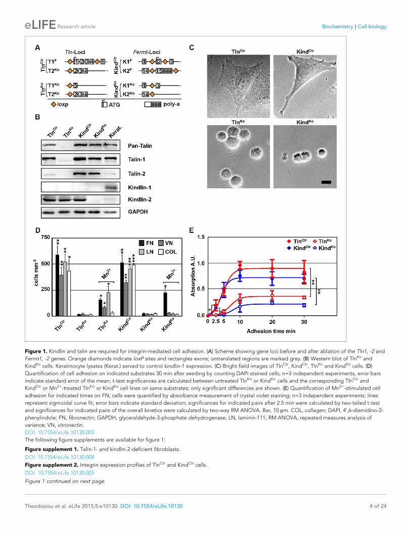
Figure 1. Kindlin and talin are required for integrin-mediated cell adhesion. (A) Scheme showing gene loci before and after ablation of the Tln1, -2 and
Fermt1, -2 genes. Orange diamonds indicate loxP sites and rectangles exons; untranslated regions are marked grey. (B) Western blot of TlnKo and
KindKo cells. Keratinocyte lysates (Kerat.) served to control kindlin-1 expression. (C) Bright field images of TlnCtr, KindCtr, TlnKo and KindKo cells. (D)
Quantification of cell adhesion on indicated substrates 30 min after seeding by counting DAPI stained cells; n=3 independent experiments, error bars
indicate standard error of the mean; t-test significances are calculated between untreated TlnKo or KindKo cells and the corresponding TlnCtr and
KindCtr or Mn2+-treated TlnKo or KindKo cell lines on same substrates; only significant differences are shown. (E) Quantification of Mn2+-stimulated cell
adhesion for indicated times on FN; cells were quantified by absorbance measurement of crystal violet staining; n=3 independent experiments; lines
represent sigmoidal curve fit; error bars indicate standard deviation; significances for indicated pairs after 2.5 min were calculated by two-tailed t-test
and significances for indicated pairs of the overall kinetics were calculated by two-way RM ANOVA. Bar, 10 mm. COL, collagen; DAPI, 4’,6-diamidino-2-
phenylindole; FN, fibronectin; GAPDH, glyceraldehyde-3-phosphate dehydrogenase; LN, laminin-111; RM ANOVA, repeated measures analysis of
variance; VN, vitronectin.
DOI: 10.7554/eLife.10130.003
The following figure supplements are available for figure 1:
Figure supplement 1. Talin-1- and kindlin-2-deficient fibroblasts.
DOI: 10.7554/eLife.10130.004
Figure supplement 2. Integrin expression profiles of TlnCtr and KindCtr cells.
DOI: 10.7554/eLife.10130.005
Figure 1 continued on next page
Theodosiou et al. eLife 2015;5:e10130. DOI: 10.7554/eLife.10130 4 of 24
Research article Biochemistry Cell biology
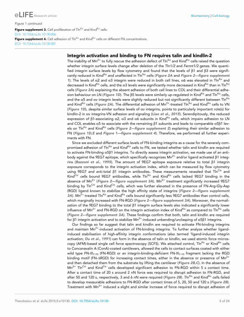
Integrin activation and binding to FN requires talin and kindlin-2The inability of Mn2+ to fully rescue the adhesion defect of TlnKo and KindKo cells raised the question
whether integrin surface levels change after deletion of the Tln1/2 and Fermt1/2 genes. We quanti-
fied integrin surface levels by flow cytometry and found that the levels of b1 and b3 were signifi-
cantly reduced in KindKo and unaffected in TlnKo cells (Figure 2A and Figure 2—figure supplement
1). The levels of a2 and a3 integrin were reduced in both cell lines, a6 was elevated in TlnKo and
decreased in KindKo cells, and the a3 levels were significantly more decreased in KindKo than in TlnKo
cells (Figure 2A) explaining the absent adhesion of both cell lines to COL and their differential adhe-
sion behaviour on LN (Figure 1D). The b5 levels were similarly up-regulated in KindKo and TlnKo cells,
and the a5 and av integrin levels were slightly reduced but not significantly different between TlnKo
and KindKo cells (Figure 2A). The differential adhesion of Mn2+-treated TlnKo and KindKo cells to VN
(Figure 1D), despite similar surface levels of av integrins, points to particularly important role(s) for
kindlin-2 in av integrins-VN adhesion and signaling (Liao et al., 2015). Serendipitously, the reduced
expression of b1-associating a2, a3 and a6 subunits in KindKo cells, which impairs adhesion to LN
and COL enables a5 to associate with the remaining b1 subunits and leads to comparable a5b1 lev-
els on TlnKo and KindKo cells (Figure 2—figure supplement 2) explaining their similar adhesion to
FN (Figure 1D,E and Figure 1—figure supplement 4). Therefore, we performed all further experi-
ments with FN.
Since we excluded different surface levels of FN-binding integrins as a cause for the severely com-
promised adhesion of TlnKo and KindKo cells to FN, we tested whether talin and kindlin are required
to activate FN-binding a5b1 integrins. To directly assess integrin activation, we made use of an anti-
body against the 9EG7 epitope, which specifically recognizes Mn2+ and/or ligand activated b1 integ-
rins (Bazzoni et al., 1995). The amount of 9EG7 epitope exposure relative to total b1 integrin
exposure corresponds to the integrin activation index, which can be measured by flow cytometry
using 9EG7 and anti-total b1 integrin antibodies. These measurements revealed that TlnCtr and
KindCtr cells bound 9EG7 antibodies, while TlnKo and KindKo cells lacked 9EG7 binding in the
absence of Mn2+ (Figure 2—figure supplement 3A). Mn2+ treatment significantly increased 9EG7
binding by TlnCtr and KindCtr cells, which was further elevated in the presence of FN-Arg-Gly-Asp
(RGD) ligand known to stabilize the high affinity state of integrins (Figure 2—figure supplement
3A). Mn2+-treated TlnKo and KindKo cells bound significantly less 9EG7 antibodies than control cells,
which marginally increased with FN-RGD (Figure 2—figure supplement 3A). Moreover, the normali-
zation of the 9EG7 binding to the total b1 integrin surface levels also indicated a significantly lower
influence of Mn2+ and FN-RGD on the integrin activation index of KindKo as compared to TlnKo cells
(Figure 2—figure supplement 3A). These findings confirm that both, talin and kindlin are required
for b1 integrin activation and to stabilize Mn2+-induced unbending/unclasping of a5b1 integrins.
Our findings so far suggest that talin and kindlin are required to activate FN-binding integrins
and maintain Mn2+-induced activation of FN-binding integrins. To further analyze whether ligand-
induced stabilisation of high-affinity integrin conformations (also termed ‘ligand-induced integrin
activation; Du et al., 1991) can form in the absence of talin or kindlin, we used atomic force micros-
copy (AFM)-based single cell force spectroscopy (SCFS). We attached control, TlnKo or KindKo cells
to Concanavalin A (ConA)-coated cantilevers, allowed the cells to contact surfaces coated with either
wild type FN-III7-10 (FN-RGD) or an integrin-binding-deficient FN-III7-10 fragment lacking the RGD
binding motif (FN-DRGD) for increasing contact times, either in the absence or presence of Mn2+
and then detached them from the substrate by lifting the cantilever (Figure 2B,C). In the absence of
Mn2+ TlnCtr and KindCtr cells developed significant adhesion to FN-RGD within 5 s contact time.
After a contact time of 20 s around 2 nN force was required to disrupt adhesion to FN-RGD, and
after 50 and 120 s, respectively, 3 and 6 nN were required (Figure 2B). TlnKo and KindKo cells failed
to develop measurable adhesions to FN-RGD after contact times of 5, 20, 50 and 120 s (Figure 2B).
Treatment with Mn2+ induced a slight and similar increase of force required to disrupt adhesion of
Figure 1 continued
Figure supplement 3. Cell proliferation of TlnKo and KindKo cells.
DOI: 10.7554/eLife.10130.006
Figure supplement 4. Cell adhesion of TlnKo and KindKo cells on different FN concentrations.
DOI: 10.7554/eLife.10130.007
Theodosiou et al. eLife 2015;5:e10130. DOI: 10.7554/eLife.10130 5 of 24
Research article Biochemistry Cell biology
Figure 2. FN binding by TlnKo and KindKo cells. (A) Quantification of integrin surface expression levels relative to the TlnCtr and KindCtr cell lines;
independent experiments: n=10 for b1; n=4 for b3, a5, av; n=3 for remaining integrin subunits; error bars indicate standard error of the mean;
significances are calculated between TlnKo and KindKo cells indicated by brackets, or between TlnKo or KindKo cells and corresponding control cells
indicated by the significances above or below bars. (B, C) Box plot representation of adhesion forces generated by cells interacting with surface
immobilized FN fragments. Cells were immobilized on ConA-coated AFM cantilevers and pressed onto surfaces coated with the FN-RGD or integrin-
binding deficient FN-DRGD fragments for varying contact times, either in the absence (B) or presence of Mn2+ (C). Coloured and grey boxplots
represent adhesion forces from at least 10–15 independent experiments with single cells; + signs represent mean; the significance between adhesion
on FN-RGD versus FN-DRGD is given on top of each boxplot and was calculated with a Mann–Whitney U test; brackets indicate two-way RM ANOVA
comparisons of the whole adhesion kinetics. (D) FN staining after plating cells on a FN-coated dish for 24 hr. (E) Quantification of cell adhesion on FN
30 min after seeding; values are normalized to TlnCtr and KindCtr; n=3 independent experiments; error bars indicate standard error of the mean. Bar, 10
mm. AFM, atomic force microscopy; ConA, Concanavalin A; FN, fibronectin; K2GFP, green fluorescent protein-tagged kindlin-2; RGD, Arg-Gly-Asp; RM
ANOVA, repeated measures analysis of variance; THD, talin-1 head domain; Tln1V, Venus-tagged full length talin-1.
DOI: 10.7554/eLife.10130.008
The following figure supplements are available for figure 2:
Figure supplement 1. Integrin expression profiles of TlnCtr, TlnKo, KindCtr and KindKo cells.
DOI: 10.7554/eLife.10130.009
Figure supplement 2. TlnKo and KindKo cells display comparable a5b1 integrin cell surface levels.
DOI: 10.7554/eLife.10130.010
Figure supplement 3. b1 i ntegrin activation in TlnCtr, TlnKo, KindCtr, KindKo cells.
DOI: 10.7554/eLife.10130.011
Figure supplement 4. Re-expression of talin-1 or kindlin-2 in TlnKo and KindKo cells.
DOI: 10.7554/eLife.10130.012
Theodosiou et al. eLife 2015;5:e10130. DOI: 10.7554/eLife.10130 6 of 24
Research article Biochemistry Cell biology
control, TlnKo and KindKo cells to FN-RGD after 5 s contact time (Figure 2C). However, with increas-
ing contact times, the AFM profiles of TlnKo and KindKo cells differ in the presence of Mn2+. While
the adhesion force increased concomitantly with longer contact times in TlnCtr, KindCtr and TlnKo
cells, adhesion forces of KindKo cells plateaued after 50 s and showed no further increase towards
120 s contact time. The latter finding suggests that kindlin stabilizes integrin–ligand complexes with
time, by inducing integrin clustering and/or by modulating the off-rate of integrin ligand bonds,
for example, through associating with the integrin-linked kinase (ILK)-Pinch-Parvin (IPP) complex that
links kindlin to the F-actin cytoskeleton (Cluzel et al., 2005; Ye et al., 2013; Montanez et al., 2008;
Fukuda et al., 2014).
We next tested whether their impaired integrin function affects the assembly of FN into fibrils,
which requires association of active a5b1 integrin with the actin cytoskeleton (Pankov et al., 2000),
and whether re-expression of talin and kindlin reverts the defects of TlnKo and KindKo cells. While
neither TlnKo nor KindKo cells were able to assemble FN fibrils, re-expression of full-length Venus-
tagged talin-1 (Tln1V) in TlnKo or GFP-tagged kindlin-2 (K2GFP) in KindKo cells (Figure 2—figure
supplement 4) rescued FN fibril assembly and adhesion to FN (Figure 2D,E). Furthermore, neither
overexpression of the talin-1 head (THD) nor K2GFP in TlnKo cells, nor Tln1V or THD in KindKo cells
rescued adhesion to FN or 9EG7 binding (Figure 2E and Figure 2—figure supplement 3B).
Altogether, our results demonstrate that both talin and kindlin are required (1) for ligand-induced
stabilisation of integrin-ligand complexes, (2) to stabilize Mn2+-activated a5b1 integrins, and (3) to
induce integrin-mediated FN fibril formation.
TlnKo cells initiate spreading and assemble b1 integrins at protrudingmembranesIt has been reported that a significant number of talin-2 small interfering RNA (siRNA)-expressing
talin-1–/– fibroblasts adhere to FN and initiate isotropic cells spreading (Zhang et al., 2008). To test
whether spreading can also be induced in adherent TlnKo and KindKo cells, we bypassed their adhe-
sion defect with Mn2+, seeded them for 30 min on FN and stained with an antibody against total b1
integrin and the b1 integrin activation epitope-reporting 9EG7 antibody. As expected, TlnCtr or
KindCtr cells clustered 9EG7-positive b1 integrins in NAs and focal adhesions (FAs), whose frequency
and size increased upon Mn2+ treatment (Figure 3A,B). In contrast, the sporadic and very weakly
adherent TlnKo and KindKo cells were small, round and formed small and finely dispersed b1 integrin
aggregates over the entire cell (Figure 3A) and lacked 9EG7-positive signals (Figure 3B) in the
absence of Mn2+ treatment. Upon Mn2+ treatment 37 ± 1% (n=684, mean ± standard deviation of
three independent experiments) of the TlnKo cells showed isotropic membrane protrusions (circum-
ferential lamellipodia) with small, dot-like aggregates of b1 integrin, kindlin-2, paxillin and ILK at the
membrane periphery (Figure 3A and Figure 3—figure supplement 1), which eventually detached
from the substrate leading to the collapse of the protruded membrane (Video 1). Furthermore,
9EG7-positive b1 integrins accumulated along the lamellipodial edge and beneath the nucleus of
TlnKo cells (Figure 3B). The remaining cells were spheroid, with half of them showing short, finger-
like protrusions, which were motile due to their poor anchorage to the substrate. In the case of
KindKo cells, we analysed 652 cells in three independent experiments and found that only 7 ± 1%
(mean ± standard deviation) of the cells established lamellipodia, which formed around the entire cir-
cumference in 2 ± 0.4% (mean ± standard deviation) of the cells. Around 93 ± 1% of the KindKo cells
were spheroid (mean ± standard deviation) and frequently had finger-like, motile protrusions with
small dot-like signals containing b1 integrin and talin but rarely paxillin or ILK (Figure 3A and Fig-
ure 3—figure supplement 1). Importantly, re-expression of Tln1V in TlnKo cells or K2GFP in KindKo
cells normalized FA formation and spreading on FN (Figure 3—figure supplement 2). These find-
ings indicate that kindlin-2 expressing TlnKo cells can initiate the formation of large lamellipodia and
assemble b1 integrins in lamellipodial edges.
To further characterize the distribution of b1 integrins in the lamellipodial edges of TlnKo cells, we
visualized them by combining direct stochastic optical reconstruction microscopy (dSTORM) and
total internal reflection fluorescence microscopy (TIRFM). Mn2+-treated and non-permeablized cells
were seeded on FN, stained with anti-total b1 integrin antibodies, and then permeabilized, immu-
nostained for paxillin and imaged with normal resolution TIRFM and dSTORM (Figure 3C). Each
localization detected by dSTORM was plotted as a Gaussian distribution around its centre with an
average spatial accuracy of ~20 nm (resolution limit of dSTORM imaging). Since two or more
Theodosiou et al. eLife 2015;5:e10130. DOI: 10.7554/eLife.10130 7 of 24
Research article Biochemistry Cell biology
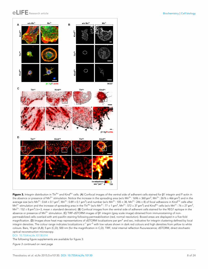
Figure 3. Integrin distribution in TlnKo and KindKo cells. (A) Confocal images of the ventral side of adherent cells stained for b1 integrin and F-actin in
the absence or presence of Mn2+ stimulation. Notice the increase in the spreading area (w/o Mn2+: 1696 ± 360 mm2, Mn2+: 2676 ± 466 mm2) and in the
average size (w/o Mn2+: 0.64 ± 0.1 mm2, Mn2+: 0.89 ± 0.1 mm2) and number (w/o Mn2+: 105 ± 38, Mn2+: 246 ± 8) of focal adhesions in KindCtr cells after
Mn2+ stimulation and the increase of spreading area in the TlnKo (w/o Mn2+: 77 ± 1 mm2, Mn2+: 572 ± 37 mm2) and KindKo cells (w/o Mn2+: 76 ± 27 mm2,
Mn2+: 152 ± 8 mm2) (n=3, mean ± standard deviation). (B) Confocal images from the ventral side of adherent cells stained for the 9EG7 epitope in the
absence or presence of Mn2+ stimulation. (C) TIRF-dSTORM images of b1 integrin (grey scale image) obtained from immunostaining of non-
permeabilized cells overlaid with anti-paxillin staining following permeabilization (red, normal resolution). Boxed areas are displayed in a five-fold
magnification. (D) Images show heat map representations of dSTORM localizations per mm2 and sec, indicative for integrin clustering defined by local
integrin densities. The colour range indicates localizations s–1 mm–2 with low values shown in dark red colours and high densities from yellow to white
colours. Bars, 10 mm (A,B); 5 mm (C,D); 500 nm (for the magnification in C,D). TIRF, total internal reflection fluourescence; dSTORM, direct stochastic
optical reconstruction microscopy.
DOI: 10.7554/eLife.10130.014
The following figure supplements are available for figure 3:
Figure 3 continued on next page
Theodosiou et al. eLife 2015;5:e10130. DOI: 10.7554/eLife.10130 8 of 24
Research article Biochemistry Cell biology
localizations from single or multiple dyes in close proximity cannot be distinguished, the number of
localizations does not directly reflect integrin numbers. However, all antibody molecules display the
same average behaviour with respect to the number of localizations per second in all areas of the
cell. This allowed to average the number of localizations per second and mm2 and to plot them in a
heat map representation (Figure 3D), which directly reflects the density of stained b1 integrin mole-
cules and thus the degree of integrin clustering. The b1 integrin staining of TlnCtr and KindCtr cells
revealed small round structures of ~50 nm diameter indicating clusters of integrins larger than the
resolution limit (Figure 3C; high magnification; see arrowheads). Furthermore, high numbers of
localizations were enriched in paxillin-positive FAs and in NAs at the lamellipodial edge (Figure 3C;
see arrowheads). In these areas, we observed a high average density of 60–120 localizations s–1 mm–
2, while outside of the adhesion sites ~0–20 localizations s–1 mm–2 were detected, indicating a high
degree of b1 integrin clustering within and a low degree of clustering outside of adhesion sites
(Figure 3D). TlnKo cells with circumferential lamellipodia showed a high density of blinking with up
to 100 localizations s–1 mm–2 at lamellipodial edges (Figure 3C,D; see arrowheads), which appeared
less compact than in control cells. KindKo cells showed >120 localizations s–1 mm–2 in the periphery
and finger-like membrane protrusions (Figure 3C,D; see arrowheads), which were also observed in
TlnKo cells that adopted a spheroid rather than an isotropic spread shape (Figure 3—figure supple-
ment 3). The exclusive presence of these large and entangled b1 integrin aggregates on TlnKo and
KindKo cells with small, spheroid shapes and protrusions suggests that they were induced by spatial
constraints rather than specific signaling.
These findings demonstrate that, in contrast to KindKo cells, Mn2+-treated kindlin-2-expressing
TlnKo cells induce circumferential membrane protrusions with b1 integrins at the protrusive edges.
Kindlin-2 binds and recruits paxillin to NAsOur data so far indicate that the expression of kindlin-2 enables initial, isotropic spreading and the
accumulation of integrins in lamellipodia of Mn2+-treated TlnKo cells. To identify binding partner(s) of
kindlin-2 that transduce this function to downstream effectors, we performed yeast-two-hybrid
assays with kindlin-2 as bait using a human
complementary DNA (cDNA) library containing
all possible open reading frames and a human
keratinocyte-derived cDNA library. Among the
124 cDNAs identified from both screenings, 17
coded for leupaxin and 11 for Hic-5. Immuno-
precipitation of overexpressed green fluorescent
protein (GFP)-tagged paxillin family members,
paxillin, Hic-5 and leupaxin in HEK-293 cells with
an anti-GFP antibody efficiently co-precipitated
FLAG-tagged kindlin-2 (K2flag) (Figure 4A).
Conversely, overexpressed GFP-tagged kindlin
family members (kindlin-1, kindlin-2, kindlin-3)
co-precipitated Cherry-paxillin (Figure 4—figure
supplement 1). Since fibroblasts express high
levels of paxillin (Figure 4—figure supplement
2), we performed all further interaction analysis
with paxillin. Immunoprecipitations of GFP-
tagged paxillin or kindlin-2 truncation mutants
(Figure 4—figure supplement 3A) revealed that
Figure 3 continued
Figure supplement 1. Localization of FAs proteins in Mn2+-treated KindCtr, TlnKo and KindKo cells.
DOI: 10.7554/eLife.10130.015
Figure supplement 2. Rescue of FA formation and spreading after expression of Tln1V in TlnKo cells or K2GFP in KindKo cells.
DOI: 10.7554/eLife.10130.016
Figure supplement 3. Distribution of b1 integrins in spheroid-shaped TlnKo cells.
DOI: 10.7554/eLife.10130.017
Video 1. Spreading KindCtr, TlnKo and KindKo cells on
FN. Assembled time lapse movies of KindCtr, TlnKo and
KindKo cells. Cell spreading was recorded 5 min after
seeding on FN. KindCtr cells were already well spread
and only a minor size increase was observed over the
following minutes. The TlnKo cells formed a
circumferential lamellipodium that rapidly collapsed
and subsequently the cells formed finger-like
protrusions of varying size and failed to reestablish a
fully formed circular lamellipodium. The KindKo cells
failed to form a lamellipodium and formed finger-like
protrusions that were not always adherent. Bar, 10
mm. FN, fibronectin.
DOI: 10.7554/eLife.10130.013
Theodosiou et al. eLife 2015;5:e10130. DOI: 10.7554/eLife.10130 9 of 24
Research article Biochemistry Cell biology
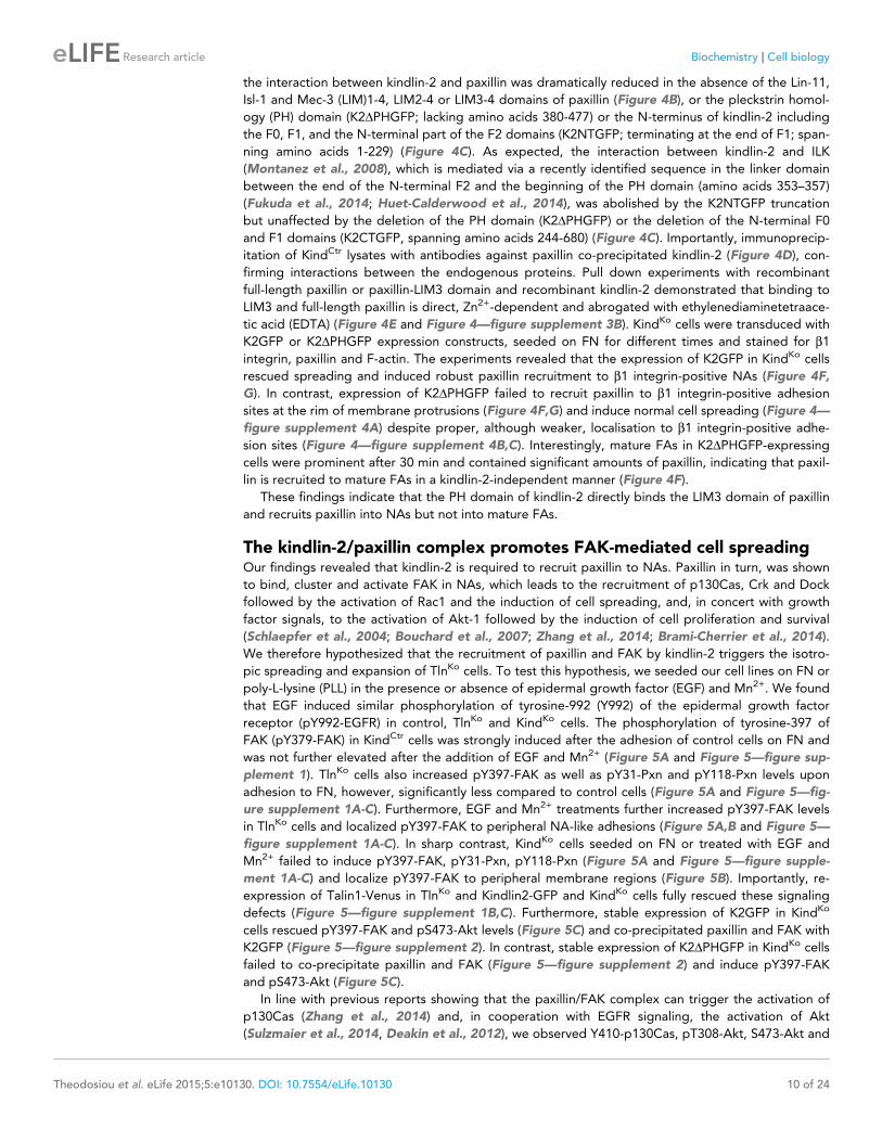
the interaction between kindlin-2 and paxillin was dramatically reduced in the absence of the Lin-11,
Isl-1 and Mec-3 (LIM)1-4, LIM2-4 or LIM3-4 domains of paxillin (Figure 4B), or the pleckstrin homol-
ogy (PH) domain (K2DPHGFP; lacking amino acids 380-477) or the N-terminus of kindlin-2 including
the F0, F1, and the N-terminal part of the F2 domains (K2NTGFP; terminating at the end of F1; span-
ning amino acids 1-229) (Figure 4C). As expected, the interaction between kindlin-2 and ILK
(Montanez et al., 2008), which is mediated via a recently identified sequence in the linker domain
between the end of the N-terminal F2 and the beginning of the PH domain (amino acids 353–357)
(Fukuda et al., 2014; Huet-Calderwood et al., 2014), was abolished by the K2NTGFP truncation
but unaffected by the deletion of the PH domain (K2DPHGFP) or the deletion of the N-terminal F0
and F1 domains (K2CTGFP, spanning amino acids 244-680) (Figure 4C). Importantly, immunoprecip-
itation of KindCtr lysates with antibodies against paxillin co-precipitated kindlin-2 (Figure 4D), con-
firming interactions between the endogenous proteins. Pull down experiments with recombinant
full-length paxillin or paxillin-LIM3 domain and recombinant kindlin-2 demonstrated that binding to
LIM3 and full-length paxillin is direct, Zn2+-dependent and abrogated with ethylenediaminetetraace-
tic acid (EDTA) (Figure 4E and Figure 4—figure supplement 3B). KindKo cells were transduced with
K2GFP or K2DPHGFP expression constructs, seeded on FN for different times and stained for b1
integrin, paxillin and F-actin. The experiments revealed that the expression of K2GFP in KindKo cells
rescued spreading and induced robust paxillin recruitment to b1 integrin-positive NAs (Figure 4F,
G). In contrast, expression of K2DPHGFP failed to recruit paxillin to b1 integrin-positive adhesion
sites at the rim of membrane protrusions (Figure 4F,G) and induce normal cell spreading (Figure 4—
figure supplement 4A) despite proper, although weaker, localisation to b1 integrin-positive adhe-
sion sites (Figure 4—figure supplement 4B,C). Interestingly, mature FAs in K2DPHGFP-expressing
cells were prominent after 30 min and contained significant amounts of paxillin, indicating that paxil-
lin is recruited to mature FAs in a kindlin-2-independent manner (Figure 4F).
These findings indicate that the PH domain of kindlin-2 directly binds the LIM3 domain of paxillin
and recruits paxillin into NAs but not into mature FAs.
The kindlin-2/paxillin complex promotes FAK-mediated cell spreadingOur findings revealed that kindlin-2 is required to recruit paxillin to NAs. Paxillin in turn, was shown
to bind, cluster and activate FAK in NAs, which leads to the recruitment of p130Cas, Crk and Dock
followed by the activation of Rac1 and the induction of cell spreading, and, in concert with growth
factor signals, to the activation of Akt-1 followed by the induction of cell proliferation and survival
(Schlaepfer et al., 2004; Bouchard et al., 2007; Zhang et al., 2014; Brami-Cherrier et al., 2014).
We therefore hypothesized that the recruitment of paxillin and FAK by kindlin-2 triggers the isotro-
pic spreading and expansion of TlnKo cells. To test this hypothesis, we seeded our cell lines on FN or
poly-L-lysine (PLL) in the presence or absence of epidermal growth factor (EGF) and Mn2+. We found
that EGF induced similar phosphorylation of tyrosine-992 (Y992) of the epidermal growth factor
receptor (pY992-EGFR) in control, TlnKo and KindKo cells. The phosphorylation of tyrosine-397 of
FAK (pY379-FAK) in KindCtr cells was strongly induced after the adhesion of control cells on FN and
was not further elevated after the addition of EGF and Mn2+ (Figure 5A and Figure 5—figure sup-
plement 1). TlnKo cells also increased pY397-FAK as well as pY31-Pxn and pY118-Pxn levels upon
adhesion to FN, however, significantly less compared to control cells (Figure 5A and Figure 5—fig-
ure supplement 1A-C). Furthermore, EGF and Mn2+ treatments further increased pY397-FAK levels
in TlnKo cells and localized pY397-FAK to peripheral NA-like adhesions (Figure 5A,B and Figure 5—
figure supplement 1A-C). In sharp contrast, KindKo cells seeded on FN or treated with EGF and
Mn2+ failed to induce pY397-FAK, pY31-Pxn, pY118-Pxn (Figure 5A and Figure 5—figure supple-
ment 1A-C) and localize pY397-FAK to peripheral membrane regions (Figure 5B). Importantly, re-
expression of Talin1-Venus in TlnKo and Kindlin2-GFP and KindKo cells fully rescued these signaling
defects (Figure 5—figure supplement 1B,C). Furthermore, stable expression of K2GFP in KindKo
cells rescued pY397-FAK and pS473-Akt levels (Figure 5C) and co-precipitated paxillin and FAK with
K2GFP (Figure 5—figure supplement 2). In contrast, stable expression of K2DPHGFP in KindKo cells
failed to co-precipitate paxillin and FAK (Figure 5—figure supplement 2) and induce pY397-FAK
and pS473-Akt (Figure 5C).
In line with previous reports showing that the paxillin/FAK complex can trigger the activation of
p130Cas (Zhang et al., 2014) and, in cooperation with EGFR signaling, the activation of Akt
(Sulzmaier et al., 2014, Deakin et al., 2012), we observed Y410-p130Cas, pT308-Akt, S473-Akt and
Theodosiou et al. eLife 2015;5:e10130. DOI: 10.7554/eLife.10130 10 of 24
Research article Biochemistry Cell biology
Figure 4. Kindlin binds and recruits paxillin to NAs. (A) GFP-IP of lysates from HEK 293T cells overexpressing GFP-tagged paxillin, Hic5 and leupaxin
constructs (Pxn, paxillin; Hic5; Lpx, leupaxin) and K2flag reveal interaction of kindlin-2 with all three paxillin family members. (B) GFP-IP of lysates from
HEK 293T cells overexpressing GFP-tagged paxillin truncation mutants and K2flag identifies the paxillin LIM3 domain as kindlin-2-binding domain. (C)
GFP-IP of lysates from HEK 293T cells overexpressing GFP-tagged kindlin-2 truncation/deletion mutants and Cherry-tagged paxillin (PxnCH) identifies
the kindlin-2 PH domain as paxillin binding domain. (D) Co-IP of endogenous paxillin and kindlin-2 from KindCtr cells. (E) Purified His-tagged paxillin-
LIM3 domain pulls down recombinant kindlin-2 in a Zn2+-dependent manner. (F) K2GFP and K2DPHGFP expressing KindKo cells seeded on FN for the
indicated times and stained for paxillin and b1 integrin. (G) Fluorescence intensity line scans from K2GFP- (n=11 cells) and K2DPHGFP- (n=17 cells)
expressing KindKo cells cultured on FN for 10 min and stained for paxillin and b1 integrin; error bars indicate standard error of the mean. Bar, 10
mm. EDTA, ethylenediaminetetraacetic acid; FN, fibronection; GAPDH, glyceraldehyde-3-phosphate dehydrogenase; GFP, green fluorescent protein;
ILK, integrin-linked kinase; IP, immunoprecipitation; K2GFP, green fluorescent protein-tagged kindlin-2; LIM, Lin-11, Isl-1 and Mec-3; NAs, nascent
adhesions; PH, pleckstrin homology.
DOI: 10.7554/eLife.10130.018
Figure 4 continued on next page
Theodosiou et al. eLife 2015;5:e10130. DOI: 10.7554/eLife.10130 11 of 24
Research article Biochemistry Cell biology
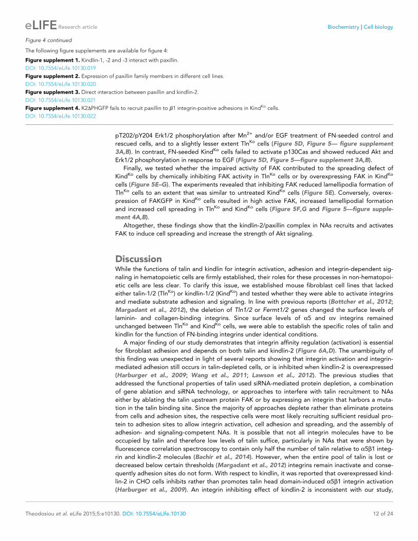
pT202/pY204 Erk1/2 phosphorylation after Mn2+ and/or EGF treatment of FN-seeded control and
rescued cells, and to a slightly lesser extent TlnKo cells (Figure 5D, Figure 5— figure supplement
3A,B). In contrast, FN-seeded KindKo cells failed to activate p130Cas and showed reduced Akt and
Erk1/2 phosphorylation in response to EGF (Figure 5D, Figure 5—figure supplement 3A,B).
Finally, we tested whether the impaired activity of FAK contributed to the spreading defect of
KindKo cells by chemically inhibiting FAK activity in TlnKo cells or by overexpressing FAK in KindKo
cells (Figure 5E–G). The experiments revealed that inhibiting FAK reduced lamellipodia formation of
TlnKo cells to an extent that was similar to untreated KindKo cells (Figure 5E). Conversely, overex-
pression of FAKGFP in KindKo cells resulted in high active FAK, increased lamellipodial formation
and increased cell spreading in TlnKo and KindKo cells (Figure 5F,G and Figure 5—figure supple-
ment 4A,B).
Altogether, these findings show that the kindlin-2/paxillin complex in NAs recruits and activates
FAK to induce cell spreading and increase the strength of Akt signaling.
DiscussionWhile the functions of talin and kindlin for integrin activation, adhesion and integrin-dependent sig-
naling in hematopoietic cells are firmly established, their roles for these processes in non-hematopoi-
etic cells are less clear. To clarify this issue, we established mouse fibroblast cell lines that lacked
either talin-1/2 (TlnKo) or kindlin-1/2 (KindKo) and tested whether they were able to activate integrins
and mediate substrate adhesion and signaling. In line with previous reports (Bottcher et al., 2012;
Margadant et al., 2012), the deletion of Tln1/2 or Fermt1/2 genes changed the surface levels of
laminin- and collagen-binding integrins. Since surface levels of a5 and av integrins remained
unchanged between TlnKo and KindKo cells, we were able to establish the specific roles of talin and
kindlin for the function of FN-binding integrins under identical conditions.
A major finding of our study demonstrates that integrin affinity regulation (activation) is essential
for fibroblast adhesion and depends on both talin and kindlin-2 (Figure 6A,D). The unambiguity of
this finding was unexpected in light of several reports showing that integrin activation and integrin-
mediated adhesion still occurs in talin-depleted cells, or is inhibited when kindlin-2 is overexpressed
(Harburger et al., 2009; Wang et al., 2011; Lawson et al., 2012). The previous studies that
addressed the functional properties of talin used siRNA-mediated protein depletion, a combination
of gene ablation and siRNA technology, or approaches to interfere with talin recruitment to NAs
either by ablating the talin upstream protein FAK or by expressing an integrin that harbors a muta-
tion in the talin binding site. Since the majority of approaches deplete rather than eliminate proteins
from cells and adhesion sites, the respective cells were most likely recruiting sufficient residual pro-
tein to adhesion sites to allow integrin activation, cell adhesion and spreading, and the assembly of
adhesion- and signaling-competent NAs. It is possible that not all integrin molecules have to be
occupied by talin and therefore low levels of talin suffice, particularly in NAs that were shown by
fluorescence correlation spectroscopy to contain only half the number of talin relative to a5b1 integ-
rin and kindlin-2 molecules (Bachir et al., 2014). However, when the entire pool of talin is lost or
decreased below certain thresholds (Margadant et al., 2012) integrins remain inactivate and conse-
quently adhesion sites do not form. With respect to kindlin, it was reported that overexpressed kind-
lin-2 in CHO cells inhibits rather than promotes talin head domain-induced a5b1 integrin activation
(Harburger et al., 2009). An integrin inhibiting effect of kindlin-2 is inconsistent with our study,
Figure 4 continued
The following figure supplements are available for figure 4:
Figure supplement 1. Kindlin-1, -2 and -3 interact with paxillin.
DOI: 10.7554/eLife.10130.019
Figure supplement 2. Expression of paxillin family members in different cell lines.
DOI: 10.7554/eLife.10130.020
Figure supplement 3. Direct interaction between paxillin and kindlin-2.
DOI: 10.7554/eLife.10130.021
Figure supplement 4. K2DPHGFP fails to recruit paxillin to b1 integrin-positive adhesions in KindKo cells.
DOI: 10.7554/eLife.10130.022
Theodosiou et al. eLife 2015;5:e10130. DOI: 10.7554/eLife.10130 12 of 24
Research article Biochemistry Cell biology
Figure 5. The kindlin/paxillin complex induces FAK signaling and cell spreading. (A) FAK and EGFR activation after seeding serum-starved KindCtr,
TlnKo and KindKo cells on PLL or FN and treating them with or without EGF and Mn2+. (B) Immunofluorescence staining of activated (Tyr-397
phosphorylated) FAK and F-actin in cells seeded on FN and treated with Mn2+ for 30 min (FAKGFP indicates exogenous expression of FAKGFP fusion
protein). (C) FAK and Akt activation in KindKo cells stably transduced with K2GFP or K2DPHGFP either seeded on FN or kept in suspension. GFP
indicates similar expression of transduced GFP-tagged constructs. GAPDH levels served to control loading. (D) Levels of phosphorylated signaling
mediators downstream of FAK in Mn2+-treated, serum-starved or EGF-treated KindCtr, TlnKo and KindKo cells. GAPDH levels served to control loading.
(E) Quantification of lamellipodia formation of FN-seeded TlnKo and KindKo cells treated with Mn2+ and either DMSO or the FAK inhibitor PF-228 (n=3
independent repeats; >100 cells/condition; error bars indicate standard error of the mean; significances are given in comparison to the DMSO control).
(F) FAK activity in TlnKo and KindKo cells stably transduced with FAKGFP (n=3 independent experiments). (G) Quantification of lamellipodia formation in
TlnKo and KindKo cells stably transduced with FAKGFP (n=3 independent experiments; significances are given in comparison to untreated control; error
bars indicate standard error of the mean). Bar, 10 mm. DMSO, dimethyl sulfoxide; EGF, epidermal growth factor; EGFR, epidermal growth factor
receptor; FAK, focal adhesion kinase; FAKGFP, green fluorescent protein-tagged FAK; FN, fibronectin; GAPDH, glyceraldehyde-3-phosphate
dehydrogenase; GFP, green fluorescent protein; PLL, poly-L-lysine.
DOI: 10.7554/eLife.10130.023
The following figure supplements are available for figure 5:
Figure supplement 1. FAK phosphorylation in TlnCtr, TlnKo, TlnKo+T1V, KindCtr, KindKo and KindKo+K2GFP cells.
DOI: 10.7554/eLife.10130.024
Figure supplement 2. Kindlin-2 forms a ternary complex with paxillin and FAK.
Figure 5 continued on next page
Theodosiou et al. eLife 2015;5:e10130. DOI: 10.7554/eLife.10130 13 of 24
Research article Biochemistry Cell biology
which identified a crucial role for kindlin in integrin activation, as well as with other studies also dem-
onstrating that kindlin-2 promotes integrin functions (Montanez et al., 2008). It could well be that
the reported inhibition of a5b1 by kindlin-2 represents an artifact that arose from protein
overexpression.
Integrin activation can be induced with Mn2+, whose binding to the ectodomain of b subunits
directly shifts integrins into the high affinity state without the requirement for inside-out signals
(Mould et al., 1995). We observed that Mn2+-treated TlnKo and KindKo cells expressed the activa-
tion-dependent epitope 9EG7 and adhered to FN, albeit at significantly lower levels and efficiencies
than the normal parental or rescued cells (Figure 6B,C). This observation strongly indicates that talin
and kindlin also cooperate to maintain the extended and unclasped conformation of active integrins.
Although it is not known how talin and kindlin keep integrins in an active state, it is possible that
they stabilize this conformation by linking the unclasped b integrin cytoplasmic domain to the
plasma membrane and/or to cortical actin, which may firmly hold separated integrin a/b subunits
apart from each other. The expression of mutant talins and kindlins in our cells should allow us to
examine these possibilities in future.
Finally, our study also revealed that Mn2+-treated TlnKo cells began to form large, circumferential
lamellipodia that eventually detached from FN, leading to the collapse of the protruded membrane.
This initial isotropic spreading was significantly less frequent in KindKo cells, and has also been
observed in talin-2-depleted talin-1–/– cells on FN, although these cells did not require Mn2+ for
inducing spreading, which is likely due to the presence of residual talin-2 that escaped siRNA-medi-
ated depletion (Zhang et al., 2008; Zhang et al., 2014). These findings strongly suggest that integ-
rin binding to FN enables kindlin-2 in TlnKo cells to cluster b1 integrins (as shown for aIIbb3 by
kindlin-3 in Ye et al., 2013) and to trigger a signaling process that initiates spreading.
To find a mechanistic explanation for the kindlin-2-mediated cell spreading, we used the yeast-
two-hybrid technology to identify paxillin as a novel and direct binding partner of kindlin-2. The
interaction of the two proteins occurs through the LIM3 domain of paxillin, which was previously
identified as integrin adhesion-targeting site (Brown et al., 1996), and the PH domain of kindlin-2. It
is not unusual that PH domains fulfill dual roles by binding phospholipids and proteins, either simul-
taneously or consecutively (Scheffzek and Welti, 2012). The expression of a PH domain-deficient
kindlin-2 in KindKo cells rescues adhesion to FN and FA maturation, however, significantly impairs
spreading and plasma membrane protrusions. This finding together with the observations that paxil-
lin-null fibroblasts and embryonic stem cells have defects in spreading, adhesion site remodeling
and formation of lamellipodia (Hagel et al., 2002, Wade et al., 2002) indicates that the kindlin-2/
paxillin complex induces the elusive signaling process, leading to initial spreading of TlnKo and talin-
depleted cells (Zhang et al., 2008). Indeed, the kindlin-2/paxillin complex in NAs recruits FAK
(Deramaudt et al., 2014, Thwaites et al., 2014; Choi et al., 2011), which cooperates with growth
factor receptors (such as EGFR) to induce signaling pathways that activate Erk and Akt to promote
proliferation and survival, as well as Arp2/3 and Rac1 to induce actin polymerization and membrane
protrusions (Figure 6B,E). Kindlin-2 also recruits ILK, which binds in the vicinity of the kindlin-2 PH
domain and links integrins to actin and additional signaling pathways (Figure 6E). The short-lived
nature of the initial spreading of TlnKo and talin-depleted (Zhang et al., 2008) cells shows that talin
concludes the integrin-mediated adhesion process in NAs (Figure 6F) and induces the maturation of
FAs. The formation of paxillin-positive FAs in cells expressing the PH domain-deficient kindlin-2 sug-
gests that the recruitment of paxillin to FAs occurs either in a kindlin-independent manner or
through a modification of kindlin in a second binding motif.
Figure 5 continued
DOI: 10.7554/eLife.10130.025
Figure supplement 3. Activity of signaling mediators downstream of FAK in TlnCtr, TlnKo, TlnKo+T1V, KindCtr, KindKo and KindKo+K2GFP cells.
DOI: 10.7554/eLife.10130.026
Figure supplement 4. Cell spreading of FAK overexpressing TlnKo and KindKo cells.
DOI: 10.7554/eLife.10130.027
Theodosiou et al. eLife 2015;5:e10130. DOI: 10.7554/eLife.10130 14 of 24
Research article Biochemistry Cell biology
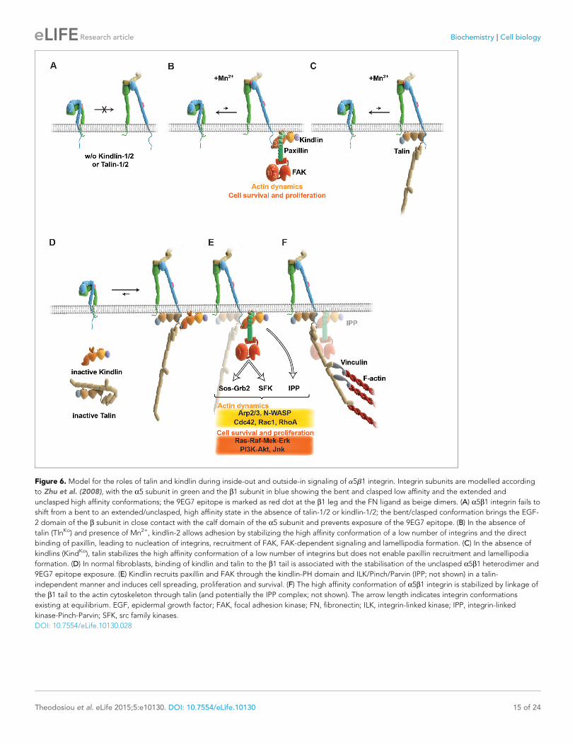
Figure 6. Model for the roles of talin and kindlin during inside-out and outside-in signaling of a5b1 integrin. Integrin subunits are modelled according
to Zhu et al. (2008), with the a5 subunit in green and the b1 subunit in blue showing the bent and clasped low affinity and the extended and
unclasped high affinity conformations; the 9EG7 epitope is marked as red dot at the b1 leg and the FN ligand as beige dimers. (A) a5b1 integrin fails to
shift from a bent to an extended/unclasped, high affinity state in the absence of talin-1/2 or kindlin-1/2; the bent/clasped conformation brings the EGF-
2 domain of the b subunit in close contact with the calf domain of the a5 subunit and prevents exposure of the 9EG7 epitope. (B) In the absence of
talin (TlnKo) and presence of Mn2+, kindlin-2 allows adhesion by stabilizing the high affinity conformation of a low number of integrins and the direct
binding of paxillin, leading to nucleation of integrins, recruitment of FAK, FAK-dependent signaling and lamellipodia formation. (C) In the absence of
kindlins (KindKo), talin stabilizes the high affinity conformation of a low number of integrins but does not enable paxillin recruitment and lamellipodia
formation. (D) In normal fibroblasts, binding of kindlin and talin to the b1 tail is associated with the stabilisation of the unclasped a5b1 heterodimer and
9EG7 epitope exposure. (E) Kindlin recruits paxillin and FAK through the kindlin-PH domain and ILK/Pinch/Parvin (IPP; not shown) in a talin-
independent manner and induces cell spreading, proliferation and survival. (F) The high affinity conformation of a5b1 integrin is stabilized by linkage of
the b1 tail to the actin cytoskeleton through talin (and potentially the IPP complex; not shown). The arrow length indicates integrin conformations
existing at equilibrium. EGF, epidermal growth factor; FAK, focal adhesion kinase; FN, fibronectin; ILK, integrin-linked kinase; IPP, integrin-linked
kinase-Pinch-Parvin; SFK, src family kinases.
DOI: 10.7554/eLife.10130.028
Theodosiou et al. eLife 2015;5:e10130. DOI: 10.7554/eLife.10130 15 of 24
Research article Biochemistry Cell biology
Materials and methods
Mouse strains and cell lines and cell cultureThe floxed kindlin-1 (Fermt1flox/flox), floxed talin-1 (Tln1flox/flox) and the constitutive talin-2-null
(Tln2–/–) mouse strains have been described (Rognoni et al., 2014; Nieswandt et al., 2007;
Conti et al., 2009). The floxed kindlin-2 (Fermt2flox/flox) mouse strain generated via recombinant
recombination in embryonic stem cells (Fassler and Meyer, 1995) carries loxP sites flanking exon
15, which contains the stop codon and the polyadenylation signal of the murine Fermt2 gene.
Homologous recombination and germ line transmission were verified by Southern blots, and the frt-
flanked neo cassette was removed with a transgenic mouse strain carrying a deleter-flipase gene.
Floxed talin-1 and talin-2-null mice, and floxed kindlin-1 and kindlin-2 mice were intercrossed to gen-
erate Tln1flox/flox Tln2–/– and Fermt1flox/flox Fermt2flox/flox mice.
The cell lines used in this study are mouse fibroblasts derived from the kidneys of 21 d old mice,
immortalized by retrovirally transducing the SV40 large T antigen, cloned (TlnCtr and KindCtr) and
finally infected with an adenovirus to transduce the Cre recombinase resulting in talin-null (TlnKo)
and kindlin-null (KindKo) cells. The parental cell lines were authenticated based on morphological cri-
teria and the surface experession of specific integrins. All cells were cultured under standard cell cul-
ture conditions using Dulbecco’s modified Eagle’s medium (DMEM) supplemented with 8% fetal calf
serum (FCS) and Penicillin/Streptomycin but not subjected to mycoplasma contamination testing.
Flow cytometryFlow cytometry was carried out with a FACSCantoTMII cytometer (BD Biosciences, Franklin Lakes,
NJ, USA) equipped with FACS DiVa software (BD Biosciences) using standard procedures. Data anal-
ysis was carried out with the FlowJo program (version 9.4.10). Fibroblasts were incubated with pri-
mary antibodies diluted in FACS-Tris buffered saline (FACS-TBS; 30 mM Tris, pH 7.4, 180 mM NaCl,
3.5 mM KCl, supplemented with 1 mM CaCl2, 1 mM MgCl2, 3% BSA, 0,02% NaN3) for 1 hr on ice,
washed twice with cold FACS-TBS and finally incubated with the secondary antibody for 45 min on
ice.
Real-time polymerase chain reactionTotal RNA was extracted with the RNeasy Mini extraction kit (Qiagen, Germany) from cultured cells,
cDNAs were prepared with an iScript cDNA Synthesis Kit (BioRad, Germany) and real-time polymer-
ase chain reaction (PCR) was performed with an iCycler (BioRad). Each sample was measured in trip-
licate and values were normalized to Gapdh. Primer sequences for Lpxn and Pxn were from
PrimerBank (Spandidos et al., 2010) (Lpxn: 26080416a1; aPxn: 114326500c2; bPxn: 22902122a1),
GAPDH primers were described before (Rognoni et al., 2014) and Hic5 primers were newly
designed (Hic5-fwd: 5’-ttcctttgcagcggttgttcc-3’; Hic5-rev: 5’-ggttacagaagccacatcgtggg-3’).
Antibodies and inhibitorsThe following antibodies or molecular probes were used at indicated concentrations for western
blot (WB), immunofluorescence (IF) or flow cytometry (FACS): kindlin-1 (home made), (Ussar et al.,
2008) WB: 1:5000, IF: 1:1000; kindlin-2 (MAB2617 from Millipore, Germany) WB: 1:1000, IF: 1:500;
talin (8D4 from Sigma, Germany) WB: 1:1000; talin (sc-7534 from Santa Cruz, Germany) IF: 1:500;
talin-1 (ab57758 from Abcam, UK) WB: 1:2000; talin-2 (ab105458 from Abcam) WB: 1:2000; GAPDH
(6C5 from Calbiochem, Billerica, MA, USA) WB: 1:10,000; Paxillin (610051 from BD Transduction
Laboratories, Franklin Lakes, NJ, USA) WB: 1:1000, IF: 1:400; integrin b1-488 (102211 from
Biolegend, San Diego, CA, USA) IF: 1:400, FACS: 1:200; integrin b1 (MAB1997 from
Chemicon, Billerica, MA, USA) FACS: 1:400; integrin b1-647 (102213 from Biolegend) IF: 1:200;
integrin b1 (home-made), (Azimifar et al., 2012) IF: 1:400; integrin b3-biotin (553345
from PharMingen, Franklin Lakes, NJ, USA) FACS: 1:200; integrin b3 (M031-0
from Emfret, Germany) IF: 1:200; integrin a2-FITC (554999 from BD Biosciences) FACS: 1:100; integ-
rin a3 (AF2787 from R&D, Germany) FACS: 1:200; integrin a5-biotin (557446 from Pharmingen)
FACS: 1:200, IP 1mg; integrin a5 (4705 from Cell Signaling, Germany) WB: 1:1000; integrin a6-FITC
(555735 from Pharmingen) FACS 1:100; integrin av-biotin (551380 from Pharmingen) FACS: 1:200;
b1-integrin 9EG7 (550531 from BD Biosciences, San Diego, CA, USA) IF: 1:200; FACS: 1:200;
Theodosiou et al. eLife 2015;5:e10130. DOI: 10.7554/eLife.10130 16 of 24
Research article Biochemistry Cell biology
fibronectin (AB2033 from Millipore) IF: 1:500; IgG2a rat isotype control (13-4321 from eBioscience,
Germany) FACS: 1:200; IP 1mg; Tritc-Phalloidin (P1951 from Sigma) IF: 1:400; Flag-tag-HRP (8592
from Sigma) WB: 1:10,000; GFP (A11122 from Invitrogen, Germany) WB: 1:2000; Cherry (PM005
from MBL, Woburn, MA, USA) WB:1:1000; Myc (05-724 from Millipore) WB 1:2000; FAK (06-543
from Upstate, Billerica, MA, USA) WB: 1:1000; FAK (3285 from Cell Signaling) WB (1:1000); phos-
pho-Y397 FAK (3283 from Cell Signaling) WB: 1:1000; phospho-Y397 FAK (44624G from
Biosource, Waltham, MA, USA) WB: 1:1000, IF: 1:400; ILK (611803 from Transduction Labs) WB:
1:5000; IF: 1:500; phospho-Y992 EGFR (2235 from Cell Signaling) WB: 1:2000; phospho-Y31 Paxillin
(44720G from Invitrogen) WB: 1:1000; phospho-Y118 Paxillin (44722G from Invitrogen) WB: 1:1000;
p130Cas (P27820 Transduction Labs) WB: 1:1000; phospho-Y410 p130 Cas (4011S from Cell Signal-
ing) WB: 1:1000; Akt (9272 from Cell Signaling) WB: 1:1000; phospho-S473 Akt (4060 from Cell Sig-
naling) WB: 1:1000; phosho-T308 Akt (9275 from Cell Signaling) WB: 1:1000; Erk1/2 (9102 from Cell
Signaling) WB: 1:1000; Erk1/2 phosphorylated T202 Y204 (4376 Cell Signaling) WB: 1:1000.
The following secondary antibodies were used: goat anti-rabbit Alexa 488 (A11008), goat anti-
mouse Alexa 488 (A11029), goat anti-rat Alexa 488 (A11006), goat anti-mouse Alexa 546 (A11003),
donkey anti-mouse Alexa 647 (A31571), goat anti-rabbit Alexa 647 (A21244), (all from Invitrogen)
FACS: 1:500, IF: 1:500; streptavidin-Cy5 (016170084) FACS: 1:400; goat anti-rat horseradish peroxi-
dase (HRP) (712035150) (both from Dianova, Germany) WB: 1:10,000, donkey anti-rabbit Cy3 (711-
165-152) (from Jackson ImmunoResearch, West Grove, PA, USA) IF: 1:500, goat anti-mouse HRP
(172-1011) and goat anti-rabbit HRP (172-1019) (both from BioRad) WB: 1:10,000.
The FAK inhibitor PF-228 (PZ0117 from Sigma) was dissolved in dimethyl sulfoxide at 10 mM and
used at 1:2000.
Expression and purification of recombinant proteinsThe recombinant expression of kindlin-2, full-length paxillin (paxillin-FL) and paxillin-LIM3 in Escheri-
chia coli Rosetta cells (Merck Millipore) was induced with 1 mM or 0.2 mM IPTG, respectively, at
18˚C for 22 hr. After cell lysis and clarification of the supernatant, kindlin-2 was purified by Ni-NTA
affinity chromatography (Qiagen). Eluate fractions containing kindlin-2 were pooled, cleaved with
SenP2 protease and purified by size-exclusion chromatography (Superdex 200 26/600, GE
Healthcare, UK) yielding unmodified murine kindlin-2. The paxillin constructs were purified by Ni-
NTA affinity chromatography (Qiagen), and subsequent size-exclusion chromatography (SEC650,
BioRad) to obtain N-terminally tagged His10-SUMO3-paxillin-FL and His10-SUMO3-paxillin-LIM3
domain, respectively.
ImmunostainingFor immunostaining, cells were cultured on plastic ibidi-m-slides (80826 from Ibidi, Germany) coated
with 20 mg ml–1 FN (Calbiochem). Cells were routinely fixed with 4% paraformaldehyde (PFA) (w/v) in
phosphate buffered saline (PBS; 180 mM NaCl, 3.5 mM KCl, 10 mM Na2HPO4, 1.8 mM K2H2PO4) for
10 min at room temperature (RT) or with –20˚C cold acetone–methanol when indicated. If necessary,
cells were solubilized with staining buffer (PBS supplemented with 0.1% Triton X-100 (v/v) and 3%
BSA (w/v)) or with –20˚C cold methanol for kindlin-2 staining. Background signals were blocked by
incubating cells for 1 hr at RT in staining buffer. Subsequently, they were incubated in the dark with
primary and secondary antibodies diluted in staining buffer. Fluorescent images were aquired with a
LSM 780 confocal microscope (Zeiss, Germany) equiped with a 100�/NA 1.46 oil objective and with
a DMIRE2-SP5 confocal microscope (Leica, Germany) equiped with a 40�/NA 1.25 or 63�/NA 1.4
oil objective using Leica Confocal software (version 2.5 build 1227). Brightfield images were aquired
with an Axioskop (Carl Zeiss) 40�/NA 0.75 objective and DC500 camera with IM50 software (Leica).
Z-stack projection and contrast adjustments ImageJ (v1.47) were used for further image analysis.
Super-resolution imaging was carried out by direct stochastic optical reconstruction microscopy
(dSTORM) (van de Linde et al., 2011), which is based on precise emitter localization. To induce
reversible switching of the Alexa 647 label and reduce photobleaching, imaging was performed in
imaging solution (50% Vectashield (v/v) (H-1000; Vector Laboratories, Burlingame, CA, USA), 50%
TBS (v/v), pH=8.0) supplemented with 50 mM b-mercaptoethylamine (Sigma-Aldrich; M9768).
dSTORM was implemented on a custom built total internal reflection fluourescence (TIRF) system
(Visitron Systems, Germany) based on a Zeiss Axiovert 200M with fiber-coupled lasers. Sample were
Theodosiou et al. eLife 2015;5:e10130. DOI: 10.7554/eLife.10130 17 of 24
Research article Biochemistry Cell biology
excited with a 640 nm laser in a TIRF mode using a Zeiss a Plan-Fluar 100�/NA 1.45 oil objective.
The emitted light was detected in the spectral range 660–710 nm through a Semrock FF02-685/40-
25 bandpass filter (Semrock Inc., Rochester, NY, USA). Images were recorded with a Photometrics
Evolve Delta emCCD camera (Photometrics, Huntington Beach, CA, USA), with its EM gain set to
250. Additional magnification by a factor of 1.6 resulted in a pixel size of 100 nm. For each final
image, a total of 20,000 frames with an exposure time of 14 ms were recorded.
A standard TIRF imaging of the same sample in the green channel (anti-paxillin) was achieved by
illumination with a 488 nm laser and detection in the spectral range 500–550 nm through a Chroma
Et 525/50 bandpass filter (Chroma Technology Corporation, Bellows Falls, VT, USA). Simultaneous
dual-colour imaging of both the green and the red channel was realized with a Hamamatsu W-View
Gemini image splitter (Hamamatsu Photonics, Bridgewater, NJ, USA) mounted between the micro-
scope and the camera. Image analysis was carried out with the ImageJ plugin ThunderSTORM
(Ovesny et al., 2014) and standard tools of ImageJ. Heat maps of density of blink events were cre-
ated using the 2D-Frequency Count/Binning module of OriginPro 9.1 (OriginLab
Corporation, Northampton, MA, USA).
AFM-based single-cell force spectroscopyTipless, 200 mm long V-shaped cantilevers (spring constants of 0.06 N m–1; NP-O, Bruker, Billerica,
MA, USA) were prepared for cell attachment as described (Friedrichs et al., 2010). Briefly, plasma
cleaned cantilevers were incubated in 2 mg ml–1 ConA (Sigma) in PBS at 4˚C overnight. Polydime-
thylsiloxan (PDMS) masks were overlaid on glass bottoms of Petri dishes (35 mm FluoroDish, World
Precision Instruments, Sarasota, FL, USA) to allow different coatings of the glass surface (Te Riet
et al., 2014). PDMS-framed glass surfaces were incubated overnight with 50 mg ml–1 FN-RGD and
50 mg ml–1 FN-DRGD in PBS at 4˚C. Overnight serum-starved fibroblasts (TlnCtr, KindCtr, TlnKo,
KindKo) grown on FN-coated (Calbiochem) 24 well plates (Thermo Scientific, Denmark) to confluency
of ~ 80% were washed with PBS and detached with 0.25% (w/v) trypsin/EDTA (Sigma). Detached
cells were suspended in single-cell force spectroscopy (SCFS) medium (DMEM supplemented with
20 mM HEPES) containing 1% (v/v) FCS, pelleted and further resuspended in serum-free SCFS
medium. Detached cells were left suspended in SCFS media to recover from detachment for ~1 hr
(Schubert et al., 2014). For the activation or chelation assay, the detached cells were incubated in
SCFS media supplemented with 0.5 mM Mn2+ or 5 mM EDTA, respectively, for ~1 hr and SCFS was
performed in the presence of the indicated supplement. SCFS was performed using an AFM (Nano-
Wizard II, JPK Instruments, Germany) equipped with a CellHesion module (JPK Instruments)
mounted on an inverted optical microscope (Zeiss Axiovert 200M). Measurements were performed
at 37˚C, controlled by a PetriDish Heater (JPK Instruments). Cantilevers were calibrated using the
equipartition theorem (Hutter and Bechhoefer, 1993).
To attach a single cell to the cantilever, cell suspensions were pipetted to the region containing
the FN-DRGD coating. The ConA functionalized cantilever was lowered onto a single cell with a
velocity of 10 mm s�1 until reaching a contact force of 5 nN. After 5 s contact, the cantilever was
retracted from the Petri dish by 50 mm and the cantilever-bound cell was left for incubation for >10
min. For adhesion experiments, the cantilever-bound cell was brought into contact with the FN-D
RGD coated support at a contact force of ~2 nN for 5, 20, 50 and 120 s and then retracted while
measuring the cantilever deflection and the distance travelled. Subsequently, the cell adhesion to
the FN-RGD coated support was characterized as described. In case cantilever attached cells
showed morphological changes (e.g. spreading) they were discarded. The approach and retract
velocity of the cantilever was 5 mm s–1. The deflection of the cantilever was recorded as force-dis-
tance curves. Adhesion forces were extracted from retraction force-distance curves using the AFM
data processing software (JPK Instruments).
Immunoprecipitations and recombinant protein pulldownGFP-IPs were performed using m-MACS anti-GFP magnetic beads (130-091-288 from Miltenyi, Ger-
many). To pulldown recombinant kindlin-2 35 mg of purified His10-LIM3 or 10 mg of purified His10-
paxillin-FL were incubated with 100 ml of 50% Ni-NTA-Agarose slurry (Qiagen) in pulldown buffer (20
mM Tris, pH 7.5, 200 mM NaCl, 1 mM TCEP, 0.05% Tween20) for 1 hr at 4˚C. After a first wash with
20 column volumes (CV) of pulldown buffer supplemented with 1 mM ZnCl2 and a second wash with
Theodosiou et al. eLife 2015;5:e10130. DOI: 10.7554/eLife.10130 18 of 24
Research article Biochemistry Cell biology
20 CV of pulldown buffer, 14 mg of purified kindlin-2 were added to 100 ml of Ni-NTA-agarose slurry
and incubated for 30 min at 4˚C. Subsequently, the Ni-NTA beads were washed three times with 20
CV of pulldown buffer supplemented with 25 mM imidazole and either 1 mM ZnCl2 or 1 mM EDTA.
The beads were eluted with 50 ml pulldown buffer supplemented with 500 mM imidazole and ana-
lysed on a 12% sodium dodecyl sulfate polyacrylamide gel electrophoresis (SDS-PAGE).
For immunoprecipitation of kindlin-2 or paxillin, control fibroblasts were lysed in lysis buffer (50
mM Tris, pH 8.0, 150 mM NaCl, 1% Triton X-100, 0.05% sodium deoxycholate, 10 mM EDTA).
Lysates were incubated with kindlin-2 or paxillin antibodies for 2 hr at 4˚C while rotating. Isotype-
matched IgG was used as a negative control. After this, lysates were incubated with 50 ml protein A/
G Plus Agarose (Santa Cruz) for 2 hr at 4˚C. Following repeated washes with lysis buffer, proteins
were eluted from the beads using Laemmli buffer and analyzed by western blotting.
For the immunoprecipitation of a5 integrin from the cell surface of live cells, a5 integrins were
labeled with a biotinylated anti-a5 integrin antibody (PharMingen #557446) or an isotype control
(eBioscience # 13-4321) for 1 hr on ice. After two washes in ice-cold PBS to remove unbound anti-
body, cells were lysed in IP buffer (50 mM Tris, pH 7.5, 150 mM NaCl, 1% Triton X-100, 0.1% sodium
deoxycholate, 1mM EDTA, and protease inhibitors) and cleared by centrifugation. a5 integrin
immuno-complexes were pulled-down by incubation with streptavidin-sepharose (GE Healthcare)
overnight at 4˚C with gentle agitation. After several washes with lysis buffer, proteins were subjected
to SDS-PAGE and western blot analysis with antibodies against a5 and b1 integrin.
Spreading and adhesion assaysCells were grown to 70% confluency and then detached using trypsin/EDTA. Suspended cells were
serum starved for 1 hr in adhesion assay medium (10 mM HEPES, pH 7.4; 137 mM NaCl; 1 mM
MgCl2; 1 mM CaCl2; 2.7 mM KCl; 4.5 g L–1 glucose; 3% BSA (w/v)) before 40,000 cells per well were
plated out in the same medium supplemented with 8% FCS, and 5 mM Mn2+ if indicated. Plastic
ibidi-m-slides (Ibidi; 80826) were coated with 10 mg ml–1 FN (Calbiochem) for adhesion or 20 mg ml–1
FN for spreading assays, 10 mg ml–1 LN (11243217001 from Roche, Germany), 10 mg ml–1 COL
(5005B from Advanced Bio Matrix, Carlsbad, CA, USA), 10 mg ml–1 VN (07180 from
StemCell, Canada) or 0.1% PLL (w/v) (Sigma; P4707) diluted in PBS. Seeded cells were centrifuged
at 600 rpm in a Beckman centrifuge for 30 min at 37˚C before they were fixed with 4% PFA (w/v) in
PBS and stained with Phalloidin-TRITC and DAPI. For cell adhesion assays, nuclear staining of the
whole well was imaged using a 2.5x objective and cell numbers were counted using ITCN plugin for
imageJ (Byun et al., 2006). For cell spreading assays, 12 confocal images of different regions of
Phalloidin and DAPI stained cells were aquired using a Leica confocal microscope, cell spreading
was quantified using imageJ.
For time dependent and ligand concentration dependent adhesion on FN, 40,000 cells were
plated on 96-well plates, vigorously washed after the indicated timepoints with PBS and fixed with
4% PFA. Cell attachment was meassured by crystal violet staining (0.1% in 20% methanol) of cells in
a absorbance plate reader at the wavelength of 570 nm.
Live cell imagingA hole of 15 mm diameter was drilled into the bottom of a 35 mm falcon tissue culture dish (353001,
Becton Dickinson) and a coverslip (Ø 25 mm, Menzel-Glaser, Germany), rinsed with ethanol, was
glued to the dish with silicon glue (Elastosil E43, Wacker, Germany). After coating coverslips with 20
mg ml–1 FN (Calbiochem) overnight at 4˚C, cells were plated and imaged in an inverted transmission
light microscope (Zeiss Axiovert 200 M, Carl Zeiss) equipped with a climate chamber. Phase contrast
images were taken with a ProEM 1024 EMCCD camera (Princeton Instruments, Acton, MA, USA)
through a Zeiss Plan Neofluar 100x objective (NA 1.3, Ph3). Frames were acquired at 30 sec or 1 min
intervals and converted to time lapse movies using ImageJ.
Constructs and transfectionsK2DPHGFP was cloned by PCR using the K2GFP cDNA (Ussar et al., 2006) as template and the
Kind2fwd (5’-ctcgaggaggtatggctctggacgggataag -3’, Kind2PHrev 5’-tggtcttgcctttaatatagtcagcaagtt
-3’), Kind2PHfwd (5’-ctatattaaaggcaagaccatggcagacag -3’) and Kind2rev (5’- tctagatcacacccaac-
cactggtga-3’) primers. The two fragments containing homologous regions (indicated with bold
Theodosiou et al. eLife 2015;5:e10130. DOI: 10.7554/eLife.10130 19 of 24
Research article Biochemistry Cell biology
letters in the primer sequences) were fused by another round of amplification using the most 5’ and
3’ primers (Kind2fwd and Kind2rev). The resulting PCR product was cloned into the K2GFP vector.
The N- and C-terminal truncation constructs of kindlin-2 were cloned by PCR using K2GFP as tem-
plate. The primer sequences were: Kind2-NT-fwd 5’-ctgtacaagtccggactc-3’, Kind2-NT-rev 5’-
gcggccgcctattttgctttatcaagaagagc-3’, Kind2-CT-fwd 5’-ctcgagctatggataaagcaaaaaccaaccaag-3’,
Kind2-CT-rev 5’-gttatctagagcggccgc-3’. Stable expression of K2DPHGFP and FAKGFP- or Myc-FAK
(a gift from Dr. Ambra Pozzi; Vanderbilt University, Nashville, USA) cDNAs was achieved with the
sleeping beauty transposase system (Bottcher et al., 2012). Kindlin-1-GFP and Kindlin-3-GFP con-
structs have been described (Ussar et al., 2008; Moser et al., 2008).
For stable expression of murine talin-1 and THD (amino acids1-443), the corresponding cDNAs
were N-terminally tagged with Venus and cloned into the retroviral pLPCX vector. The constructs for
GFP-tagged paxillin-LIM truncation mutants were generated by PCR from GFP- and Cherry-tagged
a-paxillin (Moik et al., 2013) and cloned into the retroviral pLPCX vector. The primer sequences
were: stop codon in bold: DLIM1-4fwd 5’- caccgttgccaaatgagggtctgtggagcc -’3, DLIM1-4rev 5’-
ggctccacagaccctcatttggcaacggtg -’3, DLIM2-4fwd 5’- cagcctcttctccccatgacgctgctactactg -’3, DLIM2-
4rev 5’- cagtagtagcagcgtcatggggagaagaggctg -’3, DLIM3-4fwd 5’- aagattacttcgacatgtttgcttgacc-
caagtgcggc -’3, DLIM3-4rev 5’- gccgcacttgggtcaagcaaacatgtcgaagtaatctt -’3, DLIM4fwd 5’-
ggcgcggctcgtgactgtgctccgg -’3, DLIM4rev 5’- ccggagcacagtcacgagccgcgcc -’3). The cDNA of
murine Hic5 was amplified from a cDNA derived from murine vascular smooth muscle cells, cloned
into pCR2.1-TOPO (Invitrogen) and subcloned into pEGFP-C1 vector. Murine leupaxin cDNA (clo-
neID: 5065405 from Thermo Scientific, Germany) was PCR-amplified (Lpxn-fwd: 5’- ctcgagcaatggaa-
gagctggatgccttattg -3’; Lpxn-rev 5’- gaattcctactgtgaaaagagcttagtgaagc -3’) and subcloned into the
pEGFP-C1 vector.
To express recombinant murine kindlin-2 and paxillin-LIM3 (A473-S533) cDNAs were fused with
N-terminal tandem tags consisting of 10x-Histidine followed by a SUMO3-tag and cloned into
pCoofy17. The primer sequences for amplifying the paxillin-LIM3 domain were: LIM3fwd 5’-
aaccggtggagctcccaagtgc-3’ and LIM3rev 5’-ttctcgagttacgagccgcgcc-3’. The plasmid carrying FNIII7-
10 cDNA has been described previously (Takahashi et al., 2007). For Y2H analysis, the kindlin-2
cDNA was PCR amplified using the primers K2-Bamfw: 5’-gggatcccactgggcctaatggctctggacggga-
taagg-3’ and K2-Salrev: 5’-gtgtcgacgtcacacccaaccactggtgagtttg-3’ and cloned into the pGBKT7
plasmid to obtain a kindlin-2 version that was N-terminally fused with the Gal4-DNA binding domain.
Screening of this construct against a human full ORF library was conducted by the Y2H protein inter-
action screening service of the German Cancer Research Center in Heidelberg, Germany.
Statistical analysisExperiments were routinely repeated at least three times and the repeat number was increased
according to the effect size or sample variation. Unless stated differently, all statistical significances
(*P<0.05; **P<0.01; ***P<0.001; n.s., not significant) were determined by two-tailed unpaired t-test.
In the boxplots, the middle line represents the median, the box ends represent the 25th and 75th
percentiles and the whisker ends show the 5th and 95th percentiles. Statistical analysis were per-
formed with Prism (GraphPad, La Jolla, CA, USA).
AcknowledgementsWe thank Ursula Kuhn for expert technical help. The work was supported by RO1-DK083187, RO1-
DK075594, R01-DK069221 and VA Merit Award 1I01BX002196 (to RZ), by the Deutsches Zentrum
fur Herz-Kreislauf-Forschung, partner site Munich Heart Alliance (to RTB and RF) and by the Euro-
pean Research Council (Grant Agreement no. 322652), Deutsche Forschungsgemeinschaft (SFB-863)
and the Max Planck Society (to RF).
Theodosiou et al. eLife 2015;5:e10130. DOI: 10.7554/eLife.10130 20 of 24
Research article Biochemistry Cell biology
Additional information
Funding
Funder Grant reference number Author
Veterans Affair Merit Award 1I01BX002196 Roy Zent
NIH Office of the Director RO1-DK083187 Roy Zent
NIH Office of the Director RO1-DK075594 Roy Zent
NIH Office of the Director R01-DK069221 Roy Zent
European Research Council 322652 Reinhard Fassler
DeutscheForschungsgemeinschaft
SFB-863 Reinhard Fassler
Max-Planck-Gesellschaft Reinhard Fassler
The funders had no role in study design, data collection and interpretation, or the decision tosubmit the work for publication.
Author contributions
MT, MW, Carried out most experiments and data analysis, evaluated and interpreted the data.; RTB,
Tested integrin activation, paxillin recruitment and the kindlin-2 interaction with paxillin in vivo, eval-
uated and interpreted the data.; ER, Performed immunoblottings and PCR experiments; MV, Pro-
duced recombinant kindlin-2 and paxillin.; MB, Performed AFM experiments; AL, Performed
dSTORM; KA, Analyzed FN assembly; DJM, Evaluated and interpreted the data; RZ, Crossed talin
mice, established cell lines, evaluated and interpreted the data.; RF, Initiated, conceived and
directed the project, evaluated and interpreted the data and wrote the manuscript.
Ethics
Animal experimentation: Housing and use of laboratory animals at the Max Planck Institute of Bio-
chemistry are fully compliant with all German (e.g. German Animal Welfare Act) and EU (e.g. Annex
III of Directive 2010/63/EU on the protection of animals used for scientific purposes) applicable laws
and regulations concerning care and use of laboratory animals. All of the animals were handled
according to approved license (No.5.1-568- rural districts office). The animal experiments using the
talin mice were performed with the approval of the Vanderbilt Institute Animal Care and Use Com-
mittee under protocol M09/374.
ReferencesAskari JA, Tynan CJ, Webb SED, Martin-Fernandez ML, Ballestrem C, Humphries MJ. 2010. Focal adhesions aresites of integrin extension. The Journal of Cell Biology 188:891–903. doi: 10.1083/jcb.200907174
Azimifar SB, Bottcher RT, Zanivan S, Grashoff C, Kruger M, Legate KR, Mann M, Fassler R. 2012. Induction ofmembrane circular dorsal ruffles requires co-signalling of integrin-ILK-complex and EGF receptor. Journal ofCell Science 125:435–448. doi: 10.1242/jcs.091652
Bachir AI, Zareno J, Moissoglu K, Plow EF, Gratton E, Horwitz AR. 2014. Integrin-associated complexes formhierarchically with variable stoichiometry in nascent adhesions. Current Biology 24:1845–1853. doi: 10.1016/j.cub.2014.07.011
Bazzoni G, Shih D-T, Buck CA, Hemler ME. 1995. Monoclonal antibody 9EG7 defines a novel 1 integrin epitopeinduced by soluble ligand and manganese, but inhibited by calcium. Journal of Biological Chemistry 270:25570–25577. doi: 10.1074/jbc.270.43.25570
Bennett JS. 2005. Structure and function of the platelet integrin IIb 3. Journal of Clinical Investigation 115:3363–3369. doi: 10.1172/JCI26989
Bottcher RT, Stremmel C, Meves A, Meyer H, Widmaier M, Tseng H-Y, Fassler R. 2012. Sorting nexin 17prevents lysosomal degradation of b1 integrins by binding to the b1-integrin tail. Nature Cell Biology 14:584–592. doi: 10.1038/ncb2501
Bouchard V, Demers M-J, Thibodeau S, Laquerre V, Fujita N, Tsuruo T, Beaulieu J-F, Gauthier R, Vezina A,Villeneuve L, Vachon PH. 2007. Fak/Src signaling in human intestinal epithelial cell survival and anoikis:differentiation state-specific uncoupling with the PI3-K/Akt-1 and MEK/Erk pathways. Journal of CellularPhysiology 212:717–728. doi: 10.1002/jcp.21096
Theodosiou et al. eLife 2015;5:e10130. DOI: 10.7554/eLife.10130 21 of 24
Research article Biochemistry Cell biology
Brami-Cherrier K, Gervasi N, Arsenieva D, Walkiewicz K, Boutterin M-C, Ortega A, Leonard PG, Seantier B,Gasmi L, Bouceba T, Kadare G, Girault J-A, Arold ST. 2014. FAK dimerization controls its kinase-dependentfunctions at focal adhesions. The EMBO Journal 33:356–370. doi: 10.1002/embj.201386399
Brown MC, Perrotta JA, Turner CE. 1996. Identification of LIM3 as the principal determinant of paxillin focaladhesion localization and characterization of a novel motif on paxillin directing vinculin and focal adhesionkinase binding. The Journal of Cell Biology 135:1109–1123. doi: 10.1083/jcb.135.4.1109
Byun J, Verardo MR, Sumengen B, Lewis GP, Manjunath BS, Fisher SK. 2006. Automated tool for the detectionof cell nuclei in digital microscopic images: application to retinal images. Molecular Vision 12:949–960.
Calderwood DA, Campbell ID, Critchley DR. 2013. Talins and kindlins: partners in integrin-mediated adhesion.Nature Reviews Molecular Cell Biology 14:503–517. doi: 10.1038/nrm3624
Chen W, Lou J, Zhu C. 2010. Forcing switch from short- to intermediate- and long-lived states of the a domaingenerates LFA-1/ICAM-1 catch bonds. Journal of Biological Chemistry 285:35967–35978. doi: 10.1074/jbc.M110.155770
Choi CK, Zareno J, Digman MA, Gratton E, Horwitz AR. 2011. Cross-correlated fluctuation analysis revealsphosphorylation-regulated paxillin-FAK complexes in nascent adhesions. Biophysical Journal 100:583–592. doi:10.1016/j.bpj.2010.12.3719
Cluzel C, Saltel F, Lussi J, Paulhe F, Imhof BA, Wehrle-Haller B. 2005. The mechanisms and dynamics of v 3integrin clustering in living cells. The Journal of Cell Biology 171:383–392. doi: 10.1083/jcb.200503017
Conti FJ, Monkley SJ, Wood MR, Critchley DR, Muller U. 2009. Talin 1 and 2 are required for myoblast fusion,sarcomere assembly and the maintenance of myotendinous junctions. Development 136:3597–3606. doi: 10.1242/dev.035857
Coussen F, Choquet D, Sheetz MP, Erickson HP. 2002. Trimers of the fibronectin cell adhesion domain localize toactin filament bundles and undergo rearward translocation. Journal of Cell Science 115:2581–2590.
Deakin NO, Pignatelli J, Turner CE. 2012. Diverse roles for the paxillin family of proteins in cancer. Genes &Cancer 3:362–370. doi: 10.1177/1947601912458582
Deramaudt TB, Dujardin D, Noulet F, Martin S, Vauchelles R, Takeda K, Ronde P, Parsons M. 2014. Altering FAK-paxillin interactions reduces adhesion, migration and invasion processes. PLoS ONE 9:e92059. doi: 10.1371/journal.pone.0092059
Du X, Plow EF, Frelinger AL, O’Toole TE, Loftus JC, Ginsberg MH. 1991. Ligands ’activate’ integrin aIIbb3(platelet GPIIb-IIIa). Cell 65:409–416. doi: 10.1016/0092-8674(91)90458-B
Evans R, Patzak I, Svensson L, De Filippo K, Jones K, McDowall A, Hogg N. 2009. Integrins in immunity. Journalof Cell Science 122:215–225. doi: 10.1242/jcs.019117
Fassler R, Meyer M. 1995. Consequences of lack of beta 1 integrin gene expression in mice. Genes &Development 9:1896–1908. doi: 10.1101/gad.9.15.1896
Friedland JC, Lee MH, Boettiger D. 2009. Mechanically activated integrin switch controls 5 1 function. Science323:642–644. doi: 10.1126/science.1168441
Friedrichs J, Helenius J, Muller DJ. 2010. Quantifying cellular adhesion to extracellular matrix components bysingle-cell force spectroscopy. Nature Protocols 5:1353–1361. doi: 10.1038/nprot.2010.89
Fukuda K, Bledzka K, Yang J, Perera HD, Plow EF, Qin J. 2014. Molecular basis of kindlin-2 binding to integrin-linked kinase pseudokinase for regulating cell adhesion. Journal of Biological Chemistry 289:28363–28375. doi:10.1074/jbc.M114.596692
Gailit J, Ruoslahti E. 1988. Regulation of the fibronectin receptor affinity by divalent cations. The Journal ofBiological Chemistry 263:12927–12932.
Hagel M, George EL, Kim A, Tamimi R, Opitz SL, Turner CE, Imamoto A, Thomas SM. 2002. The adaptor proteinpaxillin is essential for normal development in the mouse and is a critical transducer of fibronectin signaling.Molecular and Cellular Biology 22:901–915. doi: 10.1128/MCB.22.3.901-915.2002
Han J, Lim CJ, Watanabe N, Soriani A, Ratnikov B, Calderwood DA, Puzon-McLaughlin W, Lafuente EM,Boussiotis VA, Shattil SJ, Ginsberg MH. 2006. Reconstructing and deconstructing agonist-induced activation ofintegrin aIIbb3. Current Biology 16:1796–1806. doi: 10.1016/j.cub.2006.08.035
Harburger DS, Bouaouina M, Calderwood DA. 2009. Kindlin-1 and -2 directly bind the c-terminal region ofintegrin cytoplasmic tails and exert integrin-specific activation effects. Journal of Biological Chemistry 284:11485–11497. doi: 10.1074/jbc.M809233200
Huet-Calderwood C, Brahme NN, Kumar N, Stiegler AL, Raghavan S, Boggon TJ, Calderwood DA. 2014.Differences in binding to the ILK complex determines kindlin isoform adhesion localization and integrinactivation. Journal of Cell Science 127:4308–4321. doi: 10.1242/jcs.155879
Hutter JL, Bechhoefer J. 1993. Calibration of atomic-force microscope tips. Review of Scientific Instruments 64:1868–1873. doi: 10.1063/1.1143970
Hynes RO. 2002. Integrins. Cell 110:673–687. doi: 10.1016/S0092-8674(02)00971-6Kong F, Garcia AJ, Mould AP, Humphries MJ, Zhu C. 2009. Demonstration of catch bonds between an integrinand its ligand. The Journal of Cell Biology 185:1275–1284. doi: 10.1083/jcb.200810002
Kong F, Li Z, Parks WM, Dumbauld DW, Garcıa AJ, Mould AP, Humphries MJ, Zhu C. 2013. Cyclic mechanicalreinforcement of integrin–ligand interactions. Molecular Cell 49:1060–1068. doi: 10.1016/j.molcel.2013.01.015
Koo LY, Irvine DJ, Mayes AM, Lauffenburger DA, Griffith LG. 2002. Co-regulation of cell adhesion by nanoscaleRGD organization and mechanical stimulus. Journal of Cell Science 115:1423–1433.
Lawson C, Lim S-T, Uryu S, Chen XL, Calderwood DA, Schlaepfer DD. 2012. FAK promotes recruitment of talinto nascent adhesions to control cell motility. The Journal of Cell Biology 196:223–232. doi: 10.1083/jcb.201108078
Theodosiou et al. eLife 2015;5:e10130. DOI: 10.7554/eLife.10130 22 of 24
Research article Biochemistry Cell biology
Liao Z, Kato H, Pandey M, Cantor JM, Ablooglu AJ, Ginsberg MH, Shattil SJ. 2015. Interaction of kindlin-2 withintegrin 3 promotes outside-in signaling responses by the v 3 vitronectin receptor. Blood 125:1995–2004. doi:10.1182/blood-2014-09-603035
Maheshwari G, Brown G, Lauffenburger DA, Wells A, Griffith LG. 2000. Cell adhesion and motility depend onnanoscale RGD clustering. Journal of Cell Science 113 :1677–1686.
Margadant C, Kreft M, de Groot D-J, Norman JC, Sonnenberg A. 2012. Distinct roles of talin and kindlin inregulating integrin a5b1 function and trafficking. Current Biology 22:1554–1563. doi: 10.1016/j.cub.2012.06.060
Moik D, Bottcher A, Makhina T, Grashoff C, Bulus N, Zent R, Fassler R. 2013. Mutations in the paxillin-bindingsite of integrin-linked kinase (iLK) destabilize the pseudokinase domain and cause embryonic lethality in mice.Journal of Biological Chemistry 288:18863–18871. doi: 10.1074/jbc.M113.470476
Montanez E, Ussar S, Schifferer M, Bosl M, Zent R, Moser M, Fassler R. 2008. Kindlin-2 controls bidirectionalsignaling of integrins. Genes & Development 22:1325–1330. doi: 10.1101/gad.469408
Moser M, Nieswandt B, Ussar S, Pozgajova M, Fassler R. 2008. Kindlin-3 is essential for integrin activation andplatelet aggregation. Nature Medicine 14:325–330. doi: 10.1038/nm1722
Moser M, Legate KR, Zent R, Fassler R. 2009. The tail of integrins, talin, and kindlins. Science 324:895–899. doi:10.1126/science.1163865
Mould AP, Akiyama SK, Humphries MJ. 1995. Regulation of integrin 5 1-fibronectin interactions by divalentcations: evidence for distinct classes of binding sites for Mn2+, Mg2+, and Ca2+. Journal of BiologicalChemistry 270:26270–26277. doi: 10.1074/jbc.270.44.26270
Mould AP, Barton SJ, Askari JA, Craig SE, Humphries MJ. 2003. Role of ADMIDAS cation-binding site in ligandrecognition by integrin 5 1. Journal of Biological Chemistry 278:51622–51629. doi: 10.1074/jbc.M306655200
Nieswandt B, Moser M, Pleines I, Varga-Szabo D, Monkley S, Critchley D, Fassler R. 2007. Loss of talin1 inplatelets abrogates integrin activation, platelet aggregation, and thrombus formation in vitro and in vivo.Journal of Experimental Medicine 204:3113–3118. doi: 10.1084/jem.20071827
Ovesny M, K i ek P, Borkovec J, vindrych Z, Hagen GM. 2014. ThunderSTORM: a comprehensive ImageJ plug-infor PALM and STORM data analysis and super-resolution imaging. Bioinformatics 30:2389–2390. doi: 10.1093/bioinformatics/btu202
Pankov R, Cukierman E, Katz BZ, Matsumoto K, Lin DC, Lin S, Hahn C, Yamada KM. 2000. Integrin dynamics andmatrix assembly: tensin-dependent translocation of alpha5beta1 integrins promotes early fibronectinfibrillogenesis. The Journal of Cell Biology 148:1075–1090. doi: 10.1083/jcb.148.5.1075
Roca-Cusachs P, Gauthier NC, del Rio A, Sheetz MP. 2009. Clustering of 5 1 integrins determines adhesionstrength whereas v 3 and talin enable mechanotransduction. Proceedings of the National Academy of Sciencesof the United States of America 106:16245–16250. doi: 10.1073/pnas.0902818106
Rognoni E, Widmaier M, Jakobson M, Ruppert R, Ussar S, Katsougkri D, Bottcher RT, Lai-Cheong JE, Rifkin DB,McGrath JA, Fassler R. 2014. Kindlin-1 controls wnt and TGF-b availability to regulate cutaneous stem cellproliferation. Nature Medicine 20:350–359. doi: 10.1038/nm.3490
Scheffzek K, Welti S. 2012. Pleckstrin homology (pH) like domains – versatile modules in protein–proteininteraction platforms. FEBS Letters 586:2662–2673. doi: 10.1016/j.febslet.2012.06.006
Schlaepfer DD, Mitra SK, Ilic D. 2004. Control of motile and invasive cell phenotypes by focal adhesion kinase.Biochimica Et Biophysica Acta (BBA) - Molecular Cell Research 1692:77–102. doi: 10.1016/j.bbamcr.2004.04.008
Schubert R, Strohmeyer N, Bharadwaj M, Ramanathan SP, Krieg M, Friedrichs J, Franz CM, Muller DJ. 2014.Assay for characterizing the recovery of vertebrate cells for adhesion measurements by single-cell forcespectroscopy. FEBS Letters 588:3639–3648. doi: 10.1016/j.febslet.2014.06.012
Shattil SJ, Kim C, Ginsberg MH. 2010. The final steps of integrin activation: the end game. Nature ReviewsMolecular Cell Biology 11:288–300. doi: 10.1038/nrm2871
Spandidos A, Wang X, Wang H, Seed B. 2010. PrimerBank: a resource of human and mouse PCR primer pairs forgene expression detection and quantification. Nucleic Acids Research 38:D792–D799. doi: 10.1093/nar/gkp1005
Springer TA, Dustin ML. 2012. Integrin inside-out signaling and the immunological synapse. Current Opinion inCell Biology 24:107–115. doi: 10.1016/j.ceb.2011.10.004
Sulzmaier FJ, Jean C, Schlaepfer DD. 2014. FAK in cancer: mechanistic findings and clinical applications. NatureReviews Cancer 14:598–610. doi: 10.1038/nrc3792
Takahashi S, Leiss M, Moser M, Ohashi T, Kitao T, Heckmann D, Pfeifer A, Kessler H, Takagi J, Erickson HP,Fassler R. 2007. The RGD motif in fibronectin is essential for development but dispensable for fibril assembly.The Journal of Cell Biology 178:167–178. doi: 10.1083/jcb.200703021
te Riet J, Helenius J, Strohmeyer N, Cambi A, Figdor CG, Muller DJ. 2014. Dynamic coupling of ALCAM to theactin cortex strengthens cell adhesion to CD6. Journal of Cell Science 127:1595–1606. doi: 10.1242/jcs.141077
Thwaites T, Nogueira AT, Campeotto I, Silva AP, Grieshaber SS, Carabeo RA. 2014. The chlamydia effector TarPmimics the mammalian leucine-aspartic acid motif of paxillin to subvert the focal adhesion kinase duringinvasion. Journal of Biological Chemistry 289:30426–30442. doi: 10.1074/jbc.M114.604876
Ussar S, Wang H-V, Linder S, Fassler R, Moser M. 2006. The kindlins: subcellular localization and expressionduring murine development. Experimental Cell Research 312:3142–3151. doi: 10.1016/j.yexcr.2006.06.030
Ussar S, Moser M, Widmaier M, Rognoni E, Harrer C, Genzel-Boroviczeny O, Fassler R, van Heyningen V. 2008.Loss of kindlin-1 causes skin atrophy and lethal neonatal intestinal epithelial dysfunction. PLoS Genetics 4:e1000289. doi: 10.1371/journal.pgen.1000289
Theodosiou et al. eLife 2015;5:e10130. DOI: 10.7554/eLife.10130 23 of 24
Research article Biochemistry Cell biology
van Kooyk Y, Figdor CG. 2000. Avidity regulation of integrins: the driving force in leukocyte adhesion. CurrentOpinion in Cell Biology 12:542–547. doi: 10.1016/S0955-0674(00)00129-0
van de Linde S, Loschberger A, Klein T, Heidbreder M, Wolter S, Heilemann M, Sauer M. 2011. Direct stochasticoptical reconstruction microscopy with standard fluorescent probes. Nature Protocols 6:991–1009. doi: 10.1038/nprot.2011.336
Wade R, Bohl J, Vande Pol S. 2002. Paxillin null embryonic stem cells are impaired in cell spreading and tyrosinephosphorylation of focal adhesion kinase. Oncogene 21:96–107. doi: 10.1038/sj.onc.1205013
Wang P, Ballestrem C, Streuli CH. 2011. The c terminus of talin links integrins to cell cycle progression. TheJournal of Cell Biology 195:499–513. doi: 10.1083/jcb.201104128
Winograd-Katz SE, Fassler R, Geiger B, Legate KR. 2014. The integrin adhesome: from genes and proteins tohuman disease. Nature Reviews Molecular Cell Biology 15:273–288. doi: 10.1038/nrm3769
Xia W, Springer TA. 2014. Metal ion and ligand binding of integrin a5b
1. Proceedings of the National Academy
of Sciences of the United States of America 111:17863–17868. doi: 10.1073/pnas.1420645111Ye F, Petrich BG, Anekal P, Lefort CT, Kasirer-Friede A, Shattil SJ, Ruppert R, Moser M, Fassler R, Ginsberg MH.2013. The mechanism of kindlin-mediated activation of integrin aIIbb3. Current Biology 23:2288–2295. doi: 10.1016/j.cub.2013.09.050
Zhang X, Jiang G, Cai Y, Monkley SJ, Critchley DR, Sheetz MP. 2008. Talin depletion reveals independence ofinitial cell spreading from integrin activation and traction. Nature Cell Biology 10:1062–1068. doi: 10.1038/ncb1765
Zhang X, Moore SW, Iskratsch T, Sheetz MP. 2014. N-WASP-directed actin polymerization activates casphosphorylation and lamellipodium spreading. Journal of Cell Science 127:1394–1405. doi: 10.1242/jcs.134692
Zhu J, Luo B-H, Xiao T, Zhang C, Nishida N, Springer TA. 2008. Structure of a complete integrin ectodomain in aphysiologic resting state and activation and deactivation by applied forces. Molecular Cell 32:849–861. doi: 10.1016/j.molcel.2008.11.018
Theodosiou et al. eLife 2015;5:e10130. DOI: 10.7554/eLife.10130 24 of 24
Research article Biochemistry Cell biology
Related Documents