Kgp enhances inflammatory osteoclastogenesis 1 Porphyromonas gingivalis-derived lysine gingipain enhances osteoclast differentiation induced by tumor necrosis factor-α and interleukin-1β, but suppresses that by interleukin-17A. Importance of proteolytic degradation of osteoprotegerin by lysine gingipain* Tomohito Akiyama 1, 2 , Yoichi Miyamoto 1 , Kentaro Yoshimura 1 , Atsushi Yamada 1 , Masamichi Takami 1 , Tetsuo Suzawa 1 , Marie Hoshino 1, 2 , Takahisa Imamura 3 , Chie Akiyama 4 , Rika Yasuhara 4 , Kenji Mishima 3 , Toshifumi Maruyama 1, 2 , Chikara Kohda 5 , Kazuo Tanaka 5 , Jan Potempa 6,7 , Hisataka Yasuda 8 , Kazuyoshi Baba 2 , Ryutaro Kamijo 1 1 Department of Biochemistry, School of Dentistry, Showa University, Tokyo 142-8555, Japan 2 Department of Prosthodontics, School of Dentistry, Showa University, Tokyo 142-8555, Japan 3 Division of Pathology, Department of Oral Diagnostic Sciences, School of Dentistry, Showa University, Tokyo 142-8555, Japan 4 Department of Molecular Pathology, Faculty of life Sciences, Kumamoto University, Kumamoto 860-8556, Japan 5 Department of Microbiology, School of Medicine, Showa University, Tokyo 142-8555, Japan 6 Department of Microbiology, Faculty of Biochemistry, Biophysics and Biotechnology, Jagiellonian University, ul. Gronostajowa 7, 30-387 Krakow, Poland 7 Oral Health and Systemic Diseases Group, University of Louisville School of Dentistry, 501 S. Preston St., Louisville, KY 40202, USA 8 Bioindustry Division, Oriental Yeast Company Limited, Tokyo 174-8505, Japan *Running title: Kgp enhances inflammatory osteoclastogenesis To whom correspondence should be addressed: Yoichi Miyamoto, Department of Biochemistry, School of Dentistry, Showa University, 1-5-8 Hatanodai, Shinagawa-ku, Tokyo 142-8555, Japan, Tel: (81) 3-3784-8163; Fax: (81) 3-3784-8163; E-mail: [email protected] Keywords: cell differentiation; cytokine; inflammation; osteoclast; proteolytic enzymes; gingipain; osteoprotegerin –––––––––––––––––––––––––––––––––––––––––––––––––––––––––––––––––––––––––––––––––––––––– http://www.jbc.org/cgi/doi/10.1074/jbc.M113.520510 The latest version is at JBC Papers in Press. Published on April 22, 2014 as Manuscript M113.520510 Copyright 2014 by The American Society for Biochemistry and Molecular Biology, Inc. by guest on April 14, 2018 http://www.jbc.org/ Downloaded from

Welcome message from author
This document is posted to help you gain knowledge. Please leave a comment to let me know what you think about it! Share it to your friends and learn new things together.
Transcript
Kgp enhances inflammatory osteoclastogenesis
1
Porphyromonas gingivalis-derived lysine gingipain enhances osteoclast differentiation induced by tumor
necrosis factor-α and interleukin-1β, but suppresses that by interleukin-17A. Importance of proteolytic degradation of osteoprotegerin by lysine gingipain*
Tomohito Akiyama1, 2, Yoichi Miyamoto1, Kentaro Yoshimura1, Atsushi Yamada1, Masamichi Takami1,
Tetsuo Suzawa1, Marie Hoshino1, 2, Takahisa Imamura3, Chie Akiyama4, Rika Yasuhara4, Kenji
Mishima3, Toshifumi Maruyama1, 2, Chikara Kohda5, Kazuo Tanaka5, Jan Potempa6,7, Hisataka
Yasuda8, Kazuyoshi Baba2, Ryutaro Kamijo1
1Department of Biochemistry, School of Dentistry, Showa University, Tokyo 142-8555, Japan
2Department of Prosthodontics, School of Dentistry, Showa University, Tokyo 142-8555, Japan 3Division of Pathology, Department of Oral Diagnostic Sciences, School of Dentistry, Showa University,
Tokyo 142-8555, Japan 4Department of Molecular Pathology, Faculty of life Sciences, Kumamoto University,
Kumamoto 860-8556, Japan 5Department of Microbiology, School of Medicine, Showa University, Tokyo 142-8555, Japan
6Department of Microbiology, Faculty of Biochemistry, Biophysics and Biotechnology,
Jagiellonian University, ul. Gronostajowa 7, 30-387 Krakow, Poland 7Oral Health and Systemic Diseases Group, University of Louisville School of Dentistry, 501 S. Preston St.,
Louisville, KY 40202, USA 8Bioindustry Division, Oriental Yeast Company Limited, Tokyo 174-8505, Japan
*Running title: Kgp enhances inflammatory osteoclastogenesis
To whom correspondence should be addressed: Yoichi Miyamoto, Department of Biochemistry, School of
Dentistry, Showa University, 1-5-8 Hatanodai, Shinagawa-ku, Tokyo 142-8555, Japan, Tel: (81)
3-3784-8163; Fax: (81) 3-3784-8163; E-mail: [email protected]
Keywords: cell differentiation; cytokine; inflammation; osteoclast; proteolytic enzymes; gingipain;
osteoprotegerin
––––––––––––––––––––––––––––––––––––––––––––––––––––––––––––––––––––––––––––––––––––––––
http://www.jbc.org/cgi/doi/10.1074/jbc.M113.520510The latest version is at JBC Papers in Press. Published on April 22, 2014 as Manuscript M113.520510
Copyright 2014 by The American Society for Biochemistry and Molecular Biology, Inc.
by guest on April 14, 2018
http://ww
w.jbc.org/
Dow
nloaded from

Kgp enhances inflammatory osteoclastogenesis
2
Background: We previously reported that Kgp, a
lysine gingipain, degraded osteoprotegerin, an
osteoclastogenesis inhibitory factor, to enhance
lipopolysaccharide-induced osteoclastogenesis.
Results: Kgp enhanced tumor necrosis factor-α-
and interleukin-1β-induced osteoclastogenesis. Conclusion: Kgp degraded osteoprotegerin more
efficiently than other cytokines, which might be
related to enhancement of osteoclastogenesis by
Kgp.
Significance: Degradation of osteoprotegerin may
be a crucial event in periodontal osteolysis.
SUMMARY
Periodontitis is a chronic inflammatory
disease accompanied by alveolar bone resorption
by osteoclasts. Porphyromonas gingivalis, an
etiological agent for periodontitis, produces
cysteine proteases called gingipains, which are
classified based on their cleavage site specificity,
i.e., arginine (Rgps) and lysine (Kgps) gingipains.
We previously reported that Kgp degraded
osteoprotegerin (OPG), an osteoclastogenesis
inhibitory factor secreted by osteoblasts, and
enhanced osteoclastogenesis induced by various
Toll-like receptor (TLR) ligands (Yasuhara R, et
al. Biochem J, 419, 159-166, 2009).
Osteoclastogenesis is induced not only by TLR
ligands but also by proinflammatory cytokines,
including tumor necrosis factor-α (TNF-α),
interleukin (IL)-1β , and IL-17A, in inflammatory conditions such as periodontitis. Although Kgp
augmented osteoclastogenesis induced by TNF-α
and IL-1β in co-cultures of mouse osteoblasts and bone marrow cells, it suppressed that
induced by IL-17A. In a comparison of
proteolytic degradation of these cytokines by Kgp
in a cell-free system with that of OPG, TNF-α
and IL-1β were less susceptible, while IL-17A and OPG were equally susceptible to degradation
by Kgp. These results indicate that the enhancing
effect of Kgp on cytokine-induced
osteoclastogenesis is dependent on the difference
in degradation efficiency between each cytokine
and OPG. In addition, elucidation of the
N-terminal amino acid sequences of OPG
fragments revealed that Kgp primarily cleaved
OPG in its death domain homologous region,
which might prevent dimer formation of OPG
required for inhibition of RANKL. Collectively,
our results suggest that degradation of OPG by
Kgp is a crucial event in development of
osteoclastogenesis and bone loss in periodontitis.
––––––––––––––––––––––––––––––––––––––––––
Periodontitis is a chronic inflammatory disease
caused by infection with various bacteria including
Porphyromonas gingivalis, with alveolar bone
resorption by osteoclasts as one of the characteristic
symptoms (1). Osteoclasts are multinucleated giant
cells that have differentiated from
monocyte/macrophage lineage cells by cell-cell
interactions with osteoblasts. Osteoblasts express
receptor activator of nuclear factor κB (RANK) ligand (RANKL) on the plasma membrane as a
result of stimulation by various pathological as well
as physiological bone-resorbing factors (2). RANKL
induces osteoclast differentiation by activating
intracellular signals mediated by RANK expressed
on the plasma membrane of osteoclast precursor
cells (3, 4). On the other hand, osteoprotegerin
(OPG), a decoy receptor of RANKL secreted by
osteoblasts, interferes with the interaction between
RANKL and RANK, and suppresses
osteoclastogenesis (5, 6). Therefore, the relative
expression level of RANKL/OPG is a crucial factor
in regulation of osteoclastogenesis and bone
by guest on April 14, 2018
http://ww
w.jbc.org/
Dow
nloaded from

Kgp enhances inflammatory osteoclastogenesis
3
resorption (1).
Under physiological conditions, calcitriol and
parathyroid hormone play central roles in induction
of RANKL expression and suppression of OPG
expression in osteoblasts (7), which leads to
up-regulation of serum calcium. In pathological
situations, it is known that prostaglandin E2, one of
the chemical mediators of inflammation, Toll-like
receptor ligands including lipopolysaccharide (LPS),
and inflammatory cytokines, such as tumor necrosis
factor-α (TNF-α), interleukin (IL)-1β, IL-6, and IL-17A, are known to stimulate various types of
cells including osteoblasts to induce RANKL
expression and suppress that of OPG (8-11). It is
also known that TNF-α directly acts on osteoclast precursor cells to induce their differentiation into
osteoclasts in a RANKL-independent manner (12).
In alveolar bone resorption, which is associated
with periodontitis, LPS derived from periodontal
pathogens such as P. gingivalis is one of the major
factors contributing to augmentation of
osteoclastogenesis directly or indirectly via
induction of inflammatory cytokines (13). On the
other hand, P. gingivalis produces cysteine proteases
called gingipains. Gingipains are the products of 3
independent genes, namely, rgpA, rgpB, and kgp,
and the bacterium produces several proteases from
these genes, including RgpA(cat), HRgpA,
membrane-type (mt)-RgpA, RgpB, mt-RgpB, Kgp,
and mt-Kgp. These proteases are referred to as
arginine gingipains (Rgps) and lysine gingipains
(Kgps), depending on the specificity for hydrolysis
of either the Arg-Xaa or Lys-Xaa peptide bond,
respectively (14, 15). In our previous studies, Kgp
but not RgpB augmented osteoclastogenesis induced
by calcitriol as well as various TLR ligands in an in
vitro co-culture system that utilized mouse
osteoblasts and bone marrow cells (16, 17).
Proinflammatory cytokines, such as TNF-α,
IL-1β, IL-6, and IL-17A, are produced by host cells exposed to pathogen-derived TLR ligands and
thought to be involved in inflammatory
osteoclastogenesis in periodontitis (1, 18). On the
other hand, it has been reported that gingipains and
culture supernatants of P. gingivalis degraded and
inactivated cytokines, including IL-1β, IL-1 receptor
antagonist, IL-6, IL-8, TNF-α, and interferon
(IFN)-γ, in vitro. (19-21). Therefore, we considered it interesting to explore the effects of gingipains,
especially Kgp, on osteoclast differentiation induced
by proinflammatory cytokines.
Experimental Procedures
Gingipains - Two-types of gingipains, 105-kDa Kgp
and 50-kDa RgpB, were purified from culture
supernatants of P. gingivalis HG66, as previously
described (22), and re-activated immediately before
use by incubation for 5 minutes at 37°C with 10 mM
cysteine in 0.2 M Hepes buffer, pH 8.0, containing
10 mM CaCl2. Activated gingipains were diluted
with appropriate medium or buffer containing 0.2
mM cysteine (16, 17). In some experiments,
activated Kgp was inactivated by further incubation
for 30 minutes with 0.1 mM
benzyloxycarbonyl-L-phenylalanyl-L-lysyl-acyloxyket
one (Z-FK-ck) (Bachem, King of Prussia, PA, USA)
(23).
Cytokines and antibodies - Recombinant proteins of
human OPG (805-OS), mouse TNF-α (410-MT),
mouse IL-1β (4-1-ML), and mouse IL-17A (421-ML) were purchased from R&D Systems
(Minneapolis, MN). Human macrophage
colony-stimulating factor (M-CSF) (Leukoprol®)
was purchased from Kyowa Hakko Kogyo (Tokyo,
Japan). Goat polyclonal IgG against human OPG
by guest on April 14, 2018
http://ww
w.jbc.org/
Dow
nloaded from
Kgp enhances inflammatory osteoclastogenesis
4
(AF805), rat monoclonal IgGs against IL-1β (MAB4011) and IL-17A (MAB421), and
biotinylated polyclonal goat IgG (BAF692) for
mouse RANK were also obtained from R&D
Systems. A rabbit polyclonal antibody against
mouse TNF-α (MONOSAN® PS052) was obtained from Sanbio BV (Uden, Netherland), while that for
human RANKL (sc-9073) was obtained from Santa
Cruz Biotechnology (Dallas, TX). Horseradish
peroxidase (HRP)-linked anti-rat IgG and
HRP-linked donkey anti-rabbit IgG were purchased
from GE Healthcare (Buckinghamshire, UK).
HRP-linked donkey anti-goat IgG and HRP-linked
avidin were purchased from Santa Cruz
Biotechnology and Life Technologies (Carlsbad,
CA), respectively.
Osteoclast differentiation in co-cultures of
osteoblasts and bone marrow cells - Newborn and
6-week-old ddY mice were purchased from Japan
SLC Inc. (Hamamatsu, Japan). Primary osteoblasts
were isolated from the calvaria of newborn mice
using a conventional method with collagenase and
dispase, as previously described (24). Bone marrow
cells were obtained from the femurs and tibiae of
6-week-old mice. Osteoclasts were generated in
co-cultures of bone marrow cells and primary
osteoblasts, as previously described (24). In brief,
osteoblasts (1.25 × 10³ cells/well) and bone marrow
cells (2.5 × 10⁴ cells/well) were cultured in 50 µl of
α-minimal essential medium (α-MEM) (Wako Pure Chemicals, Osaka, Japan) supplemented with 10%
fetal bovine serum (FBS) (Life Technologies),
antibiotics, and 0.2 mM cysteine in the presence or
absence of various cytokines and gingipains in
384-well plates at 37°C in humidified air containing
5% CO2. The medium was replaced every 3 days
with fresh medium containing the same
supplemental agents. One day after the second
medium change, osteoclast generation was evaluated
by counting tartrate-resistant acid phosphatase
(TRAP)-positive multinucleated cells with 3 or more
nuclei.
In experiments to evaluate the effects of OPG
degradation by Kgp on osteoclast differentiation
induced by TNF-α, IL-1β, and IL-17A, we used primary osteoblasts and bone marrow cells isolated
from OPG-deficient and wild-type C57BL/6 mice
(Clea Japan, Inc., Tokyo, Japan). Calvarial
osteoblasts and bone-marrow cells were isolated
from 1-day- and 6-week-old male mice, respectively.
The sex of the 1-day-old mice was determined by
PCR results of the Y chromosome. Isolation of the
cells, and induction and evaluation of osteoclast
differentiation were performed as described above.
Isolation of osteoblasts and bone marrow cells
from mice was performed according to a protocol
approved by the Ethical Board for Animal
experiments of Showa University (approval number
13053).
Quantitative real time polymerase chain reaction
(PCR) analysis - Mouse calvarial osteoblasts (2.5 ×
103 cells/well) and bone marrow cells (2.5 × 10⁴
cells/well) isolated from ddY mice were cultured for
12 hours in 384-well plates in the presence or
absence of 50 nM Kgp. Expression of Rankl
(Tnfrsf11), Opg (Tnfrsf11b), Rank (Tnfrsf11a), and
Gapdh mRNAs was evaluated using TaqMan® Gene
Expression Cell-to-CTTM kit (Life Technologies)
with a StepOne Real-Time® PCR System (Life
Technologies). The probes and primers for Rankl,
Opg, Rank, and Gapdh were supplied by Life
Technologies. The assay IDs were
Mm00441908_m1 (Rankl), Mn01205928_m1 (Opg),
Mm00437135_m1 (Rank), and Mm03302249_g1
by guest on April 14, 2018
http://ww
w.jbc.org/
Dow
nloaded from
Kgp enhances inflammatory osteoclastogenesis
5
(Gapdh). Expression levels of Rankl, Opg, and Rank
were normalized to that of Gapdh and expressed as a
value relative to that obtained in the control
experiments without Kgp.
Immunoblot analysis of degradation of OPG, TNF-α,
IL-1β, and IL-17A by Kgp – Degradation of OPG,
TNF-α, IL-1β, and IL-17A by Kgp in cell-free systems was evaluated by quantitative detection of
the intact cytokines using western blotting. Kgp and
the cytokines were incubated in α-MEM containing 10% FBS and 0.2 mM cysteine at 37°C. To examine
the concentration-dependent degradation of OPG,
TNF-α, IL-1β, and IL-17A, Kgp at 0, 0.5, 5, or 50 nM was incubated for 15 hours with 25 ng/ml of
each cytokine. To examine the time-dependent
degradation of the cytokines, Kgp (50 nM) was
incubated with the cytokines (25 ng/ml) for 0, 1, 3,
or 15 hours under the same conditions as described
above.
The reaction mixtures (20 µl) were denatured by boiling in buffer containing 0.125 M Tris-HCl
(pH 6.8), 4% SDS, 10% sucrose, 0.1% bromophenol
blue, and 10% 2-mercaptoethanol at their final
concentrations, then separated by SDS/8%-PAGE
(for TNF-α, IL-1β, and IL-17A) or SDS/10%-PAGE (for OPG), and electro-transferred
onto Immobilon-P membranes (Millipore, Billerica,
MA). The membranes were blocked with 1%
non-fat dried skimmed milk powder in 20 mM
Tris-HCl (pH 7.6) containing 150 mM NaCl and
0.1% Tween 20, and subjected to immunoblotting
using the primary antibodies described above,
namely those against TNF-α, IL-1β, IL-17A, and OPG, diluted in the blocking buffer described above
at a dilution ratio of 1:10 for detection of TNF-α,
and 1:1000 for IL-1β, IL-17A, and OPG. Next, the membranes were further incubated with the
appropriate secondary antibodies conjugated with
HRP (1:5000). Secondary antibodies were
quantitatively detected using Versa Doc 5000 MP
(BioRad Laboratories, Hercules, CA) after
incubation with ECLTM Prime Western Blotting
Detection Reagent (GE Healthcare).
Degradation of TNF-α, IL-1β, and IL-17A by Kgp in the co-culture system was also examined.
Osteoblasts (1.25 × 10³ cells/well) and bone marrow
cells (2.5 × 10⁴ cells/well) isolated from
OPG-deficient male mice were co-cultured in the
presence or absence of TNF-α, IL-1β, or IL-17A (10 ng/ml) and Kgp (50 nM) as described above. At
0, 1, 3, 6, and 15 hours after changing media on day
6, the culture supernatants (20 µl) were denatured and applied to SDS/8%-PAGE. Cytokines remaining
intact were detected by western blot analyses as
described above.
Immunoblot analysis of degradation of RANKL and
RANK by Kgp in cell culture systems – Degradation
of RANKL by Kgp was evaluated using cultured
osteoblasts. Primary mouse osteoblasts were
cultured in 6-well plates for 3 days in α-MEM supplemented with 10% FBS in the presence of 10
nM calcitriol (Sigma-Aldrich, St. Louis, MO) to
induce the expression of RANKL. The cells were
further cultured for 0, 0.5, 1, 3, or 18 hours in the
presence or absence of Kgp (50 nM). Degradation of
RANK by Kgp was evaluated in cultured
macrophages. Mouse bone marrow cells were
cultured in α-MEM supplemented with 10% FBS for 3 days in the presence of M-CSF (50 ng/ml).
Attached cells were treated with Kgp (50 nM) for 0,
1, 3, or 18 hours in the same medium. Cells were
washed with PBS and lysed in 10 mM Tris-HCl (pH
7.8) containing 1% NP-40, 0.15 M NaCl, and a
protease inhibitor cocktail containing EDTA (Roche
by guest on April 14, 2018
http://ww
w.jbc.org/
Dow
nloaded from

Kgp enhances inflammatory osteoclastogenesis
6
Diagnostics, Manheim, Germany). The cell lysates
(5 µg of protein) were subjected to SDS-PAGE
(10% polyacrylamide gel) under a reducing
condition and electro-transferred onto the
membranes. The membranes were blocked and
subjected to immunoblotting using antibodies
against RANKL, RANK, and β-actin, as described above.
Determination of N-terminal amino acid sequence –
OPG (25 µg, 0.5 nmol) and Kgp (2 pmol) were incubated for 0, 15, 30, or 60 minutes at 37°C in 20
µl of Hank’s balanced salt solution (Wako Pure Chemicals) containing 0.2 mM cysteine. The
reaction products were separated using
SDS/12%-SDS PAGE under a reducing condition,
as described above. Electrophoresis was performed
in the presence of 1 mM sodium thioglycolate. After
electro-transfer to a PVDF membrane (Millipore),
staining was performed with 0.1% Coomassie
Brilliant Blue R-250 (Sigma-Aldrich) in 45%
methanol and 10% acetic acid for 5 minutes,
followed by decolorization in a solution containing
45% methanol and 7% acetic acid for 15 minutes,
and in 90% methanol for 40 seconds, then washing
in pure water. Bands of 37- and 19-kDa fragments
of OPG were cut, and the N-terminal amino acid
sequences of the fragments were analyzed using
ABI Procise® 491HT (Life Technologies) at Nippi
Research Institute of Biomatrix (Tokyo, Japan).
Fragmentation of fluorinated OPG by Kgp –
Recombinant human OPG (1 nmol) was incubated
with 10 nmol of fluorescein isothiocyanate (FITC)
isomer I (Sigma-Aldrich) for 4 hours at room
temperature in 0.1 M sodium borate buffer, pH 8.0.
It has been reported that preferential labeling of the
N-terminal amino group of proteins with FITC could
be achieved at pH 8.0 for differentiating the
dissociation constants of the α-amino and ε-amino groups, i.e., 8-9 and around 10, respectively (25).
Unreacted FITC was removed by gel filtration
(Sephadex G-25, GE Healthcare) using PBS as an
eluate. FITC-labeled OPG (F-OPG) (0.15 mg/ml)
was incubated for 0.25 to 18 hours with Kgp (25
nM) at 37°C in Hank’s balanced salt solution
containing 0.2 mM cysteine. The reaction mixtures
(30 µl) were separated by SDS/10%-SDS PAGE under a reducing condition as described above, and
fluorescence derived from F-OPG and its fragments
was detected using a fluorescence detection system
(Printgraph type DX, ATTO Co., Tokyo, Japan).
Binding of F-OPG to osteoblasts – F-OPG (1.5
mg/ml) was incubated for 15 minutes in the
presence or absence of Kgp or Z-FK-ck-inactivated
Kgp (30 nM) in α-MEM containing 10% FBS. The reaction of F-OPG and Kgp was terminated by
addition of Z-FK-ck (0.1 mM). Mouse calvarial
osteoblasts (2 × 10⁵ cells) were cultured in a 6-well
plate for 3 days in α-MEM containing 10% FBS in the presence of 10 nM calcitriol, then washed 3
times with PBS and treated for 30 minutes at 37°C
with α-MEM plus 10% FBS containing 1.5 µg/ml of F-OPG, F-OPG pretreated with Kgp, or F-OPG
pretreated with Z-FK-ck-inactivated Kgp. The cells
were washed 3 times with PBS and observed under
a fluorescence microscope.
Degradation of OPG mutants by Kgp – Full-length
human OPG cDNA was cloned into pCAGGS
mammalian expression vector (26) kindly provided
by Dr. J. Miyazaki, Department of Molecular
Therapeutics, Division of Medicine, Graduate
School of Medicine, Osaka University. The resulting
pCAGGS-OPG, an expression plasmid for wild-type
by guest on April 14, 2018
http://ww
w.jbc.org/
Dow
nloaded from

Kgp enhances inflammatory osteoclastogenesis
7
OPG, was used as a template for preparation of
expression plasmids for OPG mutants by PCR using
KOD DNA polymerase (KOD-Plus-Mutagenesis Kit,
TOYOBO Co. Ltd., Osaka, Japan). Primer pairs
used for preparation of pCAGGS-OPG(K258A),
pCAGGS-OPG(K262A), and
pCAGGS-OPG(K258/262A), the expression
plasmids of mutant OPGs of which Lys-258,
Lys-262, and both of them were substituted by Ala,
were
5’-GCACATCAAAACAAAGACCAAGATA-3’/5’
-CCATAACTTCAGCAGCTGGAAAGTC-3’,
5’-GCAGACCAAGATATAGTCAAGAAGA-3’/5’
-GTTTTGATGTTTCCATAACTTCAGC-3’, and
5’-GCACATCAAAACGCAGACCAAGATA-3’/5’
-CCATAACTTCAGCAGCTGGAAAGTC-3’,
respectively. Wild type and the mutant OPG
proteins were expressed in CHO-K1 cells. Culture
supernatants were treated with 50 nM Kgp for
varoius periods upto 3 hours, and analyzed by
western blotting using anti-human OPG antibody.
The figure illustrates the time course of the decrease
in each OPG protein as compared to the original
molecular weight.
Statistical analysis - A Mann-Whitney U test and a
Bonferroni post hoc test were used for statistical
analyses, with P values less than 0.05 considered to
be significant.
RESULTS
Kgp enhanced osteoclast differentiation
induced by IL-1β and TNF-α, but suppressed that by IL-17A – We examined the effects of Kgp on
osteoclast differentiation induced by IL-1β, TNF-α, and IL-17A using a co-culture system of mouse
bone-marrow cells and osteoblasts. Kgp (50 nM)
induced formation of TRAP-positive multinucleated
osteoclasts in culture without addition of any
cytokine. Each of the 3 cytokines (10 ng/ml)
induced osteoclast differentiation, while Kgp
significantly enhanced the number of osteoclasts
formed in the presence of either IL-1β or TNF-α
(Fig. 1A). Enhancement of IL-1β- and
TNF-α-induced osteoclast formation by Kgp was dependent on the concentration of Kgp (Fig. 1C). In
contrast, Kgp suppressed osteoclast differentiation
induced by IL-17A.
On the other hand, RgpB (50 nM) did not have
any effect on osteoclast differentiation induced by
IL-1β, TNF-α, or IL-17A in the same co-culture system (Fig. 1B). These results are consistent with
our previous observation that RgpB did not affect
osteoclast differentiation induced by calcitriol or
LPS (16).
As shown in Figure 1C, Kgp induced osteoclast
differentiation in a co-culture system of osteoblasts
and bone marrow cells in a concentration-dependent
manner, even without the addition of any cytokine.
While Kgp was prone to induce the expression of
Rankl mRNA in co-cultures of mouse osteoblasts
and bone marrow cells, we did not observe a
significant difference between the expression level
of Rankl mRNA in a control culture and that in a
culture containing Kgp. In addition, Kgp did not
affect the expression of Opg mRNA or that of Rank
mRNA in the co-cultures (Fig. 2). It cannot be
denied that the weak up-regulation of Rankl
expression by Kgp was involved in osteoclast
differentiation induced by Kgp, while it is possible
that other mechanisms were also involved, such as
degradation of factors that inhibit osteoclast
differentiation including interferons (27).
TNF-α and IL-1β more stable than OPG toward degradation by Kgp – We previously
by guest on April 14, 2018
http://ww
w.jbc.org/
Dow
nloaded from

Kgp enhances inflammatory osteoclastogenesis
8
reported that Kgp degraded OPG, a protein that
inhibits RANKL-RANK interaction, and augmented
osteoclast differentiation induced by calcitriol and
various TLR ligands including LPS (16). On the
other hand, it was shown that Kgp degrades several
cytokines including TNF-α (19). Therefore, we
examined the degradation of TNF-α, IL-1β, and IL-17A by Kgp in complete medium containing
10% FBS, which was used for the experiments of
osteoclast differentiation described above, in
comparison with that of OPG. After incubation of
each cytokine (25 ng/ml) with Kgp, cytokines that
remained intact were quantitatively detected by
western blotting. While all of the tested cytokines
were degraded by Kgp in a concentration-dependent
manner, TNF-α and IL-1β were more stable than IL-17A and OPG. After 15-hour reactions with 50
nM Kgp, 50% of TNF-α and IL-1β remained non-degraded, whereas neither IL-17A nor OPG
was detected (Fig. 3A). Time-course findings also
revealed that 3-hour reactions with Kgp (50 nM)
were adequate for clearance of IL-17A and OPG
(Fig. 3B). These results clearly showed that TNF-α
and IL-1β were more stable than OPG in regard to degradation by Kgp, whereas IL-17A and OPG were
equally susceptible to Kgp. In addition, they suggest
that the enhancing effects of Kgp on TNF-α- and
IL-1β-induced osteoclast differentiation are due to the relative stability of these cytokines toward Kgp
in comparison with OPG. On the other hand, the
susceptibility of IL-17A to Kgp might cause
suppression of IL-17A-induced osteoclast
differentiation by Kgp (Fig. 1).
Cell-associated RANKL and RANK stable in
cultures containing Kgp – To examine the
degradation of RANKL expressed in osteoblasts by
Kgp, mouse osteoblasts pretreated with calcitriol to
induce the expression of RANKL were cultured for
various periods in the presence of Kgp. The
expression level of RANKL did not decrease, but
rather increased after 18-hour culture in the presence
of Kgp (Fig. 3C). There is a possibility that a
tendency to increase the expression of Rankl mRNA
by Kgp (Fig. 2) might reflect the increased amount
of RANKL protein observed after 18-hour
incubation with Kgp (Fig. 3C). Furthermore, Kgp
did not affect the amount of RANK protein
expressed in mouse bone marrow macrophages (Fig.
3D).
Kgp did not affect osteoclast differentiation
induced by inflammatory cytokines in co-cultures of
OPG-deficient cells – The results described above
indicated that both the observed enhancement of
TNF-α- and IL-1β-induced osteoclast differentiation, and suppression of IL-17A-induced osteoclast
differentiation by Kgp were highly dependent on the
presence of OPG. Therefore, we examined the
effects of Kgp on osteoclast differentiation induced
by TNF-α, IL-1β, and IL-17A in a co-culture system with OPG-deficient osteoblasts and bone
marrow cells. While TNF-α, IL-1β, and IL-17A at 10 ng/ml significantly increased the number of
multinucleated osteoclasts in wild-type co-cultures
(Fig. 4A) and OPG-deficient co-cultures (Fig. 4B),
Kgp at 50 nM did not canged the number of
osteoclasts formed by these cytokines in
OPG-deficient co-cultures (Fig. 4B).
Since there could be a possibility that
insufficient degradation of the cytokines by Kgp
produced the faint effects of Kgp on
osteoclastogenesis in OPG-deficient co-cultures, we
performed western blot analyses of culture
supernatants for TNF-α, IL-1β, and IL-17A (Figs. 4C and D). However, half lives of theses cytokines
by guest on April 14, 2018
http://ww
w.jbc.org/
Dow
nloaded from
Kgp enhances inflammatory osteoclastogenesis
9
in the co-cultures were similar to those obtained in
the cell-free systems (Fig. 3B). Hence it would
appear that the activities of TNF-α and IL-1β to induce osteoclast formation waned partially, and
that of IL-17A decreased greately in the presence of
Kgp.
On the other hand, osteoclast differentiation
was induced by Kgp (50 nM) without addition of
any cytokine, suggesting that osteoclasts formed in
the presence of each cytokine with Kgp included
osteoclasts of which differentiation was induced by
Kgp itself.
Kgp primarily cleaved OPG at its death domain
homologous region – Results of this as well as our
previous study (16) suggest the importance of
degradation and inactivation of OPG by Kgp in
osteoclast differentiation at pathogenic foci of
periodontitis. Therefore, we attempted to determine
the sites of OPG cleavage by Kgp. Time-course
findings indicated that human OPG (56 kDa) was
primarily cleaved by Kgp, resulting in a 37-kDa
fragment (Fr. A in Fig. 5A). Analysis of the
N-terminal amino acid sequence of the 37-kDa
fragment revealed that it had the same N-terminal
sequence as that of the original OPG (Fig. 5B),
indicating that hydrolyzation by Kgp primarily
occurred at a Lys residue residing in its death
domain homologous region (Fig. 5C). Next, we
analyzed the N-terminal sequence of the fragment(s)
in another band with an approximate size of 19 kDa
(Fr. B in Fig. 5A). Although we could not identify
the specific cleavage site, sequence analysis
indicated that the band contained a mixture of
fragments including those having N-termini
indicated by arrows in Figure 5B.
On the other hand, several fragments of OPG
other than Fr. A and B were detected after treatment
with Kgp (Fig. 5A), indicating that Kgp cleaved
multiple sites in the OPG molecule. Incubation of
F-OPG with Kgp for 15 minutes resulted in the
preferential appearance of a fluorescent fragment
with a molecular weight of around 37 kDa. Since the
reaction condition of FITC labeling of OPG was
thought to preferentially label the N-terminal
α-amino group by fluorescein, the 37-kDa fluorescinated fragment was considered to
correspond to Fr. A. The 37-kDa fragment
decreased with increased incubation time with Kgp,
and bands with fluorescence at 25 kDa or lower
molecular weights were produced (Fig. 5D),
indicating that Fr. A was further degraded by Kgp.
Analysis of the N-terminal amino acid sequence
of Fr. B suggested that Kgp primarily cleaves OPG
at Lys-258 and/or Lys-262. We then prepared
amino-acid substitution mutants of OPG, in which
Lys-258, Lys-262, or both were substituted by
alanine. Contrary to our expectation, Kgp degraded
these mutants with a similar efficiency as seen in its
degradation of wild type OPG (Fig. 5E). One
possible explanation for these results is that Kgp
cleaved OPG at Lys residues other than Lys-258 and
Lys-262, due to their substitution with alanine.
Another explanation is that Kgp preferably cleaves
OPG at a lysine residue different than these. In that
case, the molecular weight of Fr. A indicates that the
primary cleavage site of OPG by Kgp resides in the
vicinity of Lys-258 and Lys-262.
OPG cleaved by Kgp did not bind to
osteoblasts – To ascertain whether Kgp-cleaved
OPG loses its ability to associate with RANKL, we
compared the binding of F-OPG to calcitriol-treated
osteoblasts with that of F-OPG after treatment with
active Kgp or Z-FK-ck-inactivated Kgp. It is well
known that calcitriol induces the expression of
by guest on April 14, 2018
http://ww
w.jbc.org/
Dow
nloaded from

Kgp enhances inflammatory osteoclastogenesis
10
RANKL in osteoblasts (28). Incubation of F-OPG
with active Kgp clearly lowered the binding of
F-OPG to calcitriol-treated osteoblasts. On the other
hand, Kgp inactivated by Z-FK-ck did not affect the
binding of F-OPG to the cells (Fig. 5F). These
results indicate that OPG loses its capability to bind
to RANKL expressed on the cell surface of
osteoblasts after fragmentation by Kgp.
DISCUSSION
Several reports have described involvement of
gingipains in periodontal bone destruction. Induced
expression of RANKL in mouse osteoblasts was
observed in vitro after infection with wild-type P.
gingivalis, but not after infection with P. gingivalis
deficient of gingipain genes (29). Immunization
with an rgpA DNA vaccine in mice (30) and that
with an RgpA-Kgp complex in rats (31) protected
the animals from alveolar bone loss after oral
infection of P. gingivalis. Involvement of gingipains
in alveolar bone loss was also suggested in a mouse
periodontitis model by using P. gingivalis mutants
deficient of gingipain genes, in which the relative
contribution of each gingipain in bone resorption
was indicated to be Kgp > RgpB > RgpA (32). In
this study, we found that osteoclast differentiation
induced by IL-1β and TNF-α was augmented by Kgp in vitro, but not by RgpB. Our observations are
consistent with the previous in-vivo study (32).
Bacterial components such as LPS not only
directly induce osteoclastogenesis, but also stimulate
production of proinflammatory cytokines from host
cells, including macrophages and periodontal
fibroblasts (13). Among the various
proinflammatory cytokines, TNF-α, IL-1β, IL-6, and IL-17A are especially considered to be major
causative factors of inflammatory bone destruction
in periodontitis (33, 34). Bone loss after infection
with P. gingivalis was found to be reduced in
TNF-α receptor deficient mice (35). Also, administration of antagonists against TNF and IL-1
reduced the number of osteoclasts as well as bone
loss in a non-human primate periodontitis model
(36), indicating that these cytokines are major
inducers of osteoclastogenesis in periodontitis. It
was also reported that alveolar bone loss induced by
P. gingivalis infection in IL-6 deficient mice was
milder than that in wild-type mice (37). Also, in
addition to its role as a chemotactic factor for
neutrophils, IL-17A induces periodontal bone
destruction (38). These cytokines stimulate
osteoblasts to express RANKL, which in turn
induces differentiation and activation of osteoclasts
via RANKL-RANK interaction (8-11). Along with
induction of RANKL expression in osteoblasts,
mouse TNF-α directly acts on osteoclast progenitor cells to induce differentiation into osteoclasts (12),
while it is also known that IL-1 stimulates
osteoclasts to enhance their osteolytic activity in a
RANKL-independent manner (39).
In the present study, Kgp enhanced osteoclast
differentiation induced by TNF-α and that by IL-1β, while it rather suppressed that induced by IL-17A
(Fig. 1A). On the other hand, RgpB did not have an
effect on osteoclast differentiation induced by these
cytokines (Fig. 1B). It is known that
serum α2-macroglobulin inhibits Rgps but not Kgp, because of a lack of lysine residues in the bait region
of the inhibitor (40), thus it is considered that RgpB
does not maintain its proteolytic activity in culture
medium. We also found that TNF-α and IL-1β were more stable than OPG toward degradation by Kgp,
whereas IL-17A and OPG were equally susceptible
to it (Fig. 3). Therefore, the relative
stability/susceptibility of a cytokine and OPG to
by guest on April 14, 2018
http://ww
w.jbc.org/
Dow
nloaded from

Kgp enhances inflammatory osteoclastogenesis
11
proteolytic degradation by Kgp is considered to be
an important factor to determine the
enhancing/suppressive effects of Kgp on
cytokine-induced osteoclast differentiation. The
importance of OPG degradation by Kgp was
confirmed in the present experiments using
OPG-deficient osteoblasts, as no enhancing effect of
Kgp on osteoclast differentiation induced by TNF-α
and IL-1β was observed in the absence of OPG (Fig. 4). On the other hand, RANKL and RANK
expressed on the cell surface of oateoblasts and
macrophages were stable agaist degradation by Kgp
(Figs. 3C and D), which might be one of the causes
of enhanced osteoclast differentiation in the
presence of Kgp.
Our findings of degradation of TNF-α, IL-1β, and IL-17A by Kgp (Figs. 3A and B) led us to
suspect that suppression by Kgp of osteoclast
differentiation induced by these cytokines,
especially that induced by IL-17A, would occur in
co-cultures with OPG-deficient cells. However, the
numbers of osteoclasts formed by treatment with
TNF-α and IL-1β in the presence of Kgp was nearly the same as in its absence. While the number of
osteoclasts formed by the treatment with IL-17A in
the presence of Kgp showed a tendency to be lower
than that formed by IL-17A without Kgp, we could
not find a significant difference between these
groups (Fig. 4B). That may have been due, at least
in part, to osteoclastogenesis caused by Kgp. Kgp
(50 nM) induced osteoclast formation even in the
absence of proinflammatory cytokines (Figs. 1A, 1C,
and 4A), which was also observed in co-cultures
with OPG-deficient cells (Fig. 4B). IL-17A with
Kgp induced the formation of almost the same
number of osteoclasts as Kgp alone did (Figs. 1A,
1C, 4A, and 4B), indicating that the contribution of
IL-17A in induction of osteoclast differentiation was
minimal as a consequence of its rapid degradation
by Kgp.
Even though it has been reported that HRgpA,
RgpB, and Kgp (10 nM each) did not induce
osteoclast differentiation in a mouse co-culture
system (41), another report noted that Rgp and/or
Kgp is required for induction of RANKL mRNA
expression in mouse primary osteoblasts after
infection with P. gingivalis in vitro (29). In our
experimental settings, Kgp was apt to induce the
expression of Rankl mRNA in a co-culture of
osteoblasts and bone marrow cells (Fig. 2).
Therefore, it is possible that Kgp stimulated
osteoblasts to produce RANKL or some components
of bone marrow cells to produce bone resorbing
factors such as inflammatory cytokines. Further
studies are required for clarification of the
mechanism of osteoclast differentiation induced by
Kgp.
Another interesting finding of our study is the
primary cleavage site(s) in OPG cleaved by Kgp. A
major fragment with a molecular weight of ca. 37
kDa emerged immediately after treatment with Kgp
and had the same N-terminal amino acid sequence as
that of the original OPG (Fig. 5), indicating that Kgp
primarily cleaved OPG at lysine residue(s) residing
in the death-domain homologous region, but not at
those in the RANKL-binding region of the OPG
molecule (42). It is known that OPG molecules form
a homodimer with a disulfide bridge at the
C-terminal cysteine residue of each subunit, which
is required for association with and inhibition of
RANKL (43). While OPG bound to osteoblasts, the
37-kDa fragment of OPG obtained after Kgp
treatment did not (Fig. 5F). Therefore, it is highly
plausible that cleavage of OPG at the death-domain
homologous region by Kgp results in loss of
RANKL inhibition activity in OPG (Fig. 5G).
by guest on April 14, 2018
http://ww
w.jbc.org/
Dow
nloaded from
Kgp enhances inflammatory osteoclastogenesis
12
To confirm that one of the primary cleavage
sites of OPG by Kgp is at Lys-258 or Lys-262, we
prepared recombinant OPG mutants of which
Lys-258 and/or Lys-262 were substituted by alanine,
and then compared their degradation by Kgp with
that of wild-type OPG. Contrary to our expectations,
the half-life of OPG in the presence of Kgp was not
elongated by these mutations (Fig. 5E). One
possible explanation is that the cleavage site
specificity of Kgp is low enough to choose lysine
residues other than Lys-258 and Lys-262 when they
are substituted by other amino acids.
Although we focused on OPG degradation,
there remains a possibility that Kgp degrades other
proteinaceous or peptide factors that inhibit
osteoclast differentiation in the co-culture system
used in this study. It was previously reported that
gingipains degrade cytokines that suppress
osteoclastogenesis such as IL-4 and IFN-γ (15). Further studies are required to elucidate the
contribution of degradation of inhibitory factors of
osteoclastogenesis other than OPG.
In addition to the degradation by Kgp of
proteinaceous factors that directly stimulate or
inhibit osteoclastogenesis, gingipains potentially
modulate osteoclast differentiation through
alteration of bacterial colonization and recruitment
of leucocytes. It is known that gingipains have
cell-adhesion domains (15), which may facilitate
colonization of P. gingivalis. Arginine residues that
appeared in matrix proteins after their cleavage by
Rgps reportedly increased the affinity of the
matrices to bacterial bodies through their pili (44).
Furthermore, gingipains degrade not only cytokines
and chemokines, but also complement component
5a, a chemotactic factor for neutrophils, and its
receptor (14), which may inhibit neutrophil
recruitment. On the other hand, Rgps activate
prekallikrein and facilitate bradykinin production
(14), which may induce accumulation of
inflammatory cells.
To elucidate the involvement of Kgp in
osteoclastogenesis after infection with P. gingivalis,
we subcutaneously inoculated wild-type,
kgp-deficient, and rgpA/rgpB-deficient strains of P.
gingivalis to the heads of mice. We could not find
the colonization of kgp-deficient bacteria, whereas
other two strains colonized and induced
inflammation and bone destruction at the infected
foci (data not shown). While these observations
reinforce the idea that Kgp is crucial for osteolysis
in periodontitis, we could not demonstrate the
specific role of Kgp in degradation of OPG and
enhancement of osteoclastogenesis in vivo.
Along with other pathogenic and physiological
situations, RANKL/OPG ratio is considered to be
important for determination of the condition of
alveolar bone in patients with periodontitis (1).
Although the expression level ratio of RANKL and
OPG is important, we think that the influence of
proteolytic degradation of these factors is another
factor for determination of RANKL/OPG ratio. In
conclusion, we propose that degradation of OPG by
Kgp is one of the crucial events in the development
of bone loss in periodontitis, where proinflammatory
cytokines play important roles in osteoclastogenesis.
REFERENCES
1. Cochran, D. L. (2008) Inflammation and bone loss in periodontal disease. J. Periodontol. 79 (Suppl),
1569-1576
2. Suda, T., Takahashi, N., Udagawa, N., Jimi, E., Gillespie, M. T., Martin, T. J. (1999) Modulation of
by guest on April 14, 2018
http://ww
w.jbc.org/
Dow
nloaded from
Kgp enhances inflammatory osteoclastogenesis
13
osteoclast differentiation and function by the new members of the tumor necrosis factor receptor and
ligand families. Endocr. Rev. 20, 345-357
3. Lacey, D. L., Timms, E., Tan, H. L., Kelley, M. J., Dunstan, C. R., Burgess, T., Elliott, R., Colombero,
A., Elliott, G., Scully, S., Hsu, H., Sullivan, J., Hawkins, N., Davy, E., Capparelli, C., Eli, A., Qian, Y. X.,
Kaufman, S., Sarosi, I., Shalhoub, V., Senaldi, G., Guo, J., Delaney, J., and Boyle, W. J. (1998)
Osteoprotegerin ligand is a cytokine that regulates osteoclast differentiation and activation. Cell 93,
165-176
4. Yasuda, H., Shima, N., Nakagawa, N., Yamaguchi, K., Kinosaki, M., Mochizuki, S., Tomoyasu, A.,
Yano, K., Goto, M., Murakami, A., Tsuda, E., Morinaga, T., Higashio, K., Udagawa, N., Takahashi, N.,
and Suda, T. (1998) Osteoclast differentiation factor is a ligand for
osteoprotegerin/osteoclastogenesis-inhibitory factor and is identical to TRANCE/RANKL. Proc. Natl.
Acad. Sci. U. S. A. 95, 3597-3602
5. Simonet, W. S., Lacey, D. L., Dunstan, C. R., Kelley, M., Chang, M. S., Luthy, R., Nguyen, H. Q.,
Wooden, S., Bennett, L., Boone, T., Shimamoto, G., DeRose, M., Elliott, R., Colombero, A., Tan, H. L.,
Trail, G., Sullivan, J., Davy, E., Bucay, N., Renshaw-Gegg, L., Hughes, T. M., Hill, D., Pattison, W.,
Campbell, P., Sander, S., Van, G., Tarpley, J., Derby, P., Lee, R., and Boyle, W. J. (1997)
Osteoprotegerin: a novel secreted protein involved in the regulation of bone density. Cell 89, 309-319
6. Yasuda, H., Shima, N., Nakagawa, N., Mochizuki, S. I., Yano, K., Fujise, N., Sato, Y., Goto, M., Yamaguchi, K., Kuriyama, M., Kanno, T., Murakami, A., Tsuda, E., Morinaga, T. and Higashio, K. (1998) Identity of osteoclastogenesis inhibitory factor (OCIF) and osteoprotegerin (OPG): a mechanism by which OPG/OCIF inhibits osteoclastogenesis in vitro. Endocrinology 139, 1329-1337
7. Suda, T., Ueno, Y., Fujii, K., Shinki, T. (2003) Vitamin D and bone. J. Cell. Biochem. 88, 259-266
8. Suda, K., Udagawa, N., Sato, N., Takami, M., Itoh, K., Woo, J. T., Takahashi, N., Nagai, K. (2004) Suppression of osteoprotegerin expression by prostaglandin E2 is crucially involved in
lipopolysaccharide-induced osteoclast formation. J. Immunol. 172, 2504-2510
9. Zwerina, J., Hayer, S., Tohidast-Akrad, M., Bergmeister, H., Redlich, K., Feige, U., Dunstan, C., Kollias,
G., Steiner, G., Smolen, J., Schett, G. (2004) Single and combined inhibition of tumor necrosis factor,
interleukin-1, and RANKL pathways in tumor necrosis factor-induced arthritis: effects on synovial
inflammation, bone erosion, and cartilage destruction. Arthritis Rheum. 50, 277-290
10. Liu, X. H., Kirschenbaum, A., Yao, S., Levine, A. C. (2006) Interactive effect of interleukin-6 and
prostaglandin E2 on osteoclastogenesis via the OPG/RANKL/RANK system. Ann. N. Y. Acad. Sci. 1068,
225-233.
11. Kotake, S., Udagawa, N., Takahashi, N., Matsuzaki, K., Itoh, K., Ishiyama, S., Saito, S., Inoue, K.,
Kamatani, N., Gillespie, M. T., Martin, T. J., Suda, T. (1999) IL-17 in synovial fluids from patients with
rheumatoid arthritis is a potent stimulator of osteoclastogenesis. J. Clin. Invest. 103, 1345-1352
12. Wei, S., Kitaura, H., Zhou, P., Ross, F. P., Teitelbaum, S. L. (2005) IL-1 mediates TNF-induced
by guest on April 14, 2018
http://ww
w.jbc.org/
Dow
nloaded from
Kgp enhances inflammatory osteoclastogenesis
14
osteoclastogenesis. J. Clin. Invest. 115, 282-290
13. Hans, M., Hans, V. M. (2011) Toll-loke receptors and their dual role in periodontitis. J. Oral Sci. 53,
263-271
14. Imamura, T. (2003) The role of gingipains in the pathogenesis of periodontal disease. J. Periodontol. 74,
111-118
15. Guo, Y, Nguyen, K.-A., Potempa, J. (2010) Dichotomy of gimgipains action as virulence factors: from
cleaving substrates with the precision of a surgeon’s knife to a meat chopper-like brutal degradation of
proteins. Periodontology 2000, 54, 15-44
16. Yasuhara, R., Miyamoto, Y., Takami, M., Imamura T., Potempa, J., Yoshimura K., Kamijo, R. (2009)
Lysine-specific gingipain promotes lipopolysaccharide- and active-vitamin D3-induced osteoclast
differentiation by degrading osteoprotegerin. Biochem. J. 419, 159-166
17. Yasuhara, R., Miyamoto, Y. (2011) Roles of gingipains in periodontal bone loss. J. Oral Biosci. 53,
197-205
18. Graves, D. T., Oates, T., Garlet, G. P. (2011) Review of osteoimmunology and the host response in
endodontic and periodontal lesions. J Oral Microbiol. 3, 5304
19. Calkins, C. C., Platt, K., Potempa, J., Travis, J. (1998) Inactivation of tumor necrosis factor-α by proteinases (gingipains) from the periodontal pathogen, Porphyromonas gingivalis. Implications of
immune evasion. J. Biol. Chem. 273, 6611-6614
20. Stathopoulou, P. G., Benakanakere, M. R., Galicia, J. C., Kinane, D. F. (2009) The host cytokine
response to Porphyromonas gingivalis is modified by gingipains. Oral Microbiol. Immunol. 24, 11–17
21. Banbula, A., Bugno, M., Kuster, A., Heinrich, P. C., Travis, J., Potempa, J. (1999) Rapid and efficient
inactivation of IL-6 gingipains, lysine- and arginine-specific proteinases from Porphyromonas gingivalis.
Biochem. Biophys. Res. Comm. 261, 598–602
22. Pike, R., McGraw, W., Potempa, J., and Travis, J. (1994) Lysine- and arginine-specific proteinases from
Porphyromonas gingivalis. Isolation, characterization, and evidence for the existence of complexes with
hemagglutinins. J. Biol. Chem. 269, 406-411
23. Potempa, J., Pike, R., Travis, J. (1997) Titration and mapping of the active site of cysteine proteinases
from Porphyromonas gingivalis (gingipains) using peptidyl chloromethanes. Biol. Chem. 378, 223-230
24. Suda, T., Jimi, E., Nakamura, I., Takahashi, N. (1997) Role of 1α,25-dihydroxyvitamin D3 in osteoclast differentiation and function. Methods Enzymol. 282, 223-235
25. Bromer, W. W., Sheehan, S. K., Berns, A. W., Arquilla, E. R. (1967) Preparation and properties of
fluoresceinthiocarbamyl insulin. Biochemistry 6, 2378-2388
26. Niwa, H., Yamamura, K., Miyazaki, J. (1991) Efficient selection for high-expression transfectants with a
novel eukaryotic vector. Gene 108, 193-200
27. Takayanagi H., Sato, K., Takaoka, A., Taniguchi, T. (2005) Interplay between interferon and other
cytokine systems in bone metabolism. Immunol. Rev. 208, 181-193
28. Haussler, M.R., Whitfield, G.K., Kaneko, I., Haussler, C.A., Hsieh, D., Hsieh, J.-C., Jurutka, P.W. (2013)
Molecular mechanisms of vitamin D action. Calcif. Tissue Int. 92, 77-98
by guest on April 14, 2018
http://ww
w.jbc.org/
Dow
nloaded from

Kgp enhances inflammatory osteoclastogenesis
15
29. Okahashi, N., Inaba, H., Nakagawa, I., Yamamura, T., Kuboniwa, M., Nakayama, K., Hamada, S.,
Amano, A. (2004) Porphyromonas gingivalis induces receptor activator of NF-κB ligand expression in osteoblasts through the activator protein 1 pathway. Infect. Immun., 72, 1706–1714
30. Miyauchi, K., Ishihara, K., Kimizuka, R., Okuda, K. (2007) Arg-gingipain A DNA vaccine prevents
alveolar bone loss in mice. J. Dent. Res. 86, 446-450
31. Rajapakse, P. S., O'Brien-Simpson, N. M., Slakeski, N., Hoffmann, B., Eric C. Reynolds, E. C. (2002)
Immunization with the RgpA-Kgp proteinase-adhesin complexes of Porphyromonas gingivalis protects
against periodontal bone loss in the rat periodontitis model. Infect. Immun. 70, 2480–2486
32. Pathirana, R. D., O'Brien-Simpson, N. M., Brammar, G. C., Slakeski, N., Reynolds, E. C. (2007) Kgp
and RgpB, but not RgpA, are important for Porphyromonas gingivalis virulence in the murine
periodontitis model. Infect. Immun. 75, 1436-1442
33. Thomas, M. V., Puleo, D. A. (2011) Infection, inflammation, and bone regeneration: a paradoxical
relationship. J. Dent. Res. 90, 1052-1061
34. Henández, M., Dutzan, N., García-Sesnich, J., Abusleme, L., Dezerega, A., Silva, N., González, F. E.,
Vernal, R., Sorsa, T., Gamonal, J. (2011) Host-pathogen interactions in progressive chronic periodontitis.
J. Dent. Res. 90, 1164-1170
35. Graves, D. T., Oskoui, M., Volejnikova, S., Naguib, G., Cai, S., Desta, T., Kakouras, A., Jiang, Y. (2001)
Tumor necrosis factor modulates fibroblast apoptosis, PMN recruitment, and osteoclast formation in
response to P.gingivalis infection. J. Dent. Res. 80, 1875-1879
36. Graves, D. T., Cochran, D. (2003) The contribution of interleukin-1 and tumor necrosis factor to
periodontal tissue destruction. J. Periodontol. 74, 391-401
37. Baker, P. J., Dixon, M., Evans, R. T., Dufour, L., Johnson, E., Roopenian, D. C. (1999) CD4(+) T cells
and the proinflammatory cytokines gamma interferon and interleukin-6 contribute to alveolar bone loss
in mice. Infect. Immun. 67, 2804-2809
38. Eskan, M. A., Jotwani, R., Abe, T., Chmelar, J., Lim, J.-H., Liang, S., Ciero, P. A., Krauss, J. L., Li, F.,
Rauner, M., Hofbauer, L. C., Choi, E. Y., Chung, K.-J., Hashim, A., Curtis, M. A., Chavakis, T.,
Hajishengallis, G. (2012) The leukocyte integrin antagonist Del-1 inhibits IL-17-mediated inflammatory
bone loss. Nat. Immunol. 13, 465-473
39. Jimi, E., Nakamura, I., Duong, L. T., Ikebe, T., Takahashi, N., Rodan, G. A., Suda, T. (1999) Interleukin
1 induces multinucleation and bone-resorbing activity of osteoclasts in the absence of osteoblasts/stromal
cells. Exp. Cell Res. 247, 84-93
40. Gron, H., Pike, R., Potempa, J., Travis, J., Thogersen, I. B., Enghild, J. J., Pizzo, S. V. (1997) The
potential role of α2-macroglobulin in the control of cysteine proteinases (gingipains) from Porphyromonas gingivalis. J. Periodont. Res. 32, 61-68
41. Fitzpatrick, R. E., Campbell, P. D., Sivagurunathan, S., Pagel, C. N., Potempa, J., Mackie, E. J., Pike, R.
N. (2009) The gingipains from Porphyromonas gingivalis do not directly induce osteoclast
differentiation in primary mouse bone marrow cultures. J. Periodont. Res. 44, 565-567
42. Yamaguchi, K., Kinosaki, M., Goto, M., Kobayashi, F., Tsuda, E., Morinaga, T., Higashio, K. (1998)
by guest on April 14, 2018
http://ww
w.jbc.org/
Dow
nloaded from

Kgp enhances inflammatory osteoclastogenesis
16
Characterization of structural domains of human osteoclastogenesis inhibitory factor. J. Biol. Chem. 273,
5117-5123
43. Schneeweis, L. A., Willard, D., Milla, M. E. (2005) Functional dissection of osteoprotegerin and its
interaction with receptor activator of NF-κB ligand. J. Biol. Chem. 280, 41155-41164 44. Kotani, M., Kimura, S., Nakagawa, I., Hamada, S. (1997) Adherence of Porphyromonas gingivalis to
matrix proteins via a fimbrial cryptic receptor exposed by its own arginine-specific protease. Mol.
Microbiol. 24, 1179-1187
by guest on April 14, 2018
http://ww
w.jbc.org/
Dow
nloaded from

Kgp enhances inflammatory osteoclastogenesis
17
Acknowledgements – The pCAGGS mammalian expression vector was generously provided by Dr. Jun-ichi
Miyazaki, Department of Molecular Therapeutics, Division of Medicine, Graduate School of Medicine, Osaka
University, Suita Yamadaoka, Japan.
FOOTNOTES
*This work was supported in part by the Japan Society for the Promotion of Sciences and the Ministry of
Education, Culture, Sports, Science, and Technology of Japan. JP has received a grant (DE22597) from the
NIDCR. 1To whom correspondence should be addressed: Department of Biochemistry, Showa University School of
Dentistry, 1-5-8 Hatanodai, Shinagawa-ku, Tokyo 142-8555, Japan, Tel: (81) 3-3784-8163; Fax: (81)
3-3784-5555; E-mail: [email protected]
FIGURE LEGENDS
FIGURE 1. Effects of Kgp and RgpB on osteoclast differentiation induced by TNF-α , IL-1β , and IL-17A in co-cultures of osteoblasts and bone marrow cells. (A) Effects of Kgp on osteoclast
differentiation induced by inflammatory cytokines. Mouse calvarial osteoblasts and bone marrow cells were
co-cultured for 7 days with 10 ng/ml of TNF-α, IL-1β, or IL-17A in the presence or absence of 50 nM of Kgp.
(Upper panel) Representative photographs of cells after TRAP-activity staining. Bar: 200 µm. (Lower panel) The numbers of osteoclasts formed in wells of 384-well plates were counted under a microscope. Results are
expressed as the mean ± SD of 4 cultures. * and ** indicate P < 0.05 and P < 0.01, respectively. (B) Effects of
RgpB on osteoclast differentiation induced by inflammatory cytokines. Mouse osteoblasts and bone marrow
cells were co-cultured for 7 days with 10 ng/ml of TNF-α, IL-1β, or IL-17A in the presence or absence of 50 nM of RgpB. (Upper panel) Representative photographs of cells after TRAP-activity staining. Bar: 200 nm.
(Lower panel) The numbers of osteoclasts formed in wells of 384-well plates were counted under a
microscope. Results are expressed as the mean ± SD of 4 cultures. NS, not significant. (C) Osteoclast
differentiation induced by inflammatory cytokines in the presence of various concentrations of Kgp. Mouse
calvarial osteoblasts and bone marrow cells were co-cultured for 7 days with 10 ng/ml of TNF-α, IL-1β, or IL-17A in the presence of Kgp at the concentrations indicated. Results are expressed as the mean ± SD of 6
cultures. * and ** indicate P < 0.02 and P < 0.005, respectively.
FIGURE 2. Expression of Rankl, Opg, and Rank mRNAs in co-cultures of mouse osteoblasts and bone
marrow cells after treatment with Kgp. Mouse calvarial osteoblasts (2.5 × 10³ cells/well) and bone marrow
cells (2.5 × 10⁴ cells/well) isolated from ddY mice were cultured for 12 hours in 384-well plates in the
presence or absence of 50 nM Kgp. The expression levels of Rankl (A), Opg (B), and Rank (C) were
normalized to that of Gapdh and expressed as a value relative to that obtained in the control culture without
by guest on April 14, 2018
http://ww
w.jbc.org/
Dow
nloaded from

Kgp enhances inflammatory osteoclastogenesis
18
addition of Kgp. Data are mean + SD (n=3).
FIGURE 3. Degradation of TNF-α , IL-1β , IL-17A, OPG, RANKL, and RANK by Kgp. (A) Degradation
of TNF-α, IL-1β, IL-17A, and OPG by Kgp at various concentrations. Kgp was incubated at the indicated
concentrations for 15 hours at 37°C with 25 ng/ml of TNF-α, IL-1β, IL-17A, or OPG in α-MEM containing 10% FBS. Cytokines remaining intact were immunologically detected using antibodies specific for them
(upper panels). (B) Time-dependent degradation of TNF-α, IL-1β, IL-17A, and OPG by Kgp. Kgp (50 nM)
was incubated for the indicated periods with 25 ng/ml of TNF-α, IL-1β, IL-17A, or OPG in α-MEM containing 10% FBS. Cytokines remaining intact were detected by western blot analysis using antibodies
specific for them (upper panels). Band densities for intact cytokines were quantitatively evaluated and are
expressed as values relative to the original amount of each cytokine (A and B, lower panels). Results are
expressed as the mean ± SD of 3 independent experiments. (C) Degradation of RANKL expressed in
osteoblasts by Kgp. Primary mouse osteoblasts were cultured for 3 days in the presence of calcitriol (10 nM)
to induce the expression of RANKL. The cells were further cultured for 0, 0.5, 1, 3, or 18 hours in the
presence or absence of Kgp (50 nM). Cell lysates (5 µg of protein) were subjected to western blot analysis
using antibodies against RANKL and β-actin. (D) Degradation of RANK by Kgp was evaluated in mouse bone marrow macrophages. Cells were cultured for 0, 1, 3, or 18 hours in the presence of Kgp (50 nM). Cell
lysates (5 µg of protein) were subjected to western blot analysis using antibodies against RANK and β-actin.
FIGURE 4. Effects of Kgp on osteoclast differentiation induced by TNF-α , IL-1β , and IL-17A in co-cultures of bone marrow cells and osteoblasts obtained from wild-type and OPG-deficient mice. (A)
Calvarial osteoblasts and bone marrow cells were isolated from 1-day- and 6-week-old wild-type male
C57BL/6 mice, respectively, then co-cultured for 6 days with 10 ng/ml of TNF-α, IL-1β, or IL-17A in the presence or absence of 50 nM of Kgp. The numbers of TRAP-positive multi-nucleated cells formed in wells
of 384-well plates were counted under a microscope. Results are expressed as the mean ± SD of 4 cultures. *
and ** indicate P < 0.05 and P < 0.01, respectively. (B) OPG-deficient osteoblasts and bone marrow cells
isolated from 1-day- and 6-week-old OPG-deficient male C57BL/6 mice were co-cultured for 6 days in
384-well plates with 10 ng/ml of TNF-α, IL-1β, or IL-17A in the presence or absence of 50 nM Kgp. The numbers of TRAP-positive multi-nucleated cells formed in wells of 384-well plates were counted under a
microscope. Results are expressed as the mean ± SD of 7 cultures.**, P < 0.01. NS, not significant. (C and D)
Degradation of TNF-α, IL-1β, and IL-17A by Kgp in OPG-deficient co-culture systems. Osteoblasts and bone
marrow cells isolated from male OPG-deficient C57BL/6 mice were co-cultured in α-MEM containing 10%
FBS in the presence or absence of TNF-α, IL-1β, or IL-17A (10 ng/ml) and Kgp (50 nM). At 0, 1, 3, 6, and
15 hours after the medium change on day 3, the culture supernatants (20 µl) were applied to SDS-PAGE. Cytokines remaining intact were detected by western blot analysis (C). Densities of immunoreactive bands for
intact cytokines were quantitatively evaluated and expressed as values relative to their original amounts (D).
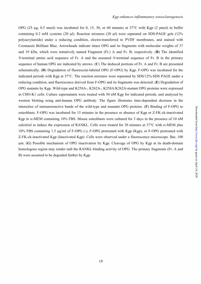
FIGURE 5. Primary sites in OPG cleaved by Kgp and inactivation of OPG after cleavage by Kgp. (A)
by guest on April 14, 2018
http://ww
w.jbc.org/
Dow
nloaded from
Kgp enhances inflammatory osteoclastogenesis
19
OPG (25 µg, 0.5 nmol) was incubated for 0, 15, 30, or 60 minutes at 37°C with Kgp (2 pmol) in buffer
containing 0.2 mM cysteine (20 µl). Reaction mixtures (20 µl) were separated on SDS-PAGE gels (12% polyacrylamide) under a reducing condition, electro-transferred to PVDF membranes, and stained with
Coomassie Brilliant Blue. Arrowheads indicate intact OPG and its fragments with molecular weights of 37
and 19 kDa, which were tentatively named Fragment (Fr.) A and Fr. B, respectively. (B) The identified
N-terminal amino acid sequence of Fr. A and the assumed N-terminal sequence of Fr. B in the primary
sequence of human OPG are indicated by arrows. (C) The deduced portions of Fr. A and Fr. B are presented
schematically. (D) Degradation of fluorescein-labeled OPG (F-OPG) by Kgp. F-OPG was incubated for the
indicated periods with Kgp at 37°C. The reaction mixtures were separated by SDS/12%-SDS PAGE under a
reducing condition, and fluorescence derived from F-OPG and its fragments was detected. (E) Degradation of
OPG mutants by Kgp. Wild-type and K258A-, K262A-, K258A/K262A-mutant OPG proteins were expressed
in CHO-K1 cells. Culture supernatants were treated with 50 nM Kgp for indicated periods, and analyzed by
western blotting using anti-human OPG antibody. The figure illustrates time-dependent decrease in the
intensities of immunoreactive bands of the wild-type and munatnt OPG proteins. (F) Binding of F-OPG to
osteoblasts. F-OPG was incubated for 15 minutes in the presence or absence of Kgp or Z-FK-ck-inactivated
Kgp in α-MEM containing 10% FBS. Mouse osteoblasts were cultured for 3 days in the presence of 10 nM
calcitriol to induce the expression of RANKL. Cells were treated for 30 minutes at 37°C with α-MEM plus
10% FBS containing 1.5 µg/ml of F-OPG (-), F-OPG pretreated with Kgp (Kgp), or F-OPG pretreated with Z-FK-ck-inactivated Kgp (Inactivated Kgp). Cells were observed under a fluorescence microscope. Bar, 100
µm. (G) Possible mechanism of OPG inactivation by Kgp. Cleavage of OPG by Kgp at its death-domain homologous region may render null the RANKL-binding activity of OPG. The primary fragments (Fr. A and
B) were assumed to be degraded further by Kgp.
by guest on April 14, 2018
http://ww
w.jbc.org/
Dow
nloaded from

Figure 1 (Akiyama T, et al.)
0 1 10 500 1 10 50 0 1 10 50 0 1 10 50
80
60
40
20
0
Ost
eocl
ast n
umbe
r (/w
ell)
Kgp (nM)Control IL-1 TNF- IL-17A
**
***
**
****
Control
RgpB
Control IL-1 TNF- IL-17A
Control
Kgp
Control IL-1 TNF- IL-17A
*
Control IL-1 TNF- IL-17AKgp
Ost
eocl
ast n
umbe
r (/w
ell)
60
40
20
0- + - + - + - +
**
**
Control IL-1 TNF- IL-17ARgpB
60
40
20
0Ost
eocl
ast n
umbe
r (/w
ell)
- + - + - + - +
NS
NS
NS
NS
A B
C
by guest on April 14, 2018
http://ww
w.jbc.org/
Dow
nloaded from

0
1
2
3
4
0
1
2
3
0
1
2
Control
Control
Control
Kgp
Kgp
Kgp
Rankl mRNA
Opg mRNA
Rank mRNA
Rel
ativ
e ex
pres
sion
(fol
d)R
elat
ive
expr
essi
on (f
old)
Rel
ativ
e ex
pres
sion
(fol
d)Figure 2 (Akiyama T, et al.)
by guest on April 14, 2018
http://ww
w.jbc.org/
Dow
nloaded from

Figure 3 (Akiyama T, et al.)
RANKL
0 1 3 18Incubation time (h)
0 1 3 180.5
RANK
β-Actinβ-Actin
Incubation time (h)
020406080
100
0.5 5 500
OPG
IL-17A
IL-1βTNF-α
Kgp (nM)
)%(
enikotycdedargedn
U
A BOPG
IL-1β
TNF-αIL-17A
0 0.5 5 50Kgp (nM)
)%(
enikotycdedargedn
U 1 3 6 150Incubation time (h)
020406080
100
0 1 3 6 15Incubation time (h)
OPG
IL-1β
TNF-αIL-17A
C D
by guest on April 14, 2018
http://ww
w.jbc.org/
Dow
nloaded from

0
10
20
30
40
50
Control IL-1β TNF-α IL-17A− + − + − +− +Kgp
A
B
0
10
20
30
40
50
NS
NS
NS
60
∗∗
∗∗
∗
∗∗
Wild-type cultures
OPG-deficient cultures
0 1 3 6 15
IL-1β
TNF-α
IL-17A
Incubation time (h)
C
Ost
eocl
ast n
umbe
r (/w
ell)
Ost
eocl
ast n
umbe
r (/w
ell)
Control IL-1β TNF-α IL-17A− + − + − +− +Kgp
120
100
80
60
40
20
00 1 3 6 15
Incubation time (h)
Und
egra
ded
cyto
kine
(%) IL-1β
TNF-αIL-17A
D
Figure 4 (Akiyama T, et al.)
by guest on April 14, 2018
http://ww
w.jbc.org/
Dow
nloaded from

Figure 5 (Akiyama T, et al.)
Fr. A
OPG
Mw755037
252015
0 15 30 60Treatment with Kgp (min)
1 mnnllccalv fldisikwtt qetfppkylh ydeetshqll cdkcppgtyl kqh ctakwkt
61 vcapcpdhyy tdswhtsdec lycspvckel qyvkqecnrt hnrvceckeg ryleiefclk
121 hrscppgfgv vqagtpernt vckrcpdgff snetsskapc rkhtncsvfg llltqkgnat
181 hdnicsgnse stqkcgidvt lceeaffrfa vptkftpnwl svlvdnlpgt kvnae sveri
241 krqhssqeqt fqllklwkhq nkdqdivkki iqdidlcens vqrhighanl tfeq lrslme
301 slpgkkvgae diektikack psdqilklls lwrikngdqd tlkglmhalk hskt yhfpkt
361 vtqslkktir flhsftmykl yqklflemig nqvqsvkisc
Signal Peptide
Fr. A
Fr. B
A B
C D
E
Fr. B
Fr. A Fr. B
22 189 209 361 401
RANKL-binding domain
Death domainhomologous
region
Kgp Inactivated KgpF
RANKL OPG
RANKL-bindingdomain
RANKL-bindingdomain
Death domainhomologous region
Death domainhomologous region
RANKL
RANKL-bindingdomain
RANKL-bindingdomain
Kgp
KgpFurther degradation of Fr. A and Fr. B
Fr. A
Fr. A
Fr. B
Fr. B
Treatment with Kgp (h)0 0.25 0.5 1 3 18
50
37
25
Mw
0
50
100
150
200
Rem
aini
ng O
PG
(%)
0 1 30.50.25Time (h)
OPGOPG (K258A)OPG (K262A)OPG (K258A/K262A)
G
by guest on April 14, 2018
http://ww
w.jbc.org/
Dow
nloaded from
Jan Potempa, Hisataka Yasuda, Kazuyoshi Baba and Ryutaro KamijoRika Yasuhara, Kenji Mishima, Toshifumi Maruyama, Chikara Kohda, Kazuo Tanaka,
Akiyama,Masamichi Takami, Tetsuo Suzawa, Marie Hoshino, Takahisa Imamura, Chie Tomohito Akiyama, Yoichi Miyamoto, Kentaro Yoshimura, Atsushi Yamada,
osteoprotegerin by lysine gingipainsuppresses that by interleukin-17A. Importance of proteolytic degradation of
, butβ and interleukin-1αdifferentiation induced by tumor necrosis factor--derived lysine gingipain enhances osteoclastPorphyromonas gingivalis
published online April 22, 2014J. Biol. Chem.
10.1074/jbc.M113.520510Access the most updated version of this article at doi:
Alerts:
When a correction for this article is posted•
When this article is cited•
to choose from all of JBC's e-mail alertsClick here
by guest on April 14, 2018
http://ww
w.jbc.org/
Dow
nloaded from
Related Documents