Keys to 2401 Models Photographs and keys prepared by Jeff Beck Department of Math and Natural Science Collin County Community College

Keys to 2401 Models Photographs and keys prepared by Jeff Beck Department of Math and Natural Science Collin County Community College.
Dec 24, 2015
Welcome message from author
This document is posted to help you gain knowledge. Please leave a comment to let me know what you think about it! Share it to your friends and learn new things together.
Transcript

Keys to 2401 Models
Photographs and keys prepared by
Jeff Beck
Department of Math and Natural Science
Collin County Community College

1. Nucleus2. Nuclear membrane3. Chromatin4. Nucleoli5. Rough endoplasmic reticulum6. Smooth endoplasmic reticulum7. Ribosomes8. Mitochondria9. Golgi apparatus12. Centrioles17. Cytosol18. Plasma membrane

1. Cell membrane2. Pinocytotic vesicle3. Cytosol4. Endoplasmic reticulum5. Golgi body (section)6. Golgi body (surface)7. Centrosome8. Lysosome9. Mitochondria10. Vacuole11. Nucleus12. Nucleolus13. Nuclear membrane

Model 1 – Prophase Model 5 - AnaphaseK = Nucleus ZF = Achromatic fibersZ1 & Z2 = Daughter chromosomes Ch = Daughter chromosomesPS = Aster rays Z1 & Z2 = SpindleN = NucleolusC = Cytoplasma Model 6 - Anaphase E = Plasma bodyModel 2 – ProphaseZS = Achromatic spindle Model 7 - AnaphaseModel 3 – Prophase ChT = Daughter plates ZF = Achromatic fibers E = Plasma bodyCh = Chromosomes Model 8 - TelophaseModel 4 – Metaphase Ch = ChromosomesCh = Chromosomes K = Nuclear membrane

1. Cell membrane2. Cytosol3. Nucleus4. Nucleolus5. Centrioles6. Aster7. Chromatin threads8. Spindle9. Spindle fibril10. Chromosome11. Chromatid12. Centromere13. Primary spermatocyte
13a. Early metaphase14. Secondary spermatocyte14a. Spermatocyte division15. Spermatids16. Spermatozoa17. Primary Oocyte17a. Metaphase18. Secondary Oocyte19. First polar body20. Ootid21. Polar bodies22. Ovum23. Diploid cell

CELL DIVISION1. Early Prophase2. Middle Prophase3. Late Prophase4. Metaphase5. Early Anaphase6. Late Anaphase7. Telophase8. Interphase
CELL STRUCTURE1. Cell membrane2. Cytosol3. Nucleus4. Nuclear membrane5. Nucleolus6. Centrioles7. Aster8. Chromatin9. Spindle10. Spindle fibril11. Chromosome12. Chromatid13. Centromere

I. EpidermisII. DermisIII. Subcutaneous layerIV. Horny layer of epidermis1. Hair shaft2. Stratum disjunctum3. Stratum conjunctum4. Clear layer of the epidermis5. Granular layer of the epidermis6. Spinous layer of the epidermis7. Basal layer of the epidermis8. Basement membrane9. Subpapillary venous plexus10. Papillary layer of the dermis11. Subpapillary arterial network12. Reticular layer of the dermis13. Sudoriferous duct14. Venous plexus of cornium15. Arterial network of cornium16. Root of hair17. Arrector muscle of hair
18. Lymph vessel19. Arteriole20. Nerve21. Venula22. Krause corpuscle23. Golgi-Mazzoni corpuscle24. Brushes of Ruffini25. Pacinian corpuscle26. Papillary artery27. Papillary vein28. Cortex of hair29. Medulla of hair30. Sweat gland31. Outer epithelial sheath32. Henle layer33. Huxley layer34. Hair cuticle35. Root of hair36. Sebaceous gland37. Body of apocrine sudoriferous gland
38. Papillary nerve39. Hair nerve40. Fascia41. Muscle42. Adipose cell43. Arrector muscle of hair44. Meissner’s tactile corpuscle45. Sweat pore46. Melanocyte47. Duct of apocrine sudoriferous gland48. Merkel corpuscles

A. Section of the Scalp
B. Section of the sole of the foot
I. Epidermis
II. Dermis
III. Subcutaneous1. Meissner’s tactile corpuscle2. Horny layer of epidermis3. Clear layer of epidermis4. Granular layer of epidermis5. Spinous layer of epidermis6. Basale layer of epidermis7. Stratum germinativum8. Papillary layer of dermis9. Sudoriferous duct10. Reticular layer of dermis11. Sweat gland12. Pacinian corpuscle13. Adipose cell14. Outer root sheath15. Inner root sheath16. Hair cuticle17. Hair bulb18. Medulla of hair19. Cortex of hair20. Arrector muscle of hair
21. Root of hair22. Sebaceous gland23. Hair shaft24. Sweat pore

1. Stratum Corneum 19. Artery-blood supply to hair2. Stratum Lucidum 20. Hair arrector muscle3. Stratum Granulosum 21. sweat gland4. Stratum Mucosum 22. Glomerulus of sweat gland5. Stratum Germinativum 23. Longitudinal section of sweat gland6. Corium layer 24. Longitudinal section of glomerulus of sweat gland7. Fatty layer 25. Vascular papilla of corneum8. Hair shaft 26. Vein-blood supply from corneum9. Longitudinal section of hair root 27. Artery-blood supply from corneum10. Sebaceous oil gland 28. Meissner’s Corpuscle11. Longitudinal section of oil gland 29. Free nerve endings12. Hair bulb 30. Tactile disks13. Longitudinal section of hair root 31. Corpuscle of Ruffini14. External root sheath 32. Bulb of Krause15. Internal root sheath 33. Pacinian Corpuscle16. Zone of growth and differentiation 34. Epidermis17. Papilla of hair 35. Dermis18. Vein-Blood supply from hair

I. Epidermis 10. Hair shaftII. Corium (Dermis) 11. Hair rootIII. Subcutaneous layer 12. Hair papilla1. Stratum corneum 13. Sebaceous glands1a. Stratum lucidum 14. Arrector pilorum muscles2. Stratum germinativum 15. Sweat gland of the armpit2a. Stratum granulosum 15a. Smooth muscle cells2b. Stratum spinosum 15b. Hyaline top2c. Stratum basale 15c. Pushed out cell body3. Papillae4. Touch corpuscles5. Adipose tissue6. Lamellated corpuscles7. Sweat glands8. Hairs8a. Medullary substance8b. Cortical substance8c. Cuticle of hair8d. Inner root sheath8e. Outer root sheath8f. Hyaloid membrane8g. Fibrous layer9. Hair cross section
Skin of scalp Skin of armpit Skin of sole of foot

1. Nucleus2. Nucleolus3. Cytoplasm4. Lysosome5. Vesicles of Golgi and ER6. Golgi apparatus7. Mitochondria8. Endoplasmic reticulum9. Nerve fibers10. Axon hillock11. Axon12. Dendrite13. Schwann cell cytoplasm14. Node of Ranvier15. Cytoplasm o0f axon (axoplasm)16. Axolemma17. Schwann cell nucleus18. Schmidt-Lantermann cleft19. Myelin sheath

A. Perikaryon of the nerve cell with dendritesB. Peripheral nerve with sheaths1. Neurite cone2. Nucleus of nerve cell with nucleolus3. Endoplasmic reticulum4. Neurofibrils5. Synaptic terminals6. Neuraxon7. Schwann cells with nucleus8. Schwann sheath9. Linked Schwann cells at node of Ranvier10. Mitochondria11. Medullary (myelin) sheath12. Perineural sheath of connective tissue13. Mesaxon14. Dendrites15. Lysosomes16. Neurotubules17. Golgi apparatus

A. Perikaryon of the nerve cell with dendrites 16. NeurotubulesB. Peripheral nerve with sheaths 17. Golgi apparatus1. Axon hillock2. Nucleus of nerve cell with nucleolus3. Endoplasmic reticulum4. Neurofibrils5. Synaptic terminals6. Neuraxon7. Schwann cell with nucleus8. Schwann sheath9. Linked Schwann cells at node of Ranvier10. Mitochondria11. Medullary (myelin ) sheath12. Endoneurium13. Mesaxon14. Dendrites15. Lysosomes

1. Presynapse
2. Presynaptic membrane
3. Neuroplasma4. Endoplasmic reticulum5. Mitochondrium6. Neurotubule7. Neurofilament8. Presynaptic grid9. Vesicle10. Vesicle with neurotransmitter11. Transmitter release12. Transmitter gap products13. Endocytic material absorption14. Pores for endocytic material absorption15. Synaptic gap16. Postsynapse17. Subsynaptic membrane
18. Postsynaptic membrane 19. Transmitter receptor molecule reaction

1. Left cerebral hemisphere 22. Eyeball 43. Skin section
2. Right cerebral hemisphere 23. Tongue 44. sensory nerve endings3. Cerebral cortex 24. Lingual nerve 45. Axon of sensory nerve4. Cerebrum 25. First cervical nerve 46. Sensory nerve cell5. Corpus callosum 26. second cervical nerve 47. Synapse6. Fornix 27. Frontal lobe 48. Dendritic nerve endings7. Third ventricle 28. Parietal lobe A. Olfactory nerve8. Thalamus 29. Temporal lobe B. Optic nerve9. Massa intermedia 30. Occipital lobe C. Oculomotor nerve10. Pineal body 31. Sensory cortex D. Trochlear nerve11. Sinus rectus 32. Motor cortex E. Trigeminal nerve12. Pituitary gland 33. Auditory cortex E-1 Ophthalmic branch13. Midbrain 34. Visual cortex E-2 Maxillary branch14. Pons 35. Broca’s area E-3 Mandibular branch15. Medulla oblongata 36. Cerebrum F. Abducens nerve 16. Fourth ventricle 37. Brachial pexus G. Facial nerve17. Cerebellum 38. Peripheral nerves to arms H. Acoustic nerve18. Olive 39. Nerves to thorax I. Glossopharyngeal nerve19. Spinal cord 40. Sacral plexus J. Vagus nerve20. Arachnoid sheath 41. Peripheral nerves to legs K. Spinal accessory nerve21. Dura mater 42. Needle point stimulus L. Hypoglossal nerve

FIFTH CERVICAL VERTEBRAE WITH SPINAL CORD AND ORIGIN OF NERVES1. Body of vertebrae 19. Spinal ganglion 36. Anterior column
2. Transverse process 20. Anterior ramus of nerve 37. Lateral column3. Vertebral arch 21. Posterior ramus of nerve 38. Posterior column4. Spinous process 22. Sympathetic trunk5. Yellow ligament 23. Ramus communicans between6. Posterior longitudinal ligament sympathetic nerve and spinal cord7. Vertebral artery 24. Pia mater8. Vertebral vein 25. Anterior median fissure9. Dura mater 26. Anterior lateral sulcus10. Epidural cavity 27. Posterior lateral sulcus11. Subdural cavity 28. Posterior median fissure 12. Arachnoid 29. Posterior glial septum13. Subarachnoid cavity 30. Anterior funicle14. Internal vertebral venous plexous 31. Lateral funicle15. Denticulate ligament 32. Posterior funicle16. Subarachnoid septum 33. White substance17. Ventral root of cervical nerve 34. Gray substance18. Dorsal root of cervical nerve 35. Central canal

THE ORIGINS OF THE NERVES IN THE SPINAL CORDA. Spinal cord section B. Spinal cord with nerves1. Gray substance 12. Gray substance2. Anterior column 13. Radicular filaments3. Lateral column 14. Gray commissure4. Gray commissure 15. Central canal5. Anterior radicular filaments 16. White substance 6. Posterior radicular filaments 17. Anterior column7. White substance 18. Lateral column8. Anterior median fissure 19. Anterior median fissure9. Posterior median fissure 20. Posterior median sulcus10a. Anterior lateral sulcus 21. Ganglion10b. Posterior lateral sulcus 22. Anterior funicle 23. Lateral funicle

1. Dorsal Column, White Matter 16. Anterior Spinal Artery2. Lateral Column, White Matter 17. Sulcal Artery3. Ventral Column, White Matter 18. Anterior Spinal vein4. Anterior White Commissure 19. Dorsal Radicular
Filaments5. Dorsal Gray Horn, Gray Matter20. Left Posterolateral Spinal Vein6. Lateral Gray Horn, Gray Matter 21. Left Posterior Spinal
Artery7. Ventral gray Horn, Gray matter 22. Posterior Spinal Vein8. Gray Commissure 23. Dorsal Root9. Central Canal 24. Spinal Ganglion10. Dorsal Median Sulcus 25. Ventral Root11. Dorsal Intermediate Sulcus 26. Mixed Spinal Nerve12. Dorsal Lateral Sulcus 27. Dorsal Ramus of Spinal
Nerve13. Ventral Lateral Sulcus 28. Ventral Ramus of Spinal
Nerve14. Ventral Median Fissure 29. Gray Ramus
Communicantes15. Ventral Radicular Filaments 30. White Ramus
Communicantes
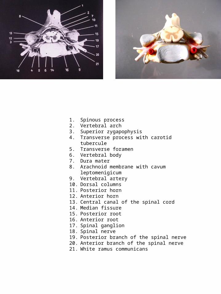
1. Spinous process2. Vertebral arch3. Superior zygapophysis4. Transverse process with carotid tubercule5. Transverse foramen6. Vertebral body7. Dura mater8. Arachnoid membrane with cavum leptomenigicum9. Vertebral artery10. Dorsal columns11. Posterior horn12. Anterior horn13. Central canal of the spinal cord14. Median fissure15. Posterior root16. Anterior root17. Spinal ganglion18. Spinal nerve19. Posterior branch of the spinal nerve20. Anterior branch of the spinal nerve21. White ramus communicans

CEREBRUM
1. Frontal lobe2. Parietal lobe3. Occipital lobe3a. Temporal lobe of telencephalon4. Cerebellum5. Bridge6. Spinal bulb7. Myel
SPINAL NERVES8. Cervical nerve9. Thoracic nerve10. Lumbar nerve11. Sacral nerve12. Terminal cone13. Cauda equina14. Urocyst15. Sympathetic trunk of neck16. Sympathetic trunk of thorax17. Sympathetic trunk pars lumbar18. Sympathetic trunk pars sacrum
NERVES OF THE PECTORAL LIMBS19. Cervicali nerves20. Axillary nerve21. Musculocutaneous nerve22. Radial nerve23. Median nerve24. Ulnar nerve25. Superior branch of radial nerve26. Branch of median nerve27. Branch of ulnar nerve28. Dorsal branch of ulnar nerve29. Cutaneous dorsal nerve of radius30. External nerves of finger31. Palm nerves of the hand
NERVES OF THE PELVIC LIMBS32. Ilioinguinal nerve33. Nerve cutaneus femoris lateralis34. Femoral nerve35. Sciatic nerve36. Obturator nerve37. Common peroneal nerve38. Tibial nerve39. Saphenous nerve40. Infrapatellar branch of saphenous nerve41. Nerve fibularis profunus42. Nerve fibularis superficialis43. Nerve cutaneous dorsal44. Dorsal nerve of toe45. Branch of muscle of sciatic nerve46. Tibial nerve47. Sural nerve48. Nerve cutaneous dorsal of forearm49. Nerve cutaneous lateral of forearm50. Nerve cutaneous medial of forearm

BRAINI. End brainII. InterbrainIII. MidbrainIV. AfterbrainV. MarrowbrainI, II, III Cerebrum1. Superior longitudinal sinus2. Tentorium cerebelli3. Little brain a. arbor vitae cerebelli4. Trabs cerebri a. rostrum corporis callosi b. genu corporis callosi5. Septum pellucidum6. Fornix7. Anterior commissure8. Middle commissure9. Posterior commissure10. Thalamus11. Foramen of Monro12. Aqueduct of the cerebrum13. Ventriculus quartus14. Corpus pineale15. Chiasma opticum16. Hypophysis17. Corpus mamillare18. Quadrigeminal plate19. Upper velum palati20. Bridge21. Lengthening of spinal marrow22. Spinal marrow

CEREBRUM1. Frontal lobe2. Parietal lobe3. Occipital lobe4. Temporal lobe5. Central sulcus6. Precentral gyrus7. Postcentral gyrus8. Olfactory bulb9. Anterior commissure10. Corpus callosuma. Genub. Trunkc. Spleniumd. Rostrum11. Septum pellucidum12. Fornix13. Posterior commissure14. Insula15. Internal capsule16. Lateral ventriclee. Frontal (anterior) hornf. Central part17. Choroid plexusg. Posterior hornh. Inferior horn18. Hippocampus
DIENCEPHALON19. Thalamus20. Hypothalamic sulcus
21. Hypothalamus22. Interthalamic adhesion23. Pineal body24. Left mamillary body25. Pituiatry gland26. Choroid plexus of third ventricle27. Caudate nucleus28. Pulvinar29. Medial geniculate body30. Lateral geniculate body31. Quadrigeminal lamina32. Tegmentum33. Cerebral peduncle34. Cerebral aquaduct
CEREBELLUM35. Cerebelluma. Vermisb. Tonsilc. Flocculusd. Arbor vitaee. Fourth ventricle36. Ponsf. Superior cerebellar peduncleg. Middle cerebellar peduncleh. Inferior cerebellar peduncle
BRAIN STEM37. Medulla oblongata38. Olive
39. Pyramid40. First cervical nerve
CRANIAL NERVEI. Olfactory nerveII. Optic nerveIII. Oculomotor nerveIV. Trochlear nerveV. Trigeminal nerveVI. Abducens nerveVII. Facial nerveVIII. Vestibulocochlear nerveIX. Glossopharyngeal nerveX. Vagus nerveXI. Accessory nerveXII. Hypoglossal nerve
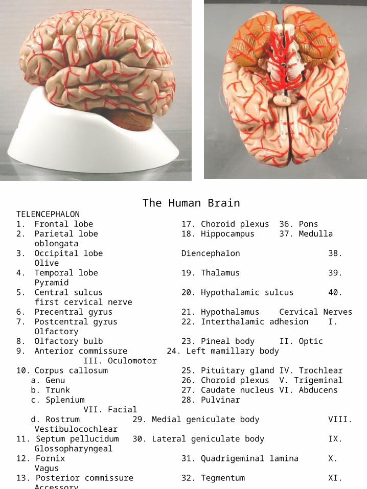
The Human BrainTELENCEPHALON1. Frontal lobe 17. Choroid plexus 36. Pons2. Parietal lobe 18. Hippocampus 37. Medulla oblongata3. Occipital lobe Diencephalon 38. Olive4. Temporal lobe 19. Thalamus 39. Pyramid5. Central sulcus 20. Hypothalamic sulcus 40. first cervical nerve6. Precentral gyrus 21. Hypothalamus Cervical Nerves7. Postcentral gyrus 22. Interthalamic adhesion I. Olfactory8. Olfactory bulb 23. Pineal body II. Optic9. Anterior commissure 24. Left mamillary body III. Oculomotor10. Corpus callosum 25. Pituitary gland IV. Trochlear a. Genu 26. Choroid plexus V. Trigeminal b. Trunk 27. Caudate nucleus VI. Abducens c. Splenium 28. Pulvinar VII. Facial d. Rostrum 29. Medial geniculate body VIII. Vestibulocochlear11. Septum pellucidum 30. Lateral geniculate body IX. Glossopharyngeal12. Fornix 31. Quadrigeminal lamina X. Vagus13. Posterior commissure 32. Tegmentum XI. Accessory14. Insula 33. Cerebral peduncle XII. Hypoglossal15. Internal capsule 34. Cerebral aquaduct Circle of Willis16. Lateral ventricle 35. Cerebellum
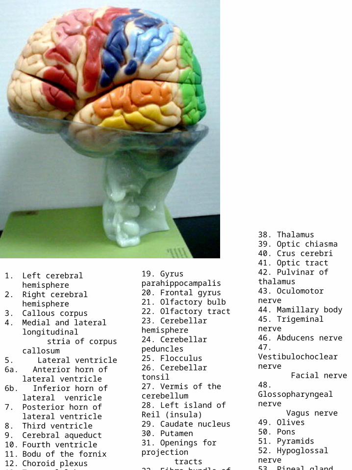
1. Left cerebral hemisphere2. Right cerebral hemisphere3. Callous corpus4. Medial and lateral longitudinal stria of corpus callosum5. Lateral ventricle6a. Anterior horn of lateral ventricle6b. Inferior horn of lateral venricle7. Posterior horn of lateral
ventricle8. Third ventricle9. Cerebral aqueduct10. Fourth ventricle11. Bodu of the fornix12. Choroid plexus13. Temporal lobe14. Occipital lobe15. Pes hippocampi16. Choroid plexus17. Dentate gyrus18. Fimbria of hippocampus
19. Gyrus parahippocampalis20. Frontal gyrus21. Olfactory bulb22. Olfactory tract23. Cerebellar hemisphere24. Cerebellar peduncles25. Flocculus26. Cerebellar tonsil27. Vermis of the cerebellum28. Left island of Reil (insula)29. Caudate nucleus30. Putamen31. Openings for projection tracts32. Fibre bundle of internal capsule33. Pallidum34. Right island of Reil35. Lentiform nucleus36. Internal capsule37. Striate body
38. Thalamus39. Optic chiasma40. Crus cerebri41. Optic tract42. Pulvinar of thalamus43. Oculomotor nerve44. Mamillary body45. Trigeminal nerve46. Abducens nerve47. Vestibulochoclear nerve Facial nerve48. Glossopharyngeal nerve Vagus nerve49. Olives50. Pons51. Pyramids52. Hypoglossal nerve53. Pineal gland54. Quadrigeminal lamina55. Trochlear nerve56. Rhomboid fossa57. Tubercule of nucleus gracilis58. Tubercule of nucleus cuneatus

1. Scalp2. Cranium3. Superior sagittal sinus4. Pachymeninx5. Falx cerebri6. Frontal lobes of the cerebrum7. Middle meningeal artery8. Cortex, grey matter9. Cerebral vessels10. Temporal lobe11. Occipital lobe12. Leptomeninx13. Superior sagittal sinus14. Subarachnoid space15. Occipital vessel and greater occipital nerve

1. Scalp2. Skull bone3. Superior sagittal sinus4. Pachymeninx5. Falx cerebri6. Frontal lobes of the cerebrum7. Middle meningeal artery8. Cortex, grey matter9. Cerebral vessels10. Temporal lobe11. Occipital lobe12. Leptomeninx13. Superior sagittal sinus14. Subarachnoid space15. Occipital vessel and greater occipital nerve

1. Cranium2. Superior sagittal sinus3. Falx cerebri4. Pachymeninx5. Frontal lobes of the cerebrum6. Anterior cerebral artery7. Corpus callosum, head of the caudate nucleus8. Anterior horn of the lateral ventricle9. Nucleus caudatus10. Septum pellucidum11. Lateral ventricle Choroid plexus12. Posterior horn of the lateral ventricle13. Inferior sagittal sinus14. Meningeal artery15. Occipital lobe16. Scalp

1. Frontal sinus2. Superior sagittal sinus3. Falx cerebri4. Pachymeninx5. Frontal lobes of the sinus6. Anterior cerebral artery7. Corpus callosum8. Anterior horn of the lateral ventricle9. Nucleus caudatus10. Septum pellucidum, Columns of fornix11. Lateral ventricle12. Anterior horn of the lateral ventricle13. Inferior sagittal sinus14. Meningeal artery15. Occipital lobe16. Internal capsule17. Insula18. Putamen19. Extreme capsule20. Globus pallidus21. External capsule22. Claustrum23. Thalamus24. Posterior crus of fornix Choroid plexus

1. Frontal sinus2. Superior sagittal sinus3. Falx cerebri4. Pachymeninx5. Frontal lobes of the cerebrum6. Anterior cerebral artery7. Head of nucleus caudatus8. Insula9. Anterior commissure10. Claustrum11. Putamen12. Thalamus13. Tail of nucleus caudatus14. Posterior horn of the lateral ventricle15. Optic radiation16. Occipital lobe17. Internal capsule18. Globus pallidus19. External capsule20. Column of fornix21. Third ventricle22. Pineal body23. Posterior crus of fornix Choroid plexus24. Corpus callosum25. Straight sinus26. Occipital bone
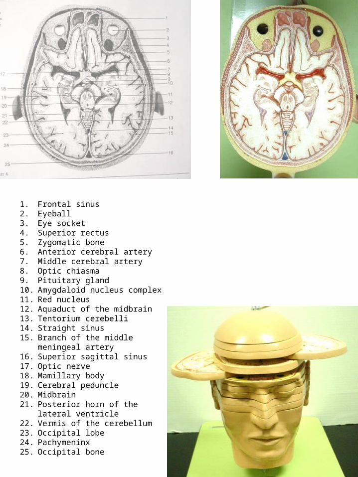
1. Frontal sinus2. Eyeball3. Eye socket4. Superior rectus5. Zygomatic bone6. Anterior cerebral artery7. Middle cerebral artery8. Optic chiasma9. Pituitary gland10. Amygdaloid nucleus complex11. Red nucleus12. Aquaduct of the midbrain13. Tentorium cerebelli14. Straight sinus15. Branch of the middle meningeal artery16. Superior sagittal sinus17. Optic nerve18. Mamillary body19. Cerebral peduncle20. Midbrain21. Posterior horn of the lateral ventricle22. Vermis of the cerebellum23. Occipital lobe24. Pachymeninx25. Occipital bone

1. Cornea2. Lens3. Eyeball, Sclera4. Medial rectus muscle5. Optic nerve6. Lateral rectus muscle7. Temporalis8. Internal carotid artery9. Pituitary gland Sinus cavernosus10. Basilar artery11. Midbrain12. Aqueduct of the midbrain13. Straight sinus14. Falx cerebri15. Superior sagittal sinus16. Nasal septum17. Ethmoidal air cells18. Sphenoid sinus19. Temporal bone20. Third cranial nerve21. Outer ear22. Cerebellar hemisphere23. Vermis of the cerebellum24. Occipital bone

1. Cartilages of the nose2. Nasal cavities3. Base of the orbit4. Nasal septum5. Ethmoidal cells6. Sphenoid7. Temporal lobe8. Cochlea9. Auditory ossicles10. Vestibulocochlear nerve Facial nerve11. Temporal bone12. Posterior semicircular canal13. Sigmoid sinus14. Occipital sinus15. Internal carotid artery16. Maxillary nerve17. Venous plexus18. Basilar artery19. Bridge20. Middle ear21. Mastoid cells22. Fourth ventricle23. Vermis of the cerebellum24. Cerebellar hemisphere25. Occipital bone

1. Cartilages of the nose2. Nasal cavity3. Base of the orbit4. Maxillary sinus5. Masseter6. Mandible7. Medial pterygoid8. Lateral pterygoid9. Articular disk10. Condyle of the mandible11. Cochlea12. Tympanic membrane13. External auditory canal14. Mastoid air cells15. Sigmoid sinus16. Cerebellar hemisphere17. Occipital sinus18. Sphenoid19. Third trigeminal nerve20. Middle meningeal artery21. Internal carotid artery22. Middle ear23. Basilar artery24. Medulla oblongata25. Vermis of the cerebellum26. Trapezius muscle
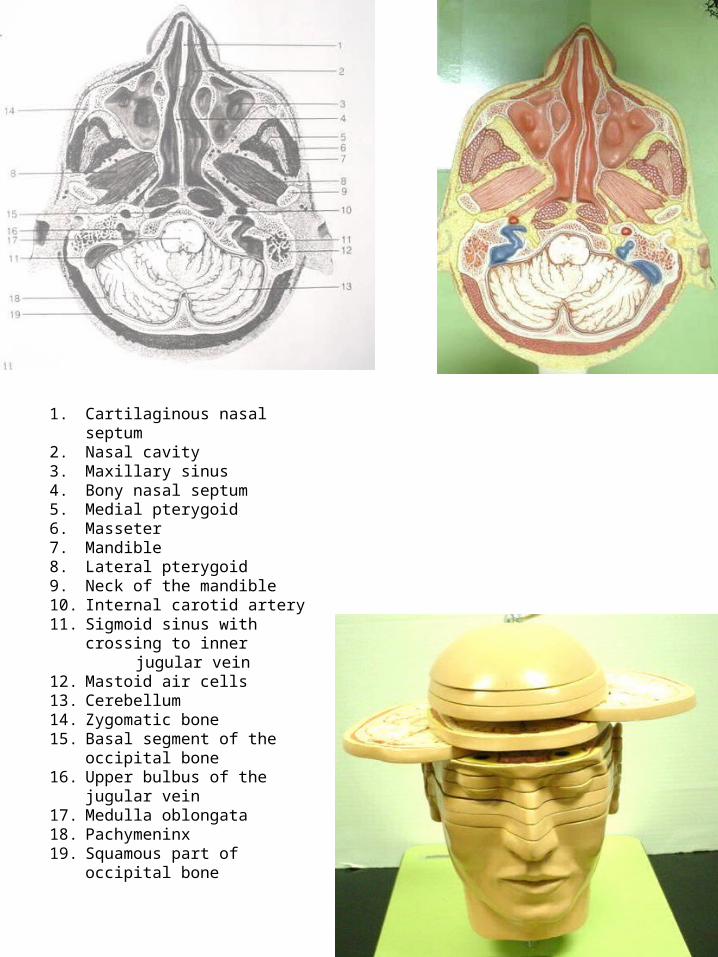
1. Cartilaginous nasal septum2. Nasal cavity3. Maxillary sinus4. Bony nasal septum5. Medial pterygoid 6. Masseter7. Mandible8. Lateral pterygoid9. Neck of the mandible10. Internal carotid artery11. Sigmoid sinus with crossing to inner jugular vein12. Mastoid air cells13. Cerebellum14. Zygomatic bone15. Basal segment of the occipital bone16. Upper bulbus of the jugular vein17. Medulla oblongata18. Pachymeninx19. Squamous part of occipital bone

EYELID1. Superior muscle of eyelid2. Superior tarsus palpebrarum3. Inferior tarsus palpebrarum4. Ligamentum palpebrale mediale5. Ligamentum palpebrale laterale6. Conjunctival fold7. Palpebral conjunctiva8. Tarsoconjunctival glands
LACRIMAL APPARATUS9. Lacrimal glanda. Orbital processb. Palpebral processc. Ductules of the lacrimal gland10. Lacrimal sac11. Lacrimal lakea. Superior lacrimal ductb. Inferior lacrimal ductc. Lacrimal caroncule12. Lacrimal tubercule, superior lacrimal point
EYE MUSCLESI. Superior rectusII. Inferior rectusIII. Medial rectusIV. Lateral rectusV. Tendon of superior obliqueVI. Inferior obliqueA. SCLEROCORNEA1. Cornea2. ScleraB. UVEA3. Iris with pupil4. Ciliary muscle5. Corona ciliaris6. Choroid membrane7. Ciliary nerves8. Vorticosa veina. Ciliary arteriesC. TUNICA INTERNA BULBI9. Retina10. Pars ciliaris retinae
11. Retinal arterioles12. Retinal venoles13. Fovea centralis14. Blind spot15. Lens16. Vitreous body

EYE MUSCLES 13. Fovea centralisI. Superior rectus 14. Optic nerve discII. II. Inferior rectus 15. LensIII. Medial rectus 16. Vitreous bodyIV. Lateral rectusV. Tendon of superior obliqueVI. Inferior oblique
EYE, EXTERNAL LAYER1. Cornea2. Sclera
EYE, CHOROID VASCULAR COAT3. Iris4. Ciliary muscle5. Ciliary zone6. Choroid coat7. Ciliary nerve8. Vorticose veins8a. Ciliary artery
EYE, RETINAL LAYER9. Retina10. Ciliary part of retina11. Retinal arteries12. Retinal veins

A. Ocular Fundus of the right eye 23. Iris1. Optic disk with retinal vessels 24. Tarsus of the upper lid2. Macula lutea , fovea centralis 25. CorneaB. Anatomy of the retina 26. Lens3. Nerve fiber layer 27. Ora serrata, pars plana corporis ciliaris4. Ganglionic layer of the optic nerve 28. Suspensory ligament5. Internal reticular layer 29. Ciliary body6. Internal nuclear layer 30. Inferior oblique7. External reticular layer D. Layers of the eye8. External nuclear layer 31. Choroid with vessels9. Layer of rods and cones 32. Sclera, Limbus corneae10. Pigmented layer of retina 33. Pigmented epithelium of the retina11. Choroid 34. Cornea12. Sclera 35. RetinaC. Sagittal section of the eye 36. Sclera13. Levator muscle 37. Inferior oblique14. Superior rectus 38. Superior rectus15. Optic nerve 39. Long posterior ciliary nerve16. Choroid 40. Short posterior ciliary nerves17. Retina with retinal vessels 41. Optic nerve18. Sclera 42. Medila rectus19. Inferior rectus 43. Inferior vorticose vein after perforation of the sclera20. Orbicularis oculi 44. Inferior rectus muscle21. Fornix conjunctivae22. Conjunctiva palpebralis

A. External Ear C. Inner Ear1. Flad of the ear 12. Vestibule2. Meatus acusticus externus 13. Oval window3. Tympanic membrane 14. Fenestra cochlea4. Annulus fibrocartilagineus 15. Lateral semicircular canal membranae tympani a. Ampulla ossea lateralisB. Middle Ear 16. Anterior semicircular canal5. Tympanic cavity b. Ampulla ossea anterior6. Musculus tensor tympani 17. Posterior semicircular canal7. Eustachian tube c. ampulla ossea posterior8. Hammer 18. Cochleaa. Head of the malleus 19. Vestibulocochlear nerveb. Manubrium mallei 20. Internal carotid arteryc. Anterior process of the malleus 21. Musculus tensor veli palatini9. Anvila. Short crusb. Long crus10. Lentiform nodule11. Stirrupa. Head of the stapesb. Crus of the stapesc. Base of the stapes

Pinna (1) Basilar membrane (37)Auditory meatus (2) Organ of Corti (37)Eustachian tube (3) Scala tympani (38)Temporal bone (5)Middle Ear (12)Tympanum (13)Malleus (14)Incus (15)Stapes (16)Semicircular canals (17, 18, 19)Ampullae (20, 21, 22)Vestibule (23)Utricle (24)Saccule (25)Superior scarpa ganglion (28)Vestibular nerve (29)Cochlear nerve (30)Oval window (32)Round window (33)Cochlea (34)Scala vestibuli (36)

1. Cochlea 26. Cochlear arteries2. Scala vestibuli 27. Tympanic lip
of3. Scala tympani the limbus4. Vestibular wall of the cochlear duct5. Osseous spiral lamina6. Secondary spiral lamina7. Epithelium of the internal spiral sulcus8. Internal hair cells9. External hair cells10. Internal pillar cells11. External pillar cells12. Tunnel of Corti13. Deiters’ supporting cells14. Cells of Hensen15. Claudius’ cells16. Nuel’s space17. Vestibular lip of the limbus18. Tectorial membrane19. Cochlear duct20. Spiral ligament of the cochlea21. Prominent vessel22. Modiolus23. Laterla wall of the cochlea24. Spiral ganglion25. Cochlear nerve

1. Capillaries2. Capillaries3. Lymphatic spaces4. Nerve5. Haversian lamellae6. Interstitial or Interhaversian lamellae7. Nucleus8. Cell body9. Canaliculi10. Lacunae – some lacunae may contain osteocytes.

1. Frontal bone2. Parietal bone2a. Interparietal bone3. Occipital bone4. Temporal bone5. Mastoid process6. Zygomatic process7. Articular tubercle8. Mandibular fossa9. Styloid process10. Greater wing of Sphenoid bone11. Zygomatic bone12. Nasale bone13. Maxillary body14. Lacrimal bone15. Ethmoid bone16. Vomer17. Palatine bone18. Sphenoid bone19. Occipital condyle20. Mandible21. Coronoid process22. Condylar process23. Coronal suture24. Sagittal suture25. Lambdoid suture25a. Transverse occipital suture26. Occipitomastoid suture27. Parietomastoid suture28. Squamosal suture29. Sphenosquamosal suture30. Zygomatico-temporal suture
31. Zygomatico-frontal suture32. Zygomatico-maxillary suture33. Frontonasal suture33a. Internasal suture34. Frontomaxillary suture34a. Nasomaxillary suture34b. Lacrimomaxillary suture35. Sphenosygomatic suture36. Intermaxillary suture37. Median palatine suture38. Transverse palatine suture39. Optical canal40. Lacrimal fossa41. Infraorbital foramen42. Acoustic meatus43. Mastoid foramen44. Foramen ovale45. Foramen spinosum46. Foramen lacerum47. Carotid canal48. Jugular foramen49. Stylomastoid foramen50. Incisive foramen51. Greater palatine foramen52. Mental foramen
54. Anterior cranial fossa55. Medial cranial fossa56. Posterior cranial fossa57. Crista frontalis58. Crista galli59. Cribriform plate
60. Jugum sphenoidale61. Anterior clinoid process62. Greater wing of sphenoid bone63. Sella turcica65. Internal occipital protuberance66. Foramen rotundum67. Internal acoustic opening68. Hypoglossal canal

1. Frontal bone (Os frontale)2. Temporal bone ( Os temporale)3. Sphenoid bone (Os sphenoidale)4. Occipital bone (Os occipitale)5. Parietal bone (Os parietale)6. Ethmoid bone (Os ethmoidale)7. Maxilla (upper jaw)8. Zygomatic bone (Os zygomaticum)9. Palatine bone (Os palatinum)10. Lacrimal bone (Os lacrimale)11. Inferior nasal concha (Concha nasalis inferior)12. Nasal bone (Os nasale)13. Vomer14. Mandible (lower jaw)15. – 21. Cervical vertebrae

1. Frontal bone2. Parietal bone3. Temporal bone4. Zygomatic bone5. Nasal bone6. Occipital bone7. Maxilla8. Vomer9. Ethmoidal bone10. Sphenoid bone11. Mandible

1. Nasal bone 24. Posterior superior dental artery2. Superior maxilla 25. Infraorbital vein3. Infraorbital foramen 26. Superior dental vein4. Nasal septum 27. Inferior dental nerve5. Facies of the eye 28. Inferior dental artery6. Zygomatico – maxillary suture 29. Inferior dental vein 7. Palatine process 30. Incisive foramen, foramen of Stenson8. Alveolar process 31. Greater palatine foramen9. Palatine bone10. Mandible11. Coronoid process of mandible12. Condyloid process13. Mandibular angle14. Mental foramen15. Mental tubercle16. Mandibular foramen17. Incisor teeth18. Eye teeth19. Premolar teeth20. Molar teeth21. Infraorbital nerve22. Posterior superior dental branches of the maxillary nerve23. Infraorbital artery

ELBOW JOINT1. Radial collateral ligament2. Radial annular ligament3. Ulnar collateral ligament4. Bicep muscle tendon of insertion5. Fibrous capsule of the articular capsule

HIP JOINT1. Iliofemoral ligament2. Ischiofemoral ligament3. Pubofemoral ligament4. Orbicular zone5. Transverse acetabulum ligament

KNEE JOINT1. Anterior cruciate2. Posterior cruciate3. Medial meniscus4. Lateral meniscus5. Transverse ligament6. Tibial collateral7. Fibular collateral8. Patellar ligament

SHOULDER JOINT1. Supraspinatus2. Infraspinous fossa3. Teres minor4. Subscapularis5. Long head biceps6. Coracoacromial ligament7. Trapezoid ligament8. Conoid ligament9. Coracohumeral ligament10. Glenoid labrum
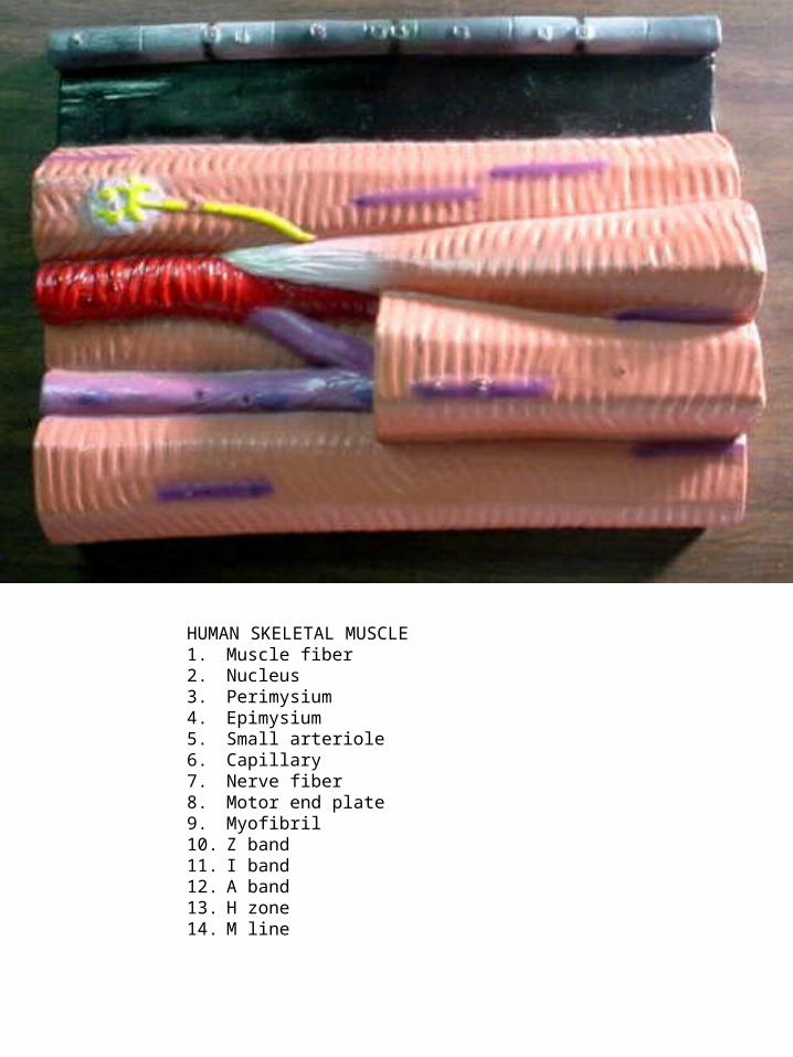
HUMAN SKELETAL MUSCLE1. Muscle fiber2. Nucleus3. Perimysium4. Epimysium5. Small arteriole6. Capillary7. Nerve fiber8. Motor end plate9. Myofibril10. Z band11. I band12. A band13. H zone14. M line

HUMAN SMOOTH MUSCLE1. Smooth muscle cell or fiber2. Capillary3. Myofibrils4. Nucleus of the muscle cell5. Mitochondria6. Nucleolus

1. Sarcoplasm cytoplasmic matrix 19. Synaptic vesicle2. Nuclei3. Myofibrils4. Cohnheim’s areas5. Sarcolemma6. Endomysium7. Medullated nerve fiber8. Axis cylinder (axon and neuraxon)9. Myelin sheath10. Granules11. Terminal branches12. Terminal neurofibrils13. Schwann cells14. Sarcoplasmic cavity15. Nuclei16. Mitochondria17. Subneural lamella18. Lamella

19. Splenius capitis20. Occipitofrontalis (frontal venter)21. Occipitofrontalis (occipital venter)22. Galea aponeurotica23. Compressor naris and dilator naris24. Orbicularis oculi (palpebral part)25. Orbicularis oculi (orbital part)26. Auricularis anterior27. Auricularis posterior28. Orbicularis oris29. Depressor anguli oris30. Risorius31. Zygomaticus major32. Zygomaticus minor33. Levator anguli oris34. Levator labii superioris alaeque nasi35. Buccinator36. Mentalis37. Depressor labii inferioris 38. Masseter39. Temporal40. Splenius cervicis41. Sternomastoid42. Scalenus medius43. Scalenus posterior44. Scalenus anterior45. Levator scapulae46. Digastric ( anterior venter)47. Digastric ( posterior venter)48. Stylohyoid49. Mylo-hyoid50. Geniohyoid51. Hyoglossus52. Sternohyoid53. Omo-hyoid ( superior venter)54. Omo-hyoid (inferior venter)55. Sternothyroid 56. Thyrohyoid57. Crico-thyroid
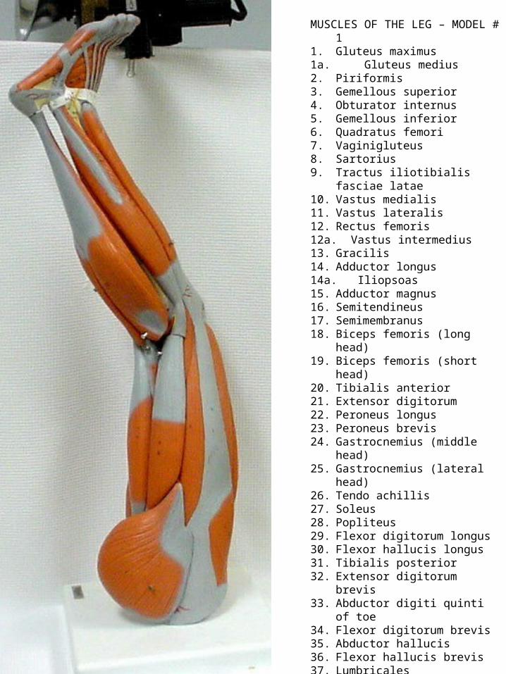
MUSCLES OF THE LEG – MODEL # 11. Gluteus maximus1a. Gluteus medius2. Piriformis3. Gemellous superior4. Obturator internus5. Gemellous inferior6. Quadratus femori7. Vaginigluteus8. Sartorius9. Tractus iliotibialis fasciae latae10. Vastus medialis11. Vastus lateralis12. Rectus femoris12a. Vastus intermedius13. Gracilis14. Adductor longus14a. Iliopsoas15. Adductor magnus16. Semitendineus17. Semimembranus18. Biceps femoris (long head)19. Biceps femoris (short head)20. Tibialis anterior21. Extensor digitorum22. Peroneus longus23. Peroneus brevis24. Gastrocnemius (middle head)25. Gastrocnemius (lateral head)26. Tendo achillis27. Soleus28. Popliteus29. Flexor digitorum longus30. Flexor hallucis longus31. Tibialis posterior32. Extensor digitorum brevis33. Abductor digiti quinti of toe34. Flexor digitorum brevis35. Abductor hallucis36. Flexor hallucis brevis37. Lumbricales38. Extensor hallucis brevis38a. Extensor hallucis longus39. Flexor digiti quinti brevis of small
toe40. Dorsal interosseous

MUSCLES OF THE LEG – MODEL # 2
11. Gluteus maximus12. Gluteus medius13. Piriformis14. Gemellus superior15. Obturator internus16. Gemellus inferior17. Quadratus femoris18. Tensor fasciae latae19. Iliotibial tract20. Rectus femoris21. Vastus lateralis22. Vastus intermedius23. Vastus medialis24. Sartorius25. Iliopsoas26. Adductor longus27. Gracilis28. Adductor magnus29. Semimembranosus30. Semitendinosus31. Biceps femoris (long head)32. Biceps femoris (short head)33. Tibialis anterior34. Extensor digitorum longus35. Peroneus longus36. Peroneus brevis37. Gastrocnemius ( lateral head)38. Gastrocnemius (medial head)39. Soleus40. Tibialis posterior41. Popliteus42. Flexor digitorum longus43. Flexor hallucis longus44. Extensor hallucis longus45. Extensor hallucis brevis46. Extensor digitorum brevis47. Abductor digiti minimi48. Flexor digiti minimi brevis49. Flexor digitorum brevis50. Lumbrical muscle of the foot51. Flexor hallucis brevis52. Abductor hallucis53. Dorsal interosseous54. Tendon of rectus femoris55. Tendon of sartorius56. Cruciate ligament of ankle57. Superior peroneal retinaculum58. Inferior peroneal retinaculum59. Tendon of extensor hallucis longus60. Tendons of extensor digitorum longus pedis61. Tendon of peroneus brevis62. Tendon of peroneus longus63. Plantar aponeurosis

MUSCLES OF THE ARM – MODEL # 21. Subscapula2. Supraspinatus3. Infraspinatus4. Teres minor5. Teres major6. Deltoid7. Biceps brachii7a. Caput longum7b. Caput breve8. Brachial9. Coracobrachialis10. Triceps brachii (caput longum)11. Triceps brachii (caput laterale)12. Triceps brachii ( caput mediale)13. Brachioradialis14. Extensor carpi radialis longus15. Extensor carpi radialis brevis16. Pronator teres17. Radiocarpus18. Palmarus longus19. Flexor carpi ulnaris20. Flexor digitorum sublimis21. Flexor digitorum profundus22. Flexor pollicis longus23. Quadratipronator24. Extensor digitorum25. Extensor digiti minimi26. Cubitalis posterior27. Cubitalis riolani28. Supinator29. Abductor pollicis longus30. Extensor pollicis longus31. Extensor indicis32. Retinaculum musculorum extensorum33. Tendo muscle extensoris pollicis longi34. Abductor pollicis brevis35. Opponens pollicis36. Flexor pollicis brevis37. Abductor digiti minimi38. Retinaculum musculorum flexorum39. Mesothenar40. Interossei dorsales41. Lumbricales42. Tendo muscle flexoris pollicis

ARM MUSCLES – MODEL # 11. Clavicle2. Acromion3. Scapular spine4. Humerus5. Subscapularis6. Supraspinatus7. Infraspinatus8. Teres minor9. Teres major10.Deltoideus11.Biceps brachii12.Biceps brachii (long head)13.Biceps brachii (medial short head)14.Brachialis15.Coracobrachialis16.Lateral head of triceps brachii17.Long head of triceps brachii18.Medial head of triceps brachii19.Brachioradialis20.Extensor carpi radialis longus21.Extensor carpi radialis brevis22.Pronator teres23.Flexor carpi radialis24.Palmaris longus25.Flexor carpi ulnaris26.Flexor digitorum supoerficialis27.Flexor digitorum profundus28.Flexor pollicis longus29.Pronator quadratus30.Extensor digitorum of hand31.Extensor digiti minimi
32. Extensor carpi ulnaris33. Anconeus muscle34. Supinator 35. Abductor pollicis longus36. Extensor pollicis longus37. Extensor indicis38. Extensor retinaculum39. Dorsal interosseous muscle of hand40. Opponens pollicis41. Abductor pollicis brevis42. Flexor pollicis brevis43. Adductor pollicis44. Lumbrical muscles of hand45. Abductor digiti minimi of hand
46. Flexor retinaculum47. Tendon of palmaris longus48. Tendon of flexor digitorum superficialis49. Tendon of flexor pollicis longus50. Tendon of extensor pollicis longus51. Tendinous junction52. Tendon of extensor digitorum muscle of hand53. Tendon of triceps brachii muscle

HEAD and NECK13. Occipitofrontalis (frontal belly)14. Temporal muscle15. Auricularis superior16. Auricularis posterior17. Auricularis anterior 18. Occipitofrontalis (occipital belly)19. Orbicularis oculi20. Compressor naris/Dilator naris21. Zygomaticus minor22. Zygomaticus major23. Orbicularis oris24. Depressor anguli oris25. Depressor labii inferioris26. Buccinator muscle27. Masseter28. Sternocleidomastoid muscle29. Sternohyoid muscle30. Sternothyroid muscle31. Thyrohyoid muscle32. Omohyoid muscle33. Cricothyroid muscle34. Trapezius muscle35. Splenius capitis muscle36. Levator scapulae muscle37. Scalenus muscles
TRUNK49. Subclavius50. Deltoid51. Pectoralis major52. Pectoralis minor53. External intercostals54. Serratus anterior55. Obliquus abdominis externus56. Rectus abdominis57. Obliquus abdominis internus58. Pyramidalis
59. Rhomboideus major60. Infraspinatus61. Teres major62. Longissimus thoracis63. Latissimus dorsi64. Serratus posterior inferiorUPPER EXTREMITY69. Teres minor70. Triceps71. Biceps72. Coracobrachialis73. Brachialis74. Pronator teres75. Flexor carpi radialis76. Palmaris longus77. Flexor carpi ulnaris78. Extensor carpi ulnaris79. Extensor digitorum80. Extensor carpi radialis brevis81. Extensor carpi radialis longus82. Brachioradialis83. Extensor pollicis brevis84. Abductor pollicis longus85. Flexor digitorum sublimis86. Supinator87. Extensor retinaculum88. Flexor retinaculum 89. Flexor pollicis brevis90. Abductor pollicis brevis91. Dorsal interosseus92. Abductor digiti minimi93. Flexor digiti minimi brevis94. Opponens digiti minimi95. Lumbrical96. Tendons of flexor sublimis97. Tendinous vaginal ligamentsLOWER EXTREMITY101. Glutaeus maximus102. Glutaeus medius103. Piriformis
104. Obturator internus105. Superior gemellus106. Inferior gemellus107. Quadratus femoris108. Tensor fasciae latae109. Sartorius110. Rectus femoris111. Vastus intermedius112. Vastus lateralis113. Vastus intermedius114. Pectineus115. Adductor longus116. Gracilis117. Great adductor118. Semitendinosus119. Semimembranosus120. Biceps femoris121. Tibialis anterior122. Extensor digitorum longus muscle123. Extensor hallucis longus124. Peroneus longus125. Peroneus brevis126. Gastrocnemius127. Soleus128. Popliteus129. Tibialis posterior130. Flexor digitorum longus131. Flexor hallucis longus132. Extensor hallucis brevis133. Extensor digitorum brevis134. Abductor hallucis135. Flexor digitorum brevis136. Lumbrical137. Abductor digiti minimi138. Flexor digiti minimi brevis139. Inferior extensor retinaculum 141. Transversus thoracis142. Diaphragm143. Transversus abdominis144. Quadratus lumborum145. Greater psoas146. Lesser psoas147. Iliacus

TORSO MODEL SECTION 1M38 – SartoriusM40 – Rectus femorisM41 – Tensor fasciae lataeM42 – Adductor brevisM43 - VastiM45 – Adductor magnusN5 – Ischiadicus nerveM44 – SemitendinosusM34 – Gluteus maximusO46 – Funiculus spermaticusV14 – Femoral veinO49 – Corpora cavernosa, penisA11 – Femoral arteryO48 – UrethraO45 – ProstateS28 – FemurO47 - Anus

TORSO MODEL SECTION 2M38 – SartoriusN6 – femoralis nerveV14 – Femoralis veinM36 – PectineusM39 – IliopsoasM40 – Rectus femorisM41 – Tensor fasciae lataeM37 – Levator aniN5 – Ischiadicus nerveM34 – Gluteus maximusO46 – Funiculus spermaticusA11 – Femoralis arteryS25 – Os pubisO45 – ProstateS28 – FemurS26 – Os ischiiO44 – RectumS27 – Os coccygis

TORSO MODEL SECTION 3M27 – Rectus abdominisM28 – Obliquus externus abdominisM29 – Obliquus internus abdominisM30 – Transversus abdominisM31 – PsoasM32 – IliacusM33 – Gluteus mediusM34 – Gluteus maximusM35 – MultifidusO39 – IleumO40 – ColonA10 – Common iliac arteryV13 – Common iliac veinS13 – Corpus vertebraeO43 – Cauda equinaS24 – Os ilium

TORSO MODEL SECTION 4M27 – Rectus abdominisV10 – Inferior vena cavaA6 – AortaM20 – Erector spinaeO38 – JejunumO40 – ColonO42 – Pancreatic ductO34 – PancreasO41 – Vesica felleaO37 – DuodenumO30 – HeparO36 – UreterO33 – LienO35 - Ren

TORSO MODEL SECTION 5M27 – Serratus anteriorM22 – IntercostalesA6 – AortaA9 – Lienalis arteryV12 – Lienalis veinM25 – Latissimus dorsiM20 – Erector spinaeS20 – CostaO30 – HeparV11 – Portae veinO31 – GasterV10 – Inferior vena cavaO32 – DiaphragmS13 – Corpus vertebraeO33 – LienO20 – Medulla spinalis

TORSO MODEL SECTION 6M22 – IntercostalesV10 – Inferior vena cavaV7 – Azygos veinA6 – AortaM26 – Serratus anteriorM20 – Erector spinaeM25 – Latissimus dorsiM13 – TrapeziusS19 – SternumO31 – GasterS20 – CostaO30 – HeparO22 – EsophagusS13 – Corpus vertebraeO24 – PulmoO20 – Medulla spinalis

TORSO MODEL SECTION 7M15 – Pectoralis majorM22 – IntercostalesM16 – Pectoralis minorA6 – AortaV6 – Superior vena cavaN4 – Brachial plexusM17 – DeltoidM23 – BicepsV9 – Brachialis veinA8 – Brachialis arteryM18 – SubscapularisM24 – TricepsM25 – Latissimus dorsiM20 – Erector spinaeM19 – InfraspinatusM13 – TrapeziusS19 – SternumO27 – Ventriculus dexterS20 – CostaO29 – Atrium dexterO26 – Ventriculus sinisterO28 – Atrium sinisterO24 – PulmoO22 – EsophagusS21 – HumerusS13 – Corpus vertebraeO20 – Medulla spinalisS22 - Scapula

TORSO MODEL SECTION 8M15 – Pectoralis majorM16 – Pectoralis minorV6 – Superior vena cavaA6 – AortaV8 – Axillaris veinN4 – Brachial plexusA7 – Axillaris arteryV7 – Azygos veinM17 – DeltoidM18 – SubscapularisM20 – Erector spinaeM19 – InfraspinatusM21 – RhomboidM13 – TrapeziusS19 – SternumO24 – PulmoS20 – CostaO25 – Bifurcation of tracheaO22 – EsophagusS21 – HumerusS13 – Corpus vertebraeO20 – Medulla spinalisS22 - Scapula

TORSO MODEL SECTION 9V5 – Anterior jugularisM7 – Sternocleidomastoid muscleV1 – Internal jugularisA5 – Common carotidM14 – ScaleniM13 – TrapeziusM10 – TransversospinalisM11 – Semispinalis capitisM12 – Splenius capitisO23 – Thyroid glandO21 – tracheaO22 – EsophagusS13 – Corpus vertebraeO24 – PulmoO20 – Medulla spinalis

TORSO MODEL SECTION 10V5 – Anterior jugularisM7 – Sternocleidomastoid muscleV1 – Internal jugularisA5 – Common carotid arteryN3 – Superior cervical ganglionA4 – VertebralisM14 – ScaleniM10 – TransversospinalisM11 – Semispinalis capitisM12 – Splenius capitisM13 – TrapeziusO21 – TracheaO23 – Thyroid glandO22 – EsophagusS14 – Foramen costotransversariumS13 – Corpus vertebraeO20 – Medulla spinalis

TORSO MODEL SECTION 11V5 – Anterior jugularisM7 – SternocleidomastoidV1 – Internal jugularisA5 – Common carotid arteryM8 – Longus colliM9 – Levator scapulaeM10 – TransversospinalisM11 – Semispinalis capitisM12 – Splenius capitisM13 – TrapeziusO17 – Plica vocalisO16 – Rima vestibuliS17 – Thyroid cartilageS18 – Arytenoid cartilageO18 – Pharynx (pars laryngea)S13 – Corpus vertebraeO19 – Cavum subarachnoidaleS15 – Transverse processO20 – Medulla spinalisS16 – Spinous process

TORSO MODEL SECTION 12M4 – MasseterM5 – Medial pterygoid muscleM6 – Longus capitisA2 – Internal carotid arteryA3 – Basilaris arteryV1 – Internal jugularisA4 – Vertebralis arteryV4 – Superior jugularisV2 – Sinus transversusS8 – Mandibula + DentesO13 – LinguaS9 – Ramus of mandibulaO14 – Pharynx (pars oralis)S10 – AtlasS11 – Occipital condylesO15 – Medulla oblongataS12 – Mastoid processO10 – CerebellumS7 – Os occipitate
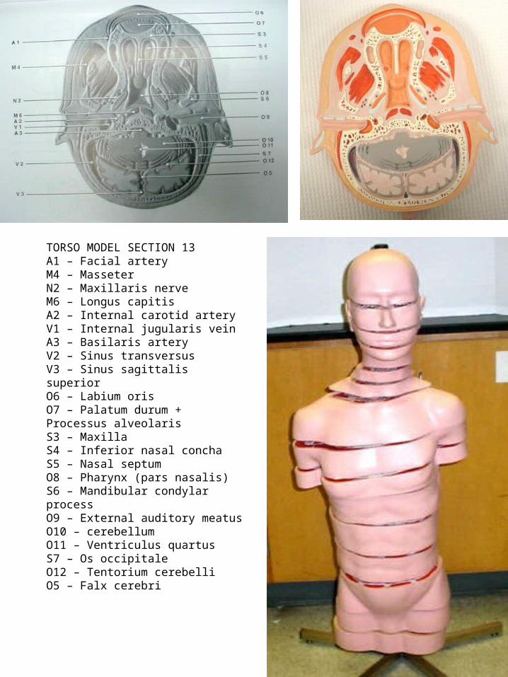
TORSO MODEL SECTION 13A1 – Facial arteryM4 – MasseterN2 – Maxillaris nerveM6 – Longus capitisA2 – Internal carotid arteryV1 – Internal jugularis veinA3 – Basilaris arteryV2 – Sinus transversusV3 – Sinus sagittalis superiorO6 – Labium orisO7 – Palatum durum + Processus alveolarisS3 – MaxillaS4 – Inferior nasal conchaS5 – Nasal septumO8 – Pharynx (pars nasalis)S6 – Mandibular condylar processO9 – External auditory meatusO10 – cerebellumO11 – Ventriculus quartusS7 – Os occipitaleO12 – Tentorium cerebelliO5 – Falx cerebri

TORSO MODEL SECTION 14M1 – Rectus medialisN1 – Optical nerveM2 – Rectus lateralisM3 – TemporalisS1 – Ethmoid sinusO1 – Bulbus oculiO2 – cerebrumO3 – Ventriculus tertiusO4 – Choroid plexusS2 – Os parietaleO5 – Falx cerebri
Related Documents