One Day Symposium sponsored by EACR Darwinian evolution and clonal heterogeneity in human cancer: biological and clinical implications Keynote Speaker Carlos Caldas Monday 29th October, 2012 Fundação Eng. António de Almeida Porto, Portugal Scientific Organising Committee Leonor David • Carmen Jerónimo • Fátima Baltazar Fátima Cardoso • Raquel Almeida • Luis Costa Proceedings

Welcome message from author
This document is posted to help you gain knowledge. Please leave a comment to let me know what you think about it! Share it to your friends and learn new things together.
Transcript
One Day Symposium sponsored by EACR
Darwinian evolution and clonal heterogeneity in human cancer: biological and clinical implications
Keynote Speaker Carlos Caldas
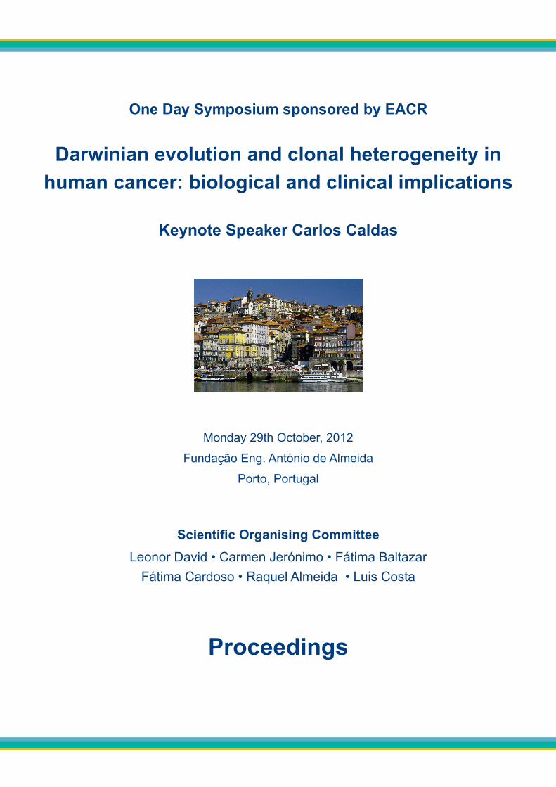
Monday 29th October, 2012
Fundação Eng. António de Almeida
Porto, Portugal
Scientific Organising CommitteeLeonor David • Carmen Jerónimo • Fátima Baltazar
Fátima Cardoso • Raquel Almeida • Luis Costa
Proceedings
ProgrammeMonday 29th October 2012
8.30 – 9.00 Registration at Fundação Eng. António de Almeida
9.00 – 10.00 Plenary Session 1 Chairs: Manuel Teixeira (IPO-Porto/ICBAS and Alexandre Quintanilha IBMC/ ICBAS, Porto)
9.00 - 9.15 Tumour banks in Portugal Fátima Carneiro (FMUP/Hospital S.João/IPATIMUP, Porto)
9.15 - 9.30 The ROR-Sul new platform, an innovative tool for cancer research and practice developmentAna Miranda (IPO-Lisbon)
9.30 - 9.45 PEM in breast cancer João Varela (IST/LIP, Lisbon) 9.45 – 10.00 From preclinical and clinical cancer diagnostic and screening to real-time in vivo dose monitoring for assisting external beam radiotherapy: an overview of the medical imaging activities and radiobiological plans at LIP Coimbra and collaborationsPaulo Crespo (Universidade Coimbra/ LIP, Coimbra)
10.00 - 10.30 Coffee break
10.30 – 11.30Plenary Discussion on platforms, facilities and technologies Chairs: José Mariano Gago (IST/LIP, Lisbon) and Manuel Sobrinho Simões (IPATIMUP/FMUP/Hospital S.João, Porto)
11.30 – 13.00
Plenary Session 2: Keynote Lecture Chair: Julio Celis (EACR)Darwinian evolution and clonal heterogeneity in human cancer – biological and clinical implicationsCarlos Caldas (Cambridge Research Institute, UK)
13.00 - 15.00 Lunch and Poster Viewing Julio Celis (EACR), Carlos Caldas (CRI), Sergio Dias (IMM/FMUL/IGC, Lisbon), Carla Oliveira (IPATIMUP/FMUP, Porto), Ana Teresa Maia (UA, Algarve), Luis Costa (FMUL/IMM, Lisbon), Carmen Jerónimo (IPO-Porto/ICBAS, Porto), Fátima Baltazar (ICVS, Braga)
15.00 – 16.20 Plenary Session 3 Chairs: Fátima Carneiro (FMUP/Hospital S.João/IPATIMUP, Porto) and Sergio Dias (IMM/FMUL/IGC, Lisbon)
15.00 – 15.20 Male Germ Cell Tumours diagnosed in 1999 and 2000 - a population-based retrospective study in Southern PortugalJosé Luis Passos Coelho (Hospital da Luz/Hospital Beatriz Angelo/FCM-UNL, Lisbon)
15.20 – 15.40 E-cadherin disfunction in gastric cancer. Celullar consequences and clinical applicationsRaquel Seruca (IPATIMUP/FMUP, Porto)
15.40 – 16.00From the environment, from within: IL-7R-mediated signaling in T-cell leukemiaJoão Barata (IMM, Lisbon)
16.00 – 16.20Telomerase is required for melanoma progressionMiguel Godinho (IGC, Oeiras) 16.20 - 17.00 Coffee break
17.00 – 17.20Young Investigators Awards Presentations
17.20 – 18.20 Address by Julio Celis, EACR Past President, and Meeting of the Portuguese Division of EACR
Julio Celis, Manuel Sobrinho Simões and the Steering Committee of the Portuguese Association for Cancer Research (Leonor David, Carmen Jerónimo, Fátima Baltazar, Fátima Cardoso, Raquel Almeida, Luis Costa and Rita Barros)
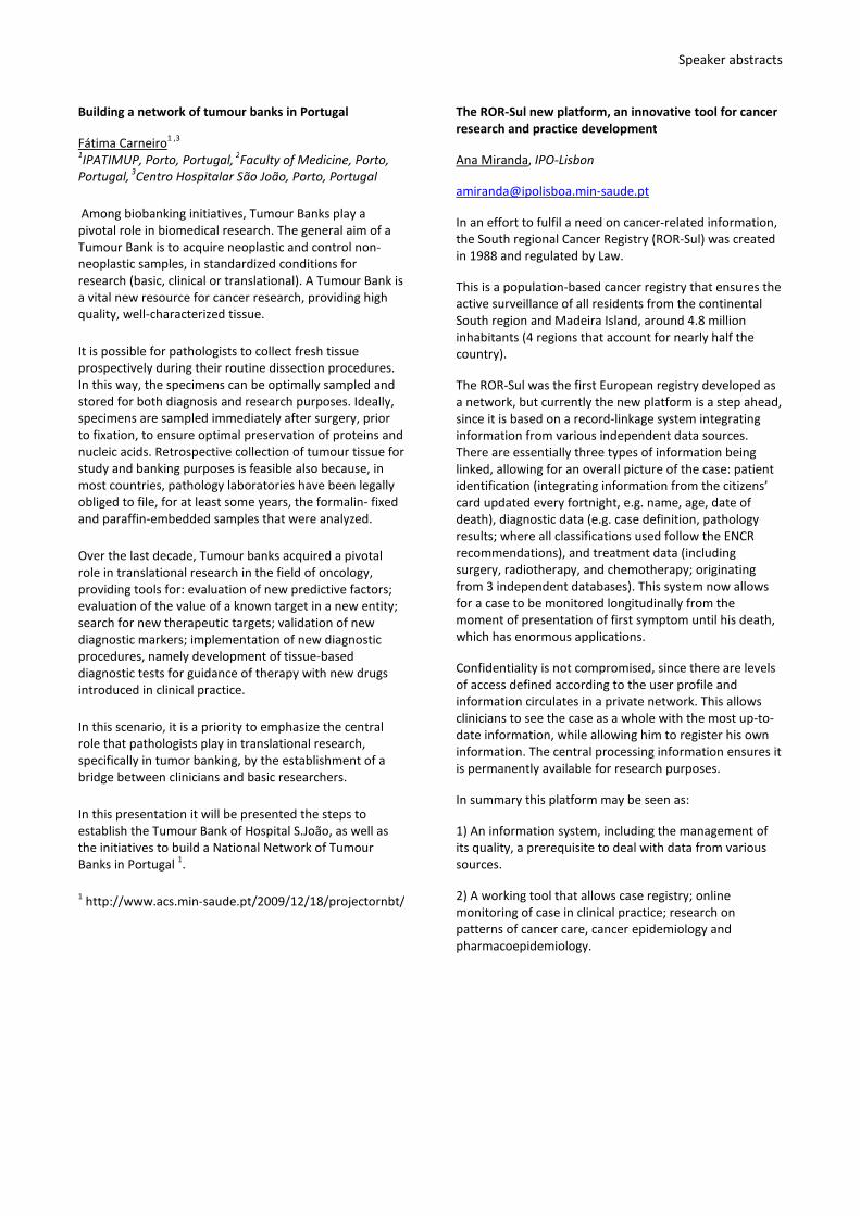
About the Keynote Lecturer: Carlos Caldas
Carlos Caldas is the leader of the Breast Cancer Functional Genomics Laboratory at the Cancer Research UK Cambridge Research Institute, which he joined in 2006. The main research interest of Carlos is breast cancer and more specifically to understand how genetic alterations accumulate, how they determine the biology of cancers, and which cell population within the breast epithelium is targeted by these alterations.
His group has identified novel cancer genes, has functionally characterized cancer associated genes and validated human breast carcinoma prognostic/predictive/therapeutic targets. The molecular profiling and description of the complex molecular taxonomy of breast cancer is the first step towards robustly identifying markers that have true clinical utility. Four Nature papers among other top journal publications on this subject were published in 2012.

25 – 28 June 2013Churchill College, Cambridge, U.K.
First AnnouncementEACR Summer Conference: Cancer Genomics
Organisers
James Brenton, UK • Carlos Caldas, UK
Jessica Downs, UK • George Vassiliou, UK
Keynote Speakers
Sam Aparicio, Canada • Shankar Balasubramanian, UK • Anne-Lise Borresen-Dale, Norway
Invited Speakers
Rene Bernards, the Netherlands • James Brenton, UK • Carlos Caldas, UK
Luis Alberto Diaz, USA • Manel Esteller, Spain • Gad Getz, USA
Mel Greaves, UK • Sean M. Grimmond, Australia • Jos Jonkers, the Netherlands
Jan Korbel, Germany • Peter Lichter, Germany • Elaine Mardis, USA • Charles Perou, USA
Martin Peifer, Germany • Nitzan Rosenfeld, UK • George Vassiliou, UK
SummerConference
2013
25 - 28 June
Cambridge, U.K.2013
Further Information:
Visit www.eacr.org/meetings for more information or sign up for RSS feeds for instant notifications
www.eacr.org/LatestMeetings.rss
EACR Meeting Bursaries
Five bursaries, each of 500 Euros, will be
awarded to assist members of the EACR to
attend the meeting
Important Deadlines
Abstract Submission: 30th April 2013
Meeting Bursary Application: 30th April 2013
Registration: 31st May 2013
Supported by the British Association for Cancer Research
Speaker abstracts
Building a network of tumour banks in Portugal
Fátima Carneiro1 ,3
1IPATIMUP, Porto, Portugal, 2Faculty of Medicine, Porto,Portugal, 3Centro Hospitalar São João, Porto, Portugal
Among biobanking initiatives, Tumour Banks play apivotal role in biomedical research. The general aim of aTumour Bank is to acquire neoplastic and control non-neoplastic samples, in standardized conditions forresearch (basic, clinical or translational). A Tumour Bank isa vital new resource for cancer research, providing highquality, well-characterized tissue.
It is possible for pathologists to collect fresh tissueprospectively during their routine dissection procedures.In this way, the specimens can be optimally sampled andstored for both diagnosis and research purposes. Ideally,specimens are sampled immediately after surgery, priorto fixation, to ensure optimal preservation of proteins andnucleic acids. Retrospective collection of tumour tissue forstudy and banking purposes is feasible also because, inmost countries, pathology laboratories have been legallyobliged to file, for at least some years, the formalin- fixedand paraffin-embedded samples that were analyzed.
Over the last decade, Tumour banks acquired a pivotalrole in translational research in the field of oncology,providing tools for: evaluation of new predictive factors;evaluation of the value of a known target in a new entity;search for new therapeutic targets; validation of newdiagnostic markers; implementation of new diagnosticprocedures, namely development of tissue-baseddiagnostic tests for guidance of therapy with new drugsintroduced in clinical practice.
In this scenario, it is a priority to emphasize the centralrole that pathologists play in translational research,specifically in tumor banking, by the establishment of abridge between clinicians and basic researchers.
In this presentation it will be presented the steps toestablish the Tumour Bank of Hospital S.João, as well asthe initiatives to build a National Network of TumourBanks in Portugal 1.
1 http://www.acs.min-saude.pt/2009/12/18/projectornbt/
The ROR-Sul new platform, an innovative tool for cancerresearch and practice development
Ana Miranda, IPO-Lisbon
In an effort to fulfil a need on cancer-related information,the South regional Cancer Registry (ROR-Sul) was createdin 1988 and regulated by Law.
This is a population-based cancer registry that ensures theactive surveillance of all residents from the continentalSouth region and Madeira Island, around 4.8 millioninhabitants (4 regions that account for nearly half thecountry).
The ROR-Sul was the first European registry developed asa network, but currently the new platform is a step ahead,since it is based on a record-linkage system integratinginformation from various independent data sources.There are essentially three types of information beinglinked, allowing for an overall picture of the case: patientidentification (integrating information from the citizens’card updated every fortnight, e.g. name, age, date ofdeath), diagnostic data (e.g. case definition, pathologyresults; where all classifications used follow the ENCRrecommendations), and treatment data (includingsurgery, radiotherapy, and chemotherapy; originatingfrom 3 independent databases). This system now allowsfor a case to be monitored longitudinally from themoment of presentation of first symptom until his death,which has enormous applications.
Confidentiality is not compromised, since there are levelsof access defined according to the user profile andinformation circulates in a private network. This allowsclinicians to see the case as a whole with the most up-to-date information, while allowing him to register his owninformation. The central processing information ensures itis permanently available for research purposes.
In summary this platform may be seen as:
1) An information system, including the management ofits quality, a prerequisite to deal with data from varioussources.
2) A working tool that allows case registry; onlinemonitoring of case in clinical practice; research onpatterns of cancer care, cancer epidemiology andpharmacoepidemiology.
Speaker abstracts
PEM in breast cancer study and diagnosis
Joao Varela1 ,2
1LIP, Lisbon, Portugal, 2IST, Lisbon, Portugal
Two prototypes of a new Positron EmissionMammography (PEM) scanner developed by a Portugueseconsortium, are currently operating at the Institute ofNuclear Sciences Applied to Health (ICNAS), in Coimbra,and at the University Hospital in Marseille. The scannerhas millimetric image resolution and high sensitivityallowing precise PET characterization of cancer tumors.Clinical tests with patients affected by breast cancer andpatients with other cancer types (control sample) wereperformed parasitically in cases where a PET exam wasprescribed. A summary of the results will be discussed.Results obtained with gelatin phantoms emulating thebreast tissue and lesions of different sizes will also bepresented. Examples of tests performed with mice will bepresented illustrating the potential of this tool inbiomedical research.
From preclinical and clinical cancer diagnostic andscreening to real-time in vivo dose monitoring forassisting external beam radiotherapy: an overview of themedical imaging activities and radiobiological plans atLIP Coimbra and collaborations
P. Crespo1 ,2, F. Alves3 ,2, M.C. Battaglia2 ,4, V. Bellini4 ,5, A.Blanco1, P. Cambraia Lopes6 ,1, M. Capela7, A. Cavaco8, S.Carmo2, M. Couceiro1 ,3, N.C. Ferreira2, R. FerreiraMarques1 ,2, P. Fonte1 ,3, F. Fraga1 ,2, S. Ghithan1 ,2, L.Lopes1, M.C. Lopes7, P. Martins1 ,2, K. Parodi9 ,10, P.J.B.M.Rachinhas8, D.R. Schaart6, H. Simões1, P.C.P.S. Simões8, P.Soares8
1LIP, Coimbra, Portugal, 2University of Coimbra, Coimbra,Portugal, 3Polytechnic of Coimbra, Coimbra, Portugal,4University of Catania, Catania, Italy, 5INFN, Catania, Italy,6Delft University of Technology, Delft, The Netherlands,7IPOCFG, E.P.E., Coimbra, Portugal, 8CHUC, E.P.E.,Coimbra, Portugal, 9University of Munich, Munich,Germany, 10HIT, Heidelberg, Germany
The theoretical and technological skills of the high energyphysics community often serve those of medical physics.For instance, Monte Carlo simulations for describingparticle interactions in physics experiments are nowcommonly used to characterize and improve modernradiotherapy treatments both with X-rays and withparticles such as protons and carbon ions. On thetechnological side, the demands for developments andimprovements of performance of detectors andassociated technology also embrace the twoaforementioned fields of physics. For these reasons LIPCoimbra is strongly engaged in several projects focusingmainly on medical imaging, but not only. Thiscommunication will summarize these projects, namely (1)preclinical and clinical high-sensitivity positron emissiontomography (PET) with new, very-high-resolutiondetectors; (2) developments aiming at real-time, in vivodose monitoring for assisting (and improving) particle andphoton radiotherapy; and (3) towards radiophysiologyand radiobiology studies with proton beams at the PETproton cyclotron of the University of Coimbra. A strongemphasis will be put on explaining the basic clinicalmotivation and physical concepts of these projects.
Speaker abstracts
Male Germ Cell Tumours diagnosed in 1999 and 2000 – apopulation-based retrospective study in southernPortugal
José Luis Passos CoelhoHospital da Luz, Lisboa e Hospital Beatriz Angelo, LouresFaculdade de Ciências Médicas, Lisboa
Results obtained in clinical trials for treatment ofoncologic diseases may not be reproduced when the sameapproach is applied to patients with the same disease inthe community. Furthermore, the reality of one countrymay not apply to a different country despite similaroverall population characteristics. This studydemonstrates the relevance of characterizing the nationalreality regarding the clinical presentation and therapeuticoutcomes for oncologic diseases. In close collaborationwith the regional cancer registry of southern Portugal(ROR-Sul) and of institutions and professionals involved indiagnosis and treatment of germ cell tumours, aretrospective population-based study was undertaken onsouthern Portugal and Madeira island, including malesdiagnosed with germ cell tumours in 1999 and 2000; thisallowed a minimum follow-up time of 5 years. Eightyseven patients were identified (incidence of 1.85/100.000males), 79 with primary testicular tumours. From 81patients with testicular or retroperitoneal tumours, 35were diagnosed with stage I, 13 with stage II and 30 withstage III (3, stage unknown). With a median follow-up of89 months, global 5-year overall survival was 80% (100%for stage I, 13 for stage II and 53% for stage III). Theresults obtained show a similar incidence to othercountries of southern Europe but a lower survival thanreported in Eurocare4 study (performed in patientsdiagnosed between 1995 and 1999), with 5 year-overallsurvival ranging between countries from 92% to 98%. Thedata suggest a possible delay in diagnosis, with a highproportion of patients diagnosed with advanced stagehigh-tumor bulk disease, as well as sub-optimal adherenceto recommended treatment algorithms. Data like thesemay be useful to identify limitations in the results oftreatment of cancer and should lead to enhanced interestfrom health care professionals and authorities.
E-cadherin disfunction in gastric cancer. Celullarconsequences and clinical applications
Carla Oliveira1 ,2, Joana Figueiredo1, Patrícia Carneiro1,Joana Carvalho1, Joana Caldeira1, Patrícia Oliveira1, JoanaParedes1 ,2, Joé Carlos Machado1 ,2, Fátima Carneiro1 ,2,Raquel Seruca1
1IPATIMUP, Porto, Portugal, 2FMUP, Porto, Portugal
Tissues in multicellular organisms consist of a variety ofcells in which cell-cell and cell-matrix adhesions are keyevents to allow correct tissue architecture and tension.Cell-cell adhesion is mediated by a variety of membraneproteins such as E-cadherin which is the major componentof the Adherens Junctions (AJs) and the major contributorto the maintenance of adult tissues integrity andhomeostasis. The critical importance of E-cadherin tonormal development is demonstrated by the lethality inthe very early stage of embryogenesis.
One of the most basic characteristics of cancer cells is thatthey adhere poorly to each other, being this fact usuallyassociated with their ability to invade the surroundingtissues. In cancer, the study of sporadic tumours andearly hereditary diffuse gastric cancer (HDGC) lesions in
germline CDH1 mutation carriers suggests that E-cadherinloss can be an early or initiating event in tumorigenesisbut also an important marker for therapeutical selectionof the patients. To unravel the molecular mechanismunderlying the role of E-cadherin in cancer, we haveperformed several in vitro and in vivo studies (animalmodels and primary gastric carcinomas). As example,using a set of 42 stable cell lines, harboring HDGCassociated E-cadherin germline mutations distributedalong the gene, we clarified E-cadherin mediated signalingpathways and associated cellular effects. Wedemonstrated that E-cadherin in tumor progressiondepends on the activation of signaling pathways related tomigration and cell survival. Further, we identified EGFRand Notch as interesting therapeutic targets in E-cadherinmediated cancer.

Speaker abstracts
From the environment, from within: IL-7R-mediatedsignaling in T-cell leukemia
João BarataInstituto de Medicina Molecular, Lisboa, Portugal
Although cancer is a genetic disease, it is currently evidentthat microenvironmental cues are essential for tumorprogression. T-cell acute lymphoblastic leukemia (T-ALL),an aggressive subtype of the most frequent childhoodcancer, is no exception. Interleukin 7 (IL-7), a cytokineproduced in the bone marrow, thymus and other organs,is mandatory for normal human T-cell development.However, there is also considerable evidence that IL-7may partake in leukemia development. We showed thatIL-7 leads to the activation of PI3K/Akt/mTOR pathway,thereby mediating viability, cell cycle progression andgrowth of human T-ALL cells in more than 70% of patientsamples. Remarkably, the involvement of PI3K/Akt/mTORpathway in these processes differs subtly between normaland malignant T-cells, in a way that may have importanttherapeutic implications. We further showed thatmicroenvironmental IL-7 can have a role in acceleratinghuman T-ALL progression in vivo. The evidence that IL-7produced by the stroma has considerable impact onleukemia maintenance led us to go "back to the basics"and evaluate whether cell-autonomous lesions couldaffect directly IL-7-mediated signaling in malignant T-cells.We showed that around 9% of T-ALL patients display gain-of-function mutations in the gene encoding the IL-7receptor (IL7R). The mutations lead, in most cases, todisulfide bond-dependent homodimerization of twomutant receptors and consequent constitutive activationof downstream signaling, with ensuing cell transformationin vitro and tumorigenic ability in vivo. Is the oncogenicpotential of deregulated IL7R-mediated signalingrestricted to mutated receptor? Our studies showing thatconditional tetracycline-inducible IL7R transgenic miceeventually develop leukemia upon treatment withdoxycline, suggest otherwise. Overall, our results revealedIL-7/IL-7R-mediated signaling as an important oncogenicaxis in T-cell leukemia.
Telomerase is required for melanoma progression
Joana Nabais, Miguel Godinho Ferreira
Instituto Gulbenkian de Ciência, Oeiras, Portugal.
Contrary to other cancers, metastatic melanoma remainspractically incurable and is responsible for 90% of skincancer deaths. There are many genetic alterationsassociated with melanoma; however, events leading tomelanomagenesis remain elusive. Zebrafish models haveshown that mutated BRAF or NRAS lead to nevi formationbut, similar to the human disease, melanoma progressionrequires loss of p53 function, which abrogates cellularsenescence.
Telomeres, the protective ends of chromosomes, providea crucial link between cell proliferation and senescence.Since cancer cells require telomerase, a reversetranscriptase responsible for telomere synthesis, forcontinuous growth, anti-telomerase therapies arecurrently in clinical trials for various cancers.
Our studies show that telomerase inhibition preventsmelanoma. We are currently investigating therequirement of telomerase for melanoma progression.Our data points to a requirement of telomerase at laterstages of carcinogenesis including metastasis. We aim todetermine the window of opportunity for anti-telomerasetherapies in metastatic melanoma by inhibitingtelomerase both genetically (using conditionaltransgenics) and pharmacologically (using anti-telomerasedrugs).

Poster abstracts
0001
Loss of WNK2 expression by promoter gene methylationoccurs in adult gliomas and triggers Rac1-mediatedtumour cell invasiveness
Sónia Moniz1, Olga Martinho2, Filipe Pinto2, BárbaraSousa3, Cláudia Loureiro1, Maria José Oliveira3, JoanaParedes3, Rui Manuel Reis2, Peter Jordan1
1Instituto Nacional de Saúde Doutor Ricardo Jorge,Lisbon, Portugal, 2University of Minho, Life and HealthSciences Research Institute (ICVS), Braga, Portugal,3IPATIMUP, Porto, Portugal
The gene encoding protein kinase WNK2 was recentlyidentified to be silenced by promoter hypermethylationin gliomas and meningiomas, suggesting a tumoursuppressor role in these brain tumours. Followingexperimental depletion in cell lines, WNK2 was furtherfound to control GTP-loading of Rac1, a signalling GTPaseinvolved in cell migration and motility. Here we show thatWNK2 promoter methylation also occurs in 17.5%(29/166) of adult gliomas, whereas it is infrequent in itspaediatric forms (1.6%; 1/66). Re-expression of WNK2 inglioblastoma cells presenting WNK2 gene silencingreduced cell proliferation in vitro, tumour growth in vivoand also cell migration and invasion, an effect correlatedwith reduced activation of Rac1. In contrast, whenendogenous WNK2 was depleted from glioblastoma cellswith unmethylated WNK2 promoter, changes in cellmorphology, an increase in invasion and activation ofRac1 were observed. Together, these results validate theWNK2 gene as a recurrent target for epigenetic silencingin glia-derived brain tumours and provide firstmechanistic evidence that the role of WNK2 as a tumoursuppressor is related to Rac1 signalling and tumour cellinvasion and proliferation.
0002
Androgen-responsive and nonresponsive prostatecancer cells present a distinct glycolytic metabolismprofile
Cátia V. Vaz1, Marco G. Alves1, Ricardo Marques1, Paula I.Moreira2, Pedro F. Oliveira1, Cláudio J. Maia1, SílviaSocorro1
1CICS-UBI - Health Sciences Research Center, University ofBeira Interior, Covilhã, Portugal, 2CNC – Center forNeuroscience and Cell Biology and Institute of Physiology,Faculty of Medicine, University of Coimbra, Coimbra,Portugal
Prostate cancer (PCa) progresses from an early stage,confined to prostate, to a more aggressive metastasizedcancer related with loss of androgen responsiveness.Although, it has been recognized that PCa cells haveunique metabolic features, their glycolytic profile inandrogen-dependent and androgen-independent stagesof disease is much less known. Hence, the main purposeof this study was to compare glucose metabolism in
androgen-responsive (LNCaP) and androgen-nonresponsive (PC3) PCa cells. Cell culture medium wascollected and differences in glucose consumption and,lactate and alanine production were measured usingProton Nuclear Magnetic Resonance (1H-NMR) spectraanalysis. The mRNA and protein expression of glucosetransporters (GLUT1 and GLUT3), Phosphofructokinase 1(PFK1), lactate dehydrogenase (LDH) andmonocarboxylate transporter (MCT4) were determinedby real-time PCR and Western Blot, respectively. Theobtained results demonstrate that androgen-responsive(LNCaP) and androgen-nonresponsive (PC3) cellsconsumed similar amounts of glucose, whereas PC3 cellspresent higher lactate production. This increase in lactateproduction was concomitant with higher levels of MCT4protein, increased LDH activity and higher lactate/alanineratio, also suggesting increased levels of oxidative stressin PC3 cells. However, protein levels of LDH, associatedwith lactate metabolism, and GLUT3, involved in glucoseuptake, were decreased in PC3 comparatively withLNCaP. Androgen-responsive and nonresponsive PCa cellspresent distinct glycolytic metabolism profiles, whichsuggest that targeting LDH and MCT4 metabolicpathways may be an important step for the developmentof new diagnostic and therapeutic strategies in thedifferent stages of PCa.
0003
SARCOPENIC OBESITY IN CANCER: A PRIORITY FORINDIVIDIALISED NUTRITIONAL INTERVENTION?
Ana Isabel Almeida1, Carolina Boléo-Tomé1, IsabelMonteiro-Grillo1 ,2, Maria Camilo1, Paula Ravasco1
1Unidade de Nutrição e Metabolismo, Instituto deMedicina Molecular e Laboratório de Nutrição, Faculdadede Medicina da Universidade de Lisboa, Lisboa, Portugal,2Departamento de Radioterapia, Hospital Universitário deSanta Maria, Lisboa, Portugal
Rationale: In cancer, worldwide data stress theprevalence of cachexia, while growing information drawsour attention to overweight/obesity at diagnosis. Thispattern may be determined by the patients’ diet, whichin turn may influence treatment’ tolerance and outcome.We aimed to analyse potential associations betweenrelevant nutrients, nutritional status & disease/treatmentrelated symptoms and tolerance. Methods: Cross-sectional study with 426 patients with solid tumours atvarious stages; weight and height were determined witha Jofre® scale+stadiometer. Body Mass Index (BMI) wascalculated and categorised by age/sex reference values.CT scans (L3-L4) were used for body composition analysis.Current intake was assessed by 24-hour recall & usualintake by a 1-year validated food frequencyquestionnaire (FFQ). Symptoms were assessed byvalidated/specific Patient Generated-Subjective GlobalAssessment (PG-SGA). Results: We included 257M:169F;by BMI 4% were underweight vs 64% overweight/obese,and CT scans showed that these patients had a pattern ofsarcopenic obesity. The prevalence of symptoms after

Poster abstracts
adjusting for the sample size, was 85%, more significantin sarcopenic obese patients (p<0.001); sarcopenicobesity was significantly associated with higher priorityfor nutrition intervention (p<0.005). Conclusion: Themajority of patients was overweight/obese withdepletion of muscle mass; their diet was characterised byboth excessive and insufficient intakes of key nutrients,likely to contribute to nutritional deterioration, toxicityand treatments’ tolerance and body composition pattern.Sarcopenic obesity is a highly complex feature of majorclinical relevance for patients’ prognosis. This emergingclinical scenario urgently argues for randomised clinicaltrials of nutritional therapy to determine an effective andtargeted intervention.
0004
CANCER, DIETARY PATTERN AND SARCOPENICOBESITY: NEW INSIGHTS
Ana Isabel Almeida1, Carolina Boléo-Tomé1, IsabelMonteiro-Grillo1 ,2, Maria Camilo1, Paula Ravasco1
1Unidade de Nutrição e Metabolismo, Instituto deMedicina Molecular e Laboratório de Nutrição, Faculdadede Medicina da Universidade de Lisboa, Lisboa, Portugal,2Departamento de Radioterapia, Hospital Universitário deSanta Maria, Lisboa, Portugal
Rationale: Diet is a major risk factor for obesity and forcancer: it may protect from, but can also worsen tissuedamage during cancer progression and treatments. Thisstudy aimed to characterise the diet pattern of a cohortof cancer patients and to identify excessive and/orinsufficient intake of key nutrients. Methods: Cross-sectional study conducted in 426 patients with solidtumours in various stages referred for Radiotherapy;weight and height were determined with a Jofre® floorscale+stadiometer. BMI was evaluated and furthercategorised by age/sex reference values. Current dietintake was assessed by 24-hour recall and usual diet by avalidated 1-year food frequency questionnaire. Fooddata were analysed by DIETPLAN® to obtain the detaileddaily nutrient intake. Results: We included 257M:169Fwith cancers of the breast, prostate, lung, colon-rectum,head-neck, oesophagus, stomach. Overweight/obesitywas prevalent (64%); 85% of pts had an inadequate dietwith excessive energy intake, of which lipids represented39%, with 18% of saturated fat. Additionally, a highintake (>2X DRI) of protein, refined carbohydrates,cholesterol, iron & sodium was found, concomitantlywith insufficient intake (<50% DRI) of fibre, folate &vitamins A, D, E, C, especially in breast, lung, prostate &head-neck cancers. Higher BMI was significantlycorrelated with an inadequate diet (p<0.002). Conclusion:There was a significant, striking and clinically worryingprevalence of inadequate intake of key nutrients duringanti-neoplastic treatment(s), in addition tooverweight/obesity. It is essential to provide nutritionalcounseling aiming to prescribe therapeutic diets, withanti-inflammatory, antioxidant and immunomodulatoryeffects, that may protect from radiation and citotoxic
injury. Plus, an adequate intake may modulate bodycomposition that will in turn contribute to improvetreatments’ tolerance and disease prognosis.
0005
BIOELECTRICAL IMPEDANCE AND PHASE ANGLE: HOWRELEVANT IN CANCER?
Ana Isabel Almeida1, Catarina Ferreira1, Isabel Monteiro-Grillo1 ,2, Maria Camilo1, Paula Ravasco1
1Unidade de Nutrição e Metabolismo, Instituto deMedicina Molecular e Laboratório de Nutrição, Faculdadede Medicina da Universidade de Lisboa, Lisboa, Portugal,2Departamento de Radioterapia, Hospital Universitário deSanta Maria, Lisboa, Portugal
Rationale: Body composition may be determinant forcancer progression and treatments. This pilot longitudinalstudy in cancer, aimed to characterise body composition,concomitantly with phase angle (PA); we also exploredpotential associations with cancer variables: histologicalaggressiveness and stage. Methods: We included 26patients with solid tumours at various stages. Height andweight were determined with a Seca® scale+stadiometer.BMI was calculated and categorised according to WHO’scriteria; %body fat mass (%FM) and phase angle (PA)were assessed by tetrapolar multifrequency bioelectricalimpedance (Biodynamics 450®, Seattle, USA); %FM andPA were compared with age/sex reference values:percentage intervals & percentiles, respectively.Descriptive analysis was performed. Results: Stages III/IVand moderately/poorly differentiated cancers wereprevalent: 54% & 69%, respectively. By BMI, 47% patientswere overweight/obese vs 6% underweight. Excessive FMwas prevalent (65%) and also found in patients withnormal BMI. Overall, 29% of patients had PA<5thpercentile. The prevalence of stage III/IV andmoderately/poorly differentiated cancers was similar innormal BMI/FM as it was in obesity/high FM. In whatconcerns PA, 86% patients with PA<5th percentile, hadcancers of stages III/IV and moderate/low differentiationvs patients with PA>5th percentile, of which 41% & 63%had advanced and more aggressive cancers, respectively.Conclusion: Excessive adiposity by BIA was prevalent andunderestimated by BMI. Advanced stage and moreaggressive cancers, indicators of worse disease status,were significantly related with a lower PA. Thus, bodycomposition analysis complemented with PAdetermination, both simple and quick for routine use,seem to bear a high clinical relevance. These preliminaryresults do support the continuation of this longitudinal,with additional in depth & validation analyses, as well asto study targeted nutritional interventions.
Poster abstracts
0006
BIOELECTRICAL IMPEDANCE PHASE ANGLE: PREDICTOROF CELL DISTURBANCE, METABOLISM AND PRIORITYFOR NUTRITIONAL INTERVENTION?
Ana Isabel Almeida1, Catarina Ferreira1, Isabel Monteiro-Grillo1 ,2, Maria Camilo1, Paula Ravasco1
1Unidade de Nutrição e Metabolismo, Instituto deMedicina Molecular e Laboratório de Nutrição, Faculdadede Medicina da Universidade de Lisboa, Lisboa, Portugal,2Departamento de Radioterapia,, Lisboa, Portugal
Rationale: Cancer and undernutrition may result indisturbed electric tissue properties, translated in alteredphase angle (PA). This pilot study aimed to assess thepredictive value of bioelectrical impedance PA inidentifying cancer patients with major priority fornutritional intervention. Methods: We included 26ambulatory pts with different cancers and stages.Nutritional assessment was performed with the validatedPatient-Generated Subjective Global Assessment (PG-SGA). PA values were assessed by tetrapolarmultifrequency bioelectrical impedance (Biodynamics450®) and compared with age/sex reference percentiles.PG-SGA scores were expressed as median (interquartilerange); comparisons were made using non-parametrictests. Results: Undernutrition was found in 38% of pts,and 71% had indication for urgent nutritionalintervention. PA<5th percentile was prevalent inundernourished pts and with indication for urgentnutritional intervention (44%). Median PG-SGAintervention score was significantly higher in pts with aPA<5th percentile vs a PA>5th percentile [12.5 (8.5-17.5)vs 4 (2-7), p=0.005]. Median PG-SGA scores on foodintake, symptoms & functional capacity were worse in ptswith a PA<5th percentile vs patients with PA>5thpercentile (p<0.05). Overall, PA<5th percentile didpredict a significantly worse PG-SGA score (p=0.005). Nosignificant differences were found on PG-SGA B, C & Dscores. Conclusion: A PA<5th percentile was associatedwith critical need for nutrition intervention. PAintegration in clinical practice may be of great value;while simple and easy to use, it provides key informationon cell disturbance and metabolism; this information maybe useful as a first approach to prioritize the critical needof nutritional intervention and symptom management.
0007
BIA PHASE ANGLE IN CANCER PREDICTS QUALITY OF LIFEAND PROGNOSIS
Ana Isabel Almeida1, Catarina Ferreira1, Isabel Monteiro-Grillo1 ,2, Maria Camilo1, Paula Ravasco1
1Unidade de Nutrição e Metabolismo, Instituto deMedicina Molecular e Laboratório de Nutrição, Faculdadede Medicina da Universidade de Lisboa, Lisboa, Portugal,2Departamento de Radioterapia, Hospital Universitário deSanta Maria, Lisboa, Portugal
Rationale: Bioelectrical phase angle (PA) in cancer hasbeen suggested as a potential prognostic diseaseindicator, and as a consequence is likely to predictpatients’ well-being and Quality of Life (QoL), a goldstandard for any clinical intervention. This pilotlongitudinal study aimed to assess the value of PA inpredicting cancer patients’ QoL. Methods: 26 patientswith different cancers and stages, referred forRadiotherapy were evaluated. PA was assessed bytetrapolar multifrequency bioelectrical impedance(Biodynamics 450®, Seattle, USA) and compared withage/sex reference percentiles. QoL was assessed by theEuropean Organization for the Research and Treatmentof Cancer Quality of Life Questionnaire version 3.0. QoLscores were expressed as median(interquartile range);comparisons were made using non-parametric tests.Results: PA<5th percentile was found in 29% of patients.Median global QoL & self-rated health status (SRHS)scores were similar [71 (57-71) and 71 (57-82),respectively]. Yet, poorer SRHS was associated withPA<5th percentile [50 (43-63) vs 71 (57-86), p<0.05]; nostatistically significant difference was found for globalQoL score [64 (50-71) vs 71 (57-86), NS]. Furthermore,physical, role, emotional and social functions weresignificantly impaired in pts with PA<5th percentile(p<0.05). On symptoms scales, fatigue, nausea/vomiting,insomnia & anorexia were worse in patients with PA<5thpercentile (p<0.05). Conclusions: PA<5th percentile didpredict poorer SRHS & anticipated impaired functionalscores, as well as worse symptoms. Thus, at diagnosisand/or disease onset, the 5th percentile for PA cut-off,may allow the identification of patients with worse QoLdimensions, SRHS and symptoms, factors associated withpoorer nutritional status and intake, all proven to bemodulated & improved by individualised nutritionalintervention, thus corroborating the need for adjuvantnutrition as therapy.
0008
N-acetylglucosaminyltransferases III and V regulate E-cadherin stability at the cell membrane. Implications inthe Epithelial to Mesenchymal Transition.
Salomé Pinho1 ,2, Sandra Carvalho1 ,2, Joana Cabral1, JoanaFigueiredo1 ,4, Patricia Oliveira1 ,4, Fátima Gärtner1 ,2,Tomoya Isaji3, Jianguo Gu3, Fátima Carneiro1 ,5, RaquelSeruca1 ,4, Carla Oliveira1 ,4, Naoyuki Taniguchi6, CelsoReis1
1IPATIMUP, Porto, Portugal, 2Institute of BiomedicalSciences of Abel Salazar (ICBAS), University of Porto,Porto, Portugal, 3Tohoku Pharmaceutical University,Sendai, Miyagi, Japan, 4Medical Faculty, University ofPorto, Porto, Portugal, 5Department of Pathology,Hospital S.João, Porto, Portugal, 6RIKEN Advanced ScienceInstitute, Wako, Saitama, Japan
E-cadherin is a cell-cell adhesion molecule whosedysfunction or inactivation is a common feature ofinvasive carcinomas. In addition, E-cadherin is a well-accepted marker of phenotypic plasticity, and is a central

Poster abstracts
molecule during Epithelial to Mesenchymal Transition(EMT) signaling pathway, that occurs during embryonicdevelopment, tissue regeneration, and thought to occurin cancer initiation/progression. The post-translationalregulation by N-glycosylation through GnT-III and GnT-Vglycosyltransferases has been reported by others and usto be an alternative mechanism of E-cadherin functionalregulation. We have demonstrated that GnT-III induced astabilizing effect on E-cadherin at the cell membrane byinducing a delay in the turnover rate of the protein whichpromotes the formation of stable and functionaladherens-junction, and further prevents clathrin-dependent E-cadherin endocytosis. This contributes to E-cadherin-mediated tumor invasion suppression function.Conversely, GnT-V promotes the destabilization of E-cadherin, leading to its mislocalization together withformation of unstable adherens-junctions andimpairment of cell-cell adhesion, therefore contributingto tumor progression. This stabilizer/destabilizer effect ofGnT-III and GnT-V on E-cadherin was further validated inclinical samples of human invasive carcinomas.Furthermore, we also found that during Epithelial toMesenchymal Transition (EMT), Mgat3 expression wasdramatically decreased and later recovered when cellsreturned to an epithelial-like phenotype (Mesenchymalto Epithelial Transition (MET)). We further identified thatMgat3 promoter methylation/demethylation is amechanism involved in this expression regulation. Theimpact of Mgat3/GnT-III expression variation, alongEMT/MET, was accompanied with a specific modificationof E-cadherin glycosylation with bisecting GlcNAcstructures. These results open new insights into themolecular mechanisms associated with the regulation ofE-cadherin in tumor cells with potential translationalclinical and therapeutic applications. In addition, we haveidentified for the first time Mgat3 glycogene expressionand GnT-III-mediated glycosylation, specifically on E-cadherin, as a novel and major component of theEMT/MET mechanism signature, supporting its roleduring EMT/MET.
0009
Role of CDX2 on the regulation of the cancer-associatedSialyl-Tn carbohydrate antigen
Rita Pinto1 ,2, Rita Barros1, Isabel Pereira-Castro1 ,2, LuisTeixeira da Costa3, Raquel Almeida1 ,2, Leonor David1 ,2
1IPATIMUP, Porto, Portugal, 2Faculdade de Medicina daUP, Porto, Portugal, 3ICAAM, Universidade de Évora,Evora, Portugal
De novo expression of sialyl-Tn antigen is one of the mostcommon features of gastric intestinal metaplasia (IM)and gastric carcinomas (GC). However, the regulation ofits expression is not fully elucidated. Previous studiesidentified the homeobox transcription factor CDX2 as adirect regulator of MUC2 mucin expression, the majorcarrier of sialyl-Tn in IM and GC. We thereforehypothesized that CDX2 might induce a cancer-associatedglycoproteome alteration in the gastric context - MUC2-
sialyl-Tn - by concomitant regulation of ST6GalNAc-I,which encodes the sialyltransferase responsible for STnbiosynthesis, and MUC2 genes. In this study, our aim is toevaluate whether CDX2 transactivates ST6GalNAc-I in agastrointestinal model, both in vitro and in vivo. The invitro spontaneous differentiation of Caco-2 cells inducesan increase in CDX2 expression which is accompanied byconcomitant increase in ST6GalNAc-I expression. On theother hand, CDX2 silencing using siRNAs in AGS cells wasfollowed by a decrease in ST6GalNAc-I transcriptionallevels. To clarify the mechanisms underlying ST6GalNAc-Igene transcriptional regulation by CDX2, luciferase assayswere performed. Co-transfection of gastric and coloniccell lines with pGL3-derived constructs covering 1.6kb ofthe human ST6GalNAc-I promoter and a CDX2 expressionvector confirmed a transactivation of ST6GalNAc-Ipromoter. Moreover, chromatin immunoprecipitation(ChIP) was carried out in order to identify relevant CDX2-binding regions to the ST6GalNAc-I promoter. ChIPanalysis proved that CDX2 was bound to it. Our worksupports the novel concept that a single homeobox gene,CDX2, is orchestrating a glycoproteome modificationduring cancer development. Future perspectives includevalidation of the results by site-directed mutagenesis ofthe ST6GalNAc-I promoter, EMSA and the use of aProximity Ligation approach for in vitro and in vivodetection of DNA-protein interactions. Moreover, in vivoChIP will be performed using human gastric IM andcancer samples to validate our hypothesis on gastriccarcinogenesis.
0010
A Novel MUC16 (CA125) Monoclonal Antibody
Lara Marcos-Silva1 ,2, Diana Campos1 ,2, Ulla Mandel1, EricPaul Bennett1, Ola Blixt1, Steven Levery1, Leonor David2 ,3,Henrik Clausen1
1Center for Glycomics, Departments of Cellular andMolecular Medicine and School of Dentistry, Faculty ofHealth Sciences, University of Copenhagen, Copenhagen,Denmark, 2IPATIMUP, Institute of Molecular Pathologyand Immunology of the University of Porto, Porto,Portugal, 3Faculty of Medicine of the University of Porto,Porto, Portugal
Introduction: The MUC16 mucin was identified as theserum antigen detected in the CA125 biomarker assayused to monitor patients with ovarian cancer. A numberof monoclonal antibodies (MAbs) including OC125 andM11 are available to detect MUC16. Despite considerableefforts the epitopes detected by these MAbs haveremained elusive and glycosylation has been proposed toplay a role for the epitopes. Existing MAbs react withMUC16 expressed in both normal cells and in cancer andhence detect enhanced levels of MUC16 derived fromboth benign and malignant cells.
Material and Methods: In this study, we used an E.coliexpressed MUC16 fragment glycosylated in vitro with Tnfor immunization of mice and selected a novel MAb

Poster abstracts
(5E11) with a cancer reactivity. Overlapping peptidescovering the tandem repeat domain were used forepitope mapping in ELISA and microarrays assays.
Results and Discussion: Comprehensive analysis of thefine specificities of existing MUC16 MAbs and the newMAb have been undertaken and we found that existingMAbs react with a conformational epitope of the 156amino acid tandem repeat sequence of MUC16 withoutdependence of O-glycosylation. The novel MAb incontrast reacts with a linear peptide epitope, whichexpression is dependent on glycosylation.
Conclusion: We characterized existing MAbs for MUC16(M11 and OC125) and produced a new MUC16 MAb(5E11) that recognizes a glycosylation dependent epitopeon the tandem repeat region of MUC16.
0011
The social network: testing the communication betweentumor cells and the surrounding stroma in tumordevelopment
Carlos F.D. Rodrigues1 ,2, Artur Paiva5 ,4, Mariana Val3 ,6,João Fonseca3 ,6, Isabel Carreira2 ,4, Mª Carmen Alpoim2 ,6
1PDBEB Programme, CNC, University of Coimbra,Coimbra, Portugal, 2CIMAGO, University of Coimbra,Coimbra, Portugal, 3CNC, University of Coimbra, Coimbra,Portugal, 4Faculty of Medicine, University of Coimbra,Coimbra, Portugal, 5Centro de Histocompatibilidade doCentro, Coimbra,, Coimbra, Portugal, 6Life SciencesDepartament, University of Coimbra, Coimbra, Portugal
It is now generally accepted that a tumor is aheterogeneous entity composed of a wide range of cellpopulations in different stages of differentiation. Underthis assumption, a specific population of cells with stem-like properties have been searched for and successfullyisolated from tumors of different origins. These cancerstem cells (CSCs) were later implicated in tumoraggressiveness and resistance to conventional therapiesas well as tumor relapse.Aiming to study the molecular mechanisms underlyinghexavalent chromium [Cr(VI)] induced lung cancers, wemalignantly transformed the normal human bronchialepithelial cell line BEAS-2B into the RenG2 system, usinglow density culture in the presence of Cr(VI). Twoadditional cell lines (DRenG2 and DDRenG2) wereattained following serial rounds of injection in nude mice.Characterization results allowed us to identify differentcellular sub-populations within each cell line, andprompted the hypothesis that CSCs may have drivenBEAS-2B' malignization. Sphere-formation assay was usedto search and isolate these cells, which were only presentin DRenG2 and DDRenG2 cell lines, forming more andbigger spheres in the DDRenG2. This suggested that adedifferentiation process featured the formation of CSCSduring RenG2 derivation in nude mice. To access theinvolvement of mice stroma in this process, chirurgical-isolated mouse stromal cells of the subcutaneous
compartment were co-cultured with RenG2 cells for 30-60 days (time needed to induce tumors in mice withRenG2), which resulted in the emergence of a CSCs sub-population. We are now able to attest that CSCs mayemerge in a tumor as a consequence of stromal-emittedparacrine signals, which brings deep implications tofuture therapeutic approaches.
0012
On the origin of cancer stem cells
João Fonseca1, Isabel Carreira1, Ana Mascarenhas1, LinaCarvalho1, Filipa Ladeirinha1, Carlos Rodrigues1 ,2, CarmenAlpoim2 ,3
1University of Coimbra, Coimbra, Portugal, 2CNC, Coimbra,Portugal, 3CIMAGO, Coimbra, Portugal
Tumors are characterized by their cellular heterogeneitydue to the co-existence of different cellular sub-populations, whose hierarchic organization in certaincancers lead to the hypothesis that the target cells oftransforming mutations are stem cells. However, in othertumors, restricted progenitors or even differentiated cellsmay be the cell of origin. Cancer stem cells (CSCs) areconsequently, stem-like cells with self-renewal andmultipotent differentiation characteristics which canoriginate all cell types found in a tumor (1).Unexpectedly,while attempting to understand the mechanismsunderlying hexavalent chromium induced lung cancer, wedemonstrated that CSCs could be obtained bydedifferentiation of the malignant bronchial epithelialcells DRenG2 and DDRenG2 and/or their precursorRenG2. The present work evaluated the proliferation rateof Cont-1, RenG2, DRenG2 and DDRenG2, and theirnormal precursor BEAS-2B cells, the cytogeneticevolution from BEAS-2B to DDRenG2, theepithelial/mesenchymal phenotype of the cell lines.Finally, the chemoresistance to gemcitabine andcisplatin, was evaluated and correlated to the presenceof the efflux pump P-Glycoprotein (P-gp). The cytogeneticanalysis of the more malignant and more proliferativeDRenG2 and DDRenG2 cell lines revealed a commonstructural difference relative to progenitor BEAS-2B cellsnamely 7p- . However, DRenG2 revealed thepredominance of 7q- and iso9q+ while DDRenG2 t(7:14)and 17q+. In contrast to the less proliferative BEAS-2B,and similarly to RenG2 both DRenG2 and DDRenG2predominant ploidy was 75/76 chromosomes.Immunocytochemistry analysis revealed that all cell lineswere mesenchymal –like cells (MNF116- and Vimentin-positive). As expected the more malignant cell lines weresignificantly more resistant to gemcitabine (GEM) andcisplatin (cDDP). Although, quite often multidrugresistance is associated to the overexpression of themembrane efflux pump P-gp, other mechanism(s) mayaccount for the observed resistance of DRenG2 andDDRenG2 cells as all the cell lines were P-gp negative.
Poster abstracts
0013
SENESCENT BRONCHIAL FIBROBLASTS DRIVEBRONCHIAL EPITHELIAL CELLS METAPLASICTRANSFORMATION FOLLOWING EXPOSURE TOHEXAVALENT CHROMIUM
Mariana Val1 ,2, Luis Mendes1, Marc Chanson3, IsabelCarreira2 ,4, Lina Carvalho2 ,4, Ana Ladeirinha2 ,4, CarlosRodrigues4 ,5, Carmen Alpoim2 ,5
1Life Sciences Departament, University of Coimbra,Coimbra, Portugal, 2CIMAGO, University of Coimbra,Coimbra, Portugal, 3Laboratory of Clinical Investigation III,Department of Pediatrics, Geneva University Hospitalsand University of Geneva, Geneva, Switzerland, 4Facultyof Medicine, University of Coimbra, Coimbra, Portugal,5CNC, University of Coimbra, Coimbra, Portugal
Cellular senescence, a phenomenon associated withaging and/or stress insults, can prevent neoplastictransformation. Nevertheless, accumulated evidencerevealed that senescent cells can stimulate malignantphenotypes in nearby cells by secreting protumorigenicfactors that stimulate epithelial cells proliferation anddisrupt epithelial differentiation. Senescent stromal cellsalso stimulate the acquisition of invasive and migratoryphenotypes by promoting epithelial to mesenchimaltransition (EMT).Here we report that sub-cytotoxic doses (0.25 and 0.5µM) of hexavalent chromium [Cr(VI)], a human lungcarcinogen known to induce squamous cell carcinomas,induced the senescence of normal human bronchialfibroblasts (E2A) which stimulated the proliferation and astriking change in the phenotype of human bronchialepithelial cells (BEAS-2B). In fact, co-cultivating senescentE2A with BEAS-2B cells in presence of 0.25 µM Cr(VI)stimulated BEAS-2B cells to acquire a basal cellphenotype (MNF+, Vimentin+) with large round nucleiand tadpole cytoplasm characteristic of epidermoidmetaplasia. Furthermore, co-cultures exposed to 0,5 µMCr(VI) lead BEAS-2B cells to acquire a cuboid morphologywith many mitotic figures and activated nuclei withvisible nucleoli, as well as fusiform shape associated withEMT phenotypic switch. Additionally, senescent E2Afibroblasts co-cultured with Cr(VI)-treated BEAS-2B cellsacquired mesenchymal features i.e., Vimentin+ and α-SMA+ large stellate cells with heterogeneous sizeenlarged nuclei, particularly abundant for 0.5 µM Cr(VI)which also lead to the appearance of crisscrossing.Altogether our results suggest that in presence of Cr(VI)the crosstalk between senescent fibroblasts andepithelial cells, mediated by secreted factors by both celltypes, induced premalignant characteristics in BEAS-2Bcells and a more undifferentiated phenotype infibroblasts.
0014
The Early Growth Response Genes as therapeutictargets in colon cancer
Andreas KoulourisQueen Mary University of London, London, UK
The Egr family of zinc finger transcription factors, whichconsists of four members; Egr-1, -2, -3 and -4, hasdynamic functions in the regulation of cell growth,developmental biology and immune responses. However,their roles in the development of tumour are not clear. Inthis study, we investigated the function of three closelyrelated Egr family members, Egr-1, -2 and -3 in theregulation of growth of colorectal cancer cells. Two celllines deriving from human colon cancer; one p53negative (DLD1) and another p53 positive (HCT116) weretransfected with Egr-1, -2 and -3, respectively. We foundthat all three Egr members can suppress tumour cellgrowth suggesting that the function of Egr in the controlof cell growth is not associated with the function of p53.In addition to the growth arrest, the transfected cellschanged morphology to round shape indicating ofsenescence. This may suggest that Egr molecules areimportant to control the unwanted growth in response tomalignant transformation. Our results not onlydemonstrated an important function of Egr moleculesand also indicate the therapeutic potential for thetreatment of tumour.
0015
Evaluation of the role of immunohistochemistrymarkers in the differential diagnosis of adrenocorticaltumors
Sofia Pereira1, Tiago Morais1, Madalena Costa1, MarianaP. Monteiro1, Duarte Pignatelli2
1Anatomy Department and UMIB, Instituto de CiênciasBiomédicas Abel Salazar, University of Oporto (ICBAS-UP),Oporto, Portugal, 2IPATIMUP. University of Porto, Oporto,Portugal
Malign adrenocortical tumors are rare and highlyaggressive, conversely benign tumors are more commonand frequently found incidentally. The diagnosis of thesetumors is based only on histological characteristics, sincethere are no established molecular markers.
The aim of the present study was to analyze themolecular profile of different adrenocortical tumors withthe purpose of identifying useful markers for thedifferential diagnosis.
The adrenocortical tumors studied (n=31) were, non-functioning adenomas/incidentalomas (n=13),functioning adenomas with Cushing syndrome (n=7),andcarcinomas (n=11); Normal adrenal glands (n=12) wereused as controls. For each sample, the percentage of thestained area and the QIC score (Quantitative
Poster abstracts
immunocitochemical score) by immunohistochemistrywere quantified for StAR, IGF2, p53, Mdm2, p21, p27,cyclin D1, Ki-67, β-catenin and E-cadherin, using amorphometric computerized analysis tool.
Of the studied markers, IGF2, p27, cyclin D1 and Ki-67were those whose percentage of marked area and QICscore was significantly higher on carcinomas whencompared with all the adenomas. Comparing thecarcinomas with the functioning Cushing syndromeadenomas, we observed significant differences in thepercentage of the stained area and QIC score for p27 andKi-67, which was increased and for StAR that wasdecreased in carcinomas. Comparing the carcinomas withincidentalomas, the marked area for IGF2, p27, cyclin D1and Ki-67 was significantly higher on carcinomas. The p27and the Ki-67 were the markers that showed the highestdiscriminative power for the differential diagnosisbetween carcinomas and adenomas, while the IGF2 andStAR only demonstrated to be useful for the differentialdiagnosis between carcinomas vs non-functioningadenomas and carcinomas vs adenomas with Cushingsyndrome, respectively.
The use of the markers StAR, IGF2, p27, cyclin D1 and Ki-67, and the quantification of its expression bycomputerized morphometric analysis could be animportant auxiliary means on the differential diagnosis ofadrenocortical tumors.
0016
Centrosome abnormalities in Barrett’s malignanttransformation
Carla A.M. Lopes1, Marta Mesquita1, Ana Isabel Cunha1,António Dias Pereira1, Mónica Bettencourt-Dias2, PaulaChaves1
1Instituto Português de Oncologia de Lisboa, Lisboa,Portugal, 2Instituto Gulbenkian de Ciência, Oeiras,Portugal
Barrett’s esophagus (BE) is a clinically importantpremalignant condition that develops in the context ofchronic gastroesophageal reflux disease. In BE the normalsquamous epithelium is replaced by a metaplasticcolumnar lining with goblet cells. It is a multistep processfrom metaplasia to dysplasia and carcinoma that isaccompanied by a progressive accumulation of well-characterized genetic lesions that are observed in manyother solid tumours. BE is the only known precursor ofesophageal adenocarcinoma, a tumour whose incidencehas increased profoundly over the last decades. Yet, only0.12% of individuals with BE develop esophagealadenocarcinoma per year and, to this date, besidesdysplasia there are no clinical or morphological markersto identify the patients that will progress. BE andassociated neoplasia exhibit abnormalities on cellularprocesses controlled by the centrosome, the primarymicrotubule-organizing centre in animal cells. Severalstudies have shown that cells from many cancers have
abnormal centrosomes that are either correlated withtumour malignancy or considered an early event duringtumorigenesis. However, a causative link betweencentrosome abnormalities and cancer remains elusive. Inthis study, we set out to investigate how centrosomedefects contribute to tumorigenesis using BE as a model.Centrosome and centriole profile were assessed inparaffin-embedded BE biopsies and esophagectomyspecimens of Barrett’s adenocarcinoma as well as inestablished cell lines derived from human esophagealadenocarcinoma. Using immunofluorescence andelectron microscopy we found that both numerical andsize centriole defects progressively accumulate in BEtumorigenesis. These findings suggest that centrosomeabnormalities are related to Barrett’s tumour progressionand may even be involved in its early steps. This studybrings us closer to a better understanding of howcentrosomal abnormalities relate to genetic changesobserved in BE, and may provide new opportunities foradvances on patient management strategy for earlydetection and prevention.
0017
Germinative cell tumors support bronchial-pulmonarydevelopment through celllineages:Immunohistochemical study
Ana Ladeirinha1, Sónia Simões3, Maria João DÁguiar1,Teresa Ferreira1, Ana Alarcão1, João Ramalho3 ,4, LinaCarvalho1 ,2
1Pathology Institute, Faculyt of Medicine, University ofCoimbra, Coimbra, Portugal, 2Service of Pathology,University Hospital of Coimbra, Coimbra, Portugal,3Faculty of Science and Technology, University ofCoimbra, Coimbra, Portugal, 4CNC- Center forNeuroscience and Cell Biology University of Coimbra,Coimbra, Portugal
The purpose of this study was to stratify the morphologyof germ cell tumors (GCT) to correlate embryogenesis,organogenesis and tissue maturation with concordantimmunohistochemical antibodies. Several studies supportthe idea that carcionogenesis carries on cellulardifferentiation and this theory was explored in germinalcell tumors to understand cell lineage in neoplasticdevelopment that would establish their classificationaccording with a specific imunohistochemical panel toidentify pluripotent stem cells and adult stem cells.
The antibodies AE1/AE3, CK7 and LP34 (cellularmaturation), CDX2, TTF1 and PLAP (organogenesis) andOct3/4, Nanog and Vimentin (embryogenesis) wereapplied to 34 benign and malignant FFPE GCT,concerning 17 males and 17 females (age range 16-73years).The immunohistochemical results were scoredsemi-quantitatively by the percentage of positive cells ina scale of 0% (0), <10% (1+),10-50%(2++) and >50%(3+++) considering cytoplasm or nuclei expression.

Poster abstracts
Our results showed Oct3/4 expression in nuclei ofseminoma cells while embrionary carcinoma cellsexpressed either nuclear and cytoplasm positivity; Nanoggene expression was seen only in 2 cases of lessdifferentiated tumors. Vimentin came out as a particularantibody in between Oct3/4 expression and CDX2/TTF1cellular maturation, indicating its value in the transitionbetween organogenesis and adult stem cells maturation.Cellular maturation was seen in neoplasias with intestinaldifferentiation due to CDX2 expression, TTF1 wassensitive for pulmonary alveolar epithelium. PLAPglycoprotein showed positive expression in the majorityof embryonic carcinomas.Cytokeratin AE1/AE3 was notdiscriminatory because it was expressed in all cases andcorresponded to a non-specific epithelial marker.
We may stress that the cellular maturation spectrumseen since embryogenesis till adult stem cells in GCT issimilar to malignant cell lineage development incarcinogenesis, according with the actual grades ofdifferentiation, also validating epithelial-mesenchimaltransition observed in less differentiatedtumors/carcinomas at least of some organs, includingbronchial-pulmonary carcinomas.
0019
Ncf1-deficient mice with impaired oxidative bursthave amore aggressive progression of Dextran Sulfate Sodium(DSS) -induced colitisTiago Rodrigues-Sousa2, Ana Alarcão1, Ana FilipaLadeirinha1, Margarida Margarida Souto-Carneiro2, LinaCarvalho1
1Institute of Pathology, Faculty of Medicine of theUniversity of Coimbra, Coimbra, Portugal, 2Immunology,Center for Neurosciences and Cell Biology, University ofCoimbra, Coimbra, Portugal
IntroductionThe most common clinical patterns in IID is chronic colitisand epithelial dysplasia. Intestinal Inflammatory Disease(IID) as a primary immunodeficiency depends onmutations in the NADPH oxidase complex, responsible forthe production of reactive oxygen species (ROS). Ncf1-mutation in mice leads to deficiency in ROS, renderingthem susceptible to autoimmunity. Here we studied howROS-deficiency of reactive oxygen species (ROS) in Ncf1-mutant mice influenced epithelial dysplasia.
Material and MethodsColitis was induced in wild type (WT) and Ncf1-mutant(Ncf1) B10.Q mice by administration of 3.5% DSS in thedrinking water for one week. After one week recovery,DSS was administered for another week. Mice weresacrificed at days 0, 7, 14 and 21, the colon was removedand folded into a Swiss roll. Sections of the colon werestained with HE and dysplasia was considered either inwild-type and Ncf1-mutant mice.
Results/ ConclusionsEpithelial dysplasia was high grade in Ncf1-mice togetherwith poor epithelium recovery (hyaline scars). The
atypical cells with mitotic higher rate were clearly locatedin the bottom of the colonic crypts. As ROS appeared tobe crucial for leukocyte recruitment and tissue-repair inDSS-induced colitis,it was seen that re-epithelization overinflammatory cells isprone to develop dysplasia(apparently high grade) ab initio. These results are usefullto understand the need of continuous anti-inflammatorytreatment of ID patients as earlier as possible.
0020
Bronchial-Pulmonary Adenocarcinomas Subtyping byPET ScanningAna Alarcão1 ,2, JA Castro e Dias1, Ana Filipa Ladeirinha1 ,2,Vitor Sousa1 ,4, Maria João DÁguiar1 ,2, Teresa Ferreira1 ,2,Maria Reis Silva1 ,3
1Institute of Pathology, Faculty of Medicine of theUniversity of Coimbra, Coimbra, Portugal, 2CIMAGO –Research Center for Environment, Genetics andOncobiology, Faculty of Medicine, University of Coimbra,,Coimbra, Portugal, 3Centre of Pulmonology, Faculty ofMedicine of the University of Coimbra, Coimbra, Portugal,4Service of Pathology, University Hospital of Coimbra,Coimbra, Portugal
Introduction:Bronchial-pulmonary carcinomas have 5 year survivalpoor, between 6% and 14% in men and 7% to 18% inwomen. Treatment depends on clinical staging andmorphological classification made in biopsies concerning70% of the cases. This is the actual state after alltherapeutic and diagnosis effort.Aim:
Immunohistochemical expression between differenthistological types was compared with Max 18F-Fluordesoxiglucose as the clinical parameter based inPET, to preview diagnosis and prognosis.
Methods:The immunohistochemistry study was performed in 41surgical specimens including Adenocarcinomas (18),Epidermoid Carcinomas (12) and the heterogeneousgroups of Large Cell Neuroendocrine Carcinoma (3), SmallCell Lung Cancer (1) , Large Cell Carcinoma (2),Adenosquamous Carcinoma (2) and PleomorphicCarcinomas (3) Max 18F-Fluordesoxiglucose (FDG) wasthe clinical parameter applied to validate the PathologicalSubtyping.
Results:Significant differences (p=0.006) between TTF-1 positive
and negative Adenocarcinomas where translated as 18F-FDG capture was lower in TTF-1 positive cases, indicatinglower metabolic activity. Epidermoid Carcinomas andTTF-1 negative Adenocarcinomas have similar and highermetabolic activity. The other histological types have FDGcapture similar and in between the two defined groups.
Conclusion:The clinical differences between Adenocarcinomas andEpidermoid Carcinomas related with

Poster abstracts
immunohistochemical expression of TTF1 and 18F-FDGcapture showed TTF-1 negative Adenocarcinomas asbiologically similar to Epidermoid Carcinomas needing adifferent medical approach as well as molecularpathology particular interpretation. These results clearlyraise need of recognizing TTF-1 negativeAdenocarcinomas because they are different from theterminal respiratory unit TTF-1 positiveAdenocarcinomas.
0021
IL-6-174 as a Gastric Carcinogenesis Marker in Biopsies
Ana Sampaio2, Sandra Balseiro2, Maria Reis Silva1 ,2,Domingos Oliveira1 ,3, Ana Alarcão1 ,2, Teresa Ferreira1,Maria João D´Aguiar1, Lina Carvalho1 ,3
1Institute of Pathology, Faculty of Medicine of theUniversity of Coimbra, Coimbra, Portugal, 2CIMAGO –Research Center for Environment, Genetics andOncobiology, Faculty of Medicine, University of Coimbra,Coimbra, Portugal, 3Service of Pathology, UniversityHospital of Coimbra, Coimbra, Portugal
IntroductionGastric carcinoma is related with cancer geneticsusceptibility that can be investigated through singlenucleotide polymorphisms (SNPs) and as cytokine genesare known to predispose to malignant disease, severalpolymorphisms of Interleukin-6 (IL-6) gene have beenreported to be associated with tumour progressionincluding inhibition of malignant epithelial cells apoptosisand stimulation of angiogenesis.
The aim of this study was to understand the associationbetween IL-6 polymorphisms and the risk for gastriccancer and chronic gastritis maintenance.
Materials and Methods
PCR-SSP genotyping for IL-6 -174C>G polymorphism wasperformed in 100 biopsies of gastric carcinoma and in100 biopsies of chronic gastritis.
ResultsThere was association between IL-6 -174C allele(p=0,0466) and -174CC, low producer, genotype(p=0,0466) and gastric carcinoma, whereas IL-6G allele(p=0,0278) and IL-6GG (p<0,0001), high producer,genotype was associated with gastritis.
ConclusionWe conclude that IL-6 -174, low producer genotypes,may have an important role in gastric carcinogenesis andthe polymorphism study of this molecule could be a goodmarker for gastric carcinoma susceptibility when highgrade dysplasia is seen in biopsies.
0022
ALK Mutation Detection in Bronchial-PulmonaryAdenocarcinomas for Tirosine-kinase InhibitionPrescription
Maria Reis Silva1 ,3, Ana Alarcão1 ,2, Ana Filipa Ladeirinha1,Maria João D´Aguiar1, Teresa Ferreira1, Vitor Sousa1 ,4,Lina Carvalho1 ,4
1Institute of Pathology, Faculty of Medicine of theUniversity of Coimbra,, Coimbra, Portugal, 2CIMAGO –Research Center for Environment, Genetics andOncobiology, Faculty of Medicine, University of Coimbra,Coimbra, Portugal, 3Centre of Pulmonology, Faculty ofMedicine of the University of Coimbra, Coimbra, Portugal,4Service of Pathology, University Hospital of Coimbra,Coimbra, Portugal
IntroductionLow cost of validation of EML4-ALK for crizotinib therapyand a rapid answer in Pathology routine was searched ina series of 35 bronchial-pulmonary carcinomas: 20adenocarcinomas, 6 epidermoid carcinomas, 4pleomorphic carcinomas (mixed type adenocarcinomaswith large/giant/fusiform cells), 4 neuroendocrinecarcinomas (NEC 1 combined large cell NEC withadenocarcinoma and 2 with combined epidermoidcarcinomas; 1 SCLC chromogranin positive combined withadenocarcinoma) and 1 adenosquamous carcinoma wereselected.
Materials and MethodsThe criteria of Histological/WHO 2004 classification andCK7, TTF1, CK5.6, CD56/chromogranin, vimentin and ALK(clone 5A4, Novocastra Laboratories Ltd, Newcastle,United Kingdom) immunohistochemical (IHC) panel wereapplied, in addition the commercially available LSI ALKDual Color, Break Apart Rearrangement Probe set (Vysis,Abbott) was used on all specimens to detect ALK-rearrangements by fluorescence in situ hybridization(FISH).
ResultsThe IHC panel specified bronchial-pulmonary carcinomassubtypes clearly. In 3 cases, ALK expression had over 50%expression in malignant cells: mixed typeadenocarcinomas with acinar, solid, micropapillary andmicroacinar patterns; one glandular mucinous pattern(mucinous BA pattern) and one BA pattern, allexpressing TTF-1, corresponding to 3 non-smokingwomen, over 60 years old. These tree cases were alsoALK- FISH positive.
ConclusionIn this study, where 3/20 adenocarcinomas of olderwomen had ALK protein expression, only one with amucinous pattern, had also FISH fusion gene detected.EML4-ALK represents a unique subset of bronchial-pulmonary adenocarcinomas and the challenge remainsto incorporate and disseminate widespread use ofdiagnostic testing for an effective therapeutic strategy.Described by S. Lantuejoul, it seems reasonable to applyFISH as the most appropriate method under the point of
Poster abstracts
view of economical purpose. It is now necessary todecide whether KRAS and EGFR mutations have to bedetermined together and/or select TTF-1 positiveadenocarcinomas (from terminal respiratory unit) asraised by this approach.
0023
The two stemness-associated genes TAZ (WWTR1) andCYR61 are early markers of Barrett's esophagusmalignant progression
Joana Cardoso1, Marta Mesquita2 ,3, António Dias Pereira2
,3, Paula Chaves2 ,3, José Pereira-Leal11Instituto Gulbenkian de Ciência, Oeiras, Portugal,2Instituto Português de Oncologia, Lisboa, Portugal,3Faculdade de Ciências da Saúde – Universidade da BeiraInterior, Covilhã, Portugal
Barrett's esophagus (BE) is the major risk factor foresophageal adenocarcinoma (EA). Given the low but non-neglectable risk of BE progression to EA and the costsassociated with monitoring such progression it isimperative to identify new risk stratification markers. Ouraim was to look for potential very early biomarkers of BEmalignant progression.We analyzed three publicly available microarray datasetswith an innovative bioinformatics biomarkerprioritization pipeline that included Gene ExpressionBarcode 2.0 and differential expression analysis.Candidate genes were validated by qRT-PCR andimmunohistochemistry (IHC) in formalin fixed paraffinembedded samples from BE patients with high-gradedysplasia/EA diagnosed during surveillance (EA-progressed) and of their index endoscopy dysplasia-freesamples. As controls, we used samples from BE patientswho have not progressed (EA-free).Under conservative criteria, we identified 19 up-regulated genes that distinguish EA-progressed from EA-free BE samples. A second filter, followed by qRT-PCRvalidation, trimmed the candidates to the two markersTAZ (WWTR1) and CYR61, two genes previouslyimplicated in malignancy-associated epithelial-to-mesenchymal-transition (EMT) and stemnessphenotypes. Importantly, qRT-PCR on time-series BEindex samples showed that these genes are up-regulatedyears before the development of EA in EA-progressed ascompared to patients who remain EA-free. IHC for theselected markers corroborated the qRT-PCR results.Finally, over-expression of TWIST1 and focal down-regulation of E-cadherin, two known EMT markers wereverified by qRT-PCR and IHC, respectively in the EA-progressed versus EA-free BE index biopsies.The two EMT-related genes TAZ and CYR61 wereidentified as early risk markers for BE-associatedneoplasia. Both genes have a potential role in BE riskstratification and their up-regulation, along withTWIST1/E-cadherin changes suggests the involvement ofEMT/stemness properties in the very early stages of BEmalignancy. Plus, our innovative bioinformatics pipelinemay be generalized to other tumors/diseases.
0024
Glycoproteomic analysis of serum from patients withgastric precancerous lesions
Catarina Gomes1, Andreia Almeida2, José AlexandreFerreira2, Luísa Silva1, Hugo Santos-Sousa4, João Pinto-de-Sousa4, Lúcio L. Santos3, Francisco Amado2, TiloSchwientek5, Steven B. Levery6, Ulla Mandel6, HenrikClausen6, Leonor David1 ,4, Celso A. Reis1 ,4, Hugo Osorio1 ,4
1Institute of Molecular Pathology and Immunology of theUniversity of Porto, IPATIMUP, Porto, Portugal, 2QOPNA,Department of Chemistry of the University of Aveiro,Aveiro, Portugal, 3Experimental Pathology andTherapeutics Group, Portuguese Institute for Oncology,Porto, Portugal, 4Faculty of Medicine, University of Porto,Porto, Portugal, 5Center of Biochemistry, University ofCologne, Cologne, Germany, 6Copenhagen Center forGlycomics, University of Copenhagen, Copenhagen,Denmark
Gastric cancer is preceded by a carcinogenesis pathwaywhich includes gastritis caused by Helicobacter pyloriinfection, chronic atrophic gastritis that may progress tointestinal metaplasia (IM), dysplasia and ultimatelygastric carcinoma of the more common intestinalsubtype. The identification of glycan changes in theprecursor lesions of gastric cancer is of high interest andcould be used as a source for finding new biomarkers forearly diagnosis applications [1]. This study applies aglycoproteomic approach to identify glycoproteinsexpressing simple mucin-type carbohydrate antigens Tand STn in the serum of patients with gastritis, IM(complete and incomplete sub-types) and in controlhealthy individuals. The immunohistochemistry analysisof the gastric mucosa of these patients showedexpression of T and STn antigens in gastric lesions, withSTn being expressed only in IM. In order to identify novelserum biomarkers associated with gastric cancerdevelopment the following methodology was performed:equalization of serum protein content usingcombinatorial peptide ligand libraries followed by proteinseparation by 2D gel electrophoresis; the glycoproteinscarrying truncated glycans were detected by 2D Westernblot using monoclonal antibodies against these glycanantigens and identified by MALDI-TOF/TOF massspectrometry. Structural characterization of thecandidate biomarkers including glycosylation siteassignment at peptide / aminoacidic level and glycoformcomposition determination is currently being performed.Some of the identified glycoproteins are promising sincethey have been reported in H. pylori chronic infection ofthe gastric mucosa and in gastric cancer cell invasion.
[1] Reis CA, Osorio H, Silva L, Gomes C, David L.Alterations in glycosylation as biomarkers for cancerdetection. J Clin Pathol. 2010; 63:322-9.

Poster abstracts
0025
THE CO-LOCALIZATION OF CARCINOMAS ANDADENOMAS FAVORS A REGIONAL FIELD DEFECT IN THECOLON
Isadora Rosa1 ,2, Paulo Fidalgo1, Paula Chaves1 ,2, AntónioDias Pereira1 ,2
1Instituto Português de Oncologia de Lisboa, FranciscoGentil, EPE, Lisboa, Portugal, 2Faculdade de Ciências daSaúde da Universidade da Beira Interior, Covilhã, Portugal
INTRODUCTION: The finding of common geneticalterations in colorectal cancers (CRC) and flat peri-tumoral mucosa, in ulcerative colitis and, recently, insporadic cancer led to the notion of colonic mosaicism.The proposed explanations remain controversial and oneof them relates to common origins from embryonic stemcells.
AIMS & METHODS: The authors aimed to explore colonicmosaicism by correlating CRC location with the presenceand location of synchronous adenomas. All patientssubmitted to surgery for CRC between November 2006and June 2010 who had a total colonoscopy performed inthe same institution in the 2 peri-operative years wereincluded. Patients' sex and age, tumor and adenomas'location and the presence of adenomas larger than 1cm,with villous component or high grade dysplasia wererecorded. Statistics: T test, Chi-square, Exact, Logisticregression (SPSS18).
RESULTS: 199 CRC patients included (57% male, meanage 67,4 years), 89 (45%) of them with synchronousadenomas. The presence of synchronous adenomas wasindependent of the CRC location (p=0,60). When rectalcancers were excluded, there was a significantcorrelation between the location of the CRC and thelocation of all adenomas (p=0,03) and adenomas largerthan 1cm (p=0,01). Adenomas of the right colon weremore frequent in patients with right colon CRC (p=0,01)and the same happened on the left colon (p=0,01). Thepresence of adenomas in the right colon was influencedby gender, total number of adenomas and CRC's location(p=0,03/0,01/0,045), while the presence of adenomas inthe left colon correlated only with the CRC's location(p=0,02).
CONCLUSION: The correlation between the locations ofthe CRC and the synchronous adenomas, mostly thelarger ones, may derive from the distinct embryonicorigins of the right and left colon and point to a commonearly defect. According to some authors, this associationmay lead to changes in CRC's surgical approach.
0026
LDL-cholesterol favors leukemia immune evasionthrough modulation of genes associated withsusceptibility and resistance to γδ T cells.
Inês Matias1,2,3, Haakan Norell1, Ana de Barros1,2, DanielV. Correia1,2, Tânia Carvalho2, Bruno Silva-Santos1,2, SérgioDias1,2,3
1 Instituto de medicina Molecular, Faculdade de Medicina,Universidade de Lisboa, Lisboa; 2 Instituto Gulbenkian deCiência, Oeiras; 3 Instituto Português de Oncologia deLisboa, Francisco Gentil, Portugal
Cancer development and progression is greatlyinfluenced by the immune system and tumor immuneevasion has been recognized as an emerging hallmark ofcancer. Among the cells that mediate tumor immunesurveillance, γδ cells comprise a distinct subset of Tlymphocytes with potent innate anti-tumor activity, inparticular against hematological cancers. Leukemicblasts, as well as many other types of malignant cells,have increased needs for many major metabolites, e.g.cholesterol, and we have previously shown that acholesterol-rich microenvironment favors leukemiaengraftment, spread and survival through VEGF signaling.
We performed an Affimetrix Microarray to characterizethe molecular alterations, of potential importance forimmune responses, which cholesterol induces in B-cellacute lymphoblastic leukemia cells (B-ALL, 697 cell line).Untreated cells were compared with those exposed tolow density lipoprotein-cholesterol (LDL) for 12 and 36hours. Some of the gene expressions that were mostdramatically up- or down-regulated by LDL have beenimplicated in increased susceptibility/resistance ofleukemic blasts to γδ T cells. The LDL-mediatedmodulation of expression of 16 genes, found to governthe interaction between γδ T cells and differenthematologic tumor types, was further analyzed by RT-qPCR in a panel of 10 leukemia cell lines. Moreimportantly, B-ALL cells exposed to LDL for 36 hours wereprotected from cytotoxic killing by activated γδ T cells.
These preliminary data suggest that LDL-cholesterolimpairs the interaction between leukemia cells and γδ Tcells, favoring tumor cell escape via modulation of genesinvolved in susceptibility vs resistance.
0027
CDX2 regulation by the RNA-binding protein MEX3A:impact on intestinal differentiation and stemness
Bruno Pereira1, Sofia Sousa1 ,7, Rita Barros1, LauraCarreto2, Patrícia Oliveira1, Carla Oliveira1 ,3, NicolasChartier4, Jean-Pierre Rouault5, Jean-Noël Freund6, MarcBillaud4, Raquel Almeida1 ,3
1IPATIMUP - Institute of Molecular Pathology andImmunology of the University of Porto, Porto, Portugal,

Poster abstracts
2RNA Biology Laboratory, Department of Biology andCESAM, University of Aveiro, Aveiro, Portugal, 3FMUP –Faculty of Medicine of the University of Porto, Porto,Portugal, 4INSERM-UJF U823, Institut Albert Bonniot,Grenoble, France, 5Institut de Génomique Fonctionnelle deLyon, UMR5242 CNRS/INRA/UCBL/ENS Ecole NormaleSupérieure de Lyon, Lyon, France, 6INSERM U682, 67200Strasbourg, France, Strasbourg, France, 7Present address:School of Pharmacy, Faculty of Health Sciences, Universityof Eastern Finland, Kuopio, Finland
BACKGROUND & AIMS: The homeobox transcriptionfactor CDX2 plays a key role in specifying intestinal cellfate, both in normal development and in tumorigenicprocesses of the gastrointestinal tract, implying a needfor tight regulation. Our objective was to identify newCDX2 regulatory mechanisms that might help tounderstand the complexity of these processes.
METHODS: Through genome-wide screening of a three-dimensional culture system comprising the gastriccarcinoma AGS cell line and an extracellular matrix(Matrigel), we disclosed the RNA-binding protein MEX3Aas a putative CDX2 regulator. Biological relevance of thisregulation was addressed by modulating MEX3A levels incell assays, performing RNA-immunoprecipitation andluciferase reporter experiments and assessing itsexpression in mouse intestine.
RESULTS: We demonstrated that MEX3A exerts atranslational repressive role over CDX2 in cellular modelsof gain- and loss-of-function, both in gastric andcolorectal cell lines. Then we proved interaction ofMEX3A with CDX2 mRNA 3`untranslated region anddefined the specific binding determinant. We furtherassessed that it impairs intestinal differentiation andcellular polarity and increases the expression of intestinalstem cell markers, namely Olfm4, Lgr5 and Msi1. Finally,we showed that MEX3A is expressed in mouse intestine,supporting an in vivo context for interaction with CDX2and modulation of stem cell features.
CONCLUSIONS: We have uncovered a novel post-transcriptional regulatory mechanism, through the RNA-binding protein MEX3A, with a major impact in intestinaldifferentiation, polarity and stemness, likely contributingto gastrointestinal homeostasis and carcinogenesis.
0028
CK2 Activity Regulates Signaling, Survival andProliferation Mediated by IL-7 in T-Cell AcuteLymphoblastic Leukemia Cells
Alice Melão, Maureen Spit, Ana Gírio, João T. BarataInstituto de Medicina Molecular, Faculdade de Medicinada Universidade de Lisboa, Lisboa, Portugal
Interleukin 7 (IL-7) and its receptor (IL-7R) are essentialfor normal T-cell development and homeostasis.
However, they also decisively contribute to the viabilityand proliferation of T-cell acute lymphoblastic leukemia(T-ALL) cells. On the other hand, the pleiotropicserine/threonine kinase CK2 is overexpressed andhyperactivated in leukemia, including in T-ALL. CK2phosphorylates and thereby inactivates PTEN,contributing to constitutive activation of PI3K/Aktpathway. To determine whether CK2 is involved in IL-7-mediated signaling, we treated HPB-ALL (IL-7-responsive)and TAIL7 (IL-7-dependent) cells, with two CK2-specificpharmacological inhibitors: 4,5,6,7-tetrabromobenzotriazole (TBB) and CX-4945, the latterbeing in phase I clinical trials for several cancers. Theefficacy of the inhibitors was first confirmed by assessingthe phosphorylation of Akt (S129), a CK2-specificphosphorylation site, or by CK2 in vitro kinase activityassays. Stimulation of T-ALL cell lines with IL-7 had a veryminor positive effect on CK2 activity. However, treatmentwith TBB or CX-4945 revealed that CK2 inhibitioncompletely abrogated IL-7-mediated activation ofPI3K/Akt pathway and prevented STAT5 activation. Theseresults suggest that although CK2 activity is not regulatedby IL-7, it is fundamental for the activation of two majorIL-7-dependent survival pathways. In agreement, bothCK2 inhibitors completely prevented IL-7 viability effectsnot only in T-ALL cell lines but also in primary leukemiacells collected from patients at diagnosis. Accordingly, IL-7-induced Bcl-2 upregulation and maintenance ofmitochondrial transmembrane potential were bothreversed by treatment with the CK2 antagonists.Furthermore, CK2 inhibition completely abrogated T-ALLcell proliferation. In summary, our study contributes tothe elucidation of the mechanisms involved in IL-7-induced viability and proliferation in T-ALL, identifyingCK2 as a master regulator not only of cell-intrinsic butalso of growth factor-dependent activation of pro-survival pathways in this malignancy.
0029
Do high systemic cholesterol levels influence theendothelia of target organs to facilitate metastasis?
Ana Magalhães1,2,4, Tânia Carvalho3, Inês Matias1,2,4,Sérgio Dias1,2,4
1Neovascularization Group, Instituto de MedicinaMolecular, Faculdade de Medicina da Universidade deLisboa, Lisbon, Portugal; 2 Neoangiogenesis Group,Instituto Gulbenkian de Ciência, Oeiras, Portugal ;3Histopathology Unit, Instituto Gulbenkian de Ciência,Oeiras, Portugal; 4Angiogenesis Group, InstitutoPortuguês de Oncologia Francisco Gentil de Lisboa,Lisbon, Portugal
The majority of metastatic dissemination occurs throughthe haematogenous route and factors that alterendothelial properties at distant organs can promoteextravasation of cancer cells. It is widely known that highlevels of systemic cholesterol lead to atherosclerosis anddysfunctional arterial endothelia. High cholesterol hasalso been associated with poor prognosis in several
Poster abstracts
cancers. We have previously shown, in a murinexenograft model of acute lymphoblastic leukemia, thatmice fed on a high-cholesterol diet, show increaseddisease burden and invasion of distant organs, such asthe central nervous system and the lungs, comparedwith mice fed on normal diet. Here we hypothesize thatsystemic cholesterol modulates the endothelia of targetorgans of metastasis, to facilitate extravasation. In vitro,we observed that LDL-cholesterol, acting through theLDL-receptor, increases the permeability of endothelialcell monolayers. Most interestingly, in vivo, we show thatupon hypercholesterolemia, endothelial cells of the liver,but not the lungs, are enriched in cholesterol. We arenow developing a model of liver metastasis through theinjection of colorectal cancer cells in the spleen. This willallow us to address the effect of cholesterol-enrichedendothelia in the colonization of the liver by cancer cells.Overall, this work will contribute to understand howsystemic (endothelial-specific) effects of high cholesterolmay promote metastasis formation.
0030
STAT5 is Essential for IL-7-Mediated Viability, CellGrowth and Proliferation of Human T-Cell AcuteLymphoblastic Leukemia Cells
Daniel Ribeiro, Cristina I. Santos, Milene C. Silva, AnaSilva, Bruno A. Cardoso, Luis F. Moita, João T. BarataInstituto de Medicina Molecular, Faculdade de Medicinada Universidade de Lisboa, Lisboa, Portugal
T-cell acute lymphoblastic leukemia (T-ALL) constitutesan aggressive subset of ALL, the most frequent childhoodmalignancy. Despite successful chemotherapeuticregimens, there are still significant long-term side effectsand relapsed patients have dismal prognosis. Newtherapies are therefore required relying on a betterunderstanding of T-ALL biology. Interleukin-7 (IL-7) isproduced by bone marrow and thymus stromal cells.Being essential for normal T-cell development, IL-7 mayalso promote leukemia expansion. Previously, we showedthat IL-7 accelerates T-ALL expansion in vivo (Silva et al,Cancer Res. 2011) and leukemia cell survival andproliferation in vitro by activating PI3K/Akt/mTORsignaling pathway (Barata et al, J Exp Med. 2004),modulating p27kip1 and Bcl-2. However, T-celllymphomas arising in IL-7 transgenic mice depend onSTAT5 activity. Thus, we investigated whether STAT5could be involved in the IL-7 pro-leukemia effects inhuman T-ALL cells. Using an IL-7-dependent leukemia T-cell line (TAIL7), we show that IL-7 induces JAK-STAT5pathway transcriptional activity in a dose- and time-dependent manner. To establish the role of STAT5, weevaluated HPB-ALL cells stably expressing STAT5a shRNAand found that STAT5 is indispensable for IL-7-mediatedT-ALL cell growth and viability. To test the potentialclinical applicability of these observations, we treatedTAIL7 and primary T-ALL cells with pharmacologicalinhibitors of JAK3 (WHI-P131), STATs in general(parthenolide) and STAT5 (N-((4-Oxo-4H-chromen-3-
yl)methylene)nicotinohydrazide). All inhibitors reverse IL-7-induced downmodulation of p27kip1, and cyclin andtransferrin receptor (CD71) upregulation. Accordingly,they abrogate IL-7-mediated T-ALL cell viability, growthand proliferation. Notably, Bcl-2 expression was notsignificantly affected by STAT5 inhibition, suggesting thatSTAT5 regulates leukemia T-cell survival by analternative, Bcl-2-independent mechanism. Overall, theseresults indicate that STAT5 plays a major role inmediating IL-7/IL-7R effects in T-ALL cells, constituting apromising target for therapeutic intervention.
0031
How Widespread are Centrosome Abnormalities inCancer?
Gaëlle Marteil1, Adan Guerrero1, Susana Godinho2, DavidPellman2, Monica Bettencourt-Dias1
1Instituto Gulbenkian de Ciência, Oeiras, Portugal, 2Dana-Farber Cancer Institute, Boston, Massachusetts, USA
The centrosome is the major microtubule organizingcenter in animal cells. Abnormalities in its number andstructure have been observed in diverse types of cancerand correlate with genomic instability. Approximately acentury ago Theodor Boveri suggested that centrosomeabnormalities lead to cancer. However, to this date, acausative link between centrosome abnormalities, whichcan be classified as numerical (e.g. extra centrosomes) orstructural (e.g. longer centrioles), and cancer remainselusive. To determine whether centriole structurechanges are a hallmark of cancer, we screened the NCI-60 cell lines for centrosome abnormalities. This is a set of59 human cancer lines derived from 10 different tissues(brain, blood and bone marrow, breast, colon, kidney,lung, ovary, prostate and skin) for which gene expressionanalysis data are publicly available. We developed analgorithm to automatically identify mitotic cells and scorecentriole number and length in 3D at ‘sub-pixel’resolution. This method allows the identificationcentrioles that are closer to the limit of resolution of theoptical light (200 nm). Interestingly, the majority of thecell lines shows both changes in centriole number andsize. These results will be useful to unravel the geneticfeatures of centriole abnormalities to better understandtheir consequences on tumorigenesis.
0032
Anti-tumoral effect of the non-nucleoside DNMTinhibitor RG108 in human prostate cancer cells
Inês Graça1 ,5, Elsa Sousa1, Tiago Baptista1, CarlosPalmeira2, Rui Henrique3 ,4, Carmen Jerónimo1 ,4
1Cancer Epigenetics Group, Research Center of thePortuguese Oncology Institute Porto, Porto, Portugal,2Department of Laboratory Medicine, PortugueseOncology Institute PortoPortuguese Oncology InstitutePorto, Porto, Portugal, 3Department of Pathology,
Poster abstracts
Portuguese Oncology Institute Porto, Porto, Portugal,4Department of Pathology and Molecular Immunology,Institute of Biomedical Sciences Abel Salazar, University ofPorto, Porto, Portugal, 5School of Allied Health SciencesESTSP, Polytechnic of Porto, Porto, Portugal
Background: Current therapeutic strategies for advancedprostate cancer (PCa) are largely ineffective. Sinceaberrant DNA methylation, associated with inappropriategene silencing, is a common feature of PCa, DNAmethylation inhibitors might constitute an alternativetherapy. In this study we aimed to evaluate the anti-cancer properties of RG108, a novel non-nucleosideinhibitor of DNA methyltransferases (DNMT), in PCa celllines.
Methods: The anti-tumoral impact of RG108 in LNCaP,22Rv1, DU145 and PC-3 cell lines was assessed throughstandard cell viability, apoptosis and cell cycle assays.Also, DNMT activity, DNMT1 expression and global levelsof DNA methylation were evaluated in the same celllines. The effectiveness of DNA demethylation wasfurther assessed through the determination of promotermethylation and transcript levels of GSTP1, APC and RAR-β2, by quantitative methylation-specific PCR and RT-qPCR, respectively.
Results: RG108 led to a significant growth inhibition andapoptosis induction in a dose and time dependentmanner, for LNCaP, 22Rv1 and DU145. LNCaP and 22Rv1also displayed decreased DNMT activity, DNMT1expression and global DNA methylation. Interestingly,chronic treatment with RG108 significantly decreasedGSTP1, APC and RAR-β2 promoter hypermethylationlevels, although mRNA re-expression was only valid forGSTP1 and APC.
Conclusions: RG108 proved to have a tumor growthsuppressor potential in most PCa cell lines tested. This ismost likely explained by reversion of aberrant DNAmethylation in promoter regions of cancer related-genessilenced in PCa. Nevertheless, additional mechanismsmight count for anti-tumoral effects of RG108. In vivostudies are now required in order to corroborate thesepromising results and evaluate the real potential of thiscompound for PCa therapy.
0033
Assessment of enoxacin effect on cancer growth andmicroRNA expression in prostate cell lines
Elsa Sousa1 ,2, Inês Graça1 ,6, Tiago Baptista1 ,2, Filipa Vieira1
,6, Carlos Palmeira3, Rui Henrique4 ,5, Carmen Jerónimo1 ,5
1Cancer Epigenetics Group, Research Center of thePortuguese Oncology Institute, Porto, Portugal,2Departments of Genetics, Portuguese Oncology Institute,Porto, Portugal, 3Departments of Immunology,Portuguese Oncology Institute, Porto, Portugal,4Departments of Pathology, Portuguese Oncology
Institute, Porto, Portugal, 5Department of Pathology andMolecular Immunology, Institute of Biomedical SciencesAbel Salazar, University of Porto, Porto, Portugal, 6Schoolof Allied Health Sciences ESTSP, Porto, Portugal
Background: Prostate cancer (PCa) is one of the mostincident malignancies worldwide. Although efficienttherapy is available for early-stage PCa, treatment ofadvanced disease is mainly ineffective and remains aclinical challenge. MicroRNA (miRNA) dysregulation isassociated with PCa development and progression. Infact, several studies have reported a widespreaddownregulation of miRNAs in PCa, which highlights theimportance of studying compounds capable of restoringthe global miRNA expression.
Aim: The main aim of this study was to define theusefulness of enoxacin as an anti-tumoral agent in PCa,due to its ability to induce miRNA biogenesis in a Trans-activator RNA-binding protein (TRBP)-mediated manner.
Methodology: Five PCa cell lines were screened forTARBP2 mutations by direct sequencing and the proteinlevels of TRBP were evaluated by Western Blot. Theprotein levels of TRBP in primary prostate carcinomaswere assessed by immunohistochemistry. After exposureof cell lines to enoxacin, cell viability, apoptosis, cell cycle,and cell invasion assays were carried out. A miRCURYLNA™ array was used to determine the impact ofenoxacin on the expression of miRNAs. The protein levelsof a target of one of the overexpressed miRNAs wereassessed by Western Blot.
Results: All PCa cell lines were TARBP2 wild-type andexpressed TRBP protein. Furthermore, primary prostatecarcinomas displayed normal levels of TRBP protein.Enoxacin was able to decrease cell viability, induceapoptosis, lead to cell cycle arrest, and inhibit theinvasiveness of PCa cell lines. Enoxacin was also effectivein restoring the global expression of miRNAs. Moreover,the overexpression of miR-449a, one of the tumor-suppressor miRNAs implicated in PCa, was associatedwith the downregulation of its target oncoprotein,histone deacetylase 1 (HDAC1).
Conclusions: These results demonstrated that PCa cellsare highly responsive to the anti-tumoral effects ofenoxacin. Therefore, enoxacin constitutes a promisingtherapeutic agent for PCa.
0034
Diagnostic and prognostic value of the IDH1 codon 132mutation and MGMT promoter methylation in Gliomas
Ana Filipa Guedes1, Herminio Tão2, Olinda Rebelo3, Mariado Rosário Almeida1
1CNC – Center for Neuroscience and Cell Biology,University of Coimbra, Coimbra, Portugal, 2NeurosurgeryDepartment, Coimbra University Hospital, Coimbra,
Poster abstracts
Portugal, 3Neurology Department, Coimbra UniversityHospital, Coimbra, Portugal
Gliomas are the most common primary brain tumors andtheir major representative forms are astrocytomas,oligodendrogliomas and ependymomas. Grade IVastrocytomas, usually referred as glioblastomamultiforme, is the most frequent and lethal type. Despiteadvances in therapeutic approaches, the prognosis ofmost patients is still extremely poor. Therefore, theidentification of molecular markers to improve theclinical outcome of these patients represents animportant challenge.
We aimed to assess the frequency and the value ofdiagnostic and/or prognostic of two markers: (i) somaticmutations in isocitrate dehydrogensase 1 and 2 genes(IDH1 and IDH2) and (ii) the methylation of O6-methylguanine DNA methyltransferase gene (MGMT), ina series of Portuguese patients with gliomas.
One hundred and twenty eight patients assisted in theUniversity Hospital of Coimbra were enrolled. Of these,103 patients with astrocitomas, 15 witholigodendrogliomas, 6 with mixed gliomas and 4 withependymomas were screened for the presence ofsomatic mutations in IDH1 and IDH2 genes using directedsequencing. Hypermethylation of the promoter region ofthe MGMT gene was evaluated using Methylation-Specific Multiplex Ligation-dependent ProbeAmplification (MS-MLPA) in 30 glioblastoma patients.
We found seventeen patients with known missensemutations in codon 132 of the IDH1 gene, includingArg132His (most frequent), Arg132Ser and Arg132Leu.No mutations were found in IDH2 gene. Our datarevealed that IDH1 mutations are frequent in secondaryglioblastomas but rare in primary glioblastomas (100% vs1%, p<0,0001). This mutation seems to be associatedwith a more favorable prognosis. The MGMT promoterwas methylated in 67% and unmethylated in 33% ofglioblastoma patients. The patients who had the MGMTmethylated and underwent chemotherapy/radiotherapyshowed longer survival (p<0,001).
In conclusion, our data suggested the importance ofthese markers evaluation, in a routine basis, due to theirdiagnostic and prognostic value.
0035
Gastric intestinal metaplasia: focusing on CDX2 role andregulation
Rita Barros1, Jean-Noël Freund2, Leonor David1 ,3, RaquelAlmeida1 ,3
1IPATIMUP - Institute of Pathology and MolecularImmunology of the University of Porto, Porto, Portugal,2INSERM U682, Strasbourg, France, 3Faculty of Medicine,University of Porto, Porto, Portugal
Gastric cancer remains a serious health burdenworldwide, ranking second as leading causes of cancer-related deaths. Worsening this scenario, gastric cancersurvival is globally low and has not significantly improvedover the last decades, with treatment being largelypalliative. Intestinal metaplasia (IM) of the most relevantpre-neoplastic lesion of the stomach, appearing followingHelicobacter pylori infection and conferring increased riskfor gastric cancer development. However, the molecularnetworks connecting infection to lesion formation andthe cellular origin of this lesion remain largely unknown,and the former has been one of our areas of research.Nevertheless, a more comprehensive understanding ofhow IM arises and is maintained will be a majorbreakthrough towards the possibility of developing noveltherapeutic interventions improving patientmanagement. After ascertaining the pivotal role playedby the intestine-specific homeobox gene CDX2 inestablishing and maintaining IM, where it appearsectopically expressed, it became important to decipherthe upstream molecular pathways leading to thisaberrant expression. Digging into CDX2 regulation in thecontext of the pathophysiology of IM has been the mainfocus of our work over the past few years. We havestudied the involvement of the BMP pathway, alone andas a downstream effector of H. pylori infection, as well asan autoregulatory loop allowing CDX2 expressionperpetuation upon initiation. In addition, we have tackledthe issue of the putative reversibility of IM lesionsfollowing H. pylori eradication. Our work thus providesmajor contributions to the understanding of IM lesions inthe context of the molecular network involved in itsestablishment and maintenance, with emphasis on CDX2function and regulation.
0036
Fibronectin overexpression is associated to a moreaggressive phenotype - in vitro and in vivo colon cancermodels
Jacinta Serpa1 ,2, Sofia Fernandes1 ,2, Tânia Carvalho2 ,3,Sérgio Dias2 ,3
1Centro de Estudos de Doenças Crónicas (CEDOC) -Faculdade de Ciências Médicas da Universidade Nova deLisboa (FCMUNL), Lisbon, Portugal, 2Instituto Portuguêsde Oncologia de Lisboa, Francisco Gentil (IPOLFG), Lisbon,Portugal, 3Instituto Gulbenkian de Ciência (IGC), Oeiras,Portugal
Reciprocal interactions among normal cells, theirmediators, components of the extracellular matrix (ECM)and genetically altered cells regulate all aspects oftumorigenicity. Fibronectin (FN), a multidomainglycoprotein, represents the major component of ECMand is implicated in a variety of cell functions, particularlythose involving interactions between cells and ECM. ECMproteolysis is a crucial step during cancer stages and FNsusceptibility to proteolytic degradation is welldocumented. Indeed, several studies have related FN

Poster abstracts
levels to tumor progression in cancer patients, observingan increase of both FN and FN fragments levels. Inparallel, some research has, on the other hand, providedevidence of the protective roles of fragments derivedfrom FN.
Here we exploited the role of FN produced by tumors inmodulating tumor development and progression. For thispurpose, we generated a stably transfected coloncarcinoma cell line (HCT15 cells) which overexpresses FN(and produces elevated amounts of FN) and compared itto its wild type counterpart in well characterized in vitroand in vivo assays.
In vitro, higher proliferation and directional migrationrates (in wound healing assays) were observed in FN-transfected cells, as well as a decrease in apoptosis. Invivo, higher FN levels were associated to larger primarytumor masses, more invasion, and, ultimately, a decreasein survival of mice inoculated with FN-transfected cells.Overall, our findings suggest a correlation betweenhigher FN levels and a more aggressive behavior of cellsin a neoplastic context (colon cancer). FN seems,therefore, to represent a potential target in therapeuticapproaches to cancer.
0037
Role of Cancer Stem Cells in bladder cancersusceptibility to chemo and Natural Killer cells-basedtherapy.
Margarida Ferreira-teixeira1 ,2, Paulo Rodrigues-Santos2 ,3,Belmiro Parada1 ,4, Flávio Reis1 ,5, Célia Gomes1 ,5
1Laboratory of Pharmacology and ExperimentalTherapeutics - Institute of Biomedical Research in Lightand Image (IBILI), Faculty of Medicine, University ofCoimbra, Coimbra, Portugal, 2Institute of Immunology -Faculty of Medicine, University of Coimbra, Coimbra,Portugal, 3Immunology and Oncology Laboratory - Centerfor Neurosciences and Cell Biology, University of Coimbra,Coimbra, Portugal, 4Urology and Renal TransplantationDepartment - Centro Hospitalar Universitário de Coimbra,Coimbra, Portugal, 5Center of Investigation inEnvironment, Genetics and Oncobiology (CIMAGO),Faculty of Medicine, University of Coimbra, Coimbra,Portugal
Bladder cancer (BC) is characterized by an aggressivephenotype with high propensity for recurrence and/ormetastasis, probably related with the presence of CancerStem Cells (CSC), hypothesized as the real driving forcebehind tumor growth, self-renewal and resistance toconventional therapies.
Natural Killer (NK) cells are lymphocytes able to kill awide range of cancer cells due to its powerful cytolyticactivity, being considered suitable candidates foradoptive immunotherapy.
We aim to explore the role of CSC in the susceptibility ofBC-cell lines to NK cell mediated-based immunotherapy.
Two human BC cell lines (HT-1376 and UM-UC3) wereassayed for their susceptibility to NK cells-induced lysis,using the CD107a-based degranulation assay. Thepresence of CSC was analyzed using the sphere-formingassay. Cells’ chemosensitivity cisplatin (CIS) andmethotrexate (MTX) was determined using the MTT-colorimetric assay after 48h incubation with increasingconcentrations of drugs.
A subset of CSCs was identified in the HT-1376 cell line incontrast to the UM-UC3 cell line. Surface expression ofCD107a in NK cells following co-incubation with BC celllines increased significantly (p<0.05) compared to thebaseline activity (17.44±2.17%). Up-regulation of CD107aon NK cells exposed to UM-UC3 cells (59.51±8.17%) wasslightly higher as compared with sphere forming HT-1376cells (43.81±8.65%). MTT results showed that HT-1376cells are more resistant to CIS and MTX than the UM-UC3cells. Drugs concentration required to inhibit cell viabilityin 50% (IC50) for HT-1376 cells was of 7.45±1.20µM for CISand0.18±0.03µM, significantly higher (p<0.05) ascompared with the UM-UC3 cell line(CIS:IC50=3.98±0.70µM; MTX:IC50=0.04±0.01µM).
The sphere-forming HT-1376 cells are morechemoresistant than the UM-UC3 cells, probably due tothe presence of CSC. Both BC cell lines are susceptible toNK cell-mediated cytotoxicity, independently of thepresence of CSC. This might be an alternative approach toeliminate drug-resistant stem cells and prevent tumorrecurrence.
0038
BRAF and NRAS mutations in thyroid carcinomasfollowing childhood scalp irradiation
Paula Boaventura1, Dina Pereira1, José Teixeira_Gomes1,Tadao Nakazawa1, Paula Soares1 ,2, Manuel Sobrinho-Simões1 ,2
1IPATIMUP - Institute of Molecular Pathology andImmunilogy of the University of Porto, Porto, Portugal,2Medical Faculty, University of Porto, Porto, Portugal
Introduction - Exposure to ionizing radiation at young ageis known as the strongest risk factor for thyroidcarcinoma development, namely papillary thyroidcarcinoma (PTC). The most frequent genetic alterationsassociated with PTC are BRAF mutations, RAS mutationsand RET/PTC rearrangements. The aim of this study wasto evaluate those genetic alterations in PTCs followingchildhood scalp irradiation.Material and methods - Wewere able to recover 60 thyroid tumours from 47individuals irradiated in childhood for tinea capitis scalpepilation, being 33 malignant and 27 benign. Themalignant lesions included 11 conventional PTCs (cPTC), 2metastized lymph nodes with cPTC growth pattern, 19follicular variants of PTC (PTC/FV) and one oncocytic

Poster abstracts
variant of PTC.The lesions were screened for the hotspotBRAF mutation at nucleotide 1799 (residue 600) (BRAFV600E mutation) and for NRAS mutations. The studyof RET/PTC rearrangements is ongoing.Results - Wedetected the presence of the BRAFV600E mutation in 7 outof 13 (54%) cPTC and 2 out of 19 PTC/FV (11%) thusshowing that the mutation is significantly more prevalentin cPTC than in PTC/FV (p=0.015). One of the 19 PTC/FVshowed the rare BRAFVK600E-1E mutation, described by ourgroup in PTC/FV. NRAS mutation was present in one caseof PTC/FV (3%). None of the benign lesions - follicularadenomas - presented BRAF or NRASmutations.Conclusion - Our results show that theprevalence of BRAFV600E mutation in PTCs from patientssubjected to childhood scalp irradiation is similar to thatobserved in series of sporadic PTCs (36-69%), but it ishigher than that previously reported in other settings ofPTCs from irradiated individuals, both with internal orexternal radiation (4-24%). As far as we are aware this isthe first study of BRAF and NRAS mutations in thyroidcarcinomas arising in patients with tinea capitis scalpirradiation.
0039
Role of Apoptotic Pathways in Chemoresistance ofOsteosarcoma Cancer Stem Cells
Cláudia Gonçalves1, Sara Martins-Neves1, Carlos FontesRibeiro1, Célia MF Gomes1 ,2
1Pharmacology and Experimental Therapeutics - Instituteof Biomedical Research in Light and Image (IBILI)Facultyof Medicine, University of Coimbra, Coimbra, Portugal,2Center of Investigation in Environment, Genetics andOncobiology (ACIMAGO), Faculty of Medicine, Universityof Coimbra, Coimbra, Portugal
Background: Osteosarcoma (OS) is the most commonprimary malignant bone tumour in children andadolescents, which results from genetic and epigeneticmutations during differentiation of mesenchymal stemcells in osteoblasts. Recent observations showed that OScontains a subset of cancer stem cells (CSC), which areresponsible for initiation and progression of tumor aswell as for the resistance to conventional therapies. Weaimed to explore the role of the intrinsic apoptoticpathway in CSC response to doxorubicin (DOX).
Methods: CSC were isolated from the human OS cell lineMNNG/HOS using the sphere formation assay and thenincubated with DOX for 48h. The cytotoxicity of DOX wasevaluated considering effects on cell viability (MTTassay), proliferation (BrdU Assay) and apoptosis (TUNELassay). Expression levels of the anti-apoptotic proteinsBcl-2 and Bcl-XL, pro-apoptotic proteins Bax and Bak, andcaspase-3 was analyzed by Western blot.
Results: The isolated CSC were relatively more resistantto DOX compared to MNNG/HOS cells. DOXconcentration required to inhibit cell viability by 50%(IC50) for CSC (2.21±0.50μM) was significantly higher
(p<0,05) than that of parental cells (0.67±0.17μM). TheIC50 that inhibits cellular proliferation was of0.03±0.01μM for CSC and 0.19±0.06μM for MNNG/HOScells (p<0.05). MNNG/HOS cells showed a dose-dependent increase in the percentage of apoptotic cellsranging from 1% to 21% in opposite to CSC whichpercentage remained approximately constant, notexceeding the 3%. DOX induced a significant increase inthe expression levels of the anti-apoptotic proteins Bcl-2and Bcl-XL and a concomitant decrease in the expressionof the pro-apoptotic protein Bak and caspase 3 in CSC.
Conclusions: MNNG/HOS contains cells with stem-likeproperties that are relatively more resistant to DOX thantheir parental cells. Overexpression of Bcl-2 and Bcl-XL
concurrently with the reduction of Bak appears tocontribute to the higher resistance of CSC to DOX-induced apoptosis.
0040
Overexpression of ST3Gal-IV induces activation of cellsignaling pathways and alterations in gastric cancer cellline phenotype
Catarina Gomes1, Hugo Osorio1 ,2, Marta T. Pinto1, CelsoA. Reis1 ,2
1Institute of Molecular Pathology and Immunology of theUniversity of Porto - IPATIMUP, Porto, Portugal, 2MedicalFaculty, University of Porto, Porto, Portugal
The overexpression of Sialyl-Lewis antigens is a commonfeature in cancer. The presence of these glycan structureshas been implicated in biological features of cancer cellsand are potential cancer biomarkers (1). We havepreviously demonstrated, in stable transfected gastriccell lines, that ST3Gal-III and ST3Gal-IV are involved in theproduction of SLea and SLex antigens in gastric carcinomacells (2). In the present study we assessed the biologicalbehavior of these cells in classical in vitro assays and inthe in vivo chicken chorioallantoic membrane (CAM)model. The in vitro characterization of the cell linesshowed that cells expressing ST3Gal-IV are more invasivealthough no differences in cell proliferation wereobserved. In addition, the ST3Gal-IV expressing cellsshowed a significant increase in cell invasion in the in vivoCAM model, while no differences were observed inangiogenesis and tumor growth. Constitutive activationof receptor tyrosine kinases was observed.
We further characterized the cell proteome andsecretome in ST3Gal-III and ST3Gal-IV transfected cells.We have observed differences in protein expression andproteins carriers of SLea and SLex were identified. The cellsecretome showed, by western blot, that the transfectedcells are secreting proteins with SLea and SLex antigensand the proteins identified are being further evaluated aspossible new biomarkers.

Poster abstracts
Our results demonstrate that these sialylated glycanscontribute for the invasive phenotype of gastric cancercells, playing a role in the process of cancer cell invasion.
References:1. Reis CA, et al. Alterations in glycosylation as biomarkersfor cancer detection. J Clin Pathol. 2010 Apr;63(4):322-9.
2. Carvalho AS, et al. Differential expression of alpha-2,3-sialyltransferases and alpha-1,3/4-fucosyltransferasesregulates the levels of sialyl Lewis a and sialyl Lewis x ingastrointestinal carcinoma cells. Int J Biochem Cell Biol. 2010Jan;42(1):80-9.
0041
Development of clonal cancer cell escape variants as thebasis for relapse in acute myeloid leukaemia
Francisco Caiado, Bruno Silva-Santos, Haakan NorellInstituto de Medicina Molecular, Lisboa, Portugal
Post-therapeutic relapse limits most acute myeloidleukemia (AML) treatments. Studies comparing matchedprimary and chemotherapy-resistant AML genomesindicate that clinical tumor escape variants arise due toclonal evolution. Preclinical systems that feasiblyevaluate clonal architectures during cancertreatments/recurrence are nevertheless lacking.Therefore, we develop novel in vivo systems to analysehuman AML clonal evolution in response to individuallyoptimized therapies. We established a zebrafishxenotransplantation model by injecting 100-200 CM-Dillabelled HEL cells (human erythroleukemia cell line) inlarvae (maintained at 34oC) and monitored tumor burdenover a week by intravital florescence microscopy. Ourmouse xenotransplantation model is based onintravenous injections of 104-107 HEL cells, transduced toexpress luciferase and GFP, into irradiated Rag2-/-γc-/-
mice. Tumor establishment and progression wasevaluated longitudinally by bioluminescence and flowcytometry analysis (% GFP+ cells in peripheral blood (PB)).The disease development constantly showed initial AMLexpansion in the bone marrow, followed by progressivespread to the PB, spleen, liver and lungs; thusrecapitulating clinical AML pathophysiology. We will nowintroduce barcoded primary patient-derived AML intothese xenotransplantation models and employ them tostudy in vivo clonal evolution upon post-therapeutic AMLrelapse. A coordinated system will enable this: 1) High-throughput drug screening in zebrafish, to define anoptimal personalized treatment for each individualprimary AML; 2) Cellular barcoding technology, whichlabels thousands of AML cells in each sample withindividual traceable molecular tags (noncoding DNA) thatallow tracking of progeny development over time; 3) Invivo modelling of primary AML development in responseto optimal therapies, using the mousexenotransplantation model. Considering the clinicalimportance of clonal evolution during cancer treatments,this project will provide a valuable experimental system
to interactively study the effects of different therapeuticregimens on AML clonal architectures and evolutionaryevents underlying emergence of therapy-resistant escapevariants mediating relapse.
0042
Phthalocyanine galacto-dendritic conjugate: decreasesthe galectin-1 protein levels and induces phototoxicityin bladder cancer cells
Patrícia M. R. Pereira1 ,2, Sandrina Silva1, José A. S.Cavaleiro1, Carlos A. F. Ribeiro2, João P. C. Tomé1, RosaFernandes2 ,3
1Department of Chemistry & QOPNA, University of Aveiro,Aveiro, Portugal, 2IBILI, Pharmacology & ExperimentalTherapeutics, Faculty of Medicine, University of Coimbra,Coimbra, Portugal, 3Center of Investigation inEnvironment, Genetics and Oncobiology, Faculty ofMedicine, University of Coimbra, Coimbra, Portugal
Introduction: Photodynamic therapy (PDT) has beenapplied to improve the treatment of several cancers,especially the ones occurring in accessible cavities, likethe bladder. PDT combines a photosensitizer (PS), lightand molecular oxygen to generate reactive oxygenspecies (ROS). Our research group recently reported thesynthesis of a powerful PS: a phthalocyanine-PcGal16-decorated with galactose units, which can be recognizedby galectins overexpressed in cancer cells. PcGal16
produces ROS, is photo-stable and may be effective intreating deep-seated tumors. The aim of this work was toevaluate the putative beneficial effects of this PS againstbladder cancer.
Methods: UM-UC-3 and HT-1376, human bladder cancercell lines, were used to perform in vitro studies. Theuptake of PcGal16 by cancer cells was evaluated byfluorescence spectroscopy and microscopy. Thedistribution of galectin-1 before/after PcGal16 uptake wasdetermined by immunofluorescence. The cytotoxicitybefore/after PcGal16-PDT was determined by Trypan blueexclusion and MTT colorimetric assays. PDT-induced ROSwas detected using the dichlorofluorescein probe.
Results: The uptake of PcGal16 by cancer cells increased ina time/concentration dependent manner. Fluorescencemicroscopy revealed that PcGal16 is distributedthroughout the cytoplasm of cells. Incubation of cancercells with PcGal16 in darkness did not induced toxicity butinduced a decrease in galectin-1 which is co-localizedwith PcGal16. Photo-toxicity induced by PcGal16-PDT wasexhibited in a dose/time dependent manner and it wasassociated with ROS generation.
Conclusion: Galectin-1 is a promising molecular target forcancer therapy due to its contribution to tumorprogression and resistance after conventional cancertherapy. The PcGal16 "dark" mode of action by decreasinggalectin-1 after its uptake in cancer cells, as well as its

Poster abstracts
photo-toxicity prompted us to envisage PcGal16 as a novelanticancer drug candidate.This work was supported by FCT (PEst-C/SAU/UI3282/2011) and the projectPTDC/CTM/101538/2008, and ACIMAGO (Ref. 12/12).
0043
Helicobacter pylori and the BMP pathway regulate CDX2and SOX2 expression in gastric cells
Vânia Camilo1, Rita Barros1, Sofia Sousa1, Ana MariaMagalhães1, Teresa Lopes2, António Mário Santos2,Teresa Pereira3, Céu Figueiredo1, Leonor David1, RaquelAlmeida1
1IPATIMUP, Porto, Portugal, 2CEDOC, Lisboa, Portugal,3Instituto Português de Oncologia Dr. Francisco Gentil,Lisboa, Portugal
Introduction: Helicobacter pylori infection is the mainrisk factor for intestinal metaplasia (IM) and gastriccancer development. IM is a pre-neoplastic lesion,induced by the transcription factor CDX2, where thegastric mucosa is converted to an intestinal phenotype.We previously demonstrated that key elements of theBMP pathway co-localize with CDX2 in IM and upregulateCDX2 expression in gastric cell lines. These observations,together with the hypothesis that CDX2 could berepressed by SOX2, led us to test whether H. pylori,through BMPs, SOX2 and CDX2 could participate in amolecular network critical for the development of IM.
Materials and Methods: AGS cells with and withoutSMAD4 knock-down were co-cultured with H. pylori orBMP2 to assess the expression of BMP pathway membersas well as CDX2 and SOX2 by qPCR and Western blot.Proximity ligation assay was also performed to evaluateSMAD proteins interaction. Immunohistochemistry andWestern blot were performed in gastric samples frommice infected with Helicobacter spp. to measure Smad4,pSmad1/5/8, Cdx2 and Sox2 expression in vivo.
Results: Increased expression and activity of the BMPpathway accompanied by CDX2 upregulation and SOX2downregulation was observed in AGS cells co-culturedwith H. pylori or BMP2. These effects were impaired bydownregulation of the BMP pathway. Finally, infectedmice present BMP pathway upregulation, focal Cdx2expression and decreased Sox2.
Conclusion: These results provide a novel link between H.pylori infection and the BMP pathway in the regulation ofintestinal and gastric-specific genes that might berelevant for gastric IM.
0044
Cholesterol Promotes Breast Cancer Growth
Catarina Rodrigues dos Santos1 ,2, Isabel Fonseca1 ,2, JoséMendes Almeida1 ,2, Sérgio Dias1 ,2
1Instituto Português Oncologia Lisboa, Francisco Gentil,Lisbon, Portugal, 2Faculdade Medicina Lisboa, Lisbon,Portugal
Obesity is as a breast cancer (BC) risk factor. Despitemajor modifications of lipid metabolism during obesity,little is known about the role of plasma cholesterol in BC.In this study, we ask if a cholesterol enrichedmacroenvironment promotes BC progression.
1) Lipid profile (Total cholesterol, Low DensityLipoprotein(LDL), High Density Lipoprotein,triglicerides) of 244 women, with BC, without previoustreatment or familiar history and not taking lipid-lowering or anti-diabetic drugs, were determine.Statistical analysis was performed using SPSS version19.0.
2)Proliferation, migration adhesion assays of BC cell lines(MDAMB231, HTB20 and HTB126) exposed to LDL(100ug/ml, 24h). Microarray analyses (AffymetrixGeneChips) were done in control and LDL conditions, andIPA Ingenuity Systems was used to explore networks andrelevant biological interactions.
3)Orthotopic mice model (MDA-MB 231cells/ BAlb-SCID) were subject to a 40-day high cholesterol diet(HD). Standard diet fed-mice (ND) were used as control.Primary tumor and metastasis target organs werecollected for histopathology.
Systemic levels of LDL correlates positively with tumorsize (rho=0,199, p0,002), T and clinical stages.
Tumor size increases significantly across LDL levelterciles. Patients in the LDLT3 are more likely tohave positive lymphovascular invasion, lymph nodemetastization and be diagnosed in advanced stages.There are no differences in lipid profile, betweensubtypes, but tumors of patients in the LDLT3 are morecommonly HER2-neu positive.
Multivariate logistic regression found that theLDL>117mg/dl is a predictor factor to tumor size≥20mm,even when adjusted to BMI and age.
In vitro, LDL, induces cell proliferation (2,6 fold),migration (control 25%; LDL:100%, p0,007), loss ofadhesion and over expression of AKT, MAPK and Her2-neu signaling pathways.
HD showed larger tumors with higher proliferation rate(Ki67:ND 25,16%; HD 51,7%, p0,02).
Poster abstracts
In conclusion, tumors in LDL enriched macroenvironmentare in survival advantage. Initial molecular studiessuggest induction of proliferative pathways and maysupport the importance of LDL control to tumortreatment.
0045
miR-34a AND miR-125b EXPRESSION IN HPV INFECTIONAND CERVICAL CANCER DEVELOPMENT
Joana Ribeiro1 ,2, Joana Dias1, Paula Monteiro1, JoanaLoureiro1, Inês Baldaque1, Rui Medeiros1 ,3, Hugo Sousa1
1Portuguese Institute of Oncology of Porto, Porto,Portugal, 2Faculty of Medicine, University of Porto(FMUP), Porto, Portugal, 3Portuguese League AgainstCancer (Liga Portuguesa Contra o Cancro - NúcleoRegional do Norte), Porto, Portugal
Background: Clinicians are still demanding forpredictive/prognostic biomarkers for HPV infection andcervical lesions progression. Recent studies suggestedmicroRNAs as possible biomarkers of HPV-associatedcancers, and therefore we aimed to characterize miR-125b and miR-34a expression in cervical samples.
Methods: miR-125b and miR-34a expression wasdetermined by qRT-PCR methodology in 110 women withdifferent cervical lesions: normal epithelium with (n=20)and without (n=29) HPV infection: LSIL (n=28); HSIL(n=21) and CIS/ICC (n=12).
Results: We observed a two-fold increased miR-125bexpression among normal cases with HPV infection (2-
ΔΔCt=2.11; p=0.038). Data also showed a trend to down-regulation of miR-25b in LSIL and HSIL cases and asignificant decreased expression in women with eitherCIS or ICC (2-ΔΔCt=0.75; 2-ΔΔCt=0.78; 2-ΔΔCt=0.33 and 2-
ΔΔCt=0.23, respectively). miR-34a expression analysisrevealed an increased expression among women withnormal cervix and HPV infection (2-ΔΔCt=1.69; p=0.049) butno significant change was observed for LSIL, HSIL orCIS/ICC.
Conclusion: This is the first study to characterize theexpression of miR-125b and miR34a in cervical samples.Results showed that while miR-34a expression remainsconstant, miR-125b expression is significantly changed inthe different cervical lesions and its levels should befurther investigated as possible predictive/prognosticbiomarkers using a non-invasive approach.
0046
IS HPV-16 INTEGRATION A PREDICTOR MARKER OFCERVICAL LESIONS?
Joana Ribeiro1 ,2, Dulce Teixeira1 ,4, Joana Dias1 ,5, InêsBaldaque1, Rui Medeiros1 ,3, Hugo Sousa1
1Portuguese Institute of Oncology of Porto, Porto,Portugal, 2Faculty of Medicine, University of Porto, Porto,Portugal, 3Portuguese League Against Cancer, NúcleoRegional do Norte, Porto, Portugal, 4ESTSP-IPP, School ofAllied Sciences of the Polytechnic Institute of Oporto,Porto, Portugal, 5ESB-UC- Superior School ofBiotechnology (Universidade Católica Portuguesa), Porto,Portugal
Objective: The persistent infection of humanpapillomavirus (HPV) has been established as the mainetiological factor for the development of squamousintraepithelial lesions of the cervix which may progress toinvasive carcinoma. The integration of HPV genome intothe host´s genome is considered the hallmark of HPV-associated carcinogenesis. However, the significance ofHPV physical status detection remains unclear. The aimof this study was to characterize the physical status ofHPV-16 in samples with different histologicalclassifications.
Methods: We have selected 53 cervical specimens fromwomen with different histological classification (7normal, 15 atypical cells of undetermined significance(ASC-US), 12 low-grade squamous intraepithelial lesion(LSIL), 15 high-grade squamous intraepithelial lesions(HSIL) and 4 invasive cervical carcinoma (ICC)) that havebeen identified with HPV-16 infection. The physical statusof HPV16 was analyzed using a multiplex Real-time PCRmethodology. HPV-16 status classification was based onthe principle that, when integration occurs, the E2 gene ispartially or totally disrupted while the E6 gene remainsintact.
Results: In this study, the prevalence of HPV16integration was of 26.4% (14/53, 13 mixed forms and 1integrated only). Prevalence of HPV-16 integrationamong different cervical lesions was 28.6% (2/7) insamples without cytological lesion, 13.3% (2/15) inASCUS, 33.3% (4/12) in LSIL, 33.3% (5/15) in HSIL and25.0% (1/4) in ICC. Additionally, we no found statisticalsignificant differences in HPV-16 integration distributionamong the histological specimens (p=0.735).
Conclusion: Our study revealed that HPV 16 integration isnot exclusive event of HSIL/ICC. It was not possible todetect integrated forms in all cases of HSIL/ICC. This factreveals the need to reconsider the role of viral genomeintegration in HPV-associated carcinogenesis andsuggests the requirement of further studies, preferablycohort studies, to follow-up normal, ASC-US and LSILcases which present HPV integration and evaluate theirprogression.

Poster abstracts
0047
Role of sodium lactate (NaLac), Epidermal GrowthFactor (EGF) and c-Myc in the expression ofMonocarboxylated Transporter 1 (MCT1) in uterinecervix cancer
Jacinta Serpa1 ,2, Lidia Silva1 ,2, Sofia Gouveia Fernandes1 ,2,Fernanda Silva1 ,2, Luís Gafeira Gonçalves5, Lara Neto4,Ana Félix1 ,2, Sérgio Dias2 ,3
1CEDOC-FCM-UNL, Lisbon, Portugal, 2UIPM-IPOLFG,Lisbon, Portugal, 3IGC, Oeiras, Portugal, 4Hemato-Oncology- IPOLFG, Lisbon, Portugal, 5ITQB-UNL, Oeiras,Portugal
Metabolism is a hallmark for cancer. Frequently, tumorshave different metabolism from normal tissues. AlthoughNaLac is considered a metabolic-waste-product, severalstudies show that it fuels some tumor cells metabolism.In cervix, NaLac rich microenvironment plays a role incancer cells selection due to their ability of consumingNaLac. In cancer, MCT1 is expressed and mediates NaLacuptake and release. Our studies showed that SiHaproliferates and migrates more when cultured withNaLac. By NMR we observed that SiHa metabolisesNaLac. EGF and c-Myc are relevant in cervicalcancerigenesis.
Our objective was to evaluate the role of NaLac and EGFthrough c-Myc in MCT1 expression in cervical cancer:adenocarcinoma(AC;HeLa) and squamous-cell-carcinoma(SCC;SiHa). SCC and AC were evaluated forMCT1 expression and c-Myc and chromosome 8 (chr8)copies.
MCT1 was detected by immunofluorescense (cell lines)and immunohistochemistry (tissues). In HeLa, EGF butnot NaLac increases MCT1 expression. In SiHa, EGF andNaLac upregulate MCT1 expression. By luciferase-reporter-gene-assay, EGF and NaLac increase MCT1promoter activity. By chromatin-immunoprecipitation(ChIP), c-Myc binds respectivelymore and less MCT1 promoter in HeLa and SiHa grownwith NaLac and EGF. By fluorescent-in-situ-hybridization-(FISH), HeLa has c-Myc amplification and SiHa chr8-tetrasomy. In AC, 2(16,7%) were MCT1(+) and chr8-disomic and 10(83,3%) were MCT1(-),8-chr8-disomic and2-chr8-aneusomic. In 47 SSC, 32(68%) were MCT1(+),15-chr8-disomic, 4-chr8-trisomic,7-chr8-aneusomic and 6-chr8-aneusomic-c-Myc amplified and 15(32%) wereMCT1(-),9-chr8-disomic,3-chr8-trisomic and 7-chr8-aneusomic. In normal cervix, 90% of cases express MCT1in basal-squamous-cells while glandular-epithelium isMCT1(-).
NaLac, EGF and c-Myc regulate MCT1 expression in HeLaand SiHa. C-Myc acts as activator and as repressor. In SSC74% of cases with cytogenetic alterations are MCT1(+). InSCC, MCT1 can be a suitable therapeutic target, since it isexpressed in the majority of cases and as we observed in
vitro there is a trend between the expression of MCT1and the proliferation rate of cells.
0048
Mitotic Misregulation and Aging
Joana Macedo1 ,2, Helder Maiato1 ,2, Elsa Logarinho1
1IBMC-Instituto de Biologia Molecular e Celular, Porto,Portugal, 2FMUP- Faculdade de Medicina da Universidadedo Porto, Porto, Portugal
Cancer is primarily an age-related disease. How agingpromotes late-life cancer remains largely unknown. Ourworking hypothesis is that cellular aging compromisesmitotic fidelity leading to chromosomal instability. Toaddress this issue we have ascertained the effect of bothnatural aging and replicative senescence (in vitro modelof aging) in mitotic progression using long-term time-lapse phase contrast microscopy. We performedcomparative analysis between dermal fibroblasts derivedfrom neonatal, young-age and old-age humans and mice,as well as between low passage and highpassage/senescent fibroblasts. We found a significantmitotic delay in both aged and replicative senescent adultfibroblasts. High resolution time-lapse microscopy ofthose fibroblasts indicated a prometaphase delay, whichwas shown to be dependent on the activation of thespindle assembly checkpoint (SAC). Consistently, fixedcell analysis revealed increased number of prometaphasecells with spindle defects and chromosome misalignment.Furthermore, we found a higher frequency of aneuploidand polyploid cell karyotypes. Our working model is thatby inducing chromosomal instability, aging might lead topotential loss of tumor suppressor pathways.
0049
Frequency and survival of second primary cancers inNorth Portugal - a population-based assessment
Luís Pacheco-Figueiredo1 ,2, Luís Antunes3, Maria JoséBento3, Nuno Lunet1 ,2
1Department of Clinical Epidemiology, Predictive Medicineand Public Health, University of Porto Medical School,Porto, Portugal, 2Institute of Public Health – University ofPorto (ISPUP), Porto, Portugal, 3North Region CancerRegistry (RORENO) – Portuguese Oncology Institute,Porto, Portugal
Background: A dramatic increase in cancer survivorshipand in the frequency of second primary cancers (SPC) hasbeen observed in the latest decades, urging theinvestigation of their burden at a population level. Weaimed to quantify the proportion of SPC among theincident cases in the North Portugal and to describe theirsurvival.Patients and methods: We identified all SPC (excludingskin non-melanoma) registered by the North RegionCancer Registry (RORENO) in 2000-2003, according to the

Poster abstracts
International Association of Cancer Registries and theInternational Agency for Research on Cancer guidelines.We classified tumors diagnosed more than 2-monthsafter a first primary cancer (FPC) as metachronous. Theobserved survival was computed using vital status atDecember 2010.Results: A total of 1,607 SPC (3.8% of all cancers) wereregistered (77.9% metachronous). The most frequentmetachronous SPC topographies and corresponding mostfrequent FPC were colon (12.2%; FPC: prostate, breastand stomach), lung (10.5%; FPC: bladder, stomach andcolon) and stomach (9.7%; FPC: prostate, breast andbladder). The overall 5-year survival of metachronous SPCwas 47.4%; within the subgroups with higher (63.1%) andlower survival (31.1%) there were no significantdifferences accross groups of FPC with expectablydifferent survival.Conclusions: The proportion of SPC was the anticipatedfor a registry with approximately one decade of activity.The most common cancers in the general populationwere also frequent metachronous SPC, while the mostfrequent FPC were high incidence and survival cancers.The survival of metachronous SPC did not vary with thesurvival expected for the FPC.
0050
Effects of SMYD3 in prostate carcinogenesis
Filipa Quintela Vieira1 ,5, Joao Ramalho-Carvalho1, InesGraca1 ,5, Tiago Baptista1, Pedro Costa-Pinheiro1, ElsaSousa1, Jorge Oliveira3, Rui Henrique2 ,4, CarmenJerónimo1 ,4
1Cancer Epigenetics Group of Research Center,Portuguese Oncology Institute Porto, Porto, Portugal,2Department of Pathology, Portuguese Oncology InstitutePorto, Porto, Portugal, 3Department Urology, PortugueseOncology Institute Porto, Porto, Portugal, 4Department ofPathology and Molecular Immunology, Institute ofBiomedical Sciences Abel Salazar, University of Porto,Porto, Portugal, 5School of Allied Health Sciences ESTSP,Polytechnic of Porto, Porto, Portugal
Prostate cancer (PCa) is a leading cause of cancer-relatedmorbidity and mortality worldwide. Because thecurrently used parameters to predict the aggressivenessof a given PCa are rather imperfect, it is extremelyimportant to identify new prognostic markers to definethe most appropriate therapeutic strategy. Alteration ofchromatin modification patterns have been attributed toaltered expression or activity of key chromatin-modifyingenzymes, including histone methyltransferases (HMTs).Deregulation of some HMTs, namely EZH2 and CARM1,has been already associated with prostatecarcinogenesis, however the importance of othermembers of HMTs is poorly understood. Therefore, ourmain goal was to clarify the role of HMTs alteredexpression in PCa development and progression and totranslate those findings into clinically useful tools for PCapatients management.To achieve this goal, geneexpression of 37 HMTs was investigated by TaqMan®
Arrays Plates analysis in 10 primary PCa and 5Morphological Normal Prostate Tissue (MNPT). This datawas further confirmed in a larger and independent seriesof 150 primary PCa and 15 MNPT, and their mRNAexpression levels were correlated with clinicopathologicaldata. We found that SMYD3, an H3K4 methyltransferase,was significantly overexpressed in tumors especially inadvanced stages (pT3b). Moreover, we stably silencedSMYD3 in a PCa cell line, LNCaP, and assessed the effectsin cell viability, apoptosis and migration through standardassays. SMYD3 knockdown did not affect global H3K4methylation levels but appears to impact in selectivepromoter regions. Although the absence of SMYD3 didnot alter cell viability, it led to a significant decrease ofPCa cells migration rate, as well as to an increase inapoptosis.Even though additional studies in other PCacell lines are required, our findings suggest that SMYD3plays a important role in prostate carcinogenesis andtherefore it may be useful as a therapeutic target inaggressive PCa.
0051
Mitotic Misregulation and Aging
Joana Macedo1 ,2, Helder Maiato1 ,2, Elsa Logarinho1
1IBMC-Instituto de Biologia Molecular e Celular, Porto,Portugal, 2FMUP- Faculdade de Medicina da Universidadedo Porto, Porto, Portugal
Cancer is primarily an age-related disease. How agingpromotes late-life cancer remains largely unknown. Ourworking hypothesis is that cellular aging compromisesmitotic fidelity leading to chromosomal instability. Toaddress this issue we have ascertained the effect of bothnatural aging and replicative senescence (in vitro modelof aging) in mitotic progression using long-term time-lapse phase contrast microscopy. We performedcomparative analysis between dermal fibroblasts derivedfrom neonatal, young-age and old-age humans and mice,as well as between low passage and highpassage/senescent fibroblasts. We found a significantmitotic delay in both aged and replicative senescent adultfibroblasts. High resolution time-lapse microscopy ofthose fibroblasts indicated a prometaphase delay, whichwas shown to be dependent on the activation of thespindle assembly checkpoint (SAC). Consistently, fixedcell analysis revealed increased number of prometaphasecells with spindle defects and chromosome misalignment.Furthermore, we found a higher frequency of aneuploidand polyploid cell karyotypes. Our working model is thatby inducing chromosomal instability, aging might lead topotential loss of tumor suppressor pathways.
Poster abstracts
0052
In Vitro and in Vivo Targeting of Chronic LymphocyticLeukemia Using CX-4945, a Clinical-Stage CK2-SpecificInhibitor
Leila R. Martins1, Paulo Lúcio2, Alice Melao1, Bruno A.Cardoso1, Ryan Stansfield3, Denis Drygin3, Maria G. Silva2,João T. Barata1
1Instituto de Medicina Molecular, Lisboa, Portugal,2Instituto Portugues de Oncologia, Lisboa, Portugal,3Cylene Pharmaceuticals Inc., San Diego, CA, USA
Despite important therapeutic advances, chroniclymphocytic leukemia (CLL) still requires more efficientand specific treatment regimens. We previouslydemonstrated that CLL cells displayoverexpression/hyperactivation of the protein kinaseCK2, which is essential for their viability. Here, we testedthe legitimacy of CK2 as a CLL therapeutic target usingCX-4945, a potent and highly specific orally-available ATP-competitive inhibitor of CK2 that is undergoing phase I/IIclinical trials for solid tumors and multiple myeloma. Weshow that CX-4945 decreases the viability andproliferation of CLL cell lines and promotes cell death inall tested primary CLL samples. This effect is time- anddose-dependent, and not overruled by the presence ofstromal cells. Moreover, sensitivity to CX-4945 correlateswith higher percentage of malignant cells in the blood,Binet stage B/C, higher plasma β2 microglobulin levels,higher proliferation rate (LDT < 12 months), and need fortreatment. We previously showed that CK2phosphorylates and thereby inactivates the tumorsuppressor PTEN in CLL cells, leading to thehyperactivation of PI3K signaling pathway. Accordingly,incubation of CLL cells with CX-4945 results in PTENactivation and a concomitant decrease in the activity ofPI3K downstream targets Akt and PKC. Furthermore, CX-4945 significantly delays tumor growth in a mouse modelwhere MO1043 CLL cells were inoculated subcutaneouslyinto Swiss Nude mice. Notably, CX-4945 is as effective asfludarabine when used as a single agent, and thecombination of the two drugs is significantly moreeffective than fludarabine alone. No significant toxicitieswere observed. Overall, our data indicate thatpharmacological inhibition of CK2 is a promisingtherapeutic strategy in CLL that may be of special benefitto patients with aggressive and advanced stage disease.These studies pave the way to the development ofclinical trials using CX-4945 or other CK2 antagonists tomanage CLL.
0053
Breast Cancer on São Miguel Island, Azores 1982-2010:trends in incidence, survival and mortality
Gonçalo Forjaz1, Joana Bastos2, José Cabral3, JorgeCâmara4, Vitor Rodrigues5
1Registo Oncológico Regional dos Açores, Centro deOncologia dos Açores, Angra do Heroísmo, Portugal,
2Registo Oncológico Regional do Centro, InstitutoPortuguês de Oncologia, Coimbra, Portugal, 3Laboratóriode Anatomia Patológica, Lda., Ponta Delgada, Portugal,4Serviço de Hemato-Oncologia, Hospital da Horta, Horta,Portugal, 5Instituto de Higiene e Medicina Social,Faculdade de Medicina de Coimbra, Coimbra, Portugal
Introduction: Breast cancer is by far the most frequentcancer among Azorean women. To reduce the limitationof having, from an epidemiological perspective, a smallpopulation at risk we looked for trends in incidence,survival and mortality on the most populous of theAzorean islands along almost 30 years. Methods: Allbreast tumours initially diagnosed between the 1st
January 1983 and the 31st December 2010 on São Miguelisland were included in the statistical analysis. Crude andstandardised (direct method, Europeanpopulation) incidence and mortality rates werecomputed. Net survival at 3- and 6-months, and 1-, 3-and 5-years of follow-up was estimated according to themethod recently developed by Maja Pohar andcollaborators. Excess mortality was estimated by using aPoisson assumption for the observed number of deaths.Software STATA version 10 was used in the statisticalanalysis. Trends were analysed trough‘Joinpoint' Program from the Statistical Research andApplications Branch of the US NCI. Results: A total of1130 new cases of breast cancer were diagnosed in 1983-2010 and 410 women died from it in the same period.The ASR has increased from 51.8 to 82.0/100,000, withan APC of 2.1% (95% CI 1.2-3.0) a year. No significanttrends were seen on breast cancer mortality on SãoMiguel. Age-standardised net survival at 5-year of follow-up improved gradually from 89.2% (50.2-98.1) for womendiagnosed during 1983-1996 to 93.0% (77.6-98.0) forwomen diagnosed during 2004-2010. Women diagnosedwith breast cancer during 2004-2010 experienced only57% (0.35-0.92) of the excess mortality experienced bythose diagnosed during 1983-1989. Conclusions: Anincrease in diagnostic activity together with changes indemography, lifestyles and reproductive factors couldhave contributed to an increase in incidence. The rate(hazard) by which women with breast cancer die due totheir cancer has slowed down in more recent years.
0054
Downregulation of CDX2 expression in gastric cells usingchitosan/siRNA nanoparticles - A strategy to revertgastric intestinal metaplasia
Ana Sadio1 ,2, Vânia Camilo1, Carla Gomes2, Liliana Pires2,Leonor David1 ,3, Ana Paula Pêgo2 ,4, Raquel Almeida1 ,3
1IPATIMUP, Porto, Portugal, 2INEB, Porto, Portugal,3Faculdade de Medicina da Universidade do Porto, Porto,Portugal, 4Faculdade de Engenharia da Universidade doPorto, Porto, Portugal
INTRODUCTION: Gastric intestinal metaplasia (IM) is apre-malignant condition associated with increased risk ofgastric adenocarcinoma. Helicobacter pylori eradication is

Poster abstracts
not able to revert or stop progression of IM in allpatients. CDX2 is the key molecular mediator of intestinaldifferentiation, both in intestine and ectopic foci. CDX2regulates its own expression and is bound to its ownpromoter in the mouse intestine and in human IM, beinghypothesised that this mechanism is crucial for themaintenance of the intestinal phenotype, making CDX2an appealing therapeutic target.
AIMS AND METHODS: To design and optimize ananoparticle (NP)-based delivery system of siRNAdirected to CDX2 in gastric cell lines (AGS and IPA220),using chitosan (CH) - a natural, biodegradable cationicpolymer with mucoadhesive properties - modified withimidazol (CHimi). NPs with different CHimi to siRNA ratios(N/P, the molar ratio of primary amines to phosphategroups) were prepared. The NP formulation wasoptimized in terms of siRNA complexation capacity(siRNA retardation assay), NP size (dynamic lightscattering) and charge (electrophoretic mobility). NPscytotoxity was evaluated using a resazurin-based assay.CDX2 downregulation was assessed at mRNA and proteinlevels. LipofectamineTM/siRNA complexes were used ascontrols.
RESULTS: CHimi with a substitution degree of the primaryamines of 10 and 16% was obtained. An N/P ratio of 50rendered the preparation of NPs with a positive netcharge (+33V) and an average size<500 nm, able to fullycomplex the total amount of siRNAs. A 50% decrease inthe CDX2 protein was obtained with NPs, withoutcompromising cell viability.
CONCLUSION: We report for the first time a NP-baseddelivery system directed to CDX2, that can be envisionedas a therapeutic strategy to revert gastric IM in vivo.
0055
NMR METABOLOMICS OF LUNG CANCER TISSUESUNVEILS METABOLIC SIGNATURES OF MALIGNANCYAND HISTOLOGICAL TYPE
Claudia M. Rocha1, Delora Baptista1, Antonio S. Barros1,Ana M. Gil1, Brian J. Goodfellow1, Isabel M. Carreira2 ,5,Carlos D. Pinto3, Joao Bernardo3 ,5, Ana Gomes3 ,5, VitorSousa4 ,5, Lina Carvalho4 ,5, Iola F. Duarte1 ,5
1Department of Chemistry (CICECO/QOPNA) University ofAveiro, Aveiro, Portugal, 2Cytogenetics Laboratory,Faculty of Medicine University of Coimbra (FMUC),Coimbra, Portugal, 3University Hospitals of Coimbra,Coimbra, Portugal, 4Institute of Pathological Anatomy,FMUC, Coimbra, Portugal, 5CIMAGO, FMUC, Coimbra,Portugal
Altered metabolism of tumour cells is currentlyrecognized as an emerging cancer hallmark, being at thebasis of the metabolomics strategy to find newbiomarkers of disease onset and progression, as well asnew therapeutic targets.
In this work, the metabolic composition of lung tumourtissues has been investigated by 1H and 31P NuclearMagnetic Resonance (NMR) spectroscopy, to unveil,respectively, alterations in metabolites andphospholipids, compared to non-involved adjacent(control) tissues. Multivariate analysis of 1H NMR spectra,obtained by High Resolution Magic Angle Spinning(HRMAS) analysis of 46 sample pairs, enabled tumourand control tissues to be clearly differentiated (sensitivityand specificity >95%). This distinction was based onvariations in metabolite levels reflecting enhancedglycolysis and perturbed lipid metabolism in tumours,together with changes in osmotic regulation, nucleotidemetabolism and antioxidant activity. 31P NMR analysis ofselected tissue pairs further revealed significant changesin the phospholipid profile: lower levels or absence ofphosphatidylethanolamine and phosphatidylserine,together with two new signals suggesting structuralchanges in the tumours' phospholipids. Furthermore,tumour histological sub-types showed differences in bothmetabolites and phospholipids, providing newinformation on the biochemical alterations accompanyingmorphological changes. Overall, in tandem with theresults obtained by metabolic profiling of biofluids (bloodplasma and urine) from the same patients and a controlgroup of healthy volunteers, these results highlight thepotential of NMR metabolomics in lung cancer diagnosisand follow up.
0056
Metabolic phenotypes associated with distinctandrogen-responsive conditions can be a valuable toolfor diagnostic and therapeutic options in prostatecancer.
Nelma Pértega-Gomes1, José Ramon Vizcaino2, VeraMiranda-Gonçalves1, Carlos Lopes2, Fátima Baltazar1
1Life and Health Sciences Research Institute, Braga,Portugal, 2Pathology Department of Centro Hospitalar doPorto, Porto, Portugal
Prostate cancer is characterized by a transition from anandrogen-dependent to androgen-independentphenotype and progresses from precursor lesions (PINlesions) to localized carcinoma and finally can lead to ametastatic disease, which is often lethal. How metabolicalterations vary according to this different stages of thedisease is poorly understood.
In this work we aimed to study a range of key metabolicproteins from non-neoplastic tissue to PIN lesions,localized tumour and finally metastasis in order tounderstand the most importance of metabolic alterationsduring prostate cancer initiation and progression, assessthe glycolytic behavior of different models of prostatecell lines and finally correlate all with clinical andpathological information in order to observe if the studyof metabolic phenotypes could represent a valuable toolfor diagnostic and therapeutic possibilities.
Poster abstracts
For this purpose we studied the expression of 15 keymetabolic proteins involved in glycolytic and lipidic β-oxidation pathway and correlate the findings with clinicaland pathological data of 480 patients. Additionally, weused different cell line models to evaluate the levels ofglycolytic metabolism. Finally we studied the viability ofthe different cell lines when treated with α-cyano-4-hydroxycinnamate, a classical inhibitor ofmonocarboxylate transporters, Thioridazine a lipidic β-oxidation inhibitor and Phytanic Acid a long branchedchain fatty acid.
The observed results indicated that important differencesin the metabolic pathways from non-neoplastic tometastatic disease are visible and have predictive valueand also showed that important differences existbetween cell line models and this differences are moreevident between dependent and independent androgenmodels. This points to the idea that enlightenment of themetabolic mechanisms during prostate cancer initiationand progression could represent a valuable tool for thedesign of new diagnosis and therapeutic options.
0057
How strong is the association between environmentalexposures and gastric cancer?
Bárbara Peleteiro1 ,2, Nuno Lunet1 ,2
1Department of Clinical Epidemiology, Predictive Medicineand Public Health, University of Porto Medical School,Porto, Portugal, 2Institute of Public Health – University ofPorto (ISPUP), Porto, Portugal
Background: Environmental risk factors for gastric cancer(GC) are potential targets for cancer prevention andcontrol strategies. The quantification of the burden ofGC, as well as the impact of such measures, depends onthe magnitude of the associations and the frequency ofthe exposures in different settings.
Aim: To review the literature on summary relative risk(RR) estimates for the association between GC and itsmain environmental determinants.
Methods: We conducted a systematic review of meta-analyses addressing the association betweenHelicobacter pylori infection, smoking, salt, red meat,fruits and vegetables intake, and GC or precancerouslesions.
Results: H. pylori infection increases the risk of chronicatrophic gastritis (CAG) by 5-fold and more than doublesthe risk of GC (RR range: 1.92-3.80), which is furtherincreased in those infected with CagA-positive strains (RRrange: 1.26-3.63). H. pylori eradication reduces the risk ofGC (RR=0.65, 95% confidence interval [CI]:0.43-0.98),with significant reduction in CAG but not for intestinalmetaplasia (IM). Current smoking increases the GC risk bynearly 50% (RR range: 1.53-1.69), and is also increased
among ex-smokers (RR=1.34, 95%CI:1.22-1.47). Saltintake is associated with a higher risk of IM (RR=1.53,95%CI:0.72-3.24), and both salt and red meatconsumption increase the risk of GC (RR=1.68,95%CI:1.17-2.41 and RR=1.07, 95%CI:1.03-1.11,respectively). Overall, fruits and vegetables intakedecrease the risk of GC (RR range: 0.53-0.89 and 0.62-0.98, respectively).
Conclusions: This study presents a summary of the mostrobust evidence on the role of environmental exposuresin gastric carcinogenesis. The protective effect of fruitsand vegetables intake is weaker than initially expected.An estimation of the most likely lag times betweenexposure to these determinants and occurrence ofcancer is precluded by the less abundant evidence on therelation with gastric precancerous lesions and on theimpact of interrupting the exposure to risk factors.
0058
Biomarkers in oral cancer: The border between theresearch and the clinical practice
Ilda Patrícia Ribeiro1, Francisco Marques2, José Ferrão1,Susana Isabel Ferreira1, Maria José Julião3, Isabel PoiaresBaptista2, Joana Barbosa Melo1, Isabel Marques Carreira1
,4
1Laboratório de Citogenética e Genómica, Faculdade deMedicina da Universidade de Coimbra, Coimbra, Portugal,2Area de Medicina Dentária, Faculdade de Medicina daUniversidade de Coimbra, Coimbra, Portugal, 3Serviço deAnatomia Patológica, Hospitais da Universidade deCoimbra, HUC, EPE, Coimbra, Portugal, 4CIMAGO – Centrode Investigação em Meio Ambiente, Genética eOncobiologia, Coimbra, Portugal
Introduction: Oral cancer is a great public healthproblem in the world. These tumors display a greatgenetic and biologic heterogeneity with alterations inalmost all chromosomes, nevertheless somechromosomal regions are appointed as consistentlyaltered. Besides all the research in this field, only a fewgenes associated with oral cancer have been identified.Therefore, until now it is not clear which geneticimbalances already identified can be used individually orcombined, in order to predict the clinical outcome of theoral tumors. In light of the above, the main goal of thisstudy was the characterization of the genomic profile oforal tumors from patients with oral cancer diagnosisthrough the application of whole genome array-Comparative Genomic Hybridization (aCGH). Methods:Biopsies of tumor were acquired from 8 patients. Healthydonors were used as controls. The aCGH was performedusing an Agilent oligonucleotide microarray 4x180K.Results: With this whole genome approach we detectedimbalances in all chromosomes. However, the mostcommonly losses and gains are detected in specificchromosomes regions. We identified 8815 genes withloss and 1409 genes with gain. Some of thesechromosomal regions and genes are already associated

Poster abstracts
with oral cancer and others play an important role intumorigenesis development. Conclusions: With thisapproach we identified several genetic alterationsconsistent with those described in the literature asassociated with oral cancer, as well as some genes withstrong possibility to be key genes in the oralcarcinogenesis. This study also highlights some putativenew biomarkers with possible diagnostic and prognosticvalue.
0059
Molecular subtyping of primary prostate cancer revealsspecific and shared target genes of different ETSrearrangements
Paula Paulo1 ,4, Franclim R Ribeiro1 ,4, Joana Santos1, DianaMesquita1, Mafalda Almeida1, João D Barros-Silva1 ,4,Harri Itkonen2, Rui Henrique3, Carmen Jerónimo1, AnitaSveen4, Ian G Mills5, Rolf I Skotheim4, Ragnhild A Lothe4,Manuel R Teixeira1 ,4
1Department of Genetics, Portuguese Oncology Instituteof Porto, Porto, Portugal, 2Centre for Molecular MedicineNorway, Nordic European Molecular Biology LaboratoryPartnership, Biotechnology Centre, University of Oslo,Oslo, Norway, 3Department of Pathology, PortugueseOncology Institute of Porto, Porto, Portugal, 4Departmentof Cancer Prevention, Institute for Cancer Research, OsloUniversity Hospital, Oslo, Norway, 5Department ofUrology, Oslo University Hospital, Oslo, Norway
INTRODUCTION: Genomic rearrangements involving ERGand ETV1 (two members of the ETS family oftranscription factors) are found in 50-70% of prostatecarcinomas (PCa). The products of specific chimeric genescould be targeted therapeutically, but the nuclearlocalization of the aberrant ETS proteins makes themdifficult therapy targets in vivo. This work aimed toevaluate whether ERG and ETV1 regulate specific orshared target genes, as some may be more amenable totargeted therapy. METHODS: We performed differentialexpression analysis on nine normal prostate tissues and50 PCa enriched for different ETS rearrangements usingexon-level expression microarrays, followed by in vitrovalidation using cell line models. RESULTS: We foundspecific deregulation of 57 genes in ERG-positive PCa and15 genes in ETV1-positive PCa, whereas deregulation of27 genes was shared in both tumor subtypes. We furthershowed that the expression of seven tumor-associatedERG target genes (PLA1A, CACNA1D, ATP8A2, HLA-DMB,PDE3B, TDRD1 and TMBIM1) and two tumor-associatedETV1 target genes (FKBP10 and GLYATL2) wassignificantly affected by specific ETS silencing in VCaP andLNCaP cell line models, respectively, whereas theexpression of three shared candidate targets (GRPR,KCNH8 and TMEM45B) was significantly affected bysilencing of either ETS. Interestingly, we demonstratethat the expression of TDRD1, the top-mostoverexpressed gene of our list of ERG-specific targets, isinversely correlated with the methylation levels of a CpGisland found at -66bp of the transcription start site in PCa
and that TDRD1 expression is regulated by direct bindingof ERG to the CpG island in VCaP cells. DISCUSSION: Weconclude that ETS transcription factors regulate specificand shared target genes and that TDRD1, FKBP10 andGRPR are promising therapeutic targets and can serve asdiagnostic markers for molecular subtypes of PCaharboring specific fusion gene rearrangements.
0060
LRP1B inhibits angiogenesis and metastatic potential ofcancer cells by modulating the abundance of multiplefactors in the extracellular environment
Hugo Prazeres, Joana Torres, Catarina Tavares, CatarinaSalgado, João Vinagre, Marta Teixeira-Pinto, ManuelSobrinho-Simões, Paula SoaresIPATIMUP, Coimbra, Portugal
The Low-density lipoprotein receptor-related protein 1B(LRP1B), encoding an endocytic LDL-family receptor, isamong the most frequently deleted genes in humancancer. We have previously reported chromosomal,epigenetic and microRNA contributions in inactivation ofthis tumour suppressor gene in thyroid cancer. However,the overall implications of LRP1B endocytic activity interms of the composition of the extracellularmicroenvironment are poorly studied. By employingantibody arrays, ELISA (and activity assays) onconditioned media from thyroid, bladder and melanomacancer cell lines we originally show that LRP1B expressionled (directly or indirectly) to changes the "extracellularproteome". Namely, a reduction in angiogenesisactivators (PIGF, VEGF and VEGF-D), growth factors (SCFand Neuregulin 1), metalloproteinases (MMP2, MMP9and TACE/ADAM17), decoy apoptotic receptors(TRAILR2), soluble adhesion molecules (NrCAM) andcytokines (TMEM, XEDAR) was seen upon LRP1Bexpression. We further validated by ELISA that TACE andPIGF are depleted in extracellular medium from LRP1Bexpressing cells. In vivo, LRP1B expression reduced theangiogenesis response and the metastatic potential of ananaplastic thyroid carcinoma cell line. These results of invitro and in vivo studies afford elucidation into themechanisms by which this endocytic receptor acts as atumour suppressor, putting emphasis on the moleculesthat may be under LRP1B-mediated regulation in thetumour microenvironment.
0061
Differential Regulation of Basal and IL-7-inducedPI3K/Akt/mTOR and Jak/STAT5 Signaling DistinguishesPediatric from Adult ALL
A. Margarida Gomes1, Maria Soares1, Patrícia Ribeiro2,Joana Caldas2, Ana Luísa Caetano1, Aida Botelho Sousa2,João Lacerda1 ,3, João Barata0
1Instituto de Medicina Molecular, Faculdade de Medicinada Universidade de Lisboa, Lisboa, Portugal, 2Hospital dos
Poster abstracts
Capuchos, Lisboa, Portugal, 3Hospital de Santa Maria,Lisboa, Portugal
Acute lymphoblastic leukemia (ALL) represents about80% of acute leukemias in children and 20% in adults,with strikingly poorer prognosis in adults. However, thebiological determinants for such profound clinicaldifference remain largely unsettled. Here, we evaluatedpotential differences in the activation of PI3K/Akt/mTORand JAK/STAT5 oncogenic pathways between pediatricand adult cases with B-cell precursor phenotype. ALLsamples collected at diagnosis from children (n=19) andadults (n=24), and bone marrow samples from healthydonors (n=8), were analyzed by flow cytometry forphosphorylation of Akt (S473 and T308), S6 (S235/236)and STAT5 (Y694), and expression of PTEN and Akt, exvivo or in interleukin-7 (IL-7)-stimulated leukemia cells.Adult ALL samples displayed significantly higher basalPI3K/Akt/mTOR pathway activation than healthy donors.In an apparent paradox, PI3K/Akt/mTOR pathwayactivation was frequently found together withoverexpression of the tumor suppressor PTEN, whichnegatively regulates PI3K signaling. However, similar toour published data on childhood ALL of T-cell origin (Silvaet al, JCI 2008), high PTEN levels were associated withdecreased PTEN phosphatase activity in leukemia cells.Notably, the levels of PI3K/Akt/mTOR pathway activationwere significantly lower in adult than in pediatric ALLcases. Moreover, in contrast to childhood samples, adultsshowed a significant positive correlation between Aktexpression and Akt phosphorylation levels. These datasuggest that basal PI3K-mediated signaling isdifferentially regulated in the two age groups. Strikingly,IL-7 promoted Akt, S6 and STAT5 phosphorylation in,respectively, 13, 13 and 50% of adults versus 25, 25, and83% of pediatric cases, with an average fold induction inphospho-STAT5 of 1.378±0.1389 in adults versus2.957±0.6766 in children (P=0.0312; 2-tailed Mann-Whitney), indicating increased responsiveness to thismicroenvironmental cytokine in pediatric ALL. Overall, wedemonstrate that diagnostic pediatric and adult ALLsamples differ significantly in their levels of constitutivePI3K/Akt/mTOR pathway hyperactivation and IL-7signaling responsiveness.
0062
Using an in vitro model of Epithelial-Mesenchymal-Epithelial transitions to uncover novel biologicalmechanisms
Patricia Oliveira1, Joana Carvalho1, Mafalda Azevedo1, AliMoussavi2, Salomé Pinho1, Jorge Lima1, ValdemarMáximo1, Celso Reis1, David Huntsman2 ,3, Carla Oliveira1
1IPATIMUP, Porto, Portugal, 2BCCA, Vancouver, Canada,3University of British Columbia, Vancouver, Canada
Epithelial-mesenchymal-transition (EMT) andmesenchymal-epithelial-transition (MET) arefundamental mechanisms controlling events such asembryogenesis and cancer. Cancerous cells undergoing
EMT, exhibit a mesenchymal-like phenotype withincreased invasion and apoptosis resistance, enablingdetachment from the primary-environment.Nevertheless, establishment of cancerous cells at novellocations is only possible for cells that are successful inundergoing the reverse process MET. Although EMTinducers/cellular outcomes have been largely studied,molecular players of cells that revert through MET are farfrom being recognized. We produced a dynamicEMT/MET model to analyze the whole transcriptomevariation and defining the biological-pathways thatdetermine these transitions. We induced EMT/MET in anormal-cell-line by adding/removing TGF-β1. Total RNAextracted at distinct timepoints was subjected to wholetranscriptome sequencing. Bioinformatic analysis wasperformed using in house pipelines and commercially-available-software. RNA expression alterations wereverified by qRT-PCR, protein-expression by Western-Blotand immunofluorescence. Metabolism intermediateswere measured using ELISA. We confirmed theoccurrence of EMT/MET via analysis of the differentialtranscription of epithelial/mesenchymal markers. Wehave found thousands of genes differentially-expressedand our bioinformatics analysis correlated thisdifferential-activation with uncounted cancer/metastasis-related pathways. Moreover, we have uncovered novelbiological mechanisms underlying EMT/MET such asdifferential glycosylation of E-cadherin, alternativeactivation of metabolic pathways and novel epigeneticmechanisms, underlying activation/repression ofrecently-annotated genes. We were able to establish adynamic in vitro model of EMT/MET, which alloweduncovering novel biological and genetic mechanismsusing a non-biased genome wide approach.
0063
Somatic mutations and deletions of the E-cadherin genepredict poor survival of gastric cancer patients
Joana Carvalho1 ,3, Giovanni Corso1 ,2, Daniele Marrelli2,Carla Vindigni4, Beatriz Carvalho5, Raquel Seruca1 ,3,Franco Roviello2, Carla Oliveira1 ,3
1IPATIMUP, Porto, Portugal, 2Department of HumanPathology and Oncology, Unit of Surgical Oncology,University of Siena and Instituto Toscano Tumori (ITT),Siena, Italy, 3Faculty of Medicine, University of Porto,Porto, Portugal, 4Unit of Pathology, Azienda OspedalieraUniversitaria Senese, Siena, Italy, 5Department ofPathology, VU University medical center, Amsterdam, TheNetherlands
The prognosis of gastric cancer (GC) is poor and themolecular pathogenesis players vastly unknown. Surgeryremains the primary option in GC treatment. The aim ofthis study was to investigate the impact of somatic CDH1alterations in prognosis and survival of GC patients. Aseries of sporadic and familial GC cases (diffuse andintestinal,n=246) were analyzed for somatic CDH1mutations, promoter-hypermethylation and loss ofheterozigosity (LOH) by PCR-Sequencing. E-cadherin
Poster abstracts
expression was determined by immunohistochemistry.CDH1 somatic-alterations were found in ~30% of allcases. Both histological types of sporadic GC displayed:LOH in 7.5%, mutations in 1.7% and hypermethylation in18.4% of the cases. Primary tumors from Hereditary-Diffuse GC, lacking germline CDH1 mutations, showedexclusively CDH1 promoter-hypermethylation in 50% ofthe cases. Familial-Intestinal GC (FIGC) tumors showedLOH in 9.4% and hypermethylation in 17.0%. CDH1alterations did not associate with a particular pattern ofE-cadherin expression. Importantly, the worst patientsurvival rate among all GC analyzed was seen in patientswith tumors carrying CDH1 structural alterations,preferentially those belonging to FIGC families. CDH1somatic alterations exist in all clinical settings andhistotypes of GC and associate with different survivalrates. Their screening at GC diagnosis may predict patientprognosis and is likely to improve GC patientmanagement (in press JCO).
0064
Breastfeeding and Helicobacter pylori infection:systematic review and meta-analysis
Helena Carreira1 ,2, Nuno Lunet1 ,2
1Department of Clinical Epidemiology, Predictive Medicineand Public Health of the University of Porto MedicalSchool, Porto, Portugal, 2Institute of Public Health of theUniversity of Porto, Porto, Portugal
Introduction: A previous systematic review including 16studies showed a protective effect of breastfeeding inmiddle- and low-income settings. However, theassessment of this relation may be improved by stratifiedanalyses taking into account more detailed and accuratedefinitions of the exposure. In last few years, a set ofadditional studies were published, making possible toupdate the existing evidence.Objective: To quantify the association betweenbreastfeeding and H. pylori infection, among children andadolescents, through systematic review and meta-analysis.Methods: Pubmed was searched up to September 2012.Odds ratios (OR) and corresponding precision estimates,or the necessary information to calculate them, wereextracted. The DerSimonian and Laird method was usedto compute summary estimates and 95% confidenceintervals (95%CI). Heterogeneity was quantified with theI2 statistic.Results: Twenty six eligible studies were identified.Fifteen studies compared breastfed versus non-breastfedsubjects; the summary OR was 0.73 (95%CI: 0.58 to 0.91,I2=46.4%). The summary OR was similar whenconsidering only the results of the 6 studies providingestimates adjusted for potential confounders, and did notdiffer across the method used for the diagnosis of theinfection, or between countries in which the prevalenceof infection among the adult population is more or lessthan 50%. The nine studies that assessed the risk of H.pylori infection among subjects breastfed for ≥4 to 6months versus <4 to 6 months yielded a summary OR of
0.89 (95%CI: 0.57 to 1.39, I2=70.9%). Only one studyassessed the effect of exclusive breastfeeding (≥6 monthsversus <6 months: OR=0.91, 95%CI: 0.61-1.34).Conclusion: Our results are compatible with a potentialprotective effect of breastfeeding. However, theassessment of broad categories of exposure, as well asthe heterogeneity on the methods and reporting ofresults, precluded bold conclusions on this issue.
0065
Effects of new steroidal aromatase inhibitors onsensitive and resistant breast cancer cells
Cristina Amaral1 ,2, Carla Varela3, Margarida Azevedo1,Elisiário Tavares da Silva3, Fernanda Roleira3, ShiuanChen4, Georgina Correia-da-Silva1 ,2, Natércia Teixeira1 ,2
1Laboratory of Biochemistry, Department of BiologicalSciences, Faculty of Pharmacy, University of Porto, Porto,Portugal, 2Institute for Molecular and Cell Biology (IBMC),University of Porto, Porto, Portugal, 3CEF, Center forPharmaceutical Studies, Pharmaceutical ChemistryGroup, Faculty of Pharmacy, University of Coimbra,Coimbra, Portugal, 4Department of Surgical Research,Beckman Research Institute of the City of Hope, Instituteof the City of Hope, Duarte, CA, USA
Several therapeutic approaches are used in estrogenreceptor positive (ER+) breast cancers, being one of themthe use of aromatase inhibitors (AIs). Although AIsdemonstrate higher efficacy than tamoxifen, they canalso exhibit de novo or acquired resistance afterprolonged treatment. Recently, we have described thesynthesis and biochemical evaluation of four newsteroidal AIs, 3β-hydroxyandrost-4-en-17-one (1),androst-4-en-17-one (12), 4α,5α-epoxyandrostan-17-one(13a) and 5α-androst-2-en-17-one (16), obtained frommodifications in the A-ring of the aromatase substrate,androstenedione. In this study, it was investigated thebiological effects of these AIs in different breast cancercell lines, an ER+ aromatase-overexpressing human breastcancer cell line (MCF-7aro cells), an estrogen-receptornegative (ER-) human breast cancer cell line (SK-BR-3cells), and a late stage of acquired resistance cell line(LTEDaro cells). The effects of an autophagic inhibitor (3-methyladenine) plus AIs 1, 12 or 13a in LTEDaro cellswere also studied to understand the involvement ofautophagy in AI acquired resistance. As a referencearomatase inhibitor it was used exemestane. Our resultsshowed that the new compounds inhibit MCF-7aro cellsaromatase and decrease cell viability in a dose- and time-dependent manner. The new AI 1 is the most potentinhibitor, although the AI 12 demonstrates to be themost effective in decreasing cell viability. Besides, and inadvantage over exemestane, AIs 12 and 13a also reducedLTEDaro cells viability. The use of the autophagic inhibitorallowed AIs to diminish viability of LTEDaro cells,reverting the acquired resistance which suggests thatautophagy is involved in resistance. Thus, inhibition ofautophagy may play a role in sensitizing these hormone-resistant breast cancer cells to anti-estrogen therapies.
Poster abstracts
The authors are grateful to Fundação para a Ciência eTecnologia (FCT) for the PhD grants attributed to CristinaAmaral and Carla Varela (SFRH/BD/48190/2008 andSFRH/BD/44872/2008, respectively) and for finantialsupport (PTDC/QUI-BIQ/120319/2010).
0066
Serrated polyposis with family history of polyps and/orcolorectal cancer: a distinct clinical and molecular entitydiffering between the proximal and the distal colon
Patrícia Silva1, Cristina Albuquerque1, Pedro Lage2 ,3,Vanessa Fontes1, Ricardo Fonseca4, Inês Vitoriano1, PaulaRodrigues3, Sara Ferreira2 ,3, Rita Sousa2, Isabel Claro2 ,3,Carlos Nobre Leitão2, Paula Chaves4, António DiasPereira2
1Unidade de Investigação em Patobiologia Molecular(UIPM) - Instituto Português de Oncologia de LisboaFrancisco Gentil, E.P.E. (IPOLFG, EPE), Lisboa, Portugal,2Serviço de Gastrenterologia - Instituto Português deOncologia de Lisboa Francisco Gentil, E.P.E. (IPOLFG, EPE),Lisboa, Portugal, 3Clínica de risco familiar - InstitutoPortuguês de Oncologia de Lisboa Francisco Gentil, E.P.E.(IPOLFG, EPE), Lisboa, Portugal, 4Serviço de AnatomiaPatológica - Instituto Português de Oncologia de LisboaFrancisco Gentil, E.P.E. (IPOLFG, EPE), Lisboa, Portugal
Introduction: Serrated polyposis (SPP) is characterized bythe development of multiple colorectal serrated polypsand increased predisposition to colorectal cancer (CRC).We aimed to characterize at clinical and molecular level acohort of SPP patients with or without family history ofpolyps (multiple or diagnosed at a young age) and/or CRCin first degree relatives (SPP-FHP/CRC). Methods: Weanalyzed 62 serrated or adenomatous lesions from 11SPP-FHP/CRC families and 6 sporadic SPP patients formicrosatellite instability (MSI), hypermethylation ofMGMT and mismatch repair (MMR) genes, and somaticmutations in WNT and RAS/RAF genes. Results: SPP-FHP/CRC patients presented an older mean age atdiagnosis (p=0.027), a more heterogeneous histologicalpattern of lesions (p=0.032) in comparison with sporadicSPP. Two forms of SPP-FHP/CRC appear to exist,according to the molecular alterations and to thepreferential location of early lesions, proximal/wholecolon and distal. Notably, MMR gene methylation wasdetected exclusively in the former [10/29 (34%) vs 0/18,p=0.0039]. Proximal/whole colon SPP-FHP/CRC presentedalso a higher frequency of MSI and WNT mutations[15/26 (58%) vs 2/15 (13%), p=0.006; 14/26 (54%) vs 4/20(20%), p=0.02, respectively] but a lower frequency ofBRAF mutations [12/20 (60%) vs 7/30 (23%), p=0.009],when compared with the distal form. Two groups ofpatients were identified in each form, whose lesionsharboured preferentially KRAS or BRAF mutations,respectively. CRC was more frequent in proximal/wholecolon SPP following a KRAS (alternate) pathway [4/4 vs1/8 (12%), p=0.01]. Conclusions: We conclude that SPP-FHP/CRC appears to be a distinct clinical and molecularentity, presenting two different forms, proximal/whole
colon and distal, the former with an early MMRdeficiency. CRC risk appears to be higher inproximal/whole colon SPP-FHP/CRC following analternate (KRAS) pathway.
0067
Evidence for reciprocal regulation between TAL1 andmiRNAs in T-cell acute lymphoblastic leukemia
Nádia Correia1, Marcos Malumbres2, Frank Speleman3,Francisco Enguita1, João Barata1
1IMM, Lisboa, Portugal, 2CNIO, Madrid, Spain, 3Center forMedical Genetics UGent, Gent, Belgium
The transcription factor TAL1 is progressivelydownregulated early in T-cell development andfrequently overexpressed by unknown mechanisms in T-cell acute lymphoblastic leukemia (T-ALL). MicroRNAs candecrease the expression of numerous genes and have aclear impact in cancer. The interplay between TAL1 andparticular miRNAs has not been explored, despite thespeculation that TAL1 might be targeted by some miRNAfamilies. In turn, although some TAL1 target genes havealready been revealed, there are no studies on theregulation of miRNA genes by TAL1.To identify putative miRNAs that target TAL1 weperformed computational prediction of miRNAs that bindto TAL1 mRNA. To validate the candidate miRNA/TAL1mRNA interaction we used a reporter plasmid with TAL13'UTR downstream of the luciferase ORF, together withenforced expression of the candidate miRNAs. Weidentified candidate miRNAs that knocked downluciferase expression in 30-50%. We are currentlyanalyzing the impact of overexpression of these miRNAson TAL1 expression in T-ALL cells.To evaluate whether TAL1 transcriptionally regulatesmiRNA genes, we enforced the expression of TAL1 in oneTAL1-negative T-ALL cell line and analyzed TAL1-dependent miRNA gene expression. A gene expressionanalysis for 372 human miRNA genes was performed and11 microRNAs were selected for further validation. Tothis purpose we checked the impact of TAL1 ectopicexpression or TAL1 siRNA-mediated knock down onmiRNA gene expression by qRT-PCR. The resultsconfirmed the data obtained in the screening for fivemiRNA genes, supporting the hypothesis that theexpression of some of the selected miRNAs is regulatedby TAL1. We are currently testing the ligation of TAL1 to aparticular miRNA gene promoter region by chromatin-immunoprecipitation (ChIP).Overall, our data suggest that TAL1 expression might beregulated by miRNAs and that, conversely, TAL1 maytranscriptionally regulate the levels of defined miRNAgenes.

Poster abstracts
0068
Regulation of the metabolic profile of cervical andbreast cancer cells by hypoxia
Filipa Morais-Santos1 ,2, Céline Pinheiro1 ,2, Vera Miranda-Gonçalves1 ,2, André Vieira3, Joana Paredes3, FernandoSchmitt3 ,4, Enrique Boccardo5, Ana Paula Lepique5,Adhemar Longatto-Filho2 ,6, Luisa L. Villa7 ,8, FátimaBaltazar1 ,2
1Life and Health Sciences Research Institute (ICVS), Schoolof Health Sciences, University of Minho, Braga, Portugal,2ICVS/3B’s-PT Government Associate Laboratory,Guimarães,Braga, Portugal, 3IPATIMUP, Institute ofMolecular Pathology and Immunology of the University ofPorto, Porto, Portugal, 4Medical Faculty of the Universityof Porto, Porto, Portugal, 5Microbiology Department,Biomedical Sciences Institute, University of Sao Paulo, SaoPaulo, Brazil, 6Laboratory of Medical Investigation (LIM-14), School of Medicine, University of Sao Paulo, SaoPaulo, Brazil, 7Faculty of Medical Sciences of Santa Casaof Sao Paulo, Sao Paulo, Brazil, 8School of Medicine,University of Sao Paulo, ICESP, Sao Paulo, Brazil
BackgroundOne essential hallmark of tumour cells is the glycolyticphenotype, which can be an adaptive consequence oftumour hypoxic microenvironment. It has been describedthat monocarboxylate transporters (MCTs), bytransporting lactate, are important in the maintenance ofthe glycolytic phenotype and intracellular pHhomeostasis, contributing to acidic microenvironment.However, MCT regulation by hypoxia is not wellunderstood, being even controversial,especially in whatconcerns MCT1.
AimsWe intended to characterize the expression of MCT1 andMCT4 and other glycolytic markers, under hypoxicconditions. We also aimed to evaluate the effect ofhypoxia in cell metabolism,as well as determine thesensitivity to CHC (an MCT inhibitor),in human cervicaland breast cell lines.
Material/MethodsHypoxia was induced in human cervical and breast cancercell lines, using a hypoxic chamber (<1%O2).Characterization of the expression ofMCT1,MCT4,CD147,GLUT-1,CAIX and LDH was performedby immunocytochemistry and Western blot. The effect ofhypoxia on cellular metabolism was assessed throughquantification of glucose consumption and lactateproduction and the effect of CHC on cell total biomassthrough the Sulforhodamine B assay.
Results/DiscussionAs expected, GLUT-1, CAIX and LDH increased withhypoxia.In general,MCT1 expression increased (e.g.:C33and Hs578T cells) or its cell location was altered mainly tothe plasma membrane (e.g.:SiHa cells).However, theincrease of MCT4,which is described as highly inducedunder hypoxia,was not so evident.Curiously,the
expression of the MCTs' chaperone (CD147) appeared todecrease in specific situations.As expected,the metabolicprofile and sensitivity to CHC was also affected byhypoxic conditions.
ConclusionIn this study,we showed that hypoxia regulates theexpression of MCTs,mainly MCT1,and is also responsibleto sensitize some cells to MCT inhibition.Further studieswill be needed,however these findings provide evidencefor the regulation of MCTs by hypoxia and for theimportance of MCT1 in glycolytic metabolism.AcknowledgementsPart of this work was supported by the FCT grant ref.PTDC/SAU-FCF/104347/2008.
0069
The role of GRIM-19 in thyroid and kidney tumors
Ana Dias1 ,3, Marcelo Correia1 ,2, André Ferreira-da-Silva1 ,4,Adriana Gaspar da Rocha1, Jorge Lima1 ,5, Paula Soares1 ,5,Valdemar Máximo1 ,5
1Cancer Biology Group, IPATIMUP - Institute of MolecularPathology and Immunology of the University of Porto,Porto, Portugal, 2FCTUC - Faculty of Sciences andTechnology, University of Coimbra, Coimbra, Portugal,3Faculty of Sciences, University of Lisbon, Lisbon,Portugal, 4Medical Faculty, University of Porto, Porto,Portugal, 5Department of Pathology, Medical Faculty,University of Porto, Porto, Portugal
Hürthle cell tumors (HCT) are a particular group ofthyroid tumors composed by cells with oncocytic features(excessive mitochondria number with structural and/orfunctional alterations). The genetic alterations underlyingthe etiopathogenesis of oncocytic tumors remains to beclarified, although recent data suggest that mitochondrialDNA (mtDNA) complex I disruptive mutations may beinvolved(1) . GRIM-19 (Gene associated with RetinoidInterferon-induced Mortality - 19) is a novel tumorsuppressor gene that is involved in interferon-ß (IFN-ß)and retinoic acid (RA)-induced cell death and is also asubunit of mitochondrial respiratory chain (MRC)complex I. STAT3 (Signal Transducer and Activator ofTranscription 3) is one of the GRIM-19 interactingproteins, participating in IFN/RA-GRIM-19-induced celldeath (2). Recently, GRIM-19 mutations and loss ofproteins expression have been reported in several tumortypes (3).Our aim was to clarify the role of GRIM-19 andinteracting STAT3 protein in tumorigenesis and in thedevelopment of the oncocytic phenotype.
We observed that GRIM-19 is downregulated in oncocytic(Hürthle) cell tumors of the thyroid, but not in non-oncocytic cell tumors, independently of the histotype andof the benign or malignant nature of the tumors.However, in kidney tumors, GRIM-19 is downregulated inall tumor histotypes. Furthermore, we did not observeany correlation between GRIM-19 expression and STAT3
Poster abstracts
activation. Preliminary data, in vitro, showed that GRIM-19 downregulation alters cell morphology andmitochondrial network. Additionally, a distinct metabolicprofile was also observed.
This study shows GRIM-19 as the first nuclear DNAencoding a MRC complex I protein downregulated in HCT,supporting the assumption that mitochondrial complex Idysfunction is involved in the etiopathogenesis ofoncocytic tumors, at least those occurring in the thyroid.Moreover, our data also suggests that GRIM-19 may havea broader role in kidney tumorigenesis playing a role incell morphology and cell metabolic remodeling.
0070
A NOVEL ORTHOTOPIC METASTATIC CANCER MODEL OFCOLORECTAL ADENOCARCINOMA WITH NON-INVASIVENUCLEAR IMAGING TUMOR EVALUATION
MA Gomes1, AM Abrantes1 ,2, AS Pires1 ,9, E Tavares-Silva3,M Laranjo1 ,2, C Lopes4, C Ortigão5, P Almeida6, RBugalho5, S Carvalho7, CS Ferreira5 ,6, NC Ferreira1, MVMartins6, N Matela6, AS Rodrigues5, J Varela8, JG Tralhão2
,3, D Priolli4, MF Botelho1 ,2
1Biophysics Unit, IBILI - Faculty of Medicine, University ofCoimbra, Coimbra, Portugal, 2Centre of Investigation onEnvironment, Genetics and Oncobiology (CIMAGO),Coimbra, Portugal, 3Faculty of Medicine, Department ofSurgery, Surgery 3, Coimbra University Hospitals (HUC),Coimbra, Portugal, 4Faculty of Medicine, S. FransciscoUniversity, Bragança Paulista, Brazil, 5Laboratory ofInstrumentation and Experimental Physics of Particles(LIP), Coimbra, Portugal, 6Faculty of Science, Institute ofBiophysics and Biomedical Engineering, University ofLisbon, Lisbon, Portugal, 7Institute of Nuclear SciencesApplied to Health (ICNAS), Coimbra, Portugal, 8InstitutoSuperior Técnico (IST), Technical University of Lisbon,Lisbon, Portugal, 9Faculty of Sciences and Technology,University of Coimbra, Coimbra, Portugal
The orthotopic (ORT) model of implantation of humantumors in immunosuppressed mice has proved to be themost appropriate model compared to the subcutaneousmodels which doesn't characterize the tumor-microenvironment or expresses an invasive/metastaticphenotype.
Currently, the ORT model of colorectal adenocarcinoma(CRC) is in the cecum, however this is an unusual site inhuman CRC, therefore becomes necessary to establishtwo ORT models of CCR, in the cecum and left colon,through microsurgery with micro-injection of human CRCcell line WiDr in the mucous-fistula. The progressionassessment was done resorting to nuclear medicinetechniques, using 99mTc-MIBI and 18F-FDG.
31 RNU rats underwent two surgical procedures(cecostomy and descending colostomy with distalmucous fistula) and injected with WiDr cells (10-14×106
cells/animal), after return to normal bowel function.
Evaluation with 99mTc-MIBI and 18F-FDG were performedafter intravenous injection and acquired using a gamma-camera and a prototype ClearPEM, respectively.
For equal amount of cells inoculated in both models,Colostomy-induced model shown higher longevity andlife quality, expressing slow progression of symptoms,contrasting to animals with cecostomies. Cecostomy-induced models expressed larger primary tumors, moreinvasive to surrounding tissues and organs, although withminor signs of distant metastases, opposing tocolostomy-induced model, which although smallerprimary tumors, evidence distant metastases (liver, lungand altered lymph mesenteric nodes). Theradiopharmaceuticals shown tumor uptake in bothmodels.
These preliminary data suggests that the colostomymodel seems as the best model of CRC, the best incharacterizing the tumor microenvironment, slowprogression of symptoms, greater longevity and lessdiffuse tumors. Although good tumor tracers, thedifferent characteristics of the radiopharmaceuticals usedhave clear advantages and disadvantages that caninfluence the proper interpretation.
This work was supported in part by FCT - Portugal and byQREN.
0071
Multidrug Resistance in Hepatocellular Carcinoma: invitro studies with 18F-FDG and 99mTc-MIBI
AF Brito1 ,3, M Mendes1 ,2, AM Abrantes1 ,3, M Ribeiro1 ,2, FVeríssimo1 ,4, AC Gonçalves3 ,5, AB Sarmento-Ribeiro3 ,5, FCastro-Sousa1 ,6, JG Tralhão1 ,6, MF Botelho1 ,3
1Biophysics Unit, IBILI - Faculty of Medicine, University ofCoimbra, Coimbra, Portugal, 2Faculty of Sciences andTechnology, University of Coimbra, Coimbra, Portugal,3Center of Investigation on Environmental, Genetics andOncobiology (CIMAGO), Faculty of Medicine, Coimbra,Portugal, 4Coimbra College of Agriculture, Coimbra,Portugal, 5Applied Molecular Biology and HematologyGroup, Faculty of Medicine, University of Coimbra,Coimbra, Portugal, 6Surgical Department, Surgery A, HUC,Coimbra, Portugal
Introduction and aim: Hepatocellular Carcinoma (HCC) isknown to be highly resistant to chemotherapy, which isdue in part to overexpression of multidrug resistanceproteins (MDR). A common method to measure thefunction of these proteins involves the study ofradiolabeled substrate 99mTc-MIBI uptake. Recent studieshave demonstrated that 18F-FDG uptake is associated withthe expression of these proteins in HCC. This study aimsto evaluate and compare the uptake and retention of 18F-FDG and 99mTc-MIBI in two human HCC cell lines withdifferent expression levels of p53 and to correlate withthe expression of three MDR proteins (Pgp, MRP1 andLRP).
Poster abstracts
Materials and Methods: The human HCC cell lines usedwere HepG2(wp53) and HuH7(mp53). Cell suspensionswere incubated with 2x10⁶cells/ml with 18F-FDG and99mTc-MIBI (25µCi/ml). Samples of 200μl were collectedat different periods of time which were centrifugedseparating the supernatant from the pellet. The retentionof 18F-FDG and 99mTc-MIBI was obtained by incubating thecell suspension with radioisotope during 60 minutes.Thereafter, the cells were centrifuged and mediumrenewed. The following procedure was similar to theuptake studies. The proteins levels of Pgp, MRP1 and LRPwere determined by flow cytometry.
Results: It was found that HuH7cell line is one that hashigher levels of uptake and retention of 99mTc-MIBI andalso 18F-FDG. The HepG2 cell line has a lower uptake andretention and a higher expression of MRP1. The levels ofPgp and LRP expression are similar for both cell lines.
Conclusions: It is clear that there is an inverserelationship between MRP1 expressions and uptake andretention of 99mTc-MIBI and 18F-FDG. The uptake andretention profiles for the two radiopharmaceuticals aresimilar, showing that the 18F-FDG can be used to studythe action of MDR proteins in HCC cells, presented as analternative to 99mTc-MIBI.
0072
Oxysterols and Cancer. A Systematic Approach to FindNew Cytotoxic Compounds and SAR Analysis.
João F. S. Carvalho1, Maria Manuel C. Silva1 ,2, M. Luisa Sáe Melo1 ,2
1CNC-Centre for Neuroscience and Cell Biology, Coimbra,Portugal, 2Faculdade de Farmácia, Universidade deCoimbra, Coimbra, Portugal
Oxysterols are oxidized derivatives of cholesterol formedvia spontaneous and/or enzymatic oxidation processesand present in mammalian tissues at very lowconcentrations. They have been gaining much focus dueto the diverse biological activities displayed in cellcultures. Oxysterols are intermediates of the biosynthesisof bile acids and steroid hormones and have beenreported to interfere with the regulation of cholesterolhomeostasis, inflammation, cell differentiation andproliferation. Moreover, they participate in the Hedgehogsignaling pathways and are important regulators of lipidrafts.[1]
Our group has become interested in the antitumoractivity of oxysterols. After studying the cytotoxicity ofthe endogenous rings A and B oxysterols present in thehuman organism, we have endeavored the synthesis of alarge library of sterols by means of oxidative reactions atrings A and B, glycosylation at position C-3, as well asregioselective enzymatic acylations in the sugar moiety.The effects of the oxysterols obtained on theproliferation of cancer and non cancer human cell lineswere investigated.
The oxysterols studied were cytotoxic in a dose-dependent manner and in the low micromolar range,against different human cells, being in general morepotent towards cancer cells, as compared to non-cancerones.Although the effects were cell-dependent, structure-activity relationships, SAR, were set up and the keyfeatures for cytotoxicity were disclosed [2-4].
0073
mtDNA-induced OXPHOS dysfunction causes enhancedtumour growth and metastatic potential: a role forOXPHOS in regulating migration?
Joana Nunes1, Paula Soares1 ,2, Adriana Gaspar da Rocha1,Maria Oliveira2 ,3, Joana Paredes1 ,2, Anabela Pinto Rolo4,Nuno Mendes1, Keshav Singh5, Manuel Sobrinho-Simões1
,2, Valdemar Máximo1 ,2, Jorge Lima1 ,2
1IPATIMUP, Porto, Portugal, 2Medical Faculty, Universityof Porto, Porto, Portugal, 3INEB, Porto, Portugal, 4CNBC,Coimbra, Portugal, 5University of Alabama, Birmingham,USA
Reprogramming of cellular metabolism is a hallmark oftumour cells and is thought to be required for tumourdevelopment by fulfilling the needs of a proliferating cell.Reports showing inactivating mutations in mitochondrialenzymes of the Krebs cycle and respiration underline amechanism for the metabolic shift in tumour cells;therefore, dysfunction of mitochondrial oxidativephosphorylation (OXPHOS) seems to be a key featurewhich is often associated with mutations in OXPHOS-encoding mtDNA genes.We have created a cell model for OXPHOS dysfunctionbased on the cybrid technology. From the same parentalcell line (143B cells), we have successfully obtained threecell lines: one depleted of mtDNA (ρ0), a cybrid harbouring wt mtDNA and a cybrid harbouring amutation in the mtDNA gene for tRNALeu(UUR). It wasshown that this mtDNA mutation alters the cellularmetabolism, leading to little or no OXPHOS activity andincreasing glycolysis.Our goal was to determine the effects of mtDNA-inducedOXPHOS dysfunction in tumourigenesis. We saw nosignificant differences in the population doubling timebetween OXPHOS deficient and OXPHOS normal celllines. However, upon injection in nude mice, the cellswith OXPHOS deficiency gave rise to bigger tumours andtended to display more invasion and distant metastasis,suggesting that OXPHOS deficiency conferred increasedmetastatic potential in vivo. By timelapse microscopy, weobserved an enhanced cellular migration in OXPHOSdeficient cells than OXPHOS normal cells, a feature thatappears to be dependent on increased production of ROSby OXPHOS deficient cells.We have shown that mitochondrial dysfunction causedby mtDNA mutation is associated with an increasedtumourigenic potential, particularly with highermigration/motility, which may be correlated withincreased capability to invade new tissues and

Poster abstracts
metastasize. Upcoming results will reveal the mechanismlinking OXPHOS dysfunction and migrationcapacity/metastic potential, which may be a putativetarget for cancer therapy.
0074
Immune Infiltration on feline endometrialadenocarcinomas
Miguel Tavares Pereira1, Rita Payan-Carreira1, Ana LauraSaraiva2, Jorge Colaço1, Maria dos Anjos Pires1
1CECAV-ECAV-UTAD, Vila Real, Portugal, 2EUVG, Coimbra,Portugal
In the queen, the uterus is the most common site ofgenital tract for the occurrence of tumors, thoughcontributing to only 0.29% of all cancers diagnosed inthese animals. Despite being considered a rare tumor incats, our studies showed that feline endometrialcarcinomas (FEA) occur more frequently than oncethought. Aware of the importance played by the immunesystem, through a dynamic relation, in tumordevelopment, this study aimed to assess the infiltrationof immune cells in FEA lesions.
Ten samples of papillary serous FEA (classified uponhematoxylin-eosin sections) were used, along with tensamples each in estrogenic and progestagenic stages ofthe oestrous cycle (controls). Immunolabelling wasperformed using antibodies against macrophages, T cellsand B cells (respectively: MAC 387, Ab-Serotec®, 1:100;CD3, Dako®, 1:50; and CD79, Cell Marque®). Immune cellswere counted in two different layers on normal uterusand in tumors. Also, for tumor´ samples, the normaltissue surrounding the tumor has been evaluated.
The immune cells infiltrates differed among stages, whichalso differed towards the tumor samples. In the latter,immune cells infiltrate, independently of the cell type,were markedly increased in comparison to controls(p<0.001). Also, we also found that in some tumorsamples (n=5:10) the tumor lesion co-existed withpyometra. In such situations, the increase in the immunecells infiltrates was even more marked, specially for Blymphocytes and macrophages.
0075
EXPRESSION OF CYTOKERATINS 7 AND 20 IN THEENDOMETRIAL ADENOCARCINOMA OF THE QUEEN
Marta Fortuna Cunha1, Rita Payan-Carreira1, JorgeColaço1, Ana Laura Saraiva2 ,1, Maria dos Anjos Pires1
1CECAV-ECAV-UTAD, Vila Real, Portugal, 2EUVG, Coimbra,Portugal
In Veterinary Pathology, endometrial adenocarcinomasare considered rare for all domestic species, except forcows and rabbits.
In human, endometrial adenocarcinomas usuallyconserve the same expression pattern for cytokeratins(CK) 7 and 20 than the normal endometrium: CK7+/CK20–.However, little is known about the expression of these CKin the queen.
For this study, 25 normal queen uteri were used tocharacterize the expression pattern of such proteins inthe follicular (n=15) and secretory (n=10) phases of theoestrous cycle. Furthermore, the expression of CK7 andCK20 in 49 samples of endometrial adenocarcinoma wasalso analysed. All the samples came from the archives ofthe Laboratory of Histology and Pathological Anatomy ofthe University of Trás-os-Montes and Alto Douro.
The analysis of normal and neoplastic tissues was madethrough the streptavidin-biotin peroxidaseimmunohistochemistry, with primary monoclonalantibodies antibodies dilution of 1:100), and anti-CK20(Eurodiagnostica®, dilution of 1:100). The intensity of thestaining was evaluated through a 3-point score (weak,moderate and strong).
In normal uteri, the follicular phase presented a greaterintensity of CK7 expression in comparison to thesecretory phase while the expression of CK20 wasmoderate in both phases. The majority of theendometrial adenocarcinoma samples displayed a strongexpression of CK7, whereas the CK20 expression wasgenerally moderate. When comparing normal withneoplastic tissues, it is possible to perceive a decrease inthe CK7 expression, while the expression of CK20increases in the neoplastic samples.
This study contributed for a better characterization offeline endometrial adenocarcinomas, improving theircorrect diagnosis and understanding their clinicalprogression and prognosis.
0076
Survey of 548 oncogenic fusion transcripts in thyroidtumours supports the importance of the alreadyestablished thyroid fusions genes.
Ricardo Celestino1 ,2, Eva Sigstad5, Marthe Løvf1 ,3, Gard O.S. Thomassen1 ,3, Krystyna K. Grøholt5, Lars H. Jørgensen8,Aasmund Berner5 ,7, Patrícia Castro2 ,4, Ragnhild A. Lothe1
,3, Trine Bjøro6 ,7, Manuel Sobrinho-Simões2 ,4, Rolf I.Skotheim1 ,3, Paula Soares2 ,4
1Department of Cancer Prevention, Institute for CancerResearch, Norwegian Radium Hospital, Oslo UniversityHospital, Oslo, Norway, 2Institute of Molecular Pathologyand Immunology of the University of Porto (IPATIMUP),Porto, Portugal, 3Centre for Cancer Biomedicine,University of Oslo, Oslo, Norway, 4Medical Faculty,

Poster abstracts
University of Porto, Portugal, Porto, Portugal,5Department of Pathology, Norwegian Radium Hospital,Oslo University Hospital, Oslo, Norway, 6Department ofMedical Biochemistry, Norwegian Radium Hospital, OsloUniversity Hospital, Oslo, Norway, 7Institute of ClinicalMedicine, University of Oslo, Oslo, Norway, 8Departmentof Thoracic and Cardiovascular Surgery, Rikshospitalet,Oslo University Hospital, Oslo, Norway
Neoplasms frequently present structural chromosomalaberrations that can lead to change in the level ofexpression of a protein or to the expression of anaberrant chimeric protein. In thyroid, PAX8-PPARGrearrangement is present in the neoplastic lesions thathave a follicular architecture - follicular thyroid adenoma,follicular thyroid carcinoma and follicular variant ofpapillary thyroid carcinoma, while the presence of theRET/PTC rearrangements is largely restricted to papillarythyroid carcinoma.
The ability to detect fusion genes is relevant for a correctdiagnosis and for therapy. We have developed a newfusion gene microarray-based approach for simultaneousanalysis of all known and predicted fusion gene variants.We did a comprehensive screen for 548 known andputative fusion genes in 29 samples of thyroid tumoursand one thyroid cancer cell line (TPC-1) using the fusiongene microarray. The TPC-1 cell line and 2 PTCs withknown CCDC6-RET (alias RET/PTC1) fusion gene wereused as positive controls. Validation of the array resultswas done by RT-PCR and FISH analysis.
Within the thyroid tumours tested, only well known,previously reported fusion genes in thyroid oncologywere identified. Our results reinforce the pathogenic roleplayed by RET/PTC1, RET/PTC3 and PAX8-PPARG fusiongenes in thyroid tumourigenesis.
0077
Interleukin-6 expression in bone marrow-derived cellsregulates metastatic disease.
Maria do Rosario Andre1 ,2, Hector Peinado1, Min Zhang3,Jacqueline Bromberg3, David Lyden1
1Weill Cornell Medical College, New York, New York, USA,2Instituto Portugues de Oncologia de Lisboa FranciscoGentil, Lisboa, Portugal, 3Memorial Sloan-KetteringCancer Center, New York, New York, USA
Metastatic disease is the primary cause of cancer-relatedmortality, being responsible for more than 90% of cancerdeaths. Improvements in cancer survival will only betangible with a deeper knowledge and a bettermanagement of metastasis. In the past decade, there is agrowing appreciation of the role of the cells comprisingthe cancer microenvironment in regulating metastaticprogression. Identifying the crucial factors regulatingthese processes is the aim of this study. Clinically,elevated levels of the pro-inflammatory cytokineInterleukin-6 (IL-6) has been associated with advanced
disease of different types. By using mice deficient in IL-6we demonstrated that IL-6 knockout mice bearingorthotopically injected tumor cells (breast andmelanoma) had a reduction in metastatic number andburden as compared to wild-type mice. Analysis of thepre-metastatic lungs and blood showed an increase ofpSTAT3 activation in bone marrow derived cells (BMDC)during metastatic progression. Conditionaloverexpression of activated STAT3 demonstrated anincrease in CD11b+Gr1+ cells in the lungs and blood thatwas abrogated after IL-6 knockout. STAT3 activation wasalso evident in the bone-marrow microenvironment, andthis was associated with an increase in IL-6 expression inBMDC. Notably, a restoration of metastatic growth oftumors was observed in IL-6 knockout mice transplantedwith wild-type bone marrow. Our results reinforce theconcept of inflammation as a principal player inmetastatic development and demonstrate an associationbetween IL-6 expression in bone-marrow derived cellsand metastatic disease.
0078
Breast tumor differentiation through DiffusionalKurtosis Imaging (DKI) in Magnetic Resonance Imaging
Filipa Borlinhas1, Raquel Conceição2, Hugo AlexandreFerreira,2
1Instituto Português de Oncologia de Lisboa FranciscoGentil, Lisbon, Portugal, 2Instituto de Biofísica eEngenharia Biomédica (IBEB), Faculdade de Ciências daUniversidade de Lisboa, Lisbon, Portugal
IntroductionDiffusion is a physical property reflecting microscopicwater molecules random motion, being influenced bysurrounding cellular environment. Diffusion-weightedimaging(DWI) quantification in Magnetic ResonanceImaging(MRI) is used to differentiate tumors throughApparent Diffusion Coefficient(ADC). New models arebeing studied such as Diffusional Kurtosis Imaging(DKI)that, through Mean Kurtosis(MK) quantificationparameter, measures non-Gaussianity of water motion intissues, providing information regarding its complexityand structure. DKI has been mainly studied in brain and itcan, potentially, add information to breast lesionscharacterization.
PurposeCharacterize and differentiate benign and malignantlesions and also histological lesion types through DKI.
Methods20female patients were included with a meanage±standard deviation of 58.8±12.3years, considering23breast tumors: 3benign(Fibroadenomas-FA) and20malignant(16Invasive Ductal Carcinomas-IDC; 2DuctalCarcinomas In Situ-DCIS; 2Invasive Lobular Carcinomas-ILC).Informed consent was obtained from all participants.A DWI sequence was used on a 1.5T MRI scanner: Single-Shot Echo-Planar Imaging sequence, with 6b values in

Poster abstracts
3diffusion-sensitizing directions. Technical parameterswere: TR/TE=12931/85ms; FOV=340x340mm2;Matrix=228x226; thickness=3mm; bandwidth=1686.5Hz.Regions-of-Interest were placed on lesions and signalintensities(S) were measured. Mean Diffusivity(MD),which corresponds to ADC when MK=0, and MK wereobtained by fitting next equation:S(b)= {η2+[S(0)∙(-b∙MD+1/6∙b2∙MD2∙MK)]2}1/2
Mean values were calculated and compared betweenlesions groups. Non-parametric statistics wasused(α=0.05).
ResultsMD and MK values for benign lesionswere:(1.70±0.27)x10-3mm2/s and 0.50±0.44, respectively,and for malignant lesions:(1.36±0.37)x10-3mm2/s and1.16±0.43. A significant difference between thesegroups(p=0.036) was observed in MK. MD and MK valuesfor IDC were:(1.28±0.35)x10-3mm2/s and 1.28±0.36;DCIS:(1.80±0.28)x10-3mm2/s and 0.84±0.28;ILC:(1.60±0.61)x10-3mm2/s and 0.54±0.62. Between FAand IDC significant differences in MD(p=0.038) andMK(p=0.019) were observed.
ConclusionThe increased cellularity, typical in malignant tumors,could explain the decreased MD and increased MK, as itis when comparing IDC vs. FA lesions, where FA showincreased MD and decreased MK. MK can potentiallydistinguish benign and malignant tumors, but also someof its subtypes.
0079
Nuclear medicine applied to prostate cancer: tumoralenvironment as a key factor
Ana Mamede1 ,2, Ana Abrantes1 ,3, Liliana Pedrosa1 ,4,Ricardo Teixo1 ,4, Salomé Pires1 ,4, Cristina Gonçalves3 ,5,Ana Bela Sarmento-Ribeiro3 ,5, Cláudio Maia2, FilomenaBotelho1 ,3
1Biophysics Unit, Institute for Biomedical Research onLight and Image, Faculty of Medicine, University ofCoimbra, Coimbra, Portugal, 2Health Sciences ResearchCentre, University of Beira Interior, Covilhã, Portugal,3Centre of Investigation on Environment, Genetics andOncobiology, Faculty of Medicine, University of Coimbra,Coimbra, Portugal, 4Faculty of Sciences and Technology,University of Coimbra, Coimbra, Portugal, 5AppliedMolecular Biology and Hematology Group, Faculty ofMedicine, University of Coimbra, Coimbra, Portugal
Introduction: 18F-Fluorodeoxyglucose (18F-FDG)application in nuclear oncology is based on upregulationof glucose transporters (GLUT) and glycolytic enzymes,associated with tumor hyperglycolysis. With regards toprostate cancer (PCa), it was not yet established a clearand direct relationship between these biochemicalalterations and 18F-FDG uptake. Recently, newoncological tracers for PCa have emerged, such as 18F-
Fluorocholine (18F-CHO), which can replace orsupplement the information given by 18F-FDG.
Material and methods: Studies were performed in twoPCa cell lines (ATCC): LNCaP (androgen/estrogendependent) and PC3 (androgen/estrogen independent).Cell suspensions were incubated with 18F-FDG or 18F-CHO(25µCi/ml) and uptake studies were conducted underhigh (4.5g/L - HG) and low (1g/L - LG) glucoseconcentrations, as well as normoxia and hypoxia (2% ofoxygen). GLUT protein expression was assessed by flowcytometry, as well as androgen receptor (AR) andHer2/neu expression.
Results: PC3 cell line has a higher uptake of 18F-FDG overtime in all conditions. When the same studies areperformed in hypoxic environment, there is a slightincrease in 18F-FDG uptake. 18F-CHO uptake is higher than18F-FDG, being the higher uptake registered when bothcell lines are in HG. PC3 uptakes more 18F-CHO thanLNCaP cell line. GLUT-1 and -3 are major responsibles forglucose uptake in PCa cell lines under study, while GLUT-5 and GLUT-12 assume a role in mobilization of cytosolicglucose. Expression of AR in LNCaP is different accordingto level of glucose in culture medium. Her2 expressionsuggests a relation between its expression, AR expressionand level of glucose in culture medium.
Conclusions: 18F-CHO seems to be a better tracer then18F-FDG for PCa. Hypoxia did not appear to affect 18F-FDGuptake, indicating that PCa does not resort to glycolysisto suppress energy needs. Her2/neu expression suggestsa relation between its expression, AR expression andglucose concentration.
0080
Cell cycle deregulation and TP53 and RAS mutations aremajor events in poorly differentiated andundifferentiated thyroid carcinomas
Jaime Pita1 ,2, Inês Figueiredo1 ,2, Valeriano Leite1 ,2, BrancaCavaco1 ,2
1Unidade de Investigacão em Patobiologia Molecular(UIPM), Instituto Português de Oncologia de LisboaFrancisco Gentil, Lisboa, Portugal, 2Centro de Estudos deDoenças Crónicas (CEDOC), Faculdade de CiênciasMédicas, Universidade Nova de Lisboa (FCM-UNL),Lisboa, Portugal
Most thyroid carcinomas are well-differentiated and canbe successfully treated. On the other hand, poorlydifferentiated (PDTC) and, particularly, anaplastic(undifferentiated) thyroid carcinomas (ATC) are amongthe most lethal malignancies, for which there is noeffective treatment. Previously, we analysed PDTCgenome-wide expression and found molecular signaturesmainly related to cell proliferation, spindle assemblycheckpoint and cell adhesion.

Poster abstracts
The aim of this study was to further elucidate themolecular pathways/alterations contributing to PDTC andATC development. We profiled the global geneexpression in 5 ATC, and analysed the mutational statusof RAS, BRAF, TP53, CTNNB1, PIK3CA genes, ofcomponents involved in cell cycle control [CDKN1A(p21CIP1); CDKN1B (p27KIP1); CDKN2A (p14ARF, p16INK4A);CDKN2B (p15INK4B); CDKN2C (p18INK4C)], and in theepithelial-to-mesenchymal transition (EMT) regulation(AXIN1), in 23 PDTC and 26 ATC. SNAI2 (SLUG) geneexpression was also validated.
ATC molecular signatures were mainly related to cellcycle/proliferation, thyroid metabolism and celljunctions. In particular, gene expression profiling andquantitative PCR revealed up-regulation of the TGF-β target, SNAI2 gene, which may be involved in the lossof epithelial/follicular morphology and increasedmesenchymal properties.
PIK3CA and CDKN2A somatic mutations were detected in15% of PDTC, but were less common in ATC (<5%).Furthermore, BRAF and CTNNB1 mutations weredetected in less than 5% of the tumours. Most somaticmutations were detected in TP53 (22% of PDTC; 42% ofATC) or RAS (17% of PDTC; 31% of ATC). Mutations inTP53 gene and mutations in RAS or BRAF genes showedevidence of mutual exclusivity (P=0.0193). Patients withTP53 and/or RAS mutations had significant lower survivalthan TP53 and RAS-negative patients (P<0.0338).
Overall, analysis of PDTC and ATC, confirmed theinvolvement of TP53, RAS, and PIK3CA genes, andrevealed the contribution of genes, such as CDKN2A, thatparticipate in cell cycle regulation.
Project supported by S.P.E.D.M.
0081
Characterization of MCT4, CD147, CD44 and GLUT1immunohistochemical expression in Hepatic Metastasesof Colorectal Cancer
Patrícia Silva1 ,2, Ricardo Amorim1 ,2, Adhemar Longatto2 ,3,Fátima Baltazar1 ,2, Sandra Martins1 ,2
1Life and Health Sciences Research Institute (ICVS), Schoolof Health Sciences, University of Minho, Campus deGualtar, Braga, Portugal, 2ICVS/3B’s – PT GovernmentAssociate Laboratory, Braga/Guimarães, Portugal,3Laboratory of Medical Investigation - LIM 14, Faculty ofMedicine, São Paulo State University, São Paulo, Brazil
Colorectal cancer (CRC) is the third most common cancerand the fourth most frequent cause of death worldwide.During the course of this disease, 50% of the patientsdevelop hepatic metastization and more than 66% havehepatic disease at the moment of death. Most patientsdie due to disease dissemination and so it is essential tounderstand the molecular events involved in tumour
progression. Solid tumours depend mostly on glycolysisto fuel their elevated rates of proliferation, thus leadingto an overload of lactic acid, which must be exported tothe extracellular milieu, through the monocarboxylatetransporters (MCTs). The expression of thesetransporters in the cell membrane requires the presenceof chaperones, namely CD147 and CD44.
The objective of this study was to assess theimmunohistochemical expression of MCT4, GLUT1,CD147 e CD44 in hepatic metastasis and adjacent healthytissue of 45 patients with histologic diagnosis colorectalcancer with hepatic metastasis. We also tried to evaluatethe association between MCTs and the remainingproteins and establish possible correlations between thetumour tissue expression and the clinicopathologicaldata.
Our results showed that MCT4, its putative chaperone(CD147 and CD44) and the glucose transporter GLUT1 areoverexpressed in the cell membrane of tumour samples.Furthermore, we observed that MCT4 membraneexpression was associated with the remaining proteinsassociated with the glycolytic phenotype. Concerning thecorrelations between tumour tissue expression and theclinicopathological data, it was observed a statisticallysignificant correlation between CD147 expression andvenous involvement.
This is the first study done in hepatic metastases of CRCassessing the immunohistochemical expression of thetransporters MCT4 and GLUT1 and the glycoproteinsCD147 and CD44. Our results confirm their importance intumour progression and the metastization process.
0082
Ascorbic acid has a cytotoxic effect in a melanoma cellline
AS Pires1 ,2, AC Mamede1 ,4, CR Marques1 ,2, AM Abrantes1
,3, AC Gonçalves3 ,5, AB Sarmento-Ribeiro3 ,5, MF Botelho1 ,3
1Biophysics Unit, IBILI - Faculty of Medicine, University ofCoimbra, Coimbra, Portugal, 2Faculty of Sciences andTechnology, University of Coimbra, Coimbra, Portugal,3Centre of Investigation on Environment Genetics andOncobiology (CIMAGO), Faculty of Medicine, University ofCoimbra, Coimbra, Portugal, 4CICS-UBI, Health SciencesResearch Centre, University of Beira Interior, Covilhã,Portugal, 5Applied Molecular Biology and HematologyGroup, Faculty of Medicine, University of Coimbra,Coimbra, Portugal
Introduction: Malignant melanoma is a type of skincancer that in the metastatic stage doesn't respond tocurrent therapies. Ascorbic acid (AA), the reduced formof vitamin C, may have a pro-oxidant activity. Theincreased production of hydrogen peroxide, coupled withthe breakdown of the activity of antioxidant enzymes andthe presence of transition metals in cancer cells, may
Poster abstracts
result in the selective cytotoxicity of vitamin C and thesubsequent revelation of its therapeutic potential. Theaim of this study is to evaluate, in vitro and in vivo, thecytotoxic effect of AA in a melanocytic melanoma cellline.
Methods: A-375 cells were incubated with differentconcentrations of AA (0,25 - 10mM). The half maximalinhibitory concentration (IC50) was calculated after 24,48, 72 and 96 hours by the SRB assay. In order toevaluate cell survival, clonogenic assays were performed.Flow cytometry was performed to determine cell viabilityand death, ROS production, alteration of mitochondrialmembrane potential and cell cycle. In order to verify invivo the evolution of tumor growth, Balb/c nu/nuxenografts were daily submitted to intraperitonealtherapy with AA.
Results: AA induces a decrease in cell proliferation andsurvival in a dose dependent manner, being the IC50 lessthan 1,4mM. AA also induces a cytotoxic effect whencells are treated with 10mM of AA, being with this dosealso observed an increase in intracellular superoxideradical and cell cycle arrest. Our studies also show adecrease in the ratio aggregates/monomers in a dosedependent way. The in vivo studies suggest that AAadministered daily at 150mg/kg inhibits tumor growth.
Conclusion: AA induces a decrease in cell survival,proliferation and viability in A-375 cells, that is supportedby the in vivo studies. These results suggest that AA mayhave a potential anti-cancer effect in melanoma cell lines.
0083
AntimiRs and miR-mimetics in the identification of miRfunctionHugo Seca1,2, Raquel T. Lima1,2,3, Gabriela M. Almeida1,Manuel Sobrinho-Simões1,4,5, J.E. Guimarães1,4,5, and M.Helena Vasconcelos1,2
1 Cancer Drug Resistance Group, IPATIMUP – Institute ofMolecular Pathology and Immunology of the University ofPorto, Porto, Portugal 2 Laboratory of Microbiology,Department of Biological Sciences, Faculty of Pharmacy,University of Porto, Portugal 3 CEQUIMED-UP, Center ofMedicinal Chemistry of the University of Porto, Porto,Portugal 4 Clinical Hematology, Hospital São João, Porto,Portugal 5Clinical Hematology, Faculty of Medicine,University of Porto, Porto, Portugal
MicroRNAs or miRs are modulators of gene expression,which have recently revolutionized the traditionalunderstanding of gene expression regulation. These smalldouble-stranded RNA molecules have the capacity tocontrol crucial functions in the cell. More importantly, asthey may bind simultaneously several mRNAs, they cancontrol the expression of various proteins which havedifferent functions in the cell and therefore influencesimultaneously several intracellular pathways [1]. Eventhough miRs have an enormous potential to controlcellular fate, there is little understanding of how they
select the mRNAs to which they bind and of how thatmay depend on the particular cellular context. Inaddition, miRs have been found in microvesicles,providing evidence for their capacity to control not onlyintracellular but also intercellular gene regulation.
The use of antimiRs (of single-stranded DNA antisenseoligonucleotides) and miR-mimetics (of double-strandedRNA sequences) provide laboratorial tools to understandthe phenotype of individual cellular miRs. We will presentdata that provides evidence that specific downregulationof miRs expression with antimiRs or the upregulation ofmiR expression with miR mimetics is possible inleukaemia cell lines. Downregulation of miR-21 waspossible with antimiRs and upregulation of miR-128-1was possible with miR-mimetics. Confirmation of miRlevels was carried out by real-time qPCR and the analysisof cellular phenotype was possible with various assayssuch as BrdU for analysis of cellular proliferation, flowcytometry for analysis of cell cycle profile, TUNEL andAnnexin V for analysis of apoptosis and confocalmicroscopy and Western Blot for analysis of authophagiccell death. Sensitization to doxorubicin and etoposidewas possible by concomitant treatment with these drugsand the antimiR-21 or the miR-128-1 mimetic.
This work demonstrates the power of antimiRs and miR-mimetics in the identification of miR-21 and miR-128-1function, in leukaemia cell lines.
[1] Seca H, Almeida GM, Guimarães JE, Vasconcelos MH(2010). miR signatures and the role of miRs in acute myeloidleukaemia. Eur J Cancer, 46:1520-7.
Acknowledgements: Fundação Calouste Gulbenkian forfinancial support to MHV and JEG. FCT for PhD and Post-Doc scholarships to HS (SFRH/BD/47428/2008) and RTL(SFRH/BPD/68787/2011). GMA supported by FCT and theEuropean Social Fund.

First Name Surname City Country Email
André Albergaria Porto PORTUGALCristina Albuquerque Lisboa PORTUGAL [email protected] Almeida porto UK [email protected] Almeida Porto PORTUGAL [email protected] Almeida Lisboa PORTUGAL [email protected] Rosario Almeida Coimbra PORTUGAL [email protected] Almeida Porto PORTUGAL [email protected] MCA Almeida Porto PORTUGAL [email protected] Carmen Alpoim Coimbra PORTUGAL [email protected] Luisa Amaral Porto PORTUGAL [email protected] Amaral Porto PORTUGAL [email protected] Amorim Braga PORTUGAL [email protected] Amorim Porto PORTUGAL [email protected] do Rosario Andre Lisboa PORTUGAL [email protected] Assis Porto PORTUGAL [email protected] Badenes Lisbon PORTUGAL [email protected]átima Baltazar Porto PORTUGALJoão Barata Lisboa PORTUGAL [email protected] Barbas Oeiras PORTUGAL [email protected] Barros Porto PORTUGAL [email protected] Beca Porto PORTUGAL [email protected] José Bento Porto PORTUGAL [email protected] Bezerra Braga PORTUGAL [email protected] Boaventura Porto PORTUGAL [email protected] Bordeira-Carriço Porto PORTUGALFilipa Borlinhas Lisbon PORTUGAL [email protected] Brito Oeiras PORTUGAL [email protected] Brito Coimbra PORTUGAL [email protected] C.G. Esteves da Silva Porto PORTUGAL [email protected] Caiado Lisbon PORTUGAL [email protected] Caldas Cambridge UKVania Camilo Porto PORTUGAL [email protected] Canedo Porto PORTUGALJoana Cardoso Oeiras PORTUGAL [email protected]átima Cardoso Porto PORTUGALFátima Carneiro Porto PORTUGAL [email protected]ícia Carneiro Porto PORTUGAL [email protected] Marques Carreira Coimbra PORTUGAL [email protected] Carvalho Porto PORTUGAL [email protected] Carvalho Braga PORTUGAL [email protected]ânia Carvalho Lisboa PORTUGAL [email protected] Maria Cavaco Lisboa PORTUGALRicardo Celestino Porto PORTUGAL [email protected] Celis Copenhagen DENMARKPaula Chaves Lisboa PORTUGAL [email protected] Clara Lisbon PORTUGAL [email protected]árcia Coimbra Porto PORTUGALMarta Correia Porto PORTUGAL [email protected]ádia Correia Lisboa PORTUGAL [email protected] Correia-da-Silva Porto PORTUGAL [email protected] Costa Braga PORTUGAL [email protected] Luis Costa Porto PORTUGALNatália Costa Porto PORTUGALLuis Costa Porto PORTUGALPaulo Crespo Coimbra PORTUGAL [email protected] Bianca Cristian Cluj Napoca ROMANIA [email protected] Damasceno Porto PORTUGAL [email protected] David Porto PORTUGAL [email protected] de Oliveira Porto PORTUGAL [email protected]
Symposium Participants List

Francisca Dias Porto PORTUGAL [email protected] Dias Porto PORTUGALCecília Durães Porto PORTUGAL [email protected] Eremina Porto PORTUGAL [email protected]
Raquel Esteves
Gandra
Paredes PORTUGAL [email protected] Fernandes Porto PORTUGAL [email protected] Fernandes Coimbra PORTUGAL [email protected] Fernandes Lisbon PORTUGAL [email protected] Fidalgo Lisboa PORTUGALAna Catarina Figueira Porto PORTUGAL [email protected]ês Figueiredo Lisbon PORTUGAL [email protected] Figueiredo Porto PORTUGALJoão Fonseca Coimbra PORTUGAL [email protected]élia Maria Freitas Gomes Coimbra PORTUGAL [email protected] Gil da Costa Porto PORTUGAL [email protected] Godinho Ferreira Oeiras PORTUGAL [email protected]ónica Gomes Porto PORTUGAL [email protected] Gomes Lisboa PORTUGAL [email protected]áudia Gonçalves Coimbra PORTUGALInês Graça Porto PORTUGAL [email protected] Filipa Guedes Coimbra PORTUGALLuisa Helguero Aveiro PORTUGAL [email protected] Jeronimo Porto PORTUGAL [email protected] Jordan Lisboa PORTUGAL [email protected] Leao Coimbra PORTUGAL [email protected] LEITE PORTO PORTUGAL [email protected] Lex Oeiras PORTUGAL [email protected] Lima Porto PORTUGALCarla Lopes Lisbon PORTUGAL [email protected] Cristina Lourenço Carrilho Oeiras PORTUGALJose Machado Porto PORTUGAL [email protected] Machado Lisbon PORTUGAL [email protected] Magalhães Porto PORTUGAL [email protected] Maia Faro PORTUGAL [email protected] Mamede Coimbra PORTUGAL [email protected]é Mariano Gago Porto PORTUGALGaëlle Marteil Oeiras PORTUGAL [email protected] Martins Lisboa PORTUGAL [email protected] Martins Lisboa PORTUGALInês Matias Lisboa PORTUGALAlice Melão Lisbon PORTUGAL [email protected]élia Mendes Porto PORTUGAL [email protected] Isabel Mendes Dias Porto PORTUGALMarta Mesquita Lisboa PORTUGAL [email protected] Mesquita Porto PORTUGAL [email protected] Miranda Porto PORTUGALAna Miranda Lisboa PORTUGAL [email protected] Moreira Porto PORTUGAL [email protected]ão Nuno Moreira Coimbra PORTUGAL [email protected] Moura Porto PORTUGAL [email protected] Nabais Oeiras PORTUGAL [email protected] Negreiros de Carvalho Lisboa PORTUGALSusana Neto Porto PORTUGAL [email protected] Nogueira Porto PORTUGAL [email protected] Norell Lisbon PORTUGAL [email protected] Nunes Porto PORTUGAL [email protected] Oliveira Porto PORTUGAL [email protected]ícia Oliveira Porto PORTUGAL [email protected] Osorio Porto PORTUGAL [email protected] Luis Passos Coelho Lisboa PORTUGAL [email protected] Cristina Paulo Porto PORTUGALBárbara Peleteiro Porto PORTUGAL [email protected]
Bruno Pereira Porto PORTUGAL [email protected] Pereira Lisboa PORTUGAL [email protected]ícia Pereira Coimbra PORTUGAL [email protected]
Andreia Pereira
Viana do
Castelo PORTUGALCristina Pereira-Wilson Braga PORTUGAL [email protected] Pértega-Gomes Braga PORTUGAL [email protected] Pignatelli Porto PORTUGAL [email protected]
Pedro Pinheiro
São João da
Madeira PORTUGALManuela Pinheiro Porto PORTUGAL [email protected]éline Pinheiro Braga PORTUGAL [email protected]é Pinho Porto PORTUGAL [email protected] Pinto Porto PORTUGALAna Pinto Porto PORTUGAL [email protected]é Pires Coimbra PORTUGAL [email protected] dos Anjos Pires Vila Real PORTUGAL [email protected] Pita Lisbon PORTUGAL [email protected] Prazeres Porto PORTUGAL [email protected] Quintanilha Porto PORTUGALAna Filipa Quintela Vieira Porto PORTUGAL [email protected] Reis Porto PORTUGAL [email protected] Ribeiro Porto PORTUGAL [email protected] Ribeiro Lisboa PORTUGAL [email protected] Patrícia Ribeiro Coimbra PORTUGAL [email protected] Sofia Ribeiro Oporto PORTUGAL [email protected] Rocha Aveiro PORTUGALCarlos F. Dias Rodrigues Coimbra PORTUGAL [email protected] Rodrigues dos Santos Faro PORTUGALIsadora Rosa Lisboa PORTUGALAna Sadio Porto PORTUGAL [email protected] Santos Porto PORTUGAL [email protected] Santos Porto PORTUGAL [email protected] Sarmento Porto PORTUGAL [email protected] Scigliano PORTO PORTUGAL [email protected] Seca Porto PORTUGAL [email protected] Serpa Lisbon PORTUGAL [email protected] Raquel Campos Seruca Porto PORTUGAL [email protected]ícia Silva Lisboa PORTUGAL [email protected] Manuel Silva Coimbra PORTUGAL [email protected] Soares Porto PORTUGAL [email protected] Soares Miranda Porto PORTUGALManuel Alberto Coimbra Sobrinho Simões Porto PORTUGAL [email protected] Socorro Covilha PORTUGALHUGO SOUSA PORTO PORTUGAL [email protected] Sousa Porto PORTUGAL [email protected]ção Sousa Amadora PORTUGAL [email protected] Sousa Braga PORTUGAL [email protected] Catarina Tavares Porto PORTUGAL [email protected] Teixeira Coimbra PORTUGALAna Luísa Teixeira Porto PORTUGAL [email protected] Teixeira Porto PORTUGALMarta Teixeira Pinto Porto PORTUGAL [email protected] M. Urbano Coimbra PORTUGAL [email protected] Val Coimbra PORTUGAL [email protected]ávia Valente Porto PORTUGAL [email protected]ão Varela Geneva SWITZERLAND [email protected]. Helena Vasconcelos Porto PORTUGAL [email protected]ís Vieira Lisboa PORTUGAL [email protected]ão Vinagre Porto PORTUGAL [email protected] Xavier Guimarães PORTUGAL
25 – 28 June 2013Churchill College, Cambridge, U.K.
First AnnouncementEACR Summer Conference: Cancer Genomics
Organisers
James Brenton, UK • Carlos Caldas, UK
Jessica Downs, UK • George Vassiliou, UK
Keynote Speakers
Sam Aparicio, Canada • Shankar Balasubramanian, UK • Anne-Lise Borresen-Dale, Norway
Invited Speakers
Rene Bernards, the Netherlands • James Brenton, UK • Carlos Caldas, UK
Luis Alberto Diaz, USA • Manel Esteller, Spain • Gad Getz, USA
Mel Greaves, UK • Sean M. Grimmond, Australia • Jos Jonkers, the Netherlands
Jan Korbel, Germany • Peter Lichter, Germany • Elaine Mardis, USA • Charles Perou, USA
Martin Peifer, Germany • Nitzan Rosenfeld, UK • George Vassiliou, UK
SummerConference
2013
25 - 28 June
Cambridge, U.K.2013
Further Information:
Visit www.eacr.org/meetings for more information or sign up for RSS feeds for instant notifications
www.eacr.org/LatestMeetings.rss
EACR Meeting Bursaries
Five bursaries, each of 500 Euros, will be
awarded to assist members of the EACR to
attend the meeting
Important Deadlines
Abstract Submission: 30th April 2013
Meeting Bursary Application: 30th April 2013
Registration: 31st May 2013
Supported by the British Association for Cancer Research
We wish to express our appreciation for the significant support
provided by the sponsors of this symposium.
Their interest and enthusiasm has enabled the meeting to take place.
Sponsorship
Related Documents