DR. ASHISH PATEL Assistant professor Dept. AGB, Veterinary College, AAU, Anand

Welcome message from author
This document is posted to help you gain knowledge. Please leave a comment to let me know what you think about it! Share it to your friends and learn new things together.
Transcript

. DR ASHISH PATEL Assistant professor
. Dept AGB, Veterinary College, AAU, Anand

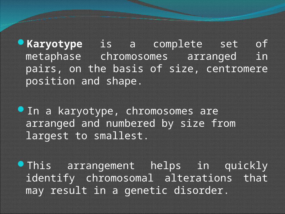
Karyotype is a complete set of metaphase chromosomes arranged in pairs, on the basis of size, centromere position and shape.
In a karyotype, chromosomes are arranged and numbered by size from largest to smallest.
This arrangement helps in quickly identify chromosomal alterations that may result in a genetic disorder.
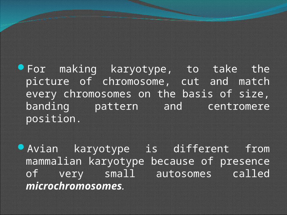
For making karyotype, to take the picture of chromosome, cut and match every chromosomes on the basis of size, banding pattern and centromere position.
Avian karyotype is different from mammalian karyotype because of presence of very small autosomes called microchromosomes.

Use of KaryotypingUse of KaryotypingIt permits an accurate count and characterization of
chromosomes of an individual/speciesIt permits rapid identification of chromosomal
abnormalities, particularly those involving structural changes
IdiogramIdiogramIdiogram is the diagrammatically representation of
the chromosomes arranged in descending order of length. Or
The karyotype can be represented diagrammatically with all the morphological features of chromosomes, such a diagram is known as idiotype.

6
CELL PREPARATION
Culture cells until sufficient mitotic activity
Add colchicine to arrest in metaphase
Add hypotonic salt solution to swell cells
Fix with mix of methanol: acetic acid

Normal karyotype of cattle[2N = 60, XY]
7

8
Karyotypes of Murrah buffalo, an Indian dairy breed [2n=50]
Karyotypes of Asiatic swamp buffalo in Thailand [2n=48]

NORMAL KARYOTYPE of DOG [2N = 78,XY]
All autosomes are acrocentricThe sex-chromosomes are metacentric
10
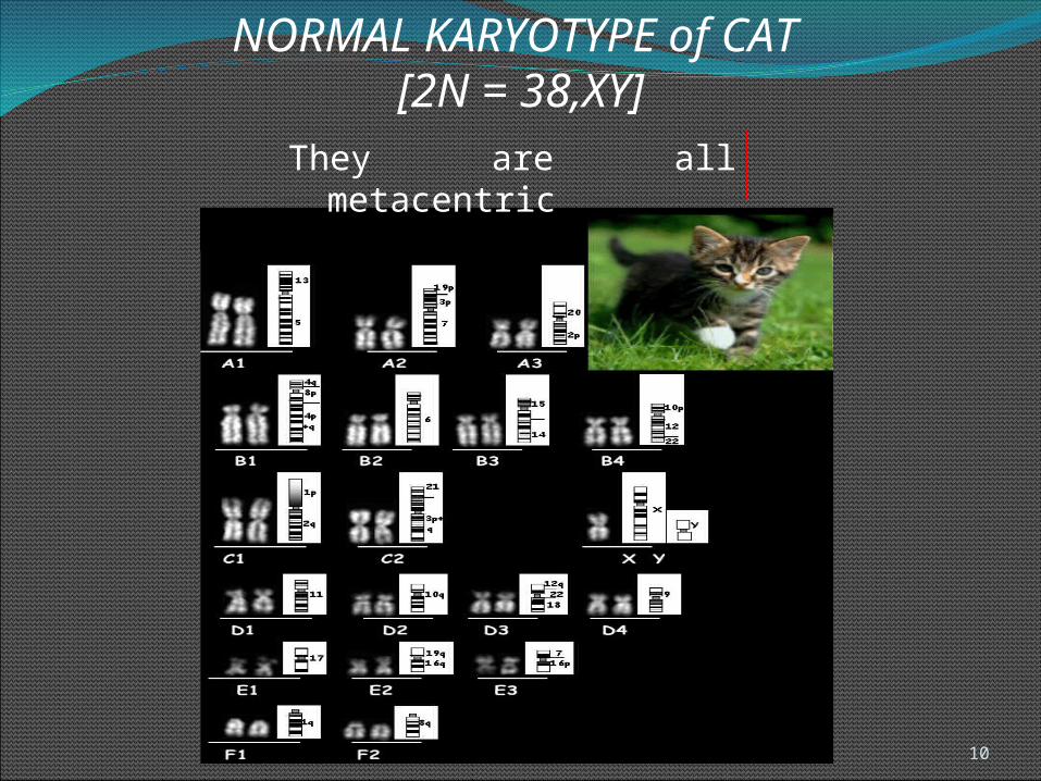
NORMAL KARYOTYPE of CAT [2N = 38,XY]
They are all metacentric

NORMAL KARYOTYPE of SWINE [2N=38,XX] Swine have 6 pairs of acrocentric chromosomes and 12 pairs of
metacentric autosomes.
The X-chromosomes are also metacentric.
11
12
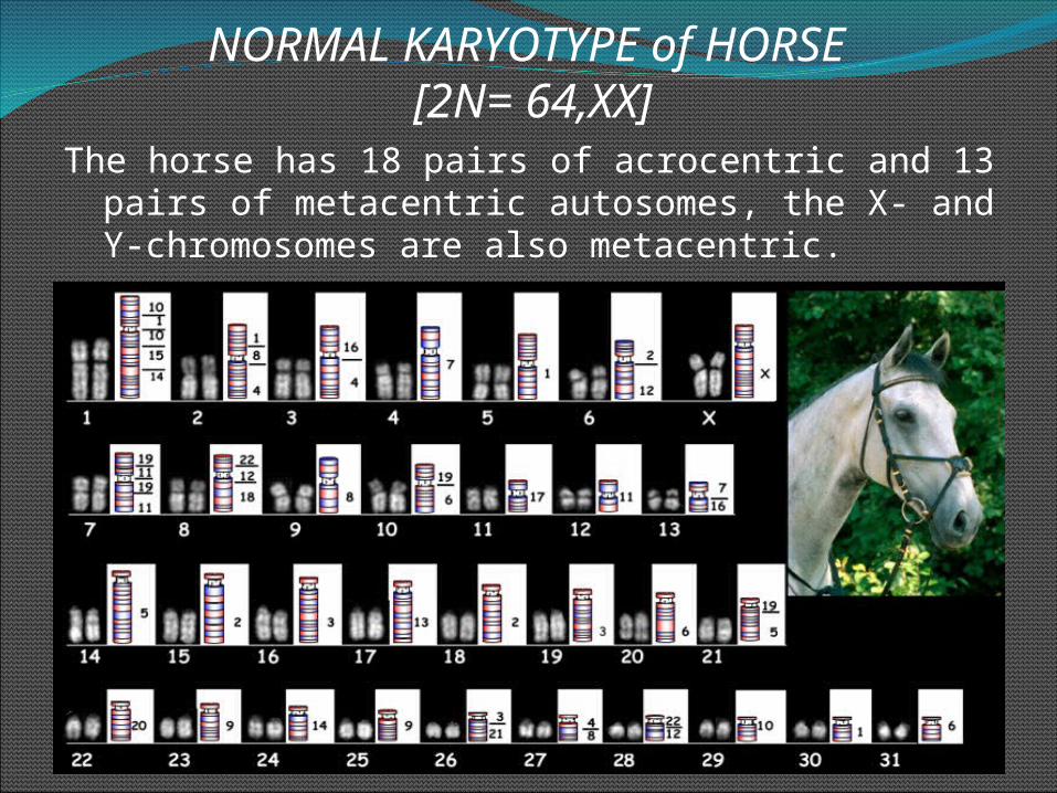
The horse has 18 pairs of acrocentric and 13 pairs of metacentric autosomes, the X- and Y-chromosomes are also metacentric.
NORMAL KARYOTYPE of HORSE [2N= 64,XX]

13
The ass has 62 chromosomes which are quite different from the ones of the horse. The mule is an infertile cross between the horse and the ass.
NORMAL KARYOTYPE of ASS [2N= 62,XX]

14

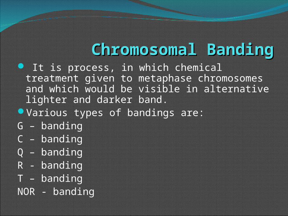
Chromosomal BandingChromosomal Banding It is process, in which chemical treatment
given to metaphase chromosomes and which would be visible in alternative lighter and darker band.
Various types of bandings are:G – bandingC – bandingQ – bandingR - bandingT – bandingNOR - banding

Lampbrush chromosomes Lampbrush chromosomes It was first observed by W. Flemming in 1882.It consists of an axis from which paired loops extend
in opposite directions, giving the appearance of a lamp brush. Hence the name Lamp Brush Chromosomes.
It is found in the Oocytes of amphibians and in some insects.
It is larger in size, So it is called a giant chromosome.

Holocentric chromosomes Holocentric chromosomes The chromosomes with a non-localized
centromere are called as either holocentric or holokinetic chromosomes
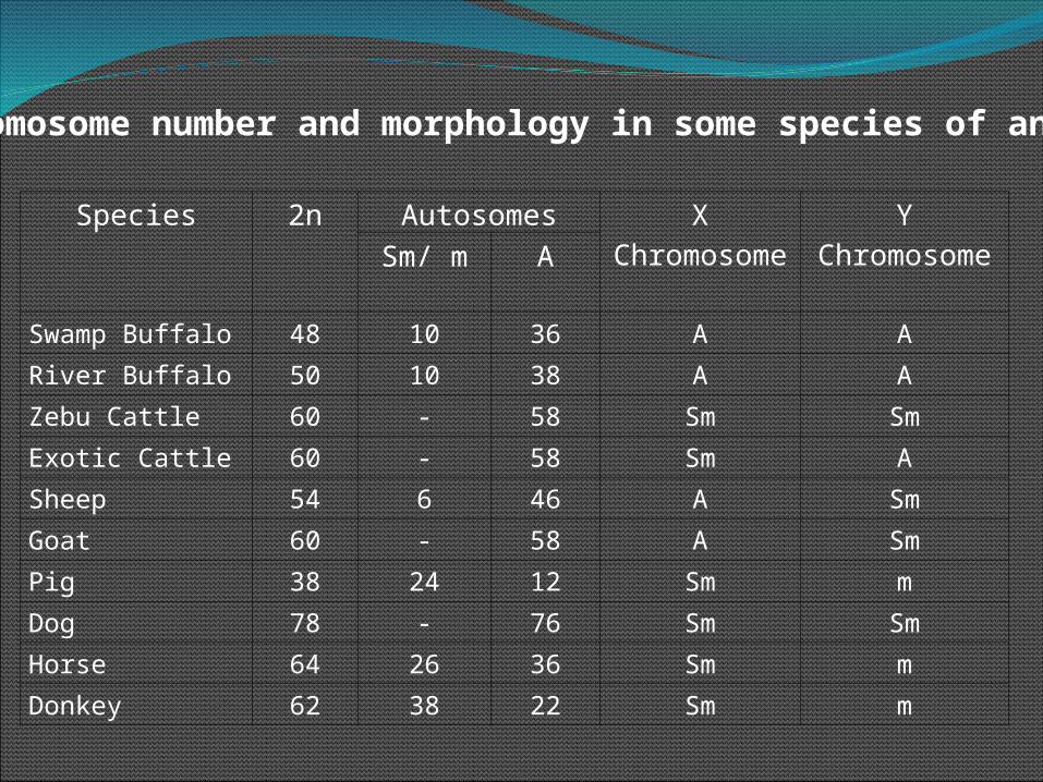
Species 2n Autosomes X Chromosome
Y ChromosomeSm/ m A
Swamp Buffalo 48 10 36 A ARiver Buffalo 50 10 38 A AZebu Cattle 60 - 58 Sm SmExotic Cattle 60 - 58 Sm ASheep 54 6 46 A SmGoat 60 - 58 A SmPig 38 24 12 Sm mDog 78 - 76 Sm SmHorse 64 26 36 Sm mDonkey 62 38 22 Sm m
Chromosome number and morphology in some species of animals:

The division of chromosome and cytoplasm of a cell into two daughter cells is known as cell division.
The cell cycle or cell-division cycle is an ordered series of events that take place in a cell leading to cell growth and its division and duplication (replication into two daughter cells).
Cytokinesis is the physical division of the cytoplasm whereby the nuclei, cytoplasm, organelles and cell membrane of a single eukaryotic cell is divided into two daughter cells containing roughly equal shares.

There are mainly two types of cell division: (1)Mitosis (2)Meiosis. The various events occurring during cell
division may be grouped in to two categories: (1)Karyokinesis (2)Cytokinesis. Karyokinesis is the division of chromosome of a
cell in to two daughter nuclei. Cytokinesis is the division of cell in to two
halves to produce two daughter cells. (As a rule, Cytokinesis follows Karyokinesis).

Types of Cell DivisionTypes of Cell DivisionMitosisMitosisThe mitotic cell division was first described by Flemming
in 1879.Somatic cells of all organisms divide through mitosis.Mitosis produces two daughter cells that are identical to
the parent cell.This type of cell division allows organisms (multicellular)
to grow and repair damaged tissue.
MeiosisMeiosis Meiosis (double cell division) produces daughter cells that
have one half the number of chromosomes as the parent cell.
Meiosis is necessary in sexually-reproducing organisms because the fusion of two gametes (fertilization) leads to doubles the number of chromosomes.

MITOSISMITOSISMitosis is a process of nuclear (karyokinesis) and
cytoplasmic (cytokinesis) division in which two daughter cells are produced that has chromosomal numbers identical to the parental cell.
Mitosis (M Phase) is part of the total cell cycle for cells undergoing division.
The initial event in the cell cycle is the growth phase, called Interphase.

INTERPHASEINTERPHASEThe interphase is much longer phase of cell
cycle, where the cell prepares itself for cell division.
Interphase is divided into three phases: G1 (first gap) S (synthesis) G2 (second gap)During all three phases of Interphase, the cell
grows and engaged in high metabolic activity to prepare its for mitosis.


G1 PHASEG1 PHASE In this phase, the cell increases in mass and prepares for DNA
replication.The metabolic rate of the cell will be high. It takes about 10 hours.
S PHASES PHASEDNA replication. Cells will take 5 to 6 hours to complete S phase.
G2 PHASEG2 PHASEThe cell undergoes of rapid growth to prepare for mitosis. G2 is shortest subphase of interphase and it lasting only 3 to 4 hours.G2 phase is after DNA synthesis but prior to the start of prophase of
mitosis.

STAGES OF MITOSISSTAGES OF MITOSISMitosis is divided into,
Prophase Metaphase Anaphase and Telophase
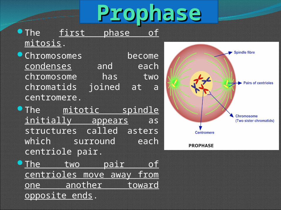
The first phase of mitosis.Chromosomes become
condenses and each chromosome has two chromatids joined at a centromere.
The mitotic spindle initially appears as structures called asters which surround each centriole pair.
The two pair of centrioles move away from one another toward opposite ends.
ProphaseProphase

MetaphaseSpindle fibers attached to all chromosomes at the kinetochore(centromere region on the chromosomes where sister chromatids are joined) .
The chromosomes begin to migrate towards the center of cell.
The condense chromosomes arrange in a line near the center of the cell, that position is called as Equatorial plane or Metaphase plate
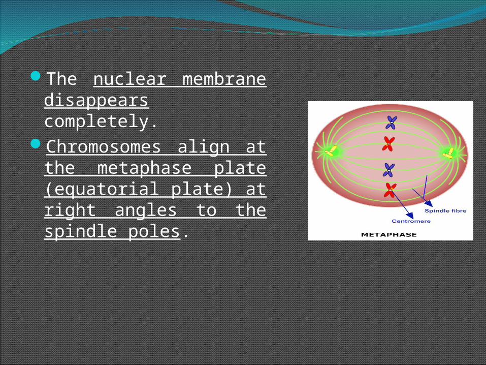
The nuclear membrane disappears completely.
Chromosomes align at the metaphase plate (equatorial plate) at right angles to the spindle poles.

AnaphaseSpindle fibers shorten, the kinetochores separate and centromeres of each chromosome begin to move apart to the cell poles.
Once the paired sister chromatids separate from one another, each is considered a "full" chromosome (daughter chromosomes).

Through the spindle apparatus, the daughter chromosomes move to the poles at opposite ends of the cell.
At the end of anaphase, each pole contains a complete compilation of chromosomes.

TelophaseTelophase is last phase of mitosis. (Latin word telos which means 'end‘).
The daughter chromosomes arrive at the poles and nucleus begin to form at opposite poles.
The nuclear envelopes of these nuclei are re- formed and reappear.
Nucleolus also reappear.

Chromatin fibers of chromosomes uncoil.
The spindle fibers also disappear.
The chromosomes begin to decondense and become more diffuse.
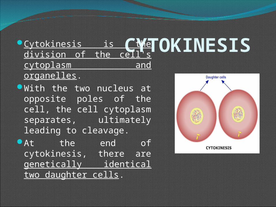
CYTOKINESIS Cytokinesis is the division of the cell's cytoplasm and organelles.
With the two nucleus at opposite poles of the cell, the cell cytoplasm separates, ultimately leading to cleavage.
At the end of cytokinesis, there are genetically identical two daughter cells.
Time Duration18-24 hours at 37 C temperature95 % time during interphase and only 5 %
during mitosisAmong mitosis: prophase takes 40 -50 % of
total timeAnaphase is shortest in duration.

Leland H. (Lee) Hartwell, Tim Hunt and Sir Paul M. Nurse were awarded the Nobel Prize in Physiology or Medicine in 2001 for their discoveries of key regulators of the cell cycle.

Related Documents