october 2009 • Vol. 1 • No. 5 Journal of THE SOCIETY OF PHYSICIANS OF HONG KONG ISSN 2072-4209 • Recent Advances in Cosmetic Dermatology Dr Chan Hin Lee, Henry (陳衍里醫生) • The Use of Topical Drug Therapy in Atopic Eczema Dr Chan Pui Yiu, Nicola (陳珮瑤醫生) • Cutaneous Manifestations of Internal Diseases Dr Yeung Chi Keung (楊志強醫生) www.soPHYSICIANSHK.org Visit the web site for our regular cMe programmes for doctors 香港內科學會 THE SOCIETY OF PHYSICIANS OF HONG KONG

Welcome message from author
This document is posted to help you gain knowledge. Please leave a comment to let me know what you think about it! Share it to your friends and learn new things together.
Transcript

october 2009 • Vol. 1 • No. 5
Journal of the Society of PhySicianS of hong Kong
ISSN 2072-4209
• Recent advances in cosmetic Dermatology
Dr Chan Hin Lee, Henry (陳衍里醫生)
• the Use of topical Drug therapy in atopic eczema
Dr Chan Pui Yiu, Nicola (陳珮瑤醫生)
• cutaneous Manifestations of internal Diseases
Dr Yeung Chi Keung (楊志強醫生)
www.sophysicianshk.orgVisit the web site for our regular cMe programmes for doctors
香港內科學會THE SOCIETY OF PHYSICIANS OF HONG KONG

C
M
Y
CM
MY
CY
CMY
K
Loceryl Ad. 206_276 20090902.ai 9/2/09 4:21:07 PM

CONTENTS
59 Recent advances in cosmetic Dermatology Dr Chan Hin Lee, Henry (陳衍里醫生)
63 t he Use of topical Drug therapy in atopic eczema Dr Chan Pui Yiu, Nicola (陳珮瑤醫生)
67 cutaneous Manifestations of internal Diseases Dr Yeung Chi Keung (楊志強醫生)
Journal of the Society of PhySicianS of hong Kong
OctOber 2009 • Vol. 1 • No. 5
execUtive coMMittee
PRESIDENTDr Lam tat chung, Paul 林達聰醫生
VIcE PRESIDENTDr tsang Wah tak, Kenneth曾華德醫生
HoN. SEcRETaRyDr Shiu cho tak, Wesely邵祖德醫生
HoN. TREaSuRERDr Lam how Mo, ignatius 林孝武醫生
cHIEF EDIToRDr Lau chu Pak 劉柱柏醫生
EDIToRSDr chan chun Kwong, Jane 陳真光醫生Dr chan hin Lee, henry 陳衍里醫生Dr chan Kwok Wing, fredriech 陳國榮醫生Dr chan Pui yiu, nicola 陳珮瑤醫生Dr chan tak hin 陳德顯醫生Dr chen yi tin 陳以天醫生Dr Lam tat chung, Paul 林達聰醫生Dr Lam tse fun, cathy 林紫芬醫生Dr Leung Wai Keung 梁偉強醫生Dr Li Siu Lung, Steven 李少隆醫生Dr Shea tat Ming, Paul 佘達明醫生Dr Shiu cho tak ,Wesely 邵祖德醫生Dr tam Kwok Kuen, vincent 譚國權醫生Dr tsang Wah tak, Kenneth 曾華德醫生
editorial
In this special dermatology issue, three contributors write on a range of topics that
cover both the medical and cosmetic aspects of dermatology. Dr Yeung Chi Keung’s
article on cutaneous manifestations of systemic diseases aims to refresh our
readers this important area of dermatology. Dr Nicola Chan’s article focuses more
on the topical management of atopic dermatitis. Finally, Dr Henry Chan’s article
updates readers on the new advances of cosmetic dermatology. We do hope that
this issue will prove to be of interest to our readers.
www.sophysicianshk.org
香港內科學會THE SOCIETY OF PHYSICIANS OF HONG KONG
editorial office:CMPMedica Pacific Ltd
9th Floor, CNT Tower, 338 Hennessy Road, Wan Chai, Hong KongT +852 2559 5888 F +852 2559 6910
advertising enquiries:Ms Chloe Wong
T +852 2155 8557e-mail: [email protected]
© 2009 The Society of Physicians of Hong Kong. All rights reserved. No part of this publication may be reproduced in any language, stored in or introduced into a retrieval system, or transmitted, in any form or by any means (elec-tronic, mechanical, photocopying, recording or otherwise), without the written consent of the copyright owner. Permission to reprint must be obtained from the publisher. Advertisements are subject to editorial acceptance and have no influence on editorial content or presentation. The Society of Physicians of Hong Kong does not guarantee, directly or indirectly, the quality or efficacy of any product or service described in the advertisements or other material which is commercial in nature.
Dr lam tat chung, Paul林達聰醫生FRCP, FHKAM (Medicine), FHKAM (Psychiatry)President
Dr chan Hin lee, Henry 陳衍里醫生MD (London), PhD (HK)FRCP (London, Edinburgh, Glasgow),FHKCP, FHKAM (Medicine)Editor

59 Journal of The Society of Physicians of Hong Kong
Recent advances in cosmetic Dermatology
Introduction
In recent years, there has been much advance in cosmetic dermatology, es-pecially in the areas of fractionated skin rejuvenation and noninvasive body con-touring. This article will address these two areas of recent interest and their ap-plication in Asian patients.
Fractionated Skin rejuvenation
Fractional photothermolysis is a new concept of cutaneous remodelling, whereby lasers are used to induce zones of microscopic thermal injury that consist of marked areas of tissue denaturation 50 to 100 microns in diameter, which are surrounded by normal viable tissue.1 As the area of thermal injury is very small, the lateral migration of keratinocytes occurs rapidly, which leads to the complete re-epithelialization of the epidermis within 24 hours. By taking into consideration the mismatch between the epidermal and dermal healing processes, fractional pho-tothermolysis allows skin rejuvenation to be achieved with a minimal risk of com-plications and a high degree of efficacy. During each treatment session, a variable ratio of the skin surface is treated, the extent of which is primarily determined by the density settings of the device and the number of passes. Usually, about 16% to 32% of the skin surface is targeted per treatment session. As the initial development involves the use of laser to induce collagen dena-turation but not vaporization, skin reju-venation can therefore be achieved in a nonablative manner. Nonablative fractional resurfacing has been shown effective in
the treatment of fine wrinkles, atrophic acne, surgical scars, and pigmentary con-ditions including melasma.1,2 The down time associated with the procedure is limited, with swelling lasting several days and erythema for no more than a week. Among Asians, the main potential compli-cation is postinflammatory hyperpigmen-tation (PIH). Our recent study indicated that the risk of PIH is associated with the density more than the energy, and that with the appropriate parameters, PIH can be reduced to about 3%.3
Ablative fractional resurfacing is a more recent advance in fractional photothermolysis that involves the use of ablative lasers (carbon diode or Erbium:YAG) not only to coagulate, but also to vaporize the central zone of the target tissue.4 Several factors determine the amount of tissue injury and depth of penetration, including the power density, pulse duration, and the type of laser. As the volume of tissue that can be removed after a single treatment session covers up
Dr chan Hin lee, Henry (陳衍里醫生)MD(London), PhD(HK), FRCP(London, Edinburgh, Glasgow), FHKCP, FHKAM(Medicine)
Specialist in Dermatology
Honorary Professor, Li Ka Shing Faculty of Medicine, University of Hong Kong
Visiting Scientist, Wellman Center of Photomedicine, Massachusetts General Hospital, Harvard Medical School, Boston, USA
Key words: cosmetic dermatology (醫學美容), fractionated technology (分段技術), cryolipolysis (冷凍熔脂術)
“With ablative fractional
resurfacing, only a single
treatment session is necessary”

Journal of The Society of Physicians of Hong Kong 60
to 45% of the target area, the procedure is associated with substantial down time. Swelling can last for 1 week and erythema for another 3 weeks. Ablative fractional resurfacing can be effective in the treatment of severe photoageing, skin laxity, atrophic acne and surgical
scars. The main advantages over non- ablative fractional resurfacing are that only a single treatment session is nec-essary, and that it can improve skin laxity due to the removal of a volume of tissue.5 (Figure 1) In Asians, the risk of PIH is sig-nificant, and more than 50% of patients
may experience the complication. The risk of PIH can be reduced by lowering the laser settings. However, this reduces the efficacy of treatment so that multiple treatment sessions will be necessary. In this case, there is little advantage over nonablative fractional resurfacing. Appro-priate patient selection is important for obtaining good clinical outcome. Male pa-tients with acne scarring or photoageing and female patients with a significant degree of skin laxity who are reluctant to have more invasive surgical procedures are ideal candidates for ablative fractional resurfacing. Fractional radiofrequency has re-cently been developed as another form of fractionated ablative skin rejuvenation that can be effective if the appropriate patient is selected for the procedure. More studies are necessary to determine its role in Asian patients. Recently, focused ultrasound has been shown to induce deep thermal tissue injury with a depth of as much as 3 to 4.5 mm.6 The epidermis is spared, making it an ideal tool for Asians. Our recent work looking at its application in Asians indicated its safety, but optimal parameters for improved efficacy are yet to be determined. The device is still pending approval by the US FDA and, therefore, its use remains experimental at this stage.
Noninvasive body contouring
While liposuction remains the gold standard for improvement of body contour, the potential adverse effects and down time associated with the surgical procedure has led to intense research in the development of noninvasive means to enhance body contour. Various methods have been tried in recent years, including chemical-induced lipolysis, devices to induce thermal or mechanical injury of the adipose tissue, and, more recently, cold-induced lipolysis (cryolipolysis). Chemical-induced lipolysis involves the injection of chemicals such as phosphatidylcholine combined with its emulsifier, deoxycholate, into the adipose tissue.7 Recent studies indicated
figure 1. improvement of periorbital fat pad (eye bag) after treatment with carbon dioxide fractional resurfacing
4 months after a single treatment session
Before treatment

61 Journal of The Society of Physicians of Hong Kong
that lipolysis occurred as a result of the detergent action of deoxycholate. As the effect is nonspecific, liver derangement has been reported. Acute extensive pan-niculitis may also occur. As a result of these adverse effects, many countries, including the United Kingdom, Korea and Singapore, have now banned the use of the substance for cosmetic purposes. Several medical devices, including laser, focused ultrasound and radio- frequency, can induce thermal injury to the adipose tissue. However, the approach is not an ideal option for improvement of body contour as it is not selective and is associated with pain. More recently,
focused ultrasound has been used to me-chanically rupture adipose tissue. While early studies did demonstrate efficacy, such result has not been found to be consistent.8 Cryolipolysis is the use of heat extraction to induce crystallization of tri-glycerides within adipose cells.9 As the freezing points of water and triglyceride are different, water, which is the main component of other cells, is not affected. When the application of heat extraction is removed, body temperature warms up the triglycerides, which then return to liquid state. The change in state of triglyc-erides from liquid to solid and then back
to liquid has been hypothesized to cause apoptosis of adipocytes, which occurs gradually over 2 to 6 months. A recent study showed a normalized fat layer reduction of 20% after 2 months, and 25% after 6 months.10 Adverse effects are mild and transient, including bruises caused by the suction applicator, mild numbness that lasted for an average of 3 weeks, and mild abdominal discomfort. In the author’s experience, good results can be obtained with appropriate patient selection. (Figure 2)
conclusion
In conclusion, fractionated skin rejuve-nation has led to successful treatment of wrinkles, scars and, more recently, skin laxity. Cryolipolysis is a novel method of noninvasive fat removal.
references:1. Manstein D, Herron GS, Sink RK, Tanner H, Anderson RR. Fractional
photothermolysis: A new concept for cutaneous remodeling using micro-scopic patterns of thermal injury. Lasers Surg Med 2004;34:426-438.
2. Tannous Z. Fractional resurfacing. Clin Dermatol 2007;25:480-486.3. Chan HH, Manstein D, Yu CS, Shek S, Kono T, Wei WI. The prevalence
and risk factors of post-inflammatory hyperpigmentation after fractional resurfacing in Asians. Lasers Surg Med 2007;39:381-385.
4. Hantash BM, Bedi VP, Kapadia B, et al. In vivo histological evaluation of a novel fractional resurfacing device. Lasers Surg Med 2007;39:96-107.
5. Rahman Z, MacFalls H, Jiang K, et al. Fractional deep dermal ablation induces tissue tightening. Lasers Surg Med 2009;41:78-86.
6. Laubach HJ, Makin IR, Barthe PG, Slayton MH, Manstein D. Intense focused ultrasound: Evaluation of a new treatment modality for precise microcoagulation within the skin. Dermatol Surg 2008;34:727-734.
7. Rotunda AM, Kolodney MS. Mesotherapy and phosphatidylcholine injections: Historical clarification and review. Dermatol Surg 2006;32: 465-480.
8. Teitelbaum SA, Burns JL, Kubota J, et al. Noninvasive body contouring by focused ultrasound: Safety and efficacy of the Contour I device in a multi-center, controlled, clinical study. Plast Reconstr Surg 2007;120:779-789.
9. Manstein D, Laubach H, Watanabe K, Farinelli W, Zurakowski D, Anderson RR. Selective cryolysis: A novel method of non-invasive fat removal. Lasers Surg Med 2008;40:595-604.
10. Coleman SR, Sachdeva K, Egbert BM, Preciado J, Allison J. Clinical efficacy of noninvasive cryolipolysis and its effects on peripheral nerves. Aesthetic Plast Surg 2009;33:482-488.
“Good results can be obtained with appropriate
patient selection for cryolipolysis”
figure 2. improvement of abdominal body contour after a single treatment of cryolipolysis
2 months after a single treatment session
Before treatment

C
M
Y
CM
MY
CY
CMY
K
The Society of Physicians of HK-Oct.pdf 9/10/09 3:56:58 PM

63 Journal of The Society of Physicians of Hong Kong
the Use of topical Drug therapy in atopic eczema
Dr chan Pui Yiu, Nicola (陳珮瑤醫生)MBBChir (Cantab), MRCP (UK), FHKCP, FHKAM(Med)
Specialist in Dermatology
Honorary Clinical Assistant Professor, Department of Medicine, University of Hong Kong
Key words: atopic eczema (異位性濕疹), emollients (潤膚霜), topical corticosteroids (外用類固醇), topical calcineurin inhibitors (外用免疫抑制劑)
atopic eczema (AE) is a common chronic inflammatory skin con-dition associated with pruritis, dryness, erythema, scaling and
exudation. The pathogenesis of AE in-volves the interplay of genetic, immuno-logical and environmental factors. Skin barrier dysfunction, primary epithelial defect with aberrant immunological re-sponses to microbial infection and al-lergen exposure all play a part in the complex pathophysiology of AE. The aims of AE treatment are to avoid any triggering factors, to restore and strengthen skin barrier function, as well as to control inflammation and in-fection. The choice of treatment follows a stepwise approach and depends mainly on the severity, body sites involved,
surface area affected and the patient’s age. (Table1) Topical therapy is the mainstay of treatment for mild to moderate AE, and is also used in combination with oral therapy or phototherapy for severe cases. It includes adequate emollient use with properly titrated topical corti-costeroids (TCs) and topical calcinuerin inhibitors (TCIs). Topical antibiotics may also be considered for short-term usage depending on the presence of super-imposed infection.
emollients
The importance of emollients, or mois-turizers, in the management of AE is often overlooked without realizing that stratum corneum abnormalities may be the primary exacerbating factors in AE.1 Apart from the reduction of scaling, roughness, and the sensations of tightness and itchiness, emollients also help to restore skin barrier function. Many different types of emollients are available, and they differ in their composition of active ingredients and excipients. Common top-ically applied lipids include petrolatum, beeswax, lanolin and triglycerides. They are often thicker in texture and exert their effects by forming an inert, epicutaneous protective membrane to reduce trans-epidermal water loss. However, recent research has shown that they may also diffuse more deeply into the skin. For example, petrolatum can be absorbed into the outer layer of delipidized stratum corneum. Besides these lipids, the in-clusion of humectants (eg, urea, pyr-rolidone carboxylic acid [PCA], lactic acid, glycerin, panthenol and sorbitol) in emol-lients further helps to maintain moisture in the stratum corneum, thereby in-creasing stratum corneum elasticity and reducing the risks of cracking and barrier
table 1. Stepwise management of atopic eczema
1. Generous emollient use (eg, barrier-repairing agents or occlusive agents) and soap avoidance
2. Mild to moderately potent topical corticosteroids • +/-antimicrobialtherapy • +/-topicalcalcinuerininhibitorsasmain-
tenance or prophylaxis
3. High to ultra-high potency topical corticosteroids • +/-antimicrobialtherapy • +/-topicalcalcinuerininhibitorsasmain-
tenance or prophylaxis
4. Wet-wrap therapy with topical corticosteroids
5. Phototherapy • NBUVB • PUVA • UVA1
6. Systemic agents or immunosuppressants • Corticosteroids • Antibiotics • Cyclosporine • Azathioprine • Mycophenolatemofetil • Intravenousimmunoglobulin

Journal of The Society of Physicians of Hong Kong 64
disruption. Newer topical lipids, which are more ‘physiological’, can penetrate into the skin and modify endogenous epi-dermal lipids as well as the rate of barrier recovery. Emollients consisting of specific ratios of cholesterol, fatty acids and cer-amides have been shown to accelerate barrier recovery.2 Emerging evidence now indicates that these lipid mixtures and humectants may be more efficient than other ingredients in improving barrier function in AE, in addition to di-minishing signs of dryness and pruritis. However, traditional topical lipids such as petrolatum still have an important role as they are cheaper and well tolerated even in infantile skin, with a lower risk of irri-tation and sensitivity. Overall, the choice of emollients ultimately depends on the degree of moisturization required, the type of ec-zematous lesion, the body site involved, the cosmetic acceptability of the product to the patient, and the cost. Whichever type is chosen, it should be dispensed in an adequate amount and be applied regularly to maintain constant cutaneous hydration even if no actual inflammatory skin lesions are apparent clinically.
topical corticosteroids
TCs, with their anti-inflammatory effects, have been for decades the mainstay in AE therapy for acute flare-ups and inter-mittent maintenance. They are available in different potencies and formulations. (Table 2) The appropriate choice depends on the severity of the AE, the body site involved, the age of the patient, and the patient’s response to previous TC treatment. Different therapeutic schemes have been established for its use, which include: 1) starting treatment with a more potent preparation to induce remission, followed by a relatively quick tapering down of preparation potency as the con-dition improves; 2) intermittent short bursts of a potent preparation followed by a steroid-free period of emollient use only until relapse occurs; or 3) more pro-longed, continuous treatment with less potent preparations.3,4
Only mild to moderately potent preparations should be used on genital, facial or intertriginous skin areas for short-term applications. Furthermore, children should not be given high or ultra- high potency TCs. The basic principle is
to use the least potent preparation that is effective for controlling the eczema. The choice of an adequate vehicle, or base, is also important to achieve the optimal effect. Ointments rather than creams may be considered for more severe or lichenifieid lesions over thicker skin areas, such as palmoplantar lesions. Foam and lotion are more acceptable for hair-bearing sites, such as the scalp. There is still no definite conclusion regarding the optimal frequency of ap-plication of the different types of TCs. A systemic review has shown that when clinical effectiveness and cost- effectiveness are considered, no clear difference in outcomes was seen between once-daily and more frequent applications of TCs.5 Patient education is important to maximize treatment efficacy, minimize side effects and increase com-pliance. Overall, TCs can be safely used if the above principles are observed. The well-reported adverse effects, listed in Table 3, are often due to the inappropriate use of preparations that are more potent than necessary, for prolonged periods over sensitive sites such as the face and intertriginous areas, especially in infants and children. For children and adults with severe or refractory AE, short-term wet-wrap treatment (WWT) has been shown to be relatively safe and effective.6,7 Wet-wrap treatment involves the application of a diluted TC ointment directly onto the ec-zematous areas, followed by a first layer of wet but wrung out gauze, and then a second layer of dry gauze on top. This treatment has the advantages of en-hancing the penetration of TC, maintaining moisture and preventing scratching. Dif-ferent regimens have been used for WWT, which include different types and concen-trations of TCs, and application of the wet wrap at different frequencies for different durations. Although no large studies have been conducted to determine the optimal regimen, one example would be to use 10% dilution of a potent corticos-teroid (eg, 0.1% mometasone furoate ointment) overnight for 12 hours per day for 7 days in cases of acute AE in children. Good long-term studies which evaluate systemic absorption and skin atrophy are still lacking. If there is overt impetigini-zation, WWT should be delayed. Common
table 2. topical corticosteroid potency chart
Potency class topical corticosteroid formulation
Ultrahigh I • Clobetasolproprionate• Diflorasonediacetate
Cream, 0.05%Ointment, 0.05%
High II • Betamethasomedipropionate• Flucinonide• Halcinonide
Ointment, 0.05%Cream, ointment, gel, 0.05%Cream, 0.1%
III • Betamethasonediproprionate• Betamethasonevalerate• Fluticasonepropionate• Mometasonefuroate• Triamcinoloneacetonide
Cream, 0.05%Ointment ,0.1%Ointment, 0.0005%Ointment, 0.1%Cream, 0.5%
Moderate IV • Flucinoloneacetonide• Hydrocortisonevalerate• Triamcinoloneacetonide• Mometasonefuroate
Ointment, 0.025%Ointment, 0.2%Cream, 0.1%Cream, 0.1%
V • Betamethasonevalerate• Flucinoloneacetonide• Hydrocortisonebutyrate
Cream, 0.1%Cream, 0.025%Cream, 0.1%
Low VI • Desonide• Aclometasonediproprionate
Cream, 0.05%Ointment, cream 0.05%
VII • Hydrocortisoneacetate• Methylprednisoloneacetate
Cream, 1%Cream, 0.25%

65 Journal of The Society of Physicians of Hong Kong
side effects of WWT include bacterial and viral skin infections, and chilling. Regular monitoring is therefore essential. Patient and parent education is also of fundamental importance to ensure compliance to therapy.
topical calcineurin Inhibitors
TCIs belong to a relatively new class of medication for the treatment of AE. They act by suppressing inflammatory cytokine production in activated T-cells and other in-flammatory cells through the inhibition of calcineurin. Two topical formulations are currently available: 1) tacrolimus ointment (Protopic®) in two concentrations (0.1% for adults, 0.03% for children); and 2) 1% pimecrolimus cream (Elidel®). They can be applied twice daily, and have been proven to be as effective as moderately potent TCs with the advantages of very low systemic absorption and no steroid-associated side effects, such as skin atrophy, rebound and tachyphylaxis. Numerous studies have proven the efficacy of TCIs for the treatment of acute phase AE, as well as for main-tenance and as prophylaxis to prevent flares.8-10 Topical calcineurin inhibitors are particularly useful options for the treatment of sensitive sites, such as the face, periorbital, intertriginous and genital areas.11 For these anatomical areas, TCIs can be used at the first sign of flare-up to prevent disease progression. They are also valuable for patients who have already developed steroid-associated side effects. Another indication is as prophylaxis after control with TCs in the acute phase in patients with frequent flare-ups. TCIs are generally well tol-erated with transient local irritation being the commonest side effect. They are contraindicated in skin with active in-fection, or in patients with weakened or compromised immune systems. In 2005, the US Food and Drug Ad-ministration (FDA) issued a ‘black-box’ warning on TCIs regarding their asso-ciated potential cancer risk. This was based on information from animal studies of unusually high oral doses, which showed a subsequently increased risk of
lymphoma. However, reviews of the ma-lignancy cases reported in humans were unable to prove any causal relationship between the development of cancer and the use of TCIs. Even though the risk is of a theoretical concern, there is no ev-idence to date to suggest an increased risk of cutaneous or visceral cancer with topical use in the dosages employed clinically. The current FDA guideline recommends TCIs as a second-line short-term or an intermittent long-term treatment of AE in patients 2 years of age or older who are unresponsive to or in-tolerant of other conventional therapies.12 Continuous long-term application should be avoided as long-term safety is un-certain. Furthermore, a minimum amount of TCIs needed to control the patient’s symptoms should be used. Patients should be advised to minimize exposure of treatment sites to sunlight. As long as these guidelines are followed, TCIs continue to be a valuable option for opti-mizing long-term treatment in AE.
conclusion
The treatment of AE follows a stepwise approach. Emollients are often over-looked but are essential not only for the relief of dryness, but also for skin barrier protection and repair. TCs remain the most commonly used anti-inflammatory agent in AE. Selection of the appro-priate potency, vehicle and duration of treatment would help to minimize their side effects. TCIs are useful alternatives for sensitive sites, and can also be used as maintenance or prophylaxis. Since their long-term safety is uncertain, they should only be prescribed for patients of 2 years of age or older and as an inter-mittent treatment.
references1. DiNardoA,WertzP,GiannettiA,SeidenariS.Ceramideandcholesterol
composition of the skin of patients with atopic dermatitis. Acta Derm Venereol 1998;78:27-30.
2. Chamlin SL, Frieden IJ, Fowler A, et al. Ceramide-dominant, barrier-repair lipids improve childhood atopic dermatitis. Arch Dermatol 2001;137:1110-1112.
3. Hanifin J, Gupta AK, Rajagopalan R. Intermittent dosing of fluticasone propionate cream for reducing the risk of relapse in atopic dermatitis patients. Br J Dermatol 2002;147:528-537.
4. Thomas KS, Armstrong S, Avery A, et al. Randomised controlled trial of short bursts of a potent topical corticosteroid versus prolonged use of a mildpreparationforchildrenwithmildormoderateatopiceczema.BMJ2002;324:768.
5. Green C, Colquitt JL, Kirby J, Davidson P. Topical corticosteroids for atopic eczema: Clinical and cost effectiveness of once-daily vs. morefrequent use. Br J Dermatol 2005;152:130-141.
6. Devillers AC, Oranje AP. Efficacy and safety of ‘wet-wrap’ dressings asan intervention treatment in childrenwith severeand/or refractoryatopic dermatitis: a critical review of the literature. Br J Dermatol 2006;154:579-585.
7. OranjeAP,DevillersAC,KunzB,etal.Treatmentofpatientswithatopicdermatitisusingwet-wrapdressingswithdilutedsteroidsand/oremol-lients. An expert panel’s opinion and review of the literature. J Eur Acad Dermatol Venereol 2006;20:1277-1286.
8. Kapp A, Papp K, Bingham A, et al. Long-term management of atopic dermatitis in infants with topical pimecrolimus, a nonsteroid anti-inflam-matory drug. J Allergy Clin Immunol 2002;110:277-284.
9. Breneman D, Fleischer AB, Abramovits W, et al. Intermittent therapy for flarepreventionandlong-termdiseasecontrol instabilizedatopicder-matitis:Arandomizedcomparisonof3-times-weeklyapplicationsoftac-rolimus ointment versus vehicle. J Am Acad Dermatol 2008;58:990-999.
10. Wollenberg A, Sidhu MK, Odeyemi I et al. Economic evaluation of secondary prophylactic treatment with tacrolimus 0.1% ointment in adults with moderate to severe atopic dermatitis. Br J Dermatol 2008;159:1322-1330.
11. Hoeger PH, Lee KH, Jautova J, et al. The treatment of facial atopic dermatitis in children who are intolerant of, or dependent on, topical corticosteroids: A randomized, controlled clinical trial. Br J Dermatol2009;160:415-422.
12. Fonacier L, Spergel J, Charlesworth EN, et al. Reports of the topical cal-cineurin inhibitor task force of the American College of Allergy, Asthma and Immunology and the American Academy of Allergy, Asthma and Im-munology. J Allergy Clin Immunol 2005;115:1249-1253.
table 3. Side effects of topical corticosteroids
atrophic changes
• Steroidatrophy• Telangiectasia• Striae• Purpura• Easybruising• Ulceration• Stellatepseudoscars
infections
• Maskedmicrobialinfections(eg,tineaincognito)• Aggravationofcutaneouscandidiasis,herpesor
demodex
ocular changes
• Ocularhypertension• Glaucoma,cataract
Pharmacologic effects
• Steroidrebound,steroidaddiction,tachyphylaxis• Suppressionofhypothalamus-pituitary-adrenal
axis, growth retardation, Cushing’s syndrome
Miscellaneous
• Steroidacne• Perioraldermatitis• Steroidrosacea• Hirsutism• Hyperpigmentationorhypopigmentation• Photosensitization• Reboundflareofsomedermatoses(eg,psoriasis)“Topical calcineurin
inhibitors are particularly useful
options for the treatment of
sensitive sites”

C
M
Y
CM
MY
CY
CMY
K
Clobex_206_276 20090902.ai 9/2/09 4:19:57 PM

67 Journal of The Society of Physicians of Hong Kong
cutaneous Manifestations of internal Diseases
Dr Yeung chi Keung (楊志強醫生)MBBS(HK), MRCP(UK), FHKCP, FHKAM(Med)
Specialist in Dermatology
Associate Consultant, Department of Medicine, Queen Mary Hospital, Hong Kong
Honorary Clinical Assistant Professor, Department of Medicine, University of Hong Kong
Key words: Paraneoplastic dermatoses (伴腫瘤性皮膚病), pruritus (皮膚搔癢現象), panniculitis (脂膜炎)
The skin is the largest organ in the body and is most accessible for clinical as-sessment. There are frequent associations of skin signs with internal diseases. (Table 1) In a survey of inpatient dermatological referrals, 48% of skin lesions were found to be related to the presenting illness, in which 36.6% of skin con-
ditions contributed to the diagnosis of systemic disease.1 Important and sometimes subtle skin signs of systemic diseases should not be overlooked. Clues to underlying systemic conditions can be obtained by careful skin examination that can lead to early diagnosis.
table 1. Skin signs of systemic disease
infections Paraneoplastic hypersensitivity
• Syphilis
• Tuberculide
• Meningococcaemia
• Infectiveendocarditis
• Scrubtyphus
• Typhoidrosespots
• Staphylococcalscaldedskinsyndrome
• Disseminatedgonococcalinfection
• Tylosis
• Leukemiacutis
• Pruritus
• Carcinoid
• Sweet’ssyndrome
• Dermatomyositis
• Tripepalms
• Paraneoplasticpemphigus
• Glucagonama
• Pyodermagangrenosum
• HenochSchonleinpurpura
• Erythemanodosum
• Serumsickness
• Erythemamultiforme
Rheumatological endocrine / Metabolic genetic disease
• SLE
• MCTD
• Scleroderma
• Psoriaticarthropathy
• Bechet’sdisease
• Dermatomyositis
• AdultStill’sdisease
• Diabeticscleredema
• Pretibialmyxedema
• Necrobiosislipiodica
• Acanthosisnigricans
• Xanthoma
• Pancreaticpanniculitis
• Porphyrias
• Neurofibromatosis
• Pseudoxanthomaelasticum
• Tuberoussclerosis
• Hereditaryhaemorrhagictelangiectasia
SLE = systemic lupus erythematosus; MCTD = mixed connective tissue disease
Paraneoplastic Dermatoses
A number of skin disorders have been linked to internal malignancy.2 The cutaneous features can sometimes precede the manifestation of underlying
malignancy for months, providing an op-portunity for diagnosis at early stages. Carcinoma of the oesophagus has been associated with palmoplanter hyper-keratosis (tylosis). Carcinoma of the gut, lung and breast may present with acan-thosis nigricans and tripe palms, which is considered a form of palmar acanthosis
Journal of The Society of Physicians of Hong Kong 68
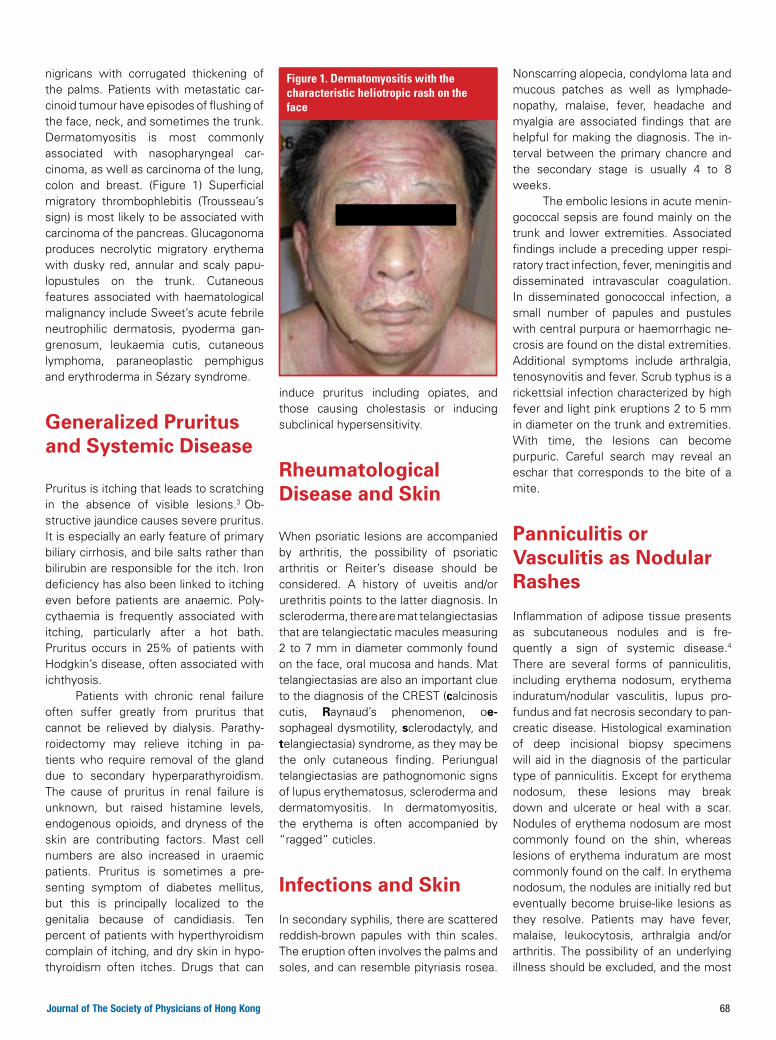
nigricans with corrugated thickening of the palms. Patients with metastatic car-cinoid tumour have episodes of flushing of the face, neck, and sometimes the trunk. Dermatomyositis is most commonly associated with nasopharyngeal car-cinoma, as well as carcinoma of the lung, colon and breast. (Figure 1) Superficial migratory thrombophlebitis (Trousseau’s sign) is most likely to be associated with carcinoma of the pancreas. Glucagonoma produces necrolytic migratory erythema with dusky red, annular and scaly papu-lopustules on the trunk. Cutaneous features associated with haematological malignancy include Sweet’s acute febrile neutrophilic dermatosis, pyoderma gan-grenosum, leukaemia cutis, cutaneous lymphoma, paraneoplastic pemphigus and erythroderma in Sézary syndrome.
Generalized Pruritus and Systemic Disease
Pruritus is itching that leads to scratching in the absence of visible lesions.3 Ob-structive jaundice causes severe pruritus. It is especially an early feature of primary biliary cirrhosis, and bile salts rather than bilirubin are responsible for the itch. Iron deficiency has also been linked to itching even before patients are anaemic. Poly-cythaemia is frequently associated with itching, particularly after a hot bath. Pruritus occurs in 25% of patients with Hodgkin’s disease, often associated with ichthyosis. Patients with chronic renal failure often suffer greatly from pruritus that cannot be relieved by dialysis. Parathy-roidectomy may relieve itching in pa-tients who require removal of the gland due to secondary hyperparathyroidism. The cause of pruritus in renal failure is unknown, but raised histamine levels, endogenous opioids, and dryness of the skin are contributing factors. Mast cell numbers are also increased in uraemic patients. Pruritus is sometimes a pre-senting symptom of diabetes mellitus, but this is principally localized to the genitalia because of candidiasis. Ten percent of patients with hyperthyroidism complain of itching, and dry skin in hypo-thyroidism often itches. Drugs that can
induce pruritus including opiates, and those causing cholestasis or inducing subclinical hypersensitivity.
rheumatological Disease and Skin
When psoriatic lesions are accompanied by arthritis, the possibility of psoriatic arthritis or Reiter’s disease should be considered. A history of uveitis and/or urethritis points to the latter diagnosis. In scleroderma, there are mat telangiectasias that are telangiectatic macules measuring 2 to 7 mm in diameter commonly found on the face, oral mucosa and hands. Mat telangiectasias are also an important clue to the diagnosis of the CREST (calcinosis cutis, raynaud’s phenomenon, oe-sophageal dysmotility, sclerodactyly, and telangiectasia) syndrome, as they may be the only cutaneous finding. Periungual telangiectasias are pathognomonic signs of lupus erythematosus, scleroderma and dermatomyositis. In dermatomyositis, the erythema is often accompanied by “ragged” cuticles.
Infections and Skin
In secondary syphilis, there are scattered reddish-brown papules with thin scales. The eruption often involves the palms and soles, and can resemble pityriasis rosea.
Nonscarring alopecia, condyloma lata and mucous patches as well as lymphade-nopathy, malaise, fever, headache and myalgia are associated findings that are helpful for making the diagnosis. The in-terval between the primary chancre and the secondary stage is usually 4 to 8 weeks. The embolic lesions in acute menin-gococcal sepsis are found mainly on the trunk and lower extremities. Associated findings include a preceding upper respi-ratory tract infection, fever, meningitis and disseminated intravascular coagulation. In disseminated gonococcal infection, a small number of papules and pustules with central purpura or haemorrhagic ne-crosis are found on the distal extremities. Additional symptoms include arthralgia, tenosynovitis and fever. Scrub typhus is a rickettsial infection characterized by high fever and light pink eruptions 2 to 5 mm in diameter on the trunk and extremities. With time, the lesions can become purpuric. Careful search may reveal an eschar that corresponds to the bite of a mite.
Panniculitis or Vasculitis as Nodular rashes
Inflammation of adipose tissue presents as subcutaneous nodules and is fre-quently a sign of systemic disease.4 There are several forms of panniculitis, including erythema nodosum, erythema induratum/nodular vasculitis, lupus pro-fundus and fat necrosis secondary to pan-creatic disease. Histological examination of deep incisional biopsy specimens will aid in the diagnosis of the particular type of panniculitis. Except for erythema nodosum, these lesions may break down and ulcerate or heal with a scar. Nodules of erythema nodosum are most commonly found on the shin, whereas lesions of erythema induratum are most commonly found on the calf. In erythema nodosum, the nodules are initially red but eventually become bruise-like lesions as they resolve. Patients may have fever, malaise, leukocytosis, arthralgia and/or arthritis. The possibility of an underlying illness should be excluded, and the most
figure 1. Dermatomyositis with the characteristic heliotropic rash on the face

69 Journal of The Society of Physicians of Hong Kong
common associations are tuberculosis, streptococcal infections and inflam-matory bowel disease in addition to drugs (oral contraceptives, sulfonamides). Er-ythema induratum is associated with the presence of Mycobacterium tuberculosis DNA by PCR assays of skin lesions. The lesions of lupus profundus are found primarily on the upper arms and buttocks, and are seen in both the cutaneous and systemic forms of lupus. The overlying skin may be normal or erythematous, and localized fat atrophy may eventually occur. Subcutaneous fat necrosis that is associated with pancreatic disease is presumably sec-ondary to circulating lipases, and is seen in patients with pancreatic carcinoma as well as in patients with acute and chronic pancreatitis. Subcutaneous erythematous nodules are also seen in medium-sized vessel vasculitis such as polyarteritis nodosa (PAN), Churg-Strauss syndrome or Wegener’s granulomatosis.4 Cutaneous PAN presents with painful subcutaneous nodules and ulcers within livedo reticularis. In both the cutaneous and systemic forms of vasculitis, skin biopsy specimens of the asso-ciated nodules will show fibrinoid necrosis characterizing vasculitis.
endocrine Disease and Skin
There are a number of cutaneous changes in diabetes mellitis.5 Diabetic dermopathy produces hyperpigmented dull-red papules with superficial scales on the shins. It heals with atrophic brown scars. Diabetic thick skin may be localized to the neck and upper back (scleroedema of Bushcke) or the dorsum of the hands (cheirarthropathy). Acanthosis nigricans is common in obese insulin-resistant diabetic patients, while generalized granuloma annulare, diabetic bullae, lipo-atrophy and perforating disorders are less common. Necrobiosis lipoidica diabet-icorum (NLD) is a characteristic eruption consisting of yellowish waxy plaques on the shin, which eventually become atrophic in the centre and may ulcerate. (Figure 2) NLD occurs in 0.3% of diabetic
patients and may predate diabetes. Hyperlipidaemia in diabetic patients may be associated with xanthoma.
Intestinal Diseases and Skin
Major aphthous ulcers are a feature of Beçhet’s syndrome, while minor aphthae are found in systemic lupus erythe-matosus, Crohn’s disease, as well as iron deficiency.6 Gastric polyposis is as-sociated with perioral lentigines in the
Peutz–Jeghers syndrome. Erythema nodosum, oral aphthous ulcers and pyoderma gangrenosum are also asso-ciated with inflammatory bowel disease, along with the secondary skin changes due to malabsorption. (Figure 3) The stigmata of chronic liver disease include spider naevi, palmar erythema, purpura, leukonychia and clubbing of the fingers. There is also loss of pubic hair. Gynaeco-mastia, xanthoma, jaundice, pruritus and pigmentation are other features. Por-phyria cutanea tarda, haemochromatosis and primary biliary cirrhosis produce diffuse skin pigmentation.
evaluation of Patients With Significant rashes
Most patients who present with a rash have no systemic disease, but patients can sometimes present with cutaneous manifestations of internal diseases. In parallel with necessary investigation, simple empirical treatment of the skin with emollients and appropriate topical medications may already result in reso-lution of rashes. The sudden onset of persistent rashes in an otherwise healthy, normal individual warrants further work-up, including thorough history taking and physical examination as well as labo-ratory studies, such as complete blood count, erythrocyte sedimentation rate, serum biochemistry, thyroid function, au-toimmune markers, chest radiograph and skin biopsy. Extensive imaging and endo-scopic studies are reserved for those with suggestive clinical evidence and positive findings in initial work-up. Follow-up as-sessment of patients with unexplained eruptions is important in order to detect emerging features of systemic diseases.
references:1. Hardwick N, Saxe N. Patterns of dermatology referrals in a general
hospital. Br J Dermatol 1986;115:167-176.2. Kleyn CE, Lai-Cheong JE, Bell HK. Cutaneous manifestations of
internal malignancy: Diagnosis and management. Am J Clin Dermatol 2006;7:71-84.
3. Lee A. Skin manifestations of systemic disease. Aust Fam Physician 2009;38:498-505.
4. Cahill J, Sinclair R. Cutaneous manifestations of systemic disease. Aust Fam Physician 2005; 34: 335-340.
5. Al-Mutairi N, Zaki A, Sharma AK, Al-Sheltawi M. Cutaneous manifes-tations of diabetes mellitus. Study from Farwaniya hospital, Kuwait. Med Princ Pract 2006;15:427-430.
6. Callen JP, Jorizzo JL, Bolognia JL, Piette W, Zone JJ. Dermatologicalsigns of internal disease. 3rd Edition. Philadelphia: W.B. Saunders; 2003.
figure 2. necrobiosis lipoidica diabeticorum with well-demarcated, raised borders and an atrophic centre in the pretibial regions of a diabetic patient
figure 3. Pyoderma gangrenosum with a rapidly enlarging ulcer with suppuration in a patient with a known history of ulcerative colitis

Online registration: www.theprimarycareclinic.com
Registration FeeHK$150 per personFree registration will be offered to members of The Society of Physicians of Hong Kong on a first come first served basis.
Payment Method Cheque (please make cheque payable to CMPMedica Pacific Ltd.)
American Express Credit Card
Cardholder’s Name:
Card Number: Expiry Date:
Signature:
Wire Transfer (please note on the transfer ‘The Primary Care Clinic’) A/C Name: CMPMedica Pacific Limited Bank Name: The Hongkong and Shanghai Banking Corporation Limited Bank Address: 1 Queen’s Road Central, Hong Kong HK$ A/C No.: 511-054801-001 Swift Code: HSBCHKHHHKH
Please complete the registration form (or a photocopy of the form) with payment and return to: CMPMedica Pacific Ltd., 9/F, CNT Tower, 338 Hennessy Road, Wan Chai, Hong Kong. (Attn.: The Primary Care Clinic). Credit card payments are acceptable via fax (852) 2559 6910.For enquiries, please call (852) 2116 4340 or e-mail [email protected]
Terms and conditions: 1. All payments must be made in Hong Kong Dollars. 2. Payment is non-refundable. 3. Registration form with payment must reach CMPMedica 2 weeks before the event. 4. Seats are confirmed only if you receive a confir-mation letter from CMPMedica. 5. Certificate of attendance will be awarded upon completion of the course.
Registration FormSaturday, 10 October 2009 (12:30 – 5:00 pm)
CMPMedica Pacific Ltd9/F, CNT Tower, 338 Hennessy Road, Wan Chai, Hong KongEmail: [email protected]: +852 2116 4340 Fax: +852 2559 6910www.theprimarycareclinic.com
Personal Information
Title: Prof Dr Mr Mrs Miss Ms
Surname: Given Name:
Mailing Address:
Tel: Fax:
Email:
Speciality:
Office-based / Hospital-based, hospital name & department:
I am a member/associate member of the Society of Physicians of Hong Kong.
The Primary Care Clinic aims to deliver a rewarding CME experience. Delegates joining this meeting will be accredited Continuing Medical Education (CME) points. Applications to the respective academic institutions and committees have been submitted and approval is pending. CME points to be awarded by the various colleges/societies will be updated on our Website soon.
We are pleased to announce the ninth in our series of multidisciplinary CME-accredited meetings, which will be held on Saturday, 10 October 2009. The programme for the meeting is as follows:
12:30 – 1:50 pm Registration and buffet lunch
1:50 – 2:00 pm Opening remarks
2:00 – 2:45 pm From evidence to action: Towards lower LDL-C target Dr Norman Chan Specialist in Endocrinology, Clinical Director, Qualigenics Diabetes Centre, Hong Kong
2:45 – 3:15 pm Updates on paediatric pneumococcal diseases Dr Yat-Wah Kwan Associate Consultant, Department of Paediatrics and Adolescent Medicine, Princess Margaret Hospital, Hong Kong
3:15 – 3:30 pm New frontiers in pneumococcal prophylaxis Dr Frank Fan Medical Director, Wyeth Hong Kong
3:30 – 3:50 pm Coffee break
3:50 – 4:35 pm Clinical approach to alopecia in general practice Dr Tze-Yuen Lee Specialist in Dermatology and Venereology, Hong Kong
4:35 – 4:55 pm Case sharing: Medico-legal issues in primary care practice Dr Stephen K S Foo Specialist in Family Medicine; Member, Preliminary Investigation Committee, Medical Council, Hong Kong
4:55 – 5:00 pm Closing remarks
CME POINTS WILL BE AWARDED
Date: Saturday, 10 October 2009 Location: The Langham Hong Kong 8 Peking Road, Tsimshatsui, KowloonTime: 12:30 – 5:00 pm
Supported by an unrestricted educational grant fromOrganizers
香港內科學會THE SOCIETY OF PHYSICIANS OF HONG KONG

C
M
Y
CM
MY
CY
CMY
K
Cetaphil Intensive Moisturiser 276_206_op.ai 9/2/09 3:52:09 PM
Related Documents