J OURNAL OF THE S PINAL R ESEARCH F OUNDATION Volume 5, Number 1 The Evolution of Spinal Health Care 2010

Welcome message from author
This document is posted to help you gain knowledge. Please leave a comment to let me know what you think about it! Share it to your friends and learn new things together.
Transcript

JOURNAL OF THESPINAL RESEARCH
FOUNDATION
Volume 5, Number 1
The Evolution of Spinal Health Care
2010

SPINAL RESEARCH FOUNDATION
SPRING 2010 VOL 5 No 1
SPINAL RESEARCH FOUNDATION
THE JOURNAL OF THE SPINAL RESEARCH FOUNDATIONA multidisciplinary journal for patients and spine specialists
Brian R. Subach, MD, FACSEditor-in-Chief
Marcus M. Martin, PhD and Anne G. Copay, PhD Managing Editors
Sherry L. McDanielEditorial Assistant
SPINAL RESEARCH FOUNDATION (SRF)BOARD OF DIRECTORS
Guy E. BeattyChairman
Thomas C. Schuler, MD, FACSPresident
Michael H. HowlandSecretary
Andrew T. GreeneMember
THE JOURNAL OF THE SPINAL RESEARCH FOUNDATIONEDITORIAL BOARD
J. Kenneth Burkus, MDHughston ClinicColumbus, GA
Christopher H. Comey, MDNew England Neurosurgical LLC
Springfield, MA
George Frey, MDColorado Comprehensive
Spine InstituteEnglewood, CO
Matthew F. Gornet, MDThe Orthopedic Center of St. Louis
Chesterfield, MO
Girard J. Girasole, MDOrthopaedic Sports
Medicine CenterTrumbull, CT
Regis W. Haid, Jr., MDAtlanta Brain and Spine Care
Atlanta, GA
Mark R. McLaughlin, MD, FACSPrinceton Brain and Spine Care
Langhorne, PA
James Schwender, MDTwin Cities Spine Center
Minneapolis, MN
Thomas C. Schuler, MD, FACSThe Virginia Spine Institute
Reston, VA
Paul J. Slosar, Jr., MDSan Francisco Spine Institute
Daly City, CA
Najeeb M. Thomas, MDSouthern Brain and Spine
Metairie, LA
Brian R. Subach, MD, FACSMember
James P. Burke, MD, PhD Allegheny Brain and Spine Surgeons
Altoona, PA
Aleksandar Curcin, MD, MBA South Coast
Orthopaedic AssociatesCoos Bay, OR
Noshir A. Langrana, PhDRutgers University - Department of
Biomedical EngineeringPiscataway, NJ
SPRING 2010 VOL 5 No 1
Spring 2010SPRING 2010
Editor’s NoteBrian R. Subach, MD, FACS 1
President’s Note Thomas C. Schuler, MD, FACS 3
Ask The Expert Matthew F. Gornet, MD 5
Anne G. Copay, PhD receives The Editor’s Choice Award for Top Rated Paper of 2008 Sherry L. McDaniel 6
Spine Tale Brian R. Subach, MD, FACS 7
The Evolution of Spinal Health Care Marcus M. Martin, PhD and Anne G. Copay, PhD 9
Advances in Cervical and Lumbar Surgery Seth S. Joseffer, MD 11
Evolution in the Treatment of Spinal Deformity and Spinal Instrumentation Christopher R. Good, MD 19
Minimally Invasive Spine Surgery: An Evolution in Progress Paul J. Slosar, MD 26 Advances in Spinal Imaging Stuart A. Fruman, MD 32
A Historical Evolution of Safe Anesthesia Michael S. Bradish, MD 36
Developments in Spinal Genetics Ayana C. Phillips, DVM, PhD and Marcus M. Martin, PhD 39
Radiofrequency Facet Joint Denervation in the Setting of Chronic Axial Low Back Pain Thomas T. Nguyen MD, DABPM 45
Physical Therapy in 2020 Richard A. Banton, PT, DPT, ATC 50
Direct Lumbar Lateral Interbody Fusion Approach in the Treatment of a Patient Suffering from Degenerative Lumbar Scoliosis, Stenosis, Lumbar Radiculopathy, and Neurogenic Claudication.
Michael W. Hasz, MD, FACS 55
Effectiveness of a Minimally Invasive Surgical Approach in the treatment of a Lumbar Disc Herniation Brian R. Subach, MD, FACS 57
Table of Contents
Special Focus: The Evolution of Spinal Health Care
Case Studies
©2010 The Spinal Research Foundation
THE JOURNAL OF THE SPINAL RESEARCH FOUNDATIONVolume 5, Number 1

SPRING 2010 VOL 5 No 1
SPINAL RESEARCH FOUNDATION
Journal of The Spinal Research Foundation 1
From the EditorBrian R. Subach, MD, FACSThe Emergence of Spinal Surgery and Fixation
Most spinal surgeons, when asked, do not know the year in which their specialty was founded.
Spinal surgery actually dates back to the 1800s. It is unclear exactly when the first spinal surgery was performed. However, in 1816, Sir Charles Bell denounced laminectomy techniques because of his perception of the pain associated with the procedure, the inevitable infection rate, and poor outcomes. A few years later, in 1829, A.G. Smith of (United States), reported the first successful lumbar laminectomy performed in a young man who was injured in a fall from a horse. During the operation, Dr. Smith removed the posterior elements of three thoracic vertebrae, resulting in the improvement of sensation in the patients legs.
Although these procedures appear to be initial attempts at surgical correction of spinal disease, it was apparent that any further advances in the field would depend upon the principles of antiseptic technique and general anesthesia. Once Henry Jacob Bigelow developed the initial techniques of general anesthesia in 1846 and Joseph Lister developed the basic principles of antiseptic technique in 1867, all that remained to initiate progress was the development of spinal imaging techniques by Wilhelm Roentgen in 1895.
The earliest spinal procedures described were laminectomies. Internal fixation of the spine (placement of screws and hardware for stabilization) did not become commonplace until later in the twentieth century. Primitive attempts at stabilizing the spine were made in the late 1800s. The first report of these attempts was a surgical procedure performed by William F. Wilkins, who described an operation on a six day old child with a fracture dislocation of T12/L1. Dr. Wilkins was able to operate on the child and stabilize the spine using a silver wire technique.
In 1887, Sir Victor Horsley performed the first successful laminectomy for removal of a spinal tumor. It should be noted that much of the credit for
developing spinal surgery must be given to the neurologist, William Gowers, who worked with Horsley. Gowers was a neurologist at the National Hospital in London. He was one of the first physicians to recognize the role spinal surgery may have had in treating disorders that involved compression of the spinal cord.
In 1891, Berthold Hadra emigrated from Germany to the United States and described a case in which he stabilized a fracture dislocation of the cervical spine by wiring the spinous processes together. In 1895, Victor Menard in France performed a costotransversectomy for evacuation of an abscess related to an infection from tuberculosis. By the end of the 1800s, there had been developments across the globe which brought spinal surgery to the forefront as a treatment for spinal disorders.
In 1910, Fritz Lange (Germany) further developed the ideas of Wilkins and Hadra and was the first to use rods to stabilize the spine. He reported the use of steel rods fixed to the spinous processes with silk, and then later with silver wire. Just one year later in the United States, there were two similar reports of steel rods to fixate the spine. These cases were performed by Russell A. Hibbs and Fred H. Albee, both in 1911, though they worked independently of each other at two different hospitals in New York, developing a technique for spinal fusion. The Albee technique involved using a tibial bone strut harvested from the patient in contrast to Hibb’s technique which involved splitting of the local spinous process to form a continuous bony bridging between vertebrae. Hibb’s technique later became the standard for scoliosis surgery.
Despite the growing success of spinal surgery, a negative report by Arthur Steindler, in 1929, curbed the enthusiasm for spinal fusion. His results of non-instrumented fusion surgery (no screws, rods or wires) for scoliosis were so poor that the surgical technique was essentially abandoned. Surgeons simply gave up the technique and returned to antiquated strategies of exercise and bracing. A second published report in

SPRING 2010 VOL 5 No 1
SPRING 2010
Journal of The Spinal Research Foundation 2
1941, from the American Orthopaedic Association, described 214 cases of scoliosis treated by fusion surgery. Of the 214 cases reported, only 31 had good or excellent outcomes, virtually condemning the technique. Fortunately, efforts were being made by surgeons to improve the results of their lumbar fusion techniques therefore supplementing with different types of internal fixation. In 1944, Don King (United States) described the technique of screw fixation of the facet joints. H.H. Boucher (Canada) in 1959, reported an improvement on King’s technique using the first pedicle screw fixation technique.
Another major advance in the understanding of the spine was a publication by William Jason Mixter and Joseph Barr in 1934. They described their theory of lumbar disc protrusion. In 1857, Rudolf Virchow in Germany had described what is known to have been a lumbar disc herniation, but its significance relating back to leg pain was unrecognized at the time. In 1911, Joel E. Goldthwait postulated that rupture of the annulus fibrosus might be the cause of back and leg pain but never applied his idea to treatment. Mixter and Barr, in 1934, showed the effectiveness of surgery for removal of disc herniations in 58 patients. In the following decades, there was a tremendous increase in the number of operations done for lumbar disc herniations.
Paralleling advances in the United States, surgeons in Great Britain, F. G. Allen in 1955 and Robert Roaf in 1966, began using hardware to stabilize scoliosis corrections. The next advance, however, came from the United States at the beginning of the 1960s, when John H. Moe introduced a greatly improved bony fusion technique combining a dissection of the soft tissues, decortication of the bone and excision of the facet joints. Paul Harrington introduced a hook and rod fixation system which dramatically reduced the incidence of failed fusion and greatly improved the overall results. He combined effective instrumentation with better bony fusion techniques, making this a standard method for scoliosis correction.
As non-surgical and surgical interventions continue to evolve, we at The Spinal Research Foundation are engaged in research to identify the most effective and least invasive techniques to improve the outcomes of our patients. As we move forward, this brief review of the advances in spinal surgery over the past two centuries should allow us to face the issues of today with a clearer perspective. As daunting as these hurdles may seem, they are nothing when compared to what the pioneers in our field have had to overcome.
1867-Joseph Lister develops basic principles in antiseptic technique
1829-First successful Lumbar Laminectomy performed by A.G. Smith.
1959-H. H. Boucher reports improvement on first pedicle screw fixation
1910-Fritz Lange of Germany uses rods to stabilize spine internally
1846-Henry Jacob Bigelow develops basic principles of anesthesia
1891-Berthold Hadra describes case of stabilizing a fracture dislocation by wiring spinous processes
1911-Hibbs and Albee develop new technique for spinal fusion that becomes a standard for scoliosis surgery
1895-Victor Menard performs a costotransversectomy and Physicist Wilhelm Roentgen discovers x-rays
1944-Don King describes the technique of screw fixation of facet joints
1934-Willam Jason Mixter and Joseph Barr showed the effectiveness disc herniation surgery
1955-Surgeon F.G. Allen begin using hardware to stabilize scoliosis corrections.
1953-Paul Harrington introduced a hook and rod fixation system which dramatically
1887-First Operation using internal fixation of the spine performed by William F. Wilkins in Kansas ANDSir VictorHorsley performs first successful laminectomy for removal of a spinal tumor
1960’s-John H. Moe introduces improved bony fusion technique
Image sources: National Library of Medicine and Image-WIlliam J. Mixter :Macmillian Publishers Ltd: Nature Publishing Group: A Brief History of Sciatica. JMS Pearce. 45:9 p 592-596; 2007

SPRING 2010 VOL 5 No 1
SPINAL RESEARCH FOUNDATION
Journal of The Spinal Research Foundation 3
From the PresidentThomas C. Schuler, MD, FACSConflicting Forces Impact The Future of Spinal Health Care
The future of spinal health care in America is going to be governed by two conflicting forces;
one is the drive to constantly improve the delivery of medical care, and the second is economics. The desire to resolve human suffering through intellectual and technical advancements is enhanced when financial support is available to maintain these important and noble efforts. Unfortunately, these efforts and advancements are stifled when cost-cutting approaches create the rationing of care.
The rationing of health care as an attempt to control federal and insurance expenditures is already progressing, irrespective of current legislation. The Center for Medicare and Medicaid Services (CMS) is creating rules, without congressional oversight or passage of legislation, to ensure Americans use less health care.
As it relates to spinal health care, CMS has initiated rules in 2010 which deny payment for medical services if policies, which they alone have created, are not met. One such example is that they will not pay hospitals or physicians for services rendered if a Medicare patient is not admitted to the hospital for more than twenty-four hours following some spinal procedures. Through technical evolution and enhanced surgical capability, many patients undergoing these procedures do not need to be admitted, and in fact would prefer to be treated as outpatients. However, CMS mandates that the patient be admitted or CMS will not pay. This is where the conflict starts.
CMS has also created rules which state that if you admit a patient to the hospital who does not
have medical conditions which require admission, then CMS will deny payment for those services. Specifically, if a physician is able to perform a given surgical procedure on a healthy Medicare patient, and due to the surgeon’s expertise that procedure can be done as an outpatient, the surgeon is not allowed to perform the procedure. Otherwise, the physician and the hospital will not be paid. If the same surgeon admits that healthy patient to the hospital to meet the requirement of Medicare, then Medicare will deny payment for those services because the patient did not have a significant medical illness requiring admission to the hospital. Just solely having the surgery is not justification for admission to the hospital. Justification for admission would be a person undergoing surgery who also has hypertension, diabetes, heart disease, pulmonary disease, etc.
To worsen the problem, CMS will not only refuse to pay the doctor and the hospital for an otherwise healthy patient admitted to the hospital following surgery, they also reserve the right to go back retrospectively and extrapolate fines back from previous surgeries performed, solely based upon apportionment. The bottom line is that this will significantly dampen the desire of medical providers to treat patients with Medicare, thereby creating the intended goal of the government- to reduce health care expenditures by having fewer people receive medical care.
The president of The Medical Society of Virginia reviewed the reimbursement rates for Medicare and Medicaid. If Medicare reimbursements were extrapolated across 100% of the patients seen by his family practice group in a given year, then the physicians would make an income of $70,000 a year. This, for having completed four years of college, four years of medical school, and at least three years of residency, and sustaining substantial debt. In addition, the physician is required to be available to cover
SPRING 2010 VOL 5 No 1
SPRING 2010
Journal of The Spinal Research Foundation 4
already failing systems, specifically Medicare and Medicaid. The increased utilization of these services will lead to increased rationing of the services provided, either by direct government regulations or by the financial forces created by eroded reimbursements.
Medicine continues to improve as we gain more knowledge through research and advanced technology. While panaceas will not exist, progress occurs down many avenues. The ongoing struggle that remains is how we are to pay for health care improvements, and ultimately, which ones are the best value for the government and third party payers. Unfortunately, improvements which are best for individual patients will be lost in the health care debate. The decisions that are being made today, and will continue to be made in the future, are based purely upon economics. This will only worsen as time progresses. With the passage of the current health care proposals on the table, more people will have health coverage; however, the only way to handle this situation without increasing the budget is to either cut services provided or increase the national debt.
patients’ medical issues 365 days a year, 24 hours a day. Since the time of the initial calculation a 21% cut has been applied to Medicare reimbursements, which would reduce the physician income to less than $50,000 for 2010.
The same calculation was performed with Medicaid. The reimbursement rate from Medicaid to physicians under the same model would reveal that after providing medical care for an entire year to a practice consisting of Medicaid patients, the physicians would each owe the government $60,000. The bottom line is that physicians cannot afford the increasing costs of staffing their offices and the increasing costs of malpractice insurance with the continued, declining reimbursements. It’s going to be difficult to find physicians willing to treat these patients, and this is already happening. The reason these reimbursements have deteriorated over time is that more people are using Medicare and Medicaid services.
The actual proposals that are being placed before Congress in 2010 will drive up the utilization of these

SPRING 2010 VOL 5 No 1
SPINAL RESEARCH FOUNDATION
Journal of The Spinal Research Foundation 5
Ask the Expert Matthew F. Gornet, MD The Orthopedic Center of St. Louis
One of the most important advance in spinal surgery this past decade has been the FDA approval of bone morphogenetic protein (rhBMP-2) for implantation in the human spine. First discovered in the 1960s, the family of bone morphogenetic proteins was characterized and rhBMP-2 was identified as a protein which stimulates stem cells from both bone and blood to form new bone. This increases patient healing rates dramatically. rhBMP-2 has virtually eliminated the need for the painful process of harvesting bone graft from the patient’s pelvis. This advance is safe and actually represents the first application of genetic engineering in spinal surgery. The protein is produced in a laboratory; however, the genetic sequence was first identified in humans.
I believe that minimally invasive surgical approaches are a natural evolution of spinal surgery. I find that smaller incisions, less tissue dissection, and better visualization of the structures actually helps both the surgeon and the patient alike. Minimally invasive procedures have limited hospital stays and reduced infection rates. One word of caution, however, is that not every patient’s problem is ideally suited for minimally invasive spinal surgery.
Advances in magnetic resonance imaging technology (MRI) have influenced my practice a great deal. Not only have the magnets become stronger,
What do you view as one of the most important advances in spine surgery in the past ten years?
How have minimally invasive surgical approaches impacted spine surgery?
How have advances in spinal imaging influenced your practice?
Matthew F. Gornet, MD Dr. Gornet is a board certified Spine Surgeon at The Orthopedic Center of St. Louis. He is a national leader in the development of dynamic stabilization, disc replacement, and “non-fusion” technology. His sub-specialty interests include treating patients with continued pain after failed spinal surgery. He
received his medical degree at Johns Hopkins University and completed his residency at Johns Hopkins Hospital. He is a member of the North American Spine Society and The Orthopedic Research Society. He is also the author of several book chapters and research publications.
producing better resolution of the spinal structures, but it is now possible to image a patient’s spine in a standing or sitting position. Previous MRI scans were all performed lying down. Much of what the spine does is dynamic and it is difficult to visualize that when the patient is simply lying there. I believe that further advances in MRI technology will give us greater insight into the structure and function of the spine.
How have new biomaterials influenced spinal surgery?
The evolution of biomaterials over the past decade has been tremendous. We have gone from simple stainless steel implants to devices that may be implanted in the spine and dissolve over time. We have screws that are coated with hydroxyapatite, which allow them to incorporate into the surrounding bone. We have plastic rods and spacers which do not interfere with the ability of an MRI to image the spine like stainless steel or titanium hardware would. We also have absorbable devices which are implanted into the spine and, as bone growth progresses, the device simply dissolves away. Also, the newest artificial discs are now being made with space-age materials, mixed metal alloys in order to endure wear and have the strength needed to support the human body.
SPRING 2010 VOL 5 No 1
SPRING 2010
Journal of The Spinal Research Foundation 6
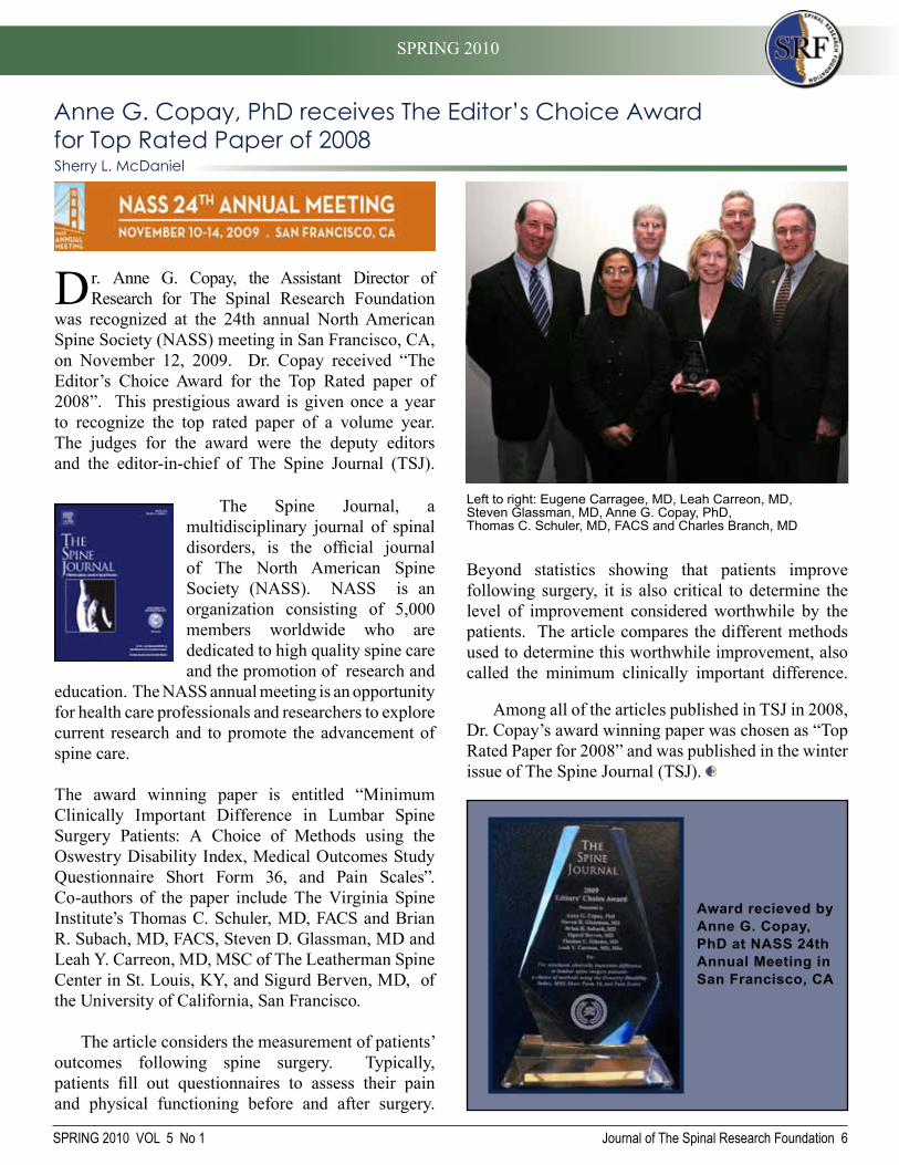
Anne G. Copay, PhD receives The Editor’s Choice Award for Top Rated Paper of 2008Sherry L. McDaniel
Dr. Anne G. Copay, the Assistant Director of Research for The Spinal Research Foundation
was recognized at the 24th annual North American Spine Society (NASS) meeting in San Francisco, CA, on November 12, 2009. Dr. Copay received “The Editor’s Choice Award for the Top Rated paper of 2008”. This prestigious award is given once a year to recognize the top rated paper of a volume year. The judges for the award were the deputy editors and the editor-in-chief of The Spine Journal (TSJ).
The Spine Journal, a multidisciplinary journal of spinal disorders, is the official journal of The North American Spine Society (NASS). NASS is an organization consisting of 5,000 members worldwide who are dedicated to high quality spine care and the promotion of research and
education. The NASS annual meeting is an opportunity for health care professionals and researchers to explore current research and to promote the advancement of spine care.
The award winning paper is entitled “Minimum Clinically Important Difference in Lumbar Spine Surgery Patients: A Choice of Methods using the Oswestry Disability Index, Medical Outcomes Study Questionnaire Short Form 36, and Pain Scales”. Co-authors of the paper include The Virginia Spine Institute’s Thomas C. Schuler, MD, FACS and Brian R. Subach, MD, FACS, Steven D. Glassman, MD and Leah Y. Carreon, MD, MSC of The Leatherman Spine Center in St. Louis, KY, and Sigurd Berven, MD, of the University of California, San Francisco.
The article considers the measurement of patients’ outcomes following spine surgery. Typically, patients fill out questionnaires to assess their pain and physical functioning before and after surgery.
Left to right: Eugene Carragee, MD, Leah Carreon, MD,Steven Glassman, MD, Anne G. Copay, PhD, Thomas C. Schuler, MD, FACS and Charles Branch, MD
Beyond statistics showing that patients improve following surgery, it is also critical to determine the level of improvement considered worthwhile by the patients. The article compares the different methods used to determine this worthwhile improvement, also called the minimum clinically important difference.
Among all of the articles published in TSJ in 2008,
Dr. Copay’s award winning paper was chosen as “Top Rated Paper for 2008” and was published in the winter issue of The Spine Journal (TSJ).
Award recieved by Anne G. Copay, PhD at NASS 24th Annual Meeting in San Francisco, CA

SPRING 2010 VOL 5 No 1
SPINAL RESEARCH FOUNDATION
Journal of The Spinal Research Foundation 7
Spine TaleUpon examination, Dr. Subach, observed signs that both the L5 and S1 spinal nerve roots were involved. Her old magnetic resonance imaging (MRI) scan showed signs of degeneration in the two lowest discs of her lumbar spine, L4-5 and L5-S1. The MRI scan showed that these
discs were actually turning black, which is indicative of a loss of fluid in the discs; thus making the crucial shock absorbing collagen of the disc useless.
A prior computed tomography (CT) scan with myelogram (dye injected into the spinal fluid space) also showed a vacuum disc at the L5/S1 level, indicative of complete degeneration of that disc. There was also evidence of a disc protrusion, at the L4/5 level. Two additional diagnostic tests were performed at The Virginia Spine Institute.
First, a nerve conduction study (electromyography or EMG) of the legs was conducted to identify the presence of progressive versus chronic nerve damage. The EMG study showed signs of a chronic right L5 radiculopathy (nerve damage). Second, a lumbar discography procedure was used to evaluate the structural integrity of the lumbar discs. This showed that her usual low back pain came from both the L4/5 and L5/S1 discs.
It was understandable why Paula Foltz felt discouraged. She had woken up one day with excruciating pain in her right leg after having undergone three operations, which were of no benefit. After her third operation, she was told that nothing else could be done for her and that the best she could hope for was some measure of pain relief from the electrical
MRI showing degeneration of the L4-L5 and L5-S1 discs
It is a pleasure to introduce you to
Paula Foltz, the focus of our Spine Tale for this issue of the Journal of The Spinal Research Foundation. Paula is a forty-four year old woman who underwent three previous low back surgeries prior to
receiving treatment from Dr. Subach in 2008. She described constant severe pain in her low back that ran down her right leg and into her toes. Her symptoms began in April 2003 and were not related to an obvious injury. She simply awoke one morning to find that she was in severe pain.
After seeing a specialist, she underwent a lumbar discectomy in December 2004 and returned to the operating room for a revision of the same procedure in June 2005. Finally, she was sent to pain management because the surgeons did not think that additional spine surgery would help her. She then had a spinal cord stimulator implanted into her spine to help alleviate her pain. A spinal cord stimulator is a device which delivers electricity to the back of the spinal cord in an attempt to override pain in the low back and legs. The goal is to produce a tingling sensation instead of severe pain.
According to Paula Foltz, the first discectomy was a complete failure: her pain never changed after the operation. The second operation on her back gave her approximately two weeks of pain relief, however, the pain returned with even greater severity. After the spinal cord stimulator was placed, she experienced some relief. The buzzing and tingling sensation from the stimulator made her symptoms more bearable, but did nothing to fix the actual problem. Unfortunately, by June 2008 her pain was back to a 10 on a pain scale ranging from 1 (minimal pain) to 10 (excruciating pain). She had right-sided low back pain and right leg pain that traveled all the way down to her foot.

The Evolution of Spinal Health Care
SPRING 2010 VOL 5 No 1
SPRING 2010
Journal of The Spinal Research Foundation 8
The story of Paula Foltz’s recovery is an amazing one. There are two reasons why our team selected her as the Spine Tale for this edition of the Journal. First, we were disappointed in the lack of medical care that she had received in the past. She had endured multiple operations and then was told that nothing could be done for her except to cover up her back pain and nerve damage with a spinal cord stimulator. Contrary to this, the multidisciplinary specialists at The Virginia Spine Institute believe in finding solutions to people’s problems, not covering them up. They believe in listening to their patients by carefully examining them and taking time to review their imaging studies. Their goal is to identify the problem and find a solution that will restore their patients’ quality of life.
The second reason why Paula was chosen as our focus for the Spine Tale this issue was, quite simply, to showcase the remarkable spirit of a courageous woman. Even after having a number of unsuccessful surgeries and excruciating pain, which forced her to the point of disability, she never abandoned hope. To this day, Paula has never given up. She has faced the challenges and hurdles placed before her with strength and determination, even when those around her were discouraged. It has been an honor for us to care for this brave and spirited woman and a privilege to call her a dear friend.
spinal cord stimulator. Instead of giving up, she came to The Virginia Spine Institute and found new hope.
In October 2008, Dr. Subach performed fusion surgery on Paula Foltz. She had some soreness over her incisions and some aching in her legs. She still felt tightness in her right leg. Her medications were refilled and she was sent to physical therapy where she continued to show consistent improvement. By March 2009, her pain was down to a 2 on the 1-10 pain scale. She was taking less medication and felt that she was more functional than she had been in the past. She was now five months out from her surgery. In October 2009, Paula returned to the operating room to remove the screws and rods which were placed at her last operation, and also to remove the spinal cord stimulator, which was no longer needed. She was not using the device and its presence made it impossible to ever have an MRI scan on any part of her body. By taking out the screws and rods, her low back soreness would get better and would hopefully prevent the screws from causing damage to the next disc up in her spine. She flew through the operation without any problems and was back in the office two weeks later, ready to start physical therapy.
X-ray of Paula’s spine prior to the fusion surgery. (Left) Post-op X-ray of Paula Foltz (Right). The spinal cord stimulator is implanted by her right hip bone and the electrodes are connected to her upper spine
Spinal Cord Stimulator
Spinal Cord Stimulator
Discography: injections in L4-L5 and L5-S1 reproduced Paula’s pain

SPRING 2010 VOL 5 No 1
SPINAL RESEARCH FOUNDATION
Journal of The Spinal Research Foundation 9
The last century has heralded phenomenal advances in the field of medical sciences. These advances
have strongly impacted life expectancy, disease progression and treatment methodology for many medical conditions. Advances in spine surgery have also been a part of this wave, incorporating many new diagnostic and therapeutic tools into the standard of spine care.
The current issue of The Journal of The Spinal Research Foundation examines several major breakthroughs which have ushered in the modern era of spine care. Many of these advances were initially developed in other fields, such as physics, dentistry and microbiology, and have significantly contributed to the improvement of standard spine care. This issue covers advances in surgical treatment, minimally invasive surgery, pain management, spine imaging, physical therapy, anesthesia and genetic technology, all of which have occurred over the past century and have greatly improved the current standard of spine surgery.
Traditional open approaches to spine surgery often require large incisions and lengthy recovery times.1 Minimally invasive approaches have provided another option for spine surgery. These approaches often involve smaller incisions, shorter operating times and the use of smaller instruments. They may result in shorter recovery times and less trauma. Though this approach is not suitable for all types of spine surgery, some patients benefit substantially when it is used for fusion and disc herniation surgeries.2,3 This approach has shortened hospital stays, reduced postoperative pain, and resulted in minimal scar formation. Paul J. Slosar, MD covers this topic in his article on minimally invasive surgery, highlighting how it has influenced modern spine treatment.
Medical imaging has progressed significantly over the last century. During this time, x-rays, MRIs and CTs have become essential in the diagnosis and treatment of spine disease. Imaging technology continues to advance, providing more detailed images for physicians. There is now shorter image
capture time as well as real-time imaging to facilitate intricate procedures. In his article on spine imaging, Stuart Fruman, MD illustrates how current imaging technology is now one of the cornerstones of orthopedic surgery. There is little doubt that it will continue to have a major impact on future of spine care.
Spine instrumentation techniques have also evolved over time. The earliest interventions were developed to treat scoliosis curves. There are now several devices and materials utilized in spine instrumentation. Modern materials such as stainless steel, titanium, titanium–alloy and other non metal materials are used to facilitate structural support and repair of the spine. The devices used include braided cables, surgical rods, plates, inter-body cages and screws.
The Evolution of Spinal Health Care Marcus M. Martin, PhD and Anne G. Copay, PhD
Fig. 1- Photograph of Walter Blount (left) and Albert Schmidt (right) who introduced the Milwaukee Brace in the early 1950s, a revolutionary removable distraction jacket for the treatment of progressive idiopathic spinal deformity. (Image courtesy of Journal of Neurosurgery: Spine)
Fig. 2- Photographs of Dwyer’s segmental cable compression system, which enabled short construct curvature correction via titanium vertebral screws along the convexity of the curve through which a threaded cable applies compressive forces. (Image courtesy of Journal of Neurosurgery: Spine)

The Evolution of Spinal Health Care
SPRING 2010 VOL 5 No 1
SPRING 2010
Journal of The Spinal Research Foundation 10
molecular basis of spine disease, a more cellular approach may be taken toward spine treatment incorporating genetics and tissue engineering. Repair on the mechanical level may be enhanced by the development of powerful molecular approaches which augment the treatment of these conditions.
References1. Armin SS, Holly LT, Khoo LT. Minimally invasive decompression for lumbar stenosis and disc herniation. Neurosurgery Focus. 2008;25(2):E11.
2. Lavelle WF, Lavelle ED, Smith HS. Interventional techniques for back pain. Clin Geriatr Med. 2008;24(2):345-368 3. Salame K, Lidar Z. Minimally invasive approach to far lateral lumbar disc herniation: technique and clinical results. Acta Neurochir. Oct 16 2009. 4. McKay B, Sandhu HS. Use of recombinant human bone morphogenetic protein-2 in spinal fusion applications. Spine. Aug 15 2002;27(16 Suppl 1):S66-85. 5. Shephard DA. History of Canadian anaesthesia. Samuel Johnston (1868-1946). Can J Anaesth. Apr 1994;41(4):353.
In his article on the treatment of spinal deformity and instrumentation, Christopher R. Good, MD highlights how these advances have contributed immeasurably
to modern spine treatment. Dr. Good’s article covers one of the ways in which these developments have facilitated and improved treatment for spine conditions, and highlights potential changes on the medical
horizon. Presently, novel materials are available to facilitate spine treatment. Both biomaterials and synthetics are part of a new wave of materials which can promote bone healing, preserve tissue margins, reduce scar formation and resist the amount of stress usually placed on the spine.
Genetic predisposition is a critical element in the development of spine disease. This knowledge has given doctors the ability to predict disease risk and will allow them to design treatments which may overcome a patient’s genetic predisposition. Through the use of genetic tools, we are now able to produce molecules which promote bone growth and healing that were previously only found in living systems.4 As time progresses, it is anticipated that genetic analysis will play an increasing role in the diagnosis and treatment of spine related disease.
Prior to the development of modern anesthesia, surgical procedures were very traumatic for both the patient and the physician.5 In his article, Michael Bradish, MD highlights the different origins, forms, and advances in anesthesia and how they contributed to spine surgery. Without these advances, modern surgical practices would not be possible.
Advances in the fields of minimally invasive spine surgery, spine imaging, instrumentation, genetics, surgical materials, and anesthesia have all made significant contributions to modern spine care. As time progresses and more is known about the
Marcus M. Martin, PhDDr. Martin’s research interests include virology, immunology and neuroimmunology. He is engaged in collaborative research through SRF, with the Medical University of South Carolina Children’s Hospital, geared toward the development of neuroprotective and neuroregenerative compounds for the
treatment of nerve pathology.
Anne G. Copay, PhD
Dr. Copay studies the outcomes of surgical and non-surgical spine treatments. She published several articles on the outcomes of spine fusion. She has on-going research projects concerning the effectiveness of new spine technologies and the long-term outcomes of surgical treatments.
Image of rhBMP-2 Courtesy of Medtronic
SPRING 2010 VOL 5 No 1
SPINAL RESEARCH FOUNDATION
Journal of The Spinal Research Foundation 11
Advances in Cervical and Lumbar SurgerySeth S. Joseffer, MD
Minimally Invasive Spine Surgery
One of the most exciting areas of development in the past decade is minimally invasive
spine surgery. Surgeons in many specialties have successfully reduced the invasiveness of surgical procedures, resulting in a tremendous impact on patient care. Many surgeries are now associated with less pain, less need for pain medications, quicker recovery and shorter hospital stays. Many procedures are now performed in outpatient centers. Developments in spine surgery in the past decade have allowed spine surgery patients to enjoy some of these benefits as well.
Conventional spine surgery involves making an
incision in the middle of the back or neck, stripping the muscle from the bone and then retracting the muscle to allow access to the spine. This muscle dissection can lead to postoperative pain, longer hospital stays, increased need for pain medications, and loss of the normal function of the muscles and ligaments. A variety of new approaches allows surgeons to work on the spine without causing so much disruption of the muscle and supporting ligaments.
One of the most important developments in
minimally invasive spine surgery is the adoption of small tubular retractors. Neurosurgeons have some familiarity with this type of retraction system, as it has been used to reach some of the deepest parts of the brain without disturbing surrounding structures. In the 1990’s, Smith and Foley developed a system of small tubular retractors that could be
used for spine surgery. With this technique, a probe is inserted with x-ray guidance to the exact point of interest, the muscles are dilated, and a small tube is passed directly to the area where the surgeon will be working. The surgeon uses either a microscope or an endoscopic camera to see through the tube. Once the surgeon is able to see the spine, the surgery proceeds in the same way as a conventional open approach.
As surgeons have become more comfortable with working through small tubular retractors, minimally invasive techniques have been applied to an increasing number of spinal disorders. These include spinal stenosis, cervical radiculopathy, cervical myelopathy, cervical trauma with C2 fractures or facet subluxation,
The past decade brought tremendous advances in spine surgery. Minimizing the invasiveness of surgery has been a goal for many decades and new techniques have now been developed to make minimally invasive spine surgery a reality. Existing procedures have improved with new technology such as better implant materials, biologic agents to promote fusion, and image guidance to facilitate instrumentation. New procedures have been developed which allow diseases such as lumbar stenosis to be treated in an entirely novel fashion. Long awaited technology has finally arrived to realize the goal of reconstruction of the spine with restoration of its normal function via joint replacement.
Key Words: minimally invasive spine surgery, disc replacement, BMP, DLIF, kyphoplasty
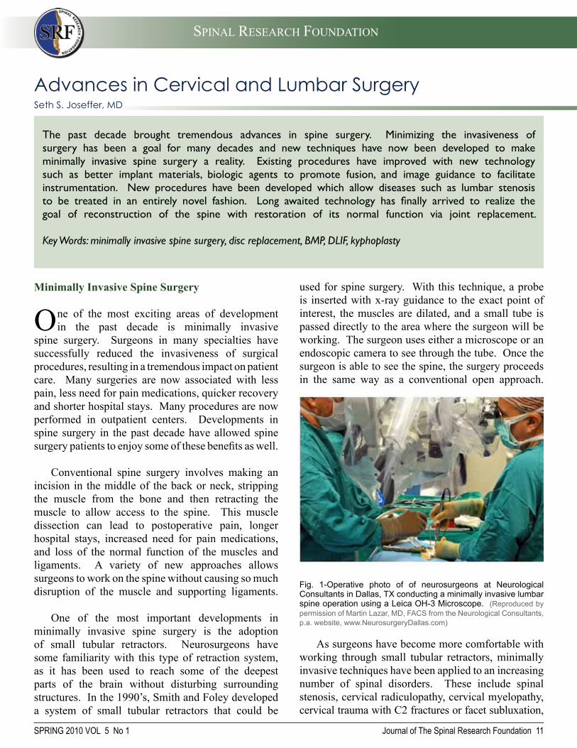
Fig. 1-Operative photo of of neurosurgeons at Neurological Consultants in Dallas, TX conducting a minimally invasive lumbar spine operation using a Leica OH-3 Microscope. (Reproduced by permission of Martin Lazar, MD, FACS from the Neurological Consultants, p.a. website, www.NeurosurgeryDallas.com)
The Evolution of Spinal Health Care
SPRING 2010 VOL 5 No 1
SPRING 2010
Journal of The Spinal Research Foundation 12
thoracic disc herniation, lumbar stenosis, lumbar spondylolisthesis and instability. Tubular retraction systems have even been used for the resection of tumors in and around the spinal cord, and for the removal of vertebral bodies that have been destroyed by tumor or trauma. Additional techniques for placing screws directly through the skin, using very small incisions, have further added to the possibilities.
Lumbar Spine
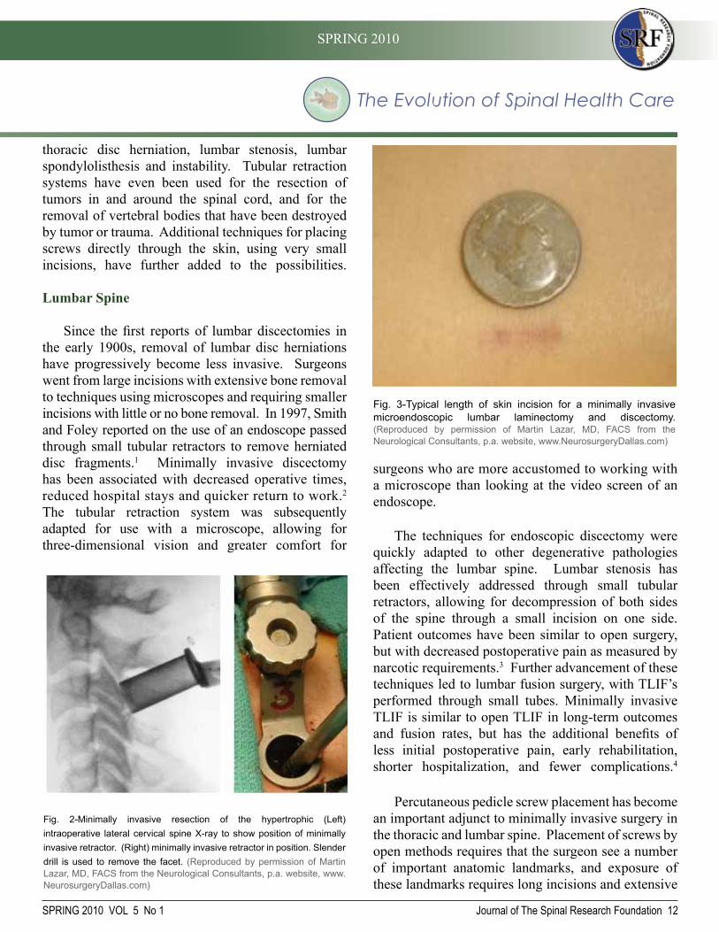
Since the first reports of lumbar discectomies in the early 1900s, removal of lumbar disc herniations have progressively become less invasive. Surgeons went from large incisions with extensive bone removal to techniques using microscopes and requiring smaller incisions with little or no bone removal. In 1997, Smith and Foley reported on the use of an endoscope passed through small tubular retractors to remove herniated disc fragments.1 Minimally invasive discectomy has been associated with decreased operative times, reduced hospital stays and quicker return to work.2
The tubular retraction system was subsequently adapted for use with a microscope, allowing for three-dimensional vision and greater comfort for
surgeons who are more accustomed to working with a microscope than looking at the video screen of an endoscope.
The techniques for endoscopic discectomy were quickly adapted to other degenerative pathologies affecting the lumbar spine. Lumbar stenosis has been effectively addressed through small tubular retractors, allowing for decompression of both sides of the spine through a small incision on one side. Patient outcomes have been similar to open surgery, but with decreased postoperative pain as measured by narcotic requirements.3 Further advancement of these techniques led to lumbar fusion surgery, with TLIF’s performed through small tubes. Minimally invasive TLIF is similar to open TLIF in long-term outcomes and fusion rates, but has the additional benefits of less initial postoperative pain, early rehabilitation, shorter hospitalization, and fewer complications.4
Percutaneous pedicle screw placement has become an important adjunct to minimally invasive surgery in the thoracic and lumbar spine. Placement of screws by open methods requires that the surgeon see a number of important anatomic landmarks, and exposure of these landmarks requires long incisions and extensive
Fig. 3-Typical length of skin incision for a minimally invasive microendoscopic lumbar laminectomy and discectomy. (Reproduced by permission of Martin Lazar, MD, FACS from the Neurological Consultants, p.a. website, www.NeurosurgeryDallas.com)
Fig. 2-Minimally invasive resection of the hypertrophic (Left) intraoperative lateral cervical spine X-ray to show position of minimally invasive retractor. (Right) minimally invasive retractor in position. Slender drill is used to remove the facet. (Reproduced by permission of Martin Lazar, MD, FACS from the Neurological Consultants, p.a. website, www.NeurosurgeryDallas.com)

SPRING 2010 VOL 5 No 1
SPINAL RESEARCH FOUNDATION
Journal of The Spinal Research Foundation 13
tissue dissection. Foley developed a technique for placement of screws directly into the bone, using very small incisions in the skin.5 With this technique, AP and lateral fluoroscopy views are used to place a small guide wire directly into the desired location in the bone. Dilators are passed to hold aside the muscle, and a cannulated screw is passed over the wire. A separate device is then passed to connect the screws with a rod for rigid fixation. Several other similar systems have been developed, including systems that allow for connection of multiple levels. This technique is very useful as an adjunct to other decompression and stabilization procedures, and has even been used on its own as treatment for certain fractures.
Cervical Spine
Anterior cervical discectomy and fusion has become the primary means of addressing cervical spine pathology. This procedure already follows the primary goal of minimally invasive spine surgery: minimizing tissue damage, as its exposure is accomplished by separating tissue planes rather than cutting muscle. As a result, the development of anterior approaches has led to the abandonment of some very useful posterior cervical procedures.
While there have been some attempts at percutaneous access to the anterior cervical spine,
this generally has not been considered safe enough to justify potential benefits. Posterior cervical procedures, such as foraminotomy and microdiscectomy, which do require more muscle dissection, do benefit significantly from a minimally invasive approach. This has led to a resurgence of interest in procedures such as posterior cervical foraminotomy. When the minimally invasive technique was compared to the conventional open technique, both groups had similar outcomes, but the minimally invasive patients had less blood loss, shorter hospitalizations, and a much lower postoperative pain medication requirement.6 Although not yet commonly adopted, minimally invasive posterior cervical laminectomy, lateral mass screw fusion, and even C1-2 instrumentation have been reported.7
Thoracic Spine
Similar to developments in the cervical spine, thoracic spine surgery started with posterior approaches which were subsequently replaced by anterior approaches. Anterior approach to the thoracic spine requires an open thoracotomy, a relatively invasive procedure, which has since been replaced in some cases by thoracoscopy, a much less invasive procedure using an endoscopic camera and small working portals. Techniques for working with tubular retractors have been combined with some of the older techniques to provide posterior, minimally invasive approaches to thoracic spine pathology. Similar to other minimally invasive procedures, thoracic discectomies have been associated with outcomes similar to open procedures, but with decreased need for pain medication, shorter hospitalizations, and quicker return to work.8
Disc Replacement For patients with neck and/or back pain, disc
replacement provides an alternative to fusion surgery. The normal function of the disc is to provide shock absorption and allow for movement between the bones of the spine (Figure 5). The only treatment for many spinal conditions has been to remove the disc and then reconstruct the disc space with an implanted
Fig. 4-2-level anterior cervical “fusion” using Medtronic’s HYDROSORB® Bioabsorbable Intervertebral “spacers”.Each “spacer” has been filled with the patient’s bone (that was saved during the removal of the bone spurs) and mixed with BMP. (Reproduced by Permission of Martin Lazar, MD, FACS from the Neurological Consultants, p.a. website, www.NeurosurgeryDallas.com)
S. Joseffer et.al./The Journal of the Spinal Research Foundation 5 (2010) 11-18

The Evolution of Spinal Health Care
SPRING 2010 VOL 5 No 1
SPRING 2010
Journal of The Spinal Research Foundation 14
bone or a device that promotes the fusion of the adjacent bones. While this procedure is very effective for treating many conditions, it eliminates the normal function of the disc. As a result, some patients experience
a loss of mobility in their neck or back. Loss of shock absorption and mobility also contributes to the development of subsequent degeneration at the adjacent segments of the spine. With the disc replacement devices that are now available, surgeons are able to treat some spinal conditions effectively while still allowing the spine to function as it would with normal discs.
Attempts at disc replacement were made over
thirty years ago, when Fernstrom implanted metal balls into the disc space. The technical requirements for a disc replacement device are so stringent that the first FDA approved devices only became available in the last decade. Biomechanically, the disc must be able to bear the load of the spine, not erode into the vertebral bodies or loosen from its position, and must replicate the function of the disc to provide rotational and translational movement. The materials must be biocompatible and not cause inflammatory response or cause any toxic reaction. It also must be very durable, standing up to mechanical testing of 5 million motion cycles over a 40 year life span.9
Both cervical and lumbar disc replacement
devices are now available. There are some important differences between the cervical and lumbar devices, as the cervical spine bears a smaller load than the lumbar spine and has different range of motion and biomechanical qualities. Lumbar devices are usually used to treat pain coming from the disc itself while the cervical devices are usually used to reconstruct the disc after the spinal cord or spinal nerves have been decompressed.
Artificial disc technology had been available in Europe for about 15 years prior to its introduction in the United States. The Charité disc was the first to become available in the US, after gaining FDA approval in 2004. The Charité device is indicated for use in the lumbar spine in patients with degenerative disc disease causing back pain that has not responded to conservative therapy. The device was approved based on data from a prospective, randomized multicenter trial which included 2 year follow up data. That trial compared patients undergoing disc replacement with patients undergoing traditional fusion surgery, and found a higher rate of patient satisfaction (73% vs. 53%), as well as shorter hospital stay and lower rate of reoperation in patients undergoing disc replacement.10 A subsequent 5 year follow-up study showed no significant difference in outcome between the two treatments, although more of the patients receiving disc replacement had returned to work.11 The ProDisc is a similar device which has also been approved by the FDA. Despite FDA approval, many insurance companies consider lumbar disc replacement to be experimental and will not pay for this treatment.
Despite the good results described in the trials leading to FDA approval, there have been subsequent reports of problems with lumbar disc replacement. The early studies included a relatively short follow-up period and late-occurring complications of subsidence, wear, device migration, and adjacent disc
Fig. 5- Artificial disc replacement. (Image courtesy of Medtronic)
Fig. 6 -A Titanium “Pyramesh” cage is in place between C3 & C7 after complete anterior resection (corpectomies) of the C4, C5 and C6 vertebral bodies. (Reproduced by permission of Martin Lazar, MD, FACS from the Neurological Consultants, p.a. website, www.NeurosurgeryDallas.com)
SPRING 2010 VOL 5 No 1
SPINAL RESEARCH FOUNDATION
Journal of The Spinal Research Foundation 15
degeneration arose. All of these have been described as causes for revision surgery.12 Most artificial joints in other parts of the body have required replacement due to wear, and it remains to be seen whether the need for replacement will be a significant problem for disc arthroplasty. Cervical disc replacement has also become available in the past decade. Cervical disc replacement differs somewhat from lumbar disc replacement in that rather than being a treatment for a disease, i.e. replacing a problematic disc, it is simply used to reconstruct the disc space after problematic disc fragments or bone spurs are removed. The traditional reconstruction with bone leads to fusion, which is associated with a 3% per year incidence of adjacent disc degeneration. It is hoped that reconstruction with a device that mimics normal disc function will avoid this problem.
The Prestige disc was the first cervical disc
replacement approved by the FDA. It became available in 2007 for treatment of symptomatic spinal cord or nerve root compression that did not respond to conservative therapy. A controlled, randomized trial was conducted to compare the Prestige disc with fusion.13 After two years, the disc replacement group had a higher rate of neurologic success and a lower rate of revision surgery. The disc replacement group retained a more normal range of motion in the neck, and had lower rates of adjacent segment disease. The ProDisc is another device which was also FDA approved for the lumbar spine. With both of these devices, long term follow up will be required to
assess their durability and potential long term complications.
There are other cervical and lumbar devices which are currently under investigation. The current concept of metal-based devices which replicate the function of
normal discs will likely give way to the creation of polymer-based implants which mimic the structure,
as well as the function of the native disc. Perhaps even further ahead lies the prospect of biologic manipulation with stem cells, growth factors, or some other technique that will help the degenerated disc to heal or regenerate itself.
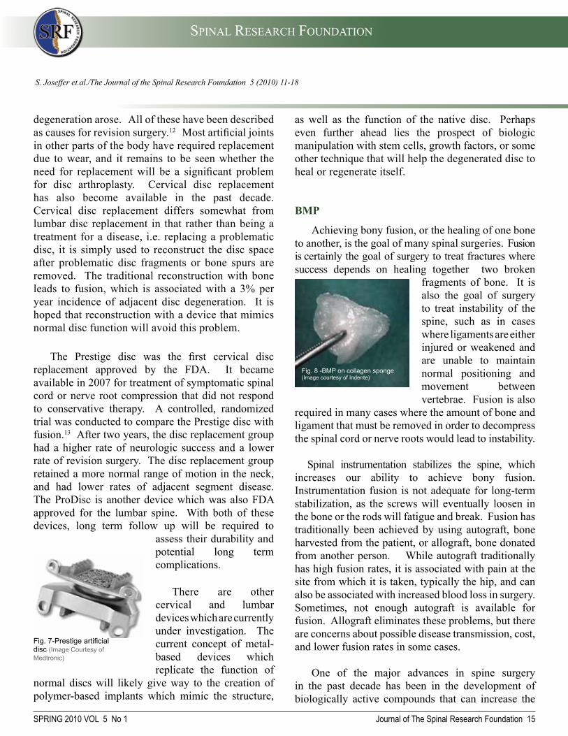
BMP
Achieving bony fusion, or the healing of one bone to another, is the goal of many spinal surgeries. Fusion is certainly the goal of surgery to treat fractures where success depends on healing together two broken
fragments of bone. It is also the goal of surgery to treat instability of the spine, such as in cases where ligaments are either injured or weakened and are unable to maintain normal positioning and movement between vertebrae. Fusion is also
required in many cases where the amount of bone and ligament that must be removed in order to decompress the spinal cord or nerve roots would lead to instability.
Spinal instrumentation stabilizes the spine, which
increases our ability to achieve bony fusion. Instrumentation fusion is not adequate for long-term stabilization, as the screws will eventually loosen in the bone or the rods will fatigue and break. Fusion has traditionally been achieved by using autograft, bone harvested from the patient, or allograft, bone donated from another person. While autograft traditionally has high fusion rates, it is associated with pain at the site from which it is taken, typically the hip, and can also be associated with increased blood loss in surgery. Sometimes, not enough autograft is available for fusion. Allograft eliminates these problems, but there are concerns about possible disease transmission, cost, and lower fusion rates in some cases.
One of the major advances in spine surgery
in the past decade has been in the development of biologically active compounds that can increase the
Fig. 7-Prestige artificial disc (Image Courtesy of Medtronic)
Fig. 8 -BMP on collagen sponge (Image courtesy of Indente)
S. Joseffer et.al./The Journal of the Spinal Research Foundation 5 (2010) 11-18
The Evolution of Spinal Health Care
SPRING 2010 VOL 5 No 1
SPRING 2010
Journal of The Spinal Research Foundation 16
likelihood that a surgery will result in successful bony fusion. Recombinant human bone morphogenic proteins (rhBMP-2) are laboratory produced versions of proteins that the human body uses to promote normal bone growth. While work in understanding these proteins began in the 1960’s, the synthetically produced versions have recently become available for use in spine surgery. These substances can promote the bone growth that is necessary to achieve spinal fusion, resulting in higher fusion rates and avoiding some of the disadvantages associated with autograft or allograft.
Two of these compounds are currently available
and FDA approved, rhBMP-2 and rhBMP-7. rhBMP-2 has been FDA approved for use in conjunction with a threaded cage device for anterior lumbar interbody fusion (ALIF), and studies of its use in ALIF surgery have indicated fusion rates that are comparable to or better than surgery performed with autograft.14 There are numerous reports of the use of rh-BMP2 in other types of spine surgery, and it has generally been associated with high fusion rates. There have been reports of some serious complications when it has been used off label, including problems with swelling and formation of bone in undesirable locations. There are ongoing studies which will help to clarify the best uses for BMPs.
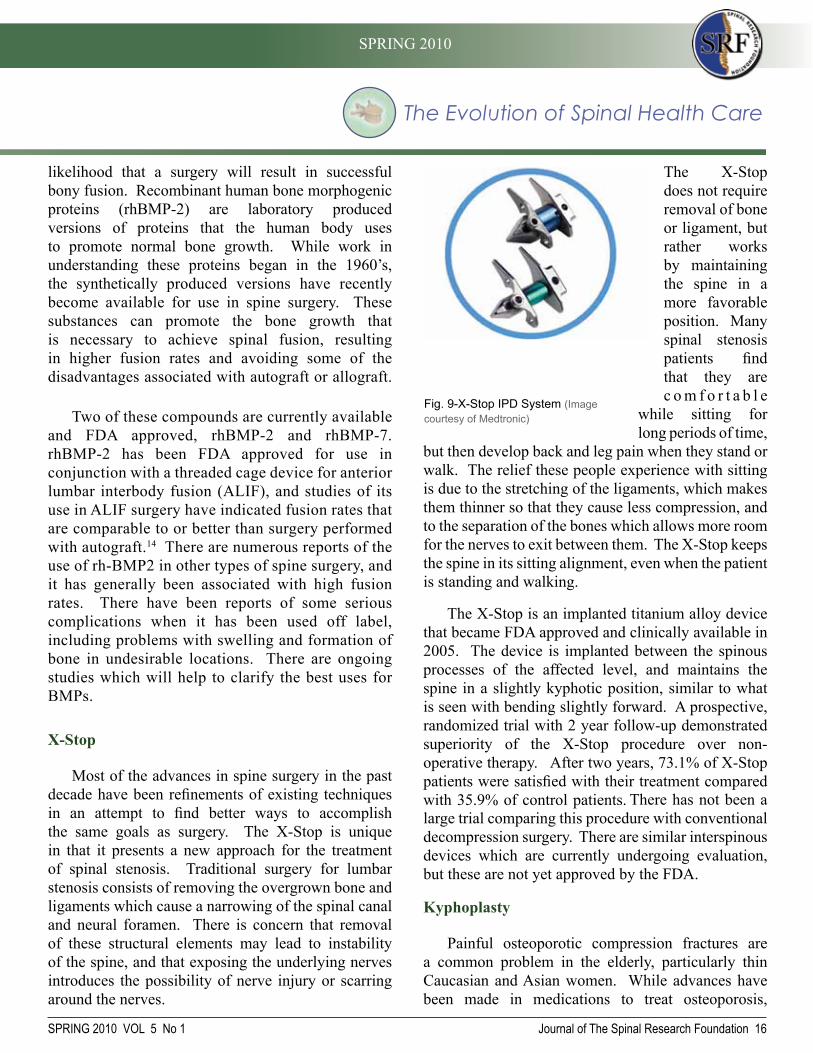
X-Stop Most of the advances in spine surgery in the past
decade have been refinements of existing techniques in an attempt to find better ways to accomplish the same goals as surgery. The X-Stop is unique in that it presents a new approach for the treatment of spinal stenosis. Traditional surgery for lumbar stenosis consists of removing the overgrown bone and ligaments which cause a narrowing of the spinal canal and neural foramen. There is concern that removal of these structural elements may lead to instability of the spine, and that exposing the underlying nerves introduces the possibility of nerve injury or scarring around the nerves.
The X-Stop does not require removal of bone or ligament, but rather works by maintaining the spine in a more favorable position. Many spinal stenosis patients find that they are c o m f o r t a b l e
while sitting for long periods of time,
but then develop back and leg pain when they stand or walk. The relief these people experience with sitting is due to the stretching of the ligaments, which makes them thinner so that they cause less compression, and to the separation of the bones which allows more room for the nerves to exit between them. The X-Stop keeps the spine in its sitting alignment, even when the patient is standing and walking.
The X-Stop is an implanted titanium alloy device that became FDA approved and clinically available in 2005. The device is implanted between the spinous processes of the affected level, and maintains the spine in a slightly kyphotic position, similar to what is seen with bending slightly forward. A prospective, randomized trial with 2 year follow-up demonstrated superiority of the X-Stop procedure over non-operative therapy. After two years, 73.1% of X-Stop patients were satisfied with their treatment compared with 35.9% of control patients. There has not been a large trial comparing this procedure with conventional decompression surgery. There are similar interspinous devices which are currently undergoing evaluation, but these are not yet approved by the FDA.
Kyphoplasty
Painful osteoporotic compression fractures are a common problem in the elderly, particularly thin Caucasian and Asian women. While advances have been made in medications to treat osteoporosis,
Fig. 9-X-Stop IPD System (Image courtesy of Medtronic)
SPRING 2010 VOL 5 No 1
SPINAL RESEARCH FOUNDATION
Journal of The Spinal Research Foundation 17
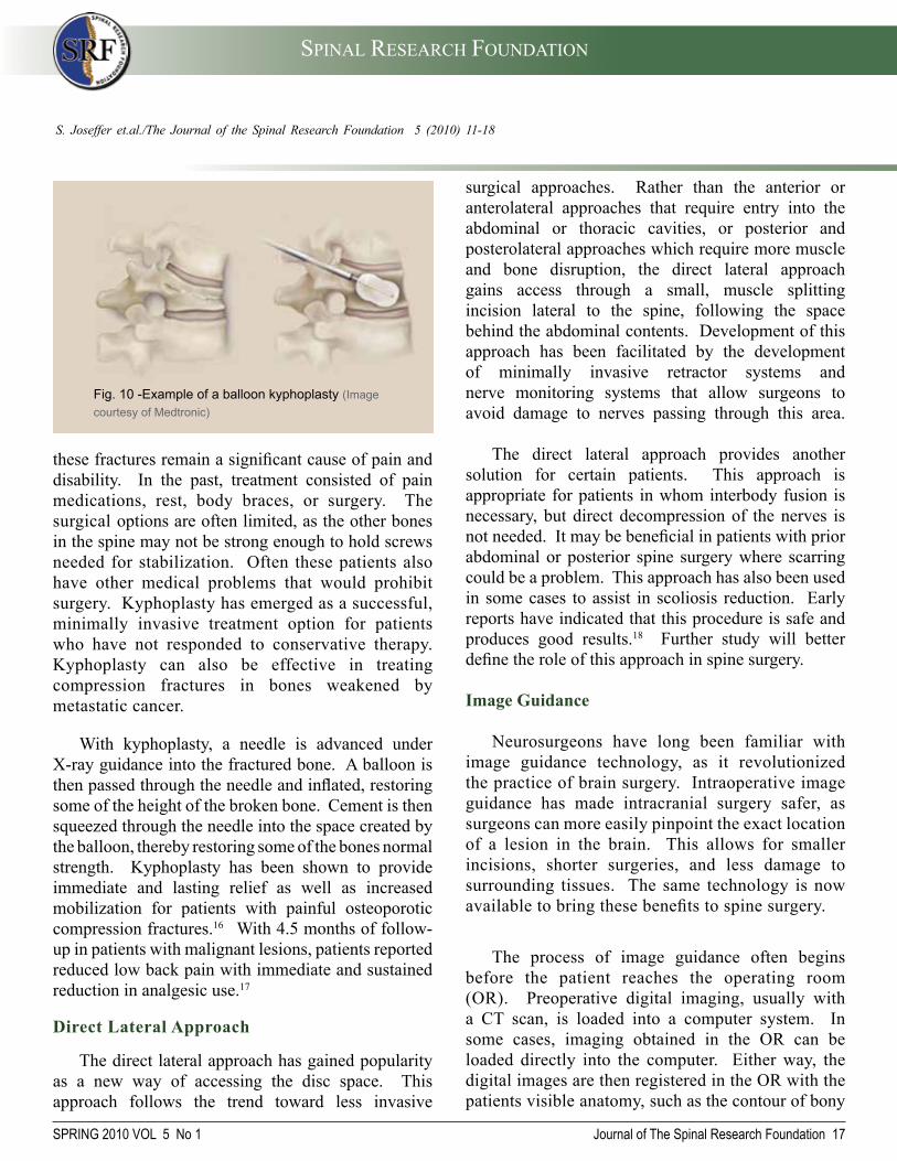
these fractures remain a significant cause of pain and disability. In the past, treatment consisted of pain medications, rest, body braces, or surgery. The surgical options are often limited, as the other bones in the spine may not be strong enough to hold screws needed for stabilization. Often these patients also have other medical problems that would prohibit surgery. Kyphoplasty has emerged as a successful, minimally invasive treatment option for patients who have not responded to conservative therapy. Kyphoplasty can also be effective in treating compression fractures in bones weakened by metastatic cancer.
With kyphoplasty, a needle is advanced under X-ray guidance into the fractured bone. A balloon is then passed through the needle and inflated, restoring some of the height of the broken bone. Cement is then squeezed through the needle into the space created by the balloon, thereby restoring some of the bones normal strength. Kyphoplasty has been shown to provide immediate and lasting relief as well as increased mobilization for patients with painful osteoporotic compression fractures.16 With 4.5 months of follow-up in patients with malignant lesions, patients reported reduced low back pain with immediate and sustained reduction in analgesic use.17
Direct Lateral Approach The direct lateral approach has gained popularity
as a new way of accessing the disc space. This approach follows the trend toward less invasive
surgical approaches. Rather than the anterior or anterolateral approaches that require entry into the abdominal or thoracic cavities, or posterior and posterolateral approaches which require more muscle and bone disruption, the direct lateral approach gains access through a small, muscle splitting incision lateral to the spine, following the space behind the abdominal contents. Development of this approach has been facilitated by the development of minimally invasive retractor systems and nerve monitoring systems that allow surgeons to avoid damage to nerves passing through this area.
The direct lateral approach provides another
solution for certain patients. This approach is appropriate for patients in whom interbody fusion is necessary, but direct decompression of the nerves is not needed. It may be beneficial in patients with prior abdominal or posterior spine surgery where scarring could be a problem. This approach has also been used in some cases to assist in scoliosis reduction. Early reports have indicated that this procedure is safe and produces good results.18 Further study will better define the role of this approach in spine surgery.
Image Guidance Neurosurgeons have long been familiar with
image guidance technology, as it revolutionized the practice of brain surgery. Intraoperative image guidance has made intracranial surgery safer, as surgeons can more easily pinpoint the exact location of a lesion in the brain. This allows for smaller incisions, shorter surgeries, and less damage to surrounding tissues. The same technology is now available to bring these benefits to spine surgery.
The process of image guidance often begins before the patient reaches the operating room (OR). Preoperative digital imaging, usually with a CT scan, is loaded into a computer system. In some cases, imaging obtained in the OR can be loaded directly into the computer. Either way, the digital images are then registered in the OR with the patients visible anatomy, such as the contour of bony
S. Joseffer et.al./The Journal of the Spinal Research Foundation 5 (2010) 11-18
Fig. 10 -Example of a balloon kyphoplasty (Image courtesy of Medtronic)

The Evolution of Spinal Health Care
SPRING 2010 VOL 5 No 1
SPRING 2010
Journal of The Spinal Research Foundation 18
8. Perez-Cruet MJ, Kim BS, Sandhu F, Samartzis D, Fessler RG. Thoracic microendoscopic discectomy. J Neurosurg Spine. 2004;100(1):58-63.
9. Geisler, FH. Lumbar disc replacement: the artificial disc, in Perez-Cruet MJ, ed. An anatomic approach to minimally invasive spine surgery. St Louis, Quality Medical Publishing, 2006.
10. Blumenthal S, McAfee PC, Guyer RD, et al. A prospective, randomized, multicenter Food and Drug Administration investigational device exemptions study of lumbar total disc replacement with the CHARITE artificial disc versus lumbar fusion: part I: evaluation of clinical outcomes. Spine. 2005; 30(14):1565-75.
11. Guyer RD, McAfee PC, Banco RJ, et al. Prospective, randomized, multicenter Food and Drug Administration investigational device exemption study of lumbar total disc replacement with the CHARITE artificial disc versus lumbar fusion: five-year follow-up. Spine J. 2009; 9(5):374-86.
12. Punt IM, Visser VM, van Rhijn LW, et al, Complications and reoperations of the SB Charité lumbar disc prosthesis. Eur Spine J. 2008; 17(1):36-43.
13. Mummaneni PV, Burkus JK, Haid RW, Traynelis VC, Zdeblick TAJ. Clinical and radiographic analysis of cervical disc arthroplasty compared with allograft fusion: a randomized controlled clinical trial. Neurosurg Spine. 2007; 6(3):198-209.
14. Burkus JK, Gornet MF, Dickman CA, Zdeblick TA. Anterior lumbar interbody fusion using rhBMP-2 with tapered interbody cages. J Spinal Disord Tech. 2002; 15(5):337-49.
15. Zucherman JF, Hsu KY, Hartjen CA, et al, A multicenter, prospective, randomized trial evaluating the X STOP interspinous process decompression system for the treatment of neurogenic intermittent claudication: two-year follow-up results. Spine. 2005; 30(12):1351-8.
16. Lieberman IJ, Dudeney S, Reinhardt M-K, et al. Initial outcome and efficacy of kyphoplasty in the treatment of painful osteoporotic compression fractures. Spine. 2001; 26:1631-1638.
17. Fourney DR, Schomer DF, Nader R, et al. Percutaneous vertebroplasty and kyphoplasty in vertebral height restoration, chronic pain and activity levels. J Neurosurg. 2003; 98: 36-42.
18. Ozgur BM, Aryan HE, Pimenta L, Taylor WR.Extreme Lateral Interbody Fusion (XLIF): a novel surgical technique for anterior lumbar interbody fusion. Spine J. 2006; 6(4):435-43.
19. Bloch O, Holly LT, Park J, et al. Effect of frameless stereotaxy on the accuracy of C1-2 transarticular screw placement. J Neurosurg Spine. 2001; 95: 74-79.
prominences. Then, like a GPS system, the computer will be able to determine the location of other, deeper structures that are not visible. This allows surgeons to place instrumentation with greater confidence that the screws will be in the desired location and not damage nearby nerves or blood vessels.
Many spine surgeries can still be conducted safely without image guidance, but there are some instances where it may prove to be particularly helpful. These would include complicated reoperations with distorted anatomy, patients with abnormal anatomy or particularly small bones, and cases where the desired position of the instrumentation is very close to important nerves or blood vessels. A cadaveric study, for example, showed that while complicated anatomy prevented C1-2 transarticular screw placement in 23 % of specimens, that number could be reduced to 6 % with image guidance.19
Conclusion
The past decade brought a variety of advances in spine surgery techniques that will allow surgeons to provide better care for patients with spinal disorders. As new techniques and new technologies continue to emerge, our understanding of the role they play in patient care will depend on continued study and evaluation of patient outcomes.
References
1. Foley KT, Smith MM. Microendoscopic discectomy. Tech Neurosurg. 1997; 2:301-307.
2. Perez-Cruet MJ, Bean JR, Fessler RG, Microendoscopic Lumbar Discectomy in Perez-Cruet MJ, ed. An anatomic approach to minimally invasive spine surgery. St Louis, Quality Medical Publishing, 2006.
3. Khoo LT, Fessler RG. Microendoscopic decompressive laminotomy for the treatment of lumbar stenosis. Neurosurgery. 2002; 51(5 Suppl):S146-54
4. Peng CW, Yue WM, Poh SY, Yeo W, Tan SB. Clinical and radiological outcomes of minimally invasive versus open transforaminal lumbar interbody fusion. Spine. 2009; 34(13):1385-9.
5. Foley KT, Gupta SK, Justis JR, et al. Percutaneous pedicle screw fixation of the lumbar spine. Neurosurg Focus. 2001; 10(4): Article 10.
6. Fessler RG, Khoo LT. Minimally invasive cervical microendoscopic foraminotomy: an initial clinical experience. Neurosurgery. 2002; 55(5 Suppl):S37-45.
7. Joseffer SS, Post N, Cooper PR, Frempong-Boadu AK. Minimally invasive atlantoaxial fixation with a polyaxial screw-rod construct: technical case report. Neurosurgery. 2006; 58(4 Suppl 2):ONS-E375
Seth S. Joseffer, MD
Neurosurgeon Dr. Joseffer, has made significant contributions to the medical community as a member of the Congress of Neurological Surgeons and the American Association of Neurological
Surgeons. In addition, he has given numerous presentations in the United States and Canada. As an active contributor to medical research, he has published several articles in the Journal of Neurology, Neurosurgery & Psychiatry, the American Journal of Neuroradiology, and Neurosurgery: Official Journal of the Congress of Neurological Surgeons.

SPRING 2010 VOL 5 No 1
SPINAL RESEARCH FOUNDATION
Journal of The Spinal Research Foundation 19
Evolution in the Treatment of Spinal Deformity and Spinal Instrumentation Christopher R. Good, MD
Treatment of spinal conditions dates back to ancient times. There has been a long history of treatment of scoliosis and other spinal deformities using both non-operative and operative techniques. One of the most common techniques presently used by spine surgeons to correct spinal problems is spine fusion. The purpose of a spinal fusion is to create a rigid union between two separate segments of the spine to correct malalignment or instability. Many different types of spinal instrumentation have been developed to help facilitate spine fusion, including devices such as rods, plates, hooks, wires and screws. Treatment of spinal deformity has improved due to the development of advanced surgical techniques and improved spinal instrumentation. These advances allow surgeons to help their patients maximize their quality of life while striving to minimize the potential for complications. Advances in the past few decades have improved correction of spinal deformity, decreased the morbidity of surgical procedures, and allowed for earlier return to activity after surgery. Current research focuses on improving and developing motion preserving surgical techniques and less invasive surgical options.
Key Words: History, Spinal Deformity, Scoliosis, Instrumentation
History of Spinal Deformity
The treatment of spinal conditions dates back to ancient times. Fractures of the bones of the neck
causing paralysis have been documented as early as 1550 B.C. in ancient Egyptian writings. At that time, patients were treated by priests who applied bandages and helped patients to rest. Hippocrates (460-337 B.C.) was an ancient Greek physician who is considered to be the father of western medicine. Hippocrates worked to develop methods for treating fractures of the spine by positioning patients in such a way as to correct a deformity that developed after a spinal fracture. Using his techniques, therapists used wooden constructs to place forces against the patient's spine in order to correct or reposition fractures1 (Figure 1A). A number of physicians built off of Hippocrates early work to develop more advanced techniques for treating fractures with a variety of traction or spinal manipulation devices. These included techniques such as hanging patients on a ladder or placing patients on a table with ropes attached around the torso and ankles (Figure 1B).
Scoliosis is derived from a Greek word meaning a lateral curvature of the spine. The word scoliosis was coined by Galen of Pergamon (129 to 200 A.D.).
Scoliosis is an abnormal curvature of the spine that affects 1% to 3% of the general population, or approximately seven million people in the United States. Bracing is used to prevent and/or limit progression of scoliosis curves during periods of patient growth for moderate curves (generally between 25oto 45º). Surgical treatment is considered
Fig. 1A -(Left) The Hippocratic board was used to place corrective forces on the spine using bands and straps to correct spinal deformities. Fig.1B -(Right) The Hippocratic ladder was used for the correction of spinal deformities with the head pointing downwards. (From the illustrated comments of Apollonius of Kitium on the Hippocratic Treatise On Articulations. Bibliotheca Medica Laurenziana, Florence)
Fig. 1B
Fig. 1A
The Evolution of Spinal Health Care
SPRING 2010 VOL 5 No 1
SPRING 2010
Journal of The Spinal Research Foundation 20
for patients with curves greater than 40 to 50º. There has been a documented risk for continued curve progression from 0.5 to 2º per year for curves greater than 50º in adults.
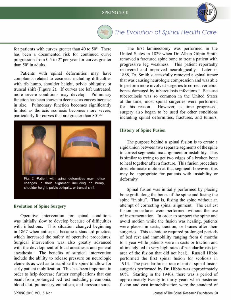
Patients with spinal deformities may have complaints related to cosmesis including difficulties with rib hump, shoulder height, pelvic obliquity, or truncal shift (Figure 2). If curves are left untreated, more severe conditions may develop. Pulmonary function has been shown to decrease as curves increase in size. Pulmonary function becomes significantly limited as thoracic scoliosis becomes more severe, particularly for curves that are greater than 80º.2,3
Evolution of Spine Surgery
Operative intervention for spinal conditions was initially slow to develop because of difficulties with infections. This situation changed beginning in 1867 when antisepsis became a standard practice, which increased the safety of operative procedures. Surgical intervention was also greatly advanced with the development of local anesthesia and general anesthesia.1 The benefits of surgical intervention include the ability to release pressure on neurologic elements as well as to stabilize the spine to allow for early patient mobilization. This has been important in order to help decrease further complications that can result from prolonged bed rest including pneumonia, blood clot, pulmonary embolism, and pressure sores.
The first laminectomy was performed in the United States in 1829 when Dr. Alban Gilpin Smith removed a fractured spine bone to treat a patient with progressive leg weakness. This patient reportedly recovered and improved neurologically. Later in 1888, Dr. Smith successfully removed a spinal tumor that was causing neurologic compression and was able to perform more involved surgeries to correct vertebral bones damaged by tuberculosis infections.4 Because tuberculosis was so common in the United States at the time, most spinal surgeries were performed for this reason. However, as time progressed, surgery also began to be used for other conditions including spinal deformities, fractures, and tumors.
History of Spine Fusion
The purpose behind a spinal fusion is to create a rigid union between two separate segments of the spine to correct segmental malalignment or instability. This is similar to trying to get two edges of a broken bone to heal together after a fracture. This fusion procedure does eliminate motion at that segment; however, this may be appropriate for patients with instability or deformity.
Spinal fusion was initially performed by placing bone graft along the bones of the spine and fusing the spine “in situ”. That is, fusing the spine without an attempt of correcting spinal alignment. The earliest fusion procedures were performed without the use of instrumentation. In order to support the spine and avoid motion while the fusion was healing, patients were placed in casts, traction, or braces after their surgeries. This technique required prolonged periods of bed rest and immobility ranging from 6 months to 1 year while patients were in casts or traction and ultimately led to very high rates of pseudarthrosis (an area of the fusion that did not heal). Russell Hibbs performed the first spinal fusion for scoliosis in 1914. The pseudarthrosis rate of initial spinal fusion surgeries performed by Dr. Hibbs was approximately 60%. Starting in the 1940s, there was a period of approximately twenty to thirty years when posterior fusion and cast immobilization were the standard of
Fig. 2 -Patient with spinal deformities may notice changes in their alignment including rib hump, shoulder height, pelvic obliquity, or truncal shift.

SPRING 2010 VOL 5 No 1
SPINAL RESEARCH FOUNDATION
Journal of The Spinal Research Foundation 21
care. As fusion techniques improved, pseudarthrosis rates were typically around 45%.
Spinal fusion was also used during this time to treat fractures of the spine. Spine trauma can result in instability due to a fracture of the bone or an injury to the ligaments that support the bones of the spine. Many fractures can be treated conservatively with bracing or casting, however, with specific instability patterns surgical intervention is recommended.
Spinal Instrumentation
Surgery for scoliosis was the first widespread application of spinal instrumentation. Over the years, many different types of techniques and instrumentation have been developed to help correct spinal curvatures and facilitate fusion. Specific instrumentation types include: metal plates, rods, hooks, and wires and screws that join together to support the spine during the time that it is fusing. The use of metallic implants to stabilize segments allows for faster and more effective fusion. The early instrumentation systems functioned as an “internal splint” which held the spine in position until the surgically applied bone graft developed into a fusion mass.
Spinal instrumentation achieves many goals. For patients with spinal deformities, implants should maintain correction of the deformity after surgery until spinal fusion can occur. Solid immobilization with spinal instrumentation enhances the rates of bony fusion. For patients with instability or fractures, spinal instrumentation allows for stabilization of this instability and facilitates early mobilization of patients to help avoid potential side effects of prolonged bed rest. The number and types of spinal implants available has greatly increased in recent years. To best understand the use of instrumentation, one must fully understand the spinal disorder that is to be treated and the specific goals of treatment.
The evolution of modern spinal instrumentation systems began in the late 1950s with the development of the Harrington hook and rod system. At the
time, this was a major medical breakthrough which allowed for enhanced stability and curve correction for patients with spinal deformity. The Harrington rod and hook system consisted of a rod with a hook at either end. These hooks attached to the spine at the top and the bottom of the curvature. By distracting across the rod, surgeons were able to partially reduce spinal deformities (Figure 4). This technique was most commonly used to treat paralytic scoliosis resulting from poliomyelitis which was very common at that time. This system was limited in that it only attached to the spine in two locations. It was also limited in the fact that the rod was straight and this did not allow surgeons to accurately re-create a normal spinal alignment, particularly in the sagittal plane (viewed from the side).
In 1973, Dr. Harrington published an eleven-year follow-up of 578 patients who were treated with spinal instrumentation. The average correction of the scoliosis curve in the frontal plane was 54%. He published a 4% rate of pseudarthrosis or non-union which was a significant improvement when compared with previous fusions performed without instrumentation. These techniques were not without significant complications which included fracture or failure of the instrumentation as well
Fig. 4 -The Harrington instrumentation system consisted of a rod with a hook at either end. These hooks attached to the spine at the top and the bottom of the curvature. By distracting across the rod, surgeons were able to partially reduce spinal deformities (Case courtesy of Keith H. Bridwell, MD)
C. Good et.al./The Journal of the Spinal Research Foundation 5 (2010) 19-25

The Evolution of Spinal Health Care
SPRING 2010 VOL 5 No 1
SPRING 2010
Journal of The Spinal Research Foundation 22
as degeneration or instability affecting portions of the spine above or below the instrumentation.5,6
Harrington’s distraction instrumentation did address the frontal curve of the scoliosis pattern; however, the sagittal contour of the patient was often negatively influenced, particularly in the lumbar spine. The distraction forces of the Harrington instrumentation tended to decrease the amount of lumbar lordosis (swayback) which led some patients to develop a “flat-back syndrome”. These patients developed low back pain and a loss of normal standing alignment when viewed from the side.
Segmental instrumentation was first introduced by Edwardo Luque of Mexico in 1973. He used a two-rod system in the back of the spine which was attached to the spinal bones with wires at each level of the spine. These rods were contoured in multiple planes which did allow for surgeons to fuse the spine in a more normal alignment (Figure 5). By attaching the implants to the spine at multiple levels, the force at each individual level is reduced and the overall potential for spinal correction was increased. By using these powerful techniques, Dr. Luque was able to treat many of his patients without the use of long-term casting or bracing after surgery.
Dr. Luque reported on a series of 322 patients treated with his techniques in 1982. Failure of the instrumentation occurred in 27 of these patients and 5% of the patients developed a pseudarthrosis. This was a particularly low rate at that time, especially considering that the majority of Dr. Luque’s
patients were treated for neuromuscular conditions including poliomyelitis and cerebral palsy, and were therefore at a high risk for postoperative problems.7
Segmental fixation with wires did improve correction of the frontal plane as well as allow for the maintenance of a physiologic sagittal contour; however, spinal deformities occur in three dimensions and none of these early techniques allowed for rotational correction during surgery. In the 1980s a new treatment system was introduced using the Cotrell-Dubousset instrumentation system (CD). The CD instrumentation system allowed for multiple fixation points along the spine using a variety of hook and rod combinations. This instrumentation system allowed for correction of the spine in the coronal, sagittal, and axial planes (rotation) during spinal reconstructions. This was a major technical advancement (Figure 6).
In Dr. Cotrell’s original report of 250 patients,
Fig. 6- Anatomical planes. The coronal plane is shown here as the patient is viewed from the front. This is the view that shows the “S shape curve” of scoliosis. The sagittal plane is shown here as if the patient is viewed from the side. The axial plane is a cross-sectional view, that describes the rotation of the spine bones in scoliosis. (Image courtesy of Medtronic)
Sag
git
al P
lan
e
C o r o n a l P l a n e
Axial Plane

SPRING 2010 VOL 5 No 1
SPINAL RESEARCH FOUNDATION
Journal of The Spinal Research Foundation 23
no patient was treated with postoperative bracing or casting. The average correction of scoliosis was 66% and sagittal contour was also improved. Less than 5% loss of the correction was noted over long-term follow-up. No failures of the instrumentation were noted.8
Another advancement in spinal instrumentation was the development of crosslinking devices. Crosslinks are simple transverse implants that connect between rods that are placed on each side of the spine (Figure 7). These devices provide additional stability to spinal instrumentation. The TSRH implant system was the first to utilize cross-links and was developed at the Texas Scottish Right Hospital in 1983. This system also made extension of a previously implanted system to another system possible.9
Recent Surgical Advances
Surgical techniques have been developed to correct spinal deformities from the front (anterior) as well as the back (posterior) of the spine. The early benefit of surgeries performed through the front of the spine was that they allowed direct access to the bones and discs in the front of the spine and did offer the benefit of fewer total levels of the spine that needed to be fused in cases of scoliosis. As techniques improved for surgery on the front of the spine, implants were also developed to help fill bone defects resulting from infections or tumors. A variety of titanium cages, bone grafts, and other devices have been developed for this purpose.
Advancements in spinal surgery technology continued on into the 1990s. These new systems have developed techniques that allow for the spine to be fixed segmentally, meaning that the attachment of metal implants to the rod is achieved at every level being addressed. Stronger segmental fixation of the spine has allowed for better correction of spinal deformities, increased rates of bone healing or fusion after surgery, and decreased rates of instrumentation failure. Most recently, there has been a trend towards increased use of pedicle screw instrumentation to allow for spinal fixation (Figure 8). Pedicle screws are placed into a specific anatomic area of the spine from a posterior approach (Figure 9). Surgeons began using pedicle screws in 1988. These were initially used in the lower lumbar spine where they were easier to place because of the size of the bones with the continued use of
Fig. 7 -Crosslinks are transverse implants that connect between rods that are placed on each side of the spine to provide additional stability to spinal instrumentation. (Image courtesy of Medtronic)
Fig. 8 -Scoliosis correction using segmental pedicle screw fixation at each level of the spine
C. Good et.al./The Journal of the Spinal Research Foundation 5 (2010) 19-25
(Image courtesy of Medtronic)
Fig. 9-Pedicle screws are placed into the vertebral bone on either side of the spinal canal.
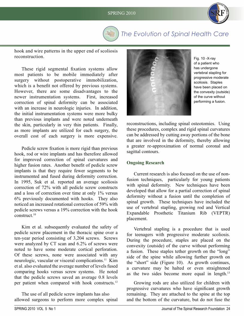
The Evolution of Spinal Health Care
SPRING 2010 VOL 5 No 1
SPRING 2010
Journal of The Spinal Research Foundation 24
hook and wire patterns in the upper end of scoliosis reconstruction.
These rigid segmental fixation systems allow most patients to be mobile immediately after surgery without postoperative immobilization, which is a benefit not offered by previous systems. However, there are some disadvantages to the newer instrumentation systems. First, increased correction of spinal deformity can be associated with an increase in neurologic injuries. In addition, the initial instrumentation systems were more bulky than previous implants and were noted underneath the skin, particularly in very thin patients. Finally, as more implants are utilized for each surgery, the overall cost of each surgery is more expensive.
Pedicle screw fixation is more rigid than previous hook, rod or wire implants and has therefore allowed for improved correction of spinal curvatures and higher fusion rates. Another benefit of pedicle screw implants is that they require fewer segments to be instrumented and fused during deformity correction. In 1995, Suk et al. reported an average scoliosis correction of 72% with all pedicle screw constructs and a loss of correction over time at only 1% versus 6% previously documented with hooks. They also noticed an increased rotational correction of 59% with pedicle screws versus a 19% correction with the hook construct.10
Kim et al. subsequently evaluated the safety of pedicle screw placement in the thoracic spine over a ten-year period consisting of 3,204 screws. Screws were analyzed by CT scan and 6.2% of screws were noted to have some moderate cortical perforation. Of these screws, none were associated with any neurologic, vascular or visceral complications.11 Kim et al. also evaluated the average number of levels fused comparing hooks versus screw systems. He noted that the pedicle screws saved an average 0.8 levels per patient when compared with hook constructs.12
The use of all pedicle screw implants has also allowed surgeons to perform more complex spinal
reconstructions, including spinal osteotomies. Using these procedures, complex and rigid spinal curvatures can be addressed by cutting away portions of the bone that are involved in the deformity, thereby allowing a greater re-approximation of normal coronal and sagittal contours.
Ongoing Research
Current research is also focused on the use of non-fusion techniques, particularly for young patients with spinal deformity. New techniques have been developed that allow for a partial correction of spinal deformity without a fusion until the completion of spinal growth. These techniques have included the use of vertebral stapling, growing rod and Vertical Expandable Prosthetic Titanium Rib (VEPTR) placement.
Vertebral stapling is a procedure that is used for teenagers with progressive moderate scoliosis. During the procedure, staples are placed on the convexity (outside) of the curve without performing a fusion. These staples tether growth on the “long” side of the spine while allowing further growth on the “short” side (Figure 10). As growth continues, a curvature may be halted or even straightened as the two sides become more equal in length.13
Growing rods are also utilized for children with progressive curvatures who have significant growth remaining. They are attached to the spine at the top and the bottom of the curvature, but do not fuse the
Fig. 10 -X-ray of a patient who has undergone vertebral stapling for progressive moderate scoliosis. Staples have been placed on the convexity (outside) of the curve without performing a fusion.

SPRING 2010 VOL 5 No 1
SPINAL RESEARCH FOUNDATION
Journal of The Spinal Research Foundation 25
References
1. Knoeller SM, Seifried C. History of Spinal Surgery. Spine. 2000; 25:2838-2843.
2. Good CR. The Genetic Basis of Idiopathic Scoliosis. Journal of the Spinal Research Foundation. 2009; 4: 13-17.
3. Nilsonne U, Lundgren KD. Long-term prognosis in idiopathic scoliosis. Acta Or-thop Scand. 1968;39:456–65.
4. Keller T, Holland MC. Some notable American spine surgeons of the 19th century. Spine. 1997;22:1413-7.
5. Harrington PR. The history and development of Harrington instrumentation: the classic. Clin Orthop. 1977;227:3-6
6. Harrington PR. Treatment of scoliosis: correction and internal fixation by spine instrumentation. J Bone Surg [AM]. 1962; 44:591-610.
7. Luque ER. Introduction to symposium: the anatomic basis and development of segmental spinal instrumentation. Spine. 1982;7(3): 256-9.
8. Cotrel Y, Dubousset J, Guillaumat M. New and universal instrumentation in spinal surgery. Clin Orthop. 1988; 227:10-23.
9. Hu SS, Pashman RS. Spinal instrumentation evolution and state of the art. Investi-gative Radiology. 1992;27:632-647.
10. Suk, Se-ll; Lee, CK, Won-Joon K, et al. Segmental Pedicle Screw Fixation in the Treatment of Thoracic Idiopathic Scoliosis. Spine. 1995; 20(12):1399-1405.
11. Kim YJ, Lenke LG, Bridwell KH, et al. Free handle pedicle screw placement in the thoracic spine: Is it safe? Spine 2004; 29(3):333-342.
12. Kim YJ, Lenke LG, Cho SK, et al. Comparative analysis of pedicle screw ver-sus hook instrumentation in posterior spinal fusion of adolescent idiopathic scoliosis. Spine 2004; 29(18):2040-2048.
13. Betz RR, Ranade A, Samdami AF, et al. Vertebral Body Stapling: A fusionless treatement option for a growing child with moderate idiopathic scoliosis. Spine 2010; 35(2):169-176.
spine in the motion segments in the middle, this allows for continued spinal growth at the non-fused segments (Figure 11). The rods are periodically lengthened as the child grows which allows for growth of the spine while slowing the progression of a curvature.14
Another technique that has been used in children with progressive curvatures is the Vertical Expandable Prosthetic Titanium Rib or “VEPTR” approach. The VEPTR device works to expand and support a deformed chest wall cavity by using telescoping titanium rods. These rods hook to the ribs or pelvis and can help to separate and support the chest. This device may slow the progression of a spinal curvature and avoid a spinal fusion in young children until they have neared the end of their growth.15
Conclusion
Treatment of spinal deformity has improved due to the development of advanced surgical techniques and improved spinal instrumentation. These advances allow surgeons to help their patients maximize their quality of life while striving to minimize the potential for complications. Advances in the past few decades have improved correction of spinal deformity, decreased the morbidity of surgical procedures, and allowed for earlier return to activity after surgery. Current research focuses on improving and developing motion preserving surgical techniques and less invasive surgical options.
Christopher R. Good, MD
Dr. Good has extensive training and experience in the treatment of complex spinal disorders with special expertise in non-operative and operative treatment of adult and pediatric spinal deformities including scoliosis, kyphosis, flatback, and spondylolisthesis. Dr. Good has
co-authored numerous articles and has been invited to lecture nationally and internationally at the Scoliosis Research Society, the International Meeting on Advanced Spinal Techniques, the American Academy of Orthopaedic Surgeons, and the North American Spine Society.
Fig. 11-Growing rods are attached to the spine at the top and the bottom of the curvature, but do not fuse the spine in the motion segments in the middle. These rods are periodically lengthened, which allows for continued spinal growth at the non-fused segments. (Case courtesy of Lawrence G Lenke, MD)
C. Good et.al./The Journal of the Spinal Research Foundation 5 (2010) 19-25

SPRING 2010 VOL 5 No 1
SPRING 2010
Journal of The Spinal Research Foundation 26
Minimally Invasive Spine Surgery: An Evolution in Progress Paul J. Slosar, Jr., MD.
“Never discourage anyone... who continually makes progress, no matter how slow.”
-Plato Greek author & philosopher in Athens (427 BC - 347 BC)
Historical Perspective
It is believed that the first laminectomy in the United States was performed by A. G. Smith in 1892 on
a patient with a herniated lumbar disc. 2, 3 Fusion surgery was first performed by Albee and Hibbs in 1911 and Robinson pioneered cervical fusions in 1955.4, 5 Many consider chymopapain injections (chemonucleolysis) for disc herniations (circa 1969) to be the advent of minimally invasive spine surgery.6 Chemonucleolysis was a needle-injection technique where after injection into the disc, the enzyme action of the chymopapain would dissolve the herniated disc. The procedure was eventually banned by the FDA after several concerning complications were published.7,8,9 Percutaneous disc removal was performed in the 1970s, with the first reports of open microdiscectomy surgeries becoming more widely adopted several years later by Williams.10 This progression toward smaller incision spine surgery came about as pioneering surgeons and technology companies collaborated to bring the needed components into the operating room. Intra-operative fluoroscopy, specialized retractor systems,
surgical microscopes, and surgeon champions were all essential to these early successes.11,12
In the early 1990’s surgeons developed endoscopic or tubular retractor access options for the spine.13 By utilizing sequentially larger tubes a surgeon can expand his working channel without cutting muscle fibers. The microendoscopic discectomy (MED™) was pioneered by Foley and Smith in 1997 but
the technical delivery of spinal surgery through small tubes proved to be very challenging for all but the most advanced surgeons (Figure 1).14, 15, 16, 17 Soon after, the METRx System™ (Medtronic, Inc.) was introduced and it allowed for more expansive, and presumably safer, access to the spine. Up to this point, most of the spine surgeries performed through the tubular retractors were simple spinal decompressions (laminotomy or foraminotomy cases). Surgeons, however, saw significant potential for performing more complex surgeries, such as fusions, by utilizing less invasive procedures.18,19 The learning curve for tubular access surgery remained high with many surgeons unable to achieve reliable visualization.
The discipline of spinal surgery has slowly caught up to other surgical specialties in the area of minimally invasive techniques. Patients have benefited from technological advancements which allow surgeons to perform surgery with less invasive approaches .1 The basic tenet of minimally invasive or least invasive spine surgery is to effectively treat patients with the most reliable technique while minimizing tissue trauma. Least invasive spine surgery has become more available over the past 10 years, paralleling the remarkable advancements in surgical visualization technologies coupled with the emergence of the internet as a patient-driven education tool. Patients are seeking smaller incision options and the surgical industry is responding. This article will offer a brief review of the evolution of spinal surgery with a focus on minimally invasive techniques. While most spine surgery is still performed using traditional (open) surgical approaches, smaller incision and less invasive options are utilized more commonly each year. The most important benefactor of this evolution is the patient.
Keywords: Minimally Invasive Surgery, Spine, TLIF, Spinal Implants
Fig. 1-MED™(Microendoscopic) tubular retractor system used to remove a herniated disc. (Image courtesy of Medtronic)
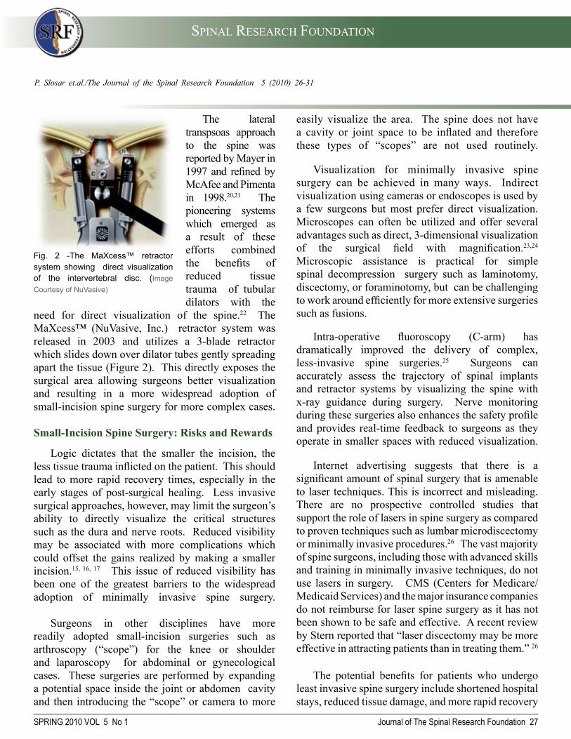
SPRING 2010 VOL 5 No 1
SPINAL RESEARCH FOUNDATION
Journal of The Spinal Research Foundation 27
The lateral transpsoas approach to the spine was reported by Mayer in 1997 and refined by McAfee and Pimenta in 1998.20,21 The pioneering systems which emerged as a result of these efforts combined the benefits of reduced tissue trauma of tubular dilators with the
need for direct visualization of the spine.22 The MaXcess™ (NuVasive, Inc.) retractor system was released in 2003 and utilizes a 3-blade retractor which slides down over dilator tubes gently spreading apart the tissue (Figure 2). This directly exposes the surgical area allowing surgeons better visualization and resulting in a more widespread adoption of small-incision spine surgery for more complex cases.
Small-Incision Spine Surgery: Risks and Rewards
Logic dictates that the smaller the incision, the less tissue trauma inflicted on the patient. This should lead to more rapid recovery times, especially in the early stages of post-surgical healing. Less invasive surgical approaches, however, may limit the surgeon’s ability to directly visualize the critical structures such as the dura and nerve roots. Reduced visibility may be associated with more complications which could offset the gains realized by making a smaller incision.15, 16, 17 This issue of reduced visibility has been one of the greatest barriers to the widespread adoption of minimally invasive spine surgery.
Surgeons in other disciplines have more readily adopted small-incision surgeries such as arthroscopy (“scope”) for the knee or shoulder and laparoscopy for abdominal or gynecological cases. These surgeries are performed by expanding a potential space inside the joint or abdomen cavity and then introducing the “scope” or camera to more
easily visualize the area. The spine does not have a cavity or joint space to be inflated and therefore these types of “scopes” are not used routinely.
Visualization for minimally invasive spine surgery can be achieved in many ways. Indirect visualization using cameras or endoscopes is used by a few surgeons but most prefer direct visualization. Microscopes can often be utilized and offer several advantages such as direct, 3-dimensional visualization of the surgical field with magnification.23,24 Microscopic assistance is practical for simple spinal decompression surgery such as laminotomy, discectomy, or foraminotomy, but can be challenging to work around efficiently for more extensive surgeries such as fusions.
Intra-operative fluoroscopy (C-arm) has dramatically improved the delivery of complex, less-invasive spine surgeries.25 Surgeons can accurately assess the trajectory of spinal implants and retractor systems by visualizing the spine with x-ray guidance during surgery. Nerve monitoring during these surgeries also enhances the safety profile and provides real-time feedback to surgeons as they operate in smaller spaces with reduced visualization.
Internet advertising suggests that there is a significant amount of spinal surgery that is amenable to laser techniques. This is incorrect and misleading. There are no prospective controlled studies that support the role of lasers in spine surgery as compared to proven techniques such as lumbar microdiscectomy or minimally invasive procedures.26 The vast majority of spine surgeons, including those with advanced skills and training in minimally invasive techniques, do not use lasers in surgery. CMS (Centers for Medicare/ Medicaid Services) and the major insurance companies do not reimburse for laser spine surgery as it has not been shown to be safe and effective. A recent review by Stern reported that “laser discectomy may be more effective in attracting patients than in treating them.” 26
The potential benefits for patients who undergo least invasive spine surgery include shortened hospital stays, reduced tissue damage, and more rapid recovery
Fig. 2 -The MaXcess™ retractor system showing direct visualization of the intervertebral disc. (Image Courtesy of NuVasive)
P. Slosar et.al./The Journal of the Spinal Research Foundation 5 (2010) 26-31

The Evolution of Spinal Health Care
SPRING 2010 VOL 5 No 1
SPRING 2010
Journal of The Spinal Research Foundation 28
times. A few reports, however, have noted more complications with certain “minimally invasive” procedures, compared to more traditional “open” surgical approaches, such as increased rates of spinal fluid leaks, dural tears, recurrent disc herniations and prolonged operative times. As the technologies and surgeon training improves over time, the outcomes are likely to continue to improve as well.27
Essential Elements for SuccessRetractor Systems
Surgeons performing more traditional (open) spine surgery use large retractors to hold the skin and muscle open for visualization. Less invasive spine surgeons use smaller retractor systems to access the spine with less tissue trauma. Most of these systems have tubular dilators which are first inserted to
gently separate rather than cut the muscles. As the dilators increase in size the opening for access increases and once the surgeon has reached the appropriate size he selects the retractor to insert over the dilator. The current retractors allow for direct visualization of the spine but do so with a much smaller footprint (Figure 3). Surgeons can then use a microscope, image guidance, or loupe magnification to visualize the spine and perform the needed procedures.
Image Guidance/ Intra-Operative X-Ray (Fluoroscopy)
Least invasive spinal surgery demands precision and accuracy. The smaller incisions reduce tissue trauma but also reduces the ability of the surgeon to
directly visualize the spine. Therefore, most surgeons using minimally invasive techniques must rely on specialized x-ray equipment (image guidance) in surgery.25 Radiographic imaging for these surgeries can be achieved with a C-arm machine, which is a mobile x-ray machine shaped like a large letter ‘C’. This open ring allows the surgeon a 360 degree view of the spine and can be coupled with more advanced imaging software packages. Intra-operative, portable CT scanning, now available in selected centers, can also assist surgeons in performing more complex surgeries with small incisions.28
Bone Graft Substitutes: Osteobiologics
Advances in surgical equipment and technologies have been essential components for the continued success and evolution of minimally invasive spine surgeries. In addition to the retractor systems discussed above, significant improvements in bone-graft alternatives have emerged. Prior to the advent of biological alternatives, patients who underwent spinal fusions had bone graft harvested from their pelvis. At least 30% of patients reported permanent pain and disability related to the bone graft procedure.29
A recent, important development in the evolution of minimally invasive spine surgery has been the commercialization of rhBMP-2 (recombinant human Bone Morphogenetic Protein-2/ InFuse™ Medtronic Minneapolis, MN).29, 30, 31 This protein is soaked on a small sponge and placed inside a titanium fusion cage. The rhBMP-2 stimulates the body to form bone fusion, effectively eliminating the need for bone graft from the iliac crest (pelvis). There are now several other bone graft substitutes which may have the capacity to biologically stimulate bone inside the fusion. Achieving a fusion without making a separate incision to harvest bone graft, is essential to providing patients a successful and minimally invasive option for spinal fusion surgery.
Implants
For spinal fusion surgery to be successful, the surgeon must often implant a fusion cage to achieve bone growth across the disc space and pedicle screws
Fig. 3 -Interbody fusion graft insertion through the MaXcess™ retractor system. (Image courtesy of Nuvasive)

SPRING 2010 VOL 5 No 1
SPINAL RESEARCH FOUNDATION
Journal of The Spinal Research Foundation 29
to stabilize the vertebrae. Originally, most of these implants were intended to be inserted using larger incisions. With the evolution of minimally invasive surgical approaches these implants had to be re-designed to be inserted under much smaller incisions. Common materials used as fusion cages include bone, PEEK (medical grade plastic), carbon fiber, and titanium. Percutaneous (small incision) pedicle screw systems are available to be placed with minimal tissue trauma to the surrounding back muscles.32
These systems, combined with specialized interbody fusion spacers, are utilized by surgeons to perform spinal fusions with minimally invasive techniques.
Surgeon Training
Not all spine surgeons have the advanced training or experience to become proficient in minimally invasive techniques. Operating through smaller incisions is technically more challenging for most surgeons. Therefore, one must first master the essential skills necessary to perform spinal surgery using traditional open approaches and then gradually move toward the minimally invasive or least invasive procedures. A one year fellowship training program in spine surgery is the most common way to gain exposure to these advanced surgical techniques, while continuing medical education training courses are essential for improving surgeon skills.
Case Examples
The following are a few examples of minimally invasive surgeries frequently performed by trained spine surgeons. While there are many other exciting techniques, due to the limited scope of this short review article only a few cases could be described here.
Lumbar Microdiscectomy
Patients with lumbar disc herniations usually complain of radicular symptoms (sciatica) into the leg. Physical examination typically yields findings of neurological irritation (positive nerve root tension signs, dermatomal sensory disturbances) and MRI scanning will demonstrate nerve compression. If a patient fails to improve with non-operative
treatment such as physical therapy or epidural spinal injections then surgical decompression has been shown to be more effective than ongoing non-surgical care.33 The most common surgery to treat a herniated lumbar disc is a microscopically assisted laminotomy with discectomy. Surgeons trained in minimally invasive spine procedures will usually perform the surgery as an outpatient, allowing the patient to go home the same day as surgery.
Lumbar Arthodesis (Fusion)
Fusion surgery is indicated for patients with axial (back >leg) low back pain that has been refractory to greater than 6 months of non-operative treatments. Usually the disc itself has been torn and becomes painful, causing symptoms of severe low back pain. The discs are located in the front (anterior) column of the spine. Therefore, spinal fusion surgery can be performed from either the anterior (front) or posterior (back) approach, depending on the specific situation and surgeon preference. If the surgeon determines that screws are needed to stabilize the spine then he can use a minimally invasive percutaneous system.
Mini-open ALIF (Anterior Lumbar Interbody Fusion)
The most accessible disc level for a small incision anterior spinal fusion is the L5-S1 segment. In many cases a surgeon can do the complete fusion from the
P. Slosar et.al./The Journal of the Spinal Research Foundation 5 (2010) 26-31
LumbarDisc Herniation
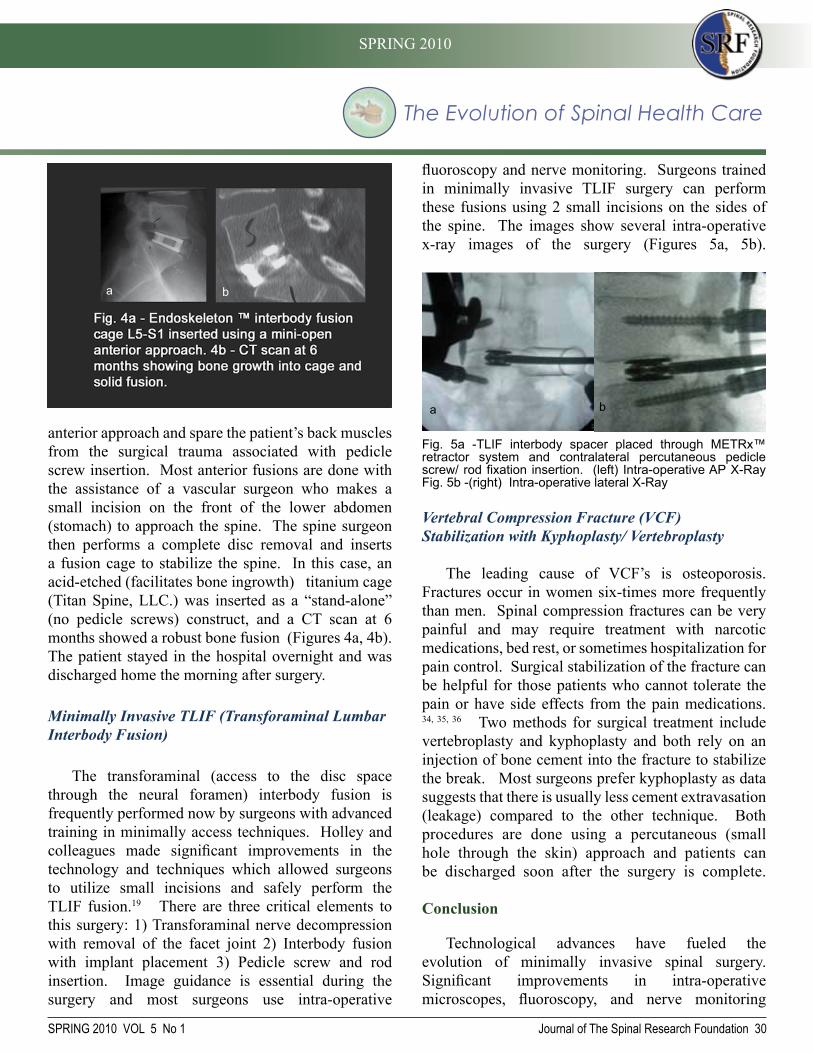
The Evolution of Spinal Health Care
SPRING 2010 VOL 5 No 1
SPRING 2010
Journal of The Spinal Research Foundation 30
fluoroscopy and nerve monitoring. Surgeons trained in minimally invasive TLIF surgery can perform these fusions using 2 small incisions on the sides of the spine. The images show several intra-operative x-ray images of the surgery (Figures 5a, 5b).
anterior approach and spare the patient’s back muscles from the surgical trauma associated with pedicle screw insertion. Most anterior fusions are done with the assistance of a vascular surgeon who makes a small incision on the front of the lower abdomen (stomach) to approach the spine. The spine surgeon then performs a complete disc removal and inserts a fusion cage to stabilize the spine. In this case, an acid-etched (facilitates bone ingrowth) titanium cage (Titan Spine, LLC.) was inserted as a “stand-alone” (no pedicle screws) construct, and a CT scan at 6 months showed a robust bone fusion (Figures 4a, 4b). The patient stayed in the hospital overnight and was discharged home the morning after surgery.
Minimally Invasive TLIF (Transforaminal Lumbar Interbody Fusion)
The transforaminal (access to the disc space through the neural foramen) interbody fusion is frequently performed now by surgeons with advanced training in minimally access techniques. Holley and colleagues made significant improvements in the technology and techniques which allowed surgeons to utilize small incisions and safely perform the TLIF fusion.19 There are three critical elements to this surgery: 1) Transforaminal nerve decompression with removal of the facet joint 2) Interbody fusion with implant placement 3) Pedicle screw and rod insertion. Image guidance is essential during the surgery and most surgeons use intra-operative
Fig. 5a -TLIF interbody spacer placed through METRx™ retractor system and contralateral percutaneous pedicle screw/ rod fixation insertion. (left) Intra-operative AP X-Ray Fig. 5b -(right) Intra-operative lateral X-Ray
a b
Vertebral Compression Fracture (VCF) Stabilization with Kyphoplasty/ Vertebroplasty
The leading cause of VCF’s is osteoporosis. Fractures occur in women six-times more frequentlythan men. Spinal compression fractures can be very painful and may require treatment with narcotic medications, bed rest, or sometimes hospitalization for pain control. Surgical stabilization of the fracture can be helpful for those patients who cannot tolerate the pain or have side effects from the pain medications. 34, 35, 36 Two methods for surgical treatment include vertebroplasty and kyphoplasty and both rely on an injection of bone cement into the fracture to stabilize the break. Most surgeons prefer kyphoplasty as data suggests that there is usually less cement extravasation (leakage) compared to the other technique. Both procedures are done using a percutaneous (small hole through the skin) approach and patients can be discharged soon after the surgery is complete.
Conclusion
Technological advances have fueled the evolution of minimally invasive spinal surgery. Significant improvements in intra-operative microscopes, fluoroscopy, and nerve monitoring
a b

SPRING 2010 VOL 5 No 1
SPINAL RESEARCH FOUNDATION
Journal of The Spinal Research Foundation 31
combined with advancements in surgeon training programs have resulted in many new options for patients suffering from back and neck pain. Least invasive spine surgery is emerging as a safe and effective alternative for an increasing number of patients to consider when consulting with their surgeons.
References
1. Legorreta AP, Silber JH, Costantino GN, Kobylinski RW, Zatz SL: Increased cholecystectomy rate after the introduction of laparoscopic cholecystectomy. JAMA. 1993; 270:1429–1432.
2. Kocher T: Die Verletzungen der Wirbelsäule zugleich als Beitragzur Physiologie des menschlichen Rückenmarks. Mitt Grenzgeb Med Chir . 1896; 1:415–480,
3. Robinson JS: Sciatica and the lumbar disc syndrome: a historical perspective. South Med J . 1983; 76:232–238.
4. Albee FH: Transplantation of a portion of the tibia into the spine for Pott’s disease. A preliminary report. JAMA. 1911; 57:885–886.
5. Robinson RA, Smith GW: Anterolateral cervical disc removal and interbody fusion for cervical disc syndrome. Bull Johns Hopkins Hosp. 1955; 96:233,.
6. Smith L: Chemonucleolysis. Clin Orthop Relat Res. 1969; 67:72–80.
7. Austin RT, Zuk JA: Epidural adhesions after chymopapain chemonucleolysis. J R Coll Surg Edinb. 1989; 34:30–32.
8. Moon MS, Kim I, Ok IY, Lee KW: The response of nerve tissue to chymopapain. Int Orthop. 1990; 14:79–83.
9. Sussman BJ: Inadequacies and hazards of chymopapain injections as treatment for intervertebral disease. J Neurosurg. 1975; 42:389–396.
10. Williams RW: Microlumbar discectomy: a conservative surgical approach to the virgin herniated lumbar disc. Spine. 1978; 3:175–182.
11. Kambin P, Brager M: Percutaneous posterolateral discectomy: anatomy and mechanism. Clin Orthop Relat Res. 1987; 223:145–154.
12. Maroon JC, Onik G: Percutaneous automated discectomy: a new method for lumbar disc removal. Technical note. J Neurosurg. 1987: 66:143–146.
13. Faubert C, Caspar W: Lumbar percutaneous discectomy. Initial experience in 28 cases. Neuroradiology. 1991; 33:407–410.
14. Foley KT, Smith MM: Microendoscopic discectomy. Tech Neurosurg. 1997; 3:301–307.
15. McLoughlin GS, Fourney DR: The learning curve of minimally invasive lumbar discectomy. Can J Neurol Sci. 2008; 35:75–78.
16. Nowitzke AM: Assessment of the learning curve for lumbar microendoscopic discectomy. Neurosurgery. 2005; 56:755–762.
17. Perez-Cruet MJ, Foley KT, Isaacs RE, Rice-Wyllie L, Wellington R, Smith MM, et al: Microendoscopic lumbar discectomy: technical note. Neurosurgery. 2002; 51(5 Suppl):S129–S136.
18. Obenchain TG: Speculum lumbar extraforaminal microdiscectomy. Spine J. 2001; 1:415–420.
19. Holly LT, Schwender JD, Rouben DP, Foley KT: Minimally invasive transforaminal interbody fusion: indications, technique, and complications. Neurosurg Focus. 2006; 20(3):E6.
20. Mayer HM: A new technique of minimally invasive anterior lumbar spine fusion. Spine .1997; 22:691–699.
21. McAfee PC, Regan JJ, Geis WP, Fedder IL: Minimally invasive anterior retroperitoneal approach to the lumbar spine. Emphasis on the lateral BAK. Spine. 1998; 23:1476–1484.
22. Pimenta L, Diaz RC, Guerrero LG: Charité artificial disc retrieval: use of a lateral minimally invasive technique. Technical note. J Neurosurg Spine. 2006; 5:556–561.
23. Barth M, Weiss C, Theme C: Two-year outcome after lumbar microdiscectomy versus microscopic sequestrectomy: part 1: evaluation of clinical outcome. Spine. 2008; 33:265–272.
24. Barth M, Deipers M, Weiss C, Thome C: Two year outcome after lumbar microdiscectomy versus microscopic sequestrectomy: part 2: radiographic evaluation and correlation with clinical outcome. Spine. 2008; 33:273–279.
25. Nolte LP, Zamorano LJ, Jiang Z, Wang Q, Langlotz F, Berlemann U: Image-guided insertion of transpedicular screws. A laboratory set-up. Spine. 1995; 20:497–500.
26. Stern, J: Lasers in Spine Surgery: A Review. SpineLine. 2009; X(5), September October.
27. Oppenheimer JH, DeCastro I, McDonnell, DE: Minimally invasive spine technology and minimally invasive surgery: a historical review. Neurosurg Focus. 2009; 27 (3): E9, 1-15.
28. Hott JS, Deshmukh VR, Klopfenstein JD, Sonntag VK, Dickman CA, Spetzler RF, et al: Intraoperative Iso-C C-arm navigation in craniospinal surgery: the first 60 cases. Neurosurgery. 2004; 54:1131–1136.
29. Burkus JK, Heim SE, Gornet MF, Zdeblick TA: Is INFUSE bone graft superior to autograft bone? An integrated analysis of clinical trials using the LT-CAGE lumbar tapered fusion device. J Spinal Disord Tech. 2003; 16:113–122.
30. McKay WF, Peckham SM, Badura JM: A comprehensive clinical review of recombinant human bone morphogenetic protein-2 (INFUSE Bone Graft). Int Orthop. 2007; 31:729–734.
31. McKay B, Sandhu HS: Use of recombinant human bone morphogenetic protein-2 in spinal fusion applications. Spine. 2002; 27(1 Suppl):S66–S85.
32. Foley KT, Gupta SK, Justis JR, Sherman MC: Percutaneous pedicle screw fixation of the lumbar spine. Neurosurg Focus. 2001; 10(4):E10.
33. Weinstein JN, Tosteson TD, Lurie JD, Tosteson AN, Hanscom B, Skinner JS, Abdu WA, Hilibrand AS, Boden SD, Deyo RA. Surgical vs nonoperative treatment for lumbar disk herniation: the Spine Patient Outcomes Research Trial (SPORT): a randomized trial. JAMA. 2006;296:2441-50.
34. Garfin SR, Reilley MA. Minimally invasive treatment of osteoporotic vertebral body compression fractures. Spine J. 2002;2:76–80.
35. Lieberman I, Reinhardt MK. Vertebroplasty and kyphoplasty for osteolytic vertebral collapse. Clin Ortho.p 2003;415(suppl):176–86.
36. Phillips FM. Minimally invasive treatments of osteoporotic vertebral compression fractures. Spine. 2003;28(suppl):45–53
Paul J. Slosar, Jr., MD
Dr. Slosar is the president of The SpineCare Medical Group in San Francisco, CA. He has been in practice since 1994 and with his current practice since 1997. Dr. Slosar is also Co-Director
of The San Francisco Spine Institute Surgical Fellowship training program and Assistant Director of Surgical Research. He is a member of the North American Spine Society and has served on editorial and research committees.
P. Slosar et.al./The Journal of the Spinal Research Foundation 5 (2010) 26-31
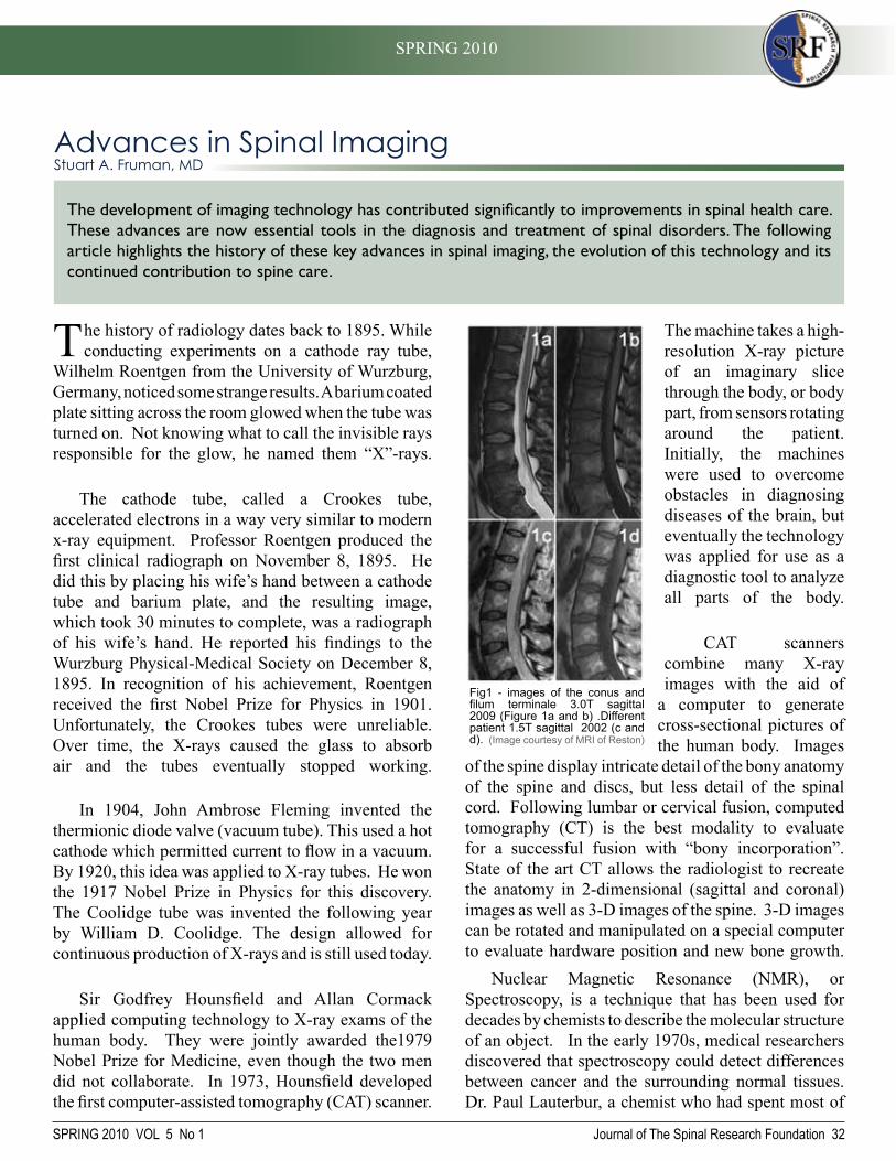
SPRING 2010 VOL 5 No 1
SPRING 2010
Journal of The Spinal Research Foundation 32
The history of radiology dates back to 1895. While conducting experiments on a cathode ray tube,
Wilhelm Roentgen from the University of Wurzburg, Germany, noticed some strange results. A barium coated plate sitting across the room glowed when the tube was turned on. Not knowing what to call the invisible rays responsible for the glow, he named them “X”-rays.
The cathode tube, called a Crookes tube, accelerated electrons in a way very similar to modern x-ray equipment. Professor Roentgen produced the first clinical radiograph on November 8, 1895. He did this by placing his wife’s hand between a cathode tube and barium plate, and the resulting image, which took 30 minutes to complete, was a radiograph of his wife’s hand. He reported his findings to the Wurzburg Physical-Medical Society on December 8, 1895. In recognition of his achievement, Roentgen received the first Nobel Prize for Physics in 1901. Unfortunately, the Crookes tubes were unreliable. Over time, the X-rays caused the glass to absorb air and the tubes eventually stopped working.
In 1904, John Ambrose Fleming invented the thermionic diode valve (vacuum tube). This used a hot cathode which permitted current to flow in a vacuum. By 1920, this idea was applied to X-ray tubes. He won the 1917 Nobel Prize in Physics for this discovery. The Coolidge tube was invented the following year by William D. Coolidge. The design allowed for continuous production of X-rays and is still used today.
Sir Godfrey Hounsfield and Allan Cormack applied computing technology to X-ray exams of the human body. They were jointly awarded the1979 Nobel Prize for Medicine, even though the two men did not collaborate. In 1973, Hounsfield developed the first computer-assisted tomography (CAT) scanner.
The machine takes a high-resolution X-ray picture of an imaginary slice through the body, or body part, from sensors rotating around the patient. Initially, the machines were used to overcome obstacles in diagnosing diseases of the brain, but eventually the technology was applied for use as a diagnostic tool to analyze all parts of the body.
CAT scanners combine many X-ray images with the aid of
a computer to generate cross-sectional pictures of the human body. Images
of the spine display intricate detail of the bony anatomy of the spine and discs, but less detail of the spinal cord. Following lumbar or cervical fusion, computed tomography (CT) is the best modality to evaluate for a successful fusion with “bony incorporation”. State of the art CT allows the radiologist to recreate the anatomy in 2-dimensional (sagittal and coronal) images as well as 3-D images of the spine. 3-D images can be rotated and manipulated on a special computer to evaluate hardware position and new bone growth.
Nuclear Magnetic Resonance (NMR), or Spectroscopy, is a technique that has been used for decades by chemists to describe the molecular structure of an object. In the early 1970s, medical researchers discovered that spectroscopy could detect differences between cancer and the surrounding normal tissues. Dr. Paul Lauterbur, a chemist who had spent most of
Advances in Spinal Imaging Stuart A. Fruman, MD
The development of imaging technology has contributed significantly to improvements in spinal health care. These advances are now essential tools in the diagnosis and treatment of spinal disorders. The following article highlights the history of these key advances in spinal imaging, the evolution of this technology and its continued contribution to spine care.
Fig1 - images of the conus and filum terminale 3.0T sagittal 2009 (Figure 1a and b) .Different patient 1.5T sagittal 2002 (c and d). (Image courtesy of MRI of Reston)

SPRING 2010 VOL 5 No 1
SPINAL RESEARCH FOUNDATION
Journal of The Spinal Research Foundation 33
his career working with NMR, realized that the signals originating from atoms varied based upon the strength of a magnetic field. A magnetic field placed across a human body could essentially create a cross-sectional image of the body, a magnetic resonance image (MRI). British physicist, Sir Peter Mansfield, had read of the work of Lauterbur and developed the technique of echo planar imaging, which dramatically increased the speed of MRI image acquisition.
The first MRI machines were introduced into hospitals in the early 1980s. In 2003, Paul Lauterbur and Sir Peter Mansfield were jointly awarded the Nobel Prize in Medicine for their work. By that time, over 20,000 MRI scanners were being used around the world, performing millions of studies per year. These machines facilitate excellent detail of the spine, including the vertebral bodies, discs, spinal cord, nerves and the path the nerves travel as they exit the spine. These images allow the radiologist to evaluate the anatomy for structural causes of neck and back pain including a disc herniation, degenerative disc disease and spinal stenosis. It is a crucial exam which helps
surgeons plan for surgeries to decompress a pinched nerve or a spinal fusion.
In patients who have acute back pain with neurological symptoms, as well as chronic back pain, MRI is the imaging modality of choice when looking for mechanical or anatomic causes of the pain. Timing of when to order the MRI study is also important. Many studies have shown that disc herniations tend to diminish with time, and if the MRI is performed too early the imaging may not be an accurate assessment for operative planning (Figure 3). It is important to note that there is a 28% prevalence of lumbar herniated discs in asymptomatic patients, and the herniated disc may not be the cause of the patient’s back pain. This problem of abnormal MRI finding in asymptomatic patients highlights the limitations of MRI as a screening tool, as well as the critical need to correlate the imaging findings with the clinical exam.
In the post-operative patient, some of the common causes of pain include a recurrent or residual disc herniation, spinal instability (such as a failed fusion or “pseudarthrosis”), spinal stenosis and discitis. The administration of gadolinium contrast is often very important in diagnosing some of these conditions. In the first three months after surgery, a soft tissue mass impinging on the thecal sac, such as an immature hematoma or scar, can mimic a recurrent or residual disc herniation. Gadolinium contrast can often help differentiate these.
While the modalities of CT (computed tomography) and MRI (magnetic resonance imaging) are not new,
S. Fruman et.al./The Journal of the Spinal Research Foundation 5 (2010) 32-35
Fig. 3 -Example of a large left L5-S1 HNP that underwent spontaneous resorption over 10 months. (Image courtesy of MRI of Reston)
Fig. 2 -Image of a Modern MRI
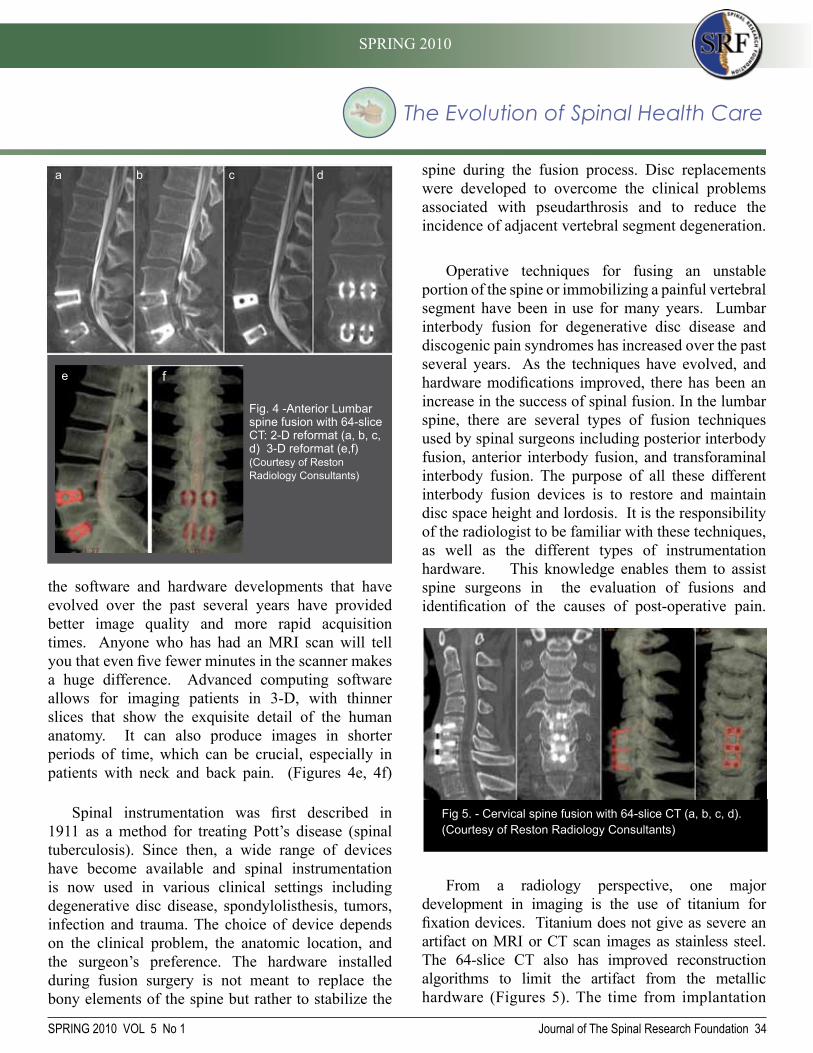
The Evolution of Spinal Health Care
SPRING 2010 VOL 5 No 1
SPRING 2010
Journal of The Spinal Research Foundation 34
spine during the fusion process. Disc replacements were developed to overcome the clinical problems associated with pseudarthrosis and to reduce the incidence of adjacent vertebral segment degeneration.
Operative techniques for fusing an unstable portion of the spine or immobilizing a painful vertebral segment have been in use for many years. Lumbar interbody fusion for degenerative disc disease and discogenic pain syndromes has increased over the past several years. As the techniques have evolved, and hardware modifications improved, there has been an increase in the success of spinal fusion. In the lumbar spine, there are several types of fusion techniques used by spinal surgeons including posterior interbody fusion, anterior interbody fusion, and transforaminal interbody fusion. The purpose of all these different interbody fusion devices is to restore and maintain disc space height and lordosis. It is the responsibility of the radiologist to be familiar with these techniques, as well as the different types of instrumentation hardware. This knowledge enables them to assist spine surgeons in the evaluation of fusions and identification of the causes of post-operative pain.
From a radiology perspective, one major development in imaging is the use of titanium for fixation devices. Titanium does not give as severe an artifact on MRI or CT scan images as stainless steel. The 64-slice CT also has improved reconstruction algorithms to limit the artifact from the metallichardware (Figures 5). The time from implantation
the software and hardware developments that have evolved over the past several years have provided better image quality and more rapid acquisition times. Anyone who has had an MRI scan will tell you that even five fewer minutes in the scanner makes a huge difference. Advanced computing software allows for imaging patients in 3-D, with thinner slices that show the exquisite detail of the human anatomy. It can also produce images in shorter periods of time, which can be crucial, especially in patients with neck and back pain. (Figures 4e, 4f)
Spinal instrumentation was first described in 1911 as a method for treating Pott’s disease (spinal tuberculosis). Since then, a wide range of devices have become available and spinal instrumentation is now used in various clinical settings including degenerative disc disease, spondylolisthesis, tumors, infection and trauma. The choice of device depends on the clinical problem, the anatomic location, and the surgeon’s preference. The hardware installed during fusion surgery is not meant to replace the bony elements of the spine but rather to stabilize the
Fig. 4 -Anterior Lumbar spine fusion with 64-slice CT: 2-D reformat (a, b, c, d) 3-D reformat (e,f) (Courtesy of Reston Radiology Consultants)
b c da
e f
Fig 5. - Cervical spine fusion with 64-slice CT (a, b, c, d). (Courtesy of Reston Radiology Consultants)
SPRING 2010 VOL 5 No 1
SPINAL RESEARCH FOUNDATION
Journal of The Spinal Research Foundation 35
: Intelec '98, October 4-8, 1998 : Hyatt Regency Embarcadero, San Francisco, California U.S.A. Piscataway, N.J.: Institute of Electrical and Electronics Engineers; 1998.
4. Petrik V, Apok V, Britton JA, Bell BA, Papadopoulos MC. Godfrey Hounsfield and the dawn of computed tomography. Neurosurgery. Apr 2006;58(4):780-787; discussion 780-787.
5. Mansfield P. Snapshot magnetic resonance imaging (Nobel lecture). Angew Chem Int Ed Engl. Oct 18 2004;43(41):5456-5464.
6. Rhee JM, Schaufele M, Abdu WA. Radiculopathy and the herniated lumbar disc. Controversies regarding pathophysiology and management. J Bone Joint Surg Am. Sep 2006;88(9):2070-2080.
7. Schwab FJ, Nazarian DG, Mahmud F, Michelsen CB. Effects of spinal instrumentation on fusion of the lumbosacral spine. Spine (Phila Pa 1976). Sep 15 1995;20(18):2023-2028.
8. Nachemson A, Zdeblick TA, O'Brien JP. Lumbar disc disease with discogenic pain. What surgical treatment is most effective? Spine (Phila Pa 1976). Aug 1 1996;21(15):1835-1838.
Stuart A. Fruman, MD
Dr. Fruman is a radiologist at Reston Radiology Associates. He has extensive experience in the field of medical imaging. He has worked at several leading hospitals and imaging centers. He is board certified with The American Board of Radiology. At
Reston Hospital he was the Chairman of the Department of Radiology from 1997-2003. He has been involved in research at the National Cancer Institute and has published articles in Academic Radiology.
S. Fruman et.al./The Journal of the Spinal Research Foundation 5 (2010) 32-35
References
1. Hesselink JR. Spine imaging: history, achievements, remaining frontiers. AJR Am J Roentgenol. Jun 1988;150(6):1223-1229.
2. Spiegel PK. The first clinical X-ray made in America--100 years. AJR Am J Roentgenol. Jan 1995;164(1):241-243.
3. IEEE Power Electronics Society. INTELEC : Twentieth International Telecommunications Energy Conference
of an interbody fusion device to solid arthrodesis is variable, but is usually a minimum of 6-12 months.
While plain film radiography may be one of the most used modalities in evaluating pre and post operative back pain, it has significant limitations and is best used as a preliminary evaluation. It is also not considered reliable due to significant intra- and interobserver variation. The level of anatomic detail demonstrated with newer MRI and CT scanners has added huge value in the evaluation of acute and chronic causes of back pain. Newer 3.0 Tesla MRI scanners as well as 64-slice CT scans have added incredible detail of the human anatomy previously not available. Despite its quality of imaging, MRI is not acceptable for monitoring the progress of an arthrodesis due to metallic artifacts which can degrade anatomic detail. Also, bony changes detectable through MRI, such as marrow edema or inflammation, have little specificity in determining fibrous union vs. early stages of arthrodesis.
For these reasons, CT has become the state of the art tool for assessing successful fusion. In practice, radiologists do not and must not work “in the dark”. It is of ultimate importance that the radiologist and the spinal surgeon work closely together to make sure that imaging findings are concordant with patient’s clinical evaluation.

SPRING 2010 VOL 5 No 1
SPRING 2010
Journal of The Spinal Research Foundation 36
A Historical Evolution of Safe AnesthesiaMichael S. Bradish, MD
As medicine has evolved throughout the centuries, so have surgical techniques and practices. Centuries ago, simple procedures such as tooth extraction, were barbaric and traumatic for both patients and surgeons. It was obvious to all parties involved that some kind of intervention was needed to keep patients comfortable and to give surgeons the best conditions possible to complete surgery successfully. Ultimately, the specialty of anesthesiology was born, and it has evolved to meet these needs. It has taken years of technical and medical advances to arrive at today’s safe, balanced anesthesia goals – amnesia (forgetting procedure), anxiolysis (sedation or sleep), analgesia (pain relief), and akinesis (an unmoving patient). The specialty continues to advance and has seen some of the greatest safety improvements of any medical specialty, especially for generally healthy patients.1
Keywords: Anesthesia, Spinal Block
Earliest attempts for meeting akinesis included strong assistants who could hold unwilling
patients down. A blow to the jaw, rendering a patient unconscious, was also favored by some. Temporary strangulation seemed to work to a certain extent. These rudimentary solutions might seem worse than the surgical procedure itself. Unsurprisingly, surgery was rare in Western culture until the advent of adequate anesthesia. Upon examination of records at Massachusetts General Hospital, only 333 cases were recorded in the 25 years between 1821 and 1846.1
Other cultures developed their own solutions for anesthesia. Some of the oldest evidence comes from
the ancient Incan civilization and suggests that they were skilled with surgery. The practice of trepanation (burr holes) in the skull has been found in skeletons from this society. The Incas, a society long known for chewing coca leaves, saw the benefits from this practice: euphoria, pain relief, and increased stimulation.
It is thought that both the patient and surgeon chewed the coca leaves. The patient may have benefitted from the euphoria and pain relief of the coca. Some speculate that the surgeons may have spit on the wound edges, utilizing the local anesthetic effects of this precursor to cocaine, a powerful local anesthetic still used today. This may have helped with pain, but it is doubtful that akinesia, amnesia, or anxiolysis were achieved. Further examination of surgical history shows anecdotal use of opium, marijuana, alcohol, ice, and strong surgical assistants.
Forefathers of Anesthesia
As is often the case in medicine, many people are responsible for any particular discovery or advance in the field. Oftentimes, these steps forward can be unplanned and accidental. For example, clergyman and scientist Joseph Priestly discovered and produced both oxygen and nitrous oxygen in the 1770s – gasses which are still used in modern day anesthesia. Nitrous oxide, also known as laughing gas, was used primarily for entertainment. It was not until 1844 that its anesthetic implications were first realized. At that time, while participating in a demonstration for
Trepanation in human skull

SPRING 2010 VOL 5 No 1
SPINAL RESEARCH FOUNDATION
Journal of The Spinal Research Foundation 37
nitrous oxide in Hartford Connecticut, an unfortunate gentleman, Samuel Cooley, staggered into a bench and injured his leg while under the influence of nitrous oxide. He was unable to feel or realize what happened to his leg until the effects of the nitrous oxide wore off. A dentist in the crowd, Horace Wells (1815-1848), witnessed these surprising results and thought about how to incorporate its use within his own practice.
Suffering from an erupting wisdom tooth himself, Wells insisted that his practice partner, Dr. Riggs, remove the tooth the following day under nitrous oxide. The feat was accomplished successfully. It is unclear whether Wells can be considered the father of anesthesia, but he was one of the very first subjects in the experiment. Unfortunately, while nitrous oxide does provide some pain relief, it cannot be rendered in doses large enough to cause unconsciousness safely. Improvements and advances for surgery were needed.
On October 16, 1846, the first general anesthetic was administered at the Bulfinch Building at the venerable Massachusetts General Hospital. The observation suite, known as the Ether Dome, is now registered as a historical landmark and is still open for specially arranged tours. For that first successful surgery with an anesthetized patient, ether was administered by William T. Morton, a pupil of Dr. Wells. Upon completing the surgery, Dr. John Warren was heard to exclaim, “Gentlemen - this is no humbug!” Suddenly, a wide variety of surgeries were possible.
As news of this feat spread, a quotation from the People’s Journal of London reflected this advancement:
“Oh, what delight for every feeling heart to find the new year ushered in with the announcement of this noble discovery of the power to still the sense of pain, and veil the eye and memory from all the horrors of an operation. ... WE HAVE CONQUERED PAIN.”
Surrounding this time, several different characters including Wells and Morton had claimed responsibility for this great discovery; ultimately, most anesthesia historians agree that Morton should get the credit. Not a physician, but rather a profiteer and questionable businessman, Morton immediately patented his discovery looking for fortune, not professional renown. He even pressed Congress to reward him for his advances for mankind. Despite multiple patent lawsuits, Morton ended up without the fortune he sought from his discovery.
Regional Anesthesia
In 1885, a New York neurologist James Leonard Corning performed the first spinal block by accident on a patient with a history of “spinal weakness”. By injecting local anesthesia into the subcutaneous tissues between spinal processes of the back, Corning reasoned he could achieve “anesthesia
M. Bradish et.al./The Journal of the Spinal Research Foundation 5 (2010) 36-38
The first public demonstration of surgical anesthesia at Massachusetts General Hospital. (Image courtesy of the National Library of Medicine)
The first dental operation performed by Horace Wells under the influence of Nitrous Oxide Gas, December 11, 1844. (Image courtesy of the National Library of Medicine)
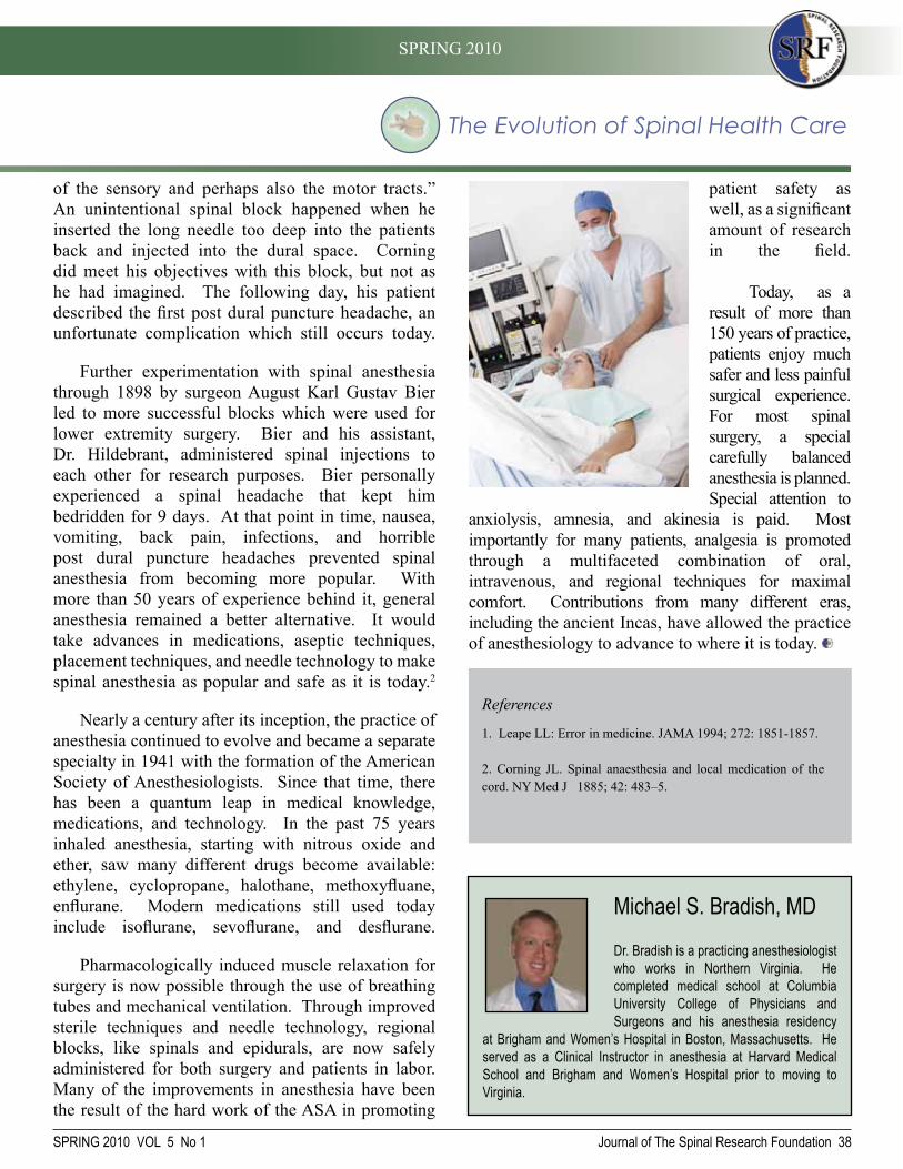
The Evolution of Spinal Health Care
SPRING 2010 VOL 5 No 1
SPRING 2010
Journal of The Spinal Research Foundation 38
patient safety as well, as a significant amount of research in the field.
Today, as a result of more than 150 years of practice, patients enjoy much safer and less painful surgical experience. For most spinal surgery, a special carefully balanced anesthesia is planned. Special attention to
anxiolysis, amnesia, and akinesia is paid. Most importantly for many patients, analgesia is promoted through a multifaceted combination of oral, intravenous, and regional techniques for maximal comfort. Contributions from many different eras, including the ancient Incas, have allowed the practice of anesthesiology to advance to where it is today.
References
1. Leape LL: Error in medicine. JAMA 1994; 272: 1851-1857.
2. Corning JL. Spinal anaesthesia and local medication of the cord. NY Med J 1885; 42: 483–5.
of the sensory and perhaps also the motor tracts.” An unintentional spinal block happened when he inserted the long needle too deep into the patients back and injected into the dural space. Corning did meet his objectives with this block, but not as he had imagined. The following day, his patient described the first post dural puncture headache, an unfortunate complication which still occurs today.
Further experimentation with spinal anesthesia through 1898 by surgeon August Karl Gustav Bier led to more successful blocks which were used for lower extremity surgery. Bier and his assistant, Dr. Hildebrant, administered spinal injections to each other for research purposes. Bier personally experienced a spinal headache that kept him bedridden for 9 days. At that point in time, nausea, vomiting, back pain, infections, and horrible post dural puncture headaches prevented spinal anesthesia from becoming more popular. With more than 50 years of experience behind it, general anesthesia remained a better alternative. It would take advances in medications, aseptic techniques, placement techniques, and needle technology to make spinal anesthesia as popular and safe as it is today.2
Nearly a century after its inception, the practice of anesthesia continued to evolve and became a separate specialty in 1941 with the formation of the American Society of Anesthesiologists. Since that time, there has been a quantum leap in medical knowledge, medications, and technology. In the past 75 years inhaled anesthesia, starting with nitrous oxide and ether, saw many different drugs become available: ethylene, cyclopropane, halothane, methoxyfluane, enflurane. Modern medications still used today include isoflurane, sevoflurane, and desflurane.
Pharmacologically induced muscle relaxation for surgery is now possible through the use of breathing tubes and mechanical ventilation. Through improved sterile techniques and needle technology, regional blocks, like spinals and epidurals, are now safely administered for both surgery and patients in labor. Many of the improvements in anesthesia have been the result of the hard work of the ASA in promoting
Michael S. Bradish, MD Dr. Bradish is a practicing anesthesiologist who works in Northern Virginia. He completed medical school at Columbia University College of Physicians and Surgeons and his anesthesia residency
at Brigham and Women’s Hospital in Boston, Massachusetts. He served as a Clinical Instructor in anesthesia at Harvard Medical School and Brigham and Women’s Hospital prior to moving to Virginia.
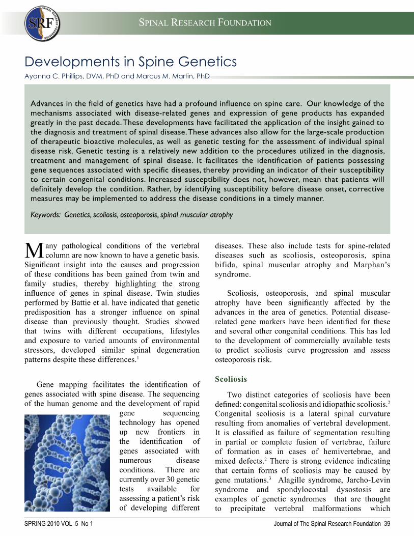
SPRING 2010 VOL 5 No 1
SPINAL RESEARCH FOUNDATION
Journal of The Spinal Research Foundation 39
Developments in Spine GeneticsAyanna C. Phillips, DVM, PhD and Marcus M. Martin, PhD
Advances in the field of genetics have had a profound influence on spine care. Our knowledge of the mechanisms associated with disease-related genes and expression of gene products has expanded greatly in the past decade. These developments have facilitated the application of the insight gained to the diagnosis and treatment of spinal disease. These advances also allow for the large-scale production of therapeutic bioactive molecules, as well as genetic testing for the assessment of individual spinal disease risk. Genetic testing is a relatively new addition to the procedures utilized in the diagnosis, treatment and management of spinal disease. It facilitates the identification of patients possessing gene sequences associated with specific diseases, thereby providing an indicator of their susceptibility to certain congenital conditions. Increased susceptibility does not, however, mean that patients will definitely develop the condition. Rather, by identifying susceptibility before disease onset, corrective measures may be implemented to address the disease conditions in a timely manner.
Keywords: Genetics, scoliosis, osteoporosis, spinal muscular atrophy
Many pathological conditions of the vertebral column are now known to have a genetic basis.
Significant insight into the causes and progression of these conditions has been gained from twin and family studies, thereby highlighting the strong influence of genes in spinal disease. Twin studies performed by Battie et al. have indicated that genetic predisposition has a stronger influence on spinal disease than previously thought. Studies showed that twins with different occupations, lifestyles and exposure to varied amounts of environmental stressors, developed similar spinal degeneration patterns despite these differences.1
Gene mapping facilitates the identification of genes associated with spine disease. The sequencing of the human genome and the development of rapid
gene sequencing technology has opened up new frontiers in the identification of genes associated with numerous disease conditions. There are currently over 30 genetic tests available for assessing a patient’s risk of developing different
diseases. These also include tests for spine-related diseases such as scoliosis, osteoporosis, spina bifida, spinal muscular atrophy and Marphan’s syndrome.
Scoliosis, osteoporosis, and spinal muscular
atrophy have been significantly affected by the advances in the area of genetics. Potential disease-related gene markers have been identified for these and several other congenital conditions. This has led to the development of commercially available tests to predict scoliosis curve progression and assess osteoporosis risk.
Scoliosis
Two distinct categories of scoliosis have been defined: congenital scoliosis and idiopathic scoliosis.2 Congenital scoliosis is a lateral spinal curvature resulting from anomalies of vertebral development. It is classified as failure of segmentation resulting in partial or complete fusion of vertebrae, failure of formation as in cases of hemivertebrae, and mixed defects.2 There is strong evidence indicating that certain forms of scoliosis may be caused by gene mutations.3 Alagille syndrome, Jarcho-Levin syndrome and spondylocostal dysostosis are examples of genetic syndromes that are thought to precipitate vertebral malformations which

The Evolution of Spinal Health Care
SPRING 2010 VOL 5 No 1
SPRING 2010
Journal of The Spinal Research Foundation 40
monozygotic twins and 36% concordance among dizygotic twins for idiopathic scoliosis.3 Wyme-Dives and Risebourg (1973) also showed that there may be an X-linked form of inherited scoliosis. Conflicting data as to the root of this disorder may implicate several genetic loci as possible causes of idiopathic scoliosis.
The adolescent idiopathic scoliosis (AIS) test produced by Axial Biotech is one of the front-runners for scoliosis detection. It is indicated in the detection of genotypes for 53 genetic markers found in clinical trials to be associated with curve progression in self-reported studies. The test is indicated for:
• A primary diagnosis of AIS• Diagnosis between the ages of 9 and 13 years
old• Diagnosis of a mild scoliotic curve (defined
as <25°)• Diagnosis of patients of Caucasian ethnicity
(North American, South American, European, Eastern European, Middle Eastern)
Further research is being done to expand the application of this test across a broader population. A separate test for disc degeneration susceptibility is expected to be released in late 2010.
Osteoporosis
Osteoporosis is a skeletal disorder characterized by compromised bone strength (bone mineral density), predisposing patients to an above average rate of fracture.8 This disease particularly affects the spine and family lineage studies illustrate a genetic component. However, all of the specific genes which are associated with the development of osteoporosis have not yet been identified.9 Several candidate genes are being studied as possible causes of osteoporosis. While many non genetic factors have been extensively investigated, study of the genetic elements of osteoporosis is relatively new. Control of this condition appears to be polygenetic and therefore identification of all the responsible genes may take some time. This condition may be
result in congenital scoliosis.4,5 These gene abnormalities may affect the shape, intensity and direction of spinal curvature.
Idiopathic scoliosis
is the term given to a curvature of the spine for which no cause can be identified.2 These anomalies, though present from
birth, may be noticeable before the age of 5 years (infantile idiopathic scoliosis) or may not present until later in childhood after spinal growth increases their prominence (adolescent idiopathic scoliosis).3,6 There is a significant amount of research directed toward understanding the basis of idiopathic scoliosis. Epidemiological studies suggest that there may be multiple modes of heritability.3 Molecular studies, including conserved human/mouse synteny data analysis, have allowed for the identification of potential candidate genes for human congenital and idiopathic scoliosis.2 Possible scoliosis related regions have been identified by map position on chromosomes 1,5,6,8,9,16,17,19 and X.2
Further, clinical observation and population studies show a higher prevalence of scoliosis among relatives compared to the general population.7 Studies have demonstrated a 73% concordance in
Genetic test that can predict curve progression
Fig. 1 -Scoliotic spine
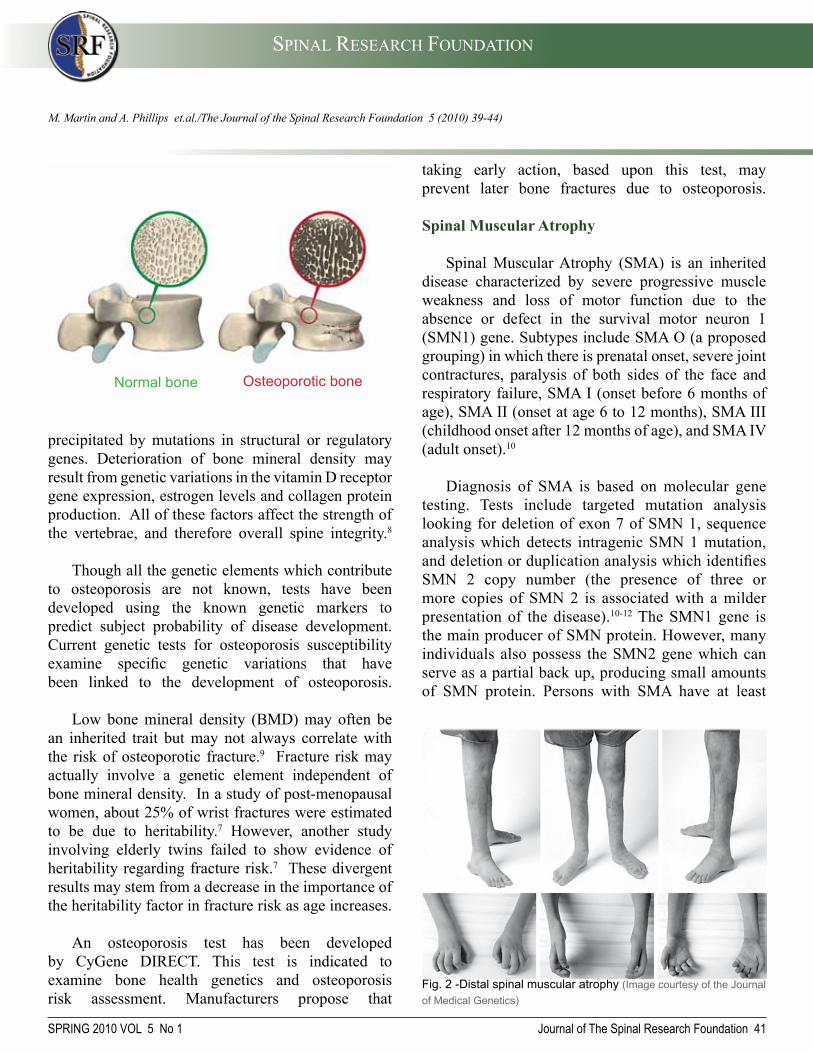
SPRING 2010 VOL 5 No 1
SPINAL RESEARCH FOUNDATION
Journal of The Spinal Research Foundation 41
M. Martin and A. Phillips et.al./The Journal of the Spinal Research Foundation 5 (2010) 39-44)
precipitated by mutations in structural or regulatory genes. Deterioration of bone mineral density may result from genetic variations in the vitamin D receptor gene expression, estrogen levels and collagen protein production. All of these factors affect the strength of the vertebrae, and therefore overall spine integrity.8
Though all the genetic elements which contribute to osteoporosis are not known, tests have been developed using the known genetic markers to predict subject probability of disease development. Current genetic tests for osteoporosis susceptibility examine specific genetic variations that have been linked to the development of osteoporosis.
Low bone mineral density (BMD) may often be an inherited trait but may not always correlate with the risk of osteoporotic fracture.9 Fracture risk may actually involve a genetic element independent of bone mineral density. In a study of post-menopausal women, about 25% of wrist fractures were estimated to be due to heritability.7 However, another study involving elderly twins failed to show evidence of heritability regarding fracture risk.7 These divergent results may stem from a decrease in the importance of the heritability factor in fracture risk as age increases.
An osteoporosis test has been developed by CyGene DIRECT. This test is indicated to examine bone health genetics and osteoporosis risk assessment. Manufacturers propose that
taking early action, based upon this test, may prevent later bone fractures due to osteoporosis. Spinal Muscular Atrophy
Spinal Muscular Atrophy (SMA) is an inherited disease characterized by severe progressive muscle weakness and loss of motor function due to the absence or defect in the survival motor neuron 1 (SMN1) gene. Subtypes include SMA O (a proposed grouping) in which there is prenatal onset, severe joint contractures, paralysis of both sides of the face and respiratory failure, SMA I (onset before 6 months of age), SMA II (onset at age 6 to 12 months), SMA III (childhood onset after 12 months of age), and SMA IV (adult onset).10
Diagnosis of SMA is based on molecular gene testing. Tests include targeted mutation analysis looking for deletion of exon 7 of SMN 1, sequence analysis which detects intragenic SMN 1 mutation, and deletion or duplication analysis which identifies SMN 2 copy number (the presence of three or more copies of SMN 2 is associated with a milder presentation of the disease).10-12 The SMN1 gene is the main producer of SMN protein. However, many individuals also possess the SMN2 gene which can serve as a partial back up, producing small amounts of SMN protein. Persons with SMA have at least
Osteoporotic boneNormal bone
Fig. 2 -Distal spinal muscular atrophy (Image courtesy of the Journal of Medical Genetics)

The Evolution of Spinal Health Care
SPRING 2010 VOL 5 No 1
SPRING 2010
Journal of The Spinal Research Foundation 42
one copy of the SMN2 gene and studies have shown that SMA patients who have more copies of SMN2 often tend to have a less severe form of the disease. Symptoms in an infant:
• Respiratory difficulty • Feeding difficulty • Floppy infant (poor muscle tone and head
control) • Minimal spontaneous movement • Progressive weakness (older infant to
toddler)
As such, one of the main therapeutic objectives for SMA treatment is to stimulate an increase in SMN2 gene expression so as to potentially slow or prevent motor neuron and muscle function loss, or possibly even restore function in cases where function had been lost.13 Some drugs under investigation that are directed towards enhancing SMN2 gene activity include hydroxyurea, indoprofen and valproic acid, among others.10
It is estimated that approximately 25,000 Americans have SMA. The rate of occurrence of SMA is approximately 1 in 6,000 to 1 in 10,000 infants. Although SMA affects all ethnic groups, the incidence of the disease may vary by population. Over 8 million Americans are carriers of SMA, having mutations in the SMN1 gene but not exhibiting SMA symptoms.13 Advances in genetics
have had a tremendous impact on the screening, diagnosis and treatment of this debilitating condition.The age of onset of SMA affects the severity. Symptoms range from benign to fatal. Disease presentation in infants is the most severe. There is no treatment for the progressive muscle weakness. However, knowledge of the condition can lead to vigilance and reduction in respiratory complications. Physical therapy is important to prevent abnormal muscle contractions which may lead to scoliosis.10 Several labs now offer genetic tests for SMA in order to diagnose or to detect if an individual is a carrier of SMA associated gene defects.
The use of genetic technology has also allowed scientists to exogenously produce molecules capable of enhancing the bone healing process. An example of this is the production of proteins such as rhBMP-2 and several other bone growth augmenting proteins. Nearly 40 years after the discovery of bone morphogenetic proteins (BMPs), their clinical application was finally realized at the beginning of this century. BMPs have greatly advanced the field of spinal surgery, facilitating faster healing of spinal fusion procedures and providing an alternative to iliac crest bone grafts for bone fusion.
BMPs are present in minute quantities within bone. It would require hundreds of kilograms of bone in order to extract milligram quantities of BMP. To overcome these limitations, scientists focused their
Fig.3 -13 year-old-girl with SMA. shows a) preoperative AP and lateral radiogram b) postoperative AP and lateral radiogram and c) final follow-up AP and lateral radiogram of spine (Image source: Journal of Orthopaedic Surgery and Research 2008 3:23 doi:10.1186/1749-799X-3-23)
a b c

SPRING 2010 VOL 5 No 1
SPINAL RESEARCH FOUNDATION
Journal of The Spinal Research Foundation 43
M. Martin and A. Phillips et.al./The Journal of the Spinal Research Foundation 5 (2010) 39-44
Genetic Tests For Spine-Related Conditions
Genetic Test Gene location(chromosome; genes)
Disease Description
Scoliosis Chromosomes 1,5,6,8,9,16,17,19, X; CHD7
Scoliosis is a C- or S- shaped curvature of the spine
OsteoporosisChromosomes 20, 1p36; RANKL, estrogen receptor 1 gene, osteoporosis gene, ZBTB40, 6p21
thinning of bone tissue and loss of bone density over time
Spina bifida Chromosome 1,11; CFL1birth defect that involves the incomplete development of the spinal cord or its coverings
Gaucher disease Chromosome 1; GBAenlarged liver and spleen, bone degeneration
Marfan syndrome Chromosome 15; FBN1
connective tissue disorder; tissues of ligaments, blood vessel walls, cartilage, heart valves and other structures abnormally weak
Spinal muscular atrophy Chromosome 5,X; SMN1
severe, often lethal progressive muscle-wasting disorder in children
Duchenne muscular dystrophy X Chromosome; Dystrophin
severe to mild muscle wasting, deterioration, weakness
Myotonic dystrophy Chromosome 19; Myotonin protein kinase
progressive muscle weakness; most common form of adult muscular dystrophy
Central Core Disease Chromosome 19; RYR1 mild to severe muscle weakness
research efforts on the determination of the amino acid sequence of these proteins and the sequencing of their associated genes. Through the use of recombinant DNA technology, researchers were able to produce these proteins in large quantities in established cell expression systems, using bioreactors, thereby producing purified BMPs for research, clinical and commercial applications.
Genetics and environmental factors both
influence the incidence of spinal disease. It was
previously thought that environmental factors were the key variables in determining the development of spinal disease. However, current research suggests that an individual’s genetic make-up appears to be the main factor in determining if a person will or will not develop spinal disease.
Behavioral modification can have a significant impact on addressing the information gained from genetic tests. It is believed that an individual who is predisposed to developing osteoporosis should attempt
The Evolution of Spinal Health Care
SPRING 2010 VOL 5 No 1
SPRING 2010
Journal of The Spinal Research Foundation 44
to establish their maximum BMD during their bone building years. Also, bone density monitoring can detect declining BMD before it reaches critical levels. Treatment can then be initiated to reduce osteoporotic fracture risk. With the scoliosis progression test, however, behavioral modification is insufficient and the disease may progress if surgical intervention is not pursued. In this case the genetic test allows the caregivers to determine the probability of the curve progressing in order to determine if surgery is prudent, or if it will remain benign, thus making surgery unnecessary.
Advances in our knowledge and the application
of genetics to the treatment of diseases of the spine have increased considerably in the last decade. These advances have facilitated the development of diagnostics for scoliosis curve progression, osteoporosis risk and several other spine conditions. A specific disease does not necessarily emerge from one genetic mutation only. There can often be several genes contributing to the same disease phenotype. As time progresses and our knowledge of the human genome increases, these tests will become more widespread and more dependable. As more gene-based diagnostics are developed and assessed, genetic procedures will play an increasing role in the diagnosis and treatment of spinal disease.
in the spinal muscular atrophy gene SMN1. Kobe J Med Sci 54:E227-36.
Ayanna C. Phillips, DVM, PhD
Dr. Phillips is a Lecturer at the University of the West Indies, School of Veterinary Medicine. In her role as a large animal veterinarian and a veterinary
surgeon she has observed and treated spine diseases in a broad range of animal species. She has extensively studied the effects of neurotoxins of marine origin on mammalian cell cycle regulators and cell cycle progression. She is committed to facilitating the improvement of human and animal health through translational research.
References
1. Battie MC, Videman T, Levalahti E, Gill K, Kaprio J. Heritability of low back pain and the role of disc degeneration. Pain. Oct 2007;131(3):272-280.
2. Giampietro PF, Blank RD, Raggio CL, et al. Congenital and idiopathic scoliosis: clinical and genetic aspects. Clin Med Res. Apr 2003;1(2):125-136.
3. Carr AJ. Adolescent idiopathic scoliosis in identical twins. J Bone Joint Surg Br. Nov 1990;72(6):1077.
4. Alagille D, Odievre M, Gautier M, Dommergues JP. Hepatic ductular hypoplasia associated with characteristic facies, vertebral malformations, retarded physical, mental, and sexual development, and cardiac murmur. J Pediatr. Jan 1975;86(1):63-71.
5. Mortier GR, Lachman RS, Bocian M, Rimoin DL. Multiple vertebral segmentation defects: analysis of 26 new patients and review of the literature. Am J Med Genet. Feb 2 1996;61(4):310 319.
6. Gillingham BL, Fan RA, Akbarnia BA. Early onset idiopathic scoliosis. J Am Acad Orthop Surg. Feb 2006;14(2):101-112.
7. Deng HW, Chen WM, Recker S, et al. Genetic determination of Colles’ fracture and differential bone mass in women with and without Colles’ fracture. J Bone Miner Res. Jul 2000;15(7):1243-1252.
8. Gardiner A, El Miedany Y, Toth M. Osteoporosis: not only in women, but in men too. Br J Nurs. Jun 28-Jul 11 2007;16(12):731-735.
9. Ralston SH. Genetics of osteoporosis. Proc Nutr Soc. May 2007;66(2):158-165.
10. Vitte J, Attali R, Warwar N, Gurt I, Melki J. Spinal muscular atrophy. Adv Exp Med Biol. 2009;652:237-246
11. Tizzano, E. 2009. Spinal muscular atrophy during human development: where are the early pathogenic findings? Adv Exp Med Biol 652:225-35.
12. Lee, L., S. E. Davies, and J. L. Liu. 2009. The spinal muscular atrophy protein SMN affects Drosophila germline nuclear organization through the U body-P body pathway. Dev Biol 332:142-55.
13. Irimura, S., K. Kitamura, N. Kato, K. Saiki, A. Takeuchi, Gunadi, M. Matsuo, H. Nishio, and M. J. Lee. 2009. HnRNP C1/C2 may regulate exon 7 splicing in the spinal muscular atrophy gene SMN1. Kobe J Med Sci 54:E227-36.
14. Irimura, S., K. Kitamura, N. Kato, K. Saiki, A. Takeuchi, Gunadi, M. Matsuo, H. Nishio, and M. J. Lee. 2009. HnRNP C1/C2 may regulate exon 7 splicing

SPRING 2010 VOL 5 No 1
SPINAL RESEARCH FOUNDATION
Journal of The Spinal Research Foundation 45
Introduction and Background
The overall lifetime prevalence of back pain is more than 70% in all industrial countries. The
ramifications of this pain on society include the loss of 1.4 working days per person per year which makes up 10-15% of all sickness related absences.1 Back disorders are also responsible for a quarter of all disabling occupational injuries, with an estimated 12 million people in the workforce with low back impairment, and 5 million with disability on the basis of back pain.2
The exact etiology of low back pain is difficult to diagnose due in part to the complex structure of the spine. In the early 1900’s, it was hypothesized that dislocation and distraction of the sacroiliac joint was a common cause for low back pain.3 In 1911, Goldthwait postulated that “the peculiarities of the facet joint” were responsible for low back pain and instability.4 By the next two decades,
the pathology of the facet joints was gaining even more notoriety as a possible cause of back pain with the introduction of the term “facet syndrome” by Ghormley in 1933.5 Multiple studies soon followed focusing on the possible etiology for low back pain. With Mixter and Barr’s description of intervertebral disc herniations as a cause for low back pain and sciatica, the treatment for low back pain shifted over the next 30-40 years.6 It was only when physicians began to realize that lumbar laminectomy and nerve root decompression were not resulting uniformly in relief of low back pain that the attention turned back to other potential causes.
There are many possible causes or pain generators for low back pain including, but not limited to, lumbar paraspinal muscles, supraspinous ligament, posterior longitudinal ligament, vertebral bodies, facet joints and intervertebral discs. Hirsch, in 1963, first demonstrated that low back pain can be reproduced or provoked by injecting hypertonic saline in the region of the facet joints.7 This theory was confirmed in 1976 by Mooney and Robertson.8
Anatomy and Physiology of the Lumbar Facet Joint
The lumbar facet, or apophyseal or zygapophyseal, joints are formed by the superior and inferior articular processes of articulating vertebrae. On the dorsolateral surface of each superior articular facet is a prominence known as the mammilary body, or process. There is also an accessory process which arises from the dorsal surface of the transverse
Radiofrequency Facet Joint Denervation in the Setting for Chronic Axial Low Back Pain Thomas T. Nguyen, MD, DABPM
One of the most common pain complaints in our society is low back pain. It has become a major socioeconomic problem for the public and its health system. It is the most frequent cause of activity limitation in people below the age of 45 years, the second most frequent reason for medical visits, the third ranking for surgical procedures and the fifth most common reason for hospital admissions. The following article highlights the prevalence of low back pain and examines how it is related to facet joint dysfunction. Facet joint anatomy, diseases and treatments are also highlighted.
Keywords: facet joints, back pain, nerve block, facet syndrome, rhyzotomy
sacroiliac joints
The Evolution of Spinal Health Care
SPRING 2010 VOL 5 No 1
SPRING 2010
Journal of The Spinal Research Foundation 46
process near its junction with the superior articular process. The size of the accessory process varies, and in the lower lumbar region it is frequently quite large, with considerable bony overgrowth of the base.
The nerve supply of the lumbar facet joints is derived from the dorsal primary ramus of the nerve root. The nerve which appears to be most closely associated with the joint is the medial branch of the dorsal primary ramus, and anatomical studies have delineated that each facet joint receives innervation from two successive medial branches. Bogduk and Long clearly established the anatomy of these nerves (Figure 2).9 They noted that the lumbar dorsal rami of L1-L4 differ from that of L5. At the L1-L4 levels, each dorsal ramus arises from the spinal nerve at the level of the intervertebral disc. It enters the back through a foramen in the intertransverse ligament. About 5mm from its origin, the dorsal ramus divides into a medial and lateral branch. The lateral branches continue into the longissimus and iliocostalis muscles of the erector spinae apparatus. The medial branch runs caudally and dorsally, lying against bone at the junction of the root of the transverse process with the root of the superior articular process. Here, the medial branch enters a
fibro-osseous canal, created by the superior articular process, the transverse process, the accessory process, and the mammillo-accessory ligament. This ligament is often calcified, creating an entirely bony canal.
Once emerging from this canal, the medial branch runs medially and caudally, just caudal to the facet joint, and becomes embedded in the fibrous tissue surrounding the joint. It continues across the lamina just deep to the multifidus muscle and sends a branch to the interspinalis muscle and the multifidus muscle. Terminal branches of the medial branch supply the ligaments and periosteum of the vertebral arches and spines.
The medial branch gives off two sets of branches to the facet joints, named by Bogduk and Long the proximal and distal facet joints. The proximal facet nerve supplies the rostral aspect of the next lower joint. Thus, each facet nerve from the medial branch is related to it laterally, and the distal facet nerve from the next rostral segment. This fact has important implications for facet nerve block and denervation procedures, as both branches need to be blocked or
Fig. 1 -Transverse and lateral views of the facet joints (Image source: The Mayfield Clinic)
Fig. 2 -Spinal Nerves. (Image Source: The Mayfield Clinic)
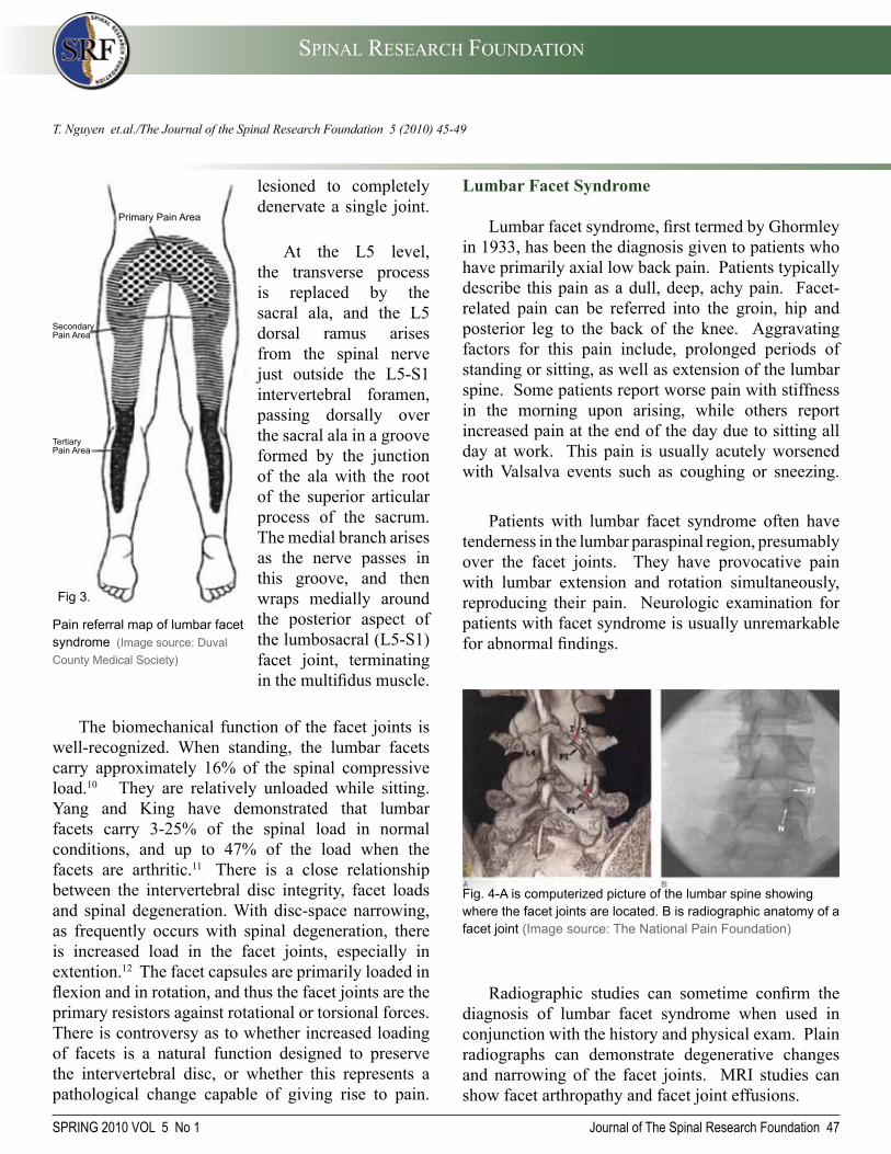
SPRING 2010 VOL 5 No 1
SPINAL RESEARCH FOUNDATION
Journal of The Spinal Research Foundation 47
lesioned to completely denervate a single joint.
At the L5 level, the transverse process is replaced by the sacral ala, and the L5 dorsal ramus arises from the spinal nerve just outside the L5-S1 intervertebral foramen, passing dorsally over the sacral ala in a groove formed by the junction of the ala with the root of the superior articular process of the sacrum. The medial branch arises as the nerve passes in this groove, and then wraps medially around the posterior aspect of the lumbosacral (L5-S1) facet joint, terminating in the multifidus muscle.
The biomechanical function of the facet joints is well-recognized. When standing, the lumbar facets carry approximately 16% of the spinal compressive load.10 They are relatively unloaded while sitting. Yang and King have demonstrated that lumbar facets carry 3-25% of the spinal load in normal conditions, and up to 47% of the load when the facets are arthritic.11 There is a close relationship between the intervertebral disc integrity, facet loads and spinal degeneration. With disc-space narrowing, as frequently occurs with spinal degeneration, there is increased load in the facet joints, especially in extention.12 The facet capsules are primarily loaded in flexion and in rotation, and thus the facet joints are the primary resistors against rotational or torsional forces. There is controversy as to whether increased loading of facets is a natural function designed to preserve the intervertebral disc, or whether this represents a pathological change capable of giving rise to pain.
T. Nguyen et.al./The Journal of the Spinal Research Foundation 5 (2010) 45-49
Lumbar Facet Syndrome
Lumbar facet syndrome, first termed by Ghormley in 1933, has been the diagnosis given to patients who have primarily axial low back pain. Patients typically describe this pain as a dull, deep, achy pain. Facet-related pain can be referred into the groin, hip and posterior leg to the back of the knee. Aggravating factors for this pain include, prolonged periods of standing or sitting, as well as extension of the lumbar spine. Some patients report worse pain with stiffness in the morning upon arising, while others report increased pain at the end of the day due to sitting all day at work. This pain is usually acutely worsened with Valsalva events such as coughing or sneezing.
Patients with lumbar facet syndrome often have tenderness in the lumbar paraspinal region, presumably over the facet joints. They have provocative pain with lumbar extension and rotation simultaneously, reproducing their pain. Neurologic examination for patients with facet syndrome is usually unremarkable for abnormal findings.
Radiographic studies can sometime confirm the diagnosis of lumbar facet syndrome when used in conjunction with the history and physical exam. Plain radiographs can demonstrate degenerative changes and narrowing of the facet joints. MRI studies can show facet arthropathy and facet joint effusions.
Fig. 4-A is computerized picture of the lumbar spine showing where the facet joints are located. B is radiographic anatomy of a facet joint (Image source: The National Pain Foundation)
Pain referral map of lumbar facet syndrome (Image source: Duval County Medical Society)
Secondary Pain Area
Tertiary Pain Area
Fig 3.
Primary Pain Area

The Evolution of Spinal Health Care
SPRING 2010 VOL 5 No 1
SPRING 2010
Journal of The Spinal Research Foundation 48
studies have tried to desensitized the facet joints using radiofrequency coagulation, injection of neurolytic phenol solution and cryoablation.14, 15
Radiofrequency Ablation of the Facet Joint
Radiofrequency facet joint denervation is a percutaneous, nonsurgical procedure to desensitize facet joints that have been identified as the major pain generator for axial low back pain. Using special insulated needles, a heat lesion is created around the region of the dorsal ramus medial branch nerves that come from above and below the facet joint in question. In addition, sensory and motor stimulation is done at the time of needle placement, prior to the radiofrequency ablation, to confirm proximity to the sensory dorsal ramus medial branch nerve while avoiding the spinal nerve root. Stimulation is carried out, using a frequency of 50 Hz and a current up to 1 mA for sensory detection, and a frequency of 2 Hz with current between 3-5 mA for motor stimulation. A positive stimulation is that which reproduces the patient’s pain, without producing other sensory or motor findings in the lower extremity or buttocks. Once the stimulation pattern is acceptable, a radiofrequency lesion is created by passing current through the electrode to raise the tissue temperature to 60-80 degrees centigrade for 60-90 seconds. Adequate local anesthesia and intravenous sedation is used during this portion of the procedure, as it may be quite uncomfortable. Complications from radiofrequency lumbar facet ablation are few, if the procedure is performed correctly. Most patients will experience significant muscular pain for several days after the procedure.
Common possible complications, such as infection and bleeding, are more likely to arise from needle placement than the actual radiofrequency ablation. Another clinical entity encountered in some patients is that of post-denervation neuritis. It manifests as, what is typically described as, a sunburn-like feeling in the paralumbar region. It is usually more annoying than painful, and resolves spontaneously in all cases within six to eight weeks. The exact etiology of this
History of Facet Joint Denervation
The primary diagnostic test to determine whether facet joint pathology causes or contributes to low back pain has been the injection of local anesthetic and corticosteroid solution into the joint or onto the medial branch of the dorsal primary ramus. Typically, 1-2 ml of local anesthetic is instilled into the joint in question; larger volumes will cause rupture of the joint capsule, with subsequent extravasation of solution to other potential pain-generating tissues, which makes interpretation of the injection results problematic.
The dorsal ramus medial branch is typically blocked with 1 ml of local anesthetic injected at the superior aspect of the root of the transverse process at the level in question. Pain relief with injection confirms that the facet joint is the primary pain generator since the small volume of injectate minimizes the spread of anesthetic and corticosteroid to any other structures. Depending on the severity of the disease, the therapeutic duration may be short-lived with the corticosteroid. Radiofrequency nerve ablation, also known as rhizotomy, is a technique which can possibly provide a longer duration of pain relief once the facet joint has been identified as the pain generator.
There have been many studies done on treating lumbar facet syndrome by interrupting its sensory innervation by the dorsal ramus medial branch nerve. The first report was done by Rees who described surgical ligation of sensory nerve supply to the joint using a #11 blade, citing a success rate of 99.8% in 1000 patients with low back pain.13 Subsequent
These joints seem like they fit together Bone Spurs Extra Fluid in Joint
Fig. 5-(Left) Normal facet joints. (Right) Example of facet degeneration. (Image courtesy of Mark Wolgin, MD)

SPRING 2010 VOL 5 No 1
SPINAL RESEARCH FOUNDATION
Journal of The Spinal Research Foundation 49
symptom is unclear. Some practitioners recommend treating the patient with membrane stabilizing agents such as gabapentin or pregabalin.
Pain relief from lumbar radiofrequency facet denervation has ranged from a dismal 9%to a gratifying 83%.16, 17 Comparison between studies is very difficult. As in many of the earlier studies, it is not clear whether an appropriate target was actually used. In some cases, it is not clear whether any type of diagnostic block was performed to identify the pain generator before radiofrequency facet denervation.
Conclusion
Chronic low back pain is a predominant problem in our society that places a heavy social and economic burden in our lives. While there can be many causes or pain generators for low back pain, radiofrequency facet denervation is one interventional, nonsurgical treatment that can provide significant pain relief to improve overall function and minimize requirements for medications.
References
1. Boachie-Adjei O. Evaluation of the patient with low back pain. Postgrad Med. 1988;84(3):110-119.
2. Wall PD, Melzack R, Bonica JJ. Textbook of Pain. 3rd edition ed. New York: Churchill Livingstone; 1994.
3. Johnson I. Radiofrequency percutaneous facet rhizotomy. J Neurosurg Nurs. 1974;6(2):92-96.
4. Goldthwait JE. The Lumbosacral Articulation: an explanation of many cases of ‘lumbago,’ ‘sciatica,’ and ‘paraplegia. Boston Medical Surgical Journal. 1911;7(64):365-372.
5. Ghormley RK. Low Back Pain: with Special Reference to the Articular Facets, with Presentation of an Operative Procedure. J Am Med Assoc. 1933;101(23):1773-1777.
6. Mixter WJ, Barr JS. Rupture of the intervertebral disc with involvement of the spinal cord. N Engl J Med 1934;211:210-214.
7. Hirsch C, Ingelmark BE, Miller M. The anatomical basis for low back pain. Studies on the presence of sensory nerve endings in ligamentous, capsular and intervertebral disc
structures in the human lumbar spine. Acta Orthop Scand. 1963;33:1-17.
8. Mooney V, Robertson J. The facet syndrome. Clin Orthop Relat Res. 1976(115):149-156.
9. Bogduk N, Long DM. The anatomy of the so-called «articular nerves» and their relationship to facet denervation in the treatment of low-back pain. J Neurosurg. 1979; 51(2):172-177.
10. Adams M, Hutton W. The effect of posture on the role of the apophysial joints in resisting intervertebral compressive forces. J Bone Joint Surg Am. 1980;62-B(3):358-362.
11. Yang KH, King AI. Mechanism of facet load transmission as a hypothesis for low-back pain. Spine 1984; 9(6):557-565.
12. Dunlop R, Adams M, Hutton W. Disc space narrowing and the lumbar facet joint. J Bone Joint Surg Am. 1984; 66-B(5):706-710.
13. Rees W. Multiple Bilateral Subcutaneous Rhizolysis of Segmental Nerves in the Treatment of the Intervertebral Disc Syndrome. Ann Gen Pract. 1971;16:126-127.
14. Shealy CN. Percutaneous radiofrequency denervation of spinal facets. Treatment for chronic back pain and sciatica. J Neurosurg. Oct 1975;43(4):448-451.
15. Shealy CN. Facet denervation in the management of back and sciatic pain. Clin Orthop Relat Res. 1976(115):157-164.
16. Lora J, Long D. So-Called Facet Denervation in the Management of Intractable Back Pain. 1976;1(2):121-126.
17. Ogsbury JS, 3rd, Simon RH, Lehman RA. Facet «denervation» in the treatment of low back syndrome. Pain. Jun 1977;3(3):257-263.
Thomas T. Nguyen, MDDr. Nguyen specializes in advanced, minimally invasive diagnostic and treatment modalities for acute and chronic pain syndromes. Dr. Nguyen has practiced pain medicine since finishing his pain fellowship at the Mayo Clinic in 1999. He was the founder and medical director of the Comprehensive Pain Management Center in Newport News,
VA from 1999-2002. He is an active member of the American Academy of Pain Medicine, the International Spine Intervention Society and the American Academy of Family Practice. Dr. Nguyen is involved in several national multicenter studies for the treatment of chronic back pain.
T. Nguyen et.al./The Journal of the Spinal Research Foundation 5 (2010) 45-49

SPRING 2010 VOL 5 No 1
SPRING 2010
Journal of The Spinal Research Foundation 50
Ultrasound Imaging
Ultrasound imaging is emerging as an invaluable tool in the detection and treatment of patients
with motor control impairments. It is particularly effective in the identification of muscle dysfunction and morphology of the multifidus and transverse abdominus muscles in patients with lower back pain. These muscles, when weakened by injury or inactivity, have been linked to lower back pain and increased recurrence of lower back pain.1
Ultrasound imaging is by no means a new concept in healthcare. It has been used since the early 1960’s as a research method of measuring muscle atrophy of the quadriceps, but only in recent years has ultrasound imaging been used as an intervention tool by physical therapists. Until 2006, physical therapists in North America were not allowed to use ultrasound imaging. However, in 2007, Rehabilitative Ultrasound Imaging (RUSI) was introduced into the physical therapy profession. RUSI is defined as, “A procedure used by physical therapists to evaluate muscle and soft tissue related morphology and behavior during physical tasks. This includes providing feedback to the patient and physical therapist to improve outcomes.1
Over the past decade, we have deepened our understanding of the mechanisms of motor control
The American Physical Therapy Association (APTA) has developed the following goals for the physical therapy profession to reach by 2020. First, treatment will be provided by physical therapists who are doctors of physical therapy and who may be board-certified specialists. Secondly, consumers will have direct access to physical therapists in all environments for patient/client management, prevention and wellness services. The third goal, will be to continue to provide direct patient/client care and maintain active responsibility for the growth of the physical therapy profession and the health of the people it serves. Lastly, physical therapists will have access to numerous types of new technologies and treatment options, as well as basic and clinical research. Advances in technology and treatment tools will continue to strengthen the physical therapy profession and allow therapists to provide the most effective care for their patients. Some of the most novel advances in the physical therapy profession include the evolution of ultrasound imaging, dry needling and virtual reality technology. This article will discuss each of these future advances in physical therapy and their potential benefits to patients.
Keywords: Ultrasound images, dry needling
and neuromuscular dysfunction, particularly of the stabilizing muscles of the spine (multifidus, transverse abdominus, pelvic floor, and diaphragm). Muscles, because of their depth, have been extremely difficult for physical therapists to palpate and even more difficult for patients to identify during re-education training. Ultrasound Imaging can assess the performance of core muscles, which helps the manual therapist to functionally re-train an individual with lumbopelvic dysfunction.3 This procedure has proven to be a reliable and accurate tool to measure the shape, size and architecture of muscles.1
The clinical application of ultrasound has been used primarily as a biofeedback method in teaching patients how to contract these muscle groups. Recent
Physical Therapy in 2020Richard Banton, MPT, T-DPT, ATC
Ultrasound of the Rectus Abdominus (Image Courtesy of Dr. Joe Antony)

SPRING 2010 VOL 5 No 1
SPINAL RESEARCH FOUNDATION
Journal of The Spinal Research Foundation 51
studies have shown that incorporating visual feedback is the most effective method of instructing patients to identify and contract muscles.2 Ultrasound imaging allows for the dynamic study of muscle groups as they contract. This assists therapists in the screening and re-training of individuals with lumbopelvic dysfunction by providing a visual image of the patient’s primary muscular stabilizers. For instance, ultrasound imaging helps patients visualize and control the muscles necessary to stabilize the spine. It has been well established, in both normal and patient populations, that simply asking an individual to contract the pelvic floor muscles correlates poorly with the ability to successfully produce a contraction.2 Bump et al. demonstrated that only 49% of a patient population could effectively perform pelvic floor contraction when given verbal or written instructions. However, when ultrasound imaging was used as visual feedback for the patient, the percentage improved to 85%.4
The future of physical therapy will involve the use of ultrasound imaging to help patients visualize their motor control system and to help them understand the anatomy of their injury. Appropriate use of this tool will enhance the rehabilitation of lumbopelvic dysfunction and lumbar instability. The challenge in the future for the physical therapy profession will be to determine when to introduce ultrasound imaging into their practice and then to educate their clinicians on its proper application.
Dry Needling
Dry needling is a treatment technique that has been used around the world for decades. Dry needling is within the scope of physical therapy practices in many countries including Canada, Spain, Ireland, South Africa, Australia, the Netherlands and Switzerland. In the United States, trigger point dry needling has just recently been approved in a growing number of states. The physical therapy boards of eight states in the US have ruled that physical therapists can engage in the practice of Dry Needling: New Hampshire, Maryland, Virginia, South Carolina, Georgia, Kentucky, New Mexico, and Colorado.8
According to The American Academy of Orthopedic Manual Physical Therapy (AAOMPT), dry needling is a neuro-physiological, evidence-based treatment technique that requires effective manual assessment of the neuromuscular system. Physical therapists are well trained in the incorporation of dry needling techniques utilized in conjunction with manual physical therapy interventions. Research supports that dry needling improves pain control, reduces muscle tension, normalizes biochemical and electrical dysfunction of motor endplates, and facilitates an accelerated return to active rehabilitation.
R. Banton et.al./The Journal of the Spinal Research Foundation 5 (2010) 50-54
Example of Dry Needling (Image Courtesy of Virginia Therapy and Fitness)
Example of Ultrasound (Image Source: Therapy Times)

The Evolution of Spinal Health Care
SPRING 2010 VOL 5 No 1
SPRING 2010
Journal of The Spinal Research Foundation 52
Positive outcomes for patients are achieved when dry needling is used in combination with other manual therapy treatments. When combined with manual therapy and exercise, it has proven to be an effective treatment for the conditions such as low back pain, whiplash, headaches, chronic pelvic pain, complex regional pain syndromes, and fibromyalgia.4 The effectiveness of dry needling is highly determined by the skill level of the clinician. Currently there are only two physical therapy curriculums that offer entry level training in dry needling. These curriculums ultimately enable the clinician to palpate myofascial trigger points and then to use the needle as a palpation tool to appreciate changes in the firmness of those tissues requiring treatment.4
Dry needling is most effectively used in the treatment of myofascial trigger points, hyperirritable spots in skeletal muscles associated with a hypersensitive palpable nodule in a taut band. Current scientific evidence strongly supports that awareness
and working knowledge of muscle dysfunction, in particular myofascial trigger points, should be incorporated into manual physical therapy practice.5,8 Myofascial trigger points are commonly treated with manual techniques, spray and stretch, dry needling, or injection therapy.
The mechanisms behind dry needling remain elusive. There is some evidence that dry needling changes the quantities of cholinesterase as these enzymes try to bind to the Ach receptors as part of normal muscle regeneration. A common finding during the process of dry needling is a phenomenon known as the “needle grasp”. This has been attributed to the muscle fibers contracting around the needle and holding it tightly in place.4 Studies are showing that during a needle grasp, fibroblasts actually change shape and lead to a variety of cellular and extracellular events, including mechanoreceptor (proprioception) stimulation and nociceptor (pain) inhibition. Maneuvering the needle by a skilled therapist may stimulate A fibers and activate chemicals, enkephalinergic and serotonergic, and noradrenergic inhibitory systems that together help modulate pain at the brain and spinal levels.5
The introduction of dry needling in the United States has many similarities with the introduction of manual therapy in the early 1960s. In the last decade, there has been much progress made towards understanding, not only myofascial trigger points, but the effects that dry needling and combined manual therapy can have upon treating them. Dry needling has been recognized by prestigious organizations such as the Cochrane Collaboration and is recommended as an option for the treatment of persons with chronic lower back pain.8 Several clinical outcome studies have demonstrated the effectiveness of trigger point dry needling.8 Physical therapists should be encouraged to educate themselves and their staff on the benefits of dry needling and integrate the treatment into their clinical practice and management of chronic pain.
Virtual Reality Technology
Although this technology first became available more than 25 years ago, it has only emerged in
Nerve Synapses (Adapted by permission from Macmillan Publishers Ltd: [Nature Reviews Immunology] “Unravelling the pathogenesis of myasthenia gravis” A Vincent; 2:10;797-804: 2002 )

SPRING 2010 VOL 5 No 1
SPINAL RESEARCH FOUNDATION
Journal of The Spinal Research Foundation 53
rehabilitation clinics over the past five years. Video capture virtual reality (VR) uses a video camera to track movement in a single plane without the need to place markers on specific body parts. The patient is thereby embedded within a simulated environment so that they may interact with animated graphics in a completely natural manner.
A major goal of any physical therapist is to make quantitative and qualitative improvements in the daily activities of their patients to help them improve their quality of independent living. The successful integration of virtual reality into physical therapy has demonstrated the potential for technology to present opportunities to engage in challenging, but safe, environments while maintaining experimental control over the stimulus delivery and measurement.
A major challenge for physical therapists is identifying effective and motivational intervention tools that enable the transfer of skills and abilities during the rehabilitation process to function in the
real world. Virtual reality technology allows physical therapists to produce simulated, interactive and multi-dimensional environments. Visual interfaces are used to create environments allowing patients to interact with images and virtual objects in real-time through multiple sensory modalities. Examples of these include desktop monitors and head mounted displays, haptic interfaces (joy sticks, gloves, pens, and exoskeletons), and real-time motion tracking devices.
Recent research has shown that virtual reality technology is similar enough to reality to provide an effective training environment for rehabilitation. Patients that benefitted the most from virtual reality technology include victims of stroke and vestibular disorders, the elderly, and patients with a variety of neurological conditions. In regards to spine pathology, those suffering from a reduction in proprioception and balance as a result of their spine condition, have shown improvement in their ability to balance by training with virtual reality technology.9 Examples of virtual technology systems are Vividgroup, IREX systems, and GaitMaster2.
These systems take a video image of the patient and use color subtraction software to remove a monochrome background and insert the patient into a virtual reality environment. They also provide the
Example of Virtual Reality Technology (Image Courtesy of Exergame Fitness USA “Bringing Exercise and Game Play Together”)
R. Banton et.al./The Journal of the Spinal Research Foundation 5 (2010) 50-54
Example of Virtual Reality Technology (Image Courtesy of Exergame Fitness USA “Bringing Exercise and Game Play Together”)

The Evolution of Spinal Health Care
SPRING 2010 VOL 5 No 1
SPRING 2010
Journal of The Spinal Research Foundation 54
References1. Whittaker, J 2004 Abdominal Ultrasound Imaging of Pelvic Floor Muscle Function in Individuals with Low Back Pain, The Journal of Manual and Manipulative Therapy: Vol. 12 No. 1 (2004) 44-49
2. Bump RC, Hurt GW, Fantl JA Wyman JF. Assessment of Kegal pelvic muscle exercise performance after brief verbal instruction. Am J Obstet Gynecol 1991;165:322-329
3. Dommerholt J. Trigger Point Dry Needling, The Journal of Manual and Manipulative Therapy: 2006 Vol. 14 (4) 70-87
4. Dommerholt J, Bron C, Franssen J. Myofascial Trigger Points: An Evidence-Informed Review. The Journal of Manual and Manipulative Therapy: 2006 Vol. 14 (4) 203-221
5. Sahed M, Stern LZ, Czyzewski K. Changes in end-plate cholinesterase and axons during muscle degeneration and regeneration. Journal of Anatomy 1985; 140: 165-176
6. Bowsher D. Mechanisms of acupuncture. In: J Filshie, A White, eds. Western Acupuncture: A Western Scientific Approach. Edinburgh, UK: Churchill Livingstone, 1998
7. Sveistrup H, Motor rehabilitation using virtual reality. Journal of NeuroEngineering and Rehabilitation: 2004 Vol. 10 (1)
8. Weiss PL, Rand D, Katz N, Kizony R. Video capture reality as a flexible and effective rehabilitation tool. Journal of NeuroEngineering and Rehabilitation. 2004 Vol. 12 (1)
patient with a sense of forward movement while his/her actual position in space is unchanged. The patient stands on two footpads that move individually with each user’s foot providing a sense of movement over a virtual terrain. The footpads follow the trajectory of a healthy individual when walking. Thus the patient experiences a corrected foot trajectory for each step.9
From the beginning, virtual reality systems were developed by the gaming industry strictly for entertainment purposes. The application of this technology was clearly evident to the physical therapy profession. Although the short-term results of using virtual reality technology to improve function in the real world appear positive, the implications for the long-term benefit of virtual reality systems has yet to be determined. Another potential disadvantage to virtual reality software is the expense to the physical therapist, although with recent advances in technology many of these systems have become more affordable. As new technology is developed, and as the press generates a considerable amount of interest in virtual reality technology, the physical therapy profession only anticipates an increase in its popularity and the successful treatment outcomes that it will bring to their patients.
As technological advances occur, physical therapists must continue to improve their knowledge and skils through continuing education, training and evidence-based research. The future success of physical therapy lies in the hands of our educators and practicing clinicians. Advances can only take place if these professionals are willing to embrace change and become educated on the benefits of new technology and treatment options, and to ensure that patients have access to the most effective treatments for their condition. Our professional goals are well within our reach as we continue to observe patient successes and expand our knowledge through evidence-based research that supports the effectiveness of physical therapy. The future of physical therapy appears bright as The American Physical Therapy Association strives to achieve its goals for 2020.
Richard Banton, PT, DPT, ATC Richard Banton has served as co-clinic director for Virginia Therapy and Fitness Center since its inception in 2004. He has been practicing physical therapy since 1998, working with a variety of
orthopedic, neurologic, and pediatric conditions. His extensive experience includes the treatment of athletes from the high school to collegiate and professional levels; including clientele such as Olympic athletes, Washington Redskins football players, and other athletes from NASCAR and the LPGA.

SPRING 2010 VOL 5 No 1
SPINAL RESEARCH FOUNDATION
Journal of The Spinal Research Foundation 55
Case Report
A seventy-seven year old female presented with ongoing back and lower extremity pain. She was
diagnosed with degenerative lumbar scoliosis, stenosis, lumbar radiculopathy and neurogenic claudication. Since she had failed extensive nonoperative conservative treatment, surgical intervention was an option for her.
Various surgical treatment options were considered. First, the goal of surgery had to be determined. One goal was to decompress the nerves. It was hoped that by decompressing the nerves, the lower extremity symptoms would improve. Options for decompressing the nerves would be laminectomy or foraminotomy. Her second chief complaint was that of axial back pain. Axial back pain can originate from many sources, including the arthritic changes associated with degenerative scoliosis. Axial back pain would not be relieved by nerve decompression. A third concern was the possibility of her lumbar scoliosis progressing even further without any surgical intervention. Fusion of the vertebrae would be indicated to relieve the axial back pain and align the lumbar spine. Additionally, an indirect decompression of the nerves at the foramina could be performed by improving the alignment of the scoliosis.
Surgical Procedure
In an effort to address these three goals, we elected to proceed with the direct lateral interbody fusion of the lumbar spine. This minimally invasive retroperitoneal approach allowed for significant improvement in the patient’s scoliosis deformity and indirect decompression of the foramina, as well as the canal, during realignment. It also required significantly less posterior surgery than would have been required if the full decompression had been done directly.
Results
This procedure allowed the patient to be mobilized sooner than traditionally would have been an option for her. This can help decrease the risk of postoperative morbidities associated with prolonged bed rest and decreased activities. Preoperative versus postoperative radiographs demonstrated significant improvement in the lumbar scoliosis. Her clinical
Direct Lateral Interbody Fusion Approach in the Treatment of a Patient Suffering from Degenerative Lumbar Scoliosis, Stenosis, Lumbar Radiculopathy, and Neurogenic ClaudicationMichael W. Hasz, MD, FACS
Background Context: Several recent surgical advances allow patients to be treated in a less invasive manner, yet still afford the patient the significant benefits that have been afforded in the past by other techniques. These advances have changed our treatment of adolescent scoliosis. A deformity can often be better corrected and anterior surgery avoided by utilizing pedicle screw instrumentation technology and segmental posterior instrumentation.
Purpose: To demonstrate the utility of a new approach for minimally invasive spinal fusion.
Methods: Case report of a patient presenting with ongoing back pain and lower extremity pain who underwent a minimally invasive interbody fusion.
Results: The patient showed significant improvement of both axial back pain as well as lower extremity symptoms.

SPRING 2010 VOL 5 No 1
SPRING 2010
Journal of The Spinal Research Foundation 56
CASE REpoRT
report demonstrated significant improvement of both axial back pain as well as her lower extremity symptoms.
Discussion
Direct lateral interbody fusion is an innovative surgical procedure. This surgical procedure expands upon the advantage of a retroperitoneal approach to the anterior lumbar spine. It involves using a technique of entering the disc, preparing the disc, and placing an implant for interbody fusion through a more lateral approach. This is directed through and between the muscle fibers of the psoas muscles to enter the disc space. This facilitates the use of smaller skin incisions while maintaining the benefits of the retroperitoneal approach. The direct lateral interbody techniques appear to offer a significant improvement in the treatment of some patient’s conditions. This approach is a useful tool for a well rounded surgeon to have.
Fig 1. -Anterior-posterior x-ray of the lumbar spine prior to surgery (left) and after surgery (right), showing scoliosis correction
Fig. 2 -Lateral x-ray of the lumbar spine prior to surgery (left) and after surgery (right), showing curve correction and lordosis restoration
Michael W. Hasz, MD, FACS
Dr. Hasz is board certified by the American Board of Spine Surgery. He is a fellow in the American Academy of Orthopaedic Surgeons and a
member of both the American Association of Orthopaedic Surgeons and the North American Spine Society. He was Chairman of the Department of Orthopaedic Surgery and Director of Spinal Surgery at the Andrews Air Force Base / Malcolm Grow Medical Center in Maryland. He currently holds an appointment as Clinical Instructor of Orthopaedic Surgery and Assistant Professor of Surgery at the Uniformed Services Health Science University in Bethesda, Maryland.
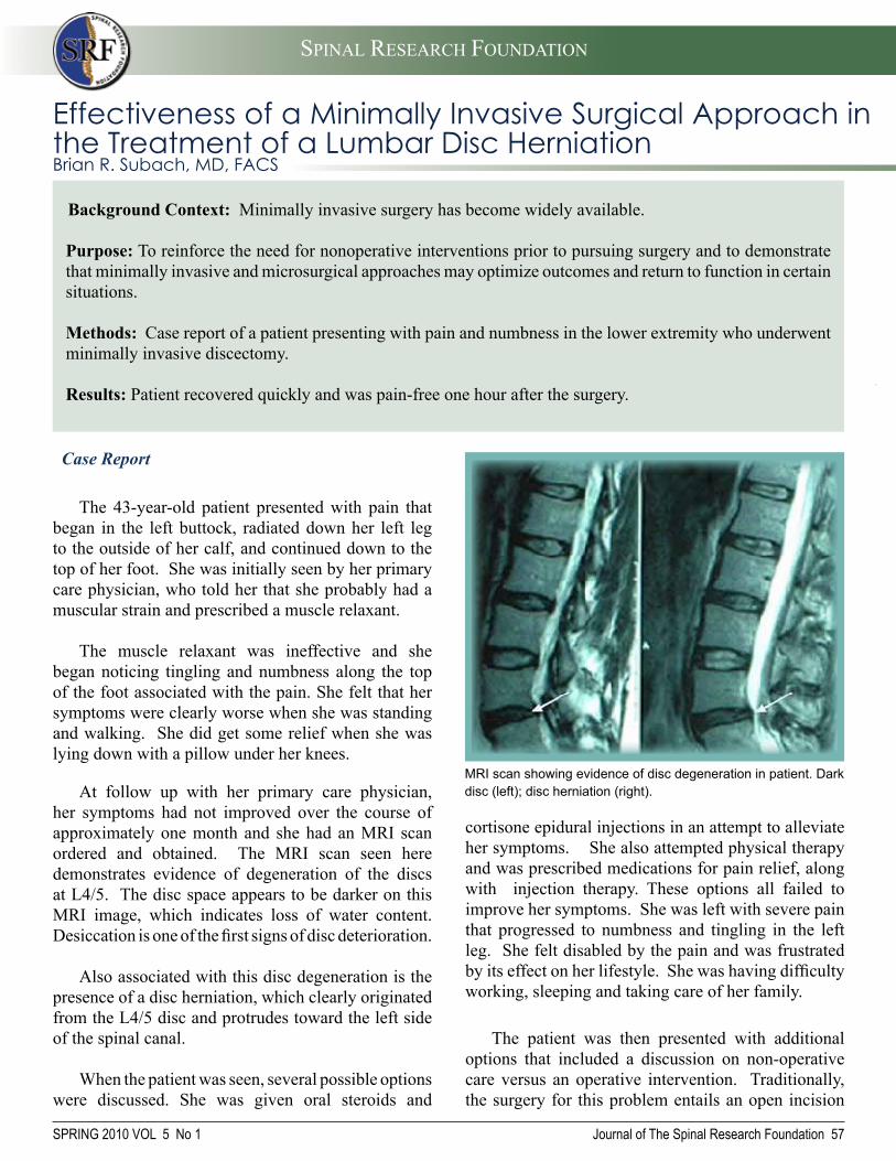
SPRING 2010 VOL 5 No 1
SPINAL RESEARCH FOUNDATION
Case Report
The 43-year-old patient presented with pain that began in the left buttock, radiated down her left leg to the outside of her calf, and continued down to the top of her foot. She was initially seen by her primary care physician, who told her that she probably had a muscular strain and prescribed a muscle relaxant.
The muscle relaxant was ineffective and she began noticing tingling and numbness along the top of the foot associated with the pain. She felt that her symptoms were clearly worse when she was standing and walking. She did get some relief when she was lying down with a pillow under her knees.
At follow up with her primary care physician, her symptoms had not improved over the course of approximately one month and she had an MRI scan ordered and obtained. The MRI scan seen here demonstrates evidence of degeneration of the discs at L4/5. The disc space appears to be darker on this MRI image, which indicates loss of water content. Desiccation is one of the first signs of disc deterioration.
Also associated with this disc degeneration is the presence of a disc herniation, which clearly originated from the L4/5 disc and protrudes toward the left side of the spinal canal.
When the patient was seen, several possible options were discussed. She was given oral steroids and
Journal of The Spinal Research Foundation 57
Background Context: Minimally invasive surgery has become widely available.
Purpose: To reinforce the need for nonoperative interventions prior to pursuing surgery and to demonstrate that minimally invasive and microsurgical approaches may optimize outcomes and return to function in certain situations.
Methods: Case report of a patient presenting with pain and numbness in the lower extremity who underwent minimally invasive discectomy.
Results: Patient recovered quickly and was pain-free one hour after the surgery.
MRI scan showing evidence of disc degeneration in patient. Dark disc (left); disc herniation (right).
Effectiveness of a Minimally Invasive Surgical Approach in the Treatment of a Lumbar Disc HerniationBrian R. Subach, MD, FACS
cortisone epidural injections in an attempt to alleviate her symptoms. She also attempted physical therapy and was prescribed medications for pain relief, along with injection therapy. These options all failed to improve her symptoms. She was left with severe pain that progressed to numbness and tingling in the left leg. She felt disabled by the pain and was frustrated by its effect on her lifestyle. She was having difficulty working, sleeping and taking care of her family.
The patient was then presented with additional options that included a discussion on non-operative care versus an operative intervention. Traditionally, the surgery for this problem entails an open incision

SPRING 2010 VOL 5 No 1
SPRING 2010
Tubular retractor (Image Courtesy of Medtronic)
Journal of The Spinal Research Foundation 58
L4
L5
Fluoroscopic guidance is used to identify the L4/5 level and to correctly place the surgical instruments.
Surgical instruments are passed through a 1 inch incision.
CASE REpoRT
which strips the muscle from the spine and allows the surgeon to remove a window of bone, removing the disc herniation directly.
The patient was also offered the possibility of a minimally invasive approach using a tubular retractor which does not cause any damage to the muscle, allowing for an operating microscope to visualize the critical structures of the spine and allowing a laser to vaporize the disc herniation, thereby minimizing damage to the disc already showing signs of degeneration. The patient decided to pursue the minimally invasive approach.
Operative Procedure
The patient was taken to the operating room where she was placed in the prone position on the Wilson frame. Fluoroscopic guidance was used to identify the L4/5 level. A one-half inch incision was made approximately one inch from the midline. Through that a series of tubular dilators were used to displace the muscle without causing any damage and identify the area of disc herniation. Using a microscope to visualize these structures the nerve roots were gently mobilized, the disc herniation was identified and vaporized using a microsurgical laser. The nerve roots were carefully returned to the normal position, absorbable sutures were placed and a band-aid placed over the wound.
Results
The patient awoke from anesthesia and was taken to the recovery room. She was discharged home approximately one hour later, no longer having any pain in the left leg.

SPRING 2010 VOL 5 No 1
SPINAL RESEARCH FOUNDATION
Journal of The Spinal Research Foundation 59
5k race Reston, VA
4 Mile Race Newtown, PA
CoMiNg This FAll! 5k RACe sAN FRANCisCo BAy AReA
We’ve Got Your Back
SPRING 2010 VOL 5 No 1
SPRING 2010
Journal of The Spinal Research Foundation 60
Kids corner
SPINAL HEALTH FAIR
FUN WALK

SPRING 2010 VOL 5 No 1
SPINAL RESEARCH FOUNDATION
Journal of The Spinal Research Foundation 61
The Spinal Research Foundation has named 22 Regional Research Affiliates across the country that share one core mission: improving spinal health care for the future. These centers offer the best quality spinal health care while focusing on research programs
designed to advance spinal treatments and techniques.
The Virginia Spine InstituteThomas C. Schuler, MD, FACS, President
Brian R. Subach, MD, FACS, Director of Research
1831 Wiehle AveReston, VA 20190
703-709-1114
The Orthopaedic and Sports Medicine Center
Contact: Girard J. Girasole, MD888 White Plains RdTrumbull, CT 06611
203-268-2882
New England Neurosurgical
AssociatesNew England Neurosurgical
Associates, LLCContact: Christopher H. Comey, MD
300 Carew St Ste OneSpringfield, MA 01104
413-781-2211
Colorado Comprehensive Spine Institute
Contact: George Frey, MD3277 South Lincoln St.Englewood, CO 80113
303-762-0808
Spinal Research Foundation Regional Research Affiliates
MUSC Darby Children’s Research Institute
Contact: Inderjit Singh, PhD59 Bee St MSC 201
Charleston, SC 294251-800-424-MUSC
The Spine Clinicof Los Angeles
Contact: Larry T. Khoo, MD1245 Wilshire Blvd, Ste 717
Los Angeles, CA 90017213-481-8500
Menlo Medical ClinicContact: Allan Mishra, MD
1300 Crane St. Menlo Park, CA 94025
650-498-6500
stanford university
Virginia Therapy & Fitness CenterContact: Richard A. Banton, PT, DPT, ATC and E. Larry Grine, PT, MSPT, ATC,
CSCS1831 Wiehle AveReston, VA 20190
703-709-1116

SPRING 2010 VOL 5 No 1
SPRING 2010
Journal of The Spinal Research Foundation 62
Spinal Research Foundation Regional Research Affiliates
Atlanta Brain and Spine CareContact: Regis W. Haid, Jr., MD2001 Peachtree Rd, NE, Ste 575
Atlanta, GA 30309404-350-0106
SpineCare Medical GroupContact: Paul J. Slosar,Jr., MDSan Francisco Spine Institute
1850 Sullivan AveDaly City, CA 94015
650-985-7500
Southern Brain and SpineContact: Najeeb M. Thomas, MD
3601 Houma Blvd. Ste 400Metairie, LA 70006
504-889-7200
Princeton Brain and Spine CareContact: Mark R. McLaughlin, MD, FACS
713 Executive DrPrinceton, NJ 08540
609-921-9001
The Orthopedic Center of St. LouisContact: Matthew F. Gornet, MD
14825 N. Outer Forty Rd, Ste 200Chesterfield, MO 63017
314-336-2555
The Hughston ClinicContact: J. Kenneth Burkus, MD
6262 Veterans ParkwayColumbus, GA 31909
706-324-6661
Twin Cities Spine CenterContact: James Schwender, MD
913 East 26th St Ste 600Minneapolis, MN 55404
612-775-6200
South Coast Orthopaedic Associates Contact: Aleksandar Curcin, MD, MBA
2699 N. 17th StCoos Bay, OR 97420
541-266-3600
Allegheny Brain and Spine Surgeons Contact: James P. Burke, MD, PhD
501 Howard Ave, Building E-1Altoona, PA 16601
814-946-9150
Rutgers University Department of Biomedical Engineering
Contact: Noshir A. Langrana, PhD, PE 599 Taylor Rd
Piscataway, NJ 08854732-445-4500
RUTGERS
Indiana Spine GroupContact: Richard C.Sasso, MD,
FACS8402 Harcourt Rd
Suite #400Indianapolis, IN 46260
Inova Research CenterContact: Zobair M. Younossi, MD,
MPH3300 Gallows Rd
Falls Church, VA 22042-3300703-776-2580
INOVA ReseARchceNteR
University of Minnesota Medical Center, Fairview
Contact: David W. Polly, Jr., MD2450 Riverside Ave, South
Minneapolis, MN 55454612-672-7575
Oregon Neurosurgery SpecialistsContact:Robert J. Hacker, MD and
Andrea Halliday, MD3355 RiverBend Dr
Ste 400Springfield, OR 97477
541-686-8353

Your Online Source for Spinal Health Care!
We provide a forum for addressing questions related to the problems encountered daily by Americans who suffer from acute or
chronic spinal conditions. Topics include:
• preventative care• non-surgical treatment options• minimally invasive procedures• laser spine surgery• diagnostic procedures• surgical care
Visit us at www.SpineHealthSource.com
Support Cutting Edge
Research
The Spinal Research Foundation has made remarkable progress in scientific research associated with
neck and back pain. The Foundation
has been involved in numerous studies, some of which include:
Your Gift Matters
• Visit www.SpineRF.org to make a secure online donation.
• Call 703-766-5405 to make a donation over the phone.
• The Spinal Research Foundation is a non-profit 501(c)(3) organization. Donations are tax deductible.
• The use of novel perioperative drug therapy to improve surgical outcomes
• The evaluation of medical devices for the relief of back pain
• The development of non-operative techniques to resolve disabling neck and back pain
• Investigating the use of BMP (Bone Morphogenetic Protein) in minimally invasive spinal surgery to minimize post-operative pain and dysfunction
• The development of cervical and lumbar disc replacement technologies
• The investigation of lactic acid polymers to prevent fibroblast in-growth in surgical wounds
• A nation-wide multi-center prospective spine treatment outcomes study
For more information on current research projects please visit:
You can help! The Spinal Research Foundation is America’s leading non-profit health organization dedicated to spinal health. Friends like you have made it possible for us to make huge strides and groundbreaking research discoveries. Join us in our mission to promote spinal health. Support cutting edge research by making a
donation to The Spinal Research Foundation.
The Spinal Research Foundation recognizes our outstanding clinicians and researchers in the
field of spinal research and profiles them as Spinal Champions. These dedicated spine care professionals embrace excellence in both research and education, contributing significantly to improvements in the diagnosis and treatment of spinal disorders. We recognize Paul J. Slosar, Jr., MD, Orthopedic Spine Surgeon at SpineCare Medical Group in the San Francisco Bay area.
Spinal ChampionPaul J. Slosar, Jr., MDSpineCare Medical Group
“I find getting to know my patients personally is as
interesting as the surgery I perform. I seek to understand
them and how their spinal problems affect their lives. This approach acts as my
compass. It guides all of my recommendations. I consider
the individuals and their lives, not just their spines. The
quality of care they receive and their mental and physical well-being are of the utmost
importance to me.”

Education
Research
Volunteerism
Future
Thank You! The Board of Directors of The Spinal Research
Foundation is grateful for the continued
investment of our donors and extends its
appreciation to all who have contributed.
Through the generous support of our donors,
The Spinal Research Foundation has been
able to significantly expand the scope of our
scientific research and educational programs.
These gifts have been utilized to establish
scholarship programs and embark on projects
geared toward understanding the mechanisms
of spinal diseases, and develop new treatments
for these conditions. This work would not be
possible without the support of our donors.
To make a donation and help improve the
quality of spinal health care in America visit:
www.SpineRF.org or contact us at:
The Spinal Research Foundation1831 Wiehle Ave, Ste 200
Reston, VA 20190Phone: 703-766-5405
Fax: 703-709-1397
Related Documents