http://www.diva-portal.org This is the published version of a paper published in Journal of Neuroinflammation. Citation for the original published paper (version of record): Lindqvist, R., Mundt, F., Gilthorpe, J D., Woelfel, S., Gekara, N O. et al. (2016) Fast type I interferon response protects astrocytes from flavivirus infection and virus-induced cytopathic effects. Journal of Neuroinflammation, 13: 277 http://dx.doi.org/10.1186/s12974-016-0748-7 Access to the published version may require subscription. N.B. When citing this work, cite the original published paper. Permanent link to this version: http://urn.kb.se/resolve?urn=urn:nbn:se:umu:diva-127599

Welcome message from author
This document is posted to help you gain knowledge. Please leave a comment to let me know what you think about it! Share it to your friends and learn new things together.
Transcript

http://www.diva-portal.org
This is the published version of a paper published in Journal of Neuroinflammation.
Citation for the original published paper (version of record):
Lindqvist, R., Mundt, F., Gilthorpe, J D., Woelfel, S., Gekara, N O. et al. (2016)Fast type I interferon response protects astrocytes from flavivirus infection and virus-inducedcytopathic effects.Journal of Neuroinflammation, 13: 277http://dx.doi.org/10.1186/s12974-016-0748-7
Access to the published version may require subscription.
N.B. When citing this work, cite the original published paper.
Permanent link to this version:http://urn.kb.se/resolve?urn=urn:nbn:se:umu:diva-127599
RESEARCH Open Access
Fast type I interferon response protectsastrocytes from flavivirus infection andvirus-induced cytopathic effectsRichard Lindqvist1,2, Filip Mundt3, Jonathan D. Gilthorpe4, Silke Wölfel5, Nelson O. Gekara6, Andrea Kröger7,8
and Anna K. Överby1,2*
Abstract
Background: Neurotropic flaviviruses such as tick-borne encephalitis virus (TBEV), Japanese encephalitis virus (JEV),West Nile virus (WNV), and Zika virus (ZIKV) are causative agents of severe brain-related diseases includingmeningitis, encephalitis, and microcephaly. We have previously shown that local type I interferon response withinthe central nervous system (CNS) is involved in the protection of mice against tick-borne flavivirus infection.However, the cells responsible for mounting this protective response are not defined.
Methods: Primary astrocytes were isolated from wild-type (WT) and interferon alpha receptor knock out (IFNAR−/−)mice and infected with neurotropic flaviviruses. Viral replication and spread, IFN induction and response, andcellular viability were analyzed. Transcriptional levels in primary astrocytes treated with interferon or supernatantfrom virus-infected cells were analyzed by RNA sequencing and evaluated by different bioinformatics tools.
Results: Here, we show that astrocytes control viral replication of different TBEV strains, JEV, WNV, and ZIKV. Incontrast to fibroblast, astrocytes mount a rapid interferon response and restrict viral spread. Furthermore, basalexpression levels of key interferon-stimulated genes are high in astrocytes compared to mouse embryonicfibroblasts. Bioinformatic analysis of RNA-sequencing data reveals that astrocytes have established a basal antiviralstate which contributes to the rapid viral recognition and upregulation of interferons. The most highly upregulatedpathways in neighboring cells were linked to type I interferon response and innate immunity. The restriction in viralgrowth was dependent on interferon signaling, since loss of the interferon receptor, or its blockade in wild-type cells,resulted in high viral replication and virus-induced cytopathic effects. Astrocyte supernatant from TBEV-infected cellscan restrict TBEV growth in astrocytes already 6 h post infection, the effect on neurons is highly reinforced, andastrocyte supernatant from 3 h post infection is already protective.
Conclusions: These findings suggest that the combination of an intrinsic constitutive antiviral response and the fastinduction of type I IFN production by astrocytes play an important role in self-protection of astrocytes and suppressionof flavivirus replication in the CNS.
Keywords: Astrocytes, Interferon, TBEV, Flavivirus, Viperin
* Correspondence: [email protected] of Clinical Microbiology, Virology, Umeå University, 90185Umeå, Sweden2The Laboratory for Molecular Infection Medicine Sweden (MIMS), 90187Umeå, SwedenFull list of author information is available at the end of the article
© The Author(s). 2016 Open Access This article is distributed under the terms of the Creative Commons Attribution 4.0International License (http://creativecommons.org/licenses/by/4.0/), which permits unrestricted use, distribution, andreproduction in any medium, provided you give appropriate credit to the original author(s) and the source, provide a link tothe Creative Commons license, and indicate if changes were made. The Creative Commons Public Domain Dedication waiver(http://creativecommons.org/publicdomain/zero/1.0/) applies to the data made available in this article, unless otherwise stated.
Lindqvist et al. Journal of Neuroinflammation (2016) 13:277 DOI 10.1186/s12974-016-0748-7

BackgroundThe genus Flavivirus belonging to the family Flaviviridaeinclude important pathogens causing severe humandisease including meningitis, encephalitis, hemorrhagicfevers, and microcephaly. The most significant neu-rotropic flaviviruses are arthropod-borne tick-borneencephalitis virus (TBEV), West Nile virus (WNV),Japanese encephalitis virus (JEV), and Zika virus (ZIKV).TBEV is transmitted by Ixodes ticks, whereas WNV, JEV,and ZIKV are transmitted via mosquitos. No treatmentsare available for any of these viral infections, and patientsare dependent on innate and adaptive parts of the hostimmune response to fight infections [1–6].The innate immune system presents a first line of
defense against viral infections, for which type I inter-ferons (IFNs) are particularly important. After flavivirusinfection, double-stranded RNA (dsRNA) is produced asan intermediate during viral replication. This is sensedas a danger signal in the infected cell by pattern recogni-tion receptors (PRRs) and a signaling cascade is initiated,which leads to the upregulation of IFNs [7, 8]. IFNs arepowerful cytokines that mediate antiviral effects via bothautocrine and paracrine signaling mechanisms via the IFNalpha receptor (IFNAR). Binding to IFNAR activates thedownstream kinases Janus kinase 1 and tyrosine kinase 2,which phosphorylate signal transducer and activator oftranscription-1 and transcription-2 (STAT1, STAT2). To-gether with interferon regulatory factor-9 (IRF9), theseform a signaling complex referred to as IFN-stimulatedgene factor-3 (ISGF3). ISGF3 translocates to the nucleusand activates the transcription of a large number ofinterferon-stimulated genes (ISGs) by binding to theinterferon response elements. ISGs can inhibit almostevery step of a viral life cycle [9, 10].In mice, the type I IFN response is essential for
protection against TBEV, JEV, WNV, and ZIKVinfections [11–15]. The CNS has been considered as animmune-privileged tissue; however, recent studies haveimplicated the importance of intrinsic, innate antiviralresponses within the CNS [16–19]. In Langat virus(LGTV, Langat virus, low-virulent member of TBEVserogroup) infection, the local type I IFN response in theCNS has been shown to be critical for the protection ofmice against lethal encephalitis [11]. However, the CNScell type responsible for producing IFN during TBEVinfection has not been defined.While neurons are the main target of neurotropic flavi-
viruses, other cell types might also become infected andcontribute to the resolution of infection [20]. Previousstudies have shown that the IFN response and ISG expres-sion in neurons restrict neurotropic flavivirus infection inneurons [19, 21]; however, not much is known about therole of the IFN response in astrocytes during neurotropicflavivirus infection. Recent studies have shown that
astrocytes are important IFN-producing cells in variousneurotropic viral infections [18, 22, 23]. Astrocytes areone of the most abundant cell types in the brain andmediate diverse supportive functions including ionhomeostasis [24, 25], uptake of glutamate [26], free radicalscavenging [27], and immune regulation [28]. In TBEVinfection, autopsy studies have revealed astrogliosis in postmortem human brains [20, 29], which has been observedfor WNV and JEV as well [30, 31]. Indeed, astrocytes havebeen found to be a site of these infections [32]. However,only a few astrocytes were found to be infected inLGTV-infected mice [33]. They resist infection in aninterferon-beta promoter stimulator 1 (IPS-1)-dependentmanner and show an activated phenotype, indicating theirinvolvement in LGTV clearance. Both rat and humanastrocytes have been shown to be infected in vitro withTBEV; however, the number of infected cells neverexceeded 20 %, and the infection did not affect astrocyteviability [34, 35]. Similar findings have also been observedfor other neurotropic flaviviruses [36–39]. Therefore, weset out to investigate how the type I IFN system inprimary mouse astrocytes contributes to cell survival andrestriction of neurotropic flavivirus growth.We found that astrocytes respond very quickly after
viral infection by upregulation of type I IFNs. Thisupregulation restricts virus replication and spread inprimary cultures and contributes to cell survival. ByRNA sequencing (RNASeq), we could show thatuninfected astrocytes exist in an active antiviral state,which enables fast recognition and response to viralinfection by upregulating important antiviral ISGs andthat this antiviral state is dependent on IFNAR expression.
MethodsMiceC57BL/6 (wild-type (WT)) mice and IFNAR−/− mice onC57BL/6 background were bred at Umeå TransgeneFacility.
Isolation of astrocytes and neuronsThe mice were sacrificed between postnatal day 1 and 4for astrocyte isolation. Cerebral cortices were isolated,and cells were seeded in poly-D-lysine (Sigma) coatedT75 tissue culture flasks as previously described [40].Monolayers of astrocytes were shaken at 200 rpm for1 h to remove microglia and oligodendrocyte precursorsbefore seeding for experiments. Primary cortical neuronswere derived from cerebral cortices of embryonic day 17mice. The cortices were isolated, and cells were seededin Dulbecco’s modified Eagle’s medium (DMEM, SigmaD5648-10L) containing 10 % heat-inactivated foetalbovine serum (FBS, Gibco) and 0.1 U/mL penicillin and0.1 μg/mL streptomycin (Gibco) in poly-D-lysine (Sigma)coated 96-well plates as previously described [41]. After
Lindqvist et al. Journal of Neuroinflammation (2016) 13:277 Page 2 of 15

3 h, the medium was replaced with Neurobasal medium(Gibco) containing B27 (Gibco), 0.1 U/mL penicillin,0.1 μg/mL streptomycin, and 2 mM L-glutamine (Gibco).The neurons were infected at day 7 post seeding.
Viruses and cellsVeroB4 cells were cultured in medium 199/EBSS(HyClone) containing 10 % FBS, 0.1 U/mL penicillin,and 0.1 μg/mL streptomycin (Gibco). Mouse embryonicfibroblasts (MEFs) were grown supplemented with 10 %FBS, 50 μM β-mercaptoethanol (AppliChem), and 2 μg/mL tetracycline DMEM (Sigma). TBEV strains Hypr 71(isolated in 1953 from blood of a patient in the CzechRepublic), Aina (isolated in 1963 in Irkutsk from theblood of a patient), and Sofjin (isolated in 1937 frompatient in Russia and showed 99 % sequence identity tostrains Sofjin-Chumakov and SofjinKSY with BLAST)were a kind gift of G. Dobler (the Bundeswehr Instituteof Microbiology, Munich, Germany). JEV (Nakayamastrain) was a kind gift of S. Vene (Folkhälsoinstitutet,Stockholm, Sweden). WNV (isolated in 2003 in IsraelWNV_0304h_ISR00, passage number 5) is a kind gift ofS. Vene. Stocks were generated in VeroB4 cells. Vesicu-lar stomatitis virus (VSV)-eGFP was propagated inVeroB4 cells. ZIKV MR 766 (isolated in 1947 in ZikaForest, Uganda) was propagated in VeroB4 cells andoriginally provided by Robert Shope (Yale ArbovirusResearch Unit, New Haven, CT, USA) as a referencestrain to Jürgen Pilaski (University of Düsseldorf,Germany), who kindly transferred the strain to theBundeswehr Institute of Microbiology, Munich, Germany.TBEV strains, JEV, and ZIKV are cell culture-adaptedreference strains with an unknown passage history. Cellmonolayers were infected with virus for 1 h at 37 °C.The virus inoculum was then removed and replacedwith DMEM supplemented with 2 % FBS, 0.1 U/mLpenicillin, and 0.1 μg/mL streptomycin. Viral titerswere determined by focus forming assay as previouslydescribed [42].
RNA isolation and qPCRTotal RNA was isolated at the indicated time points usingNucleospin RNA II kit (Macherey-Nagel), and cDNA wassynthesized from 200 to 600 ng RNA as previously de-scribed [42]. mRNA expression of GAPDH, IFNβ, IFNα2,viperin and tripartite motif 79α (TRIM79α) were detectedby QuantiTect primer assay (Qiagen) and the KAPA SYBRFAST qPCR kit (KAPA Biosystems) using a StepOnePlusfast real-time PCR system (Applied Biosystems). TBEVRNA was quantified using primers previously described[43] and the KAPA PROBE FAST qPCR kit (KAPABiosystems). Gene expression was normalized to theendogenous GAPDH expression.
Viral spread assay and immunofluorescenceCells were grown on 96-well plates (Greiner CELLSTAR®),fixed in 4 % formaldehyde and permeabilized in PBScontaining 0.5 % Triton X-100 and 20 mM glycine. Thecells were stained with primary antibodies; a mouse mono-clonal TBEV anti-E antibody (Hypr, 1493 1:1000 [44], Sofjinand Aina, 1786 1:1000 [44]), flavivirus anti-E antibody (JEV,WNV, and ZIKV, HB112 1:1000, ATCC D1-4G2-4-15 [45]),and rabbit polyclonal anti-GFAP antibody (1:1000 Abcam,ab7260 [46]). Secondary antibodies were as follows(Thermo Fisher Scientific); donkey anti-rabbit Alexa Fluor488 (A21206), donkey anti-mouse Alexa Fluor 555(A31570), and goat anti-mouse Alexa Fluor 647 (A21236)which were diluted 1:500. Nuclear counterstaining wasperformed using DAPI (Life Technologies, D1306) 1 μg/mL. Viral spread was quantified using a TROPHOS PlateRUNNER HD® (TROPHOS SA, Marseille, France).Images of immunofluorescence staining were acquiredunder a Zeiss Axiovert 25 microscope using an infinity3luminera® camera.
IFN bioassaySupernatants from virus-infected astrocytes were inacti-vated using β-propiolactone (β-PL) (Acros Organics,269040050), diluted in water to 0.96 % and used at a finalconcentration of 0.05 %, incubated at 4 °C for 16 h followedby 2 h incubation at 37 °C for hydrolysis, performed inplates (VWR, 734-2325) to avoid acidification of thesamples [47, 48]. The cells (MEFs, astrocytes, or neurons)were seeded in 96-well plates (Greiner CELLSTAR®) andtreated with β-PL-inactivated supernatants or seriallydiluted IFNαB/D [49] (a kind gift of Peter Stäheli, Virology,University of Freiburg, Freiburg, Germany). The cells wereinfected 24 h post treatment with VSV-eGFP (multiplicityof infection (MOI) 0.01) (MEFs) or TBEV (astrocytes orneurons) and fixed with 4 % formaldehyde at 16 h post in-fection (hpi). The number of infected cells was determinedusing a TROPHOS Plate RUNNER HD®, and the IFN levelswere quantified using a standard curve (calculated from 1,5, 10, 50, 100, and 500 U of IFNαB/D).
Western blotThe cells were lyzed, proteins were separated, andWestern blot was performed as previously described [50].The primary antibodies directed against TBEV E (1493),actin (rabbit polyclonal anti-actin, 1:2500, Sigma [50]),and viperin (mouse monoclonal anti-viperin, 1:500 Abcam[50]) were used.
RNASeqPrimary WT and IFNAR−/− astrocytes were seeded at200,000 cells per 12-well. Upon reaching confluence, WTcells were either stimulated with β-PL-inactivated super-natant from WT astrocytes (24 hpi), 5000 U IFNαB/D [49],
Lindqvist et al. Journal of Neuroinflammation (2016) 13:277 Page 3 of 15

or left untreated. Six hours after stimulation, total RNAwas isolated using Nucleospin RNA II kit (Macherey-Nagel) according to the manufacturers’ instructions. Onethousand to three thousand nanograms of RNA was sentto GATC Biotech (Konstanz, Germany) for RNAseq(InViewTM Transcriptome Explore).
BioinformaticsFor analyses of the RNASeq data, all treatments werenormalized to their respective control and gene nameswere converted to human HGNC gene names beforebioinformatical analyses were performed. IngenuityPathway Analysis (IPA; Ingenuity Systems, http://www.ingenuity.com/) was used to analyze activatedpathways and predicted upstream activators as previ-ously described [51, 52]. In brief, activation of Z valueswere used to determine activation of pathway or re-gulators (−2 ≥ Z, significant inhibition; 2 ≤ Z, significantactivation), p values were calculated using right-tailedFisher’s exact representing the significance for the over-lap between dataset and pathway or regulator. Gene setenrichment analysis (GSEA; http://www.broadinstitute.org/gsea) was performed to evaluate functional gene setsthat are associated with each of the treatmentscompared to controls. The fold-change regulation of thegenes after treatment was used to perform pre-rankedGSEA. The BIOCARTA, KEGG, and REACTOMEdatabases were queried; p values were estimated by 100permutations and a null-distribution of the enrichmentscores. P values were adjusted using the Benjamini-Hochberg method. A normalized enrichment score(NES) was computed by mean normalization [53]. TheConnectivity Map database (http://www.lincscloud.org/;[54]) was used to analyze upstream regulators based onregulated genes after IFNαB/D or supernatant treatment.The top 50 expressed genes and top 50 downregulatedgenes were selected from, each treatment, per analysis,and a connectivity score >90 or <−90 was consideredsignificant.
Blocking of IFNAR receptorCells were incubated with DMEM, 10 % heat-inactivatedFBS, and 2 mM glutamine with MAR1-5A3 antibody(Affymetrix eBioscience, 16-5945-85 [19]) or IgG1 κ iso-type control (eBioscience, 14-4714-85) at a concentrationof 10 μg/mL for 2 h at 37 °C. After 2 h, the antibodieswere removed, and cells were infected with TBEV for 1 h.Following removal of the viral infection inoculum, themedium containing 10 μg/mL antibody was returned tothe cells.
Resazurin viability testThree hours before the indicated time point, the cells weretreated with 40 μM resazurin; after 3 h of incubation,
fluorescent signal was quantified using a plate reader(Paradigm, Beckman Coulter).
Statistical analysesData from quantitative reverse transcription PCR (qPCR),bioassay, focus forming assays, and viral spread assays wereanalyzed with unpaired t test using GraphPad Prism soft-ware. Statistical analyses of RNASeq data were performedby GATC Biotech using Cufflinks [55, 56].
ResultsThe type I IFN response limits TBEV replication inastrocytesAstrocytes are the most abundant glial cell type in thebrain and an important source of type I IFN during vari-ous neurotropic viral infections [18, 22, 23]. To investi-gate the importance of type I IFNs for astrocyte functionin neurotropic flavivirus infection, primary astrocyteswere isolated from wild-type (WT) and IFNAR−/− mice.These were infected with TBEV strain Hypr at a MOI of0.1 and analyzed by immunofluorescence (Fig. 1a). BothWT and IFNAR−/− infected astrocytes expressed thecharacteristic marker glial fibrillary acidic protein(GFAP) and were found positive for TBEV E (Fig. 1a).Viral replication was analyzed over time, and low viraltiters and replication were observed in WT astrocytes. Incontrast, TBEV titers and RNA replication increaseddramatically in IFNAR−/− astrocytes over time (Fig. 1b, c).Infection was also analyzed in MEFs where anunrestricted growth profile was observed in both WT andIFNAR−/− cells (Fig. 1d, e). Our results indicate thatIFNAR restricts TBEV replication in a cell type-dependentmanner.
IFNAR restricts neurotropic flavivirus spread in astrocytesThe type I IFN response could limit viral replication bydifferent mechanisms: (i) restricting the spread ofprogeny viruses to neighboring cells, (ii) reducing overallviral replication, or (iii) both. To investigate this, WTand IFNAR−/− astrocytes and MEFs were infected byTBEV at a MOI of 0.1, stained by immunofluorescence,and the numbers of infected cells were quantified (Fig. 2ashows a representative picture of infected wells). Eventhough the initial infections (24 hpi) in WT and IFNAR−/−
astrocytes were comparable, the outcome of infectiondiffered dramatically. Infection of WT astrocytes was lowand never exceeded 20 %; however, infection of IFNAR−/−
astrocytes was significantly higher at the late time points,reaching an average of 69 % at 72 hpi (Fig. 2b). In MEFs,IFNAR expression failed to restrict TBEV spread, and nodifference were observed between WT and IFNAR−/− cells(data not shown). These results suggest that IFNARrestricts the spread of TBEV in astrocytes.
Lindqvist et al. Journal of Neuroinflammation (2016) 13:277 Page 4 of 15

TBEV can be categorized into European, Siberian, andFar Eastern subtypes according to phylogenetic differ-ences. The Siberian and Far Eastern subtypes have beenassociated with more severe disease compared to theEuropean subtype [57, 58]. Increased pathogenicity couldbe due to several factors, e.g., ability to interfere with thetype I IFN response. Therefore, viral growth of tworeference strains; the Siberian Aina and Far Eastern Sofjin,was analyzed in WT and IFNAR−/− astrocytes (Fig. 2c, d).Similar to the European subtype of TBEV, an interferonresponse also restricted the spread of Siberian and FarEastern TBEV subtypes (Fig. 2c, d). Although the type IIFN response plays an important role in mosquito-borneneurotropic flavivirus (WNV, JEV, and ZIKV) infections invivo [12–14, 59, 60], the specific role of IFNAR signalingin restricting viral growth in astrocytes is unknown. WTand IFNAR−/− astrocytes were infected with JEV, WNV,and ZIKV, and the numbers of infected cells were deter-mined (Fig. 2e–g). For all three mosquito-borne viruses,the IFN response was required for the restriction of viralspread in astrocytes. Our data shows that the type I IFNresponse in astrocytes efficiently restricts the spread ofboth tick- and mosquito-borne neurotropic flaviviruses.
Astrocytes induce a rapid IFN response upon TBEVinfectionPrevious studies have indicated the importance ofastrocyte-produced IFN in various viral infections[18, 22, 23]. The observed differences in the impact ofIFNAR on astrocytes and MEFs could either be explainedby the ability to respond to, or the capacity to induce, IFNfollowing infection. To determine the impact of the type IIFN response in the overall antiviral response, astrocytesand MEFs were pretreated with IFNαB/D [49] Sixteenhours before TBEV infection (Fig. 3a, b). TBEV growth wasinhibited to a similar extent showing that both cell-typeswere able to mount a strong antiviral response upon IFNtreatment. Thus, the ability to respond to IFN (Fig. 3a, b)could not explain the lack of control of viral replication inIFNAR−/− MEFs (Fig. 1d, e). To test whether astrocytes andMEFs differ in their capability to induce type I IFNs afterinfection, IFNβ and IFNα2 mRNAs were quantified by real-time qPCR (Fig. 3c, d). Interestingly, the IFN responses inthe two cell types were quite different. Whereas astrocytesinduced IFN mRNA after 6 h, the response in MEFs wasdelayed and IFNβ and IFNα2 mRNAs were detected onlyafter 24 or 48 hpi, respectively. Next, a VSV-GFP-based
Fig. 1 IFNAR restricts TBEV replication in astrocytes. Primary astrocytes were isolated from WT and IFNAR−/− mice and infected with Hypr MOI 0.1.a Cell were stained 72 hpi with GFAP (astrocytes), TBEV E protein, and DAPI. Scale bar 193 μm. Primary astrocytes (b, c) and MEF (d, e) wereinfected with TBEV (MOI 0.1), and viral growth was determined over time by focus forming assay (b, d) and qPCR (c, e). Data are cumulative fromat least two independent experiments performed in triplicates (n = 6). Asterisks indicate data were statistically significant: ***p < 0.0001
Lindqvist et al. Journal of Neuroinflammation (2016) 13:277 Page 5 of 15

bioassay on MEFs was applied to quantify secreted IFN,and astrocytes were found to secrete higher levels of IFNs24 hpi compared to MEFs (Fig. 3e). These data indicate thatastrocytes react to viral infection by the induction of IFNsmuch more rapidly than MEFs.In order to define the time point after infection at
which astrocytes initiate an antiviral response and limitviral spread in astrocytes, a TBEV-based bioassay onastrocytes was developed. Pretreated astrocytes wereinfected, and the numbers of infected cells werequantified at 48 hpi (Fig. 3f ). The supernatants showedan inhibitory effect as early as 6 hpi on WT, but not onIFNAR−/− cells, suggesting a very early type I IFN-mediated antiviral effect.The antiviral effect of the supernatant was further
investigated using qPCR as a more sensitive method ofquantifying viral replication. Sixteen hours before TBEV
infection, the cells were treated with either supernatantof 24-h TBEV-infected cells or IFNαB/D. The cells wereinfected with TBEV, and total RNA was extracted 48 hpito detect TBEV RNA (Fig. 3g). Pretreatment withsupernatant of infected cells and 5000 U IFNαB/D had asimilar inhibitory effect on TBEV replication in WTastrocytes. However, no difference in viral replicationwas detected in IFNAR−/− astrocytes treated with orwithout supernatant, indicating a type I IFN-dependentantiviral effect. These results further suggest that therapid type I IFN response is responsible for limiting viralinfection and replication in astrocytes.
IFNAR mediates antiviral preparedness in astrocytesOur data indicate that WT astrocytes are quite resistantto viral infection and that the main effectors in thesupernatant from virus-infected astrocytes are type I
Fig. 2 IFNAR restricts neurotropic flavivirus spread in astrocytes. Primary astrocytes isolated from WT and IFNAR−/− mice were infected with 0.1MOI of different flaviviruses. A number of infected cells were determined by immunofluorescence assay staining for DAPI (blue) and flavivirus Eprotein (red). Scale bar 0.66 mm. a Representative picture of TBEV-infected WT and IFNAR−/− astrocytes and MEFs (resolution 1024 × 1024) 72 hpi.a, b Hypr European subtype. c Aina, Siberian subtype. d Sofjin Far Eastern subtype. e JEV. f WNV. g ZIKV. Data are cumulative from at least twoindependent experiments n = 8.*p < 0.05, ***p < 0.001, ****p < 0.0001
Lindqvist et al. Journal of Neuroinflammation (2016) 13:277 Page 6 of 15

IFNs. To characterize these responses in more detail, weanalyzed transcriptional regulation by RNAseq. Gene ex-pression levels from uninfected IFNAR−/− astrocytes andIFNαB/D-treated, or supernatant-treated, WT astrocyteswere compared to untreated WT astrocytes. Fold-changevalues of at least twofold over the uninfected controland a q value of <0.05 were considered indicative of up-or downregulation. Our data yielded 732 upregulatedtranscripts while 944 were found to be downregulated inIFNAR−/− astrocytes. Treatment of primary astrocyteswith IFNαB/D or supernatant led to 634 or 1092 upreg-ulated and 149 or 423 downregulated genes, respectively(Additional file 1: Table S1).The majority of all significantly regulated genes
showed a unique pattern of regulation for each sample(Fig. 4a–c). Looking more closely at the differences be-tween astrocytes treated with supernatant or IFNαB/D,relatively few genes were downregulated, and wedetected 257 genes upregulated in both samples, out oftotal 894 upregulated genes in the supernatant and 550in IFNαB/D (Fig. 4b, c). This indicated that although themain transcriptional response is type I IFN-dependentand overlaps with IFNαB/D treatment, virus infectioninduces numerous other IFN-independent responses inneighboring, uninfected astrocytes.Only 112 transcripts were found to be significantly
regulated between all three samples compared to WT(Fig. 4a). However, they showed a very different expres-sion profile (Fig. 4d). Interestingly, IFNAR-deficientastrocytes showed lower levels of PRR, innate immunesignaling, transcription factors, ISGs, and adaptive
immune response compared to untreated WT astrocytes,whereas, IFNαB/D- and supernatant-treated WT astro-cytes showed an upregulated profile (Fig. 4d, Additionalfile 1: Table S2).
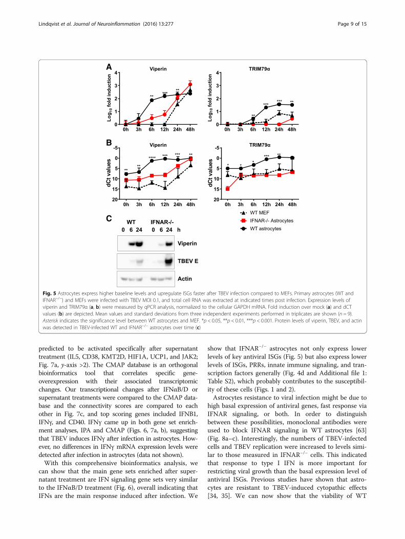
WT astrocytes show higher expression and rapidinduction of antiviral ISGs after TBEV infectionThe susceptibility of IFNAR−/− astrocytes to flavivirusinfection might be due to lowered basal expression ofantiviral ISGs, such as viperin. Together with TRIM79α,viperin has recently been identified as an inhibitor ofTBEV in vitro [50, 61]. Viperin has also been shown tohave antiviral activity against WNV [62]. Viperin andTRIM79α mRNAs were quantified over time by qPCR inWT and IFNAR−/− astrocytes and WT MEFs (Fig. 5a, b).ISGs were rapidly induced in WT astrocytes at earlytime points whereas induction was delayed in IFNAR−/−
astrocytes and WT MEFs. The basal levels of viperinand TRIM79α were higher in WT astrocytes comparedto WT MEFs and IFNAR−/− astrocytes (Fig. 5b) andmight contribute to limit initial viral infection. Viperinprotein was also strongly induced in WT astrocytes asearly as 6 hpi whereas corresponding protein levels wereonly reached after 24 hpi in IFNAR−/− astrocytes(Fig. 5c). Together, these data show that WT astrocytesexpress higher basal levels of a subset of antiviral genesand rapidly respond upon viral infection to express anti-viral protein that can limit viral infection. MEFs andIFNAR−/− astrocytes show lower basal expression ofsome antiviral ISGs and a delayed IFN response and ISG
Fig. 3 Astrocytes induce a fast type I IFN response upon TBEV infection which restricts virus replication. Primary WT Astrocytes (a) and MEFs (b)were pretreated with 5000 U IFNαB/D for 16 h before infection with TBEV MOI 0.1. Viral titers were determined by focus forming assay atindicated time points (n = 6). Primary astrocytes and MEFs were infected with TBEV using MOI 0.1. Expression levels of IFN-β (c) and IFNα2 (d) weredetermined by qPCR (n = 9). Supernatants were collected at the indicated time points, and antiviral activity was determined by VSV-GFP bioassay onMEFs (e, n = 6) or TBEV-based bioassay on WT and IFNAR−/− astrocytes (f, n = 12). g WT and IFNAR−/− astrocytes were treated with either 5000 UIFNαB/D or virus-inactivated supernatant from WT astrocytes 24 hpi (n= 6). TBEV replication was quantified using qPCR and normalized to input viralRNA. Mean values and standard deviations from three independent experiments are shown. *p < 0.05; **p < 0.01; ***p < 0.001
Lindqvist et al. Journal of Neuroinflammation (2016) 13:277 Page 7 of 15

induction which is likely to render them moresusceptible to viral infection.
IFN signaling restricts TBEV spread in astrocytesGene set enrichment analysis was used to identify classesof genes overrepresented in the dataset. IFNAR−/− cellsshowed a negative enrichment compared to WT inimmune pathways and interferon signaling, while thepositively enriched gene sets were below our cut-offadjusted p value < 0.05 (Fig. 6). Both IFNαB/D andsupernatant treatment lead to enrichment in “interferonsignaling” and “interferon alpha beta signaling” (Fig. 6).Differentially expressed genes between WT and the threesamples were analyzed using IPA to uncover predictedactivation of canonical pathways and disease functions.Pathways involved in viral recognition and antiviralsignaling as well as functions involved in viability ofleukocytes and antiviral and inflammatory responseswere found to be downregulated in IFNAR−/− comparedto WT astrocytes (Additional file 1: Tables S3 and S4),which can explain why the IFNAR−/− cells responded
poorly after viral infection. IPA analysis revealed activationof pathways involved in innate immune signaling and viralrecognition, antiviral and immune response after treat-ment with IFNαB/D and supernatant (Additional file 1:Table S3 and S4). To extend our analyses and investigatewhat might be responsible for these transcriptomicchanges, with focus on the difference between the IFNαB/D and the supernatant treatment, we performed an IPAupstream analysis, and a Connectivity Map (CMAP) ana-lysis. Both of these analyses evaluate the relationship(s)that might have led to the pattern of transcript regulationthat we found after treatment. In Fig. 7a, we have plottedall upstream regulators that are predicted by the IPA ana-lysis to be either activated or inhibited after IFNαB/D orsupernatant treatment. Upstream regulators that are pre-dicted to be activated after both treatments include severalinterferon regulated factors (IRF3, IRF7, STAT1) as well asIFNAR and IFNγ as the most activated. Each dot in Fig. 7arepresents a regulator e.g., (IFN alpha/beta) with a circleof markers of upregulated transcripts found in the dataset(Fig. 7b). Interestingly, five upstream regulators were
Fig. 4 Astrocytes treated with either supernatant or IFNαB/D mount an antiviral response. Primary astrocytes from WT and IFNAR−/− mice wereisolated, and WT cells were either mock treated, treated with IFNαB/D, or treated with inactivated supernatant from TBEV-infected astrocytes. Geneexpression was analyzed by deep sequencing. Venn diagram of differentially expressed transcripts in IFNAR−/−, IFNαB/D, and supernatant-treatedastrocytes compared to mock-treated WT astrocytes (fold-change ±2, q value <0.05). Number of totally regulated genes (a), downregulated (b), andupregulated (c). d Overlap of differentially expressed transcripts under all three conditions (n = 112); red color indicates an upregulation whereas bluecolor correlates with a downregulation. Dynamic range in the heat map is 7.16 to −5.99 log2 fold difference compared to WT untreated astrocytes.Sd standard deviation (n = 3)
Lindqvist et al. Journal of Neuroinflammation (2016) 13:277 Page 8 of 15
predicted to be activated specifically after supernatanttreatment (IL5, CD38, KMT2D, HIF1A, UCP1, and JAK2;Fig. 7a, y-axis >2). The CMAP database is an orthogonalbioinformatics tool that correlates specific gene-overexpression with their associated transcriptomicchanges. Our transcriptional changes after IFNαB/D orsupernatant treatments were compared to the CMAP data-base and the connectivity scores are compared to eachother in Fig. 7c, and top scoring genes included IFNB1,IFNγ, and CD40. IFNγ came up in both gene set enrich-ment analyses, IPA and CMAP (Figs. 6, 7a, b), suggestingthat TBEV induces IFNγ after infection in astrocytes. How-ever, no differences in IFNγ mRNA expression levels weredetected after infection in astrocytes (data not shown).With this comprehensive bioinformatics analysis, we
can show that the main gene sets enriched after super-natant treatment are IFN signaling gene sets very similarto the IFNαB/D treatment (Fig. 6), overall indicating thatIFNs are the main response induced after infection. We
show that IFNAR−/− astrocytes not only express lowerlevels of key antiviral ISGs (Fig. 5) but also express lowerlevels of ISGs, PRRs, innate immune signaling, and tran-scription factors generally (Fig. 4d and Additional file 1:Table S2), which probably contributes to the susceptibil-ity of these cells (Figs. 1 and 2).Astrocytes resistance to viral infection might be due to
high basal expression of antiviral genes, fast response viaIFNAR signaling, or both. In order to distinguishbetween these possibilities, monoclonal antibodies wereused to block IFNAR signaling in WT astrocytes [63](Fig. 8a–c). Interestingly, the numbers of TBEV-infectedcells and TBEV replication were increased to levels simi-lar to those measured in IFNAR−/− cells. This indicatedthat response to type I IFN is more important forrestricting viral growth than the basal expression level ofantiviral ISGs. Previous studies have shown that astro-cytes are resistant to TBEV-induced cytopathic effects[34, 35]. We can now show that the viability of WT
Fig. 5 Astrocytes express higher baseline levels and upregulate ISGs faster after TBEV infection compared to MEFs. Primary astrocytes (WT andIFNAR−/−) and MEFs were infected with TBEV MOI 0.1, and total cell RNA was extracted at indicated times post infection. Expression levels ofviperin and TRIM79α (a, b) were measured by qPCR analysis, normalized to the cellular GAPDH mRNA. Fold induction over mock (a) and dCTvalues (b) are depicted. Mean values and standard deviations from three independent experiments performed in triplicates are shown (n = 9).Asterisk indicates the significance level between WT astrocytes and MEF. *p < 0.05, **p < 0.01, ***p < 0.001. Protein levels of viperin, TBEV, and actinwas detected in TBEV-infected WT and IFNAR−/− astrocytes over time (c)
Lindqvist et al. Journal of Neuroinflammation (2016) 13:277 Page 9 of 15

astrocytes after TBEV infection is dependent on the type1 IFN response, since IFNAR−/−, as well as WT astro-cytes treated with an IFNAR-specific antibody, renderedthem susceptible to TBEV-induced cytopathic effect48 hpi (Fig. 8d).Neurons are the main target of neurotropic flavivirus
infection; however, it is not clear if and how astrocytesinfluence viral growth in neurons. Therefore, superna-tants from infected astrocytes were collected at differenttime points post infection and inactivated. Primaryneurons were treated with supernatants before TBEVinfection. The number of infected neurons decreasedalready when pretreated with supernatants fromastrocytes infected for 3 h (Fig. 8e). These data indicatethat astrocytes can mediate a very fast antiviral effect onneurons already 3 h post infection.Here, we show that astrocytes are in an antiviral state
and respond quickly to flavivirus infection by upregulat-ing type 1 IFNs which limits neurotropic flavivirusspread. Secreted IFNs control viral replication by upreg-ulating several innate immune pathways and induce cellsurvival. Our data suggest that although neurons seemto represent the main target for neurotropic flavivirusinfection in vivo [64], astrocytes are likely to play an
important role in responding to infection, amplifying thetype I IFN response and limiting viral spread in bothastrocytes and neurons.
DiscussionPrevious work has indicated that a local type I IFNresponse is indispensable for the control of viral replica-tion in the CNS after flavivirus infection [11]. TBEVpreferentially replicates in neurons [11, 33], but whyastrocytes are less susceptible to TBEV infection remainsunclear. In the current study, we showed that astrocytesare initially infected by TBEV but showed strong inhib-ition of viral spread. Astrocytes showed a higher basallevel of ISG expression which enables the cells to rapidlyrespond with type I IFN production. Thus, astrocytesinduce an early and strong antiviral response that limitsviral spread in neurons and thereby plays a specific rolein the innate antiviral defense in the CNS.Local type I IFN production is critical to limit viral
spread within the CNS [65] whereas CNS deficiency inIFNAR increases the susceptibility of lethal virus infec-tion [13, 17, 18, 66–68]. Using the TBEV model LGTV,we have previously demonstrated the impact of locallyproduced type I IFN response within the CNS [11];
Fig. 6 Gene set enrichment analysis of transcriptomic changes. The schematic histogram shows a typical expression profile of the transcriptomicchanges after treatment (y-axis = fold changes; x-axis = one vertical line (stick) represents one transcript; red, up; blue, down). mRNA transcripts(sticks) and gene sets (names) that are high in IFNAR−/− astrocytes, IFNαB/D and supernatant-treated WT astrocytes, compared to WT astrocytes,are marked as red. Genes and gene sets that are downregulated or have a negative normalized enrichment score (NES), respectively, comparedto WT astrocytes are blue. pval nominal p value, adj.pval Benjamini-Hochberg adjusted p value. An adj.pval <0.05 is regarded significant
Lindqvist et al. Journal of Neuroinflammation (2016) 13:277 Page 10 of 15

however, it is not clear which cells produce type I IFN inthe CNS during TBEV infection. Astrocytes have beenshown to produce an array of innate inflammatory medi-ators upon stimulation using polyinosinic:polycytidylicacid, lipopolysaccharide, and toll-like receptor (TLR)-7and TLR-9 agonists [69–72]. Studies have shown that as-trocytes are the main producers of type I IFN within theCNS during VSV and La Cross virus infections [18, 23].Recent studies have shown that TBEV and WNV can infectastrocytes in vitro but fail to spread from cell to cell. How-ever, what prevents viral spread in astrocytes is not known[34, 36]. Our recent results show that neurons are the maintarget of LGTV infection; however, a small number of as-trocytes also become infected. During infection, astrocytesshow an activated phenotype in vivo and more cells
become infected after LGTV infection in IPS-1-deficientmice [33], indicating that type I IFN response mightcontribute to the restricted growth of virus in astrocytes.In this study, we show that, although primary astrocytes
are infected in a comparable manner to MEF initially, viralreplication and spread is dramatically inhibited, indicatingthat astrocytes are abortively infected with neurotropicflavivirus similar to La Crosse virus infections [73]. Thisphenotype is dependent on IFNAR expression, since viralreplication is uncontrolled in IFNAR-deficient astrocytes.Consistent with previous results for WNV, we now showfor all three subtypes of TBEV, JEV, and ZIKV that therapidly produced type I IFN in astrocytes after neurotropicflavivirus infection limits the viral spread and preventvirus-induced killing of the cells.
Fig. 7 Upstream activators related to treatment with either supernatant or interferon alpha (IFN). a Scatter blot of ingenuity pathway analysis (IPA)showing upstream regulators that are predicted to be activated (>2 activation score) or inhibited (<−2 activation score) according to experimental andliterature findings. Y-axis show upstream regulators predicted from supernatant-treated astrocytes, and x-axis show upstream regulators predicted fromIFNαB/D-treated astrocytes. Five upstream regulators show an idiosyncratic activation specific for the supernatant-treated cells, aligning the y-axis and withan activations score of >2. b An example of predicted activators by IPA. The circle of markers is upregulated transcripts (red), and the center is a regulator(IFN alpha/beta), predicted to be active based on information (orange arrows) in the IPA knowledge-database. Each black dot in a is representing a similarcircle of regulated markers. c Genes that, when overexpressed, lead to a similar expression pattern as either supernatant treatment (y-axis) or IFNαB/Dtreatment (x-axis) according to the connectivity map database (CMAP). Genes predicted by both treatments are in the top right corner (e.g., IFNG and IFNB1)while effects exclusive to supernatant treatment are high (>90) on the y-axis and low (<90) on the x-axis
Lindqvist et al. Journal of Neuroinflammation (2016) 13:277 Page 11 of 15

Weak IFN signals, transmitted independently of viralinfection, could be crucial for predisposing cells to amp-lify their IFN production in response to viral infectionand enhance their response to other cytokines [74, 75].Intriguingly, WT astrocytes were in an antiviral statewith higher levels of ISGs compared to MEFs, and thesewere rapidly induced to even higher levels after TBEVinfection. Because no difference in basal levels of IFNβor IFNα2 was observed between MEFs and WT astro-cytes (data not shown), a different mechanism comparedto IFN priming may exist [76]. However, it seems thatthe responsiveness to viral infection is determined bybasal levels of innate immune components. We havereported previously that TBEV is able to delay IFNβinduction by hiding its dsRNA within replication vesiclesin A549 cells [42, 77] and a similar observation wasmade in the MEFs in this study. No delay in type I IFNinduction was observed in astrocytes, clearly indicatingthat if certain antiviral factors are present in the cell at abasal level the virus is unable to delay the IFN response.High basal expression of ISGs have also been linked toviral resistance to influenza A infection in bronchialepithelial cells [78] and contribute to neuronal tropismof WNV [19]. Furthermore, we previously showed thatviperin is a strong inhibitor of TBEV infection [50].Therefore, the increased basal expression of some keyISGs could contribute to lower flavivirus replication in
astrocytes. However, the basal expression was notenough by itself to restrict TBEV replication or spreadas neutralization of IFNAR with antibodies in WTastrocytes rendered the WT cells susceptible to TBEVinfection. Virus infection has been shown to directly in-duce antiviral ISGs independently of IFN signaling [79];however, in the case of TBEV infection of astrocytes, thisresponse does not seem to inhibit TBEV growth.Although IFNAR−/− astrocytes did induce ISGs at the
mRNA level and viperin at the protein level, the kineticswere delayed compared to WTastrocytes indicating the im-portance of a rapid IFN response in order to control TBEVinfection. To further characterize what makes the bystandercells resistant to flavivirus infection, astrocytes were treatedwith either inactivated supernatant from virus-infected cellsor with IFNαB/D followed by RNAseq. The RNAseqrevealed that IFN signaling was the most upregulated path-way in supernatant-treated cells suggesting the IFN mightbe the most important signaling molecule produced byastrocytes upon TBEV infection. This is also true for otherviruses, as increased expression of IFN signaling moleculesand ISGs was observed in human astrocytes infected withJunin virus [52].Comparison of the differently expressed genes among
WT, IFNAR−/− astrocytes, supernatant or IFNαB/D-treatedWT astrocytes showed an overlap of 112 transcripts, andthese transcripts might be of particular importance as most
Fig. 8 Type I IFN response in astrocytes protects against viral spread of TBEV and cell death. Primary astrocytes isolated from WT and IFNAR−/−
mice were infected with TBEV MOI of 0.1 and viral growth was determined at indicated time points. Two hours before infection, cells weretreated with anti-IFNAR antibody or control IgG (IgC). Number of infected cells were measured by immunofluorescent staining of TBEV E antigenand DAPI (a, n = 8). Quantification of virus RNA determined by qPCR analysis (b, n = 6). Viral titers in cell culture supernatants determined by focusforming assay (c, n = 6). Cell viability after infection was measured using a resazurine viability assay (d, n = 8). Supernatants from TBEV-infectedastrocytes were collected at the indicated time points and inactivated. Primary neurons were pretreated with the supernatants followed by TBEVinfection, and viral infection was determined 48 h post infection by immunofluorescent staining of TBEV E antigen and DAPI (e, n = 6). Meanvalues and standard deviations from three independent experiments are shown. *p < 0.05; **p < 0.01; ***p < 0.001; ****p < 0.0001
Lindqvist et al. Journal of Neuroinflammation (2016) 13:277 Page 12 of 15

of them were downregulated in IFNAR−/− astrocytes, whichwere highly susceptible to TBEV infection, whereas theywere upregulated in supernatant- and IFNαB/D-treatedcells, which were resistant to the infection. Several antiviralISGs were identified among the overlap such as viperin andTRIM79α, which have been identified as inhibitors ofTBEV [50, 61]. ISG15, viperin, and Oas1b, which werefound to be downregulated in IFNAR−/− astrocytes and up-regulated after treatment with IFNαB/D and supernatant,have previously been identified as inhibitors of WNV andcould thus contribute to antiviral response against WNV inastrocytes [19, 62, 80, 81].Predicting upstream regulators responsible for the
different expression patterns in IFNAR−/−-, supernatant-,and IFNαB/D-treated astrocytes identified type I IFN aswell as IFN signaling molecules to have the highest activa-tion scores. Similar findings were found in a previousstudy where IFN signaling and antiviral mediators wereamong the most upregulated pathways and genes inWNV-infected mice brains [51]. IFNγ-STAT1-IRF-1signaling cascade was predicted as an upstream regulatorboth in supernatant and IFNαB/D-treated astrocytes.Although IFNγ transcripts were not detected in TBEV in-fected, astrocytes IRF-1 could be directly induced by virusinfection and could be responsible for the induction of theoverlap between inducible genes among the type I and IIIFNs [79, 82–84]. Taken together, the RNAseq confirmsthe potent IFN response of the astrocytes and identifies asubset of genes as key players in determining the outcomeof TBEV infection in astrocytes.Previous studies have revealed that although TBEV
infects astrocytes, viral infection did not affect the viabilityof the cells [34, 35]. Similar findings have been observedwith other viral infections such as WNV, JEV, and Juninvirus whereas infection with Venezuelan equine encephal-itis virus infection induced cell death in culturedastrocytes [31, 37, 39, 85, 86]. However, the mechanismunderlying the resistance to virus-induced cell death inastrocytes is not well understood. Here, we show that thetype I IFN response prevents TBEV-mediated cytopathiceffect. Blocking of IFNAR using antibodies as well asIFNAR knockout induced TBEV-mediated cell death ofastrocytes whereas the WTcontrol remained unaffected.
ConclusionsOur results show that astrocytes mount a rapid type IIFN response upon flavivirus infection that inhibits viralspread and replication. The IFN response also protectsastrocytes from virus-induced cell death. We furtherpropose that astrocytes are important IFN producers inflavivirus infection, which can sense the viral infectionand then mediate a local IFN response within the CNS,which could protect not only astrocytes but also otherCNS resident cells.
Additional file
Additional file 1: Supplemental Table S1, Table S2, Table S3, and Table S4.(DOC 210 kb)
AbbreviationsCMAP: Connectivity Map; dsRNA: Double-stranded RNA; GFAP: Glial fibrillaryacidic protein; GSEA: Gene set enrichment analysis; hpi: Hours post infection;IFN: Interferon; IFNAR: Interferon alpha receptor; IPA: Ingenuity PathwayAnalysis; IPS-1: Interferon-beta promoter stimulator 1; IRF: Interferonregulatory factor; ISG: Interferon-stimulated gene; ISGF3: Interferon-stimulatedgene factor-3; JEV: Japanese encephalitis virus; LGTV: Langat virus;MEF: Mouse embryonic fibroblast; NES: Normalized enrichment score;PRR: Pattern recognition receptor; qPCR: Quantitative reverse transcriptionPCR; RNAseq: RNA sequencing; STAT: Signal transducer and activator oftranscription; TBEV: Tick-borne encephalitis virus; TRIM79α: Tripartite motif 79α; VSV: Vesicular stomatitis virus; WNV: West Nile virus; WT: Wild-type;ZIKV: Zika virus; β-PL: Beta propiolactone
AcknowledgementsWe thank Gerhard Dobler from the Bundeswehr Institute of Microbiology(Munich, Germany) for providing the virus strains TBEV strain Hypr 71, Aina,and Sofjin and Sirkka Vene for providing JEV and WNV.
FundingThis work was supported by the Kempe Foundations, the Laboratory forMolecular Medicine Sweden (MIMS), the Umeå Center for Microbial Research(UCMR) and Linneus Support, and the Swedish Foundation for InternationalCooperation in Research and Higher Education (STINT), Swedish ResearchCouncil (AKÖ).
Availability of data and materialsAll data and materials are available upon request.
Authors’ contributionsRL designed and performed the experiments; analyzed, compiled, andinterpreted the data; and wrote the manuscript. FM did all the bioinformaticsanalysis and wrote the manuscript. JDG, SW, and NOG provided uniqueexpertise and reagents and critically revised the manuscript. AK helped withthe design of the study and critically revised the manuscript. AKÖ designed,interpreted, and compiled the data; wrote the manuscript; and providedfunding. All authors read and approved the final manuscript.
Competing interestsThe authors declare that they have no competing interests.
Consent for publicationNot applicable.
Ethics approvalAnimal experiments were approved and performed according to theguidelines set out by the Regional Animal Ethic Committee approval noA77-14, Umeå, Sweden.
Author details1Department of Clinical Microbiology, Virology, Umeå University, 90185Umeå, Sweden. 2The Laboratory for Molecular Infection Medicine Sweden(MIMS), 90187 Umeå, Sweden. 3The Broad Institute of MIT and Harvard,Proteomics and Biomarkers, 415 Main Street, #5033-A, Cambridge, MA 02142,USA. 4Department of Pharmacology and Clinical Neuroscience, UmeåUniversity, 90187 Umeå, Sweden. 5Bundeswehr Institute of Microbiology,Neuherbergstraße 11, 80937 Munich, Germany. 6Department of MolecularBiology, Umeå University, 90187 Umeå, Sweden. 7Innate Immunity andInfection, Helmholtz Centre for Infection Research, Inhoffen Str 7, 38124Braunschweig, Germany. 8Institute of Medical Microbiology,Otto-von-Guericke-University Magdeburg, Leipziger Str. 44, 39120Magdeburg, Germany.
Received: 9 August 2016 Accepted: 16 October 2016
Lindqvist et al. Journal of Neuroinflammation (2016) 13:277 Page 13 of 15
References1. Mansfield KL, Johnson N, Phipps LP, Stephenson JR, Fooks AR, Solomon T.
Tick-borne encephalitis virus—a review of an emerging zoonosis. J GenVirol. 2009;90:1781–94.
2. Dumpis U, Crook D, Oksi J. Tick-borne encephalitis. Clin Infect Dis. 1999;28:882–90.3. Beasley DW. Vaccines and immunotherapeutics for the prevention and
treatment of infections with West Nile virus. Immunotherapy. 2011;3:269–85.4. Diamond MS. Progress on the development of therapeutics against West
Nile virus. Antiviral Res. 2009;83:214–27.5. Bossi P, Tegnell A, Baka A, Van Loock F, Hendriks J, Werner A, Maidhof H,
Gouvras G, Task Force on b, Chemial Agent Threats PHDECL. Bichatguidelines for the clinical management of viral encephalitis andbioterrorism-related viral encephalitis. Euro Surveill. 2004;9:E21–2.
6. Tiroumourougane SV, Raghava P, Srinivasan S. Japanese viral encephalitis.Postgrad Med J. 2002;78:205–15.
7. Stetson DB, Medzhitov R. Type I interferons in host defense. Immunity.2006;25:373–81.
8. Takeuchi O, Akira S. Innate immunity to virus infection. Immunol Rev.2009;227:75–86.
9. Gonzalez-Navajas JM, Lee J, David M, Raz E. Immunomodulatory functionsof type I interferons. Nat Rev Immunol. 2012;12:125–35.
10. Platanias LC. Mechanisms of type-I- and type-II-interferon-mediatedsignalling. Nat Rev Immunol. 2005;5:375–86.
11. Weber E, Finsterbusch K, Lindquist R, Nair S, Lienenklaus S, Gekara NO, JanikD, Weiss S, Kalinke U, Overby AK, Kroger A. Type I interferon protects micefrom fatal neurotropic infection with Langat virus by systemic and localantiviral responses. J Virol. 2014;88:12202–12.
12. Lazear HM, Pinto AK, Vogt MR, Gale Jr M, Diamond MS. Beta interferoncontrols West Nile virus infection and pathogenesis in mice. J Virol.2011;85:7186–94.
13. Samuel MA, Diamond MS. Alpha/beta interferon protects against lethalWest Nile virus infection by restricting cellular tropism and enhancingneuronal survival. J Virol. 2005;79:13350–61.
14. Aoki K, Shimada S, Simantini DS, Tun MM, Buerano CC, Morita K, HayasakaD. Type-I interferon response affects an inoculation dose-independentmortality in mice following Japanese encephalitis virus infection. Virol J.2014;11:105.
15. Lazear HM, Govero J, Smith AM, Platt DJ, Fernandez E, Miner JJ,Diamond MS. A mouse model of Zika virus pathogenesis. Cell HostMicrobe. 2016;19:720–30.
16. Harris MG, Hulseberg P, Ling C, Karman J, Clarkson BD, Harding JS, ZhangM, Sandor A, Christensen K, Nagy A, et al. Immune privilege of the CNS isnot the consequence of limited antigen sampling. Sci Rep. 2014;4:4422.
17. Nair S, Michaelsen-Preusse K, Finsterbusch K, Stegemann-Koniszewski S,Bruder D, Grashoff M, Korte M, Koster M, Kalinke U, Hauser H, Kroger A.Interferon regulatory factor-1 protects from fatal neurotropic infection withvesicular stomatitis virus by specific inhibition of viral replication in neurons.PLoS Pathog. 2014;10, e1003999.
18. Detje CN, Lienenklaus S, Chhatbar C, Spanier J, Prajeeth CK, Soldner C,Tovey MG, Schluter D, Weiss S, Stangel M, Kalinke U. Upon intranasalvesicular stomatitis virus infection, astrocytes in the olfactory bulb areimportant interferon beta producers that protect from lethal encephalitis.J Virol. 2015;89:2731–8.
19. Cho H, Proll SC, Szretter KJ, Katze MG, Gale Jr M, Diamond MS. Differentialinnate immune response programs in neuronal subtypes determinesusceptibility to infection in the brain by positive-stranded RNA viruses. NatMed. 2013;19:458–64.
20. Gelpi E, Preusser M, Garzuly F, Holzmann H, Heinz FX, Budka H. Visualizationof Central European tick-borne encephalitis infection in fatal human cases.J Neuropathol Exp Neurol. 2005;64:506–12.
21. Cho H, Shrestha B, Sen GC, Diamond MS. A role for Ifit2 in restricting WestNile virus infection in the brain. J Virol. 2013;87:8363–71.
22. Reinert LS, Harder L, Holm CK, Iversen MB, Horan KA, Dagnaes-Hansen F,Ulhoi BP, Holm TH, Mogensen TH, Owens T, et al. TLR3 deficiency rendersastrocytes permissive to herpes simplex virus infection and facilitatesestablishment of CNS infection in mice. J Clin Invest. 2012;122:1368–76.
23. Kallfass C, Ackerman A, Lienenklaus S, Weiss S, Heimrich B, Staeheli P.Visualizing production of beta interferon by astrocytes and microglia inbrain of La Crosse virus-infected mice. J Virol. 2012;86:11223–30.
24. Lothman EW, Somjen GG. Extracellular potassium activity, intracellular andextracellular potential responses in the spinal cord. J Physiol. 1975;252:115–36.
25. Kimelberg HK, Biddlecome S, Bourke RS. SITS-inhibitable Cl- transport andNa+-dependent H+ production in primary astroglial cultures. Brain Res.1979;173:111–24.
26. Martinez-Hernandez A, Bell KP, Norenberg MD. Glutamine synthetase: gliallocalization in brain. Science. 1977;195:1356–8.
27. Aschner M. Neuron-astrocyte interactions: implications for cellularenergetics and antioxidant levels. Neurotoxicology. 2000;21:1101–7.
28. Farina C, Aloisi F, Meinl E. Astrocytes are active players in cerebral innateimmunity. Trends Immunol. 2007;28:138–45.
29. Kornyey S. Contribution to the histology of tick-borne encephalitis. ActaNeuropathol. 1978;43:179–83.
30. German AC, Myint KS, Mai NT, Pomeroy I, Phu NH, Tzartos J, Winter P,Collett J, Farrar J, Barrett A, et al. A preliminary neuropathological study ofJapanese encephalitis in humans and a mouse model. Trans R Soc TropMed Hyg. 2006;100:1135–45.
31. van Marle G, Antony J, Ostermann H, Dunham C, Hunt T, Halliday W,Maingat F, Urbanowski MD, Hobman T, Peeling J, Power C. West Nile virus-induced neuroinflammation: glial infection and capsid protein-mediatedneurovirulence. J Virol. 2007;81:10933–49.
32. Sips GJ, Wilschut J, Smit JM. Neuroinvasive flavivirus infections. Rev MedVirol. 2012;22:69–87.
33. Kurhade C, Zegenhagen L, Weber E, Nair S, Michaelsen-Preusse K, SpanierJ, Gekara NO, Kroger A, Overby AK. Type I interferon response in olfactorybulb, the site of tick-borne flavivirus accumulation, is primarily regulated byIPS-1. J Neuroinflammation. 2016;13:22.
34. Palus M, Bily T, Elsterova J, Langhansova H, Salat J, Vancova M, Ruzek D.Infection and injury of human astrocytes by tick-borne encephalitis virus.J Gen Virol. 2014;95:2411–26.
35. Potokar M, Korva M, Jorgacevski J, Avsic-Zupanc T, Zorec R. Tick-borneencephalitis virus infects rat astrocytes but does not affect their viability.PLoS One. 2014;9, e86219.
36. Hussmann KL, Samuel MA, Kim KS, Diamond MS, Fredericksen BL.Differential replication of pathogenic and nonpathogenic strains of WestNile virus within astrocytes. J Virol. 2013;87:2814–22.
37. Diniz JA, Da Rosa AP, Guzman H, Xu F, Xiao SY, Popov VL, VasconcelosPF, Tesh RB. West Nile virus infection of primary mouse neuronal andneuroglial cells: the role of astrocytes in chronic infection. Am J TropMed Hyg. 2006;75:691–6.
38. Lucas M, Frenkiel MP, Mashimo T, Guenet JL, Deubel V, Despres P, CeccaldiPE. The Israeli strain IS-98-ST1 of West Nile virus as viral model for West Nileencephalitis in the Old World. Virol J. 2004;1:9.
39. Chang CY, Li JR, Chen WY, Ou YC, Lai CY, Hu YH, Wu CC, Chang CJ, ChenCJ. Disruption of in vitro endothelial barrier integrity by Japaneseencephalitis virus-infected astrocytes. Glia. 2015;63:1915–932.
40. Schildge S, Bohrer C, Beck K, Schachtrup C. Isolation and culture of mousecortical astrocytes. J Vis Exp. 2013;e50079:1–7.
41. Dunning CJ, McGauran G, Willen K, Gouras GK, O’Connell DJ, Linse S. Directhigh affinity interaction between Abeta42 and GSK3alpha stimulateshyperphosphorylation of tau. A new molecular link in Alzheimer’s disease?ACS Chem Neurosci. 2016;7:161–70.
42. Overby AK, Popov VL, Niedrig M, Weber F. Tick-borne encephalitis virusdelays interferon induction and hides its double-stranded RNA inintracellular membrane vesicles. J Virol. 2010;84:8470–83.
43. Schwaiger M, Cassinotti P. Development of a quantitative real-time RT-PCRassay with internal control for the laboratory detection of tick borneencephalitis virus (TBEV) RNA. J Clin Virol. 2003;27:136–45.
44. Niedrig M, Klockmann U, Lang W, Roeder J, Burk S, Modrow S, Pauli G.Monoclonal antibodies directed against tick-borne encephalitis virus withneutralizing activity in vivo. Acta Virol. 1994;38:141–9.
45. Cargnelutti JF, Brum MC, Weiblen R, Flores EF. Stable expression andpotential use of West Nile virus envelope glycoproteins preM/E as antigenin diagnostic tests. Braz J Microbiol. 2011;42:1161–6.
46. Yousef H, Conboy MJ, Morgenthaler A, Schlesinger C, Bugaj L, Paliwal P, GreerC, Conboy IM, Schaffer D. Systemic attenuation of the TGF-beta pathway by asingle drug simultaneously rejuvenates hippocampal neurogenesis andmyogenesis in the same old mammal. Oncotarget. 2015;6:11959–78.
47. Perler L, Pfister H, Schweizer M, Peterhans E, Jungi TW. A bioassay forinterferon type I based on inhibition of Sendai virus growth. J ImmunolMethods. 1999;222:189–96.
48. Barrett AD, Hunt N, Dimmock NJ. A rapid method for the inactivation ofvirus infectivity prior to assay for interferons. J Virol Methods. 1984;8:349–51.
Lindqvist et al. Journal of Neuroinflammation (2016) 13:277 Page 14 of 15
49. Horisberger MA, de Staritzky K. A recombinant human interferon-alpha B/Dhybrid with a broad host-range. J Gen Virol. 1987;68(Pt 3):945–8.
50. Upadhyay AS, Vonderstein K, Pichlmair A, Stehling O, Bennett KL, Dobler G,Guo JT, Superti-Furga G, Lill R, Overby AK, Weber F. Viperin is an iron-sulfurprotein that inhibits genome synthesis of tick-borne encephalitis virus viaradical SAM domain activity. Cell Microbiol. 2014;16:834–48.
51. Clarke P, Leser JS, Quick ED, Dionne KR, Beckham JD, Tyler KL. Deathreceptor-mediated apoptotic signaling is activated in the brain followinginfection with West Nile virus in the absence of a peripheral immuneresponse. J Virol. 2014;88:1080–9.
52. Kolokoltsova OA, Yun NE, Paessler S. Reactive astrogliosis in response tohemorrhagic fever virus: microarray profile of Junin virus-infected humanastrocytes. Virol J. 2014;11:126.
53. Subramanian A, Tamayo P, Mootha VK, Mukherjee S, Ebert BL, Gillette MA,Paulovich A, Pomeroy SL, Golub TR, Lander ES, Mesirov JP. Gene setenrichment analysis: a knowledge-based approach for interpreting genome-wide expression profiles. Proc Natl Acad Sci U S A. 2005;102:15545–50.
54. Lamb J, Crawford ED, Peck D, Modell JW, Blat IC, Wrobel MJ, Lerner J,Brunet JP, Subramanian A, Ross KN, et al. The connectivity map: using gene-expression signatures to connect small molecules, genes, and disease.Science. 2006;313:1929–35.
55. Trapnell C, Roberts A, Goff L, Pertea G, Kim D, Kelley DR, Pimentel H,Salzberg SL, Rinn JL, Pachter L. Differential gene and transcript expressionanalysis of RNA-seq experiments with TopHat and Cufflinks. Nat Protoc.2012;7:562–78.
56. Trapnell C, Williams BA, Pertea G, Mortazavi A, Kwan G, van Baren MJ,Salzberg SL, Wold BJ, Pachter L. Transcript assembly and quantification byRNA-Seq reveals unannotated transcripts and isoform switching during celldifferentiation. Nat Biotechnol. 2010;28:511–5.
57. Gritsun TS, Frolova TV, Zhankov AI, Armesto M, Turner SL, Frolova MP,Pogodina VV, Lashkevich VA, Gould EA. Characterization of a Siberian virusisolated from a patient with progressive chronic tick-borne encephalitis.J Virol. 2003;77:25–36.
58. Dorrbecker B, Dobler G, Spiegel M, Hufert FT. Tick-borne encephalitis virusand the immune response of the mammalian host. Travel Med Infect Dis.2010;8:213–22.
59. Hamel R, Dejarnac O, Wichit S, Ekchariyawat P, Neyret A, Luplertlop N,Perera-Lecoin M, Surasombatpattana P, Talignani L, Thomas F, et al. Biologyof Zika virus infection in human skin cells. J Virol. 2015;89:8880–96.
60. Aliota MT, Caine EA, Walker EC, Larkin KE, Camacho E, Osorio JE.Characterization of lethal Zika virus infection in AG129 mice. PLoS Negl TropDis. 2016;10, e0004682.
61. Taylor RT, Lubick KJ, Robertson SJ, Broughton JP, Bloom ME, Bresnahan WA,Best SM. TRIM79alpha, an interferon-stimulated gene product, restricts tick-borne encephalitis virus replication by degrading the viral RNA polymerase.Cell Host Microbe. 2011;10:185–96.
62. Szretter KJ, Brien JD, Thackray LB, Virgin HW, Cresswell P, Diamond MS. Theinterferon-inducible gene viperin restricts West Nile virus pathogenesis.J Virol. 2011;85:11557–66.
63. Sheehan KC, Lai KS, Dunn GP, Bruce AT, Diamond MS, Heutel JD, Dungo-Arthur C, Carrero JA, White JM, Hertzog PJ, Schreiber RD. Blockingmonoclonal antibodies specific for mouse IFN-alpha/beta receptor subunit1 (IFNAR-1) from mice immunized by in vivo hydrodynamic transfection.J Interferon Cytokine Res. 2006;26:804–19.
64. Kurhade C, Zegenhagen L, Weber E, Nair S, Michaelsen-Preusse K, SpanierJ, Gekara NO, Kröger A, Överby AK. Type I interferon response in olfactorybulb, the site of tick-borne flavivirus accumulation, is primarily regulated byIPS-1. J Neuroinflammation. 2016;13.
65. Griffin DE. Immune responses to RNA-virus infections of the CNS. Nat RevImmunol. 2003;3:493–502.
66. Muller U, Steinhoff U, Reis LF, Hemmi S, Pavlovic J, Zinkernagel RM, AguetM. Functional role of type I and type II interferons in antiviral defense.Science. 1994;264:1918–21.
67. Ryman KD, Klimstra WB, Nguyen KB, Biron CA, Johnston RE. Alpha/betainterferon protects adult mice from fatal Sindbis virus infection and is animportant determinant of cell and tissue tropism. J Virol. 2000;74:3366–78.
68. Detje CN, Meyer T, Schmidt H, Kreuz D, Rose JK, Bechmann I, Prinz M,Kalinke U. Local type I IFN receptor signaling protects against virus spreadwithin the central nervous system. J Immunol. 2009;182:2297–304.
69. De Miranda J, Yaddanapudi K, Hornig M, Lipkin WI. Astrocytes recognizeintracellular polyinosinic-polycytidylic acid via MDA-5. FASEB J. 2009;23:1064–71.
70. Lieberman AP, Pitha PM, Shin HS, Shin ML. Production of tumor necrosisfactor and other cytokines by astrocytes stimulated with lipopolysaccharideor a neurotropic virus. Proc Natl Acad Sci U S A. 1989;86:6348–52.
71. Butchi NB, Du M, Peterson KE. Interactions between TLR7 and TLR9 agonistsand receptors regulate innate immune responses by astrocytes andmicroglia. Glia. 2010;58:650–64.
72. Rivieccio MA, Suh HS, Zhao Y, Zhao ML, Chin KC, Lee SC, Brosnan CF. TLR3ligation activates an antiviral response in human fetal astrocytes: a role forviperin/cig5. J Immunol. 2006;177:4735–41.
73. Pfefferkorn C, Kallfass C, Lienenklaus S, Spanier J, Kalinke U, Rieder M,Conzelmann KK, Michiels T, Staeheli P. Abortively infected astrocytes appearto represent the main source of interferon beta in the virus-infected brain.J Virol. 2015;90:2031–8.
74. Taniguchi T, Takaoka A. A weak signal for strong responses: interferon-alpha/beta revisited. Nat Rev Mol Cell Biol. 2001;2:378–86.
75. Lienenklaus S, Cornitescu M, Zietara N, Lyszkiewicz M, Gekara N, JablonskaJ, Edenhofer F, Rajewsky K, Bruder D, Hafner M, et al. Novel reporter mousereveals constitutive and inflammatory expression of IFN-beta in vivo.J Immunol. 2009;183:3229–36.
76. Kuri T, Zhang X, Habjan M, Martinez-Sobrido L, Garcia-Sastre A, Yuan Z, WeberF. Interferon priming enables cells to partially overturn the SARS coronavirus-induced block in innate immune activation. J Gen Virol. 2009;90:2686–94.
77. Overby AK, Weber F. Hiding from intracellular pattern recognition receptors,a passive strategy of flavivirus immune evasion. Virulence. 2011;2:238–40.
78. Seng LG, Daly J, Chang KC, Kuchipudi SV. High basal expression ofinterferon-stimulated genes in human bronchial epithelial (BEAS-2B) cellscontributes to influenza A virus resistance. PLoS One. 2014;9, e109023.
79. Stirnweiss A, Ksienzyk A, Klages K, Rand U, Grashoff M, Hauser H, Kroger A.IFN regulatory factor-1 bypasses IFN-mediated antiviral effects throughviperin gene induction. J Immunol. 2010;184:5179–85.
80. Dai J, Pan W, Wang P. ISG15 facilitates cellular antiviral response to dengueand west Nile virus infection in vitro. Virol J. 2011;8:468.
81. Kajaste-Rudnitski A, Mashimo T, Frenkiel MP, Guenet JL, Lucas M, Despres P.The 2′,5′-oligoadenylate synthetase 1b is a potent inhibitor of West Nilevirus replication inside infected cells. J Biol Chem. 2006;281:4624–37.
82. Sanda C, Weitzel P, Tsukahara T, Schaley J, Edenberg HJ, Stephens MA,McClintick JN, Blatt LM, Li L, Brodsky L, Taylor MW. Differential geneinduction by type I and type II interferons and their combination.J Interferon Cytokine Res. 2006;26:462–72.
83. Metz P, Dazert E, Ruggieri A, Mazur J, Kaderali L, Kaul A, Zeuge U, WindischMP, Trippler M, Lohmann V, et al. Identification of type I and type IIinterferon-induced effectors controlling hepatitis C virus replication.Hepatology. 2012;56:2082–93.
84. Crotta S, Davidson S, Mahlakoiv T, Desmet CJ, Buckwalter MR, Albert ML,Staeheli P, Wack A. Type I and type III interferons drive redundantamplification loops to induce a transcriptional signature in influenza-infected airway epithelia. PLoS Pathog. 2013;9, e1003773.
85. Pozner RG, Collado S, Jaquenod de Giusti C, Ure AE, Biedma ME,Romanowski V, Schattner M, Gomez RM. Astrocyte response to Junin virusinfection. Neurosci Lett. 2008;445:31–5.
86. Schoneboom BA, Fultz MJ, Miller TH, McKinney LC, Grieder FB. Astrocytes astargets for Venezuelan equine encephalitis virus infection. J Neurovirol.1999;5:342–54.
• We accept pre-submission inquiries
• Our selector tool helps you to find the most relevant journal
• We provide round the clock customer support
• Convenient online submission
• Thorough peer review
• Inclusion in PubMed and all major indexing services
• Maximum visibility for your research
Submit your manuscript atwww.biomedcentral.com/submit
Submit your next manuscript to BioMed Central and we will help you at every step:
Lindqvist et al. Journal of Neuroinflammation (2016) 13:277 Page 15 of 15
Related Documents