Journal of Contemporary Medicine YEAR: 2020 VOLUME: 10 ISSUE: 1

Welcome message from author
This document is posted to help you gain knowledge. Please leave a comment to let me know what you think about it! Share it to your friends and learn new things together.
Transcript
Journal ofContemporary MedicineYEAR: 2020 VOLUME: 10 ISSUE: 1

Resul YILMAZ, Prof. Dr. Çocuk Sağlığı ve Hastalıları A.D., Çocuk Yoğun Bakım B.D.Tıp Fakültesi, Selçuk Üniversitesi, Konya, TÜRKIYEE-mail: [email protected]
Mustafa ALTAY, Prof. Dr. İç Hastalıkları A.D., Endokrinoloji ve Metabolizma Hastalıkları B.D. Tıp Fakültesi, Sağlık Bilimleri Üniversitesi Keçiören Eğitim ve Araştırma Hastanesi, Ankara, TÜRKİYEE-mail: [email protected]
Fikret ERDEMIR, Prof. Dr. Üroloji ADTıp Fakültesi, Tokat Gaziosmanpaşa Üniversitesi, Tokat, TÜRKIYE E-mail: [email protected]
Mustafa ÖZÇETIN, Prof. Dr.Çocuk Sağlığı ve Hastalıları A.D.İstanbul Tıp Fakültesi, İstanbul Üniversitesi, İstanbul, TÜRKIYEE-mail: [email protected]
Atilla ŞENAYLI, Doç. Dr.Çocuk Cerrahisi A.D., Tıp Fakültesi, Yıldırım Beyazıt Üniversitesi, Yenimahalle Eğitim ve Araştırma Hastanesi, Ankara, TÜRKIYEE-mail: [email protected]
Yeşim ŞENAYLI, Dr.Anesteziyoloji ve Reanimasyon A.D.Ankara Gülhane Eğitim Araştırma Hastanesi, Ankara, TÜRKIYE E-mail: [email protected]
Raziye ÇELEN, Dr. Çocuk Sağlığı ve Hastalıkları Hemşireliği A.D.Hemşirelik Fakültesi, Selçuk Üniverstesi, Konya, TÜRKIYE E-mail: [email protected]
EDITOR-IN-CHIEF / BAŞ EDİTÖR
EDITORS / EDİTÖRLER
JOURNAL OF CONTEMPORARY MEDICINE
Formerly Çağdaş Tıp Dergisi
e-ISSN 2667-7180
The Owner and Publishing Manager on behalf of the Journal of Contemporary Medicine
Address:
Phone:Fax:
e-mail:web:
Prof. Dr. Resul YILMAZSelçuk Üniversitesi, Tıp Fakültesi Çocuk Yoğun Bakım Bilim Dalı Alaeddin Keykubat Yerleşkesi Selçuklu/Konya 42075 Türkiye+90 (332) 241 50 00-44513+90 (332) 241 21 84cagdastipdergisi @gmail.comhttp://www.jcontempmed.com
VOLUME 10 ISSUE 2 YEAR 2020
Hulya BAYIR, Prof. Dr. Professor of Critical Care Medicine and Endowed Chair of Pediatric Critical Care Medicine Research at the University of Pittsburgh. USA
Maciej BURA, Dr. Department of Infectious Diseases, Poznan University of Medical Sciences, POLAND
Sancak YÜKSEL, Associate Prof. Dr.Otorhinolaryngology – Head & Neck Surgery at McGovern Medical School, University of Texas, USA
INTERNATIONAL EDITORIAL
BOARD / ULUSLARARASI YAYIN
KURULU
Süreyya SAVAŞAN, Prof. Dr.Director, Pediatric Blood and Marrow Transplantation Program. Children's Hospital of Michigan ,Barbara Ann Karmanos Cancer Center, Central Michigan University College of Medicine, USA
Yau Sui YU, Associate Prof. Dr.Department of Nursing The Open University of Hong Kong, HONG KONG
Ashrarur Rahman MITUL, Prof. Dr.Professor of Pediatric Surgery, Dhaka Shishu ( Children) Hospital & Bangladesh Institute of Child Health, BAGLADESH
Ismail Ibrahim LATIF, Prof. Dr.Immunology, University of Diyala /College of medicine, IRAQ
Zhiqiang LIU , Prof. Dr.Biochemistry and Molecular Biology Tianjin Medical University: Tianjin, Tianjin, CN
Abid QAZI, MD/Dr.Consultant Paediatric Surgeon at Al Jalila Children's Specialty Hospital. UNITED ARAB EMIRATES
Obehi H OKOJIE, Prof. Dr.Department of Community Health, College of Medical Sciences, School of Medicine, University of Benin, Benin Edo State, NIGERIA
Ilhama JAFARLI, Associate Prof. Dr.Paediatric Surgeon at Cardiff and Vale University Health Board, UK
Areej Atyia HUSSEIN, Prof. Dr.Virology, University of Diyala /College of medicine, IRAQ
Zafar ZAHEER, PhD. DrBioststistics, Institute of Management Sciences, Peshawar University. PAKISTAN
JOURNAL OF CONTEMPORARY MEDICINE
Formerly Çağdaş Tıp Dergisi
e-ISSN 2667-7180
Sevil ÇAYLI, Prof. Dr. Histoloji ve Embriyoloji A.D. Yıldırım Beyazıt Üniversitesi Tıp Fakültesi, Ankara, TÜRKİYE
Galip GÜZ, Prof. Dr. Nefroloji B.D. Gazi Üniversitesi Tıp Fakültesi, Ankara, TÜRKIYE
Murat KEKİLLİ, Prof. Dr. Gastroenteroloji B.D. Gazi Üniversitesi Tıp Fakültesi, Ankara, TÜRKIYE
İbrahim HAZNEDAROĞLU, Prof. Dr. Hematoloji B.D. Hacettepe Üniversitesi Tıp Fakültesi, Ankara, TÜRKIYE
Nihal HATIPOĞLU, Prof. Dr. Çocuk Endokrinoloji ve Metabolizma B.D. Erciyes Üniversitesi Tıp Fakültesi, Kayseri, TÜRKIYE
Ömer ERDEVE, Prof. Dr. Neonatoloji B.D. Ankara Üniversitesi Tıp Fakültesi, Ankara, TÜRKIYE
İlhan ÇETIN, Prof. Dr. Halk Sağlığı A.D. Cumhuriyet Üniversitesi Tıp Fakültesi, Sivas, TÜRKIYE
Faruk KUTLUTÜRK, Prof. Dr. Endokrinoloji B.D. Tokat Gaziosmanpaşa Üniversitesi Tıp Fakültesi, Tokat, TÜRKIYE
Banu ÇELIKEL ACAR, Prof. Dr. Çocuk Romatoloji, Sağlık Bilimleri Üniveristesi Ankara Şehir Hastanesi, Ankara, TÜRKIYE
Fatih ÖZKAN, Prof. Dr. Aanesteziyoloji ve Reanimasyon A.D. 19 Mayıs Üniversitesi Tıp Fakültesi, Samsun, TÜRKIYE
Akif Büyükbeşe, Prof. Dr. İç Hastalıkları ve Diyabet, Medistate Kavacık Hastanesi,İstanbul, TÜRKIYE
JOURNAL OF CONTEMPORARY MEDICINE
Formerly Çağdaş Tıp Dergisi
e-ISSN 2667-7180
Ayşe Feyda NURSAL, Associate Prof. Dr.Tıbbi Biyoloji ve Genetik A.D. Hitit Üniversitesi Tıp Fakültesi, Çorum, TÜRKİYE
Ünal BIÇAKÇI, Associate Prof. Dr.Çocuk Cerrahisi A.D. 19 Mayıs Üniversitesi Tıp Fakültesi, Samsun, TÜRKİYE
Murat AŞÇI, Associate Prof. Dr.Ortopedi, Acibadem Eskişehir Hastanesi, Eskişehir, TÜRKİYE
İsmail OKAN, Prof. Dr. Cerrahi Onkoloji B.D.. Tokat Gaziosmanpaşa Üniversitesi Tıp Fakültesi, Tokat, TÜRKIYE
Osman Demir, Assistant Prof.Dr.Biyoistatistik A.D., Tokat Gaziosmanpaşa Üniversitesi Tıp Fakültesi, Tokat E-mail: [email protected]
Tamer SEKMENLI, Associate Prof. Dr.Çocuk Cerrahisi A.D. Selçuk Üniverstesi Tıp Fakültesi, Konya, TÜRKİYE
BIOSTATISTIC EDITOR /
BIYOISTATISTIK EDITÖRÜ
İlknur BOSTANCI, Prof. Dr.Çocuk Alerji ve İmmünoloji, Dr. Sami Ulus Kadın Doğum ve Çocuk Sağlığı ve Hastalıkları Eğitim ve Araştırma Hastanesi, Ankara, TÜRKİYE
Sacide PEHLIVAN, Prof. Dr.Tıbbi Biyoloji A.D. İstanbul Üniversitesi İstanbul Tıp Fakültesi, İstanbul, TÜRKİYE
Taner SEZER, Associate Prof. Dr.Tıbbi Biyoloji A.D. İstanbul Üniversitesi İstanbul Tıp Fakültesi, İstanbul, TÜRKİYE
EDITORIAL ADVISORY BOARD /
DANIŞMA KURULU

INSTRUCTIONS FOR AUTHORS
AIM AND SCOPEThe Journal will not consider manuscripts any that have been published elsewhere, or manuscripts that are being considered for another publication, or are in press. Studies previously announced in the congresses are accepted if this condition is stated. If any part of a manuscript by the same author(s) contains any information that was previously published, a reprint or a copy of the previous article should be submitted to the Editorial Office with an explanation by the authors
A technical review is performed to confirm that all of the required documentation has been submitted and to conduct a preliminary evaluation of the manuscript and supplementary files to assess suitability for the Journal. The manuscript will be returned to the Author in the event of any deficiency.
Journal of Contemporary Medicine operates a blind review process. Contributions deemed suitable are then typically sent to a minimum of two independent expert reviewers in the field of study to assess the scientific quality of the paper. (You can see at the picture below).
The Editor/Editors are responsible for the final decision regarding acceptance or rejection of articles. The Editor's decision is final. If necessary, author(s) may be invited to submit a revised version of the manuscript. This invitation does not imply that the manuscript will be accepted for publication. Revised manuscripts must be sent to the Editorial Office within 4 (four) weeks, otherwise they will be considered as a new application. The corresponding author will be notified of the decision to accept or reject the manuscript for publication.
Statements and suggestions published in manuscripts are the authors’ responsibility and do not reflect the opinions of the Editor, Associate Editors and the Editorial Board members.
The manuscript will not be returned to the authors whether the article is accepted or not. Copyright fee is not paid for the articles published in the journal. A copy of the journal will be sent to the corresponding author.
Language of the Journal The official languages of the Journal are Turkish and English. The manuscripts that are written in Turkish have abstracts in English, which makes the abstracts available to a broader audience.
Authorship Criteria After accepted for publication, all the authors will be asked to sign “CoyrightTransfer Form” which states the following: “ This work is not under active consideration for publication, has not been accepted for publication, nor has it been published, in full or in part (except in abstract form). I confirm that the study has been approved by the ethics committee. ” All authors should agree to the conditions outlined in the form.
Journal of Contemporary Medicine has agreed to use the standards of the International Committee of Medical Journal Editors. The author(s) should meet the criteria for authorship according to the "Uniform Requirements for Manuscripts Submitted to Biomedical Journals: Writing and Editing for Biomedical Publication. It is available at www.icmje.org.
Ethical Responsibility The protocol of clinical research articles must be approved by the Ethics Committee.
In all studies conducted on humans, the “Material and Method” section was approved by the relevant committee or the Helsinki Declaration of Principles (https://www.wma.net/what-we-do/medical-ethics/declaration-of-helsinki/).
It should be stated in the text that all persons included in the study signed the am Informed Consent Form ”.
The articles submitted to the Journal of Contemporary Medicine will be deemed to have been conducted in accordance with the Helsinki Declaration of Principles, and have received ethical and legal permissions and will not be held responsible.
If the “Animal” item was used in the study, the authors stated that in the Material and Method section of the article, they protect the animal rights in their studies in accordance with the principles of Guide for the Care and Use of Laboratory Animals (www.nap.edu/catalog/5140.html) and that they have received approval from the ethics committees of their institutions. must specify.
In case reports, Informed Consent a should be obtained from patients regardless of the identity of the patient.
If the article includes the institution (directly or indirectly) providing financial support for the commercial connection or work, the authors; the commercial product used, the drug, the company has no commercial relationship with, or if there is any relationship (consultant, other agreements, etc.), the editor must inform the presentation page.
If Ethics Committee Approval is required in the article; the received document should be sent with the article.
JOURNAL OF CONTEMPORARY MEDICINE
Formerly Çağdaş Tıp Dergisi
The manuscript should be submitted to the Academic Plagiarism Prevention Program by the authors.
It is the authors' responsibility to ensure that the article complies with the ethical rules.
Policy of Screening for PlagiarismThe manuscripts are scanned by the Journal using the iThenticate program for determination of plagiarism and non-ethical situations. Journal of Contemporary Medicine will immediately reject manuscripts leading to plagiarism.
TYPES OF MANUSCRIPT Manuscripts should be submitted online via www.jcontempmed.com
Original Articles should not exceed 3000 words and should be arranged under the headings of Abstract (not more than 250 words), Introduction, Materials and Methods, Results, Discussion, Conclusion and References.
Case Reports should not exceed 1000 words and 10 references, and should be arranged as follows: Abstract, Introduction, Case Report, Discussion and References. It may be accompanied by only one figure or table.
Letter to the Editor should not exceed 500 words. Short relevant comments on medical and scientific issues, particularly controversies, having no more than five references and one table or figure are encouraged. Where letters refer to an earlier published paper, authors will be offered right of reply.
Reviews are not accepted unless written on the invitation of the Editorial Board.
PREPARATION OF MANUSCRIPTS All articles submitted to the Journal must comply with the following instructions:
a) Submissions should be doubled-spaced and typed in Arial 10 points.
b) All pages should be numbered consecutively in the top right-hand corner, beginning with the title page.
c) The title page should not include the names and institutions of the authors.
d) The manuscript should be presented in the following order: Title page, Abstract (English, Turkish), Keywords (English, Turkish), Introduction, Materials and Methods, Results, Discussion, Conclusion, Acknowledgements (if present),
References, Figure Legends, Tables (each table, complete with title and foot-notes, on a separate page) and Appendices (if present) presented each on a separate page.
Title The title should be short, easy to understand and must define the contents of the article.
Abstract Abstract should be in both English and Turkish and should consist “Aim, Materials and Methods, Results and Conclusion”. The purpose of the study, the setting for the study, the subjects, the treatment or intervention, principal outcomes measured, the type of statistical analysis and the outcome of the study should be stated in this section (up to 250 words). Abstract should not include reference. No abstract is required for the letters to the Editor.
Keywords Not more than five keywords in order of importance for indexing purposes should be supplied below the abstract and should be selected from Index Medicus Medical Subject Headings (MeSH), available at www.nlm.nih.gov/meshhome.html.
Text Authors should use subheadings to divide sections regarding the type of the manuscript as described above. Statistical methods used should be specified in the Materials and Methods section.
References In the text, references should be cited using Arabic numerals in parenthesis in the order in which they appear. If cited only in tables or figure legends, they should be numbered according to the first identification of the table or figure in the text. Names of the journals should be abbreviated in the style used in Index Medicus. The names of all authors should be cited when there are six or fewer; when seven or more, the first three should be followed by et al. The issue and volume numbers of the referenced journal should be added.
References should be listed in the following form:
Journal articleTeke Z, Kabay B, Aytekin FO et al. Pyrrolidine dithiocarbamate prevents 60 minutes of warm mesenteric ischemia/reperfusion injury in rats. Am J Surg 2007;194(6):255-62.
Supplement Solca M. Acute pain management: Unmet needs and new advances in pain management. Eur J Anaesthesiol 2002; 19(Suppl 25): 3-10.
JOURNAL OF CONTEMPORARY MEDICINE
Formerly Çağdaş Tıp Dergisi
Online article not yet published in an issueButterly SJ, Pillans P, Horn B, Miles R, Sturtevant J. Off-label use of rituximab in a tertiary Queensland hospital. Intern Med J doi: 10.1111/j.1445-5994.2009.01988.x
Book Sample1: Murray PR, Rosenthal KS, Kobayashi GS, Pfaller MA. Medical microbiology. 4th ed. St. Louis: Mosby; 2002. Sample 2: Sümbüloğlu K, Akdağ B. Regresyon Yöntemleri ve Korelasyon Analizi. Hatiboğlu Yayınevi: Ankara; 2007.
Chapter in a bookMeltzer PS, Kallioniemi A, Trent JM. Chromosome alterations in human solid tumors. I n: Vogelstein B, Kinzler KW, editors. The genetic basis of human cancer. New York: McGraw-Hill; 2002. p. 93113.
Journal article on the InternetAbood S. Quality improvement initiative in nursing homes: The ANA acts in an advisory role. Am J Nurs [serial on the Internet] 2002 [cited 12 Aug 2002]; 102. Available from:www.nursingworld.org/AJN/2002/june/wawatch.htm
Website Cancer-pain.org [homepage on the Internet]. New York: Association of Cancer Online Resources [updated 16 May 2002; cited 9 Jul 2002]. Available from: www.cancer-pain.org
An organization as an authorThe Intensive Care Society of Australia and New Zealand. Mechanical ventilation strategy in ARDS: Guidelines. Int Care J Aust 1996;164:282-4.
Acknowledgements The source of financial grants and the contribution of colleagues or institutions should be acknowledged.
Tables Tables should be complementary, but not duplicate information contained in the text. Tables should be numbered consecutively in Arabic numbers, with a descriptive, self-explanatory title above the table. All abbreviations should be explained in a footnote. Footnotes should be designated by symbols in the following order: *,†, ‡, §, ¶.
Figures All illustrations (including line drawings and photographs) are classified as figures. Figures must be added to the system as separate .jpg or .gif files (approximately 500x400 pixels, 8 cm in width and at least 300 dpi resolution). Figures should be numbered consecutively in Arabic numbers and should be cited in parenthesis in consecutive order in the text.
JOURNAL OF CONTEMPORARY MEDICINE
Formerly Çağdaş Tıp Dergisi
Figure LegendsLegends should be self-explanatory and positioned on a separate page. The legend should incorporate definitions of any symbols used and all abbreviations and units of measurements should be explained. A letter should be provided stating copyright authorization if figures have been reproduced from another source.
Measurements and Abbreviations All measurements must be given in metric system (Système International d'Unités, SI). Example: mg/kg, µg/kg, mL, mL/kg, mL/kg/h, mL/kg/min, L/min, mmHg, etc. Statistics and measurements should always be given in numerals, except where the number begins a sentence. When a number does not refer to a unit of measurement, it is spelt out, except where the number is greater than nine. Abbreviations that are used should be defined in parenthesis where the full word is first mentioned. Some common abbreviations can be used, such as iv, im, po, and sc. Drugs should be referred to by their generic names, rather than brand names.
Editorial Correspondence Prof. Dr. Resul YILMAZSelçuk Üniversitesi, Tıp Fakültesi Çocuk Yoğun Bakım Bilim Dalı Alaeddin Keykubat Yerleşkesi Selçuklu/Konya 42075 TürkiyePhone: +90 (332) 241 50 00-44513Faks: +90 (332) 241 21 84
Journal of Contemporary Medicine(Çağdaş Tıp Dergisi)http://www.jcontempmed.come-posta: [email protected]
Checklist for Manuscripts Review guide for authors and instructions for submitting manuscripts through the electronic submission, website at
http://www.jcontempmed.com

YAZARLARA BİLGİ
AMAÇ ve KAPSAMÇağdaş Tıp Dergisi, üç ayda bir yayımlanır ve dört sayı ile bir cilt tamamlanır. Dergi; tüm tıp alanlarıyla ilgili nitelikli klinik ve deneysel araştırmaları, olgu sunumlarını ve editöre mektupları yayımlar.Çağdaş Tıp Dergisi, bilimsel yayınlara açık erişim sağlar. Dergi basımından hemen sonra, makalelerin tam metinlerine ücretsiz ulaşılabilir.Dergide yayımlanmak üzere gönderilen yazıların daha önce başka bir yerde yayımlanmamış veya yayımlanmak üzere gönderilmemiş olması gerekir. Daha önce kongrelerde sunulmuş çalışmalar, bu durum belirtilmek koşuluyla kabul edilir. Makale, yazar(lar)ın daha önce yayımlanmış bir yazısındaki konuların bir kısmını içeriyorsa bu durum belirtilmeli ve yeni yazı ile birlikte önceki makalenin bir kopyası da Yayın Bürosu’na gönderilmelidir.Gerekli tüm belgelerin sunulduğunu teyit etmek ve dergiye uygunluğunu değerlendirmek için makale ve ek dosyaların ön değerlendirmesini yapmak üzere teknik bir inceleme yapılır. Herhangi bir eksiklik olması halinde makale yazara iade edilecektir. Journal of Contemporary Medicine kör bir inceleme süreci yürütmektedir. Uygun görülen yazılar daha sonra makalenin bilimsel kalitesini değerlendirmek için çalışma alanında en az iki bağımsız uzmana gönderilir. Editör / Editörler makalelerin kabulü veya reddi ile ilgili nihai karardan sorumludur.(Aşağıdaki akış şemasında görüldüğü gibi).Editörün kararı kesindir. Gerekli olduğu durumlarda, yazar(lar)dan düzeltme istenebilir. Yazardan düzeltme istenmesi, yazının yayımlanacağı anlamına gelmez. Bu düzeltmelerin en geç 21 gün içinde tamamlanıp dergiye gönderilmesi gereklidir. Aksi halde yeni başvuru olarak değerlendirilir. Sorumlu yazara yazının kabul veya reddedildiğine dair bilgi verilir.Dergide yayımlanan yazıların etik, bilimsel ve hukuki sorumluluğu yazar(lar)a ait olup Editör, Editör Yardımcısı ve Yayın Kurulu’nun görüşlerini yansıtmaz.Dergide yayımlanması kabul edilse de edilmese de, yazı materyali yazarlara geri verilmez. Dergide yayımlanan yazılar için telif hakkı ödenmez. Bir adet dergi, sorumlu yazara gönderilir.
Derginin Yazı Dili Derginin yazı dili Türkçe ve İngilizcedir. Dili Türkçe olan yazılar, İngilizce özetleri ile yer alır. Yazının hazırlanması sırasında, Türkçe kelimeler için Türk Dil Kurumundan (www.tdk.gov.tr), teknik terimler için Türk Tıp Terminolojisinden (www.tipterimleri.com) yararlanılabilir.
JOURNAL OF CONTEMPORARY MEDICINE
Formerly Çağdaş Tıp Dergisi
Yazarlık Kriterleri Dergide yayınlanması uygun bulunan tüm yazıların araştırma ve yayın etiğine uygun hazırlandığı, varsa sağlanan fonun kaynağının tanımlandığı, başka yerde yayımlanmadığı veya yayımlanmak üzere gönderilmediği, çalışmaya katılan tüm yazarlar tarafından yazının son halinin onaylandığı, yayımlanacak yazı ile ilgili telif haklarının dergiye devredildiği, tüm yazarların imzaları ile “Yayın Hakkı Devir Formu”nda belirtilmesi gerekir.Çağdaş Tıp Dergisi, Uluslararası Tıp Dergileri Editörleri Kurulu’nun (International Committee of Medical Journal Editors) “Biyomedikal Dergilere Gönderilen Makalelerin Uyması Gereken Standartlar: Biyomedikal Yayınların Yazımı ve Baskıya Hazırlanması (Uniform Requirements for Manuscripts Submitted to Biomedical Journals: Writing and Editing for Biomedical Publication)” standartlarını kullanmayı kabul etmektedir. Bu konudaki bilgiye www.icmje.org adresinden ulaşılabilir.
Etik Sorumluluk Etik Sorumluluk / Kurallar: Klinik araştırma makalelerinin protokolü Etik Komitesi tarafından onaylanmış olmalıdır.İnsanlar üzerinde yapılan tüm çalışmalarda “Gereç ve Yöntem” bölümünde çalışmanın ilgili komite tarafından onaylandığı veya çalışmanın Helsinki İlkeler Deklarasyonu’na (https://www.wma.net/what-we-do/medical-ethics/declaration-of-helsinki/) uyularak gerçekleştirildiğine dair bir cümle yer almalıdır.Çalışmaya dahil edilen tüm kişilerin Bilgilendirilmiş Onam Formu’nu imzaladığı metin içinde belirtilmelidir.Journal of Contemporary Medicine’e gönderilen makalelerdeki çalışmaların Helsinki İlkeler Deklarasyonu’na uygun olarak yapıldığı, kurumsal etik ve yasal izinlerin alındığı varsayılacak ve bu konuda sorumluluk kabul edilmeyecektir.Çalışmada “Hayvan” öğesi kullanılmış ise yazarlar, makalenin Gereç ve Yöntem bölümünde hayvan haklarını Guide for the Care and Use of Laboratory Animals (www.nap.edu/catalog/5140.html) prensipleri doğrultusunda koruduklarını, çalışmalarında ve kurumlarının etik kurullarından onay aldıklarını belirtmek zorundadır.Olgu sunumlarında hastanın kimliğinin ortaya çıkmasına bakılmaksızın hastalardan “Bilgilendirilmiş rıza” alınmalıdır.Makalede ticari bağlantı veya çalışma için maddi destek veren kurum (doğrudan veya dolaylı) mevcut ise yazarlar; kullanılan ticari ürün, ilaç, firma ile ticari hiçbir ilişkisinin olmadığını veya varsa nasıl bir ilişkisinin olduğunu (konsültan, diğer anlaşmalar vs.), editöre sunum sayfasında bildirmek zorundadır.Makalede Etik Kurul Onayı alınması gerekli ise; alınan belge makale ile birlikte gönderilmelidir.
Makale yazarlar tarafından akademik intihal önleme programından geçirilmelidir.
Makalenin etik kurallara uygunluğu yazarların sorumluluğundadır.
İntihal Taraması PolitikasıMakaleler, intihal ve etik olmayan durumların belirlenmesi için iThenticate programı kullanılarak Journal tarafından taranır. Journal of Contemporary Medicine intihallere yol açan makaleleri derhal reddedecektir.
YAZI TÜRLERİ Yazılar, elektronik ortamda www.cagdastipdergisi.com adresine gönderilir.
Orijinal makaleler, 3000 sözcük sayısını aşmamalı, “Öz (250 sözcükten fazla olmamalı), Giriş, Gereç ve Yöntem, Bulgular, Tartışma, Sonuç, Kaynaklar” bölümlerinden oluşmalıdır.
Olgu Sunumu, “Öz, Giriş, Olgu Sunumu, Tartışma, Kaynaklar” şeklinde düzenlenmelidir. En fazla 1000 sözcük ve 10 kaynak ile sınırlıdır. Sadece bir tablo veya şekil ile desteklenebilir.
Editöre Mektup, yayımlanan metinlerle veya mesleki konularla ilgili olarak 500 sözcüğü aşmayan ve beş kaynak ile bir tablo veya şekil içerecek şekilde yazılabilir. Ayrıca daha önce dergide yayınlanmış metinlerle ilişkili mektuplara cevap hakkı verilir.
Yayın Kurulu’nun daveti üzerine yazılanlar dışında derleme kabul edilmez.
MAKALENİN HAZIRLANMASI Dergide yayınlanması istenilen yazı için aşağıdaki kurallara uyulmalıdır. a) Yazı; iki satır aralıklı olarak, Arial 10 punto ile yazılmalıdır. b) Sayfalar başlık sayfasından başlamak üzere, sağ üst köşesinde numaralandırılmalıdır. c) Online makale sistemine yüklenen word dosyasının başlık sayfasında (makalenin adını içeren başlık sayfası), yazarlara ait isim ve kurum bilgileri yer almamalıdır. d) Makale, şu bölümleri içermelidir: Her biri ayrı sayfada yazılmak üzere; Türkçe ve İngilizce Başlık Sayfası, Öz, Abstract, Anahtar Sözcükler, Keywords, Giriş, Gereç ve Yöntem, Bulgular, Tartışma, Sonuç, Açıklamalar (varsa), Kaynaklar, Şekil Alt Yazıları, Tablolar (başlıkları ve açıklamalarıyla beraber), Ekler (varsa).
Yazının Başlığı Kısa, kolay anlaşılır ve yazının içeriğini tanımlar özellikte olmalıdır.
JOURNAL OF CONTEMPORARY MEDICINE
Formerly Çağdaş Tıp Dergisi
Özetler Türkçe (Öz) ve İngilizce (Abstract) olarak yazılmalı, Amaç, Gereç ve Yöntem, Bulgular ve Sonuç (Aim, Materials and Methods, Results, Conclusion) olmak üzere dört bölümden oluşmalı, en fazla 250 sözcük içermelidir. Araştırmanın amacı, yapılan işlemler, gözlemsel ve analitik yöntemler, temel bulgular ve ana sonuçlar belirtilmelidir. Özette kaynak kullanılmamalıdır. Editöre mektup için özet gerekmemektedir.
Anahtar Sözcükler Türkçe Öz ve İngilizce Abstract bölümünün sonunda, Anahtar Sözcükler ve Keywords başlığı altında, bilimsel yazının ana başlıklarını yakalayan, Index Medicus Medical Subject Headings (MeSH)’e uygun olarak yazılmış en fazla beş anahtar sözcük olmalıdır. Anahtar sözcüklerin, Türkiye Bilim Terimleri’nden (www.bilimterimleri.com) seçilmesine özen gösterilmelidir.
Metin Yazı metni, yazının türüne göre yukarıda tanımlanan bölümlerden oluşmalıdır. Uygulanan istatistiksel yöntem, Gereç ve Yöntem bölümünde belirtilmelidir.
Kaynaklar Çağdaş Tıp Dergisi, Türkçe kaynaklardan yararlanmaya özel önem verdiğini belirtir ve yazarların bu konuda duyarlı olmasını bekler.
Kaynaklar metinde yer aldıkları sırayla, cümle içinde atıfta bulunulan ad veya özelliği belirten kelimenin hemen bittiği yerde ya da cümle bitiminde noktadan önce parantez içinde Arabik rakamlarla numaralandırılmalıdır. Metinde, tablolarda ve şekil alt yazılarında kaynaklar, parantez içinde Arabik numaralarla nitelendirilir. Sadece tablo veya şekil alt yazılarında kullanılan kaynaklar, tablo ya da şeklin metindeki ilk yer aldığı sıraya uygun olarak numaralandırılmalıdır. Dergi başlıkları, Index Medicus’ta kullanılan tarza uygun olarak kısaltılmalıdır. Kısaltılmış yazar ve dergi adlarından sonra nokta olmamalıdır. Yazar sayısı altı veya daha az olan kaynaklarda tüm yazarların adı yazılmalı, yedi veya daha fazla olan kaynaklarda ise üç yazar adından sonra et al. veya ve ark. yazılmalıdır. Kaynak gösterilen derginin sayı ve cilt numarası mutlaka yazılmalıdır.
Kaynaklar, yazının alındığı dilde ve aşağıdaki örneklerde görüldüğü şekilde düzenlenmelidir.
Dergilerdeki yazılarTeke Z, Kabay B, Aytekin FO et al. Pyrrolidine dithiocarbamate prevents 60 minutes of warm mesenteric ischemia/reperfusion injury in rats. Am J Surg 2007;194(6):255-62.
Ek sayı (Supplement) Solca M. Acute pain management: Unmet needs and new advances in pain management. Eur J Anaesthesiol 2002;19(Suppl 25):3-10.Henüz yayınlanmamış online makaleButterly SJ, Pillans P, Horn B, Miles R, Sturtevant J. Off-label use of rituximab in a tertiary Queensland hospital. Intern Med J doi: 10.1111/j.1445-5994.2009.01988.x
KitapÖrnek 1: Murray PR, Rosenthal KS, Kobayashi GS, Pfaller MA. Medical microbiology. 4th ed. St. Louis: Mosby; 2002. Örnek 2: Sümbüloğlu K, Akdağ B. Regresyon Yöntemleri ve Korelasyon Analizi. Hatiboğlu Yayınevi: Ankara; 2007.
Kitap bölümüMeltzer PS, Kallioniemi A, Trent JM. Chromosome alterations in human solid tumors. I n: Vogelstein B, Kinzler KW, editors. The genetic basis of human cancer. New York: McGraw-Hill; 2002. p. 93113.
İnternet makalesi
Abood S. Quality improvement initiative in nursing homes: The ANA acts in an advisory role. Am J Nurs [serial on the Internet] 2002 [cited 12 Aug 2002]; 102. Available from: www.nursingworld.org/AJN/2002/june/wawatch.htm
Web SitesiCancer-pain.org [homepage on the Internet]. New York: Association of Cancer Online Resources [updated 16 May 2002; cited 9 July 2002]. Available from: www.cancer-pain.org
Yazar olarak bir kuruluşThe Intensive Care Society of Australia and New Zealand. Mechanical ventilation strategy in ARDS: Guidelines. Int Care J Aust 1996;164:282-4.
Açıklamalar Varsa finansal kaynaklar, katkı sağlayan kurum, kuruluş ve kişiler bu bölümde belirtilmelidir.
Tablolar Tablolar metni tamamlayıcı olmalı, metin içerisinde tekrarlanan bilgiler içermemelidir. Metinde yer alma sıralarına göre Arabik sayılarla numaralandırılıp tablonun üstüne kısa ve açıklayıcı bir başlık yazılmalıdır. Tabloda yer alan kısaltmalar, tablonun hemen altında açıklanmalıdır. Dipnotlarda sırasıyla şu semboller kullanılabilir: *, †, ‡, §, ¶.
Şekiller Şekil, resim, grafik ve fotoğrafların tümü “Şekil” olarak adlandırılmalı ve ayrı birer .jpg veya .gif dosyası olarak (yaklaşık
JOURNAL OF CONTEMPORARY MEDICINE
Formerly Çağdaş Tıp Dergisi
500x400 piksel, 8 cm eninde ve en az 300 dpi çözünürlükte) sisteme eklenmelidir. Şekiller metin içinde kullanım sıralarına göre Arabik rakamla numaralandırılmalı ve metinde parantez içinde gösterilmelidir.
Şekil Alt Yazıları Şekil alt yazıları, her biri ayrı bir sayfadan başlayarak, şekillere karşılık gelen Arabik rakamlarla çift aralıklı olarak yazılmalıdır. Şeklin belirli bölümlerini işaret eden sembol, ok veya harfler kullanıldığında bunlar alt yazıda açıklanmalıdır. Başka yerde yayınlanmış olan şekiller kullanıldığında, yazarın bu konuda izin almış olması ve bunu belgelemesi gerekir.
Ölçümler ve Kısaltmalar Tüm ölçümler metrik sisteme (Uluslararası Birimler Sistemi, SI) göre yazılmalıdır. Örnek: mg/kg, µg/kg, mL, mL/kg, mL/kg/h, mL/kg/min, L/min, mmHg, vb. Ölçümler ve istatistiksel veriler, cümle başında olmadıkları sürece rakamla belirtilmelidir. Herhangi bir birimi ifade etmeyen ve dokuzdan küçük sayılar yazı ile yazılmalıdır. Metin içindeki kısaltmalar, ilk kullanıldıkları yerde parantez içinde açıklanmalıdır. Bazı sık kullanılan kısaltmalar; iv, im, po ve sc şeklinde yazılabilir.
İlaçların yazımında jenerik isimleri kullanılmalıdır.
İletişimProf. Dr. Resul YILMAZSelçuk Üniversitesi, Tıp Fakültesi Çocuk Yoğun Bakım Bilim Dalı Alaeddin Keykubat Yerleşkesi Selçuklu/Konya 42075 TürkiyeTel: +90 (332) 241 50 00-44513Faks: +90 (332) 241 21 84
Journal of Contemporary Medicine
(Çağdaş Tıp Dergisi)http://www.cagdastipdergisi.come-posta: [email protected] Kontrol Listesi · Türkçe ve İngilizce başlık,· Türkçe ve İngilizce özet · Türkçe ve İngilizce anahtar sözcükler (En fazla 5 sözcük) · İki satır aralıklı yazılmış metin (Arial, 10 punto) · Kurallara uygun hazırlanmış tablo ve şekiller · Kurallara uygun yazılmış kaynaklar · İmzalı “Yayın Hakkı Devir Formu” (makale yayın için kabul edildikten sonra istenmektedir)

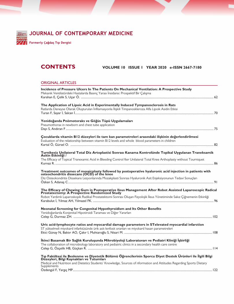
CONTENTS VOLUME 10 ISSUE 1 YEAR 2020 e-ISSN 2667-7180
ORIGINAL ARTICLES
JOURNAL OF CONTEMPORARY MEDICINE
Formerly Çağdaş Tıp Dergisi
Endobronşiyal Ultrasonografi Eşliğinde Gerçekleştirilen Transbronşiyal İğne Aspirasyonu BiyopsiMateryallerine Histopatolojik YaklaşımHistopathological Approach for Determining the Diagnostic Value of Endobronchial Ultrasonography-GuidedTransbronchial Needle Aspiration Biopsy MaterialsÖzmen S, Ceylan O. ...............................................................................................................................................................................................1
Differentiation between neoplastic and nonneoplastic brain masses using intermediate echo time MR SpectroscopySerebral Kitlesel Lezyonların Ayırıcı Tanısında Manyetik Rezonans Spektroskopik İncelemeLeblebisatan Ş, Bıçakcı YK. ....................................................................................................................................................................................7
Yoğun bakim ünitesinde tedavi edilen yaşli hastalarin D vitamini düzeylerinin prognoz üzerine etkisiThe effect of vitamin-d levels on prognosis of elderly patients treated in intensive care unitYiğit Özay H, Mungan İ, Çobanoğlu Ercan G, Turan S, Eler Çevik Bç ..................................................................................................... 13
Plasenta Previa Olgularının Retrospektif DeğerlendirilmesiRetrospective Evaluation of Cases with Placenta PreviaKunt İşgüder Ç, Gülücü S, Bulut YE, Delibaş İB, Yurt T, Özsoy AZ ........................................................................................................ 18
Perceptions of parents of children with cancer about medications errorsKanserli çocuğu olan ebeveynlerin ilaç hatalarına ilişkin algılarıBilsin E, Bal Yılmaz H, Özalp Gerçeker G, Binay Ş, Başbakkal Z, Kantar M. .......................................................................................... 23
H. pylori infection and antral nodular gastritis in childrenÇocuklarda Helicobacter pylori enfeksiyonu ve antral nodüleriteUrgancı N, Kalyoncu D, Yılmaz Özgüven B. .................................................................................................................................................. 31
Analysis of the relationship between smoking and chronic respiratory symptoms, level of income and education Sigara kullanımı ile kronik solunumsal semptomlar, gelir seviyesi ve eğitim düzeyi arasındaki ilişkinin analizi Özdemir T, Kasapoğlu B, Akkuş İH, Kaya F, Pirinçci E, Eren S, Türkkanu MH, Özdilekcan Ç, Bulut İ. ........................................... 35
Tracheal Bronchus; A Computed Tomography StudyTrakeal Bronkus; Bilgisayarlı Tomografi ÇalışmasıÇelebioğlu EC, Çalıkan S, Akkaşoğlu S, Nakkaş HG, Sancak İT, ............................................................................................................... 40
Oxidative stress, DNA damage and apoptosis levels in those who use borderline high level fluoride content drinking water / Sınırda Yüksek Düzeyde Florid İçeren İçme Suyu Kullananlarda Oksidatif Stres, DNA Hasarı ve Apoptoz DüzeyleriKirmit A, Yeşilnacar Mİ, Çalışır M, Bayhan İ, Çelik H. ................................................................................................................................. 45
Prevalence of Gaucher’s Disease in a Hematology Outpatient ClinicHematoloji polikliniğinde Gaucher Hastalığı sıklığı Yanardağ Açık D, Aygün B. ................................................................................................................................................................................ 51
Substance Abuse Protection Self-Efficacy of Students from Two Different Types of High Schools and Related Factors / İki Farklı Lise Türündeki Öğrencilerin Madde Bağımlılığından Korunmada Özyeterlilik Düzeyleri ve Bununla İlişkili FaktörlerKırağ N, Tanılmışoğlu E. ...................................................................................................................................................................................... 55
CONTENTS VOLUME 10 ISSUE 1 YEAR 2020 e-ISSN 2667-7180
ORIGINAL ARTICLES
JOURNAL OF CONTEMPORARY MEDICINE
Formerly Çağdaş Tıp Dergisi
Incidence of Pressure Ulcers In The Patients On Mechanical Ventilation: A Prospective StudyMekanik Ventilatördeki Hastalarda Basınç Yarası İnsidansı: Prospektif Bir Çalışma Karahan E, Çelik S, Uçar Ö. ............................................................................................................................................................................... 62
The Application of Lipoic Acid in Experimentally Induced Tympanosclerosis in RatsRatlarda Deneyse Olarak Oluşturulan İnfilamasyonla İlişkili Timpanoskleroza Alfa Lipoik Asidin EtkisiTuran F, Sayar İ, Salcan İ ....................................................................................................................................................................................... 70
Yenidoğanda Pnömotoraks ve Göğüs Tüpü UygulamalarıPneumothorax in newborn and chest tube applicationDayı S, Andıran F ................................................................................................................................................................................................... 75
Çocuklarda vitamin B12 düzeyleri ile tam kan parametreleri arasındaki ilişkinin değerlendirilmesi Evaluation of the relationship between vitamin B12 levels and whole blood parameters in childrenKartal Ö, Gürsel O. ............................................................................................................................................................................................. 82
Turnikesiz Unilateral Total Diz Artoplastisi Sonrası Kanama Kontrolünde Topikal Uygulanan Traneksamik Asitin Etkinliği /The Efficacy of Topical Tranexamic Acid in Bleeding Control fter Unilateral Total Knee Arthoplasty without TourniquetKurnaz R. ................................................................................................................................................................................................................. 86
Treatment outcomes of mosaicplasty followed by postoperative hyaluronic acid injection in patients with osteochondritis dissecans (OCD) of the knee Diz Osteokondritis Dissekans Lezyonlarında Mozakplasti Sonrası Hyaluronik Asit Enjeksiyonunun Tedavi SonuçlarıÖzkan S, Adanaş C ................................................................................................................................................................................................ 91
The Efficacy of Chewing Gum in Postoperative Ileus Management After Robot Assisted Laparoscopic Radical Prostatectomy: A Prospective Randomized StudyRobot Yardımlı Laparoskopik Radikal Prostatektomi Sonrası Oluşan Fizyolojik İleus Yönetiminde Sakız Çiğnemenin Etkinliği Karabulut İ, Yılmaz AH, Yılmazel FK. ............................................................................................................................................................... 96
Neonatal Screening for Congenital Hypothyroidism and Its Other BenefitsYenidoğanlarda Konjenital Hipotiroidi Taraması ve Diğer Yararları Celep G, Durmaz ZH. ....................................................................................................................................................................................... 102
Uric acid-lymphocyte ratios and myocardial damage parameters in STelevated myocardial infarctionST yükselmeli miyokard infarktüsünde ürik asit-lenfosit oranları ve miyokard hasarı parametreleriEkici Günay N, Baktır AO, Çakır I, Muhtaroğlu S, Nisari M. .................................................................................................................... 108
İkinci Basamak Bir Sağlık Kuruluşunda Mikrobiyoloji Laboratuvarı ve Pediatri Kliniği İşbirliğiThe collaboration of microbiology laboratory and pediatric clinics in a secondary health care centreCelep G, Özçelik HB, Güçkan R. .................................................................................................................................................................... 114
Tıp Fakültesi ile Beslenme ve Diyetetik Bölümü Öğrencilerinin Sporcu Diyet Destek Ürünleri ile İlgili Bilgi Düzeyleri, Bilgi Kaynakları ve TutumlarıMedical and Nutrition and Dietetics Students’ Knowledge, Sources of nformation and Attitudes Regarding Sports Dietary SupplementsÖzdengül F, Yargıç MP ........................................................................................................................................................................................ 122
CONTENTS VOLUME 10 ISSUE 1 YEAR 2020 e-ISSN 2667-7180
REVIEWS
JOURNAL OF CONTEMPORARY MEDICINE
Formerly Çağdaş Tıp Dergisi
Fetal sex determination in light of interdisciplinary current studies: a forensic anthropological approachDisiplinlerarası Güncel Çalışmalar Işığında Fetal Cinsiyet Tahmini: Adli Antropolojik Bir YaklaşımMehder Ö. ........................................................................................................................................................................................................... 131
Kanser Hastalarının Manevi Gereksinimlerini Değerlendirmeye İlişkin Ölçek Çalışmalarının İncelemesiInvestigation of the Scales Studies on the Evaluation of the Spiritual Needs of Cancer PatientsOtuzoğlu M. ......................................................................................................................................................................................................... 138
Atypical celiac disease presenting with clubbingÇomak Parmak İle Başvuran Atipik Çölyak Hastalığı OlgusuBükülmez A, Özeke E, Gürsoy Koca T. ........................................................................................................................................................ 146
Penile agenesis Penil agenezTarakçı Emiroğlu N, Konak M, Yılmaz FH, Gültekin ND, Altunhan H, Örs R. .................................................................................. 149
LETTER TO THE EDİTOR
Hepatit A Enfeksiyonu Olan Bir Çocuk Hastada Delta-Bilirubinemi ve İnatçı KaşıntıDelta-Bilirubinemia and Persistent Itching in a Child Patient with epatitis A InfectionEmiroğlu M, Emiroğlu HH..................................................................................................................................................................................151
CASE REPORT
Corresponding (İletişim): Sevilay Özmen, Dr. Öğretim Üyesi, Atatürk Üniversitesi Tıp Fakültesi, Patoloji Anabilimdalı, Erzurum, TÜRKİYE E-mail (E-posta): [email protected] (Geliş Tarihi): 31.12.2019 Accepted (Kabul Tarihi): 11.02.2020
DOI: 10.16899/jcm.667970J Contemp Med 2020;10(1):1-6
Orjinal Araştırma / Original Article
JOURNAL OF
CONTEMPORARY MEDICINEJournal ofContemporary MedicineYEAR: 2019 VOLUME: 9 ISSUE: 4
Endobronsiyal Ultrasonografi Esliğinde Gerçeklestirilen Transbronsiyal İğne Aspirasyonu Biyopsi Materyallerine
Histopatolojik Yaklasım
Histopathological Approach for Determining the Diagnostic Value of Endobronchial Ultrasonography-Guided Transbronchial Needle Aspiration
Biopsy Materials
Amaç: Çalışmamızın amacı Endobronşiyal Ultrason-Transbronşiyal İğne Aspirasyonu (EBUS-TBİA) yönteminin etkinliğini histopatolojik veriler eşliğinde göstermek ve materyallerin kliniğimize gelme aşamasından tanı anına kadarki süreci en uygun şekilde yönetmeyi belirlemektir.Gereç ve Yöntem: Çalışmamızda 2015-2019 tarihleri arasında incelenen EBUS-TBİA materyalleri retrospektif olarak tarandı. 4 yıl içerisinde 552 olgu mevcuttu ve bu vakalar çalışmaya dâhil edildi.Bulgular: Çalışmamızda EBUS-TBİA materyallerine ait tanılarımızda sensitivite %90, spesifite %98, Pozitif Prediktif Değer (PPD) %89, Negatif Prediktif Değer (NPD) %98 oranında bulundu. İlk işlem sonucu tanı için yetersiz doku örneği şeklinde tanı verilen olgularda ikinci EBUS-TBİA sonrası alınan biyopsi materyallerinin %75'ine efektif tanı verilebildi. Biyopsi materyallerine efektif tanı verebilmek için ortalama 2 biyopsi camının (ilk H&E camı+1 seri kesit), camlar üzerine yayılarak gönderilen sitolojik preperatlar için ise ortalama 12-13 camın yeterli olduğu görüldü.Sonuç: Çalışmamızdaki bulgular EBUS-TBİA yönteminin tanı ve evrelemede son derece önemli bir yöntem olduğunu destekler özelliktedir. Komplikasyon oranının düşük olduğu dikkate alınacak olursa kemo-radyoterapi sonrası ve cerrahi işlem öncesi yeniden evrelemede ilk seçilecek yöntem olabilir. Ayrıca düşük komplikasyon riskinden dolayı ilk EBUS-TBİA ile tanı için yetersiz materyal gelen vakalarda ikinci işlem olarak yine ilk tercih edilecek işlem olabilir. Son olarak da sitoloji camlarının değerlendirilmesinde harcanan zamanı kısaltmak ve her cam başına düşen maliyeti azaltmak için klinik tarafından doku biyopsisi yanı sıra ortalama 12-13 cama yayılarak gönderilen sitoloji materyalinin yeterli olacağı düşüncesindeyiz.
Anahtar Kelimeler: Endobronşiyal ultrason-transbronşiyal iğne aspirasyonu, histopatolojik yaklaşım, akciğer lezyonları
Abstract Öz
Sevilay Özmen1, Onur Ceylan1,
Aim: The aim of this study was to demonstrate the efficacy of the Endobronchial Ultrasound-Transbronchial Needle Aspiration (EBUS-TBNA) method with histopathological data and to determine the optimal management of the pro cess from the time of arrival of the materials to the diagnosis.Material and Method: In this study, EBUS-TBNA materials examined between 2015-2019 were reviewed retrospectively. There were 552 cases within 4 years and these cases were included in the study.Results: In our study, the sensitivity, specificity, positive predictive value (PPD) was 90%, negative predictive value (NPD) was 98%, 90%, 98% respectively. After the first procedure, 75% of the biopsy specimens obtained after the second EBUS-TBNA could be effectively diagnosed in cases previously diagnosed as insufficient tissue samples for diagnosis. In order to give an effective diagnosis to biopsy materials, it was seen that an average of 2 biopsy glasses (one H&E glass + 1 serial section) was sufficient for the biopsy materials and 12-13 glasses were sufficient for the cytology materials.Conclusion: The findings of our study support that EBUS-TBNA method is a very important method in diagnosis and staging. Considering that the complication rate is low, it may be the first method to be selected after chemo-radiotherapy and before the surgical procedure. In addition, in cases where the EBUS-TBNA method results in insufficient material for diagnosis, the same method could again be considered first due to the low risk of complications. Finally, in order to shorten the time spent on the evaluation of cytology glasses and reduce the cost per glass, we think that a tissue biopsy accompanied by the cytology material spread on 12-13 glasses will be sufficient.
Keywords: Endobronchial ultrasound-transbronchial needle aspiration, histopathological approach, lung lesions
1Atatürk Üniversitesi Tıp Fakültesi, Patoloji Anabilimdalı, Erzurum/Türkiye

2 Journal of Contemporary Medicine
GIRIŞEBUS-TBİA bronkoskop ile birlikte gonderilen bir ultrason probu ile hava yolları, mediastinal ve hiler bolgedeki lenf nodlarının incelenmesi ve bu bolgedeki lezyonların teşhisi için kullanılan minimal invaziv bir girişimdir.[1] Diğer bir ifadeyle hava yollarına komşu yapılar ya da hava yolu duvarındaki lezyonlar bu işlem esnasında gorüntülenebilmektedir. Aynı zamanda ultrason probu biyopsi sırasında lenf nodunun (LN) doğrudan gorüntülenmesini sağlarken eş zamanlı biyopsi işlemini de kolaylaştırır.[2] Komplikasyonları nadirdir.[2] Primer tümor tanısı yanı sıra mediastinal evrelemede de onemli bir role sahiptir.[3] EBUS-TBİA tanı ve evrelemede tüm dünyada yaygın olarak kullanılan ve hastaların morbidite sebebi olabilecek mediastinoskopi gibi işlemlerden kaçınmasını sağlayabilen kabul gormüş bir yontemdir.[4] Literatürdeki review ve meta-analizlerde de gosterildiği gibi mediastinel evrelemede EBUS-TBİA ile mediastinoskopi arasında onemli bir fark yokken daha az komplikasyon gelişmesi açısından EBUS-TBİA’nın mediastinoskopiye oranla üstünlüğü bulunmaktadır.[5] Yapılan çalışmalarda histopatolojik olarak etkin bir orneklemenin nasıl yapılacağı konusunda yeterli çalışma bulunmamaktadır. Çalışmamızın amacı EBUS-TBİA materyallerinin kliniğimize gelme aşamasından tanı anına kadarki süreci en uygun şekilde yonetmek ve en efektif sonuç için uygun histopatolojik değerlendirmeyi tartışmaktır.
GEREÇ VE YÖNTEMÇalışmamıza 2015-2019 yılları arasında Atatürk Üniversitesi Tıp Fakültesi Patoloji Anabilim Dalına EBUS-TBİA materyali olarak gonderilmiş 552 olgu dâhil edildi. Her olguya ait parafin bloklar, Hematoksilen-Eozin (H&E) ve immunhistokimyasal çalışma preparatları, sitoloji camları yanı sıra patoloji raporları anabilim dalımız arşivinden çıkarılarak tüm olgular iki patoloji uzmanı tarafından yeniden değerlendirilmeye tabi tutuldu. H&E ile boyanmış ilk camlar, seri kesit camları, immünhistokimyasal çalışma camları, malignite veya granülom yüzdeleri, yetersiz ornekleme olduğu için yeniden biyopsi onerilen vakalar, rebiyopsilerin sonuçları, sitoloji tanısı ve doku biyopsisi tanısı arasındaki uyum oranı incelendi. Bunların yanısıra EBUSTBİA yontemiyle laboratuvarımıza camlar üzerine yayılarak gonderilen preparat sayısı ile tanısal sonuçlarımız arasındaki ilişki incelendi. Hastaların cinsiyeti, yaşı, hastalara uygulanan
ileri tetkik gibi klinik bilgiler hastanemizin bilgi sisteminden elde edildi. Çalışmamız Atatürk Üniversitesi Klinik Araştırmalar etik kurulu tarafından onaylanmıştır (2019 / B.30.2.ATA.0.01.00).
İstatistik analizler IBM SPSS20 isatatistik analiz programı ile yapıldı. Veriler ortalama, standart sapma, medyan, minimum, maksimum, yüzde ve sayı olarak sunuldu. Sürekli değişkenlerin normal dağılımına orneklem büyüklüğü <50 olduğu durumda Shapiro Wilk-W testi ile >50 olduğu durumda Kolmogorov Simirnov testi ile bakıldı. İki bağımsız grup arasındaki kıyaslamalarda normal dağılım şartı sağlandığı durumda İndependent Samples t testi, sağlanmadığı durumda Mann Whitney u testi kullanıldı. Kategorik değişkenler arasındaki 2x2’lik kıyaslamalarda beklenen değer (>5) ise Pearson Ki-kare testi, beklenen değer (3-5) arasında ise kikare yates testi ve beklenen değer (<3) ise Fisher’s Exact testi kullanılarak yapıldı. Klinik altın standart ile yeni geliştirilen tanı testinin tutarlılığı Cohen’in Kappa katsayısı hesaplanarak değerlendirilmiştir. Tanı testi sonuçlarının geçerliliği için duyarlılık, ozgüllük, pozitif ongorü değeri, negatif ongorü değeri, prevalans, pozitif olabilirlik oranı, negatif olabilirlik oranı ve doğruluk oranı hesaplanarak yeni testin hasta ve sağlamları ayırt etme gücü belirlenmiştir. İstatistiksel anlamlılık düzeyi p<0,05 olarak alındı.
BULGULARÇalışmamızda toplam 552 olgu değerlendirildi. Olgularda ortalama yaş 55 (±18,4), erkek/kadın oranı 1,45 tespit edildi. Olgulardaki tanısal sonuçlarımız skuamoz hücreli karsinom (SHK), adenokarsinom, küçük hücreli karsinom (KHK), metastatik tümor (diğer organlardan), non-nekrotizan granülom (NNG), nekrotizan granülom (NG), atipik hücresel elemanlar (AHE), malignite negatif/benign/reaktif lenfoid doku, malignite pozitif doku (Büyük Hücreli Karsinom (BHK), lenfoma, vs), yetersiz sitoloji ve kan fibrin kitlesinden oluşan yetersiz materyal olmak üzere 10 kategoriye ayrıldı.
EBUS-TBİA yontemiyle alınan biyopsi tanılarımızın sınıflaması şu şekildeydi. 552 olgunun 179'u yetersiz sitoloji ve doku/kan fibrin kitlesi; 152'si malignite negatif, 87'si NNG, 9'u NG, 23'ü AHE tanısı alırken 102'si malign tanısı almıştı.
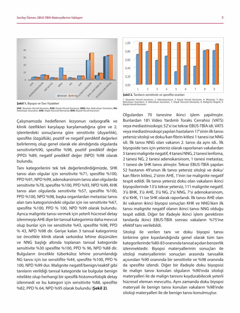
Malign tanısı alan vakalardan 32'si adenokarsinom, 21'i KHK, 26'sı BHK ve lenfoma, 14'ü SHK, 9'u metastaz pozitif doku orneği (diğer organlardan) olarak raporlanmıştı (Şekil 1).
3Sevilay Özmen, EBUS-TBİA Materyallerine Yaklaşım
Çalışmamızda hedeflenen lezyonun radyografik ve klinik ozellikleri karşılayıp karşılamadığına gore ve 2. işlemlerdeki sonuçlarına gore sensitivite (duyarlılık), spesifite (ozgüllük), pozitif ve negatif perdiktif değerleri belirlenmiş olup genel olarak ele alındığında olgularda sensitivite%90, spesifite %98, pozitif prediktif değer (PPD) %89, negatif prediktif değer (NPD) %98 olarak bulundu.
Tanı kategorilerini tek tek değerlendirdiğimizde, SHK tanısı alan olgular için sensitivite %71, spesifite %100, PPD %91, NPD %99, adenokarsinom tanısı alan olgularda sensitivite %78, spesifite %100, PPD %93, NPD %99, KHK tanısı alan olgularda sensitivite %57, spesifite %100, PPD %100, NPD %98, başka organlardan metastaz tanısı alan tanı kategorisindeki olgular için ise sensitivite %67, spesifite %100, PPD % 100, NPD %99 olarak bulundu. Ayrıca malignite tanısı vermek için yeterli hücresel detay izlenmeyip AHE diye bir tanısal kategorimiz daha mevcut olup bunlar için ise sensitivite %43, spesifite %98, PPD % 43, NPD %98 dir. Geriye kalan 3 tanısal kategorimiz ise oncelikle klinik olarak sarkoidoz lehine düşünülen ve NNG başlığı altında toplanan tanısal kategoride sensitivite %30 spesifite %100, PPD % 96, NPD %88 dir. Bulguların oncelikle tüberküloz lehine yorumlandığı NG tanısı için ise sensitifite %44, spesifite %100, PPD % 100, NPD %99 dur. Malignite negatif/benign/reaktif gibi tanıların verildiği tanısal kategoride ise bulgular benign nitelikte olup herhangi bir spesifik histomorfolojik detay izlenmedi ve bu kategori için sensitivite %88, spesifite %82, PPD % 64, NPD %95 olarak bulundu (Şekil 2).
Olgulardan 70 tanesine ikinci işlem yapılmıştır. Bunlardan 18’i Video Yardımlı Toraks Cerrahisi (VATS) veya mediastinoskopi; 52'si ise tekrar EBUS-TBİA idi. VATS veya mediastinoskopi yapılan hastaların 17'sinin ilk tanısı yetersiz sitoloji ve doku/kan fibrin kitlesi 1 tanesi ise NNG idi. İlk tanısı NNG olan vakanın 2. tanısı da aynı idi.. İlk biyopside tanı için yetersiz olarak raporlanan vakalardan 5 tanesi malignite negatif, 4 tanesi NNG, 2 tanesi lenfoma, 2 tanesi NG, 2 tanesi adenokarsinom, 1 tanesi metastaz, 1 tanesi de SHK tanısı almıştır. Tekrar EBUS-TBİA yapılan 52 hastanın 49'unun ilk tanısı yetersiz sitoloji ve doku/ kan fibrin kitlesi, 2'sinin AHE, 1'inin ise malignite negatif tespit edildi. İlk tanısı yetersiz doku olan vakaların ikinci biyopsilerinde 13'ü tekrar yetersiz, 11'i malignite negatif, 3'ü BHK, 3'ü AHE, 3'ü NG, 2'si NNG, 7'si adenokarsinom, 6'si KHK, 1'i ise SHK olarak raporlandı. İlk tanısı AHE olan iki vakanın ikinci biyopsi sonuçları KHK ve NNG'iken ilk tanısı malignite negatif olanın ikinci tanısı NNG olduğu tespit edildi. Diğer bir ifadeyle ikinci işlem gerektiren tanılarda ikinci EBUS-TBİA sonrası vakaların %75'ine efektif tanı verilebildi.
Sitoloji ile verilen tanı ve doku biyopsi tanısı birbirine gore kıyaslandığında genel olarak tüm tanı kategorilerinde %80-83 oranında tanısal açıdan benzerlik izlenmektedir. Biyopsi materyallerinin sonuçları ile sitoloji materyallerinin sonuçları arasında tanısallık açısından %90 oranında bir sensitivite ve %98 oranında da spesifite izlendi. Diğer bir ifadeyle doku biyopsisi ile malign tanısı konulan olguların %90’ında sitoloji materyalleri ile de malign tanısını koydurabilecek yeterli hücresel eleman mevcuttu. Aynı zamanda doku biyopsi materyali ile benign tanısı konulan vakaların %98'inde sitoloji materyalleri ile de benign tanısı konulmuştur.
Şekil 2. Tanıların sensitivite ve spesifite oranları1: Skuamoz Hücreli Karsinom, 2: Adenokarsinom, 3: Küçük Hücreli Karsinom, 4: Metastaz, 5: Non Nekrotizan Granülom, 6: Nekrotizan Granülom, 7: Atipik Hücresel Elemanlar, 8: Malignite Negatif, 9: Büyük Hücreli KarsinomŞekil 1. Biyopsi ve Tanı Yüzdeleri
SHK: Skuamoz Hücreli Karsinom, KHK: Küçük Hücreli Karsinom, NNG: Non Nekrotizan Granülom, NG: Nekrotizan Granülom, AHE: Atipik Hücresel Elemanlar, BHK: Büyük Hücreli Karsinom
4 Journal of Contemporary Medicine
Arşivimizdeki biyopsilere ait kesit sayısı ortalama 4 adet olup daha sonraki immünhistokimyasal ve moleküler çalışmalar için gerekli olan dokunun kaybına ve tanısal zorluğa neden olmaktadır. Gonderilen sitoloji materyallerinin de 15-20 adet gibi fazla miktarda olması günlük pratiği zorlaştırmaktadır. EBUS-TBİA yontemiyle laboratuvarımıza gonderilen sitoloji preparat sayısı ve doku biyopsileri için toplam alınan kesit sayısının tanısal sonuçları arasındaki ilişki: biyopsi materyallerine tanı verebilmek için ortalama 2 biyopsi camının (ilk H&E camı+ 1 seri kesit), camlar üzerine yayılarak gonderilen sitolojik materyaller için ise ortalama 12-13 camın tanı vermek için yeterli olduğu gorüldü.
TARTIŞMAEBUS-TBİA hem malign hem de benign lezyonlar için yüksek tanı verme oranına sahip minimal invaziv girişim olma ozelliği ile mediastinoskopinin onüne geçmiştir.[6] Akciğer malignitelerinin tanısında olduğu kadar evrelemesinde de son derece onemlidir. Birçok durumda akciğer kanseri şüphesi olan hastalarda EBUS-TBİA patolojik tanı ve evreleme için ilk basamak yaklaşım olmuştur. Kemoterapi tedavisi sonrası ve cerrahi rezeksiyon oncesi mediastenin yeniden evrelemesi çoğu zaman gerekli olmaktadır. Tekrarlayan mediastinoskopinin yüksek komplikasyon riskinden dolayı EBUS-TBİA sıklıkla onerilmektedir. Geniş hasta gruplu çalışmalarda major komplikasyon oranı %0,15 olarak bildirilmiş olup literatürde sadece 2 olüm rapor edilmiştir.[7,8] Çalışmamızda genel olarak ele alındığında olgularda sensitivite %90, spesifite %98, PPD % 89, NPD %98 olarak bulundu. Vaidya ve ark.[6] çalışmalarında sensitivite%89, spesifite %100, PPD % 100, NPD %53 oranında bulunmuştur. Herth ve ark.[9] çalışmalarında sensitivite, spesifite, PPD, NPD oranlarını sırasıyla 76, %100, %100, %20 olarak bulmuşlardır ve düşük NPD nedeniyle, malignite negatif gelen sonuçların diğer ek işlemler ile doğrulanması gerektiğini savunmuşlardır.
Yine Herth'in de dâhil olduğu Szlubowski ve ark.[9] çalışmalarında ise sensitivite, spesifite, PPD, NPD oranlarını sırasıyla 67, %86, %91, %78 olarak bulmuşlardır ve 2. çalışmalarındaki yüksek NPD değerinin altını çizerek malignite negatif sonuç gelen hastaların mediastinoskopi gibi ek girişimsel işlemlere gerek olmadığını belirtmişlerdir.
Dhooria ve ark.[10] yaptığı meta-analizde sensitivite, spesifite oranları sırasıyla ortalama olarak %80,3 (73,7–
85,9), %100 (98,7–100) bulunmuştur.Çalışmamızdaki değerler bu haliyle literatürdeki çalışmalar ile uyumlu olup EBUS-TBİA yonteminin tanı ve evrelemede son derece onemli bir yontem olduğunu destekler ozelliktedir. Komplikasyon oranının düşük olduğu dikkate alınacak olursa ozellikle cerrahi işlem ve kemoradyoterapi sonrası yeniden evrelemede ilk seçilecek işlem olma ozelliğini korumaktadır.
Çalışmamızda literatüden farklı şekilde NNG ve NG tanılarında sensitivite sırasıyla %30 ve %44 gibi değerlerde bulunmuştur. Oysaki literatürde ki çoğu çalışmada gerek sarkoidoz gerekse tüberküloz tanısında EBUS-TBİA'nın çok etkili bir yontem olduğu savunulmaktadır. Agarwal ve ark.[11] yapmış olduğu meta-analiz niteliğindeki çalışmada ozellikle sarkoidozda EBUS-TBİA’nın ortalama %80 oranında tanı doğruluğu oranı bildirmişlerdir. Bizim çalışmamızda aslında sensitivite değerleri düşük olsa da PPD ve NPD'in yüksek olması nedeniyle EBUS-TBİA’nın bu hastalıkların tanısında değerli bir yontem olduğu kanaatine varılabilir.
Çalışmamızda 70 hastaya efektif tanı için ikinci teknik işleme gerek duyulmuştur ve bunlardan 18'inde mediastinoskopi veya VATS yontemiyle; 52'sinde ise EBUS-TBİA yontemiyle tekrar biyopsi alınmıştır. Mediastinoskopi veya VATS yontemiyle alınan ikinci biyopsilerin tamamına efektif tanı verilebilirken ikinci EBUS-TBİA sonrası alınan biyopsilerin %75'ine efektif tanı verilebilmiştir. Çalışmamızda her ne kadar ikinci işlem olarak efektif tanı verme oranı mediastinoskopi ve VATS yontemleriyle az bir oranda daha yüksek bulunsa da bu yontemlerin EBUS-TBİA yontemine gore gerek mortalite gerekse morbidite oranlarının yüksek olduğu goz onüne alınacak olursa ikinci işlem gerektiren durumlarda ilk planda EBUS-TBİA yonteminin tercih edilmesi gerektiğini soyleyebiliriz. Üstelik ikinci işlemde %75 gibi yüksek bir oranda efektif tanı verme oranına sahip bulduğumuz bu yontemin düşük mortalite ve morbitidite oranları nedeniyle tedavi ve cerrahi sonrası yeniden evreleme gerektiren durumlarda da etkili bir yontem olduğunu savunabiliriz.
EBUS-TBİA yonteminin tanısal değerinin yüksek olduğunu gostermenin yanısıra aynı zamanda bu yontemle alınan biyopsi materyallerinin kliniğimize gelme aşamasından tanı anına kadarki süreci en uygun şekilde yonetmek ve en efektif sonuç için uygun histopatolojik değerlendirmeyi bulmayı amaçladığımız çalışmamızda literatürdeki çalışmalardan farklı olarak
5Sevilay Özmen, EBUS-TBİA Materyallerine Yaklaşım
bu yontemle laboratuvarımıza camlar üzerine yayılarak gonderilen preparat sayısı ve doku biyopsileri için toplam aldığımız kesit sayısı ile tanısal sonuçlarımız arasındaki ilişkiyi inceledik. Biyopsi materyallerine tanı verebilmek için ortalama 2 biyopsi camının (ilk H&E camı+1 seri kesit), camlar üzerine yayılarak gonderilen sitolojik materyaller için ise ortalama 12-13 camın tanı vermek için yeterli olduğunu gordük. Bu durum ilk H&E kesitinde yeterli hücresel eleman gorülmüyorsa ilk etapta en fazla 1 adet H&E kesiti istenmesi gerektiğini gosterir nitelikteydi. Biyopsi materyallerinin histolojik tip tayini ve moleküler çalışma için gerekli olduğunu da düşünecek olursak ilk etapta çok fazla seri kesit alınıp doku kaybının yaşanmaması adına bu uygulama çok onemlidir. Çalışmamızda yetersiz biyopsi materyali olarak raporladığımız olguları tekrar incelediğimizde ilk seri kesit ile sonraki seri kesitler arasında farklı sonuçlar olmadığını tespit ettik bu nedenle 1 tane seri kesitin tanısal açıdan yeterli hücresel eleman gorebilmek için yeterli olduğunu düşünecek olursak ilk seri kesiti gormeden immünhistokimyasal çalışmaya geçilmemesi gerektiğini düşünmekteyiz.
Ayrıca çalışmamızda sitoloji materyalleri ile doku biyopsisi tanıları karşılaştırıldığında %80-83 oranında tanısal açıdan benzerlik izlenmekteydi. Doku biyopsi materyallerinin sonuçları ile sitoloji materyallerinin sonuçları arasında tanı koyma açısından %90 oranında bir sensitivite ve %98 oranında da spesifite izlendi. Bazı olgularda tek bir hastaya ait 20 ve üzeri yayılmış cam preparat gonderildiği goz onüne alınacak olursa, bunların değerlendirilmesinde harcanan zamanı kısaltmak ve her cam başına düşen maliyeti azaltmak için klinik tarafından doku biyopsisi yanısıra ortalama 12-13 cama sitoloji materyalinin yayılarak gonderilmesinin yeterli olacağı düşüncesindeyiz.
SONUÇÇalışmamızdaki bulgular EBUS-TBİA yonteminin tanı ve evrelemede son derece onemli bir yontem olduğunu destekler ozelliktedir. Komplikasyon oranının düşük olduğu dikkate alınacak olursa kemo-radyoterapi sonrası ve cerrahi işlem oncesi yeniden evrelemede ilk seçilecek yontem olabilir. Ayrıca düşük komplikasyon riskinden dolayı ilk EBUS-TBİA ile tanı için yetersiz materyal gelen vakalarda ikinci işlem olarak yine ilk tercih edilecek işlem olabilir. Son olarak da sitoloji camlarının değerlendirilmesinde harcanan zamanı kısaltmak ve
her cam başına düşen maliyeti azaltmak için klinik tarafından doku biyopsisi yanısıra ortalama 12-13 cama yayılarak gonderilen sitoloji materyalinin yeterli olacağı düşüncesindeyiz.
ETIK BEYANLAREtik Kurul Onayı: Çalışmamız Atatürk Üniversitesi Klinik Araştırmalar etik kurulu tarafından onaylanmıştır (2019 / B.30.2.ATA.0.01.00).Aydınlatılmış Onam: Çalışma retrospektif olarak dizayn edildiği için hastalardan aydınlatılmış onam alınmamıştır. Hakem Değerlendirme Süreci: Harici çift kor hakem değerlendirmesi.Çıkar Çatışması Durumu: Yazarlar bu çalışmada herhangi bir çıkara dayalı ilişki olmadığını beyan etmişlerdir.Finansal Destek: Yazarlar bu çalışmada finansal destek almadıklarını beyan etmişlerdir.Yazar Katkıları: Yazarların tümü; makalenin tasarımına, yürütülmesine, analizine katıldığını ve son sürümünü onayladıklarını beyan etmişlerdir.
KAYNAKLAR1. Ariza-Prota MA, Bango Alvarez A, Perez L, Pando-Sandoval A, Fuentes N,
Casan P. From cytology to histology: diagnosis of a relapsed mediastinal lymphoma by endobronchial ultrasound transbronchial histological needle. respirology case reports. 2015;3(2):68-71.
2. Yasufuku K, Chiyo M, Koh E, et al. Endobronchial ultrasound guided transbronchial needle aspiration for staging of lung cancer. Lung Cancer (Amsterdam, Netherlands). 2005;50(3):34754.
3. Medford AR. Endobronchial ultrasound-guided versus conventional transbronchial needle aspiration: time to re-evaluate the relationship? Journal of thoracic disease. 2014;6(5):411-5.
4. Herth F, Becker HD. Endobronchial ultrasound of the airways and the mediastinum. Monaldi archives for chest disease = Archivio Monaldi per le malattie del torace. 2000;55(1):36-44.
5. Steinhauser Motta JP, Lapa ESJR, Samary Lobato C, Mendonca VS, Steffen RE. Endobronchial ultrasound-guided transbronchial needle aspiration versus mediastinoscopy for mediastinal staging of lung cancer: A protocol for a systematic review of economic evaluation studies. Medicine. 2019;98(39):e17242.
6. Vaidya PJ, Saha A, Kate AH et al. Diagnostic value of core biopsy histology and cytology sampling of mediastinal lymph nodes using 21-gauge EBUS-TBNA needle. Journal of cancer research and therapeutics. 2016;12(3):1172-7.
7. Gu P, Zhao YZ, Jiang LY, Zhang W, Xin Y, Han BH. Endobronchial ultrasound-guided transbronchial needle aspiration for staging of lung cancer: a systematic review and meta-analysis. European journal of cancer (Oxford, England : 1990). 2009;45(8):1389-96.
8. Navani N, Brown JM, Nankivell M, et al. Suitability of endobronchial ultrasound-guided transbronchial needle aspiration specimens for subtyping and genotyping of nonsmall cell lung cancer: a multicenter study of 774 patients. American journal of respiratory and critical care medicine. 2012;185(12):1316-22.
6 Journal of Contemporary Medicine
9. Herth FJ, Annema JT, Eberhardt R, et al. Endobronchial ultrasound with transbronchial needle aspiration for restaging the mediastinum in lung cancer. Journal of clinical oncology official journal of the American Society of Clinical Oncology. 2008;26(20):3346-50.
10. Dhooria S, Aggarwal AN, Gupta D, Behera D, Agarwal R. Utility and safety of endoscopic ultrasound with bronchoscope-guided fine-needle aspiration in Mediastinal Lymph Node Sampling: Systematic Review and Meta-Analysis. Respiratory care. 2015;60(7):1040-50.
11. Agarwal R, Srinivasan A, Aggarwal AN, Gupta D. Efficacy and safety of convex probe EBUS-TBNA in sarcoidosis: a systematic review and meta-analysis. Respiratory medicine.2012;106(6):883-92.
Corresponding (İletişim): Şerife Leblebisatan, M.D., Adana City Training and Research Hospital, Department of Radiology, Adana, Turkey E-mail (E-posta): [email protected] (Geliş Tarihi): 20.08.2019 Accepted (Kabul Tarihi): 30.11.2019
DOI: 10.16899/jcm.667970J Contemp Med 2020;10(1):7-12
Orjinal Araştırma / Original Article
JOURNAL OF
CONTEMPORARY MEDICINEJournal ofContemporary MedicineYEAR: 2019 VOLUME: 9 ISSUE: 4
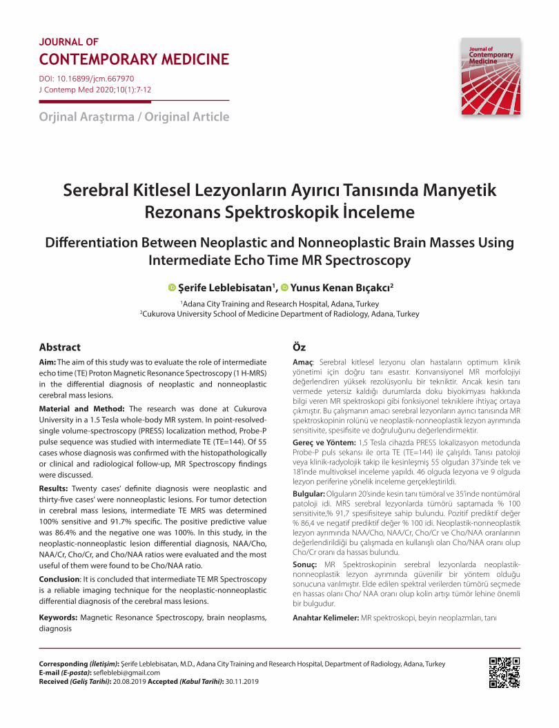
Serebral Kitlesel Lezyonların Ayırıcı Tanısında Manyetik Rezonans Spektroskopik İnceleme
Differentiation Between Neoplastic and Nonneoplastic Brain Masses Using Intermediate Echo Time MR Spectroscopy
Amaç: Serebral kitlesel lezyonu olan hastaların optimum klinik yönetimi için doğru tanı esastır. Konvansiyonel MR morfolojiyi değerlendiren yüksek rezolüsyonlu bir tekniktir. Ancak kesin tanı vermede yetersiz kaldığı durumlarda doku biyokimyası hakkında bilgi veren MR spektroskopi gibi fonksiyonel tekniklere ihtiyaç ortaya çıkmıştır. Bu çalışmanın amacı serebral lezyonların ayırıcı tanısında MR spektroskopinin rolünü ve neoplastik-nonneoplastik lezyon ayrımında sensitivite, spesifisite ve doğruluğunu değerlendirmektir.
Gereç ve Yöntem: 1,5 Tesla cihazda PRESS lokalizasyon metodunda Probe-P puls sekansı ile orta TE (TE=144) ile çalışıldı. Tanısı patoloji veya klinik-radyolojik takip ile kesinleşmiş 55 olgudan 37’sinde tek ve 18’inde multivoksel inceleme yapıldı. 46 olguda lezyona ve 9 olguda lezyon periferine yönelik inceleme gerçekleştirildi.
Bulgular: Olguların 20’sinde kesin tanı tümöral ve 35’inde nontümöral patoloji idi. MRS serebral lezyonlarda tümörü saptamada % 100 sensitivite,% 91,7 spesifisiteye sahip bulundu. Pozitif prediktif değer % 86,4 ve negatif prediktif değer % 100 idi. Neoplastik-nonneoplastik lezyon ayrımında NAA/Cho, NAA/Cr, Cho/Cr ve Cho/NAA oranlarının değerlendirildiği bu çalışmada en kullanışlı olan Cho/NAA oranı olup Cho/Cr oranı da hassas bulundu.
Sonuç: MR Spektroskopinin serebral lezyonlarda neoplastik-nonneoplastik lezyon ayrımında güvenilir bir yöntem olduğu sonucuna varılmıştır. Elde edilen spektral verilerden tümörü seçmede en hassas olanı Cho/ NAA oranı olup kolin artışı tümör lehine önemli bir bulgudur.
Anahtar Kelimeler: MR spektroskopi, beyin neoplazmları, tanı
Abstract Öz
Şerife Leblebisatan1, Yunus Kenan Bıçakcı2
Aim: The aim of this study was to evaluate the role of intermediate echo time (TE) Proton Magnetic Resonance Spectroscopy (1 H-MRS) in the differential diagnosis of neoplastic and nonneoplastic cerebral mass lesions.
Material and Method: The research was done at Cukurova University in a 1.5 Tesla whole-body MR system. In point-resolved-single volume-spectroscopy (PRESS) localization method, Probe-P pulse sequence was studied with intermediate TE (TE=144). Of 55 cases whose diagnosis was confirmed with the histopathologically or clinical and radiological follow-up, MR Spectroscopy findings were discussed.
Results: Twenty cases’ definite diagnosis were neoplastic and thirty-five cases’ were nonneoplastic lesions. For tumor detection in cerebral mass lesions, intermediate TE MRS was determined 100% sensitive and 91.7% specific. The positive predictive value was 86.4% and the negative one was 100%. In this study, in the neoplastic-nonneoplastic lesion differential diagnosis, NAA/Cho, NAA/Cr, Cho/Cr, and Cho/NAA ratios were evaluated and the most useful of them were found to be Cho/NAA ratio.
Conclusion: It is concluded that intermediate TE MR Spectroscopy is a reliable imaging technique for the neoplastic-nonneoplastic differential diagnosis of the cerebral mass lesions.
Keywords: Magnetic Resonance Spectroscopy, brain neoplasms, diagnosis
1Adana City Training and Research Hospital, Adana, Turkey2Cukurova University School of Medicine Department of Radiology, Adana, Turkey

8 Journal of Contemporary Medicine
INTRODUCTIONProton MRS (1 H-MRS) is a diagnostic technique that can measure the metabolites of tissues non-invasively and show it in a spectrum. In addition to the morphological information obtained on conventional MRI, MRS provides information on the biochemistry of the sampled tissue like cellularity, energy, neuron viability, necrosis and ischemia.[1] It is clearly shown that the spectra obtained from normal brain tissue and brain tumors are different. Thus, magnetic resonance spectroscopy is increasingly used in the classification of lesions detected in the brain.[2] The aim of this study was to evaluate the role of intermediate echo time (TE) Proton Magnetic Resonance Spectroscopy (1 H-MRS) in the differential diagnosis of neoplastic and nonneoplastic cerebral mass lesions.
Different TE sequences give different spectrums. The main metabolites identified with proton MRS with TE=135-288 milliseconds include the following: N-Acetyl Aspartate (NAA, 2.02 parts per million (ppm)), Choline (Cho, 3.22 ppm), Creatine (Cr, 3.02 ppm), Lactate (Lac, 1.33 ppm) and Lipids (Lip, 1.3 and 0.9 ppm). With intermediate TE (TE=135-144 ms) doublet Lac peak inverts below the baseline and Lip peak remains above the baseline. So lipid-lactat differentiation can be made easily. At TE 270-288 ms Lac peak doesn’t invert below the baseline, the same applies for short TE ( TE=30 ms). Short TE demonstrates more metabolites in the spectrum in addition to those at long TE sequences, like myoinositol (Myo, 3,56 ppm) and glutamine-glutamate (Glx, 2.05-2.50 ppm). More metabolite peaks can give more information but overlapping of the peaks in the spectrum can make evaluation difficult.[1]
NAA indicates neuronal and axonal viability and density. A decrease in NAA level is observed in a wide range of disease characterized by neuronal destruction.[1,3] Because Cr is the most stable cerebral metabolite in the spectrum, it is used as an internal reference.[1] Cho is a cellular membrane turnover marker that reflects cellular proliferation. Increase in choline concentrations is detected in a large number of tumors and shows rapid proliferation in tumor cells.[1,4,5]
The spectral changes frequently observed in brain tumors are increased Cho level, increased Cho/Cr and Cho/NAA ratios; decreased or absence NAA, and lactate or lipid presence.[1-3,6,7]
MATERIAL AND METHODMR spectroscopy was performed on cerebral lesions of 100 cases, to none of whom a definite diagnosis was made with other imaging modalities and the definite diagnosis was necessary for clinical follow-up and treatment, between may 2004-June 2006 at the Cukurova University Faculty of Medicine. The main purpose of this retrospective study was to differentiate between tumoral and non-tumoral lesions in consecutive patients. In treated cases with central nervous system (CNS) tumor, MRS was used for the differentiation of secondary changes after radiotherapy from tumor recurrence. In the nontumoral lesion group, the second step was to make a distinction between infarct, demyelination, abscess, encephalitis, etc.
In 55 out of 100 examinations performed, pathologic or clinicalradiological follow-up confirmed the definite diagnosis. The diagnosis was confirmed by clinical and radiological followup in 41 cases and pathology in 14 cases. Here the data of 55 confirmed cases will be discussed.
Cerebral MR imaging was performed with a 1.5 Tesla wholebody MR system (GE, Signa Excite). 38 patients with single focal, 15 patients with multiple focal and 2 patients with diffuse lesions were examined. Single voxel spectroscopy (SVS) in 37 cases and multivoxel spectroscopy (Chemical Shift Imaging, CSI) in 18 cases were performed. Single voxel spectroscopy was performed in focal and small lesions. Multivoxel spectroscopic examination was performed in lesions with diffuse, large area and peripheral edema. In 46 cases, the center of lesions and in 9 cases periphery of lesions were examined spectroscopically.
The localization method used was point-resolved-single volume-spectroscopy (PRESS), the pulse sequence was probe P and was run with intermediate TE (TE=144 milliseconds). N-acetyl aspartate (NAA), Choline (Cho) and Creatine (Cr) levels and NAA/Cho, NAA/Cr, Cho/Cr and Cho/NAA ratios were evaluated in the obtained spectrum.
SPSS 14.0 was used in the analysis of the results. In the analyzes, chi-square test, ROC analysis, and T-test were performed. Sensitivity and specificity of intermediate TE MR spectroscopy in detecting the tumor in cerebral lesions were determined.

9Şerife Leblebisatan, Differentiation of brain masses by MRS
RESULTSThe 20 cases were female and 35 were male and the mean age ±SD was 39.35±19.97 (between 3 and 75 years old). The exact diagnosis of 20 cases was neoplastic (36%) and 35 were nonneoplastic (64%). In the tumor group, 14 cases were the primary tumor (Figure 1), 2 cases were metastasis and 4 cases were relapse/residual
disease. In the nontumoral pathology group, 13 cases were infarct, 8 cases were demyelination, 3 cases were hematoma, 2 cases were encephalitis (Figure 2), 2 cases were posttreatment abnormality, 2 cases were vascular malformation, 1 case abscess, 1 case tuberculoma, 1 case myotonic dystrophy, 1 case arachnoid cyst, 1 case SSPE (subacute sclerosing panencephalitis).
2a
Figure 2. 47 years old male patient. Encephalitis sequela findings. Pontine and cerebellar hyperintensities. Multivoxel MR Spectroscopy (2a) demonstrates lipid-lactate peaks and decreased NAA (2b, 2c).
2b
Figure 1. 38 years old female patient. Diffuse infiltrating astrocytoma. Axial T2W image of brain shows right parietal hyperintensity (1a). Single voxel spectroscopy demonstrates increased Cho, Cho/Cr and Cho /NAA (1b).
1b1a
10 Journal of Contemporary Medicine
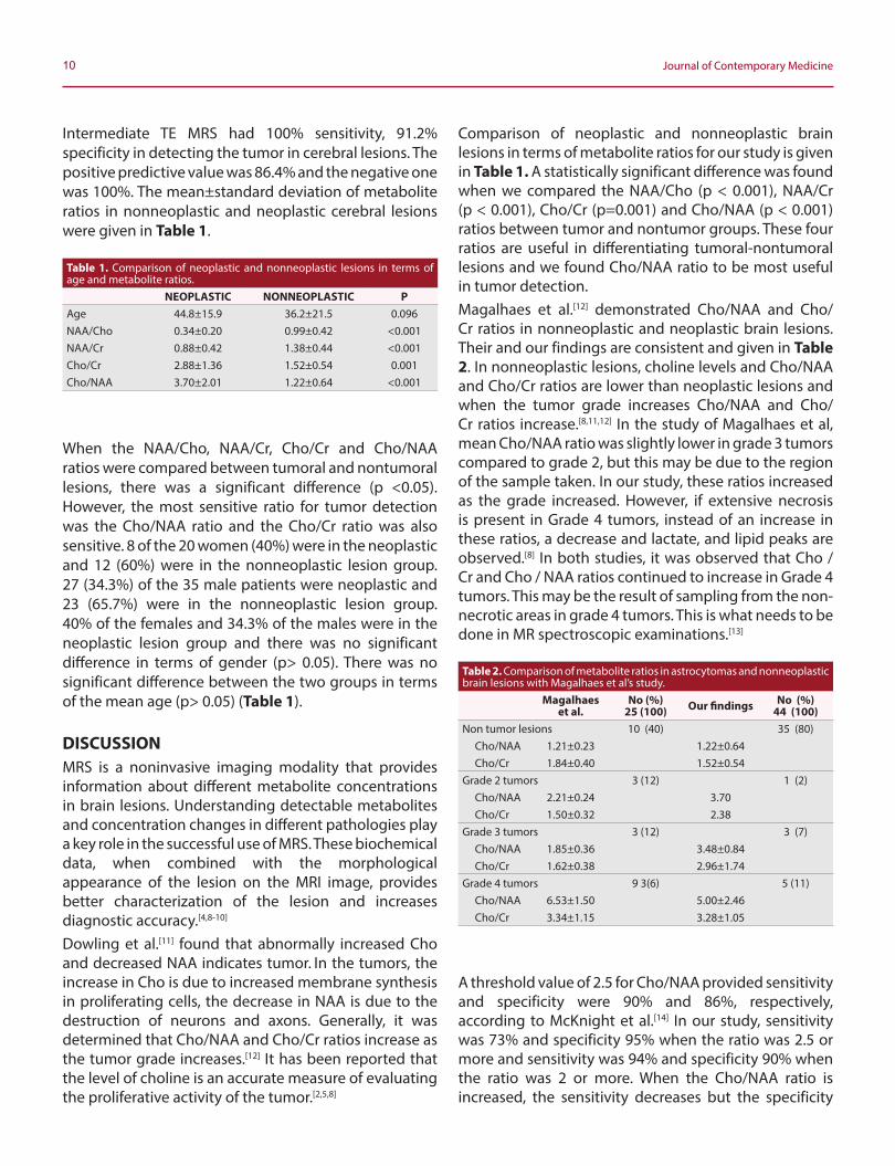
Intermediate TE MRS had 100% sensitivity, 91.2% specificity in detecting the tumor in cerebral lesions. The positive predictive value was 86.4% and the negative one was 100%. The mean±standard deviation of metabolite ratios in nonneoplastic and neoplastic cerebral lesions were given in Table 1.
When the NAA/Cho, NAA/Cr, Cho/Cr and Cho/NAA ratios were compared between tumoral and nontumoral lesions, there was a significant difference (p <0.05). However, the most sensitive ratio for tumor detection was the Cho/NAA ratio and the Cho/Cr ratio was also sensitive. 8 of the 20 women (40%) were in the neoplastic and 12 (60%) were in the nonneoplastic lesion group. 27 (34.3%) of the 35 male patients were neoplastic and 23 (65.7%) were in the nonneoplastic lesion group. 40% of the females and 34.3% of the males were in the neoplastic lesion group and there was no significant difference in terms of gender (p> 0.05). There was no significant difference between the two groups in terms of the mean age (p> 0.05) (Table 1).
DISCUSSIONMRS is a noninvasive imaging modality that provides information about different metabolite concentrations in brain lesions. Understanding detectable metabolites and concentration changes in different pathologies play a key role in the successful use of MRS. These biochemical data, when combined with the morphological appearance of the lesion on the MRI image, provides better characterization of the lesion and increases diagnostic accuracy.[4,8-10]
Dowling et al.[11] found that abnormally increased Cho and decreased NAA indicates tumor. In the tumors, the increase in Cho is due to increased membrane synthesis in proliferating cells, the decrease in NAA is due to the destruction of neurons and axons. Generally, it was determined that Cho/NAA and Cho/Cr ratios increase as the tumor grade increases.[12] It has been reported that the level of choline is an accurate measure of evaluating the proliferative activity of the tumor.[2,5,8]
Comparison of neoplastic and nonneoplastic brain lesions in terms of metabolite ratios for our study is given in Table 1. A statistically significant difference was found when we compared the NAA/Cho (p < 0.001), NAA/Cr (p < 0.001), Cho/Cr (p=0.001) and Cho/NAA (p < 0.001) ratios between tumor and nontumor groups. These four ratios are useful in differentiating tumoral-nontumoral lesions and we found Cho/NAA ratio to be most useful in tumor detection.Magalhaes et al.[12] demonstrated Cho/NAA and Cho/Cr ratios in nonneoplastic and neoplastic brain lesions.
Their and our findings are consistent and given in Table 2. In nonneoplastic lesions, choline levels and Cho/NAA and Cho/Cr ratios are lower than neoplastic lesions and when the tumor grade increases Cho/NAA and Cho/Cr ratios increase.[8,11,12] In the study of Magalhaes et al, mean Cho/NAA ratio was slightly lower in grade 3 tumors compared to grade 2, but this may be due to the region of the sample taken. In our study, these ratios increased as the grade increased. However, if extensive necrosis is present in Grade 4 tumors, instead of an increase in these ratios, a decrease and lactate, and lipid peaks are observed.[8] In both studies, it was observed that Cho / Cr and Cho / NAA ratios continued to increase in Grade 4 tumors. This may be the result of sampling from the non-necrotic areas in grade 4 tumors. This is what needs to be done in MR spectroscopic examinations.[13]
A threshold value of 2.5 for Cho/NAA provided sensitivity and specificity were 90% and 86%, respectively, according to McKnight et al.[14] In our study, sensitivity was 73% and specificity 95% when the ratio was 2.5 or more and sensitivity was 94% and specificity 90% when the ratio was 2 or more. When the Cho/NAA ratio is increased, the sensitivity decreases but the specificity
Table 2. Comparison of metabolite ratios in astrocytomas and nonneoplastic brain lesions with Magalhaes et al’s study.
Magalhaes et al.
No (%)25 (100) Our findings No (%)
44 (100)Non tumor lesions 10 (40) 35 (80)
Cho/NAA 1.21±0.23 1.22±0.64Cho/Cr 1.84±0.40 1.52±0.54
Grade 2 tumors 3 (12) 1 (2)Cho/NAA 2.21±0.24 3.70 Cho/Cr 1.50±0.32 2.38
Grade 3 tumors 3 (12) 3 (7)Cho/NAA 1.85±0.36 3.48±0.84 Cho/Cr 1.62±0.38 2.96±1.74
Grade 4 tumors 9 3(6) 5 (11)Cho/NAA 6.53±1.50 5.00±2.46Cho/Cr 3.34±1.15 3.28±1.05
Table 1. Comparison of neoplastic and nonneoplastic lesions in terms of age and metabolite ratios.
NEOPLASTIC NONNEOPLASTIC PAge 44.8±15.9 36.2±21.5 0.096NAA/Cho 0.34±0.20 0.99±0.42 <0.001NAA/Cr 0.88±0.42 1.38±0.44 <0.001Cho/Cr 2.88±1.36 1.52±0.54 0.001Cho/NAA 3.70±2.01 1.22±0.64 <0.001

11Şerife Leblebisatan, Differentiation of brain masses by MRS
increases.[13] In a study in which there was no statistically significant difference in metabolite ratios between different types of tumors, MRS was found to be successful in distinguishing infiltrative or limited lesions.[10]
In noninfiltrating processes such as abscesses, meningiomas, and metastases, no pathological findings were detected in spectral analyzes of nonenhanced peripheral portions.[1,15] In spectral analysis of periphery of infiltrating high-grade gliomas, it was determined that the NAA/Cho ratio is smaller than one.[10] In our study, in nine cases voxel has positioned periphery of the lesion. The NAA/Cr ratio was higher than one in the five noninfiltrating cases (two hematomas, one arachnoid cyst, one radiation necrosis, and one vascular malformation), in infiltrative four cases (two primer tumors and two tumor recurrences) it was smaller than one. Our findings are consistent with the literature.The differentiation residual or recurrent tumor from radiation necrosis is an important application of MRS. Generally, high choline peak indicates tumor progression and low choline peak indicates radiation necrosis. Lipid peak shows necrosis that both tumor and radiotherapy can give the same appearance.[12,16] It has been reported that in tumor progression, the mean± SD values of Cho/Cr 2.30±1.29, Cho/NAA 3.44±2.76 and NAA/Cr 0.93±0.81, in radiation necrosis these values 1.26±0.61,1.29±1.17 and 1.31±0.78 respectively. Cho/Cr and Cho/NAA ratios have 82% and 81% accuracy, respectively, in differentiating neoplastic and nonneoplastic lesions. In this study, lipid-lactate values and NAA/Cr ratio were not found to be useful.[16] In our study, MRS examinations were performed in 6 cases in order to distinguish recurrent/residual tumor from radiation necrosis and we accurately detected the recurrent/ residual tumor in 4 cases and radiation necrosis in 2 cases.It is known that TE has a significant effect on spectral data obtained in MRS. A comparative study of short to intermediate TE in discriminating between high and low-grade tumors has reported that Cho/Cr and LL/Cr ratios are useful in both TE, but short TE has a slightly higher accuracy rate.[17] In our study, we used intermediate TE (144 milliseconds) and it was reported to be ideal to assess neoplasms.[13]
In our study our priority was to make tumor-nontumor distinction. We preferred moderate TE because of less metabolite and easier evaluation option. Again, the fact that the lactate peak can be more clearly distinguished is another plus. For more specific diagnosis, short TE may be preferred. It has been reported that MRS is 95% to 100% accurate in distinguishing neoplastic and nonneoplastic
lesions.[13] In our study, intermediate TE MRS had 100% sensitivity and 91.2% specificity in detecting the tumor in cerebral lesions. The positive predictive value was 86.4% and the negative predictive value was 100%. The limitations of our study were the heterogeneity of the tumor and non-tumor groups. Another limitation is that age groups vary widely. As we could not compare between intermediate TE, short and long TE MR spectroscopy, we could not determine the advantages and disadvantages of intermediate TE MR spectroscopy.
CONCLUSIONMRS has high sensitivity, specificity and accuracy rates when it is supported by conventional MR in the differentiation of neoplastic-nonneoplastic lesions in cerebral lesions. Metabolite levels and ratios detected in intermediate TE MRS can be used to distinguish malignant and nonmalignant lesions from normal brain tissue. Cho/Cr and Cho/NAA ratios are the most sensitive values for choosing a tumor, and increase at these ratios indicate the tumor.[11-13,16] However, the obtained spectrum does not always give a specific diagnosis. Different TE sequences combination and additional other imaging methods should be used to increase the diagnostic accuracy.
ETHICAL DECLARATIONSEthics Comittee Approval: This study derived from Dr. Şerife Leblebisatans's medical specialty thesis in radiology (Çukurova University School of medicine, 2006/Adana, TURKEY).Informed Consent: Because the study was designed retrospectively, no written informed consent form was obtained from patients. Status of Peer-review: Externally peer-reviewed. Conflict of Interest Statement: The authors have no conflicts of interest to declare. Financial Disclosure: The authors declared that this study has received no financial support. Author Contributions: All of the authors declare that they have all participated in the design, execution, and analysis of the paper, and that they have approved the final version.
REFERENCES1. Bertholdo D, Watcharakorn A, Castillo M. Brain Proton Magnetic
Resonance Spectroscopy: Introduction and Overview. Neuroimaging Clin N Am 2013;23(3):35980.
2. Howe FA, Opstad KS. 1H MR spectroscopy of brain tumours and masses. NMR Biomed 2003;16:123-31.
3. Young RJ, Knopp EA. Brain MRI: tumor evaluation. J Magn Reson Imaging. 2006;24(4):709-24.
4. Sibtain NA, Howe FA, Saunders DE. The clinical value of proton
12 Journal of Contemporary Medicine
magnetic resonance spectroscopy in adult brain tumours. Clin Radiol. 2007;62(2);109-19.
5. Herminghaus S, Pilatus U, Moller-Hartmann W, Raab P, Lanfermann H, Schlote W, Zanella FE. Increased choline levels coincide with enhanced proliferative activity of human neuroepithelial brain tumors. NMR Biomed 2002;15(6):385-92.
6. Kim SH, Chang KH, Song IC, Han MH, Kim HC, Kang HS, Han MC. Brain abscess and brain tumor: discrimination with in vivo H-1 MR spectroscopy. Radiology 1997;204:239-45.
7. Poptani H, Gupta RK, Roy R, Pandey R, Jain VK, Chhabra DK. Characterization of intracranial mass lesions with in vivo proton MR spectroscopy. AJNR Am J Neuroradiol 1995;16:1593-603.
8. Brandao LA, Castillo M. Adult Brain Tumors: Clinical Applications of Magnetic Resonance Spectroscopy. Magn Reson Imaging Clin N Am 2016;24(4):781-809.
9. Shih MT, Singh AK, Wang A, Patel S. Brain Lesions with Elevated Lactic Acid Peaks on Magnetic Resonance Spectroscopy. Curr Probl Diagn Radiol 2004;33:85-95.
10. Burtscher IM, Skagerberg G, Geijer B, Englund E, Stahlberg F, Holtas S. Proton MR spectroscopy and preoperative diagnostic accuracy: an evaluation of intracranial mass lesions characterized by stereotactic biopsy findings. AJNR 2000;21:84-93.
11. Dowling C, Bollen AW, Noworolski SM, Mc Dermott MW, Barbaro NM, Day MR, Henry RG, Chang SM, Dillon WP, Nelson SJ, Vigneron DB. Preoperative proton MR spectroscopic imaging of brain tumors: correlation with histopathologic analysis of resection specimens. AJNR 2001;22:604-12.
12. Magalhaes A, Godfrey W, Shen Y, Hu J, Smith W. Proton magnetic resonance spectroscopy of brain tumors correlated with pathology. Acad Radiol 2005;12(1):51-7.
13. Brandao LA. MR Spectroscopy of the Brain. Lippincott Williams & Wilkins, 2003.
14. McKnight TR, von dem Bussche MH, Vigneron DB, Lu Y, Berger MS, McDermott MW, Dillon WP, Graves EE, Pirzkall A, Nelson SJ. Histopathological validation of a threedimensional magnetic resonance spectroscopy index as a predictor of tumor presence. J Neurosurg 2002;97(4):794802.
15. Law M, Cha S, Knopp EA, Johnson G, Arnett J, Litt AW. Highgrade gliomas and solitary metastases: differentiation by using perfusion and proton spectroscopic MR imaging. Radiology. 2002;222(3):715-21.
16. Schlemmer HP, Bachert P, Herfarth KK, Zuna I, Debus J, van Kaick G. Proton MR spectroscopic evaluation of suspicious brain lesions after stereotactic radiotherapy. Am J Neuroradiol 2001;22:1316-24.
17. Kim JH, Chang KH, Na DG, Song IC, Kwon BJ, Han MH, Kim K. 3T 1H-MR Spectroscopy in Grading of Cerebral Gliomas: Comparison of Short and Intermediate Echo Time Sequences Am J Neuroradiol 2006;27:1412-8.
Corresponding (İletişim): Hülya Yiğit Özay, SBÜ Türkiye Yüksek İhtisas Eğitim ve Araştırma Hastanesi, Anesteziyoloji ve Reanimasyon Kliniği, Ankara, Türkiye E-mail (E-posta): [email protected] (Geliş Tarihi): 09.09.2018 Accepted (Kabul Tarihi): 18.12.2019
DOI: 10.16899/jcm.705176J Contemp Med 2020;10(1):13-17
Orjinal Araştırma / Original Article
JOURNAL OF
CONTEMPORARY MEDICINEJournal ofContemporary MedicineYEAR: 2019 VOLUME: 9 ISSUE: 4
Yoğun Bakım Ünitesinde Tedavi Edilen Yaslı Hastaların D-Vitamini Düzeylerinin Prognoz Üzerine Etkisi
The Effect of Vitamin-D Levels on Prognosis of Elderly Patients Treated In Intensive Care Unit
Amaç: Dünyada yaşlı nüfusun %50-60’ında vitamin D eksikliği vardır. Kritik hastalarda ise vitamin D eksikliğinin (25 (OH) D<20ng/ml) prevalansı %70’i aşmaktadır. Bu çalışmada Yoğun Bakım Ünitesi (YBÜ)’nde takip ve tedavi edilen 65 yaş üstü; vitamin D verilen ve verilmeyen hastaların prognoz açısından karşılaştırılması amaçlanmıştır.
Gereç ve Yöntem: Kasım 2014-Nisan 2015 tarihleri arası hastanemiz YBÜ ’de takip ve tedavi edilen hastaların kayıtları geriye dönük olarak incelendi. Enteral beslenen ve vitamin D desteği alan 20 hasta (Grup D), enteral beslenen ancak vitamin D desteği almayan 20 hasta (Grup K) arasında yoğun bakıma kabul sırasında (T1), 7. (T2) ve 15. (T3) günlerde ölçülen vitamin D, albümin, prealbumin, total lenfosit, CRP düzeyleri ve triceps kas kalınlığı açısından fark olup olmadığı değerlendirildi.
Bulgular: İki grup demografik veriler açısından benzerdi. Grup D ile Grup K arasında T1,T2 ve T3 zamanlarında vitamin D düzeyleri, albüminprealbümin düzeyleri, enfeksiyon oranları, mekanik ventilasyon süreleri, yoğun bakım yatış günü ve mortalite açısından istatistiksel olarak anlamlı bir fark saptanmazken T1 ve T2 zamanında total lenfosit (TLS) düzeyleri (p<0,05) vitamin D grubunda istatistiksel olarak anlamlı yüksek idi. Hastaların sadece T3 zamanında vitamin D ve diğer değişkenler ele alındığında ise APACHE II değerleri, MV gün, T3 albümin, T3 prealbümin ve T3 total proteinle istatistiki olarak anlamlı ilişki gözlendi.
Sonuç: Çalışmamızda vitamin D verilen grupla verilmeyen grup arasında bazı değişkenlerde anlamlı fark gözlenmesi, prospektif ve daha fazla sayıda hasta ile yapılacak çalışmalar açısından yol gösterici olacağını düşünmekteyiz.
Anahtar Kelimeler: Vitamin D, yaşlı hasta, yoğun bakım
Abstract Öz
Hülya Yiğit Özay1, Ibrahim Mungan2, Güneş Çobanoğlu Ercan3, Sema Turan2, Banu Eler Çevik4
Aim: 50-60% of elderly population in the world has vitamin D deficiency. In critically ill patients, the prevalence of vitamin D deficiency (25 (OH) D <20 ng/ml) exceeds 70%. The aim of this study was to compare the prognosis of patients over 65 years of age with and without vitamin D supplementation in the Intensive Care Unit (ICU).Material and Method: The records of patients who were followed-up and treated in our hospital between November 2014 and April 2015 were retrospectively reviewed. The patients were grouped as enteral-fed with vitamin D supplementation (20 patients, Group D) and enterally fed without vitamin D supplementation (20 patients, Group K). Vitamin D, albumin, prealbumin, total lymphocyte, CRP levels and triceps muscle thickness were measured during the admission to intensive care unit (T1), on days 7 (T2) and 15 (T3).Results: The two groups were similar in terms of demographic data. There was no statistically significant difference between Group D and Group K in terms of vitamin D levels, albumin-prealbumin levels, infection rates, duration of mechanical ventilation, intensive care unit stay and mortality in T1, T2 and T3 times, while total lymphocyte levels (p<0,05) in T1 and T2 were statistically significant in vitamin D group. When vitamin D and other variables were evaluated at time T3, a statistically significant relationship was observed with APACHE II values, MV day, T3 albumin, T3 prealbumin and T3 total protein.Conclusion: In our study, a significant difference was observed between the vitamin D supplementation group and the group which was not given. In our opinion, the result of this study will be a guide to prospective randomized trials involving more patients.Keywords: Vitamin D, elderly patient, intensive care unit
1SBÜ Türkiye Yüksek İhtisas Eğitim ve Araştırma Hastanesi, Anesteziyoloji ve Reanimasyon Kliniği, Ankara, Türkiye 2SBÜ Türkiye Yüksek İhtisas Eğitim ve Araştırma Hastanesi, Yoğun Bakım Kliniği, Ankara, Türkiye
3Ilgın Devlet Hastanesi, Anesteziyoloji ve Reanimasyon Kliniği, Konya,Türkiye4SBÜ Dr. Lütfi Kırdar Kartal Eğitim ve Araştırma Hastanesi, Anesteziyoloji ve Reanimasyon Kliniği, İstanbul,Türkiye
14 Journal of Contemporary Medicine
GIRIŞGünümüzde, ortalama yaşam süresinin uzamasıyla birlikte yoğun bakımlarda tedavi edilen yaşlı hastaların oranı da artmıştır. Literatürde yoğun bakıma yatan hastaların %42-52’sini yaşlı hastaların oluşturduğu bildirilmektedir.[1] Genellikle yoğun bakıma yatış nedenlerini, altta yatan kronik hastalıklarının akut alevlenmeleri veya birçok organ sistemini ilgilendiren bilişsel bozukluk, düşme, inkontinans, gorme ya da işitme kaybı, baş donmesi, düşük vücut kitle indeksi gibi sorunlar oluşturmaktadır. Bu sebeplerden dolayı yoğun bakımdaki yaşlı hastalar oldukça karmaşık ve ayrıcalıklı bir hasta grubunu oluşturmaktadır.[2,3]
Son yıllarda yapılan çalışmalarla vitamin D eksikliğinin ya da yetersizliğinin yaşlı hastalarda birçok organ sistemini ilgilendiren sorunlara sebep olabileceği gozlemlenmiştir.[4] Vitamin D’nin normal kalsiyum ve fosfor metabolizmasını düzenlemesi yanında vücudumuzda hemen her hücrede vitamin D reseptorlerinin tespiti; immun modülasyon etkisi, hipertansiyon, diyabet, KOAH, demans gibi kronik hastalıklarla ilişkilendirilmesi vitamin D’nin farklı etkilerini yeniden tartışmaya açmıştır.[2,5-7] Literatürde düşük vitamin D düzeyleri; yoğun bakımda uzun yatış süreleri, yüksek enfeksiyon oranları ve yüksek mortalite ile ilişkilendirilmiştir.[8,9]
Bu çalışmada, yoğun bakım ünitemizde tedavi edilen 65 yaş üstü; D vitamini verilen ve verilmeyen hastaların prognoz açısından karşılaştırılması amaçlanmıştır.
GEREÇ VE YÖNTEMÇalışmamızda hastanemiz etik kurul onayı alınarak Anesteziyoloji ve Reanimasyon Klinikleri yoğun bakım ünitelerinde Kasım 2014-Nisan 2015 tarihleri arasında takip ve tedavi edilen 341 hastanın dosyası retrospektif olarak incelendi.
Enteral beslenme yanında vitamin D verilen 65 yaş ve üzeri olan 33 olgu tespit edildi. D vitamini düzeyleri bilinmeyen olgular çalışma dışı bırakıldı. Yoğun bakıma kabulde (T1), birinci haftada (T2) ve ikinci haftada (T3) vitamin D düzeyleri olçülmüş 20 hasta saptandı. Kontrol grubu olarak vitamin D verilmeyen ancak yoğun bakıma kabulünde ve tedavileri süresince vitamin D düzeyi bakılmış 20 hasta sırasıyla protokol defterinden kayıt edildi. Enteral beslenen (20-25kcal/gün) ve vitamin D (1000 IU/gün)desteği alan 20 hastaya vitamin D grubu (Grup D), enteral beslenen (20-25kcal/gün) ancak vitamin D desteği almayan 20 hastaya kontrol grubu (Grup K) denildi. Hastaların demografik ozellikleri, yatış endikasyonları ve yandaş hastalıkları, yoğun bakıma kabulde Glaskow koma skoru (GKS) ve APACHE II skoru, mekanik ventilasyon (MV) ihtiyacı, yoğun bakım yatış süreleri, T1, T2ve T3 zamanlarında vitamin D, albümin, prealbümin, total lenfosit sayısı, total protein düzeyi, triceps çevresi, CRP, enfeksiyon ve mortalite oranları incelenerek kayıt edildi.
Retrospektif olarak toplanan çalışma verileri Excel (Microsoft Corp., Redmond, WA, USA) programında kayıt altına alındı. Verilerin istatistik analizinde SPSS (SPSS Statistics for Windows, Version 20.0, IBM Corp., Armonk, NY) programı kullanıldı. Verilerin tanımlanmasında sürekli değişkenler için ortalama (ort) ve ±standart sapma (SS), kategorik değişkenler için sayı (n) ve yüzde (%) kullanıldı. Verilerin normal dağılıma uygunluğu KolmogorovSmirnov testi ile test edildi. Kolmogorov Smirnov testi ile normal dağılım olup olmadığı değerlendirildi. Dağılım normal olmadığından ve normal dağılıma donüştürülemediğinden ikili grup karşılaştırmaları için nonparametrik test olan Mann-Whitney U testi kullanıldı. Ayrıca T3 değerlerine anlamlı ilişki açısından spearman rho testi uygulandı. Sonuçların değerlendirilmesinde %95 güven aralığında (GA) p <0,05 değeri anlamlı olarak kabul edildi.
BULGULARDünyada yaşlı nüfusun %50-60’ında vitamin D eksikliği vardır. Kritik hastalarda ise vitamin D eksikliğinin (25 (OH) D<20ng/ml) prevalansı %70’i aşmaktadır.[8,10,11] Vitamin D’nin kemik üzerine etkilerinden başka kardiyovasküler, immun sistem ve inflamatuar sistem üzerine olan etkilerinin ortaya çıkmasıyla kritik hastalardaki rolü daha dikkat çekici olmuştur.[12,13] Buradan yola çıkarak değerlendirdiğimiz 65 yaş üstü 40 hastanın yoğun bakıma kabulünde %95.13’ünde ciddi vitamin D eksikliği ( 25 (OH) D < 10ng/ml), %4.87’inde vitamin D eksikliği (10ng/ml <25 (OH) D<20ng/ml) saptandı.Olguların %42,5’i erkek, % 57.5’i kadın, ortalama yaş 76.98 (61-95) idi. Hastalar gruplara ayrıldığında Grup D’nin %35’i erkek, %65’i kadın ortalama yaş 76.15, Grup K’ nın %50’si erkek, % 50’si kadın ortalama yaş 77.80 yıl saptandı. Her iki grup arasında cinsiyet ve yaş açısından istatistiksel olarak anlamlı fark yoktu (p>0,05) (Tablo 1).
Yoğun bakıma yatış endikasyonları açısından istatistiksel olarak anlamlı bir fark yoktu. En sık yatış nedenleri pnomoni (%37.5), KOAH (%22.5) ve serebrovasküler olay (%17.5);yandaş hastalıkları ise hipertansiyon (%67.5), Alzheimer (%32.5) ve diyabet (%20) idi (Tablo 2-3).
Tablo 1. Yaş ve cinsiyete gore hasta dağılımıD vit kontrol p
Cinsiyet-erkek % %35 %50 0,34Yaş( ortalama) 76,15 77,80 0,22
Tablo 2. Yatış endikasyonlarına gore hasta dağılımıTanı n %Akut bobrek yetmezliği 1 2,5Femur fraktürü 6 15KOAH 9 22,5KKY 1 2,5SVO 7 17,5Pnomoni 15 37,5Anafilaksi 1 2,5KOAH: Kronik obstrüktif akciğer hastalığı, KKY: Konjestif kalp yetersizliği, SVO: Serebro-vasküler olay

15Hülya Yiğit Özay, Kritik hastada D vitamininin prognoza etkisi
Hastaların yoğun bakım takip ve tedavileri sırasında vitamin D grubunda %9.65, kontrol grubunda %8.70 oranında mekanik ventilator desteğine ihtiyacı oldu. Yoğun bakım yatış süreleri sırasıyla 20.95 ve 23.05 gün bulundu. Hastaların yogun bakım yatışlarındaki hastalık şiddet skorları -apache 2 ve gks ile değerlendirilmiştiile hastane sonuçları (outcome) her iki grupta karşılaştırıldığında istatistiki olarak belirgin fark olmadığı gozlendi (p>0.05) Mortalitemorbidite skoru açısından karşılaştırıldığında, GKS ve APACHE II değerleri benzerlik gostermekte idi (Tablo 4).
Grup D ile Grup K arasında T1, T2 ve T3 yatış günlerindeki vitamin D, albümin, prealbümin, total lenfosit sayısı, total protein düzeyi, triceps çevresi, CRP, enfeksiyon oranları karşılaştırıldığında; T1 ve T2 zamanında total lenfosit (TLS) düzeyleri açısından iki grup arasında istatistiki olarak belirgin fark gozlendi (p=0.002 ve p=0.021). Diger veriler açısından istatistiksel olarak anlamlı bir fark bulunmadı (p>0.05) (Tablo 5).
Hastaların sadece T3 vitamin D ve diğer değişkenler değerleri ele alındığında yapılan korrelasyon incelemesinde (spearman rho testi ile) ise APACHE II değerleri, MV gün, T3 albümin, T3 prealbümin ve T3 total proteinle istatistiki olarak anlamlı ilişki gozlendi.
TARTIŞMABu retrospektif çalışmada, yoğun bakımda vitamin D verilen ve verilmeyen geriatrik hastaların prognozunu değerlendirmeyi amaçladık. Yapılan prospektif çalışmaların büyük çoğunluğu vitamin D eksikliğinin, mortaliteyi onemli derecede arttırdığını gostermektedir.[14,15] Biz kritik yaşlı hastaların yoğun bakıma kabulünde %100 oranında vitamin D eksikliği saptadık. Aynı zamanda vitamin D’nin antimikrobial ve immunmodülator etkilerinin de olduğu ifade edilmektedir.[16,17] Dolayısıyla yoğun bakımlarda kritik yaşlı hastalarda vitamin D düzeylerinin takibinin ve tedavisinin onemli olduğuna inanmaktayız.
Geriatrik hastalarda vitamin D eksikliğinin başlıca nedenlerini, daha az gün ışığına maruz kalmaları, giyim tarzları, daha koyu cilt rengine sahip olmaları ve ciltlerinin vitamin D sentez yeteneğini kaybetmesi oluşturmaktadır.[18] Rastgele kontrollü çalışmalarda, kritik hastalara vitamin D takviyesinin mortaliteyi azalttığı ifade edilmektedir.[19,20] Vitamin D takviyesi için, literatürde vitamin D dozları ve hedef seviyeleri hala tartışmalıdır.[21-23] Kuzey Avrupa ülkelerinde genel popülasyonun; beslenme, belli oranda güneşe maruz kalma ve günde 800 IU vitamin D almaları onerilmekte ve 20 ng/ml altındaki vitamin D değerlerinin onemli sağlık sorunu oluşturacağı ifade edilmektedir.[24] Bu yüzden biz olgularımızın yeterli vitamin D düzeylerini sağlayabilmek için günde 1000
Tablo 3. Yandaş hastalıklara gore hasta dağılımı
Komorbidite N %
Alzheimer-demans 13 32,5
Hipertansiyon 27 67,5
AstımKOAH 5 12,5
DM 8 20
Post CPR 1 2,5
Meme kanseri 1 2,5
KAH 1 2,5
KKY 2 5KOAH: Kronik obstruktif akciğer hastalığı, DM: Diabetes mellitus, CPR: Kardiyopulmoner resusitasyon, KAH: Koroner arter hastalığı, KKY: Konjestif kalp yetmezliği
Tablo 4. Vitamin D ve kontrol grubundaki hastaların hastalık şiddet skorları ve hastane sonuçlarının karşılaştırılması
Grup D Grup K p
GKS 8,10 9,75 0,134
APACHE II 17,85 17,10 0,42
Mekanik ventilasyon süresi(gün) 9,65 8,70 0,978
Yoğun bakım kalış 20,95 23,05 0,184
Mortalite(%) 4(%20) 3(%15) 0,68GKS: Glaskov koma skoru, APACHE II: Akut fizyoloji ve kronik sağlık değerlendirmesi
Tablo 5. Vitamin D ve kontrol grubundaki hastaların yoğun bakıma kabulünde ve belirlenen diğer zaman dilimlerinde bakılan labarotuar ve klinik parametrelerinin karşılaştırılması.
Grup D Grup K pT1 vitamin D 4,395000 4,045000 0,935T2 vitamin D 3,9765 3,7150 0,704T3 vitamin D 3,516500 4,065000 0,989T1 albümin 2,435000 2,600000 0,444T2 albümin 2,275000 2,375000 0,817T3 albümin 2,325000 2,190000 0,66T1 prealbümin 8,010000 8,450000 0,383T2 prealbümin 7,4500 7,6500 0,934T3 prealbümin 8,300 6,725 0,211T1 total lenfosit ,980000 ,680000 0,002T2 total lenfosit 1,065000 ,710000 0,021T3 total lenfosit 1,060000 ,835000 0,13T1 triceps 26,840 26,730 0,871T2 triceps 26,600 26,215 0,694T3 triceps 26,620 25,970 0,440T1 CRP 124,675000 124,230000 0,695T2 CRP 111,325000 147,525000 0,675T3 CRP 88,510000 122,230000 0,317T1 total protein 5,755000 5,360000 0,377T2 total protein 5,405000 5,115000 0,370T3 total protein 5,540000 5,035000 0,267T1 enfeksiyon 0 0 1T2 enfeksiyon 4-%20 1-%5 0,157T3 enfeksiyon 2-%10 3-%15 0,637T1: yoğun bakıma kabul zamanı, T2: yoğun bakıma kabulünün 7. günü, T3: yoğun bakıma kabulünün 15.günü, CRP: C reaktif protein
16 Journal of Contemporary Medicine
IU vitamin D takviyesi yaptık. Ancak onbeş günlük tedavi sonunda vitamin D düzeylerinde anlamlı bir artış saptamadık. Bunun nedeninin takip süresinin kısa olması ve kısıtlı hasta sayısı olduğunu düşünüyoruz.Vitamin D’nin en onemli etkilerinden biri de kemik mineralizasyonu için yeterli kalsiyum düzeyini sağlamak ve boylece raşitizm ve osteomalaziyi onlemektir.[2,5,25]
Randomize kontrollü çalışmaların meta-analizinde vitamin D desteğinin kemik kırıklarını onemli olçüde azalttığı tespit edilmiştir.[26] Kemik kırıklarını onlemek için günlük yaklaşık 800-2000 IU vitamin D dozunun yeterli olduğu gosterilmiştir.[26,27] Bizim olgularımızın da altısı femur fraktürü nedeniyle takip ediliyordu. Hastalarımızın bir çoğunda vitamin D eksikliğinin ilişkili bulunduğu.[6,7] Hipertansiyon, diyabet, KOAH gibi komorbiditeler de mevcuttu.Literatürde vitamin D eksikliğinin mekanik ventilator ihtiyacını, yoğun bakım kalış süresini ve mortaliteyi arttırdığına dair veriler vardır.[8,9] Aksine VITdAL-ICU çalışmasında yüksek doz vitamin D kullanımı plasebo ile karşılaştırılmış hastane yatış süresi, hastane mortalitesi ve 6 aylık mortaliteyi azaltmadığı saptanmıştır.[28] Bizim çalışmamızda ise, grup D’de hastaların ortalama MV süresi daha uzun bulundu ancak istatistiksel olarak anlamlı değildi (p>0.05). Bunu grup K’daki hastaların yoğun bakıma kabulünde klinik durumlarının daha iyi olmasına bağladık. Grup D’de yoğun bakım kalış süresi istatistiksel olarak anlamlı olmamakla birlikte daha kısa idi. Mortalite açısından her iki grup arasında anlamlı bir fark yoktu. Atalan ve arkadaşlarının yoğun bakım girişindeki vitaminin D düzeylerinin mortalite ile ilişkisini değerlendirdikleri bir çalışmada, düşük serum vitamin D düzeylerinin yalnızca APACHE II skorunun artmasıyla ilişkili olabileceğini ve YBÜ girişindeki en kotü klinik durumdan sorumlu olabileceğini belirtmişlerdir.[29] Biz de mortalite-morbidite skoru açısından değerlendirdiğimizde kontrol grubunda GKS ve APACHE II değerleri daha iyi olmasına rağmen her iki grup arasında istatistiksel olarak anlamlı bir fark bulunmadı. Sadece T3 zamanında vitamin D ve diğer değişkenlere uygulanan korelasyon testinde APACHE II değerleri, MV gün, T3 albümin, T3 prealbümin ve T3 total proteinle istatistiki olarak anlamlı ilişki bulundu. Yine Grup D ve grup K, T1 ve T2 zamanında total lenfosit (TLS) düzeyleri açısından karşılaştırıldığında iki grup arasında istatistiki olarak belirgin fark gozlendi (p<0,05). Verilerimiz vitamin D’nin akut faz reaksiyonu, antiinflamatuar ve immunmodülator etkilerini destekler niteliktedir.[16,17]
SONUÇSonuç olarak çalışmamızda vitamin D verilen grupla verilmeyen grup arasında bazı değişkenlerde anlamlı fark gozlenmesi, prospektif ve daha fazla sayıda hasta ile yapılacak çalışmalar açısından yol gosterici olacağını düşünmekteyiz.
ETIK BEYANLAREtik Kurul Onayı: Bu araştırmada 2020 oncesi veriler kullanılmış ve araştırma 2020 oncesinde sonuçlanmıştır. 3 Kasım 2015 tarihli 28617 sayılı olarak resmi gazetede yayınlanan Klinik Araştırmalar Hakkında Yonetmelik’e gore
“Retrospektif çalışmalar yonetmeliğin kapsamı dışındadır (madde 2-(2))”maddesi gereği etik kurul onayı alınmamıştır. Bu çalışma Kişisel Verilerin Korunması Kanununa uygun şekilde hasta verileri anonim hale getirtilerek ve İyi Klinik Uygulamalar ve Helsinki Deklarasyonun 2013 Brezilya güncellemesine uygun olarak hazırlanmıştır.Aydınlatılmış Onam: Çalışma retrospektif olarak dizayn edildiği için hastalardan aydınlatılmış onam alınmamıştır. Hakem Değerlendirme Süreci: Harici çift kor hakem değerlendirmesi.Çıkar Çatışması Durumu: Yazarlar bu çalışmada herhangi bir çıkara dayalı ilişki olmadığını beyan etmişlerdir.Finansal Destek: Yazarlar bu çalışmada finansal destek almadıklarını beyan etmişlerdir.Yazar Katkıları: Yazarların tümü; makalenin tasarımına, yürütülmesine, analizine katıldığını ve son sürümünü onayladıklarını beyan etmişlerdir.Kısıtlılıklar: Çalışmanın retrospektif olması, hasta sayısının az olması, süre olarak hasta yatış süreleri kısa olması nedeniyle kısa izlem süresi mevcuttu.
KAYNAKLAR1. Marik PE. Management of the critically ill geriatric patient. Cri Care Med
2006; 34: 176-82.
2. Holick MF. Vitamin D: a D-light fulhealth perspective. Nutr Rev 2008;66:182-94.
3. Hypponen E, Boucher BJ, Berry DJ, Power C. 25-hydroxyvitamin D, IGF-1, and metabolic syndrom at 45 years of age: a cross-sectionalstudy in the 1958 British Birth Cohort. Diabetes 2008;57:298-305.
4. Lee P, Eisman JA, Center JR. Vitamin D Deficiency in Critically Ill Patients. N Engl J Med 2009; 360:1912-1914April 30, 2009DOI: 10.1056/NEJMc08099967. authors.NEJM.org
5. Autier P, Boniol M, Pizot C, Mullie P. Vitamin D status and ill health: a systematic review. Lancet Diabetes Endocrinol 2014; 2: 76-89.
6. de Haan K, Groeneveld AB, de Geus HR, Egal M, Struijs A. Vitamin D deficiency as a risk factor for infection, sepsis and mortality in the critically ill: systematic review and meta-analysis. Crit Care 2014; 18: 660.
7. Gilbert CR, Arum SM, Smith CM. Vitamin D deficiency and chronic lung disease. Can Respir J 2009;16:75-80.
8. Sadeq A. Quraishi,, Edward A. Bittner, Livnat Blum. Prospective Study of Vitamin D Status at Initiation of Care in Critically Ill Surgical Patient sand Risk of 90-Day Mortality. Crit. Caremed. 2014 Jun;42 (6):1365-1371.
9. Kvaran RB, Sigurdsson MI, Skarphedinsdottir SJ, Sigurdsson GH. Severe vitamin D deficiency is common in criticallyillpatients at a highnorthernlatitude.2016 Oct;60 (9):1289-96. doi: 10.1111/ aas.12748. Epub 2016 Jun 12.
10. Arnson Y, Gringuaz I, Itzhaky D, Amithal H. Vitamin D deficiency is associated with poor outcomes and increased mortality in severely ill patients. QJM 2012;105:633-9
11. Venkatram S, Chilimuri S, Adrish M at al. Vitamin D deficiency is associated with mortality in the medical intensive care unit. Crit Care 2011;15:R292.
12. Ünal T, Özkan B, Çayır A, ve ark. Serum 25 (OH) D vitamin düşüklüğü çocukluk çağı pnomonileri için bir risk faktorü müdür? Dicle Tıp Dergisi 2012;39:531535.
13. Pilz S, Tomaschitz A, Drechsler C, de Boer RA. Vitamin D deficiency and heart disease. Kidney International Supplements 2011;1:111-115.
17Hülya Yiğit Özay, Kritik hastada D vitamininin prognoza etkisi
14. Schottker B, Haug U, Schomburg L,Kohrle J,et al. Strong associations of 25-hydroxyvitamin D concentrations with all-cause, cardiovascular, cancer, and respiratory disease mortality in a large cohort study. Am J Clin Nutr 97: 782-793, 2013.
15. Grober U,Reichrath J, Holic MF. Live longer with vitamin D? Nutrients 7:1871-1880,2015.
16. Bivona G, Agnello L, Ciaccio M.VitaminD and Immunomodulation: Is It Time to Change the Reference Values? Ann Clin Lab Sci. 2017 Aug;47 (4):508-510.
17. Vanherwegen AS, Gysemans C, Mathieu C.Vitamin D endocrinology on the cross-road between immunity and metabolism. Mol Cell Endocrinol. 2017 Sep 15;453:52-67. doi: 10.1016/j. mce.2017.04.018. Epub 2017 Apr 28.
18. Gürbüz P, Yetiş G. Yaşlılarda D Vitamini Eksikliği. İ.Ü. Sağlık Hizmetleri Meslek Yüksekokulu Dergisi, ISSN:2147-7892, Cilt 5, Sayı 2 (2017).
19. Autier P, Gandini S. Vitamin D supplementation and total mortality: a meta-analysis of randomized controlled trials. Arch Intern Med 2007; 167: 1730-7.
20. Bjelakovic G, Gluud LL, Nikolova D, At al. Vitamin D supplementation for prevention of mortality in adults. Cochrane Database Syst Rev 2011; CD007470.
21. Ross AC, Manson JE, Abrams SA, At al: The 2011 report on dietary reference intakes for calcium and vitamin D from the Institute of Medicine: What clinicians need to know. J Clin Endocrinol Metab 96: 53-58, 2011.
22. Rosen CJ, Abrams SA, Aloia JF, At al. IOM committee members respond to Endocrine Society vitamin D guideline. J Clin Endocrinol Metab 97: 1146-1152, 2012.
23. Hintzpeter B, Mensink GB, Thierfelder W, Müller MJ, Scheidt-NaveC: Vitamin D status and health correlates among German adults. Eur J Clin Nutr 62: 1079-1089, 2008.
24. Pilz S, Grübler M, Gaksch M, At al. Vitamin D and Mortality.Anticancer Res. 2016 Mar;36 (3):1379-87.
25. Wu H, Pang Q.The effect of vitamin D and calcium supplementation on falls in older adults : A systematic review and meta-analysis. Orthopade. 2017 Sep;46 (9):729-736. doi: 10.1007/s00132017-3446-y.
26. Bischoff-Ferrari HA, Willett WC, Orav EJ, At al. A pooled analysis of vitamin D dose requirements for fracture prevention. N Engl J Med 367: 40-49, 2012.
27. Reid IR. Vitamin D Effect on Bone Mineral Density and Fractures. Endocrinol Metab Clin North Am. 2017 Dec;46 (4):935-945. doi: 10.1016/j.ecl.2017.07.005. Epub 2017 Sep 29.
28. Amrein K, Schnedl C, Holl A, At al. Effect of high-dose vitamin D3 on hospital length of stay in critically ill patients with vitamin D deficiency: the VITdAL-ICU randomized clinical trial. JAMA 2014; 312: 1520-30.
29. Atalan HK , Güçyetmez B. Serum Vitamin D Level at ICU Admission and Mortality. Turk J Anaesthesiol Reanim 2017; 45: 193-6 DOI: 10.5152/TJAR.2017.60234
Corresponding (İletişim): Çiğdem Kunt İşgüder, İstanbul Üniversitesi İstanbul Tıp Fakültesi, Kadın Hastalıkları ve Doğum Anabilim Dalı, Turgut Özal Millet Caddesi, Fatih / Çapa Posta kodu: 34098, İstanbul/TürkiyeE-mail (E-posta): [email protected] (Geliş Tarihi): 13.07.2019 Accepted (Kabul Tarihi): 18.12.2019
DOI: 10.16899/jcm.591664J Contemp Med 2020;10(1):18-22
Orjinal Araştırma / Original Article
JOURNAL OF
CONTEMPORARY MEDICINEJournal ofContemporary MedicineYEAR: 2019 VOLUME: 9 ISSUE: 4
Plasenta Previa Olgularının Retrospektif Değerlendirilmesi
Retrospective Evaluation of Cases with Placenta Previa
Amaç: Bu çalışmada, plasenta previa (PP) olgularında maternal sosyo-demografik özelliklerin, obstetrik risk faktörlerin ve perinatal sonuçların değerlendirilmesini amaçladık.
Gereç ve Yöntem: Çalışmada Ocak 2013Kasım 2017 tarihleri arasında kliniğimizde takip edilen 71 PP olgusu retrospektif olarak incelendi. Olguların obstetrik öyküleri, prepartum ve postpartum hemoglobin değerleri, kan transfüzyon ihtiyacı, 1. ve 5. dakika apgar skorları, kanama kontrolünü sağlamak amacıyla yapılan cerrahi manevralar, plasental invazyon bulgusu, intrauterin ölü fetus ve neonatal ölüm gibi komplikasyonlar incelendi.
Bulgular: Değerlendirilen 71 PP olgusunun %19,7 (n=14)’sini nullipar kadınlar oluştururken, %22,5 (n=16)’ini daha önce 1 kez, %42,3 (n=30)’ünü 2 kez, %15,5 (n=11)’ini üç veya daha fazla sayıda doğum yapmış kadınlar oluşturuyordu. Olguların %53,5 (n=38)’inin önceden sezaryen öyküsü yoktu, %45 (n=32)’inin geçirilmiş 1 ve 2 sezaryen öyküsü, %1,5 (n=1)’ünün üç sezaryen öyküsü vardı. Ortalama bebek doğum ağırlığı 2749.79±777.38 gr (980 gr -4680 gr), 1. ve 5. dakika apgar skorları sırası ile 7.31±1.39 ve 8.24±1.33 olarak tespit edildi. İnvazyon bulguları nedeniyle 9 olguya peripartum histerektomi uygulandı. On üç olguya perioperatif, sekiz olguya postoperatif kan transfüzyonu yapıldı.
Sonuç: PP, hem erken doğum ve buna bağlı yenidoğan sorunları hem de plasental invazyon anomalileri ve maternal kanama nedeni ile günümüzde neonatal ve maternal morbidite ve mortalitenin önemli nedenleri arasındadır. Optimal sonuç elde edebilmek için PP’den şüphelenildiğinde ve antenatal dönemde tanı konulduğunda peripartum histerektomi ve uterin devaskülarizasyon manevraları konusunda tecrübeli ve oluşabilecek komplikasyonlara müdahale edebilecek deneyimli cerrahların bulunduğu ve kan ürünlerinin temin edilebileceği üçüncü basamak merkezlerde operasyonların yapılmasında fayda vardır.
Anahtar Kelimeler: Plasenta previa, obstetrik risk faktörleri, kan transfüzyonu
Abstract Öz
Çiğdem Kunt Işgüder1, Selim Gülücü2, Yunus Emre Bulut3, Ilhan Bahri Delibaş2, Tuğba Yurt2, Asker Zeki Özsoy2
Objective: In this study, we aimed to evaluate the maternal socio-demographic features, perinatal outcomes and obstetric risk factors in patients with placenta previa.Material and Method: In this study 71 patients with placenta previa who(opsiyonel) followed up in our clinic between January 2013 and November 2017 were evaluated retrospectively. The complications of cases such as obstetrical stories, prepartum and postpartum hemoglobin values, need of transfusion, 1st and 5th minutes APGAR scores and surgical maneuvers to provide bleeding control, placental invasion findings, in utero mort fetalis and neonatal mortality were all observed.Results: When 71 cases were evaluated; 14 (19.7%) of the cases were nulliparas, 16 (22.5%) of the cases gave birth to a child only once, 30(42.3%) of the cases gave birth two times and 11 (15.5%) of the cases gave birth three or more times. The women who had no cesarean delivery previously numbers were 38 (53.5%), whereas 32 (45%) had one or two, and one woman had three previous cesarean delivery. Mean birth weight was 2749.79±777.38 gr (980gr-4680 gr). Mean first and fifth minute APGAR scores were 7.31±1.39 and 8.24±1.33 respectively. 9 peripartum hysterectomy operation were performed due to placental invasion abnormalities. The number of patients who underwent blood tranfusion intraoperative and postoperative were 13 and 8 respectively.Conclusion: PP is currently one of the important causes of neonatal and maternal morbidity and mortality not only for preterm birth and accordingly newborn problems but also placental invasion abnormalities and maternal bleeding. In order to achive the optimal result, when there is a suspicion or antenatal diagnosis of placenta previa, delivery should be planned in tertiary centers where the surgeons are experienced in the field of peripartum hysterectomy and uterine devascularization maneuvers and interventions in the complications that may occur and where the blood products are provided easily.
Keywords: Placenta previa, obstetric risk factors, blood transfusion
1İstanbul Üniversitesi İstanbul Tıp Fakültesi Kadın Hastalıkları ve Doğum A.D., İstanbul 2Gaziosmanpaşa Üniversitesi Tıp Fakültesi Kadın Hastalıkları ve Doğum A.D., Tokat
3Ankara İl Sağlığı Müdürlüğü, Ankara
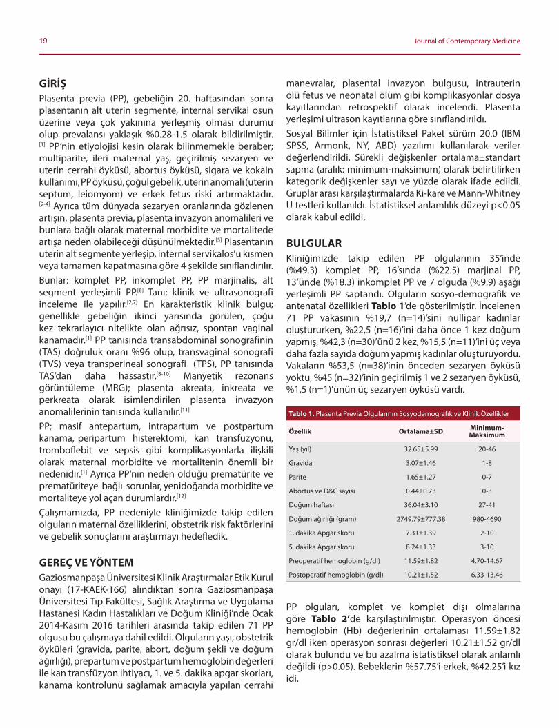
19 Journal of Contemporary Medicine
GIRIŞPlasenta previa (PP), gebeliğin 20. haftasından sonra plasentanın alt uterin segmente, internal servikal osun üzerine veya çok yakınına yerleşmiş olması durumu olup prevalansı yaklaşık %0.28-1.5 olarak bildirilmiştir.[1] PP’nin etiyolojisi kesin olarak bilinmemekle beraber; multiparite, ileri maternal yaş, geçirilmiş sezaryen ve uterin cerrahi oyküsü, abortus oyküsü, sigara ve kokain kullanımı, PP oyküsü, çoğul gebelik, uterin anomali (uterin septum, leiomyom) ve erkek fetus riski artırmaktadır.[2-4] Ayrıca tüm dünyada sezaryen oranlarında gozlenen artışın, plasenta previa, plasenta invazyon anomalileri ve bunlara bağlı olarak maternal morbidite ve mortalitede artışa neden olabileceği düşünülmektedir.[5] Plasentanın uterin alt segmente yerleşip, internal servikalos’u kısmen veya tamamen kapatmasına gore 4 şekilde sınıflandırılır.Bunlar: komplet PP, inkomplet PP, PP marjinalis, alt segment yerleşimli PP.[6] Tanı; klinik ve ultrasonografi inceleme ile yapılır.[2,7] En karakteristik klinik bulgu; genellikle gebeliğin ikinci yarısında gorülen, çoğu kez tekrarlayıcı nitelikte olan ağrısız, spontan vaginal kanamadır.[1] PP tanısında transabdominal sonografinin (TAS) doğruluk oranı %96 olup, transvaginal sonografi (TVS) veya transperineal sonografi (TPS), PP tanısında TAS’dan daha hassastır.[8-10] Manyetik rezonans gorüntüleme (MRG); plasenta akreata, inkreata ve perkreata olarak isimlendirilen plasenta invazyon anomalilerinin tanısında kullanılır.[11]
PP; masif antepartum, intrapartum ve postpartum kanama, peripartum histerektomi, kan transfüzyonu, tromboflebit ve sepsis gibi komplikasyonlarla ilişkili olarak maternal morbidite ve mortalitenin onemli bir nedenidir.[1] Ayrıca PP’nın neden olduğu prematürite ve prematüriteye bağlı sorunlar, yenidoğanda morbidite ve mortaliteye yol açan durumlardır.[12]
Çalışmamızda, PP nedeniyle kliniğimizde takip edilen olguların maternal ozelliklerini, obstetrik risk faktorlerini ve gebelik sonuçlarını araştırmayı hedefledik.
GEREÇ VE YÖNTEMGaziosmanpaşa Üniversitesi Klinik Araştırmalar Etik Kurul onayı (17-KAEK-166) alındıktan sonra Gaziosmanpaşa Üniversitesi Tıp Fakültesi, Sağlık Araştırma ve Uygulama Hastanesi Kadın Hastalıkları ve Doğum Kliniği’nde Ocak 2014-Kasım 2016 tarihleri arasında takip edilen 71 PP olgusu bu çalışmaya dahil edildi. Olguların yaşı, obstetrik oyküleri (gravida, parite, abort, doğum şekli ve doğum ağırlığı), prepartum ve postpartum hemoglobin değerleri ile kan transfüzyon ihtiyacı, 1. ve 5. dakika apgar skorları, kanama kontrolünü sağlamak amacıyla yapılan cerrahi
manevralar, plasental invazyon bulgusu, intrauterin olü fetus ve neonatal olüm gibi komplikasyonlar dosya kayıtlarından retrospektif olarak incelendi. Plasenta yerleşimi ultrason kayıtlarına gore sınıflandırıldı.Sosyal Bilimler için İstatistiksel Paket sürüm 20.0 (IBM SPSS, Armonk, NY, ABD) yazılımı kullanılarak veriler değerlendirildi. Sürekli değişkenler ortalama±standart sapma (aralık: minimum-maksimum) olarak belirtilirken kategorik değişkenler sayı ve yüzde olarak ifade edildi. Gruplar arası karşılaştırmalarda Ki-kare ve Mann-Whitney U testleri kullanıldı. İstatistiksel anlamlılık düzeyi p<0.05 olarak kabul edildi.
BULGULARKliniğimizde takip edilen PP olgularının 35’inde (%49.3) komplet PP, 16’sında (%22.5) marjinal PP, 13’ünde (%18.3) inkomplet PP ve 7 olguda (%9.9) aşağı yerleşimli PP saptandı. Olguların sosyo-demografik ve antenatal ozellikleri Tablo 1’de gosterilmiştir. İncelenen 71 PP vakasının %19,7 (n=14)’sini nullipar kadınlar oluştururken, %22,5 (n=16)’ini daha once 1 kez doğum yapmış, %42,3 (n=30)’ünü 2 kez, %15,5 (n=11)’ini üç veya daha fazla sayıda doğum yapmış kadınlar oluşturuyordu. Vakaların %53,5 (n=38)’inin onceden sezaryen oyküsü yoktu, %45 (n=32)’inin geçirilmiş 1 ve 2 sezaryen oyküsü, %1,5 (n=1)’ünün üç sezaryen oyküsü vardı.
PP olguları, komplet ve komplet dışı olmalarına gore Tablo 2’de karşılaştırılmıştır. Operasyon oncesi hemoglobin (Hb) değerlerinin ortalaması 11.59±1.82 gr/dl iken operasyon sonrası değerleri 10.21±1.52 gr/dl olarak bulundu ve bu azalma istatistiksel olarak anlamlı değildi (p>0.05). Bebeklerin %57.75’i erkek, %42.25’i kız idi.
Tablo 1. Plasenta Previa Olgularının Sosyodemografik ve Klinik Özellikler
Özellik Ortalama±SD Minimum-Maksimum
Yaş (yıl) 32.65±5.99 20-46
Gravida 3.07±1.46 1-8
Parite 1.65±1.27 0-7
Abortus ve D&C sayısı 0.44±0.73 0-3
Doğum haftası 36.04±3.10 27-41
Doğum ağırlığı (gram) 2749.79±777.38 980-4690
1. dakika Apgar skoru 7.31±1.39 2-10
5. dakika Apgar skoru 8.24±1.33 3-10
Preoperatif hemoglobin (g/dl) 11.59±1.82 4.70-14.67
Postoperatif hemoglobin (g/dl) 10.21±1.52 6.33-13.46
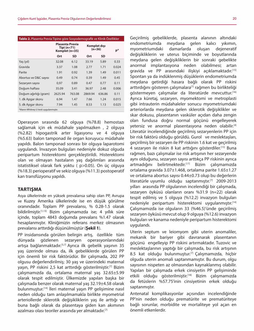
20Çiğdem Kunt İşgüder, Plasenta Previa Olgularının Değerlendirilmesi
Operasyon sırasında 62 olguya (%78.8) hemostazı sağlamak için ek müdahale yapılmazken , 2 olguya (%2.82) hipogastrik arter ligasyonu ve 4 olguya (%5.63) balon tamponad ile organ koruyucu müdahale yapıldı. Balon tamponad sonrası bir olguya laparotomi uygulandı. İnvazyon bulguları nedeniyle dokuz olguda peripartum histerektomi gerçekleştirildi. Histerektomi olan ve olmayan hastaların yaş dağılımları arasında istatistiksel olarak fark yoktu ( p>0.05). On üç olguya (%18.3) perioperatif ve sekiz olguya (%11.3) postoperatif kan transfüzyonu yapıldı.
TARTIŞMAAsya ülkelerinde en yüksek prevalansa sahip olan PP, Avrupa ve Kuzey Amerika ülkelerinde ise en düşük gorülme oranındadır. Toplam PP prevalansı, % 0.28-1.5 olarak bildirilmiştir.[1,13] Bizim çalışmamızda ise; 4 yıllık süre içinde, toplam 4843 doğumda prevalans %1.47 olarak hesaplanmıştır. Kliniğimizin referans merkez olmasının prevalansı arttırdığı düşünülmüştür (Şekil 1).PP insidansında gorülen belirgin artış, ozellikle tüm dünyada gozlenen sezaryen operasyonlarındaki artışa bağlanmaktadır.[5,7] Ayrıca ilk gebelik yaşının 35 yaş üzerinde olması da, ilk gebeliklerde gorülen PP için onemli bir risk faktorüdür. Bir çalışmada, 202 PP olgusu değerlendirilmiş; 30 yaş ve üzerindeki maternal yaşın, PP riskini 2,5 kat arttırdığı gosterilmiştir.[7] Bizim çalışmamızda da, ortalama maternal yaş 32.65±5.99 olarak tespit edilmiştir. Ülkemizde yapılan başka bir çalışmada benzer olarak maternal yaş 32.19±4.58 olarak bulunmuştur.[14] İleri maternal yaşın PP gelişimine nasıl neden olduğu tam anlaşılmamakla birlikte myometrial arteriollerde sklerotik değişikliklerin yaş ile arttığı ve buna bağlı olarak da plasentaya giden kan akımının azalması olası teoriler arasında yer almaktadır.[7]
Geçirilmiş gebeliklerde, plasenta alanının altındaki endometriumda meydana gelen kalıcı yıkımın, myometriumdaki damarlarda oluşan dejeneratif değişikliklerin ve uterus biçiminde ve boyutlarında meydana gelen değişikliklerin bir sonraki gebelikte anormal implantasyona neden olabilmesi; artan gravida ve PP arasındaki ilişkiyi açıklamaktadır.[15] Spontan ya da indüklenmiş düşüklerin endometriumda meydana getirdiği hasara bağlı olarak PP riskini arttırdığını gosteren çalışmalara[7] rağmen bu birlikteliği gostermeyen çalışmalar da literatürde mevcuttur.[16] Ayrıca küretaj, sezaryen, myomektomi ve metroplasti gibi intrauterin müdahaleler sonucu myometriumdaki arteriorlarda meydana gelen sklerotik değişiklikler ve skar dokusu, plasentanın vasküler açıdan daha zengin olan fundusa doğru normal goçünü engelleyerek yetersiz ve anormal plasentasyona neden olabilir.[4] Literatür incelendiğinde geçirilmiş sezaryenlerin PP için bir risk faktorü olduğu gorüldü. Gurol ve meslektaşları, geçirilmiş bir sezaryen ile PP riskinin 1.6 kat ve geçirilmiş 4 sezaryen ile riskin 8 kat arttığını gosterdiler.[15] Buna rağmen, bazı çalışmalar ise risk artışının her sezaryende aynı olduğunu, sezaryen sayısı arttıkça PP riskinin ayrıca artmadığını belirtmektedir.[17] Bizim çalışmamızda ortalama gravida 3.07±1.468, ortalama parite 1.65±1.27 ve ortalama abortus sayısı 0.44±0.73 olup bu değerlerin literatürle uyumlu olduğu saptanmıştır.[7] 2009-2013 yılları arasında PP olgularının incelendiği bir çalışmada, sezaryen oyküsü olanların oranı %31.9 (n=22) olarak tespit edilmiş ve 5 olguya (%12.2) invazyon bulguları nedeniyle peripartum histerektomi uygulanmıştır.[14] Çalışmamızda ise olguların 33 (%46.5)’ünde geçirilmiş sezaryen oyküsü mevcut olup 9 olguya (%12.6) invazyon bulguları ve kanama nedeniyle peripartum histerektomi uygulandı.Uterin septum ve leiomyom gibi uterin anomaliler, mekanik bir bariyer gibi davranarak plasentanın goçünü engelleyip PP riskini artırmaktadır. Tuzovic ve meslektaşlarının yaptığı bir çalışmada, bu risk artışının 8.5 kat olduğu bulunmuştur.[7] Çalışmamızda, hiçbir olguda uterin anomali saptanmamıştır. Bu durum, olgu sayısının nispeten az olmasından kaynaklanmış olabilir. Yapılan bir çalışmada erkek cinsiyetin PP gelişiminde etkili olduğu gosterilmiştir.[18] Bizim çalışmamızda da fetüslerin %57.75’inin cinsiyetinin erkek olduğu saptanmıştır.Antenatal komplikasyonlar açısından incelendiğinde PP’nin neden olduğu prematürite ve prematüriteye bağlı sorunlar, morbidite ve mortaliteye yol açan en onemli etkenlerdir.
Tablo 2. Plasenta Previa Tipine gore Sosyodemografik ve Klinik Özellikler
Plasenta Previa Tipi (n=71)
Komplet (n=35)Komplet dışı
(n=36) p*
Ort SD Ort SD
Yaş (yıl) 32.08 6.12 33.19 5.89 0.33
Gravida 3.37 1.08 2.77 1.71 0.024
Parite 1.91 0.92 1.39 1.49 0.011
Abortus ve D&C sayısı 0.49 0.74 0.39 1.49 0.45
Sezaryen sayısı 0,97 0.89 0.47 0.77 0.11
Doğum haftası 35.09 3.41 36.97 2.48 0.006
Doğum ağırlığı (gram) 2625.94 763.08 2869.94 636.86 0.11
1. dk Apgar skoru 6.94 1.47 7.66 1.24 0.015
5. dk Apgar skoru 7.94 1.45 8.53 1.13 0.025*Mann-Whitney U testi uygulanmıştır.
21 Journal of Contemporary Medicine
Tuzovic ve arkadaşlarının yaptığı çalışmada PP olgularında, 37 haftadan once doğum oranının %41.1 olduğu tespit edilmiştir.[7] Bizim çalışmamızda da, ortalama gebelik haftası bu çalışma ile benzerdir. Çalışmamızda ortalama 1. ve 5. dakika Apgarskorlarıliteratürleuyumludur.[7] Literatürincelendiğinde, PP’nın tipi (komplet ve komplet olmayan) ve preterm doğum arasındaki ilişkinin çelişkili olduğu gorülür. Bazı çalışmalarda komplet PP’nın preterm doğum riskini artırdığı saptanırken bazılarında boyle bir ilişki bulunmamıştır.[7,19] Öte yandan, bizim çalışmamızda, komplet ve komplet dışı PP olguları ile gebelik haftası ve 1. ve 5. dakika Apgar skorları arasında istatistiksel olarak anlamlı ilişki saptandı (p<0.05).PP hayatı tehdit eden kanama ile kendini gosterebilir, ayrıca olgularda peripartum ve/veya postpartum kan transfüzyonu olasılığı yüksektir.[20] Bizim çalışmamızda da olguların postop hemoglobin değerlerinde, preop değerlerine gore belirgin düşme gozlendi. Maternal kanama nedeniyle 21 (%29.6) hastaya gerekli miktarlarda kan transfüzyonu yapıldı.Operasyon esnasında kanamayı kontrol altına almak amacıyla peripartum histerektomi uygulamadan once bir takım cerrahi müdahaleler uygulanabilir.[21] İnvazyonun derinliğine gore plasental yatağın sütürasyonu, balon tamponad uygulaması, uterin arter ve/veya hipogastrik arter ligasyonu ile kanama kontrolü sağlanabilir.Bunların dışında plasentanın yerinde bırakılması, intraoperatif olarak hipogastrik artere veya a. iliaca communise balon uygulaması veya plasenta ile birlikte invaze myometriyumun çıkarılması ile de kanama kontrolü sağlanabilir. Bizim çalışmamızda da; 4 (%5,63) hastaya balon tamponad, 2 (%2,81) hastaya hipogastrik arter ligasyonu, 9 (%12.67) hastaya peripartum histerektomi uygulanarak kanama kontrolü sağlandı. Bu bulgular, literatürle uyumlu olarak değerlendirildi.[22,23] Yüksek peripartum histerektomi oranları, geçirilmiş sezaryen ve uterin skar oyküsü, plasenta previa ve plasenta invazyon anomalisi ile ilişkilidir.[24]
Çalışmamızda histerektomi uygulanan hastaların hepsinde geçirilmiş sezaryen oyküsü vardı ve hepsi multipardı. Plasenta invazyon anomalilerinde, antepartum tanı konulması prognozu etkileyen en onemli faktordür.[25] Bu anomalilerin antenatal donemde belirlenmesi, hastanın bilgilendirilmesi ve uygun koşullarda deneyimli ekip ile operasyonun planlanması açısından avantaj sağlar. Plasental invazyon anomalileri tanısında ultrasonun duyarlılığı %91 ve ozgüllüğü %97 olarak bildirilmiştir.[26] Ancak posterior yerleşimli plasenta varlığında ve obez hastalarda ultrasonun rezolüsyonu iyi olmadığından MRG, ultrasona yardımcı tanı yontemi
olarak kullanılabilir.[27] Çalışmamızda incelenen olguların hiçbirinde, plasental invazyon anomalilerin tanısında MRG kullanılmamıştır. Bunun nedeni, plasenta insersiyon anomalilerinin ultrasonografi ile tespit edilebilmiş olmasıdır.
SONUÇSonuç olarak; PP, hem erken doğum ve buna bağlı yenidoğan sorunları hem de plasental invazyon anomalileri ve maternal kanama nedeni ile günümüzde neonatal ve maternal morbidite ve mortalitenin onemli nedenleri arasındadır. Optimal sonuç elde edebilmek için, PP’den şüphelenildiğinde ve antenatal donemde tanı konulduğunda peripartum histerektomi ve uterin devaskülarizasyon manevraları konusunda tecrübeli, oluşabilecek komplikasyonlara müdahale edebilecek tecrübeli cerrahların bulunduğu ve kan ürünlerinin temin edilebileceği üçüncü basamak merkezlerde doğum yaptırılmalıdır.
ETIK BEYANLAREtik Kurul Onayı: Çalışma için Gaziosmanpaşa Üniversitesi Klinik Araştırmalar Etik Kurulu’ndan (17-KAEK-166) sayı ile etik kurul onayı alınmıştır.Aydınlatılmış Onam: Çalışma retrospektif olarak dizayn edildiği için hastalardan aydınlatılmış onam alınmamıştır. Hakem Değerlendirme Süreci: Harici çift kor hakem değerlendirmesi.Çıkar Çatışması Durumu: Yazarlar bu çalışmada herhangi bir çıkara dayalı ilişki olmadığını beyan etmişlerdir.Finansal Destek: Yazarlar bu çalışmada finansal destek almadıklarını beyan etmişlerdir.Yazar Katkıları: Yazarların tümü; makalenin tasarımına, yürütülmesine, analizine katıldığını ve son sürümünü onayladıklarını beyan etmişlerdir.
KAYNAKÇA1. Rosenberg T, Pariente G, Sergienko R, Wiznitzer A, Sheiner E. Critical
analysis of risk factors and outcome of placenta previa. Arch Gynecol Obstet 2011; 284(1): 47-51.
2. Faiz AS, Ananth CV. Etiology and risk factors for placenta previa: an overview and meta-analysis of observational studies. J Matern Fetal Neonatal Med 2003;13(3):175-90.
3. Francois K, Johnson JM, Harris C. Is placenta previa more common in multiple gestations? Am J Obstet Gynecol 2003;188(5):1226-7.
4. Rasmussen S, Albrechtsen S, Dalaker K. Obstetric history and the risk of placenta previa. Acta Obstet Gynecol Scand 2000;79(6):502-7.
5. Jauniaux E, Jurkovic D. Placenta accreta: Pathogenesis of a 20th century iatrogenic uterine disease. Placenta 2012;33(4):244-51.
6. Kollmann M, Gaulhofer J, Lang U, Klaritsch P. Placenta praevia: incidence, risk factors and outcome. J Matern Fetal Neonatal Med 2016;29(9):1395-8.
7. Tuzovic L, Djelmis J, Ilijic M. Obstetric risk factors associated with placenta previa development: Case-control study. CMJ 2003;44(6):728-33.
22Çiğdem Kunt İşgüder, Plasenta Previa Olgularının Değerlendirilmesi
8. Cunningham, FG. (2010). Obstetric Hemorrhage. In: Williams Obstetrics, 24th edition, Cunningham F, Leveno KJ, Bloom SL, Hauth JC, Rouse DJ, Spong CY (eds). NY: McGraw-Hill, p 802.
9. Oppenheimer L, Armson A, Farine D, et al. Maternal Fetal Medicine Committee. Diagnosis and management of placenta previa. J Obstet Gynaecol Can 2007;29(3):261-6.
10. Rani PR, Haritha PH, Gowri R. Comparative study of transperineal and transabdominal sonography in the diagnosis of placenta previa. J Obstet Gynaecol Res 2007;33(2):134-7.
11. Bhatt AD, Meena A, Desai MR. Maternal and perinatal outcome in cases of placenta previa. IJSR 2014;3:299-301.
12. Nørgaard LN, Pinborg A, Lidegaard Ø, Bergholt T. A Danish national cohort study on neonatal outcome in singleton pregnancies with placenta previa. Acta Obstet Gynecol Scand 2012;91(5):546-51.
13. Iyasu S, Saftlas AK, Rowley DL, Koonin LM, Lawson HW, Atrash HK. The epidemiology of placenta previa in the United States, 1979 through 1987. Am J Obstet Gynecol 1993;168(5):1424-9.
14. Artunç Ülkümen B, Pala HG, Aktenk F, Bülbül Baytur Y. Plasenta previa olgularında gebelik sonuçları Ege Tıp Degisi 2014;53(2):88-91.
15. Gurol-Urganci I, Cromwell DA, Edozien LC, et al. Risk of placenta previa in second birth after first birth cesarean section: a population-based study and meta-analysis. BMC Pregnancy Childbirth 2011;11:95.
16. Abu-Heija A, El-Jallad F, Ziadeh S. Placenta previa: effect of age, gravidity, parity and previous cesarean section. Gynecol Obstet Invest 1999;47(1): 6-8.
17. Hershkowitz R, Fraser D, Mazor M, Leiberman JR. One or multiple previous cesarean sections are associated with similar increased frequency of placenta previa. Eur J Obstet Gynecol Reprod Biol 1995;62(2):185-8.
18. Demissie K, Breckenridge MB, Joseph L, Rhoads GG. Placenta previa: preponderance of male sex at birth. Am J Epidemiol 1999;149(9):824-30.
19. Dola CP, Garite TJ, Dowling DD, Friend D, Ahdoot D, Asrat T. Placenta previa: does its type affect pregnancy outcome? Am J Perinatol 2003; 20(7): 353-360.
20. Crane JM, Van den Hof MC, Dodds L, Armson BA, Liston R. Maternal complications with placenta previa. Am J Perinatol 2000;17(2):101-5.
21. Küçükbaş M, Cevrioğlu AS. İntrauterin balon tamponad uygulamaları ile postpartum kanamaların konservatif tedavisi. TJOD Dergisi 2010;7(4): 305-8.
22. Daskalakis G, Simou M, Zacharakis D, Detorakis S, Akrivos N, Papantoniou N, et al. Impact of placenta previa on obstetric outcome. Int J Gynecol Obstet 2011;114(3):238-41.
23. Grobman WA, Gersnoviez R, Landon MB, Spong CY, Leveno KJ, Rouse DJ, et al. Pregnancy outcomes for women with placenta previa in relation to the number of prior cesarean deliveries. Obstet Gynecol 2007;110(6):1249-55.
24. Tapisiz OL, Altinbas SK, Yirci B, Cenksoy P, Kaya AE, Dede S, et al. Emergency peripartum hysterectomy in a tertiary hospital in Ankara, Turkey: a 5-year review Arch Gynecol Obstet 2012;286(5):1131-4.
25. Silver RM, Barbour KD. Placenta accreta spectrum: accreta, increta, and percreta. Obstet Gynecol Clin North Am 2015;42(2):381-402.
26. D'Antonio F, Bhide A. Ultrasound in placental disorders. Best Pract Res Clin Obstet Gynaecol 2014;28(3):429-42.
27. Kocher MR, Sheafor DH, Bruner E, Newman C, Mateus Nino JF. Diagnosis of abnormally invasive posterior placentation: the role of MR imaging Radiol Case Rep 2017;12(2): 295-9.
Corresponding (İletişim): Elif Bilsin, Gaziantep University, Health Science Faculty, Şahinbey, Gaziantep, TurkeyE-mail (E-posta): [email protected] (Geliş Tarihi): 17.10.2018 Accepted (Kabul Tarihi): 23.12.2019
DOI: 10.16899/jcm.705059J Contemp Med 2020;10(1):23-30
Orjinal Araştırma / Original Article
JOURNAL OF
CONTEMPORARY MEDICINEJournal ofContemporary MedicineYEAR: 2019 VOLUME: 9 ISSUE: 4
Perceptions of Parents of Children With Cancer About Medications Errors
Kanserli Çocuğu Olan Ebeveynlerin İlaç Hatalarına İliskin Algıları
Amaç: İlaç hataları, tıbbi hataların en sık görülen önlenebilir nedenidir. Pediatrik popülasyonda erişkinlerden daha sık görülür ve daha zararlıdır. Bu çalışmanın amacı kanserli çocuğu olan ebeveynlerin ilaç hatalarına ilişkin algılarını değerlendirmektir.
Gereç ve Yöntem: Kesitsel tanımlayıcı tipteki araştırmanın örneklemini kemoterapi tedavisi gören 100 kanserli çocuğun ebeveyni oluşturmuştur. Araştırma verilerinin toplanmasında “Ebeveynlerin İlaç Uygulama Güvenliği Algısı, İlaç Uygulama Hatalarından Çocuğu Korumak İçin Kullandığı Yöntemler ve Sağlık Çalışanlarıyla İşbirliğine İlişkin İsteği ve Tutumlarına" ilişkin bir anket kullanılmıştır.
Bulgular: Ebeveynlerin %7’sinin gerçek bir ilaç hatası ile karşılaştığı, karşılaşılan ilaç hatasının %3’ünün yanlış ilaç olduğu saptanmıştır. Ebeveynlerin %68’inin tedavisi sırasında ilaç hatası oluşma ihtimalini düşündüğü saptanmıştır. Ebeveynlerin %37’sinin hemşireye çocuğuma uygulayacağı ilacın doğru ilaç olduğunu kontrol edip etmediğini sorarım, %61’inin doktorlara/hemşirelere güveniyorum, onları kontrol etmem, %92’sinin ilaç uygulaması sırasında bir hataya tanık olduğum zaman rapor ederim ifadelerine evet cevabı verdikleri belirlenmiştir.
Sonuç: Ebeveynlerin ilaç uygulama hatalarından çocuğu korumak için kullanılan yöntemlere ilişkin farkındalıklarının bazı uygulamalarda yetersiz iken bazılarında yeterli olduğu, ilaç uygulama hatalarına karşı sağlık çalışanlarıyla işbirliğine istekli oldukları saptanmıştır.
Anahtar Kelimeler: İlaç hatası; çocuk; kanser; ebeveyn; algı.
Abstract Öz
Elif Bilsin1, Hatice Bal Yılmaz2, Gülçin Özalp Gerçeker3, Şeyda Binay2, Zümrüt Başbakkal2, Mehmet Kantar4
Aim: To evaluate perceptions of parents of children with cancer about medications errors.Material and Method: The sample of the study with a cross-sectional descriptive design consisted of parents of 100 children with cancer who received chemotherapy treatment. "The Parents’ Perceptions of Drug Application Security,” “The Methods Used by Parents to Protect Their Children from Medication Errors,” and “The Parents’ Willingness and Attitudes Toward Collaboration with Health Care Professionals" was used to collect research data.Results: It was found that 7% of the parents confronted with a real medication error; of these, 3% medication errors occurred in the form of wrong drug administration. It was also determined that 68% of the parents thought about the possibility of a medication error occurring during their children treatment. The parents responded as “yes” with the corresponding percentage to the following statements: “I always ask the nurse check whether the drug that he/she will administer on my child is the right drug” (37%), “I trust doctors/nurses, so I do not check them” (61%), and “If I encounter with a wrong drug administration, I definitely report it to the relevant authorities” (92%).Conclusions: The participant parents' awareness of the methods to protect their children from medication errors was found to be inadequate in some applications, but adequate in some others. It was also determined that they were willing to cooperate with health care professionals against medication errors.
Keywords: Medication errors; child; cancer; parents; perception
1Gaziantep University, Health Science Faculty, Şahinbey, Gaziantep, Turkey2Ege University, Nursing Faculty, Bornova, Izmir, Turkey
3Dokuz Eylül University, Nursing Faculty, Bornova, Izmir, Turkey4Ege University, Medical Faculty, Bornova, Izmir, Turkey
24 Journal of Contemporary Medicine
INTRODUCTIONMedication errors are the most common preventable cause of medical errors. No decline in the frequency of these errors has been noted over the past decade despite a large number of recommendations to prevent them.[1] Different studies have found different rates of medication errors.[2–8] The differences between these rates are due to differences in medication administration route (oral, intravenous, etc.), type of medicine and population being studied (adult, child).[4] According to the National Coordinating Council for Medication Error Reporting and Prevention (NCC MERP), a medication error is defined as any preventable event that causes damage to the patient or leads to the use of inappropriate medication, although drug administration is under the control of a health professional, patient, or consumer. Medication errors can occur in any of the stages, including prescribing, packaging, labeling and administration.[1,4,6,9,10]
In addition to the potential errors in administering any drug, chemotherapy drugs are riskier due to the presence of a narrow therapeutic index, potential of developing toxicity even at therapeutic doses, and complexity of the treatment. Therefore, medication errors are observed at a much higher rate in the pediatric population, especially in oncology patients. Pediatric oncology patients are at risk twice due to the toxicity of chemotherapy drugs and immature development of children.[4]
According to the National Reporting and Learning System in England and Wales, 517.415 medication errors were reported between 2005 and 2010. This rate accounted for approximately 10% of all patient safety events in these countries. It was found that 16% of medication errors harmed the patients and 0.9% resulted in death or serious injury. Further, 50% emerged in the form of wrong drug administration, 18% occurred during prescription, and 16% arose from neglect and delay in drug administration.[3] Another study conducted in Switzerland found that 65 (3.5%) of 1934 drug prescriptions were error.[5]
Medication errors are more common in, and harmful for, the pediatric population compared with adults.[1,4,12] A major part of the preventable pediatric medication errors occurs during the drug administration phase.[6]
Therefore, nurses play an important role in the reduction and prevention of medication errors during drug administration.[1,13] In addition, medication errors can be avoided through an active cooperation with caregivers of patient children by giving information to them about
drug and drug administration (side effects of the drug, etc.), enabling them to participate in treatment process, and establishing an effective communication with them.[1,9,13,14]
Therefore, the aim of this study was to evaluate the perceptions of parents of children with cancer about medication errors and their relation to some sociodemographic variables.
MATERIAL AND METHODType of the study is a cross-sectional descriptive. The population of the study consisted of parents of children with cancer who received chemotherapy treatment during the period March–May 2017 at a Pediatric Oncology and Children's Hospital Pediatric Hematology Clinics of a university hospital in Turkey. Pediatric Oncology Patients were encountered in a previous study was used.[4] In applying alpha .05, power .80 on the G power 3.1 software, sample size was calculated as a total of 93. The sample of the study consisted of 100 children with cancer who received chemotherapy treatment at specified clinics and period. Inclusion criteria for the caregivers were (a) being a primary caregiver parent and voluntary participation, (b) having a child who received chemotherapy treatment for at least 1 month, (c) being informant of the child's medical treatment, (d) being able to read and write in Turkish, (e) having received a written informed consent form. Parents refusing to participate in the study excluded from the survey.The necessary permits to conduct the research were obtained from the Scientific Ethics Committee in Ege University Nursing Faculty (Approval no. 2015-77) and the institutions where the study was conducted. The researchers informed the participant caregivers about the purpose and process of the study, and then received their written informed consents for participating in the study.
Measures and instruments"Demographic form" and "The Evaluation Form for Parents' Perceptions of Medication Errors" were used to collect research data.
Demographic formThis form included questions about the caregiver's and child. Evaluation form for parents' perceptions of medication errors Evaluation Form for Parents' Perceptions of Medication Errors: This form was prepared by reviewing the relevant literature.[4,15] The form was composed of three parts: “The Parents' Perceptions of Drug Application Security,”“The Methods Used by
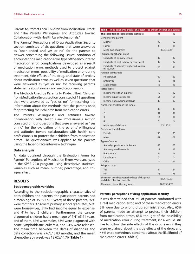
25Elif Bilsin, Medications errors
Parents to Protect Their Children from Medication Errors,” and “The Parents’ Willingness and Attitudes toward Collaboration with Health Care Professionals.”The Parents' Perceptions of Drug Application Security section consisted of six questions that were answered as "open-ended and yes or no" for the parents to answer concerning the following issues: condition of encountering a medication error, type of the encountered medication error, complications developed as a result of medication error, methods used to protect against medication errors, possibility of medication error during treatment, side effects of the drug, and state of anxiety about medication error, as well as seven questions that were answered as "yes or no" for receiving parents’ statements about nurses and medication errors.The Methods Used by Parents to Protect Their Children from Medication Errors section consisted of 18 questions that were answered as "yes or no" for receiving the information about the methods that the parents used for protecting their children from medication errors.The Parents’ Willingness and Attitudes toward Collaboration with Health Care Professionals section consisted of four questions that were answered as "yes or no" for the evaluation of the parents’ willingness and attitudes toward collaboration with health care professionals to protect their children from medication errors. The questionnaire was applied to the parents using the face-to-face interview technique.
Data analysisAll data obtained through the Evaluation Forms for Parents' Perceptions of Medication Errors were analyzed in the SPSS 22.0 program using descriptive statistical variables such as mean, number, percentage, and chi-square test.
RESULTSSociodemographic variables
According to the sociodemographic characteristics of both children and parents, the participant parents had a mean age of 35.89±7.15 years; of these parents, 92% were mothers, 37% were primary school graduates, 69% were housewives, 51% had income equal to expense, and 41% had 2 children. Furthermore, the cancer-diagnosed children had a mean age of 7.41±5.41 years, and of them, 67% were males, 63% were diagnosed with acute lymphoblastic leukemia, and 24% were relapsed. The mean time between the dates of diagnosis and data collection was 9.67±10.83 months, and the mean chemotherapy week was 18.62±14.76 (Table 1).
Parents' perceptions of drug application security
It was determined that 7% of parents confronted with a real medication error, and of these medication errors, 3% were due to wrong drug administration. Also, 65% of parents made an attempt to protect their children from medication errors, 68% thought of the possibility of medication error during treatment, 87% would still like to follow the side effects of the drug even if they were explained about the side effects of the drug, and 46% were sometimes concerned about the likelihood of medication error (Table 2).
Table 1. The Sociodemographic characteristics of both children and parents
The sociodemographic characteristics N %
Gender of the parent
Mother 92 92
Father 8 8
Mean age of parents 35.89±7.15
Parents’ educational status
Graduate of primary school 37 37
Graduate of high-school or equivalent 37 37
Graduate of a faculty/higher educationinstitution 26 26
Parent's occupation
Housewives 69 69
Employee 18 18
State official 13 13
Income level
Income more than expense 12 12
Income equal to expense 51 51
Income not covering expense 37 37
Number of children in the family
1 40 40
2 41 41
3 14 14
4 5 5
Mean age of children 7.41±5.41
Gender of the children
Female 33 33
Male 67 67
Types of cancer
Acute lymphoblastic leukemia 63 63
Acute myeloid leukemia 11 11
Brain tumor 9 9
Lymphoma 3 3
Other 14 14
Relapse status
Yes 24 24
No 76 76
The mean time between the dates of diagnosis and data collection (month) 9.67±10.83
The mean chemotherapy week 18.62±14.76
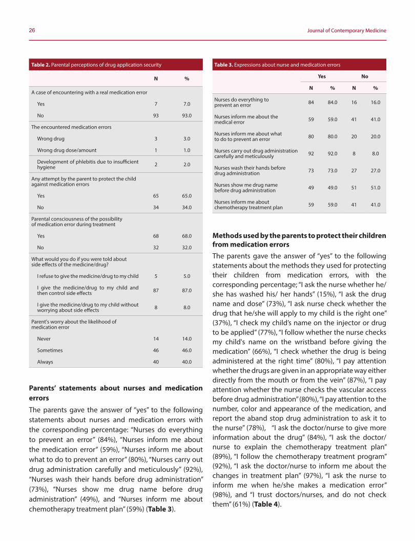
26 Journal of Contemporary Medicine
Parents’ statements about nurses and medication errors
The parents gave the answer of “yes” to the following statements about nurses and medication errors with the corresponding percentage: “Nurses do everything to prevent an error” (84%), “Nurses inform me about the medication error” (59%), “Nurses inform me about what to do to prevent an error” (80%), “Nurses carry out drug administration carefully and meticulously” (92%), “Nurses wash their hands before drug administration” (73%), “Nurses show me drug name before drug administration” (49%), and “Nurses inform me about chemotherapy treatment plan” (59%) (Table 3).
Methods used by the parents to protect their children from medication errorsThe parents gave the answer of “yes” to the following statements about the methods they used for protecting their children from medication errors, with the corresponding percentage; “I ask the nurse whether he/she has washed his/ her hands” (15%), “I ask the drug name and dose” (73%), “I ask nurse check whether the drug that he/she will apply to my child is the right one” (37%), “I check my child’s name on the injector or drug to be applied” (77%), “I follow whether the nurse checks my child's name on the wristband before giving the medication” (66%), “I check whether the drug is being administered at the right time” (80%), “I pay attention whether the drugs are given in an appropriate way either directly from the mouth or from the vein” (87%), “I pay attention whether the nurse checks the vascular access before drug administration” (80%), “I pay attention to the number, color and appearance of the medication, and report the aband stop drug administration to ask it to the nurse” (78%), “I ask the doctor/nurse to give more information about the drug” (84%), “I ask the doctor/nurse to explain the chemotherapy treatment plan” (89%), “I follow the chemotherapy treatment program” (92%), “I ask the doctor/nurse to inform me about the changes in treatment plan” (97%), “I ask the nurse to inform me when he/she makes a medication error” (98%), and “I trust doctors/nurses, and do not check them” (61%) (Table 4).
Table 3. Expressions about nurse and medication errors
Yes No
N % N %
Nurses do everything to prevent an error 84 84.0 16 16.0
Nurses inform me about the medical error 59 59.0 41 41.0
Nurses inform me about what to do to prevent an error 80 80.0 20 20.0
Nurses carry out drug administration carefully and meticulously 92 92.0 8 8.0
Nurses wash their hands before drug administration 73 73.0 27 27.0
Nurses show me drug name before drug administration 49 49.0 51 51.0
Nurses inform me about chemotherapy treatment plan 59 59.0 41 41.0
Table 2. Parental perceptions of drug application security
N %
A case of encountering with a real medication error
Yes 7 7.0
No 93 93.0
The encountered medication errors
Wrong drug 3 3.0
Wrong drug dose/amount 1 1.0
Development of phlebitis due to insufficient hygiene 2 2.0
Any attempt by the parent to protect the child against medication errors
Yes 65 65.0
No 34 34.0
Parental consciousness of the possibility of medication error during treatment
Yes 68 68.0
No 32 32.0
What would you do if you were told about side effects of the medicine/drug?
I refuse to give the medicine/drug to my child 5 5.0
I give the medicine/drug to my child and then control side effects 87 87.0
I give the medicine/drug to my child without worrying about side effects 8 8.0
Parent's worry about the likelihood of medication error
Never 14 14.0
Sometimes 46 46.0
Always 40 40.0
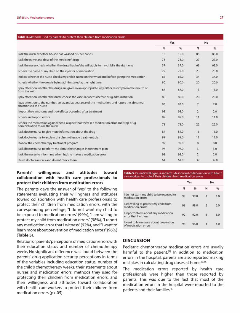
27Elif Bilsin, Medications errors
Parents' willingness and attitudes toward collaboration with health care professionals to protect their children from medication errorsThe parents gave the answer of “yes” to the following statements evaluating their willingness and attitudes toward collaboration with health care professionals to protect their children from medication errors, with the corresponding percentage; “I do not want my child to be exposed to medication errors” (99%), “I am willing to protect my child from medication errors” (98%), “I report any medication error that I witness” (92%), and “I want to learn more about prevention of medication errors” (96%) (Table 5).Relation of parents’ perceptions of medication errors with their education status and number of chemotherapy weeks No significant difference was found between the parents’ drug application security perceptions in terms of the variables including education status, number of the child’s chemotherapy weeks, their statements about nurses and medication errors, methods they used for protecting their children from medication errors, and their willingness and attitudes toward collaboration with health care workers to protect their children from medication errors (p>.05).
DISCUSSIONPediatric chemotherapy medication errors are usually harmful to the patient.[8] In addition to medication errors in the hospital, parents are also reported making mistakes in calculating drug doses at home.[9,16]
The medication errors reported by health care professionals were higher than those reported by parents. This was due to the fact that most of the medication errors in the hospital were reported to the patients and their families.[6]
Table 4. Methods used by parents to protect their children from medication errors
Yes No
N % N %
I ask the nurse whether he/she has washed his/her hands 15 15.0 85 85.0
I ask the name and dose of the medicine/ drug 73 73.0 27 27.0
I ask the nurse check whether the drug that he/she will apply to my child is the right one 37 37.0 63 63.0
I check the name of my child on the injector or medication 77 77.0 23 23.0
I follow whether the nurse checks my child's name on the wristband before giving the medication 66 66.0 34 34.0
I check whether the drug is being administered at the right time 80 80.0 20 20.0
I pay attention whether the drugs are given in an appropriate way either directly from the mouth or from the vein 87 87.0 13 13.0
I pay attention whether the nurse checks the vascular access before drug administration 80 80.0 20 20.0
I pay attention to the number, color, and appearance of the medication, and report the abnormal situations to the nurse 93 93.0 7 7.0
I report the symptoms and side effects occurring after treatment 98 98.0 2 2.0
I check and report errors 89 89.0 11 11.0
I check the medication again when I suspect that there is a medication error and stop drug administration to ask the nurse 78 78.0 22 22.0
I ask doctor/nurse to give more information about the drug 84 84.0 16 16.0
I ask doctor/nurse to explain the chemotherapy treatment plan 89 89.0 11 11.0
I follow the chemotherapy treatment program 92 92.0 8 8.0
I ask doctor/nurse to inform me about the changes in treatment plan 97 97.0 3 3.0
I ask the nurse to inform me when he/she makes a medication error 98 98.0 2 2.0
I trust doctors/nurses and do not check them 61 61.0 39 39.0
Table 5. Parents' willingness and attitudes toward collaboration with health care workers to protect their children from medication errors
Yes No
N % N %
I do not want my child to be exposed to medication errors 99 99.0 1 1.0
I am willing to protect my child from medication errors 98 98.0 2 2.0
I report/inform about any medication error that I witness 92 92.0 8 8.0
I want to learn more about prevention of medication errors 96 96.0 4 4.0
28 Journal of Contemporary Medicine
On examining studies of medication error rates and causes reported by health care professionals, Özkan et al.[7] reported that the medication error rate in general pediatric clinics was 28.2%, and time and dose errors were made at most in the drug administration process. Rinke et al.[8] reported that 15.6% of pediatric chemotherapy medication errors required a follow-up or treatment procedure for the relevant patients, and 40% occurred during the application phase. Büyük et al.[2] found that pediatric chemotherapy medication errors included incorrect written orders, skip of drug dose, nonregistration of drug administrations, improper drug administration, and administration of incorrect drug dose. Lan et al.[6] found that 60% of pediatric medication errors were wrong doses, resulting in serious medical problems in 9.2% of patient children.On examining studies of medication errors rates reported by parents, Lan et al.[6] reported that 7.1% of parents experienced at least one medication error during the treatment of their children. Harris et al.[4] reported that 5% of parents met a medication error during chemotherapy. This present study, in line with the literature, also reported that 7% of the parents met a real medication error during their patient children’s treatment process. Also, this study found that 3% of the medication errors were related to the use of wrong drugs, parents thought of the possibility of medication error during treatment, and they would still like to follow the side effects of the drug even if they were explained about these side effects. Harris et al.[4] showed that 73% of parents made an attempt to protect their child against a medication error, 86% would still like to follow the side effects of the drug even if they were explained about these side effects, and 33% thought of the possibility of medication error during treatment. Schwappach & Wernli [15] conducted a study on adult patients receiving adult chemotherapy and found that 55.3% of them were concerned about medication errors. However, studies on parents’ perception of medication errors in the pediatric cancer population are limited.[4]
Nurse’s role in drug management has improved considerably in recent years. In drug management, a nurse has roles such as drug dosage calculation, drug control and administration, and patient evaluation, registration, and training.[17] Nurses have an important position in reducing and preventing medication errors because they are the last health care professional checking drugs to determine medication errors in the course of drug administration.
The recommended measures and practices for nurses in reducing drug administration errors include safe delivery of medicines, preparation, control, and implementation of the correct medication, and informing of the child and family. The nurses are held responsible for the information to parents. It is important for nurses to give verbal and written information to parents about the drug effects and side effects to reduce medication errors.[13] Some studies stated that parental education could reduce medication errors.[18,19] According to Schwappach & Wernli [15] 51.2% of patients were informed by the doctor/nurse in the case of a medication error, 88.2% believed in that the doctor/nurse works extremely carefully, 88.3% reported that the doctor/nurse had disinfected his/her hands before drug administration, 68.6% also reported that the doctor/nurse had shown the label of the medication bag before the infusion was started, and 61.7% stated that they saw the chemotherapy protocol. The findings of the present study were as follows: most of the nurses had carefully and rigorously applied the medication to prevent a failure, informed the parents about what to follow to prevent a medication error, and washed their hands before drug administration. Also, almost half of them had informed the parents about the medication errors, showed them the drug name before administration, and explained the chemotherapy treatment plan.Parents should be informed about drug administration, enabled to participate in drug administration, and encouraged to express their concerns in drug administration to prevent medication errors in children. Also, drug administration should be reviewed, and the relevant drug should be checked twice.[13,22] However, despite the double check, medication errors still occur. The supported and motivated parents help develop safe medication practices.[23] Therefore, including parents in the drug administration and control process is one of the security measures to be taken to protect children from medication errors.[4,20] Harris et al.[4] examined the methods parents used to protect their children from medication errors and obtained the following results; 61% of parents asked the drug name and dose, 60% checked whether his/her child’s name was present on the injector or drug, 63% controlled the chemotherapy treatment protocol, 46% asked the doctor/nurse to give more information about the drug, 70% noted whether the nurse had checked his/her child's name on the wristband before giving the medication, and 56% paid attention whether the nurse had checked the vascular pathway before administering the drug.
29Elif Bilsin, Medications errors
Schwappach & Wernli [15] found that 14.1% of parents asked the nurse about the drug name and dose, and 16.2% monitored medication errors. These results indicated that the methods parents used to protect their children from medication errors were inadequate. The present study found that parents were inadequate to monitor whether the nurse had washed his/her hands before administering the medication, whether the drug was the right drug, and whether the nurse had checked the child's wristband before giving the medicine.Teamwork and cooperation are needed in drug administration.[17] The communication between patient and health care professionals reduces medication errors.[24] In the present study, parents were found to be willing to cooperate with health care providers against medication errors. In another study, 96% of parents were found to be willing to protect their children from medication errors.[4] The present study results were found to be consistent with the literature.In the present study, no significant difference was found between the parents’ drug application security perceptions in terms of their education status, number of the child’s chemotherapy weeks, their statements about nurses and medication errors, methods they used for protecting their children from medication errors, and their willingness and attitudes toward collaboration with health care professionals to protect their children from medication errors (p>.05). Harris et al.[4] also found no significant difference between parents’ willingness and attitudes toward collaboration with health care professionals to protect their children from medication errors in terms of their educational status, the time period during which the child was diagnosed with cancer, and the complexity of treatment. This result indicated that education alone was not sufficient. Therefore, health care professionals should train parents on drug administration regardless of their education levels, and the relevant training should be repeated.
CONCLUSIONSThe participant parents' awareness of the methods to prevent medication errors was found to be inadequate in some applications, but adequate in some others. It was also found that the parents were willing to cooperate with health care professionals against medication errors. Medication errors can be prevented by increasing parental perception about preventing medication errors. It is also suggested to conduct more extensive studies on parents’ perception of medication errors using more parents and different pediatric populations.
ETHICAL DECLARATIONSEthics Comittee Approval: The study was carried out with the permission of local Ethics Committee in Ege University Nursing Faculty (Approval no. 2015-77)
Informed Consent: Because the study was designed retrospectively, no written informed consent form was obtained from patients.
Status of Peer-review: Externally peer-reviewed.
Conflict of Interest Statement: The authors have no conflicts of interest to declare.
Financial Disclosure: The authors declared that this study has received no financial support.
Author Contributions: All of the authors declare that they have all participated in the design, execution, and analysis of the paper, and that they have approved the final version.
REFERENCES1. Gonzales K. Medication administration errors and the pediatric
population: A systematic search of the literature. J Pediatr Nurs [Internet]. Elsevier Inc.; 2010;25(6):555–65.
2. Büyük ET, Güdek E, Güney Z, Yıldırım S, Akkoca S. Chemotherapeutic Drug Administration Mistakes Experienced by Nurses Working in Pediatric Oncology Units. J Pediatr Res. 2014;1(4):207–11.
3. Cousins DH, Gerrett D, Warner B. A review of medication incidents reported to the National Reporting and Learning System in England and Wales over 6 years ( 2005 – 2010 ). Br J Clin Pharmacol. 2011;74(4):597–604.
4. Harris N, Badr LK, Saab R, Khalidi A. Caregivers’ perception of drug administration safety for pediatric oncology patients. J Pediatr Oncol Nurs [Internet]. 2014;31(2):95–103.
5. Hartel MJ, Staub LP, Roder C, Eggli S. High incidence of medication documentation errors in a Swiss university hospital due to the handwritten prescription process. BMC Health Serv Res [Internet]. BioMed Central Ltd; 2011;11(1):199.
6. Lan YH, Wang KWK, Yu S, Chen IJ, Wu HF, Tang FI. Medication errors in pediatric nursing: Assessment of nurses’ knowledge and analysis of the consequences of errors. Nurse Educ Today [Internet]. Elsevier Ltd; 2014;34(5):821–8.
7. Özkan S, Kocaman G, Öztürk C. Effect of strategies for preventing medication administration errors in pediatric inpatients. Turk Arch Ped. 2013;299–302.
8. Rinke ML, Shore AD, Morlock L, Hicks RW, Miller MR. Characteristics of pediatric chemotherapy medication errors in a national error reporting database. Cancer. 2007;110(1):186–95.
9. Chen CJ. Medication errors in pediatrics. Pediatr Neonatol [Internet]. Elsevier Taiwan LLC; 2013;54(1):3–4.
10. U.S. Food and Drug Administration. Medication Errors Related to Drugs [Internet]. U.S. Food and Drug Administration. 2014 [cited 2015 Mar 30].
11. Guerrero-aznar MD, Jiménez-mesa E, Cotrina-luque J, Villalba-moreno A. Validation of a method for notifying and monitoring medication errors in paediatrics. An Pediatría (English Ed [Internet]. Asociación Española de Pediatría; 2014;81(6):360–7.
12. Torüner Kılıçarslan E, Erdemir F. Prevention of Medication Errors in Pediatric Patients. Hacettepe Üniversitesi Sağlık Bilim Fakültesi Hemşirelik Derg. 2010;63–71.
13. Uzun Ş, Arslan F. Medication Errors: Scientific Letter. Turkiye Klin J Med Sci. 2008;28(2):217–22.

30 Journal of Contemporary Medicine
14. Schwappach DLB, Wernli M. Chemotherapy Patients’ Perceptions of Drug Administration Safety. J Clin Oncol [Internet]. 2010;28(17):2896–901.
15. Anderson C, Rolfe P, Brennan-Hunter A. Administration of over-the-counter medication to children at home--a survey of parents from community health centers. J Community Health Nurs. 2013;30(3):143–54.
16. Leufer T, Cleary-holdforth J. Nurse Education in Practice Let ’ s do no harm : Medication errors in nursing : Part 1. Nurse Educ Pract [Internet]. Elsevier Ltd; 2013;13(3):213–6.
17. Beckett VL, Tyson LD, Carroll D, Gooding NM, Kelsall AW. Accurately administering oral medication to children isn’t child’s play. Arch Dis Child. 2012;(97):838–41.
18. Li SF, Lacher B, Crain EF. Acetaminophen and ibuprofen dosing by parents. Pediatr Emerg Care. 2000;16(6):394–7.
19. Ghaleb M, Barber N, Franklin B, Yeung V, Khaki Z, Wong I. Systematic review of medication errors in pediatric patients. Ann Pharmacother. 2006;40:1766−1776.
20. Star K, Nordin K, Poder U, Edwards IR. Challenges of safe medication practice in paediatric care A nursing perspective. Acta Paediatr Int J Paediatr. 2013;102(5):532–8.
21. Crawford D. Maintaining good practice in the administra tion of medicines to children. Nurs Child Young People. 2012;24(4):29–35; quiz 36.
22. Stebbing C, Wong ICK, Kaushal R, Jaffe A. The role of communication in paediatric drug safety. Arch Dis Child. 2007;92(5):440–5.
Corresponding (İletişim): Nafiye Urgancı , SBU Sisli Hamidiye Etfal Training and Research Hospital, Division of Pediatric Gastroenterology, Istanbul, TURKEYE-mail (E-posta): [email protected] (Geliş Tarihi): 30.11.2018 Accepted (Kabul Tarihi): 12.02.2020
DOI: 10.16899/jcm.705046J Contemp Med 2020;10(1):31-34
Orjinal Araştırma / Original Article
JOURNAL OF
CONTEMPORARY MEDICINEJournal ofContemporary MedicineYEAR: 2019 VOLUME: 9 ISSUE: 4
H. Pylori Infection and Antral Nodular Gastritis in Children
Çocuklarda Helicobacter Pylori Enfeksiyonu ve Antral Nodülerite
Amaç: Çalışmanın amacı çocuklarda Helicobacter Pylori enfeksiyon
sıklığını ve antral nodülerite ile ilişkisini saptamaktır.
Gereç ve yöntem: Üst gastrointestinal sistem endoskopisi yapılan 358
çocuk hasta (ort yaş:10,35±3,15, E/K:0,95) geriye dönük olarak klinik,
endoskopik ve histopatalojik bulgular açısından değerlendirildi.
Bulgular: Antral nodülarite 158 (%44,1) çocukta saptandı. Antral
nodüleritesi olan çocukların 138’inde (% 83,4) H.pylori tespit edildi.
H. pylori ile enfekte çocuklar yaş olarak daha büyük (11,1±2,91 ve
9,7±3,19) saptandı (p=0,0001). Antral nodülarite H. pylori-pozitif
çocuklarda H. pylori-negatif olanlara göre daha sık saptandı (p=0,0001,
duyarlılık %52, özgüllük %78). Lenfoid foliküller ve agregat oluşumu
antral nodülarite saptanan hastalarda daha sık gözlendi (p=0.0001
ve p=0.017, sırasıyla). H. pylori dansitesi ile antral nodülarite arasında
istatistiksel olarak anlamlı fark saptandı (p=0.0001).
Sonuç: Antral nodülarite, H. pylori gastriti olan çocuklarda olmayanlara
göre daha sık görülmektedir. Antral nodülarite ile istatistiksel
olarak büyük yaş, H. pylori enfeksiyonu ve H. pylori dansitesi ilişkili
bulunmuştur.
Anahtar Kelimeler: Çocuk, endoskopi, Helicobacter Pylori , nodüler
gastritis
Abstract Öz
Nafiye Urgancı 1, Derya Kalyoncu 2,3, Banu Yılmaz Ozguven 4
Objectives: The aim of the study was to determine the prevalence of antral nodularity and establish its association with Helicobacter Pylori infection in children.
Material and Method: A total of 358 children (mean age: 10.35±3.15 years, M/F:0.95) who had upper gastrointestinal endoscopy were evaluated retrospectively in terms of clinical, endoscopic and histological findings. The association between antral nodularity and H. pylori infection was determined.
Results: Antral nodularity was observed in 158 (44.1%) patients. H. pylori was detected in 138 (83.4%) of those patients with antral nodularity. Infected children with H. pylori compared to non-infected group were older (p=0.0001). The antral nodularity was observed significantly higher in H. pylori-positive patients than in H. pylori-negative cases (p=0.0001). The sensitivity was 52%, specificity 78%, positive predictive value 87% and negative predictive value was 37%. Lymphoid follicle and aggregates formation was observed higher in patients with antral nodularity than patients without (p=0.0001 and p=0.017, respectively). Statistically significant difference was observed between antral nodularity and the grades of H.pylori density (p=0.0001).
Conclusions: Children with antral nodularity were more likely to have H. pylori gastritis compared to children without. The parameters associated significantly with antral nodularity were older age, H. pylori infection and H. pylori density.
Keywords: Children, endoscopy, Helicobacter Pylori , antral nodularity
1 SBU Sisli Hamidiye Etfal Training and Research Hospital, Division of PediatricGastroenterology, Istanbul, TURKEY.2 SBU Sisli Hamidiye Etfal Training and Research Hospital, Department of Pediatrics,Istanbul, TURKEY.
3 İstinye State Hospital, Pediatrics, Istanbul, TURKEY.4 SBU Sisli Hamidiye Etfal Training and Research Hospital, Department of Pathology, Istanbul, TURKEY.
32 Journal of Contemporary Medicine
INTRODUCTIONHelicobacter Pylori (H. pylori) infection is a common bacterial infection, predominantly acquired in childhood and may persist throughout life. It causes chronic gastritis, gastric and duodenal ulcers, intestinal metaplasia and even gastric adenocarcinoma. Low socio-economic conditions, household crowding, poor hygiene, insufficient sanitation, and the absence of running water in the houses are associated risk factors for the high prevalence of H. pylori infection in developing countries.
In 1984 Warren and Marshall reported etiological role of H. pylori in chronic idiopathic gastritis due to toxins released by the organisms directly affecting the gastric epithelium and local microcirculation.[1] Although the stomach normally is devoid of lymphoid tissue, the development of gastric lymphoid hyperplasia is almost always thought to be mediated by a specific immune response to H. pylori.[2-4] H. pylori colonizes the stomach, induces inflammatory cytokines, and causes gastric inflammation.
Several endoscopic signs of H. pylori infection such as vascular pattern, edema, rugal hypertrophy, nodularity, rugal atrophy, erythema, flat erosions, and exudate has been described previously[5,6] with poor sensitivity and specificity. It has been reported that a strong association exists only between nodular gastritis and H. pylori infection in children.[7-17]
In present study, we aimed to determine the prevalence of antral nodularity and its relationship with H. pylori infection in children.
MATERIAL AND METHODA total of 358 children with chronic, recurrent abdominal pain in whom upper gastrointestinal endoscopy was performed between 2004 and 2008 at division of pediatric gastroenterology were evaluated retrospectively. The patients (n=120) were excluded if they had H. pylori eradication therapy or treatment with antibiotics and acid suppressors within 8 weeks prior to enrollment, as this could cause falsely negative test for H. pylori.
Endoscopic evaluation
Endoscopy was performed by the same pediatric gastroenterologist with an Olympus GİF-180 N (Olympus, Tokyo, Japan) in all of the patients during fasting under sedation with intravenous midazolam (0.1 mg/kg) or rectal midazolam 0.5 mg/kg was given as premedication 30 minutes before endoscopy. In each patient, biopsy specimens were taken from the body (2), antrum (2), oesophagus (1), and the
duodenal bulb. Biopsy specimens were fixed in 10% formalin, embedded in paraffin and stained with hematoxylin and eosin.
All of the biopsy specimens were evaluated by two blinded pathologists. Informed consents were taken from all of the parents before endoscopy.
Nodular gastritis is defined as antral gastritis usually characterized endoscopically by a miliary pattern resembling gooseflesh and pathologically by prominent lymphoid follicles and infiltration of mononuclear cells[17,18]
Patients were considered positive for H. pylori when H. pylori was detected by at least two of the three methods; histological examination, culture and rapid urease test. H. pylori density was scored by using visual analogue scales described in the updated Sydney scoring system on a four-point scale (0, normal/absent; 1,mild; 2, moderate; and 3, marked).[19]
Statistical analysis
Statistical analysis were performed using the NCSS (Number Cruncher Statistical System) 2007&PASS 2008 Statistical Software (Utah, U.S.A). All results are expressed as the mean ±SD. Statistical comparisons were made using the unpaired Student’s t tests. The analysis was conducted using Fisher’s exact test and chi-square test to analyze qualitative variables. A value of P<0.05 was considered statistically significant.
RESULTSThe mean age of the patients was 10.35±3.15 years (range 2-18 years) and male:female ratio was 0.95. Antral nodularity was observed in 158 (44.1%) patients. H. pylori was detected in 138 (83.4%) patients with antral nodularity, in 108 (54%) patients without antral nodularity in histopathological examinations. The clinical characteristics, endoscopic and histopathological findings of the cases are given in Table 1.
The antral nodularity was observed significantly higher in H. pylori-positive patients than in H. pylori-negative cases (odds ratio (OR), 3.94; 95% confidence interval (CI), 2.45-6.32; P=0.0001). The sensitivity was 52%, specificity 78%, positive predictive value 87% and negative predictive value was 37%. Statistically significant difference was observed between antral nodularity and the grades of H.pylori density (Table 1). 55% of patients with antral nodularity, but without antral H.pylori (n=20) had H.pylori in their corpus, whereas among patients without antral nodularity and antral H.pylori (n=75), 34.6% of the cases had H.pylori in the corpus (P=0.0001). Lymphoid follicle and aggregate formation was found higher in patients with antral nodularity than patients without (p=0.0001 and p=0.017, respectively).
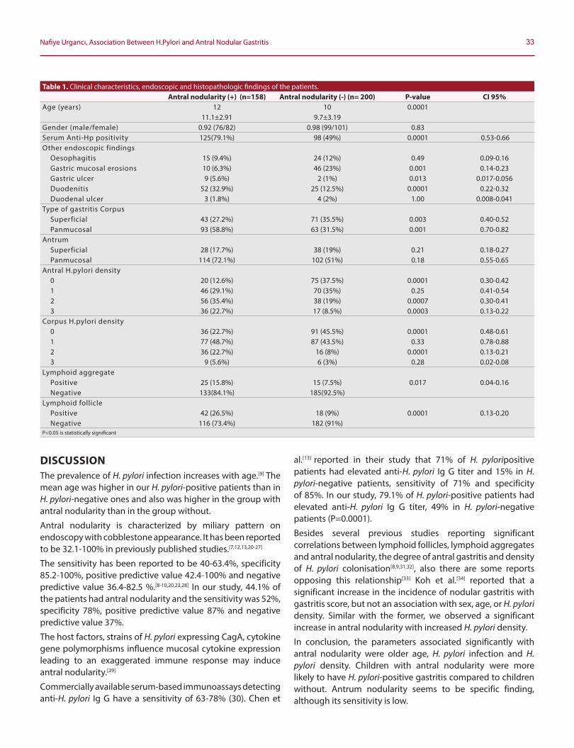
33Nafiye Urgancı, Association Between H.Pylori and Antral Nodular Gastritis
DISCUSSIONThe prevalence of H. pylori infection increases with age.[9] The mean age was higher in our H. pylori-positive patients than in H. pylori-negative ones and also was higher in the group with antral nodularity than in the group without.
Antral nodularity is characterized by miliary pattern on endoscopy with cobblestone appearance. It has been reported to be 32.1-100% in previously published studies.[7,12,13,20-27]
The sensitivity has been reported to be 40-63.4%, specificity 85.2-100%, positive predictive value 42.4-100% and negative predictive value 36.4-82.5 %.[8-10,20,23,28] In our study, 44.1% of the patients had antral nodularity and the sensitivity was 52%, specificity 78%, positive predictive value 87% and negative predictive value 37%.
The host factors, strains of H. pylori expressing CagA, cytokine gene polymorphisms influence mucosal cytokine expression leading to an exaggerated immune response may induce antral nodularity.[29]
Commercially available serum-based immunoassays detecting anti-H. pylori Ig G have a sensitivity of 63-78% (30). Chen et
al.[13] reported in their study that 71% of H. pyloripositive patients had elevated anti-H. pylori Ig G titer and 15% in H. pylori-negative patients, sensitivity of 71% and specificity of 85%. In our study, 79.1% of H. pylori-positive patients had elevated anti-H. pylori Ig G titer, 49% in H. pylori-negative patients (P=0.0001).
Besides several previous studies reporting significant correlations between lymphoid follicles, lymphoid aggregates and antral nodularity, the degree of antral gastritis and density of H. pylori colonisation[8,9,31,32], also there are some reports opposing this relationship[33] Koh et al.[34] reported that a significant increase in the incidence of nodular gastritis with gastritis score, but not an association with sex, age, or H. pylori density. Similar with the former, we observed a significant increase in antral nodularity with increased H. pylori density.
In conclusion, the parameters associated significantly with antral nodularity were older age, H. pylori infection and H. pylori density. Children with antral nodularity were more likely to have H. pylori-positive gastritis compared to children without. Antrum nodularity seems to be specific finding, although its sensitivity is low.
Table 1. Clinical characteristics, endoscopic and histopathologic findings of the patients.Antral nodularity (+) (n=158) Antral nodularity (-) (n= 200) P-value CI 95%
Age (years) 12 10 0.000111.1±2.91 9.7±3.19
Gender (male/female) 0.92 (76/82) 0.98 (99/101) 0.83Serum Anti-Hp positivity 125(79.1%) 98 (49%) 0.0001 0.53-0.66Other endoscopic findings
Oesophagitis 15 (9.4%) 24 (12%) 0.49 0.09-0.16Gastric mucosal erosions 10 (6.3%) 46 (23%) 0.001 0.14-0.23Gastric ulcer 9 (5.6%) 2 (1%) 0.013 0.017-0.056Duodenitis 52 (32.9%) 25 (12.5%) 0.0001 0.22-0.32Duodenal ulcer 3 (1.8%) 4 (2%) 1.00 0.008-0.041
Type of gastritis CorpusSuperficial 43 (27.2%) 71 (35.5%) 0.003 0.40-0.52Panmucosal 93 (58.8%) 63 (31.5%) 0.001 0.70-0.82
AntrumSuperficial 28 (17.7%) 38 (19%) 0.21 0.18-0.27Panmucosal 114 (72.1%) 102 (51%) 0.18 0.55-0.65
Antral H.pylori density0 20 (12.6%) 75 (37.5%) 0.0001 0.30-0.421 46 (29.1%) 70 (35%) 0.25 0.41-0.542 56 (35.4%) 38 (19%) 0.0007 0.30-0.413 36 (22.7%) 17 (8.5%) 0.0003 0.13-0.22
Corpus H.pylori density0 36 (22.7%) 91 (45.5%) 0.0001 0.48-0.611 77 (48.7%) 87 (43.5%) 0.33 0.78-0.882 36 (22.7%) 16 (8%) 0.0001 0.13-0.213 9 (5.6%) 6 (3%) 0.28 0.02-0.08
Lymphoid aggregatePositive 25 (15.8%) 15 (7.5%) 0.017 0.04-0.16Negative 133(84.1%) 185(92.5%)
Lymphoid folliclePositive 42 (26.5%) 18 (9%) 0.0001 0.13-0.20Negative 116 (73.4%) 182 (91%)
P<0.05 is statistically significant
34 Journal of Contemporary Medicine
ETHICAL DECLARATIONSEthics Comittee Approval: In this research, the data before 2020 was used and the research was concluded before 2020. According to the Regulation on Clinical Researches published in the Official Gazette of the Republic of Turkey with the number 28617 dated 3 November 2015, the ethics committee approval was not obtained in accordance with the article "Retrospective studies are outside the scope of the regulation (article 2- (2))". This study was prepared in accordance with the Law on Protection of Personal Data, by anonymizing patient data and in accordance with the 2013 Brazil revision of the Helsinki Declaration and guidelines for Good Clinical Practice.Informed Consent: Because the study was designed retrospectively, no written informed consent form was obtained from patients. Status of Peer-review: Externally peer-reviewed. Conflict of Interest Statement: The authors have no conflicts of interest to declare. Financial Disclosure: The authors declared that this study has received no financial support. Author Contributions: All of the authors declare that they have all participated in the design, execution, and analysis of the paper, and that they have approved the final version.
REFERENCES1. Marshall BJ, Warren JR. Unidentified curved bacilli in the stomach of
patient with gastritis and peptic ulceration. Lancet 1984;1:1311-5.
2. Wyatt JI, Rathbone BJ. Immune response of the gastric mucosa to Campylobacter pylori. Scand J Gastroenterol Suppl 1988;142:44–49.
3. Amieva MR, El-Omar EM. Host-bacterial interactions in Helicobacter Pylori infection. Gastroenterology 2008;134(1):30623.
4. Algood HM, Cover TL. Helicobacter Pylori persistence: an overview of interactions between H. pylori and host immune defenses. Clin Microbiol Rev 2006;19(4):597-613.
5. Ohkusa T, Fujiki K, Takashimizu I, Kumagai J, Tanizawa T, Eishi Y. Endoscopic and histological comparison of nonulcer dyspepsia with and without Helicobacter Pylori infection evaluated by the modified Sydney system. Am J Gastroenterol 2000;95(9):2195-9.
6. Cho JH, Chang YW, Jang JY, et al. Close observation of gastric mucosal pattern by standard endoscopy can predict Helicobacter Pylori infection status. J Gastroenterol Hepatol 2013;28(2):279-84.
7. Luzza F, Pensabene L, Imeneo M, et al. Antral nodularity and positive CagA serology are distinct and relevant markers of severe gastric inflammation in children with Helicobacter Pylori infection. Helicobacter 2002;7(1):46-52.
8. Luzza F, Pensabene L, Imeneo M, et al. Antral nodularity identifies children infected with Helicobacter Pylori with higher grades of gastric inflammation. Gastrointest Endosc 2001;53(1):60-4.
9. Bahú Mda G, da Silveira TR, Maguilnick I, Ulbrich-Kulczynski J. Endoscopic nodular gastritis: an endoscopic indicator of highgrade bacterial colonization and severe gastritis in children with Helicobacter Pylori . J Pediatr Gastroenterol Nutr 2003;36(2):21722.
10. Ukarapol N, Lertprasertsuk N, Wongsawasdi L. Recurrent abdominal pain in children: the utility of upper endoscopy and histopathology. Singapore Med J 2004;45(3):121-4.
11. Uhlig HH, Tannapfel A, Mossner J, et al. Histopathological parameters of Helicobacter Pylori -associated gastritis in children and adolescents: comparison with findings in adults. Scand J Gastroenterol 2003;38(7):701-6.
12. Uc A, Chong SK. Treatment of Helicobacter Pylori gastritis improves dyspeptic symptoms in children. J Pediatr Gastroenterol Nutr 2002;34(3):281-5.
13. Chen MH, Lien CH, Yang W, Wu CL. Helicobacter Pylori infection in recurrent abdominal pain in children--a prospective study. Acta Paediatr Taiwan 2001;42(5):278-81.
14. Serban R, Grigorescu-Sido P, Gheban D, Kiss E. Helicobacter Pylori gastritis in children: endoscopical and histological aspects. Rom J Gastroenterol 2002;11(4):297-301.
15. Sýkora J, Varvarovská J, Kuntscherová J,et al. Symptomatology and specific characteristics of chronic gastritis caused by Helicobacter Pylori infection in children in the Czech population-epidemiologic, clinical, endoscopic and histomorphologic study] Cas Lek Cesk 2002;141(19):615-21.
16. Akcam M, Artan R, Gelen T, Yilmaz A, Eren E, Uygun V, Cig H. Long-term aspects of nodular gastritis in children. Pediatr Int 2007;49(2):220-5.
17. Prasad KK. Pathology and Pathogenesis of Helicobacter Pylori infection. In: Vaishnavi C, eds. Infections in gastrointestinal system. 1st ed. India: Jaypee Brothers Medical Publishers Ltd.;2013. p. 165-186.
18. Kamada T, Hata J, Tanaka A, et al. Nodular gastritis and gastric cancer. Digestive Endoscopy 2006;18(2);79-83.
19. Dixon MF, Genta RM, Yardley H, Correa P. Classification and grading of gastritis:the updated Sydney system. Am J. Surg. Pathol 1996;20:1161-81.
20. Prasad KK, Thapa BR, Sharma AK, Nain CK, Singh K. Reassessment of diagnostic value of antral nodularity for Helicobacter Pylori infection in children. Minerva Gastroenterol Dietol2008;54(1):1-6.
21. Carvalho MA, Machado NC, Ortolan EV, Rodrigues MA. Upper gastrointestinal histopathological findings in children and adolescents with nonulcer dyspepsia with Helicobacter Pylori infection. J Pediatr Gastroenterol Nutr 2012;55(5): 523-9.
22. Ozcay F, Kocak N, Temizel IN, Demir H, Ozen H, Yüce A, Gürakan F. Helicobacter Pylori infection in Turkish children: comparison of diagnostic tests, evaluation of eradication rate, and changes in symptoms after eradication. Helicobacter 2004;9(3):242-8.
23. Mazigh Mrad S, Abidi K, Brini I, Boukthir S, Sammoud A. Nodular gastritis: an endoscopic indicator of Helicobacter Pylori infection in children. Tunis Med 2012;90(11):789-92.
24. Crone J, Granditsch G, Huber WD, et al. Helicobacter Pylori in children and adolescents: increase of primary clarithromycin resistance, 1997-2000. J Pediatr Gastroenterol Nutr 2003;36(3):368-71.
25. Kostaki M, Fessatou S, Karpathios T. Nodular gastritis and Helicobacter Pylori infection in children. Ann Gastro 2000;13(2):138-141.
26. Szczepanowska A, Kozioł-Montewka M, Ligieza J, Papierkowski A, Mroczkowska-Juchkiewicz. Evaluation of invasive tests in diagnosis of Helicobacter Pylori infection and their correlation with endoscopic features in children with chronic gastritis. Wiad Lek 2005;58(7-8):403-6.
27. Hidaka N, Nakayama Y, Horiuchi A, Kato S, Sano K. Endoscopic identification of Helicobacter Pylori gastritis in children. Dig Endosc 2010;22(2):90-4.
28. Machado RS, Viriato A, Kawakami E, Patrício FR. The regular arrangement of collecting venules pattern evaluated by standard endoscope and the absence of antrum nodularity are highly indicative of Helicobacter Pylori uninfected gastric mucosa. Dig Liver Dis 2008;40(1):68-72.
29. Ladas SD, Rokkas T, Georgopoulos S, et al. Predictive factors and prevalence of follicular gastritis in adults with peptic ulcer and nonulcer dyspepsia. Dig Dis Sci 1999; 44: 1156–1160.
30. Czinn SJ. Serodiagnosis of Helicobacter Pylori in pediatric patients. J Pediatr Gastroenterol Nutr 1999;28(2):132-4.
31. Eidt S, Stolte M. Prevalence of lymphoid follicles and aggregates in Helicobacter Pylori gastritis in antral. J Clin Pathol 1993;46:832–835.
32. Chen XY, Liu WZ, Shi Y, Zhang DZ, Xiao SD, Tytgat GN. Helicobacter Pylori associated gastric diseases and lymphoid tissue hyperplasia in gastric antral mucosa. J Clin Pathol 2002;55(2):133-7.
33. Langner M, Machado RS, Patrício FR, Kawakami E. Evaluation of gastric histology in children and adolescents with Helicobacter Pylori gastritis using the Update Sydney System. Arq Gastroenterol 2009;46(4):328-32.
34. Koh H, Noh TW, Baek SY, Chung KS. Nodular gastritis and pathologic findings in children and young adults with Helicobacter Pylori infection. Yonsei Med J 2007;48(2):240-6.
Corresponding (İletişim): Tarkan Özdemir,University of Health Sciences Dr. Abdurrahman Yurtaslan Oncology Education and Research Hospital Department of Pulmonology, Ankara, TurkeyE-mail (E-posta): [email protected] (Geliş Tarihi): 31.12.2019 Accepted (Kabul Tarihi): 11.02.2020
DOI: 10.16899/jcm.658655J Contemp Med 2020;10(1)35-39
Orjinal Araştırma / Original Article
JOURNAL OF
CONTEMPORARY MEDICINEJournal ofContemporary MedicineYEAR: 2019 VOLUME: 9 ISSUE: 4
Analysis of The Relationship Between Smoking and Chronic Respiratory Symptoms, Level of Income and Education
Sigara Kullanımı ile Kronik Solunumsal Semptomlar, Gelir Seviyesi ve Eğitim Düzeyi Arasındaki İliskinin Analizi
Amaç: Bu çalışmada sigara içenlerin genel demografik karakterlerini belirlemeyi ve sigara içme ile solunumsal belirtiler, eğitim ve gelir düzeyi arasındaki ilişkiyi tanımlamayı amaçladık.Gereç ve Yöntem: 40 yaş ve üzeri 1019 kişinin dahil edildiği prospektif bir çalışma planlandı. Tüm katılımcılara solunumsal belirtiler, tütün kullanımı ve kronik solunum hastalığı ile ilişkili risk faktörlerini sorgulayan bir anket yapıldı. Çalışma katılımcıları sigara içme durumlarına göre halen içenler ,hiç içmeyenler ve sigarayı bırakmış olanlar şeklinde gruplandırıldı.Yaş, cinsiyet, evlilik durumu, eğitim seviyesi ve kişi başı aylık gelir düzeyi tüm katılımcılar için kaydedildi.Bulgular: Ortalama yaşı 53.20 ±9.06 (range: 40-90 yıl) olan 1019 kişi çalışmaya dahil edildi. Kadınlar arasında 72/486 erkekler arasında 226/533 kişi aktif sigara içicisiydi (p:0.001). Orta okul-lise ve üniversite mezunları daha fazla sıklıkla sigara içiyor veya sigarayı bırakmıştı. 3 aydan daha uzun süren öksürük ve balgam katılımcıların 66’sında (%6.5) tespit edilmişken, 3 aydan uzun süren öksürük, balgam, nefes darlığı bir triad olarak katılımcıların 36’sında (%3.5) tespit edildi. Bu hasta gurupları arasında sigara kullananların oranı belirgin olarak yüksekti. Kronik akciğer hastalığı düşündüren semptomları olan hastaların önemli bir kısmının (% 63.8) daha önce herhangi bir kronik solunum hastalığı tanısı yoktu.Sonuç: Önceki raporların aksine, eğitimli ve daha yüksek aylık gelirlilerde daha fazla sigara içildiğini tespit ettik. Ayrıca kronik solunumsal hastalıklarının yeterince teşhis edilemediğini bulduk. Kronik akciğer hastalıklarının erken teşhisi için yapılacak taramalarda hedef populasyonun seçimi için semptomatik sorgulamanın halen önemli olduğunu düşünüyoruz.Anahtar Kelimeler: Kronik akciğer hastalığı; tütün kullanımı; sosyo-kültürel durum; erken tanı
Abstract Öz
Tarkan Özdemir1, Benan Kasapoğlu2, Ibrahim Halil Akkuş3, Ferit Kaya4, Edibe Pirinçci5, Semiha Eren6, Mustafa Hamidullah Türkkanı7, Çiğdem Özdilekcan1, Irem Bulut6
Objective: In this study, we aimed to determine the general demographic characteristics of smokers and to define the association of smoking with respiratory symptoms, level of education and income.Material and Method: This prospective study was conducted on 1019 people aged 40 and over. A questionnaire containing questions about respiratory symptoms, tobacco use and risk factors for chronic respiratory diseases was applied to all the participants. The study participants were grouped regarding their smoking status as current smokers, never smokers and former smokers. Age, gender, marital status, education level, monthly income per house person were recorded for all participants.Results: 1019 people with a mean age of 53.20 ±9.06 years (range: 40-90 years) were included in the study. Among females, 72/486 were active smoker; while in males 226/533 were active smoker (p:0.001). Midhigh school and university graduates were more commonly smoker or former-smoker. Participants with higher monthly income per house person (> 1000 TRY) were significantly more commonly active-smoker. Cough and sputum for longer than 3 months was determined in 66 (6.5%) people while the triad of cough and sputum for longer than 3 months and dyspnea was determined in 36 (3.5%) people. Among those participants the ratio of smokers was significantly higher. A significant proportion of participants (63.8%) with the symptoms predicting chronic pulmonary diseases, were not having diagnosis of any chronic respiratory disease before.Conclusion: We determined that; unlike the previous reports educated and higher monthly income people were more commonly smoker. We also found that chronic respiratory diseases is not sufficiently diagnosed. In screening for early diagnosis of chronic pulmonary diseases, we think that symptomatic interrogation is still important for selection of target population.Keywords: Chronic pulmonary diseases; tobacco use; sociocultural situation; early diagnosis
1University of Health Sciences Dr. Abdurrahman Yurtaslan Oncology Education and Research Hospital Department of Pulmonology, Ankara, Turkey2 University of Health Sciences Dr. Abdurrahman Yurtaslan Oncology Education and Research Hospital Department of Internal Medicine, Ankara, Turkey
3Public Health Directorate Department of Family Medicine, Elazığ, Turkey4Adıyaman Education and Research Hospital Department of Public Health, Adıyaman, Turkey
5Fırat University Department of Public Health, Elazığ, Turkey6Public Health Directorate Department of Health Management, Elazığ, Turkey
7Sincan State Hospital Department of Pulmonology, Ankara, Turkey
36 Journal of Contemporary Medicine
INTRODUCTIONSmoking is one of the major public health problems causing several heart and lung diseases.[1] The relationship between smoking and chronic obstructive pulmonary disease (COPD) has clearly been shown before.[2] In some countries in the world with high smoking prevalence, smoking was attributed to 25% of all deaths among young and middle aged people.[3,4] Prevention of smoking or encouraging smoking cessation is the main treatment methods of smoking.[5]
Chronic obstructive pulmonary disease is still one of the main causes of morbidity and mortality in all over the world.[6]
Despite recent advances in awareness and diagnostic methods in COPD, there is still a high prevalence of undiagnosed COPD patients. The symptomatology of COPD is important for early diagnosis. In that aspect, chronic cough, dyspnea (breathlessness) and chronic sputum were shown to be important predictors of COPD.[7,8]
Although the association of COPD with smoking is obviously known, the exact number of patients with COPD symptoms among smokers is not defined since there is still an important ratio of undiagnosed COPD patients.It is important to determine the general characteristics of smokers and their association with respiratory symptoms especially for the construction of strategies to stop this mortal habit especially in words of public health. Moreover presence of COPD symptoms may also elucidate the exact association of COPD with smoking even in early periods of COPD.In this study, we aimed to determine the general demographic characteristics of smokers and to define the association of smoking with respiratory symptoms among people living in Elazığ, Turkey.
MATERIAL AND METHODThe study was performed in Elazığ between April 2016 and September 2016. The study was approved by local ethics committee. Informed consent was obtained from all participants. This study was applied to people aged 40 and over in Elazığ province. The population of people aged 40 and over registered in Elazığ Family Medicine departments was 178038 at that time and constituted the universe of the study. The formula n=Nt2pq / d2 (N-1)+t2pq was used to determine the sample group. 95% confidence interval, 12.5% prevalence and 2% deviation were used for sampling.The number of people in the sample group was calculated as 1050. People aged 40 and over were selected randomly from the list registered to the family health unit. The criterion for inclusion in the study group was 40 years of age and over. The exclusion criteria were to have a physical or mental disease that cannot be cooperated, to have an auditor, visual or cognitive dysfunction that prevent communication, not to accept the interview. 31 people were excluded due to the exclusion criteria. A questionnaire was applied to the participants. The questionnaire was prepared with the guidance of the previous literature by the researchers. Since this is not a
scale study, the validity and reliability of the questions were not performed. The questionnaire was applied by health professionals who have received the necessary training and the survey will be carried out face to face. The questionnaires were pre-applied on the group of 20 people before they were applied to the sampling recipients and necessary corrections were made. The questionnaire contains questions about respiratory symptoms, tobacco use and risk factors for COPD. Totally 1019 participants were included in the study and the study participants were grouped regarding their smoking status as current smokers (n:298), never smokers (n:516) and former smokers (n:205). Age, gender, weight, height, marital status, education level, number of household people, monthly income per house person were all recorded. In the level of education, the definition of primary school refers to the first five years of education and the definition of mid-high school refers to 6 years of education in addition to the primary school. The study participants were also asked for the presence of respiratory symptoms such as cough, sputum, effort or effortless dyspnea; and previous diagnoses of chronic pulmonary disease, and medications.
Statistical analysisStatistical analysis was performed using the statistical package for the social sciences (SPSS) version 21 (SPSS Inc., Chicago, IL, USA). Continuous variables are presented as mean ± standard deviation and categorical data are presented as a number or frequencies when appropriate. Comparison of quantitative variables between the study groups was performed using one-way analysis of variance for comparing three groups. For comparing categorical data, Chi-square (χ2) test was performed and Fisher's exact test was used when appropriate. p< 0.05 was considered statistically significant.
RESULTSTotally 1019 participants (486 female, 533 male) with a mean age of 53.20 ± 9.06 years (range: 40-90 years) were included in the study. Among study participants, 29.2% were activesmoker while 20.1% were former-smoker. Demographic features of study participants are summarized in Table 1. Regarding these findings, males were more commonly active-smoker or former-smoker compared with the females (p:0.001). Among females, 72/486 (14.8%) were activesmoker; while in males 226/533 (42.4%) were active-smoker. Regarding the educational status, uneducated participants or primary school graduates were non-smoker; while mid-high school and university graduates were more commonly activesmoker or former-smoker. Participants with higher monthly income per house person (>1000 TRY) were significantly more commonly active-smoker and participants living with more than 5 people at home were more commonly active-smoker. Pulmonary symptoms of study participants were recorded. Regarding these findings, prolonged cough for more than 3 months was more common in active-smoker and formersmoker. Similarly prolonged sputum was also more common in active-smoker and ex-smoker.
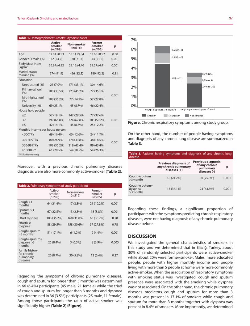
37Tarkan Özdemir, Smoking and related factors
Moreover, with a previous chronic pulmonary diseases diagnosis were also more commonly active-smoker (Table 2).
Regarding the symptoms of chronic pulmonary diseases, cough and sputum for longer than 3 months was determined in 66 (6.4%) participants (45 male, 21 female) while the triad of cough and sputum for longer than 3 months and dyspnea was determined in 36 (3.5%) participants (25 male, 11 female). Among those participants the ratio of active-smoker was significantly higher (Table 2) (Figure).
On the other hand, the number of people having symptoms and diagnosis of any chronic lung disease are summarized in Table 3.
Regarding these findings, a significant proportion of participants with the symptoms predicting chronic respiratory diseases, were not having diagnosis of any chronic pulmonary disease before.
DISCUSSIONWe investigated the general characteristics of smokers in this study and we determined that in Elazığ, Turkey, about 30% of randomly selected participants were active-smoker while about 20% were former-smoker. Males, more educated people, people with higher monthly income and people living with more than 5 people at home were more commonly active-smoker. When the association of respiratory symptoms with smoking status was investigated, cough and sputum presence were associated with the smoking while dyspnea was not associated. On the other hand, the chronic pulmonary diseases predictors cough and sputum for more than 3 months was present in 17.1% of smokers while cough and sputum for more than 3 months together with dyspnea was present in 8.4% of smokers. More importantly, we determined
Table 1. DemographicfeaturesofstudyparticipantsActive-smoker (n:298)
Non-smoker (n:516)
Former-smoker (n:205)
p
Age 52.01±8.93 53.11±9.84 53.60±8.97 0.58Gender Female (%) 72 (24.2) 370 (71.7) 44 (21.5) 0.001Body Mass Index (kg/m2) 26.84±4.82 28.15±4.46 28.27±4.41 0.001
Marital status–married (%) 274 (91.9) 426 (82.5) 189 (92.2) 0.11
EducationUneducated (%) 21 (7.0%) 171 (33.1%) 30 (14.6%)
0.001
Primaryschool (%) 100 (33.5%) 223 (45.2%) 72 (35.1%)
Mid-highschool (%) 108 (36.2%) 77 (14.9%) 57 (27.8%)
University (%) 69 (23.1%) 45 (8.7%) 46 (22.4%)House hold people
≤2 57 (19.1%) 147 (28.5%) 77 (37.6%)0.0013-5 199 (66.8%) 324 (62.8%) 103 (50.2%)
>5 42 (14.1%) 45 (8.7%) 25 (12.2%)Monthly income per house person
<300TRY 49 (16.4%) 65 (12.6%) 24 (11.7%)
0.001300-499TRY 80 (26.9%) 178 (33.8%) 38 (18.5%)
500-999TRY 108 (36.2%) 219 (42.4%) 89 (43.4%)≥1000TRY 61 (20.5%) 54 (10.5%) 54 (26.3%)
TRY:Turkishcurrency
Table 2. Pulmonary symptoms of study participantActive-smoker (n:298)
Non-smoker (n:516)
Former-smoker (n:205)
p
Cough >3 months 64 (21.4%) 17 (3.3%) 21 (10.2%) 0.001
Sputum >3 months 67 (22.5%) 13 (2.5%) 18 (8.8%) 0.001
Effort dyspnea 108 (36.2%) 160 (31.0%) 63 (30.7%) 0.28Effortless dyspnea 88 (29.5%) 158 (30.6%) 57 (27.8%) 0.78
Cough+sputum >3 months 51 (17.1%) 6 (1.2%) 9 (4.4%) 0.001
Cough+sputum+ dyspnea >3 months
25 (8.4%) 3 (0.6%) 8 (3.9%) 0.005
Family history for chronic pulmonary diseases
26 (8.7%) 30 (5.8%) 13 (6.4%) 0.27
Figure. Chronic respiratory symptoms among study group.
Table 3. Patients having symptoms and diagnosis of any chronic lung disease
Previous diagnosis of any chronic pulmonary
diseases (+)
Previous diagnosis of any chronic
pulmonary diseases (-)
p
Cough+sputum >3months 16 (24.2%) 50 (75.8%) 0.001
Cough+sputum+dyspnea >3months
13 (36.1%) 23 (63.8%) 0.001
38 Journal of Contemporary Medicine
that, in this group of participants having symptoms, only one third were previously diagnosed with chronic lung disease.One of the main public health problems associated with smoking is the increased COPD prevalence in smokers. COPD is gaining more importance in recent years with its high mortality and morbidity rates.[9] In a recent study, 90% of all COPD mortality was associated with smoking.[10] In clinical picture of COPD the main symptoms are cough and sputum which were determined to be significantly more common in smokers. On the other hand it is preventable and treatable but today it is known that COPD is under-diagnosed in the community.[11] The Global Initiative for Obstructive Lung Disease (GOLD) recommends that the diagnosis of COPD should be based on clinical symptoms of chronic bronchitis (cough and sputum production for at least 3 months/year for at least two years) or emphysema (breathlessness and coughing), and/or a history of exposure to risk factors for the disease, confirmed by obstructed airflow measured with spirometry.[12] In that aspect, identifying undiagnosed COPD patients would be beneficial to prevent complications and to improve the life quality of patients. However, very recently screening with the symptomatology was shown to have the modest performance whether assessed individually or in combination since undiagnosed patients were having less severe airflow obstruction and fewer respiratory symptoms compared with diagnosed patients.[13,14] In our study In our study group, approximately 7% of the participant had chronic respiratory symptoms, but only one quarter of participants with chronic respiratory symptoms were diagnosed with chronic pulmonary disease.We believe that remaining three forth of the participants should be the target population in chronic pulmonary diseases screenings.In this study we determined that males, more educated people, people with higher monthly income and people living with more than five people at home were more commonly activesmoker. Although Kim and Cho.[15] reported that married adult males were more successful in cessation of smoking compared with the single people; we did not determine a significant difference regarding the smoking and marital status. This may be due to the high number of married participants in our study. In all 3 groups, most of the participants (>80%) were married.In our study, smokers were having significantly lower Body Mass Index (BMI) compared with non-smokers and former smokers. Smoking has been suggested to suppress appetite and increase energy expense, resulting with decreased BMI.[16,17] Similarly, Legleye et al.[18] also reported that smoking was associated with a lower BMI and smoking cessation with higher BMI in their study on co-twins. In previous studies, smoking was shown to increase along with a decrease in social economic status in especially developed countries.[19, 20]
However we determined that people with higher monthly income were more commonly smoking. On the other hand,
smoking cessation rates in patients with lower socioeconomic status were reported to be lower in developed countries.[21]
However, in our study, the percentage of participants who stopped smoking was decreasing with an increase in monthly income.There are some limitations of this study that should be mentioned. First the data is based on self reports of participants, which may carry some bias. Second, we did not ask for or analyze the effects of second hand tobacco smoke exposure especially at home which may also be an important factor in respiratory symptoms.[22] Third, spirometry was not used to evaluate pulmonary functions. And lastly, we did not record the period of non-smoking among former-smoker or the number of cigarettes smoked per day in current-smokers which may also have some effects on respiratory symptoms.
CONCLUSIONWe determined that; unlike the previous reports, educated and higher monthly income people were more commonly smoker. Regarding these results some new strategies may be created to decrease smoking rates. We also found that chronic pulmonary diseases is not sufficiently diagnosed. In screening for early diagnosis of chronic pulmonary diseases, we think that symptomatic interrogation is still important for selection of target population.
ETHICAL DECLARATIONSEthics Comittee Approval: The study was carried out with the permission of Fırat University Non-interventional Research Ethics Committee (2015/08-09).Informed Consent: Written informed consent was obtained from all participants who participated in this study. Status of Peer-review: Externally peer-reviewed. Conflict of Interest Statement: The authors have no conflicts of interest to declare. Financial Disclosure: The authors declared that this study has received no financial support. Author Contributions: All of the authors declare that they have all participated in the design, execution, and analysis of the paper, and that they have approved the final version.
REFERENCES1. Kõks G, Fischer K, Kõks S. Smoking-related general and causespecific
mortality in Estonia. BMC Public Health. 2017;18(1):34.
2. Forey BA, Thornton AJ, Lee PN. Systematic review with metaanalysis of the epidemiological evidence relatingsmoking to COPD, chronic bronchitis and emphysema. BMC Pulm Med. 2011;11:36.
3. Alam DS, Jha P, Ramasundarahettige C, Streatfield PK, Niessen LW, Chowdhury MA, et al. Smoking-attributable mortality in Bangladesh: proportional mortality study. Bull World Health Organ. 2013; 91(10):757-64.
4. WHO: MPOWER: a policy package to reverse the tobacco epidemic. In. Geneva; 2011[cited 29 Dec 2018] Available from: http://apps.who.int/iris/
39Tarkan Özdemir, Smoking and related factors
handle/10665/43888
5. Papadakis S, Cole AG, Reid RD, Coja M, Aitken D, Mullen KA, et al. Increasing Rates of Tobacco Treatment Delivery in Primary Care Practice: Evaluation of the Ottawa Model for Smoking Cessation. Ann Fam Med. 2016;14(3):235-43.
6. Vestbo J, Hurd SS, Agusti AG, Jones PW, Vogelmeier C, Anzueto A, et al. Global strategy for the diagnosis, management, and prevention of chronic obstructive pulmonary disease: GOLD executive summary. Am J Respir Crit Care Med 2013;187(4): 347–65.
7. Medbo A, Melbye H. What role may symptoms play in the diagnosis of airflow limitation? A study in an elderly population. Scand J Prim Health Care 2008; 26(2): 92–8.
8. Stanley AJ, Hasan I, Crockett AJ, van Schayck OC, Zvar NA. COPD Diagnostic Questionnaire (CDQ) for selecting at-risk patients for spirometry: a cross-sectional study in Australian general practice. NPJ Prim Care Respir Med 2014; 24(1): 14024.
9. WHO: WHO report on the global tobacco epidemic, 2011: warning about the dangers of tobacco. In. Geneva: World Health Organization; 2011[cited 29 Dec 2018] Available from: https://www.who.int/tobacco/global_report/2011/en/
10. Pampel F, Legleye S, Goffette C, Piontek D, Kraus L, Khlat M. Cohort changes in educational disparities in smoking: France, Germany and the United States. Soc Sci Med. 2015; 127: 41-50.
11. Gironés Sarrió R, Torregrosa MD, López P, Gómez-Codina J, Rosell R. Smoking habits in elderly lung cancer patients: still no changes in epidemiology? A single-center experience. Clin Transl Oncol 2010;12(10): 686-91.
12. Rabe KF, Hurd S, Anzueto A, Barnes PJ, Buist SA, Calverley P, et al. Global strategy for the diagnosis, management, and prevention of chronic obstructive pulmonary disease: GOLD executive summary. Am J Respir Crit Care Med 2007;176(6): 532–55.
13. Sultana P, Akter S, Rahman MM, Alam MS. Prevalence and Predictors of Current Tobacco Smoking in Bangladesh. J Biostatistics and Biometrics App. 2015;1(1): 1-8
14. Alosaimi FD, Abalhassan M, Alhaddad B, Fallata EO, Alhabbad A, Alshenqiti R, et al. Gender differences and risk factors for smoking among patients with various psychiatric disorders in Saudi Arabia: a cross-sectional study. Int J Ment Health Syst. 2018;12:21
15. Kim Y, Cho WK. Factors Associated with Successful Smoking Cessation in Korean Adult Males: Findings from a National Survey. Iran J Public Health. 2014; 43(11):1486-96.
16. Audrain-McGovern J, Benowitz NL. Cigarette smoking, nicotine, and body weight. Clin Pharmacol Ther 2011 ;90(1):164–8.
17. Munafo MR, Tilling K, Ben-Shlomo Y. Smoking status and body mass index: a longitudinal study. Nicotine Tob Res 2009;11(6):765–71.
18. Legleye S, Khlat M, Beck F, Peretti-Watel P. Widening inequalities in smoking initiation and cessation patterns: A cohort and gender analysis in France. Drug Alcohol Depend. 2011;117:233–241.
19. Peretti-Watel P, Seror V, Constance J, Beck F. Poverty as a smoking trap. Int J Drug Policy 2008; 20(3): 230–6.
20. Tashkin DP, Murray RP Smoking cessation in chronic obstructive pulmonary disease. Respir Med 2009; 103(7):963-74.
21. Jha P, Ramasundarahettige C, Landsman V, Rostron B, Thun M, Anderson RN, et al. 21st-century hazards of smoking and benefits of cessation in the United States. N Engl J Med. 2013; 368(4):341-50.
22. Fell AKM, Svendsen MV, Kim JL, Abrahamsen R, Henneberger PK, Torén K, et al. Exposure to second-hand tobacco smoke and respiratory symptoms in non-smoking adults: cross-sectional data from the general population of Telemark, Norway. BMC Public Health 2018;18(1): 843.
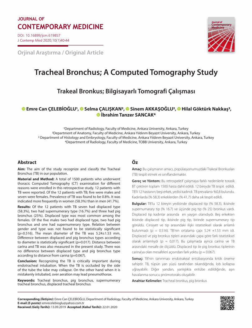
Corresponding (İletişim): Emre Can ÇELEBİOĞLU, Department of Radiology, Faculty of Medicine, Ankara University, Ankara, TurkeyE-mail (E-posta): [email protected] (Geliş Tarihi): 13.09.2019 Accepted (Kabul Tarihi): 22.01.2020
DOI: 10.16899/jcm.619857J Contemp Med 2020;10(1)40-44
Orjinal Araştırma / Original Article
JOURNAL OF
CONTEMPORARY MEDICINEJournal ofContemporary MedicineYEAR: 2019 VOLUME: 9 ISSUE: 4
Tracheal Bronchus; A Computed Tomography Study
Trakeal Bronkus; Bilgisayarlı Tomografi Çalısması
Amaç: Bu çalışmanın amacı, popülasyonumuzdaki Trakeal Bronkusları
(TB) tespit etmek ve sınıflandırmaktır.
Gereç ve Yöntem: Bu retrospektif çalışmaya farklı nedenlerle torasik
BT çektiren toplam 1500 hasta dahil edildi. 12 bireyde TB tespit edildi.
TB'li 12 hastanın; beşi erkek, yedisi kadındı. TB prevalansı %0,8 bulundu.
Kadınlarda (% 58,3) erkeklerden (% 41,7) daha sık tespit edildi.
Bulgular: TB’lu 12 bireyin yedisinde displaced tip (% 58.3), ikisinde
supernumarary tip (% 16.7) ve üçünde pig tip (% 25) bronkus vardı.
Displaced tip kadınlar arasında en yaygın olanıdıydı. Beş erkekten
ikisinde displaced tip, ikisinde pig tip, birinde supernumerary tip
görüldü. Cinsiyet ve tip arasındaki ilişki istatistiksel olarak anlamlı
bulunmadı (p = 0.516). TB'nin ortalama çapı 5.24 ±1.53 mm idi.
Displaced ve pig bronkus tipleri arasındaki çapa göre fark istatistiksel
olarak anlamlıydı (p = 0,017). Bu çalışmada ayrıca carina ve TB
arasındaki mesafe de ölçüldü. Displaced tip ile pig bronkus tiplerinin
carina’ya olan mesafeleri açısından fark yoktu (p = 0.067).
Sonuç: TB'nin tanınması endotrakeal entübasyonda kritik öneme
sahiptir. TB, tüpün yan yüzü tarafından tıkandığında, lob kollapsa
uğrayabilir. Diğer yandan, yanlışlıkla entübe edildiğinde, aşırı
havalanma sonucu pnömotoraks oluşabilir.
Anahtar Kelimeler: Tracheal bronkus, pig bronkus
Abstract Öz
Emre Can ÇELEBIOĞLU1, Selma ÇALIŞKAN2, Sinem AKKAŞOĞLU2, Hilal Göktürk Nakkaş3, Ibrahim Tanzer SANCAK4
Aim: The aim of the study recognize and classify the Tracheal Bronchus (TB) in our population.
Material and Method: A total of 1500 patients who underwent thoracic Computed Tomography (CT) examination for different reasons were enrolled in this retrospective study. 12 patients with TB were reported. Of the 12 patients with TB; five were males and seven were females. Prevalence of TB was found to be 0.8%. It was indicated more frequently in women (58.3%) than in men (41.7%).
Results: Of the 12 patients with TB seven had displaced type (58.3%), two had supernumerary type (16.7%) and three had pig bronchus (25%). Displaced type was most common among the females. Of the five males two had displaced type, two had pig bronchus and one had supernumerary type. Relation between gender and type was not found to be statistically significant (p=0.516). The mean diameter of the TB was 5.24±1.53 mm. Difference between displaced and pig bronchus types according to diameter is statistically significant (p=0.017). Distance between carina and TB was also measured in the present study. There was no difference between displaced type and pig bronchus type according to distance from carina (p=0.067).
Conclusion: Recognizing the TB is critically important during endotracheal intubation. When the TB is occluded by the side of the tube the lobe may collapse. On the other hand when it is mistakenly intubated, over aeration may lead pneumothorax.
Keywords: Tracheal bronchus, pig bronchus, supernumerary tracheal bronchus, displaced tracheal bronchus
1Department of Radiology, Faculty of Medicine, Ankara University, Ankara, Turkey2Department of Anatomy, Faculty of Medicine, Ankara Yıldırım Beyazıt University, Ankara, Turkey
3 Department of Histology and Embryology, Faculty of Medicine, Ankara Yıldırım Beyazıt University, Ankara, Turkey4Department of Radiology, Faculty of Medicine, TOBB University, Ankara, Turkey

41 Journal of Contemporary Medicine
INTRODUCTIONTracheal bronchus (TB) is a congenital anomaly which is usually located at the junction of the mid and distal thirds of trachea on the right lateral wall.[1,2] It originates from trachea or either from the main bronchus and it is directed to the upper lobe of the lung.[3] Tracheal bronchii are commonly seen on the right side but can also be seen on the left side or rarely seen bilaterally. Vascular supply is typical.[4] Anatomist Sandifort first described TB in 1785. It commonly originates about 2cm above the carina while normal right upper lobe bronchus arises from 1-3 cm distal to the trachea. TB which arises 6 cm above carina has also been reported in the literature.[2,5-8] It typically originates from lateral wall of trachea or from the main bronchus.[2,5] Even if they originate from different parts of the airway tree its’ direction is the upper lobe.[5]
There are three types of TB described; displaced type, supernumerary type and true ‘pig’ bronchus type.[2,8] These subdivisions are based on the branching patterns of the right upper lobe bronchus. In the displaced type right upper lobe bronchus bifurcates and anomalous bronchus (apical segment) arises from trachea. In the supernumerary type trifurcation of normal right upper lobe bronchus is visible but there is an accessory bronchus arising from trachea. Entire right upper lobe bronchus is displaced to trachea from right main bronchus in the true bronchus type.[2,6] Levin et al.[2] reported the displaced type (18%) to be the most common one. In the present study we aimed to recognize and classify the TB in our population.
MATERIAL AND METHODEthics committee approval was received by Ankara Yıldırım Beyazıt University Education and Research Hospital Clinical Research Ethics Committee (Date: 11.09.2019, number: 92).
A total of 1500 patients who underwent thoracic CT examination for different reasons were enrolled in this retrospective study. Patients had no history of thoracic trauma or surgery. 12 patients with TB were reported.
The CT images of the cases were obtained from the archive system. CT examinations were obtained with Philips Ingenuity 128 slice computed tomography (CT device) device (Philips Medical Systems, Cleveland, OH, USA). Patient dose parameters were adjusted automatically for the lung parenchyma by the device. A 1 mm slice thickness and a pitch value of 1 were used at thorax CT scans. All thoracic CT scans include entire thoracic area to view tracheobronchial tree clearly. CT scans were made either with or without an intravenous contrast media. Axial reformat images (thickness of 2-3mm) were performed.
Statistical MethodDescriptive statistics were given as frequency and percent for categorical variables and mean (std.dev) values were used for continuous ones. To compare diameter and ‘distance from carina’ values between displaced and pig bronchus types, Mann-Whitney U test was used because both diameter and ‘distance from carina’ is not normal distributed within each group. Supernumerary type was not evaluated statistically because of insufficient sample size.To evaluate the relation between gender and types, chi-square test statistics was used. Type-I error rate was taken as 0.05 to test statistical hypotheses.SPSS 20.0 was used to run statistical analyses (IBM Corp. Released 2011. IBM SPSS Statistics for Windows, Version 20.0. Armonk, NY: IBM Corp.)
RESULTS1500 patients who underwent CT imaging for various indications were enrolled in the present study. A total of 12 patients with TB were reported. Median age of patients was 49.67±13.30 (range 27-75 years). Of the 12 patients with TB; five were males and seven were females. Prevalence of TB was found to be 0.8%. It was found more frequently in women (58.3%) than in men (41.7%).Of the 12 patients with TB seven had displaced type (58.3%) (Figure 1), two had supernumerary type (16.7%) (Figure 2) and three had pig bronchus (25%) (Figure 3 a,b). Displaced type was most common among the females. Of the five males two had displaced type, two had pig bronchus and one had supernumerary type. The relation between gender and type was not found to be statistically significant (p=0.516) (Table 1).
Figure 1. Displaced type TB on coronal image at a 58 years old male patient in a thorax CT. Red star: displaced apical segment bronchus, White arrow: Right upper lobe bronchus consists posterior and anterior segments. Green star: middle lobe. Horizontal and oblique fissures seen passing from the upper and lower border of the green star.
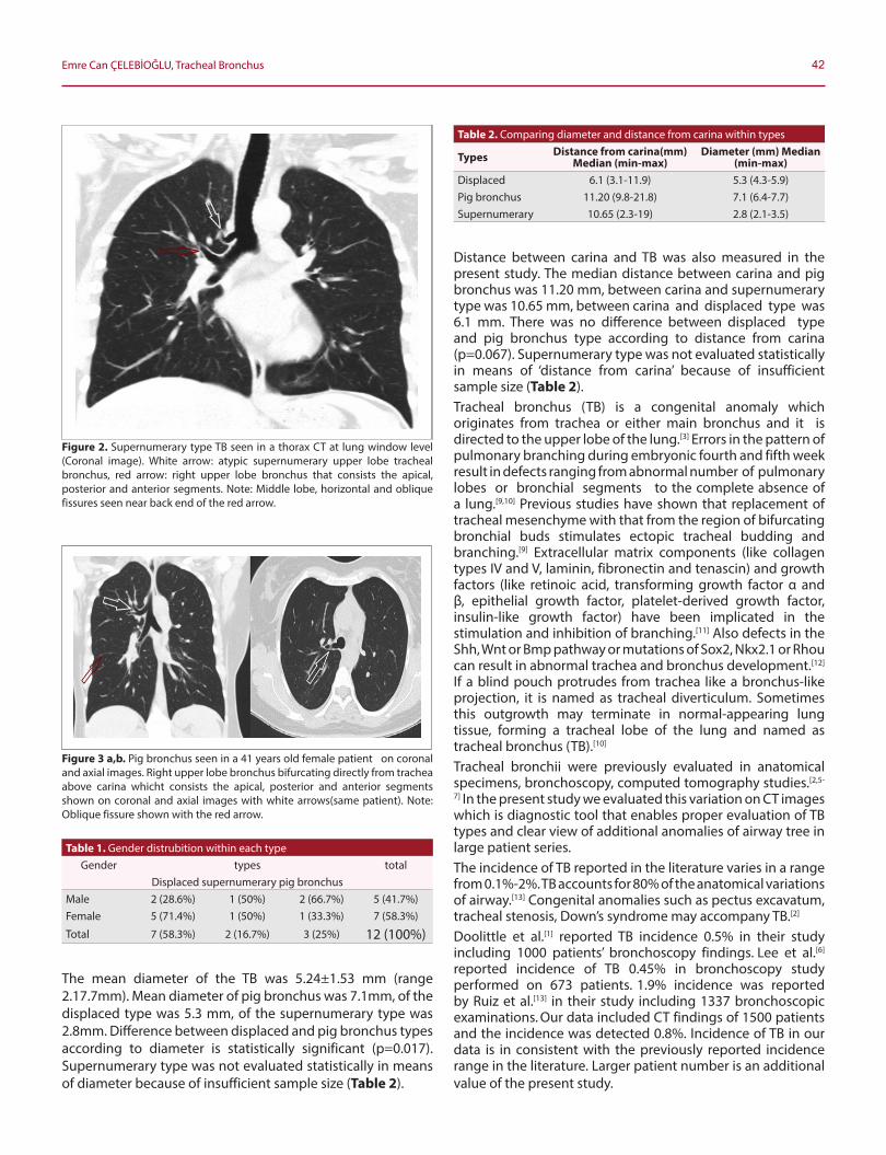
42Emre Can ÇELEBİOĞLU, Tracheal Bronchus
The mean diameter of the TB was 5.24±1.53 mm (range 2.17.7mm). Mean diameter of pig bronchus was 7.1mm, of the displaced type was 5.3 mm, of the supernumerary type was 2.8mm. Difference between displaced and pig bronchus types according to diameter is statistically significant (p=0.017). Supernumerary type was not evaluated statistically in means of diameter because of insufficient sample size (Table 2).
Distance between carina and TB was also measured in the present study. The median distance between carina and pig bronchus was 11.20 mm, between carina and supernumerary type was 10.65 mm, between carina and displaced type was 6.1 mm. There was no difference between displaced type and pig bronchus type according to distance from carina (p=0.067). Supernumerary type was not evaluated statistically in means of ‘distance from carina’ because of insufficient sample size (Table 2).Tracheal bronchus (TB) is a congenital anomaly which originates from trachea or either main bronchus and it is directed to the upper lobe of the lung.[3] Errors in the pattern of pulmonary branching during embryonic fourth and fifth week result in defects ranging from abnormal number of pulmonary lobes or bronchial segments to the complete absence of a lung.[9,10] Previous studies have shown that replacement of tracheal mesenchyme with that from the region of bifurcating bronchial buds stimulates ectopic tracheal budding and branching.[9] Extracellular matrix components (like collagen types IV and V, laminin, fibronectin and tenascin) and growth factors (like retinoic acid, transforming growth factor α and β, epithelial growth factor, platelet-derived growth factor, insulin-like growth factor) have been implicated in the stimulation and inhibition of branching.[11] Also defects in the Shh, Wnt or Bmp pathway or mutations of Sox2, Nkx2.1 or Rhou can result in abnormal trachea and bronchus development.[12] If a blind pouch protrudes from trachea like a bronchus-like projection, it is named as tracheal diverticulum. Sometimes this outgrowth may terminate in normal-appearing lung tissue, forming a tracheal lobe of the lung and named as tracheal bronchus (TB).[10]
Tracheal bronchii were previously evaluated in anatomical specimens, bronchoscopy, computed tomography studies.[2,5-
7] In the present study we evaluated this variation on CT images which is diagnostic tool that enables proper evaluation of TB types and clear view of additional anomalies of airway tree in large patient series.The incidence of TB reported in the literature varies in a range from 0.1%-2%. TB accounts for 80% of the anatomical variations of airway.[13] Congenital anomalies such as pectus excavatum, tracheal stenosis, Down’s syndrome may accompany TB.[2]
Doolittle et al.[1] reported TB incidence 0.5% in their study including 1000 patients’ bronchoscopy findings. Lee et al.[6] reported incidence of TB 0.45% in bronchoscopy study performed on 673 patients. 1.9% incidence was reported by Ruiz et al.[13] in their study including 1337 bronchoscopic examinations. Our data included CT findings of 1500 patients and the incidence was detected 0.8%. Incidence of TB in our data is in consistent with the previously reported incidence range in the literature. Larger patient number is an additional value of the present study.
Figure 2. Supernumerary type TB seen in a thorax CT at lung window level (Coronal image). White arrow: atypic supernumerary upper lobe tracheal bronchus, red arrow: right upper lobe bronchus that consists the apical, posterior and anterior segments. Note: Middle lobe, horizontal and oblique fissures seen near back end of the red arrow.
Figure 3 a,b. Pig bronchus seen in a 41 years old female patient on coronal and axial images. Right upper lobe bronchus bifurcating directly from trachea above carina whicht consists the apical, posterior and anterior segments shown on coronal and axial images with white arrows(same patient). Note: Oblique fissure shown with the red arrow.
Table 1. Gender distrubition within each typeGender types total
Displaced supernumerary pig bronchusMale 2 (28.6%) 1 (50%) 2 (66.7%) 5 (41.7%)Female 5 (71.4%) 1 (50%) 1 (33.3%) 7 (58.3%)
Total 7 (58.3%) 2 (16.7%) 3 (25%) 12 (100%)
Table 2. Comparing diameter and distance from carina within types
Types Distance from carina(mm) Median (min-max)
Diameter (mm) Median (min-max)
Displaced 6.1 (3.1-11.9) 5.3 (4.3-5.9)Pig bronchus 11.20 (9.8-21.8) 7.1 (6.4-7.7)Supernumerary 10.65 (2.3-19) 2.8 (2.1-3.5)
43 Journal of Contemporary Medicine
TB is reported to be located about 2-6 cm superior to carina in the literature.[2,5-8] In our study distance of TB from carina was analyzed within each type (Table 2). This data provides a contribution to literature. Detailed measurement provided in this study will give an idea to doctors when intubating patients and prevent adverse outcomes after intubation.Detailed anatomical definition of this variation has critical importance for lung transplantation. Mendogni et al reported that size of bronchial orifice, distance between TB and right upper lobe bronchus are important features for proper anastomosis between recipient and donor tissues.[14] In the present study we measured diameter of TB 5.24±1.53. We think distance between TB and right upper lobe bronchus measured among patients with TB will provide precious contribution to literature in the following studies.Recognizing the TB is critically important during endotracheal intubation. When the TB is occluded by the side of the tube the lobe may collapse. On the other hand when it is mistakenly intubated, over aeration may lead pneumothorax.[1,15] The tube itself can also migrate into tracheal bronchus and cause atelectasis.[16] Anesthesiologists should spend special care during intubation of patients with TB. The position of tip of the endotracheal tube must be kept in the mid to proximal trachea.[1,15]
Distal placement of the endotracheal tube occurs in 3.7-10.7% of intubations. Accidentally displaced endotracheal tube may result in a drop in oxygen saturation in patients with tracheal bronchus. Although TB is a rare congenital anomaly a physician may encounter this anomaly several times a year. Obstruction of TB due to distal placement of endotracheal tube is a conceivable problem.[17,18]
Patients with TB incidentally detected on CT images should be informed to prevent probable anesthesia complications in future. ‘One lung ventilation’ is an anesthesia procedure commonly performed in thoracic surgery and video assisted thoracoscopic surgery.Anesthesiologists may face problems in patients with undiagnosed TB when isolating lung in these types of surgical modalities. Detailed bronchoscopic examination before thoracic surgery provides sufficient information for proper anesthesia.[6] This study will contribute to literature and will serve data of incidence to anesthesiologists in our country.Lai et al.[18] suggest that TB is an asymptomatic entity which is incidentally diagnosed on CT. They also argue that TB causes recurrent infections and respiratory distress especially in children. Oi et al.[5] suggested that individuals with TB have higher risk of recurrent local infections, cough and acute respiratory distress than healthy ones. Incidental or accidental obstruction of TB in intensive care units or during general anesthesia may cause pneumothorax, atelectasis, post obstructive pneumonia or respiratory failure. TB which is an asymptomatic anatomical variation may cause serious adverse outcomes in certain conditions. Pulmonologists, thoracic surgeons, intensivists and anesthesiologists should keep this anomaly in mind.
Our study has some limitations. TB was found more frequently in women than in men in the present study but larger patient series are required to claim this idea statistically. Supernumerary type was not evaluated statistically in means of diameter because of insufficient sample size. Distance of supernumerary type from carina was not also evaluated statistically because of insufficient sample size. We predict further studies with larger sample size will enlighten these missing points in the present study.
CONCLUSIONSPulmonologists, thoracic surgeons, intensivists and anesthesiologists should keep this variation in mind for proper management of adverse outcomes of intubation pitfalls.
ETHICAL DECLARATIONSEthics Comittee Approval: Approval was received by Ankara Yıldırım Beyazıt University Education and Research Hospital Clinical Research Ethics Committee (Date: 11.09.2019, no: 92).
Informed Consent: Written informed consent was obtained from all participants who participated in this study.
Status of Peer-review: Externally peer-reviewed.
Conflict of Interest Statement: The authors have no conflicts of interest to declare.
Financial Disclosure: The authors declared that this study has received no financial support.
Author Contributions: All of the authors declare that they have all participated in the design, execution, and analysis of the paper, and that they have approved the final version.
REFERENCES1. Doolıttle AM, Maır EA Tracheal bronchus: classification, endoscopic
analysis, and airway management Otolaryngol Head Neck Surg2002;126:240-3. doi:10.1067/mhn.2002.122703.
2. Levin E, Bowling MR. Malignancy in the tracheal bronchus: A case series and review of the literature. Clin Respir J. 2018; 12:2441–2445. doi: 10.1111/crj.12943.
3. Manjunatha YC, Gupta AK. Tracheal Bronchus (Pig Bronchus). Indian J Pediatr 2010; 77:1037–1038. doi: 10.1007/s12098-0100162-6.
4. Han J, Xiang H, Ridley WE, Ridley LJ. Pig bronchus. 2018 The Royal Australian and New Zealand College of Radiologists. doi:10.1111/1754-9485.21_12785.
5. Qi S, Zhang B, Yue Y, Shen J, Teng Y, Qian W, Wu J. Airflow in Tracheobronchial Tree of Subjects with Tracheal Bronchus Simulated Using CT Image Based Models and CFD Method. Med Syst. 2018;42(4):65. doi: 10.1007/s10916-017-0879-0.
6. Lee DK, Kim YM, Kim HZ, Lim SH. Right upper lobe tracheal bronchus: anesthetic challenge in one-lung ventilated patients-A report of three casesKorean J Anesthesiol 2013; 64(5): 448-450.
7. Barbetta C,Tamburini N, Marchi I, Forini G, Papi A. True tracheal bronchus. Anaesthesia 2016; 71(7): 846. doi:10.1111/anae.13531.
8. Sindhwani G, Rawat J, Gupta M, Chandra S. Lung Cancer in “True Tracheal Bronchus” A Rare Coincidence. J Bronchol Intervent Pulmonol 2012; 19: 340–342.
44Emre Can ÇELEBİOĞLU, Tracheal Bronchus
9. Schoenwolf GC, Blely SB, Brauer PR, Francis-West PH. Larsen’s Human Embryology, 5th Edt.(2015),Elsevier, 252-259.
10. Moore KL, Persaud TVN, Torchia MG. The Developing HumanClinically Oriented Embryology, 10th Edt.(2016), Elsevier, 195200.
11. Carlson BM, Human Embryology and Developmental Biology, 5th Edt.(2014), Elsevier, 359-362.
12. Snowball J, Ambalavanan M, Whitsett J, Sinner D. Endodermal Wnt signaling is required for tracheal cartilage formation. Dev. Biol. 2015; 405(1): 56-70. doi: 10.1016/j.ydbio.2015.06.009.
13. Ruiz EP, Aguilera PC, Valdivielso AI, Cárdenas SS, García YM, Frías JP. Tracheal bronchus diagnosed in children undergoing flexible bronchoscopy. Paediatric Respiratory Reviews 2018;28:26–30.
14. Mendognia P, Tosia D, Rossoa L, Palleschia A, Righia I, Montolia M, Marioloa AV, Nosottia M. Lung Transplant From Donor With Tracheal Bronchus: Case Report and Literature Review. Transplant Proc. 2019; 51(1): 239–241.
15. Jain D, Bhardwaj N, Pareek A, Parikh B. Tracheal bronchus: an unusual cause of difficult ventilation. Can J Anaesth. 2017; 64(9): 979–980. doi: 10.1007/s12630-017-0919-8.
16. Wong F, Detterbeck F, Kurup V. Airway Management in Patients with Tracheal Bronchus. Images in Anesthesiology. Anesthesiology, 2016;125(2): 395. doi:10.1097/ aln.0000000000001019.
17. Ho AK, Ho AMH, Mizubuti GB. Blockage of the tracheal bronchus: effects on blood oxygen content, partial pressure of oxygen, and intrapulmonary shunt. Adv Physiol Educ 2018; 42: 383–6, doi:10.1152/advan.00017.2018.
18. Lai KM, Hsieh MH, Lama F, Chen CY, Chen TL, Chang CC. Anesthesia for patients with tracheal bronchus. Asian Journal of Anesthesiology 2017; 55: 87-8.
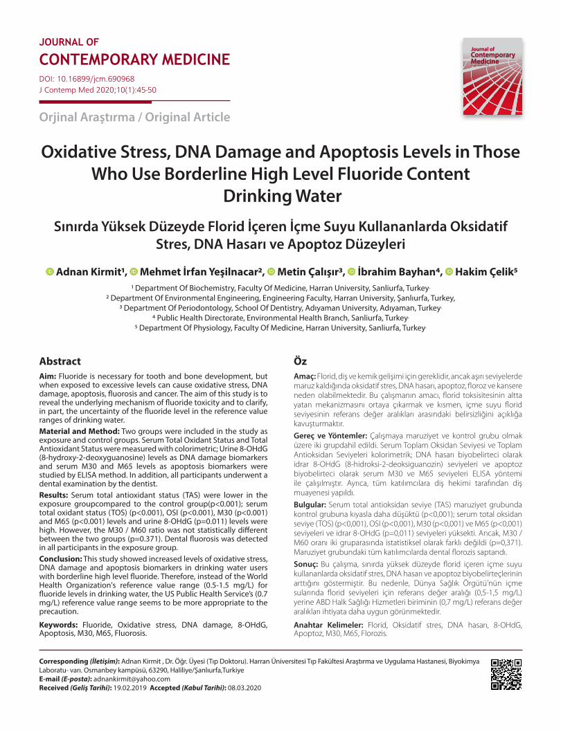
Corresponding (İletişim): Adnan Kirmit , Dr. Öğr. Üyesi (Tıp Doktoru). Harran Üniversitesi Tıp Fakültesi Araştırma ve Uygulama Hastanesi, Biyokimya Laboratu- varı. Osmanbey kampüsü, 63290, Haliliye/Şanlıurfa,TurkiyeE-mail (E-posta): [email protected] (Geliş Tarihi): 19.02.2019 Accepted (Kabul Tarihi): 08.03.2020
DOI: 10.16899/jcm.690968J Contemp Med 2020;10(1):45-50
Orjinal Araştırma / Original Article
JOURNAL OF
CONTEMPORARY MEDICINEJournal ofContemporary MedicineYEAR: 2019 VOLUME: 9 ISSUE: 4
Oxidative Stress, DNA Damage and Apoptosis Levels in Those Who Use Borderline High Level Fluoride Content
Drinking Water
Sınırda Yüksek Düzeyde Florid İçeren İçme Suyu Kullananlarda Oksidatif Stres, DNA Hasarı ve Apoptoz Düzeyleri
Amaç: Florid, diş ve kemik gelişimi için gereklidir, ancak aşırı seviyelerde maruz kaldığında oksidatif stres, DNA hasarı, apoptoz, floroz ve kansere neden olabilmektedir. Bu çalışmanın amacı, florid toksisitesinin altta yatan mekanizmasını ortaya çıkarmak ve kısmen, içme suyu florid seviyesinin referans değer aralıkları arasındaki belirsizliğini açıklığa kavuşturmaktır.
Gereç ve Yöntemler: Çalışmaya maruziyet ve kontrol grubu olmak üzere iki grupdahil edildi. Serum Toplam Oksidan Seviyesi ve Toplam Antioksidan Seviyeleri kolorimetrik; DNA hasarı biyobelirteci olarak idrar 8-OHdG (8-hidroksi-2-deoksiguanozin) seviyeleri ve apoptoz biyobelirteci olarak serum M30 ve M65 seviyeleri ELISA yöntemi ile çalışılmıştır. Ayrıca, tüm katılımcılara diş hekimi tarafından diş muayenesi yapıldı.
Bulgular: Serum total antioksidan seviye (TAS) maruziyet grubunda kontrol grubuna kıyasla daha düşüktü (p<0,001); serum total oksidan seviye (TOS) (p<0,001), OSI (p<0,001), M30 (p<0,001) ve M65 (p<0,001) seviyeleri ve idrar 8-OHdG (p=0,011) seviyeleri yüksekti. Ancak, M30 / M60 oranı iki gruparasında istatistiksel olarak farklı değildi (p=0,371). Maruziyet grubundaki tüm katılımcılarda dental florozis saptandı.
Sonuç: Bu çalışma, sınırda yüksek düzeyde florid içeren içme suyu kullananlarda oksidatif stres, DNA hasarı ve apoptoz biyobelirteçlerinin arttığını göstermiştir. Bu nedenle, Dünya Sağlık Örgütü'nün içme sularında florid seviyeleri için referans değer aralığı (0,5-1,5 mg/L) yerine ABD Halk Sağlığı Hizmetleri biriminin (0,7 mg/L) referans değer aralıkları ihtiyata daha uygun görünmektedir.
Anahtar Kelimeler: Florid, Oksidatif stres, DNA hasarı, 8-OHdG, Apoptoz, M30, M65, Florozis.
Abstract Öz
Adnan Kirmit1, Mehmet Irfan Yeşilnacar2, Metin Çalışır3, Ibrahim Bayhan4, Hakim Çelik5
Aim: Fluoride is necessary for tooth and bone development, but when exposed to excessive levels can cause oxidative stress, DNA damage, apoptosis, fluorosis and cancer. The aim of this study is to reveal the underlying mechanism of fluoride toxicity and to clarify, in part, the uncertainty of the fluoride level in the reference value ranges of drinking water.Material and Method: Two groups were included in the study as exposure and control groups. Serum Total Oxidant Status and Total Antioxidant Status were measured with colorimetric; Urine 8-OHdG (8-hydroxy-2-deoxyguanosine) levels as DNA damage biomarkers and serum M30 and M65 levels as apoptosis biomarkers were studied by ELISA method. In addition, all participants underwent a dental examination by the dentist.Results: Serum total antioxidant status (TAS) were lower in the exposure groupcompared to the control group(p<0.001); serum total oxidant status (TOS) (p<0.001), OSI (p<0.001), M30 (p<0.001) and M65 (p<0.001) levels and urine 8-OHdG (p=0.011) levels were high. However, the M30 / M60 ratio was not statistically different between the two groups (p=0.371). Dental fluorosis was detected in all participants in the exposure group.Conclusion: This study showed increased levels of oxidative stress, DNA damage and apoptosis biomarkers in drinking water users with borderline high level fluoride. Therefore, instead of the World Health Organization's reference value range (0.5-1.5 mg/L) for fluoride levels in drinking water, the US Public Health Service’s (0.7 mg/L) reference value range seems to be more appropriate to the precaution.
Keywords: Fluoride, Oxidative stress, DNA damage, 8-OHdG, Apoptosis, M30, M65, Fluorosis.
1 Department Of Biochemistry, Faculty Of Medicine, Harran University, Sanliurfa, Turkey.2 Department Of Environmental Engineering, Engineering Faculty, Harran University, Şanlıurfa, Turkey,
3 Department Of Periodontology, School Of Dentistry, Adıyaman University, Adıyaman, Turkey.4 Public Health Directorate, Environmental Health Branch, Sanliurfa, Turkey.
5 Department Of Physiology, Faculty Of Medicine, Harran University, Sanliurfa, Turkey.
46 Journal of Contemporary Medicine
INTRODUCTIONOne of the inorganic substances that the organism needs and needs to be taken from outside is fluoride.[1,2] Although fluoride is one of the vital elements for human health, it has a doubleedged sword effect on human health.[3] When taken at the normal dose, it contributes to bone and tooth development, and when taken at an excessive level, it can cause dental, skeletal and visceral fluorosis and cancer depending on the dose and duration of exposure.[4,5] Fluoride is the ion form of fluorine, which is gas and neutral, and is found in compounds, not free in nature. It is mostly found as Sodium Fluoride (NaF2) and Calcium fluoride (CaF2). Fluoride is found in the atmosphere, soil, water and food.[6] Surface waters generally do not contain more than 0.3 mg/L of fluoride unless they are contaminated from external sources. However, depending on many variables, fluoride concentrations in groundwater have a wide range (1.0 35.0 mg/L). The main source of people's daily fluoride consumption is drinking water (75-90% of daily intake).[7]
Prolonged consumption of drinking water containing fluoride ions greater than 1.5 mg/L causes fluorosis in the teeth and skeletal system, neurological damage and further toxic effects are seen when exposed to concentrations of more than 4 mg/L.[8] Previous studies have showed that fluorine can induce genotoxicity, cytotoxicity, immunotoxicity, oxidative damage, apoptosis and lesions in the broiler peripheral blood, liver, kidney, thymus, spleen, cecal tonsil, and intestine, and in the mouse spleen.[9-16]
Reactive oxygen species (ROS), one of the by-products of the metabolic process, can normally be removed by numerous antioxidant defence components.[17]
The imbalance between ROS and antioxidants is called “oxidative stress”.[18] Fluoride is known to be an inhibitor of antioxidant enzymes, which promotes ROS accumulation.[19,20]
Due to the acceptance of fluoride as an environmental pollutant, many studies have been conducted on exposure to fluoride. In the studies, it was found that exposure to fluoride could induce oxidative stress in liver, kidneys, testis, spleen, brain, heart and cecal tonsils and reduce the activities of superoxide dismutase (SOD), catalase (CAT), glutathione peroxidase (GSH-PX) and glutathione-s-transferase (GST).[21-26]
The most important free oxygen radical that damages cellular biomolecules (proteins, membrane lipids and DNA) is the hydroxyl radical (HO). 8-OHdG (8-hydroxy-2-deoxyguanosine) is one of the main biomarkers of damage caused by free radicals in nuclear and mitochondrial DNA and is widely used for this purpose.[27] Cells whose DNA is irreparably damaged must be removed by apoptosis as they may have the potential for malignancy. Lack of apoptosis can cause cancer and autoimmune diseases.[28]
In cell death, creatine (CK18) is released outside the cell. The M30 measurement kit detects a neoepitope of a fragment of CK18 that appears only after caspase cleavage with positions 387 to 396, and is considered a selective biomarker of apoptosis.[29]
The M65 kit detects a common epitope found in both unfragmented CK18 (released from necrosis cells) and its fragment (released from apoptotic cells) and is therefore considered a marker of total cell death (apoptosis and necrosis).[30] Therefore, high M30/M65 ratio shows that cell death is mainly due to apoptosis.[31,32] The increase in isolated M65 indicates necrosis.[33]
As the concentration of toxic substance increases, apoptosis increases up to a point, then apoptosis decreases and the increases in toxic substance concentration, resulting in a linear increase in necrosis.[31]
The World Health Organization (WHO) states that the reference range for fluoride in drinking water is 0.5–1.5 mg/L.[34] The American Environmental Protection Association (EPA) and the American Public Health Service (PHS) previously reported the fluoride level in drinking water to be 0.7–1.2 mg/L, while the PHS revised its optimal value to 0.7 mg/L.[35] EPA stated that there is no need for a reference update for the time being but it will continue to follow up the researches done on this subject and go to the reference regulation when necessary.[36]
Although there are many studies in the literature regarding the outcome of the fluoride effect, most of these are limited to animal experiments using drinking water containing very high amounts of fluoride. There is still uncertainty as to whether a high level of fluoride at the boundary in drinking water is toxic. This prospective study was designed to investigate whether the use of drinking water with borderline high level fluoride is dangerous or not.
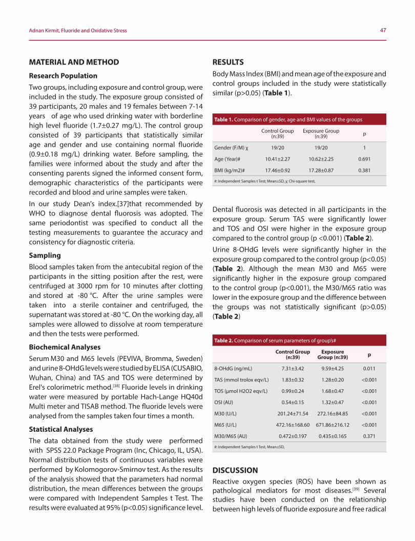
47Adnan Kirmit, Fluoride and Oxidative Stress
MATERIAL AND METHOD
Research PopulationTwo groups, including exposure and control group, were included in the study. The exposure group consisted of 39 participants, 20 males and 19 females between 7-14 years of age who used drinking water with borderline high level fluoride (1.7±0.27 mg/L). The control group consisted of 39 participants that statistically similar age and gender and use containing normal fluoride (0.9±0.18 mg/L) drinking water. Before sampling, the families were informed about the study and after the consenting parents signed the informed consent form, demographic characteristics of the participants were recorded and blood and urine samples were taken.
In our study Dean's index.[37]that recommended by WHO to diagnose dental fluorosis was adopted. The same periodontist was specified to conduct all the testing measurements to guarantee the accuracy and consistency for diagnostic criteria.
SamplingBlood samples taken from the antecubital region of the participants in the sitting position after the rest, were centrifuged at 3000 rpm for 10 minutes after clotting and stored at -80 °C. After the urine samples were taken into a sterile container and centrifuged, the supernatant was stored at -80 °C. On the working day, all samples were allowed to dissolve at room temperature and then the tests were performed.
Biochemical AnalysesSerum M30 and M65 levels (PEVIVA, Bromma, Sweden) and urine 8-OHdG levels were studied by ELISA (CUSABIO, Wuhan, China) and TAS and TOS were determined by Erel's colorimetric method.[38] Fluoride levels in drinking water were measured by portable Hach-Lange HQ40d Multi meter and TISAB method. The fluoride levels were analysed from the samples taken four times a month.
Statistical AnalysesThe data obtained from the study were performed with SPSS 22.0 Package Program (Inc, Chicago, IL, USA). Normal distribution tests of continuous variables were performed by Kolomogorov-Smirnov test. As the results of the analysis showed that the parameters had normal distribution, the mean differences between the groups were compared with Independent Samples t Test. The results were evaluated at 95% (p<0.05) significance level.
RESULTSBody Mass Index (BMI) and mean age of the exposure and control groups included in the study were statistically similar (p>0.05) (Table 1).
Dental fluorosis was detected in all participants in the exposure group. Serum TAS were significantly lower and TOS and OSI were higher in the exposure group compared to the control group (p <0.001) (Table 2).
Urine 8-OHdG levels were significantly higher in the exposure group compared to the control group (p<0.05) (Table 2). Although the mean M30 and M65 were significantly higher in the exposure group compared to the control group (p<0.001), the M30/M65 ratio was lower in the exposure group and the difference between the groups was not statistically significant (p>0.05) (Table 2)
DISCUSSIONReactive oxygen species (ROS) have been shown as pathological mediators for most diseases.[39] Several studies have been conducted on the relationship between high levels of fluoride exposure and free radical
Table 1. Comparison of gender, age and BMI values of the groups
Control Group (n:39)
Exposure Group (n:39) p
Gender (F/M) χ 19/20 19/20 1
Age (Year)# 10.41±2.27 10.62±2.25 0.691
BMI (kg/m2)# 17.46±0.92 17.28±0.87 0.381
#: Independent Samples t Test; Mean±SD, χ: Chi-square test,
Table 2. Comparison of serum parameters of group’s#
Control Group (n:39)
Exposure Group (n:39) p
8-OHdG (ng/mL) 7.31±3.42 9.59±4.25 0.011
TAS (mmol trolox eqv/L) 1.83±0.32 1.28±0.20 <0.001
TOS (μmol H2O2 eqv/L) 0.99±0.24 1.68±0.47 <0.001
OSI (AU) 0.54±0.15 1.32±0.47 <0.001
M30 (U/L) 201.24±71.54 272.16±84.85 <0.001
M65 (U/L) 472.16±168.60 671.86±216.12 <0.001
M30/M65 (AU) 0.472±0.197 0.435±0.165 0.371
#: Independent Samples t Test, Mean±SD,
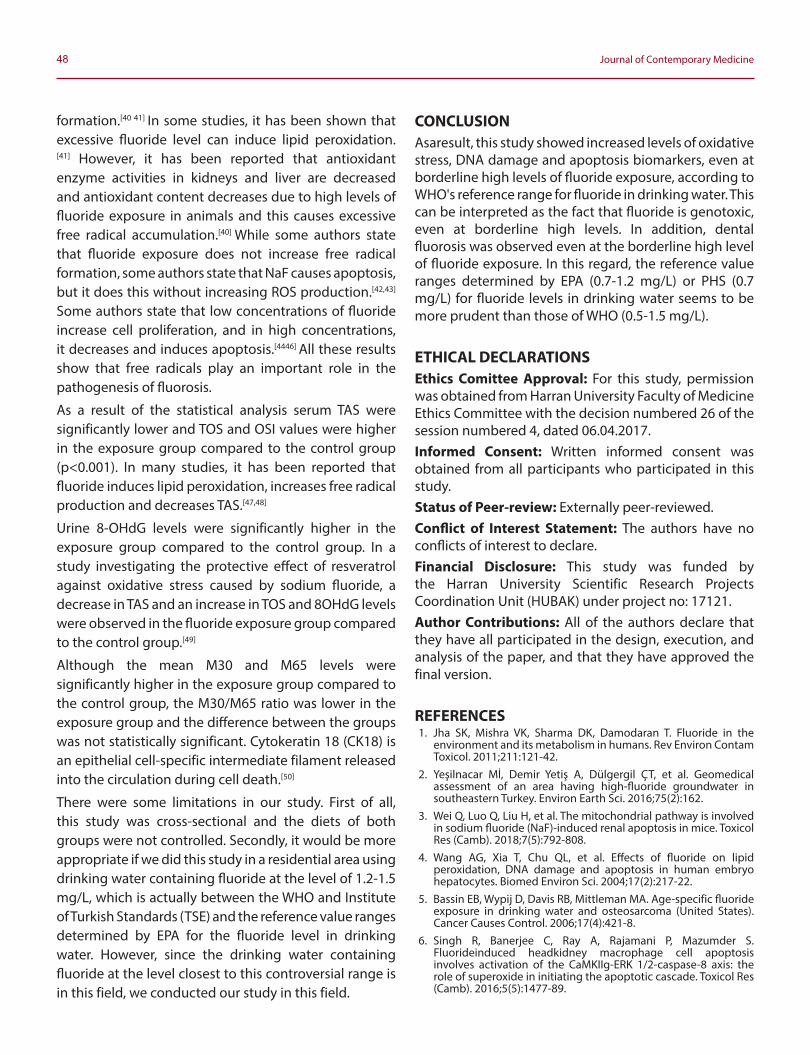
48 Journal of Contemporary Medicine
formation.[40 41] In some studies, it has been shown that excessive fluoride level can induce lipid peroxidation.[41] However, it has been reported that antioxidant enzyme activities in kidneys and liver are decreased and antioxidant content decreases due to high levels of fluoride exposure in animals and this causes excessive free radical accumulation.[40] While some authors state that fluoride exposure does not increase free radical formation, some authors state that NaF causes apoptosis, but it does this without increasing ROS production.[42,43] Some authors state that low concentrations of fluoride increase cell proliferation, and in high concentrations, it decreases and induces apoptosis.[4446] All these results show that free radicals play an important role in the pathogenesis of fluorosis.
As a result of the statistical analysis serum TAS were significantly lower and TOS and OSI values were higher in the exposure group compared to the control group (p<0.001). In many studies, it has been reported that fluoride induces lipid peroxidation, increases free radical production and decreases TAS.[47,48]
Urine 8-OHdG levels were significantly higher in the exposure group compared to the control group. In a study investigating the protective effect of resveratrol against oxidative stress caused by sodium fluoride, a decrease in TAS and an increase in TOS and 8OHdG levels were observed in the fluoride exposure group compared to the control group.[49]
Although the mean M30 and M65 levels were significantly higher in the exposure group compared to the control group, the M30/M65 ratio was lower in the exposure group and the difference between the groups was not statistically significant. Cytokeratin 18 (CK18) is an epithelial cell-specific intermediate filament released into the circulation during cell death.[50]
There were some limitations in our study. First of all, this study was cross-sectional and the diets of both groups were not controlled. Secondly, it would be more appropriate if we did this study in a residential area using drinking water containing fluoride at the level of 1.2-1.5 mg/L, which is actually between the WHO and Institute of Turkish Standards (TSE) and the reference value ranges determined by EPA for the fluoride level in drinking water. However, since the drinking water containing fluoride at the level closest to this controversial range is in this field, we conducted our study in this field.
CONCLUSIONAsaresult, this study showed increased levels of oxidative stress, DNA damage and apoptosis biomarkers, even at borderline high levels of fluoride exposure, according to WHO's reference range for fluoride in drinking water. This can be interpreted as the fact that fluoride is genotoxic, even at borderline high levels. In addition, dental fluorosis was observed even at the borderline high level of fluoride exposure. In this regard, the reference value ranges determined by EPA (0.7-1.2 mg/L) or PHS (0.7 mg/L) for fluoride levels in drinking water seems to be more prudent than those of WHO (0.5-1.5 mg/L).
ETHICAL DECLARATIONSEthics Comittee Approval: For this study, permission was obtained from Harran University Faculty of Medicine Ethics Committee with the decision numbered 26 of the session numbered 4, dated 06.04.2017.Informed Consent: Written informed consent was obtained from all participants who participated in this study. Status of Peer-review: Externally peer-reviewed. Conflict of Interest Statement: The authors have no conflicts of interest to declare. Financial Disclosure: This study was funded by the Harran University Scientific Research Projects Coordination Unit (HUBAK) under project no: 17121.Author Contributions: All of the authors declare that they have all participated in the design, execution, and analysis of the paper, and that they have approved the final version.
REFERENCES1. Jha SK, Mishra VK, Sharma DK, Damodaran T. Fluoride in the
environment and its metabolism in humans. Rev Environ Contam Toxicol. 2011;211:121-42.
2. Yeşilnacar Mİ, Demir Yetiş A, Dülgergil ÇT, et al. Geomedical assessment of an area having high-fluoride groundwater in southeastern Turkey. Environ Earth Sci. 2016;75(2):162.
3. Wei Q, Luo Q, Liu H, et al. The mitochondrial pathway is involved in sodium fluoride (NaF)-induced renal apoptosis in mice. Toxicol Res (Camb). 2018;7(5):792-808.
4. Wang AG, Xia T, Chu QL, et al. Effects of fluoride on lipid peroxidation, DNA damage and apoptosis in human embryo hepatocytes. Biomed Environ Sci. 2004;17(2):217-22.
5. Bassin EB, Wypij D, Davis RB, Mittleman MA. Age-specific fluoride exposure in drinking water and osteosarcoma (United States). Cancer Causes Control. 2006;17(4):421-8.
6. Singh R, Banerjee C, Ray A, Rajamani P, Mazumder S. Fluorideinduced headkidney macrophage cell apoptosis involves activation of the CaMKIIg-ERK 1/2-caspase-8 axis: the role of superoxide in initiating the apoptotic cascade. Toxicol Res (Camb). 2016;5(5):1477-89.
49Adnan Kirmit, Fluoride and Oxidative Stress
7. Meenakshi, Maheshwari RC. Fluoride in drinking water and its removal. J Hazard Mater. 2006;137(1):456-63.
8. Altundağ H, Dündar MŞ, Yüceel Ç, Albayrak S. Sakarya İli İçme Sularında Flor Düzeylerinin İyon Seçici Elektrot Ve İyon Kromotografisi İle Tayini. SAÜ Fen Bilimleri Dergisi. 2011;15(2):129-38.
9. Chen T, Cui Y, Bai C, Gong T, Peng X, Cui H. Decreased percentages of the peripheral blood T-cell subsets and the serum IL-2 contents in chickens fed on diets excess in fluorine. Biol Trace Elem Res. 2009;132(1-3):122-8.
10. Yujiao Lu, Qin Luo, Hengmin Cui, et al. Sodium fluoride causes oxidative stress and apoptosis in the mouse liver. Aging (Albany NY). 2017;9(6):1623-39.
11. Bai C, Chen T, Cui Y, Gong T, Peng X, Cui HM. Effect of high fluorine on the cell cycle and apoptosis of renal cells in chickens. Biol Trace Elem Res. 2010;138(1-3):173-80.
12. Chen T, Cui H, Cui Y, Bai C, Gong T, Peng X. Cell-cycle blockage associated with increased apoptotic cells in the thymus of chickens fed on diets high in fluorine. Hum Exp Toxicol. 2011;30(7):685-92.
13. Chen T, Cui H, Cui Y, Bai C, Gong T. Decreased antioxidase activities and oxidative stress in the spleen of chickens fed on high-fluorine diets. Hum Exp Toxicol. 2011;30(9):1282-6.
14. Liu J, Cui H, Peng X, et al. Dietary High Fluorine Induces Apoptosis and Alters Bcl-2, Bax, and Caspase-3 Protein Expression in the Cecal Tonsil Lymphocytes of Broilers. Biol Trace Elem Res. 2013;152(1):25-30.
15. Luo Q, Cui H, Peng X, et al. Suppressive effects of dietary high fluorine on the intestinal development in broilers. Biol Trace Elem Res. 2013;156(1-3):153-65.
16. Kuang P, Deng H, Cui H, et al. Sodium fluoride (NaF) causes toxic effects on splenic development in mice. Oncotarget. 2017;8(3):4703-17.
17. Cabiscol E, Tamarit J, Ros J. Oxidative stress in bacteria and protein damage by reactive oxygen species. Int Microbiol. 2000;3(1):3-8.
18. Mittler R. Oxidative stress, antioxidants and stress tolerance. Trends Plant Sci. 2002;7(9):405-10.
19. Kale M, Rathore N, John S, Bhatnagar D. Lipid peroxidative damage on pyrethroid exposure and alterations in antioxidant status in rat erythrocytes: a possible involvement of reactive oxygen species. Toxicol Lett. 1999;105(3):197-205.
20. Shivarajashankara YM, Shivashankara AR, Bhat PG, Rao SH. Lipid peroxidation and antioxidant systems in the blood of young rats subjected to chronic fluoride toxicity. Indian J Exp Biol. 2003;41(8):857-60.
21. Ghosh D, Das Sarkar S, Maiti R, Jana D, Das UB. Testicular toxicity in sodium fluoride treated rats: association with oxidative stress. Reprod Toxicol. 2002;16(4):385-90.
22. Bharti VK, Srivastava RS. Fluoride-induced oxidative stress in rat's brain and its amelioration by buffalo (Bubalus bubalis) pineal proteins and melatonin. Biol Trace Elem Res. 2009;130(2):131-40.
23. Nabavi SF, Nabavi SM, Ebrahimzadeh MA, Eslami S, Jafari N, Moghaddam H. The protective effect of curcumin against sodium fluoride-induced oxidative stress in rat heart. Arch Biol Sci. 2011;63(3):563-9.
24. Xiao-ying Guo, Gui-fan Sun, Sun Y-c. Oxidative Stress From Fluoride-Induced Hepatotoxicity in Rats. Fluoride. 2003;36(1):259.
25. R Ranjan, D Swarup, RC Patra. Oxidative stress indices in erythrocytes, liver, and kidneys of fluoride-exposed rabbits. Fluoride. 2009;42(2):88-93.
26. Mukhopadhyay D, Chattopadhyay A. Induction of oxidative stress and related transcriptional effects of sodium fluoride in female zebrafish liver. Bull Environ Contam Toxicol. 2014;93(1):64-70.
27. Valavanidis A, Vlachogianni T, Fiotakis C. 8-hydroxy-2' -deoxyguanosine (8-OHdG): A critical biomarker of oxidative stress and carcinogenesis. J Environ Sci Health C Environ Carcinog Ecotoxicol Rev. 2009;27(2):120-39.
28. Adams JM, Cory S. The BCL-2 arbiters of apoptosis and their growing role as cancer targets. Cell Death Differ. 2018;25(1):2736.
29. Olofsson MH, Ueno T, Pan Y, et al. Cytokeratin-18 is a useful serum biomarker for early determination of response of breast carcinomas to chemotherapy. Clin Cancer Res. 2007;13(11):3198206.
30. Cummings J, Hodgkinson C, Odedra R, et al. Preclinical evaluation of M30 and M65 ELISAs as biomarkers of drug induced tumor cell death and antitumor activity. Mol Cancer Ther. 2008;7(3):455-63.
31. Kumbicak U, Cavas T, Cinkilic N, Kumbicak Z, Vatan O, Yilmaz D. Evaluation of in vitro cytotoxicity and genotoxicity of copperzinc alloy nanoparticles in human lung epithelial cells. Food Chem Toxicol. 2014;73:105-12.
32. Dasarathy S, Yang Y, McCullough AJ, Marczewski S, Bennett C, Kalhan SC. Elevated hepatic fatty acid oxidation, high plasma fibroblast growth factor 21, and fasting bile acids in nonalcoholic steatohepatitis. Eur J Gastroenterol Hepatol. 2011;23(5):382-8.
33. Abdellatif H, NasrEldin E, Galal S, Ibrahim M. Study of m65 hepatocyte death marker in multitransfused patients with β-thalassemia major in upper Egypt. Egypt J Haematol. 2019;44(2):124-7.
34. Fawell J, Bailey K, Chilton J, Dahi E, Magara Y. Fluoride in Drinkingwater: IWA publishing; 2006.
35. U.S. Public Health Service Recommendation for Fluoride Concentration in Drinking Water for the Prevention of Dental Caries. Public health reports (Washington, DC : 1974). 2015;130(4):318-31.
36. Review of the Fluoride Drinking Water Regulation.[Internet] Environmental Protection Agency..[cited 07.11.2019] Available from: https://www.epa.gov/dwsixyearreview/review-fluoridedrinking-water-regulation.
37. Rozier RG. Epidemiologic Indices for Measuring the Clinical Manifestations of Dental Fluorosis: Overview and Critique. Adv Dent Res. 1994;8(1):39-55.
38. Erel O. A novel automated method to measure total antioxidant response against potent free radical reactions. Clin Biochem. 2004;37(2):112-9.
39. Halliwell B, Gutteridge JM. Oxygen free radicals and iron in relation to biology and medicine: some problems and concepts. Arch Biochem Biophys. 1986;246(2):501-14.
40. Shivarajashankara YM, Shivashankara AR, Rao SH, Bhat PG. Oxidative stress in children with endemic skeletal fluorosis. Fluoride. 2001;34:103-7.
41. Shivarajashankara YM, Shivashankara AR, Bhat PG, Rao SH. Effect of Fluoride Intoxication on Lipid Peroxidation and Antioxidant Systems in Rats. Fluoride. 2001;34(2):108-13.
42. Chlubek D, Grucka-Mamczar E, Birkner E, Polaniak R, StawiarskaPieta B, Duliban H. Activity of pancreatic antioxidative enzymes and malondialdehyde concentrations in rats with hyperglycemia caused by fluoride intoxication. J Trace Elem Med Biol. 2003;17(1):57-60.
43. Lee JH, Jung JY, JeongYJ, et al. Involvement of both mitochondrialand death receptor-dependent apoptotic pathways regulated by Bcl-2 family in sodium fluoride-induced apoptosis of the human gingival fibroblasts. Toxicology. 2008;243(3):340-7.
44. Jothiramajayam M, Sinha S, Ghosh M, Nag A, Jana A, Mukherjee Sodium fluoride promotes apoptosis by generation of reactive oxygen species in human lymphocytes. J Toxicol Environ Health A. 2014;77(21):1269-80.
50 Journal of Contemporary Medicine
45. Qu WJ, Zhong DB, Wu PF, Wang JF, Han B. Sodium fluoride modulates caprine osteoblast proliferation and differentiation. J Bone Miner Metab. 2008;26(4):328-34.
46. Xu H, Wang CH, Zhao ZT, Zhang WB, Li GS. Role of oxidative stress in osteoblasts exposed to sodium fluoride. Biol Trace Elem Res. 2008;123(1-3):109-15.
47. Jundong Wang, Yaming Ge, Hongmei Ning, Shaolin Wang. Effects of High Fluoride and Low Iodine on Oxidative Stress And Antioxidant Defense of The Brain in Offspring Rats. Fluoride. 2004;37(4):264–70.
48. Jianhai Zhang, Chen Liang, Junjie Ma, Bianhua Zhou, Wang J. Effects Of Sodium Fluoride and Sulfur Dioxide on Oxidative Stress and Antioxidant Defenses in Rat Testes. Fluoride. 2006;39(3):185–90.
49. Atmaca N, Atmaca HT, Kanici A, Anteplioglu T. Protective effect of resveratrol on sodium fluoride-induced oxidative stress, hepatotoxicity and neurotoxicity in rats. Food Chem Toxicol. 2014;70:191-7.
50. Ueno T, Toi M, Linder S. Detection of epithelial cell death in the body by cytokeratin 18 measurement. Biomed Pharmacother. 2005;59 Suppl 2:S359-62.
Corresponding (İletişim): Yanardağ Açık Didar, Department of Internal Medicine and Haematology, Adana City Education and Research Hospital, Mithat Özsan Bulvarı Kışla Mah. 4522 Sok. No:1, 01260 Yüreğir, Adana, TurkeyE-mail (E-posta): [email protected] (Geliş Tarihi): 28.12.2019 Accepted (Kabul Tarihi): 02.03.2020
DOI: 10.16899/jcm.666822J Contemp Med 2020;10(1):51-54
Orjinal Araştırma / Original Article
JOURNAL OF
CONTEMPORARY MEDICINEJournal ofContemporary MedicineYEAR: 2019 VOLUME: 9 ISSUE: 4
Prevalence of Gaucher’s Disease in a Hematology Outpatient Clinic
Hematoloji Polikliniğinde Gaucher Hastalığı Sıklığı
Giris: Gaucher hastalığı (GD) glukoserebrosidaz enzimi eksikliği sonucu
meydana gelen ve retiküloendotelyal sistemde glukozilseramid
birikimi ile karakterize bir hastalıktır. Bu çalışmada hematoloji
polikliniğinde GD tanısı konulan hastalarda GD sıklığını belirlemeyi
amaçladık.
Yöntem: 2014-2018 tarihleri arasında hematoloji polikliniğine başvuran
26 000 hastanın retrospektif olarak tanıları incelendi. Gaucher hastalığı
tanısı alan hastaların sayısı toplam hasta ve hepatosplenomegali
tanısı olan hastalarla karşılaştırıldı. Sonuçlar oransal olarak kaydedildi.
Bulgular: Çalışmamızda GD sıklığı 23 / 100 000 olarak saptandı. GD
tanısı konulan vakaların hemen hemen hepsinde splenomegalinin var
olduğu saptandı. Splenomegalisi olan vakaların trombosit düzeyleri
düşük gözlendi.
Sonuç: Sonuç olarak GD, özellikle akraba evliliklerinin fazla
olduğu bölgelerde yaşayanlarda literatürdeki kadar nadir değildir.
Hepatosplenomegali ve trombositopeni saptanmış olguların
lizozomal depo hastalıkları açısından da araştırılması gerektiği hem
klinisyenlere hem de patologlara hatırlatılmalıdır
Anahtar Sözcükler: Gaucher hastalığı, hepatomegali, lizozomal depo hastalığı, splenomegali, trombositopeni
Abstract Öz
Yanardağ Açık Didar1, Aygün Bilal1
Objective: Gaucher’s disease (GD) is a disease caused by glucocerebrosidase enzyme deficiency and characterized by glucoceramide accumulation in the reticuloendothelial system. In this study, we aimed to determine the prevalence of GD in patients who were diagnosed with GD in a hematology clinic.
Method: The diagnoses of 26,000 patients who had applied to the hematology polyclinic between 2014 and 2018 were examined retrospectively. The number of patients diagnosed with GD was compared to the number of total patients and the number of patients with hepatosplenomegaly diagnosis. The results were recorded as ratios.
Results: The prevalence of GD was found as 23/100,000 in our study. It was found that splenomegaly was present in almost all of the diagnosed cases of GD. The thrombocyte levels of patients with splenomegaly were low.
Conclusion: GD is not as rare as is claimed in the literature, especially in people living in regions in which consanguineous marriages are common. Both clinicians and pathologists should be reminded that patients with diagnosed hepatosplenomegaly and thrombocytopenia need to be investigated with regard to lysosomal storage diseases.
Keywords: Gaucher’s disease, hepatomegaly, lysosomal storage disease, splenomegaly, thrombocytopenia
1Department of Internal Medicine and Haematology, Adana City Education and Research Hospital, Adana, Turkey

52 Journal of Contemporary Medicine
INTRODUCTION Gaucher’s disease (GD) is a disease caused by glucocerebrosidase enzyme deficiency and characterized by glucoceramide accumulation in the reticuloendothelial system.[1] Clinically, there are three types: Type 1, the chronic non-neuronopathic type; Type 2, the acute infantile neuronopathic type; and Type 3, the juvenile or subacute neuronopathic type.[2] GD mainly accompanies pancytopenia, hepatosplenomegaly, and bone deformities. In Type 3 GD, there are central neural system symptoms in addition to organ involvement.[3]
Delays are frequent in GD diagnosis and these delays are mostly due to the lack of awareness of the disease. Irreversible complications occur in patients due to these delays, and the patients undergo unnecessary invasive procedures.[4,5] GD and similar genetic metabolic diseases are more prevalent in regions in which consanguineous marriages are frequent, as in the Ashkenazi Jewish population.[3] The Çukurova region of Turkey has received migrants from the eastern and southeastern provinces of the country. Research indicates that the rate of consanguineous marriages reaches 40.4% in these regions, while the frequency of consanguineous marriage is 20.9% in the entire country.[6,7] Therefore, the prevalence of GD in the Çukurova region is anticipated to be higher when compared to the national population.
The GD prevalence in the general population varies between 0.4 and 5.8 per 100,000.[8] We could not identify any comprehensive study on GD prevalence in Turkey, and there are also no studies with that purpose conducted in the Çukurova region. Therefore, in this study we aimed to determine GD prevalence in patients diagnosed with GD in a hematology polyclinic.
MATERIAL AND METHODA total of 26,000 patients who had applied to the hematology clinic between 2014 and 2018 were included in the study. The beta-glucosidase enzyme was checked in retrospective screenings, and patients were identified who had received
a diagnosis of GD with the mutation test after their enzyme levels were found to be low. Clinical characteristics and laboratory test results of the GD patients were recorded from their files.
Patients whose glucosidase enzyme levels were normal or who were not found to be abnormal in the mutation tests were not considered as having GD.
Approval for the study was obtained from the University Faculty of Medicine Ethical Committee in line with the Helsinki Declaration criteria, revised in Brazil in 2013.
Statistical Analysis
Statistical evaluations were conducted using SPSS 20 for Windows (IBM SPSS Inc., Armonk, NY, USA). The normal distribution of the data was tested with the KolmogorovSmirnov test. Normally distributed numerical variables are presented as average ± standard deviation, and nonnormally distributed variables are presented as median (minmax). Categorical variables are presented as numbers and percentages.
RESULTSTable 1 presents the detailed clinical and demographic findings of the patients diagnosed with GD. During the screening of patients who had been referred to our clinic because of cytopenia or hepatosplenomegaly, six were diagnosed with GD and one with Niemann-Pick disease. The GD prevalence in the patients who had applied to our polyclinic was found to be 23/100,000. In five of the GD patients, splenomegaly was found. One patient was found to have had splenectomy due to splenomegaly. Thrombocyte levels for all patients, except for the one who had undergone splenectomy, were low. The youngest patient was 25 years old and the oldest patient was 59 years old. The patient with the earliest diagnosis had received the diagnosis with a 25-year delay. When we examined the physicians’ notes in the patients’ files, we found that patients were recommended to undergo a family screening, but the patients and/or their relatives had not followed this recommendation.
Table 1. Clinical and demographic characteristics of Gaucher patients
Gender Age (years) Type HB (g/dL) MCV (fL) WBC (103µ) PLT (103µ) Spleensize (mm) Liversize (mm)
Male 40 Type3 14 82.6 4.48 66000 220 Normal
Female 59 Type1 13.4 90.1 5.4 83000 180 Normal
Female 46 Type1 11.8 81.4 4.4 66000 165 180
Male 38 Type3 15 88.5 16.5 247000 Splenectomy Normal
Female 25 Type1 11.7 78.3 4.8 77000 170 200
Female 52 Type1 12.1 88 5.6 55000 200 162
53Yanardağ Açık Didar, Gaucher Disease
DISCUSSIONThis is the first study to determine the prevalence of GD in the Çukurova region in Turkey. The GD prevalence was found to be 23/100,000 in our study. Splenomegaly was found in almost all of the diagnosed cases of GD. Thrombocyte levels were found to be low in the patients with splenomegaly.
GD is among the rare diseases in the literature and its awareness among clinicians is very low. Therefore, there are delays in diagnosis in patients who apply with different findings and symptoms. When the disease appears before the clinician with severe findings and symptoms in childhood, the odds of a diagnosis may be higher. However, since awareness is low in cases that progress with mild symptoms and findings, and with slowly developing hepatosplenomegaly, pancytopenia, and kidney pathologies in adults, delays might occur in diagnosis. Indeed, some patients continue their lives with multiple organ dysfunction without receiving any diagnosis. The GD prevalence in the general population varies between 0.4 and 5.8 per 100,000.[8] In our study, this ratio was found to be much higher. There could be two distinct reasons for this difference. First, our hospital is a large center, treating patients who could not be diagnosed in the peripheral hospitals in the region and who require a multidisciplinary approach. This could be the reason for the GD prevalence to be found higher. A second possible reason is that consanguineous marriages are very frequent in Adana province, which has received migrants in great numbers from the eastern and southeastern provinces of Turkey. GD is a genetic and metabolic disease, the prevalence of which increases with consanguineous marriage. Since GD is a genetic disease, family screenings should be done immediately after diagnosis. The elder sister of the 25-year-old patient whom we had diagnosed with Type 1 GD had undergone a liver transplantation; a reason for liver failure could not be found and the sister died. Despite this, consent for family screenings could not be obtained from the families of the patients we diagnosed with GD. We believe there are several reasons for this. We think that the most basic reason is that the socioeconomic levels of the people living in the region are very low. This brings about concerns that their family is a “diseased” one and that their children of marrying age would face the risk of refusals, as well as concerns about increasing hospital expenses and difficulty in accessing higher levels of healthcare centers. To solve these problems, the patients and their relatives should receive comprehensive genetic guidance, and economic burdens should not be imposed upon them during the diagnosis and treatment stages.
The failure to diagnose GD patients, in spite of the fact that they apply to hospitals many times and are referred to many departments, causes psychological fatigue, reduced confidence in physicians, and lower quality of life due to complications.[9-11] In addition, this lack of diagnosis may cause unnecessary invasive procedures such as splenectomy and costly extreme or even fatal treatments such as transplantations when organ failure occurs (e.g., cirrhosis of the liver). For instance, a 40-year-old patient diagnosed with Type 3 GD had started combined antiepileptic treatment with an epilepsy diagnosis; the dosages and combinations of the medications had been increased due to increasing epileptic seizures, but it had not been identified that the underlying reason was Type 3 GD. Likewise, a 38-year-old male patient diagnosed with Type 3 GD had undergone splenectomy due to splenomegaly and thrombocytopenia; however, the reason for splenomegaly could not be clarified. The failure to diagnose GD despite splenectomy indicates that pathologists, in addition to clinicians, do not think of lysosomal storage diseases and do not conduct examinations from this perspective. In a study that included 12 countries, it was concluded that the delays in diagnosis were mostly due to lack of awareness of the disease. In the same study, it was found that GD is diagnosed in hematology clinics the most.[4] In our center, all patients were diagnosed in the hematology clinic, in line with the literature.
Our main limitation is that our study is a retrospective one. Therefore, we believe that there might be patients remaining undiagnosed since different genetic examinations were not conducted although they had applied to our clinic and had undergone detailed screenings; there might be numerous genetic mutations in GD and we could examine only a portion of these. Another limitation is that the study was conducted in a single center in the region. This limitation might cause the real prevalence to not be reflected.
In conclusion, GD is not as rare as is claimed in the literature, especially in people living in regions in which consanguineous marriages are common. Both clinicians and pathologists should be reminded that patients diagnosed with hepatosplenomegaly and thrombocytopenia also need to be investigated with regard to lysosomal storage diseases.[5] It might be necessary to call family practitioners’ attention to this issue as well, and to provide them with the means to conduct research on this disease. This would make it possible to reach a wider population and prevent delays in diagnosis. Patients should also be informed about the risks of consanguineous marriages and the occurrence of new cases should be prevented.
54 Journal of Contemporary Medicine
ETHICAL DECLARATIONSEthics Comittee Approval: Approval for the study was obtained from Çukurova University Faculty of Medicine Ethical Committee with the decision number: 91, dated: 2019.
Informed Consent: Because the study was designed retrospectively, no written informed consent form was obtained from patients.
Status of Peer-review: Externally peer-reviewed.
Conflict of Interest Statement: The authors have no conflicts of interest to declare.
Financial Disclosure: The authors declared that this study has received no financial support.
Author Contributions: All of the authors declare that they have all participated in the design, execution, and analysis of the paper, and that they have approved the final version.
REFERENCES1. Kiykim, E., T. Zubarioglu, O. Gorukmez, S. Gunes, M. Cansever, and A.A.
Zeybek, A novel aspartylglucosaminuria mutation in a patient with co-existence of Gaucher disease. Genet Couns. 2015;26(4):463–466
2. Orvisky, E., J.K. Park, M.E. LaMarca, E.I. Ginns, B.M. Martin, N. Tayebi, et al., Glucosylsphingosine accumulation in tissues from patients with Gaucher disease: correlation with phenotype and genotype. Mol Genet Metab. 2002;76(4):262–270. doi:10.1016/ s1096-7192(02)00117-8
3. Grabowski, G.A., G. Andria, A. Baldellou, P.E. et al., Pediatric non-neuronopathic Gaucher disease: presentation, diagnosis and assessment. Consensus statements. Eur J Pediatr. 2004;163(2):58–66.
4. Mehta, A., N. Belmatoug, B. Bembi, P. Deegan, D. Elstein, Ö. Goker-Alpan, et al., Exploring the patient journey to diagnosis of Gaucher disease from the perspective of 212 patients with Gaucher disease and 16 Gaucher expert physicians. Mol Genet Metab. 2017;122(3):122–129.
5. Di Rocco, M., G. Andria, F. Deodato, F. Giona, C. Micalizzi, and A. Pession, Early diagnosis of Gaucher disease in pediatric patients: proposal for a diagnostic algorithm. Pediatr Blood Cancer. 2014;61(11):1905–1909.
6. Akın, G., Denizli kırsal kesiminde akraba evliliği ve bunu etkileyen faktorler. Ankara Üniversitesi Dil ve Tarih-Coğrafya Fakültesi Dergisi, 2000; 40(3-4): 67-80.
7. Dursun, A., R.B. ŞEN, S. YURTKURAN, and Ü. Gülsen, Akraba Evliliğinin Kültür Birikiminde Ve Toplum Hayatındaki Bazı Gorünümleri: Dil, Din Ve Tıp. Aile ve Toplum 2002; 5(1):5
8. Nguyen, Y., J. Stirnemann, and N. Belmatoug, Gaucher disease: A review. Rev Med Interne. 2019; 40(5): 313–22.
9. Beutler, E., Gaucher's disease. N Engl J Med. 1991;325(19):1354– 1360.
10. Žnidar, I., T. Collin-Histed, P. Niemeyer, J. Parkkinen, A.-G. Lauridsen, S. Zariņa, et al., The European Gaucher Alliance: a survey of member patient organisations’ activities, healthcare environments and concerns. Orphanet J Rare Dis.2014;9:134.
11. McCabe, E.R., B.A. Fine, M.S. Golbus, J.B. Greenhouse, G.L. McGrath, M. New, et al., Gaucher disease: Current issues in diagnosis and treatment. JAMA. 1996;275(7):548–553.
Corresponding (İletişim): Nükhet Kırağ,, Assistant Professor, P.H.D, Aydin Adnan Menderes University, Nursing Faculty, Aydın, TurkeyE-mail (E-posta): [email protected] (Geliş Tarihi): 28.06.2019 Accepted (Kabul Tarihi): 16.02.2020
DOI: 10.16899/jcm.583850J Contemp Med 2020;10(1):55-61
Orjinal Araştırma / Original Article
JOURNAL OF
CONTEMPORARY MEDICINEJournal ofContemporary MedicineYEAR: 2019 VOLUME: 9 ISSUE: 4
Substance Abuse Protection Self-Efficacy of Students from Two Different Types of High Schools and Related Factors
Hematoloji İki Farklı Lise Türündeki Öğrencilerin Madde Bağımlılığından Korunmada Özyeterlilik Düzeyleri ve Bununla İliskili Faktörler Gaucher
Hastalığı sıklığı
Amaç: İki farklı lise türünde öğrenim gören öğrencilerin madde
bağımlılığından korunmaya ilişkin özyeterlilikleri ve ilişkili faktörleri
incelemektir.
Gereç ve Yöntem: Bu çalışma, 2018-2019 akademik yılında il
merkezinde bir Anadolu Lisesi ve ilçede bir Teknik Liseye kayıtlı 170
gönüllü öğrenci ile yürütülmüştür. Veriler Sosyodemografik Özellik
Formu ve Madde Bağımlılığına Karşı Korunma Özyeterlik Ölçeği
kullanılarak toplanmıştır. Veriler, SPSS 20.0 yazılımı, yüzde, ki-kare, t testi
ve bağımsız gruplar için Anova testi kullanılarak analiz edildi.
Bulgular: Katılımcıların yaş ortalaması 15,2±0,85’dir. Bunların %58,8'i
kadın, %55,3'ü Anadolu Lisesi'ne kayıtlı ve %11,8'i hem sigara hem de
alkol kullanmıştır. Madde Bağımlılığından Korunma Öz-Yeterlilik Ölçeği
puan ortalaması ile cinsiyet, ikamet edilen yer, annenin eğitim durumu,
gelir düzeyi, lise türü, alkol ve sigara içme alışkanlıkları arasında anlamlı
bir ilişki vardı (p<.05). Teknik Lise’de öğrenim gören öğrencilerin
madde bağımlılığından korunma öz yeterlik düzeyleri Anadolu Lisesi
öğrencilerinden daha düşüktü.
Sonuç: Madde bağımlılığı açısından risk altındaki öğrenciler, gelir
düzeyi düşük olanlar, ilçelerde ve köylerde yaşayanlar, Teknik Lise
öğrencileri, sigara içenler ve alkol kullananlardır.Yüksek riskli gruplara
yönelik erken müdahale önlemleri alınmalıdır. Okullarda yüksek riskli
olan bu gruplar yakından takip edilerek gerekirse aile ile işbirliği
yapılmalıdır.
Anahtar Kelimeler: Madde bağımlılığı, lise, sigara kullanımı
Abstract Öz
Nükhet Kırağ1, Ece Tanılmışoğlu2
Aim: This study aims to investigate self-efficacy and the related factors of students enrolled in two different types of high schools regarding protection from substance abuse.
Material and Method: This study was conducted with 170 volunteer students enrolled in an Anatolian High School in city center and a Technical High School in district in the academic year 2018-2019. Data were collected using a sociodemographic characteristic form and Self-Efficacy Scale for Protection from Substance Abuse. Data were analyzed using SPSS 20.0 software, percentage, chi-square, t-test and Anova test for independent groups.
Results: The mean age of the participants was 15.2±0.85. Of them, 58.8% were female and 55.3% were enrolled in Anatolian High School, 11.8% both smoked and used alcohol. There was a significant relationship between the mean score on the Self-Efficacy Scale for Protection from Substance Abuse and gender, residence, mother’s education background, income level, type of high school, alcohol and smoking habits (p<.05). Substance abuse protection self-efficacy levels of the Technical High School students were lower than that of the Anatolian High School students.
Conclusion: At risk students of substance abuse were male, those with lower income, those living in districts and villages, the Technical High School students, smokers and alcohol users. The high-risk groups should be diagnosed early and be aware of the risk. Early intervention measures should be taken for high-risk groups. These high-risk groups in schools should be closely monitored and, if necessary, collaborated with the family.
Keywords: Substance abuse, secondary schools, smoking
1Aydın Adnan Menderes University School of Nursing Faculty Department of Public Health Nursing, Aydın, Turkey2 Adnan Menderes University School of Nursing Faculty Department of Public Health Nursing, Aydın, Turkey
56 Journal of Contemporary Medicine
INTRODUCTIONSubstance use is an intoxicant preventing individuals from normal mobility and thought patterns with a narcotizing effect.[1] This leads an individual to become dependent on the substance when used continuously. It causes addiction in the following periods. This can lead to certain mental problems when not used regularly.[2] Drugs have an effect on the neurological system. Mental activities deteriorate and decreased attention span is observed. So, individuals experience mental disorders. Furthermore, narcotic drugs cause somnolence, thus individuals have less sensitivity to their environment.[2]
Although addiction has several meanings, it can be defined as an inescapable hunger for an object, a person or being under the influence of another will power. D opamine, a neurotransmitter, plays an important role in substance dependence. Substance use becomes attractive for people as it increases the level of dopamine.[3] This dependency may take a long time and cause harm to the individual or others. This is a primary problem globally with many negative effects such as health problems, increase in health care expenditures, and increase in crimes resulting in social unrest.[4]
According to the 2016 report by the United Nations, there are 29 million substance abusers throughout the world. The report shows that nearly 5% of the adult population equaling 250 million people between the ages of 15-64 years used at least one narcotic drug in 2014. The number of substance abusers increased in the last six months from 27 million to 29 million, including around 12 million who used intravenous drugs. The report shows that men are three times more likely to use marijuana, cocaine or amphetamine than women. Conversely, women are more likely to use opioids or sedatives for non-medical purposes. The report shows the number of substance-user deaths were 85,900 in Asia, 52,500 in America, and 39,200 in Africa.[5]
Article according to Turkey Drug Report data was reported to be 11 years of age of first use. Turkey Drugs and Drug Addiction Monitoring Center (TUBİM), according to published by the conclusions of the report is, any drug cannabis, including the proportion of trying at least once (lifetime drug use prevalence) was found to be 1.5% in the 15-16 age group..[5]The belief in self-efficacy has a role in adolescents starting to take drugs and the continued use of them. The self-belief of an individual regarding whether they can overcome a problematic situation in life they are experiencing or will experience is called self-efficacy. This belief develops through observing other individuals within society and considering the opinions of others regarding events or experiences which adds to self-confidence.[6]
Self-efficacy is a self-judgment or belief that an individual has the ability to be successful in coping with possible difficult situations. It involves an individual’s self-confidence and beliefs developed through experiences over time. Furthermore, people develop self-efficacy beliefs through observations or
listening to other interpretations.[7] The self-efficacy belief has an effect on adolescents’ decision to start taking drugs and continue using them.[8]
This study aims to investigate self-efficacy and the related factors of students enrolled in two different types of high school types regarding protection from substance abuse.
MATERIAL AND METHODResearch TypeThis is a comparative descriptive study
Setting and Time Period of the StudyThis study was conducted with 9th through 12th grade students enrolled in an Anatolian High School in the city center and a Technical High School in the district in the academic year of 2018-2019.
Population and Research SampleBefore the study, the minimum sample size was calculated to be 102 made on the basis of 0.05 margin of error, 0.80 power, and 0.50 effect size using g-power. The population of the study consisted of 800 students (450 girls, 350 boys) in Anatolian high schools and 500 students (320 girls, 180 boys) from Technical high schools. All students who volunteered to participate in the study without selecting the sample were included. The study was conducted with 170 students who were enrolled in two different types of high school and agreed to participate.
Data Collection ToolsThe data were collected using a sociodemographic characteristic form and Self-Efficacy Scale for Protection from Substance Abuse.
Sociodemographic Characteristics Form This form consisted of 20 questions related to students’ and their families’ demographic information (age, gender, residence, type of high school, income status, smoking and alcohol habits, relationship within the family, loss of a relative, education background of the mother and father, etc.).
Self-Efficacy Scale for Protection from Substance Abuse for AdolescentsThis was developed by Eker et al. (2012) and analyzed for validity and reliability of the study.[6] The total internal consistency coefficient of the scale (Cronbach’s) was .81. The internal consistency coefficient ranged between .45 and .87 in the subscales. The five-point Likert type scale included 24 items. The lowest possible score was 23, and the highest was 120. A higher score indicated the students had self-efficacy regarding substance abuse protection. The scale had four subscales: staying away from drugs/stimulants, staying away from drugs/stimulants under pressure, seeking help regarding drugs/stimulants, and giving support to friends regarding drugs/stimulants.

57Nükhet Kırağ, Substance Abuse Protection Self-Efficacy of Students
Data Collection
Data were collected by questionnaires and completed by students under observation at the schools. Names were not required on the questionnaires. Completing the questionnaire took around 15-20 minutes.
Ethical Consideration
Aydın Adnan Menderes University Nursing Department Ethics Committee of Non-Interventional Clinical Studies gave permission to conduct the study (2018/122). Approval of the Directorate of National Education and the school directorates as well as the written consent form of students and their parents were obtained.
Data Analysis
The data were analyzed using SPSS 16.0 software, percentage, chi-square, and t-test and Anova test for independent groups. Variables were adapted to normal distribution using Kolmogrov Smirnov test. The significance level was taken as p<.05.
RESULTSThe mean age of the participants was 15.2±0.85. Of them, 58.8% were female, 52.4% were living in the city center, 89.4% had a nuclear family structure, of their mothers 60.6% and 42.4% of fathers were primary school graduates, 64.7% of mothers did not work, 41.8% of fathers were laborers, 45.9% had a monthly income level above 2000 TRY, 58.8% had equal income and expense.
Of the students, 55.3% were enrolled in an Anatolian High School, 88.2% of them described their relationships within the family as positive, 11.8% had smoking habits, 88.2% did not use alcohol, and 76.5% did not have a loss of a relative (Table 1).
The total mean score of the subscales were higher in female students than male students (p<.05). Those who were 14 years old had significantly higher scores than other age groups from the scale and subscale (p<.05) (Table 2).
There was a significant relationship between the type of school and sociodemographic characteristics of the students in terms of smoking habits, alcohol intake, mother and fathers’ education background, mother and fathers’ profession, perception of family income level, family income level and type of high school (p<.05). Smoking habits in the Technical High School students were significantly higher than those in the Anatolian High School (p<.05).
Table 1. Sociodemographic characteristics of high school studentsVariables
N %Age 15.2±0.85 (min:14, max:17)Gender
Female 100 58.8Male 70 41.2
ResidenceProvince 89 52.4District 63 37.1Village 18 10.6
Family typeNuclear 152 89.4Single-parent 18 10.6
Mother’s education backgroundIlliterate 5 2.9Primary school 103 60.6High school 45 26.5University 17 10.0
Father’s education backgroundIlliterate 7 4.1Primary school 72 42.4High school 56 33University 35 20.6
Mother’s professionNot working 110 64.7Laborer 31 18.2Office worker 22 12.9Shopkeeper 7 4.1
Father’s professionNot working 18 10.6Laborer 71 41.8Office worker 52 30.6Shopkeeper 29 17.1
Monthly family income levelBelow 1000 TRY 17 101000-2000 TRY 75 44.1Above 2000 TRY 78 45.9
Perception of family income levelLess income than expense 31 18.2Equal income and expense 100 58.8More income than expense 39 23
High schoolAnatolian High School 94 55.3Technical High School 76 44.7
Relationship within the familyPositive 150 88.2Negative 20 11.8
Smoking habitsYes 20 11.8No 150 88.2
Alcohol intakeYes 20 11.8No 150 88.2
Loss of a relativeYes 40 23.5No 130 76.5

58 Journal of Contemporary Medicine
Alcohol intake in the Technical High School students was significantly higher than those in the Anatolian High School (p<.05). In comparing school types based on mothers’ education background, the education level of the students enrolled in Anatolian High School was higher than those in the Technical High School (p<.05). Also, in comparing school types based on fathers’ education background, the education level of the students enrolled in Anatolian High School was higher than those in the Technical High School (p<.05). In comparing high school types based on fathers’ profession, the
number of unemployed fathers was higher in the Technical High School than in the Anatolian High School (p<.05). The fathers of the students in the Technical High School worked primarily as laborers and the fathers of the students in the Anatolian High School worked primarily as office workers (p<.05). A relationship was found between the perception of family income level and the type of high school (p<.05). The numbers of those with a monthly income below 1000 TRY was higher in the Technical High School than the Anatolian High School (p<.05) (Table 3).
Table 2. The relationship of the self-efficacy scale for protection from substance abuse of high school students and the total mean score of the subscale with sociodemographic characteristics
VariablesMean score on
staying away from drugs/ stimulants
Mean score on staying away from drugs/stimulants
under pressure
Mean score on staying away from drugs/stimulants
regarding seeking help
Mean score on staying away from drugs/stimulants regarding giving
support to friends
Total mean score of the scale
GenderFemale 55.06±8.38 18.65±3.33 16.78±4.39 13.42±2.08 107.73±16.27Male 46.14±16.54 15.78±5.86 14.62±6.05 10.97±4.66 91.27±30.99P value .000* .000* .008 .000* .000*
Age14 55.39±8.06** 19.12±2.38** 17.30±4.34** 13.24±2.70** 109.45±16.71**15 54.71±9.68 18.34±3.70 16.67±4.29 13.02±2.92 106.42±18.3916 44.52±17.66 16.17±5.71 14.55±6.47 10.92±4.62 89.95±3.0317 42.35±14.64 12.28±7.06 11.85±6.06 11.14±4.41 80.78±26.73P value .000 .000 .001 .005 .000
ResidenceProvince 54.08±10.19 18.23±3.94 16.73±4.68 13.26±2.75 106.21±19.99District 46.82±15.74** 16.01±5.62** 14.52±5.68** 11.23±4.31** 92.28±28.95**Village 54.0±12.20 18.77±3.94 16.55±5.45 12.27±3.44 105.27±22.91P value .002* .008* .031* .002* .002*
Mother’s education backgroundIlliterate 35.2±16.99** 10.80±6.53** 12.60±5.17** 8.60±5.12** 70.60±28.34**P r i m a r y school 51.56±13.49 17.31±4.92 16.23±5.13 12.30±3.65 101.29±25.55High school 53.51±10.79 18.13±4.26 15.91±5.45 13.31±2.94 104.71±21.72University 49.47±12.99 18.64±2.44 14.76±5.17 11.82±3.67 97.88±21.83P value .026* .007* .363 .028* .030*
Income statusLess income than expense 40.26±17.10** 13.66±6.19** 12.46±5.78** 9.93±4.58** 79.90±31.24**Equal income and expense 53.98±10.81 18.23±4.23 16.61±5.01 13.03±3.22 105.89±21.46More income than expense 52.71±11.04 18.28±3.31 16.42±4.44 12.71±2.85 103.36±18.98P value .000* .000* .000* .000* .000*
Type of high schoolAnatolian High School 55.50±8.40 18.96±2.40 17.11±4.19 13.34±2.54 108.81±16.24Technical High School 46.30±15.92 15.61±6.11 14.38±5.97 11.26±4.31 91.22±29.68P value .000* .000* .000* .000* .000*
Smoking habitsYes 44.05±15.87 14.70±6.40 13.55±5.90 10.0±4.81 86.95±30.45No 52.36±12.45 17.84±4.37 16.20±5.07 12.73±3.28 102.82±23.39P value .037* .002* .090 .000* .035*
Alcohol intakeYes 42.30±16.41 13.85±6.69 11.80±6.52 10.05±4.57 80.75±29.05No 52.60±12.18 17.95±4.22 16.44±4.80 12.72±3.33 103.64±22.93P value .001 .000 .000 .002 .000
*p<0.05, **differential variable
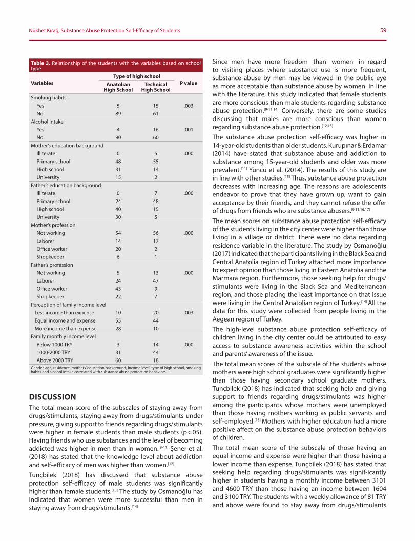
59Nükhet Kırağ, Substance Abuse Protection Self-Efficacy of Students
DISCUSSIONThe total mean score of the subscales of staying away from drugs/stimulants, staying away from drugs/stimulants under pressure, giving support to friends regarding drugs/stimulants were higher in female students than male students (p<.05). Having friends who use substances and the level of becoming addicted was higher in men than in women.[9-11] Şener et al. (2018) has stated that the knowledge level about addiction and self-efficacy of men was higher than women.[12]
Tunçbilek (2018) has discussed that substance abuse protection self-efficacy of male students was significantly higher than female students.[13] The study by Osmanoğlu has indicated that women were more successful than men in staying away from drugs/stimulants.[14]
Since men have more freedom than women in regard to visiting places where substance use is more frequent, substance abuse by men may be viewed in the public eye as more acceptable than substance abuse by women. In line with the literature, this study indicated that female students are more conscious than male students regarding substance abuse protection.[9-11,14] Conversely, there are some studies discussing that males are more conscious than women regarding substance abuse protection.[12,13]
The substance abuse protection self-efficacy was higher in 14-year-old students than older students. Kurupınar & Erdamar (2014) have stated that substance abuse and addiction to substance among 15-year-old students and older was more prevalent.[11] Yüncü et al. (2014). The results of this study are in line with other studies.[15] Thus, substance abuse protection decreases with increasing age. The reasons are adolescents endeavor to prove that they have grown up, want to gain acceptance by their friends, and they cannot refuse the offer of drugs from friends who are substance abusers.[9,11,16,17]
The mean scores on substance abuse protection self-efficacy of the students living in the city center were higher than those living in a village or district. There were no data regarding residence variable in the literature. The study by Osmanoğlu (2017) indicated that the participants living in the Black Sea and Central Anatolia region of Turkey attached more importance to expert opinion than those living in Eastern Anatolia and the Marmara region. Furthermore, those seeking help for drugs/ stimulants were living in the Black Sea and Mediterranean region, and those placing the least importance on that issue were living in the Central Anatolian region of Turkey.[14] All the data for this study were collected from people living in the Aegean region of Turkey.The high-level substance abuse protection self-efficacy of children living in the city center could be attributed to easy access to substance awareness activities within the school and parents’ awareness of the issue.The total mean scores of the subscale of the students whose mothers were high school graduates were significantly higher than those having secondary school graduate mothers. Tunçbilek (2018) has indicated that seeking help and giving support to friends regarding drugs/stimulants was higher among the participants whose mothers were unemployed than those having mothers working as public servants and self-employed.[13] Mothers with higher education had a more positive affect on the substance abuse protection behaviors of children.The total mean score of the subscale of those having an equal income and expense were higher than those having a lower income than expense. Tunçbilek (2018) has stated that seeking help regarding drugs/stimulants was signif-icantly higher in students having a monthly income between 3101 and 4600 TRY than those having an income between 1604 and 3100 TRY. The students with a weekly allowance of 81 TRY and above were found to stay away from drugs/stimulants
Table 3. Relationship of the students with the variables based on school type
VariablesType of high school
P valueAnatolian High School
Technical High School
Smoking habitsYes 5 15 .003No 89 61
Alcohol intakeYes 4 16 .001No 90 60
Mother’s education backgroundIlliterate 0 5 .000Primary school 48 55High school 31 14University 15 2
Father’s education backgroundIlliterate 0 7 .000Primary school 24 48High school 40 15University 30 5
Mother’s professionNot working 54 56 .000Laborer 14 17Office worker 20 2Shopkeeper 6 1
Father’s professionNot working 5 13 .000Laborer 24 47Office worker 43 9Shopkeeper 22 7
Perception of family income levelLess income than expense 10 20 .003Equal income and expense 55 44More income than expense 28 10
Family monthly income levelBelow 1000 TRY 3 14 .0001000-2000 TRY 31 44Above 2000 TRY 60 18
Gender, age, residence, mothers’ education background, income level, type of high school, smoking habits and alcohol intake correlated with substance abuse protection behaviors.
60 Journal of Contemporary Medicine
and give support to their friends regarding drugs/stimulants significantly more than those receiving an allowance between 21 and 40 TRY. Students with a weekly allowance of 81 TRY and above were found to seek help regarding drugs/stimulants significantly more than those receiving an allowance below 40 TRY. Fur-thermore, students with a weekly allowance between 61 and 80 TRY and above were found to seek help regard-ing drugs/stimulants significantly more than those receiving an allowance between 21 and 40 TRY.[13] Increase in the family income level increased the awareness of children about substance abuse protection.The total mean score of the subscale of the students enrolled in Anatolian High School were significantly higher than those of the students enrolled in the Technical High School. The study by Osmanoğlu (2017) investigating students’ avoidance of drugs/stimulants based on the school type indicated that the students enrolled in Science High Schools developed a more positive attitude towards substance abuse protection than any other type of high school.[14] Şener et al. (2018) discussed that the addiction knowledge score measured in Anatolian High School and vocational school for girls and the total score on self-efficacy were significantly higher than those in the industrial vocational high school.[12] The results obtained in this study show similarities with other studies. Substance abuse protection self-efficacy of students enrolled in the Technical High School is affected negatively. Students in the Technical High School form a high-risk group regarding substance abuse. The families’ socio-economic level, par-ents’ awareness regarding substance abuse, the amount and effectiveness of school activities on informing stu-dents about substance abuse have an important effect.[12] The number of students selected based on certain cri-teria or enrolled voluntarily in vocational high schools is limited. Students enrolled in these schools are individuals who have not considered continuing higher education, wanted to have an occupation in a short period of time or because their parents wanted them to register.[18] Students who do not obtain a sufficient grade to be enrolled in general high schools tend to enroll in these schools to be able to start their professional life as soon as possible. In brief, voca-tional high schools host students primarily with low income and education, from rural areas, and with crowded family structures.[19]
The total mean scores of substance abuse protection selfefficacy of the students who did not smoke were high-er than those who smoked. Similarly, the total mean scores of the students who did not use alcohol were higher than those who did. A higher substance abuse protection self-efficacy provides better protection against sub-stances which lead to addiction such as cigarettes and alcohol. Kılıç identified the number of those smoking eve-ryday as 23.6%; the biggest reason for starting to smoke was curiosity (28.4%) and the effect of friends (21.6%). A study conducted with 13,438 students indicated that 34.4% of students first experienced alcohol with their friends and 8.4% experienced it on their own. Among the reasons for alcohol intake were trial, curiosity,
sorrow, anger and fun.[20] The major reason for starting to use alcohol at puberty was either peer pressure or it shows they have grown up and become sophisticated. Some adolescents use substances to rebel against their parents or so-ciety. Most adults and adolescents use alcohol to feel high or smoke because they believe smoking helps to re-lieve tension.[21]
CONCLUSIONSubstance abuse protection activities should be increased in schools. Also, the students at risk and enrolled espe-cially in the Technical High School should be identified. It is suggested to conduct educational intervention re-search on substance abuse protection oriented at adolescents. Adolescents who are at risk for substance use should be identified and awareness training should be provided.
ETHICAL DECLARATIONSEthics Comittee Approval: Aydın Adnan Menderes University Nursing Department Ethics Committee of Non-Interventional Clinical Studies gave permission to conduct the study (2018/122). Informed Consent: Written informed consent was obtained from all participants who participated in this study. Status of Peer-review: Externally peer-reviewed. Conflict of Interest Statement: The authors have no conflicts of interest to declare. Financial Disclosure: The authors declared that this study has received no financial support. Author Contributions: All of the authors declare that they have all participated in the design, execution, and analysis of the paper, and that they have approved the final version.
REFERENCES1. Turkish Language Society glossary. Available from URL: http:// www.tdk.
gov.tr/.
2. Kaygısız M. Forensic Sciences. 5th ed. Ankara: Seçkin publications; 2005. p.6-10.
3. Öztürk O, Uluşahin A. Mental Health and Disorders. Ankara: Psychiatric Society Journal. 2015; 11:6-30.
4. Volkow N. D. M.D. Drugs, Brains, And Behavior The Science Of Addiction. National Instituted On Drug Abuse.[cited 2018 Oct 20]; 2(1). Available from URL: https://www.Drugabuse.Gov/ Sites/Default/Files/Soa_2014.Pdf.
5. Green Crescent (2017). Available from: http://www.yesilay.org.tr/tr/haberler/detay/2016-dunya-uyusturucu-raporu-aciklandi.
6. Eker F, Akkuş D, Kapısız Ö. Development and psychometric evaluation of substance dependence self-efficacy scale for adolescents. Journal of Psychiatric Nursing. 2013 4(1): 7-12.
7. Yardımcı F, Başbakkal Z. Validity and reliability study of secondary school self-efficacy scale. Journal of Anatolian Psychiatry. 2010;11:321-6.
8. Carpenter CM, Howard D. Development of a drug use resistance self-efficacy (DURSE) scale. Am J Health Behav. 2009;33:147-57.
9. Arslan H. N, Terzi Ö, Dabak Ş, Pekşen Y. Smoking, Alcohol and Substance Use in High School Students in. Erciyes Medical Journal. 2012;34(2): 79-84.
61Nükhet Kırağ, Substance Abuse Protection Self-Efficacy of Students
10. Boyacı H, Çorapçıoğlu A, Ilgazlı A, Başyiğit İ, Yıldız F. Evaluation of Smoking Habits of Kocaeli University Students. Journal of respiratory diseases. 2003; 14:169-175.
11. Erdamar G, Kurupınar A. Habit Addiction and Prevalence of Secondary School Students: The Case of Bartin Province. Journal of Social Sciences. 2014; 16(1): 65-84.
12. Şener D. K, Akkuş D. Factors affecting high school students' substance abuse behaviors. Addicta:The Turkish Journal on Addictions. 2018; 5(3):1-25.
13. Tunçbilek M. 15-18 yaş arası oğrencilerin madde bağımlılığından korunma oz yeterliliğinin araştırılması[Ph.D. thesis] University of Bartın; 2018; p. 25-9.
14. Osmanoğlu D. E. To determine the reasons of drinking and drug addiction self-efficacy behaviors of university students and their opinions on the causes of drinking. Atatürk University Kazım Karabekir Journal of Faculty of Education. 2017; 35(3):198-222.
15. Yüncü Z, Saatçıoğlu H, Aydın C, Özbaran N. B, Altıntoprak E, Kose S. (2014). The Legend of a City: Is the Age of Beginning to Use Dropping?.[cited 2018 Oct 15] Availablefrom:http://www. literatursempozyum.com:http://www.literatursempozyum. com/pdf/m_1420637421.pdf.
16. Akkuş D, Karaca A, Konuk Şener D, Ankaralı A. Smoking and alcohol use among high school students and factors affecting the frequency. Anadolu Clinic Journal. 2017;22(1):36–45.
17. Ünlü A, Evcin U. Evaluation of Drug Use Prevalence, Risk and Protection Factors Between 2008 and 2010 in. Anadolu University Journal of Social Sciences. 2014;14(3):127-140.
18. Donmez F, Azizoğlu N. Examination of the scientific process skill levels of students in vocational high schools: Balıkesir case. Necatibey Faculty of Education Journal of Electronic Science and Mathematics Education. 2010; 4(2):79–109.
19. Günbayı İ, Tokel A. Manager, teacher and student opinions about the effectiveness of vocational courses in technical and vocational high schools. Journal of Educational Research. 2014;3(4): 59–73.
20. Kılıç N. Adnan Menderes University Health School and Health Services Vocational School students' knowledge, attitude and behavior related to smoking. Journal of Health Sciences. 2006; 15(2): 87.
21. Essau C. A. Concluding remarks. In Substance Abuse and Dependence in Adolescence, Routledge.: 2014, pp. 241-256.

Corresponding (İletişim): Elif KARAHAN, PhD, RN, Assistant Professor, Department of Nursing, Faculty of Health Sciences, Zonguldak Bulent Ecevit University, TurkeyE-mail (E-posta): [email protected] (Geliş Tarihi): 02.10.2019 Accepted (Kabul Tarihi): 16.02.2020
DOI: 10.16899/jcm.628095J Contemp Med 2020;10(1):62-69
Orjinal Araştırma / Original Article
JOURNAL OF
CONTEMPORARY MEDICINEJournal ofContemporary MedicineYEAR: 2019 VOLUME: 9 ISSUE: 4
Incidence of Pressure Ulcers in The Patients on Mechanical Ventilation: A Prospective Study
Mekanik Ventilatördeki Hastalarda Basınç Yarası İnsidansı: Prospektif Bir Çalısma
Amaç: Bu çalışmanın amacı mekanik ventilatördeki hastalarda basınç yarası insidansını belirlemektir.
Gereç ve Yöntem: Araştırmanın tipi kesitsel tipte prospektiftir. Batı Karadeniz Bölgesinde yer alan bir Devlet Hastanesinin 5 yoğun bakım ünitesinde yürütülmüştür. Araştırmanın örneklemini mekanik ventilasyon desteği başlatılan 260 hasta oluşturmuştur. Braden Risk Değerlendirme Ölçek puanı ile hastalardaki basınç yarası oluşma riski mekanik ventilasyon desteği başladıktan sonra 10. saat, 1. hafta ve 2. hafta zaman dilimlerinde değerlendirilmiştir. İstatistiksel analizlerde sayı, yüzde, ortalama, t testi, Anova ve Pearson Korelasyon analizi kullanılmıştır.
Bulgular: Hastaların yaş ortalaması 54±10 yaş; %52,7’si erkek; %55,4’ünün Beden Kütle İndeksleri normal aralıktadır. Hastaların %46,5’inde serebrovasküler olay tanısı ile yatış yapıldığı; %52,3’ünün bilinç durumunun konfüze olduğu ve Braden Risk Değerlendirme Ölçek puanının 11,0±2,64 şeklinde yüksek risk grubunda olduğu belirlenmiştir. Kadın cinsiyette (t=5,95, p=0,01), bilinç durumu stupor olan hastalarda (F=97,56, p=0,00) ve motor yanıtı ağrıya lokalize olan hastalarda (F=9,54, p=0,00) basınç yarası riski anlamlı olarak daha yüksektir. Mekanik ventilasyon tedavisi başlatıldıktan sonraki 10. saatte basınç yarası insidansının %10,8; birinci haftada %58,5; ikinci haftada %71,15 olduğu görülmüştür. İkinci haftanın sonunda hastaların %30’unda sakrum, %22,7’sinde koksik bölgesinde Evre 1 basınç yarası gelişmiştir.
Sonuç: Mekanik ventilasyon tedavisi alan hastalarda ventilatörle ilişkili olarak basınç yarası gelişme insidansının oldukça yüksek olduğu sonucu ortaya çıkmıştır. Mekanik ventilatör ve diğer faktörlerin de etkisi göz önünde tutularak basınç yarasını önleme konusunda primer koruma önlemlerinin alınması oldukça önemlidir.
Anahtar Kelimeler: Mekanik ventilasyon, basınç yarası, yoğun bakım
Abstract Öz
Elif KARAHAN1, Sevim ÇELIK2, Özge UÇAR3
Aim: The aim of this study was to determine the incidence of pressure ulcers among the patients on mechanical ventilation.Material and Method: The type of the study was cross-sectional and prospective. It was carried out in 5 intensive care units of a state hospital located in Western Black Sea region of Turkey. The sample of the study included 260 patients who were initiated mechanical ventilatory support. Pressure ulcer risk in patients was assessed by Braden Risk Assessment Scale at 10th hour, 1st week and 2nd week timepoints following the initiation of mechanical ventilatory support.Results: Mean age of the patients was 54±10 years old; 52.7% were males; and 55.4% had a Body Mass Index within normal range. It was determined that 46.5% of the patients were hospitalized due to the diagnosis of a cerebrovascular event; the state of consciousness was confused in 52.3% and mean Braden Risk Assessment Scale was 11.0±2.64. Pressure ulcer risk was found to be significatly higher among the patients who were females (t=5.95, p=0.01), whose state of consciousness was stupor (F=97.56, p=0.00) and whose motor response was localized to pain (F=9.54, p=0.00). It was observed that the incidence of pressure ulcers was 10.8% at 10th hour, 58.5% at first week and 71.15% at second week from the start of mechanical ventilation treatment. At the end of second week, stage I pressure ulcer was developed on the sacrum in 30% and on coccyx area in 22.7% of the patients.Conclusion: It was found that the incidence of pressure ulcer development risk was significantly high among the patients who underwent mechanical ventilation treatment. Considering the effect of mechanical ventilator and other factors, primary protective measures were suggested to be very important in the prevention of pressure ulcers.
Keywords: Mechanical ventilation, pressure ulcer, intensive care
1Department of Nursing, Faculty of Health Sciences, Zonguldak Bulent Ecevit University, Turkey2Department of Nursing, Faculty of Health Sciences, Bartın University, Turkey
3Institute of Health Sciences, Zonguldak Bulent Ecevit University, Turkey
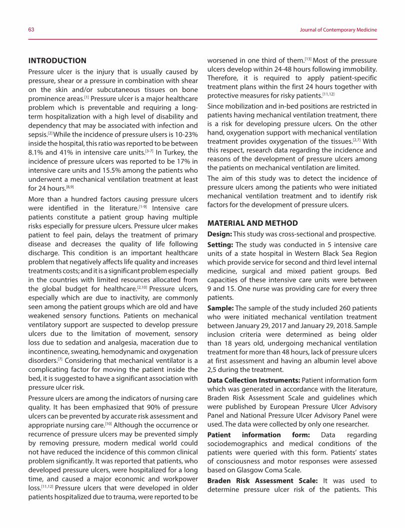
63 Journal of Contemporary Medicine
INTRODUCTIONPressure ulcer is the injury that is usually caused by pressure, shear or a pressure in combination with shear on the skin and/or subcutaneous tissues on bone prominence areas.[1] Pressure ulcer is a major healthcare problem which is preventable and requiring a long-term hospitalization with a high level of disability and dependency that may be associated with infection and sepsis.[2] While the incidence of pressure ulsers is 10-23% inside the hospital, this ratio was reported to be between 8.1% and 41% in intensive care units.[3-7] In Turkey, the incidence of pressure ulcers was reported to be 17% in intensive care units and 15.5% among the patients who underwent a mechanical ventilation treatment at least for 24 hours.[8,9]
More than a hundred factors causing pressure ulcers were identified in the literature.[1-9] Intensive care patients constitute a patient group having multiple risks especially for pressure ulcers. Pressure ulcer makes patient to feel pain, delays the treatment of primary disease and decreases the quality of life following discharge. This condition is an important healthcare problem that negatively affects life quality and increases treatments costs; and it is a significant problem especially in the countries with limited resources allocated from the global budget for healthcare.[2,10] Pressure ulcers, especially which are due to inactivity, are commonly seen among the patient groups which are old and have weakened sensory functions. Patients on mechanical ventilatory support are suspected to develop pressure ulcers due to the limitation of movement, sensory loss due to sedation and analgesia, maceration due to incontinence, sweating, hemodynamic and oxygenation disorders.[7] Considering that mechanical ventilator is a complicating factor for moving the patient inside the bed, it is suggested to have a significant association with pressure ulcer risk.Pressure ulcers are among the indicators of nursing care quality. It has been emphasized that 90% of pressure ulcers can be prevented by accurate risk assessment and appropriate nursing care.[10] Although the occurrence or recurrence of pressure ulcers may be prevented simply by removing pressure, modern medical world could not have reduced the incidence of this common clinical problem significantly. It was reported that patients, who developed pressure ulcers, were hospitalized for a long time, and caused a major economic and workpower loss.[11,12] Pressure ulcers that were developed in older patients hospitalized due to trauma, were reported to be
worsened in one third of them.[13] Most of the pressure ulcers develop within 24-48 hours following immobility. Therefore, it is required to apply patient-specific treatment plans within the first 24 hours together with protective measures for risky patients.[11,12]
Since mobilization and in-bed positions are restricted in patients having mechanical ventilation treatment, there is a risk for developing pressure ulcers. On the other hand, oxygenation support with mechanical ventilation treatment provides oxygenation of the tissues.[2,7] With this respect, research data regarding the incidence and reasons of the development of pressure ulcers among the patients on mechanical ventilation are limited.The aim of this study was to detect the incidence of pressure ulcers among the patients who were initiated mechanical ventilation treatment and to identify risk factors for the development of pressure ulcers.
MATERIAL AND METHODDesign: This study was cross-sectional and prospective. Setting: The study was conducted in 5 intensive care units of a state hospital in Western Black Sea Region which provide service for second and third level internal medicine, surgical and mixed patient groups. Bed capacities of these intensive care units were between 9 and 15. One nurse was providing care for every three patients.Sample: The sample of the study included 260 patients who were initiated mechanical ventilation treatment between January 29, 2017 and January 29, 2018. Sample inclusion criteria were determined as being older than 18 years old, undergoing mechanical ventilation treatment for more than 48 hours, lack of pressure ulcers at first assessment and having an albumin level above 2,5 during the treatment.Data Collection Instruments: Patient information form which was generated in accordance with the literature, Braden Risk Assessment Scale and guidelines which were published by European Pressure Ulcer Advisory Panel and National Pressure Ulcer Advisory Panel were used. The data were collected by only one researcher.Patient information form: Data regarding sociodemographics and medical conditions of the patients were queried with this form. Patients’ states of consciousness and motor responses were assessed based on Glasgow Coma Scale.Braden Risk Assessment Scale: It was used to determine pressure ulcer risk of the patients. This
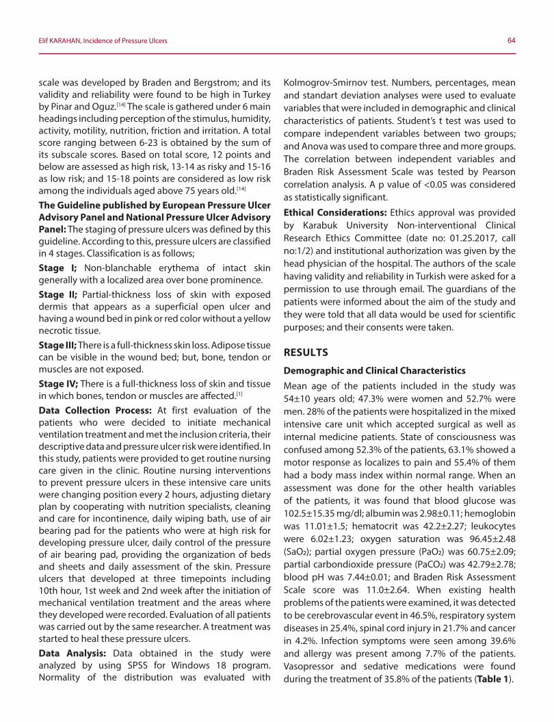
64Elif KARAHAN, Incidence of Pressure Ulcers
scale was developed by Braden and Bergstrom; and its validity and reliability were found to be high in Turkey by Pinar and Oguz.[14] The scale is gathered under 6 main headings including perception of the stimulus, humidity, activity, motility, nutrition, friction and irritation. A total score ranging between 6-23 is obtained by the sum of its subscale scores. Based on total score, 12 points and below are assessed as high risk, 13-14 as risky and 15-16 as low risk; and 15-18 points are considered as low risk among the individuals aged above 75 years old.[14]
The Guideline published by European Pressure Ulcer Advisory Panel and National Pressure Ulcer Advisory Panel: The staging of pressure ulcers was defined by this guideline. According to this, pressure ulcers are classified in 4 stages. Classification is as follows;Stage I; Non-blanchable erythema of intact skin generally with a localized area over bone prominence.Stage II; Partial-thickness loss of skin with exposed dermis that appears as a superficial open ulcer and having a wound bed in pink or red color without a yellow necrotic tissue.Stage III; There is a full-thickness skin loss. Adipose tissue can be visible in the wound bed; but, bone, tendon or muscles are not exposed.Stage IV; There is a full-thickness loss of skin and tissue in which bones, tendon or muscles are affected.[1]
Data Collection Process: At first evaluation of the patients who were decided to initiate mechanical ventilation treatment and met the inclusion criteria, their descriptive data and pressure ulcer risk were identified. In this study, patients were provided to get routine nursing care given in the clinic. Routine nursing interventions to prevent pressure ulcers in these intensive care units were changing position every 2 hours, adjusting dietary plan by cooperating with nutrition specialists, cleaning and care for incontinence, daily wiping bath, use of air bearing pad for the patients who were at high risk for developing pressure ulcer, daily control of the pressure of air bearing pad, providing the organization of beds and sheets and daily assessment of the skin. Pressure ulcers that developed at three timepoints including 10th hour, 1st week and 2nd week after the initiation of mechanical ventilation treatment and the areas where they developed were recorded. Evaluation of all patients was carried out by the same researcher. A treatment was started to heal these pressure ulcers.Data Analysis: Data obtained in the study were analyzed by using SPSS for Windows 18 program. Normality of the distribution was evaluated with
Kolmogrov-Smirnov test. Numbers, percentages, mean and standart deviation analyses were used to evaluate variables that were included in demographic and clinical characteristics of patients. Student’s t test was used to compare independent variables between two groups; and Anova was used to compare three and more groups. The correlation between independent variables and Braden Risk Assessment Scale was tested by Pearson correlation analysis. A p value of <0.05 was considered as statistically significant.
Ethical Considerations: Ethics approval was provided by Karabuk University Non-interventional Clinical Research Ethics Committee (date no: 01.25.2017, call no:1/2) and institutional authorization was given by the head physician of the hospital. The authors of the scale having validity and reliability in Turkish were asked for a permission to use through email. The guardians of the patients were informed about the aim of the study and they were told that all data would be used for scientific purposes; and their consents were taken.
RESULTS
Demographic and Clinical CharacteristicsMean age of the patients included in the study was 54±10 years old; 47.3% were women and 52.7% were men. 28% of the patients were hospitalized in the mixed intensive care unit which accepted surgical as well as internal medicine patients. State of consciousness was confused among 52.3% of the patients, 63.1% showed a motor response as localizes to pain and 55.4% of them had a body mass index within normal range. When an assessment was done for the other health variables of the patients, it was found that blood glucose was 102.5±15.35 mg/dl; albumin was 2.98±0.11; hemoglobin was 11.01±1.5; hematocrit was 42.2±2.27; leukocytes were 6.02±1.23; oxygen saturation was 96.45±2.48 (SaO2); partial oxygen pressure (PaO2) was 60.75±2.09; partial carbondioxide pressure (PaCO2) was 42.79±2.78; blood pH was 7.44±0.01; and Braden Risk Assessment Scale score was 11.0±2.64. When existing health problems of the patients were examined, it was detected to be cerebrovascular event in 46.5%, respiratory system diseases in 25.4%, spinal cord injury in 21.7% and cancer in 4.2%. Infection symptoms were seen among 39.6% and allergy was present among 7.7% of the patients. Vasopressor and sedative medications were found during the treatment of 35.8% of the patients (Table 1).
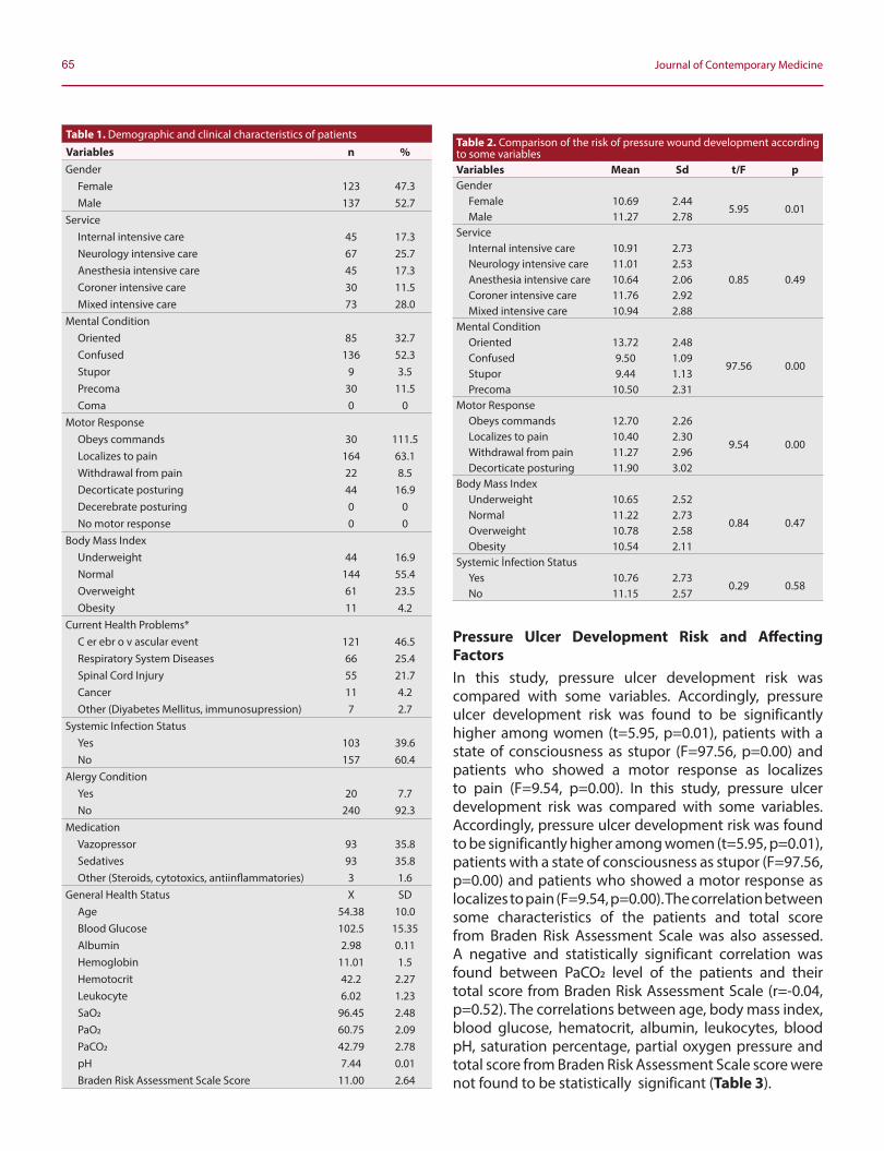
65 Journal of Contemporary Medicine
Pressure Ulcer Development Risk and Affecting FactorsIn this study, pressure ulcer development risk was compared with some variables. Accordingly, pressure ulcer development risk was found to be significantly higher among women (t=5.95, p=0.01), patients with a state of consciousness as stupor (F=97.56, p=0.00) and patients who showed a motor response as localizes to pain (F=9.54, p=0.00). In this study, pressure ulcer development risk was compared with some variables. Accordingly, pressure ulcer development risk was found to be significantly higher among women (t=5.95, p=0.01), patients with a state of consciousness as stupor (F=97.56, p=0.00) and patients who showed a motor response as localizes to pain (F=9.54, p=0.00). The correlation between some characteristics of the patients and total score from Braden Risk Assessment Scale was also assessed. A negative and statistically significant correlation was found between PaCO2 level of the patients and their total score from Braden Risk Assessment Scale (r=-0.04, p=0.52). The correlations between age, body mass index, blood glucose, hematocrit, albumin, leukocytes, blood pH, saturation percentage, partial oxygen pressure and total score from Braden Risk Assessment Scale score were not found to be statistically significant (Table 3).
Table 2. Comparison of the risk of pressure wound development according to some variablesVariables Mean Sd t/F pGender
Female 10.69 2.445.95 0.01
Male 11.27 2.78Service
Internal intensive care 10.91 2.73
0.85 0.49Neurology intensive care 11.01 2.53Anesthesia intensive care 10.64 2.06Coroner intensive care 11.76 2.92Mixed intensive care 10.94 2.88
Mental ConditionOriented 13.72 2.48
97.56 0.00Confused 9.50 1.09Stupor 9.44 1.13Precoma 10.50 2.31
Motor ResponseObeys commands 12.70 2.26
9.54 0.00Localizes to pain 10.40 2.30Withdrawal from pain 11.27 2.96Decorticate posturing 11.90 3.02
Body Mass IndexUnderweight 10.65 2.52
0.84 0.47Normal 11.22 2.73Overweight 10.78 2.58Obesity 10.54 2.11
Systemic İnfection StatusYes 10.76 2.73
0.29 0.58No 11.15 2.57
Table 1. Demographic and clinical characteristics of patients
Variables n %Gender
Female 123 47.3Male 137 52.7
ServiceInternal intensive care 45 17.3Neurology intensive care 67 25.7Anesthesia intensive care 45 17.3Coroner intensive care 30 11.5Mixed intensive care 73 28.0
Mental ConditionOriented 85 32.7Confused 136 52.3Stupor 9 3.5Precoma 30 11.5Coma 0 0
Motor ResponseObeys commands 30 111.5Localizes to pain 164 63.1Withdrawal from pain 22 8.5Decorticate posturing 44 16.9Decerebrate posturing 0 0No motor response 0 0
Body Mass IndexUnderweight 44 16.9Normal 144 55.4Overweight 61 23.5Obesity 11 4.2
Current Health Problems*C er ebr o v ascular event 121 46.5Respiratory System Diseases 66 25.4Spinal Cord Injury 55 21.7Cancer 11 4.2Other (Diyabetes Mellitus, immunosupression) 7 2.7
Systemic Infection StatusYes 103 39.6No 157 60.4
Alergy ConditionYes 20 7.7No 240 92.3
MedicationVazopressor 93 35.8Sedatives 93 35.8Other (Steroids, cytotoxics, antiinflammatories) 3 1.6
General Health Status X SDAge 54.38 10.0Blood Glucose 102.5 15.35Albumin 2.98 0.11Hemoglobin 11.01 1.5Hemotocrit 42.2 2.27Leukocyte 6.02 1.23SaO2 96.45 2.48PaO2 60.75 2.09PaCO2 42.79 2.78pH 7.44 0.01Braden Risk Assessment Scale Score 11.00 2.64
66Elif KARAHAN, Incidence of Pressure Ulcers
State of Developing Pressure UlcersDuring evaluation at 10th hour following the initiation of mechanical ventilation treatment, it was found that there was no impairment in the skin integrity among 89.2% of the patients (n=232), and Stage I pressure ulcer was developed on sacrum and right heel in 3.5% (n=9), on coccyx in 2.3% (n=6) and on left heel in 1.5% (n=4) (Graphic 1). The incidence of Stage I pressure ulcer was found to be 10.8% at 10 hours.
During evaluation at first week after the initiation of mechanical ventilation treatment, it was found that there was no impairment in the skin integrity among 41.5% of the patients (n=108), and Stage I pressure ulcer was developed on sacrum in 23.1% (n=60), on coccyx in 19.2% (n=50) and on the nape, iliac bone, ear, ankle and heel regions at varying rates between 0.4% (n=1) and 6.9% (n=18) (Graphic 2). The incidence of Stage I pressure ulcer at first week was found to be 58.5%.
During evaluation at second week after the initiation of mechanical ventilation treatment, it was found that there was no impairment in the skin integrity among 28.1% of the patients (n=73); but, Stage I pressure ulcer was developed on sacrum in 30% (n=78), on coccyx in 22.7% (n=59) and on the nape, iliac bone, ear, ankle and heel regions at varying rates between 0.4% (n=1) and 7.7% (n=20) (Graphic 3). The incidence of Stage I pressure ulcer at second week was found to be 71.15%. A transition to Stage II pressure ulcer was observed only in two patients who had Stage I pressure ulcer on the sacrum. The incidence of Stage II pressure ulcer was identified to be 0.76% at second week.
State of Developing Pressure UlcersDuring evaluation at 10th hour following the initiation of mechanical ventilation treatment, it was found that there was no impairment in the skin integrity among 89.2% of the patients (n=232), and Stage I pressure ulcer was developed on sacrum and right heel in 3.5% (n=9), on coccyx in 2.3% (n=6) and on left heel in 1.5% (n=4) (Graphic 1). The incidence of Stage I pressure ulcer was found to be 10.8% at 10 hours.During evaluation at first week after the initiation of mechanical ventilation treatment, it was found that there was no impairment in the skin integrity among 41.5% of the patients (n=108), and Stage I pressure ulcer was developed on sacrum in 23.1% (n=60), on coccyx in 19.2% (n=50) and on the nape, iliac bone, ear, ankle and heel regions at varying rates between 0.4% (n=1) and 6.9% (n=18) (Graphic 2). The incidence of Stage I pressure ulcer at first week was found to be 58.5%.During evaluation at second week after the initiation of mechanical ventilation treatment, it was found that there was no impairment in the skin integrity among 28.1% of the patients (n=73); but, Stage I pressure ulcer was developed on sacrum in 30% (n=78), on coccyx in 22.7% (n=59) and on the nape, iliac bone, ear, ankle and heel regions at varying rates between 0.4% (n=1) and 7.7% (n=20) (Graphic 3). The incidence of Stage I pressure ulcer at second week was found to be 71.15%.
Graphic 3. Developed a pressure ulcers regions in the second week
Table 3. Relationship between some variables and braden risk assessment Variables X Sd r pAge 54.38 10,0 0.07 0.24BMI 22.94 3.59 -0.05 0.41Blood Glucose 102.5 15.35 0.05 0.38Albumin 2.98 0.11 -0.06 0.27Hemoglobin 11.01 1.5 0.00 0.91Hemotocrit 42.2 2.27 0.05 0.38Leukocyte 6.02 1.23 0.02 0.68SaO2 96.45 2.48 -0.04 0.43PaO2 60.75 2.09 -0.04 0.48PaCO2 42.79 2.78 -0.23 0.00pH 7.44 0.01 -0.04 0.52
Graphic 1. Developed a pressure ulcers regions at 10th hour
Graphic 2. Developed a pressure ulcers regions in the first week
67 Journal of Contemporary Medicine
A transition to Stage II pressure ulcer was observed only in two patients who had Stage I pressure ulcer on the sacrum. The incidence of Stage II pressure ulcer was identified to be 0.76% at second week.
DISCUSSIONIn this study, the incidence of pressure ulcer development and the areas where they developed were evaluated at three different timepoints among the patients who were hospitalized in intensive care units and began to undergo mechanical ventilation treatment. Mechanical ventilation treatment has been reported to be a factor causing pressure ulcers due to its impairing effect on blood circulation at epidermal and subcutaneous tissue and on the balance between ventilation-perfusion by affecting pumping function of the heart.[9,11] In a study which was conducted in six centers, it was indicated that patients who were on mechanical ventilation treatment were at a higher risk compared to the others receiving inpatient treatment.[3] In a study which was carried out in surgical intensive care units, development of pressure ulcers was significantly enhanced by the increase in the days of ventilation, dialysis and gaita incontinence; and besides, the number of days for using norepinephrine and lactic acid level in arterial blood were determined to create a high risk for pressure ulcers.[11] At the end of this current study, majority of the patients, who were accepted to intensive care, were found to be hospitalized due to the diagnoses of cerebrovascular event, respiratory system diseases and spinal cord injury. In addition to the disturbance in respirationcirculation and sensory-motor functions by these diseases, pressure ulcer risk was found to be increased by the initiation of mechanical ventilation treatment. Advanced age has been known to be a factor in the development of pressure ulcers due to its outcomes such as decrease in skin elasticity, chronic diseases and sensory defects. However, various results were found about the relationship between age and development of pressure ulcers in the previous studies.[4,13] The mean age of the patients included in this study showed that they were not included in advanced age group; but, development of pressure ulcers was enhanced with the extension of the duration of mechanical ventilation treatment.In the literature, the studies investigating the possibility that age may be a factor for the development of pressure ulcers, showed difference results.[15,16] Pressure ulcer risk was found to be significantly higher among women in this study. This situation may be explained by
some basic physiological differences between women and men which may be associated with pressure ulcer development.[15] Due to the facts that women have a larger pelvis, two times more adipose tissue, less percentage of muscle mass, and weaker muscle tone and strength, the risk for the development of pressure ulcers may be increased. Some risk factors have been described for the development of pressure ulcers including extrinsic factors such as pressure, friction, shear and moist, and intrinsic factors such as limited physical activity and mobilization, use of anesthetic, sedative and neuromuscular blocking agents, reduction in sensual perception and circulatory and ventilatory disorders.[1,2] Mechanical ventilation treatment may make adverse effects of extrinsic as well as intrinsic factors more negative. It has been reported that stage I pressure ulcers occur within an average of 1-10 days in the intensive care units.[4,8] In the previous studies, it was shown that pressure ulcers developed significantly at second week of mechanical ventilation treatment and mechanical ventilation treatment lasting longer than 20 days might be associated with pressure ulcers.[3,9] At the end of this current study, the incidence of stage I pressure ulcer was found to be 10.8% at 10 hours after the initiation of mechanical ventilation treatment and the incidences were found to be 58.5% at first week and 71.15% at second week as parallel to the data in the literature. This situation confirmed that pressure ulcer development rates increased with the extension of the duration of mechanical ventilation treatment.SaO2, PaO2 and PaCO2 values of the patients are among the parameters showing the efficiency of mechanical ventilation treatment. However, it may be sometimes difficult to maintain these values within the desired range due to several reasons such as respiratory and circulatory diseases, acid-base imbalances and care interventions. In the study by Karayurt et al., pressure ulcers were found to be developed significantly with the decreases in PaO2 and SaO2 levels and increase in PaCO2 levels.[9] In another study evaluating trauma patients, a significant relationship was revealed between decreasing hemoglobin level and O2 saturation and the development of pressure ulcer.[13] The negative correlation between pressure ulcer development risk and PaCO2 in this study reminded the necessity for following up this condition together with other variables.In the national and international studies, the areas where pressure ulcers developed among the patients treated in intensive care were reported to be mostly sacrum, coccyx, trochanter and heels.[7,17,18] Since patients undergoing
68Elif KARAHAN, Incidence of Pressure Ulcers
mechanical ventilation treatment often stay in supine position, pressure is increased on these areas. In this study, stage I pressure ulcers were found to develop on sacrum and coccyx at a ratio of 2-3% at 10 hours after the initiation of mechanical ventilation treatment, and these ratios increased up to 20s% at first week and 30% at second week. Development of pressure ulcer represents the inadequacy in nursing care quality and development of stage III or IV pressure ulcer is considered as a “never intolerable” event.[10] In a study that was performed in India, a high incidence was reported for stage 3 pressure ulcers on sacrum and heel[19]; and in a study performed in Portugal, a high prevalence of stage 1 pressure ulcer was reported on sacrum and coccyx.[20]
In this study, it was observed that stage 1 pressure ulcers that were seen at second week of mechanical ventilation treatment showed a bad course towards stage 2. This situation reminded that incidences of stage 1 and 2 pressure ulcers might be increased among the intensive care patients requiring long term treatment.The results of this study showed that the incidence of pressure ulcers increased with the extension of the duration of treatment among the patients who underwent mechanical ventilation treatment; and therefore, it is absolutely necessary to practice additional interventions for the prevention of pressure ulcers in this patient group. Knowing the risk of pressure ulcer development and the areas where they mostly develop will be guiding for the measures to be taken. The frequent use of supine position as a result of mechanical ventilation treatment will increase the risk on coccyx and sacrum regions.[7] According to the results of a systematic review including the studies performed in Turkey, it was revealed that nurses performed a limited number of interventions to prevent pressure ulcers, clinical decisions were insufficient and the follow ups were not conducted.[10] Yet, nurses can use their independent roles more efficiently and they can contribute to the reduction of hospitalization time, increase in the life quality of the patient and avoidance of economic losses.
CONCLUSIONSIt was observed that the development of pressure ulcers enhanced with the extension of the duration of treatment among the patients who were initiated mechanical ventilation treatment. Besides, coccyx and sacrum were found to be the regions where pressure ulcers mostly develop. An attention must paid on the care of these regions in order to prevent the formation
of pressure ulcers. Based on this, it is highly important for the intensive care nurses to be aware of the high incidence of pressure ulcers among the patients who undergo mechanical ventilation treatment and to take necessary precautions. Since the risk of developing pressure ulcer is higher especially among women and patients having a worsening state of consciousness, additional precautions must be taken for this patient gorup. Considering that the incidence of Stage 1 pressure ulcers will highly increase at second week of mechanical ventilation and there will be transitions to Stage 2, interventions for speeding up recovery process of the patients should be increased. Inservice trainings should be given regularly and evaluations should be performed to prevent pressure ulcers. It can be suggested to perform long term and prospective incidence studies on the intensive care patients who undergo mechanical ventilation treatment.
ETHICAL DECLARATIONSEthics Comittee Approval: The study was carried out with the permission of Karabuk University Non-interventional Clinical Research Ethics Committee (date no: 01.25.2017, call no:1/2) and institutional authorization was given by the head physician of the hospital. Informed Consent: Written informed consent was obtained from all participants who participated in this study. Status of Peer-review: Externally peer-reviewed. Conflict of Interest Statement: The authors have no conflicts of interest to declare. Financial Disclosure: The authors declared that this study has received no financial support. Author Contributions: All of the authors declare that they have all participated in the design, execution, and analysis of the paper, and that they have approved the final version.
REFERENCES1. National Pressure Ulcer Advisory Panel, Europan Pressure Ulcer
Advisory Panel and Pan Pacific Pressure Injury Alliance. (2014). Prevention and treatment of pressure ulcers: quick reference guide. (Publication No: 978-0-9579343-6-8) Cambridge Media: Perth, Australia. Emily Haesler (Ed.)..[updated 2014; cited 18 October 2019] Available from: https://www.npuap.org/wp-content/uploads/2014/08/Updated-10-16-14-Quick-ReferenceGuide-DIGITAL-NPUAP-EPUAP-PPPIA-16Oct2014.pdf
2. National Institute for Health and Care Excellence. (2014). Pressure ulcers: Prevention and management, Clinical guideline. (Publication No: CG179)..[updated 2014, cited 18 October, 2019] Available from: nice.org.uk/guidance/cg179
69 Journal of Contemporary Medicine
3. Apostolopoulou E, Tselebis A, Terzis K, Kamarinou E, Lambropoulos I, Kalliakmanis A. Pressure ulcer incidence and risk factors in ventilated intensive care patients. Health Science Journal 2014;8 (3):333-342
4. Borghardt AT, Prado TN, Bicudo SDS, Castro DS, Bringuente MEO. Pressure ulcers in critically ill patients: incidence and associated factors. Rev Bras Enferm 2016;69(3):431-8.
5. Cooper KL. Evidence-based prevention of pressure ulcers ın the ıntensive care unit. Crit Care Nurse 2013;33(6):57-67.
6. González-Méndez MI, Lima-Serrano M, Martín-Castaño C, Alonso-Araujo I,Lima-Rodríguez JS. Incidence and risk factors associated with the development of pressure ulcers in an intensive care unit. J Clin Nurs 2018;27(5-6):1028-1037
7. Nijs N, Toppets A, Defloor T, Bernaerts K, Milisen K, Van Den Berghe G.Incidence and risk factors for pressure ulcers in the intensive care unit. J Clin Nurs 2009; 18(9):1258-66
8. Özyürek P, Yavuz Van Giersbergen M, Yildiz Ö. Investigation of the risk factors of pressure ulcers in intensive care unit patient according to the Braden Scale, East J Med 2016; 21(1):1-9.
9. Karayurt Ö, Akyol Ö, Kılıçaslan N, Akgün N, Sargın Ü, Kondakçı M, et al. The incidence of pressure ulcer in patients on mechanical ventilation and effects of selected risk factors on pressure ulcer development. Turk J Med Sci 2016; 46(5):1314-1322.
10. Çınar F, Kula Şahin Ş, Eti Aslan F. Evaluation of studies in Turkey on the prevention of pressure sores ın the ıntensive care unit: a systematic review. Balıkesir Health Sciences Journal 2018; 7(1): 42-50.
11. Wang XR, Han BR. Logistic regression analysis and nursing interventions for high-risk factors for pressure sores in patients in a surgical intensive care unit. Chin Nurs Res 2015; 2(2-3): 5154.
12. Mitchell A. Adult pressure area care: preventing pressure ulcers. Br J Nurs 2018; 4;27(18):1050-1052.
13. Alderden J, Zhao YL, Zhang Y, Thomas D, Butcher R, Zhang Y, et al. Outcomes associated with stage 1 pressure ınjuries: a retrospective cohort study. Am J Crit Care 2018 Nov;27(6):471476.
14. Pınar R, Oğuz S. Norton ve Braden Bası Yarası değerlendirme olçeklerinin yatağa bağımlı aynı hasta grubunda güvenirlik ve geçerliğinin sınanması: Uluslar arası katılımlı VI. Ulusal Hemşirelik Kongresi, Kongre Kitabı, Ankara; 1998. p.172-5.
15. Latifa K, Sondess S, Hajer G, Manel BH, Souhir K, Nadia B, et al. Evaluation of physiological risk factors, oxidant-antioxidant imbalance, proteolytic and genetic variations of matrix metalloproteinase-9 in patients with pressure ulcer. Sci Rep 2016; 11(6):29371.
16. He M, Tang A, Ge X, Zheng J. Pressure ulcers in the ıntensive care unit: an analysis of skin barrier risk factors. Adv Skin Wound Care. 2016;29(11):493-498.
17. Kıraner E, Terzi B, Uzun Ekinci A, Tunalı B. Yoğun bakım ünitemizdeki basınç yarası insidansı ve risk faktorlerinin belirlenmesi. Yoğun Bakım Hemşireliği Dergisi. 2016; 20(2): 7883.
18. Loudet CI, Marchena MC, Maradeo MR, Fernández SL, Romero MV, Valenzuela GE, et al. Reducing pressure ulcers in patients with prolonged acute mechanical ventilation: A quasi-experimental study. Rev Bras Ter Intensiva. 2017; 29(1):39-46.
19. Mehta C, George JV, Mehta Y, Wangmo N. Pressure ulcer and patient characteristics--A point prevalence study in a tertiary hospital of India based on the European Pressure Ulcer Advisory Panel minimum data set. J Tissue Viability. 2015;24(3):123-30.
20. Garcez Sardo PM, Simões CS, Alvarelhão JJ, de Oliveira e Costa CT, Simões CJ, Figueira JM, et al. Analyses of pressure ulcer point prevalence at the first skin assessment in a Portuguese Hospital. J Tissue Viability. 2016;25(2):75-82.
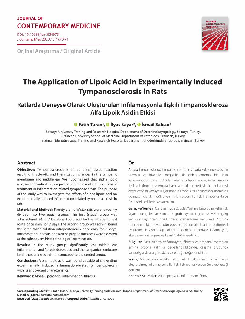
Corresponding (İletişim): Fatih Turan, Sakarya University Traning and Research Hospital Department of Otorhinolaryngology, Sakarya, TurkeyE-mail (E-posta): [email protected] (Geliş Tarihi): 20.10.2019 Accepted (Kabul Tarihi): 01.03.2020
DOI: 10.16899/jcm.634978J Contemp Med 2020;10(1):70-74
Orjinal Araştırma / Original Article
JOURNAL OF
CONTEMPORARY MEDICINEJournal ofContemporary MedicineYEAR: 2019 VOLUME: 9 ISSUE: 4
The Application of Lipoic Acid in Experimentally Induced Tympanosclerosis in Rats
Ratlarda Deneyse Olarak Olusturulan İnfilamasyonla İliskili Timpanoskleroza Alfa Lipoik Asidin Etkisi
Amaç: Timpanoskleroz timpanik membran ve orta kulak mukozasının
sklerotik ve hiyalinize değişikliği ile giden anormal bir doku
reaksiyonudur. Bir antioksidan olan alfa lipoik asidin, inflamasyonla
ile ilişkili timpanosklerozda basit ve etkili bir tedavi biçimini temsil
edebileceğini varsaydık. Çalışmanın amacı, alfa lipoik asidin sıçanlarda
deneysel olarak indüklenen infilamasyon ile ilşkili timpanoskleroz
üzerindeki etkilerini araştırmaktı.
Gereç ve Yöntem:Çalışmamızda 20 adet Wistar albino sıçan kullanıldı.
Sıçanlar rastgele olarak onarlı iki gruba ayrıldı. 1. gruba ALA 50 mg/kg
yedi gün boyunca günde bir defa intraperitoneal uygulandı. 2. gruba
salin aynı miktarda yedi gün boyunca günde bir defa intraperitone al
uygulandı. Histopatolojik olarak değerlendirmemizde inflamasyon,
fibrozis ve lamina propria kalınlığı değerlendirildi.
Bulgular: Orta kulakta enflamasyon, fibrozis ve timpanik membran
lamina propria kalınlığı değerlendirildiğinde, çalışma grubunda
kontrol gurubuna göre daha az olduğu değerlendirildi.
Sonuç: Antiokstidan özellik gösteren alfa lipoik asit’in deneysel olarak
oluşturulmuş inflamasyonla ile ilişkili timpanosklerozu önleyebileceği
görüldü.
Anahtar Kelimeler: Alfa-Lipoik asit, inflamasyon, fibroz
Abstract Öz
Fatih Turan1, Ilyas Sayar2, Ismail Salcan3
Objectives: Tympanosclerosis is an abnormal tissue reaction resulting in sclerotic and hyalinization changes in the tympanic membrane and middle ear. We hypothesized that alpha lipoic acid, an antioxidant, may represent a simple and effective form of treatment in inflammation-related tympanosclerosis. The purpose of the study was to investigate the effects of alpha lipoic acid on experimentally induced inflammation-related tympanosclerosis in rats.
Material and Method: Twenty albino Wistar rats were randomly divided into two equal groups. The first (study) group was administered 50 mg/ kg alpha lipoic acid by the intraperitoneal route once daily for 7 days. The second group was administered the same saline solution intraperitoneally once daily for 7 days. Inflammation, fibrosis and lamina propria thickness were assessed at the subsequent histopathological examination.
Results: In the study group, significantly less middle ear inflammation and fibrosis developed and the tympanic membrane lamina propria was thinner compared to the control group.
Conclusions: Alpha lipoic acid was found capable of preventing experimentally induced inflammation-related tympanosclerosis with its antioxidant characteristics.
Keywords: Alpha-Lipoic acid; inflammation; fibrosis.
1Sakarya University Traning and Research Hospital Department of Otorhinolaryngology, Sakarya, Turkey.2Erzincan University School of Medicine Department of Pathology, Erzincan, Turkey
3Erzincan Mengücekgazi Traning and Research Hospital Department of Otorhinolaryngology, Erzincan, Turkey
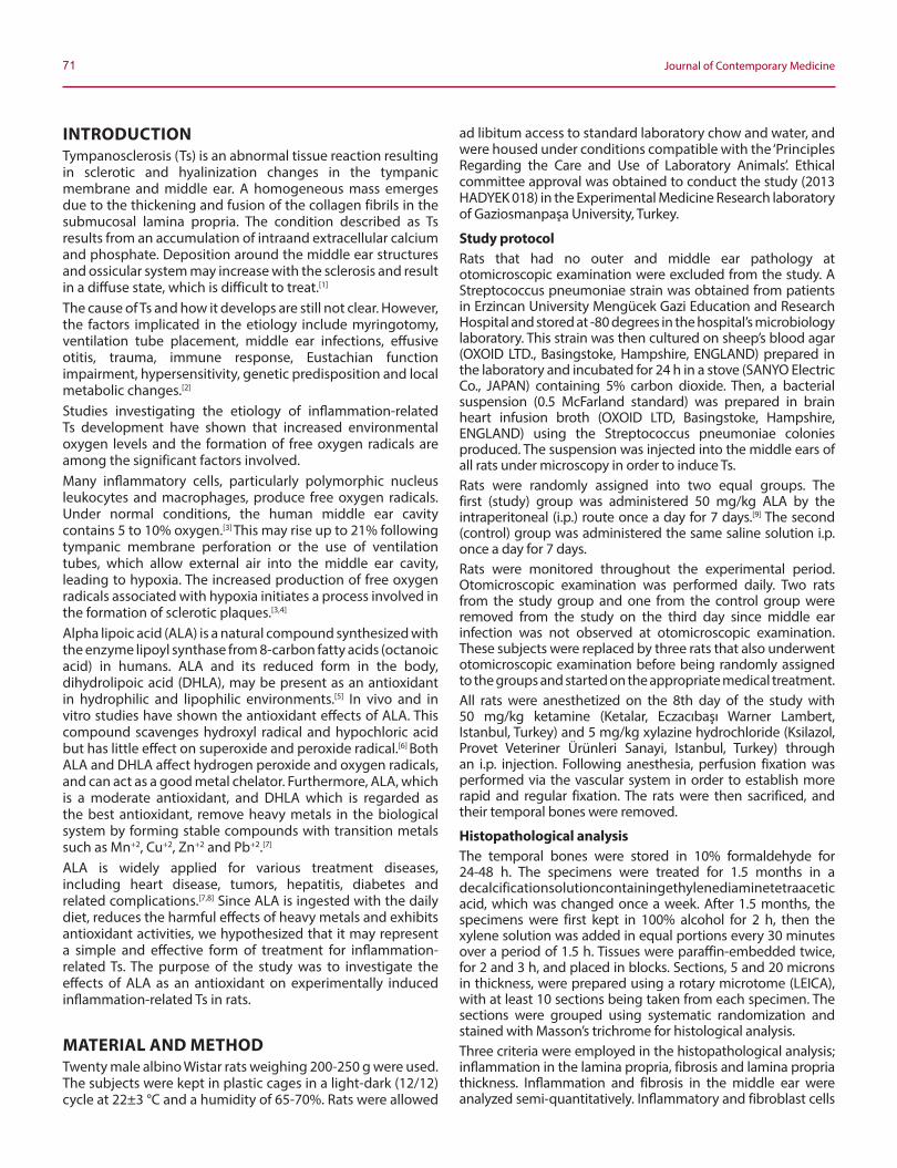
71 Journal of Contemporary Medicine
INTRODUCTIONTympanosclerosis (Ts) is an abnormal tissue reaction resulting in sclerotic and hyalinization changes in the tympanic membrane and middle ear. A homogeneous mass emerges due to the thickening and fusion of the collagen fibrils in the submucosal lamina propria. The condition described as Ts results from an accumulation of intraand extracellular calcium and phosphate. Deposition around the middle ear structures and ossicular system may increase with the sclerosis and result in a diffuse state, which is difficult to treat.[1]
The cause of Ts and how it develops are still not clear. However, the factors implicated in the etiology include myringotomy, ventilation tube placement, middle ear infections, effusive otitis, trauma, immune response, Eustachian function impairment, hypersensitivity, genetic predisposition and local metabolic changes.[2]
Studies investigating the etiology of inflammation-related Ts development have shown that increased environmental oxygen levels and the formation of free oxygen radicals are among the significant factors involved.Many inflammatory cells, particularly polymorphic nucleus leukocytes and macrophages, produce free oxygen radicals. Under normal conditions, the human middle ear cavity contains 5 to 10% oxygen.[3] This may rise up to 21% following tympanic membrane perforation or the use of ventilation tubes, which allow external air into the middle ear cavity, leading to hypoxia. The increased production of free oxygen radicals associated with hypoxia initiates a process involved in the formation of sclerotic plaques.[3,4]
Alpha lipoic acid (ALA) is a natural compound synthesized with the enzyme lipoyl synthase from 8-carbon fatty acids (octanoic acid) in humans. ALA and its reduced form in the body, dihydrolipoic acid (DHLA), may be present as an antioxidant in hydrophilic and lipophilic environments.[5] In vivo and in vitro studies have shown the antioxidant effects of ALA. This compound scavenges hydroxyl radical and hypochloric acid but has little effect on superoxide and peroxide radical.[6] Both ALA and DHLA affect hydrogen peroxide and oxygen radicals, and can act as a good metal chelator. Furthermore, ALA, which is a moderate antioxidant, and DHLA which is regarded as the best antioxidant, remove heavy metals in the biological system by forming stable compounds with transition metals such as Mn+2, Cu+2, Zn+2 and Pb+2.[7]
ALA is widely applied for various treatment diseases, including heart disease, tumors, hepatitis, diabetes and related complications.[7,8] Since ALA is ingested with the daily diet, reduces the harmful effects of heavy metals and exhibits antioxidant activities, we hypothesized that it may represent a simple and effective form of treatment for inflammation-related Ts. The purpose of the study was to investigate the effects of ALA as an antioxidant on experimentally induced inflammation-related Ts in rats.
MATERIAL AND METHODTwenty male albino Wistar rats weighing 200-250 g were used. The subjects were kept in plastic cages in a light-dark (12/12) cycle at 22±3 °C and a humidity of 65-70%. Rats were allowed
ad libitum access to standard laboratory chow and water, and were housed under conditions compatible with the ‘Principles Regarding the Care and Use of Laboratory Animals’. Ethical committee approval was obtained to conduct the study (2013 HADYEK 018) in the Experimental Medicine Research laboratory of Gaziosmanpaşa University, Turkey.
Study protocolRats that had no outer and middle ear pathology at otomicroscopic examination were excluded from the study. A Streptococcus pneumoniae strain was obtained from patients in Erzincan University Mengücek Gazi Education and Research Hospital and stored at -80 degrees in the hospital’s microbiology laboratory. This strain was then cultured on sheep’s blood agar (OXOID LTD., Basingstoke, Hampshire, ENGLAND) prepared in the laboratory and incubated for 24 h in a stove (SANYO Electric Co., JAPAN) containing 5% carbon dioxide. Then, a bacterial suspension (0.5 McFarland standard) was prepared in brain heart infusion broth (OXOID LTD, Basingstoke, Hampshire, ENGLAND) using the Streptococcus pneumoniae colonies produced. The suspension was injected into the middle ears of all rats under microscopy in order to induce Ts.Rats were randomly assigned into two equal groups. The first (study) group was administered 50 mg/kg ALA by the intraperitoneal (i.p.) route once a day for 7 days.[9] The second (control) group was administered the same saline solution i.p. once a day for 7 days.Rats were monitored throughout the experimental period. Otomicroscopic examination was performed daily. Two rats from the study group and one from the control group were removed from the study on the third day since middle ear infection was not observed at otomicroscopic examination. These subjects were replaced by three rats that also underwent otomicroscopic examination before being randomly assigned to the groups and started on the appropriate medical treatment.All rats were anesthetized on the 8th day of the study with 50 mg/kg ketamine (Ketalar, Eczacıbaşı Warner Lambert, Istanbul, Turkey) and 5 mg/kg xylazine hydrochloride (Ksilazol, Provet Veteriner Ürünleri Sanayi, Istanbul, Turkey) through an i.p. injection. Following anesthesia, perfusion fixation was performed via the vascular system in order to establish more rapid and regular fixation. The rats were then sacrificed, and their temporal bones were removed.
Histopathological analysisThe temporal bones were stored in 10% formaldehyde for 24-48 h. The specimens were treated for 1.5 months in a decalcificationsolutioncontainingethylenediaminetetraacetic acid, which was changed once a week. After 1.5 months, the specimens were first kept in 100% alcohol for 2 h, then the xylene solution was added in equal portions every 30 minutes over a period of 1.5 h. Tissues were paraffin-embedded twice, for 2 and 3 h, and placed in blocks. Sections, 5 and 20 microns in thickness, were prepared using a rotary microtome (LEICA), with at least 10 sections being taken from each specimen. The sections were grouped using systematic randomization and stained with Masson’s trichrome for histological analysis.Three criteria were employed in the histopathological analysis; inflammation in the lamina propria, fibrosis and lamina propria thickness. Inflammation and fibrosis in the middle ear were analyzed semi-quantitatively. Inflammatory and fibroblast cells
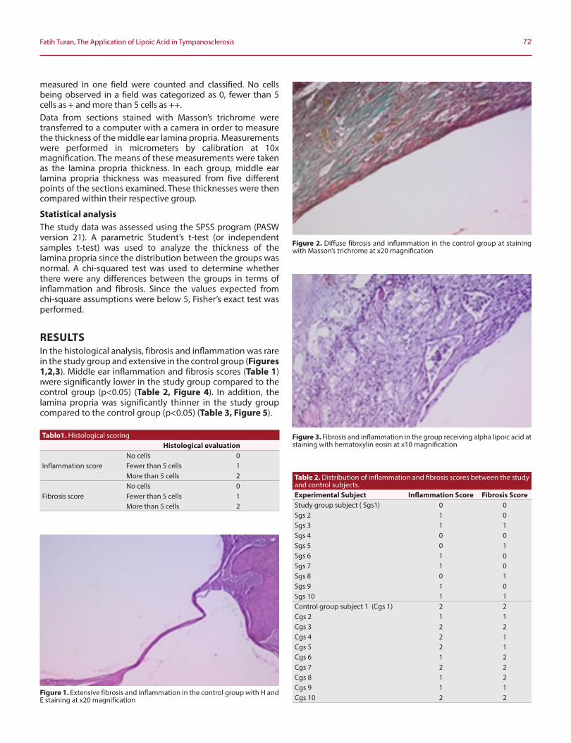
72Fatih Turan, The Application of Lipoic Acid in Tympanosclerosis
measured in one field were counted and classified. No cells being observed in a field was categorized as 0, fewer than 5 cells as + and more than 5 cells as ++.Data from sections stained with Masson’s trichrome were transferred to a computer with a camera in order to measure the thickness of the middle ear lamina propria. Measurements were performed in micrometers by calibration at 10x magnification. The means of these measurements were taken as the lamina propria thickness. In each group, middle ear lamina propria thickness was measured from five different points of the sections examined. These thicknesses were then compared within their respective group.
Statistical analysisThe study data was assessed using the SPSS program (PASW version 21). A parametric Student’s t-test (or independent samples t-test) was used to analyze the thickness of the lamina propria since the distribution between the groups was normal. A chi-squared test was used to determine whether there were any differences between the groups in terms of inflammation and fibrosis. Since the values expected from chi-square assumptions were below 5, Fisher’s exact test was performed.
RESULTSIn the histological analysis, fibrosis and inflammation was rare in the study group and extensive in the control group (Figures 1,2,3). Middle ear inflammation and fibrosis scores (Table 1) ıwere significantly lower in the study group compared to the control group (p<0.05) (Table 2, Figure 4). In addition, the lamina propria was significantly thinner in the study group compared to the control group (p<0.05) (Table 3, Figure 5).
Figure 1. Extensive fibrosis and inflammation in the control group with H and E staining at x20 magnification
Figure 2. Diffuse fibrosis and inflammation in the control group at staining with Masson’s trichrome at x20 magnification
Figure 3. Fibrosis and inflammation in the group receiving alpha lipoic acid at staining with hematoxylin eosin at x10 magnification
Tablo1. Histological scoringHistological evaluation
Inflammation scoreNo cells 0Fewer than 5 cells 1 More than 5 cells 2
Fibrosis scoreNo cells 0Fewer than 5 cells 1 More than 5 cells 2
Table 2. Distribution of inflammation and fibrosis scores between the study and control subjects.Experimental Subject Inflammation Score Fibrosis ScoreStudy group subject ( Sgs1) 0 0Sgs 2 1 0Sgs 3 1 1Sgs 4 0 0Sgs 5 0 1Sgs 6 1 0Sgs 7 1 0Sgs 8 0 1Sgs 9 1 0Sgs 10 1 1Control group subject 1 (Cgs 1) 2 2Cgs 2 1 1Cgs 3 2 2Cgs 4 2 1Cgs 5 2 1Cgs 6 1 2Cgs 7 2 2Cgs 8 1 2Cgs 9 1 1Cgs 10 2 2
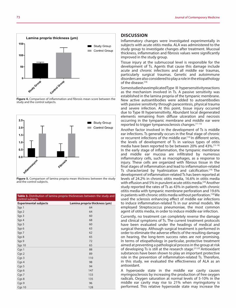
73 Journal of Contemporary Medicine
DISCUSSIONInflammatory changes were investigated experimentally in subjects with acute otitis media. ALA was administered to the study group to investigate changes after treatment. Mucosal thickness, inflammation and fibrosis values were significantly improved in the study group.Tissue injury at the submucosal level is responsible for the development of Ts. Agents that cause this damage include acute and chronic infections and all middle ear traumas, particularly surgical traumas. Genetic and autoimmune disorders are also considered to play a role in the etiopathology of the disease.[10]
SomestudieshaveimplicatedType III hypersensitivityreactions as the mechanism involved in Ts. A passive sensitivity was established in the lamina propria of the tympanic membrane. New active autoantibodies were added to autoantibodies with passive sensitivity through paracentesis, physical trauma and severe infection. At this point, tissue injury occurred due to Type III hypersensitivity. Abundant local degenerated elements remaining from diffuse ulceration and necrosis occurring in the tympanic membrane and middle ear were reported to trigger tympanosclerosis changes.[11-13]
Another factor involved in the development of Ts is middle ear infections. Ts generally occurs in the final stage of chronic or recurrent infections of the middle ear.[14] In different series, the levels of development of Ts in various types of otitis media have been reported to be between 20% and 43%.[15-18] In the early stage of inflammation, the tympanic membrane and middle ear mucosa are infiltrated by numerous inflammatory cells, such as macrophages, as a response to injury. These cells are organized with fibrous tissue in the final stages of inflammation and lead to inflammation-related Ts characterized by hyalinization and calcification.[19] The development of inflammation-related Ts has been reported at levels of 24.2% in chronic otitis media, 16.6% in otitis media with effusion and 5% in purulent acute otitis media.[20] Another study reported the rates of Ts as 43% in patients with chronic otitis media with tympanic membrane perforation and 19.6% in patients with chronic otitis media without perforation.[18] We used the sclerosis enhancing effect of middle ear infections to induce inflammation-related Ts in our animal models. We employed Streptococcus pneumoniae, the most common agent of otitis media, in order to induce middle ear infection.Currently, no treatment can completely reverse the damage and clinical symptoms of Ts. The current treatment protocols have been evaluated under the headings of medical and surgical therapy. Although surgical treatment is performed in order to eliminate the adverse effects of the resulting damage on hearing, the long-term success rates are not promising. In terms of etiopathology in particular, protective treatment aimed at preventing a pathological process in the group at risk of developing Ts is still at the research stage.[21,22] Antioxidant substances have been shown to play an important protective role in the prevention of inflammation-related Ts. Therefore, in this study, we evaluated the effectiveness of ALA as an antioxidant.A hyperoxide state in the middle ear cavity causes myringosclerosis by increasing the production of free oxygen radicals. Oxygen saturation at normal levels of 5-10% in the middle ear cavity may rise to 21% when myringotomy is performed. This relative hyperoxide state may increase the
Figure 4. Comparison of inflammation and fibrosis mean score between the study and the control subjects.
Figure 5. Comparison of lamina propria mean thickness between the study and the control subjects.
Table 3. Distribution of lamina propria thicknesses between the study and control subjects.Experimental subjects Lamina propria thickness (µm)Sgs 1 64Sgs 2 64Sgs 3 60Sgs 4 68Sgs 5 60Sgs 6 63Sgs 7 62Sgs 8 65Sgs 9 72Sgs 10 65Cgs 1 88Cgs 2 89Cgs 3 110Cgs 4 98Cgs 5 94Cgs 6 147Cgs 7 133Cgs 8 135Cgs 9 96Cgs 10 128
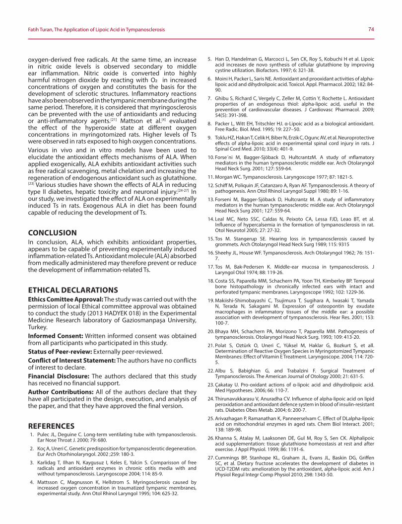
74Fatih Turan, The Application of Lipoic Acid in Tympanosclerosis
oxygen-derived free radicals. At the same time, an increase in nitric oxide levels is observed secondary to middle ear inflammation. Nitric oxide is converted into highly harmful nitrogen dioxide by reacting with O2 in increased concentrations of oxygen and constitutes the basis for the development of sclerotic structures. Inflammatory reactions have also been observed in the tympanic membrane during the same period. Therefore, it is considered that myringosclerosis can be prevented with the use of antioxidants and reducing or anti-inflammatory agents.[21] Mattson et al.[4] evaluated the effect of the hyperoxide state at different oxygen concentrations in myringotomized rats. Higher levels of Ts were observed in rats exposed to high oxygen concentrations.Various in vivo and in vitro models have been used to elucidate the antioxidant effects mechanisms of ALA. When applied exogenically, ALA exhibits antioxidant activities such as free radical scavenging, metal chelation and increasing the regeneration of endogenous antioxidant such as glutathione.[23] Various studies have shown the effects of ALA in reducing type II diabetes, hepatic toxicity and neuronal injury.[24-27] In our study, we investigated the effect of ALA on experimentally induced Ts in rats. Exogenous ALA in diet has been found capable of reducing the development of Ts.
CONCLUSIONIn conclusion, ALA, which exhibits antioxidant properties, appears to be capable of preventing experimentally induced inflammation-related Ts. Antioxidant molecule (ALA) absorbed from medically administered may therefore prevent or reduce the development of inflammation-related Ts.
ETHICAL DECLARATIONSEthics Comittee Approval: The study was carried out with the permission of local Ethical committee approval was obtained to conduct the study (2013 HADYEK 018) in the Experimental Medicine Research laboratory of Gaziosmanpaşa University, Turkey.Informed Consent: Written informed consent was obtained from all participants who participated in this study. Status of Peer-review: Externally peer-reviewed. Conflict of Interest Statement: The authors have no conflicts of interest to declare. Financial Disclosure: The authors declared that this study has received no financial support. Author Contributions: All of the authors declare that they have all participated in the design, execution, and analysis of the paper, and that they have approved the final version.
REFERENCES1. Pulec JL, Deguine C. Long-term ventilating tube with tympanosclerosis.
Ear Nose Throat J. 2000; 79: 680.
2. Koç A, Uneri C. Genetic predisposition for tympanosclerotic degeneration. Eur Arch Otorhinolaryngol. 2002 ;259: 180-3.
3. Karlidag T, Ilhan N, Kaygusuz I, Keles E, Yalcin S. Comparisson of free radicals and antioxidant enzymes in chronic otitis media with and without tympanosclerosis. Laryngoscope 2004; 114: 85-9.
4. Mattsson C, Magnusson K, Hellstrom S. Myringosclerosis caused by increased oxygen concentration in traumatized tympanic membranes, experimental study. Ann Otol Rhinol Laryngol 1995; 104: 625-32.
5. Han D, Handelman G, Marcocci L, Sen CK, Roy S, Kobuchi H et al. Lipoic acid increases de novo synthesis of cellular glutathione by improving cystine utilization. Biofactors. 1997; 6: 321-38.
6. Moini H, Packer L, Saris NE. Antioxidant and prooxidant activities of alpha-lipoic acid and dihydrolipoic acid. Toxicol. Appl. Pharmacol. 2002; 182: 84-90.
7. Ghibu S, Richard C, Vergely C, Zeller M, Cottin Y, Rochette L. Antioxidant properties of an endogenous thiol: alpha-lipoic acid, useful in the prevention of cardiovascular diseases. J Cardiovasc Pharmacol. 2009; 54(5): 391-398.
8. Packer L, Witt EH, Tritschler HJ. α-Lipoic acid as a biological antioxidant. Free Radic. Biol. Med. 1995; 19: 227–50.
9. Toklu HZ, Hakan T, Celik H, Biber N, Erzik C, Ogunc AV, et al. Neuroprotective effects of alpha-lipoic acid in experimental spinal cord injury in rats. J Spinal Cord Med. 2010; 33(4): 401-9.
10. Forse´ni M, Bagger-Sjoback D, HultcrantzM. A study of ınflamatory mediators in the human tympanosclerotic middle ear. Arch Otolaryngol Head Neck Surg. 2001; 127: 559-64.
11. Morgan WC. Tympanosclerosis. Laryngoscope 1977; 87: 1821-5.
12. Schiff M, Poliquin JF, Catanzaro A, Ryan AF. Tympanosclerosis. A theory of pathogenesis. Ann Otol Rhinol Laryngol Suppl 1980; 89: 1-16.
13. Forseni M, Bagger-Sjoback D, Hultcrantz M. A study of inflammatory mediators in the human tympanosclerotic middle ear. Arch Otolaryngol Head Neck Surg 2001; 127: 559-64.
14. Leal MC, Neto SSC, Caldas N, Peixoto CA, Lessa FJD, Leao BT, et al. Influence of hypercalsemia in the formation of tympanosclerosis in rat. Otol Neurotol 2005; 27: 27-32.
15. Tos M. Stangerup SE. Hearing loss in tympanosclerosis caused by grommets. Arch Otolaryngol Head Neck Surg 1989; 115: 9315
16. Sheehy JL, House WF. Tympanosclerosis. Arch Otolaryngol 1962; 76: 151-7.
17. Tos M, Bak-Pedersen K. Middle-ear mucosa in tympanosclerosis. J Laryngol Otol 1974; 88: 119-26.
18. Costa SS, Paparella MM, Schachern PA, Yoon TH, Kimberley BP. Temporal bone histopathology in chronically infected ears with intact and perforated tympanic membranes. Laryngoscope 1992; 102: 1229-36.
19. Makiishi-Shimobayashi C, Tsujimura T, Sugihara A, Iwasaki T, Yamada N, Terada N, Sakagami M. Expression of osteopontin by exudate macrophages in infammatory tissues of the middle ear: a possible association with development of tympanosclerosis. Hear Res. 2001; 153: 100-7.
20. Bhaya MH, Schachern PA, Morizono T, Paparella MM. Pathogenesis of tympanosclerosis. Otolaryngol Head Neck Surg. 1993; 109: 413-20.
21. Polat S, Oztürk O, Uneri C, Yüksel M, Haklar G, Bozkurt S, et all. Determination of Reactive Oxygen Species in Myringotomized Tympanic Membranes: Effect of Vitamin E Treatment. Laryngoscope. 2004; 114: 720-5.
22. Albu S, Babighian G, and Trabalzini F. Surgical Treatment of Tympanosclerosis. The American Journal of Otology 2000; 21: 631-5.
23. Çakatay U. Pro-oxidant actions of α-lipoic acid and dihydrolipoic acid. Med Hypotheses. 2006; 66: 110-7.
24. Thirunavukkarasu V, Anuradha CV. Influence of alpha-lipoic acid on lipid peroxidation and antioxidant defence system in blood of insulin-resistant rats. Diabetes Obes Metab. 2004; 6: 200-7.
25. Arivazhagan P, Ramanathan K, Panneerselvam C. Effect of DLalpha-lipoic acid on mitochondrial enzymes in aged rats. Chem Biol Interact. 2001; 138: 189-98.
26. Khanna S, Atalay M, Laaksonen DE, Gul M, Roy S, Sen CK. Alphalipoic acid supplementation: tissue glutathione homeostasis at rest and after exercise. J Appl Physiol. 1999; 86: 1191-6.
27. Cummings BP, Stanhope KL, Graham JL, Evans JL, Baskin DG, Griffen SC, et al. Dietary fructose accelerates the development of diabetes in UCD-T2DM rats: amelioration by the antioxidant, alpha-lipoic acid. Am J Physiol Regul Integr Comp Physiol 2010; 298: 1343-50.

Corresponding (İletişim): Sabriye DAYI, Doktor Öğretim Üyesi, Çocuk Cerrahisi, Bursa Yüksek İhtisas Eğitim ve Araştırma Hastanesi, Yıldırım , Bursa, TürkiyeE-mail (E-posta): [email protected] (Geliş Tarihi): 08.01.2020 Accepted (Kabul Tarihi): 23.02.2020
DOI: 10.16899/jcm.634978J Contemp Med 2020;10(1):75-81
Orjinal Araştırma / Original Article
JOURNAL OF
CONTEMPORARY MEDICINEJournal ofContemporary MedicineYEAR: 2019 VOLUME: 9 ISSUE: 4
Yenidoğanda Pnömotoraks ve Göğüs Tüpü Uygulamaları
Pneumothorax in Bewborn and Chest Tube Application
Objective: Pneumothorax, which is more common in the newborn period compared to other periods of life, is a condition that requires urgent intervention. Chest tube placement remains the only and valid treatment for pneumothorax. A retrospective study was performed in terms of guiding the diagnosis and treatment, which can be obtained from technical features, complications, clinical findings, demographic characteristics of chest tube applications.
Material and Method: Infants diagnosed with pneumothorax in a 3 year neonatal intensive care unit were included in this retrospective study. Pregnancy weeks, delivery type, weight, height, hospitalization, respiratory status before and after pneumothorax, side of pneumothorax development, tube placement, length of tube stay, length of hospital stay, blood, urine, chest tube, tracheal tube cultures, necrotizing enterocolitis development, healing conditions, follow-up periods and pulmonary pathologies during these follow-up periods were recorded.
Results: The recovery status of the infants from pneumothorax is not affected by the mode of delivery, weight, height, the side on which the pneumothorax develops, the side on which the tube is placed, the age of the baby on which the tube is placed, and the duration of the tube stay. Pulmonary status before, during and after pneumothorax is important. The underlying lung pathology affects prognosis.
For early diagnosis, medial radiolucent areas and loculated radiolucent areas on radiographs are noteworthy.
Conclusion: The baby should be closely monitored for vital signs In order to reduce the mortality and morbidity of the pneumothorax and an emergency chest tube should be placed.
Keywords: Newborn, pneumothorax, chest tube
Öz Abstract
Sabriye Dayı1, Fatih Andıran2
Amaç: Pnomotoraks, yenidoğan doneminde hayatın diğer donemlerine gore daha sık gorülür ve acil müdahale gerektiren bir durumdur. Pnomotoraks tedavisinde goğüs tüpü yerleştirilmesi tek ve geçerli tedavi şekli olma ozelliğini sürdürmektedir. Bu çalışma; pnomotoraks nedeniyle goğüs tüpü uygulaması yapılmış yenidoğanlarda retrospektif olarak planlanmıştır. Klinik bulgular ve demografik karakteristikler, teknik ozellikler, komplikasyonlar araştırılmış ve bunların tanı ve tedaviyi nasıl yonlendirdiği incelenmiştir.Gereç ve Yöntem: Üç senelik periyot içerisinde yenidoğan yoğun bakıma yatan pnomotoraks gelişmiş bebeklerde; gebelik haftaları, doğum şekli, ağırlık, boy, yatış tanıları, pnomotoraks oncesi, pnomotoraks anı ve sonrasındaki solunum durumları, pnomotoraks gelişiminin olduğu taraf, tüpün konulması, tüp kalış süresi, hastanede kalış süresi, kan, idrar, goğüs tüpünün uç kısmının kültürü, trakeal tüp kültürleri, nekrotizan enterokolit gelişimi, şifa durumları, takip süreleri ve bu takip sürelerinde oluşan akciğer patolojileri kaydedildi.Bulgular: Bebeklerin pnomotorakstan şifa durumlarını doğum şekli, ağırlık, boy, pnomotoraksın geliştiği taraf, tüpün konulduğu taraf, tüpün konulduğu anki bebeğin yaşı, tüp kalış süresi etkilememektedir. Pnomotoraks oncesi, pnomotoraks anındaki ve sonrasındaki solunum durumları onemlidir. Altta yatan akciğer patolojisi prognozu etkilemektedir.Erken tanı için rontgen grafilerindeki medial radyolüsen alanlar ve loküle radyolüsen alanlar dikkati çekmektedir.Sonuç: Pnomotoraks mortalitesini ve morbiditesini azaltmada yenidoğan yoğun bakım ünitesindeki bebekler vital bulgular ve radyografik işaretler açısından yakından takip edilmeli, bebekte stabiliteyi bozan durumlarda acil goğüs tüpü yerleştirilmelidir. Klinik durumu bozmayan minimal pnomotorakslar goğüs tüpü konulmadan yakından izlenmelidir.
Anahtar kelimeler: Yenidoğan, pnomotoraks, goğüs tüpü
1Sağlık Bilimleri Üniversitesi, Bursa Yüksek İhtisas Eğitim ve Araştırma Hastanesi, Bursa, Turkey2Beştepeler mah.Yaşam cd. Neorama İş Merkezi, Ankara, Turkey
76 Journal of Contemporary Medicine
GIRIŞYenidoğanda pnomotoraks hayatı tehdit eden ve acil müdahale gerektiren durumlardan birisidir. Pnomotoraks oluşumunda anne ve bebekle ilgili risk faktorlerinin bilinmesi, müdahale zamanı, tedavisi, takibi, mortalitesi ve morbiditesi onemlidir.
Pnomotoraks yenidoğan doneminde çocukluk donemine gore daha sık rastlanılmaktadır. İnsidansı neonatal asfiksi, resusitasyon teknikleri, eşlik een respiratuar problemler, asiste ventilasyonun veriliş şekli ve hatta x-ray kalitesi ve bu filmleri yorumlayanın deneyimi gibi birçok ozelliğe bağlı olarak değişir.
Yüksek basınçlı ventilasyon, zorlu resusitasyon gibi çeşitli müdahaleler ve respiratuar distres sendromu (RDS), mekonyum aspirasyon sendromu ve pulmoner hipoplazi gibi bazı hastalıklar pulmoner hava kaçaklarını onemli olçüde artırmaktadır.
Yenidoğan akciğer hastalıklarında bu derece yaygın ve hayati oneme sahip pnomotoraks gelişiminde tek ve geçerli tedavi acil goğüs tüpü yerleştirilmesidir. Doğru endikasyon ve uygun teknikle goğüs tüpü yerleştirilmesi ve izlemi her ne kadar çok acil ve hayat kurtarıcı oneme sahip ise de hastadan hastaya, klinik duruma gore değişebilen ozelliklere ve çeşitli komplikasyonlara neden olabilmektedir.
Bu nedenlerle, yenidoğan yoğun bakım servisinde gelişen pnomotoraks vakalarında gerçekleştirilen goğüs tüpü uygulamaları retrospektif olarak incelenmiştir. Klinik bulgular ve demografik karakteristikler, teknik ozellikler, komplikasyonlar araştırılmış ve bunların tanı ve tedaviyi nasıl yonlendirdiği incelenmiştir.
GEREÇ VE YÖNTEM1998-2001 yılları arasında yenidoğan yoğun bakım ünitesinde pnomotoraks nedeniyle takip edilen dosya ve filmlerine sağlıklı bir şekilde ulaşılan 36 bebek retrospektif olarak değerlendirilmiştir.
Pnomotoraks tanısı için oncelikle radyolojik bulgular olmak üzere klinik ve torasentez bulguları değerlendirilmiştir. Goğüs tüpü takılması 2. interkostal aralıktan, toraks anteriorundan bebeğin boyutuna uygun 10-12 F goğüs tüpü takılmış ve su altı drenajına alınmıştır.
Bu çalışmada pnomotoraks nedeniyle takip edilmiş bebeklerin gebelik haftaları, doğum şekilleri, anne yaşı, cinsiyet, boy, kilo, doğumda resusitasyon uygulanıp uygulanmaması, apgar skorları, yatış tanıları, pnomotoraks oncesi, pnomotoraks anı
ve sonrasındaki solunum durumu, pnomotoraks olan taraf, goğüs tüpü konulduğu andaki yaşı, pnomotoraks oranı, pnomotoraks oncesi ve sonrasında alınmış kan, Idrar, goğüs tüpü kültürleri, nekrotizan enterokolit gelişimi, tüpün kalış süreleri, bebeklerin şifa, eksitus, durumları ve takip süreleriyle bu sürede geçirmiş oldukları alt solunum yolu infeksiyonları kaydedildi.
İstatiksel değerlendirmede SPSS bilgisayar programı ve ki-kare testi kullanıldı. p<0,05 ise anlamlı kabul edildi.
Bu araştırma Dr. Sabriye DAYI’nın ‘’Yenidoğanda Pnomotoraks ve Goğüs Tüpü Uygulamaları’’ başlıklı tezinden üretilmiştir.
BULGULARÜç yılda yenidoğan yoğun bakım ünitesine yatan pnomotoraks gelişmiş 36 hasta çalışmaya dahil edilmiştir. Bunların 12’si (%33,3) kız, 24’ü (%67,7) erkek idi. 14 bebek (%38,8) 38 hafta ve üzeri (matür), 22 (%61,2) bebek 37 hafta ve altı (prematür) gebelik haftasıyla doğdu. Bebeklerin ortalama ağırlıkları 2553 gram (ortanca: 3125, minimum: 1200, maksimum: 4000, SD: 736), ortalama boyları 46 cm (ortanca: 47, minimum: 36, maksimum: 52, SD: 3,8) idi. Matür bebeklerin ortalama ağırlığı 3199 gram (ortanca: 3125, minimum: 2790, maksimum: 4000, SD: 380), prematür bebeklerin ortalama ağırlığı 2143 gram (ortanca: 2180, minimum: 1200, maksimum3000, SD: 600) idi. Apgar skorları 10. dakika ortalama 8 (ortanca: 9, minimum: 2, maksimum: 10, SD: 1,9) idi. Ortalama anne yaşı 28 (ortanca 28, minimum: 17, maksimum: 38, SD: 5,4) idi. Anne yaşları prematür bebeklerde ortalama 28 (ortanca: 29, minimum: 17, maksimum: 38, SD: 5,01), matür bebeklerde ortalama 27 (ortanca: 26, minimum: 19, maksimum: 38, SD: 6,2) idi. Bebeklerin ortalama doğum haftaları 35 (ortanca 36, minimum: 28, maksimum: 41, SD: 3,5) idi. Hastanede kalış süreleri ortalama 9 gün (ortanca: 8, minimum: 1, maksimum: 34, SD: 6,4) idi. Sekiz bebeğe doğumda resusitasyon uygulandı. Resusitasyon uygulanan bebeklerin 6’sı süreçte eksitus oldu. Kalan 28 bebeğin ise 4’ü eksitus oldu. Eksitus olan bebeklerin 8’i pnomotoraks anında mekanik ventilasyonda idi. Doğumların 22’si (%61,2) Üniversite Hastanesinde gerçekleşti. Doğumların 27’si (%75) sezeryan, 9’u (%25) vajinal yolla gerçekleşti. Sezaryen doğumların 10’u (%37) matür, 17’si (%63) prematürken, vajinal yolla olan doğumların 4’ü (% 44,4) matür, 5’i (%55,6) prematür idi. Bebeklerin 27’si (%75) doğduğu gün, 7’si (%19,4) 1 günlükken, 2’si (%5,6) 2 günlükken, yoğun bakıma yatışı yapıldı. Bir (%2,7) bebekte doğduğu gün, 15 (%41,8) bebekte 1.gün, 10 (%27,7) bebekte 2.gün, 4 (%11,2) bebekte 3.gün, 4 (%11,2) bebekte 4.gün, 1 (%2,7) bebekte 6.gün, 1(%2,7) bebekte 8.gün pnomotoraks gelişti (Tablo 1-2).
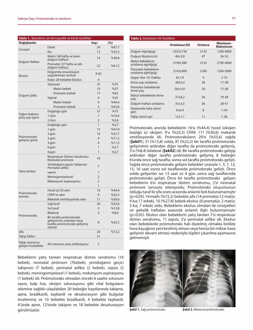
77Sabriye Dayı, Pnömotoraks in newborn
Bebeklerin yatış tanıları respiratuar distres sendromu (16 bebek), neonatal pnomoni (7bebek), yenidoğanın geçici takipnesi (7 bebek), perinatal asfiksi (2 bebek), sepsis (2 bebek), meningomiyelosel (1 bebek), mekonyum aspirasyonu (1 bebek) idi. Pnomotoraks olmadan onceki 6 saatte solunum sayısı, kalp hızı, oksijen saturasyonu gibi vital bulguların izlemine sağlıklı ulaşılabilen 20 bebeğin kayıtlarında takipne, apne, bradikardi, taşikardi ve desaturasyon gibi bulgular incelenmiş ve 10 bebekte bradikardi, 4 bebekte taşikardi, 4’ünde apne, 12’sinde takipne ve 18 bebekte desaturasyon gorülmüştür.
Pnomotoraks anında bebeklerin 16’sı (%44,4) hood (oksijen başlığı) içi oksijen, 9’u (%25,5) CPAP, 11’i (%30,6) mekanik ventilasyonda idi. Pnomotoraksların 20’si (%55,6) sağda (Şekil1), 5’i (%13,8) solda, 8’i (%22,2) bir tarafta pnomotoraks gelişiminin ardından diğer tarafta da pnomotoraks gelişmiş, 3’ü (%8,4) bilateral (Şekil2) idi. Bir tarafta pnomotoraks gelişip ardından diğer tarafta pnomotoraks gelişmiş 8 bebeğin 6’sında once sağ tarafta, sonra sol tarafta pnomotoraks gelişti. Sağda once pnomotoraks gelişen bebekler sırasıyla 1, 3, 7, 12, 15, 16 saat sonra sol taraflarında pnomotoraks gelişti. Önce solda gelişenler ise 13 saat ve 4 gün sonra sağ taraflarında pnomotoraks gelişti. Önce bir tarafta pnomotoraks gelişen bebeklerin 6’sı respiratuar distres sendromu, 2’si neonatal pnomoni tanısıyla izleniyordu. Pnomotoraks oluşumunun olduğu taraf ile şifa oranı arasında anlamlı fark bulunamamıştır (p>0,05). Yirmialtı (%72,2) bebekte şifa (14 prematür,12 matür, 9 kız,17 erkek), 10 (%27,8) bebek eksitus (8 prematür, 2 matür, 3 kız, 7 erkek) oldu. Bebeklerin eksitus olmaları ile cinsiyetleri ve gebelik haftaları arasında anlamlı ilişki bulunmamıştır (p>0,05). Eksitus olan bebeklerin yatış tanıları 7’si respiratuar distres sendromu, 1’i sepsis, 2’si perinatal asfiksi idi. Eksitus olan bebeklerde pnomotoraks hali düzelmiş olmakla birlikte hava kaçağının yeni kesilmiş olması veya henüz bir miktar hava gelişinin devam etmesi nedeniyle tüpleri çıkarılma aşamasına gelmemişti.
Tablo 1. Bebeklere ait demografik ve klinik ozelliklerDeğişkenler Sayı (%)
CinsiyetErkek 24 %67,7Kız 12 %33,3
Doğum Haftası
Matür (38 hafta ve üzeri doğum haftası) 14 %38,8
Prematür (37 hafta ve altı doğum haftası) 22 %61,2
EksitusDoğumda resusitasyon uygulanması (exitus) 8 (6)
Kalan 28 bebekte Eksitus 4
Doğum Şekli;
Sezeryan 27 %75Matür bebek 10 %37Prematür bebek 17 %63
Vajinal 9 %25Matür bebek 4 %44,4Prematür bebek 5 %55,6
Yoğun bakıma yatış yaşı (gün)
Doğduğu gün 27 %751.Gün 7 %19,42.Gün 2 %5,6
Pnomotoraks gelişme günü
Doğduğu gün 1 %2,71.gün 15 %41,82.gün 10 %27,73.gün 4 %11,24.gün 4 %11,26.gün 1 %2,78.gün 1 %2,7
Yatış tanıları;
Respiratuar Distres Sendromu Neonatal pnomoni 16
Yenidoğanın geçici takipnesi Perinatal asfiksi 7
sepsis 7Meningomiyelosel 2Mekonyum aspirasyonu 2
1
Pnomotoraks anında ;
Hood içi O2 alan 16 %44,4CPAP te olan 9 %25,5Mekanik ventilasyonda olan 11 %30,6
Pnomotoraks
Sağ taraf 20 %55,6Sol taraf 5 %13,8Bilateral 3 %8,4Bir tarafta pnomotoraks gelişiminin ardından karşı tarafta pnomotoraks gelişmiş olanlar
8 %22,2
Şifa 26 %72,2Takip Edilen 21
Takip süresince gelişen hastalıklar Alt solunum yolu enfeksiyonu 2
Tablo 2. Bebeklere Ait Özellikler
Ortalama±SD Ortanca Minimum-Maksimum
Doğum Ağırlığı(g) 2553±736 3125 1200-4000Doğum Boyları(cm) 46±3,8 47 36-52Matür bebeklerin ortalama ağırlığı(g) 3199±380 3125 2790-4000
Prematür bebeklerin ortalama ağırlığı(g) 2143±600 2180 1200-3000
Apgar skor 10. Dakika 8±1,9 9 2-10Anne yaşı ortalama 28±5,4 28 17-38Prematür bebeklerde Anne yaşı 28±5,01 29 17-38
Matür bebeklerde Anne yaşı 27±6,2 26 19-38
Doğum haftası ortalama 35±3,5 36 28-41Hastanede kalış süresi gün 9±6,4 8 1-34
Takip süresi (ay) 12±1,1 11 1-38
Şekil 1. Sağ pnomotoraks Şekil 2. Bilateral pnomotoraks
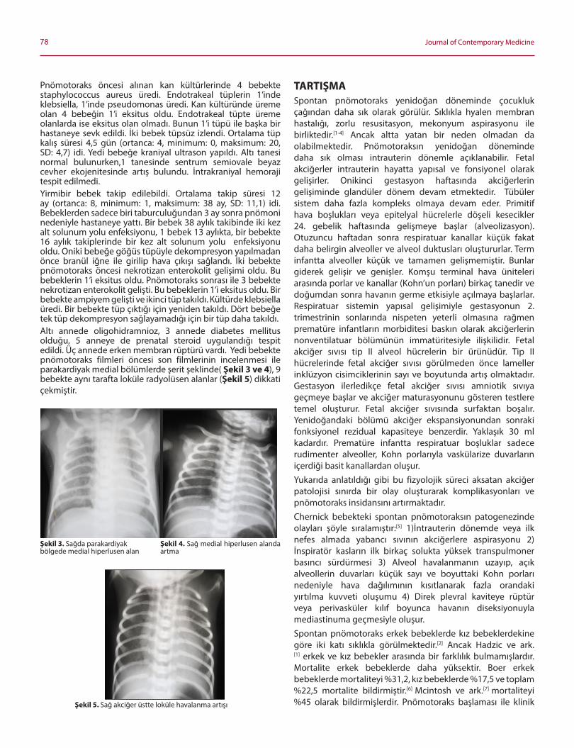
78 Journal of Contemporary Medicine
Pnomotoraks oncesi alınan kan kültürlerinde 4 bebekte staphylococcus aureus üredi. Endotrakeal tüplerin 1’inde klebsiella, 1’inde pseudomonas üredi. Kan kültüründe üreme olan 4 bebeğin 1’i eksitus oldu. Endotrakeal tüpte üreme olanlarda ise eksitus olan olmadı. Bunun 1’i tüpü ile başka bir hastaneye sevk edildi. İki bebek tüpsüz izlendi. Ortalama tüp kalış süresi 4,5 gün (ortanca: 4, minimum: 0, maksimum: 20, SD: 4,7) idi. Yedi bebeğe kraniyal ultrason yapıldı. Altı tanesi normal bulunurken,1 tanesinde sentrum semiovale beyaz cevher ekojenitesinde artış bulundu. İntrakraniyal hemoraji tespit edilmedi. Yirmibir bebek takip edilebildi. Ortalama takip süresi 12 ay (ortanca: 8, minimum: 1, maksimum: 38 ay, SD: 11,1) idi. Bebeklerden sadece biri taburculuğundan 3 ay sonra pnomoni nedeniyle hastaneye yattı. Bir bebek 38 aylık takibinde iki kez alt solunum yolu enfeksiyonu, 1 bebek 13 aylıkta, bir bebekte 16 aylık takiplerinde bir kez alt solunum yolu enfeksiyonu oldu. Oniki bebeğe goğüs tüpüyle dekompresyon yapılmadan once branül iğne ile girilip hava çıkışı sağlandı. İki bebekte pnomotoraks oncesi nekrotizan enterokolit gelişimi oldu. Bu bebeklerin 1’i eksitus oldu. Pnomotoraks sonrası ile 3 bebekte nekrotizan enterokolit gelişti. Bu bebeklerin 1’i eksitus oldu. Bir bebekte ampiyem gelişti ve ikinci tüp takıldı. Kültürde klebsiella üredi. Bir bebekte tüp çıktığı için yeniden takıldı. Dort bebeğe tek tüp dekompresyon sağlayamadığı için bir tüp daha takıldı. Altı annede oligohidramnioz, 3 annede diabetes mellitus olduğu, 5 anneye de prenatal steroid uygulandığı tespit edildi. Üç annede erken membran rüptürü vardı. Yedi bebekte pnomotoraks filmleri oncesi son filmlerinin incelenmesi ile parakardiyak medial bolümlerde şerit şeklinde( Şekil 3 ve 4), 9 bebekte aynı tarafta loküle radyolüsen alanlar (Şekil 5) dikkati çekmiştir.
TARTIŞMASpontan pnomotoraks yenidoğan doneminde çocukluk çağından daha sık olarak gorülür. Sıklıkla hyalen membran hastalığı, zorlu resusitasyon, mekonyum aspirasyonu ile birliktedir.[1-4] Ancak altta yatan bir neden olmadan da olabilmektedir. Pnomotoraksın yenidoğan doneminde daha sık olması intrauterin donemle açıklanabilir. Fetal akciğerler intrauterin hayatta yapısal ve fonsiyonel olarak gelişirler. Onikinci gestasyon haftasında akciğerlerin gelişiminde glandüler donem devam etmektedir. Tübüler sistem daha fazla kompleks olmaya devam eder. Primitif hava boşlukları veya epitelyal hücrelerle doşeli kesecikler 24. gebelik haftasında gelişmeye başlar (alveolizasyon). Otuzuncu haftadan sonra respiratuar kanallar küçük fakat daha belirgin alveoller ve alveol duktusları oluştururlar. Term infantta alveoller küçük ve tamamen gelişmemiştir. Bunlar giderek gelişir ve genişler. Komşu terminal hava üniteleri arasında porlar ve kanallar (Kohn’un porları) birkaç tanedir ve doğumdan sonra havanın germe etkisiyle açılmaya başlarlar. Respiratuar sistemin yapısal gelişimiyle gestasyonun 2. trimestrinin sonlarında nispeten yeterli olmasına rağmen prematüre infantların morbiditesi baskın olarak akciğerlerin nonventilatuar bolümünün immatüritesiyle ilişkilidir. Fetal akciğer sıvısı tip II alveol hücrelerin bir ürünüdür. Tip II hücrelerinde fetal akciğer sıvısı gorülmeden once lameller inklüzyon cisimciklerinin sayı ve boyutunda artış olmaktadır. Gestasyon ilerledikçe fetal akciğer sıvısı amniotik sıvıya geçmeye başlar ve akciğer maturasyonunu gosteren testlere temel oluşturur. Fetal akciğer sıvısında surfaktan boşalır. Yenidoğandaki bolümü akciğer ekspansiyonundan sonraki fonksiyonel rezidual kapasiteye benzerdir. Yaklaşık 30 ml kadardır. Prematüre infantta respiratuar boşluklar sadece rudimenter alveoller, Kohn porlarıyla vaskülarize duvarların içerdiği basit kanallardan oluşur. Yukarıda anlatıldığı gibi bu fizyolojik süreci aksatan akciğer patolojisi sınırda bir olay oluşturarak komplikasyonları ve pnomotoraks insidansını artırmaktadır. Chernick bebekteki spontan pnomotoraksın patogenezinde olayları şoyle sıralamıştır:[5] 1)İntrauterin donemde veya ilk nefes almada yabancı sıvının akciğerlere aspirasyonu 2) İnspirator kasların ilk birkaç solukta yüksek transpulmoner basıncı sürdürmesi 3) Alveol havalanmanın uzayıp, açık alveollerin duvarları küçük sayı ve boyuttaki Kohn porları nedeniyle hava dağılımının kısıtlanarak fazla orandaki yırtılma kuvveti oluşumu 4) Direk plevral kaviteye rüptür veya perivasküler kılıf boyunca havanın diseksiyonuyla mediastinuma geçmesiyle oluşur.Spontan pnomotoraks erkek bebeklerde kız bebeklerdekine gore iki katı sıklıkla gorülmektedir.[2] Ancak Hadzic ve ark.[1] erkek ve kız bebekler arasında bir farklılık bulmamışlardır. Mortalite erkek bebeklerde daha yüksektir. Boer erkek bebeklerde mortaliteyi %31,2, kız bebeklerde %17,5 ve toplam %22,5 mortalite bildirmiştir.[6] Mcintosh ve ark.[7] mortaliteyi %45 olarak bildirmişlerdir. Pnomotoraks başlaması ile klinik
Şekil 3. Sağda parakardiyak bolgede medial hiperlusen alan
Şekil 5. Sağ akciğer üstte loküle havalanma artışı
Şekil 4. Sağ medial hiperlusen alanda artma
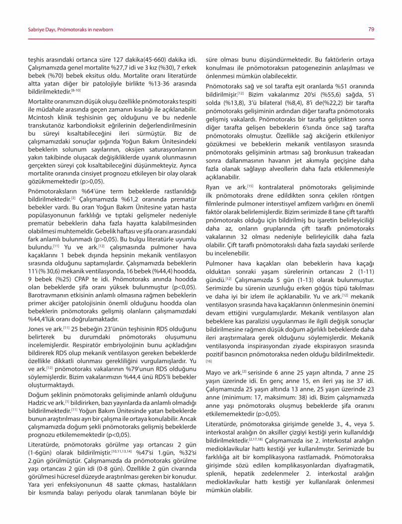
79Sabriye Dayı, Pnömotoraks in newborn
teşhis arasındaki ortanca süre 127 dakika(45-660) dakika idi. Çalışmamızda genel mortalite %27,7 idi ve 3 kız (%30), 7 erkek bebek (%70) bebek eksitus oldu. Mortalite oranı literatürde altta yatan diğer bir patolojiyle birlikte %13-36 arasında bildirilmektedir.[8-10]
Mortalite oranımızın düşük oluşu ozellikle pnomotoraks tespiti ile müdahale arasında geçen zamanın kısalığı ile açıklanabilir. Mcintosh klinik teşhisinin geç olduğunu ve bu nedenle transkutanoz karbondioksit eğrilerinin değerlendirilmesinin bu süreyi kısaltabileceğini ileri sürmüştür. Biz de çalışmamızdaki sonuçlar ışığında Yoğun Bakım Ünitesindeki bebeklerin solunum sayılarının, oksijen saturasyonlarının yakın takibinde oluşacak değişikliklerde uyanık olunmasının gerçekten süreyi çok kısaltabileceğini düşünmekteyiz. Ayrıca mortalite oranında cinsiyet prognozu etkileyen bir olay olarak gozükmemektedir (p>0,05). Pnomotoraksların %64’üne term bebeklerde rastlanıldığı bildirilmektedir.[2] Çalışmamızda %61,2 oranında prematür bebekler vardı. Bu oran Yoğun Bakım Ünitesine yatan hasta popülasyonunun farklılığı ve tıptaki gelişmeler nedeniyle prematür bebeklerin daha fazla hayatta kalabilmesinden olabilmesi muhtemeldir. Gebelik haftası ve şifa oranı arasındaki fark anlamlı bulunmadı (p>0,05). Bu bulgu literatürle uyumlu bulundu.[11] Yu ve ark.[12] çalışmasında pulmoner hava kaçaklarını 1 bebek dışında hepsinin mekanik ventilasyon sırasında olduğunu saptamışlardır. Çalışmamızda bebeklerin 11’i (% 30,6) mekanik ventilasyonda, 16 bebek (%44,4) hoodda, 9 bebek (%25) CPAP te idi. Pnomotoraks anında hoodda olan bebeklerde şifa oranı yüksek bulunmuştur (p<0,05). Barotravmanın etkisinin anlamlı olmasına rağmen bebeklerin primer akciğer patolojisinin onemli olduğunu hoodda olan bebeklerin pnomotoraks gelişmiş olanların çalışmamızdaki %44,4’lük oranı doğrulamaktadır. Jones ve ark.[11] 25 bebeğin 23’ünün teşhisinin RDS olduğunu belirterek bu durumdaki pnomotoraks oluşumunu incelemişlerdir. Respirator embriyolojinin bunu açıkladığını bildirerek RDS olup mekanik ventilasyon gereken bebeklerde ozellikle dikkatli olunması gerekliliğini vurgulamışlardır. Yu ve ark.[12] pnomotoraks vakalarının %79’unun RDS olduğunu soylemişlerdir. Bizim vakalarımızın %44,4 ünü RDS’li bebekler oluşturmaktaydı. Doğum şeklinin pnomotoraks gelişiminde anlamlı olduğunu Hadzic ve ark.[1] bildirirken, bazı yayınlarda da anlamlı olmadığı bildirilmektedir.[11] Yoğun Bakım Ünitesinde yatan bebeklerde bunun araştırılması ayrı bir çalışma ile ortaya konulabilir. Ancak çalışmamızda doğum şekli pnomotoraks gelişmiş bebeklerde prognozu etkilememektedir (p<0,05). Literatürde, pnomotoraks gorülme yaşı ortancası 2 gün (1-6gün) olarak bildirilmiştir.[10,11,13,14] %47’si 1.gün, %32’si 2.gün gorülmüştür. Çalışmamızda da pnomotoraks gorülme yaşı ortancası 2 gün idi (0-8 gün). Özellikle 2 gün civarında gorülmesi hücresel düzeyde araştırılması gereken bir konudur. Yara yeri enfeksiyonunun 48 saatte çıkması, hastalıkların bir kısmında balayı periyodu olarak tanımlanan boyle bir
süre olması bunu düşündürmektedir. Bu faktorlerin ortaya konulması ile pnomotoraksın patogenezinin anlaşılması ve onlenmesi mümkün olabilecektir.
Pnomotoraks sağ ve sol tarafta eşit oranlarda %51 oranında bildirilmişir.[12] Bizim vakalarımız 20’si (%55,6) sağda, 5’i solda (%13,8), 3’ü bilateral (%8,4), 8’i de(%22,2) bir tarafta pnomotoraks gelişiminin ardından diğer tarafta pnomotoraks gelişmiş vakalardı. Pnomotoraks bir tarafta geliştikten sonra diğer tarafta gelişen bebeklerin 6’sında once sağ tarafta pnomotoraks olmuştur. Özellikle sağ akciğerin etkileniyor gozükmesi ve bebeklerin mekanik ventilasyon sırasında pnomotoraks gelişiminin artması sağ bronkusun trakeadan sonra dallanmasının havanın jet akımıyla geçişine daha fazla olanak sağlayıp alveollerin daha fazla etkilenmesiyle açıklanabilir.
Ryan ve ark.[15] kontralateral pnomotoraks gelişiminde ilk pnomotoraks drene edildikten sonra çekilen rontgen filmlerinde pulmoner interstisyel amfizem varlığını en onemli faktor olarak belirlemişlerdir. Bizim serimizde 8 tane çift taraftlı pnomotoraks olduğu için bildirilmiş bu işaretin belirleyiciliği daha az, onların gruplarında çift taraflı pnomotoraks vakalarının 32 olması nedeniyle belirleyicilik daha fazla olabilir. Çift taraflı pnomotorakslı daha fazla sayıdaki serilerde bu incelenebilir.
Pulmoner hava kaçakları olan bebeklerin hava kaçağı olduktan sonraki yaşam sürelerinin ortancası 2 (1-11) gündü.[12] Çalışmamızda 5 gün (1-13) olarak bulunmuştur. Serimizde bu sürenin uzunluğu erken goğüs tüpü takılması ve daha iyi bir izlem ile açıklanabilir. Yu ve ark.[12] mekanik ventilasyon sırasında hava kaçaklarının onlenmesinin onemini devam ettiğini vurgulamışlardır. Mekanik ventilasyon alan bebeklere kas paralizisi uygulanması ile ilgili değişik sonuçlar bildirilmesine rağmen düşük doğum ağırlıklı bebeklerde daha ileri araştırmalara gerek olduğunu soylemişlerdir. Mekanik ventilasyonda inspirasyondan ziyade ekspirasyon sırasında pozitif basıncın pnomotoraksa neden olduğu bildirilmektedir.[16]
Mayo ve ark.[2] serisinde 6 anne 25 yaşın altında, 7 anne 25 yaşın üzerinde idi. En genç anne 15, en ileri yaş ise 37 idi. Çalışmamızda 25 yaşın altında 13 anne, 25 yaşın üzerinde 23 anne (minimum: 17, maksimum: 38) idi. Bizim çalışmamızda anne yaşı pnomotoraks oluşmuş bebeklerde şifa oranını etkilememektedir (p>0,05).
Literatürde, pnomotoraksa girişimde genelde 3., 4., veya 5. interkostal aralığın on aksiller çizgiyi kestiği yerin kullanıldığı bildirilmektedir.[2,17,18] Çalışmamızda ise 2. interkostal aralığın medioklavikular hattı kestiği yer kullanılmıştır. Serimizde bu farklılığa ait bir komplikasyona rastlamadık. Pnomotoraksa girişimde sozü edilen komplikasyonlardan diyafragmatik, splenik, hepatik zedelenmeler 2. interkostal aralığın medioklavikular hattı kestiği yer kullanılarak onlenmesi mümkün olabilir.
80 Journal of Contemporary Medicine
Moessinger ve ark.[19] espiratuar distres sendromu olan bebeklerde plevral drenajı takip eden %25 oranında akciğer perforasyonu olduğunu bildirmişlerdir.Bu komplikasyon ozellikle hava kaçağının devam etmesi ve/veya akciğerin ekspanse olamaması nedeniyle şüphelenilir. Bu ozellikle trokar eşliğinde tüp konulmasıyla birliktedir. Çalışmamızda 2 bebekte(%5,5) plevral drenaj için 2.tüp takılmak zorunda kalınmıştır ancak operatif veya postmortem çalışma bulguları olmadığı için primer nedene mi bağlı yoksa komplikasyon mu olduğu yorumu yapılamamaktadır. Temesvari ve ark.[20] yenidoğan pigletlerde oluşturdukları pnomotoraksta serebral pia-arachnoidal arteriyel hava embolizasyonunu demonstre etmişlerdir. Exitus olan bebeklere otopsi yapılması pnomotoraksın patofizyolojisini ve komplikasyonlarını onemli oranda ortaya çıkmasına yardımcı olacaktır.
Lipscomb ve ark.[21] mekanik ventilasyon alan preterm bebeklerde periventriküler hemorajinin belirgin arttığını bildirmişlerdir. Çalışmamızda 7 bebeğe kraniyal ultrason yapılmış. Hastalardan 1’inde sadece sentrum semiovalede beyaz cevher ekojenitesinde artış gorülmüştür.
Mayo ve ark.[2] pnomotoraks gelişmiş bebeklerin 8-15 yıllık takiplerinde pnomotoraksla ilgili bir durum saptamamışlardır. Bizim takip süremiz ortancası 8 ay idi. Bu takip süresinde bizde rekürrens veya pnomotoraksla ilgili bir patolojiye rastlamadık. Rekürrens hayatın diğer donemlerinde olan spontan pnomotorakslarda %30 gibi yüksek oranlarda gorülmektedir.[22] Ancak 4 bebekte (%11,1) bu takip aylarında alt solunum yolu enfeksiyonu gozükmüştür.
Grim 3rd ve ark.[23] anterior neonatal pnomotoraksın radyografik işareti olarak %19 oranında medial olarak uzanan şerit gorüntüsünde radyolüsen alan tanımlamışlardır. Çalışmamızda filmleri taradığımızda 9 (%25) bebekte buna rastladık. Boyle bir film gorüldüğünde pnomotoraks akla gelip derhal dik pozisyonda film çekilmelidir. Pnomotoraks az miktarda olduğunda hava anteriorda birikip boyle bir gorüntüye yol açabilir. Pnomotoraksın ilerlemesiyle pnomotoraksın klasik gorüntüsü oluşabilir.
Çalışmamızda dikkat çeken bir noktada rontgen filmlerinde akciğerin çeşitli bolümlerinde loküle havalanma artışı olan bebekler olmasıdır (Şekil 1). Bu şekilde 7 bebeğe(%19,4) rastladık. Bu şekilde filmleri olan bebeklerde dikkatli olunması gerektiğini düşünmekteyiz.
Pnomotoraks düşündüren filmler dikkatli değerlendirilmeli, ayırıcı tanı yonünden uyanık olunmalıdır. Liang ve ark.[24] bir yenidoğanda konjenital diyafragmatik herninin yanlışlıkla pnomotoraks olarak teşhis edilip iğne aspirasyonu olduğunu yayınlamışlar ve rontgen filminin yorumlamada sol üst kadrandaki mide gazı gorüntüsüne dikkat edilmesi gerektiğini soylemişlerdir. Goğüs tüpü uygulamasında bizim serimizde bu tür bir komplikasyona rastlamadık.
ETIK BEYANLAR
Etik Durum: Bu araştırma Dr. Sabriye DAYI’nın ‘’Yenidoğanda Pnomotoraks ve Goğüs Tüpü Uygulamaları’’ başlıklı tezinden üretilmiştir. Bu çalışma Kişisel Verilerin Korunması Kanununa uygun şekilde hasta verileri anonim hale getirtilerek ve İyi Kilinik Uygulamalar ve Helsinki Deklarasyonun 2013 Brezilya güncellemesine uygun olarak hazırlanmıştır.
Aydınlatılmış Onam: Çalışma retrospektif olarak dizayn edildiği için hastalardan aydınlatılmış onam alınmamıştır.
Hakem Değerlendirme Süreci: Harici çift kor hakem değerlendirmesi.
Çıkar Çatışması Durumu: Yazarlar bu çalışmada herhangi bir çıkara dayalı ilişki olmadığını beyan etmişlerdir.
Finansal Destek: Yazarlar bu çalışmada finansal destek almadıklarını beyan etmişlerdir.
Yazar Katkıları: Yazarların tümü; makalenin tasarımına, yürütülmesine, analizine katıldığını ve son sürümünü onayladıklarını beyan etmişlerdir.
KAYNAKLAR 1. Hadzic D, Skokic F, Husaric E, Alihodzic H, Softic D, Kovacevic D. Risk
factors and outcome of neonatal pneumothorax in Tuzla Canton. Mater Sociomed 2019;31(1):66-70.
2. Mayo P, Saha SP. Spontaneous Pneumothorax in the newborn. Am Surg 1983;49(4):192-5.
3. Cheng CY, Soong WJ. Local interstitial emphysema caused by meconium aspiration: report of one case. Acta Paediatr Taiwan 2001;42(4):236-8.
4. Maksic H, Heljic S, Maksic S, Jonuzi F. Pulmonary complications during mechanical ventilation in the neonatal period. Med Arh 2000;54(5-6):271-2.
5. Chernick V, Reed MH. Pneumothorax and chylothorax in the neonatal period. J Pediatr 1970 Apr;76(4):624-32
6. Boer HR, Andrews BF. Spontaneous pneumothorax in the neonate. South Med J. 1977 Jul;70(7):841-6.
7. McIntosh N, Becher JC, Cunningham S, et al. Clinical diagnosis of pneumothorax is late: use of tred data and excision support might allow preclinical detection. Pediatr Res 2000;48(3): 408-15.
8. Vibede L, Vibede E, Bendtsen M, Pedersen L, Ebbesen F. Neonatal pneumothorax: a descriptive Regional Danish study. Neonatology 2017;111: 303-8.
9. Bhat Yellanthoor R, Ramdas V. Frequency and intensive care related risk factors of pneumothorax in ventilated neonates. Pulm Med; 2014: 727732.2014
10. Esme H,Dogru O,Eren S, et al. The factors affecting persistent pneumothorax and mortality in neonatal pneumothorax.Turk J Pediatr;50(3): 242-246.2008
11. Yu VY, Wong PY, Szymonowic W. Pulmonary air leak in extremely low birthweight infants. Arch Dis Child 1986;61(3): 239-41.
12. Jones RM, Rutter N, Cooper AC, Pullan CR. Pneumothox in neonatal period. Anaesthesia, 1983;38: 948-52.
13. Hakkinen PJ, Morse CC, Martin FM, Dalbey WE, Haschek WM, Witschi HR. Potentiating effects of oxygen in lungs damaged by methylcyclopentadienyl manganese tricarbonyl, cadmium chloride, oleic acid, and antitumor drugs. Toxicol Appl Pharmacol 1983;67(1): 55-69.
81Sabriye Dayı, Pnömotoraks in newborn
14. Aly H, Massaro A, Acun C, Ozen M. Pneumothorax in the newborn: clinical presentation, risk factors and outcomes. J Matern Fetal Neonatal Med 2014;27(4): 402-6.
15. Ryan CA, Barrington KJ, Philips HJ, Finer NN. Contralateral pneumothoraces in the newborn: incidence and predisposing factors. Pediatrics 1987;79(3): 417-21.
16. Greenough A, Morley CJ. Pneumothorax in infants who fight ventilators. Lancet 1984;24: 1(8378): 689.
17. Arıncı K, Elhan A. Anatomi 1. Cilt, Güneş Kitabevi. 1995.
18. Garry RF, Ludwig S. Textbook of Pediatric Emergency Medicine, 4th edition, Lippincott William&Wilkins, Philadelphia, 2000.
19. Moessinger AC, Driscoll JM Jr, Wigger HJ. High incidence of lung perforation by chest tube in neonatal pneumothorax. J Pediatr. 1978 Apr;92(4):635-7.
20. Temesvari P, Kovacs J, Abraham CS. Pneumothorax and neonatal stroke. Neuropediatrics 1996;27(3): 167-8.
21. Lipscomb AP, Reynolds EOR, Blackwell RJ, et al. Pneumothorax and cerebral haemorrhage in preterm infants. Lancet 1981;1(8217): 414-6.
22. Sahn SA, Heffner JE. Spontaneous pneumothorax. N Engl J Med 2000;342(12): 868-74.
23. Grim 3rd P, Keenan WJ. Two uncommon radiographic signs of an anterior neonatal pneumothorax, correlated with clinical finding. Clin Pediatr (Phila) 1986;25(9): 440-4.
24. Liang JS, Lu FL, Tang JR, Yau KI. Congenital diaphragmatic hernia misdiagnosed as pneumothorax in a newborn. Acta Paediatr Taiwan. 2000;41(4): 221-3.
Corresponding (İletişim): Ömer Kartal, M.D., Gülhane Eğitim ve Araştırma Hastanesi, Çocuk Hematolojisi ve Onkolojisi kliniği, Ankara, TürkiyeE-mail (E-posta): [email protected] (Geliş Tarihi): 02.20.2020 Accepted (Kabul Tarihi): 17.03.2020
DOI: 10.16899/jcm.691996J Contemp Med 2020;10(1):82-85
Orjinal Araştırma / Original Article
JOURNAL OF
CONTEMPORARY MEDICINEJournal ofContemporary MedicineYEAR: 2019 VOLUME: 9 ISSUE: 4
Çocuklarda Vitamin B12 Düzeyleri ile Tam Kan Parametreleri Arasındaki İliskinin Değerlendirilmesi
Evaluation of The Relationship Between Vitamin B12 Levels and Whole Blood Parameters in Children
Introduction: Vitamin B12 is a vitamin that can not be produced in the human body, and it could be taken only with animal-sourced foods. Vitamin B12 plays an important role in DNA synthesis, and in its deficiency, DNA synthesis is disrupted. As a result of this, megaloblastic anemia develops. Then later, conditions such as psychiatric, neurological and hematological disorders may occur.
In our study, we aimed to determine the relationship between vitamin B12 levels and whole blood parameters in patients between 2-12 years old who have no known chronic disease, and no history of chronic drug use, who applied to our clinic.
Material and Method: This study included 98 patients who were evaluated for vitamin B12 level between 2-12 years olds, and it was carried out in Gülhane Training and Research Hospital pediatric hematology clinic between January 2016 and December 2019. The patients were divided into two groups as normal vitamin B12 level (vitamin B12 level ≥200 pg/ml; Group 1) and vitamin B12 deficiency (serum vitamin B12 level <200 pg/ml; Group 2).
Results: Vitamin B12 levels of Group 1 and 2 were found as 311±43.5 and 104.8±15.7, respectively. (p<0.05), (Table). No statistically significant difference was found between white blood cell and platelet counts of groups 1 and 2. White blood cell counts of groups are 7.6±4.1; 6.9±3.5, respectively, and platelet counts of groups are 273.5±74.6; 267.6±95.2, respectively (p>0.05), (Table).
Conclusion: As a result, vitamin B12 deficiency is frequently encountered in our society. White blood cells, platelet, RDW, MPV, and MCV values, which are full blood parameters, are not sufficient in diagnosis in many cases. In patients suspected of vitamin B12 deficiency, the diagnosis should be checked by measuring the serum vitamin B12 level.
Keywords: MCV, MPV, RDW, vitamin B12, whole blood parameters
Öz Abstract
Ömer Kartal1, Orhan Gürsel2
Amaç: Vitamin B12 insan vücudunda üretilemeyen ve sadece hayvansal gıdalar ile alınan bir vitamindir. Vitamin B12 DNA sentezinde onemli rol oynar, eksikliğinde DNA sentezi bozulur ve megaloblastik anemi gelişir. Ayrıca psikiyatrik, norolojik ve hematolojik bozukluklar da meydana gelebilir. Çalışmamızda kliniğimize başvuran, bilinen kronik hastalığı ve herhangi bir kronik ilaç kullanım oyküsü olmayan, 2-12 yaş arası hastalarda vitamin B12 düzeyleri ile tam kan parametreleri arasındaki ilişkiyi belirlemeyi amaçladık.
Gereç ve Yöntem: Çalışmaya Ocak 2016 ile Aralık 2019 tarihleri arasında Gülhane Eğitim ve Araştırma Hastanesi çocuk hematolojisi polikliniğine başvuran, vitamin B12 düzeyi bakılmış, 2-12 yaş arası 98 hasta dahil edildi. Hastalar vitamin B12 düzeyi normal (vitamin B12 düzeyi ≥200 pg/ml; Grup 1) olan ve vitamin B12 eksikliği (serum vitamin B12 düzeyi <200 pg/ml; Grup 2) olan olarak iki gruba ayrıldı.
Bulgular: Grup 1 ve 2 nin vitamin B12 düzeyi sıra ile 311±43,5; 104,8±15,7 olarak bulundu (p -<0,05) (Tablo). Grup 1 ve 2 nin beyaz küre ve trombosit sayıları arasında istatistisel olarak bir fark bulunmadı, sıra ile beyaz küre sayıları 7,6±4,1; 6,9±3,5, sıra ile trombosit sayıları 273,5±74,6; 267,6±95,2 (p>0,05) (Tablo).
Sonuç: Sonuç olarak vitamin B12 eksikliğine toplumumuzda sık olarak karşılaşılmaktadır. Tam kan parametreleri olan beyaz küre, trombosit, RDW, MPV ve MCV değerleri birçok durumda tanıda yeterli olmamaktadır. Vitamin B12 düşüklüğünden şüphelenilen hastalarda mutlaka tanı serum vitamin B12 düzeyi olçülerek kontrol edilmelidir.
Anahtar Sözcükler: MCV, MPV, RDW, tam kan parametreleri, vitamin B12
1Gülhane Eğitim ve Araştırma Hastanesi, Çocuk Hematolojisi ve Onkolojisi kliniği, Ankara, Türkiye2Sağlık Bilimleri Üniversitesi, Çocuk Hematolojisi ve Onkolojisi kliniği, Ankara, Türkiye
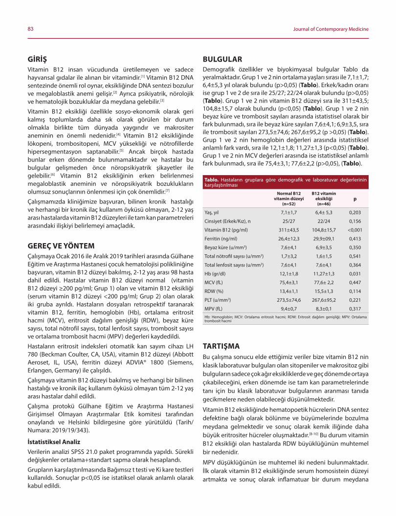
83 Journal of Contemporary Medicine
GIRIŞVitamin B12 insan vücudunda üretilemeyen ve sadece hayvansal gıdalar ile alınan bir vitamindir.[1] Vitamin B12 DNA sentezinde onemli rol oynar, eksikliğinde DNA sentezi bozulur ve megaloblastik anemi gelişir.[2] Ayrıca psikiyatrik, norolojik ve hematolojik bozukluklar da meydana gelebilir.[3]
Vitamin B12 eksikliği ozellikle sosyo-ekonomik olarak geri kalmış toplumlarda daha sık olarak gorülen bir durum olmakla birlikte tüm dünyada yaygındır ve makrositer aneminin en onemli nedenidir.[4] Vitamin B12 eksikliğinde lokopeni, trombositopeni, MCV yüksekliği ve notrofillerde hipersegmentasyon saptanabilir.[5] Ancak birçok hastada bunlar erken donemde bulunmamaktadır ve hastalar bu bulgular gelişmeden once noropsikiyatrik şikayetler ile gelebilir.[6] Vitamin B12 eksikliğinin erken belirlenmesi megaloblastik aneminin ve noropsikiyatrik bozuklukların olumsuz sonuçlarının onlenmesi için çok onemlidir.[7]
Çalışmamızda kliniğimize başvuran, bilinen kronik hastalığı ve herhangi bir kronik ilaç kullanım oyküsü olmayan, 2-12 yaş arası hastalarda vitamin B12 düzeyleri ile tam kan parametreleri arasındaki ilişkiyi belirlemeyi amaçladık.
GEREÇ VE YÖNTEMÇalışmaya Ocak 2016 ile Aralık 2019 tarihleri arasında Gülhane Eğitim ve Araştırma Hastanesi çocuk hematolojisi polikliniğine başvuran, vitamin B12 düzeyi bakılmış, 2-12 yaş arası 98 hasta dahil edildi. Hastalar vitamin B12 düzeyi normal (vitamin B12 düzeyi ≥200 pg/ml; Grup 1) olan ve vitamin B12 eksikliği (serum vitamin B12 düzeyi <200 pg/ml; Grup 2) olan olarak iki gruba ayrıldı. Hastaların dosyaları retrospektif taranarak vitamin B12, ferritin, hemoglobin (Hb), ortalama eritrosit hacmi (MCV), eritrosit dağılım genişliği (RDW), beyaz küre sayısı, total notrofil sayısı, total lenfosit sayısı, trombosit sayısı ve ortalama trombosit hacmi (MPV) değerleri kaydedildi.
Hastaların eritrosit indeksleri otomatik kan sayım cihazı LH 780 (Beckman Coulter, CA, USA), vitamin B12 düzeyi (Abbott Aeroset, IL, USA), ferritin düzeyi ADVIA® 1800 (Siemens, Erlangen, Germany) ile çalışıldı.
Çalışmaya vitamin B12 düzeyi bakılmış ve herhangi bir bilinen hastalığı ve kronik ilaç kullanım oyküsü olmayan tüm 2-12 yaş arası hastalar dahil edildi.
Çalışma protokü Gülhane Eğitim ve Araştırma Hastanesi Girişimsel Olmayan Araştırmalar Etik komitesi tarafından onaylandı ve Helsinki bildirgesine gore yürütüldü (Tarih/Numara: 2019/19/343).
Istatistiksel AnalizVerilerin analizi SPSS 21.0 paket programında yapıldı. Sürekli değişkenler ortalama+standart sapma olarak hesaplandı.
Grupların karşılaştırılmasında Bağımsız t testi ve Ki kare testleri kullanıldı. Sonuçlar p<0,05 ise istatiksel olarak anlamlı olarak kabul edildi.
BULGULARDemografik ozellikler ve biyokimyasal bulgular Tablo da yeralmaktadır. Grup 1 ve 2 nin ortalama yaşları sırası ile 7,1±1,7; 6,4±5,3 yıl olarak bulundu (p>0,05) (Tablo). Erkek/kadın oranı ise grup 1 ve 2 de sıra ile 25/27; 22/24 olarak bulundu (p>0,05) (Tablo). Grup 1 ve 2 nin vitamin B12 düzeyi sıra ile 311±43,5; 104,8±15,7 olarak bulundu (p<0,05) (Tablo). Grup 1 ve 2 nin beyaz küre ve trombosit sayıları arasında istatistisel olarak bir fark bulunmadı, sıra ile beyaz küre sayıları 7,6±4,1; 6,9±3,5, sıra ile trombosit sayıları 273,5±74,6; 267,6±95,2 (p >0,05) (Tablo). Grup 1 ve 2 nin hemoglobin değerleri arasında istatistiksel anlamlı fark vardı, sıra ile 12,1±1,8; 11,27±1,3 (p<0,05) (Tablo). Grup 1 ve 2 nin MCV değerleri arasında ise istatistiksel anlamlı fark bulunmadı, sıra ile 75,4±3,1; 77,6±2,2 (p>0,05), (Tablo).
TARTIŞMABu çalışma sonucu elde ettiğimiz veriler bize vitamin B12 nin klasik laboratuvar bulguları olan sitopeniler ve makrositoz gibi bulguların sadece çok ağır eksikliklerde ve geç donemde ortaya çıkabileceğini, erken donemde ise tam kan parametrelerinde tanı için bu klasik laboratuvar bulgularının aranması tanıda gecikmelere neden olabileceği düşünülmektedir.
Vitamin B12 eksikliğinde hematopoetik hücrelerin DNA sentez defektine bağlı olarak bolünme ve büyümelerinde bozulma meydana gelmektedir ve sonuç olarak kemik iliğinde daha büyük eritrositer hücreler oluşmaktadır.[8-10] Bu durum vitamin B12 eksikliği olan hastalarda RDW büyüklüğünün muhtemel bir nedenidir.
MPV düşüklüğünün ise muhtemel iki nedeni bulunmaktadır. İlk olarak vitamin B12 eksikliğinde serum homosistein düzeyi artmakta ve sonuç olarak inflamatuar bir durum meydana
Tablo. Hastaların gruplara gore demografik ve laboratuvar değerlerinin karşılaştırılması
Normal B12 vitamin düzeyi
(n=52)
B12 vitamin eksikliği
(n=46)p
Yaş, yıl 7,1±1,7 6,4± 5,3 0,203
Cinsiyet (Erkek/Kız), n 25/27 22/24 0,156
Vitamin B12 (pg/ml) 311±43,5 104,8±15,7 <0,001
Ferritin (ng/ml) 26,4±12,3 29,9±09,1 0,413
Beyaz küre (u/mm3) 7,6±4,1 6,9±3,5 0,350
Total notrofil sayısı (u/mm3) 1,7±3,2 1,6±1,5 0,541
Total lenfosit sayısı (u/mm3) 7,6±4,1 7,6±4,1 0,364
Hb (gr/dl) 12,1±1,8 11,27±1,3 0,031
MCV (fL) 75,4±3,1 77,6± 2,2 0,447
RDW (%) 13,4±1,1 15,5±1,3 0,114
PLT (u/mm3) 273,5±74,6 267,6±95,2 0,221
MPV (fL) 9,4±0,7 8,3±0,1 0,317Hb: Hemoglobin; MCV: Ortalama eritrosit hacmi; RDW: Eritrosit dağılım genişliği; MPV: Ortalama trombosit hacmi
84Ömer Kartal, Vitamin B12 eksikliğinde tam kan parametreleri
gelmektedir.[11] Bu da kemik iliğinde daha küçük trombositler oluşmasına neden olmaktadır. MPV düşüklüğünün diğer bir açıklaması ise inflamasyon sırasında daha büyük olma eğiliminde olan trombositler tüketilmekte ve daha küçük trombositlerin oranı artmaktadır, sonuç olarak da MPV değeri düşmektedir.[12]
Aktaş ve ark.[13] vitamin B12 düzeyi 250 pg/ml altında olan 116 hasta ile yaptıkları çalışmada RDW değeri vitamin B12 düzeyi düşük olan hastalarda diğer gruba gore daha yüksek, MPV değeri ise daha düşük bulunmuş ve grupların RDW ve MPV değerleri arasında istatistiksel olarak anlamlı farklılık bulunmuş. Bizim yapmış olduğumuz çalışmada da Aktaş ve ark.[13] buldukları sonuca benzer şekilde RDW değeri vitamin B12 düzeyi düşük olan grupta diğer gruba gore daha yüksek, MPV değeri ise daha düşüktü. Ancak grupların ortalama RDW ve MPV değerleri arasında anlamlı bir farklılık bulmadık. Bu duruma çalışmaya alınan katılımcı sayıları ve yaş farkının neden olabileceğini düşündük.
Literatürde vitamin B12 düşüklüğü durumunda MCV artışı olduğunu gosteren çok sayıda çalışma olmasına rağmen tanıda bu bulgu tek başına yanıltıcı olabilir.[14-16] Örneğin; Vitamin B12 düşüklüğüne eşlik eden demir eksikliği, talasemi taşıyıcılığı ve kronik hastalık anemisi gibi durumlarda MCV artışı maskelenebilir.[17] Kwok ve ark.[18] yapmış oldukları bir çalışmada vitamin B12 eksikliği olan grupla kontrol grubu arasında MCV arasında bir fark bulunmamıştır.
Bor ve ark.[19] yapmış oldukları çalışmada ise vitamin B12 eksikliği olan 98 hastanın sadece 2 tanesinde MCV değeri yüksek bulunmuştur.Bizim yapmış olduğumuz çalışmada da bu çalışmalarara benzer şekilde her iki grup arasında MCV değeri açısından anlamlı bir fark saptamadık.
Yapılmış olan bazı çalışmalarda vitamin B12 eksikliği olan hastalarda beyaz küre ve trombosit düşüklüğü olabileceği soylenmesine rağmen bazı çalışmalarda ise vitamin B12 eksikliği olan hastalar ile kontrolleri karşılaştırıldığında anlamlı bir farklılık saptanmamıştır.[20-22] Bizim yapmış olduğumuz çalışmada da vitamin B12 eksikliği olan ile olmayanlar arasında beyaz küre ve trombosit sayıları arasında anlamlı bir farklılık saptamadık.
Çalışmamızın retrospektif tarzda olması, rolatif olarak düşük katılımcı sayıları, vitamin B12 düzeyi ile ilgili olarak homosistein ve metilmalonik asit düzeylerinin bakılmamış olması kısıtlılıklarıdır.
SONUÇSonuç olarak vitamin B12 eksikliğine toplumumuzda sık olarak karşılaşılmaktadır. Tam kan parametreleri olan beyaz küre, trombosit, RDW, MPV ve MCV değerleri birçok durumda tanıda yeterli olmamaktadır. Vitamin B12 düşüklüğünden şüphelenilen hastalarda mutlaka tanı serum vitamin B12 düzeyi olçülerek kontrol edilmelidir.
ETIK BEYANLAREtik Kurul Onayı: Çalışma protokü Gülhane Eğitim ve Araştırma Hastanesi Girişimsel Olmayan Araştırmalar Etik komitesi tarafından onaylandı ve Helsinki bildirgesine gore yürütüldü (Tarih/Numara: 2019/19/343).Aydınlatılmış Onam: Çalışma retrospektif olarak dizayn edildiği için hastalardan aydınlatılmış onam alınmamıştır.Hakem Değerlendirme Süreci: Harici çift kor hakem değerlendirmesi.Çıkar Çatışması Durumu: Yazarlar bu çalışmada herhangi bir çıkara dayalı ilişki olmadığını beyan etmişlerdir.Finansal Destek: Yazarlar bu çalışmada finansal destek almadıklarını beyan etmişlerdir.Yazar Katkıları: Yazarların tümü; makalenin tasarımına, yürütülmesine, analizine katıldığını ve son sürümünü onayladıklarını beyan etmişlerdir.
KAYNAKLAR1. S. Katar. Çocuklarda B12 Vitamin Eksikliği. Dicle Tıp Dergisi 2007;34(1):25–
8.
2. Gomber S, Kela K, Dhingra N. Clinico-Hematological Profile of Megaloblastic Anemia. Indian Pediatr 1998;35(1):55-8.
3. Lu SY, Wu HC. Initial diagnosis of anemia from sore mouth and improved classification of anemias by MCV and RDW in 30 patients. Oral Surg Oral Med Oral Pathol Oral Radiol Endod 2004;98(6):679-85.
4. Oosterhuis WP, Niessen RW, Bossuyt PM, Sanders GT, Sturk Diagnostic value of the mean corpuscular volume in the detection of vitamin B12 deficiency. Scand J Clin Lab Invest 2000;60(1):9-18.
5. Bhatia P, Kulkarni JD, Pai SA. Vitamin B12 deficiency in India: Mean corpuscular volume is an unreliable screening parameter. Natl Med J India 2012;25(6):336-8.
6. Chan CW, Liu SY, Kho CS, et al. Diagnostic clues to megaloblastic anaemia without macrocytosis. Int J Lab Hematol 2007;29(3):16371.
7. Kondo H. Haematological Effects of Oral Cobalamin Preparations on Patients with Megaloblastic. Acta Haematol 1998;99(4):2005.
8. Green R. Vitamin B12 deficiency from the perspective of a practicing hematologist. Blood 2017;129(19):2603-11.
9. Friso S, Udali S, De Santis D, Choi SW. One-carbon metabolism and epigenetics. Mol Aspects Med 2017;54:28-36.
10. Koury MJ, Ponka P. New insights into erythropoiesis: the roles of folate, vitamin B12, and iron. Annu Rev Nutr 2004;24:105-31.
11. Azimi S, Faramarzi E, Sarbakhsh P, Ostadrahimi A, Somi MH, Ghayour M. Folate and vitamin B12 status and their relation to hematological indices in healthy adults of Iranians: Azar cohort study. Nutr Health 2019;25(1):29-36.
12. Abuhandan M, Gümüş H, Solmaz A, Güzel B. Vitamin B12 Eksiliğinde Hastalarda Ortalama Trombosit Volümün Değerlendirilmesi. Journal of Harran University Medical Faculty 2014;11(3):253–6.
13. Aktas G, Alcelik A, Tekce BK, Sit M, Savli H, Tekce H. Could Mean Platelet Volume and Red Cell Distribution Width Predict Vitamin Could Mean Platelet Volume and Red Cell Distribution Width Predict Vitamin B12 Deficiency? British Journal of Medicine & Medical Research 2014;4(31):4965-71.
14. Castelli MC, Friedman K, Sherry J, et al. Comparing the efficacy and tolerability of a new daily oral vitamin B12 formulation and intermittent intramuscular vitamin B12 in normalizing low cobalamin levels: a randomized, open-label, parallel group study. Open-Label, Parallel-

85 Journal of Contemporary Medicine
Group Study. Clin Ther 2011;33(3):358-71.
15. Green R, Datta Mitra A. Megaloblastic Anemias Nutritional and Other Causes. Med Clin North Am 2017;101(2):297-317.
16. Oberley MJ, Yang DT. Laboratory testing for cobalamin deficiency in megaloblastic anemia. Am J Hematol 2013;88(6):522-6.
17. Çolak AA, Anıl M, Toprak B, Kose E, Üstüner F. Çocuklarda B12 vitamin düzeyi ve periferik tam kan sayımı değerleri ile ilişkisi. İzmir Dr. Behçet Uz Çocuk Hast. Dergisi 2012;2(2):75-9.
18. Kwok T, Cheng G, Woo J, Lai WK, Pang CP. Independent Effect of Vitamin B 12 Deficiency on Hematological Status in Older Chinese Vegetarian Women. Am J Hematol 2002;70(3):186-90.
19. Bor MV, Lydeking-Olsen E, Møller J, Nexø E. A daily intake of approximately 6 microg vitamin B-12 appears to saturate all the vitamin B-12-related variables in Danish postmenopausal women. Am J Clin Nutr 2006;83(1):52-8.
20. Stabler SP. Vitamin B 12 Deficiency. N Engl J Med 2013;368(2):14960.
21. Carmel R. How I treat How I treat cobalamin (vitamin B12) deficiency. Blood 2008;112(6):2214-21.
22. Schneede J, Ueland PM. Novel and Established Markers of Cobalamin Deficiency: Complementary or Exclusive Diagnostic Strategies. Semin Vasc Med 2005;5(2):140-55.
Corresponding (İletişim): Recep Kurnaz, Acıbadem Eskişehir Hastanesi, Ortopedi ve Travmatoloji Kliniği, Eskişehir, TürkiyeE-mail (E-posta): [email protected] (Geliş Tarihi): 06.12.2019 Accepted (Kabul Tarihi): 08.03.2020
DOI: 10.16899/jcm.655991J Contemp Med 2020;10(1):86-90
Orjinal Araştırma / Original Article
JOURNAL OF
CONTEMPORARY MEDICINEJournal ofContemporary MedicineYEAR: 2019 VOLUME: 9 ISSUE: 4
Turnikesiz Unilateral Total Diz Artoplastisi Sonrası Kanama Kontrolünde Topikal Uygulanan Traneksamik Asitin Etkinliği
The Efficacy of Topical Tranexamic Acid in Bleeding Control After Unilateral Total Knee Arthoplasty without Tourniquet
Amaç: Total diz artroplastisi(TDA) sonrası kan kaybını, hemoglobin düşüklüğünü, kan transfüzyon ihtiyacını ve hastanede kalış süresini azaltmak için traneksamik asit (TXA)’ in topikal uygulamasındaki etkinliğini değerlendirmektir.Gereç ve Yöntem: Total diz artroplastisi yapılan 40 hasta, 59-83 yaşları arasından seçilmiştir. VAS 7’ nin üzerinde, Ahlback evre 4-5 ve ASA 1. ve 2. grup olan hastalar çalışmaya dahil edilmiştir. Ameliyatlar aynı cerrah ve standart medial parapatellar artrotomi ile aynı marka implantlar kullanılarak yapılmıştır. Ameliyat boyunca turnike sadece sementleme aşamasında kullanılmıştır. Koagülopati, pulmoner emboli, akut myokard enferktüsü, beyin felci, kronik arter hastalığı hikayesi olanlar ve hemoglobin seviyesi 10 g/dl nin altında olan hastalar çalışmaya alınmamıştır. Hastalar 2 gruba ayrıldı. Grup A’ daki hastalara artrotomi kapatıldıktan sonra 4 ampül 250 mg %10 TXA salin ile seyreltilerek diz eklemine enjekte edildi. 2 saat boyunca dren kapalı tutuldu. Grup B’ deki hastalarda TXA kullanılmadı. Drenler 48. Saat sonunda çekildi. Kan hemoglobin değerleri 8 g/dl’ nin altına düşmedikçe kan transfüzyonu yapılmadı. Hastalar ko-morbiditeleri açısından da değerlendirmeye alındı.Bulgular: 40 hastanın 8 (%20)’ ine kan transfüzyonu yapıldı. Bunların 6 (%75)’ sı TXA kullanılmayan Grup B’ deki hastalar idi. Grup A’ da sadece hastaların 2 (%10)’ sine kan trasfüzyonu yapıldı. Grup B’ deki kan trasfüzyonu yapılan hastaların 1 tanesine 2 ünite kan replase edilmiştir. Ameliyat sonrası ilk 24 saatteki kan drenaj miktarlarına bakıldığında Grup A’ da 232,5 cc, Grup B’ de 407,75 cc idi. 48. saat sonundaki toplam drenaj miktarına bakıldığında Grup A’ da 370,5 cc, Grup B’ de 552,2 cc olarak gorüldü.Ameliyat sırasında ortalama kanama miktarı Grup A’ da 247,5 cc, Grup B’ de 235 cc idi. Ameliyat oncesi ortalama hemoglobin değeri Grup A’ da 13 g/dl iken Grup B’ de 12,75 g/dl idi. Ameliyattan sonraki 6. Saatte alınan hemoglobin değeri Grup A’ da 11,7, Grup B’ de 11 g/dl olarak gozlendi. Ameliyat sonrası ilk 24. saatteki hemoglobin değerleri Grup A’ da 11,07 iken Grup B’ de 10,6 idi. 48. saatteki hemoglobin değerleri Grup A’ da 10,3 iken Grup B’ de 9,5 olarak gozlendi. Postop donemdeki hemoglobin değişim seviyelerine ve drenaj miktarlarına bakıldığında TXA kullanılan grupta ozellikle ilk 24. saatteki drenaj miktarında diğer gruba oranla onemli bir azalma olduğu gozlemlenmiştir. Çıkarımlar: Total diz artroplastisinde traneksamik asit kullanımı postoperatif kanama miktarını anlamlı şekilde azaltır. TXA' nın intravenoz yerine eklem içi olarak uygulanmasıyla sistemik absorpsiyon azaltılabilir ve istenmeyen yan etkiler hafifletilebilir. Bu çalışmanın bulguları toplam kan kaybında ve hemoglobin seviyelerindeki azalmada bir düşüş olduğunu ve dolayısıyla kan transfüzyonu ihtiyacını azalttığını gostermiştir.
Anahtar Kelimeler: Diz protezi, traneksamik asit, kanama kontrolü
AbstractÖz
Recep Kurnaz
Aim: The aim of this study is to evaluate the efficacy of tranexamic acid (TXA) in the topical application of total knee arthroplasty (TKA) to reduce blood loss, low hemoglobin, need for blood transfusion and hospital stay.
Material and Method: Forty patients undergoing total knee arthroplasty were selected from 59-83 years of age. Patients with VAS 7 above Ahlback stage 4-5 and ASA group 1 and 2 were included in the study. The operations were performed using the same surgeon and standard medial parapatellar arthrotomy using the same brand implants. During the operation, the tourniquet was used only during cementation. Patients with a history of coagulopathy, pulmonary embolism, acute myocardial infarction, cerebral palsy, chronic arterial disease and patients with hemoglobin levels below 10 g/dl were excluded from the study. The patients were divided into two groups. In Group A, 4 ampoules of 250 mg 10% TXA saline were injected into the knee joint after arthrotomy was closed. The drain was kept closed for 2 hours. TXA was not used in Group B patients. Drains drained at 48 hours. Blood transfusion was not performed unless the blood hemoglobin values fell below 8 g/dl. The patients were also evaluated for their co-morbidities.
Results: Blood transfusion was performed in 8 (20%) of 40 patients. Six (75%) of these patients were in Group B without TXA. In Group A, only 2 (10%) patients underwent blood transfusion. In Group B, 2 units of blood were replaced in 1 patient. Blood drainage levels in the first 24 hours were 232.5 cc in Group A and 407.75 cc in Group B. When the total drainage amount at the end of 48th hour was examined, it was seen as 370.5 cc in Group A and 552.2 cc in Group B. The mean amount of bleeding during surgery was 247.5 cc in Group A and 235 cc in Group B. The mean preoperative hemoglobin level was 13 g/dl in Group A and 12.75 g/dl in Group B. Hemoglobin levels taken at the 6th postoperative hour were 11.7 in Group A and 11 g/dl in Group B. Hemoglobin levels in the first 24 hours postoperatively were 11.07 in Group A and 10.6 in Group B. Hemoglobin values at the 48th hour were 10.3 in Group A and 9.5 in Group B. When the postoperative hemoglobin change levels and the amount of drainage were examined, it was observed that there was a significant decrease in the amount of drainage in the first 24 hours compared to the other group.
Conclusion:The use of tranexamic acid in total knee arthroplasty significantly reduces the amount of postoperative bleeding. Systemic absorption can be reduced and undesirable side effects can be alleviated by intra-articular administration of TXA instead of intravenous. The results of this study showed that there was a decrease in total blood loss and decrease in hemoglobin levels and thus reduced the need for blood transfusion.
Keywords: Knee replacement, tranexamic acid, bleeding control
Acıbadem Eskişehir Hastanesi, Ortopedi ve Travmatoloji Kliniği
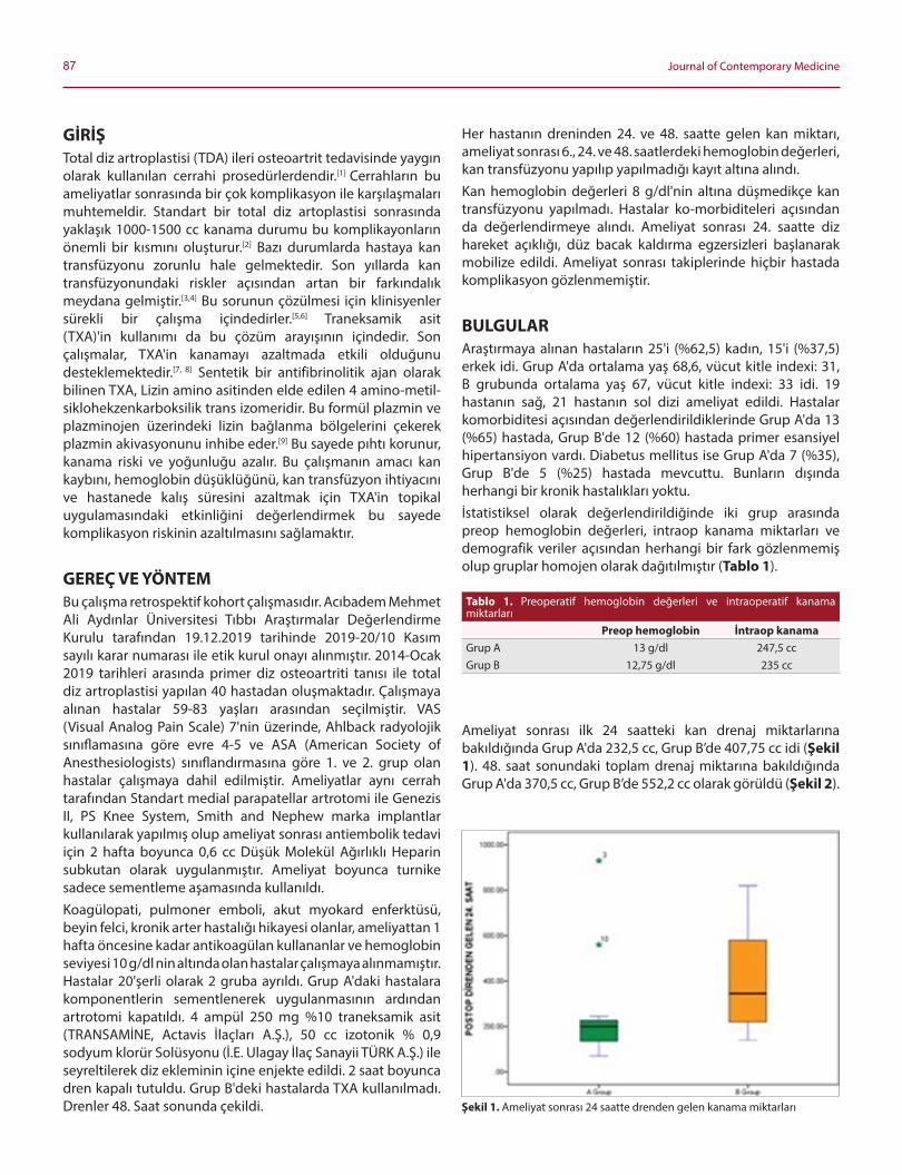
87 Journal of Contemporary Medicine
GIRIŞTotal diz artroplastisi (TDA) ileri osteoartrit tedavisinde yaygın olarak kullanılan cerrahi prosedürlerdendir.[1] Cerrahların bu ameliyatlar sonrasında bir çok komplikasyon ile karşılaşmaları muhtemeldir. Standart bir total diz artoplastisi sonrasında yaklaşık 1000-1500 cc kanama durumu bu komplikayonların onemli bir kısmını oluşturur.[2] Bazı durumlarda hastaya kan transfüzyonu zorunlu hale gelmektedir. Son yıllarda kan transfüzyonundaki riskler açısından artan bir farkındalık meydana gelmiştir.[3,4] Bu sorunun çozülmesi için klinisyenler sürekli bir çalışma içindedirler.[5,6] Traneksamik asit (TXA)'in kullanımı da bu çozüm arayışının içindedir. Son çalışmalar, TXA'in kanamayı azaltmada etkili olduğunu desteklemektedir.[7, 8] Sentetik bir antifibrinolitik ajan olarak bilinen TXA, Lizin amino asitinden elde edilen 4 amino-metil-siklohekzenkarboksilik trans izomeridir. Bu formül plazmin ve plazminojen üzerindeki lizin bağlanma bolgelerini çekerek plazmin akivasyonunu inhibe eder.[9] Bu sayede pıhtı korunur, kanama riski ve yoğunluğu azalır. Bu çalışmanın amacı kan kaybını, hemoglobin düşüklüğünü, kan transfüzyon ihtiyacını ve hastanede kalış süresini azaltmak için TXA'in topikal uygulamasındaki etkinliğini değerlendirmek bu sayede komplikasyon riskinin azaltılmasını sağlamaktır.
GEREÇ VE YÖNTEMBu çalışma retrospektif kohort çalışmasıdır. Acıbadem Mehmet Ali Aydınlar Üniversitesi Tıbbı Araştırmalar Değerlendirme Kurulu tarafından 19.12.2019 tarihinde 2019-20/10 Kasım sayılı karar numarası ile etik kurul onayı alınmıştır. 2014-Ocak 2019 tarihleri arasında primer diz osteoartriti tanısı ile total diz artroplastisi yapılan 40 hastadan oluşmaktadır. Çalışmaya alınan hastalar 59-83 yaşları arasından seçilmiştir. VAS (Visual Analog Pain Scale) 7'nin üzerinde, Ahlback radyolojik sınıflamasına gore evre 4-5 ve ASA (American Society of Anesthesiologists) sınıflandırmasına gore 1. ve 2. grup olan hastalar çalışmaya dahil edilmiştir. Ameliyatlar aynı cerrah tarafından Standart medial parapatellar artrotomi ile Genezis II, PS Knee System, Smith and Nephew marka implantlar kullanılarak yapılmış olup ameliyat sonrası antiembolik tedavi için 2 hafta boyunca 0,6 cc Düşük Molekül Ağırlıklı Heparin subkutan olarak uygulanmıştır. Ameliyat boyunca turnike sadece sementleme aşamasında kullanıldı.Koagülopati, pulmoner emboli, akut myokard enferktüsü, beyin felci, kronik arter hastalığı hikayesi olanlar, ameliyattan 1 hafta oncesine kadar antikoagülan kullananlar ve hemoglobin seviyesi 10 g/dl nin altında olan hastalar çalışmaya alınmamıştır. Hastalar 20'şerli olarak 2 gruba ayrıldı. Grup A'daki hastalara komponentlerin sementlenerek uygulanmasının ardından artrotomi kapatıldı. 4 ampül 250 mg %10 traneksamik asit (TRANSAMİNE, Actavis İlaçları A.Ş.), 50 cc izotonik % 0,9 sodyum klorür Solüsyonu (İ.E. Ulagay İlaç Sanayii TÜRK A.Ş.) ile seyreltilerek diz ekleminin içine enjekte edildi. 2 saat boyunca dren kapalı tutuldu. Grup B'deki hastalarda TXA kullanılmadı. Drenler 48. Saat sonunda çekildi.
Her hastanın dreninden 24. ve 48. saatte gelen kan miktarı, ameliyat sonrası 6., 24. ve 48. saatlerdeki hemoglobin değerleri, kan transfüzyonu yapılıp yapılmadığı kayıt altına alındı.Kan hemoglobin değerleri 8 g/dl'nin altına düşmedikçe kan transfüzyonu yapılmadı. Hastalar ko-morbiditeleri açısından da değerlendirmeye alındı. Ameliyat sonrası 24. saatte diz hareket açıklığı, düz bacak kaldırma egzersizleri başlanarak mobilize edildi. Ameliyat sonrası takiplerinde hiçbir hastada komplikasyon gozlenmemiştir.
BULGULARAraştırmaya alınan hastaların 25'i (%62,5) kadın, 15'i (%37,5) erkek idi. Grup A'da ortalama yaş 68,6, vücut kitle indexi: 31, B grubunda ortalama yaş 67, vücut kitle indexi: 33 idi. 19 hastanın sağ, 21 hastanın sol dizi ameliyat edildi. Hastalar komorbiditesi açısından değerlendirildiklerinde Grup A'da 13 (%65) hastada, Grup B'de 12 (%60) hastada primer esansiyel hipertansiyon vardı. Diabetus mellitus ise Grup A'da 7 (%35), Grup B'de 5 (%25) hastada mevcuttu. Bunların dışında herhangi bir kronik hastalıkları yoktu.İstatistiksel olarak değerlendirildiğinde iki grup arasında preop hemoglobin değerleri, intraop kanama miktarları ve demografik veriler açısından herhangi bir fark gozlenmemiş olup gruplar homojen olarak dağıtılmıştır (Tablo 1).
Ameliyat sonrası ilk 24 saatteki kan drenaj miktarlarına bakıldığında Grup A'da 232,5 cc, Grup B’de 407,75 cc idi (Şekil 1). 48. saat sonundaki toplam drenaj miktarına bakıldığında Grup A'da 370,5 cc, Grup B’de 552,2 cc olarak gorüldü (Şekil 2).
Tablo 1. Preoperatif hemoglobin değerleri ve intraoperatif kanama miktarları
Preop hemoglobin Intraop kanamaGrup A 13 g/dl 247,5 ccGrup B 12,75 g/dl 235 cc
Şekil 1. Ameliyat sonrası 24 saatte drenden gelen kanama miktarları
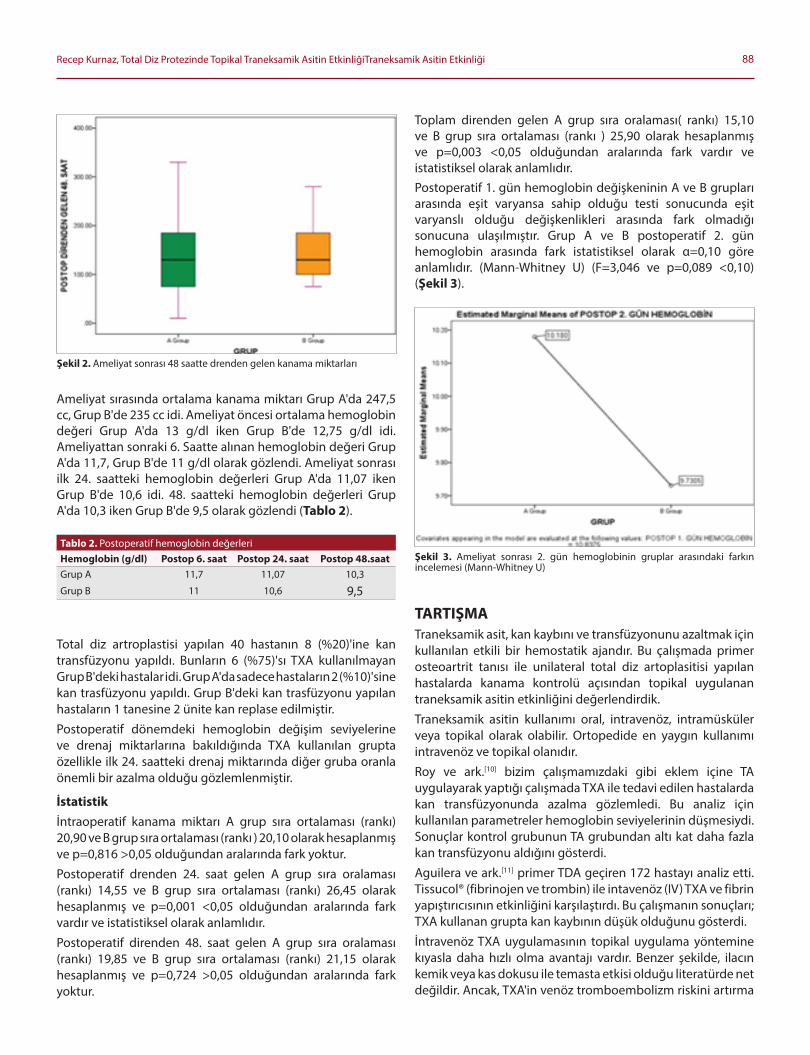
88Recep Kurnaz, Total Diz Protezinde Topikal Traneksamik Asitin EtkinliğiTraneksamik Asitin Etkinliği
Ameliyat sırasında ortalama kanama miktarı Grup A'da 247,5 cc, Grup B'de 235 cc idi. Ameliyat oncesi ortalama hemoglobin değeri Grup A'da 13 g/dl iken Grup B'de 12,75 g/dl idi. Ameliyattan sonraki 6. Saatte alınan hemoglobin değeri Grup A'da 11,7, Grup B'de 11 g/dl olarak gozlendi. Ameliyat sonrası ilk 24. saatteki hemoglobin değerleri Grup A'da 11,07 iken Grup B'de 10,6 idi. 48. saatteki hemoglobin değerleri Grup A'da 10,3 iken Grup B'de 9,5 olarak gozlendi (Tablo 2).
Total diz artroplastisi yapılan 40 hastanın 8 (%20)'ine kan transfüzyonu yapıldı. Bunların 6 (%75)'sı TXA kullanılmayan Grup B'deki hastalar idi. Grup A'da sadece hastaların 2 (%10)'sine kan trasfüzyonu yapıldı. Grup B'deki kan trasfüzyonu yapılan hastaların 1 tanesine 2 ünite kan replase edilmiştir.
Postoperatif donemdeki hemoglobin değişim seviyelerine ve drenaj miktarlarına bakıldığında TXA kullanılan grupta ozellikle ilk 24. saatteki drenaj miktarında diğer gruba oranla onemli bir azalma olduğu gozlemlenmiştir.
Istatistikİntraoperatif kanama miktarı A grup sıra ortalaması (rankı) 20,90 ve B grup sıra ortalaması (rankı ) 20,10 olarak hesaplanmış ve p=0,816 >0,05 olduğundan aralarında fark yoktur.
Postoperatif drenden 24. saat gelen A grup sıra oralaması (rankı) 14,55 ve B grup sıra ortalaması (rankı) 26,45 olarak hesaplanmış ve p=0,001 <0,05 olduğundan aralarında fark vardır ve istatistiksel olarak anlamlıdır.
Postoperatif direnden 48. saat gelen A grup sıra oralaması (rankı) 19,85 ve B grup sıra ortalaması (rankı) 21,15 olarak hesaplanmış ve p=0,724 >0,05 olduğundan aralarında fark yoktur.
Toplam direnden gelen A grup sıra oralaması( rankı) 15,10 ve B grup sıra ortalaması (rankı ) 25,90 olarak hesaplanmış ve p=0,003 <0,05 olduğundan aralarında fark vardır ve istatistiksel olarak anlamlıdır.Postoperatif 1. gün hemoglobin değişkeninin A ve B grupları arasında eşit varyansa sahip olduğu testi sonucunda eşit varyanslı olduğu değişkenlikleri arasında fark olmadığı sonucuna ulaşılmıştır. Grup A ve B postoperatif 2. gün hemoglobin arasında fark istatistiksel olarak α=0,10 gore anlamlıdır. (Mann-Whitney U) (F=3,046 ve p=0,089 <0,10) (Şekil 3).
TARTIŞMATraneksamik asit, kan kaybını ve transfüzyonunu azaltmak için kullanılan etkili bir hemostatik ajandır. Bu çalışmada primer osteoartrit tanısı ile unilateral total diz artoplasitisi yapılan hastalarda kanama kontrolü açısından topikal uygulanan traneksamik asitin etkinliğini değerlendirdik.
Traneksamik asitin kullanımı oral, intravenoz, intramüsküler veya topikal olarak olabilir. Ortopedide en yaygın kullanımı intravenoz ve topikal olanıdır.
Roy ve ark.[10] bizim çalışmamızdaki gibi eklem içine TA uygulayarak yaptığı çalışmada TXA ile tedavi edilen hastalarda kan transfüzyonunda azalma gozlemledi. Bu analiz için kullanılan parametreler hemoglobin seviyelerinin düşmesiydi. Sonuçlar kontrol grubunun TA grubundan altı kat daha fazla kan transfüzyonu aldığını gosterdi.
Aguilera ve ark.[11] primer TDA geçiren 172 hastayı analiz etti. Tissucol® (fibrinojen ve trombin) ile intavenoz (IV) TXA ve fibrin yapıştırıcısının etkinliğini karşılaştırdı. Bu çalışmanın sonuçları; TXA kullanan grupta kan kaybının düşük olduğunu gosterdi.
İntravenoz TXA uygulamasının topikal uygulama yontemine kıyasla daha hızlı olma avantajı vardır. Benzer şekilde, ilacın kemik veya kas dokusu ile temasta etkisi olduğu literatürde net değildir. Ancak, TXA'in venoz tromboembolizm riskini artırma
Şekil 2. Ameliyat sonrası 48 saatte drenden gelen kanama miktarları
Şekil 3. Ameliyat sonrası 2. gün hemoglobinin gruplar arasındaki farkın incelemesi (Mann-Whitney U)
Tablo 2. Postoperatif hemoglobin değerleriHemoglobin (g/dl) Postop 6. saat Postop 24. saat Postop 48.saatGrup A 11,7 11,07 10,3
Grup B 11 10,6 9,5
89 Journal of Contemporary Medicine
ihtimali vardır. Bu nedenle TXA'nın intravenoz yerine topikal olarak uygulanmasıyla sistemik absorpsiyon azaltılabilir ve istenmeyen yan etkiler hafifletilebilir.[12,13]
Bobrek yetmezliği, kalp ve serebrovasküler hastalıklar, DVT gibi durumlar TXA'in intravenoz kullanımında sorun yaratıp kullanımını kısıtlayabilir.[14-20]
Son çalışmalar TXA'in güvenli olabileceğini gostermiştir.[21,22] Wong ve ark.[23] IV ve topikal TXA uygulamasından sonra plazma konsantrasyonlarını olçmüşler. Sonucunda topikal TXA uygulamasında plazma seviyelerini intravenoz uygulmaya gore % 70 daha az olarak rapor etmişlerdir.
Patel ve ark.[24] topikal TXA'nın güvenliği ve etkinliğini randomize edilmiş, çift kor bir prospektif çalışmada IV TXA ile karşılaştırmışlardır. Diğer yazarların raporları ile birlikte, bu çalışmalar, topikal TXA'nın, ilaç tedavisi için kontrendike olmayan hastalar arasında, IV TXA'e benzer bir güvenlik ve etkinlik profili sergilediğini gostermiştir.[25]
Lin ve ark.[26] nın yaptığı çalışmada 120 hastayı randomize olarak üç gruba ayrmışlar. Bir grupa 20 mL serum fizyolojik içinde tek 1,0 g TXA dozu eklem kapsülü kapanmasından sonra (topikal grup); ikinci gruba cilt insizyonundan 15 dakika once 1,0 g intravenoz TXA enjeksiyonu ardından lokal eklem kapsülü kapanmasından sonra 1,0 g'lık eklem içi uygulama ve üçüncü gruba ise sadece 20 mL normal salin solüsyonu vermişlerdir. Sonuçta beklendiği gibi, ortalama toplam kan kaybı hem topikal hem de kombine gruplarda plaseboya oranla daha düşük bulunmuştur. İki TXA grubu arasında ortalama toplam kan kaybını karşılaştırırken anlamlı bir fark olmadığını gozlemlemişlerdir.
2013 yılında Kim ve ark.[27] tek taraflı ve iki taraflı total diz artroplastisinde TXA'in kan kaybını ve transfüzyon hızını azaltmadaki etkinliğini incelemek amacıyla bir çalışma yürütmüştür. Tek taraflı artroplasti uygulanan 180 hasta ve iki taraflı işlem geçirilen 146 hasta dahil edilmiştir. Bu çalışma, TXA'in toplam kan kaybını azalttığını, ancak kan transfüzyon hızı üzerindeki etkilerin, artroplastinin tipine gore değişebileceğini gostermiştir. Bilateral artroplastisi uygulanan hastalarda, tek taraflı artroplastisi uygulanan hastalara gore kan transfüzyonu oranında bir azalma olmuştur. Bu karmaşıklığa sebep olamamak için sadece unilateral TDA yapılan hastalar çalışmaya dahil edilmiştir. Kakar ve ark.[1] tek taraflı TDA geçiren 24 hasta ile bilateral ameliyat edilen 26 hastayı karşılaştırmışlar ve benzer sonuçlar gostermişlerdir. Bu çalışma aynı zamanda TXA'in, her iki grupta da ameliyat sonrası kan transfüzyonlarını azalttığı sonucuna varmıştır.
Farklı TXA uygulama yontemleri arasındaki karşılaştırmanın, en etkili uygulama yontemine ilişkin daha doğru bir sonuç elde etmek için araştırılması gerekir. Ancak, son çalışmalar sürekli olarak TXA'in TDA ameliyatlarında kanama miktarını ve kan transfüzyon ihtiyacını azalttığını doğrulamıştır.[28-30]
SONUÇTotal diz artroplastisinde traneksamik asit kullanımı postoperatif kanama miktarını anlamlı şekilde azaltır. TXA'nın intravenoz yerine topikal olarak uygulanmasıyla sistemik absorpsiyon azaltılabilir ve istenmeyen yan etkiler hafifletilebilir. Bu çalışmanın bulguları toplam kan kaybında ve hemoglobin seviyelerindeki azalmada bir düşüş olduğunu ve dolayısıyla kan transfüzyonu ihtiyacını azalttığını gostermiştir.
ETIK BEYANLAREtik Kurul Onayı: Çalışma için Acıbadem Mehmet Ali Aydınlar Üniversitesi Tıbbı Araştırmalar Değerlendirme Kurulu tarafından 19.12.2019 tarihinde 2019-20/10 Kasım sayılı karar numarası ile etik kurul onayı alınmıştır. Aydınlatılmış Onam: Çalışma retrospektif olarak dizayn edildiği için hastalardan aydınlatılmış onam alınmamıştır.Hakem Değerlendirme Süreci: Harici çift kor hakem değerlendirmesi.Çıkar Çatışması Durumu: Yazarlar bu çalışmada herhangi bir çıkara dayalı ilişki olmadığını beyan etmişlerdir.Finansal Destek: Yazarlar bu çalışmada finansal destek almadıklarını beyan etmişlerdir.Yazar Katkıları: Yazarların tümü; makalenin tasarımına, yürütülmesine, analizine katıldığını ve son sürümünü onayladıklarını beyan etmişlerdir.
KAYNAKÇA1. Kakar, P.N., et al., Efficacy and Safety of Tranexamic Acid in Control of
Bleeding Following TKR: A Randomized Clinical Trial. Indian J Anaesth, 2009. 53(6): p. 667-71.
2. Karam, J.A., et al., Evaluation of the efficacy and safety of tranexamic acid for reducing blood loss in bilateral total knee arthroplasty. J Arthroplasty, 2014. 29(3): p. 501-3.
3. Lemaire, R., Strategies for blood management in orthopaedic and trauma surgery. J Bone Joint Surg Br, 2008. 90(9): p. 1128-36.
4. Levy O, M.U., Oran A, et al. The use of fibrin tissue adhesive to reduce blood loss and the need for blood transfusion after total knee arthroplasty. A prospective, randomized, multicenter study. J Bone Joint Surg Am 1999;81(11):1580-8.
5. Moonen, A.F., T.D. Neal, and P. Pilot, Peri-operative blood management in elective orthopaedic surgery. A critical review of the literature. Injury, 2006. 37 Suppl 5: p. S11-6.
6. Tobias, J.D., Strategies for minimizing blood loss in orthopedic surgery. Semin Hematol, 2004. 41(1 Suppl 1): p. 145-56.
7. Carvalho Junior LH, C.C., Gonçalves MBJ, Rodrigues LCM, Lopes FL, et al. Complicações de curto prazo da artroplastia total do joelho: avaliação de 120 casos. Rev Bras Ortop. 2006;41(5):162-6.
8. Tan J, C.H., Liu Q, Chen C, Huang W. A meta-analysis of the effectiveness and safety of using tranexamic acid in primary unilateral total knee arthroplasty. J Surg Res. 2013;184(2):880-7.
9. Carvalho Junior LH, Castro CAC, Gonçalves MBJ, Rodrigues LCM, Lopes FL, et al. Complicações de curto prazo da artroplastia total do joelho: avaliação de 120 casos. Rev Bras Ortop. 2006;41(5):162-6.
10. Roy SP, T.U., Dutta A, Jain SK, Nagi ON. Efficacy of intra-articular tranexamic acid in blood loss reduction following primary unilateral total knee arthroplasty. Knee Surg Sports Traumatol Arthrosc. 2012;20(12):2494-501.

90Recep Kurnaz, Total Diz Protezinde Topikal Traneksamik Asitin EtkinliğiTraneksamik Asitin Etkinliği
11. Aguilera X, M.-Z.M., Bosch A, Urrútia G, González JC, Jordan M, et al. Efficacy and safety of fibrin glue and tranexamic acid to prevent postoperative blood loss in total knee arthroplasty: a randomized controlled clinical trial. J Bone Joint Surg Am. 2013;95(22):2001-7.
12. Ipema HJ, Tanzi MG. Use of Topical Tranexamic Acid or Aminocaproic Acid to Prevent Bleeding After Major Surgical Procedures. Ann Pharmacother 2012;46:97–107. doi:10.1345/ aph.1Q383.
13. Abrishami A, C.F., Wong J. Topical application of antifibrinolytic drugs for on-pump cardiac surgery: a systematic review and meta-analysis. Can J Anaesth 2009;56:202–12. doi:10.1007/ s12630-008-9038-x.
14. Endo Y, N.S., Miura A. Deep-vein thrombosis induced by tranexamic acid in idiopathic thrombocytopenic purpura. JAMA 1988;259(24):3561–3562.
15. Bruce-Brand R, D.R., Baker J, Harty J. Cerebrovascular infarction following bilateral total knee arthroplasty and tranexamic acid administration. Acta Orthop Belg 2013;79(3):351–354.
16. Sundstrom A, S.H., Kieler H, Alfredsson L. The risk of venous thromboembolism associated with the use of tranexamic acid and other drugs used to treat menorrhagia: a case-control study using the General Practice Research Database. BJOG 2009;116(1):91–97.
17. Salam A, K.C., Orhan O, Mak V. The great deception: tranexamic acid and extensive pulmonary emboli. BMJ Case Rep 2013;2013: pii: bcr2012007808.
18. Fodstad H, Liliequist B. Spontaneous thrombosis of ruptured intracranial aneurysms during treatment with tranexamic acid (AMCA). Report of three cases. Acta Neurochir (Wien) 1979;49; (3–4):129–144.
19. Lindoff C, R.G., Astedt B. Treatment with tranexamic acid during pregnancy, and the risk of thrombo-embolic complications. Thromb Haemost 1993;70(2):238–240.
20. Gupta PN, M.U., Sabin P, Vellappan P. Acute MI in a young hypertensive woman: could it be due to tranexamic acid? BMJ Case Rep 2013;2013:pii: bcr2013009979.
21. Duncan CM, Gillette BP, Jacob AK, Sierra RJ, Sanchez-Sotelo J, Smith HM. Venous thromboembolism and mortality associated with tranexamic acid use during total hip and knee arthroplasty. J Arthroplasty 2015;30(2):272–276.
22. Whiting DR, Gillette BP, Duncan C, Smith H, Pagnano MW, Sierra RJ. Preliminary results suggest tranexamic acid is safe and effective in arthroplasty patients with severe comorbidities. Clin Orthop Relat Res 2014;472(1):66–72.
23. Wong J, A.A., El Beheiry H, et al. Topical application of tranexamic acid reduces postoperative blood loss in total knee arthroplasty: a randomized, controlled trial. J Bone Joint Surg Am 2010;92(15):2503–2513.
24. Seo JG, Moon YW, Park SH, Kim SM, Ko KR. The comparative efficacies of intra-articular and IV tranexamic acid for reducing blood loss during total knee arthroplasty. Knee Surg Sports Traumatol Arthrosc 2013;21(8):1869–1874.
25. Ishida K, Tsumura N, Kitagawa A, et al. Intra-articular injection of tranexamic acid reduces not only blood loss but also knee joint swelling after total knee arthroplasty. Int Orthop 2011;35(11): 1639–1645.
26. Lin SY, Chen CH, Fu YC, et al. The Efficacy of Combined Use of Intraarticular and Intravenous Tranexamic Acid on Reducing Blood Loss and Transfusion Rate in Total Knee Arthroplasty. J Arthroplasty 2015;30(5):776-80.
27. Kim TK, Chang CB, Kang YG, Seo ES, Lee JH, Yun JH, et al. Clinical value of tranexamic acid in unilateral and simultaneous bilateral TKAs under a contemporary blood-saving protocol: a randomized controlled trial. Knee Surg Sports Traumatol Arthrosc. 2014;22(8):1870-8.
28. Tan J, Chen H, Liu Q, Chen C, Huang W. A meta-analysis of the effectiveness and safety of using tranexamic acid in primary unilateral total knee arthroplasty. J Surg Res. 2013;184(2):880-7.
29. Oremus K. Tranexamic acid for the reduction of blood loss in total knee arthroplasty. Ann Transl Med. 2015;3(Suppl 1):S40.
30. Delanois RE, Mont MA. Does tranexamic acid reduce blood loss in total knee arthroplasty? Commentary on an article by
31. X. Aguilera, MD, et al.:"Efficacy and safety of fibrin glue and tranexamic acid to prevent postoperative blood loss in total knee arthroplasty. A randomized controlled clinical trial". J Bone Joint Surg Am. 2013;95(22):e179.

Corresponding (İletişim): Sezai Özkan, Van Yuzuncu Yıl University Dursun Odabaş Medical Center Department of Orthopedics and Traumatology Van, TURKEYE-mail (E-posta): [email protected] (Geliş Tarihi): 28.12.2019 Accepted (Kabul Tarihi): 08.03.2020
DOI: 10.16899/jcm.666741J Contemp Med 2020;10(1):91-95
Orjinal Araştırma / Original Article
JOURNAL OF
CONTEMPORARY MEDICINEJournal ofContemporary MedicineYEAR: 2019 VOLUME: 9 ISSUE: 4
Treatment Outcomes of Mosaicplasty Followed by Postoperative Hyaluronic Acid Injection in Patients With
Osteochondritis Dissecans (OCD) of The Knee
Diz Osteokondritis Dissekans Lezyonlarında Mozakplasti Sonrası Hyaluronik Asit Enjeksiyonunun Tedavi Sonuçları
Aim: Mosaicplasty is a common surgical technique performed in patients with osteochondritis dissecans (OCD) of the knee. In the present study, we aimed to investigate the effectivity of mosaicplasty followed by intraarticular hyaluronic acid (HA) injection on the functional outcomes of the knee joint in patients with osteochondral lesions of the knee.
Material and Method: The retrospective study included 41 patients (32 men and 9 women) that underwent mosaicplasty due to the detection of an OCD lesion larger than 1 cm2 on magnetic resonance imaging (MRI). The patients were randomly divided into HA (n=22) and control (n=19) groups. The HA group received 2 ml of HA injection at two weeks after surgery and the control group received no additional treatment. At postoperative months 6 and 12, functional outcomes of the patients were graded using the Lysholm knee score and pain intensity was assessed using Visual Analog Scale (VAS) in both groups.
Results: The HA group included 22 (53.7%) and the control group included 19(46.3%) patients. The mean age was 31.5 in the HA group and 31.47 years in the control group. The Lysholm scores at postoperative months 6 and 12 were significantly higher in the HA group compared to the control group (p<0.01).
Keywors: Mosaicplasty, knee joint
ÖzAbstract
Sezai Özkan, Cihan Adanaş
Amaç: Mosaikplasti dizde osteokondritis dissekans (OCD) lezyonu olan hastaların tedavisinde yaygın olarak uygulanan cerrahi bir tekniktir. Mozaikplastiden sonra eklem içi hyaluronik asit (HA) enjeksiyonunu öneren birçok çalışma olmasına rağmen, literatürde fikir birliği yoktur. Bu çalışmadaki amacımız diz osteokondral lezyonlarında uygulanan mozaikoplasti sonrası HA enjeksiyonu yapılan hastaların diz fonksiyonel sonuçlarını araştırmayı amaçladık.
Gereç ve Yöntem: Diz MR (Mağnetik Rezonans) görüntülerine göre OCD lezyonu 1 cm2 den büyük ve mozaikplasti yapılan 41 hasta (32 erkek ve 9 kadın) çalışmaya dahil edildi. Hastalar HA enjeksiyonu yapılan ve kontrol grubu olmak üzere rastgele 2 gruba ayrıldı. HA uygulanan guruba ameliyattan 2 hafta sonra diz eklemi içine 2 ml HA enjeksiyonu uygulanırken kontrol grubuna ek bir tedavi uygulanmadı. Her iki gurubun ameliyattan sonra 6. ve 12. ay daki fonksiyonel sonuçları Lysholm diz skorlama sistemi, diz ağrı seviyeleri için de Visual Analog Scale (VAS) kullanılarak karşılaştırıldı.
Bulgular: HA gurup 22 (%53.7), kontrol gurubu ise 19 (%46,3) hastadan oluşuyordu. HA gurubun yaş ortalaması 31,5 yaş ve kontrol gurubun yaş ortalaması 31,47 yaş idi. Ameliyat sonrası 6.ve 12. aydaki Lysholm skorları ve VAS değerleri HA uygulanan gurupta kontrol gurubuna göre daha yüksek olup bu fark istatistiksel olarak anlamlı idi (p<0.01).
Sonuç: Mozaikplasti sonrası diz eklemine HA uygulanması daha az ağrı olmasını ve daha iyi fonksiyonel sonuçlar elde etmemizi sağlar.
Anahtar Kelimeler: Mosaikplasti, diz eklemi, hyaluronik Asit
Van Yuzuncu Yıl University Dursun Odabaş Medical Center Department of Orthopedics and Traumatology Van, TURKEY
92 Journal of Contemporary Medicine
INTRODUCTIONFull-thickness articular cartilage lesions of the knee are commonly seen after sports injuries and constitute the most common type of cartilage defects in the body.[1] These lesions rarely heal spontaneously and result in articular degeneration in advanced ages.[2] Surgical treatment options include bone marrow stimulation (microfracture), articular cartilage replacement (mosaicplasty, osteochondral allograft transplantation), autologous chondrocyte implantation, scaffold without cells, and surface arthroplasty.[3,4] Articular cartilage replacement (mosaicplasty) is a well-established technique used in surgical treatment of symptomatic articular cartilage defects and has been shown to provide favorable functional outcomes.[5,6] This technique involves harvesting osteochondral grafts from non-weight-bearing areas and transplanting them to the defective site. The key advantage of the technique is the formation of hyaline cartilage rather than fibrous cartilage that forms after micro fracture and the complete filling of the defect with new hyaline cartilage, which leads to the formation of a congruent articular cartilage surface similar to that of the natural joint.[7,8] Hyaluronic acid (HA) is the mucopolysaccharide component of synovial fluid that is responsible for its viscoelastic properties. The concentration of HA decreases as osteoarthritis progresses with aging.[9,10] Intraarticular HA injection has been shown to prevent cartilage degeneration, decrease synovial inflammation, and to enhance articular proteoglycan synthesis.[11,12] On the other hand, although mosaicplasty is commonly performed in the treatment of osteochondritis dissecans (OCD) of the knee, to our knowledge there are a limited number of studies reporting on the functional outcomes of mosaicplasty and intraarticular HA injection on the treatment of knee joint. The aim of this study was to investigate the effectivity of mosaicplasty followed by intraarticular HA injection on the functional outcomes of the knee joint and postoperative pain intensity in patients with osteochondral lesions of the knee.
MATERIAL AND METHODThe retrospective study included 41 patients (32 men and 9 women) that presented to our clinic with a knee pain and underwent mosaicplasty due to the detection of an OCD lesion larger than 1 cm2 on magnetic resonance imaging (MRI).Patients with knee arthrosis, malalignment of the lower extremity, prior surgery for the knee joint, rheumatological diseases, and knee joint infection were excluded from the study. Mean age was 31.48 (range, 22-43) years. Surgery was performed in the right knee in 26 (63.4%) and in the left knee in 15 (36.6%) patients.The lesion was localized on the medial femoral condyle in 39 (95.1%) and in the left knee in 2 (4.9%) patients. The mechanism of injury was traumatic sports injury in 23 (56.1%), industrial accident in 7 (17.1%), fall from height in 5 (12.2%) patients and no history of trauma was present in 6 (14.6%) patients.
Mosaicplasty was performed under general or spinal anesthesia in all patients. Following surgery, compression bandaging and cold therapy were administered in each patient. The patients were advised to avoid weight bearing in the operated extremity for 8 weeks and were instructed on how to perform active quadriceps-strengthening exercises and passive range of motion exercises.The patients were randomly divided into HA (n=22) and control (n=19) groups based on postoperative HA injection. The HA group received 2 ml of HA injection at two weeks after surgery and the control group received no additional treatment. In both groups, functional outcomes of the patients at postoperative months 6 and 12 were graded using the Lysholm knee score, which consists of 8 different items graded on a 100-point scale.[11] Pain intensity was assessed using Visual Analog Scale (VAS) at postoperative months 6 and 12. The patients were asked to rate their pain intensity on a 1-10 VAS scale, where 1 indicates ‘no pain’ and 10 indicates ‘the most severe pain.[12] In both Lysholm knee score and VAS, lower scores indicate worse outcomes while higher scores indicate better outcomes.
Statistical analysisData were analyzed using SPSS for Windows version 22.0 (IBM SPSS Inc., Armonk, NY, USA). Normality of gender distribution between the two groups was assessed using two-sample proportion test and the normality of age distribution was assessed using independent two-sample t-test. The Lysholm scores at postoperative months 6 and 12 were compared between the two groups using independent two sample t-test. Differences among VAS scores were determined using One-Way ANOVA test followed by post hoc Duncan’s multiple comparison test. A p value of <0.05 was considered significant.
RESULTSThe HA and control groups comprised 81.8% (n=18) and 79% (n=15) men and 19.2% (n=4) and 21% (n=4) women, respectively. No significant difference was found between the groups with regard to gender distribution (p>0.05).Mean age was 31.5 in the HA group and 31.47 years in the control group. No significant difference was found between the groups with regard to mean age (p>0.05) (Table 1).
No postoperative complication was observed in both groups. However, two patients in the HA group had a short-term pain after HA injection and the pain was eliminated by a two-day cold therapy.
Table 1. Age and gender distributionsHA group Control group p
Number of patients 22 19 >0.05Male (n) 18 15 >0.05Female (n) 4 4 >0.05Mean age (years) 31.5 31.47 >0.05HA=hyaluronic acid
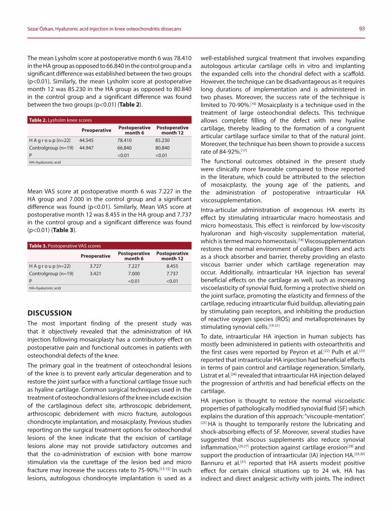
93Sezai Özkan, Hyaluronic acid injection in knee osteochondritis dissecans
The mean Lysholm score at postoperative month 6 was 78.410 in the HA group as opposed to 66.840 in the control group and a significant difference was established between the two groups (p<0.01). Similarly, the mean Lysholm score at postoperative month 12 was 85.230 in the HA group as opposed to 80.840 in the control group and a significant difference was found between the two groups (p<0.01) (Table 2).
Mean VAS score at postoperative month 6 was 7.227 in the HA group and 7.000 in the control group and a significant difference was found (p<0.01). Similarly, Mean VAS score at postoperative month 12 was 8.455 in the HA group and 7.737 in the control group and a significant difference was found (p<0.01) (Table 3).
DISCUSSIONThe most important finding of the present study was that it objectively revealed that the administration of HA injection following mosaicplasty has a contributory effect on postoperative pain and functional outcomes in patients with osteochondral defects of the knee.
The primary goal in the treatment of osteochondral lesions of the knee is to prevent early articular degeneration and to restore the joint surface with a functional cartilage tissue such as hyaline cartilage. Common surgical techniques used in the treatment of osteochondral lesions of the knee include excision of the cartilaginous defect site, arthroscopic debridement, arthroscopic debridement with micro fracture, autologous chondrocyte implantation, and mosaicplasty. Previous studies reporting on the surgical treatment options for osteochondral lesions of the knee indicate that the excision of cartilage lesions alone may not provide satisfactory outcomes and that the co-administration of excision with bone marrow stimulation via the curettage of the lesion bed and micro fracture may increase the success rate to 75-90%.[13-15] In such lesions, autologous chondrocyte implantation is used as a
well-established surgical treatment that involves expanding autologous articular cartilage cells in vitro and implanting the expanded cells into the chondral defect with a scaffold. However, the technique can be disadvantageous as it requires long durations of implementation and is administered in two phases. Moreover, the success rate of the technique is limited to 70-90%.[16] Mosaicplasty is a technique used in the treatment of large osteochondral defects. This technique allows complete filling of the defect with new hyaline cartilage, thereby leading to the formation of a congruent articular cartilage surface similar to that of the natural joint. Moreover, the technique has been shown to provide a success rate of 84-92%.[17] The functional outcomes obtained in the present study were clinically more favorable compared to those reported in the literature, which could be attributed to the selection of mosaicplasty, the young age of the patients, and the administration of postoperative intraarticular HA viscosupplementation.Intra-articular administration of exogenous HA exerts its effect by stimulating intraarticular macro homeostasis and micro homeostasis. This effect is reinforced by low-viscosity hyaluronan and high-viscosity supplementation material, which is termed macro homeostasis.[18] Viscosupplementation restores the normal environment of collagen fibers and acts as a shock absorber and barrier, thereby providing an elasto viscous barrier under which cartilage regeneration may occur. Additionally, intraarticular HA injection has several beneficial effects on the cartilage as well, such as increasing viscoelasticity of synovial fluid, forming a protective shield on the joint surface, promoting the elasticity and firmness of the cartilage, reducing intraarticular fluid buildup, alleviating pain by stimulating pain receptors, and inhibiting the production of reactive oxygen species (ROS) and metalloproteinases by stimulating synovial cells.[19-21] To date, intraarticular HA injection in human subjects has mostly been administered in patients with osteoarthritis and the first cases were reported by Peyron et al.[22] Pulh et al.[23]
reported that intraarticular HA injection had beneficial effects in terms of pain control and cartilage regeneration. Similarly, Listrat et al.[24] revealed that intraarticular HA injection delayed the progression of arthritis and had beneficial effects on the cartilage. HA injection is thought to restore the normal viscoelastic properties of pathologically modified synovial fluid (SF) which explains the duration of this approach: “viscouple-mentation”.[25] HA is thought to temporarily restore the lubricating and shock-absorbing effects of SF. Moreover, several studies have suggested that viscous supplements also reduce synovial inflammation,[26,27] protection against cartilage erosion[28] and support the production of intraarticular (IA) injection HA.[29,30] Bannuru et al.[31] reported that HA asserts modest positive effect for certain clinical situations up to 24 wk. HA has indirect and direct analgesic activity with joints. The indirect
Table 2. Lysholm knee scores
Preoperative Postoperative month 6
Postoperative month 12
H A g r o u p (n=22) 44.545 78.410 85.230Controlgroup (n=19) 44.947 66.840 80.840P <0.01 <0.01HA=hyaluronic acid
Table 3. Postoperative VAS scores
Preoperative Postoperative month 6
Postoperative month 12
H A g r o u p (n=22) 3.727 7.227 8.455Controlgroup (n=19) 3.421 7.000 7.737P <0.01 <0.01HA=hyaluronic acid
94 Journal of Contemporary Medicine
effect is through the anti-inflammatory properties of HA. The direct effect is direct inhibition of nociceptors and decreased synthesis of bradykinin and P substance.[32,33] In our study, both functional outcomes and pain scores were significantly better in the patients that received intraarticular HA injection compared to control subjects, as consistent with the literature.Several studies indicated that the administration of arthroscopic debridement followed by intraarticular HA injection provided favorable outcomes in selected OCD patients. The studies also noted that suitable patient selection and the administration of a suitable technique followed by supplementation resulted in favorable shortand long-term outcomes.[34-36] The present study evaluated patients with OCD of the knee and compared patients that underwent mosaicplasty alone and patients that underwent mosaicplasty followed by intraarticular HA injection. The patients that underwent both mosaicplasty and intraarticular HA injection had significantly better pain and functional scores compared to patients that underwent mosaicplasty alone. To our knowledge, the only experimental study in the literature that administered a combination of mosaicplasty and intraarticular HAinjection was conducted by Tytherleigh-Strong et al. in which the administration of mosaicplasty followed by instant intraarticular HA injection was found to have beneficial effects on graft cartilage in experimental sheep model.[9] The findings of our study supported the findings obtained by Tytherleigh Strong et al.
CONCLUSIONIn conclusion, mosaicplasty is a useful technique for the treatment of patients with OCD of the knee. Additionally, the administration of intraarticular HA injection following mosaicplasty may provide beneficial clinical effects. In the present study, the administration of mosaicplasty followed by instant intraarticular HA injection led to better clinical outcomes and greater patient comfort.
ETHICAL DECLARATIONSEthics Committee Approval: In this research, the data before 2020 was used and the research was concluded before 2020. According to the Regulation on Clinical Researches published in the Official Gazette of the Republic of Turkey with the number 28617 dated 3 November 2015, the ethics committee approval was not obtained in accordance with the article "Retrospective studies are outside the scope of the regulation (article 2- (2))". This study was prepared in accordance with the Law on Protection of Personal Data, by anonymizing patient data and in accordance with the 2013 Brazil revision of the Helsinki Declaration and guidelines for Good Clinical Practice.Informed Consent: Because the study was designed retrospectively, no written informed consent form was obtained from patients.
Status of Peer-review: Externally peer-reviewed. Conflict of Interest Statement: The authors have no conflicts of interest to declare. Financial Disclosure: The authors declared that this study has received no financial support. Author Contributions: All of the authors declare that they have all participated in the design, execution, and analysis of the paper, and that they have approved the final version.
REFERENCES1. Gill TJ, McCulloch PC, Glasson SS, Blanchet T, Morris EA. Chondral defect
repair after the microfracture procedure: a nonhuman primate model. Am J Sports Med. 2005;33:680-685
2. Marder RA, Hopkins GJ, Timmerman LA. Arthroscopic microfracture of chondral defects of the knee: a comparison of two postoperative treatments. Arthroscopy. 2005;21:152-158.
3. Prado MP, Kennedy JG, Raduan F, Nery C. Diagnosis and treatment of osteochondral lesions of the ankle: current concepts. Rev Bras Ortop.2016;51(5):489–500.
4. Murawski CD, Kennedy JG. Operative treatment of osteochondral lesions of the talus. J Bone Joint Surg Am. 2013;95(11):1045– 1054.
5. Mithoefer K, Williams RJ 3rd, Warren RF, et al. The microfracture technique for the treatment of articular cartilage lesions in the knee: a prospective cohort study. J Bone Joint Surg Am. 2005;87: 1911-1920.
6. Steadman JR, Rodkey WG, Rodrigo JJ. Microfracture: surgical technique and rehabilitation to treat chondral defects. Clin Orthop Relat Res. 2001;(391):362-369.
7. Buckwalter JA. Articular cartilage injuries. Clin Orthop Relat Res. 2002;402:21-37.
8. Frisbie DD, Trotter GW, Powers BE, et al. Arthroscopic subchondral bone plate microfracture technique augments healing of large chondral defects in the radial carpal bone and medial femoral condyle of horses. Vet Surg. 1999;28:242-255.
9. Tytherleigh-Strong G, Hurtig M, Miniaci A. Intra-articular hyaluronan following autogenous osteochondral grafting of the knee. Arthroscopy. 2005;21:999-1005.
10. Greenwald RA. Oxygen radicals, inflammation, and arthritis: pathophysiological considerations and implications for treatment. Semin Arthritis Rheum. 1991;20:219-240.
11. Tegner Y, Lysholm J. Rating systems in the evaluation of knee ligament injuries. Clin Orthop Relat Res.1985;(198):43-49.
12. Price DD, McGrath PA, Rafii A, Buckingham B. The validation of visual analogue scales as ratio scale measures for chronic and experimental pain. Pain. 1983;17(1):45-56.
13. Alparslan B, Özkan İ, Acar U, Çullu E, Şavk ŞÖ. Diz ekleminde tam kat kıkırdak lezyonlarının tedavisinde mikrokırık yontemi. Acta Orthop Traumatol Turc. 2007;41 Suppl 2:62-6.
14. Steadman JR, Miller BS, Karas SG, Schlegel TF, Briggs KK, Hawkins RJ. The microfracture technique in the treatment of full-thickness chondral lesions of the knee in National Football League players. J Knee Surg. 2003;16(2):83-6.
15. Magnussen RA, Dunn WR, Carey JL, Spindler KP. Treatment of focal articular cartilage defects in the knee: a systematic review. Clin Orthop Relat Res. 2008;466(4):952-7.
16. Brittberg M, Lindahl A, Nilsson A, Ohlsson C, Isaksson O, Peterson L. Treatment of deep cartilage defects in the knee with autologous chondrocyte transplantation. N Engl J Med.1994;331:889-895.
95Sezai Özkan, Hyaluronic acid injection in knee osteochondritis dissecans
17. Haklar U, Tüzüner T, Kocaoğlu B, Güven O. Mosaicplasty technique in the treatment of osteochondral lesions of the knee. Acta Orthop Traumatol Turc. 2008;42(5):344-349.
18. Robert H. Chondral repair of the knee joint using mosaicplasty. Orthop Traumatol Surg Res. 2011;97(4):418-429.
19. Asari A, Miyauchi S, Matsuzaka S, et al. Molecular weightdependent effects of hyaluronate on the arthritic synovium. Arch Histol Cytol.1998;61:125-135.
20. Kelly MA, Kurzweil PR, Moskowitz RW. Intra-articular hyaluronans in knee osteoarthritis: rationale and practical considerations. Am J Orthop.2004;33:15-22.
21. Gerald NS, Stephen LM, Kenneth DB, Elizabeth AM. Effect of intraarticular hyaluronan injection in experimental canine osteoarthritis. Arthritis and Rheumatism. 1998;41:976-984
22. Peyron JG: Preliminary clinical assessment of Nahyaluronate injection into human arthritic joints. Pathol Biol (Paris).1974;22(8):731-736.
23. Puhl W: Intra-articular sodium hyaluronate in osteoarthritis of the knee: a multicenter, double-blind study. Osteoarthritis Cartilage.1993;1(4):233-41
24. Listrat V: Arthroscopic evaluation of potential structure modifying activity of hyaluronan (Hyalgan) in osteoarthritis of the knee. Osteoarthritis Cartilage. 1997;5(3):153-60.
25. Balazs EA, Denlinger JL. Viscosupplementation: a new concept in the treatment of osteoarthritis. J Rheumatol Suppl. 1993; 39: 3-9
26. Wang Y, Hall S, Hanna F, Wluka AE, Grant G, et all. Effects of Hylan G-F 20 supple-mentation on cartilage preservation detected by magnetic resonance imaging in osteoarthritis of the knee: a two-year single-blind clinical trial. BMC Musculoskelet Disord. 2011; 12: 195
27. Guidolin DD, Ronchetti IP, Lini E, Guerra D, Frizziero L. Morphological analysis of articular cartilage biopsies from a randomized, clinical study comparing the effects of 500-730 kDa sodium hyaluronate (Hyalgan) and methylpredniso-lone acetate on primary osteoarthritis of the knee. Osteoar-thritis Cartilage. 2001; 9: 371-381
28. Wenz W, Breusch SJ, Graf J, Stratmann U. Ultrastructural findings after intraarticular application of hyaluronan in a canine model of arthropathy. J Orthop Res. 2000; 18: 604-612
29. Moreland LW. Intra-articular hyaluronan (hyaluronic acid) and hylans for the treatment of osteoarthritis: mechanisms of action. Arthritis Res Ther. 2003; 5: 54-67
30. Goldberg VM, Buckwalter JA. Hyaluronans in the treatment of osteoarthritis of the knee: evidence for disease-modifying activity. Osteoarthritis Cartilage. 2005; 13: 216-224
31. Bannuru RR, Natov NS, Dasi UR, Schmid CH, McAlindon TE. Therapeutic trajectory following intra-articular hyaluronic acid injection in knee osteoarthritis meta-analysis. Osteoarthritis Cartilage. 2011; 19: 611-619
32. Pozo MA, Balazs EA, Belmonte C. Reduction of sensory responses to passive movements of inflamed knee joints by hylan, a hyaluronan derivative. Exp Brain Res 1997; 116: 3-9
33. Gomis A, Miralles A, Schmidt RF, Belmonte C. Intra-articular injections of hyaluronan solutions of different elastoviscosity reduce nociceptive nerve activity in a model of osteoarthritic knee joint of the guinea pig.Osteoarthritis Cartilage 2009;17:798-804
34. Ozturk A, Ozdemir MR, Ozkan Y. Osteochondral autografting (mosaicplasty) in grade IV cartilage defects in the knee joint: 2to 7-year results. Int Orthop. 2006;30:200-204.
35. Peyron JG. Intraarticular hyaluronan injections in the treatment of osteoarthritis: State-of-the-art review. J Rheumatol.1993;20:10-15.
36. Scale D, Wobig M, Wolbert W. Viscosupplementation of osteoarthritic knees with hylan: A treatment schedule study. Ther Res. 1994;55:220-232.
Corresponding (İletişim): Ali Haydar Yılmaz, Bilecik State Hospital, Department of Urology, Bilecik,TURKEYE-mail (E-posta): [email protected] (Geliş Tarihi): 09.12.2019 Accepted (Kabul Tarihi): 13.03.2020
DOI: 10.16899/jcm.656495J Contemp Med 2020;10(1):96-101
Orjinal Araştırma / Original Article
JOURNAL OF
CONTEMPORARY MEDICINEJournal ofContemporary MedicineYEAR: 2019 VOLUME: 9 ISSUE: 4
The Efficacy of Chewing Gum in Postoperative Ileus Management After Robot Assisted Laparoscopic Radical
Prostatectomy: A Prospective Randomized Study
Robot Yardımlı Laparoskopik Radikal Prostatektomi Sonrası Olusan Fizyolojik İleus Yönetiminde Sakız Çiğnemenin Etkinliği
Aim: To examine the efficacy of chewing gum on bowel motility in the early postoperative period in patients with prostate cancer undergoing robot-assisted laparoscopic prostatectomy (RALP) we prepared this study.Material and Method: Prior to the study, approval was obtained from the local ethics committee. A total of 67 patients were included in the study. The patients were randomly assigned to the control group (Group C) and two groups. Patients in the study environment were chewed gum from the postoperative eye. Intestinal function improvement was examined. The study was terminated at the first gas transition. This time interval has been recorded. Demographic photos, intraoperative parameters, surgical results were analyzed and compared. The first time of bowel movement, length of hospital stay, and surgical wound site pain and abdominal pain were recorded at the 12th and 24th hours postoperatively.Results: The mean age of the patients in the study group was 60.56±6.07 years, and that of the control group was 60.26±6.87 years. At the end of the study, 23 patients in group C and 25 patients in group G were evaluated. There was no significant difference in demographic data between the groups. No significant difference was observed between the two groups in the perioperative parameters. A significant statistical difference was found in favor of group G at the 24th and 36th hours of initial bowel movements, postoperative abdominal and wound pain.Conclusions: This study indicated that chewing gum is also efficient in early solution of postoperative ileus.
Keywords: Chewing gum, prostate cancer, paralitic ileus, robotic surgery
ÖzAbstract
Ibrahim Karabulut1, Ali Haydar Yılmaz2, Fatih Kursat Yılmazel1
Amaç: Major ürolojik cerrahi sonrası görülen ileus, hasta iyileşmesini geciktirebilir . Biz bu çalışmamızda kliniğimizde prostat kanseri nedeni ile robot yardımlı laparoskopik prostatektomi (RALP) uygulanan hastalarda postoperatif erken dönemde sakız çiğnemenin bağırsak hareketliliği üzerindeki etkinliğinin araştırılmasını amaçladık.
Gereç ve Yöntem: Çalışma öncesi yerel etik kuruldan onam alındı. Çalışmaya toplam 67 hasta dahil edildi. Hastalar rastgele kontrol grubu (Grup C) ve çalışma grubu (Grup G) olarak iki gruba ayrıldı. Çalışma grubunda ki hastalara postoperatif dönemde sakız çiğnetildi. Bağırsak fonksiyon iyileşmesi incelendi. Çalışma ilk gaz geçişinde sonlandırıldı. Bu zaman aralığı kaydedildi. Demografik veriler, intraoperatif parametreler, cerrahi sonuçlar analiz edildi ve karşılaştırıldı. Ameliyat sonrası 12. ve 24. saatlerde barsak hareketinin ilk zamanı, hastanede kalış süresi ve cerrahi yara bölgesi ağrısı ve karın ağrısı kaydedildi.
Bulgular: Çalışma grubundaki hastaların yaş ortalaması 60,56±6,07 yıl, kontrol grubundaki hastaların ise 60.26±6.87 yıl olarak ölçüldü. Çalışmanın sonunda grup C de 23 hasta, grup G de 25 hasta değerlendirildi. Gruplar arasında demografik verilerde anlamlı fark yoktu. Perioperatif parametrelerde her iki grup arasında anlamlı bir fark gözlemlenmedi. İlk bağırsak hareketleri , postoperatif karın ve yara bölgesi ağrısı 24. ve 36. saatler de grup G lehine anlamlı derecede istatistiksel fark tespit edildi.
Sonuç: Bu çalışma sakız çiğnemenin postoperatif ileusun erken dönemde çözümlenmesinde etkili olduğunu göstermiştir.
Anahtar Kelimeler: Sakız, prostat kanseri, paralitik ileus, robotik cerrahi
1Education and Research Hospital, Deparment of Urology, Erzurum, TURKEY2Bilecik State Hospital, Department of Urology, Bilecik, TURKEY
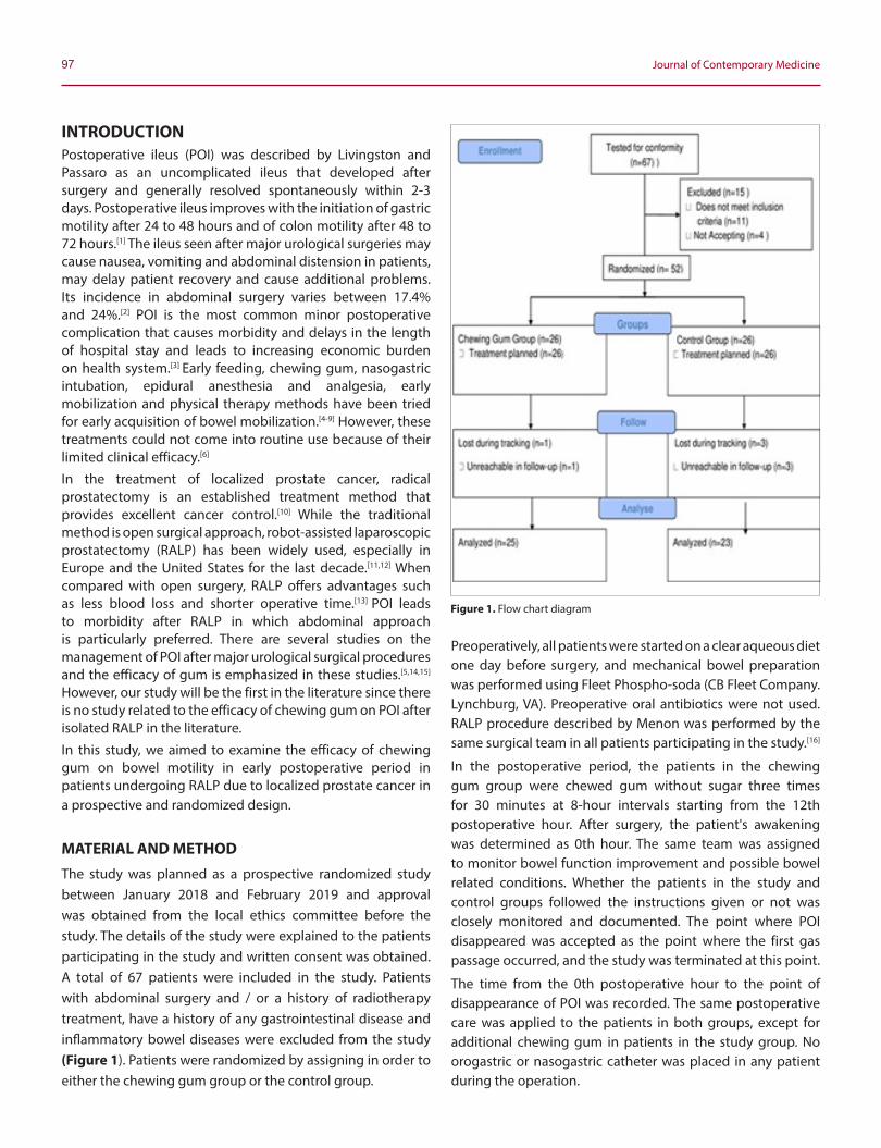
97 Journal of Contemporary Medicine
INTRODUCTIONPostoperative ileus (POI) was described by Livingston and Passaro as an uncomplicated ileus that developed after surgery and generally resolved spontaneously within 2-3 days. Postoperative ileus improves with the initiation of gastric motility after 24 to 48 hours and of colon motility after 48 to 72 hours.[1] The ileus seen after major urological surgeries may cause nausea, vomiting and abdominal distension in patients, may delay patient recovery and cause additional problems. Its incidence in abdominal surgery varies between 17.4% and 24%.[2] POI is the most common minor postoperative complication that causes morbidity and delays in the length of hospital stay and leads to increasing economic burden on health system.[3] Early feeding, chewing gum, nasogastric intubation, epidural anesthesia and analgesia, early mobilization and physical therapy methods have been tried for early acquisition of bowel mobilization.[4-9] However, these treatments could not come into routine use because of their limited clinical efficacy.[6]
In the treatment of localized prostate cancer, radical prostatectomy is an established treatment method that provides excellent cancer control.[10] While the traditional method is open surgical approach, robot-assisted laparoscopic prostatectomy (RALP) has been widely used, especially in Europe and the United States for the last decade.[11,12] When compared with open surgery, RALP offers advantages such as less blood loss and shorter operative time.[13] POI leads to morbidity after RALP in which abdominal approach is particularly preferred. There are several studies on the management of POI after major urological surgical procedures and the efficacy of gum is emphasized in these studies.[5,14,15] However, our study will be the first in the literature since there is no study related to the efficacy of chewing gum on POI after isolated RALP in the literature.
In this study, we aimed to examine the efficacy of chewing gum on bowel motility in early postoperative period in patients undergoing RALP due to localized prostate cancer in a prospective and randomized design.
MATERIAL AND METHOD
The study was planned as a prospective randomized study between January 2018 and February 2019 and approval was obtained from the local ethics committee before the study. The details of the study were explained to the patients participating in the study and written consent was obtained. A total of 67 patients were included in the study. Patients with abdominal surgery and / or a history of radiotherapy treatment, have a history of any gastrointestinal disease and inflammatory bowel diseases were excluded from the study (Figure 1). Patients were randomized by assigning in order to either the chewing gum group or the control group.
Preoperatively, all patients were started on a clear aqueous diet one day before surgery, and mechanical bowel preparation was performed using Fleet Phospho-soda (CB Fleet Company. Lynchburg, VA). Preoperative oral antibiotics were not used. RALP procedure described by Menon was performed by the same surgical team in all patients participating in the study.[16]
In the postoperative period, the patients in the chewing gum group were chewed gum without sugar three times for 30 minutes at 8-hour intervals starting from the 12th postoperative hour. After surgery, the patient's awakening was determined as 0th hour. The same team was assigned to monitor bowel function improvement and possible bowel related conditions. Whether the patients in the study and control groups followed the instructions given or not was closely monitored and documented. The point where POI disappeared was accepted as the point where the first gas passage occurred, and the study was terminated at this point.
The time from the 0th postoperative hour to the point of disappearance of POI was recorded. The same postoperative care was applied to the patients in both groups, except for additional chewing gum in patients in the study group. No orogastric or nasogastric catheter was placed in any patient during the operation.
Figure 1. Flow chart diagram
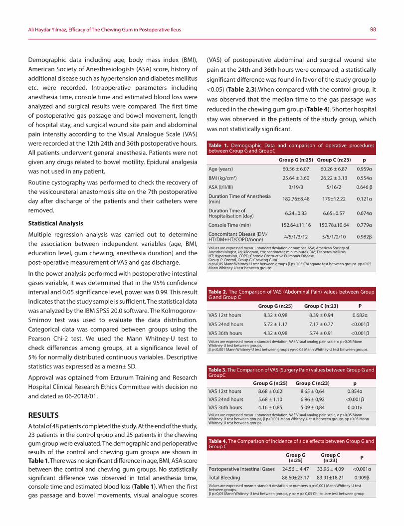
98Ali Haydar Yılmaz, Efficacy of The Chewing Gum in Postoperative Ileus
Demographic data including age, body mass index (BMI), American Society of Anesthesiologists (ASA) score, history of additional disease such as hypertension and diabetes mellitus etc. were recorded. Intraoperative parameters including anesthesia time, console time and estimated blood loss were analyzed and surgical results were compared. The first time of postoperative gas passage and bowel movement, length of hospital stay, and surgical wound site pain and abdominal pain intensity according to the Visual Analogue Scale (VAS) were recorded at the 12th 24th and 36th postoperative hours. All patients underwent general anesthesia. Patients were not given any drugs related to bowel motility. Epidural analgesia was not used in any patient.
Routine cystography was performed to check the recovery of the vesicoureteral anastomosis site on the 7th postoperative day after discharge of the patients and their catheters were removed.
Statistical Analysis
Multiple regression analysis was carried out to determine the association between independent variables (age, BMI, education level, gum chewing, anesthesia duration) and the post-operative measurement of VAS and gas discharge.
In the power analysis performed with postoperative intestinal gases variable, it was determined that in the 95% confidence interval and 0.05 significance level, power was 0.99. This result indicates that the study sample is sufficient. The statistical data was analyzed by the IBM SPSS 20.0 software. The Kolmogorov-Smirnov test was used to evaluate the data distribution. Categorical data was compared between groups using the Pearson Chi-2 test. We used the Mann Whitney-U test to check differences among groups, at a significance level of 5% for normally distributed continuous variables. Descriptive statistics was expressed as a mean± SD.
Approval was optained from Erzurum Training and Research Hospital Clinical Research Ethics Committee with decision no and dated as 06-2018/01.
RESULTSA total of 48 patients completed the study. At the end of the study, 23 patients in the control group and 25 patients in the chewing gum group were evaluated. The demographic and perioperative results of the control and chewing gum groups are shown in Table 1. There was no significant difference in age, BMI, ASA score between the control and chewing gum groups. No statistically significant difference was observed in total anesthesia time, console time and estimated blood loss (Table 1). When the first gas passage and bowel movements, visual analogue scores
(VAS) of postoperative abdominal and surgical wound site pain at the 24th and 36th hours were compared, a statistically significant difference was found in favor of the study group (p <0.05) (Table 2,3).When compared with the control group, it was observed that the median time to the gas passage was reduced in the chewing gum group (Table 4). Shorter hospital stay was observed in the patients of the study group, which was not statistically significant.
Table 1. Demographic Data and comparison of operative procedures between Group G and GroupC
Group G (n:25) Group C (n:23) p
Age (years) 60.56 ± 6.07 60.26 ± 6.87 0.959α
BMI (kg/cm2) 25.64 ± 3.60 26.22 ± 3.13 0.554α
ASA (I/II/III) 3/19/3 5/16/2 0.646 β
Duration Time of Anesthesia (min) 182.76±8.48 179±12.22 0.121α
Duration Time of Hospitalisation (day) 6.24±0.83 6.65±0.57 0.074α
Console Time (min) 152.64±11,16 150.78±10.64 0.779α
Concomitant Disease (DM/HT/DM+HT/COPD/none) 4/5/1/3/12 5/5/1/2/10 0.982β
Values are expressed mean ± standart deviation or number, ASA; American Society of Anesthesiologist, kg; kilogram, cm; centimeter, min; minutes, DM; Diabetes Mellitus,HT; Hypertansion, COPD; Chronic Obstructive Pulmoner Disease.Group C: Control, Group G: Chewing Gumα p>0,05 Mann Whitney-U test between groups β p>0,05 Chi-square test between groups. γp<0.05 Mann Whitney-U test between groups.
Table 2. The Comparison of VAS (Abdominal Pain) values between Group G and Group C
Group G (n:25) Group C (n:23) P
VAS 12st hours 8.32 ± 0.98 8.39 ± 0.94 0.682α
VAS 24nd hours 5.72 ± 1.17 7.17 ± 0.77 <0.001β
VAS 36th hours 4.32 ± 0,98 5.74 ± 0.91 <0.001βValues are expressed mean ± standart deviation, VAS:Visual analog pain scale. α p>0,05 Mann Whitney-U test between groups,β p<0,001 Mann Whitney-U test between groups γp<0.05 Mann Whitney-U test between groups.
Table 3. The Comparison of VAS (Surgery Pain) values between Group G and GroupC
Group G (n:25) Group C (n:23) pVAS 12st hours 8.68 ± 0,62 8.65 ± 0,64 0.854α
VAS 24nd hours 5.68 ± 1,10 6.96 ± 0,92 <0.001β
VAS 36th hours 4.16 ± 0,85 5.09 ± 0,84 0.001γValues are expressed mean ± standart deviation, VAS:Visual analog pain scale, α p>0,05 Mann Whitney-U test between groups, β p<0,001 Mann Whitney-U test between groups, γp<0.05 Mann Whitney-U test between groups.
Table 4. The Comparison of incidence of side effects between Group G and Group C
Group G (n:25)
Group C (n:23) P
Postoperative Intestinal Gases 24.56 ± 4,47 33.96 ± 4,09 <0.001α
Total Bleeding 86.60±23.17 83.91±18.21 0.909βValues are expressed mean ± standart deviation or numbers α p<0,001 Mann Whitney-U test between groups,β p>0,05 Mann Whitney-U test between groups, γ p> γ p> 0,05 Chi-square test between group
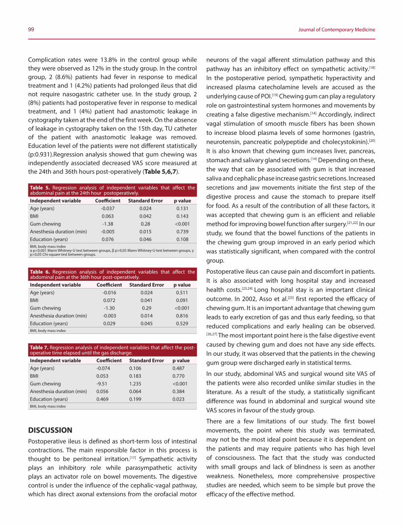
99 Journal of Contemporary Medicine
Complication rates were 13.8% in the control group while they were observed as 12% in the study group. In the control group, 2 (8.6%) patients had fever in response to medical treatment and 1 (4.2%) patients had prolonged ileus that did not require nasogastric catheter use. In the study group, 2 (8%) patients had postoperative fever in response to medical treatment, and 1 (4%) patient had anastomotic leakage in cystography taken at the end of the first week. On the absence of leakage in cystography taken on the 15th day, TU catheter of the patient with anastomotic leakage was removed. Education level of the patients were not different statistically (p:0.931).Regression analysis showed that gum chewing was independently associated decreased VAS score measured at the 24th and 36th hours post-operatively (Table 5,6,7).
DISCUSSIONPostoperative ileus is defined as short-term loss of intestinal contractions. The main responsible factor in this process is thought to be peritoneal irritation.[17] Sympathetic activity plays an inhibitory role while parasympathetic activity plays an activator role on bowel movements. The digestive control is under the influence of the cephalic-vagal pathway, which has direct axonal extensions from the orofacial motor
neurons of the vagal afferent stimulation pathway and this pathway has an inhibitory effect on sympathetic activity.[18] In the postoperative period, sympathetic hyperactivity and increased plasma catecholamine levels are accused as the underlying cause of POI.[19] Chewing gum can play a regulatory role on gastrointestinal system hormones and movements by creating a false digestive mechanism.[14] Accordingly, indirect vagal stimulation of smooth muscle fibers has been shown to increase blood plasma levels of some hormones (gastrin, neurotensin, pancreatic polypeptide and cholecystokinin).[20]
It is also known that chewing gum increases liver, pancreas, stomach and salivary gland secretions.[14] Depending on these, the way that can be associated with gum is that increased saliva and cephalic phase increase gastric secretions. Increased secretions and jaw movements initiate the first step of the digestive process and cause the stomach to prepare itself for food. As a result of the contribution of all these factors, it was accepted that chewing gum is an efficient and reliable method for improving bowel function after surgery.[21,22] In our study, we found that the bowel functions of the patients in the chewing gum group improved in an early period which was statistically significant, when compared with the control group.
Postoperative ileus can cause pain and discomfort in patients. It is also associated with long hospital stay and increased health costs.[23,24] Long hospital stay is an important clinical outcome. In 2002, Asso et al.[25] first reported the efficacy of chewing gum. It is an important advantage that chewing gum leads to early excretion of gas and thus early feeding, so that reduced complications and early healing can be observed.[26,27] The most important point here is the false digestive event caused by chewing gum and does not have any side effects. In our study, it was observed that the patients in the chewing gum group were discharged early in statistical terms.
In our study, abdominal VAS and surgical wound site VAS of the patients were also recorded unlike similar studies in the literature. As a result of the study, a statistically significant difference was found in abdominal and surgical wound site VAS scores in favour of the study group.
There are a few limitations of our study. The first bowel movements, the point where this study was terminated, may not be the most ideal point because it is dependent on the patients and may require patients who has high level of consciousness. The fact that the study was conducted with small groups and lack of blindness is seen as another weakness. Nonetheless, more comprehensive prospective studies are needed, which seem to be simple but prove the efficacy of the effective method.
Table 5. Regression analysis of independent variables that affect the abdominal pain at the 24th hour postoperatively.Independent variable Coefficient Standard Error p valueAge (years) -0.037 0.024 0.131BMI 0.063 0.042 0.143Gum chewing -1.38 0.28 <0.001Anesthesia duration (min) -0.005 0.015 0.739Education (years) 0.076 0.046 0.108BMI, body mass indexα p<0,001 Mann Whitney-U test between groups, β p>0,05 Mann Whitney-U test between groups, γ p>0,05 Chi-square test between groups.
Table 6. Regression analysis of independent variables that affect the abdominal pain at the 36th hour post-operatively.Independent variable Coefficient Standard Error p valueAge (years) -0.016 0.024 0.511BMI 0.072 0.041 0.091Gum chewing -1.30 0.29 <0.001Anesthesia duration (min) -0.003 0.014 0.816Education (years) 0.029 0.045 0.529BMI, body mass index
Table 7. Regression analysis of independent variables that affect the post-operative time elapsed until the gas discharge.Independent variable Coefficient Standard Error p valueAge (years) -0.074 0.106 0.487BMI 0.053 0.183 0.770Gum chewing -9.51 1.235 <0.001Anesthesia duration (min) 0.056 0.064 0.384Education (years) 0.469 0.199 0.023BMI, body mass index
100Ali Haydar Yılmaz, Efficacy of The Chewing Gum in Postoperative Ileus
CONCLUSIONRobot-assisted laparoscopic prostatectomy is being applied with increasing frequency in the treatment of loacized prostate cancer in the decad. This situation increases the frequency of encountering postoperative complications. We are convinced that tasteless chewing gum will be effective in the early resolution of the ileus observed in these surgeries.
ETHICAL DECLARATIONSEthics Comittee Approval: The ethic approval was optained from Erzurum Training and Research Hospital Clinical Research Ethics Committee with decision no and dated as 06-2018/01.
Informed Consent: Written informed consent was obtained from all participants who participated in this study.
Status of Peer-review: Externally peer-reviewed.
Conflict of Interest Statement: The authors have no conflicts of interest to declare.
Financial Disclosure: The authors declared that this study has received no financial support.
Author Contributions: All of the authors declare that they have all participated in the design, execution, and analysis of the paper, and that they have approved the final version.
REFERENCES1. Livingston EH, Passaro EP. Postoperative ileus. Dig Dis Sci 1990;
35.121-32.
2. Shibata Y, Toyoda S, Miyati M, et al. Patterns of intestinal motility recovery during the early stage f ollowing abdominal surgery: clinical and manometric study. World J Surg. 1997; 21: 806-809.
3. Lee H, Cho CW, Yoon S, et al. Effect of sham feeding with gum chewing on postoperative ileus after liver transplantation — a randomized controlled trial. Clin Transplant 2016; 30: 1501-7.
4. Matros E, Rocha F, Zinner M, et al. Does gum chewing ameliorate postoperative ileus? Results of a prospective, randomized, placebo controlled trial. J Am Coll Surg 2006; 202: 773-8.
5. Choi H, Kang SH, Yoon DK, et al. Chewing gum has a stimulatory effect on bowel motility in patients after open or robotic radical cystectomy for bladder cancer: a prospective randomized comparative study. Urology 2011; 77: 884–90.
6. Asgeirsson T, El-Badawi KI, Mahmood A, et al. Postoperative ileus: it costs more than you expect. J Am Coll Surg 2010; 210: 228–31.
7. Noble EJ, Harris R, Hosie KB, et al. Gum chewing reduces postoperative ileus? A systematic review and meta-analysis. Int J Surg 2009; 7: 100-5.
8. Bisanz A, Palmer JL, Reddy S, et al. Characterizing postoperative paralytic ileus as evidence for future research and clinical practice. Gastroenterol Nurs 2007; 31: 336–44.
9. Holte K, Kehlet H. Postoperative ileus: a preventable event. Br J Surg 2000; 87: 1480-93.
10. Heidenreich A, Bellmunt J, Bolla M et al. EAU guidelines on prostate cancer. Part 1: screening, diagnosis and treatment of clinically localised disease. Eur Urol 2011; 59: 61–71.
11. Leow JJ, Chang SL, Meyer CP et al. Robot-assisted versus open radical prostatectomy: a contemporary analysis of an all-payer discharge database. Eur Urol 2016; 70: 837–45
12. Stolzenburg JU, Kyriazis I, Fahlenbrach C et al. National trends and differences in morbidity among surgical approaches for radical prostatectomy in Germany. World J Urol 2016; 34: 1515–20
13. Yaxley JW, Coughlin GD, Chambers SK et al. Robot-assisted laparoscopic prostatectomy versus open radical retropubic prostatectomy: early outcomes from a randomised controlled phase 3 study. Lancet 2016; 388: 1057–66
14. Kouba EJ, Wallen EM, Pruthi RS, et al. Gum chewing stimulates bowel motility in patients undergoing radical cystectomy with urinary diversion. Urology 2007; 70: 1053-6.
15. Maffezzini M, Campodonico F, Parodi D, et al. Current perioperative management of radical cystectomy with intestinal urinary reconstruction for muscle-invasive bladder cancer and reduction of the incidence of postoperative ileus. Surg Oncol 2008; 17: 41–8.
16. Menon M, Hemal AK. Vattikuti Institute prostatectomy: a technique of robotic radical prostatectomy: experience in more than 1000 cases. J Endourol 2004; 18: 611-9.
17. Dayton MT, Townsend CM, Beauchamp RD, et al. Sabiston Textbook of Surgery, The Biological Basis of Modern Surgical Practice, 17thed. New York: Elsevier Saunders; 2004.
18. Quah HM, Samad A, Neathey AJ, et al. Does gum chewing reduce postoperative ileus following open colectomy for left-sided colon and rectal cancer? A prospective randomized controlled trial. Colorectal Dis. 2006; 8: 64-70.
19. Wille-JP, Guenaga KF, Castro AA, et al: pre-operative mechanical bowel cleansing or not? an updated meta-analysis. Colorectal Dis.2005; 7: 304-310.
20. Matros E, Rocha F, Whang E, et al. Does gum chewing ameliorate postoperative ileus? Results of a prospective, randomized, placebo controlled trial. J Am Coll Surg. 2006; 202: 773-778.
21. Gustafsson UO, Scott MJ, Schwenk W, et al. Guidelines for perioperative care in elective colonic surgery: enhanced recovery after surgery (ERAS (®)) society recommendations. World J Surg 2013; 37: 259–84.
22. Nygren J, Thacker J, Carli F, et al. Guidelines for perioperative care in elective rectal / pelvic surgery: enhanced recovery after surgery (ERAS (®)) society recommendations. World J Surg 2013; 37: 285–305.
23. Senagore A. Pathogenesis and clinical and economic consequences of postoperative ileus. Clin Exp Gastroenterol 2010; 3: 87–9.
24. Saclarides TJ. Current choices good or bad for the proactive management of postoperative ileus: a surgeon's view. J Perianesthesia Nurs 2006; 21: 7–15.

101 Journal of Contemporary Medicine
25. Asao T, Kuwano H, Nakamura J, et al. Gum chewing enhances early recovery from postoperative ileus after laparoscopic colectomy. J Am Coll Surg. 2002; 195: 30-32.
26. Stewart B, Woods RJ, Collopy RJ, et al. Early feeding after elective open colorectal resections: A prospective randomized trial. Aust NZ J Surg. 1998; 68: 125-128.
27. Ballantyne JC, Mao J. Opioid therapy for chronic pain. N Engl J Med. 2003; 349: 1943-1953.
Corresponding (İletişim): Gökce Celep, Sabuncuoğlu Şerefeddin Education and Research Hospital, Department of Pediatrics, Merkez, Amasya, TURKEYE-mail (E-posta): [email protected] (Geliş Tarihi): 29.12.2019 Accepted (Kabul Tarihi): 09.03.2020
DOI: 10.16899/jcm.666918J Contemp Med 2020;10(1):102-107
Orjinal Araştırma / Original Article
JOURNAL OF
CONTEMPORARY MEDICINEJournal ofContemporary MedicineYEAR: 2019 VOLUME: 9 ISSUE: 4
Neonatal Screening for Congenital Hypothyroidism and Its Other Benefits
Yenidoğanlarda Konjenital Hipotiroidi Taraması ve Diğer Yararları
Aim: We aimed to evaluate the prevalence of congenital hypothyroidism and analyze neonatal thyroid stimulating hormone (TSH) levels to estimate iodine status in our province indirectly
Material and Method: This study was conducted in the pediatric clinics of a government hospital in middle-northern Turkey. The data of national neonatal screening program (NNSP) for congenital hypothyroidism and thyroid function tests (free tetraiodothyronine (fT4), TSH) through hospital records were presented. The rate and severity of iodine deficiency was assessed by using the WHO epidemiologic criteria
Results: In this retrospectively designed cross-sectional study 1324 newborns were evaluated. Most of the participants had normal thyroid functions, 15.4% had transient hyperthyrotropinemia of the newborn, 2.7% had subclinical hypothyroidism 1.5% had congenital hypothyroidism (CH). The prevalence of hyperthyrotropinemia was high (26.1%), revealing moderate iodine deficiency indirectly.
Conclusion: Hyperthyrotropinemia is a significant problem in our province. The clinical follow up of the patients can be managed by general pediatricians. Evaluating TSH and fT4 levels seems to be the only way of diagnosing thyroid disorders of the newborn and provides indirect epidemiologic clues for iodine status of the local population. A preventable nutritional public health problem can be solved with a single step by interpreting the data of NNSP.
Keywords: Neonatal screening, thyroid functions, hypertyhrotropinemia, iodine, congenital hypotyhroidism
ÖzAbstract
Gökce Celep1, Zeynep Hülya Durmaz2
Amaç: Bu çalışmada ilimizde konjenital hipotiroidi sıklığı ve yenidoğanlarda tiroid hormonu uyarıcı hormon (tiroid stimülan hormon (TSH)) düzeyleri üzerinden iyot eksikliği durumunun saptanması amaçlandı.
Gereç ve Yöntem: Çalışma Orta Karadeniz’de bir devlet hastanesinin Pediatri bölümünde yürütüldü. Ulusal tarama programı ile hastane kayıtlarından elde edilen tiroid işlev testlerinin sonuçları sunuldu. İyot eksikliği durumu Dünya Sağlık Örgütü (DSÖ) tarafından belirlenen epidemiyolojik kriterlere göre dolaylı olarak tanımlandı.
Bulgular: Bin üç yüz yirmi dört term, sağlıklı yenidoğanın değerlendirildiği bu retrospektif, kesitsel çalışmada katılımcılarının çoğunda tiroid işlevleri normal sınırlarda idi. Katılımcıların %15,4’ünde yenidoğanın geçici tirotropinemisi, %2,7’sinde subklinik hipotiroidi, %1,5’inde konjenital hipotiroidi saptandı. Hipertiritropinemi sıklığı %26,1 idi, bu durum DSÖ’ne göre “orta düzeyde iyot eksikliği bölgesi” kriterleri ile uyumluydu.
Sonuç: İlimiz için konjenital hipotirioidi ve iyot eksikliği önemli bir halk sağlığı sorunudur. Bu hastaların klinik takibi ve tedavisi çocuk sağlığı ve hastalıkları uzmanları tarafından yürütülebilir. Tiroid işlev testlerinin değerlendirilmesi yenidoğan dönemi tiroid bozukluklarının tanısında tek araç olarak görünmektedir. Bu sayede saptanan dolaylı epidemiyolojik veriler, yaşanılan bölgenin iyot eksikliği sıklığı hakkında da bilgi sağlar. Yenidoğan tarama programı sonuçlarının doğru yorumlanması önlenebilir bir besinsel eksiklik sorununun da çözülmesine yardımcı olur.
Anahtar Kelimeler: Yenidoğan tarama programı, tiroid işlev testleri, hipertirotropinemi, iyot, konjenital hipotiroidi
1Amasya University Medical Faculty, Sabuncuoğlu Şerefeddin Education and Research Hospital, Department of Paediatrics, Amasya2 Amasya University Medical Faculty, Sabuncuoğlu Şerefeddin Education and Research Hospital, Department of Biochemistry, Amasya
103 Journal of Contemporary Medicine
INTRODUCTIONCongenital hypothyroidism (CH) is the most common endocrinological disorder of the newborn and the reason of preventable mental retardation. Recently, the incidence of CH has increased as the cut−off levels of thyroid− stimulating hormone (TSH) have been lowered. The incidence was reported as 1/3000−1/4000 live births throughout the world and 1/ 650 in Turkey.[1-3] Because thyroid hormones are essential for neurodevelopment and healthy growth, hypothyroidism results in psychomotor retardation and failure to thrive.[4]
The treatment is easy, inexpensive and effective when the patients are diagnosed early; thus screening programmes during the neonatal period have been established in several countries.[5] Unfortunately only 5% of the patients can be clinically diagnosed during the early infancy, but the response to treatment is insufficient to prevent neurologic sequela when L-thyroxine is initiated late.[6,7] Thus, CH satisfies all the criteria of neonatal screening along with effective treatment.[8] National neonatal screening program (NNSP) was initiated in Turkey in 2006 through heel prick blood spot tests under the supervision of the Turkish Ministry of Health.[9] NNSP is based on capillary TSH levels.[10] Suspected cases are referred to hospitals for further evaluation based on venous blood free tetraiodothyronine (fT4) and TSH levels.[11]
Iodine is an essential nutrient for the thyroid gland and the neurological development and thyroid functions are related to the iodine status. Assessing the neonatal TSH level is one of the recommended monitoring methods to detect the iodine status of the target population as well as the results of iodine supplementation programmes.[12,13] The rate and severity of iodine deficiency (ID) may be assessed using the World Health Organization (WHO) epidemiologic criteria.[12] These criteria are based on the proportion of newborns with serum TSH levels of >5 mIU/ ml in venous blood samples so that this nutritional problem of the local population may be detected indirectly.[14] Since 1998, the salt iodization notification obliged the manufacturers to enrich table salt with 25−40 mg/ kg iodine compounds.[15] Consequently, the iodine uptake has improved and the median urinary iodine concentration in school-aged children has increased from 25.5 mcg/ L to 130 mcg/ L in 2007. Nonetheless, ID was reported to be an ongoing common public health problem.[16,17] We aimed to evaluate thyroid dysfunctions in the newborn population of our region a decade after the initiation of NNSP and interpret the neonatal TSH levels to contribute the estimation of iodine status in our province.
MATERIAL AND METHODThis study was conducted in the pediatric clinics of a secondary health care center from 1 January 2016 to 31 December 2017. The study center is situated in a small city in the West Black Sea Region of Turkey, according to the Turkish Statistical Institute’s grouping.[18] During the study period, 7514 babies were born, and 7638 capillary blood samples were sent to the central laboratories from our province. Blood specimens were collected from the third to fifth day after birth or at discharge from the hospital if it was planned earlier. In this study, specimens taken after 48 hours were considered. The standard procedure of obtaining capillary blood samples for neonatal screening is as follows: The heel is warmed for 3 minutes before the process. The blood specimen is obtained by puncturing the heel, and taken onto the standard filter paper cards provided by the Ministry of Health. Circles on the card are appropriately saturated by blood until the drop of blood appears behind. The cards are air-dried at room temperature in the horizontal position for 3 hours, avoiding heat and light, and then sent to the central laboratories of the Public Health Institution (PHI) for testing. These cards contain information regarding the registration number, surname of the newborn, identity information of the mother, residence, date, time and place of birth, gender, gestational week, type of delivery, birth weight, contact address, phone number and date of sampling along with the name and signature of the healthcare worker who obtained the sample and filled the card. The samples are tested within three working days after being received. NNSP in Turkey is based on capillary TSH level, and the cut-off level is determined to be 5.5 mIU/ml (1 mIU/ml= 1 IU/L). Values <5.5 mIU/ml are defined as “normal”. Infants having TSH levels between 5.5−20 mIU/ml are recalled for control screening tests. If the second sample measurement is above 5.5 mIU/ ml or the TSH level is higher than 20 mIU/ml even in the first heel prick test, the newborn must be examined further with venous fT4 and TSH (11,19). Overall 660 babies (49.8%) needed venous fT4 and TSH evaluation, and 33 (4.6%) of them were identified as “possible CH” according to the local PHI data. The remaining babies were tested because of family concerns (n= 203; 15.3%) and clinical necessity [jaundice (n=19; 1.4%), constipation (n= 28; 2.1%), family history (n=83; 6.3%) and sleep disturbances (n=12; 0.9%)]. The medical records of all term, healthy, appropriate in weight for gestational age (AGA) newborns, who were evaluated for thyroid functions were investigated through the hospital information system.
Details such as the age on the date of examination and blood sampling for thyroid functions, gender, and the follow up diagnosis of patients were recorded.
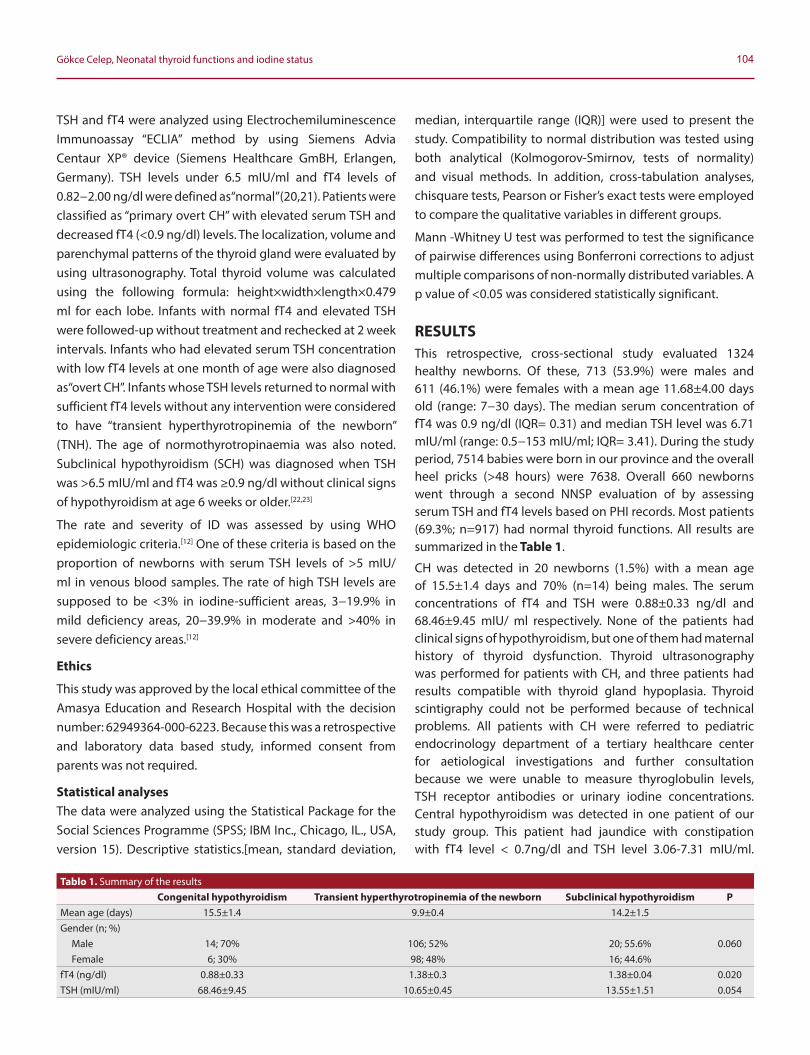
104Gökce Celep, Neonatal thyroid functions and iodine status
TSH and fT4 were analyzed using Electrochemiluminescence Immunoassay “ECLIA” method by using Siemens Advia Centaur XP® device (Siemens Healthcare GmBH, Erlangen, Germany). TSH levels under 6.5 mIU/ml and fT4 levels of 0.82−2.00 ng/dl were defined as“normal”(20,21). Patients were classified as “primary overt CH” with elevated serum TSH and decreased fT4 (<0.9 ng/dl) levels. The localization, volume and parenchymal patterns of the thyroid gland were evaluated by using ultrasonography. Total thyroid volume was calculated using the following formula: height×width×length×0.479 ml for each lobe. Infants with normal fT4 and elevated TSH were followed-up without treatment and rechecked at 2 week intervals. Infants who had elevated serum TSH concentration with low fT4 levels at one month of age were also diagnosed as“overt CH”. Infants whose TSH levels returned to normal with sufficient fT4 levels without any intervention were considered to have “transient hyperthyrotropinemia of the newborn” (TNH). The age of normothyrotropinaemia was also noted. Subclinical hypothyroidism (SCH) was diagnosed when TSH was >6.5 mIU/ml and fT4 was ≥0.9 ng/dl without clinical signs of hypothyroidism at age 6 weeks or older.[22,23]
The rate and severity of ID was assessed by using WHO epidemiologic criteria.[12] One of these criteria is based on the proportion of newborns with serum TSH levels of >5 mIU/ ml in venous blood samples. The rate of high TSH levels are supposed to be <3% in iodine-sufficient areas, 3−19.9% in mild deficiency areas, 20−39.9% in moderate and >40% in severe deficiency areas.[12]
Ethics
This study was approved by the local ethical committee of the Amasya Education and Research Hospital with the decision number: 62949364-000-6223. Because this was a retrospective and laboratory data based study, informed consent from parents was not required.
Statistical analysesThe data were analyzed using the Statistical Package for the Social Sciences Programme (SPSS; IBM Inc., Chicago, IL., USA, version 15). Descriptive statistics.[mean, standard deviation,
median, interquartile range (IQR)] were used to present the study. Compatibility to normal distribution was tested using both analytical (Kolmogorov-Smirnov, tests of normality) and visual methods. In addition, cross-tabulation analyses, chisquare tests, Pearson or Fisher’s exact tests were employed to compare the qualitative variables in different groups.
Mann -Whitney U test was performed to test the significance of pairwise differences using Bonferroni corrections to adjust multiple comparisons of non-normally distributed variables. A p value of <0.05 was considered statistically significant.
RESULTSThis retrospective, cross-sectional study evaluated 1324 healthy newborns. Of these, 713 (53.9%) were males and 611 (46.1%) were females with a mean age 11.68±4.00 days old (range: 7−30 days). The median serum concentration of fT4 was 0.9 ng/dl (IQR= 0.31) and median TSH level was 6.71 mIU/ml (range: 0.5−153 mIU/ml; IQR= 3.41). During the study period, 7514 babies were born in our province and the overall heel pricks (>48 hours) were 7638. Overall 660 newborns went through a second NNSP evaluation of by assessing serum TSH and fT4 levels based on PHI records. Most patients (69.3%; n=917) had normal thyroid functions. All results are summarized in the Table 1.
CH was detected in 20 newborns (1.5%) with a mean age of 15.5±1.4 days and 70% (n=14) being males. The serum concentrations of fT4 and TSH were 0.88±0.33 ng/dl and 68.46±9.45 mIU/ ml respectively. None of the patients had clinical signs of hypothyroidism, but one of them had maternal history of thyroid dysfunction. Thyroid ultrasonography was performed for patients with CH, and three patients had results compatible with thyroid gland hypoplasia. Thyroid scintigraphy could not be performed because of technical problems. All patients with CH were referred to pediatric endocrinology department of a tertiary healthcare center for aetiological investigations and further consultation because we were unable to measure thyroglobulin levels, TSH receptor antibodies or urinary iodine concentrations. Central hypothyroidism was detected in one patient of our study group. This patient had jaundice with constipation with fT4 level < 0.7ng/dl and TSH level 3.06-7.31 mIU/ml.
Tablo 1. Summary of the resultsCongenital hypothyroidism Transient hyperthyrotropinemia of the newborn Subclinical hypothyroidism P
Mean age (days) 15.5±1.4 9.9±0.4 14.2±1.5Gender (n; %)
Male 14; 70% 106; 52% 20; 55.6% 0.060Female 6; 30% 98; 48% 16; 44.6%
fT4 (ng/dl) 0.88±0.33 1.38±0.3 1.38±0.04 0.020TSH (mIU/ml) 68.46±9.45 10.65±0.45 13.55±1.51 0.054

105 Journal of Contemporary Medicine
Subsequently, he was referred to the pediatric endocrinology clinic for further evaluation, and the diagnosis was confirmed. Treatment with Lthyroxine was commenced in all cases of CH during the neonatal period. Even though the CH group had higher TSH levels with a male preponderance, no statistically significant effects were observed regarding TSH levels, symptoms or gender among CH, SCH and TNH groups (p= 0.054, p=0.08, and p=0.06, respectively).
TNH was detected in 204 patients (15.4%). Of these 98 (48%) were girls, and 106 (52%) were boys with a mean age of 9.9±0.4 days. The average TSH level of this group was 10.65±0.45 mIU/ ml and normalized within 30.16±15.26 days. The mean serum concentration of fT4 was 1.38±0.38 ng/dl.
Overall 36 patients (2.7%) were diagnosed with SCH because they had high TSH levels even 6 weeks after birth. The mean age of these patients was 14.2±1.5 days at the first evaluation with 55.6% (n=20) being boys and 44.6% (n=16) girls. The mean serum concentrations of fT4 and TSH were 1.38±0.04 ng/dl and 13.55±1.51 mIU/ml, respectively. Among these patients, 16 had normal thyroid functions during the first year of life and L-thyroxine treatment was commenced in one infant because of increasing TSH in repeating measurements (6.84-9.10mIU/ml) in the fourth month of age, but there were no clinical signs of hypothyroidism.
Overall, 87 newborns with high TSH levels were lost to followup during the study because of unavailable records after the first evaluation.
The rate of hyperthyrotropinaemia in the study population, ignoring the diagnosis, was 26.1% (n=347). When compared with the total newborn population, the rate was 4.6% and prevalence was 1:376. This data revealed the iodine status of our province indirectly. The prevalence of high TSH levels was compatible with moderate ID area according to the WHO criteria.
DISCUSSIONThis study evaluated 1324 healthy, term newborns for thyroid functions. Approximately 70% of participants had normal thyroid functions, whereas 15.4% had TNH, 2.7% SCH, and 1.5% overt CH. The prevalence of hyperthyrotropinemia was high in our study population (26.1%). The prevalence of patients diagnosed as “possible CH” was 4.6% based on the PHI data. This inconsistency between the hospital records and the PHI data could probably be due to patients with TNH whose TSH levels decreased at the second examination.
CH, TNH and SCH are expected to be high when there is ID or excess iodine status in the community. The recall rate of NNSP in ID areas is expected to be high, like the present study (8.78%).
Furthermore, the rate of neonatal thyroid disorders was high in our study group because the data reflected the results of the referral hospital of our province. The prevalence of high TSH levels was compatible with moderate ID area according to the WHO criteria, although our region was reported to be in a mild ID area previously. Iodine nutrition status in Turkey was reported to be “adequate” per WHO reports which presented the data based on the median urinary iodine concentrations in school children.[24,25]
CH is a common endocrinological problem in neonates. Notably, several studies have been conducted to determine its local and global prevalence. According to the NNSP of Turkey the rate of possible CH cases was 0.15% with an incidence of 1:650 in 2008−2010. The recall rate was reported to be 2.6% in this study.[3] Moreover, the researchers have reported the possible incidence of CH to be 1:418 in the West Black Sea Region of Turkey.[3] The incidence of CH was 1:376 in our study with a rate of 0.26% which is higher than the Turkish data. This finding may be related to the technical and population differences between the two studies. Our hospital was the referral center for further evaluation of the suspected cases; therefore our data reflected the results of a disadvantaged population. CH could be transient or permanent, but substantial followup is required to ascertain this. Transient CH is considered when thyroid functions remain within normal ranges without clinical features of hypothyroidism or hyperthyroidism or the clinician does not need to increase the dosage of L-thyroxine. The treatment can be stopped at approximately at age 3 if the neurodevelopment and growth compatible with age are provided. Nevertheless, the patient’s clinical followup and laboratory examinations are continued periodically to detect hypothyroidism. If retreatment is needed, the diagnosis is confirmed as “permanent CH”. However, if re-initiation of therapy is not required, the case can be assessed as “transient CH”. The results of this study do not reflect the follow-up data, thereby rendering us unable to differentiate the CH cases as permanent or transient, which is one of the limitations of the present study.
“TNH” is the elevation of TSH levels with normal fT4 levels and the absence of clinical signs of hypothyroidism. This situation resolves without any intervention within 4−6 weeks. In the present study, the rate was 15.4%. Notably, TNH is a common clinical problem in ID areas and the aetiology is usually related with iodine exposure. Here, as some of the limitations of the study, we did not mention the iodine status, salt consumption habits, types of delivery, iodine exposure during or after delivery or the thyroid functions of the mothers.
106Gökce Celep, Neonatal thyroid functions and iodine status
Therefore, it was just a clinical observation that salt consumption habits would have been changed because of speculative dietary rules and popular information regarding unrefined salt forms to be considered healthier. Even though populationbased health studies in 2008 reported an increase in iodized salt consumption, no investigations have been reported recently.[26] Another reason for this could be the usage of iodine-containing antiseptics for skin cleaning during obstetrical procedures.[17] Iodine can be transferred to the baby through the placenta and breast milk, which can hinder organification during thyroid hormone synthesis because of the Wolff-Chaikoff effect in early postnatal life.[27] The prevalence of TNH was reported to be 12% in another study from the middle Black Sea Region.[28] When this laboratory situation lasts more than 4−6 weeks this is called SCH. The prevalence of SCH was reported to be 2% in pediatric age group.[29] SCH must be followed periodically because hypothyroidism could become overt with clinical signs or lowered fT4 levels, like in one of our cases or thyroid functions may normalize, as in 16 cases of our study. Because thyroid hormones are necessary for brain development, authors recommend initiating L-thyroxine for TSH levels above10 mIU/ml. When TSH is 6−10 mIU/ml, the treatment decision depends on clinical signs of hypothyroidism or continued TSH elevation, as in our study case.
Study limitations:
This study had several limitations. The study was based on hospital records and the follow-up duration was short. The results of thyroid scintigragphy, thyroglobulin, TSH receptor antibodies and other tests were not presented. Clinical information regarding iodine nutrition, maternal thyroid functions, salt consumption, consanguinity, familial history of thyroid disease and iodine exposure were not mentioned. In addition, our data regarding iodine status reflected a limited region Nonetheless, in this study, we focused on the outcomes of the cooperation between primary and secondary health care centers for the provision of preventive medicine. Even though the results are not novel, we would like to draw attention to the prevalence of hyperthyrotropinemia in neonates and the role of general paediatricians in clinical followup through the outcomes of a well-organized NNSP.
CONCLUSIONThis study established that CH and ID are significant public health concerns in our province. NNSP was implemented successfully with adequate cooperation between the local PHI and pediatric clinics of the local referral hospital. Notably, clinical follow-up of patients can be managed by general pediatricians. Whenever pediatric endocrinology consultation is needed, the general pediatrician can consult the patient for further examination. This approach prevents waste of time
and money owing to unnecessary consultations. Screening newborn babies for CH is the most efficient and cost-effective way of preventing CH which is the most common reason for preventable mental retardation. The followup of clinical signs and TSH levels seem to be the only way of ascertaining CH, TNH, SCH because we could not detect any statistically significant factors for predicting the followup diagnosis. Moreover, this process provides indirect epidemiologic clues for regarding the iodine status of the local population, thereby enabling the detection of the effects of salt iodization programmes and the prevention of ID related disorders. Therefore, a preventable nutritional public health problem can be solved in a single step by interpreting the NNSP data.
ETHICAL DECLARATIONSEthics Comittee Approval: This study was approved by the local ethical committee of the Amasya Education and Research Hospital with the decision number: 62949364-000-6223.
Informed Consent: Because the study was designed retrospectively, no written informed consent form was obtained from patients.
Status of Peer-review: Externally peer-reviewed.
Conflict of Interest Statement: The authors have no conflicts of interest to declare.
Financial Disclosure: The authors declared that this study has received no financial support.
Author Contributions: All of the authors declare that they have all participated in the design, execution, and analysis of the paper, and that they have approved the final version.
REFERENCES1. Rastogi MV, LaFranci SH. Congenital hypothyroidism. Orphanet J Rare Dis.
[serial on the Internet] 2010; 5:17 (doi: 10.1186/17501172-5-17).[cited in September 2019] Available at: http://www. ojrd.com/content/5/1/17
2. Ford G, La Franchi SH. Screening for congenital hypothyroidism: a world wide view of strategies. Best Pract Res Clin Endocrinol Metab 2013;28(2):175–87.
3. Dilli D, Özbaş S, Acıcan D, Yamak N, Ertek M, Dilmen U. Establishment and development of a national newborn screening programme for congenital hypothyroidism in Turkey. J Clin Res Pediatr Endocrinol 2013;5(2):73-9.
4. Fisher DA. Disorders of the thyroid in the newborn and infant. In: Sperling MA, editor. 3 rd ed. Clinical Pediatric and Adolescent Endocrinology. Philadelphia, PA: Saunders; 2002. p. 164
5. Jacob H, Peters C. Screening, diagnosis and management of congenital hypothyroidism: European society for paediatric endocrinology consensus guideline. Arch Dis Child Educ Pract Ed 2015;100:260e3.
6. Bargagna S, Canepa G, Costagli C, et al. Neuropsychological follow-up in early treated congenital hypothyroidism: a problem-oriented approach. Thyroid 2000;10(3):243-9.
7. Kooistra L, Stemerdink N, van der MJ, Vulsma T, Kalverboer AF. Behavioural correlates of early-treated congenital hypothyroidism. Acta Paediatr 2001;90(10):1141-6.
8. Gopalakrishnan V, Joshi K, Phadke S, et al. Newborn screening for congenital hypothyroidism, galactosemia and biotinidase deficiency in Uttar Pradesh, India. Indian Pediatr. 2014;51(9):701– 5.
107 Journal of Contemporary Medicine
9. Eren E, Sağlam H, Zengin A, Gül Y, Çakır EDP, Özgür T, Tarım Ö. Evaluation of the Patients with Congenital Hypothyroidism: Effect of the National Screening Program. Journal of Current Pediatrics 2011; 9(1): 28-33
10. Delange F, de Benoist B , Pretell E , Dunn JT . Iodine defi ciency in the world: where do we stand at the turn of the century? Thyroid 2001; 11(5): 437–47 .
11. Public Health Institution of Turkey. Congenital Hypothyroidism Screening Algorhytim available at:https://hsgm.saglik.gov.tr/ tr/224-cocuk-ve-ergen-sagligi-dairesi-baskanligi.html. Accessed in June 2019
12. WHO Indicators for assessing iodine deficiency disorders and their control through salt iodisation. Geneva: WHO, 1994. WHO/ NUT/94. 6. Available from: http://apps.who.int/ iris/ bitstream/10665/70715/1/WHO_NUT_94.6.pdf
13. Delange F. Neonatal thyroid screening as a monitoring tool for the control of iodine deficiency. Acta Paediatrica Supplement 1999;88(S432):21-4.
14. Aburto NJ, Abudou M, Candeias V, Wu T. Effect and safety of salt iodization to prevent iodine deficiency disorders: a systematic review with meta-analyses. Geneva: WHO, 2014. Available from: http://apps.who.int/iris/ bitstream/ 10665/148175/1/ 9789241508285_eng.pdf.
15. Official Journal of the Turkish Government. 1998, 9 June 1 23397, p 29.
16. Erdoğan MF, Demir O, Emral R, Kamel AN, Erdoğan G. More than a decade of iodine prophylaxis is needed to eradicate goiter among school age children in a moderately iodine deficient region. Thyroid 2009;19(3):265-8.
17. Yaman AK, Demirel F, Ermiş B, Pişkin IE. Maternal and neonatal urinary iodine status and its effect on neonatal TSH levels in a mildly iodine-deficient area. J Clin Res Pediatr Endocrinol 2013;5(2):90-4.
18. Turkısh National Institution of Statistisc. Coğrafi bolgeler. available at: http://www.tuik.gov.tr/VeriBilgi.do%3Ftb_ id=45%26ust id= 13accessed in June 2019
19. Çaylan N, Tezel B, Özbaş S, Şahin N, Aydın Ş, Acıcan D, et al. Neonatal Thyroid-Stimulating Hormone Screening as a Monitoring Tool for Iodine Deficiency in Turkey. J Clin Res Pediatr Endocrinol 2016(2);8:187-191 DOI: 10.4274/jcrpe.2526
20. Gonç EN. Thyroid Diseases in Childhood and Adolescence. In: Eds: Cinaz P, Darendeliler F, Akıncı A, Özkan B, Dündar BN, Abacı A, Akçay. Basic Pediatric Endochrinology 2013, Istanbul, Nobel Tıp Kitabevleri T.pp 317-27
21. Mutlu M, Karagüzel G, Alıyazicioğlu Y, Eyüpoğlu I, Okten A, Aslan Y. Reference intervals for thyrotropin and thyroid hormones and ultrasonographic thyroid volume during the neonatal period. J Matern Fetal Neonatal Med 2012;25(2):120-4.
22. La Franchi SH. Approach to the diagnosis and treatment of neonatal hypothyroidism. J Clin Endocrinol Metab 2011;96(10):2959-67
23. Andıran N. Hypothyroidism. In Basic Pediatric Endochrinology 2013, Istanbul, Nobel Tıp Kitabevleri Eds: Cinaz P, Darendeliler F, Akıncı A, Özkan B, Dündar BN, Abacı A, Akçay T. pp 329-47.
24. Erdoğan MF, Ağbaht K, Altunsu T, et al. Current iodine status in Turkey. J Endocrinol Invest 2009 32(7): 617. https://doi. org/10.1007/BF03346519
25. WHO, UNICEF, ICCIDD. Assessment of iodine defi ciency disorders and monitoring theirelimination: A guide for programme managers. Geneva, World Health Organization, 2001 (WHO/ NHD/01.1).
26. Demographic and Health Survey (2008) Hacettepe Üniversitesi Nüfus Etütleri Enstitüsü (2009) Türkiye Nüfus ve Sağlık Araştırması, 2008. Hacettepe Üniversitesi Nüfus Etütleri Enstitüsü, Sağlık Bakanlığı Ana Çocuk Sağlığı ve Aile Planlaması Genel Müdürlüğü, Başbakanlık Devlet Planlama Teşkilatı Müsteşarlığı ve TÜBİTAK, Ankara, Türkiye. Turkish
27. Nishiyama S, Mikeda T, Okada T, Nakamura K, Kotani T, Hishinuma Transient hypothyroidism or persistent hyperthyrotropinemia in neonates born to mothers with excessive iodine intake. Thyroid. 2004;14(12):1077–83
28. Kara C, Günindi F, Yılmaz GC, Aydın M. Transient Congenital Hypothyroidism in Turkey: An Analysis on Frequency and Natural Course. J Clin Res Pediatr Endocrinol 2016;8(2):170-179 DOI:10.4274/jcrpe.2345
29. Bona G, Prodam F, Monzani A. Subclinical hypothyroidism in children: natural history and when to treat. J Clin Res Pediatr Endocrinol 2013;5(Suppl 1) :23-8.
Corresponding (İletişim): Nahide Ekici GÜNAY , Healthy Science University, Training and Research Hospital, Clinical Biochemistry Department, Kayseri, TurkeyE-mail (E-posta): [email protected] (Geliş Tarihi): 15.05.2019 Accepted (Kabul Tarihi): 06.02.2020
DOI: 10.16899/jcm.565897J Contemp Med 2020;10(1):108-113
Orjinal Araştırma / Original Article
JOURNAL OF
CONTEMPORARY MEDICINEJournal ofContemporary MedicineYEAR: 2019 VOLUME: 9 ISSUE: 4
Uric Acid-Lymphocyte Ratios and Myocardial Damage Parameters in ST Elevated Myocardial Infarction
ST Yükselmeli Miyokard İnfarktüsünde Ürik Asit-Lenfosit Oranları ve Miyokard Hasarı Parametreleri
Aim: Major negative cardiovascular outcomes and uric acid (UA), neutrophil: lymphocyte ratio (NLR) and lymphocyte levels have been demonstrated in up-to-date studies. In this study, it was aimed to investigate ULAR in patients with STEMI and unstable angina pectoris (UAP) who underwent primary percutaneous coronary intervention. For this purpose, we evaluated relationships between UALR and markers of myocardial injury (troponin I, creatine kinase-MB) inflammatory markers (high-sensitivity C-reactive protein [hs-CRP]) and blood count parameters), serum lipids and myocardial contractility.Material and Method: In this study, 346 patients were enrolled including 176 patients with STEMI and 170 patients with UAP. Mann Whitney U test and t-test were used to identify significant differences among parameters while ROC analyses were performed for parameters with significant difference including UA, NLR and ULAR. Results: The UA level, NLR and ULAR were significantly higher in patients with STEMI when compared to those with UAP (p<0.001; p=0.014 and p<0.001, respectively). Optimal cut-off point was determined as 6.05 mg/dL for UA (AUC: 0.561; specificity: 50%, sensitivity: 72%), 0.179 for ULAR (AUC: 0.980; specificity: 96%, sensitivity: 92%) and 2.3 for NLR (AUC: 0.913; specificity: 82%, sensitivity: 88%). Conclusion: The ULAR was significantly correlated with NLR, hsCRP, LVEF1 and hs-troponin I. We recommend ULAR, which was found to be more specific and sensitive than NLO, UA and hs-CRP in STEMI patients at presentation, as an inexpensive and readily available laboratory parameter which can be used as independent predictor for impairment in myocardial contractility
Keywords: Uric acid, lymphocyte, neutrophil-to-lymphocyte ratio, myocardial infarction, coronary angiography
ÖzAbstract
Nahide Ekici GÜNAY 1, Ahmet Oğuz BAKTIR2, Işıl ÇAKIR3, Sabahattin MUHTAROĞLU4, Mustafa NISARI5
Amaç: Majör kardiyovasküler kötü sonuçlar ile ürik asit (UA), nötrofil/lenfosit oranı (NLO), lenfosit düzeyleri arasındaki ilişki güncel çalışmalar ile gösterilmiştir. Bu çalışmada, primer perkütan koroner girişim (PCI) yapılan STEMI ve anstabil angina pektoris (UAP) hastalarında UALO düzeylerini incelemek amaçlandı. Bu bağlamda, UALO 'nun miyokardiyal hasarlanma belirteçleri (troponin I, kreatin kinaz-MB), inflamatuar belirteçler (yüksek duyarlıklı C-reaktif protein(hsCRP) ve kan sayımı değişkenleri), serum lipidleri ve miyokardi yal kontraktilite ile ilişkileri incelendi.Gereç ve Yöntem: Çalışmaya 176 STEMI ve 170 UAP olmak üzere toplam 346 hasta dâhil edildi. Parametreler arası anlamlı farkları görmek için T testi veya Mann-Witney U testi kullanıldı. STEMI ve UAP hastaları arasında anlamlı olarak farklı bulunan UA, NLO ve UALO için ROC analizleri yapıldı.Bulgular: UA, NLR ve UALO düzeyleri STEMI’de ve UAP hastalarına göre anlamlı olarak farklı ve yüksek idi (sırasıyla p<0,001, p=0,014 ve p<0,001). Optimum kesme noktaları UA için 6.05 mg/dL (AUC 0,561, özgüllük %50, duyarlılık %72; UALO için 0,179 (AUC 0,980, özgüllük %96, duyarlılık %92); NLR için 2.3 (AUC 0,913, özgüllük%82, duyarlılık,%88 olarak bulundu.Sonuç: STEMI hastalarında UALO ile NLO, hsCRP, LVEF1 ve hs-troponin I arasında anlamlı bir ilişki bulunmaktadır. STEMI hastalarının başvuru anında NLO, UA ve hsCRP’den daha dözgül ve duyarlı olarak bulduğumuz UALO düzeylerinin incelenmesini, miyokardın kasılma performansındaki bozulmanın bağımsız bir öngördürücüsü olarak kullanılabilecek, ucuz ve kolay erişilebilir bir rutin laboratuvar parametresi olarak öneriyoruz.
Anahtar Kelimeler: Ürik asit, lenfosit, nötrofil/lenfosit oranı, miyokard enfarktüsü, koroner anjiyografi
1Healthy Science University, Training and Research Hospital, Clinical Biochemistry Department, Kayseri, Turkey.2Healthy Science Universty, Training and Research Hospital, Cardiology Department, Kayseri, Turkey
3Healthy Science Universty, Training and Research Hospital, Biochemistry Department, Kayseri, Turkey 4Medical Scool of Erciyes University, Clinical Biochemistry, Kayseri, Turkey
5Faculty of Health Science, University of Nuh Naci Yazgan, Department of Nutrition and Dietetics, Kayseri, Turkey
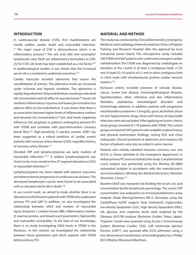
109 Journal of Contemporary Medicine
INTRODUCTIONIn cardiovascular disease (CVD), first manifestations are mostly sudden cardiac death and myocardial infarction.[1] The major cause of CVD is atherosclerosis which is an inflammatory process.[2] The uric acid (UA) and neutrophil/ lymphocyte ratio (NLR) are inflammatory biomarkers in CVD.[3] For CVD, UA levels has been established as a risk factor.[4,5] In epidemiological studies, it was shown that the increased serum UA is correlated to undesired outcomes.[6,7]
Cardiac myocytes secreted adenosine that causes the vasodilatation of arteries. The adenosine levels are increased under ischemia and hypoxia condition. The adenosine is rapidly degraded into UA by endothelium, resulting in elevated UA concentration and UA efflux to vascular lumen.[8] Serum UA mediates inflammatory response and leukocyte activation has adverse effect on the endothelium. It was shown that there is an association between higher levels of inflammatory markers and elevated UA concentrations.[9] Uric acid levels negatively influences the prognosis in patients undergoing primery PCI with STEMI and correlates with post-intervention coronary blood flow.[10] High-sensitivity C-reactive protein (CRP) has been suggested as a robust predictor of cardiac events patients with coronary artery disease (CAD), regardless history of coronary artery disease.[11]
Elevated CRP and lymphocytopenia are early markers of myocardial infarction.[12,13] A relative lymphocytopenia was shown to be more sensitive than ST segment elevation on ECG in myocardial infarction.[14]
Lymphocytopenia has been related with adverse outcomes and atherosclerosis progression in cardiovascular diseases. The decreased lymphocytes counts were found to be associated with an elevated risk for MI or death.[15]
In our current work, we aimed to study whether there is an alteration in UALR levels in patients with STEMI who underwent primary PCI and UAP. In addition, we also investigated the relationship between UALR and markers of myocardial injury (troponin I, creatine kinase MB), inflammatory markers (C-reactive protein, and blood count parameters), lipid profile and myocardial contractility. To the best of our knowledge, there is no study investigating UALR levels in STEMI in the literature.. In this context, we investigated the relationship between these parameters and UALR, patients with STEMI before primary PCI.
MATERIAL AND METHODThis study was conducted by Clinical Biochemistry, Emergency Medicine and Cardiology (Internal medicine) Clinics of Kayseri Training and Research Hospital after the approval by local instutional rewiev board. This retrospective study included 346 STEMI and UAP patients who underwent emergent cardiac catheterization.The STEMI was diagnosed by cardiologists an elevation at the J point in at least 2 contiguous leads ≥1,52 mm in leads V2–V3 and/or of ≥1 mm in other contiguous limb or chest leads with simultaneously positive cardiac necrosis markers.[16]
Exclusion criteria included presence of valvular disease, cancer, severe liver disease, immunopathological diseases, hypothyroidism, other infectious and also inflammatory disorders, polisitemia, hematological disorders and hemorrhagic diathesis. In addition, patients with progressive renal disorders (creatinine>1,8 mg/dL), gout, alcoholism, those on anti-hyperuricemic drugs, those with history of myocardial infarction were also excluded. After applying exclusion criteria, study group consisted of 176 patients with STEMI. The control group consisted of UAP patients with available medical history and physical examination findings, resting ECG and chest radiograph. Clinical symptoms at admission and coronary risk factors of patients were also recorded in same manner
Patients who initially admitted intensive coronary care unit (ICCU) or those admitted to the emergency department or before primary PCI were included to the study. Complete blood count analysis was performed using the Mindray BC-6800 automated analyzer in accordance with the manufacturer's recommendations (Mindray Bio-Medical Electronics Nanshan Shenzhen, China).[17]
Baseline UALR was measured via dividing the serum uric acid concantration by the lymphocyte percentage. The serum CRP concentration was analysed in an immunoturbidimetric assay analyzer (Dade Behring/Siemens BN II, Germany) using the CardioPhase hsCRP reagent. Total cholesterol, triglycerides, low density lipoprotein (LDL), high density lipoprotein (HDL), UA, glucose, and creatinine levels were analysed by the Olympus AU2700 analyzer (Beckman Coulter, Tokyo, Japan). Troponin I levels were assessed using Access 2 Immunoassay System (Beckman Coulter, USA). Left ventricular ejection fraction (LVEF1) was assessed after ICCU admission using a two dimensional transthoracic echocardiography by a Philips IE33 XMatrix Ultrasound Machines.
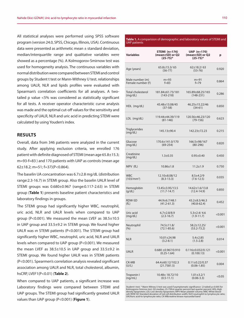
110Nahide Ekici GÜNAY, Uric acid-to-lymphocyte ratio in myocardial infarction
All statistical analyses were performed using SPSS software
program (version 24.0, SPSS, Chicago, Illinois, USA). Continuous
data were presented as arithmetic mean ± standard deviation,
median/interquartile range and qualitative variables were
showed as a percentage (%). A Kolmogorov-Smirnow test was
used for homogeneity analysis. The continuous variables with
normal distribution were compared between STEMI and control
groups by Student t test or Mann-Withney U test. relationships
among UALR, NLR and lipids profiles were evaluated with
Spearman’s correlation coefficients for all analyses. A two-
tailed p value <5% was considered as statistically significant
for all tests. A receiver operator characteristic curve analysis
was made and the optimal cut-off values for the sensitivity and
specificity of UALR, NLR and uric acid in predicting STEMI were
calculated by using Youden’s index.
RESULTSOverall, data from 346 patients were analyzed in the current
study. After applying exclusion criteria, we enrolled 176
patient with definite diagnosed of STEMI (mean age 65.8±15.3;
m=93-f=83 ) and 170 patients with UAP as controls (mean age
62±18.2; m=51; f=37)(P<0.864).
The baselin UA concantration was 6.7±2.8 mg/dL (distribution
range:2.3-16.7) in STEMI group. Also the baselin UALR level of
STEMI groups was 0.680±0.967 (range:0.117-2.63) in STEMI
group (Table 1) presents baseline patient characteristics and
laboratory findings in groups.
The STEMI group had significantly higher WBC, neutrophil,
uric acid, NLR and UALR levels when compared to UAP
group (P<0.001). We measured the mean LVEF as 38.5±10.5
in UAP group and 33.5±9.2 in STEMI group. We found higher
UALR was ın STEMI patients (P<0.001). The STEMI group had
significantly higher WBC, neutrophil, uric acid, NLR and UALR
levels when compared to UAP group (P<0.001). We measured
the mean LVEF as 38.5±10.5 in UAP group and 33.5±9.2 in
STEMI group. We found higher UALR was in STEMI patients
(P<0.001). Spearmen’s correlation analysis revealed significant
association among UALR and NLR, total cholesterol, albumin,
hsCRP, LVEF1(P<0.01) (Table 2).
When compared to UAP patients, a significant increase was
Laboratory findings were compared between STEMI and
UAP groups. The STEMI group had significantly greated UALR
values than UAP group (P<0.001) (Figure 1).
Table 1. A comparision of demographic and laboratory values of STEMI and UAP patients
VariablesSTEMI (n=176)
(mean±SD) or Q2 (25-75)*
UAP (n=170) (mean±SD) or Q2
(25-75)*p
Age (years) 65.8±15.3/ 65 (56-77)
62±18.2/ 63 (53-76) 0.920
Male number (m) Female number (f )
m=93f=83
m=91f=79 0.864
Total cholesterol (mg/dL)
181.84±61.73/181 (143-218)
165.89±68.25/165 (148-231) 0.286
HDL (mg/dL) 45.48±13.08/45 (37-58)
46.25±15.22/46 (34-61) 0.850
LDL (mg/dL) 119.44±44.39/119 (81-146)
120.56±46.23/120 (79-156) 0.623
Triglycerides (mg/dL) 145.13±90.4 142.23±72.23 0.215
Glucose (mg/dL)
170.6±141.0/170 (69-254)
166.5±98/167 (80-296) 0.820
Creatinine (mg/dL) 1.3±0.35 0.95±0.40 0.450
MPV (fL) 10.86±1.8 11.2±1.9 0.750
WBC (103/mm3)
12.10±8.08/12 (8.3-13.3)
8.5±4.2/9 (7.6-12.5) 0.035
Hemoglobin (g/dL)
13.45±3.95/13.5 (11.7-14.7)
14.62±1.6/13.8 (12.4-14.9) 0.850
RDW-SD (fL)
44.9±6.7/48.1 (41.2-61.3)
43.2±8.5/46.3 (40.8-62.4) 0.452
Uric acid (mg/dL)
6.7±2.8/8.9 (2.3-16.7)
5.3±2.4/ 4.6 (1.9-11.7) <0.001
Neutrophil (%)
74.5±11.8/ (72.1-85.6)
58.2±12.25/ (53.2-73.2) <0.001
NLR 10.01±24.98 (3.2-8.1)
5.4±2.85 (1.5-2.8) 0.014
UALR 0.680 ±0.967/0.910 (0.25-1.64)
0.116±0.032/0.121 (0.100.13) <0.001
CK-MB(U/L)
64.4±60.12/102.3 (21.7581.5)
0.11±0.22/0.37 (0.06-1.85) 0.004
Troponin I (ng/mL)
10.48± 18.72/10 (0.5-11.1)
1.01±3.2/1 (0.06-3.3) <0.05
Student t-test. * Mann-Witney U test was used if asymptomatic significance (2-tailed) p<0.005 for Kolmogorov-Smirnov test. Q2-median; 25-75first quarter percent-last quarter percent; HDL-High density lipoprotein; LDL-Low density lipoprotein; MPV-Mean platelet volume; WBC-White blood cell; RDW-SD-Standard deviation of red cell distribution width; NLR-neutrophil-to-lymphocyte ratio; UALRuric acid-to-lymphocyte ratio; CK-MBcreatine kinase myocardial band
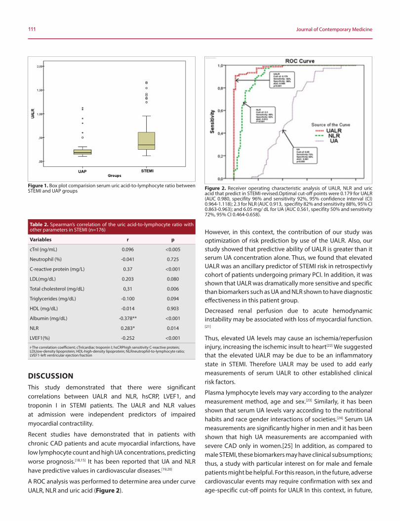
111 Journal of Contemporary Medicine
However, in this context, the contribution of our study was optimization of risk prediction by use of the UALR. Also, our study showed that predictive ability of UALR is greater than it serum UA concentration alone. Thus, we found that elevated UALR was an ancillary predictor of STEMI risk in retrospectivly cohort of patients undergoing primary PCI. In addition, it was shown that UALR was dramatically more sensitive and specific than biomarkers such as UA and NLR shown to have diagnostic effectiveness in this patient group.
Decreased renal perfusion due to acute hemodynamic instability may be associated with loss of myocardial function.[21]
Thus, elevated UA levels may cause an ischemia/reperfusion injury, increasing the ischemic insult to heart[22] We suggested that the elevated UALR may be due to be an inflammatory state in STEMI. Therefore UALR may be used to add early measurements of serum UALR to other established clinical risk factors.
Plasma lymphocyte levels may vary according to the analyzer measurement method, age and sex.[23] Similarly, it has been shown that serum UA levels vary according to the nutritional habits and race gender interactions of societies.[24] Serum UA measurements are significantly higher in men and it has been shown that high UA measurements are accompanied with severe CAD only in women.[25] In addition, as compared to male STEMI, these biomarkers may have clinical subsumptions; thus, a study with particular interest on for male and female patients might be helpful. For this reason, in the future, adverse cardiovascular events may require confirmation with sex and age-specific cut-off points for UALR In this context, in future,
Figure 2. Receiver operating characteristic analysis of UALR, NLR and uric acid that predict in STEMI-revised.Optimal cut-off points were 0.179 for UALR (AUC 0.980, specifity 96% and sensitivity 92%, 95% confidence interval (CI) 0.964-1.118); 2.3 for NLR (AUC 0.913, specifity 82% and sensitivity 88%, 95% CI 0.863-0.963); and 6.05 mg/ dL for UA (AUC 0.561, specifity 50% and sensitivity 72%, 95% CI 0.464-0.658).
DISCUSSIONThis study demonstrated that there were significant correlations between UALR and NLR, hsCRP, LVEF1, and troponin I in STEMI patients. The UALR and NLR values at admission were independent predictors of impaired myocardial contractility.
Recent studies have demonstrated that in patients with chronic CAD patients and acute myocardial infarctions, have low lymphocyte count and high UA concentrations, predicting worse prognosis.[18,15] It has been reported that UA and NLR have predictive values in cardiovascular diseases.[19,20]
A ROC analysis was performed to determine area under curve UALR, NLR and uric acid (Figure 2).
Figure 1. Box plot comparision serum uric acid-to-lymphocyte ratio between STEMI and UAP groups
Table 2. Spearman’s correlation of the uric acid-to-lymphocyte ratio with other parameters in STEMI (n=176)
Variables r p
cTnI (ng/mL) 0.096 <0.005
Neutrophil (%) -0.041 0.725
C-reactive protein (mg/L) 0.37 <0.001
LDL(mg/dL) 0.203 0.080
Total cholesterol (mg/dL) 0,31 0.006
Triglycerides (mg/dL) -0.100 0.094
HDL (mg/dL) -0.014 0.903
Albumin (mg/dL) -0.378** <0.001
NLR 0.283* 0.014
LVEF1(%) -0.252 <0.001
r-The correlation coefficient; cTnIcardiac troponin I; hsCRPhigh sensitivity C-reactive protein; LDLlow-density lipoprotein; HDL-high-density lipoprotein; NLRneutrophil-to-lymphocyte ratio; LVEF1-left ventricular ejection fraction
112Nahide Ekici GÜNAY, Uric acid-to-lymphocyte ratio in myocardial infarction
it may be considered dose, age and sex dependent effects of specific uric acid lowering therapies on STEMI prevention.
Our study has some limitations. First, this was a single center, retrospective and observational, non-randomized work and enrolled a relatively small number of patients. In addition, the IRA patency was used only in the selection of the diagnosed patients. On the other hand, UAP group had previously elective PCI patients (n=21). Finally and more importantly, the correlation of UALR with ECG (which is accepted as a diagnostic standard for myocardial infarction) myocardial contractility, classical biochemical or inflammatory markers were studied while we did not study correlations between UALR affected major coronary arteries, stenosis levels, coronary artery disease severity scores. The data were anonymously collected due to due to the retrospective nature of the study.
Although this study had these limitations, this is the first study to evaluate the predictive value of UA, UALR, and NLR surgoing STEMI patients. The large scale randomized and controlled studies must be conducted to address the association of UALR with more cardiac scors and inflamatory markers among STEMI patients.
RESULTSThe results of our study suggest that the UALR which is cheaply and easily measured, may be used an ancillary biomarker for in risk-stratifying in STEMI patients. STEMI patients with high, hsCRP, UALR and, NLR levels should be monitored more closely in order to recognize early adverse outcomes. These values may also help clinicians in following their patients and they can also help in decision-making process for treatment modalities in order to determine how these biomarkers may influence treatments required.
ETHICAL DECLARATIONSEthics Comittee Approval: Ethics committee approval was received for this study from institutional ethics committee of Kayseri Training and Research Hospital (Decision Number: 2016-52).
Informed Consent: Written informed consent was obtained from all participants who participated in this study.
Status of Peer-review: Externally peer-reviewed.
Conflict of Interest Statement: The authors have no conflicts of interest to declare.
Financial Disclosure: The authors declared that this study has received no financial support.
Author Contributions: All of the authors declare that they have all participated in the design, execution, and analysis of the paper, and that they have approved the final version.
Acknowledgments: We thank all of our patients, investigator and laboratory staff for their participation in this study. Also we would like to thank the Health Ministry of XXXX for archival research permits by the Institutional Scientific Committee.
REFERENCES1. Pozo E, Agudo-Quilez P, Rojas-Gonzalez A, Alvarado T, Olivera MJ.
Noninvasive diagnosis of vulnerable coronary plaque. World J Cardiol 2016; 26: 520-33.
2. Geovanini GR, Libby P. Atherosclerosis and inflammation: overview and updates. Clin Sci (Lond). 2018; 132(12):1243–1252.
3. Acet H, Ertaş F, Akıl MA, et al. Novel predictors of infarct-related artery patency for ST-segment elevation myocardial infarction: Platelet-to-lymphocyte ratio, uric acid, and neutrophil-tolymphocyte ratio. Anatol J Cardiol. 2015;15(8):648–656.
4. Purnima S, El-Aal BG. Serum uric acid as prognostic marker of coronary heart disease (CHD). Clin Investig Arterioscler. 2016;28(5):216-224.
5. Gazi E, Temiz A, Altun B, Barutçu A, Bekler A, Güngor O, et al. The association between serum uric acid level and heart failure and mortality in the early period of ST-elevation acute myocardial infarction. Turk Kardiyol Dern Ars. 201;42(6):501-508.
6. Brand FN, McGee DL, Kannel WB, Stokes J, Castelli WP. Hyperuricemia as a risk factor of coronary heart disease: The Framingham Study. Am J Epidemiol. 1985; 121, 11-8.
7. Rahimi-Sakak F, Maroofi M, Rahmani J. et al. Serum uric acid and risk of cardiovascular mortality: a systematic review and dose-response meta-analysis of cohort studies of over a million participants. BMC Cardiovasc Disord. 2019; 19: 218.
8. Raatikainen MJ, Peuhkurinen KJ, Hassinen IE. Contribution of endothelium and cardiomyocytes to hypoxia-induced adenosine release. J Mol Cell Cardiol1994;26: 1069-80.
9. Lazzeri C, Valente S, Chiostri M, Sori A, Bernardo P, Gensini GF. Uric acid in the acute phase of ST elevation myocardial infarction submitted to primary PCI: its prognostic role and relation with inflammatory markers: a single center experience. Int J Cardiol. 2010; 21:138 (2):206-209.
10. Wang R, Mei B, Liao X, et al. Determination of risk factors affecting the in-hospital prognosis of patients with acute ST segment elevation myocardial infarction after percutaneous coronary intervention. BMC Cardiovasc Disord. 2017;17(1):243.
11. Brodov Y, Behar S, Goldenberg I, Boyko V. Usefulness of Combining Serum Uric Acid and C-Reactive Protein for Risk Stratification of Patients With Coronary Artery Disease Am J Cardiol 2009; 104:194–8.
12. Gül İ, Aykan AÇ, Gokdeniz T, Boyacı F, Kalaycıoğlu E, Hatem E, et al. The relationship between C-reactive protein and the lapse of time since the onset of the symptoms after acute myocardial infarction: an prospective-observational study. Anadolu Kardiyol Derg. 2014;14(7):599-605.
13. Annen B, Mang G, Schuiki E, Strebel U, Knoblauch M C-reactive protein and relative lymphocytopenia: early markers of acute myocardial infarction? Schweiz Med Wochenschr 1999; 11: 1931-4.
14. Thomson SP, Gibbons RJ, Smars PA, Suman VJ, Pierre RV, Santrach PJ, Jiang NS. Incremental value of the leukocyte differential and the rapid creatine kinase-MB isoenzyme for the early diagnosis of myocardial infarction. Ann Intern Med. 1995 Mar 1;122(5):335341.
15. Núnez J, Sanchis J, BodíV, Núnez E, Mainar L. Relationshipbetween low lymphocyte count and major cardiac events in patients with acute chest pain, a non-diagnostic electrocardiogram and normal troponin levels. Atherosclerosis 2009; 206: 251-7.
16. Hwang C, Levis JT. ECG diagnosis: ST-elevation myocardial infarction. Perm J. 2014;18(2):e133.
113 Journal of Contemporary Medicine
17. Pipitone S, Buonocore R, Gennari D, Lippi G. Comparison of nucleated red blood cell count with four commercial hematological analyzers. Clin. Chem. Lab. Med 2015; 53: 315–8.
18. Freedman DS, Williamson DF, Gunter EW, Byers T. Relation of serum uric acid to mortality and ischemic heart disease. The Nhanes Epidemiologic Follow-up Study. Am J Epidemiol 1995;141: 637-44.
19. Akpek M, Kaya MG, Uyarel H, Yarlioglues M, Kalay N, Günebakmaz O. (2011) The association of serum uric acid levels on coronary flow in patients with STEMI undergoing primary PCI. Atherosclerosis, 219: 334-41.
20. Park JJ, Jang HJ, Oh IY, Yoon CH, Suh JW, Cho YS, et al. Prognostic value of neutrophil to lymphocyte ratio in patients presenting with ST-elevation myocardial infarction undergoing primary percutaneous coronary intervention. Am J Cardiol. 2013;111(5):636-642.
21. Duan SY, Xing CY, Zhang B, Chen Y. Detection and evaluation of renal biomarkers in a swine model of acute myocardial infarction and reperfusion. Int J Clin Exp Pathol 2015; 8: 8336-47.
22. Ghaffar S, Salehi R, Mazani S, Aghavali S. Association of serum uric acid level with mortality and morbidity of patients with acute ST-elevation myocardial infarction. J CardiovascThorac Res 2016; 8: 56-60.
23. Liu HZ, Deng W, Li JL, Tang YM, Zhang LT, Cui Y. Peripheral blood lymphocyte subset levels differ in patients with hepatocellular carcinoma. Oncotarget. 2016; 22: 7(47):77558-64.
24. Beydoun MA, Fanelli-Kuczmarski MT, Canas JA, Beydoun HA, Evans MK, Zonderman AB. Dietary factors are associated with serum uric acid trajectory differentially by race among urban adults. Br J Nutr. 2018;120(8):935–945.
25. Barbieri L, Verdoia M, Schaffer A, Marino P, Suryapranata H, De Luca G. Impact of sex on uric acid levels and its relationship with the extent of coronary artery disease: A single-centre study. Atherosclerosis 2015; 241(1):241-8.
Corresponding (İletişim): Gökce Celep, Amasya Üniversitesi Tıp Fakültesi, Sabuncuoğlu Şerefeddin Eğitim Araştırma Hastanesi, Çocuk Sağlığı Ve Hastalıkları Bölümü, Amasya, TürkiyeE-mail (E-posta): [email protected] (Geliş Tarihi): 08.08.2019 Accepted (Kabul Tarihi): 13.03.2020
DOI: 10.16899/jcm.603548J Contemp Med 2020;10(1):114-121
Orjinal Araştırma / Original Article
JOURNAL OF
CONTEMPORARY MEDICINEJournal ofContemporary MedicineYEAR: 2019 VOLUME: 9 ISSUE: 4
İkinci Basamak Bir Sağlik Kurulusunda Mikrobiyoloji Laboratuvari ve Pediatri Kliniği İsbirliği
The Collaboration of Microbilogy Laboratuary And Pediatric Clinics in A Secondary Health Care Centre
Giriş: Enfeksiyon hastalıkları pediatri pratiğinde sık rastlanan hastalıklardır. Tanı ve tedavi yonetiminde kültür tetkikleri oldukça onemlidir. Pediatri kliniği ve mikrobiyoloji laboratuvarı iş birliği kurumların akılcı ilaç kullanımı politikasının belirlenmesinde vazgeçilmezdir. Bu yazıda bir il merkezinde pediatrik hasta grubunda kültür tetkiklerinin kullanım sıklığı ve etkinliğinin değerlendirilmesi, kültür materyali alım tekniklerinin okuyucuya hatırlatılması amaçlanmıştır.Gereç ve Yöntem: 1 Nisan 2016-31 Mart 2017 tarihleri arasında mikrobiyoloji laboratuvarına kabul edilen tüm kaliteli kültür ornekleri hastane ve laboratuvar kayıt sistemi üzerinden retrospektif olarak değerlendirilmiştir. Her ornek için uygun kültür alma, işleme ve değerlendirme yontemi kullanılmıştır.Bulgular: Çalışma süresince 515 kan, 3640 idrar, 209 konjonktiva, 32 yara, 143 gobek sürüntüsü, 220 boğaz, 69 solunum yolu sekresyonu ve 65 gaita orneği kültür yontemleriyle değerlendirilmiştir. Üreme sonuçlarında mikroorganizma dağılımı ve antibiyogram duyarlılıkları pediatrik yaş grubuna uygun seçeneklerle belirtilmiştir. Üremelerin %18’inde çoklu dirençli mikroorganizmalar soz konusudur.Sonuç: Pediatri kliniği ve mikrobiyoloji laboratuvarı iş birliğinin arttırılması, güçlendirilmesi için deneyimlerin paylaşılması gereklidir. Belli aralıklarla tekrarlanan surveyans çalışmaları etkin ampirik tedavilerin belirlenmesini sağlamaktadır. Özellikle pediatrik yaş grubunda akılcı ilaç kullanımı gelecek nesillerin daha sağlıklı olmasında onemlidir. Boylelikle antibiyotik direnç sorununun azaltılmasına katkıda bulunulabilir.
Anahtar Sözcükler: Pediatri, kültür tetkikleri, antibiyogram
AbstractÖz
Gökce Celep1, Hüseyin Burak Özçelik2, Rıdvan Güçkan2
Aim: Infectious diseases are common in pediatric practice. Culture tests are very important in diagnosis and treatment management. The collaboration of the pediatrics clinic and microbiology laboratory is indispensable in determining the rational drug use policy of the institutions. In this article, it was aimed to evaluate the frequency and effectiveness of the use of culture tests in a pediatric patient group in a provincial center and to remind the culture material acquisition techniques to readers.
Material and Method: All quality culture specimens accepted to microbiology laboratory between April 1, 2016 and March 31, 2017 were evaluated retrospectively through the hospital and laboratory registry system. The appropriate culture taking, processing and evaluation methods for each sample are explained in detail in the article.
Results: During the study, 515 blood, 3640 urine, 209 conjunctiva, 32 wound, 143 umbilical swabs, 220 throat cultures, 69 respiratory secretions and 65 stool samples were evaluated. Distribution of microorganisms and antibiogram sensitivity results were reported with appropriate treatment options for pediatric age group. The increase in multiple resistant microorganisms and their frequency in community-acquired infections are remarkable.
Conclusion: Pediatrics clinic and microbiology laboratory need to share experiences to increase and strengthen cooperation. Periodic repetitive surveillance studies allow the determination of effective empirical treatments. Especially in the pediatric age group, rational drug use is important for the health of future generations. Thus, antibiotic resistance problem can be reduced
Keywords: Pediatrics, culture tests, antibiotics
1Amasya Üniversitesi Tıp Fakültesi, Sabuncuoğlu Şerefeddin Eğitim Araştırma Hastanesi, Çocuk Sağlığı Ve Hastalıkları Bolümü2Amasya Üniversitesi Tıp Fakültesi, Sabuncuoğlu Şerefeddin Eğitim Araştırma Hastanesi, Mikrobiyoloji Bolümü
115 Journal of Contemporary Medicine
GIRIŞEnfeksiyon hastalıkları Çocuk Sağlığı ve Hastalıkları pratiğinde en sık karşılaşılan hastalık grubudur. Teknolojik gelişmeler oykü ve fizik incelemenin laboratuvarla desteklenmesini sağlamıştır. Mikrobiyolojik laboratuvar değerlendirmelerinde moleküler testler gibi gelişmiş yontemlerin kullanım alanı giderek artsa da doğru teknikle alınmış ve işlenmiş orneklerle kültür çalışmaları hala enfeksiyon etkenin saptanmasında altın standart olma ozelliğini korumaktadır.[1] Kan akımı enfeksiyonlarının değerlendirilmesinde kan kültürleri, üst solunum yolu enfeksiyonlarında boğaz kültürü, alt solunum yolu enfeksiyonlarında kaliteli balgam, endotrakeal aspirat veya açlık mide suyu, üriner sistem enfeksiyonlarında orta akım idrar orneği, merkezi sinir sistemi enfeksiyonlarında beyin omurilik sıvısı, gastroenterit olgularında dışkı, kapalı alan enfeksiyonlarında vücut sıvılarının uygun kültür ve antibiyogram çalışmaları ile incelenmesi kesin tanı ve tedaviye etkin katkı sağlamaktadır.[1] Bu yazıda amaç Orta Anadolu’da bir il merkezinde referans hastane olarak hizmet veren bir ikinci basamak sağlık kuruluşunda pediatrik yaş grubunda istenen kültür orneklerinin dağılımını, antibiyogram ozelliklerini gozden geçirmek ve kurum politikası olarak belirlenen kültür alma tekniklerini okuyucuyla paylaşmaktır.
GEREÇ VE YÖNTEMHastanemiz Mikrobiyoloji Laboratuvarı’nda 1 Nisan 2017-31 Mart 2018 tarihleri arasında pediatrik yaş grubu olgularından elde edilerek incelenen tüm kültür ornekleri hastane ve laboratuvar kayıt sistemleri üzerinden retrospektif olarak değerlendirildi. Hastaların yaş, cinsiyet, başvuru birimi, istemi yapan servis, yatış durumu, istenen ornek, üreyen mikroorganizma ve antibiyotik duyarlılığı verileri not edildi.
Kan kültürü ornekleri hastaya antibiyotik tedavisi başlamadan hemen once veya antibiyotik tedavisi alıyorsa son dozdan once periferik venlerden alındı. Kan orneği alınmadan once yüzey ilk once alkol, sonra povidon iyot çozeltisi, daha sonra yine alkol ile silinip kuruması beklendi. Çocuğun yaşı dikkate alınarak 1-5 ml kan alındı. Kan orneği alındıktan sonra kan kültürü şişesi steril olarak açılıp materyal besi yerine inoküle edildi. Kan kültürleri Mikrobiyoloji Laboratuarında BacT/ Alert 3D (Biomérieux, MarcyI’Etoile, Fransa) sisteminde 37 °C ısı da beş gün süre ile inkübe edilerek incelendi, Cihazın üreme sinyali verdiği şişeden alınan ornek %5 koyun kanlı agar ve “Eosin Methylen-blue (EMB)” besi yerine ekildi. Bundan sonra 1824 saat 37 °C de inkübasyon sonunda üreyen kolonilerin tür düzeyinde tanımlanması ve antibiyotik minimum inhibitor konsantrasyon (MİK) düzeylerinin saptanması VİTEK
2 Compact System (Biomérieux, Fransa) otomatize sistem aracılığı ile gerçekleştirildi. Antibiyotik duyarlılık test sonuçları Clinical & Laboratory Standarts Institute (CLSI) onerileri doğrultusunda raporlandı.[2] Kan akımı enfeksiyonları onemli mortalite nedeni olduğundan raporlama sırasında klinisyene bilgi verildi ve kontaminasyon şüphesi olan olgulardan 2. ornek istendi. Değerlendirme sırasında bu ornek dikkate alınarak veriler analiz edildi.
İdrar ornekleri sadece beş yaş üstü ve/veya tuvalet eğitimi olan çocuklardan alındı. Tuvalet eğitimi olmayan çocuklardan alınan torba idrar ornekleri standardizasyonun sağlanması için çalışma dışı bırakıldı. İdrar kültürü alınırken tuvalet eğitimi olan çocuklarda anne tarafından perine bolgesi sabunlu suyla yıkanıp kurulanarak yüzey temizliği yapıldı. İdrarın bir kısmı dışarı yapılıp, orta akım idrar orneği steril kaba alındı. Sondası olan hastadan idrar kültürü alınırken sondanın üretraya yakın yaklaşık 5 cm’lik kısmı alkolle silindikten sonra enjektor ucu yukarıya bakacak şekilde sondaya sokulduktan sonra idrar aspire edildi. Alınan ornek enjektorle laboratuvara gonderildi. En az 3 ml olarak alınan ornek kantitatif olarak standart ozelerle %5 koyun kanlı agar ve EMB agar besi yerlerine ekildi. Otuz yedi derecede bir gecelik (1824 saat) inkübasyon sonrası üreme olan kültürlerdeki bakteriler tanımlandı. Bakteri tanımlama ve antibiyogram çalışmalarında VİTEK 2 Compact System (Biomérieux, Fransa) otomatize sistemi kullanıldı, duyarlılık sonuçları (CLSI) kriterleri esas alınarak belirlendi. Ayrıca Geniş Spektrumlu Beta Laktamaz (GSBL) pozitifliği CLSI kriterleri doğrultusunda çift-disk sinerji yontemiyle araştırıldı; sefotaksim, seftazidim ve amoksisilin-klavulanat diskleri kullanıldı.[2]
Yara örnekleri: Enfeksiyon belirtisi olan akıntılı yara ve yenidoğanların gobekleri steril gazlı bez ve serum fizyolojik ile silindikten sonra dezenfeksiyon yapılmaksızın eküvyon la sürüntü orneği alındı. Standart transport besi yerine yerleştirildi, 30 dk içersinde laboratuvara iletildi. Burada %5 koyun kanlı agar ve EMB besi yerine ekilip1848 saat 37 °C de inkübe edildi. Üreyen kolonilerin tanımlanması ve antibiyotik duyarlılıklarının saptanması için VİTEK 2 otomatize sistemi kullanıldı. Goz sürüntü ornekleri konjoktival akıntıdan eküvyonla alınarak standart transport besi yerinde laboratuvara getirildi, %5 koyun kanlı agar ve EMB besi yerine ekilerek 18-24 saat 37 °C’de inkübe edildi. Üreyen mikroorganizmaların tanımlanması ve antibiyotik duyarlılığının belirlenmesi VİTEK 2 otomatize sistemiyle sağlandı. Sürüntü yoluyla alınan yara, gobek ve konjonktiva ornekleri kültür için değerlendirilmeden once mikroskobik olarak incelendi, gram boyamada lokosit varlığı ve epitel hücresi azlığı “kaliteli materyal” olarak
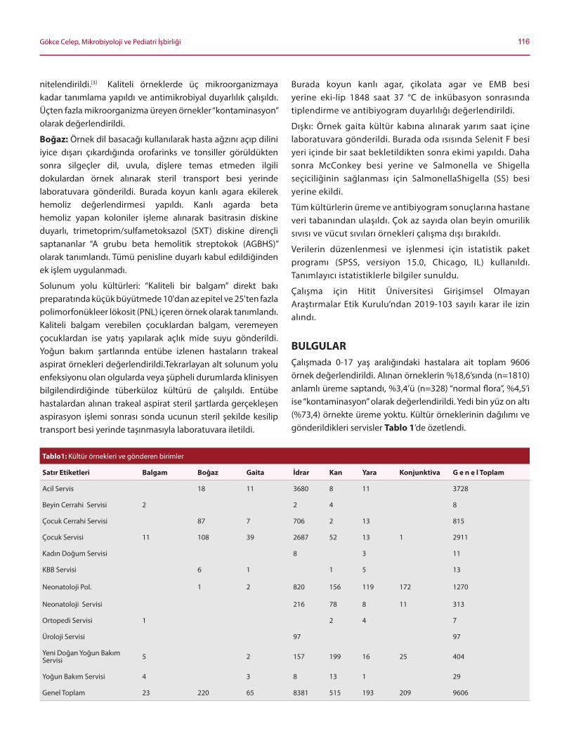
116Gökce Celep, Mikrobiyoloji ve Pediatri İşbirliği
nitelendirildi.[3] Kaliteli orneklerde üç mikroorganizmaya kadar tanımlama yapıldı ve antimikrobiyal duyarlılık çalışıldı. Üçten fazla mikroorganizma üreyen ornekler “kontaminasyon” olarak değerlendirildi.
Boğaz: Örnek dil basacağı kullanılarak hasta ağzını açıp dilini iyice dışarı çıkardığında orofarinks ve tonsiller gorüldükten sonra silgeçler dil, uvula, dişlere temas etmeden ilgili dokulardan ornek alınarak steril transport besi yerinde laboratuvara gonderildi. Burada koyun kanlı agara ekilerek hemoliz değerlendirmesi yapıldı. Kanlı agarda beta hemoliz yapan koloniler işleme alınarak basitrasin diskine duyarlı, trimetoprim/sulfametoksazol (SXT) diskine dirençli saptananlar “A grubu beta hemolitik streptokok (AGBHS)” olarak tanımlandı. Tümü penisline duyarlı kabul edildiğinden ek işlem uygulanmadı.
Solunum yolu kültürleri: “Kaliteli bir balgam” direkt bakı preparatında küçük büyütmede 10'dan az epitel ve 25'ten fazla polimorfonükleer lokosit (PNL) içeren ornek olarak tanımlandı. Kaliteli balgam verebilen çocuklardan balgam, veremeyen çocuklardan ise yatış yapılarak açlık mide suyu gonderildi. Yoğun bakım şartlarında entübe izlenen hastaların trakeal aspirat ornekleri değerlendirildi.Tekrarlayan alt solunum yolu enfeksiyonu olan olgularda veya şüpheli durumlarda klinisyen bilgilendirdiğinde tüberküloz kültürü de çalışıldı. Entübe hastalardan alınan trakeal aspirat steril şartlarda gerçekleşen aspirasyon işlemi sonrası sonda ucunun steril şekilde kesilip transport besi yerinde taşınmasıyla laboratuvara iletildi.
Burada koyun kanlı agar, çikolata agar ve EMB besi yerine eki-lip 1848 saat 37 °C de inkübasyon sonrasında tiplendirme ve antibiyogram duyarlılığı değerlendirildi.
Dışkı: Örnek gaita kültür kabına alınarak yarım saat içine laboratuvara gonderildi. Burada oda ısısında Selenit F besi yeri içinde bir saat bekletildikten sonra ekimi yapıldı. Daha sonra McConkey besi yerine ve Salmonella ve Shigella seçiciliğinin sağlanması için SalmonellaShigella (SS) besi yerine ekildi.
Tüm kültürlerin üreme ve antibiyogram sonuçlarına hastane veri tabanından ulaşıldı. Çok az sayıda olan beyin omurilik sıvısı ve vücut sıvıları ornekleri çalışma dışı bırakıldı.
Verilerin düzenlenmesi ve işlenmesi için istatistik paket programı (SPSS, versiyon 15.0, Chicago, IL) kullanıldı. Tanımlayıcı istatistiklerle bilgiler sunuldu.
Çalışma için Hitit Üniversitesi Girişimsel Olmayan Araştırmalar Etik Kurulu’ndan 2019-103 sayılı karar ile izin alındı.
BULGULARÇalışmada 0-17 yaş aralığındaki hastalara ait toplam 9606 ornek değerlendirildi. Alınan orneklerin %18,6’sında (n=1810) anlamlı üreme saptandı, %3,4’ü (n=328) “normal flora”, %4,5’i ise “kontaminasyon” olarak değerlendirildi. Yedi bin yüz on altı (%73,4) ornekte üreme yoktu. Kültür orneklerinin dağılımı ve gonderildikleri servisler Tablo 1’de ozetlendi.
Tablo1: Kültür ornekleri ve gonderen birimler
Satır Etiketleri Balgam Boğaz Gaita Idrar Kan Yara Konjunktiva G e n e l Toplam
Acil Servis 18 11 3680 8 11 3728
Beyin Cerrahi Servisi 2 2 4 8
Çocuk Cerrahi Servisi 87 7 706 2 13 815
Çocuk Servisi 11 108 39 2687 52 13 1 2911
Kadın Doğum Servisi 8 3 11
KBB Servisi 6 1 1 5 13
Neonatoloji Pol. 1 2 820 156 119 172 1270
Neonatoloji Servisi 216 78 8 11 313
Ortopedi Servisi 1 2 4 7
Üroloji Servisi 97 97
Yeni Doğan Yoğun Bakım Servisi 5 2 157 199 16 25 404
Yoğun Bakım Servisi 4 3 8 13 1 29
Genel Toplam 23 220 65 8381 515 193 209 9606

117 Journal of Contemporary Medicine
Kan kültürleri
Çalışmada 515 kan kültürü orneği değerlendirildi. Uygun alınan orneklerin %6,79’unda (n=35) üreme oldu. Kan kültürü orneklerinin %94,6’sı (n=487) yatan hastalara aitti. Tetkikin en fazla istendiği birim yeni doğan yoğun bakım ünitesi (YDYBÜ) idi (n=199; %38,6), üreme olan orneklerin de %45,7’si (n=16) YDYBÜ’den gonderilmişti. Uygun olmayan veya kontaminasyon olarak değerlendirilen ornek sayısı 3 (%0,6) idi. En sık üreyen 3 mikroorganizma sırası ile Staphylococcus epidermidis (n=15; %42,8), Staphylococcus hominis(n=7; % 20), Staphylococcus aureus (n=3; %8,6) idi. Tüm mikroorganizmalar vankomisine duyarlı olarak rapor edildi.
Idrar kültürleri
Çalışma donemi boyunca mikrobiyoloji laboratuvarına 8381 idrar kültürü orneği kabul edildi. Çalışmada beş yaş ve üstü tuvalet eğitimi olan çocuklar değerlendirildiğinden 3640 ornek dikkate alındı. Kültürün %56,9’u (n=2071) çocuk acil servisinden istenmişti. Örneklerin %96,7’si (n=3519) ayaktan başvuran hastalara aitti (n=3519; %96,7). Buna gore 381 (%10,5) ornekte üreme saptandı. İdrar kültürlerinde üreme kızlarda 5,9 kat daha fazla idi. En çok üreyen mikroorganizmalar Escherichia coli (n= 289; %75,9) ve Klebsiella pneumoniae (n=25; %6,5) idi. Tüm üremeler içinde geniş beta laktamaz direnci (GBSL) oranı %18 (n=72) olarak saptandı. Karbapenem ve amikasin duyarlılığı %100 idi. GBSL (-) orneklerde ise seftriakson duyarlılığı % 79,8 (n= 312/249), seftazidim duyarlılığı %80,2 (n=318/ 255) olarak raporlandı.
Konjonktiva sürüntü kültürleri
Çalışmada 209 konjonktiva orneği değerlendirildi. Örneklerin tümü yenidoğan birimlerinden gonderilmişti, %77,5’i (n=162) ayaktan başvuran neonatoloji polikliniği hastalarına aitti. Örneklerin %42,6’sında (n=89) üreme saptandı. En sık etkenler
S. aureus (n=41; %46,0), S. epidermidis (n= 20; %22,4) olmak üzere Gram pozitif koklardı. Etkene gore değişebilmekle birlikte gentamisin, fusidik asit, siprofloksasin duyarlılığı yüksek olduğundan lokal tedavi için uygun antibiyotik seçenekleriydi.
Yara sürüntü kültürleri
Toplam 32 yara sürüntü kültürü alındı, 13 tanesi çocuk cerrahisi servisinden, kalanlar çocuk hastalıkları servisinden gonderilmişti. Çocuk cerrahisi servisinden gonderilen 13 orneğin 9’unda (%69,2) üreme saptandı, S. aureus ve E. coli en sık etkenlerdi. Çocuk servisinden gonderilen 19 orneğin onunda üreme saptandı (%52,6). Gram pozitiflerden S. aureus (n=6), Gram negatiflerden K. pneumoniae (n=4) üreyen mikroorganizmalardı. Antibiyogram ozellikleri idrar ve kan orneklerine benzerdi.
Göbek sürüntü kültürü
Örneklerin tümü yenidoğan birimlerinden gonderilmişti. Örneklerin 96’sı ayaktan başvuran hastalara aitti. Toplam 143 orneğin 126’sında üreme oldu. En sık etkenler S. aureus (n= 55; %43,6), E.coli (n=26; %20,6), S. epidermidis (n= 17; %13,5) olarak saptandı. Antibiyogram çalışmalarında vankomisin, karbapenemler ve aminoglikozitlerde duyarlılık oranı yüksekti (Tablo 2).
Boğaz kültürleri
Çalışma sırasında toplam 220 boğaz sürüntüsü orneği değerlendirildi. Örneklerin %88,6’sı (n=195) çocuk polikliniklerinden gonderilmişti. Klinik yakınmaları nedeniyle tonsillofarenjit on tanısıyla istem yapıldı, taşıyıcılık taraması değerlendirilmedi. Olguların %4,1’inde (n=9) A grubu beta hemolitik streptokok üremesi oldu.
Tablo 2. Üreme olan kültürlerdeki en sık mikroorganizmalar ve antibiyotik duyarlılığı
IDRAR (n= 381) Escherichia coli (n=289)
Klebsiella pneumoniae
(n= 25)
Amoksisilin klavulanik asit 269/155; %57,6 25/15; %60
Seftriakson 267/223; %83,5 25/16; %64
Seftazidim 267/219; %82 25/20; %80
Meropenem 268/265; %98,8 25/25; %100
Amikasin 270/262; %97 25/25; %100
Nitrofurantoin 252/248; %98,4 22/17; %77,3
Göbek Sürüntüsü (n=126)
Staphylococcus aureus(n=55)
Escherichia coli (n= 26)
Staphylococcus epidermidis
(n=17)
Vankomisin 52/52; %100 17/8; %47
Fusidik asit 53/52; %98,1 8/2; %25
Teikoplanin 52/52; %100
Gentamisin 52/52; %100
Ampisilin 24/11; %45,8
Seftazidim 25/19; %76
Seftriakson 24/19; %79,1
Meropenem 26/25; %96,1
Gentamisin 26/20; %76,9 8/7; %87,5
Linezolid 8/8; %100
Konjonktiva Sürüntüsü(n= 89)
Staphylococcus aureus (n=41)
Staphylo co ccus epidermidis
(n=20)
Gentamisin 40/39; %97,5 14/9; %64,2
Fusidik asit 40/36; %90 14/4; %28,5
Siprofloksasin 40/39; %97,5 14/13; %92,8
118Gökce Celep, Mikrobiyoloji ve Pediatri İşbirliği
Solunum yolu örnekleri kültürleri
Laboratuvara alınan toplam ornek sayısı 69 idi, 23 hastadan ardışık 3 gün boyunca gonderilen ornekler tek sonuç olarak değerlendirildi. Balgam ve açlık mide suyu değerlendirmesinde tüm ornekler “normal flora üremesi” olarak raporlandı. İstenen hiçbir tüberküloz orneğinde üreme olmadı.
Dort hastada üreme saptandı, tümü yoğun bakım birimlerinden gonderilen trakeal aspirat ornekleri idi, 3 hastada Pseudomonas aeruginosa, 1 hastada maya üremesi oldu.
Gaita kültürü
Çalışılan toplam gaita orneği 65 tane idi; 39’u (%60) pediatri birimlerinden gonderilmişti. On yedi ornek yatan hastalardan alınmıştı. Gonderilen olguların hiç birinde invaziv gastroenterit etkenleri (Salmonella, Shigella) üremedi.
Klinisyenden talep gelmediği için clostridial veya E. coli tiplendirmesine yonelik çalışma yapılmadı.
TARTIŞMAÇocuk Sağlığı ve Hastalıkları pratiğinde sık karşılaşılan enfeksiyon hastalıklarının tanısında kültür tetkikleri en onemli tanı araçlarındandır. Klinik değerlendirmenin yanında tedavi planı aşamasında ozellikle invaziv enfeksiyonların yonetiminde klinisyene yol gostermektedir.[1] Bu çalışmada bir il merkezinde referans hastane olarak hizmet veren ikinci basamak sağlık kuruluşunda Pediatri Kliniği ve Mikrobiyoloji Laboratuvarının işbirliği gozden geçirilmiştir. Ülkemizde yapılan çok sayıda çalışma genelde tüm hastane servislerinin değerlendirildiği sonuçları ortaya koymaktadır. Bu çalışma çocuk odaklı olarak planlanmıştır, pediatrik hasta grubunun izlendiği tüm birimler dahil edilmiştir. En sık istenen orneklerin idrar ve kan olduğu gorülmüştür; solunum yolu orneklerinin olgu yonetiminde klinik değerlendirmeye katkısının beklenenden az olduğu düşünülmüştür.
Ateş çocuk acil ve polikliniklerine en sık başvuru nedenidir, ozellikle odağı bilinmeyen ateşte kan kültürü sık başvurulan bir tetkiktir, ampirik tedavi için etkenin belirlenmesini sağlamaktadır.[4] Kan akımı enfeksiyonları neden oldukları yük sek mortalite nedeniyle ozellikle kritik hasta bakımında her zaman dikkatli olunması gereken durumlardandır.[5] Literatürle benzer olarak kan kültürü üremeleri bu çalışmada da daha çok yoğun bakımlardan gonderilen orneklerde saptanmıştır. Hastanemizde çocuk yoğun bakım ünitesi bulunmadığından yoğun bakım gereksinimi olan hastalar genel yoğun bakımda izlenmektedir. Ancak Düzey 2 yenidoğan yoğun bakım
(YYBÜ) ünitesi mevcuttur. Değerlendirilen kan kültürü orneklerindeki mikroorganizma dağılımı literatürle benzerdir. Merkezimizden daha once yapılan başka bir çalışmada kan kültürü orneklerinin %77.7’sinde Gram pozitif, %22.3’ünde Gram negatif üreme olmuştur. Gram negatiflere odaklanan bu çalışmada üremelerin çok büyük kısmının yoğun bakımlardan kaynaklandığı ve yüksek antibiyotik direnci dikkat çekmektedir. Karbapenemler, aminoglikozitler, kolistin üreyen mikroorganizmaların duyarlı olduğu sayılı antibiyotiklerdir.[6]
Tokat’tan yayınlanan ve erişkin hasta grubunun değerlendirildiği bir çalışmanın sonuçları farklıdır. Değerlendirilen kan kültürü orneklerinin %64.3’ünde Gram negatif bakteriler, %30.9’unda Gram pozitif bakteriler, %4.8’inde mayalar üremiştir. En sık gorülen Gram negatif ajanlar E. coli ve Acinetobacter baumannii, Gram pozitifler, enterokoklar ve Staphylococcus aureus, mantar ise Candida albicans’tır.[7] Üremelerin çoğu dahili servislerden kaynaklıdır, bu durumun yoğun bakım – servis sirkülasyonunun hızlı olmasından kaynaklandığı düşünülmüştür.[7] Ankara’da çocuk acil servisinden istenen kan kültürü orneklerinin değerlendirildiği başka bir çalışmada istenen 10.642 orneğin %94,2’sinde üreme olmamış, üreme olanların ise %93,7’ si “kontaminasyon” olarak değerlendirilmiştir. Maliyet etkinlik ve klinik yararlılık açısından ornek alımı sırasında asepsi kurallarını uyulması gerektiği vurgulanan bu çalışmada üremelerin %91,7’si Gram pozitif, %8,3’ü Gram negatif olarak raporlanmıştır. Pnomokok üremeleri dikkat çekmiş, ayrıca üreyen koagülaz negatif stafilokoklarda metisilin direncinin onemli olduğu gozlenmiştir.[4] Bu çalışma ile kıyaslandığında merkezimizde çocuk hastalardan istenen kan kültürü sayısı az olmakla birlikte kontaminasyon sadece üç hasta için rapor edilmiştir. Benzer şekilde bizim kan kültürlerimizde de anlamlı üreme oranı düşüktür. Gram pozitif etkenlerin sıklığı aynı şekilde dikkat çekicidir.
İdrar yolu enfeksiyonları (İYE) nedeni bilinmeyen ateşin onemli bir nedenidir. Özellikle kendini sozel olarak ifade edemeyen çocuklarda huzursuzluk, iştahsızlık gibi ozgün olmayan bulgular dışında ateş tek ve en çarpıcı bulgu olabilir.[8,9] Bu nedenle kültür tanı ve tedavi yonetiminde çok onemlidir. Sonda veya suprapubik aspirasyon gibi invaziv yontemler klinik pratikte çok uygulanamadığından küçük bebeklerde torba idrar orneği sık kullanılmaktadır; ancak yalancı pozitiflik oranı yüksektir.[10] Bu çalışmada sadece tuvalet eğitimi olan çocuklardaki orta akım idrarları değerlendirilmiştir, lokal antiseptiklerle kıyaslandığında sonuçlar benzer olduğundan genital bolge temizliğinde sabun kullanılmıştır.[11] Tüm dünyada ve ülkemizde olduğu gibi Gram negatif enterik
119 Journal of Contemporary Medicine
basiller en sık etken olarak karşımıza çıkmıştır.[12,13] E. coli ve K. pneumoniae’nın başı çektiği bu etkenlerde en onemli sorun son yıllarda sadece hastane enfeksiyonlarında değil, toplum kaynaklı idrar yolu enfeksiyonlarında da genişlemiş spektrumlu beta-laktamaz (GSBL) enzimi varlığının tedavi sürecini zorlaştırmasıdır.[13] Çalışmamızda GBSL pozitifliği oranı %.18’dir. Bu etkenler için aminoglikozit duyarlılığı yüksektir. Gentamisin ve amikasin oral alımı iyi olan çocuklarda ayaktan tedavide uygun seçenek olarak gorünmektedir. Karbapenem duyarlılığı da %100 olarak saptanmıştır.
Amerika Bileşik Devletleri’nde onerilmesine karşın Ko-trimaksazol (SXT) direnci yüksek olması nedeniyle ülkemiz için uygun bir seçenek değildir. Kinolonların pediatrik yaş grubunda kullanımı onerilmemekte, artan direnç hızı nedeniyle ergen ve erişkinlerde de ilk seçenek olması istenmemektedir.[14] Tüm bu nedenlerle İYE’ de tek başına tam idrar tetkiki ile tanı ve tedavi yonetimi yetersiz olabileceğinden kültür değerlendirmesinin son derece onemli olduğu düşünülmektedir.
Konjonktivit ve blefarit yenidoğan doneminde en sık bakteriyel enfeksiyon olarak karşımıza çıkar.[15] Chlamydia trachomatis (Ch. Trochomatis), S. aureus, S. pneumoniae, S. viridans, H. influenzae, E. coli, Pseudomonas türleri, Klebsiella türleri, Enterobakter türleri, Proteus türleri, Neisseria gonorrhea ve virüsler de doğum sonrası 72. saatten sonra yenidoğan konjonktivitine sebep olabilir.[16-20] Literatürde en sık klamidyal konjonktivitler bildirilmesine karşın bu çalışmada Gram pozitif koklar en sık etken olarak saptanmıştır, bunun nedeni çalışma tekniğindeki farklar ve ozel çalışma gerektiren Ch. trochomatis değerlendirmesinin yapılamamış olması olabilir. Üreme olmayan 120 olguda klamidyal lenfeksiyon veya kimyasal konjonktival inflamasyon soz konusu olabilir.
Yara sürüntüsü orneklerinin değerlendirilmesinde uygun teknik kullanıldığında ve gram boyama ile mikroskobik inceleme yapıldıktan sonra işlenmesi tetkikin tanısal değerini arttırmaktadır. Pek çok çalışmada S. aureus ve E. coli en sık etkenler olarak karşımıza çıkmaktadır.[21,22] S. aureus endojen bir enfeksiyon kaynağı olmasının yanında deri bütünlüğü bozulduğunda çevresel kaynaklı enfeksiyonlar da oluşturabilir. Koagülaz, katalaz, agregasyon faktorü A ve lokosidinler gibi virülans faktorleri sayesinde cilt florası patojen nitelik kazanabilmektedir.[23] Bu çalışmada da üreme saptanan orneklerde literartürle uyumlu olarak en sık S. aureus ve Gram negatif enterik basiller saptanmıştır.
Yenidoğanlarda gobek kordonunun enfeksiyonu “funusit”, gobek kordonu kokünün enfeksiyonu “omfalit” olarak tanımlanmaktadır. Burada da en sık etkenler S. aureus ve E. coli
olarak karşımıza çıkmaktadır. Enfeksiyon lokal seyredebileceği gibi, portal ven üzerinden sepsise ve portal hipertansiyona neden olabilir. Lokal antibiotik tedavisi gereklidir. Etraftaki selülitten yayılım bulguları varsa parenteral antibiotikler de uygulanmalıdır.[24,25] Bu nedenle yenidoğanda tüm inflamasyon bulguları ozenle değerlendirilmelidir. Bizim çalışmamızda da en sık üreyen mikroorganizmalar ve antibiogram duyarlılıkları literatürle uyumludur.
A grubu beta hemolitik streptokok (AGBHS); yani Streptococcus pyogenes hayatı tehdit eden enfeksiyonlardan asemptomatik taşıyıcılığa kadar değişen geniş klinik yelpazeye sahip bir ajandır. Neden olduğu üst solunum yolu enfeksiyonlarının klinik olarak viral nedenlerden ayrılması zordur. Ayrıca akut enfeksiyondan çok mikroorganizmanın bazı duyarlı bireylerde neden olduğu süpüratif olmayan komplikasyonlar ( akut romatizmal ateş, poststreptokoksik glomerülonefrit) onemli morbidite ve mortalite nedenidir. Bu hastalıklardan korunmak için enfeksiyonun tedavisine dokuz gün içinde başlanmalıdır.[26-28]
Tüm bu nedenler etkenin tanımlanmasını onemli kılmaktadır, hızlı antijen testleri sık kullanılmakla birlikte boğaz kültürleri hala tanıda altın standarttır Ülkemizde sağlıklı ilkoğretim oğrencilerinin değerlendirildiği bir çalışmada AGBHS taşıyıcılık oranı yaklaşık %20 olarak saptanmıştır.[29] Gelişmiş ülkelerde tonsillofarenjit etkeni olarak %21-48 prevalans bildirilmekte, vakaların büyük kısmının 5-15 yaş aralığında olduğuna dikkat çekilmektedir.[30] Bizim çalışmamızdaki orneklerin tümü klinik yakınmalar doğrultusunda alınmıştır, taşıyıcılık taraması yapılmamıştır, klinik yakınması olan hastalardan alınan orneklerin %4’ünde AGBHS üremesi saptanmıştır.
Çocukların kaliteli balgam orneği vermesindeki zorluk ve açlık mide suyu alınmasının aile tarafından toleransı zor olması nedeniyle bu orneklerin elde edilmesi kolay değildir . Son yıllarda daha hızlı sonuç veren ve bakteri dışı etkenlerin de tanınmasına olanak sağlayan polimeraz zincir reaksiyonu gibi daha gelişmiş teknikler kültürlere gore daha sık kullanılmaktadır.[31,32] Toplum kaynaklı pnomoninin en sık etkenleri S. pneumoniae, Haemophilus influenzae, Mycoplasma pneumoniae, Chlamydia pneumoniae ve Legionella pneumophila’dır.[33] Bu çalışmada tüm balgam ornekleri sonuçları “normal flora” olarak yoğun bakım dışında gonderilen yorumlanmıştır. Ancak nazokomial pnomonisi olan ve solunum yolu ornekleri değerlendirilen az sayıdaki olguda literatürle uyumlu olarak Pseudomonas aeuriginosa (Ps .aeuriginosa) ve Candida albicans üremeleri saptanmıştır.[34]
120Gökce Celep, Mikrobiyoloji ve Pediatri İşbirliği
Gastrointestinal infeksiyonlarda, hastalık tablosunun ciddiyetine bağlı olarak tedavinin yonlendirilmesinde ve antibiyotik duyarlılığının saptanmasında dışkı kültürleri onemlidir. Dışkı kültürlerinde araştırılan bakteriler, etkenlerin bolgesel dağılımına bağlı olarak belirlenir, klinik bilgiler doğrultusunda farklı patojenler için ozel çalışmalar gerekebilir. Klinik mikrobiyoloji laboratuvarından olası tüm patojenleri rutin olarak araştırması beklenmez.[35] Ülkemizden yapılan ve 1300 orneğin değerlendirildiği bir çalışmada Shigella sıklığı %4,5, Salmonella sıklığı %2,5 olarak saptanmıştır.[36] Bizim çalış ma donemimizde dışkı orneklerinde her hangi bir patojen mikroorganizma ürememiştir. Merkezimizde bu çalışmanın çok az yapılmasının akut gastroenteritli olguların daha çok mikroskobik değerlendirme ile tanı almasından kaynaklandığı düşünülmektedir.
Çalışmamızın en büyük kısıtlılığı orneklerin ve hasta grubunun homojen olmaması, merkezimizde Mikrobioloji Bolümü ile en çok işbirliği içinde olması gereken birim olan çocuk yoğun bakım ünitesinin olmaması olduğunu düşünüyoruz.
SONUÇPediatri Kliniği ile Mikrobiyoloji Laboratuvarı arasındaki ilişkiyi değerlendirmeye çalıştığımız bu araştırmada işbirliğinin Pediatri Kliniği birimleri arasında istenen düzeyde homojen dağılım gostermediği gorülmüştür. Tüm Pediatri birimlerinin Mikrobiyoloji Bolümü ile işbirliği içinde olması, orneklerin uygun hastalardan doğru zamanda, doğru teknikle alınmasını sağlayacak, dolayısı ile tanı ve tedavi başarısını arttıracak tır. Akılcı ilaç kullanımının sağlanması klinisyen-laboratuvar işbirliğinin artmasına bağlıdır.
ETIK BEYANLAREtik Kurul Onayı: Çalışma için Hitit Üniversitesi Girişimsel Olmayan Araştırmalar Etik Kurulu’ndan 2019-103 sayılı karar ile izin alındı.
Aydınlatılmış Onam: Çalışma retrospektif olarak dizayn edildiği için hastalardan aydınlatılmış onam alınmamıştır.
Hakem Değerlendirme Süreci: Harici çift kor hakem değerlendirmesi.
Çıkar Çatışması Durumu: Yazarlar bu çalışmada herhangi bir çıkara dayalı ilişki olmadığını beyan etmişlerdir.
Finansal Destek: Yazarlar bu çalışmada finansal destek almadıklarını beyan etmişlerdir.
Yazar Katkıları: Yazarların tümü; makalenin tasarımına, yürütülmesine, analizine katıldığını ve son sürümünü onayladıklarını beyan etmişlerdir.
KAYNAKLAR1. Ceyhan M. Mikrobiyoloji laboratuvarlarının klinikte kullanımı. Katkı
Pediatri Dergisi. 2006; 28: 413-7
2. Clinical and Laboratory Standards Institute. Performance Standards for Antimicrobial Susceptibility Testing: Twenty-first Informational Supplement M 100-S21. CLSI, Wayne, PA, USA, 2011.
3. York MK, Sharp SE, Bowler PG, Church DL. Wound/abscess and soft tissue cultures. In: Lynne Shore G, Isenberg HD, editors. Clinical Microbiology Procedures Handbook, 3rd ed. Washington: ASM Press, 2007: section. 3.13.1.
4. Oğuz, S, Kurt F, Korkmaz V, Tekin D, Suskan E. Çocuk Acil Servisi’nde Kan Kültürü Kullanımımı. Türkiye Çocuk Hast Derg/Turkish J Pediatr Dis / 2016; 4: 265-9
5. Doğanay M. Sepsis, Willke Topcu A, Soyletir G, Doğanay M (eds). Enfeksiyon Hastalıkları kitabında İstanbul: Nobel Tıp Kitabevleri, 1996: 473-86.
6. Kılınç Ç, Güçkan R, Kahveci M, Kayhan Y, Pirhan Y, Özalp T. Kan kültürlerinde üreyen Gram negatif izolatların dağılımı ve antibiyotik direnç profilleri. Int J Basic Clin Med. 2015;3(3):125-30.
7. Coşkun US. Kan Kültüründe Üreyen Mikroorganizmalar ve Antibiyotik Duyarlılıkları. ANKEM Derg 2018; 32(2):45-52
8. Downing H, Thomas-Jones E, Gal M, Waldron CA, Sterne J, Hollingworth W, et al. The diagnosis of urinary tract infections in young children (DUTY): protocol for a diagnostic and prospective observational study to derive and validate a clinical algorithm for the diagnosis of UTI in children presenting to primary care with an acute illness. BMC Infect Dis. 2012;12:158.[3575241]
9. Desai DJ, Gilbert B, McBride CA. Paediatric urinary tract infections: diagnosis and treatment. Aust Fam Physician. 2016;45(8):558–63.[PMID: 27610444]
10. Çetin M, Karaman K, Geylan H, Tuncer O, Kırımi E. Comparison of Urine Culture Methods in 0-3 Year Old Children. Van Tıp Derg. 2018; 25(2): 243-250
11. Isıyel E, Soydan S. Çocuklarda İdrar Kültürü İçin Örnek Almada İki Temizlik Yonteminin Karşılaştırılması. Flora 2019;24(2):107-112 doi: 10.5578/flora.67606
12. Hooton TM. Clinical practice. Uncomplicated urinary tract infection, N Engl J Med. 2012;366(11):1028-37.PMID: 22417256.
13. Temoçin F, Kose H. Poliklinik hastalarının idrar kültürlerinden izole edilen Escherichia coli ve Klebsiella pneumoniae suşlarının genişletilmiş spektrumlu beta-laktamaz üretim oranları ve antibiyotik duyarlılıklarının değerlendirilmesi. ANKEM Derg 2018;32(3):79-86 doi: 10.5222/ankem.2018.1811
14. Coşkun MV, Uyanık MH, Ağan İ, Uslu H, Çelebi S. Hastanede yatan hastaların üriner sistem infeksiyonlarından izole edilen genişlemiş spektrumlu beta-laktamaz üreten Klebsiella pneumoniae ve Escherichia coli suşlarının fosfomisin ve nitrofurantoine duyarlılıklarının araştırılması, ANKEM Derg 2016;30(2):37-41.
15. O’Hara MA. Ophthalmia neonatorum. Pediatr Clin North Am 1993; 40(4): 715-25.
16. Hammerschlag MR. Neonatal conjunctivitis. Pediatr Ann 1993; 22(6): 346-51.
17. Prentiss KA, Dorfman DH. Pediatric ophthalmology in the emergency department. Emerg Med Clin North Am 2008; 26(1): 181-98.
18. Fransen L, Van der Berghe P, Mertens A, Van Brussel K, Clara R, Piot P. Incidence and bacterial aetiology of neonatal conjunctivitis. Eur J Pediatr 1987; 146(2): 152-5.
19. Sandstrom KI, Bell TA, Chandler JW, et al. Microbial causes of neonatal conjunctivitis. J Pediatr 1984; 105(5): 706-11.
121 Journal of Contemporary Medicine
20. Taşkapılı M, Balıkoğlu Yılmaz M. Çocuklarda konjonktivitler. Türk Ped Arş 2012; 47(4): 240-6
21. Bhatt C, Lakhey M. The distribution of pathogens causing wound infection and their antibiotic susceptibility pattern. J Nepal Health Res Council 2006;5(1):22–6.
22. Davarcı İ, Koçoğlu ME, Barlas N, Samastı M. Yara kültürlerinde izole edilen bakterilerin antimikrobiyal duyarlılıkları: Üç yıllık değerlendirme. ANKEM Derg 2018;32(2):53-61 doi: 10.5222/ ankem.2018.053
23. Dissemond J. Methicillin resistant Staphylococcus aureus (MRSA): diagnostic, clinical relevance and therapy. J Dtsch Dermatol Ges. 2009;7(6):544-51.
24. Satar M, Arısoy AE. Çelik İH. Yenidoğan Enfeksiyonları Tedavi ve İzlem Rehberleri. Türk Neonatoloji Derneği Protokolleri 2018,Erişim adresi: www.neonatology.org.tr/neonatoloji/ tani-ve-tedavi-protokolleri. erişim tarihi: Temmuz 2019
25. Overturf G, Muller M, Nizet V. Focal Bacterial Infections. In: Wilson C, Nizet V, Maldonado Y, Remington J, Klein J (editors). Infectious Diseases of the Fetus and Newborn Infant. 8 ed., Philadelphia: Elsevier Saunders, 2015:325-355.
26. Martin JM, Green M. Group A streptococcus. Semin Pediatr Infect Dis 2006; 17(3): 140-8.
27. Martin JM, Barbadora KA. Continued high caseload of rheumatic fever in western Pennsylvania: Possible rheumatogenic emm types of Streptococcus pyogenes. J Pediatr 2006; 149(1): 58-63.
28. Bryant PA, Robins-Browne R, Carapetis JR, Curtis N. Some of the people, some of the time: susceptibility to acute rheumatic fever. Circulation 2009; 119(5): 742-53.
29. Otlu B, Karakurt C, Bayındır Y, Kayabaş Ü, Yakupoğulları Y, Gozükara Bağ H. İlkokul Çocuklarında Streptococcus pyogenes Taşıyıcılığı: M-Protein Tipleri, Pirojenik Toksin Genleri ve İzolatlar Arası Klonal İlişkinin Araştırılması. Mikrobiyol Bul 2015; 49(3): 301-313
30. Cohen JF, Cohen R, Levy C, et al. Selective testing strategies for diagnosing group A streptococcal infection in children with pharyngitis: a systematic review and prospective multicentre external validation study. CMAJ 2015; 187:23-32
31. Templeton KE, Scheltinga SA, van den Eeden WC, Graffelman AW, van den Broek PJ, Claas EC. Improved diagnosis of the etiology of community-acquired pneumonia with real-time polymerase chain reaction. Clin Infect Dis 2005; 41: 345-51.
32. Stralin K, Tornqvist E, Kaltoft MS, Olce´n P, Holmberg H. Etiologic diagnosis of adult bacterial pneumonia by culture and PCR applied to respiratory tract samples. J Clin Microbiol 2006; 44(2): 643-5.
33. Özlü T, Bülbül Y, Alataş F ve ark. Türk Toraks Derneği Erişkinlerde Toplumda Gelişen Pnomoni Tanı ve Tedavi Uzlaşı Raporu. Türk Toraks Dergisi 2009; 10(Ek 9): 1-16
34. Srinivasan R, Asselin J, Gildengorin G, Wiener-Kronish J, Flori HR. A prospective study of ventilator-associated pneumonia in children. Pediatrics 2009; 123(4): 110815.
35. Echeverria P, Taylor DN, Lexsomboon U, et al. Case control study of endemic diarrheal disease in Thai children. J Infect Dis 1989;159:543-8
36. Zarakolu P, Akbaş E, Levent B, Gozalan A. İshalli Çocuk Hastalardan İzole Edilen Bakteriyel Patojenlerin Dağılımı. Flora 1999;4(3):190-194
Corresponding (İletişim): Faik Özdengül, Assistant Professor, M.D., Necmettin Erbakan University Meram Faculty of Medicine Department of Physiology, Konya, TurkeyE-mail (E-posta): [email protected] (Geliş Tarihi): 12.02.2020 Accepted (Kabul Tarihi): 05.03.2020
DOI: 10.16899/jcm.688464J Contemp Med 2020;10(1):114-119
Orjinal Araştırma / Original Article
JOURNAL OF
CONTEMPORARY MEDICINEJournal ofContemporary MedicineYEAR: 2019 VOLUME: 9 ISSUE: 4
Tıp Fakültesi ile Beslenme ve Diyetetik Bölümü Öğrencilerinin Sporcu Diyet Destek Ürünleri ile İlgili Bilgi Düzeyleri, Bilgi
Kaynakları ve Tutumları
Medical and Nutrition and Dietetics Students’ Knowledge, Sources of Information and Attitudes Regarding Sports Dietary Supplements
Amaç: Rehberler sporculara destek ürünü kullanımı oncesi hekim ya da beslenme uzmanına danışılmasını onermektedir. Bu çalışmanın amacı, tıp fakültesi oğrencileri ile beslenme diyetetik bolümü oğrencilerinin sporcu destek ürünleri hakkında bilgi düzeyi, bilgi kaynakları ve tutumlarını olçmektir.
Gereç ve Yöntem:Yüz elli bir (53 tıp fakültesi, 55beslenme ve diyetetik bolümü ve kontrol grubu olarak 43 İletişim fakültesi) dordüncü sınıf oğrencisine, toplam 13 adet destek ürün hakkında bilgi ve tutumlarını olçen anket uygulanmıştır.
Bulgular: Beslenme ve diyetetik oğrencileri, tıp oğrencilerinden anlamlı olarak daha fazla ürün hakkında farkındalık sahibidir (p<0,001). Beslenme oğrencilerinin etkisini doğru bildiği ürün sayısı tıp oğrencilerinden anlamlı olarak fazladır (p<0,001). Tıp fakültesi oğrencilerinin %45,3’ünün, beslenme diyetetik oğrencilerinin %30,9’unun, iletişim fakültesi oğrencilerinin ise %60,5'inin bu ürünleri daha once kullandığı saptanmıştır.
Sonuç: Beslenme diyetetik oğrencilerinin farkındalığı ve bilgi düzeyi genel olarak tıp oğrencilerinden daha yüksek olmakla birlikte, her iki grup oğrencinin de sporculara yeterli yonlendirme yapabilmeleri için bilgi düzeylerini artırmaları gerekmektedir.
Anahtar Sözcükler: Performans arttırıcı maddeler; Besin destekleri; Beslenme bilimleri
AbstractÖz
Faik Özdengül1, Melda Pelin Yargıç2
Objective: Guides recommend that athletes consult a physician or nutritionist before using dietary supplements. The aim of this study was to measure the level of knowledge, sources of information and attitudes of medical students and nutrition-dietetics students about sports dietary supplements.
Material and Method: A questionnaire was applied to 151 fourth grade university students (53 medical faculty, 55 nutrition and dietetics, and 43 communication faculty as a control group) measuring their knowledge and attitudes about a total of 13 support products.
Results: Nutrition and dietetics students were aware of significantly more products than medical students (p<0.001). The number of products that nutrition students correctly knowis more than the medical students (p<0.001). It was determined that 45.3% of medical school students, 30.9% of nutrition dietetic students and 60.5% of communication faculty students have used these products before.
Conclusion: The awareness and knowledge level of nutrition and dietetics students is generally higher than that of medical students, but both groups of students need to increase their knowledge level in order to provide adequate guidance to athletes.
Keywords: Performance-Enhancing Substances; Dietary Supplements; Nutritional Sciences
1Necmettin Erbakan University Meram Faculty of Medicine Department of Physiology, Konya, Turkey 2Necmettin Erbakan University Meram Faculty of Medicine Department of Sports Medicine, Konya, Turkey
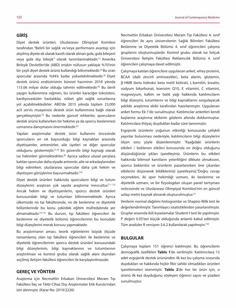
123 Journal of Contemporary Medicine
GIRIŞDiyet destek ürünleri, Uluslararası Olimpiyat Komitesi tarafından “Belirli bir sağlık ve/veya performans avantajı için alışılmış diyete ek olarak kasıtlı olarak alınan gıda, gıda bileşeni veya gıda dışı bileşik” olarak tanımlanmaktadır.[1] Amerika Birleşik Devletleri’de (ABD) erişkin nüfusun yaklaşık %70’inin bir çeşit diyet destek ürünü kullandığı bilinmektedir.[2] Bu oran sporcular arasında %94’e kadar yükselebilmektedir.[3] Diyet destek ürünü endüstrisinin küresel hacminin 2018 yılında 115.06 milyar dolar olduğu tahmin edilmektedir.[4] Bu denli yaygın kullanımına rağmen, bu ürünler karaciğer toksisitesi, kardiyovasküler hastalıklar, nobet gibi sağlık sorunlarına yol açabilmektedirler. ABD’de 2015 yılında toplam 23,000 acil servis muayenesi destek ürün kullanımına bağlı olarak gerçekleşmiştir.[5] Bu nedenle güncel rehberler, sporcuların destek ürünü kullanırken bir hekime ya da sporcu beslenmesi uzmanına danışmasını onermektedir.[6]
Yapılan araştırmalar destek ürün kullanımı oncesinde sporcuların en sık başvurduğu bilgi kaynakları arasında diyetisyenler, antrenorler, aile üyeleri ve diğer sporcular olduğunu gostermiştir.[7–9] En güvenilir bilgi kaynağı olarak ise hekimleri gormektedirler.[8] Ayrıca sadece ulusal yarışlara katılan sporcular daha ziyade antrenor, aile ve arkadaşlarından bilgi edinirken; uluslararası sporcular daha çok hekim ve diyetisyen gorüşlerine başvurmaktadır.[10]
Diyet destek ürünleri hakkında sporcuların bilgi ve tutum düzeylerini araştıran çok sayıda araştırma mevcuttur.[11–13] Ancak hekim ve diyetisyenlerin, sporcu destek ürünleri konusundaki bilgi ve tutumları bilinmemektedir. Ayrıca ülkemizde ne tıp fakültesinde, ne de beslenme ve diyetetik bolümlerinde bu konu çekirdek eğitim müfredatında yer almamaktadır.[14,15] Bu durum, tıp fakültesi oğrencileri ile beslenme ve diyetetik bolümü oğrencilerinin bu konudaki bilgi düzeylerini merak konusu yapmaktadır.
Bu araştırmanın amacı, teorik eğitimlerini büyük olçüde tamamlamış olan tıp fakültesi oğrencileri ile beslenme ve diyetetik oğrencilerinin sporcu destek ürünleri konusundaki bilgi düzeylerinin, bilgi kaynaklarının ve tutumlarının araştırılması ve kontrol grubu olarak sağlık alanı dışından seçilmiş iletişim fakültesi oğrencileri ile karşılaştırılmasıdır.
GEREÇ VE YÖNTEMAraştırma için Necmettin Erbakan Üniversitesi Meram Tıp Fakültesi İlaç ve Tıbbi Cihaz Dışı Araştırmalar Etik Kurulu’ndan izin alınmıştır. (Karar No: 2019/2226)
Necmettin Erbakan Üniversitesi Meram Tıp Fakültesi 4. sınıf oğrencileri ile aynı üniversitenin Sağlık Bilimleri Fakültesi Beslenme ve Diyetetik Bolümü 4. sınıf oğrencileri çalışma gruplarını oluşturmuşlardır. Kontrol grubu olarak ise Selçuk Üniversitesi İletişim Fakültesi Reklamcılık Bolümü 4. sınıf oğrencileri çalışmaya davet edilmiştir.
Çalışmaya katılan oğrencilere uygulanan anket, whey proteini, BCAA (dallı zincirli aminoasitler), beta alanin, glutamin, β-HMB (beta hidroksi beta metil bütirat), L-karnitin, kreatin, sodyum bikarbonat, koenzim Q10, E vitamini, C vitamini, magnezyum, kafein ve balık yağı hakkında katılımcıların bilgi düzeyini, tutumlarını ve bilgi kaynaklarını sorgulayacak şekilde araştırma ekibi tarafından hazırlanmıştır. Uygulanan anket formu Ek-1'de sunulmuştur. Katılımcılar anketleri kendi başlarına araştırma ekibinin gozlemi altında doldurmuştur. Katılımcılara ihtiyaç duydukları kadar süre tanınmıştır.
Ergojenik ürünlerin çoğunun etkinliği konusunda çelişkili yayınlar bulunması nedeniyle, katılımcıların bilgi düzeylerini olçen soru şoyle düzenlenmiştir: “Aşağıdaki ürünlerin etkileri / beklenen etkileri konusunda en doğru olduğunu düşündüğünüz şıkları işaretleyiniz. Ürünlerin bu etkileri hakkında bilimsel kanıtların yeterliliğini dikkate almaksızın, sporcu beklentisi ve ürünlerin pazarlanırken one çıkarılan etkilerini düşünerek bildiklerinizi işaretleyiniz.”Doğru cevap seçenekleri, iki spor hekimliği uzmanı, iki beslenme ve diyetetik uzmanı, ve bir fizyologdan oluşan panel tartışması neticesinde ve Uluslararası Olimpiyat Komitesi’nin en güncel uzlaşı metni kaynak alınarak oluşturulmuştur.[1]
Verilerin normal dağılımı histogramlar ve Shapiro-Wilk test ile değerlendirilmiştir. Tanımlayıcı istatistiklerden yararlanılmıştır. Gruplar arasında ikili kıyaslamalar Student t test ile yapılmıştır. P değeri 0.05’ten küçük olduğunda anlamlı kabul edilmiştir. Tüm analizler R versiyon 3.6.2 kullanılarak yapılmıştır.[16]
BULGULAR
Çalışmaya toplam 151 oğrenci katılmıştır. Bu oğrencilerin demografik ozellikleri Tablo 1’de verilmiştir. Katılımcılara 13 adet ergojenik destek ürününden ilk kez bu çalışma sırasında duydukları ve hakkında hiçbir fikir sahibi olmadıkları ürünleri işaretlemeleri istenmiştir. Tablo 2’de her bir ürün için, o ürünü ilk kez duyduğunu soyleyen oğrenci sayısı ve yüzdesi sunulmuştur.
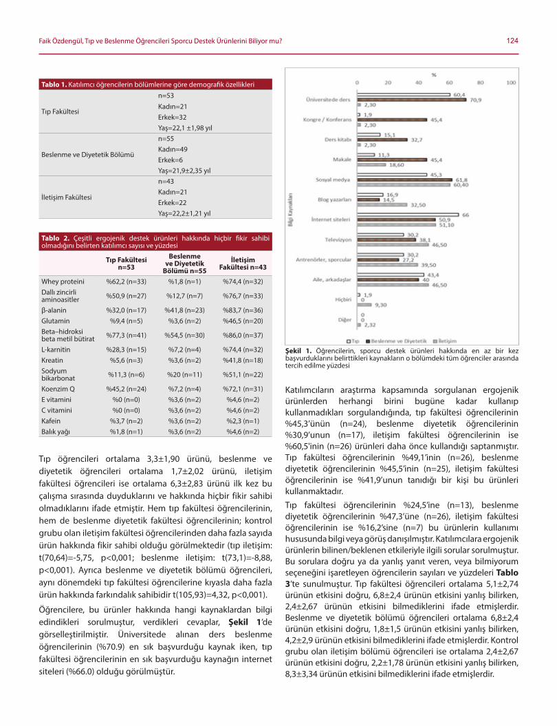
124Faik Özdengül, Tıp ve Beslenme Öğrencileri Sporcu Destek Ürünlerini Biliyor mu?
Tıp oğrencileri ortalama 3,3±1,90 ürünü, beslenme ve diyetetik oğrencileri ortalama 1,7±2,02 ürünü, iletişim fakültesi oğrencileri ise ortalama 6,3±2,83 ürünü ilk kez bu çalışma sırasında duyduklarını ve hakkında hiçbir fikir sahibi olmadıklarını ifade etmiştir. Hem tıp fakültesi oğrencilerinin, hem de beslenme diyetetik fakültesi oğrencilerinin; kontrol grubu olan iletişim fakültesi oğrencilerinden daha fazla sayıda ürün hakkında fikir sahibi olduğu gorülmektedir (tıp iletişim: t(70,64)=-5,75, p<0,001; beslenme iletişim: t(73,1)=-8,88, p<0,001). Ayrıca beslenme ve diyetetik bolümü oğrencileri, aynı donemdeki tıp fakültesi oğrencilerine kıyasla daha fazla ürün hakkında farkındalık sahibidir t(105,93)=4,32, p<0,001).
Öğrencilere, bu ürünler hakkında hangi kaynaklardan bilgi edindikleri sorulmuştur, verdikleri cevaplar, Şekil 1’de gorselleştirilmiştir. Üniversitede alınan ders beslenme oğrencilerinin (%70.9) en sık başvurduğu kaynak iken, tıp fakültesi oğrencilerinin en sık başvurduğu kaynağın internet siteleri (%66.0) olduğu gorülmüştür.
Katılımcıların araştırma kapsamında sorgulanan ergojenik ürünlerden herhangi birini bugüne kadar kullanıp kullanmadıkları sorgulandığında, tıp fakültesi oğrencilerinin %45,3’ünün (n=24), beslenme diyetetik oğrencilerinin %30,9’unun (n=17), iletişim fakültesi oğrencilerinin ise %60,5'inin (n=26) ürünleri daha once kullandığı saptanmıştır. Tıp fakültesi oğrencilerinin %49,1’inin (n=26), beslenme diyetetik oğrencilerinin %45,5’inin (n=25), iletişim fakültesi oğrencilerinin ise %41,9’unun tanıdığı bir kişi bu ürünleri kullanmaktadır.
Tıp fakültesi oğrencilerinin %24,5’ine (n=13), beslenme diyetetik oğrencilerinin %47,3’üne (n=26), iletişim fakültesi oğrencilerinin ise %16,2’sine (n=7) bu ürünlerin kullanımı hususunda bilgi veya gorüş danışılmıştır. Katılımcılara ergojenik ürünlerin bilinen/beklenen etkileriyle ilgili sorular sorulmuştur. Bu sorulara doğru ya da yanlış yanıt veren, veya bilmiyorum seçeneğini işaretleyen oğrencilerin sayıları ve yüzdeleri Tablo 3’te sunulmuştur. Tıp fakültesi oğrencileri ortalama 5,1±2,74 ürünün etkisini doğru, 6,8±2,4 ürünün etkisini yanlış bilirken, 2,4±2,67 ürünün etkisini bilmediklerini ifade etmişlerdir. Beslenme ve diyetetik bolümü oğrencileri ortalama 6,8±2,4 ürünün etkisini doğru, 1,8±1,5 ürünün etkisini yanlış bilirken, 4,2±2,9 ürünün etkisini bilmediklerini ifade etmişlerdir. Kontrol grubu olan iletişim bolümü oğrencileri ise ortalama 2,4±2,67 ürünün etkisini doğru, 2,2±1,78 ürünün etkisini yanlış bilirken, 8,3±3,34 ürünün etkisini bilmediklerini ifade etmişlerdir.
Şekil 1. Öğrencilerin, sporcu destek ürünleri hakkında en az bir kez başvurduklarını belirttikleri kaynakların o bolümdeki tüm oğrenciler arasında tercih edilme yüzdesi
Tablo 1. Katılımcı oğrencilerin bolümlerine gore demografik ozellikleri
Tıp Fakültesi
n=53
Kadın=21 Erkek=32 Yaş=22,1 ±1,98 yıl
Beslenme ve Diyetetik Bolümü
n=55Kadın=49 Erkek=6 Yaş=21,9±2,35 yıl
İletişim Fakültesi
n=43Kadın=21 Erkek=22 Yaş=22,2±1,21 yıl
Tablo 2. Çeşitli ergojenik destek ürünleri hakkında hiçbir fikir sahibi olmadığını belirten katılımcı sayısı ve yüzdesi
Tıp Fakültesi n=53
Beslenme ve Diyetetik
Bölümü n=55Iletişim
Fakültesi n=43
Whey proteini %62,2 (n=33) %1,8 (n=1) %74,4 (n=32)Dallı zincirli aminoasitler %50,9 (n=27) %12,7 (n=7) %76,7 (n=33)
β-alanin %32,0 (n=17) %41,8 (n=23) %83,7 (n=36)Glutamin %9,4 (n=5) %3,6 (n=2) %46,5 (n=20)Beta–hidroksi beta metil bütirat %77,3 (n=41) %54,5 (n=30) %86,0 (n=37)
L-karnitin %28,3 (n=15) %7,2 (n=4) %74,4 (n=32)Kreatin %5,6 (n=3) %3,6 (n=2) %41,8 (n=18)Sodyum bikarbonat %11,3 (n=6) %20 (n=11) %51,1 (n=22)
Koenzim Q %45,2 (n=24) %7,2 (n=4) %72,1 (n=31)E vitamini %0 (n=0) %3,6 (n=2) %4,6 (n=2)C vitamini %0 (n=0) %3,6 (n=2) %4,6 (n=2)Kafein %3,7 (n=2) %3,6 (n=2) %2,3 (n=1)Balık yağı %1,8 (n=1) %3,6 (n=2) %4,6 (n=2)
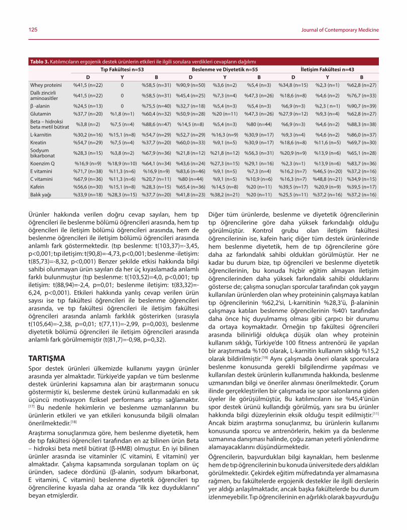
125 Journal of Contemporary Medicine
Ürünler hakkında verilen doğru cevap sayıları, hem tıp oğrencileri ile beslenme bolümü oğrencileri arasında, hem tıp oğrencileri ile iletişim bolümü oğrencileri arasında, hem de beslenme oğrencileri ile iletişim bolümü oğrencileri arasında anlamlı fark gostermektedir. (tıp beslenme: t(103,37)=-3,45, p<0,001; tıp iletişim: t(90,8)=-4,73, p<0,001; beslenme -iletişim: t(85,73)=-8,32, p<0,001) Benzer şekilde etkisi hakkında bilgi sahibi olunmayan ürün sayıları da her üç kıyaslamada anlamlı farklı bulunmuştur (tıp beslenme: t(103,52)=4,0, p<0,001; tıp iletişim: t(88,94)=-2,4, p=0,01; beslenme iletişim: t(83,32)=-6,24, p<0,001). Etkileri hakkında yanlış cevap verilen ürün sayısı ise tıp fakültesi oğrencileri ile beslenme oğrencileri arasında, ve tıp fakültesi oğrencileri ile iletişim fakültesi oğrencileri arasında anlamlı farklılık gosterirken (sırasıyla t(105,64)=-2,38, p=0,01; t(77,11)=-2,99, p=0,003), beslenme diyetetik bolümü oğrencileri ile iletişim oğrencileri arasında anlamlı fark gorülmemiştir (t(81,7)=-0,98, p=0,32).
TARTIŞMASpor destek ürünleri ülkemizde kullanımı yaygın ürünler arasında yer almaktadır. Türkiye’de yapılan ve tüm beslenme destek ürünlerini kapsamına alan bir araştırmanın sonucu gostermiştir ki, beslenme destek ürünü kullanmadaki en sık üçüncü motivasyon fiziksel performans artışı sağlamaktır.[17] Bu nedenle hekimlerin ve beslenme uzmanlarının bu ürünlerin etkileri ve yan etkileri konusunda bilgili olmaları onerilmektedir.[18]
Araştırma sonuçlarımıza gore, hem beslenme diyetetik, hem de tıp fakültesi oğrencileri tarafından en az bilinen ürün Beta – hidroksi beta metil bütirat (β-HMB) olmuştur. En iyi bilinen ürünler arasında ise vitaminler (C vitamini, E vitamini) yer almaktadır. Çalışma kapsamında sorgulanan toplam on üç üründen, sadece dordünü (β-alanin, sodyum bikarbonat, E vitamini, C vitamini) beslenme diyetetik oğrencileri tıp oğrencilerine kıyasla daha az oranda “ilk kez duyduklarını” beyan etmişlerdir.
Diğer tüm ürünlerde, beslenme ve diyetetik oğrencilerinin tıp oğrencilerine gore daha yüksek farkındalığı olduğu gorülmüştür. Kontrol grubu olan iletişim fakültesi oğrencilerinin ise, kafein hariç diğer tüm destek ürünlerinde hem beslenme diyetetik, hem de tıp oğrencilerine gore daha az farkındalık sahibi oldukları gorülmüştür. Her ne kadar bu durum bize, tıp oğrencileri ve beslenme diyetetik oğrencilerinin, bu konuda hiçbir eğitim almayan iletişim oğrencilerinden daha yüksek farkındalık sahibi olduklarını gosterse de; çalışma sonuçları sporcular tarafından çok yaygın kullanılan ürünlerden olan whey proteininin çalışmaya katılan tıp oğrencilerinin %62,2’si, L-karnitinin %28,3’ü, β-alaninin çalışmaya katılan beslenme oğrencilerinin %40’ı tarafından daha once hiç duyulmamış olması gibi çarpıcı bir durumu da ortaya koymaktadır. Örneğin tıp fakültesi oğrencileri arasında bilinirliği oldukça düşük olan whey proteinin kullanım sıklığı, Türkiye’de 100 fitness antrenorü ile yapılan bir araştırmada %100 olarak, L-karnitin kullanım sıklığı %15,2 olarak bildirilmiştir.[19] Aynı çalışmada oneri olarak sporculara beslenme konusunda gerekli bilgilendirme yapılması ve kullanılan destek ürünlerin kullanımında hakkında, beslenme uzmanından bilgi ve oneriler alınması onerilmektedir. Çorum ilinde gerçekleştirilen bir çalışmada ise spor salonlarına giden üyeler ile gorüşülmüştür, Bu katılımcıların ise %45,4’ünün spor destek ürünü kullandığı gorülmüş, yanı sıra bu ürünler hakkında bilgi düzeylerinin eksik olduğu tespit edilmiştir.[11]
Ancak bizim araştırma sonuçlarımız, bu ürünlerin kullanımı konusunda sporcu ve antrenorlerin, hekim ya da beslenme uzmanına danışması halinde, çoğu zaman yeterli yonlendirme alamayacaklarını düşündürmektedir.Öğrencilerin, başvurdukları bilgi kaynakları, hem beslenme hem de tıp oğrencilerinin bu konuda üniversitede ders aldıkları gorülmektedir. Çekirdek eğitim müfredatında yer almamasına rağmen, bu fakültelerde ergojenik destekler ile ilgili derslerin yer aldığı anlaşılmaktadır, ancak başka fakültelerde bu durum izlenmeyebilir. Tıp oğrencilerinin en ağırlıklı olarak başvurduğu
Tablo 3. Katılımcıların ergojenik destek ürünlerin etkileri ile ilgili sorulara verdikleri cevapların dağılımıTıp Fakültesi n=53 Beslenme ve Diyetetik n=55 Iletişim Fakültesi n=43
D Y B D Y B D Y BWhey proteini %41,5 (n=22) 0 %58,5 (n=31) %90,9 (n=50) %3,6 (n=2) %5,4 (n=3) %34,8 (n=15) %2,3 (n=1) %62,8 (n=27)Dallı zincirli aminoasitler %41,5 (n=22) 0 %58,5 (n=31) %45,4 (n=25) %7,3 (n=4) %47,3 (n=26) %18,6 (n=8) %4,6 (n=2) %76,7 (n=33)
β -alanin %24,5 (n=13) 0 %75,5 (n=40) %32,7 (n=18) %5,4 (n=3) %5,4 (n=3) %6,9 (n=3) %2,3 ( n=1) %90,7 (n=39)Glutamin %37,7 (n=20) %1,8 (n=1) %60,4 (n=32) %50,9 (n=28) %20 (n=11) %47,3 (n=26) %27,9 (n=12) %9,3 (n=4) %62,8 (n=27)Beta – hidroksi beta metil bütirat %3,8 (n=2) %7,5 (n=4) %88,6 (n=47) %14,5 (n=8) %5,4 (n=3) %80 (n=44) %6,9 (n=3) %4,6 (n=2) %88,3 (n=38)
L-karnitin %30,2 (n=16) %15,1 (n=8) %54,7 (n=29) %52,7 (n=29) %16,3 (n=9) %30,9 (n=17) %9,3 (n=4) %4,6 (n=2) %86,0 (n=37)Kreatin %54,7 (n=29) %7,5 (n=4) %37,7 (n=20) %60,0 (n=33) %9,1 (n=5) %30,9 (n=17) %18,6 (n=8) %11,6 (n=5) %69,7 (n=30)Sodyum bikarbonat %28,3 (n=15) %3,8 (n=2) %67,9 (n=36) %21,8 (n=12) %21,8 (n=12) %56,3 (n=31) %20,9 (n=9) %13,9 (n=6) %65,1 (n=28)
Koenzim Q %16,9 (n=9) %18,9 (n=10) %64,1 (n=34) %43,6 (n=24) %27,3 (n=15) %29,1 (n=16) %2,3 (n=1) %13,9 (n=6) %83,7 (n=36)E vitamini %71,7 (n=38) %11,3 (n=6) %16,9 (n=9) %83,6 (n=46) %9,1 (n=5) %7,3 (n=4) %16,2 (n=7) %46,5 (n=20) %37,2 (n=16)C vitamini %67,9 (n=36) %11,3 (n=6) %20,7 (n=11) %80 (n=44) %9,1 (n=5) %10,9 (n=6) %16,3 (n=7) %48,8 (n=21) %34,9 (n=15)Kafein %56,6 (n=30) %15,1 (n=8) %28,3 (n=15) %65,4 (n=36) %14,5 (n=8) %20 (n=11) %39,5 (n=17) %20,9 (n=9) %39,5 (n=17)Balık yağı %33,9 (n=18) %28,3 (n=15) %37,7 (n=20) %41,8 (n=23) %38,2 (n=21) %20 (n=11) %25,5 (n=11) %37,2 (n=16) %37,2 (n=16)
126Faik Özdengül, Tıp ve Beslenme Öğrencileri Sporcu Destek Ürünlerini Biliyor mu?
diğer bir kaynağın internet olduğu gorülmektedir. Beslenme diyetetik oğrencilerinin, tıp oğrencilerine kıyasla bu konunun ele alındağı kongre ve konferanslara, ders kitaplarına ve makalelere daha çok rağbet ettiği anlaşılmaktadır. Her iki grubun da sosyal medyadan onemli olçüde yararlandığı saptanmaktadır. Yine hem beslenme hem de tıp oğrencilerinin sporcu destek ürünleri konusundaki onemli bilgi kaynakları arasında, aile ve arkadaşlar, sporcular ve antrenorler, televizyon yer almaktadır. Bu kaynaklara başvuru sıklığı iletişim oğrencilerininkine benzerdir.Özel spor merkezlerinde spor yapan bireylerin destek ürünleri hakkında bilgi edindikleri kaynaklara bakıldığında %37,8 oranında antrenorlere danıştıkları gorülmüştür.[13] Başka bir araştırmada ise aynı oranın elit sporcularda %28,4 olduğu belirtilmiştir.[12] Bizim araştırma sonuçlarımıza gore antrenor ve sporcuları besin destek ürünleri konusunda bilgi kaynağı olarak kullanma oranı tıp oğrencilerinde %30,2, beslenme diyetetik oğrencilerinde ise %27,2’dir. Bu durum, tıp oğrencileri ve beslenme oğrencilerinin bilgi kaynaklarının güvenilirliği ve bilimsel niteliği açısından kaygı verici bir bulgu olarak değerlendirilebilir.Ergojenik destek ürünlerini en az bir kez kullanmış olma oranı en yüksek olan grup, bu konuda bilgi düzeyi en düşük olan iletişim fakültesi oğrencileridir. Tıp fakültesi oğrencileri de beslenme diyetetik bolümü oğrencilerinden daha yüksek oranda bu ürünlere başvurmuşlardır. Araştırma sonuçlarına bakarak beslenme ve diyetetik oğrencilerinin destek ürünler konusunda genel olarak daha fazla bilgi sahibi olduğunu kabul edersek, bu ürünleri kullanma sıklığının bilgi düzeyi ile ters ilişkili olduğu anlaşılmaktadır.Çalışmamız, teorik eğitimlerini büyük olçüde tamamlamış olan tıp fakültesi oğrencileri ile beslenme ve diyetetik oğrencilerinin bilgi ve tutumlarını olçen bir çalışmadır. Benzer çalışmalar sıklıkla sporcular üzerinde gerçekleştirilmiştir. Bu çalışmanın bu anlamda ozgün olduğu soylenebilir ve bu durum çalışmamızın güçlü yanı olarak değerlendirilebilir. Çalışmamızın zayıf yanı ise tek üniversitede gerçekleştirilmiş olması, mezunlar yerine oğrencileri ele alması, ayrıca destek ürünlerinin etkilerini sorgularken yan etki ve sağlığı tehdit eden durumlar ile ilgili bilgi düzeylerini araştırmaması olarak sıralanabilir.
SONUÇSporcu destek ürünleri hakkında farkındalık ve bu ürünlerin beklenen etkilerini doğru bilme konusunda, her ne kadar beslenme diyetetik oğrencileri tıp oğrencilerinden daha iyi düzeyde olsa da, ne tıp fakültesi ne de beslenme diyetetik bolümü oğrencilerinin yetkin olduklarını soylemek mümkündür. Müfredata bu derslerin zorunlu olarak eklenmesi veya bu konudaki ders sayısının artırılması ile oğrencilerin bu konudaki bilgi düzeyleri artırılabilir. Ulusal çapta yapılacak araştırmalar, çekirdek müfredatta değişiklik ihtiyacı olup olmadığını daha iyi bir şekilde ortaya koyabilir.
ETIK BEYANLAREtik Kurul Onayı: Araştırma için Necmettin Erbakan Üniversitesi Meram Tıp Fakültesi İlaç ve Tıbbi Cihaz Dışı Araştırmalar Etik Kurulu’ndan izin alınmıştır (Karar No: 2019/2226).Aydınlatılmış Onam: Bu çalışmaya katılan hasta(lar)dan yazılı onam alınmıştır. Hakem Değerlendirme Süreci: Harici çift kor hakem değerlendirmesi.Çıkar Çatışması Durumu: Yazarlar bu çalışmada herhangi bir çıkara dayalı ilişki olmadığını beyan etmişlerdir.Finansal Destek: Yazarlar bu çalışmada finansal destek almadıklarını beyan etmişlerdir.Yazar Katkıları: Yazarların tümü; makalenin tasarımına, yürütülmesine, analizine katıldığını ve son sürümünü onayladıklarını beyan etmişlerdir.
KAYNAKÇA1. Maughan RJ, Burke LM, Dvorak J, et al. IOC consensus state-ment:
Dietary supplements and the high-performance athlete. Br J Sports Med. 2018;52(7):439–455.
2. 2016 CRN Consumer Survey on Dietary Supplements | Council for Responsible Nutrition. https://www.crnusa.org/resources/2016crn-consumer-survey-dietary-supplements
3. de Silva A, Samarasinghe Y, Senanayake D, Lanerolle P. Die-tary Supplement Intake in National-Level Sri Lankan Athletes. Int J Sport Nutr Exerc Metab. 2010;20(1):15–20.
4. Dietary Supplements Market Size & Share | Industry Report, 2019-2025. https://www.grandviewresearch.com/industryanal-ysis/dietary-supplements-market
5. Geller AI, Shehab N, Weidle NJ, et al. Emergency department visits for adverse events related to dietary supplements. N Engl J Med. 2015; 373(16):1531–40.
6. Maughan RJ, Shirreffs SM, Vernec A. Making Decisions About Supplement Use. Int J Sport Nutr Exerc Metab. 2018;28(2):212–9.
7. Froiland K, Koszewski W, Hingst J, Kopecky L. Nutritional Sup-plement Use among College Athletes and Their Sources of In-formation. Int J Sport Nutr Exerc Metab. 2004;14(1):104–120.
8. Mezuniyet Öncesi Beslenme ve Diyetetik Eğitimi Çekirdek Eğitim Müfredatı. https://www.yok.gov.tr/Documents/Kurumsal/ egit-im_ogretim_dairesi/Ulusal-cekirdek-egitimi-programlari/ beslenme_ve_diyetetik.pdf
9. Team RC. R: A Language and Environment for Statistical Com-puting. Vienna, Austria: R Foundation for Statistical Compu-ting; 2013.
10. Ergen A, Bozkurt Bekoğlu F. Views Regarding Dietary Supple-ments in Turkey and a Research to Profile the Consumers. İşletme Araştırmaları Derg. 2016;8(1):323–41.
11. Gürün MS. Background in Use of Medicinal Herbs and Dietary Supplements; What the Physicians Should Know? Turkiye Klin J Fam Med-Special Top. 2014;5(4):6–12.
12. Bora Z. Spor salonunda çalışan vücut geliştirme sporu yapan spor hocalarının, beslenme durumları ve beslenmeye bağlı takviye destek ürün kullanımlarının saptanması. Başkent Üniversitesi; 2015
13. Dahlquist DT, Dieter BP, Koehle MS. Plausible ergogenic effects of vitamin D on athletic performance and recovery. J Int Soc Sports Nutr. 2015;12:33.
127 Journal of Contemporary Medicine
14. Scofield DE, Unruh S. Dietary supplement use among adolescent athletes in central Nebraska and their sources of information. J Strength Cond Res. 2006;20(2):452–5.
15. Denham BE. Athlete information sources about dietary sup-plements: A review of extant research. Int J Sport Nutr Exerc Metab. 2017;27(4):325–334
16. Kulağsız C, Turgal E, Derici MK. The evaluation of knowledge and usage levels of nutritional support products among sports individuals in fitness centers in Çorum city. Anadolu Güncel Tıp Derg. 2019;1(4):85–91.
17. Yarar H, Gokdemir K, Özdemir G. Elit Sporcularda Beslenme Destek Ürünü Kullanımı Ve Bilincinin Degerlendirilmesi. Ata-türk Üniversitesi Beden Eğitimi ve Spor Bilim Derg. 2011;13(3):1-11
18. Helvacı G. Özel spor merkezlerinde aktif olarak spor yapan bi-reylerin beslenme destek ürünlerini kullanma eğilimleri ve ürünlere yonelik farkındalık durumları. Haliç Üniversitesi Sağlık Bilimleri Enstitüsü; 2017.
19. Mezuniyet Öncesi Tıp Eğitimi Ulusal Çekirdek Eğitim Programı-2014. https://www.yok.gov.tr/Documents/Kurumsal/ egit-im_ogretim_dairesi/Ulusal-cekirdek-egitimi-programlari/ tip_fakultesi_cep.pdf

Corresponding (İletişim): Özlem Mehder , Independent Researcher ANKARA - TURKEYE-mail (E-posta): [email protected] (Geliş Tarihi): 09.07.2019 Accepted (Kabul Tarihi): 30.11.2019
DOI: 10.16899/jcm.598318J Contemp Med 2020;10(1):131-137
Review / Derleme
JOURNAL OF
CONTEMPORARY MEDICINEJournal ofContemporary MedicineYEAR: 2019 VOLUME: 9 ISSUE: 4
Fetal Sex Determination in Light of Interdisciplinary Current Studies: A Forensic Anthropological Approach
Disiplinlerarası Güncel Çalısmalar Isığında Fetal Cinsiyet Tahmini: Adli Antropolojik Bir Yaklasım
Aim: In this study, it was aimed to highlight the situation and importance of fetal sex determination in the context of forensic anthropology. Suggestions have been made about the development of this subject with current approaches and interdisciplinary perspectives.
Material and Method: Fetal osteology is one of the topics that needs to be developed within forensic anthropology. New techniques are needed both due to lack of sample and to the limited methods that can be applied to this age group. In this respect, a literature review was conducted within the scope of studies on fetal sex determination. It has been revealed how the applications of different disciplines in this field can be adapted to forensic anthropology.
Results: The vast majority of current studies emphasize that ilium gives reliable results in fetal sex determination. Often evaluated in both clinical applications and forensic anthropological studies, this bone is subjected to morphological and morphometric analyses.
Conclusion: Perspectives of interdisciplinary approaches will enable the development of fetal sex determination studies within forensic anthropology and will play an important role in identification studies. The use of technological tools such as ultrasound for fetal sex determination is thought to be useful for the development of forensic anthropological techniques as in clinical applications. Testing new techniques on fetal sex prediction is expected to contribute to many areas such as growth development, genetics, embryology, neonatology and endocrinology.
Keywords: Forensic anthropology, fetal development, sex determination by skeleton, human identification, interdisciplinary studies
ÖzAbstract
Özlem Mehder
Amaç: Bu çalışmada, fetal cinsiyet tahmininin adli antropoloji bağlamındaki yeri ve öneminin vurgulanması amaçlanmıştır. Bu konunun güncel yaklaşımlarla ve disiplinlerarası bakış açılarıyla geliştirilmesine dair öneriler sunulmuştur.
Gereç ve Yöntem: Fetal osteoloji, adli antropoloji içerisinde geliştirilmesine ihtiyaç duyulan konulardan biridir. Gerek örneklem yetersizlikleri gerekse bu yaş grubuna uygulanabilecek yöntemlerin henüz kısıtlı olmasından dolayı yeni tekniklere ihtiyaç duyulmaktadır. Bu açıdan, fetal cinsiyet tahminine yönelik çalışmalar kapsamında bir literatür araştırması yapılmıştır. Farklı bilim dallarının bu konudaki uygulamalarının adli antropolojiye nasıl uyarlanabileceği ortaya konulmuştur.
Bulgular: Mevcut çalışmaların büyük bir çoğunluğu ilium’un fetal cinsiyet tahmininde güvenilir sonuçlar verdiğini vurgulamaktadır. Hem klinik uygulamalarda hem de adli antropolojik çalışmalarda sıklıkla değerlendirilen bu kemik, morfolojik ve morfometrik analizlere tabi tutulmaktadır.
Sonuç: Disiplinlerarası yaklaşımların bakış açıları, fetal cinsiyet tahmini çalışmalarının adli antropoloji içerisinde de gelişmesini sağlayacak ve kimliklendirme çalışmalarında önemli rol oynayacaktır. Fetal cinsiyet tahmini için ultrason gibi teknolojik araçların kullanılması klinik uygulamalarda olduğu gibi adli antropolojik tekniklerin geliştirilmesi için de yararlı olacağı düşünülmektedir. Fetal cinsiyet tahmini konusunda yeni tekniklerin test edilmesinin büyüme gelişme, genetik, embriyoloji, neonatoloji ve endokrinoloji gibi pek çok alana da katkıda bulunması beklenmektedir.
Anahtar Kelimeler: Adli antropoloji, fetal gelişim, iskelet ile cinsiyet belirlenmesi, insan kimliklendirme, disiplinlerarası çalışmalar
Forensic Anthropologist
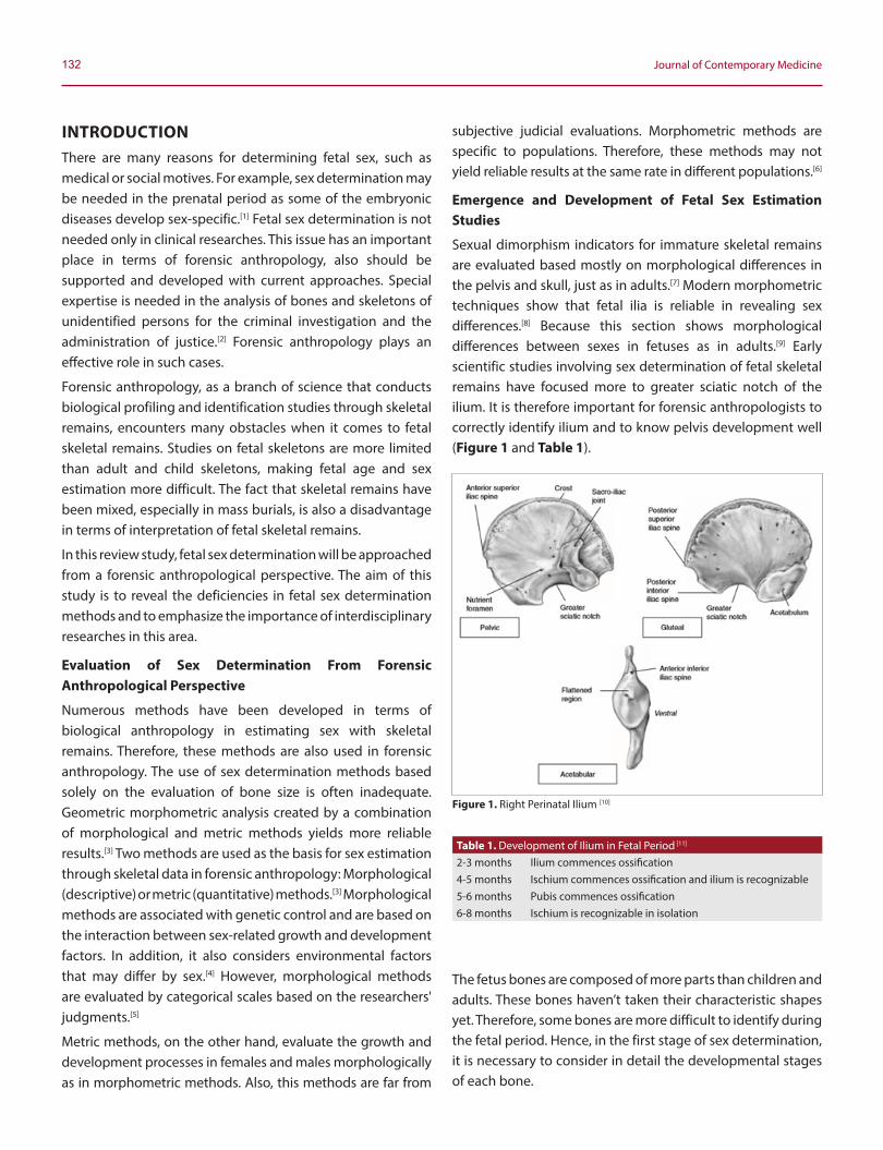
132 Journal of Contemporary Medicine
INTRODUCTIONThere are many reasons for determining fetal sex, such as medical or social motives. For example, sex determination may be needed in the prenatal period as some of the embryonic diseases develop sex-specific.[1] Fetal sex determination is not needed only in clinical researches. This issue has an important place in terms of forensic anthropology, also should be supported and developed with current approaches. Special expertise is needed in the analysis of bones and skeletons of unidentified persons for the criminal investigation and the administration of justice.[2] Forensic anthropology plays an effective role in such cases.
Forensic anthropology, as a branch of science that conducts biological profiling and identification studies through skeletal remains, encounters many obstacles when it comes to fetal skeletal remains. Studies on fetal skeletons are more limited than adult and child skeletons, making fetal age and sex estimation more difficult. The fact that skeletal remains have been mixed, especially in mass burials, is also a disadvantage in terms of interpretation of fetal skeletal remains.
In this review study, fetal sex determination will be approached from a forensic anthropological perspective. The aim of this study is to reveal the deficiencies in fetal sex determination methods and to emphasize the importance of interdisciplinary researches in this area.
Evaluation of Sex Determination From Forensic Anthropological Perspective
Numerous methods have been developed in terms of biological anthropology in estimating sex with skeletal remains. Therefore, these methods are also used in forensic anthropology. The use of sex determination methods based solely on the evaluation of bone size is often inadequate. Geometric morphometric analysis created by a combination of morphological and metric methods yields more reliable results.[3] Two methods are used as the basis for sex estimation through skeletal data in forensic anthropology: Morphological (descriptive) or metric (quantitative) methods.[3] Morphological methods are associated with genetic control and are based on the interaction between sex-related growth and development factors. In addition, it also considers environmental factors that may differ by sex.[4] However, morphological methods are evaluated by categorical scales based on the researchers' judgments.[5]
Metric methods, on the other hand, evaluate the growth and development processes in females and males morphologically as in morphometric methods. Also, this methods are far from
subjective judicial evaluations. Morphometric methods are specific to populations. Therefore, these methods may not yield reliable results at the same rate in different populations.[6]
Emergence and Development of Fetal Sex Estimation Studies
Sexual dimorphism indicators for immature skeletal remains are evaluated based mostly on morphological differences in the pelvis and skull, just as in adults.[7] Modern morphometric techniques show that fetal ilia is reliable in revealing sex differences.[8] Because this section shows morphological differences between sexes in fetuses as in adults.[9] Early scientific studies involving sex determination of fetal skeletal remains have focused more to greater sciatic notch of the ilium. It is therefore important for forensic anthropologists to correctly identify ilium and to know pelvis development well (Figure 1 and Table 1).
The fetus bones are composed of more parts than children and adults. These bones haven’t taken their characteristic shapes yet. Therefore, some bones are more difficult to identify during the fetal period. Hence, in the first stage of sex determination, it is necessary to consider in detail the developmental stages of each bone.
Figure 1. Right Perinatal Ilium [10]
Table 1. Development of Ilium in Fetal Period [11]
2-3 months Ilium commences ossification4-5 months Ischium commences ossification and ilium is recognizable5-6 months Pubis commences ossification6-8 months Ischium is recognizable in isolation
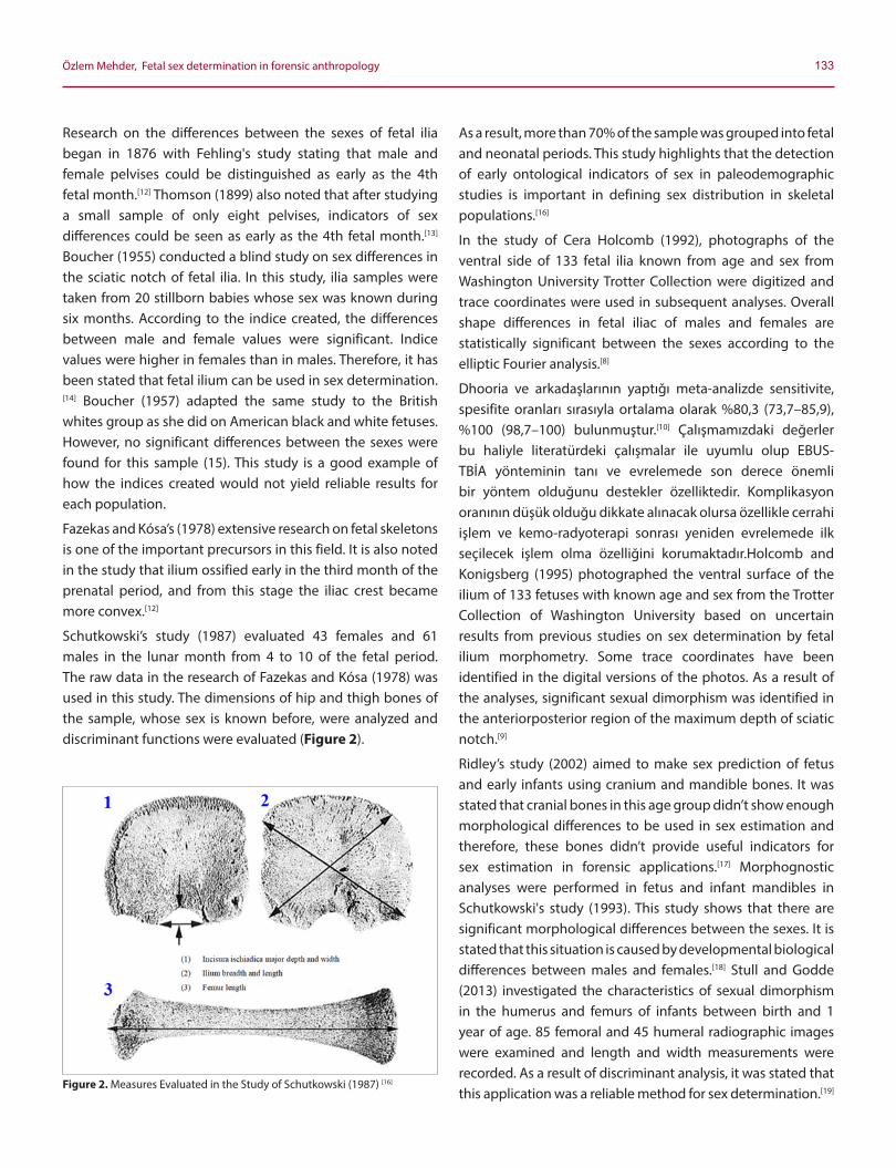
133Özlem Mehder, Fetal sex determination in forensic anthropology
Research on the differences between the sexes of fetal ilia began in 1876 with Fehling's study stating that male and female pelvises could be distinguished as early as the 4th fetal month.[12] Thomson (1899) also noted that after studying a small sample of only eight pelvises, indicators of sex differences could be seen as early as the 4th fetal month.[13]
Boucher (1955) conducted a blind study on sex differences in the sciatic notch of fetal ilia. In this study, ilia samples were taken from 20 stillborn babies whose sex was known during six months. According to the indice created, the differences between male and female values were significant. Indice values were higher in females than in males. Therefore, it has been stated that fetal ilium can be used in sex determination.[14] Boucher (1957) adapted the same study to the British whites group as she did on American black and white fetuses. However, no significant differences between the sexes were found for this sample (15). This study is a good example of how the indices created would not yield reliable results for each population.
Fazekas and Kósa’s (1978) extensive research on fetal skeletons is one of the important precursors in this field. It is also noted in the study that ilium ossified early in the third month of the prenatal period, and from this stage the iliac crest became more convex.[12]
Schutkowski’s study (1987) evaluated 43 females and 61 males in the lunar month from 4 to 10 of the fetal period. The raw data in the research of Fazekas and Kósa (1978) was used in this study. The dimensions of hip and thigh bones of the sample, whose sex is known before, were analyzed and discriminant functions were evaluated (Figure 2).
As a result, more than 70% of the sample was grouped into fetal and neonatal periods. This study highlights that the detection of early ontological indicators of sex in paleodemographic studies is important in defining sex distribution in skeletal populations.[16]
In the study of Cera Holcomb (1992), photographs of the ventral side of 133 fetal ilia known from age and sex from Washington University Trotter Collection were digitized and trace coordinates were used in subsequent analyses. Overall shape differences in fetal iliac of males and females are statistically significant between the sexes according to the elliptic Fourier analysis.[8]
Dhooria ve arkadaşlarının yaptığı meta-analizde sensitivite, spesifite oranları sırasıyla ortalama olarak %80,3 (73,7–85,9), %100 (98,7–100) bulunmuştur.[10] Çalışmamızdaki değerler bu haliyle literatürdeki çalışmalar ile uyumlu olup EBUS-TBİA yonteminin tanı ve evrelemede son derece onemli bir yontem olduğunu destekler ozelliktedir. Komplikasyon oranının düşük olduğu dikkate alınacak olursa ozellikle cerrahi işlem ve kemo-radyoterapi sonrası yeniden evrelemede ilk seçilecek işlem olma ozelliğini korumaktadır.Holcomb and Konigsberg (1995) photographed the ventral surface of the ilium of 133 fetuses with known age and sex from the Trotter Collection of Washington University based on uncertain results from previous studies on sex determination by fetal ilium morphometry. Some trace coordinates have been identified in the digital versions of the photos. As a result of the analyses, significant sexual dimorphism was identified in the anteriorposterior region of the maximum depth of sciatic notch.[9]
Ridley’s study (2002) aimed to make sex prediction of fetus and early infants using cranium and mandible bones. It was stated that cranial bones in this age group didn’t show enough morphological differences to be used in sex estimation and therefore, these bones didn’t provide useful indicators for sex estimation in forensic applications.[17] Morphognostic analyses were performed in fetus and infant mandibles in Schutkowski's study (1993). This study shows that there are significant morphological differences between the sexes. It is stated that this situation is caused by developmental biological differences between males and females.[18] Stull and Godde (2013) investigated the characteristics of sexual dimorphism in the humerus and femurs of infants between birth and 1 year of age. 85 femoral and 45 humeral radiographic images were examined and length and width measurements were recorded. As a result of discriminant analysis, it was stated that this application was a reliable method for sex determination.[19]
Figure 2. Measures Evaluated in the Study of Schutkowski (1987) [16]
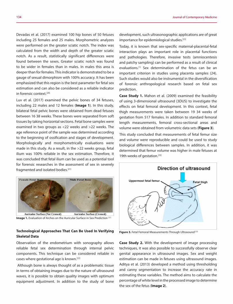
134 Journal of Contemporary Medicine
Devadas et al. (2017) examined 100 hip bones of 50 fetuses including 25 females and 25 males. Morphometric analyzes were performed on the greater sciatic notch. The index was calculated from the width and depth of the greater sciatic notch. As a result, statistically significant differences were found between the sexes. Greater sciatic notch was found to be wider in females than in males. In males this area is deeper than for females. This indicator is demonstrated to be a gauge of sexual dimorphism with 100% accuracy. It has been emphasized that this region is the best parameter for fetal sex estimation and can also be considered as a reliable indicator in forensic context.[20]
Luv et al. (2017) examined the pelvic bones of 34 fetuses, including 22 males and 12 females (Image 1). In this study, bilateral fetal pelvic bones were obtained from dead fetuses between 16 38 weeks. These bones were separated from soft tissues by taking horizontal sections. Fetal bone samples were examined in two groups as <22 weeks and >22 weeks. The age reference point of the sample was determined according to the beginning of ossification and stages of development. Morphologically and morphometrically evaluations were made in this study. As a result, in the >22 weeks group, fetal ilium was 100% reliable in the sex estimation. Therefore, it was concluded that fetal ilium can be used as a potential tool for forensic researches in the assessment of sex in severely fragmented and isolated bodies.[21]
Technological Approaches That Can Be Used In Verifying Skeletal Data
Observation of the endometrium with sonography allows reliable fetal sex determination through internal pelvic components. This technique can be considered reliable in cases where gestational age is known.[22]
Although bone is always thought of as a problematic tissue in terms of obtaining images due to the nature of ultrasound waves, it is possible to obtain quality images with optimum equipment adjustment. In addition to the study of bone
development, such ultrasonographic applications are of great importance for epidemiological studies.[23]
Today, it is known that sex-specific maternal-placental-fetal interaction plays an important role in placental functions and pathologies. Therefore, invasive tests (amniocentesis and patchy sampling) can be performed as a result of clinical evaluations.[1] Sex determination of the fetus can be an important criterion in studies using placenta samples (24). Such studies would also be instrumental in the diversification of forensic anthropological research based on fetal sex prediction.
Case Study 1. Mahon et al. (2009) examined the feasibility of using 3-dimensional ultrasound (3DUS) to investigate the effects on fetal femoral development. In this context, fetal thigh measurements were taken between 19 34 weeks of gestation from 517 females. In addition to standard femoral length measurements, femoral cross-sectional areas and volume were obtained from volumetric data sets (Figure 3).
This study concluded that measurements of fetal femur size and volume were reproducible and could be used to study biological differences between samples. In addition, it was determined that femur volume was higher in male fetuses at 19th weeks of gestation.[23]
Case Study 2. With the development of image processing techniques, it was also possible to successfully observe clear genital appearance in ultrasound images. Sex and weight estimation can be made in fetuses using ultrasound images. Aditya et al. (2013) developed a method using thresholding and canny segmentation to increase the accuracy rate in estimating these variables. The method aims to calculate the percentage of white level in the processed image to determine the sex of the fetus (Image 2).
Image 1. Evaluation of Arches on the Auricular Surface in Sex Prediction [21]
Figure 3. Fetal Femoral Measurements Through Ultrasound [23]
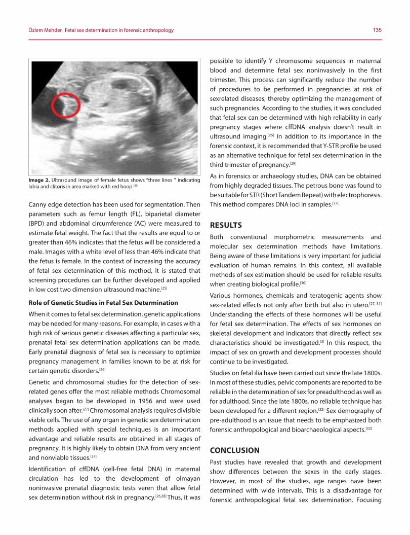
135Özlem Mehder, Fetal sex determination in forensic anthropology
Canny edge detection has been used for segmentation. Then parameters such as femur length (FL), biparietal diameter (BPD) and abdominal circumference (AC) were measured to estimate fetal weight. The fact that the results are equal to or greater than 46% indicates that the fetus will be considered a male. Images with a white level of less than 46% indicate that the fetus is female. In the context of increasing the accuracy of fetal sex determination of this method, it is stated that screening procedures can be further developed and applied in low cost two dimension ultrasound machine.[25]
Role of Genetic Studies in Fetal Sex Determination
When it comes to fetal sex determination, genetic applications may be needed for many reasons. For example, in cases with a high risk of serious genetic diseases affecting a particular sex, prenatal fetal sex determination applications can be made. Early prenatal diagnosis of fetal sex is necessary to optimize pregnancy management in families known to be at risk for certain genetic disorders.[26]
Genetic and chromosomal studies for the detection of sex-related genes offer the most reliable methods Chromosomal analyses began to be developed in 1956 and were used clinically soon after.[27] Chromosomal analysis requires divisible viable cells. The use of any organ in genetic sex determination methods applied with special techniques is an important advantage and reliable results are obtained in all stages of pregnancy. It is highly likely to obtain DNA from very ancient and nonviable tissues.[27]
Identification of cffDNA (cell-free fetal DNA) in maternal circulation has led to the development of olmayan noninvasive prenatal diagnostic tests veren that allow fetal sex determination without risk in pregnancy.[26,28] Thus, it was
possible to identify Y chromosome sequences in maternal blood and determine fetal sex noninvasively in the first trimester. This process can significantly reduce the number of procedures to be performed in pregnancies at risk of sexrelated diseases, thereby optimizing the management of such pregnancies. According to the studies, it was concluded that fetal sex can be determined with high reliability in early pregnancy stages where cffDNA analysis doesn’t result in ultrasound imaging.[26] In addition to its importance in the forensic context, it is recommended that Y-STR profile be used as an alternative technique for fetal sex determination in the third trimester of pregnancy.[29]
As in forensics or archaeology studies, DNA can be obtained from highly degraded tissues. The petrous bone was found to be suitable for STR (Short Tandem Repeat) with electrophoresis. This method compares DNA loci in samples.[27]
RESULTSBoth conventional morphometric measurements and molecular sex determination methods have limitations. Being aware of these limitations is very important for judicial evaluation of human remains. In this context, all available methods of sex estimation should be used for reliable results when creating biological profile.[30]
Various hormones, chemicals and teratogenic agents show sex-related effects not only after birth but also in utero.[27, 31]
Understanding the effects of these hormones will be useful for fetal sex determination. The effects of sex hormones on skeletal development and indicators that directly reflect sex characteristics should be investigated.[3] In this respect, the impact of sex on growth and development processes should continue to be investigated.
Studies on fetal ilia have been carried out since the late 1800s. In most of these studies, pelvic components are reported to be reliable in the determination of sex for preadulthood as well as for adulthood. Since the late 1800s, no reliable technique has been developed for a different region.[32] Sex demography of pre-adulthood is an issue that needs to be emphasized both forensic anthropological and bioarchaeological aspects.[32]
CONCLUSIONPast studies have revealed that growth and development show differences between the sexes in the early stages. However, in most of the studies, age ranges have been determined with wide intervals. This is a disadvantage for forensic anthropological fetal sex determination. Focusing
Image 2. Ultrasound image of female fetus shows “three lines " indicating labia and clitoris in area marked with red hoop [25]
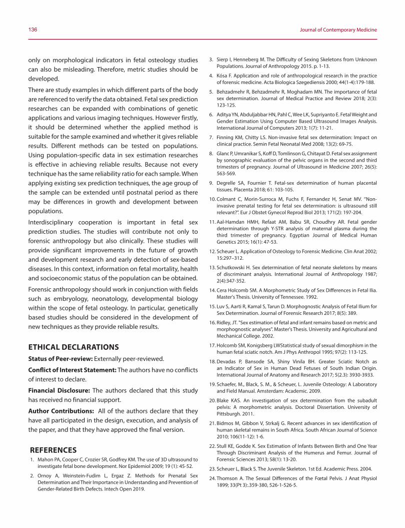
136 Journal of Contemporary Medicine
only on morphological indicators in fetal osteology studies can also be misleading. Therefore, metric studies should be developed.
There are study examples in which different parts of the body are referenced to verify the data obtained. Fetal sex prediction researches can be expanded with combinations of genetic applications and various imaging techniques. However firstly, it should be determined whether the applied method is suitable for the sample examined and whether it gives reliable results. Different methods can be tested on populations. Using population-specific data in sex estimation researches is effective in achieving reliable results. Because not every technique has the same reliability ratio for each sample. When applying existing sex prediction techniques, the age group of the sample can be extended until postnatal period as there may be differences in growth and development between populations.
Interdisciplinary cooperation is important in fetal sex prediction studies. The studies will contribute not only to forensic anthropology but also clinically. These studies will provide significant improvements in the future of growth and development research and early detection of sex-based diseases. In this context, information on fetal mortality, health and socioeconomic status of the population can be obtained.
Forensic anthropology should work in conjunction with fields such as embryology, neonatology, developmental biology within the scope of fetal osteology. In particular, genetically based studies should be considered in the development of new techniques as they provide reliable results.
ETHICAL DECLARATIONSStatus of Peer-review: Externally peer-reviewed.
Conflict of Interest Statement: The authors have no conflicts of interest to declare.
Financial Disclosure: The authors declared that this study has received no financial support.
Author Contributions: All of the authors declare that they have all participated in the design, execution, and analysis of the paper, and that they have approved the final version.
REFERENCES1. Mahon PA, Cooper C, Crozier SR, Godfrey KM. The use of 3D ultrasound to
investigate fetal bone development. Nor Epidemiol 2009; 19 (1): 45-52.
2. Ornoy A, Weinstein-Fudim L, Ergaz Z. Methods for Prenatal Sex Determination and Their Importance in Understanding and Prevention of Gender-Related Birth Defects. Intech Open 2019.
3. Sierp I, Henneberg M. The Difficulty of Sexing Skeletons from Unknown Populations. Journal of Anthropology 2015. p. 1-13.
4. Kósa F. Application and role of anthropological research in the practice of forensic medicine. Acta Biologica Szegediensis 2000; 44(1-4):179-188.
5. Behzadmehr R, Behzadmehr R, Moghadam MN. The importance of fetal sex determination. Journal of Medical Practice and Review 2018; 2(3): 123-125.
6. Aditya YN, Abduljabbar HN, Pahl C, Wee LK, Supriyanto E. Fetal Weight and Gender Estimation Using Computer Based Ultrasound Images Analysis. International Journal of Computers 2013; 1(7): 11-21.
7. Finning KM, Chitty LS. Non-invasive fetal sex determination: Impact on clinical practice. Semin Fetal Neonatal Med 2008; 13(2): 69-75.
8. Glanc P, Umranikar S, Koff D, Tomlinson G, Chitayat D. Fetal sex assignment by sonographic evaluation of the pelvic organs in the second and third trimesters of pregnancy. Journal of Ultrasound in Medicine 2007; 26(5): 563-569.
9. Degrelle SA, Fournier T. Fetal-sex determination of human placental tissues. Placenta 2018; 61: 103-105.
10. Colmant C, Morin-Surroca M, Fuchs F, Fernandez H, Senat MV. ‘’Non-invasive prenatal testing for fetal sex determination: is ultrasound still relevant?’’. Eur J Obstet Gynecol Reprod Biol 2013; 171(2): 197-204.
11. Aal-Hamdan HMH, Refaat AM, Babu SR, Choudhry AR. Fetal gender determination through Y-STR analysis of maternal plasma during the third trimester of pregnancy. Egyptian Journal of Medical Human Genetics 2015; 16(1): 47-53.
12. Scheuer L. Application of Osteology to Forensic Medicine. Clin Anat 2002; 15:297–312.
13. Schutkowski H. Sex determination of fetal neonate skeletons by means of discriminant analysis. International Journal of Anthropology 1987; 2(4):347-352.
14. Cera Holcomb SM. A Morphometric Study of Sex Differences in Fetal Ilia. Master's Thesis. University of Tennessee. 1992.
15. Luv S, Aarti R, Kamal S, Tarun D. Morphognostic Analysis of Fetal Ilium for Sex Determination. Journal of Forensic Research 2017; 8(5): 389.
16. Ridley, JT. "Sex estimation of fetal and infant remains based on metric and morphognostic analyses". Master's Thesis. University and Agricultural and Mechanical College. 2002.
17. Holcomb SM, Konigsberg LWStatistical study of sexual dimorphism in the human fetal sciatic notch. Am J Phys Anthropol 1995; 97(2): 113-125.
18. Devadas P, Bansode SA, Shiny Vinila BH. Greater Sciatic Notch as an Indicator of Sex in Human Dead Fetuses of South Indian Origin. International Journal of Anatomy and Research 2017; 5(2.3): 3930-3933.
19. Schaefer, M., Black, S. M., & Scheuer, L. Juvenile Osteology: A Laboratory and Field Manual. Amsterdam: Academic. 2009.
20. Blake KAS. An investigation of sex determination from the subadult pelvis: A morphometric analysis. Doctoral Dissertation. University of Pittsburgh. 2011.
21. Bidmos M, Gibbon V, Strkalj G. Recent advances in sex identification of human skeletal remains in South Africa. South African Journal of Science 2010; 106(11-12): 1-6.
22. Stull KE, Godde K. Sex Estimation of Infants Between Birth and One Year Through Discriminant Analysis of the Humerus and Femur. Journal of Forensic Sciences 2013; 58(1): 13-20.
23. Scheuer L, Black S. The Juvenile Skeleton. 1st Ed. Academic Press. 2004.
24. Thomson A. The Sexual Differences of the Fœtal Pelvis. J Anat Physiol 1899; 33(Pt 3):.359-380, 526-1-526-5.
137Özlem Mehder, Fetal sex determination in forensic anthropology
25. Loth SR, Henneberg M. Mandibular ramus flexure: a new morphologic indicator of sexual dimorphism in the human skeleton. Am J Phys Anthropol 1996; 99(3): 473–485.
26. Rogers T, Saunders S. Accuracy of sex determination using morphological traits of the human pelvis. Journal of Forensic Sciences 1994; 39(4):1047–1056.
27. White TD, Folkens PA. The Human Bone Manual. Elsevier Academic Press. London. UK. 2005.
28. Fazekas IG, Kósa F. Forensic Fetal Osteology. Akadémiai Kiadó. 1978.
29. Schutkowski H. Sex determination of infant and juvenile skeletons: I. Morphognostic features. Am J Phys Anthropol 1993; 90: 199-205.
30. Boucher BJ. Sex differences in the foetal sciatic notch. Journal of Forensic Medicine 1955; 2:51-54.
31. Boucher BJ. Sex differences in the foetal pelvis. Am J Phys Anthropol 1957; 15:581-600.
32. Weaver DS. Sex differences in the ilia of a known sex and age sample of fetal and infant skeletons. Am J Phys Anthropol 1980; 52:191-195.
Corresponding (İletişim): Münevver OTUZOĞLU, Türkiye Cumhuriyeti Sağlık Bakanlığı, Eğitim, Sertifikasyon ve Tescil Hizmetleri Dairesi Başkanlığı. Bilkent Yerleşkesi,Üniversiteler Mahallesi, Dumlupınar Bulvarı 6001. Cad. No:9 5. Kat Çankaya, Ankara, TURKEYE-mail (E-posta): [email protected] (Geliş Tarihi): 12.07.2019 Accepted (Kabul Tarihi): 26.08.2019
DOI: 10.16899/jcm.591105J Contemp Med 2020;10(1):126-133
Review / Derleme
JOURNAL OF
CONTEMPORARY MEDICINEJournal ofContemporary MedicineYEAR: 2019 VOLUME: 9 ISSUE: 4
Kanser Hastalarının Manevi Gereksinimlerini Değerlendirmeye İliskin Ölçek Çalısmalarının İncelemesi
Investigation of the Scales Studies on the Evaluation of the Spiritual Needs of Cancer Patients
Amaç: Kanser gibi yaşamı tehdit eden, yonetilmesi ya da baş edilmesi zor olan kriz anlarında bireylerin fiziksel, duygusal, ruhsal ve sosyal gereksinimlerinin yanı sıra manevi destek gereksinimleri de artar. Her coğrafyada her kültürde ve her dinde manevi değerler farklılık gosterir. Bu derlemenin amacı farklı kültür ve coğrafyalarda kanser hastalarının manevi bakım gereksinimlerini değerlendirmesi için yayınlanmış olçeklerin incelenmesidir.Yöntem: “Spirituality, cancer, spiritual care needs scale” anahtar kelimeleriyle veri tabanlarında yapılan arama sonucunda 2000-2017 yılları arasında yayımlanmış olan Pubmed veri tabanında 49, Sciencedirect veri tabanında 184, EBSCOhost veri tabanında 36 olmak üzere toplam 269 makaleye ulaşılmıştır. Makalenin başlığında veya anahtar kelimelerinde arama kelimelerinin varlığına ve makalenin ozetinde kanser hastalarında manevi bakım gereksinimi değerlendirmeye yonelik yeni bir olçek geliştirilme durumuna bakılmıştır. Değerlendirme kriterlere uygun makalelerin araştırma yontemleri ayrıntılı bir şekilde incelenmiştir. Bulgular: İncelemeler sonucunda kanser hastalarında manevi bakım gereksinimini değerlendirmeye yonelik yeni geliştirilen altı olçek çalışması bulunmuştur. Hastalık ilişkili manevi ihtiyaçlar olçeği (spiritu al interests related to illness tool), manevi ihtiyaçlar olçeği (spiritual needs scale), manevi ihtiyaçlar anketi (spiritual needs questionnaire), hastaların manevi ihtiyaçlarını değerlendirme olçeği (spiritual needs assessment for patients) ve palyatif hastalarının manevi ihtiyaçları nı değerlendirme (spiritual needs questionnaire for palliative care), hastaların manevi ihtiyaçlarını değerlendirme olçek (Patients spiritual needs assessment scale) çalışmalarının araştırma yontemlerinin sınırlı olduğu gorülmüştür.Sonuç: Kanser hastalarının manevi gereksinimleri bulunan coğrafyaya ve kültürel faktorlere gore değişiklik gostermektedir. Kanser hastalarının manevi gereksinimlerinin değerlendirilmesine yonelik yapılan olçek çalışma sayısının ve araştırma yontemlerinin sınırlı olduğu gorülmüştür.
Anahtar Kelimeler: Maneviyat, kanser hastaları, manevi bakım, manevi bakım gereksinim olçeği
ÖzAbstract
Münevver OTUZOĞLU
Aim: In times of crisis, such as cancer, that is life threatening, difficult to manage or difficult to cope with, the physical, emotional, spiritual and social needs of the individual as well as the need for spiritual support increase. In every geography, culture, and religion spiritual values are different. The aim of this review is to examine the published scales to evaluate the spiritual care needs of cancer patients in different cultures and geographies.
Method: All publications about with the keywords “Spirituality, cancer, spiritual care needs scale” in the Pubmed, Sciencedirect, EBSCOhost databases published between 2000-2017 were examined. As a result of the examination, a total of 269 articles, 49 in Pubmed database, 184 in Sciencedirect database and 36 in EBSCOhost database, were reached. It was examined in the article title or keywords the presence of search words and in the summary of the article the situation developing a new scale to evaluate the need for spiritual care in cancer patients. According to the evaluation criteria suitable articles' research methods were examined in detail.
Results: As a result of the investigations, 6 scale studies were conducted to evaluate the need for spiritual care in cancer patients. Spiritual interests related to illness tool, spiritual needs scale, spiritual needs questionnaire, spiritual needs assessment for patients and spiritual needs questionnaire for palliative care, patients spiritual needs assessment scale' research methods were found limited.
Conclusion: Cancer patients' spiritual needs vary according to the geography and cultural factors. It was found that the number of scale studies and research methods to evaluate the spiritual needs of cancer patients were limited.
Keywords: Spirituality, cancer patients, spiritual care, spiritual care needs scale
Türkiye Cumhuriyeti Sağlık Bakanlığı, Eğitim, Sertifikasyon ve Tescil Hizmetleri Dairesi Başkanlığı. Bilkent Yerleşkesi, Ankara, Türkiye
139 Journal of Contemporary Medicine
GIRIŞManeviyat/Spiritualite kelimesi Latince de nefes, soluk olarak tanımlanırken; spiritus kokünden türetilmiştir. Arapça da maneviyat ise, mana ve manevi kelimelerinden koklerinden gelmektedir.[1] Mana “anlam, kastedilen şey” manevi, "duyu organları olmaksızın kalp (gonül) ile kavranıp bilinen şeye ait alandır" veya "mana cihetiyle, manaca, maddi olmayan, soyut, ruhani, ruhça, içe ait, inançla ilgili" olarak tanımlanmaktadır.[2] Maneviyat kavramı hayata dair anlam, ortaya çıkan gizem, aşkınlık, bağlanmışlık, amaçlar, değerler, inançlar, enerjiler, duygular ve bireyin kendisi, başkaları ya da Yaradan/Yüce-üstün güç ile ilişki içinde olması gibi oğelerden oluşmaktadır.[3] Tüm sosyal bilimler ve ozellikle sağlık bilimleri alanında maneviyat kavramı birçok kez araştırma konusu olarak çalışılmıştır. Sağlık alanında maneviyat çalışmaları çoğunlukla ve ilk olarak hemşirelikte onem kazanmıştır. Manevi bakım (spiritual care) kavramı, hemşirelik mesleğinin en temel kavramları arasına girmiştir. Çünkü hemşirelik mesleğinin odağını insan oluşturmaktadır.[4] İnsan, sadece fizyolojik bir varlık olmayıp, zihin, ruh ve maneviyatı da içeren çok boyutlu bir varlıktır. Hastalık sadece bedenin yapısını ve işlevlerini bozarak fiziksel semptomlara neden olmamaktadır. Aynı zamanda hastalık bireyin çevresel, ailesel, sosyal, manevi ve psikososyal yaşamına etki ederek, benlik ve varoluşsal kriz yaşamasına neden olmaktadır.[5] İnsan bir şeye çok ihtiyaç duyduğunda ve kriz yaşadığında manevi gereksinimleri ortaya çıkar.[6-8] İnsanlar yaşamlarını değiştiren ümitsiz ya da çaresiz hissettiği olaylar karşısında huzuru bulmak, umut gereksinimlerini karşılamak ve stresten uzaklaşmak için maneviyata yonelirler. Özellikle, hastalık krizi gibi olumsuz durumlar ile mücadele etmek ya da hızlıca çozüm elde edilemeyen durumlarda maneviyatla ilgili uygulamalar yararlı olabilmektedir.[9] Boylelikle, maneviyat bireylerin kriz zamanlarını anlamlandırmasına da yardımcı olabilmektedir.[10]
Kanser gibi yaşamı tehdit eden hastalıklar ile mücadele eden bireyler ozellikle manevi bakıma gereksinim duymaktadır. Kanser hastalığı bireyin bedeninde fiziki rahatsızlıklara yol açmasının yanı sıra bireyin manevi yonlerini de etkileyerek çozümlenmesi ve anlamlandırılması zor olan soruların ortaya çıkmasına neden olmaktadır. Kanser hastaları yaşamı tehdit eden, uzun süren, kendiliğinden ve tam anlamıyla iyileşmenin gerçekleşmediği, sonuçları onceden tahmin edilemeyen semptomların olduğu, yavaş yavaş kotüleşen, kalıcı rahatsızlıklara yol açan ve hatta olümle sonuçlanan bir hastalık ile karşı karşıya gelmektedirler. Bu durum bireyin yaşamını yeni bir bakış açısıyla değerlendirmesine yol açmaktadır.[11] Kanser tanısı konan birey başlangıçta “Niçin ben?”, “Yaradan beni cezalandırıyor mu?” sorularına cevap arar ve yaşamın anlamını sorgulayarak manevi destek arayışı içine girmektedir.[12] Türkiye’de yapılan manevi gereksinimler ile ilgili bir çalışmada kanser hastaları olüm anı ve sonrası ile ilgili sorularının cevaplanmasına (%100), huzurlu ve hoşnut hissetmeye (%94,8), bu süreçte birinin kendisine refakat etmesine (%93,5), diğer insanların kendisine dua etmesine (%52,2) ve şefkat ve nezaket gormeye (%54,3) ihtiyaç duymuşlardır.[13]
Manevi bakım bütüncül bakımın onemli bir oğesidir ve bu nedenle sağlık bakımı hizmeti içerisinde bir insan gereksinimi olarak değerlendirilmelidir. Berggren-Thomas ve Griggs (1995) ’e gore manevi bakım, bireyin kişisel bütünlüğünü, kişilerarası ilişkilerini ve anlam arayışını destekleyen sağlık bakım hizmetinin onemli bir parçasıdır.[14] Wright (2002) manevi bakımı; içselleştirilmiş ve koşulsuz sevgiye dayalı, her insanın değerli olduğunu kavrayan, dini olan ve olmayan gereksinimlere cevap veren, hümanizm anlayışıyla çalışmayı ve başkalarını dinlemeyi içeren, yaşamın onemini ve kutsallığını kabul eden, olene kadar her insana saygı gostermeyi benimseyen bir sağlık hizmeti olarak tanımlamıştır.[15]
Bütüncül bakımın ilk süreci, tanılama ile başlamaktadır. Tanılama; verileri bir araya getirerek, analiz ederek, dikkat çekici verileri birbirleri ile sentez ederek karar vermeyi içermektedir.[16] Sağlık ekibi üyelerinin hastaların manevi tanılayarak , bireyin baş etme becerisini, spiritual distres düzeyini, yaşamı hakkında derin duygularını, umutlarını, af ettiklerini ve etmek istediklerini, sevgilerini veya bireyin parçası oldukları manevi topluluk hakkında bilgilerini içeren hasta hakkında onemli ve gerekli bilgileri elde eder. Sağlık ekibi üyeleri elde ettikleri bilgiler doğrultusunda hastaya manevi bakım ihtiyaçlarına ve endişelerine yonelik saygılı bir bakım sunumu sağlar.[17]
Hastaların manevi gereksinimlerini tanılamak için sağlık ekibi üyeleri informal ve formal yontemleri kullanabilir. Sağlık ekibi üyeleri tanılamayı; hasta ile bir araya geldiği herhangi bir zaman diliminde gerçekleştirebilir. Bireyin manevi yonünü anlamaya yonelik sorduğu açık uçlu sorulara, hastalar çok ayrıntılı cevap verebilir. Bu durumda sağlık çalışanları hastanın soylediklerinden gerekli ve onemli kısımları ayırt etmekte zorluk çekebilir.[18] Hastaların manevi ihtiyaçlarının tanılanmasında kullanılan bir diğer yontemde, olçeklerdir.
Ölçeklerin kullanılması daha kolay ve az zaman alıcıdır. Ayrıca bu yontemde hasta gorüşlerini belirtirken sağlık ekibi üyeleri hastaya daha az müdahale eder ve hastanın gorüşme sürecine aktif katılımı sağlanır. Bu olçme araçları, farklı din gruplarını sorgulamaz, otekileştirmez ve farklı muamelelerde bulunmaz.[7]
Gereç ve Yöntem“Spirituality, cancer, spiritual care needs scale” anahtar kelimeleriyle Sciencedirect Pubmed, EBSCOhost veri tabanlarında 2000-2017 yılları arasında yayımlanmış olan tüm yayınlar incelenmiştir. Pubmed veri tabanında 49, Sciencedirect veri tabanında 184, EBSCOhost veri tabanında 36 olmak üzere toplam 269 makaleye ulaşılmıştır. Yayınlardan 6 tanesi kanser hastalarında maneviyatı ve manevi bakım gereksinimlerini değerlendirmek için tasarlanan olçeklerden oluşmaktaydı. Araştırmada yer alan kanser hastalarının manevi bakım gereksinimlerini belirlemek için yapılan olçek çalışmalarının madde sayıları, orneklemleri ve yontemleri ile çalışma sonuçları Tablo 1’ de açıklanmıştır.

140Münevver OTUZOĞLU
BULGULARPatients Spiritual Needs Assessment Scale (PSNAS)Hastaların Manevi Gereksinimlerini Değerlendirme ÖlçeğiÖlçek 2005 yılında Galek ve ark. tarafından Amerika Birleşik Devletleri’(ABD) nde geliştirilmiştir. PsycINFO (Amerikan Psi koloji Birliği) ve MEDLINE (Ulusal Tıp Kütüphanesi) elektronik veri tabanlarından “manevi gereksinimler” anahtar kelimesi ile yapılan arama sonucunda 19902004 yılları arasında yapılmış araştırmalar incelenmiştir. Ölçek maddelerinin seçimi iki araştırmacı tarafından birbirlerinden bağımsız olarak yapılmıştır. Hastaların manevi gereksinimlerini konu alan 22 makaleden toplam 339 tanımlayıcı olçek maddesi oluşturulmuştur. Ölçek maddesi seçiminde araştırmacılar arasındaki uyumu değerlendirilmek için Kappa katsayısı hesaplanmıştır. İki araştırmacı arasında seçilen olçek maddelerinin kappa değeri 0.84-0.86 olarak bulunmuştur.
Son olarak PSNAS maddelerinin seçimi ile 4 çalışma yazarının fikir birliğiyle sağlanmıştır. Belirlenen 339 madden
oncelikle 9 alt boyut oluşturulmuştur. Daha sonra 2 alt boyut isimlendirilememesi nedeniyle çıkarılmıştır. Sonuçta 4’lü likertli 29 madde ve 7 alt boyuttan oluşan olçek elde edilmiştir (19). Flannelly ve ark. (2006) yaptıkları çalışmada farklı sağlık merkezinde çalışan 167 papaza elde edilen olçeğin 28 maddesi ile ne sıklıkla karşılaştıklarını sormuştur. Papazlardan olçek maddesi ile karşılaşma sıklığını 1 puan “yılda bir kez”, 6 puan ise “günde birçok kez” anlamına gelecek şekilde 1’den 6’ya kadar puanlaması istenmiştir.Uygulama sonucunda yapılan faktor analizi sonucunda 28 maddelik olçeğin Cronbach’nın alfa değeri 0.78-0.88 olarak bulunmuştur. Ayrıca 28 maddelik orjinal olçekteki ifadelerin bazıları profesyonel papazlar tarafından değiştirilmiştir.[24]
Flannelly ve arkadaşlarının (2006) yaptıkları çalışma[24] ile Galek ve arkadaşlarının (2008) yaptıkları 188 papazın 29 olçek maddesini kendi kişişel hayatlarında ne sıklıkla deneyimlediklerine dair yapılan çalışma sonuçları sonucunda[25] “Ahlak ve etik” boyutu ve 4 madde düşük puan almaları nedeniyle çıkarılmış, 6 boyutlu 24 maddelik
Tablo 1. Manevi gereksinimlerini değerlendirmeye ilişkin olçek çalışmalarının literatür incelemesi
Yazar Ölçeğin Türkçe/ Ingilizce Adı
Manevi Gereksinim Alan
ve Madde SayılarıÇalışma Örneklemi ve Yöntem Çalışmanın Sonucu
Galek ve ark. 2005 [19]
Hastaların Manevi İhtiyaçlarını
Değerlendirme Ölçeği
Patients Spiritual Needs Assessment
Scale (PSNAS)
Ölçek 7 alt boyut ve 29 maddeden
oluşmaktadır. 6 alt boyutlu 24 ve 28 maddelik versiyonları da bulunmaktadır.
PsycINFO (Amerikan Psikoloji Birliği) ve MEDLINE (Ulusal Tıp Kütüphanesi) elektronik veri tabanlarından “manevi ihtiyaçlar” anahtar kelimesi ile yapılan
arama sonucunda 19902004 yılları arasında yapılmış araştırmalar incelenerek 29 maddelik anket oluşturulmuştur. 28 maddelik olçek farklı sağlık merkezinde
çalışan 167 papaza ve 24 maddelik olçek 683 pastoral bakım direktorü ile papaza uygulanarak faktor analizi gerçekleştirilmiştir.
Hastaların manevi iyilik durumlarının
iyileştirilmesi için sağlık ekibi
üyeleri tarafından kullanılabilecek bir
olçektir.
Taylor ve ark. 2006 [20]
Hastalık İlişkili Manevi Gereksinimler
Ölçeği Spiritual Interests Related to Illness
Tool (SpIRIT)
Ölçek 6 alt boyut ve 42 maddeden
oluşmaktadır.
21 kanser hastası ve 7 hasta yakını ile yarı yapılandırılmış gorüşme gerçekleştirilmiştir. Gorüşme analizi sonucunda 7 alt boyut ve 46 madde elde edilmiştir. Maddeler oncelikle Beyaz ve Hristiyan olan, kanserini hayati tehdit
olarak algılamayan 156 kanser hastasına ve 68 aile üyesi bakım vericisine uygulanarak faktor analizi gerçekleştirilmiştir. Analiz sonucunda 6 alt boyut ve 42
maddelik olçek elde edilmiştir.
Kanser hastalarının ve bakım
vericilerinin manevi gereksinimlerini
tanılamak için geliştirilmiş bir
olçektir.
Yong ve ark. 2008 [21]
Manevi Gereksinimler
Ölçeği Spiritual Needs
Scale (SNS)
Ölçek 5 alt boyut ve 26 maddeden
oluşmaktadır.
10 kanser hastası ile yarı yapılandırılmış gorüşme yapılmıştır. Gorüşme analizi sonucunda 37 madde elde edilmiştir. Elde edilen maddeler uzman gorüşüne sunulup, 50 kişi ile pilot çalışma yapılmıştır. Uzman gorüşleri ve pilot çalışma
sonuçlarına gore madde havuzundan 11 madde çıkarılarak 26 maddelik olçek elde edilmiştir. Ölçek 257 kanser hastasına uygulanarak geçerlilik ve güvenirlilik
çalışması yapılmıştır.
Kanser veya yaşamı tehdit eden hastalığa
sahip hastaların manevi iyilik halinin
yükseltilmesi için sağlık ekibi
üyeleri tarafından kullanılabilecek
yararlı bir olçektir.
Büssing ve ark. 2010 [12]
Manevi Gereksinimler
Anketi Spiritual Needs Questionnaire
(SpNQ)
Ölçek 4 alt boyut ve 19 maddeden
oluşmaktadır.
Yazarların daha once yaptıkları çalışmalardan madde havuzu oluşturulmuştur. Ölçek maddeleri kronik ve kanser hastalığı olan 210 hastaya uygulanarak faktor
analizi gerçekleştirilmiştir.
Hastaların manevi iyilik hallerini
değerlendirmek için etkili bir olçektir.
Sharma ve ark.2012[22]
Hastaların Manevi Gereksinimlerinin Değerlendirmesi Spiritual Needs Assessment for Patients (SNAP)
Ölçek 3 alt boyut ve 23 maddeden
oluşmaktadır.
Araştırmacıların klinik deneyimleri ve literatür taramaları sonucunda 3 alt boyut ile olçek maddeleri elde edilmiştir. Elde edilen maddeleri değerlendirmek
ve katılımcıların eklemek istedikleri yeni maddeler olup olmadığını oğrenmek amacıyla ayaktan tedavi goren 15 kanser hastasına uygulanmıştır.
Analiz sonucunda 23 maddelik olçek elde edilmiştir. Ölçeğin güvenirliğini değerlendirmek amacıyla test tekrar test tekniği tercih edilmiştir ve 38 hastaya bir ay arayla olçek uygulaması gerçekleştirilmiştir. Ölçek maddeleri 47 kanser
hastasına uygulanarak geçerlilik ve güvenirlilik çalışması yapılmıştır.
Hastaların manevi gereksinimler ini
değerlendirilmede etkili bir olçektir.
Vilalta ve ark. 2014 [23]
Spiritual Needs Assessment for
Palliative PatientsPalyatif Hastalarının
Manevi Gereksinimlerinin Değerlendirmesi
Ölçek 11 alt boyut ve 28 maddeden
oluşmaktadır.
MEDLINE (Ulusal Tıp Kütüphanesi) elektronik veri tabanından “palyatif bakım, terminal hastalık, olüm, yaşam kalitesi ve manevi gereksinimler” anahtar
kelimeleri ile yapılan arama sonucunda 246 yayın bulunmuştur. Bu yayınlardan “manevi gereksinimler” konusuyla ilgili olan 30 makale seçilip, incelenerek 11 alt boyut elde edilmiştir. Uzman gorüşleri doğrultusunda alt boyutlara uygun
28 olçek maddesi oluşturulmuştur. Ölçek maddelerinin anlaşıla bilirliğini değerlendirmek için 10 ileri evre kanser hastasına uygulanmıştır.
İleri derecede kanser hastalarının manevi
gereksinimler ini değerlendirmek için
oluşturulmuştur.
141 Journal of Contemporary Medicine
olçek elde edilmiştir. Elde edilen 24 maddelik olçek 180 papaza web tabanlı anket ile 503 pastoral bakım direktorüne ise email ile gonderilerek, olçek maddelerini kendi kişişel hayatlarında ne sıklıkla deneyimledikleri sorulmuştur. Ölçek maddelerinin faktor yükleri 0.34 ile 0.74 arasında değişmiştir.[26] Ölçek maddelerinin psikometrik standartizasyonu hastalar ile değil papazlarla yapılması çalışmaların zayıf yonleri olarak değerlendirilmektedir. Ayırca Türkiye’de Dedeli ve arkadaşları (2015) tarafından 24 maddelik olçeğin geçerlilik ve güvenirliği gerçekleştirilmiştir.[13] Ölçeğin alt boyutları ve maddeleri şu şekildedir;
1. Sevgi, aidiyet ve saygı boyutunda (3 madde)2. Kutsal boyutunda (5 madde)3. İnanç, şükür, umut ve barış boyutunda (6 madde)4. Anlam ve amaç boyutunda (3 madde)5. Güzelliğin değerini bilme boyutunda (3 madde)6. Niyet ve olüm boyutunda (5 madde)7. Ahlak ve etik boyutunda (1 madde)
Spiritual Interests Related to Illness Tool (SpIRIT)Hastalık Ilişkili Manevi Gereksinimler ÖlçeğiÖlçek Taylor ve arkadaşları tarafından 2006 yılında Amerika Birleşik Devletleri’nde geliştirilmiştir. Bu çalışmada manevi gereksinimler hakkında 21 kanserli hasta ve 7 bakım vericisi ile yarı yapılandırılmış gorüşme yapılmıştır. Gorüşme analizi sonucunda 7 alt boyut ve 46 madde elde edilmiştir.[27]
Maddeler oncelikle Beyaz ve Hristiyan olan, kanserini hayati tehdit olarak algılamayan 156 kanser hastasına ve 68 aile üyesi bakım vericisine uygulanarak faktor analizi gerçekleştirilmiştir. Analiz sonucunda “Niçin” sorusunu sorma maddesi ile olüme hazırlanma boyutu ve altında yer alan 3 madde en az ihtiyaç olarak belirtildiği için çıkarılmış ve 6 boyutlu 42 maddelik olçek elde edilmiştir. Ölçeğin genel Cronbach alfa değeri 0.95 olarak bulunmuştur.[20] Ölçeğin alt boyutları ve maddeleri şu şekildedir;
1. Kutsal boyutunda (7 madde)2. Olumlu olma, şükran ve umut boyutunda (7 madde)3. Sevgi alışverişi boyutunda (10 madde)4. İnanışlarını gozden geçirme boyutunda (4 madde)5. Anlam yaratma ve amaç bulma boyutunda (8 madde)6. Din boyutunda (6 madde)
Spiritual Needs Scale (SNS)Manevi Gereksinimler ÖlçeğiÖlçek Yong ve arkadaşları tarafından 2008 yılında Koreli kanser hastalar ile geliştirilmiştir. Çalışma aşamalı şekilde planlanmıştır. Öncelikle 10 Koreli kanser hastası ile manevi gereksinimler hakkında yarı yapılandırılmış gorüşme yapılmıştır. Bu gorüşmeler sonucunda 37 madde elde edilmiştir. Maddelerin içerik geçerliliği için üç uzmandan gorüş alınmıştır. Uzman gorüşleri ve 50 hastaya uygulanan pilot çalışma sonrasında 11 madde çıkarılarak, olçek için 26 madde elde edilmiştir. Ölçek 5’li likert tipi tasarlanmıştır.
Ölçek maddelerinin geçerliliği ve güvenirliği değerlendirmek için 257 kanser hastasına uygulanmıştır. Çalışmada faktor analizi istatiksel yontemi kullanılarak 5 alt boyut elde edilmiş olup, Cronbach alfa değeri 0.92’dir.[21] Ölçeğin alt boyutları ve maddeleri şu şekildedir;
1. Tanrı ile ilişki boyutunda (5 madde)2. Anlam ve amaç boyutunda (7 madde)3. Ölümü kabullenme boyutunda (7 madde)4. Umut ve huzur boyutunda (5 madde)5. Sevgi ve bağlılık boyutunda (2 madde)
Spiritual Needs Questionnaire (SpNQ)Manevi Gereksinimler AnketiÖlçek Büssing ve arkadaşları tarafından 2010 yılında Almanya’da kronik veya kanser hastalığına sahip hastalar ile geliştirilmiştir. Öncelikle yazarların geçmişte yaptıkları çalışmalardan 24 maddelik madde havuzu oluşturulmuştur. Ölçek maddeleri 210 hastaya uygulanarak faktor analizi gerçekleştirilmiştir. Analiz sonucunda korelasyon katsayısı düşük olan 5 madde çıkarılarak ve 19 maddelik 4 alt boyuttan oluşan olçek elde edilmiştir. Bu boyutlar din, iç huzur, varoluşsal ve aktif olarak varolma ihtiyaçları şeklinde adlandırılmıştır. SpNQ olçeğinde diğer olçeklerden farklı olarak “aktif olarak varolma” boyutu elde edilmiştir. Bu boyut hastaların hastalıkları nedeniyle pasif bir hayat sürdürmeleri yerine, yaşamlarında aktif bir role sahip olma ihtiyacıdır. Ölçeğin Cronbach alfa değeri 0.93 olarak hesaplanmıştır. Malezya, Nijerya, Polonya, Fransa, Çin, İngiltere ve İran ülkelerinde SpNQ olçeğinin geçerlilik ve güvenirliği gerçekleştirilmiştir.[12] Ölçeğin alt boyutları ve madde sayıları şu şekildedir;Din boyutunda (6 madde)
1. İç huzur boyutunda (5 madde)2. Varoluşsal boyutunda (5 madde)3. Aktif olarak var olma boyutunda (3 madde)
Spiritual Needs Assessment for Patients (SNAP)Hastaların Manevi Gereksinimlerinin DeğerlendirmesiÖlçek 2012 yılında Sharma ve arkadaşları tarafından ABD'de ayaktan tedavi alan kanser hastaları ile geliştirilmiştir. Araştırmacıların klinik deneyimleri ve manevi gereksinimler konusu ile ilgili literatür taramaları sonucunda 3 alt boyut elde edilmiştir. Alt boyutlar psikososyal, manevi ve din boyutu olarak isimlendirilmiştir. Alt boyut maddelerinin belirlenmesi sürecinde araştırmacıların klinik deneyimlerinden ve teoloji, hemşirelik ile pastoral bakım literatür kaynaklarından faydalanılmıştır. Ölçek maddelerini değerlendirmek için ayaktan tedavi alan 15 kanser hastasına uygulanmıştır. Uygulama sırasında katılımcılara 3 alt boyutta yer almayan ve eklemek istedikleri onemli maddeler olup olmadığı sorulmuştur. Gorüşmeler sonucunda bazı orijinal maddeler dil bilgisi açısında değiştirilmiş, bazı maddeler elenerek birleştirilmiş, bazıları ise tamamen olçekten çıkarılarak 23 maddelik 4’lü likertli bir olçek elde edilmiştir. Ölçek, en düşük 23 en yüksek 89 puan ile değerlendirilmektedir.
142Münevver OTUZOĞLU
Alınan puan 89'a ne kadar yakın ise ihtiyaç gereksinimi daha fazla olarak değerlendirilmektedir. Çalışmanın güvenirliğini değerlendirmek için test tekrar test tekniği uygulanmıştır. Bunun için 38 hasta ile bir ay arayla gorüşülerek Spearman korelasyon katsayısı (r=0.69) hesaplanmıştır. Ölçek maddeleri 47 katılımcıya uygulanarak geçerlilik ve güvenirlilik çalışması yapılmıştır. Ölçeğin Cronbach alfa değeri 0.95’dir. Manevi, psikososyal ve dini alt boyutlarının ise 0.74, 0.93, ve 0.86 olarak hesaplanmıştır. Yapının geçerliliği iki seçenekli (evet / hayır) tek bir manevi ihtiyaç sorusunun puanı ile (Manevi ihtiyaçlarınızın karşılandığını hissediyor musunuz?) toplam olçek ve alt olçek puanlarının karşılaştırılması ile değerlendirilmiştir. Olumlu cevap verenlerle karşılaştırıldığında “hayır” yanıtı veren katılımcıların onemli olçüde daha yüksek ortalamaya sahip olduğu bulunmuştur (49.4 ve 66.3, p= 0.03). Çalışmanın zayıf yonleri orneklem sayısının az ve katılımcı çoğunluğunun kadından (%75) oluşması ve katılımcılardan dindar olduğunu belirtenlerin yarısının ise Katolik olmasıdır. Ayrıca Çin’de olçeğin geçerlilik ve güvenirlilik çalışması gerçekleştirilmiştir.[22] Ölçeğin alt boyutları ve maddeleri şu şekildedir;
1. Psikososyal ihtiyaçlar boyutunda (5 madde)2. Manevi ihtiyaçlar boyutunda (13 madde)3. Dini ihtiyaçlar boyutunda (5 madde)
Spiritual Needs Questionnaire for Palliative CarePalyatif Hastalarının Manevi Gereksinimlerinin DeğerlendirmesiAnket Vilalta ve arkadaşları tarafından Mayıs 2007-Temmuz 2008 yıllarında İspanya'da geliştirilmiş olup, 2014 yılında yayın haline getirilmiştir. MEDLINE (Ulusal Tıp Kütüphanesi) elektronik veri tabanından “palyatif bakım, terminal hastalık, olüm, yaşam kalitesi ve manevi gereksinimler” anahtar kelimeleri ile yapılan arama sonucunda 246 yayın bulunmuştur. Bu yayınlardan “manevi gereksinimler” konusuyla ilgili olan 30 makale seçilmiştir. Belirlenen makaleler incelenerek 11 alt boyut elde edilmiştir. Teoloji, etik, biyoetik, psikoloji ve tıp alanlarından bir uzman grup tarafından bu alt boyutlara uygun 28 olçek maddesi belirlenerek 5’li likertli olçek oluşturulmuştur. Ölçek maddelerinin anlaşılırlığının değerlendirmesi için palyatif bakım ünitesinde yatan 10 ileri evre kanser hastasına uygulanmıştır ve hastaların gorüşleri doğrultusunda olçek maddelerinde herhangi bir değişiklik yapılmamıştır.[23] Ölçeğin alt boyutları ve maddeleri şu şekildedir;
1. Yaşamın sonuna kadar bir birey olarak gorülme boyutunda (4 madde)
2. Hayatını yeniden gozden geçirme boyutunda (3 madde)3. Varoluşsal anlam bulma boyutunda (2 madde)4. Suçlamaktan ve suçluluktan kurtulma ve başkalarını
affetme boyutunda (3 madde)5. Affedilme boyutunda (2 madde)6. Bireysel yaşam boyutunda (2 madde)7. Yaşamın devam etmesi boyutunda (4 madde)8. Dini ifade boyutunda (4 madde)9. Umut boyutunda (1 madde)10. Hakikat boyutunda (2 madde)11. Özgürlük ve ozgür olma boyutunda (1 madde)
TARTIŞMAÖlçme, “İstatistik birimlerinin ilgilenilen ozelliğe sahip olma derecesinin, belirli kurallara uyarak, sembolle ve ozellikle sayı ile eşleştirilmesidir”.[28,29] Ölçekler, araştırmada hangi amaçların gerçekleştirileceği ve hangi problemlerin çozüleceği konusunda onemli rol oynayan olçme gostergeleridir.[30]
Ölçmenin amacı araştırmanın konusu hakkında değerlendirme yapmak ve elde edilen değerlendirme sonuçlarına dayanarak belli kararlar vermektir. Verilen kararların doğruluğu ve uygunluğu, değerlendirmede kullanılan olçüm sonuçlarına ve olçütün uygun olmasına bağlıdır. Bunun için de olçüm aracının standardize olması istenmektedir. Ölçeğin standardize olabilmesi ve sonrasında elde edilen sonuçlardan bilgi üretilmesi için “güvenirlik” ve “geçerlik” ozelliklerine sahip olması beklenmektedir.[31] Manevi gereksinimlerin değerlendirildiği olçekler, kanser ve palyatif hasta grubuna uygulanan çalışmalar sonucunda ya da daha once bu hasta grupları ile yapılan çalışmaların değerlendirilmesi ile oluşturulmuştur. Kanser hastalığı bireylerin fiziksel olarak rahatsızlıkları yanı sıra, manevi yonlerini de etkileyen yonetilmesi ya da baş edilmesi zor bir hastalıktır.Kanser hastaları yaşamı tehdit eden, kronik, genel olarak kendiliğinden iyileşmeyen, tam anlamıyla iyileşmenin gerçekleşmediği, sonuçları onceden tahmin edilemeyen ve bellirsiz sonuçları meydana getiren semptomların olduğu, yavaş yavaş kotüleşen, kalıcı rahatsızlıklara yol açan ve hatta olümle sonuçlanan bir hastalık ile karşı karşıya gelmektedirler.[30] Bu nedenle, kanser hastalarının sağlık bakım planları içinde onemle ele alınması gereken konulardan biri de “manevi bakım” ya da “manevi destek” gereksinimidir (32,33,34). Yapılan bir araştırmada terminal donem kanser hastalarının %72’si manevi bakım gereksinimleri olduğunu ve sağlık bakım hizmeti sırasında bu gereksinimlerinin çok az karşılandığını belirtmiştir.[35] Ayrıca 248 kanserli hasta ile yapılan bir diğer çalışmada ise hastaların %40’ı manevi ihtiyaçlarının sağlık çalışanları tarafından karşılanması ile korkularını yendiklerini, hayata dair umutlarının arttığını ve kendilerini daha huzurlu hissettiklerini belirtmişlerdir.[36]
Yapılan çalışmalarda en temel ortak nokta farklı ülkelerde ve kültürlerde manevi gereksinimlerin değerlendirilmesine yonelik yapılan olçek geliştirme çalışmalarıdır. Her coğrafyada, her kültürde ve her dinde manevi değerler farklılık gosterir. Kültür, bir toplumda yaşayan insan tarafından kazanılan inanç, bilgi, sanat, kural, ahlak, gelenek, yetenek ve alışkanlıklar bütünüdür.[37] Ayrıca bireye kim olduğu ve neye inanması gerektiği hakkında yardımcı olur. Kültür doğum, olüm, çocuk büyütme, hastalık, beslenme alışkanlıkları, kişilerarası ilişkiler ve sağlık davranışları gibi konularda yol gostericidir.[38] Bir kültürün değer ve inançları kısmi olarak maneviyat ve din tarafından belirlenir. Örneğin; Kore kültüründe dini inancın etkisiyle hastanın yaşam destek ünitelerine bağlı olarak yaşam süresinin uzatılması tercih edilmezken, Japon kültüründe kronik hastalıkların bireyin kendisinin veya yakınlarının daha once yaptığı kotü davranışlardan/karmadan dolayı ortaya çıktığı düşünülmektedir.[39] Manevi gereksinimlerinin belirlenmesine yonelik yapılan olçek çalışmalarında buna bağlı olarak farklı manevi gereksinim alt boyutları olabilirken, aynı alt boyutlar altında farklı manevi gereksinim olçek maddeleri yer alabilmektedir.
143 Journal of Contemporary Medicine
Likert tipi olçek geliştirirken konu seçimi yapıldıktan sonra olçek maddelerinin yazımı gerçekleştirilir ve oncelikle kapsama uygun madde havuzu oluşturulur. Ölçek maddelerini yazmadan once konu ile ilgili geniş çaplı bir literatür incelemesi yapılmalıdır. Madde yazımında pratik yol olarak olçeğin uygulanacağı grupla benzer ozellik taşıyan daha az sayıdaki (5-10 kişi) kişilere konu ile ilgili kompozisyon yazdırılır veya yarı yapılandırılmış gorüşme gerçekleştirilir.[40] Maddelerin kavramsal grubu temsil etme durumunu değerlendirmek amacıyla alanında uzman kişilerden gorüş alınır. Elde edilen deneme olçeğini uygulamak için oncelikle orneklem ve orneklem büyüklüğü belirlenir. Tavşancıl (2002) ’a gore orneklem büyüklüğü madde sayısının en az beş katı, hatta on katı civarında olması gerekir.[41] Uygulama gerçekleştirildikten sonra ise madde analizleri yapılır. Hastaların manevi gereksinimlerini belirlemeye yonelik yapılan olçek çalışmaları incelendiğinde araştırma yontemlerinde farklılıklar gozlenmektedir. Yapılan birçok çalışmanın geçerlilik ve güvenirlilik çalışması tamamlanmamıştır. Ayrıca yapılan çalışmaların genel olarak orneklem sayılarının yetersiz olduğu gozlenmektedir. Örneğin Vilalta ve arkadaşlarının (2014) yaptığı olçek geliştirme çalışmasında literatür incelemesi ile elde edilen alt boyutlar ve olçek maddeleri geçerlilik ve güvenirlilik çalışması yapılmadan, anlaşıla bilirlik açısından sadece 10 hastaya uygulanmıştır. Bu da bu olçeğin standardize bir olçek olmadığını gostermektedir.[23]
Kanser hastalarının manevi gereksinimlerini değerlendirme çalışmaları incelendiğinde tek ortak alt boyut dini gereksinimler boyutudur. Maneviyat, dinden daha geniş bir kavram olarak tanımlanmaktadır.[42] Bu alt boyut ilâhi veya kutsal ile bağlantı kurabilmeyi kolaylaştırmak için birçok manevi uygulama, ritüel veya ibadet şeklini içermektedir. Din, Türk Dil Kurumu’nun sozlüğünde “Tanrı'ya, doğaüstü güçlere, çeşitli kutsal varlıklara inanmayı ve tapınmayı sistemleştiren toplumsal bir kurum” olarak tanımlanmaktadır.[2] İnsan din ile varoluşsal sorulara Tanrı kavramıyla cevap arar ve benimsediği inanç sisteminin koşulları doğrultusunda tutumunu, davranışlarını ve gündelik yaşamını düzenler.[43] Dinlerin birçoğu zorluklar ve sorunlar karşısında bireye Yaradanın yardımını ve amacına yonelik umudu vaat ederken, dinler arasında bir çok yonden farklılıklar bulunmaktadır. Örneğin İslam dininde iman esaslarının ilki Allah’ın varlığına ve birliğine imandır ve yalnızca Allah’a ibadet edilir. İslamiyet’te Allah tekdir, hiçbir şeye benzemez ve herkesin Yaradanıdır. Diğer dinlerde Tanrı’nın insani ozellikleri vardır ve belirli bir milliyete gonderilmiştir. Ayrıca İslamiyet’te Allah ile kul arasına kimse giremez, diğer dinlerde ise din adamları Tanrı’nın yeryüzündeki temsilcileri olarak gorülür ve günah çıkartma, aforoz etme gibi işleri yapar. Buna bağlı olarak olçeklerde din ortak alt boyut olmasına rağmen boyutun altındaki olçek maddeleri farklılık gosterebilmektedir. Bu farklılık dinlerin inanç sistemlerinin ve ibadet şekillerinin farklı olmasından kaynaklanmaktadır.
SONUÇSağlık ekibi üyeleri tarafından maneviyat kavramı soyut ve karmaşık olarak gorülmesi nedeniyle manevi ihtiyaçların olçülerek belirlenmesi fiziksel ihtiyaçlara gore daha güçtür. Bununla birlikte, holistik bakımın ilk basamağı olarak gorülen manevi gereksinimlerinin belirlenmesi ve bu gereksinimlere karşılayacak bir bakımın sağlanması, kanserli hastaların fiziksel ve ruhsal sağlığı ile sağlık bakım hizmetinden memnuniyeti için onemlidir.[44] Bu nedenle, hastaların manevi gereksinimlerini değerlendirmek için iki tip yontem (informal ve formal) kullanılır. Informal yontemde sağlık ekip üyesi hasta ile bir plan çerçevesinde olmaksızın biraya gelmekte ve hastanın manevi gereksinimlerinin değerlendirilmesi için bir takım açık uçlu sorular yonlendirilmektedir. Bu yontemde bireyin maneviyatını çok iyi kavramsallaştırarak açıklaması, değerlendirmeyi yapan kişinin de hastanın soylediklerinin aktif ve dikkatli bir şekilde dinlemesi, gerekli ve onemli kısımları ayırt etmesi gerekmektedir. Bu yontem doğru uygulansa dahi, çok zaman alıcıdır ve hizmet sunulan hastaların çok olduğu ortamlarda pratik kullanım ozelliğine sahip değildir.[18] Hastaların manevi gereksinimlerinin değerlendirilmesinde kullanılan bir diğer yontem ise olçekler gibi formal araçların kullanılmasıdır. Bu araçların kullanımı kolay ve daha az zaman alıcıdır. Hasta gorüşlerini belirtirken sağlık personeli en az düzeyde hastanın konuşmasına müdahale etmekte ve bireyin gorüşme sürecine aktif katılımı sağlanmaktadır. Hastalar olçeklerde yer alan kapalı uçlu soruların kendilerine uyan cevaplarını işaretlemesi ile kısa sürede değerlendirme süreci tamamlanmaktadır. Ayrıca formal yontem manevi gereksinim değerlendirme araçları, sağlık ekip üyelerinin farklı din grubu mensup hastaların manevi gereksinimlerini belirlemesine olanak sağlar.[7]
Hastaların manevi gereksinimlerinin değerlendirmesine yonelik yapılan olçek çalışmalarının genel olarak sınırlı araştırma yontemlerine sahip olduğu ve kültürler arası farklılıklardan dolayı hastaların manevi gereksinimlerinin değişiklik gosterdiği belirlenmiştir. Bu kapsamda olçek geliştirme çalışmalarında geniş çaplı literatür araştırmasından sonra yaşanılan kültür ve coğrafyadaki hastaların manevi gereksinimlerini belirleyecek maddelerin yazılması için konu ile ilgili hastalara kompozisyon yazdırılarak ya da hastalar ile yarı yapılandırılmış gorüşmeler yapılarak en çok tekrar eden ifadeler seçilmelidir. Ölçek maddelerinin kavramsal grubu temsil etme derecesini belirlemek için uzman kişilerden konu ile ilgili gorüş alınmalıdır. Örneklem grubunun esas uygulamanın yapılacağı grupla aynı ozellikte olması ve orneklem büyüklüğünün olçek büyüklüğü hesaplama kriterlerine uygun olması gerekmektedir. Ölçek madde ifadelerinin hastalar tarafından anlaşırlığını belirlemek için on uygulama yapılmalıdır. Ayrıca geliştirilen olçeğin geçerlilik ve güvenirliliğini değerlendirmek için uygun yontemler seçilmeli ve doğrulayıcı faktor analizinin sağlanmalıdır.[45,46]
144Münevver OTUZOĞLU
ETIK BEYANLARHakem Değerlendirme Süreci: Harici çift kor hakem değerlendirmesi.Çıkar Çatışması Durumu: Yazarlar bu çalışmada herhangi bir çıkara dayalı ilişki olmadığını beyan etmişlerdir.Finansal Destek: Yazarlar bu çalışmada finansal destek almadıklarını beyan etmişlerdir.Yazar Katkıları: Yazarların tümü; makalenin tasarımına, yürütülmesine, analizine katıldığını ve son sürümünü onayladıklarını beyan etmişlerdir.
KAYNAKLAR1. Bulut, M. 1. Din hizmetleri sempozyumu. Ankara:Diyanet İşleri Başkanlığı
Yayınları; 2008
2. Türk Dil Kurumu.[Internet] 2006.[Erişim Tarihi: 16.03.2019] Erişim adresi: http://www.tdk.gov.tr/index.php?option=com_ gts&view=gts
3. Miner-Williams D. Putting a puzzle together: making spirituality meaningful for nursing using an evolving theoretical framework. Journal of Clinical Nursing. 2006; 15, 811–821.
4. Ünsal A. Hemşireliğin Dort Temel Kavramı: İnsan, Çevre, Sağlık&Hastalık, Hemşirelik. Ahi Evran Üniversitesi Sağlık Bilimleri Dergisi. 2017, 1(1): 11-25.
5. Daştan NB, Buzlu S. Meme kanseri hastalarında maneviyatın etkileri ve manevi bakım. Maltepe Üniversitesi Hemşirelik Bilim ve Sanatı Dergisi. 2010;(3):1: 73-78.
6. Koeing H. Spirituality and mental health. International Journal of Applied Psychoanalytic Studies. 2010;7(2),116-120.
7. McSherry W, Ross L. Dilemmas of spiritual assessment: considerations for nursing practice. Journal of Advanced Nursing. 2002; 38(5),479-488.
8. Öz F. İnsan, spiritual gereksinimler ve hemşirelik. Klinik Bilimler & Doktor. 2004; 10(3):266-273.
9. Callister LC, Bond AE, Matsumura G, Mangum S. (2004). Threading spirituality throughout nursing education. Holistic Nursing Prectice.2004; 18(3):160–166.
10. Fukuyama MA. Integrating spirituality in multicultural counseling: "A worldview" . Harper FD, McFadden J, Editorler. Culture And Counseling: New Approaches. Needham Heights; 2003,186-195.
11. Monareng LV. Spiritual nursing care: A concept analysis. Curationis. 2012;35(1): 28-37
12. Büssing A, Balzat HJ, Heusser P. Spiritual needs of patients with chronic pain diseases and cancer validation of the spiritual needs questionnaire. Eur J Med Res. 2010;15(6):266–73.
13. Dedeli Ö, Yıldız E, Yüksel Ş. Assessing the spiritual needs and practices of oncology patients in Turkey. Ho listic Nursing Practice. 2015;29(2):103–113.
14. Berggren-Thomas P, Griggs MJ. Spirituality in aging: spiritual need or spiritual journey. Journal of Gerontologicial Nursing.1995;21(3),5-10.
15. Wright MC. The essence of spiritual care. a phenomenological enquiry. Palliative Medicine. 2002;16(2), 125-132.
16. Hodge DR. Spiritual assessment: a handbook for helping professionals, Botsford, CT: North American Association of Christians In Social Work; 2003.
17. Muncy JF. Compherensive spiritual asssesment. American Journal of Hospice&Palliative Care. 1996;13(5), 44-45
18. Anandarajah G, Hıght E. Spirituality and medical practice: using the hope questions as a practical tool for spiritual assessment. Am Fam Physician. 2001; 63(1):81-89.
19. Galek K, Flannelly KJ, Vane A, Galek RM. Assessing a patient’s spiritual needs. Holistic Nursing Practice. 2005;19(2):62-69.
20. Taylor EJ. Prevalence and associated factors of spiritual needs among patients with cancer and family caregivers. Oncology Nursing Forum. 2006;33(4):729–35.
21. Yong J, Kim J, Han SS, Puchalsk CM. Development and validation of a scale assessing spiritual needs for korean patients with cancer. Journal of Palliative Care, 2008; 24(4):240–6.
22. Sharma RK, Astrow AB, Texeira K, Sulmasy DP. The spiritual needs assessment for patients (SNAP): development and validation of a comprehensive instrument to assess unmet spiritual needs. Journal of Pain Symptom Manage. 2012; 44:4451.
23. Vilalta A, Valls J, Porta J, Vinas J. Evaluation of spiritual needs of patients with advanced cancer in a palliative care unit. Journal of Palliative Medicine. 2014;17(5):592–600.
24. Flannelly KJ, Galek K, Bucchino J, Vane A. The relative prevalence of various spiritual needs. Scot J Healthcare Chaplaincy. 2006;9(2):25–30.
25. Galek K, Flannelly KJ, Jacobs MR, Barone JD. Spiritual needs: gender differences among professional spiritual care providers. J Pastoral Care Counsel. 2008; 62(1-2):29-35.
26. Flannelly KJ, Galek K, Flannelly LT. A test of the factor structure of the patient spiritual needs assessment scale. Holist Nurs Pract. 2006;20(4):187–90.
27. Taylor EJ. Spiritual Needs of Patients With Cancer and Family Caregivers. Cancer Nursing. 2003; 26, 4,
28. Carmines EG, Zeller RA. Reliability and validity assessment. 5th printing. Beverly Hills: Sage Publications Inc; 1982:9
29. Çomlekçi N. Temel istatistik ilke ve teknikleri. Eskişehir: Bilim Teknik Yayınevi; 1989:12
30. Özdamar K. Paket programlar ile istatistiksel veri analizi. Eskişehir: Kaan Kitabevi; 2004.
31. Ercan İ, Kan İ. Ölçeklerde güvenirlik ve geçerlik. Uludağ Üniversitesi Tıp Fakültesi Dergisi. 2004; 30 (3) 211-216.
32. Balboni MJ, Sullivan A, Enzinger AC, Epstein-Peterson ZD ve ark. Nurse and physician barriers to spiritual care provision at the end of life. Journal of Pain Symptom Manage. 2014; 48(3): 400–410.
33. Balboni T, Balboni M, Paulk ME, Phelps A, Wright A., Peteet J, Block S, Lathan C, Vanderweele T & Prigerson H. Support of cancer patients’ spiritual needs and associations with medical care costs at the end of life. Cancer. 2011; 117, 5383– 5391.
34. Balboni TA, Paulk ME, Balboni MJ, Phelps AC, Loggers ET, Wright AA, Block SD, Lewis EF, Peteet JR & Prigerson HG. Provision of spiritual care to patients with advanced cancer: associations with medical care and quality of life near death. Journal of Clinical Oncology. 2010; 28, 445–452.
35. Balboni TA, Vanderwerker LC, Block SD, Paulk ME, Lathan CS, Peteet JR & Prigerson, HG. Religiousness and spiritual support among advanced cancer patients and associations with endof-life treatment preferences and quality of life. Journal of Clinical Oncology,2007; 25, 555–560.
36. Moadel A, Morgan C, Fatone A. Seeking meaning and hope: self-reported spiritual and existential needs among an ethnically-diverse cancer patient population. Pschooncology. 1999; 8(5):378-385.
37. Engebretson J, Headley J. Cultural diversity and care. Dossey L. Keegan CE. Guzzetta, Editorler. Holistic nursing: A handbook for practice. Sudbury; 2005:305-34
38. Hitchcock JE, Schubert PE, Thomas SA. Community health nursing: Carin. New York;1999.
145 Journal of Contemporary Medicine
39. Health Care Chaplaincy. Handbook of patients’ spiritual and cultural values for health care professionals.[Internet] 2013.[Erişim Tarihi: 06.12.2018] Erişim adresi: http:// www.healthcarechaplaincy.org/userimages/Cultural%20Sensitivity%20handbook%20from%20HealthCare%20 Chaplaincy%20%20(3-12%202013).pdf
40. Tezbaşaran A. Likert tipi olçek hazırlama Kılavuzu.[Elektronik Sürüm] .[Internet] 2008.[Erişim Tarihi: 26.12.2018] Erişim adresi: http://www.academia.edu/1288035/Likert_Tipi_Ölçek_ Hazırlama_Kılavuz
41. Tavşancıl E. Tutumların olçülmesi ve spss ile veri analizi, Ankara: Nobel Yayıncılık; 2002.
42. Moberg, D.O. Spirituality Research: Measuring the Immeasurable?. Perspectives on Science and Chiristian Faith, 2010; 62 (2), 99-114.
43. Abuzar C. Dinin Toplumsal Yaşam Üzerindeki Etkisi. Harran Üniversitesi İlahiyat Fakültesi Dergisi. 26, 2011: 145
44. Ergül Ş, Bayık A. Hemşirelik ve manevi bakım. Cumhuriyet Üniversitesi Hemşirelik Yüksekokulu Dergisi, 2004;8 (1), 37-45
45. Öncü H. Eğitimde olçme ve değerlendirme. Ankara: Matser Basım San. Ve Tic. Ltd. Şti.; 1994.
46. Yurdugül, H. Ölçek geliştirme çalışmalarında kapsam geçerliği için kapsam geçerlik indekslerinin kullanılması. XIV. Ulusal Eğitim Bilimleri Kongresi; 2005.
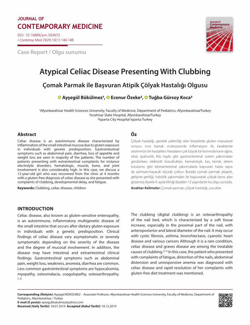
Corresponding (İletişim): Ayşegül BÜKÜLMEZ - Associate Professor, Afyonkarahisar Health Sciences University, Faculty of Medicine, Department of Pediatrics, Afyonkarahisar / TurkeyE-mail (E-posta): [email protected] (Geliş Tarihi): 18.07.2019 Accepted (Kabul Tarihi): 18.12.2019
DOI: 10.16899/jcm.593672J Contemp Med 2020;10(1):146-148
Case Report / Olgu sunumu
JOURNAL OF
CONTEMPORARY MEDICINEJournal ofContemporary MedicineYEAR: 2019 VOLUME: 9 ISSUE: 4
Atypical Celiac Disease Presenting With Clubbing
Çomak Parmak ile Basvuran Atipik Çölyak Hastalığı Olgusu
Celiac disease is an autoimmune disease characterized by inflammation of the small intestinal mucosa due to gluten exposure in individuals with genetic predisposition. Gastrointestinal symptoms such as abdominal pain, diarrhea, loss of appetite and weight loss are seen in majority of the patients. The number of patients presenting with extraintestinal complaints for instance electrolyte disorders, hematologic, muscle, bone, and joint involvement is also considerably high. In this case, we discuss a 12-year-old girl who was recovered from the clinic at 4 months with a gluten-free diagnosis of celiac disease as she presented with complaints of clubbing, developmental delay, and fatigue.
Keywords: Clubbing, celiac disease, children
ÖzAbstract
Ayşegül Bükülmez1, Ecenur Özeke2, Tuğba Gürsoy Koca3
Çölyak hastalığı, genetik yatkınlığı olan bireylerde gluten maruziyeti sonucu ince barsak mukozasında inflamasyon ile karakterize otoimmün bir hastalıktır. Hastaların çok büyük bir kısmında karın ağrısı, ishal, iştahsızlık, kilo kaybı gibi gastrointestinal sistem yakınmaları görülürken; elektrolit bozuklukları, hematolojik, kas, kemik, eklem tutulumu gibi ekstraintestinal yakınmalarla başvuran hasta sayısı da azımsanmayacak ölçüde çoktur. Burada çomak parmak şikayeti, gelişme geriliği, halsizlik yakınmaları ile başvurarak çölyak tanısı alan glutensiz diyetle 4. ayda kliniği düzelen 12 yaşında bir kız olgu sunuldu.
Anahtar Kelimeler: Çomak parmak, çölyak hastalığı, çocuklar
1Afyonkarahisar Health Sciences University, Faculty of Medicine, Department of Pediatrics, Afyonkarahisar/Turkey2Iscehisar State Hospital, Afyonkarahisar/Turkey
3Isparta City Hospital Isparta /Turkey
INTRODUCTIONCeliac disease, also known as gluten-sensitive enteropathy, is an autoimmune, inflammatory multigenetic disease of the small intestine that occurs after dietary gluten exposure in individuals with a genetic predisposition. Clinical findings of celiac disease vary asymptomatic or severely symptomatic depending on the severity of the disease and the degree of mucosal involvement. In addition, the disease may have intestinal and extraintestinal clinical findings. Gastrointestinal symptoms such as abdominal pain, weight loss, weakness, anorexia, diarrhea are common. Less common gastrointestinal symptoms are hypocalcemia, myopathy, osteomalacia, coagulopathy, osteoarthropathy.[1,2]
The clubbing (digital clubbing) is an osteoarthropathy of the nail bed, which is characterized by a soft tissue increase, especially in the proximal part of the nail, with anteroposterior and lateral diameter of the nail. It may occur with cystic fibrosis, asthma, bronchiectasis, cyanotic heart disease and various cancers Although it is a rare condition, celiac disease and graves disease are among the treatable causes of clubbing.[3-5] In this case, the patient who presented with complaints of fatigue, distortion of the nails, abdominal distension and unresponsive anemia was diagnosed with celiac disease and rapid resolution of her complaints with gluten-free diet treatment was mentioned.
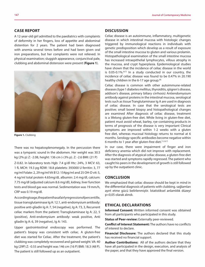
147 Journal of Contemporary Medicine
CASE REPORTA 12-year-old girl admitted to the paediatrics with complaints of deformity in her fingers, loss of appetite and abdominal distention for 2 years. The patient had been diagnosed with anemia several times before and had been given oral iron preparations, but her complaints were not relieved. In physical examination; sluggish appearance, conjunctival pale, clubbing and abdominal distension were present (Figure 1).
There was no hepatosplenomegaly. In the percussion there was a tympanic sound in the abdomen. Her weight was 30.5 kg (2% p) Z: -2.08, height: 136 cm (<3% p). Z: -2.6 BMI: (21 P)
Z-0.82. In laboratory tests Hgb: 7.4 g/dl Htc: 24%, 3 MCV: 63, 1 fL MCH: 19.3 pg RDW: 18.8 platelets: 393000 U ferritin: 3, 77 ng/ml Folate: 2, 29 ng/ml Vit B12: 154pg/ml and 25 OH-D vit: 5, 4 ng/ml total protein 4.65mg/dL albumin: 2.4 mg/dl, calcium: 7.75 mg/dl (adjusted calcium 8.6 mg/dl), kidney, liver function tests and blood gas was normal. Sedimentation was 19 mm/h, CRP was 0.19 mg/dl.
Accordingtoage,thepatienthasafairlyregressionofpercentille, tissue transglutaminase Ig A: 12.1, anti-endomysium antibody: positive anti-gliadin Ig A: 7, 54 (negative), Ig A: 73, 3, Recurrent celiac markers from the patient: Transglutaminase Ig A: 22, 5 (positive), Anti-endomysium antibody: weak positive, Anti gliadin Ig A: 6, 39 (negative), Ig A: 143.
Upper gastrointestinal endoscopy was performed. The patient's biopsy was consistent with celiac. A gluten-free diet was started for Celiac. After the treatment, the patient's clubbing was completely recovered and gained weight. VA: 40 kg (29P) Z: -0.55 and height was 146 cm (16 P) BMI: 18.3 (48 P). The patient is still followed up as an outpatient.
DISCUSSIONCeliac disease is an autoimmune, inflammatory, multigenetic disease in which intestinal mucosa with histologic changes triggered by immunological reactions in individuals with genetic predisposition which develop as a result of exposure of the small intestine mucosa to gluten and various prolamin. Histopathological examination of the small intestine mucosa has increased intraepithelial lymphocytes, villous atrophy in the mucosa, and crypt hyperplasia. Epidemiological studies have shown that the incidence of celiac disease in the world is 0.05-0.1%.[6,7] In a study conducted in our country, the incidence of celiac disease was found to be 0.47% in 20.190 healthy children in the 6-17 age group.[8]
Celiac disease is common with other autoimmune-related diseases (type 1 diabetes mellitus, thyroiditis, sjogren's disease, addison's disease, primary biliary cirrhosis) Antiendomysium antibody against proteins in the intestinal mucosa, serological tests such as tissue Tranglutaminase Ig A are used in diagnosis of celiac disease. In case that the serological tests are positive; small bowel biopsy and histopathological changes are examined After diagnosis of celiac disease, treatment is a lifelong gluten-free diet. While living in gluten-free diet, patient must avoid wheat, barley, rye-containing products in terms of prognosis of the disease is very important Clinical symptoms are improved within 1-2 weeks with a gluten free diet, whereas mucosal histology returns to normal at 6 months. Serology-specific antibodies become negative within 6 months to 1 year after gluten-free diet.[1,2,6,7]
In our case, there were impairment of finger and iron deficiency anemia which did not improve with replacement. After the diagnosis of atypical celiac disease, a gluten-free diet was started and symptoms rapidly regressed. The patient who caught his peers in the development of growth is still followed up by the outpatient clinic.
CONCLUSIONWe emphasized that celiac disease should be kept in mind in the differential diagnosis of patients with clubbing. sağlamları ayırt etme gücü belirlenmiştir. İstatistiksel anlamlılık düzeyi p<0,05 olarak alındı.
ETHICAL DECLARATIONSInformed Consent: Written informed consent was obtained from all participants who participated in this study. Status of Peer-review: Externally peer-reviewed. Conflict of Interest Statement: The authors have no conflicts of interest to declare. Financial Disclosure: The authors declared that this study has received no financial support. Author Contributions: All of the authors declare that they have all participated in the design, execution, and analysis of the paper, and that they have approved the final version.
Figure 1. Clubbing
148Ayşegül Bükülmez, Clubbing and Celiac Disease
REFERENCES1. Lebwohl B, Sanders,DS Green PHR. Coeliac disease Lancet 2018; 391:
70–81
2. Durham J Heide S. Temples HS.Celiac Disease in the Pediatric Population J Pediatr Health Care. 2018 ;32(6):62731.
3. Hanssen-flaschen J, Nordberg J. Clubbing and hypertrophic osteoarthropathy. Clin Chest Med 1987;8(2): 287–98.
4. Vandemergel X, Renneboog B. Prevalence, aetiologies and significance of clubbing in a department of general internal medicine. Eur J Intern Med. 2008;19(5):325–29.
5. Shah K, Rubian AI. Nail Disorders as Signs of Pediatric Systemic Disease Curr Probl Pediatr Adolesc Health Care 2012;42:204-11
6. Hill ID, Dirks MH, Liptak GS, Colletti RB, Fasano A, Guandalini
7. S. et al. Guideline for the diagnosis and treatment of celiac disease in children: Recommendations of the North American Society for Pediatric Gastroenterology, Hepatology and Nutrition. J Pediatr Gastroenterol Nutr. 2005;40(1):1–19.
8. Husby S, Koletzko S, Korponay-Szabó IR, Mearin ML, Phillips A, Shamir R, et al. European society for pediatric gastroenterology, hepatology, and nutrition guidelines for the diagnosis of coeliac disease. J Pediatr Gastroenterol Nutr. 2012;54(1) 136–60.
9. Dalgic B, Sari S, Basturk B, Ensari A, Egritas O, Bukulmez A, Baris Z.;Turkish Celiac Study Group Prevalence of celiac disease in healthy Turkish school children Am J Gastroenterol. 2011 Aug;106(8):1565 Note: Yasar, Aslan.[corrected to Dogan, Yasar]] Am J Gastroenterol. 2011;106(8):1512–17.
10. Moore JK, West SR, Robins G. Advances in celiac disease. Vol. 27, Curr Opin Gastroenterol. 2011; 27(2): 112–18.
11. Kaukinen K, Lindfors K, Collin P, Koskinen O, Maki M. Coeliac disease A diagnostic and therapeutic challenge. Clin Chem Lab Med. 2010;48(9):1205–16.

Corresponding (İletişim): Nuriye Tarakci Emiroğlu, Department of Neonatology, Department of Pediatrics, Meram School of Medicine Hospital, Necmettin Erbakan University, Konya, TurkeyE-mail (E-posta): [email protected] (Geliş Tarihi): 01.11.2018 Accepted (Kabul Tarihi): 12.02.2020
DOI: 10.16899/jcm.705187J Contemp Med 2020;10(1):149-150
Case Report / Olgu sunumu
JOURNAL OF
CONTEMPORARY MEDICINEJournal ofContemporary MedicineYEAR: 2019 VOLUME: 9 ISSUE: 4
Penile Agenesis
Penil Agenez
Penile agenesis is a rarely seen congenital anomaly with dramatic psychological outcomes. In more than half of the patients, other genitourinary anomalies can accompany penile agenesis. We herein present a newborn with rectovesical fistula associated with vesicoureteral fistula.
Keywords: Penile agenesis, rectovesical-fistula
ÖzAbstract
Nuriye Tarakcı Emiroğlu1, Murat Konak2, Fatma Hilal Yılmaz1, Nazlı Dilay Gültekin1, Hüseyin Altunhan1, Rahmi Örs3
Penil agenezi dramatik psikolojik sonuçları olan oldukça nadir konjenital bir anomalidir. Hastaların yarısından fazlasında diğer genitoüriner anomaliler eşlik edebilir. Biz burada veziko-üreteral reflünün eşlik ettiği ve rekto-vezikal fistülü olan bir yenidoğanı sunduk.
Anahtar Kelimeler: Penil agenezi, rekto-vezikal fistül
¹ Department of Neonatology, Department of Pediatrics, Meram School of Medicine Hospital, Necmettin Erbakan University, Konya, Turkey2 Department of Neonatology, Department of Pediatrics, Selcuk University, Faculty of Medicine, Konya, Turkey
3 Department of Neonatology, Medova Hospital, Konya, Turkey
INTRODUCTIONDevelopment of male genitourinary system is a complex phenomenon. Penile agenesis is a rarely seen congenital anomaly with dramatic psychological outcomes, which is encountered in 1 of 30 million births. This condition is believed to arise from absence or incomplete development of genital tubercle, and more than half of them can be associated with other genitourinary anomalies. This anomaly was firstly reported by Imminger in 1853.[1] To date, almost 100 patients have been reported in the World literature.[2] We herein report a neonatal case with rectovesical fistula associated with vesicoureteral fistula because of its rarity.
CASE REPORTA 7-hour-old newborn was referred to our clinic because of penile agenesis. Birth weight, head circumference, and height of the term baby who was born to a gravida 1,parity 1 40-yearold mother were 2750 gr, 33 cm, and 44 cm, respectively. On clinical examination, penis was not detected. Scrotum and both testicles were normal in appearance. Anus was at its normal anatomical location and urethral orifice was not detected on perineal region (Figure 1). Figure 1. Shows the absence of penis
150 Journal of Contemporary Medicine
Any evidence of dysmorphism, and clinical characteristics of any possible comorbidity were not found. Direct cystourethrogram revealed presence of a rectovesical fistula. On voiding cystourethrogram grade 3 vesicoureteral reflux was observed. Results of other imaging modalities (abdominal X-ray, echocardiography, cranial MR) were within normal limits. Baby had a 46 XY karyotype. Three days after the birth, the patient underwent cystostomy. Ovaries not visualized on pelvic ultrasound. ACTH: 27,4 pg/ml, cortisol: 9,97 µgr/ dl, FSH: 1,09 mIU/mL LH: 0,2 mIU/Ml, SHBG: 42,7 nmol/L, DHEAS: 133 µgr/dl, 17.aOHP: 2,66 ng/ml was normal. During his monitorization, symptoms of irritability, vomiting, and fever. Consequently, levels of acute phase reactants increased, growth of Escherichia coli was observed on urine culture media. Antibiotherapy was initiated. When the general health state of the patient improved, construction of colostomy from the proximal part of the rectovesical fistula was planned, and the patient was transferred to the department of pediatric surgery. For genital reconstruction the departments of pediatric urology, pediatric endocrinology, pediatric genetics, and pediatric psychiatry were consulted.
DISCUSSIONPenile agenesis is a rarely seen anomaly. At 4th week of gestation, it is a dysfunction of genital tubercle which results in incomplete separation of urogenital sinus from hindgut by urorectal septum.[2] This leads to total absence of all three components of penile shaft ie, both corpora cavernosa and spongiosum. Kessler and Mc Laughlin reported the incidence of genitourinary anomalies as 50 % which included cryptoorchidism, renal agenesis and dysplasia, prostate and bladder agenesis, as well as rectovesical and rectourethral fistulas. Besides non-genitourinary anomalies such as spina bifida, VATER–related anomalies, gastrointestinal anomalies, prune-belly syndrome, Potter syndrome, Treacher Collins syndrome, and mental retardation have been reported.[3] Diagnosis is made by meticulously clinical examination, karyotyping, and radiological examinations including a cystogram, and a magnetic resonance imaging.[1] Comorbidities of the patient were examined, and vesicoureteral reflux was observed.In the literature, penile agenesis has been classified in various ways. Evans et al.[4] classified them according to the presence or absence of major anomalies. Penile agenesis is classified in two major groups as isolated penile agenesis and its complex form associated with congenital anomalies, including genitourinary (54%) and gastrointestinal anomalies, as well as developmental defects of caudal axis.[2] It was included in the complex group as genitourinary abnormalities accompanied our case. Skoog and Bellman[5] divided these patients into 3 groups according to the relationship between anal sphincter and the ectopic urethral meatus; namely, postsphincteric form with anterior perianal urethra, presphincteric urethrorectal fistula, and vesicourethral fistula associated with urethral atresia. Urethral orifice can be localized on perineal region, skin tag resembling foreskin or anterior wall of rectum.[5] Position of the urethral orifice is related to the prognosis. Proximal urethral orifice and presence of other associated anomalies are associated with poor prognosis.[6]
Penile agenesis should be differentiated from disorders of sexual development, including hypospadiasis, severe forms of epispadiasis, intrauterine penile amputation, pseudohermaphroditism, rudimentary penis, concealed penis, and micropenis.[6]
Treatment of penile agenesis is debatable. Urethral transposition should be performed at an early stage to achieve excretion of urine and feces through separate routes. Due to fluctuations in serum testosterone levels between postnatal 10th and 60th days, early stage gonadectomy is recommended to prevent psychological stress of the child.[1] During long-term follow-up of these patients, a shift towards male type has been demonstrated in physiological and
psychological development of the child.[7] In recent years, hormonal production and raising of the patients according to their karyotypes have been advocated to prevent sexual dysphoria.[8,9] However, construction of a functional fallus is still the most problematic issue. Therefore, a viewpoint suggesting postponing sexual assignment till the child expresses his/her sexual identity has also been proposed.[8]
Our patient was the first child born to an advanced-aged mother who was living in a patriarchal community. The family was given information about this anomaly of their baby by a council composed of a neonatologist, a psychiatrist, an endocrinologist, and a pediatric urologist.The family declined sexual reconstruction at an early age, so construction of a colostomy was planned to separate excretion routes of urine and feces.
CONCLUSIONIn this rarely encountered case of penile agenesis, we wished to emphasize the process of management and multidisciplinary approach for these patients.
ETHICAL DECLARATIONSInformed Consent: Written informed consent was obtained from all participants who participated in this study. Status of Peer-review: Externally peer-reviewed. Conflict of Interest Statement: The authors have no conflicts of interest to declare. Financial Disclosure: The authors declared that this study has received no financial support. Author Contributions: All of the authors declare that they have all participated in the design, execution, and analysis of the paper, and that they have approved the final version.
REFERENCES1. Rattan KN, Kajal P, Pathak M, Kadian YS, Gupta R. Aphallia: experience with
3 cases. Pediatr Surg. 2010;45:13-6
2. Demirer Z, Aip BF, Uguz S, Guragac A, Irkilata HC. A rare case of penis agenesis (Aphallia) with associated multiple urogenital anomalies. Int J Surg Case Rep. 2015;15:10-2.
3. Kessler WO, McLaughlin III AP. Agenesis of penis. Embryology and management. Urology 1973;1:226-9.
4. Evans JA, Erdile LB, Greenberg CR, et al. Agenesis of the penis: patterns of associated malformations. Am J Med Genet 1999;84:47-55.
5. S.J. Skoog, A.B. Belman, Aphallia its classification and management, J. Urol. 1989;141:589–2.
6. Shamsa A, Kajbafzadeh AM, Javad Parizadeh SM, Zare MA, Abolbashari M. Aphallia associated with urethro-rectal fistula and stones in the bladder and urethra. Saudi J Kidney Dis Transpl. 2008;19: 435-8.
7. W.G. Reiner, B.P. Kropp, A 7-year experience of genetic males with severephallic inadequacy assigned female. J Urol. 2004;172:2395-8
8. Reiner WG. Gender identity and sex-of-rearing in children with disorders of sexual differentiation. J Pediatr Endocrinol Metab 2005;18:549-53.
9. De Castro R, Merlini E, Rigamonti W, Macedo A Jr.Phalloplasty and urethroplasty in children with penile agenesis: preliminary report. J Urol 2007;177:1112-6.
Corresponding (İletişim): Melike Emiroğlu, Selçuk Üniversitesi Tıp Fakültesi Çocuk Sağlığı ve Hastalıkları Anabilim Dalı, Çocuk Enfeksiyon Hastalıkları Bilim Dalı, Konya, TürkiyeE-mail (E-posta): [email protected] (Geliş Tarihi): 26.02.2018 Accepted (Kabul Tarihi): 10.02.2020
DOI: 10.16899/jcm.705165J Contemp Med 2020;10(1):140-141
Letter to the editor/ Editöre Mektup
JOURNAL OF
CONTEMPORARY MEDICINEJournal ofContemporary MedicineYEAR: 2019 VOLUME: 9 ISSUE: 4
Hepatit A Enfeksiyonu Olan Bir Çocuk Hastada Delta-Bilirubinemi ve İnatçı Kasıntı
Delta-Bilirubinemia and Persistent Itching in a Child Patient with Hepatitis A Infection
Sayın Editor,
Melike Emiroğlu1, Halil Haldun Emiroğlu21 Selçuk Üniversitesi Tıp Fakültesi Çocuk Sağlığı ve Hastalıkları Anabilim Dalı, Çocuk Enfeksiyon Hastalıkları Bilim Dalı, Konya, Türkiye
2 Selçuk Üniversitesi Tıp Fakültesi Çocuk Sağlığı ve Hastalıkları Anabilim Dalı, Çocuk Gastroenterolojisi Bilim Dalı, Konya, Türkiye
Hepatit A kronikleşmemekle birlikte ozellikle yaş büyüdükçe ağır bir seyir gosterebilmektedir.[1] Delta-bilirubin konjuge bilirubinin albümine kovalent bağ ile irreversibl olarak bağlanması sonucu ortaya çıkan, glomerüllerden süzülemediği için idrar yoluyla atılamayan ve toksik olmayan bir bilirubin türüdür. Delta-bilirubin, biyokimyasal incelemede direkt bilirubinin içinde yer alır. Kovalent bağ ile irreversibl bağlı bulunduğu albüminin yarı omrünün uzunluğuna bağlı olarak, delta-bilirubinemide kanda uzun süre direkt bilirubin düzeyi yüksek olçülürken idrarla atılmaması sonucu idrar incelemesinde bilirubinin negatif saptanır.[2] Kaşıntı kolestatik karaciğer hastalıklarının ortak bir semptomudur. Kolestazda meydana gelen kaşıntının nedeni olarak onceleri artmış safra asitleri sorumlu tutulurken son yıllarda ozellikle ototaksin enziminin rolü üzerinde durulmaktadır.[3] Ototaksin kolestatik kaşıntılarda rolü olduğu son yıllarda anlaşılan bir lizofosfolipazdır.[4] Hepatit A enfeksiyonu olan 13 yaşındaki erkek çocuk hasta, direkt hiperbilirubinemi nedeni olarak delta-bilirubineminin ve kolestaza bağlı inatçı kaşıntı tedavisinde rifampisinin hatırlatılması amacı ile sunulmuştur. Gozler ve tüm vücut cilt renginde sarılık, halsizlik ve oral alım azlığı ile başvuran, oyküsünde oz ve soy geçmişine ait herhangi bir karaciğer hastalığı bildirilmeyen hastanın tam kan sayımı normal iken tam idrar incelemesinde
bilirubin pozitif bulundu. Kan biyokimyasında hafif düzeyde hipertransaminazemi (AST: 82 U/L, normal referans aralığı: 0-50; ALT: 103 U/L, normal referans aralığı: 0-55), direkt hiperbilirubinemiye (total bilirubin: 11.65 mg/dL, normal referans aralığı: 0.22-1.3; direkt bilirubin: 6.95 mg/dL, normal referans aralığı: 0-0.2) karşın kolestatik enzim düzeyleri (GGT: 23 U/L, normal referans aralığı: 11-57; ALP: 366 U/L, normal referans aralığı:<750) yaşına gore normal sınırlarda saptandı. Karaciğerin sentez fonksiyonları (albümin, PT, INR, aPTT, amonyak) normal olarak değerlendirildi. Etiyolojik incelemede anti-HAV IgM antikoru pozitif bulunan hasta akut viral hepatit A’ya bağlı oral alım azlığı ve belirgin düzeydeki halsizlik yakınmaları nedeni ile yatırıldı. Hastaneye yatışının dordüncü günü inatçı kaşıntı yakınması ortaya çıkan hastanın kolestatik enzimleri normal, transaminaz düzeyleri (AST: 65 U/L, ALT: 83 U/L) azalmakta olarak değerlendirildi. Serum bilirubin düzeylerindeki yüksekliğin değişmemesine (total bilirubin: 11.82 mg/dL, direkt bilirubin: 6.64 mg/dL) karşın 2 kez bakılan kontrol tam idrar incelemesinde bilirubin negatif bulunan hastada klinik tablonun iyileşmekte ve mevcut direkt hiperbilirubinemi nedeninin ise delta-bilirubinemi olduğu kanısına varıldı. İnatçı kaşıntı yakınmasına yonelik olarak serum safra asit düzeyi gonderilen hastaya ursodeoksikolik asit tedavisi başlandı. Tedavide 1 hafta geçmesine karşın kaşıntı yakınması gerilemeyen hastaya açlık serum total

152 Journal of Contemporary Medicine
safra asit düzeyinin (68.9 µmol/L, normal referans aralığı: 0-10) yüksek bulunmasının ardından kolestiramin (4 g/gün) tedavisi eklendi. İkili tedaviye 1 hafta devam edilmesine rağmen kaşıntısının sürmesi üzerine ototaksin düzeyini azalttığı bilinen rifampisin (10 mg/kg/gün) tedaviye eklendi.[3] Üçlü tedavinin üçüncü günü hastanın kaşıntısı belirgin düzeyde geriledi. Genel durumu ve oral alımı iyi olan hasta, kontrol kan biyokimyasında serum transaminaz ve kolestatik enzim düzeyleri normal, delta-bilirubinemi nedeni ile daha once yüksek bulunan bilirubin düzeyleri ise azalmış bulunarak (total bilirubin: 4.46 mg/dL, direkt bilirubin:2.05 mg/dL) ayaktan kontrole gelmek üzere taburcu edildi. Sonuç olarak karaciğer enzimleri normale donmesine karşın direkt hiperbilirubinemisi devam eden hastalarda basit bir inceleme olan idrar bilirubininin negatif olduğunun gorülerek delta-bilirubinemi tanısının konulması gereksiz ve pahalı ileri tetkiklerin onlenmesi açısından onemlidir. Ursodeoksikolikasit ve kolestiramin tedavisine rağmen düzelmeyen kolestaza bağlı inatçı kaşıntısı olan hastaların rifampisinden yarar gorebileceği hatırlanmalıdır. Ancak kolestazın, hepatit A gibi karaciğer hasarına eşlik ettiği olgularda kaşıntıya yonelik kullanılacak ilaçların (kolestiramin, rifampisin) yol açabileceği hepatotoksisite yonünden dikkatli olunmalıdır.
Anahtar Kelimeler: Hepatit A, delta-bilirubin, kaşıntı
Kaynaklar1. Fawaz R, Jonas MM. Acute and Chronic Hepatitis. In: Wyllie R, Hyams
JS, Kay M, eds. Pediatrik Gastrointestinal and Liver Disease. 5th ed. Philadelphia: Elsevier; 2016. p.906-27.
2. Kozaki N, Shimizu S, Higashijima H, et al. Significance of serum delta-bilirubin in patients with obstructive jaundice. J Surg Res 1998; 79 (1): 61-5.
3. Kremer A. E, van Dijk R, Leckie P, et al. Serum autotaxin is increased in pruritus of cholestasis, but not of other origin, and responds to therapeutic interventions. Hepatology. 2012;56 (4):1391–1400.
4. Kremer AE, Gonzales E, Schaap FG, Oude Elferink RP, Jacquemin E, Beuers U. Serum autotaxin activity correlates with pruritus in pediatric cholestatic disorders. J Pediatr Gastroenterol Nutr. 2016;62(4):530-5.
Related Documents