JOURNAL OF APPLIED PHYSIOLOGY Vol. 41, No. 6, December 1976. Printed in U.S.A. Computer simulation of brainstem respiratory activity STUART GEMAN AND MICHAEL MILLER Department of Physiology, Dartmouth Medical School, Hanover, New Hampshire 03755 GEMAN, STUART, AND MICHAEL MILLER. Computer simula- tion of brainstem respiratory activity. J. Appl. Physiol. 41(6): 931438. 1976. -A mathematical model of the medullary res- piratory oscillator, composed of two mutually inhibiting popu- lations (inspiratory and expiratory) of computer-simulated neurons, is presented. Each population consists of randomly interconnected subpopulations of excitatory and inhibitory neurons. Neuronal coupling is such that either the inspiratory or expiratory population alone is capable of cyclic activity. Weak inhibitory connections between inspiratory and expira- tory populations provide satisfactory reciprocating activity in- dependent of the natural frequency of either population alone. Initiation and persistence of rhythmic activity is dependent on a diffuse noncyclic excitatory input. Vagal discharge, simu- lated by phasic inhibition of inspiratory neurons, results in increased respiratory frequency with decreased inspiratory activity. In the absence of simulated vagal discharge, uniform facilitation of synaptic connections increases averaged activi- ties of inspiratory and expiratory populations, with minor effect on frequency. In the presence of simulated vagal dis- charge, facilitation of synaptic connections increases both fre- quency and amplitude. The simulated effects of synaptic facili- tation, with and without vagal discharge, mimic the physio- logical response to CO, in the intact and vagotimized animal. respiratory centers; mathematical model; control of breathing SINCE THE CLASSICAL EXPERIMENTS OfGalenithasbeen known that respiratory movements are dependent on the passage of activity from the brain. Controversy exists still, however, as to the nature and, indeed, the anatomical location of the mechanism in the brainstem responsible for respiratory periodicity. With regard to the location of the respiratory oscillator, two basic theo- ries have been supported: a) a pontine-medullary sys- tem of respiratory neurons, and b) a neuronal organiza- tion confined to the medulla. The fundamental difYer- ence between the two proposals has been that in the medullary system, respiratory periodicity arises from medullary inspiratory (I) and expiratory (E) neurons exclusively (5, 17, 30, 39), while in the pontine-medul- lary system, reciprocating activity of medullary I and E neurons is dependent on the discharge of pontine phase- spanning neurons, the medulla having no intrinsic os- cillator properties (9, 13, 29). The purpose of this paper is to support the hypothesis that the basic respiratory rhythm, as well as its qualitative response to several physiological influences, arises from a system of medul- lary I and E neurons, rather than from a more complex pontine-medullary system. While an exhaustive review of the literature pertinent to each of these proposals is beyond the scope of this paper, arguments forming the basis for our stance will be briefly outlined. Support for a pontine-medullary oscillator mecha- nism has evolved, essentially, from four important ob- servations 1) Upon transection of the brainstem at various pon- tine levels, a change from eupneic ventilation to gasp- ing and/or apnea has often been observed (20, 22, 37). 2) The apneustic and pneumotaxic “centers” have been shown by ablation experiments to reside within the pons (20, 22, 25). 3) Stimulation of pontine structures leads to stimula- tion or inhibition of ventilation (2, 19, 25). 4) Neurons with phasic, respiratory-related activity have been demonstrated in pontine structures (3, 10, 20) None of these observations, however, offers compel- ling evidence in favor of a pontine-medullary system. Breckenridge and Hoff (4, 17), Hukuhara (18), Lumsden (22), Salmoiraghi and Burns (31), and Wang et al. (37), for example, have shown that rhythmic respiratory ac- tivity can continue for hours after brainstem transec- tion at the pontine-medullary junction, or under condi- tions in which pontine respiratory activity is absent. Also, the fact that the classical apneustic and pneumo- taxic centers are part of the pons and that stimulation of certain pontine structures affects respiration does not argue conclusively for a pontine role in the generation of respiratory periodicity. Rather, these findings may indi- cate that pontine centers exert powerful regulatory in- fluences on a more basic oscillator mechanism. Finally, although large numbers of pontine neurons with respi- ratory-related activity have been reported in the cat (3, 10,20), no such neurons have been found during system- atic exploration of the pons in the dog (6), monkey (6), or rabbit (38). In summary, there seems little justification for con- sidering the pons as essential for respiratory rhythmic- ity, or for incorporating complex pontine neural ele- ments into models of the basic respiratory oscillator mechanism. This study, therefore, models a basic med- ullary oscillator mechanism utilizing only I and E neu- rons. Our intent is twofold: first, to develop a mathematical model of respiratory periodicity based on available neu- rophysiological data; and second, to examine the dy- 931

Welcome message from author
This document is posted to help you gain knowledge. Please leave a comment to let me know what you think about it! Share it to your friends and learn new things together.
Transcript
JOURNAL OF APPLIED PHYSIOLOGY
Vol. 41, No. 6, December 1976. Printed in U.S.A.
Computer simulation of brainstem
respiratory activity
STUART GEMAN AND MICHAEL MILLER Department of Physiology, Dartmouth Medical School, Hanover, New Hampshire 03755
GEMAN, STUART, AND MICHAEL MILLER. Computer simula- tion of brainstem respiratory activity. J. Appl. Physiol. 41(6): 931438. 1976. -A mathematical model of the medullary res- piratory oscillator, composed of two mutually inhibiting popu- lations (inspiratory and expiratory) of computer-simulated neurons, is presented. Each population consists of randomly interconnected subpopulations of excitatory and inhibitory neurons. Neuronal coupling is such that either the inspiratory or expiratory population alone is capable of cyclic activity. Weak inhibitory connections between inspiratory and expira- tory populations provide satisfactory reciprocating activity in- dependent of the natural frequency of either population alone. Initiation and persistence of rhythmic activity is dependent on a diffuse noncyclic excitatory input. Vagal discharge, simu- lated by phasic inhibition of inspiratory neurons, results in increased respiratory frequency with decreased inspiratory activity. In the absence of simulated vagal discharge, uniform facilitation of synaptic connections increases averaged activi- ties of inspiratory and expiratory populations, with minor effect on frequency. In the presence of simulated vagal dis- charge, facilitation of synaptic connections increases both fre- quency and amplitude. The simulated effects of synaptic facili- tation, with and without vagal discharge, mimic the physio- logical response to CO, in the intact and vagotimized animal.
respiratory centers; mathematical model; control of breathing
SINCE THE CLASSICAL EXPERIMENTS OfGalenithasbeen known that respiratory movements are dependent on the passage of activity from the brain. Controversy exists still, however, as to the nature and, indeed, the anatomical location of the mechanism in the brainstem responsible for respiratory periodicity. With regard to the location of the respiratory oscillator, two basic theo- ries have been supported: a) a pontine-medullary sys- tem of respiratory neurons, and b) a neuronal organiza- tion confined to the medulla. The fundamental difYer- ence between the two proposals has been that in the medullary system, respiratory periodicity arises from medullary inspiratory (I) and expiratory (E) neurons exclusively (5, 17, 30, 39), while in the pontine-medul- lary system, reciprocating activity of medullary I and E neurons is dependent on the discharge of pontine phase- spanning neurons, the medulla having no intrinsic os- cillator properties (9, 13, 29). The purpose of this paper is to support the hypothesis that the basic respiratory rhythm, as well as its qualitative response to several physiological influences, arises from a system of medul- lary I and E neurons, rather than from a more complex
pontine-medullary system. While an exhaustive review of the literature pertinent to each of these proposals is beyond the scope of this paper, arguments forming the basis for our stance will be briefly outlined.
Support for a pontine-medullary oscillator mecha- nism has evolved, essentially, from four important ob- servations
1) Upon transection of the brainstem at various pon- tine levels, a change from eupneic ventilation to gasp- ing and/or apnea has often been observed (20, 22, 37).
2) The apneustic and pneumotaxic “centers” have been shown by ablation experiments to reside within the pons (20, 22, 25).
3) Stimulation of pontine structures leads to stimula- tion or inhibition of ventilation (2, 19, 25).
4) Neurons with phasic, respiratory-related activity have been demonstrated in pontine structures (3, 10, 20)
None of these observations, however, offers compel- ling evidence in favor of a pontine-medullary system. Breckenridge and Hoff (4, 17), Hukuhara (18), Lumsden (22), Salmoiraghi and Burns (31), and Wang et al. (37), for example, have shown that rhythmic respiratory ac- tivity can continue for hours after brainstem transec- tion at the pontine-medullary junction, or under condi- tions in which pontine respiratory activity is absent. Also, the fact that the classical apneustic and pneumo- taxic centers are part of the pons and that stimulation of certain pontine structures affects respiration does not argue conclusively for a pontine role in the generation of respiratory periodicity. Rather, these findings may indi- cate that pontine centers exert powerful regulatory in- fluences on a more basic oscillator mechanism. Finally, although large numbers of pontine neurons with respi- ratory-related activity have been reported in the cat (3, 10,20), no such neurons have been found during system- atic exploration of the pons in the dog (6), monkey (6), or rabbit (38).
In summary, there seems little justification for con- sidering the pons as essential for respiratory rhythmic- ity, or for incorporating complex pontine neural ele- ments into models of the basic respiratory oscillator mechanism. This study, therefore, models a basic med- ullary oscillator mechanism utilizing only I and E neu- rons.
Our intent is twofold: first, to develop a mathematical model of respiratory periodicity based on available neu- rophysiological data; and second, to examine the dy-
931

932 S. GEMAN AND M. MILLER
namic characteristics and implications of such a model by computer simulation. We feel that the ability of the model to mimic certain aspects of physiological experi- ment supports the notion of a basic medullary respira- tory oscillator.
For clarity, the formal mathematical development of the model, corresponding to a heuristic development in the text, is presented separately in the appendixes.
THEORY
EZements. The model consists of a large collection of randomly interconnected neuronlike elements. For each element, the rate of change of membrane potential (P) is a linear function of input. With the cessation of input, P decays exponentially to resting potential. Physiological evidence supports the use of these characteristics in approximating neuron behavior (12, 16, 28), and ele- ments of this type have formed the basis for numerous previous models of neuronal activity (1, 13, 14,23,29).
The output of an element is derived from its potential. No output is generated when potential is below thresh- old value; if potential exceeds threshold the signal transmitted to other elements is an increasing function of the difference between potential and threshold. Transmission between elements is determined by a “synaptic weight” (W) of positive, negative, or zero value. The presynaptic signal is multiplied by W and the result is added to the input of the postsynaptic element. Positive and negative values of W model the effects of excitatory (+) and inhibitory (-) presynaptic neurons, respectively.
Characteristics of randomly arranged neural net- works. This study utilizes randomly constructed popula- tions of neural elements. Random networks have sev- eral properties which support their use in modelling a basic respiratory oscillator (see APPENDIXES for details).
1) In a large and randomly arranged collection of elements, the values of a small number of appropriate parameters reliably determines the mean activity of the entire network. In a neural network, these parameters may represent the tendencies of certain classes of neu- rons to contact other classes of neurons, the average size of a synaptic contact between two particular types of neurons, etc. It should be emphasized that the gross (average) activity of the network does not depend on the specific distributions, only their means are relevant. Therefore, if in a large collection of neurons the func- tional output is derived from an average of individual element activities, the amount of genetic information required is enormously reduced by allowing a random generation of the specific interconnections.
2) In a random system the accuracy with which the relevant parameters determine the gross activity in- creases with the number of elements. Thus the network is tuned to an arbitrary precision by sufficiently increas- ing the number of its components.
3) A large network of this type functions reliably even if a significant fraction of its components are lost.
These properties, combined with the following ana- tomic and physiological observations, motivated our hy- pothesizing a random component in the arrangment of
medullary respiratory neurons. Histologically, the med- ullary reticular formation is characterized by vast num- bers of nerve cell bodies showing little cytoarchitectural organization (33). This disorganized appearance con- trasts sharply with the meticulous structure which characterizes other locations in the CNS. In addition, ablation and lesion experiments (27) suggest that respi- ratory motor output s diffusely derived from I and E neuron .a1 populations It is perha .ps true, then, that the gross or averaged activities of the inspiratory and expi- ratory populations of neurons, rather than a small col- lection of individual elements, governs the motor pat- terns subserving respiration. If so, then a random gen- eration of the specific structure would allow for an effi- cient genetic coding while the large numbers of respira- tory neurons would insure accuracy and reliability of the motor output.
Fundamental oscillator. The potential for stable os- cillation in randomly constructed networks of excitatory (+> and inhibitory (--> neural-like elements has been demonstrated by Amari (1). Details of the construction of such an oscillator are developed in APPENDIX A and APPENDIX B (section on Conditions for stable oscilla- tions). A computer-generated numerical solution of the equations derived in the appendixes is shown in (parameters used in each of the simulations are gi
Fig. 1 ven in
Table 1). Note that the phase difference between + and - activities is small in comparison to the period of oscillation. This condition arises from the exceedingly short synaptic delay and conduction times characteristic of neural structures: thus the activity of the inhibitory subpopulation must closely follow the activity of the excitatory elements.
Inspiratory and expiratory oscillators. The model pre- sented in this study utilizes two populations of neurons, governing inspiratory (I) and expiratory (E) output functions. Neurophysiological evidence strongly sug- gests mutually inhibiting and functionally distinct in- spiratory and expiratory neuronal networks (25, 27, 30, 31). In addition, the following observations argue for the capability of independent oscillation within both I and E populations.
1) The decline of inspiratory activity following peak I neuron discharge is unlikely to be due to inhibition from expiratory neurons, since expiratory activity during this period is minimal (3, 31). Thus I activity appears self-limiting. Expiratory neurons behave similarly.
2) I activity is initiated in the presence of E activity, a presumably inhibitory influence (3, 31, 32). This con-
FIG. 1. Fundamental oscill ator. Phase relation between excit- atory and inhibitory elements. Abscissa represents an arbitrary time axis, while ordinate represents averaged membrane potential. Zero line represents resting potential; +, excitatory neural elements; -, inhibitory elements. kbove conventions apply to all figures. Note that phase difference between + and - components is small in comparison with cycle time of population as a whole. Tab1 tains parameters used in th is and remaining si .mulations.
.e 1 con-

COMPUTER SIMULATION OF BRAINSTEM RESPIRATORY ACTIVITY 933
TABLE I. Parameters used in generating figures -L
Pa- rame- ters
-~
l/a,’ l/a,
l/a, +
l/al.
WI ’
w,, +
w:,+ w,:
WI, W,,
Wik'
WI.;
Wk.'
W,t;
WEIC WC
T’
T,- TE’
T . C’
c
K, K,
- I Figure
~-___- 1
0.5 0.5
0.25 0.25
0 0.266
0 0.133
2 2
2 2
0 0
1 1
1 1 0 0
0 1.065 0 1.065 0 0
0 0.533
0 0.533
0 0
0.2 0.2
0.1 0.1 0 0.107
0 0.053
0 0
0 0
0 0
0 0
3 4
--
0.5
0.25
0.266
0.133
2
2
0.2
1
1 0.1
1.065 1.065 0.107
0.533
0.533
0.053
0.2
0.1 0.107
0.053
0
0
0
0
5A 5B 5C 5D 5F 6B
3.5
0.25
0.266
0.133
2
2
0.2
1
1 0.1
1.065 1.065 0.107
0.533
0.533 0.053
0
0 0
0 0
0
0 0
3.5
0.25
0.266
0.133
2
2
0.2
1
1 0.1
1.065 1.065 0.107
0.533
0.533
0.053
0.025
0.013
0.013
0.007
0
0
0
0
I.5
3.25
3.266
3.133
2
2
0.2
1
1 0.1
1.065 1.065 0.107
0.533
0.533
0.053
0.05
0.025
0.027
0.013
0
0
0
0
0.5
0.25 0.266
0.133
2
2
0.2
1 1
0.1
1.065 1.065 0.107
0.533
0.533 0.053
0.1 0.05
0.053
0.025
0
0
0
0 ---
0.5 0.5
0.25 0.25
0.266 0.266
0.133 0.133
2 3
2 3
0.2 0.3
1 1.5 1 1.5
0.1 0.15
1.065 1.598 1.065 1.598 0.107 0.160
0.533 0.799
0.533 0.799
0.053 0.080
0.6 0.2
0.3 0.1
0.32 0.107
7A -- 0.5
0.25 0.266
0.133
2
2
0.2
1 1
0.1 1.065
1.065 0.107
0.533
0.533
0.053
0.2
0.1
1.107 0.553
0
0
0
0
7B 8B
0.5 0.5
0.25 0.25 0.266 0.266
0.133 0.133
2 2 2 2
0.2 0.2
1 1 1 1
0.1 0.1 1.065 1.065
1.065 1.065 0.107 0.107
0.533 0.533
0.533 0.533
0.053 0.053
-1.8 0.2
-0.9 0.1
0.107 0.107
0.053 0.053
0 1
0 0.5
0 0.1 0 0.1
9B -- 0.5
0.25
0.266
0.133
3
3
0.3
1.5
1.5
0.15
1.598
1.598 0.160
0.799
0.799
0.080
0.2
0.1
0.107
0.053
1
0.5
0.1 0.1
Simulated respiratory output was obtained by numerical solution ofEg. Z-5, APPENDIX
c. For each figure, the parameters used in the simulation are indicated. In each case S(X)
was defined as 0 for X 5 0, X for 0 < X -C 4, and 4 f’or X 2 4. The parameters used to generate the second half of Fig. 3, the first halves of Fig. 7A and B, as well as Figs. 5E, 6A, and 8A,
are identical to those used in Fig. 4. Also, Fig. 9A is identical to Fig. 6Z3.
sideration applies to E discharge as well. 3) As noted previously, + and - components of a
single population could not be used to generate I and E output since they oscillate nearly in phase (Fig. 1).
Thus, the model presented in this study utilizes a system of + and - neurons within both I and E popula- tions. Either population alone is capable of independent oscillation. Microelectrode recordings of discharge dura- tion from inspiratory and expiratory neurons (3, 9, 31) demonstrate a longer period of expiratory activity. In the present model, therefore, the frequency of oscilla- tion of the E population was chosen to be lower than that of the I population. Qualitatively, the results de- scribed below are independent of the precise ratio of these frequencies.
Coupled inspiratory and expiratory oscillators. As noted above inspiratory and expiratory populations are related in a mutually inhibiting manner. This hypothe- sis has been utilized previously in models proposed by Burns (5), Cohen (9), Feldman and Cowan (13), Rubio (29), Salmoiraghi and Baumgarten (30), and other au- thors. In the present model, a random sampling of expi- ratory inhibitory neurons synapse with a random collec- tion of both + and - inspiratory neurons. Similarly, both + and - expiratory neurons receive samplings from inspiratory inhibitory neurons (Fig. 2). With this type of coupling, reciprocating inspiratory and expira- tory oscillation is obtained (Fig. 3). Only a minor per- centage of total inhibitory output of each population is required to convert unrelated I and E discharge into reciprocating activity.
It should be noted that similar mechanisms have been previously suggested (5, 30, 36). For example, Salmoir- aghi and Baumgarten (30) in summarizing an intracel- lular study of respiratory neuron potentials state: “It
/ I
I I
I I
I I
FIG. 2. Arrangement of neural elements. Solid lines indicate dis- tribution of excitatory influence between subpopulations; broken lines indicate distribution of inhibitory influence between subpopu- lations. I and E refer to inspiratory and expiratory populations, respectively; + and - refer to excitatory and inhibitory subpopula- tions, respectively.
FIG. 3. Coupling of independently cycling inspiratory and expira-
tory populations. I and E refer to averaged activities of excitatory subpopulations of inspiratory and expiratory neurons. As shown in Fig. 1, + and - subpopulations within either oscillator are nearly in phase. Therefore, for clarity, I and E inhibitory activities are omit- ted from this and remaining figures. Arrow indicates incorporation of mutually inhibitory coupling between I and E populations. It may be seen that phasically unrelated I and E activities become recipro- cating upon addition of weak negative coupling. Details of coupling procedure are provided in APPENDIX B.
would appear that three factors contribute to produce rhythmicity of respiratory neurons - namely, a self- reexciting mechanism within the inspiratory and the expiratory network of neurons tending to maintain ac- tivity within the networks; self-limiting systems tend- ing to limit the frequency and the duration of the dis- charge; and reciprocal innervation which provides inhi- bition of one network when the other is active.”
Factors influencing respiratory rhythm. The present model was tested for its ability to alter frequency and amplitude following various manipulations (see APPEN-
DIX c for mathematical details). 1) The importance of diffuse tonic excitatory influ-
ences on the activity of the respiratory center has been demonstrated by Salmoiraghi and Burns (32). There- fore, the effect of a constant excitatory input to this model has been examined.
2) Short-term changes in synaptic function have been

934
demonstrated in an enormous variety of physiological experiments. In light of this plasticity, the effect of uniformly altering all synaptic weights within the en- tire system (including the weak, mutually inhibiting coupling between I and E populations) was tested.
3) It is well known that vagal afferent discharge, arising from pulmonary mechanoreceptors, influences the frequency and volume of respiration. The effects of a simulated vagal discharge on I and E outputs were examined. Simulated vagal activity was derived from integrated inspiratory output and would presumably be proportional to lung volume (21). The nature of interac- tion between vagal discharge and respiratory neurons is not known; in this study two possible mechanisms were examined.
RESULTS
Simulated respiratory oscillator. The reciprocating character of I and E activities generated by this model is illustrated in Figure 4. The tracing represents averaged activities of excitatory subpopulations within I and E oscillators, a convention used for all curves generated for this paper. This activity is supported by a moderate level of diffuse excitatory, noncyclic, tonic input. This particular level of tonic input is present in all curves generated unless otherwise specified. It may be noted (Fig. 4) that expiratory activity occupies the greater portion of a complete simulated respiratory cycle, due to the assignment of a lower natural frequency to the E population, as described in the Theory Section.
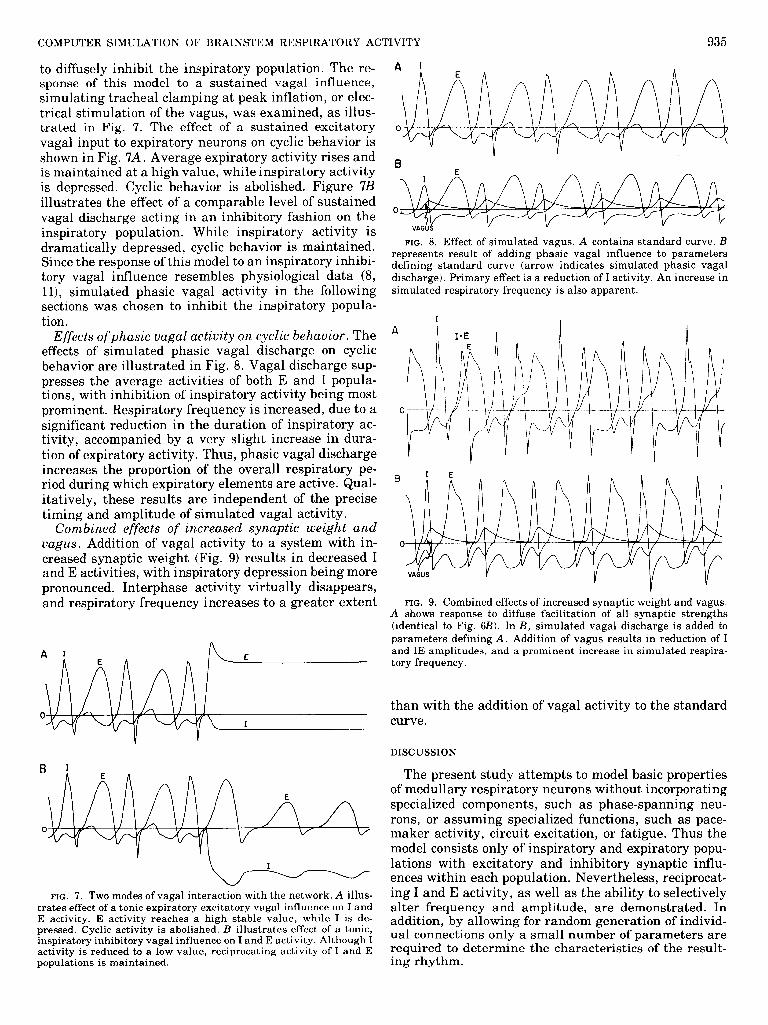
Effect of alterations in tonic input. Figure 5 illus- trates the effect of increasing tonic excitatory input. In the absence of this influence, cyclic activity does not occur. As may be seen in Fig. 5, B, C, and D, moderate elevations of tonic influence increase the amplitude of both I and E activities, with only a slight increase in frequency. With further increments (Fig. 5, E and F), amplitudes of both I and E activities continue to in- crease, accompanied by a more pronounced increase in frequency.
Effect of increased synaptic weight. Figure 6 illus- trates the result of a diffuse increase in synaptic weight. The amplitudes of both I and E activities increase, with virtually no effect on frequency. It may also be noted that increased synaptic weight induces significant I ac- tivity during the I-E interphase. Possible implications of these observations are discussed later.
Possible mechanisms of vagal interaction with inspi- ratory and expiratory neurons. As mentioned previ- ously, simulated vagal afferent discharge is generated
S. GEMAN AND M. MILLER
A.
0
FIG. 5. Effect of alterations in tonic activity. A illustrates the lack of cyclic activity in the absence of tonic input. Tonic excitatory input is progressively increased in B, C, and D, resulting in in-
creased averaged activities of both I and E populations. E is identical to standard curve (Fig. 4). Further increments (E and F) result in increased I and E activities, as well as increased simulated respira-
tory frequency (shortened I-I interval).
A 1
FIG. 6. Effect of increased synaptic weight. A contains standard curve. B shows response of model to a diffuse facilitation of all synaptic inputs. Result is a prominent increase in I activity, with a somewhat smaller increase in E activity. In addition, a second peak (IE) of I activity in each cycle is apparent, occurring between the I and E phases.
FIG. 4. Standard curve. Generation of this curve involves addi- tion of a specific, moderate amount of tonic input to the network. This specific value of tonic input is maintained in all other records in this study, except in Fig. 5.
as a function of average inspiratory activity. We have examined two possible mechanisms by which vagal acti- vation may interact with inspiratory and expiratory populations: a) vagal discharge acting to diffusely excite the expiratorv ponulation. or b) vagal discharge acting
COMPUTER SIMULATION OF BRAINSTEM RESPIRATORY ACTIVITY 935
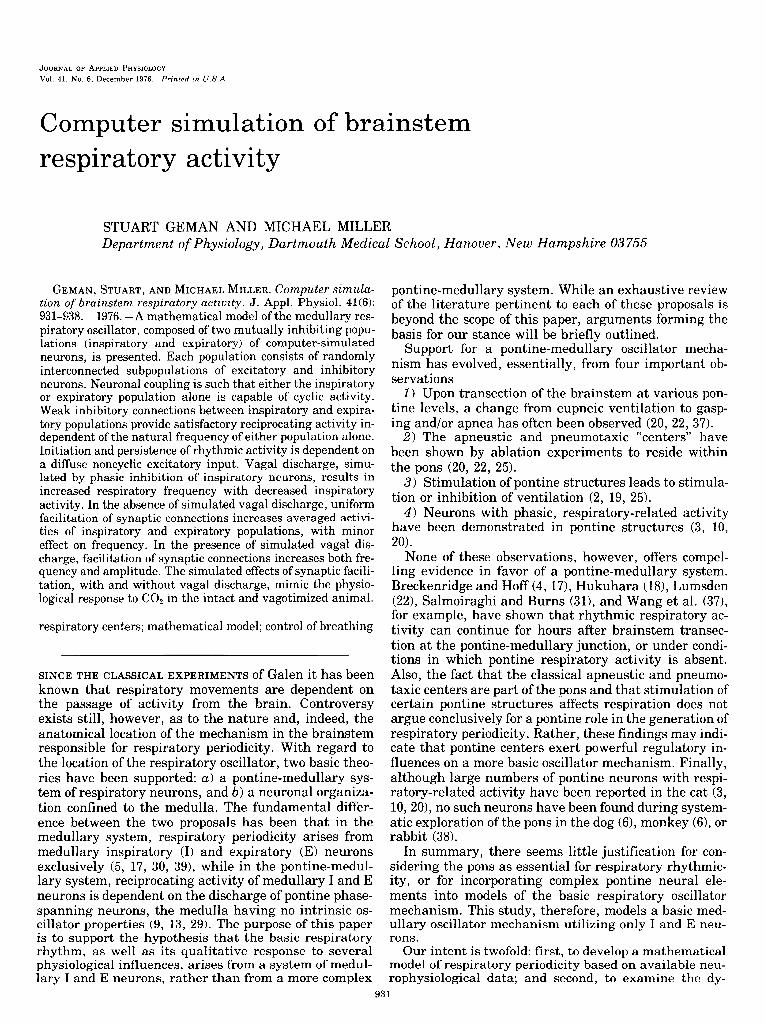
to diffusely inhibit the inspiratory population. The re- sponse of this model to a sustained vagal influence, simulating tracheal clamping at peak inflation, or elec- trical stimulation of the vagus, was examined, as illus- trated in Fig. 7. The effect of a sustained excitatory vagal input to expiratory neurons on cyclic behavior is shown in Fig. 7A. Average expiratory activity rises and is maintained at a high value, while inspiratory activity is depressed. Cyclic behavior is abolished. Figure 7B illustrates the effect of a comparable level of sustained vagal discharge acting in an inhibitory fashion on the inspiratory population. While inspiratory activity is dramatically depressed, cyclic behavior is maintained. Since the response of this model to an inspiratory inhibi- tory vagal influence resembles physiological data (8, ll), simulated phasic vagal activity in the following sections was chosen to inhibit the inspiratory popula- tion.
Effects ofphasic vagal activity on cyclic behavior. The effects of simulated phasic vagal discharge on cyclic behavior are illustrated in Fig. 8. Vagal discharge sup- presses the average activities of both E and I popula- tions, with inhibition of inspiratory activity being most prominent. Respiratory frequency is increased, due to a significant reduction in the duration of inspiratory ac- tivity, accompanied by a very slight increase in dura- tion of expiratory activity. Thus, phasic vagal discharge increases the proportion of the overall respiratory pe- riod during which expiratory elements are active. Qual- itatively, these results are independent of the precise timing and amplitude of simulated vagal activity.
Combined effects of increased synaptic weight and vagus. Addition of vagal activity to a system with in- creased synaptic weight (Fig. 9) results in decreased I and E activities, with inspiratory depression being more pronounced. Interphase activity virtually disappears, and respiratory frequency increases to a greater extent
A 1 E
I
R I ” -
E
FIG. 7. Two modes of vagal interaction with the network. A illus- trates effect of a tonic expiratory excitatory vagal influence on I and E activity. E activity reaches a high stable value, while I is de- pressed. Cyclic activity is abolished. B illustrates effect of a tonic, inspiratory inhibitory vagal influence on I and E activity. Although I activity is reduced to a low value, reciprocating activity of I and E populations is maintained.
FIG. 8. Effect of simulated vagus. A contains standard curve. B represents result of adding phasic vagal influence to parameters defining standard curve (arrow indicates simulated phasic vagal discharge). Primary effect is a reduction of I activity. An increase in simulated respiratory frequency is also apparent.
B
0
I FIG. 9. Combined effects of increased synaptic weight and vagus.
A shows response to diffuse facilitation of all synaptic strengths (identical to F ig. 6B). In B, simulated vagal discharge is added to parameters defining A. Addition of vagus results in reduction of I and IE amplitudes, and a prominent increase in simulated respira- tory frequency.
than with the addition of vagal activity to the standard curve.
DISCUSSION
The present study attempts to model basic properties of medullary respiratory neurons without incorporating specialized components, such as phase-spanning neu- rons, or assuming specialized functions, such as pace- maker activity, circuit excitation, or fatigue. Thus the model consists only of inspiratory and expiratory popu- lations with excitatory and inhibitory synaptic influ- ences within each population. Nevertheless, reciprocat- ing I and E activity, as well as the ability to selectively alter frequency and amplitude, are demonstrated. In addition, by allowing for random generation of individ- ual connections only a small number of parameters are required to determine the characteristics of the result- ing rhythm.

936 S. GEMAN AND M. MILLER
The response of the model to specific influences may have physiological significance. For example, decreas- ing tonic excitatory input progressively depresses both I and E activities. This progressive decrement in respira- tory activity is analogous to the results obtained by Salmoiraghi and Burns (32) with progressive surgical isolation of the brainstem. A gradual loss of respiratory activity is also seen with increasing doses of lipid-solu- ble anesthetics or hypothermia (26). For each of these influences one might expect that a large percentage of respiratory neurons are affected in a qualitatively simi- lar fashion. This type of influence is modeled by the tonic input utilized here: members of both I and E populations are-diffusely affected by the same variable. It should be noted that this tonic input simulates a number of possible physiological parameters; such as average membrane resistance, average threshold value, or gross level of a synaptically transmitted tonic excita- tion.
In the present model vagal discharge inhibits inspira- tory elements. This hypothesized interaction between vagal feedback and respiratory neurons is consistent with physiological data in that no amount of tonic vagal discharge can abolish cyclic activity (8, 11). In addition, simulated vagotomy in this model (Fig. 8, B to A)
produces increased amplitude with decreased frequency of respiration, as is true in the laboratory animal.
In the absence of simulated vagal discharge, facilita- tion of synaptic weights produces an increase in ampli- tude of I and E activity while having little effect on frequency. An analogous response is produced by hyper- capnia in the vagotomized animal (15, 34,35). In light of this similarity, it is interesting to note that in the nervous system of ApZysia, subthreshold synaptic input produces spiking in the postsynaptic neuron with the administration of CO, (7). Also, synaptic facilitation in the presence of vagal influence (Fig. 8B to Fig. 9B) produces an increase in both amplitude and frequency of simulated respiratory rhythm, mimicking the results of CO, administration to the intact animal (15, 34, 35).
Figure 6B shows a prominent peak of activity in the I- E interphase during simulated synaptic facilitation. This additional peak arises from within the inspiratory population. It should be noted that parameters for the model can be chosen so that no significant phase span- ning activity is produced, while the other qualitative results discussed above are maintained. However, it is worth noting that I-E interphase activity can be gener- ated from within the inspiratory network, and the;*efore does not necessarily represent a functionally distinct population. Whether this second I peak would be com- posed of elements which had not peaked during the inspiratory phase is dificult to determine, since only average network activity has been simulated.
It is worthwhile to consider briefly an implication of randomly interconnected excitatory and inhibitory neural elements. From probability considerations, it is evident that some neurons in a population will be acted upon by predominantly inhibitory influences, while oth- ers will be expected to receive a preponderance of excit- atory inputs. Thus, excitation of a network, while rais- ing the average activity of excitatory and inhibitory
subpopulations as a whole, would be expected to result in the inhibition of particular members of the popula- tion. The possibility exists, for example, that a neuron related phasically to inspiration would be inhibited by factors increasing average inspiratory activity. This consideration applies to expiratory neurons as well. Thus a model of this type predicts the existence of paradoxical responses of inspiratory and expiratory neurons as witnessed in physiological experiments (8, 24)
The model presented here provides a basis for further investigation of the neural control of breathing. For example, an enormous body of physiological data exists concerning the responses of medullary respiratory neu- rons to various influences. Many of these experiments can be easily simulated and the appropriateness of the model thus tested. In fact, simulated activities of indi- vidual neurons can be examined by computer-generated random I and E networks composed of hundreds of elements. With an elaborate simulation of this type, subtle effects, such as paradoxical responses of particu- lar elements, can be sought. These studies will undoubt- edly lead to modification of the simple scheme presented here. A more accurate and sophisticated model is a worthwhile goal: since each parameter has a physiologi- cal interpretation, manipulation of these parameters may lead to better understanding of respiratory mecha- nisms in the laboratory animal.
APPENDIX A
Behavior of Averaged Activity in Large and Randomly Constructed Neural Populations
The equations of this study model the average activity in large populations of neurons. This approach has been utilized by several previous authors (1, 13, 14, 29). The passage from a description of individual neural components to a small set of equations governing averaged activities requires several assumptions. This appendix outlines a simple derivation of these “reduced equations” and explic- itly states the important assumptions.
Network of excitatory elements. Consider a network of n simplified excitatory neurons. Each neuron will be modeled by an equation of the form
. d 1 a Pj(t) = - - Pj(t) + 2 W,jF( Pi( t)) + Tj
a i=l
where P,(t) represents the extent of depolarization of the jth neuron, a is the decay time to resting potential, and Wij is the “synaptic weight” or coupling strength from the i to the j neuron. If no synapse exists from i to j then Wij is 0, otherwise Wij is positive. F(P,(t)) models the conversion from depolarization at the ith neuron soma (Pi(t)) to fr equency of action potentials (F) generated in the ith neuron axon. Finally, Tj represents a constant (tonic) input which originates from an external source.
Membrane and synaptic conduction times necessarily introduce some delay between a presynaptic potential (Pi(t)) and its effect on a postsynaptic neuron (dldt P,(t)). This delay is not explicitly repre- sented in the above equation. However, to the extent that these factors produce a slowing in the response time of the individual units, they merely contribute to the magnitude of the decay term a. Beyond an effective increase in decay time membrane and synaptic conduction times, which represent a small fraction of the period of oscillation, will not significantly alter the qualitative behavior being studied here.
Average activity of a homogeneous random network. Consider a network of the type described above in which: 1) the synaptic weights are independent and identically distributed random variables (iid); 2) the tonic inputs are iid; and 3) the initial conditions (Pj(0) for each
COMPUTER SIMULATION OF BRAINSTEM RESPIRATORY ACTIVITY 937
j) are identical, or are idd random variables, or affect only transient behavior.
Under these conditions the equations of section one define a system of random processes such that, for each t, the potentials (P,(t)), as well as their derivatives, are identically distributed ran- dom variables. At each t, expected values (E) may be taken
E $ Pj(t) = - ‘E[Pj(t)l + nE[W,jF(Pi(t))] + E[Tj] I 1 a
Further simplification requires the following assumptions. 1) In neural systems each neuron integrates a very large number
of inputs. It follows that the activity of a single neuron should have only a small effect on the activity of any other neuron. Since at any fixed time the distribution of potentials is determined by the distri- butions of synaptic weights and initial conditions, information about the depolarization of one neuron reveals little about the activity of any other neuron.
Assumption 1: at any fixed time membrane potentials (as well as their derivatives) are pairwise uncorrelated.
2) In a large network, the synaptic weight (W,) and the presynap-
tic frequency (F(P, (t)) should be essentially uncorrelated. Assumption 2: E[Wij F(Pi (t))] = E[W,] E[F(P, (t))]. 3) The distribution of potentials changes with time. If it is well-
behaved, then E[F(P, (t))] can be approximated by a function of E[Pi (t))]. The def ‘t’ ml ion of F implies that it is bounded and monotonically increasing: in general the approximating function will share these properties (e.g., consider a rigid distribution whose mean is chang- ing with time).
Assumption 3: there exists a bounded and increasing function S such that
Let ELF (Pi (t))l = S<E[P, WI)
P(t) = 12 Pj (t), W = nE[W,], and ‘I’ = E[Tj] n j=l
Assumption 1 allows use of the weak law of large numbers
E d
[ 1 dt Pj (t) n d d
z i ? dt Pj (t) = dt P(t) j
ErP, (t>3 z P(t)
giving (approximately) the following “reduced equation” for the average activity P
d & P(t) = - 1 p(t) + w S <P(t)) + T
a
Average activities in a network of excitatory and inhibitory eLe- ments. Consider a network composed of two randomly interconnected classes of neurons, excitatory (+) and inhibitory (->. The super- scripts + and - will be used to designate which of the two classes the various parameters and functions belong to (e.g., P+ (t) is the aver- age activity in the excitatory class). With no further assumptions the techniques (section on Average activity of a homogeneous random network) can be expanded to this system
d -& P+ (t) = - $ P+ (t> + w++ s+ (P’ (t)) - iv-+ s- (P- (t)) + T+
d 1 dt p- (t) = - F P- (t) + w+- s+ (P’ (t)) - W- S- (P- (t)) + T-
where W+- is n+ times the expectation of the synaptic weight be- tween a presynaptic + and a postsynaptic - element, etc.
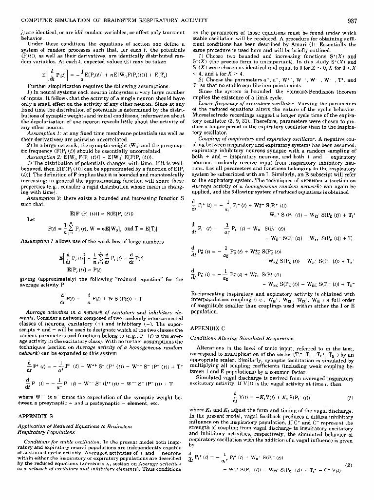
APPENDIX B
Application of Reduced Equations to Brainstem Respiratory Populations
Conditions for stable oscilZation. In the present model both inspi- ratory and expiratory neural populations are independently capable of sustained cyclic activity. Averaged activities of + and - neurons within either the inspiratory or expiratory populations are described by the reduced equations (APPENDIX A, section on Average activities in a network of excitatory and inhibitory elements). Thus conditions
on the parameters of those equations must be found under which stable oscillation will be produced. A procedure for obtaining suffi- cient conditions has been described by Amari (1). Essentially the same procedure is used here and will be briefly outlined.
I ) Choose two bounded and increasing functions S+(X) and S(X) (the precise form is unimportant). In this study S+(X) and S(X) were chosen as identical and equal to 0 for X 5 0, X for 0 < X
< 4, and 4 forX 2 4. 2) Choose the parameters a+, a-, W++, W-+, W+-, W--, T+, and
T- so that no stable equilibrium point exists. Since the system is bounded, the Poincare-Bendixson theorem
implies the existence of a limit cycle. Lower frequency of expiratory oscillator. Varying the parameters
of the reduced equations alters the nature of the cyclic behavior. Microelectrode recordings suggest a longer cycle time of the expira- tory oscillator (3, 9, 31). Therefore, parameters were chosen to pro- duce a longer period in the expiratory oscillator than in the inspira- tory oscillator.
Coupling of inspiratory and expiratory oscillator. A negative cou- pling between inspiratory and expiratory systems has been assumed: expiratory inhibitory neurons synapse with a random sampling of both + and - inspiratory neurons, and both + and - expiratory neurons randomly receive input from inspiratory inhibitory neu- rons. Let all parameters and functions belonging to the inspiratory system be subscripted with an I. Similarly, an E subscript will refer to the expiratory system. The techniques of APPENDIX A (section on Average activity of a homogeneous random network) can again be applied, and the following system of reduced equations is obtained
d & PI+ (t> = - 1 PI+ (t) + w;:+ s(P,+ (t))
a1 - w,+ s (PI (t)) - W,,+ S(P, (t)) + T,+
d 1 -& p*- (0 = - -_ PI- (t) + WI:- s(P,+ (t))
aI - W,- SO’, (t)) - W,, S(P, (t)) + T,-
d & Pk (t> = - --$ PE+ (t) + w,+,+ S(P,+ (t))
- w,$ s(P, (t)) - W,-,+ S(P,- (t)) + T,+
d -& P, (t) = l - Ti p, (t) + wg, S(P$ (t))
- w,, S(P, (t)) - W,, S(P,- (t)) + T,-
Reciprocating inspiratory and expiratory activity is obtained with interpopulation coupling (i.e., W& WE,, Wh+, WE-) a full order of magnitude smaller than couplings used within either the I or E population.
APPENDIX C
Conditions Altering Simulated Respiration
Alterations in the level of tonic input, referred to in the text, correspond to multiplication of the vector (T1+, TI-, TE+, T,-) by an
appropriate scaler. Similarly, synaptic facilitation is simulated by multiplying all coupling coefficients (including weak coupling be- tween I and E populations) by a common factor.
Simulated vagal discharge is derived from averaged inspiratory excitatory activity. If V( t> is the vagal activity at time t, then
d & V(t) = -KJ(t) + K2 s(P,+ (t)) (1)
where K, and K2 adjust the form and timing of the vagal discharge. In the present model, vagal feedback produces a diffuse inhibitory influence on the inspiratory population. If C+ and C- represent the strength of coupling from vagal discharge to inspiratory excitatory and inhibitory activities, respectively, the simulated behavior of respiratory oscillation with the addition of a vagal influence is given
bY
d & PI+ (t> = - L+ PI+ (t) + WI:+ S(P,’ (t))
a1
(2) - W,+ SW,- (t)) - W,,+ S(P,- (t)) + T,+ - C+ V(t)

938 S. GEMAN AND M. MILLER
d 1 & PI- @) = - --- PI- (t) + W&- S(P,’ (t>) - w,- S(P,- (t))
aI
d 1
(3)
& P, (t) = - Li G v> + w, s(P; (t)) - w,, S(P, (t))
(5) - W,, SG’, (t)) + T,- -C- V(t) - WrE- S(P,- (t)) + T,
d -&pm= . - --$ P$ (t) + wg,+ S(P$ (t))
This study was supported in part by Public Health Service Re- search Grant HL-02888-17 and Graduate Training Grant 5TL-HL-
(4) 05322-13 from the National Heart and Lung Institute.
- W,,+ S(P, (t>) - W,-,+ S(P,- (t>) + TEf Received for publication 23 February 1976.
REFERENCES
1. AMARI, S. Characteristics of random nets of analog neuron-like elements. IEEE Trans. Systems Man Cybernetics 2: 643-657, 1972.
2. BAXTER, D. W., AND J. OLSZEWSKI. Respiratory responses evoked by electrical stimulation of pons and mesencephalon. J. Neuro- physiol. 18: 276-287, 1955.
3. BERTRAND, F., A. HUGELIN, AND J. F. VIBERT. Quantitative study of anatomical distribution of respiration related neurons in the pons. Exptl. Brain Res. 16: 383-399, 1973.
4. BRECKINRIDGE, C. G., AND H. E. HOFF. Pontine and medullary regulation of respiration in the cat. Am. J. Physiol. 160: 385-394, 1950.
5. BURNS, B. D. The central control of respiratory movements. Brit. Med. J. 19: 7-9, 1963.
6. CARREGAL, E. J. A., B. WILLIAMS, AND L. BIRZIS. Respiratory centers in the dog and squirrel monkey: a comparative study. Respiration Physiol. 3: 333-348, 1967.
7. CHALAZONITIS, N. Effects of changes in Pco2 and PO, on rhythmic potentials from giant neurons. Ann. N.Y. Acad. Sci. 109: 451-
479, 1963. 8. COHEN, M. I. Discharge patterns of brain-stem respiratory neu-
rons during Hering-Breuer reflex evoked by lung inflation. J. Neurophysiol. 32: 356-374, 1969.
9. COHEN, M. I. How respiratory rhythm originates: evidence from discharge patterns of brainstem respiratory neurons. In: Breath- ing: Hering-Breuer Centenary Symposium. London: Churchill, 1970, p. 125-150.
10. COHEN, M. I., AND S. C. WANG. Respiratory neuronal activity in pons of cat. J. Neurophysiol. 22: 33-50, 1959.
11. DIRKEN, M. N. J., AND S. WOLDRING. Unit activity in bulbar respiratory centres. J. NeurophysioZ. 14: 211-225, 1951.
12. ECCLES, J. C. The Physiology of Nerve CeZZs. Baltimore, Md.: Johns Hopkins Press, 1957, p. 30-36.
13. FELDMAN, J. L., AND J. D. COWAN. Large-scale activity in neural nets. II. A model for the brainstem respiratory oscillator. BioZ. Cybernetics 17: 39-51, 1975.
14. GROSSBERG, S. A neural theory of punishment and avoidance. II. Quantitative theory. Math. Biosci. 15: 253-285, 1972.
15. Guz, A., M. I. M. NOBLE, J. G. WIDDICOMBE, D. TRENCHARD, AND
W. W. MUSHIN. The effect of bilateral block of vagus and glosso- pharyngeal nerves on the ventilatory response to CO, of con- scious man. Respiration Physiol. 1: 206-210, 1966.
16. HODGKIN, A. L., AND W. A. H. RUSHTON. The electrical constants of a crustacean nerve fiber. Proc. Roy. Sot., London, Ser. B. 133: 444-479, 1946.
17. HOFF, H. E., AND C. G. BRECKENRIDGE. The medullary origin of respiratory periodicity in the dog. Am. J. Physiol. 158: 157-172, 1949.
18. HUKUHARA, T., JR. Neuronal organization of the central respira- tory mechanisms in the brain stem of the cat. Acta NeurobioZ. Exptl. 33: 219-244, 1973.
19. JOHNSON, F. H., AND G. V. RUSSELL. The locus coeruleus as a pneumotoxic center. Anat. Record 112: 348, 1952.
20. KAHN, N., AND S. C. WANG. Electrophysiologic basis for pontine apneustic center and its role in integration of the Hering-Breuer
reflex. J. Neurophysiol. 30: 301-318, 1967. 21. LOURENGO, R. V., N. S. CHERNIACK, J. R. MULM, AND A. P.
FISHMAN. Nervous output of the respiratory center during ob- structed breathing. J. AppZ. Physiol. 21: 527-533, 1966.
22. LUMSDEN, T. Observations on the respiratory centres in the cat. J. Physiol., London 57: 153-160, 1923.
23. MORISHITA, I., AND A. YAJIMA. Analysis and simulation of net- works of mutually inhibiting neurons. Kybernetik 11: 154-165, 1972.
24. NESLAND, R., AND F. PLUM. Subtypes of medullary respiratory neurons. Exptl. NeuroZ. 12: 337-348, 1965.
25. NGAI, S. H., AND S. C. WANG. Organization of central respira- tory mechanisms in the brainstem of the cat: localization by stimulation and destruction. Am. J. PhysioL. 190: 343-349, 1957.
26. NICHOLSON, H. C. Localization of the central respiratory mecha- nisms studied by local cooling of the surface of the brain stem.
Am. J. PhysioZ. 115: 402-409, 1936. 27. PITTS, R. F. The respiratory center and its descending pathways.
J. Comp. NeuroZ. 72: 605-625, 1940. 28. RALL, W. Theoretical significance of deadridic trees for neuronal
input-output relations. In: Neural Theor.y and Modeling; Pro- ceedings of the 1962 Ojai Symposium. Stanford: Stanford Univ. Press, 1964, p. 73-97.
29. RUBIO, J. E. A new mathematical model of the respiratory center. BUZZ. Math. Biophys. 34: 467-481, 1972.
30. SALMOIRAGHI, G. C., AND R. VON BAUMGARTEN. Intracellular potentials from respiratory neurones in brain-stem of cat and mechanism of rhythmic respiration. J. Neurophysiol. 24: 203- 218, 1961.
31. SALMOIRAGHI, G. C., AND B. D. BURNS. Localization and patterns of discharge of respiratory neurons in brain-stem of cat. J. Neurophysiol. 23: 2-13, 1960.
32. SALMOIRAGHI, G. C., AND B. D. BURNS. Notes on mechanism of rhythmic respiration. J. Neurophysiol. 23: 14-26, 1960.
33. SCHEIBEL, M. E., AND A. B. SCHEIBEL. Anatomical basis of attention mechanisms in vertebrate brains. In: The Neurosci- ences: A Study Program. New York: Rockefeller Univ. Press, 1967, p. 577.
34. SCOTT, F. H. On the relative parts played by nervous and chemi- cal factors in the regulation of respiration. J. PhysioZ., London 37: 301-326, 1908.
35. TANG, P. C. Brain stem control of respiratory depth and rate in the cat. Respiration Physiol. 3: 349-366, 1967.
36. WANG, S. C., AND S. H. NGAI. General organization of central respiratory mechanisms. In: Handbook of Physiology. Respira- tion. Washington, D.C.: Am. Physiol. Sot., 1964, sect. 3, vol. I,
chapt. 19, p. 487-505. 37. WANG, S. C., S. H. NGAI, AND M. J. FRUMIN. Organization of
central respiratory mechanisms in the brain stem of the cat: genesis of normal respiratory rhythmicity. Am. J. Physiol. 190: 333-342, 1957.
38. WOLDRING, S., AND M. N. J. DIRKEN. Site and extension of bulbar respiratory centre. J. NeurophysioZ. 14: 226-241, 1951.
39. YOUNG, A. C. Neural control of respiration. In: Physiology and Biophysics. Philadelphia & London: Saunders, 1965, p. 788-802.
Related Documents