Alternative translation initiation augments the human mitochondrial proteome Lawrence Kazak*, Aurelio Reyes*, Anna L. Duncan, Joanna Rorbach, Stuart R. Wood, Gloria Brea-Calvo, Payam A. Gammage, Alan J. Robinson, Michal Minczuk and Ian J. Holt* MRC-Mitochondrial Biology Unit, Wellcome Trust-MRC Building, Cambridge CB2 0XY, UK Received September 3, 2012; Revised December 2, 2012; Accepted December 3, 2012 ABSTRACT Alternative translation initiation (ATI) is a mechanism of producing multiple proteins from a single tran- script, which in some cases regulates trafficking of proteins to different cellular compartments, including mitochondria. Application of a genome-wide compu- tational screen predicts a cryptic mitochondrial targeting signal for 126 proteins in mouse and man that is revealed when an AUG codon located down- stream from the canonical initiator methionine codon is used as a translation start site, which we term downstream ATI (dATI). Experimental evidence in support of dATI is provided by immunoblotting of endogenous truncated proteins enriched in mito- chondrial cell fractions or of co-localization with mitochondria using immunocytochemistry. More detailed cellular localization studies establish mitochondrial targeting of a member of the cytosolic poly(A) binding protein family, PABPC5, and of the RNA/DNA helicase PIF1a. The mitochondrial isoform of PABPC5 co-immunoprecipitates with the mitochondrial poly(A) polymerase, and is markedly reduced in abundance when mitochondrial DNA and RNA are depleted, suggesting it plays a role in RNA metabolism in the organelle. Like PABPC5 and PIF1a, most of the candidates identified by the screen are not currently annotated as mitochondrial proteins, and so dATI expands the human mitochon- drial proteome. INTRODUCTION Products of nuclear genes dominate the mitochondrial proteome. They are synthesized by cytosolic ribosomes and imported into mitochondria via specific pathways according to their final destination in the organelle (1). The most extensively used system for importing matrix-destined mitochondrial proteins depends on a posi- tively charged amphipathic a helix, located at the amino (N-) terminus of the protein, which functions as a mito- chondrial targeting signal (MTS). Cytosolic proteins chap- erone mitochondrial precursors to an import complex located on the outer surface of the mitochondrion, termed the translocase of the outer membrane (TOM) complex. The MTS can interact with import receptors and direct proteins across both the outer and inner mito- chondrial membranes. Matrix-destined proteins depend additionally on the translocase of the inner membrane (TIM) complex, specifically TIM23, to direct them to the innermost compartment of the organelle. The insertion of proteins into the TIM23 channel requires a membrane potential across the inner mitochondrial membrane, and a further driving force is provided by the presequence translocase-associated motor complex. Upon entry to the matrix, many proteins have the MTS removed by the mitochondrial processing peptidase, and chaperones facilitate the proper folding of the mature protein into its active conformation (1,2). Although many genes encode dedicated mitochondrial proteins, an increasing number are recognised to specify multiple protein isoforms that are found in more than one cellular compart- ment. Protein variants that are targeted to different cellular compartments can be synthesized from a single gene, or transcript, via the use of alternative splice sites, transcrip- tion start sites or translation initiation sites (3). Alternative translation initiation (ATI), first discovered in viruses (4,5), and subsequently in eukaryotes (6), is a mechanism by which more than one initiation codon within a single mRNA results in the translation of proteins with distinct N-termini (3,7). ATI diversifies the proteome and may alter a protein’s function or cellular location. The use of an MTS lends itself to ATI, as essentially the same mature protein can be made for two compartments from one gene. RNase H1 is typical of this class of genes (8). Other documented examples of ATI-dependent *To whom correspondence should be addressed. Tel: +44 1223 252840; Fax: +44 1223 252845; Email: [email protected] Correspondence may also be addressed to Lawrence Kazak. Tel: +44 1223 252840; Fax:+44 1223 252845; Email: [email protected] Correspondence may also be addressed to Aurelio Reyes. Tel: +44 1223 252840; Fax: +44 1223 252845; Email: [email protected] 2354–2369 Nucleic Acids Research, 2013, Vol. 41, No. 4 Published online 28 December 2012 doi:10.1093/nar/gks1347 ß The Author(s) 2012. Published by Oxford University Press. This is an Open Access article distributed under the terms of the Creative Commons Attribution Non-Commercial License (http://creativecommons.org/licenses/ by-nc/3.0/), which permits unrestricted non-commercial use, distribution, and reproduction in any medium, provided the original work is properly cited.

Journal Bioassesment
Dec 23, 2015
jurnal mata kuliah biomonitoring
Welcome message from author
This document is posted to help you gain knowledge. Please leave a comment to let me know what you think about it! Share it to your friends and learn new things together.
Transcript

Alternative translation initiation augments thehuman mitochondrial proteomeLawrence Kazak*, Aurelio Reyes*, Anna L. Duncan, Joanna Rorbach, Stuart R. Wood,
Gloria Brea-Calvo, Payam A. Gammage, Alan J. Robinson, Michal Minczuk and
Ian J. Holt*
MRC-Mitochondrial Biology Unit, Wellcome Trust-MRC Building, Cambridge CB2 0XY, UK
Received September 3, 2012; Revised December 2, 2012; Accepted December 3, 2012
ABSTRACT
Alternative translation initiation (ATI) is a mechanismof producing multiple proteins from a single tran-script, which in some cases regulates trafficking ofproteins to different cellular compartments, includingmitochondria. Application of a genome-wide compu-tational screen predicts a cryptic mitochondrialtargeting signal for 126 proteins in mouse and manthat is revealed when an AUG codon located down-stream from the canonical initiator methionine codonis used as a translation start site, which we termdownstream ATI (dATI). Experimental evidence insupport of dATI is provided by immunoblotting ofendogenous truncated proteins enriched in mito-chondrial cell fractions or of co-localization withmitochondria using immunocytochemistry. Moredetailed cellular localization studies establishmitochondrial targeting of a member of the cytosolicpoly(A) binding protein family, PABPC5, and of theRNA/DNA helicase PIF1a. The mitochondrialisoform of PABPC5 co-immunoprecipitates with themitochondrial poly(A) polymerase, and is markedlyreduced in abundance when mitochondrial DNAand RNA are depleted, suggesting it plays a role inRNA metabolism in the organelle. Like PABPC5 andPIF1a, most of the candidates identified by thescreen are not currently annotated as mitochondrialproteins, and so dATI expands the human mitochon-drial proteome.
INTRODUCTION
Products of nuclear genes dominate the mitochondrialproteome. They are synthesized by cytosolic ribosomesand imported into mitochondria via specific pathwaysaccording to their final destination in the organelle (1).
The most extensively used system for importingmatrix-destined mitochondrial proteins depends on a posi-tively charged amphipathic a helix, located at the amino(N-) terminus of the protein, which functions as a mito-chondrial targeting signal (MTS). Cytosolic proteins chap-erone mitochondrial precursors to an import complexlocated on the outer surface of the mitochondrion,termed the translocase of the outer membrane (TOM)complex. The MTS can interact with import receptorsand direct proteins across both the outer and inner mito-chondrial membranes. Matrix-destined proteins dependadditionally on the translocase of the inner membrane(TIM) complex, specifically TIM23, to direct them tothe innermost compartment of the organelle. The insertionof proteins into the TIM23 channel requires a membranepotential across the inner mitochondrial membrane, and afurther driving force is provided by the presequencetranslocase-associated motor complex. Upon entry tothe matrix, many proteins have the MTS removed bythe mitochondrial processing peptidase, and chaperonesfacilitate the proper folding of the mature protein intoits active conformation (1,2). Although many genesencode dedicated mitochondrial proteins, an increasingnumber are recognised to specify multiple proteinisoforms that are found in more than one cellular compart-ment. Protein variants that are targeted to different cellularcompartments can be synthesized from a single gene, ortranscript, via the use of alternative splice sites, transcrip-tion start sites or translation initiation sites (3). Alternativetranslation initiation (ATI), first discovered in viruses(4,5), and subsequently in eukaryotes (6), is a mechanismby which more than one initiation codon within a singlemRNA results in the translation of proteins with distinctN-termini (3,7). ATI diversifies the proteome andmay altera protein’s function or cellular location.
The use of an MTS lends itself to ATI, as essentially thesame mature protein can be made for two compartmentsfrom one gene. RNase H1 is typical of this class of genes(8). Other documented examples of ATI-dependent
*To whom correspondence should be addressed. Tel: +44 1223 252840; Fax: +44 1223 252845; Email: [email protected] may also be addressed to Lawrence Kazak. Tel: +44 1223 252840; Fax: +44 1223 252845; Email: [email protected] may also be addressed to Aurelio Reyes. Tel: +44 1223 252840; Fax: +44 1223 252845; Email: [email protected]
2354–2369 Nucleic Acids Research, 2013, Vol. 41, No. 4 Published online 28 December 2012doi:10.1093/nar/gks1347
� The Author(s) 2012. Published by Oxford University Press.This is an Open Access article distributed under the terms of the Creative Commons Attribution Non-Commercial License (http://creativecommons.org/licenses/by-nc/3.0/), which permits unrestricted non-commercial use, distribution, and reproduction in any medium, provided the original work is properly cited.

dual targeting include iron–sulfur cluster assembly enzyme(NFS1) and insulin-degrading enzyme (9,10). Translationinitiation from the second, or a subsequent, AUG codon,which we term downstream ATI (dATI), is a less obviousmethod of achieving mitochondrial targeting, as themature mitochondrial protein necessarily lacks a portionof the N-terminus that is present when initiation occursfrom the first AUG codon. The thyroid hormone receptor,c-Erb A a1, was thought to be a rare case of mitochondrialtargeting via dATI (11).
Our studies of nucleic acid-transacting proteins inmitochondria led us to the finding that dATI yields amitochondrial isoform of flap endonuclease 1, FEN1(manuscript in preparation). Taken together with theprevious instance of dATI-mediated mitochondrial target-ing of the thyroid hormone receptor, the possibility arosethat this might be a commonplace mechanism of mito-chondrial targeting. Therefore, using a computationalapproach, an inventory comprising 126 genes encodingcandidate dATI-dependent mitochondrial isoforms wasassembled. Experimental validation of a subset of theputative genes from the list provided empirical evidenceof mitochondrial localization, indicating that mitochon-drial targeting via dATI is much more widespread thanrecognized hitherto.
MATERIALS AND METHODS
Cell culture and transfections
Human 143B osteosarcoma (HOS) cells were maintainedin DMEM supplemented with 0.1% penicillin/strepto-mycin and 10% FBS. Flp-In
TM
T-RExTM
293 (HEK293T)cells (Invitrogen) were cultured in DMEM, 0.1%penicillin/streptomycin, 10% tetracycline-free FBS,15 mg/ml Blasticidin (InvivoGen) and 100 mg/ml Zeocin(Invivogen). HEK293T cells were co-transfected, usingLipofectamine 2000 (Invitrogen), with 1350 ng pOG44plasmid (invitrogen) and 150 ng of cDNA ligated intopcDNA5/FRT/TO (Invitrogen). Stable transfectantswere selected with 100mg/ml Hygromycin B (Invivogen).
Construct design and site-directed mutagenesis
Pif1, Pabpc5 and Pop1 cDNAs were ligated as KpnI-XhoIfragments into the pcDNA5/FRT/TO MCS. Mutant con-structs were made using the QuikChange site-directedmutagenesis kit (Stratagene).
Confocal microscopy
HOS cells were grown on glass coverslips and transientlytransfected using Lipofectamine 2000 with 1.5 mg of Pif1,Pabpc5 and Pop1 cDNAs. Twenty-four hours aftertransfection, HOS cells were incubated with anti-HA(1:200; Roche Diagnostics) or anti-FLAG (1:400; Sigma)antibodies, followed by treatment with appropriatefluorescently labelled secondary antibodies. Coverslipswere mounted with 1,4-diazabicyclo[2.2.2]octane(Sigma), containing 4’,6-diamidino-2-phenylindole,dihydrochloride (DAPI). Images were acquired with anLSM 510 META confocal microscope (Carl Zeiss, Jena,Germany).
Immunoblotting
Proteins were resolved by 4–12% NuPAGE Bis–TrisSDS-PAGE (Invitrogen) and transferred to nitrocellulosemembrane (Whatman), which were incubated at 4�C over-night with primary antibodies. The concentrations ofprimary antibodies from Abcam were as follows:GAPDH (1:20 000), HSP60 (1:60 000), SF2 (1:3000),BMS1L (1:200), B23 (1:1000), PABPC1 (1:1000),FIBRILLARIN (1:2000), ARAP1 (1:500), MTSS1L(1:1000), FBXL12 (1:2000), LEPRE1 (1:1000), PABPC5(1:500), NOX3 (1:1000), TNIK (1:2000), MBRL (1:1000),FOXH1, (1:1000), PRPSAP2 (1:1000), CLASP2 (1:2000),GCN1L1 (1:2000), Histone H2A (1:1000), mtPAP(1:1000), MRPS18 (1:1000), COXIV (1:1000), AconitaseII (1:1000), CYT C (1:5000) and NRF1 (1:500). The con-centrations of primary antibodies from Santa Cruz wereas follows: PIF1 (1:200), POLG1 (1:500), PCNA (1:400),LRPPRC (1:2000), Utrophin (1:200), TOM20 (1:4000)and SSBP1 (1:500). The concentrations of primaryantibodies from Sigma were as follows: FLAG (1:1000)and Tubulin (1:2000). Other antibodies used were asfollows: NDUFB8 (1:1000; Invitrogen), HA (1:2000;Roche), TWINKLE (1:200; A. Suomalainen), TFAM(1:80 000; R. Wiesner) and TRIM32 (1:1000; D. Blake).Secondary antibodies were anti-rabbit or anti-mouse HRP(Promega). Membranes were visualized with enhancedchemiluminescence (ECL) plus immunoblotting detectionsystem (GE Healthcare).
Mitochondrial import
[35S]-methionine-labelled proteins were generated with theTNT� Quick Coupled Transcription/Translation System(Promega) according to the manufacturer’s instructions.Labelled proteins were incubated with rat livermitochondria prepared by differential centrifugation andimport was assessed via trypsin protection andFCCP-dependent inhibition (12). Import reactions werecarried out at 37�C for 1 h and then subjected toSDS-PAGE. Gels were dried, exposed to storagephosphor screens (GE Healthcare), visualized on theTyphoon 9410 Variable Mode Imager (AmershamBiosciences) and quantified using ImageQuant 5.2.
Nuclear, cytosolic and mitochondrial isolationfrom cultured cells
HEK293T cells were homogenized in hypotonic buffer(20mM HEPES–NaOH [pH 7.8], 5mM KCl, 1.5mMMgCl2, 2mM DTT, 1mg/ml BSA, 1mM PMSF,protease inhibitor cocktail [Roche]). Low speed centrifu-gation of HEK293T cells resulted in a pellet that was usedto isolate intact nuclei as previously described (13).Cytosolic extracts were obtained from post-mitochondrialsupernatants, and mitochondria were prepared asdescribed previously (14).
Submitochondrial fractionation
Freshly isolated mitochondria were resuspended in 1ml of20mM potassium phosphate buffer, pH 7.4, with 150mMKCl, and sonicated 3� 10 s at 70W (Soniprep 150).
Nucleic Acids Research, 2013, Vol. 41, No. 4 2355

Samples were centrifuged at 100 000g for 1 h at 4�C. Thesupernatants were retained as the matrix fractions,whereas pellets comprised mitochondrial membraneproteins.
Immunoprecipitation
Mitochondria were isolated from PABPC533.HA-,PABPC533.F-, or PDE12.F-overexpressing HEK293Tcells and treated with proteinase K (PK) (0.02mg/5mgmitochondria) for 30min on ice. PK was inactivatedwith PMSF and then mitochondria were lysed in lysisbuffer (50mM Tris–HCl, [pH 7.4]; 150mM NaCl, 1mMEDTA, 1% triton X-100). Debris was removed by centri-fugation at 8000g max, and supernatant was incubatedwith EZview Red Anti-HA affinity gel (Sigma) oranti-FLAG M2 affinity gel (Sigma) for 1 h at 4�C. Beadswere washed three times in wash buffer (50mM Tris–HCl,pH 7.4; 150mM NaCl). In the case of HA immunopre-cipitation, beads were boiled, centrifuged and supernatantwas used for immunoblotting. In the case of FLAGco-immunoprecipitation experiments, proteins wereeluted from beads using 3X FLAG peptide (Sigma).
Iodixanol density gradients
Purified mitochondria were treated with 100 mg/ml trypsinfor 30min at room temperature, washed, lysed with 0.4%dodecyl maltoside (DDM) and loaded on to a 20–42.5%iodixanol density gradient (Sigma) and ultracentrifuged at100 000g for 14 h at 4�C.
Mitochondrial DNA analysis
Mitochondrial DNA was extracted and analysed viaSouthern blotting as previously described (14).
In silico analysis for generating the dATI inventory
Protein sequences from the human and mouse genomeswere downloaded from the ensembl genome browser,release 61 (15). Human–mouse orthology data wereobtained from the ensembl genome browser via BioMart(www.ensembl.org/biomart.martview). For all protein se-quences, each methionine occurring in the first quarter ofthe sequence was queried for possible dATI, with mito-chondrial targeting predicted using Mitoprot (16),TargetP (17) and iPSORT (18). Genes were consideredmitochondrial when they encoded at least one protein,which, when encoded from an alternative translationsite, satisfied two of the following three criteria: (i)Mitoprot score >0.95; (ii) TargetP score >0.95 and (iii)positive iPSORT result. Retaining only those genes con-sidered mitochondrial for both their human and mouseorthologs further filtered the inventory. Finally, thegenes whose protein products were predicted to localizeto mitochondria if their translation would start at M1were removed from the inventory. Genes whose proteinproducts that started at M1 were considered mitochon-drial when they, satisfied two of the following threecriteria: (i) Mitoprot score >0.5; (ii) TargetP score >0.5and (iii) positive iPSORT result.
RESULTS
A genome-wide screen identifies 126 candidatedATI-dependent mitochondrial proteins
As a test of the idea that many proteins achieve mitochon-drial targeting via downstream alternative translation ini-tiation (dATI), every in-frame AUG codon within the firstquarter of all annotated human and mouse protein-codinggenes of the ensembl database was assessed as a potentialtranslation initiation site. The choice to restrict theanalysis to the first quarter of the longest annotatedprotein isoform was made to reduce the probability ofidentifying gene products that were truncated at theN-terminus to such an extent that functional domainswithin the open reading frame (ORF) were lost. Putativemitochondrial targeting was evaluated based on predic-tions from Mitoprot (16), TargetP (17) and iPSORT(18). Two of three conditions were required: a Mitoprotscore >0.95, a TargetP score >0.95 and a positiveiPSORT result. These criteria were established with theaim of minimizing false positives; however, this riskedfailing to identify genuine mitochondrial proteins thatdepend on dATI for targeting to the organelle.
When the computational analysis was applied, 886entries (473 and 413, human and mouse genes, respect-ively) scored above the threshold set (Figure 1A).Human and mouse data sets were combined and onlyorthologous genes were retained, yielding 168 candidates.Forty-two of these were excluded (Supplementary TableS1) because the full-length protein has a plausible N-ter-minal MTS (an MTS starting at M1), and approximatelyhalf of these are found in the Mitocarta database (19).This suggests that these proteins can achieve mitochon-drial targeting without the need for dATI. Thus, thefinal catalogue (Supplementary Table S2) comprised 126genes that potentially depend on dATI to generate amitochondrially targeted protein.
Antibody- and immunocytochemistry-basedidentification of dATI candidates
Of the 126 genes identified by the in silico screen(Supplementary Table S2), five are recognized mitochon-drial proteins in the Mitocarta database (19), and 17others can be found in the MitoMiner database (20).Thus, the computational screen identified >100 proteinsthat have had no prior evidence of mitochondrial localiza-tion. This might be taken to imply that the screengenerated many false positives. However it is noteworthythat the established dATI-dependent mitochondrialprotein, c-Erb A a1, is missing from the Mitocarta andMitominer databases. As an initial test of the validity ofthe dATI screen, the cellular distribution of 26 proteinswith no prior localization to mitochondria was analysed.Fifteen proteins predicted by the in silico screen to have acryptic MTS, and 11 negative controls, were studied inenriched nuclear, cytosolic and mitochondrial prepar-ations, by immunoblotting or by immunocytochemistry.The proteins were selected without bias, while aiming toreflect a range of biological processes. For immunoblot-ting analyses, the prediction, a priori, was that the
2356 Nucleic Acids Research, 2013, Vol. 41, No. 4

antibody would cross-react with a protein that was (i)enriched in mitochondrial extracts, (ii) shorter than theannotated full-length protein, while being maximally ofa size predicted by dATI and (iii) resistant to trypsin deg-radation prior to mitochondrial lysis, like other internalmitochondrial proteins. For immunocytochemistry
analysis, two versions of the cDNA were cloned, thefull-length ORF, and a truncated form starting at theinternal AUG predicted to mark the start of the crypticMTS. In the immunoblotting experiments, 10 of 15proteins tested fulfilled the criteria, suggesting they havea mitochondrial isoform. The 10 positive proteins were
Figure 1. Output of a computational screen for genes encoding dATI-dependent mitochondrial isoforms and identification of eight truncated mito-chondrial isoforms via immunoblotting. (A) Venn diagram of genome-wide in silico identification of putative genes targeted to mitochondria via dATI.473 human and 413 mouse genes are predicted to encode a cryptic MTS, of which 168 are conserved between the two species. After filtering out 42 genespredicted to produce a mitochondrial protein based on translation starting at the first methionine, M1, 126 genes remained (Supplementary Table S2).(B) Immunoblot analysis of candidate gene products [ARAP1, FBXL12, MTSS1L, LEPRE1, TRIM32, NOX3, CLASP2 and FOXH1 from nuclear(Nuc), cytosolic (Cytos) and mitochondrial fractions of HEK293T cells]. Mitochondrial fractions were treated with 0, 10, 50 or 100mg/ml trypsin. L,mitochondria lysed with 1% Triton X-100 and treated with 100mg/ml trypsin. Gray arrowhead, full-length protein; white arrowhead, putative dATIisoform. Full-length and predicted dATI protein products of candidate genes are schematically depicted to the right of each immunoblot. Domains are asfollows: PH, Pleckstrin homology; ArfGAP, Arf GTPase-activating protein; RhoGAP, Rho GTPase-activating protein; RA, Ras association; FBL,F-box-like; IMD, IRSp53/MIM homology domain; 2OG-Fell Oxy, 2OG-Fe(II) oxygenase superfamily; Zf-RING LisH: RING-type zinc-finger, LisHdimerization motif; NHL: NHL repeat; (NCL-1, HT2A and Lin-41); Ferric reduct: Ferric reductase like transmembrane component; FADB:FAD-binding domain; Fe reduct NADB: Ferric reductase NAD-binding domain; CLASP_N: N-terminal region of CLIP-associated proteins.TOM20 was used to show efficiency of trypsin treatment. TFAM, CALNEXIN, Splicing factor 2 (SF2) and glyceraldehyde 3-phosphate dehydrogenase(GAPDH) were used as mitochondrial, endoplasmic reticulum, nuclear and cytosolic markers, respectively.
Nucleic Acids Research, 2013, Vol. 41, No. 4 2357
ARAP1, FBXL12, MTSS1L, LEPRE1, TRIM32, NOX3,CLASP2 and FOXH1 (Figure 1B), as well as PABPC5and PIF1a (see below). In the case of FBXL12, theMTS may not always be cleaved after import, as theantibody to this protein detected two bands specific tothe mitochondrial cell fraction (Figure 1B).There was no putative mitochondrial isoform in the
cases of TNIK, PRPSAP2, MBRL or NRF1(Supplementary Figure S1A). POP1 was another falsepositive, based on immunocytochemistry of the full-lengthand putative dATI isoform, as both long and short formsof the protein were targeted exclusively to nucleoli(Supplementary Figure S2). None of the negativecontrols tested yielded a short, trypsin-resistant, mito-chondrial isoform based on immunoblotting(Supplementary Figure S1B). Therefore, our screenachieved a sensitivity and specificity of 77 and 72% re-spectively, with an estimated false discovery rate (FDR)of 33%. These results suggest that �85 of the 126identified genes will prove to encode dATI-dependentmitochondrial proteins, and thus are strong candidatesfor further validation. Nevertheless, multiple methodswill be required to demonstrate that each predicteddATI-dependent gene product is a bona fidemitochondrialprotein. On the other hand, a dATI variant that is pre-dicted to be mitochondrial, but not detected in the organ-elle at first instance, should not be discounted, as dATImight be tightly regulated in some cases, and so the mito-chondrial isoform might only be apparent in specific celltypes, environmental conditions or stages of development.
The predicted polyA-binding protein PABPC5 has adATI-dependent mitochondrial isoform
Poly(A) tails are attached to the 30-end of almost alleukaryotic messenger RNAs, including those inmitochondria (21,22), and polyA-binding proteins(PABPs) bind to and modulate polyA tail length, withimplications for mRNA stability and translation (22).Hence, mitochondria are expected to contain one ormore PABPs, yet none had been identified hitherto.Therefore, the appearance of PABPC5 in the list ofcandidate proteins (Supplementary Table S2) wasof particular interest, as it could be the long sought mito-chondrial PABP (23).
Initiation from an internal AUG codon of Pabpc5 occursin vitro, and human mitochondria contain a truncatedisoform of PABPC5 concordant with dATI
The computational analysis predicted that translation ini-tiation from methionine 33 of PABPC5 would generate amitochondrial isoform of the protein. To test this predic-tion, Pabpc5 cDNAs were introduced into a coupled tran-scription/translation (TnT) system. Full-length Pabpc5cDNA (PABPC51) produced two polypeptides(Figure 2A; lane 2) corresponding to translationproducts initiating at M1 and M33, based on comparisonswith an N-terminally truncated template (PABPC533)(Figure 2A; lane 1), whereas a full-length mutant formof the protein, where M33 was replaced by isoleucine(PABPC5M33I) (Figure 2A; lane 3), yielded a single
polypeptide. Thus, the downstream AUG at position 33is a functional start site.
An antibody to PABPC5 detected two proteins in thecytosol of HEK293T cells, the lower of which was alsodetected in trypsin-treated mitochondria (Figure 2B).Sub-mitochondrial fractionation localized the protein tothe matrix compartment of the organelle (SupplementaryFigure S3A), and in vitro synthesized PABPC533 resolvedat the same position as the endogenous mitochondrialisoform of PABPC5 on SDS-PAGE gels (Figure 2C). Inaddition, full-length PABPC5 tagged at thecarboxyl-terminus (PABPC51.HA) was targeted princi-pally to the cytosol based on immunocytochemistry(Figure 2D), whereas deletion of the first 32 residues(PABPC533.HA) resulted in targeting of the protein tomitochondria, as predicted by the in silico screen(Figure 2E). Next, mitochondria were purified from trans-genic HEK293T cells expressing FLAG-tagged PABPC5,starting at M33 (PABPC533.F), or full-length PABPC5with a methionine-to-isoleucine mutation at the predictedinternal start site (PABPC5M33I.F). The full-length mutantprotein was readily detectable in whole cell extracts, andas expected for a cytosolic protein, the little associatedwith isolated mitochondria was completely degradedwith the addition of trypsin (Figure 2F). In contrast,tagged PABPC533 co-purifying with mitochondriasurvived trypsin treatment (Figure 2F). Therefore, trans-lation initiation from methionine 33 of Pabpc5 yields aprotein that is targeted to mitochondria in living cells.
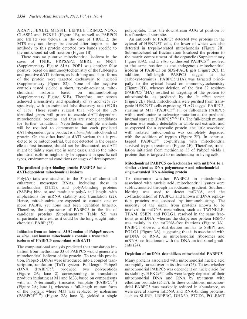
Mitochondrial PABPC5 co-fractionates with mtDNA to asimilar extent as DNA polymerase c and mitochondrialsingle-stranded DNA-binding protein
To determine whether PABPC5 in mitochondriaassociated with nucleic acid, mitochondrial lysates weresubfractionated through an iodixanol gradient. Southernblotting was used to detect mtDNA, and theco-fractionation of PABPC5 and known mtDNA replica-tion proteins was assessed by immunoblotting. Themajority of the signal from proteins known to beinvolved in mtDNA metabolism, such as TWINKLE,TFAM, SSBP1 and POLG1, resolved in the same frac-tions as mtDNA, whereas the chaperone protein HSP60was mainly in the mtDNA-free fractions (Figure 3A).PABPC5 showed a distribution similar to SSBP1 andPOLG1 (Figure 3A), suggesting that it is associated withmtDNA or RNA, as mitochondrial ribosomes andmRNAs co-fractionate with the DNA on iodixanol gradi-ents (24).
Depletion of mtDNA destabilizes mitochondrial PABPC5
Many proteins associated with mitochondrial nucleic acidare rapidly turned over in its absence (25). To test whethermitochondrial PABPC5 was dependent on nucleic acid forits stability, HEK293T cells were largely depleted of theirmitochondrial DNA and RNA by treatment withethidium bromide (26,27). In these conditions, mitochon-drial PABPC5 was markedly reduced in abundance, aswere several known mtDNA and RNA-binding proteins,such as SLIRP, LRPPRC, DHX30, PTCD3, POLRMT
2358 Nucleic Acids Research, 2013, Vol. 41, No. 4

AM33
PABPC533M1
PABPC51
PABPC5M33IX
1
2
3
M33 M1 M33I
M33
M1
1 2 3
PABPC5
37PABPC5
Cytos
Mitochondria:Trypsin (μg/ml)
B
D
PABPC51.HA
DAPI Mitotracker HA Merge
PABPC533.HA
E
F
3 4 521
MitochondriaTrypsin
FLAG37
6 9 10 1187 12
HSP6060
TOM20
GAPDH
15
37
M33
PABPC533 F
M1
PABPC5M33IX F
WCE
MitochondriaTrypsin
WCE
M1M33
LL
PABPC5
PABPC533
PABPC5M33
I
37
CTM Mtrx
RRM1 RRM1 RRM1 RRM1
Figure 2. PABPC5 has a mitochondrial isoform consistent with dATI from M33. (A) SDS-PAGE of [35S]-methionine-labelled PABPC5 polypeptidevariants generated in vitro. M33 and M1 denote the methionines where translation starts, while M33I indicates a methionine to isoleucine pointmutation at residue 33 of PABPC5. A consensus Kozak sequence (gccacc) was placed upstream of the first AUG (methionine) codon in eachconstruct. Schematics of the constructs used as templates are indicated to the right of the representative gel image. ‘X’ in the cDNA schematic(lane 3) indicates mutation of the methionine residue. (B) Immunoblot analysis of PABPC5 cytosolic (Cytos), and mitochondrial fractions fromHEK293T cells. Mitochondrial fractions were treated with 0, 10 or 50 mg/ml trypsin. Gray arrowhead, full-length protein; white arrowhead, putativedATI isoform. (C) Immunoblot analysis using anti-PABPC5 to PABPC533 and PABPC5M33I TnT products, total mitochondrial lysate (TM) and amitochondrial matrix fraction (Mtrx). Confocal analysis of transiently transfected HOS cells with C-terminal HA-tagged cDNAs encoding(D) full-length human PABPC5 (PABPC51.HA) or (E) the dATI isoform with the first methionine at residue 33 (PABPC533.HA). Recombinantproteins were labelled with anti-HA antibody (green), while nuclei (blue) and mitochondria (red) were visualized by staining cells with DAPI andMitotracker, respectively. (F) Mitochondria from PABPC5M33I.F- and PABPC533.F-expressing cells (10 ng/ml doxycycline, 24 h) were purified
Nucleic Acids Research, 2013, Vol. 41, No. 4 2359
(continued)

and TFAM, whereas the mitochondrial protein AconitaseII, which does not interact with mtDNA, was unaffectedby this treatment (Figure 3B). These results suggest thatPABPC5 interacts with nucleic acid in mitochondria.
PABPC5 co-immunoprecipitates the mitochondrialpoly(A) polymerase, but not mitochondrial ribosomes
Co-immunoprecipitation experiments, followed by im-munoblotting, were used to determine potential protein–protein interactions, using the predicted dATI isoform ofPABPC5, fused to a carboxy-terminal haemagglutinin(HA) or FLAG tag, as bait. Tagged PABPC533
co-immunoprecipitated the mitochondrial poly(A) poly-merase (mtPAP), and to a lesser extent LRPPRC,whereas a FLAG-tagged version of the mitochondrialdeadenylase, PDE12 (28), did not interact with either ofthese proteins (Figure 3C and Supplementary FigureS3B). There was no enrichment of the mitochondrialRNA-binding protein SLIRP, nor was there any signifi-cant co-immunoprecipitation of the mitochondrial riboso-mal protein, MRPS18, or the abundant respiratory chainsubunit cytochrome c oxidase (COXIV) (Figure 3C andSupplementary Figure S3B). The specific interaction ofPABPC533 with mtPAP and LRPPRC, but notMRPS18, suggests it associates with mRNAs that are in-dependent of mitochondrial ribosomes.
The RNA/DNA helicase PIF1a has a dATI-dependentmitochondrial isoform
Another dATI candidate from the in silico screen was theRNA/DNA helicase PIF1a. In yeasts, the PIF1 geneproduct is essential for mitochondrial DNA maintenance(29), and the mitochondrial isoforms of budding andfission yeast PIF1 are known to be generated via ATI,not dATI. Translation initiation from the first AUGcodon of the ORF yields the mitochondrial isoform,whereas translation from a downstream in-frame AUGcodon generates yeast nuclear PIF1 (30,31). In contrast,none of the annotated PIF1 transcripts in a variety ofvertebrates possesses an in-frame AUG that couldappend an MTS to the protein (Supplementary TableS3). Nor do these PIF1 orthologs contain a proximalupstream ORF (uORF) adjacent to the main codingsequence (CDS), of the type associated with vertebrateRNase H1 genes (8), based on the annotated 50 UTRsequences of all available PIF1 sequences in the NCBIand ensembl databases (Supplementary Table S3). It hasbeen suggested that human PIF1a is a nuclear protein(32,33), and that a mitochondrial form of PIF1 (PIF1b)is generated via alternatively splicing (33). However, PIF1
was identified in our computational screen as a candidatedATI protein (Supplementary Table S2), and sequencealignments of PIF1 of several mammals revealed a poten-tial MTS that would be revealed by dATI, from humanM54 and mouse M67 (Figure 4A and B).
To screen for the predicted PIF1a product of dATI,cDNAs were translated in vitro. Full-length Pif1a cDNAproduced two polypeptides (Figure 4C; lane 2), the shorterof which was not detected when the predicted dATI startsite was modified by substituting methionine 54 with iso-leucine (PIF1aM54I) (Figure 4C; lane 3), and a PIF1acDNA lacking the first 53 codons of the ORF (PIF1a54)yielded a single polypeptide that was the same size as theshort form of PIF1a1 (Figure 4C; lane 1). These resultsindicate that dATI occurs from the AUG encoding M54of Pif1a, in vitro. Because evidence already existed fornuclear localization of PIF1a (32,33) and the full lengthprotein is not detectable in mitochondria, we tested theability of PIF1a54 to be imported into isolated rat livermitochondria, using PIF1a1 and TFAM as negative andpositive controls, respectively. When the PIF1a transla-tion products were incubated with isolated mitochondria,the MTS of the predicted dATI isoform, PIF1a54, wasprocessed and imported in a membranepotential-dependent manner (Figure 4D, bottom panel),whereas there was no detectable mitochondrial import offull-length PIF1a (PIF1a1) (Figure 4D middle panel).Although the efficiency of import of PIF1a54 intoisolated mitochondria was lower than that of TFAM,this is also true of other well-established mitochondrialproteins, including components of the cytochrome coxidase holoenzyme (12). The proposed mature form,PIF1am, was only seen when PIF1a54 translationproducts were incubated with mitochondria, and it alonewas resistant to trypsin (Figure 4D, lanes 2 and 3, bottompanel), strongly suggesting import and processing ofPIF1a54 by mitochondria. The size of PIF1am correspondsto an endogenous protein detected by a PIF1 antibody(Figure 3A) whose expression was reduced by a siRNAtargeting PIF1 (Figure 4E) (34).
The predicted dATI forms of human and mouse PIF1are targeted to mitochondria in cultured cells, andmutation of an internal AUG codon ablatesmitochondrial localization of PIF1a
PIF1a was shown to display nuclear localization based onan N-terminal FLAG-tagged (FLAG.PIF1a) form of theprotein (32,33). However, dATI bypasses N-terminal tags;therefore, cDNAs specifying full-length Pif1a tagged withHA, either at the N-terminus (HA.PIF1a1) or theC-terminus (PIF1a1.HA), were transiently expressed in
Figure 2. Continuedand subjected to trypsin protection assays, followed by immunoblotting. Schematic representations of the transgenes are depicted above theimmunoblots. M1, indicates translation initiation at the annotated start methionine of PABPC5 according to ensembl genome browser, release61; M33, indicates the band corresponding to the cDNA product starting from the dATI residue of PABPC5. Whole cell extract (WCE, lanes 1 and7); black slope, indicates increasing trypsin concentrations (mg/ml) of 0 (lanes 2 and 8); 10 (lanes 3 and 9); 50 (lanes 4 and 10); and 100mg/ml (lanes 5and 11). L, mitochondria lysed with 1% Triton X-100 and treated with 100 mg/ml trypsin, lanes 6 and 12. PABPC5.F transgenes were detected withanti-FLAG antibody. Heat shock protein 60 (HSP60), mitochondrial marker; GAPDH, cytosolic marker. TOM20 was used to show efficiencyof trypsin treatment. Full-length and predicted dATI protein products of candidate genes are schematically depicted to the right. RRM1, RNArecognition motif.
2360 Nucleic Acids Research, 2013, Vol. 41, No. 4
human cells. HA.PIF1a1, like FLAG.PIF1a, was locatedexclusively in the nucleus (Figure 5A), whereas theproducts of the C-terminally tagged, full-length PIF1acDNA (PIF1a1.HA) co-localized chiefly withmitochondria, with a small amount of signal detected innuclei (Figure 5B). Enforced translation from M54(PIF1a54.HA) resulted in exclusive mitochondrial target-ing of the protein (Figure 5C), whereas replacing M54with isoleucine (I54) yielded a protein that did notco-localize with mitochondria (Figure 5D). These resultsshow that a functional MTS is revealed when humanPIF1a starts at M54, and that this residue is required togenerate a mitochondrial isoform of PIF1a. Analysis ofmouse PIF1 constructs provided further support for thegeneration of a mitochondrial-targeted form of PIF1 viadATI, as mPIF167.HA (the mouse equivalent ofPIF1a54.HA) co-localized with mitochondria (Figure5E), whereas there was no detectable PIF1 inmitochondria after mutating the AUG codon predictedto yield translation initiation of the mitochondrialisoform of mouse PIF1 (mPIF1M67I.HA) (Figure 5F).
N-terminally truncated human PIF1a is imported andprocessed by mitochondria in cultured cells
Subcellular fractions were prepared from human cells ex-pressing PIF1a1.HA, PIF1a54.HA or PIF1aM54I.HA todetermine the location of the recombinant proteins.Tagged full-length PIF1a (M1) was the only signaldetected in purified nuclei (Figure 6A, lane 1), andalthough some of this isoform was also associated withmitochondrial fractions (Figure 6A, lane 2), it wasdegraded by trypsin treatment (Figure 6B, lanes 3–5).The mitochondrial extracts additionally contained twoshorter PIF1a products, one corresponding to thepre-protein (M54) and the other to a shorter protein (m)(Figure 6A, lane 2). We infer that the shorter of the two isthe mature form of mitochondrial PIF1a, both because itis the more abundant and it is similar in size (the smalldifference being attributable to the tag) to an endogenousmitochondrial protein identified by the PIF1 antibody(Figures 3A and 4E). Consistent with this interpretation,cells expressing PIF1a54.HA produced only the twopresumed mitochondrial-specific species, M54 and m,both of which were resistant to degradation by trypsin(Figure 6C). Thus, full-length PIF1a is outsidemitochondria, whereas M54 and m are located inside theorganelle. Both these mitochondrial isoforms depend on
B
mtDNA
5 6 7 8 9 14 1510 11 12 13 16 17
Con
Dep
poTmottoB
Iodixanol Gradient Fractions
37
37
150
15
15
PABPC5
SLIRP
Con
Dep
Con
Dep
7575
150
150
Con
Dep
150
150
PTCD3
DHX30ConDep
Dep
LRPPRCCon
25
25TFAM
POLRMTCon
Dep
Con
Dep
150
150
75 ACONITASE IICon
ImmunoprecipitationC
5
75
IIDep
37
75
150 LRPPRC
mtPAP
Input FT Wash Boil
HA
IB
20
15
15
SLIRP
COXIV
MRPS18
HEK293T
5 6 7 8 9 14 1510 11 12 13
ATopBottom
37
75
25
mtDNA
TFAM
TWINKLE
SSBP1
PABPC5
15
15
60
75
POLG1
SSBP1
HSP60
PIF1βPIF1
55PIF1αm
Figure 3. PABPC5 co-precipitates mitochondrial RNA-bindingproteins and is dependent on mitochondrial nucleic acids for its stabil-ity. (A) Mitochondria were isolated from HEK293T cells,trypsin-treated (50mg/ml), lysed and centrifuged through an iodixanol
Figure 3. Continuedgradient. Fractions were collected from the bottom of the tube andthen subjected either to DNA extraction, followed by Southernblotting, or to SDS-PAGE, followed by immunoblotting. Numbersabove the immunoblots indicate iodixanol fractions, in order of collec-tion. (B) After HEK293T cells were either grown under normal condi-tions (Con) or treated with ethidium bromide (100 ng/ml) for 72 h(Dep), mitochondrial lysates were fractionated and analysed as perpanel A. (C) Mitochondria were isolated from cells overexpressingPABPC533.HA after 72 h of doxycycline (30 ng/ml) treatment,followed by immunoprecipitation with anti-HA agarose beads.Immunoblotting was used to determine the extent ofco-immunoprecipitation of selected proteins using 2.5% of input,flow-through (FT) and wash, and 5% of the eluate.
Nucleic Acids Research, 2013, Vol. 41, No. 4 2361

Human -------------MLSGIEAAAGEYEDSELRCRVAVEELSPGGQPRRRQALRTAELSLGR 47Orangutan -------------MLSGIEAAAGEYEDSELRCRVAVEELSPGGQPRRRQALRTAELSLGR 47Mouse MRSGLCTPAEALEMPSSTEAATDECDDAELRCRVAVEELSPGGQPRKRQALRAAELSLGR 60Pig -------------MLSGTQAAAAESEDAELRCRVAVEELSPGGQPRRRQALRTAELSLGR 47
M1PIF1
M1A
g SG Q S C V V S GGQ Q S GCow -LRSFCRPAEAAAMLSGTQAAAAECEDGELRCRVAVEELSPGGQPRRRQSLRTAELSLGR 59Dolphin -------------MLSGTQAVAAECADAELRCRVAVEELSPGGQPRRRQALRTAELRLGR 47Megabat -------------TLSGTPAEAVECEDAELRCRVAVEELSPGGQPRRRQALRTAELSLGR 47
*. * : * *.******************:**:**:*** ***
Human NERRELMLRLQAPGPAGRPRCFPLRAARLFTRFAEAGRSTLRLPAHDTPGAGAVQLLLSD 107Orangutan NERRELMLRLQAPGPAGRPRCFPLRAARLFTRFAEAGRSTLRLPAHGAPGAGAVQLLLSD 107
Q G G C G S G G GS Q S 120
M54(human)
M67(mouse)
Mouse NERRELMLRLQAPGPTGRPRCFPLRAVRLFTRFAATGRSTLRLPTDGVPGAGSVQLLLSD 120Pig NERRELMLRLKAPGPAGRPRCFPLRAARLFTRFAAMGRSTLRLPAYGASPAGAVQLLLSD 107Cow NERRELMLRLQAPGPAGRPRCFPVRAARLFTRFAAAGRSTLRFPADSTPRASAVQLLLSD 119Dolphin NERRELMLSLQAPGPAGRPRCFPLRAARLFTRFAASGRSTLRLPADGAPRTGAVQLLLSD 107Megabat NERRELMLRLQAPGPAGRPRCYPLRAARLFTRFAATGRSTLRLPAEGAPRTGAVQLLLSD 107
******** *:****:*****:*:**.******* ******:*: ... :.:*******
M1
M54
M1
M14
No
Yes
No
No
B
0.8 – 1
0.6 – 0.8
Mitochondrialtargeting score
5
M310
M453
M482
Human
M67
M189
M209
Mouse
Yes
No
No
No Yes
Yes
No 0 - 0.2
0.6 0.8
0.4 – 0.6
0.2 – 0.4
CPIF1α
M1
M54IM1M54
M54
M1 1PIF1α54
M54
1 2 3 X
2
3
PIF1α1
PIF1αM54I
Mitochondria
Triton X-100
D
35S TnT
Trypsin
FCCP
+ + + + + ++ + + + +
++-
- + + - + +- + - - +
---
- - + + +--
E75
50
siCON siPIF1
GAPDH
PIF1α1
PIF1αm
M54M1
50PIF1α1
75
Mature protein
Pre-protein
TFAM 25
37
75
50PIF1α54 M54
3 4 5 6 721PIF1αm
Figure 4. Mammalian PIF1 has a predicted dATI-dependent mitochondrial isoform, some of which can be imported into isolated mitochondria.(A) N-terminal amino acid sequence alignment of PIF1 between human, orangutan, mouse, pig, cow, dolphin and megabat. M1 and M54 above thealignment correspond to methionines 1 and 54, respectively of human PIF1a. Green font and box, canonical start sites in mouse and human; red fontand box, putative methionine residue where dATI begins; blue font, Mitoprot-predicted cleavable MTS. (B) In silico mitochondrial targetingprediction scores from: Mitoprot, TargetP, Predotar (35), PSORTII (36) and iPSORT of human and mouse PIF1. ‘M’ followed by a number, onthe left side of the heat map, indicate the methionine residue numbers; red-coloured boxes in the heat map indicate strong prediction for
2362 Nucleic Acids Research, 2013, Vol. 41, No. 4
(continued)

Figure 4. Continuedmitochondrial targeting. (C) SDS-PAGE of [35S]-methionine-labelled human PIF1a polypeptide variants generated in vitro. M54 and M1 refer to themethionines where translation starts, while M54I indicates a methionine to isoleucine point mutation at residue 54 of PIF1a. A consensus Kozaksequence (gccacc) was placed upstream of the first AUG (methionine) codon in each construct. Schematics of the constructs used as templates areindicated to the right of the representative gel image. ‘X’ in the cDNA schematic (lane 3) indicates mutation of the methionine residue.(D) [35S]-methionine-labelled TFAM (positive control), PIF1a1 and PIF1a54 were incubated with isolated rat liver mitochondria. 1 mM FCCP(lanes 5–7) was used to dissipate membrane potential. White arrowheads indicate imported polypeptide. Import efficiency was determined relativeto import of TFAM. Start methionines are indicated to the right of the gel images. (E) HEK293T cells were transfected with 200 pmol of scrambledsiRNA (siCON) or siRNA targeted towards PIF1 (siPIF1). Forty-eight hours later, whole cell extracts were used for immunoblotting with anti-PIF1.GAPDH was used as a loading control. The chart accompanying the immunoblots shows the extent of knockdown relative to GAPDH protein(n=3 independent experiments).
ADAPI Mitotracker HA Merge
PIF1
B
HA.PIF1α1
PIF1α1.HA
C
PIF1α54.HA
D
PIF1αM54I.HA
E
mPIF167.HA
F
mPIF1M67I.HA
Figure 5. Immunocytochemistry of ectopically expressed human and mouse PIF1 suggests mitochondrial targeting via dATI. HOS cells weretransiently transfected with human Pif1a full-length cDNA containing a HA tag on either the (A) N-terminus (HA.PIF1a1) or (B) C-terminus(PIF1a1.HA). The remaining Pif1a constructs were HA-tagged on the C-terminus, consisting of Pif1a.HA cDNAs that were (C) forced to start atM54 (PIF1a54.HA) or (D) with a methionine to isoleucine substitution at residue 54 (PIF1aM54I.HA). Mouse PIF1 (mPIF1) constructs wereC-terminally tagged and included (E) a cDNA forced to start translation at M67 (mPIF167.HA) or (F) a cDNA encoding a methionine-to-isoleucinemutation at residue 67 (mPIF1M67I.HA). Recombinant proteins were labelled with anti-HA antibody (green), nuclei were stained blue with DAPI,and mitochondria were stained red with Mitotracker.
Nucleic Acids Research, 2013, Vol. 41, No. 4 2363

initiation at M54, as neither was detected in cells express-ing the PIF1aM54I.HA variant, and all derivatives ofPIF1aM54I.HA were degraded when mitochondria wereexposed to trypsin (Figure 6D).A species (cPIF1a) migrating between M54 and m was
detected in cells expressing PIF1a1.HA (Figure 6B, lane1), and this was the major product in PIF1aM54I.HA cells(Figure 6D, lanes 1 and 2). In the latter cells, the recom-binant protein was dispersed throughout the cytosol(Figure 5D), and cPIF1a did not survive trypsin treatmentof intact mitochondria (Figure 6B and D). Thus, cPIF1aprobably represents a proteolytic cleavage product that isformed after export of tagged PIF1a from the nucleus, asoccurs to the native protein during the course of the cellcycle (32). In contrast, murine PIF1M67I.HA wasconcentrated in the nucleus (Figure 5F), and so it maynot be recognized by the human nuclear export machin-ery. Similarly, the HA N-terminal tag may interfere withnuclear export of PIF1a1 (Figure 5A). In summary, dATIfrom the AUG corresponding to M54 of human Pif1agenerates a pre-protein, PIF1a54, which is cleaved aftermitochondrial import, yielding a mature mitochondrialisoform, PIF1am, and mutation of the predicted dATIsites ablates mitochondrial isoforms of human andmouse PIF1.
Endogenous N-terminally truncated PIF1a is present inmitochondria and co-fractionates with mtDNA
The mitochondrial lysates fractionated by iodixanolgradient sedimentation and probed for PABPC5 werealso used to evaluate endogenous forms of PIF1. Themost abundant form of PIF1 detected by immunoblottingcorresponded to PIF1am (the presumed mature dATI
isoform of PIF1a), 30% of which co-fractionated withmtDNA (Figure 3A). The largest species detected by thePIF1 antibody was a polypeptide of �75 kDa, which is thepredicted size of PIF1b (33), and it was concentrated inthe same fractions as the mtDNA. Thus, there appear tobe two forms of PIF1 in human mitochondria, PIF1am
and PIF1b, both of which may interact with mtDNA.
DISCUSSION
The mammalian mitochondrial proteome is estimated at1500 proteins. If accurate, then some 400 mitochondrialproteins remain to be identified (19). The dATI-generatedmitochondrial proteins predicted from our analysis couldaccount for a substantial fraction of the ‘missing’ mito-chondrial proteome, as >100 gene products in our listhave not previously been annotated as mitochondrialproteins. Ten of 15 candidates tested from the 126-geneinventory appear to have a form of the protein inmitochondria corresponding to the predicted dATIisoform. Taking account of the estimated false discoveryrate of 33%, then �85 of the identified genes may prove toyield proteins targeted to mitochondria in adATI-dependent manner. However, in view of the lowspecificities of mitochondrial prediction programs, it is in-evitable that many genes will be missing from the inven-tory. The thyroid hormone receptor c-Erb A a1 (11) is aknown example. Thus, the number of proteins targeted tomitochondria via dATI could be in the hundreds.Although the dATI screen could be refined further toinclude the likes of c-Erb A a1, simply lowering the thresh-old for acceptance will inevitably produce a markedincrease in false positives. This might be offset in other
A B C D
M1 M54 M1 I54M54
PIF1α1 HA PIF1α M54I HAPIF1α54 HA X
TOM20
HA
25
15
75
50
37 SF2
M54
Nuc Mt PIF1α1.HA
M54M1
mM54
TFAM
TOM203 4 5 62 65431 2 65431 21
m
M1
m
WCE
WCE
WCE
MitochondriaTrypsin
MitochondriaTrypsin
MitochondriaTrypsin
TFAM
L LL
21
cPIF
1α.H
A
cPIF
1?.H
A
Figure 6. Subcellular fractionation confirms mitochondrial targeting and processing of the dATI forms of PIF1a54 in cells. (A) Purified nuclei (Nuc)and mitochondria (Mt) were isolated from HEK293T cells stably expressing PIF1a1.HA followed by immunoblot analyses. Mitochondria fromtransgenic HEK293T cells expressing (B) PIF1a1.HA, (C) PIF1a54.HA, or (D) PIF1aM54I.HA were purified and subjected to trypsin protectionassays, followed by immunoblotting. Whole cell extracts (WCE) were fractionated alongside mitochondrial lysates to show all products of thecDNAs. Transgenes were expressed by the addition of 10 ng/ml doxycycline for 24 h. Schematic representations of the transgenes are depicted belowthe immunoblots. M1, indicates translation initiation at the annotated start methionine of PIF1a according to ensembl genome browser, release 61;M54, the band corresponding to the dATI residue of PIF1a; m, the putative mature product of PIF1a.HA after mitochondrial import and removalof the MTS. (B–D) WCE (lane 1); black slope, indicates increasing trypsin concentrations of 0, 10, 50 and 100 mg/ml (lanes 2–5, respectively). L,mitochondria lysed with 1% Triton X-100 and treated with 100mg/ml trypsin (lane 6). PIF1a.HA transgenes were detected with anti-HA antibody.TFAM and SF2 are mitochondrial and nuclear markers, respectively. TOM20 was used to show efficiency of trypsin treatment. (B and D)cPIF1a.HA, processed full-length PIF1a that resides in the cytoplasm, not in mitochondria.
2364 Nucleic Acids Research, 2013, Vol. 41, No. 4

ways, such as a greater demand for conservation amongdiverse species, or the incorporation of additional algo-rithms that predict mitochondrial targeting (37).Conservation is a critical filter because randomlygenerated peptides can create an MTS (38). As notedabove, evidence of mitochondrial involvement will be animportant guide in many cases and this is likely to befacilitated by the explosion in RNA expression dataproviding comprehensive details of co-expression in avariety of cell and tissue types. Detailed mining ofexisting and new mitochondrial proteome studies willalso doubtless reap reward, as it is likely that many mito-chondrial proteins have been mistakenly dismissed as con-taminants, b-actin being a case in point (14). Conversely,it is possible that some genes on the list have genuinemitochondrial isoforms that do not depend on a canonicalN-terminal MTS, or are generated via alternative tran-scripts, rather than by dATI. Ultimately any in silicoprediction of putative internal start codons will requireexperimental verification by multiple methods for eachand every candidate, as per PIF1 and PABPC5.
Although the known or inferred functions of the 126candidate mitochondrial dATI proteins are highlyvaried, they can be grouped into a number of categories(Table 1). Thirty-one of the candidates have links to RNAor DNA metabolism, and these can be further sub-dividedinto transcriptional regulation, post-transcriptional modi-fication and DNA replication or repair. The earlier iden-tification of nuclear transcription factors in mitochondrialed others to suggest that the regulation of mitochondrialgene expression might share certain aspects with nucleargene expression (39), and 17 candidate dATI-generatedmitochondrial proteins are recognized transcriptionalregulators (Table 1). Actomyosin has recently beenfound inside mammalian mitochondria and implicated inmtDNA maintenance (14), and 12 candidates have linksto actin binding and organization, two of which weresubstantiated by immunoblotting (Figure 1B).Furthermore, the list includes two members of the WNK(WNK lysine-deficient protein kinase) serine/threoninekinase subfamily (WNK1 and WNK2), which act onRho GTPases and control actin dynamics. These twoproteins physically interact with one another (40), andWNK1 enhances the activity of the annotated mitochon-drial protein, SGK1 (serum- and glucocorticoid-inducedkinase 1) (41). Thus, dATI may be a mechanism of proteintrafficking that maintains the balance between mitochon-drial and cytoplasmic actin.
dATI-dependent mitochondrial isoforms confound con-ventional genetic analysis, as gene knockdown andablation will affect all the protein isoforms, and so therelative contributions of the mitochondrial andnon-mitochondrial proteins cannot be judged. Gene re-placement of one particular isoform could circumventthis problem, as was achieved for the alternativelytranslated DNA ligase III, which identified the mitochon-drial, not the nuclear, enzyme as the essential variant forcell viability (42).
Based on the immunoblotting results and the domainstructure of some of the dATI candidates (Figure 1B), lossof a portion of the N-terminus will affect their function. For
instance, the dATI isoform of FBXL12 is predicted to lackthe F-box-like domain that gives it its name (Figure 1B), andhalf of a RNA recognition motif is missing from the mito-chondrial isoform of PABPC5 (Figure 2F).
The dATI-mediated mitochondrial isoform of PABPC5
Although several mitochondrial proteins have been shownpreviously to bind RNA and poly(A) sequences inmitochondria (43–45), none binds poly(A) tails preferen-tially. Of the four human PABPC genes, three (PABPC1,PABPC3 and PABPC4) are established polyA-bindingproteins (46–48); however, none has a predictedN-terminal MTS based on the annotated first AUGcodon, or downstream AUG codon, and only PABPC5predicts a dATI-dependent mitochondrial isoform.Several lines of evidence provide support for theproposed dATI-mediated mitochondrial isoform ofPABPC5. A cDNA of the complete ORF yields twoproteins, one of which is of the size predicted by dATI,and when expressed in cells the shorter form is importedinto mitochondria. Endogenous PABPC5 is present inmitochondria (PABPC5m) based on immunoblotting,and it co-fractionates with mitochondrial nucleic acids.PABPC5m is unstable when mitochondrial DNA andRNA are depleted, and it interacts with mitochondrialRNA-binding proteins, implying PABPC5 inmitochondria is bound to RNA. Hence, PABPC5m isproposed to play a role in post-transcriptional mitochon-drial RNA metabolism, and based on its high homologyto other PABP family members, it is a highly crediblecandidate for the long sought mitochondrial poly(A)-binding protein. All four PABPCs contain fournon-identical RNA recognition motifs (RRMs).Typically, these motifs are linked to a C-terminaldomain through a proline-rich (P-rich) region (22).However, the C-terminus of PABPC1 can be deletedwithout affecting poly(A) binding in vitro (49), orcompromising translation in Xenopus (50) or viability inyeast (51). Only RRM motifs 1 and 2 need be retained topreserve the protein’s ability to bind to poly(A) tails (52).Moreover, whereas RRMs 1 and 2 are highly selective forpolyadenylated RNA, RRMs 3 and 4 are less discrimina-tive, and can also bind AU-rich RNA (48), and this maybe an important feature of the mitochondrial variant ofPABPC5, as some mitochondrial transcripts have beenshown to be polyuridinylated, poly(U) (53,54).Therefore, although full-length PABPC5 lacks the P-richlinker region and the C-terminal domain of the otherPABPCs (55), and the dATI form lacks RRM1(Figure 2F), both cytosolic and mitochondrial isoformsof PABPC5 are still likely to have the capacity to bindto polyadenylated RNA. Nevertheless, the structural dif-ferences between PABPC5 and the other family membersmight be indicative of a distinct property or role, and thefunction of the shorter mitochondrial isoform may differfrom its cytosolic counterpart.
The dATI mediated mitochondrial isoform of PIF1a
In the case of PIF1a, dATI exposes the MTS and simul-taneously removes the nuclear localization signal (NLS).
Nucleic Acids Research, 2013, Vol. 41, No. 4 2365

Prior research on PIF1a assigned it a nuclear location,based chiefly on N-terminal tagging of the recombinantprotein (32,33). However, N-terminal tags mask mito-chondrial targeting signals that are present at theN-terminus (24), and a protein derived from an internalMTS will be undetectable because dATI bypassesN-terminal tags. Therefore, the previous approach wasnot capable of revealing a mitochondrial isoform ofPIF1a. Accordingly, C-terminal tags are most appropriatefor localization studies of potential mitochondrialproteins.
Based on its mobility on denaturing gels, the mass of thedATI-dependent mitochondrial PIF1a (PIF1am) isestimated at 55±2.5 kDa, placing the cleavage site atresidue 165±30. Despite PIF1a54 being processed to aneven shorter form upon mitochondrial import, to generatePIF1am, the mature form is not expected to differ substan-tially from the full-length protein in terms of its coreactivities, because the first defined functional SFIhelicase motif begins at residue 224 (56), which PIF1am
appears to retain. Furthermore, a recombinant form ofhuman PIF1a (PIF1�N) lacking the first 166 residues
Table 1. Functional categories of candidate dATI genes
Proposed function of dATI-dependent proteins
Transcriptionalregulation
Post-transcriptionalmodification
DNAReplication/Repair
Actincytoskeleton
Kinases Translation
ASH2L C19orf6 C9orf102 ANK2 MINK1 RPL4C11orf30 DHX15 TEP1 ANK3 MAP4K4 FTSJ3PHTF1 ERI3 PIF1 ARAP1 MLKL DUS4LSMARCB1 PABPC5 ZNF335 FARP2 PRKCE EIF2S1TRRAP POP1 ZRANB3 IQSEC1 TNIK RPS15ZMYM3 RBM25 IQSEC2 WNK1 BMS1ZMYM4 SRRM2 MTSS1 WNK2ELK4 NFKBIL1 MTSS1L WNK4FOXH1 YTHDC2 MYO10 TBRG4GATA4 MYO15A CDK12MLF1 STARD13 DAPK1NR1I2 ARHGAP32 DGKDNRF1 DSTYKPAX4MYST3NR6A1SRFBP1
Collagen Microtubules Beta oxidation Receptors GTP binding Calcium sensing
COL16A1 CLASP2 CPT1B CHRM1 C9orf86 MCTP1COL27A1 SFI1 CPT1C CHRM5 RASAL3 IQCF5LEPRE1 ASPM DECR2 GPR162
Channels Trafficking Ubiquitination Cell–cell communication Proteases Transporters
ACCN1 NUP155 FBXL12 DSP MMP25 CNNM2SCN11A YIPF3 RNF111 DTX4 MMP28 ABCC6TRPM2 DOPEY1 TRIM32 ICA1 PSMD1 ABCC4CACNA1B CLINT1 ZYG11B PKP4 RHBDL3CACNA1F CCDC157 FBXO38 SRCIN1 TMPRSS11FHCN1 COMMD10 C1QTNF3 CTSFTMC6 AGBL1CYBB USP49
Apoptosis Phospho-lipase TCA cycle ROS Nucleotide synthesis
SPATA17 PNPLA6 IDH1 NOX3 PRPSAP2
NOX4
Cysteine modification Retinoic acid metabolism Phospholipid biosynthesis Unknown function
HHAT CYP26A1 PISD FAM160B1PLCB4 C14orf118
All 126 genes from Supplementary Table S2 were grouped according to known function.Bold font, genes tested in this study, that have a product in mitochondria based on experiment, unless coloured gray; blue font, genes found inhuman Mitocarta; red font, genes found in Mitominer.
2366 Nucleic Acids Research, 2013, Vol. 41, No. 4

has the same ATPase and helicase activities as thefull-length protein (57). Therefore, the activities ofPIF1am are expected to be similar to its full-lengthnuclear counterpart, although contextual differencesbetween nuclei and mitochondria might mean that thefunction of PIF1am is distinct from the long form ofPIF1a, in vivo.
Human PIF1b—the alternatively splicedmitochondrial isoform
The detection, with an antibody to PIF1, of a protein of amass of �75 kDa co-fractionating with mtDNA(Figure 3A), lends support to the proposal that alternativesplicing gives rise to a dedicated transcript encoding ahuman mitochondrial PIF1 isoform, PIF1b (33). PIF1bis annotated as a manually verified transcript in theensembl database. Assuming it is correctly assigned,PIF1b starts at M1 and so depends on signals located atthe C-terminus (which PIF1a lacks) to achieve mitochon-drial targeting (33). However, a genetic approach thattargets PIF1b, while sparing PIF1a will be needed toclarify its physiological importance, as PIF1b does notappear to be conserved even among primates(Supplementary Figure S4). Orangutan lacks the consen-sus splice acceptor site, which in humans gives rise to Pif1bmRNA, and in mouse there are no annotated alternativemouse PIF1 isoforms. Although there is a putative AGsplice acceptor site in the mouse Pif1 gene, the resultantfour-nucleotide insertion directly downstream wouldcreate a variant PIF1 unlike human PIF1b(Supplementary Figure S4). Moreover, this hypotheticalmouse PIF1 mRNA does not contain any appreciablehomology (at the nucleotide level) to the portion of thehuman PIF1b protein that is required for mitochondrialtargeting (33). Therefore, there is no evidence of a PIF1bmouse variant. Nevertheless, there is considerablesequence variation among mitochondrial targetingsignals and so it remains possible that the mouse PIF1sequence contains a carboxy-terminal MTS.
The extent of dATI-dependent mitochondrial targeting
An increasing number of nuclear DNA transactingproteins are also found in mitochondria (33,58–60), butthe task of defining the organelle-specific forms is far fromcomplete. Information regarding the subcellular localiza-tion of these proteins is essential for determining theirprecise roles within the cell. The identification ofdATI-dependent mitochondrial isoforms of PABPC5and PIF1a suggests that this trafficking mechanism willprove to be a significant contributor to the dual targetingof proteins. Other candidates well worthy of further inves-tigation for a role in mitochondrial nucleic acid metabol-ism include the putative RNA exonuclease ERI3 and theRNA helicase YTHDC2 (Table 1 and SupplementaryTable S2).
The in silico screen and cell biology data of this reportstrongly support the idea of dATI playing an importantrole in targeting proteins to mitochondria, potentiallyaccounting for up to half of the unassigned mitochondrialproteins, or �10% of the total mitochondrial proteome.
This raises the question of how expression of this class ofgenes is regulated and the nature of the mitochondrial–nuclear and mitochondrial–cytoplasmic communicationpathways involved. The use of dATI permits antagonisticregulation, as translation of one isoform inherentlyopposes translation of the other isoform (61); therefore,deregulated ATI could result in category of humandisease.
SUPPLEMENTARY DATA
Supplementary Data are available at NAR Online:Supplementary Tables 1–3 and Supplementary Figures1–4.
ACKNOWLEDGEMENTS
We thank Professors A. Suomalainen, R. Wiesner and D.Blake for generous gifts of the TWINKLE, TFAM andTRIM32 antibodies, respectively.
FUNDING
Cambridge University Commonwealth Trust, Fellowship(to L.K.); UK Medical Research Council. Funding foropen access charge: UK Medical Research Council.
Conflict of interest statement. None declared.
REFERENCES
1. Chacinska,A., Koehler,C.M., Milenkovic,D., Lithgow,T. andPfanner,N. (2009) Importing mitochondrial proteins: machineriesand mechanisms. Cell, 138, 628–644.
2. Wiedemann,N., Frazier,A.E. and Pfanner,N. (2004) The proteinimport machinery of mitochondria. J. Biol. Chem., 279,14473–14476.
3. Bazykin,G.A. and Kochetov,A.V. (2011) Alternative translationstart sites are conserved in eukaryotic genomes. Nucleic AcidsRes., 39, 567–577.
4. Prats,A.C., De Billy,G., Wang,P. and Darlix,J.L. (1989) CUGinitiation codon used for the synthesis of a cell surface antigencoded by the murine leukemia virus. J. Mol. Biol., 205, 363–372.
5. Curran,J. and Kolakofsky,D. (1988) Ribosomal initiation from anACG codon in the Sendai virus P/C mRNA. EMBO J., 7,245–51.
6. Strubin,M., Long,E.O. and Mach,B. (1986) Two forms of the Iaantigen-associated invariant chain result from alternativeinitiations at two in-phase AUGs. Cell, 47, 619–625.
7. Ivanov,I.P., Firth,A.E., Michel,A.M., Atkins,J.F. andBaranov,P.V. (2011) Identification of evolutionarily conservednon-AUG-initiated N-terminal extensions in human codingsequences. Nucleic Acids Res., 39, 4220–4234.
8. Suzuki,Y., Holmes,J.B., Cerritelli,S.M., Sakhuja,K., Minczuk,M.,Holt,I.J. and Crouch,R.J. (2010) An upstream open reading frameand the context of the two AUG codons affect the abundance ofmitochondrial and nuclear RNase H1. Mol. Cell. Biol., 30,5123–5134.
9. Leissring,M.A., Farris,W., Wu,X., Christodoulou,D.C.,Haigis,M.C., Guarente,L. and Selkoe,D.J. (2004) Alternativetranslation initiation generates a novel isoform ofinsulin-degrading enzyme targeted to mitochondria. Biochem. J.,383, 439–446.
10. Land,T. and Rouault,T.A. (1998) Targeting of a humaniron-sulfur cluster assembly enzyme, nifs, to different subcellularcompartments is regulated through alternative AUG utilization.Mol. Cell, 2, 807–815.
Nucleic Acids Research, 2013, Vol. 41, No. 4 2367

11. Wrutniak,C., Cassar-Malek,I., Marchal,S., Rascle,A., Heusser,S.,Keller,J.M., Flechon,J., Dauca,M., Samarut,J. and Ghysdael,J.(1995) A 43-kDa protein related to c-Erb A alpha 1 is located inthe mitochondrial matrix of rat liver. J. Biol. Chem., 270,16347–16354.
12. Petruzzella,V., Tiranti,V., Fernandez,P., Ianna,P., Carrozzo,R.and Zeviani,M. (1998) Identification and characterization ofhuman cDNAs specific to BCS1, PET112, SCO1, COX15, andCOX11, five genes involved in the formation and function of themitochondrial respiratory chain. Genomics, 54, 494–504.
13. Cooper,H.M. and Spelbrink,J.N. (2008) The human SIRT3protein deacetylase is exclusively mitochondrial. Biochem. J., 411,279–285.
14. Reyes,A., He,J., Mao,C.C., Bailey,L.J., Di Re,M., Sembongi,H.,Kazak,L., Dzionek,K., Holmes,J.B., Cluett,T.J. et al. (2011) Actinand myosin contribute to mammalian mitochondrial DNAmaintenance. Nucleic Acids Res., 39, 5098–5108.
15. Flicek,P., Amode,M.R., Barrell,D., Beal,K., Brent,S., Carvalho-Silva,D., Clapham,P., Coates,G., Fairley,S., Fitzgerald,S. et al.(2012) Ensembl 2012. Nucleic Acids Res., 40, D84–D90.
16. Claros,M.G. and Vincens,P. (1996) Computational method topredict mitochondrially imported proteins and their targetingsequences. Eur. J. Biochem., 241, 779–786.
17. Emanuelsson,O., Brunak,S., von Heijne,G. and Nielsen,H. (2007)Locating proteins in the cell using TargetP, SignalP and relatedtools. Nat. Protoc., 2, 953–971.
18. Bannai,H., Tamada,Y., Maruyama,O., Nakai,K. and Miyano,S.(2002) Extensive feature detection of N-terminal protein sortingsignals. Bioinformatics, 18, 298–305.
19. Pagliarini,D.J., Calvo,S.E., Chang,B., Sheth,S.A., Vafai,S.B.,Ong,S.-E., Walford,G.A., Sugiana,C., Boneh,A., Chen,W.K. et al.(2008) A mitochondrial protein compendium elucidates complexI disease biology. Cell, 134, 112–123.
20. Smith,A.C. and Robinson,A.J. (2009) MitoMiner, an integrateddatabase for the storage and analysis of mitochondrial proteomicsdata. Mol. Cell. Proteomics, 8, 1324–1337.
21. Rorbach,J. and Minczuk,M. (2012) The post-transcriptional lifeof mammalian mitochondrial RNA. Biochem. J., 444, 357–373.
22. Kuhn,U. and Wahle,E. (2004) Structure and function of poly(A)binding proteins. Biochim. Biophys. Acta., 1678, 67–84.
23. Wydro,M., Bobrowicz,A., Temperley,R.J., Lightowlers,R.N. andChrzanowska-Lightowlers,Z.M. (2010) Targeting of the cytosolicpoly(A) binding protein PABPC1 to mitochondria causesmitochondrial translation inhibition. Nucleic Acids Res., 38,3732–3742.
24. He,J., Cooper,H.M., Reyes,A., Di Re,M., Sembongi,H.,Litwin,T.R., Gao,J., Neuman,K.C., Fearnley,I.M., Spinazzola,A.et al. (2012) Mitochondrial nucleoid interacting proteins supportmitochondrial protein synthesis. Nucleic Acids Res., 40,6109–6121.
25. Poulton,J., Morten,K., Freeman-Emmerson,C., Potter,C.,Sewry,C., Dubowitz,V., Kidd,H., Stephenson,J., Whitehouse,W.and Hansen,F.J. (1994) Deficiency of the human mitochondrialtranscription factor h-mtTFA in infantile mitochondrial myopathyis associated with mtDNA depletion. Hum. Mol. Genet., 3,1763–1769.
26. Maniura-Weber,K., Goffart,S., Garstka,H.L., Montoya,J. andWiesner,R.J. (2004) Transient overexpression of mitochondrialtranscription factor A (TFAM) is sufficient to stimulatemitochondrial DNA transcription, but not sufficient to increasemtDNA copy number in cultured cells. Nucleic Acids Res., 32,6015–6027.
27. Wiseman,A. and Attardi,G. (1978) Reversible tenfod reduction inmitochondria DNA content of human cells treated with ethidiumbromide. Mol. Gen. Genet., 167, 51–63.
28. Rorbach,J., Nicholls,T.J.J. and Minczuk,M. (2011) PDE12removes mitochondrial RNA poly(A) tails and controlstranslation in human mitochondria. Nucleic Acids Res., 39,7750–7763.
29. Foury,F. and Kolodynski,J. (1983) pif mutation blocksrecombination between mitochondrial rho+ and rho- genomeshaving tandemly arrayed repeat units in Saccharomyces cerevisiae.Proc. Natl Acad. Sci. USA, 80, 5345–5349.
30. Pinter,S.F., Aubert,S.D. and Zakian,V.A. (2008) TheSchizosaccharomyces pombe Pfh1p DNA helicase is essential forthe maintenance of nuclear and mitochondrial DNA. Mol. Cell.Biol., 28, 6594–6608.
31. Zhou,J., Monson,E.K., Teng,S.C., Schulz,V.P. and Zakian,V.A.(2000) Pif1p helicase, a catalytic inhibitor of telomerase in yeast.Science, 289, 771–774.
32. Mateyak,M.K. and Zakian,V.A. (2006) Human PIF helicase iscell cycle regulated and associates with telomerase. Cell Cycle, 5,2796–2804.
33. Futami,K., Shimamoto,A. and Furuichi,Y. (2007) Mitochondrialand nuclear localization of human Pif1 helicase. Biol. Pharm.Bull., 30, 1685–1692.
34. Gagou,M.E., Ganesh,A., Thompson,R., Phear,G., Sanders,C. andMeuth,M. (2011) Suppression of apoptosis by PIF1 helicase inhuman tumor cells. Cancer Res., 71, 4998–5008.
35. Small,I., Peeters,N., Legeai,F. and Lurin,C. (2004) Predotar: atool for rapidly screening proteomes for N-terminal targetingsequences. Proteomics, 4, 1581–1590.
36. Nakai,K. and Horton,P. (1999) PSORT: a program for detectingsorting signals in proteins and predicting their subcellularlocalization. Trends Biochem. Sci., 24, 34–36.
37. Calvo,S., Jain,M., Xie,X., Sheth,S.A., Chang,B., Goldberger,O.A.,Spinazzola,A., Zeviani,M., Carr,S.A. and Mootha,V.K. (2006)Systematic identification of human mitochondrial disease genesthrough integrative genomics. Nat. Genet., 38, 576–582.
38. Lemire,B.D., Fankhauser,C., Baker,A. and Schatz,G. (1989) Themitochondrial targeting function of randomly generated peptidesequences correlates with predicted helical amphiphilicity. J. Biol.Chem., 264, 20206–20215.
39. Leigh-Brown,S., Enriquez,J.A. and Odom,D.T. (2010) Nucleartranscription factors in mammalian mitochondria. Genome Biol.,11, 215.
40. Lenertz,L.Y., Lee,B.-H., Min,X., Xu,B., Wedin,K., Earnest,S.,Goldsmith,E.J. and Cobb,M.H. (2005) Properties of WNK1 andimplications for other family members. J. Biol. Chem., 280,26653–26658.
41. Chen,W., Chen,Y., Xu,B., Juang,Y.-C., Stippec,S., Zhao,Y. andCobb,M.H. (2009) Regulation of a third conservedphosphorylation site in SGK1. J. Biol. Chem., 284, 3453–3460.
42. Simsek,D., Furda,A., Gao,Y., Artus,J., Brunet,E.,Hadjantonakis,A.K., Van Houten,B., Shuman,S., McKinnon,P.J.and Jasin,M. (2011) Crucial role for DNA ligase III inmitochondria but not in Xrcc1-dependent repair. Nature, 471,245–248.
43. Nakagawa,J., Waldner,H., Meyer-Monard,S., Hofsteenge,J.,Jeno,P. and Moroni,C. (1995) AUH, a gene encodingan AU-specific RNA binding protein with intrinsicenoyl-CoA hydratase activity. Proc. Natl Acad. Sci. USA, 92,2051–2055.
44. Ponamarev,M.V., She,Y.-M., Zhang,L. and Robinson,B.H.Proteomics of bovine mitochondrial RNA-binding proteins:HES1/KNP-I is a new mitochondrial resident protein.J. Proteome Res., 4, 43–52.
45. Preiss,T., Chrzanowska-Lightowlers,Z.M. and Lightowlers,R.N.(1997) Glutamate dehydrogenase: an organelle-specificmRNA-binding protein. Trends Biochem. Sci., 22, 290.
46. Hosoda,N., Lejeune,F. and Maquat,L.E. (2006) Evidence thatpoly(A) binding protein C1 binds nuclear pre-mRNA poly(A)tails. Mol. Cell. Biol., 26, 3085–3097.
47. Kimura,M., Ishida,K., Kashiwabara,S. and Baba,T. (2009)Characterization of two cytoplasmic poly(A)-binding proteins,PABPC1 and PABPC2, in mouse spermatogenic cells. Biol.Reprod., 80, 545–554.
48. Sladic,R.T., Lagnado,C.A., Bagley,C.J. and Goodall,G.J. (2004)Human PABP binds AU-rich RNA via RNA-binding domains 3and 4. Eur. J. Biochem., 271, 450–457.
49. Nietfeld,W., Mentzel,H. and Pieler,T. (1990) The Xenopuslaevis poly(A) binding protein is composed of multiplefunctionally independent RNA binding domains. EMBO J., 9,3699–3705.
50. Gray,N.K., Coller,J.M., Dickson,K.S. and Wickens,M. (2000)Multiple portions of poly(A)-binding protein stimulate translationin vivo. EMBO J., 19, 4723–4733.
2368 Nucleic Acids Research, 2013, Vol. 41, No. 4

51. Sachs,A.B., Davis,R.W. and Kornberg,R.D. (1987) A singledomain of yeast poly(A)-binding protein is necessary andsufficient for RNA binding and cell viability. Mol. Cell. Biol., 7,3268–3276.
52. Kuhn,U. and Pieler,T. (1996) Xenopus poly(A) binding protein:functional domains in RNA binding and protein-proteininteraction. J. Mol. Biol., 256, 20–30.
53. Brown,T.A., Tkachuk,A.N. and Clayton,D.A. (2008) NativeR-loops persist throughout the mouse mitochondrial DNAgenome. J. Biol. Chem., 283, 36743–36751.
54. Szczesny,R.J., Borowski,L.S., Brzezniak,L.K., Dmochowska,A.,Gewartowski,K., Bartnik,E. and Stepien,P.P. (2010) Humanmitochondrial RNA turnover caught in flagranti: involvement ofhSuv3p helicase in RNA surveillance. Nucleic Acids Res., 38,279–298.
55. Blanco,P., Sargent,C.A., Boucher,C.A., Howell,G., Ross,M. andAffara,N.A. (2001) A novel poly(A)-binding protein gene(PABPC5) maps to an X-specific subinterval in the Xq21.3/Yp11.2 homology block of the human sex chromosomes.Genomics, 74, 1–11.
56. Bochman,M.L., Sabouri,N. and Zakian,V.A. (2010) Unwindingthe functions of the Pif1 family helicases. DNA Repair, 9,237–249.
57. Huang,Y., Zhang,D.-H. and Zhou,J.-Q. (2006) Characterizationof ATPase activity of recombinant human Pif1. Acta. Biochim.Biophys. Sin., 38, 335–341.
58. Kalifa,L., Beutner,G., Phadnis,N., Sheu,S. and Sia,E. (2009)Evidence for a role of FEN1 in maintaining mitochondrial DNAintegrity. DNA Repair, 8, 1242–1249.
59. Lakshmipathy,U. and Campbell,C. (1999) The human DNAligase III gene encodes nuclear and mitochondrial proteins. Mol.Cell. Biol., 19, 3869–3876.
60. Zheng,L., Zhou,M., Guo,Z., Lu,H., Qian,L., Dai,H., Qiu,J.,Yakubovskaya,E., Bogenhagen,D.F., Demple,B. et al. (2008)Human DNA2 is a mitochondrial nuclease/helicase for efficientprocessing of DNA replication and repair intermediates. Mol.Cell, 32, 325–336.
61. Mueller,J.C., Andreoli,C., Prokisch,H. and Meitinger,T. (2004)Mechanisms for multiple intracellular localization of humanmitochondrial proteins. Mitochondrion, 3, 315–325.
Nucleic Acids Research, 2013, Vol. 41, No. 4 2369
Related Documents