JONI LEIVO ADVANCED SURFACE TREATMENT OF ELASTOMERIC POLY- DIMETHYLSILOXANE FOR CELL STRETCHING APPLICATIONS Master’s thesis Examiner: Professor Pasi Kallio Examiner and topic approved at the Faculty of Natural Sciences council on 31.5.2017

Welcome message from author
This document is posted to help you gain knowledge. Please leave a comment to let me know what you think about it! Share it to your friends and learn new things together.
Transcript

JONI LEIVO
ADVANCED SURFACE TREATMENT OF ELASTOMERIC POLY-
DIMETHYLSILOXANE FOR CELL STRETCHING APPLICATIONS
Master’s thesis
Examiner: Professor Pasi Kallio
Examiner and topic approved at the
Faculty of Natural Sciences council
on 31.5.2017
i
TIIVISTELMÄ
TAMPEREEN TEKNILLINEN YLIOPISTO Biotekniikan diplomi-insinöörin tutkinto-ohjelma JONI LEIVO: Edistyneet polydimetyylisiloksaanielastomeerin pintakäsittely-menetelmät soluvenytyssovelluksille Diplomityö, 71 sivua Kesäkuu 2017 Pääaine: Kudosteknologia Tarkastaja: professori Pasi Kallio Avainsanat: Polydimetyylisiloksaani (PDMS), pintakäsittely, dynaaminen soluviljely, kantasolu, soluvenytys, physisorptio, kovalenttinen sitoutuminen, tyypin I kollageeni
Polydimetyylisiloksaani (PDMS) on elastomeeri, jota käytetään laajalti biologisissa
dynaamisissa mikrofluidisissa sovelluksissa. Hydrofobinen PDMS ei kuitenkaan tue
solujen kiinnittymistä viljelyn aikana, varsinkaan muuttuvissa olosuhteissa, kuten
venytyksessä. Toisaalta PDMS ominaisuudet ovat soluvenytyssovelluksille liian
hyödyllisiä, jotta se olisi helppo korvata. PDMS:n elastisuus, muovailtavuus, kemiallinen
inerttisyys ja bioyhteensopivuus selittävät sen laajamittaisen käytön biolääketieteen
alalla. Sen vuoksi PDMS:n pintakäsittely on välttämätön osa materiaalin käyttöä,
varsinkin monimutkaisemmissa solusovelluksisa. Jotta kantasolujen käyttäytymistä
ymmärrettäisiin paremmin, on tärkeää tutkia kestäviä pintakäsittelymenetelmiä.
Tämän diplomityön päätavoitteena oli etsiä menetelmiä, joilla pystytään sitomaan tyypin
I kollageenia kovalenttisesti PDMS soluviljelyalustoihin pitkäaikaisia
soluvenytyskokeita varten. Toissijaisena tavoiteena oli kehittää uusia paranneltuja
pintakäsittelymenetelmiä. Työssä esitellään ja tutkitaan seitsemää eri
pintakäsittelymenetelmää, joista neljä perustui uuteen tapaan käyttää askorbiinihappoa
(AA) kollageenin sitomiseen. Menetelmiä tutkittiin käyttämällä
immunofluoresenssivärjäyksiä ja soluviljelyä. Tehdyt kokeet on jaettu viiteen
vaiheeseen. Ensimmäisessä vaiheessa (P1) physisorptioon pohjautuvat menetelmät ja
glutaraldehydipohjainen Kovalenttinen Menetelmä 1 kuvattiin fluoresenssimikroskopian
avulla. Toisessa vaiheessa (P2) uusi AA:n perustuva Kovalenttinen Menetelmä 2, kahtena
eri versiona, kuvattiin myös fluoresenssimikroskopian avulla. Vaiheessa kolme (P3)
fysisorptiomenetelmä, Kovalenttinen Menetelmä 1, sekä Kovalenttinen Menetelmä 2,
kolmena eri versiona, testattiin viljelemällä rasvakudoksen kantasoluja (hAdSC) niiden
päällä staattisesti 14 päivää. Vaiheessa neljä (P4) Kovalenttinen Menetelmä 1 ja
Kovalenttinen Menetelmä 2, kahtena versiona, testattiin viljelemällä hAdSC:ja niiden
päällä staattisesti ja dynaamisesti 13 päivää. Vaihe viisi (P5) esitteli uudentyyppisen
Kovalenttisen Menetelmän 3, jota kuvattiin fluoresenssimikroskopian avulla, ja testattiin
viljelemällä hAdSC:ja pinnoituksen päällä staattisesti neljä päivää.
Tyypin I kollageeni onnistuttiin kuvaamaan kaikissa pinnoitusmenetelmissä. Solujen
viljely onnistui myös niin staattisessa kuin dynaamisessakin ympäristössä. Kokeiden
tulokset osoittivat, että uusi AA ristisilloitettu Kovalenttinen Menetelmä 2 oli parempi
sitomaan kollageenia, sekä sopivampi soluviljelyyn kuin physisorptiomenetelmä tai
Kovalenttinen Menetelmä 1. Soluviljelykokeet tehtiin ja PDMS:n
pintakäsittelymenetelmät kehitettiin osana innovaatiorahoituskeskus Tekesin rahoittamaa
Ihmisen Varaosat hanketta ja Suomen Akatemian rahoittamaa WoodBone projektia.
ii
ABSTRACT
TAMPERE UNIVERSITY OF TECHNOLOGY Master’s Degree Programme in Biotechnology JONI LEIVO: Advanced surface treatment of elastomeric polydimethylsiloxane for cell stretching applications Master of Science Thesis, 71 pages June 2017 Major: Tissue engineering Examiner: Professor Pasi Kallio Keywords: polydimethylsiloxane (PDMS), surface treatment, dynamic culture, stem cell, cell stretching, physisorption, covalent immobilization, collagen type I
Polydimethylsiloxane (PDMS) elastomer is widely used in dynamic biological microflu-
idic applications. Hydrophobic pristine PDMS does not support cell attachment and cul-
ture, especially in dynamic conditions. Regardless, PDMS has too many useful properties
as a base material for dynamic cell culture systems to be easily replaced. The good elastic
properties, mouldability, transparency, chemical inertness, and biocompatibility of
PDMS are enough to justify its use in large scale in the biomedical field. Therefore,
PDMS surface treatment is nowadays considered as an essential step in using the material,
especially for longer culture periods and dynamic culture conditions. To understand cell
behaviour and stem cell differentiation better during cyclic stretching, it is important to
study different durable surface treatment methods.
The primary goal of this thesis work was to covalently bind collagen type I to cell culture
substrates fabricated from Sylgard® 184 PDMS composite for long term cell stretching
experiments with methods found in the literature. The secondary goal was to propose a
novel surface treatment method to improve upon the existing methods. Seven different
surface treatment methods, four of which were novel ascorbic acid (AA) based methods,
were studied in this thesis using immunofluorescent imaging and cell culture experiments.
The experiments were divided in five phases in chronological order to reflect the evolu-
tion of the surface treatment methods and the experiments. In phase one (P1), physisorp-
tion and Covalent Method 1 were imaged using fluorescent microscope. In phase two
(P2), a novel Covalent Method 2 in two variations was proposed and subsequently imaged
using fluorescent microscope. In phase three (P3), physisorption, Covalent Method 1, and
Covalent Method 2 in three variations were tested in static human adipose stem cell
(hAdSC) culture for 14 days. In phase four (P4), Covalent Method 1 and Covalent Method
2 in two variations were tested in static and dynamic hAdSC culture for 13 days. Phase
five (P5) introduced Covalent Method 3 that was imaged with fluorescent microscope
and tested in static hAdSC culture for four days.
Collagen type I was successfully labelled and imaged from all of the coatings. Cells were
also successfully cultured in static and dynamic environments. The results showed that
the novel AA crosslinked Covalent Method 2 was superior to the physisorption method
and Covalent Method 1 in immobilizing collagen as well as in cell culture tests.
The cell culture tests were conducted and PDMS surface treatment methods were devel-
oped for Human Spare Parts project funded by Tekes, the Finnish Funding Agency for
Innovation and WoodBone project funded by the Academy of Finland.

iii
FOREWORD
This thesis work was conducted in Micro- and Nanosystems Research group of BioMed-
iTech Institute and Faculty of Biomedical Sciences of Tampere University of Technology
in collaboration of Human Spare Parts and WoodBone projects, and was supervised and
examined by the group leader professor Pasi Kallio. Many researchers from the group
and the Institution assisted me in the experiments along the years leading to this thesis
including Joose Kreutzer, Juha Hirvonen, Feihu Zhao, Marlitt Viehrig, Sanni Virjula,
Anna-Maija Honkala, Lassi Sukki, Samu Hemmilä and many others in small but signifi-
cant assisting roles.
I want to emphasize my gratefulness to Joose Kreutzer and Professor Pasi Kallio for tak-
ing my application and me under review, and then accepting me into the group as a sum-
mer trainee in 2012. This marked the inception of my ongoing scientific career, which
also directly led to this thesis work and topic.
Cell experiments included in this thesis were conducted by Sanni Virjula assisted by
Anna-Maija Honkala in Adult Stem Cell Group led by docent Susanna Miettinen of Bio-
MediTech Institute and Faculty of Medicine and Life Sciences of University of Tampere.
The cell stretching devices used in this study were made according to the published de-
signs by Joose Kreutzer. He also provided invaluable technical help in setting up, opti-
mizing, and using the stretching system.
Finally, I want to thank my ever loving and understanding wife, Susanna, and our won-
derful miniature schnauzers Roope and Pyry, for keeping me sane during the tough pro-
cess that resulted in this thesis. I am extremely grateful for your love and support!
Tampere, 20.5.2017
Joni Henrik Gustaf Leivo
“Hope for the best, yet do none in jest.
Prepare for the worst, though not headfirst.
Expect the average, but lose not your leverage.
I say,
follow this rule.
You shan’t find yourself
under ridicule.”
J.L, 2017

iv
TABLE OF CONTENTS
1. INTRODUCTION .................................................................................................... 1
THEORETICAL PART .................................................................................................... 4
2. CELLS AND CELL STRETCHING ........................................................................ 5
2.1 Cells and tissues for mechanical stimulation research ................................... 5
2.2 Extra cellular matrix ....................................................................................... 7
2.3 Stem cells and differentiation ......................................................................... 9
2.3.1 Stem cell basics ................................................................................ 9
2.3.2 Adult stem cells .............................................................................. 12
2.3.3 Induced pluripotent stem cells ....................................................... 13
2.3.4 Application of stem cell differentiation ......................................... 14
2.4 Concept of cell stretching ............................................................................. 15
2.5 Studies and Devices ..................................................................................... 17
3. POLYDIMETHYLSILOXANE SURFACE MODIFICATION ............................ 23
3.1 Chemical characteristics of PDMS .............................................................. 23
3.2 Elastomeric PDMS composite Sylgard® 184 .............................................. 24
3.3 Surface treatment of PDMS ......................................................................... 27
3.3.1 Plasma treatments .......................................................................... 28
3.3.2 Physical methods............................................................................ 29
3.3.3 Chemical methods .......................................................................... 30
3.3.4 Basis for advanced surface treatment of PDMS ............................ 32
3.3.5 Advanced surface treatment of PDMS in biomedical research ..... 33
EXPERIMENTAL PART ............................................................................................... 38
4. MATERIALS AND METHODS ............................................................................ 39
4.1 PDMS device manufacture........................................................................... 40
4.2 Cell stretching system .................................................................................. 41
4.3 Collagen type I coatings on PDMS .............................................................. 41
4.3.1 Physisorption methods ................................................................... 42
4.3.2 Covalent Method 1 with glutaraldehyde crosslinker ..................... 42
4.3.3 Covalent Method 2 with ascorbic acid crosslinker ........................ 43
4.3.4 Covalent Method 3 for collagen type I gel .................................... 43
4.4 Studying the coatings ................................................................................... 44
4.4.1 Fluorescent characterization .......................................................... 44
4.4.2 Adipose stem cell culture ............................................................... 45
5. RESULTS AND DISCUSSION ............................................................................. 46
5.1 Phase one: Fluorescent characterization of Covalent Method 1 .................. 46
5.2 Phase two: Fluorescent characterization of Covalent Method 2 .................. 48
5.3 Phase three: Static adipose stem cell culture ................................................ 49
5.4 Phase four: Dynamic adipose stem cell culture ........................................... 51
5.5 Phase five: Preliminary tests with Covalent Method 3 ................................ 53
5.5.1 Cell free incubation test ................................................................. 53

v
5.5.2 Adipose stem cell culture test ........................................................ 56
6. CONCLUSION ....................................................................................................... 57
REFERENCES ................................................................................................................ 59
vi
LIST OF ABBREVIATIONS
2D Two-dimensional
3D Three-dimensional
AA L-ascorbic acid
ABS n-4-(azidobenzoyloxy)succinimide
ASC Adult stem cell
APTES (3-aminopropyl)triethoxysilane
COGA Coating with glutaraldehyde immobilized collagen I
COAA1-3 Coatings with ascorbic acid immobilized collagen I 1-3
COGEL Coating with ascorbic acid immobilized collagen gel
CSD Cell stretching device
CVD Chemical vapour deposition
DI water Deionized water
DPBS Dulbecco’s phosphate buffered saline
ECM Extracellular matrix
ESC Embryonic stem cell
GA Glutaraldehyde
hAdSC Human adipose stem cell
iPSC Induced pluripotent stem cell
MSC Mesenchymal stem cell
NHS N-hydroxysuccinimide
PAA Polyallylamine
PDMS Polydimethylsiloxane
P1 – 5 Experimental phases 1 – 5 of the thesis study
PHY1-2 Coatings with physisorbed collagen type I 1-2
PS Polystyrene
RGD Arginine-glysine-aspartic acid
Sulfo-SAND Sulfosuccinimidyl 2-(m-azido-o-nitrobenzamido)ethyl-3-dithio-
propionate
Sulfo-SANPAH Sulfosuccinimidyl-6-(4-azido-2-nitrophenylamino)hexanoate

1
1. INTRODUCTION
Cell culture techniques have evolved rapidly during the last few decades. What started as
a simple two-dimensional culture on a simple plastic or glass plates can today be a com-
plex system with not only controlled temperature and humidity but also controllable dy-
namics and chemistry of the substrate or the culture medium. Today, researchers routinely
grow cells on three-dimensional (3D) scaffolds (Chevallay, Herbage 2000, Tirkkonen,
Haimi et al. 2013), in multi-cell co-cultures (Goers, Freemont et al. 2014), and dynamic
culture systems (Leung, Glagov et al. 1977, Lee, Delhaas et al. 1996, Wipff, Majd et al.
2009, Ahmed, Kural et al. 2010, Majd, Quinn et al. 2011, Figueroa, Kemeny et al. 2011,
Zhao, Zhou et al. 2011, Kreutzer, Ikonen et al. 2013, Ugolini, Rasponi et al. 2016) in vitro
to mimic the natural habitat of the cells. Dynamic culture systems often exploit microflu-
idic principles or microfabricated substrates along with rapid prototyping to create versa-
tile controllable platforms for various cell culturing needs.
Single cells are often viewed as rather passive creatures that mostly consume and prolif-
erate. If we take a look at native tissues, however, it becomes obvious that cells are active
sensing beings that react to not only chemical and biological, but also physical cues. For
example, in our tissues muscle cells and bone cells are affected by constant forces in
various directions. They are also necessary for the healthy growth of these tissues. As
researchers’ interest in this topic increased, it eventually grew into a completely new field
of study. Cell stretching is one of the older concepts in this field (Leung, Glagov et al.
1977) that focuses on physical stretching of cells in vitro to study and control cell behav-
iour. For this reason, biomedical engineers focus on creating devices that can mimic these
forces in vitro. Ultimately, the aim is to create culture systems with conditions closer to
native tissues, and to fully integrate measurement components as basic parts of the full
system. Eventually, this can lead to the control over cell fate, and to even growing fully
functional tissues in a laboratory environment.
Polydimethylsiloxane (PDMS) based elastomers are one of the most widely used silicone
materials for constructing devices for a wide range of biomedical applications (Berthier,
Young et al. 2012), although it is especially useful for cell stretching applications. It is
often chosen as the substrate and also the device material which is in direct contact with
tissues, cells and biological fluids, a sign of its versatility as a material. PDMS that is used
in biomedical devices is a silicone elastomer with controllable rubber-like elasticity,
glass-like transparency, and it is non-hazardous to any cells growing on the material.
Nowadays, rapid prototyping with the material in laboratories worldwide is a common
practice that requires no special facilities. PDMS can be permanently bonded to itself,
glass or polystyrene (PS) after a simple plasma treatment, enabling the creation of sur-
prisingly complex structures that are seamless and adhesive free.

2
The main drawback of PDMS in biomedical applications lies in its surface properties.
While it is technically non-hazardous to cells, PDMS surface is highly hydrophobic and
completely unsuitable for cell adhesion in its native state. However, by exploiting the
chemistry at the PDMS elastomer surface, the situation can be critically improved. A
common practice is to functionalize PDMS with extra cellular matrix (ECM) proteins
before using it as a cell culture substrate. Different plasma, physical, chemical, and more
complex advanced surface treatment methods have been used to improve the suitability
of PDMS substrate for the cells. Additional challenges are brought by the dynamic cul-
ture, especially cell stretching, as the physically strained substrate can easily lose hold of
the coating and along with it the cells. Step-by-step and layer-by-layer chemical treat-
ments aimed at covalent immobilization of ECM proteins have been created to circum-
vent the disadvantages that basic physical adsorption has. However, the complexity of
such treatments raise highly relevant questions about the effects these types of treatments
can have on different types of cells. A massive amount of basic research is needed in this
field to propose more durable and biocompatible alternatives for current treatment meth-
ods. Furthermore, cell culture experiments using different cells and different ECM pro-
teins are necessary to create a bigger picture about the cues leading to stem cell differen-
tiation, or just to propose the optimal parameters for complex cell culture systems. One
must bear in mind that dead cells tell no tales.
All of the concepts mentioned above are visited in the theoretical part of this thesis. Chap-
ter 2 presents cell stretching as a concept and introduces the reader to the cells and tissues
relevant to the field. Mechanically active tissue types and ECM are presented along with
the concept of stem cells and differentiation. In the end of Chapter 2, a set of cell stretch-
ing studies and devices is introduced. Chapter 3 provides a thorough introduction to
PDMS as a material and its properties. Furthermore, basic PDMS surface treatment meth-
ods are presented and explained. Finally, Chapter 3 ends with a literature survey of the
most relevant advanced surface treatment methods that have been recently used to modify
PDMS.
The aim of this work is to find suitable coating techniques for the use in the pneumatic
cell stretching devices (CSD) made of PDMS and glass as described by Kreutzer et al.
(Kreutzer, Ikonen et al. 2013) and study them under the fluorescent microscope. The main
aim is to find and implement coating methods from literature that withstand the stretching
caused by the device in normal cell culture conditions and that can be utilized without
special equipment. A secondary aim is to propose a novel surface treatment method for
implementation. In the experimental part of this thesis, seven different coating methods
for PDMS are proposed, implemented and studied. In Chapter 4, the experimental set up
and the experiments are thoroughly explained. In this work, Collagen type I ECM protein
is adhered or bound to the PDMS CSDs using physisorption or covalent immobilization
via crosslinker molecules. These treatment methods are studied in their ability to bind
collagen type I to the PDMS surface and support long term static and dynamic hAdSC

3
culture. Chapter 5 presents the results from the experiments in five distinguishable phases
in chronological order. The thesis work is concluded in Chapter 6.

4
THEORETICAL PART

5
2. CELLS AND CELL STRETCHING
The concept of cell stretching has been a target of studies for a long time (Leung, Glagov
et al. 1977), but only recently, it has started to spark more interest in a wider range of
research groups. While it has been common knowledge for a while now that skeletal,
vascular and heart muscle cells can feel strain and stretch, and bone and cartilage cells
compression, the main interest in dynamic cell culture studies has been focused towards
stem cells. Today, as stem cells are rather easy to harvest, isolate or induce, and overall
to get a hold on, the interest is to achieve full control over the cells’ differentiation path
in the hope of creating certain cell types. In the future, these techniques could be used in
creation of functional natural tissues, such as bones, muscles or heart in the confinement
of a laboratory from the patient’s own cells.
This Chapter will bring forth relevant information about cells, tissues and ECM regarding
mechanical stimulation studies and then move on to stem cells and differentiation. Fur-
thermore, the Chapter will survey some of the recent studies in the field of cell stretching
and describe different methods for applying stretch to cells.
2.1 Cells and tissues for mechanical stimulation research
A cell is the fundamental building block of all life. From the smallest of bacteria to largest
of sea mammals the cell is the smallest, and in the case of bacteria, only, functional unit
in a living organism. There is a wide variety of different cell types, but only some of them
are relevant in cell stretching studies. Cells living in physically moving tissues make the
most obvious targets in mechanical stimulation or stretching research.
Muscle tissue has the ability to convert energy into contractive movement. Muscle cells
contain special filaments which consist of proteins myosin and actin. When actin slides
past myosin, the ends of the filaments move closer to the centre resulting in contraction.
Skeletal muscle tissue found in voluntary muscles, cardiac muscle tissue found in the
heart and smooth muscle tissue found in blood vessels, stomach, and intestines all func-
tion in similar fashion by utilizing myosin and actin filaments. All muscle cells are thus
affected by stretching in their native environment. Heart cells, especially, are under much
interest due to the amount of heart disease today, and the limited regenerative capabilities
of the heart tissue. Creating functional beating heart tissue from stem cells and seeding
them into a heart scaffold (Guyette, Charest et al. 2016) could make a difference for mil-
lions of people every year. However, culture systems and complex mechanisms of cellular
differentiation must be opened first to create tissues that are truly comparable to native
tissues.
6
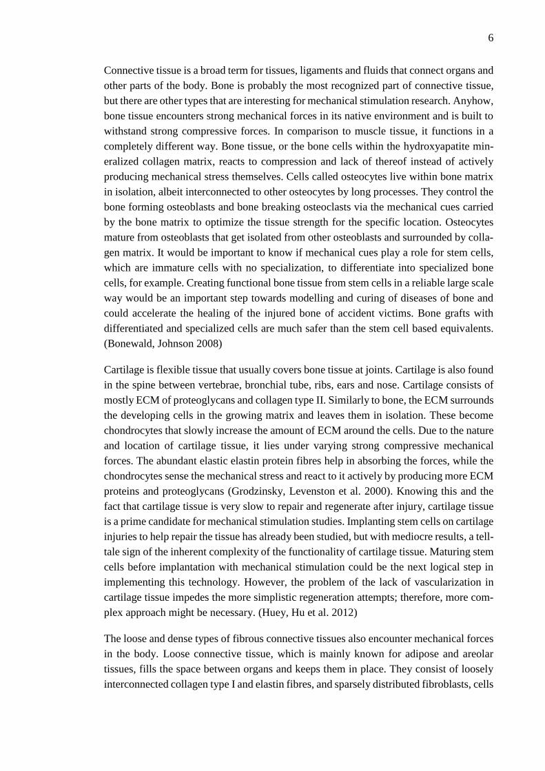
Connective tissue is a broad term for tissues, ligaments and fluids that connect organs and
other parts of the body. Bone is probably the most recognized part of connective tissue,
but there are other types that are interesting for mechanical stimulation research. Anyhow,
bone tissue encounters strong mechanical forces in its native environment and is built to
withstand strong compressive forces. In comparison to muscle tissue, it functions in a
completely different way. Bone tissue, or the bone cells within the hydroxyapatite min-
eralized collagen matrix, reacts to compression and lack of thereof instead of actively
producing mechanical stress themselves. Cells called osteocytes live within bone matrix
in isolation, albeit interconnected to other osteocytes by long processes. They control the
bone forming osteoblasts and bone breaking osteoclasts via the mechanical cues carried
by the bone matrix to optimize the tissue strength for the specific location. Osteocytes
mature from osteoblasts that get isolated from other osteoblasts and surrounded by colla-
gen matrix. It would be important to know if mechanical cues play a role for stem cells,
which are immature cells with no specialization, to differentiate into specialized bone
cells, for example. Creating functional bone tissue from stem cells in a reliable large scale
way would be an important step towards modelling and curing of diseases of bone and
could accelerate the healing of the injured bone of accident victims. Bone grafts with
differentiated and specialized cells are much safer than the stem cell based equivalents.
(Bonewald, Johnson 2008)
Cartilage is flexible tissue that usually covers bone tissue at joints. Cartilage is also found
in the spine between vertebrae, bronchial tube, ribs, ears and nose. Cartilage consists of
mostly ECM of proteoglycans and collagen type II. Similarly to bone, the ECM surrounds
the developing cells in the growing matrix and leaves them in isolation. These become
chondrocytes that slowly increase the amount of ECM around the cells. Due to the nature
and location of cartilage tissue, it lies under varying strong compressive mechanical
forces. The abundant elastic elastin protein fibres help in absorbing the forces, while the
chondrocytes sense the mechanical stress and react to it actively by producing more ECM
proteins and proteoglycans (Grodzinsky, Levenston et al. 2000). Knowing this and the
fact that cartilage tissue is very slow to repair and regenerate after injury, cartilage tissue
is a prime candidate for mechanical stimulation studies. Implanting stem cells on cartilage
injuries to help repair the tissue has already been studied, but with mediocre results, a tell-
tale sign of the inherent complexity of the functionality of cartilage tissue. Maturing stem
cells before implantation with mechanical stimulation could be the next logical step in
implementing this technology. However, the problem of the lack of vascularization in
cartilage tissue impedes the more simplistic regeneration attempts; therefore, more com-
plex approach might be necessary. (Huey, Hu et al. 2012)
The loose and dense types of fibrous connective tissues also encounter mechanical forces
in the body. Loose connective tissue, which is mainly known for adipose and areolar
tissues, fills the space between organs and keeps them in place. They consist of loosely
interconnected collagen type I and elastin fibres, and sparsely distributed fibroblasts, cells
7
that create the ECM proteins for expanding and repairing the tissue. Dense connective
tissue, such as tendons and ligaments, consist of densely packed ECM of collagen type I
fibres with varying amounts of proteoglycans and elastin. Both tendons and ligaments
also have a small amount of cells residing among the fibres. Most of these cells are spe-
cialized fibroblasts that make and repair the ECM, but there are also reports of isolating
stem cells from ligaments (Cheng, Liu et al. 2010). Tenocyte, which is a special type of
fibroblast distributed in tendons, has been shown to sense mechanical cues which make
them also a prime target for tissue engineering and mechanical stimulation research
(Schiele, Marturano et al. 2013). Tendons and ligaments both lack blood vessels, so their
repair rate after injury is very slow, similarly to cartilage. While their composition is ra-
ther simple and well defined, they are prone to injuries, because of their function and
position in locations known for strong physical forces. For this reason, artificial tendons
and ligaments as spare parts are constantly studied (Cheng, Liu et al. 2010, Scott, Dan-
ielson et al. 2011, Schiele, Marturano et al. 2013, Yang, Rothrauff et al. 2013), but much
work is still needed to be able to grow e.g. tendons in a laboratory. Stretching applications
could help in achieving information about the differentiation cues needed for tenocyte
culture or providing mechanical load for the ECM and the cells to get closer to the natural
mechanical integrity of the tendons, for example. Knowing also what type of stimulation
or stretch causes stem cells to take the differentiation path into fibroblasts, can help re-
searchers to avoid this type of stimulation as other cell types of mesenchymal origin such
as bone, cartilage, or cardiac cells are often preferred.
2.2 Extra cellular matrix
ECM is an integral part of most tissue types in multicellular organisms. It is a mesh,
network or a sheet of molecules that provide a support structure for the body and its tis-
sues and the main component of connective tissues. The composition of ECM and its
function changes depending on the location and the needs of the surrounding tissues.
ECM literally translates to a “matrix outside of the cell” and it is basically filler to the
space that is not occupied by cells. It is created and secreted by the cells that reside in it
as a supporting structure to provide mechanical durability and a substrate to the nimble
cells of the surrounding tissues. In general, ECM types can be divided between collagen-
dense fibrous matrices and sheets, and polysaccharide-dense negatively charged gels. The
blood plasma is also a type of ECM but it is not further discussed here. Depending on the
type, location, and function, the ECM can consist of fibrous proteins collagen and elastin,
special proteins and glycosaminoglycans or proteoglycans, which are huge complexes of
proteins and glycosaminoglycans.
Fibrous proteins dominate in connective tissues and their main component is collagen,
which is also the most abundant protein in the human body. Collagen fibrils and bundled
fibrils called collagen fibres provide most of the tensile strength of human tissues. They
are created, bundled together and interwoven into a strong matrix by fibroblasts in most

8
tissues, by osteoblasts in bone and by chondroblasts in cartilage. Tendons and ligaments
are basically aligned fibres of collagen attached to the bones and muscles. Elastin pro-
vides the elasticity required by some tissue types. It is responsible for returning the orig-
inal shape of skin, lungs and blood vessels, for example, after deformation. (Alberts, Bray
et al. 2010)
The empty spaces between cells and fibrous ECM is filled by a gel of proteoglycans,
glycosaminoglycans and proteins. While proteoglycans come in a myriad of different
conformations they are typically glycosaminoglycans linked to a core protein that is in
turn linked to another glycosaminoglycan chain. These complexes are huge space fillers
and can be as large as a bacterial cell. Their negative charge attracts cations, such as Na+
and K+, which are plentiful in the blood plasma and extracellular fluid. These cations are
osmotically active and they cause the formed gel to absorb water many times its own
weight. The swelling pressure caused by this is utilized by many tissues to withstand
pressure. When combined with collagen matrix, the ECM can withstand enormous pres-
sure from the inside and from the outside, as can be witnessed in the cartilage tissues of
joints. The proteoglycan ECM gel can vary in pore size and guide or block cell migration
and differentiation, regulate passage of signalling factors or bind growth factors, all in
addition to providing hydrated space around cells. These molecules, as with every ECM
related component, are created and secreted by cells residing in the immediate area. (Al-
berts, Bray et al. 2010)
Cells bind to the ECM via proteins called integrins. Integrins are small two part proteins
that attach to the cytoskeleton with one end and to the ECM with the other end that sticks
out of the plasma membrane. Signals from the cytoskeleton and vice versa can be trans-
mitted via this interaction between the cell and the ECM. Integrins can attach the cell
directly to the ECM networks or indirectly by binding secondary binding proteins such
as fibronectin. Fibronectin is an important protein that is able to bind collagen fibrils.
Cells attach to bare collagen poorly, which is why fibronectin is a necessary intermediary
between the cell and collagen. Cells cannot attach or crawl over collagen unless fibron-
ectin is present. (Alberts, Bray et al. 2010)
The cells have an undeniable relationship with all ECM types. For this reason, almost all
biomedical technologies utilizing cells must also utilize ECM components. Nowadays it
is understood that many of the critical reactions that cells feel and go through can be
manipulated by a proper use of ECM components. One piece of evidence about this was
the discovery of the so-called arginine-glysine-aspartic acid (RGD) tripeptide sequence.
This RGD-peptide is recognized as a binding site by many integrins. Synthetic RGD-
peptide treated surfaces or materials, for example, can be made recognizable to cells as
the integrins naturally bind to RGD sequence: cell attachment can be manipulated or mol-
ecules like drugs made bondable to cells without any complete ECM component (Ru-
oslahti 1996). It is, however, more common to use complete proteins to functionalize
materials for implantation or cell culture purposes. Proteins such as collagen type I and
9
type IV, laminin, and fibronectin are widely used, as is proteoglycan hyaluronic acid.
These ECM molecules are easy to extract from tissues and are readily available for sale.
They often behave in predictable manner in appropriate solutions, which makes their use
straightforward. Collagen type I, and the other types, can self-assemble into fibrils and
fibres after being extracted and dissolved in an acidic solution (Pins, Christiansen et al.
1997). In physiological conditions, the broken collagen molecules form natural fibre ma-
trix that cells can adhere and grow on. If there is enough collagen it can form even a
hydrogel or porous sponge which can be used as a 3D scaffold in cell culture and other
biomedical applications (Chevallay, Herbage 2000). Furthermore, when combined with
proteoglycans or hyaluronic acid the gel or sponge scaffold can become more rigid, hy-
drophilic, and resistant to dissolution as explained by Davidenko et. al. (Davidenko,
Campbell et al. 2010). Laminin, a key protein component of basal lamina of the basement
membrane, is also used to form cell adhesive networks on culture substrata, as is glyco-
protein fibronectin. They can both, along with hyaluronic acid, interact with collagens to
create even more complicated ECM networks. Finding the optimal combination of ECM
molecules and the resultant physical properties of 2D or 3D matrix for different applica-
tions is one of the biggest challenges of biomedical research and it lies at its spearhead.
Engineers need to find ways to combine the knowledge from cell biologists and biomed-
ical engineering to incorporate biomolecules from ECM into their applications.
2.3 Stem cells and differentiation
Stem cell is a special type of cell with two characteristic properties: They can make iden-
tical copies of themselves, and form other cell types via the process of differentiation.
They act as the mothers of all other cells and with the ability of self-renewal they can be
potentially immortal. Overall, stem cells hold most of the potential in cell based applica-
tions, thus they are the star players in the current biomedical research.
2.3.1 Stem cell basics
During the cell division of a stem cell the formed daughter cells have two options: They
can keep their stem cell phenotype and continue multiplying and act as a source of im-
mature cells, or they can take the path into differentiation and specialized cell types. The
first option is called self-renewal and it gives the stem cells their ability to be a potentially
unlimited source of cells. In adult tissues, stem cells control the self-renewal process via
chemical, electrical and mechanical cues to only happen when needed and to produce the
right type of progeny. Being in control of self-renewal process is crucial as any rogue
cells multiplying out of control can quickly form tumours (Erdö, Bührle et al. 2003). This
also counts implanted stem cells. Lack of control is still one of the issues regarding tech-
nologies dependent on implanted stem cells (Erdö, Bührle et al. 2003, Pittenger, Kerr
2015), and it critically limits their immediate clinical significance. Stem cells are gener-
ally divided into embryonic, adult, and induced stem cells depending on their source.
10
Embryonic stem cells (ESC) can be found in the developing embryo and they have the
widest differentiation potency of the cells that are relevant for research. Adult stem cells
(ASC) are found in adult tissues and they maintain and repair healthy tissue, and regen-
erate and heal damaged tissue. Induced stem cells are artificially created or induced from
harvested specialized cell types. (Pittenger, Kerr 2015)
Stem cell differentiation capability can be organized through the concept of potency. Stem
cells are totipotent, pluripotent, multipotent, oligopotent, or unipotent depending on the
number of tissue types they can create. Totipotent stem cells can create any cell for the
entire organism, including the embryo’s supporting tissues placenta and umbilical cord.
Only the first few cell divisions after the fertilization of the oocyte are totipotent, thus
they are rarely used for research purposes, even though they have the ultimate differenti-
ation capacity. Pluripotent stem cells can create any tissue of all three germ layers en-
doterm, mesoderm and ectoderm, but not placenta or umbilical cord. For research pur-
poses, pluripotent stem cells can be harvested from the inside of a blastocyst and brought
to culture as ESCs, and renewed indefinitely. While ESCs could be regarded as one of
the most valuable cell types for research due to the infinite differentiation potential, in
reality, they are quickly losing relevance in the studies of the day, due to the difficulties
in the acquisition of these cells. Furthermore, there was a significant underlying ethical
issue in harvesting ESCs from available embryos. Today, ESCs are created in vitro by
fertilizing donated eggs. While the process is not ethically as troublesome as harvesting
embryos in situ, the efficiency of the in vitro processes is quite low. Those points com-
bined have led to the favouring of other strategies in stem cell studies, such as induced
pluripotency, or harvesting multipotent stem cells from adult individuals. Multipotent
stem cells have already dedicated themselves into a specialized role. These cells have the
capacity to differentiate into multiple, but not all, cell types. They are sometimes called
progenitor or precursor cells. They still have the ability for self-renewal and keeping their
progenitor or stem cell state. Mesechymal stem cell (MSC) that can form cartilage, bone,
muscle and fat tissues, is an example of a multipotent stem cell. There are also oligopotent
stem cells that can create two or more cell types and unipotent stem cells which can create
cells from single lineage only. The neural progenitor cell that can create cells of the neural
system is an example of an oligopotent stem cell and the progenitor cell that creates the
male sperm cells is a unipotent stem cell. (Pittenger, Kerr 2015)
Differentiation from stem cell state to specialized functional tissues is not a simple one
step process. In fact, it often involves multiple sequences of cell division and sensitive
evaluation of internal and outer influences. During this process, the expression of some
genes deactivate, while some activate. The combined effect dictates what type of cell will
be the final result. A signal that causes differentiation can be a change in basal nutrients,
change in the cell’s environment, stimulation or lack of thereof, introduction of a signal-
ling molecule such as growth factor, a new cell-to-cell interaction, or loss of such, for
instance. In the body, the stem cells reside inside so called stem cell niches, where they

11
can keep their replicating phenotype indefinitely (Scadden 2006). The niche can be a
physical structure limited by ECM, or a habitat of stem cells flanked by other types of
cells (Scadden 2006). A stem cell can replicate either symmetrically where it produces
two identical daughter cells, or asymmetrically where it produces two nonidentical daugh-
ter cells. During self-renewal the cells, or at least one of them, get to keep their potency,
in other words they stay inside the niche of their parent cell. The niches are usually struc-
tured in such way that only certain number of cells fit in while others are forced out
(Scadden 2006). In the end, this forces asymmetry between the progeny of the parent cell
as the other begins differentiation due to the change in the environment. Sometimes the
asymmetry is the product of unequal distribution of cell organelles, or other cell fate de-
terminants that can also force the daughter cell out of the niche, and cause the initiation
of its differentiation. Stem cells may leave their niche to differentiate also without repli-
cation, a strategy commonly used for harvesting bone marrow stem cells (Broxmeyer,
Orschell et al. 2005). After leaving the stem cell niche, the cell enters into a series of
symmetric divisions that amplify the cell number, most notably in developing tissues or
in vitro (Morrison, Kimble 2006). This stage is called transit amplification stage and the
dividing cells transit-amplifying cells; these cells are progenitors that have abilities some-
where in between stem cells and fully differentiated cells and are often identified by their
potency and potential progeny (Figure 1). In vitro the transit amplification phase can
quickly lead to confluence and the need of passaging the cells into a subculture until the
final number of cell divisions is reached (Uzgare, Xu et al. 2004), and the cells terminally
differentiate into their final form. (Pittenger, Kerr 2015)
Figure 1. A depiction of the development of a differentiated cell population that de-
scends from a stem cell capable of self-renewal. Edited from (Pittenger,
Kerr 2015)

12
2.3.2 Adult stem cells
ASCs refer to cells that have capabilities for self-renewal and differentiation, but can be
found in fully developed adult tissues. These cells are used by the body for regeneration
and repair of aged and damaged tissues, and as a reservoir of new cells (Weissman 2000).
ASCs are not pluripotent, however, so their differentiation capacity is more restricted, as
they cannot produce every cell type. Rather, the body hosts a multitude of multipotent
ASCs which are generally limited to produce the cell types of their home tissue. From the
research standpoint, they can still offer advantages over pluripotent stem cells; harvesting
has less ethical concerns, because every patient is a source of immunocompatible autolo-
gous stem cells that can be expanded and differentiated ex vivo (Pittenger, Kerr 2015).
ASCs were first discovered in bone marrow in the 60s in the form of hematopoietic stem
cells (Till, McCulloch 1961), which develop into blood cells, and mesenchymal stem cells
(Friedenstein, Chailakhjan et al. 1970) that can develop into bone, cartilage and muscle
cells. After the first discoveries and the inevitable realization of the source of human’s
intrinsic tissue regeneration capabilities, stem cells have been found and isolated from
many adult tissues; epidermal stem cells from epidermis (Lavker, Sun 1983), neural stem
cells from brain (Carpenter, Cui et al. 1999), muscle stem cells from skeletal muscle
(Baroffio, Bochaton-Piallat et al. 1995), lung stem cells from lung (Kajstura, Rota et al.
2011), intestinal stem cells from small and large intestine (Potten, Loeffler 1990), olfac-
tory stem cells from nasal neuroepithelium (Roisen, Klueber et al. 2001), testicular stem
cells from testicles (Conrad, Renninger et al. 2008) and MSCs or MSC like cells from
many tissues such as skeletal muscle (Bosch, Musgrave et al. 2000), adipose tissue (Zuk,
Zhu et al. 2001), dental pulp (Gronthos, Mankani et al. 2000), skin dermis (Young, Steele
et al. 2001), bone periosteum (Nakahara, Goldberg et al. 1991), blood circulation (Chong,
Selvaratnam et al. 2012) and walls of the peripheral vascular system (Covas, Panepucci
et al. 2008).
ASCs are usually considered to be multipotent as they only produce cells of distinct lin-
eage. On some rare cases, ASCs have been reported to have pluripotency (Roisen, Klue-
ber et al. 2001, Jiang, Jahagirdar et al. 2002, Wagers, Weissman 2004, Guan, Nayernia et
al. 2006), but more research is needed before that can be taken as a well-founded fact.
However, there is evidence, where tissue based ASCs of distinct niche can produce wider
range of progeny than previously thought. Migration to different tissue types and trans-
differentiation is a real ability of these stem cells. MSCs have been shown to transdiffer-
entiate into neural progenitor cells and show marks of astroglial and neuronal phenotypes
(Ries, Egea 2012). Being readily available for harvest in adult bone marrow and adipose
tissues, MSCs might have potential for neural system regeneration among their potential
for connective tissue and muscle regeneration capabilities. Furthermore, neural stem cells
have shown capabilities for differentiation into hematopoietic lineage (Bjornson, Rietze
et al. 1999), while in turn according to some reports hematopoietic stem cells have shown

13
capabilities for differentiation into cardiomyocytes (Orlic, Kajstura et al. 2001) and neu-
rons (Mezey, Chandross et al. 2000). However, the factors behind transdifferentiation are
possibly even more complicated than those with regular differentiation. In another study,
the cardiac differentiation of hematopoietic stem cells was specifically studied but no
evidence of it could be observed (Murry, Soonpaa et al. 2004). Today, the factors behind
natural stem cell transdifferentiation are still largely shrouded in mystery, but big steps
have been taken to understand the apparent reprogramming of the cell phenotypes, and
how it can be induced at least in vitro, if not in vivo. All in all, ASCs have been taken as
an integral part of today’s regenerative medicine doctrine. The availability and lack of
ethical concerns in harvesting certain types of ASCs, such as adipose tissue derived
MSCs, give an indefinite source of material for researchers to study differentiation, and
to optimize methods for the regenerative cell therapies of the future.
2.3.3 Induced pluripotent stem cells
A quite recent advancement in stem cell technologies is the induced pluripotency of adult
cells that otherwise lack any abilities of a stem cell. The obvious advantage of pluripotent
stem cells over the less potent ASCs is the capability to produce any mature cell type.
The ethical concerns regarding the harvesting of ESCs from human embryos, and the
inefficiency of in vitro fertilized eggs, however, have largely inhibited the wider use of
these cells. The vacuum left behind by this mismatch of supply and demand provided the
impetus for finding alternative sources of pluripotent cells. Today, we know that there are
potentially pluripotent ASCs that can be harvested from nasal cavity (Roisen, Klueber et
al. 2001) or testes (Conrad, Renninger et al. 2008) without previously mentioned ethical
concerns. In addition to these the induced pluripotent stem cell (iPSC) is another promis-
ing alternative to ESCs without the ethical or some of the immunological issues the use
of ESCs usually has.
The genome of the ESCs and all of their progeny are the same in the same individual.
This means that there are intrinsic factors that cause stem cells to be able to differentiate
and also factors that cause mature cells to function in their special way. In their 2006
published study, Takahashi and Yamanaka (Takahashi, Yamanaka 2006) were able to
convert differentiated fibroblasts into pluripotent ESC-like cells by retroviral infection of
several transcription factors and oncogenes which were known to play role in the cells of
an early embryo. Since then, the methods for iPSC generation have been extensively doc-
umented, tested for human cells, and polished for efficiency. Still, the efficiency of con-
verting adult cells to iPSCs is 0.001-10 % at best and it is dependent on the cell source
where the less differentiated cells usually convert to iPSCs more efficiently (Yu, Vod-
yanik et al. 2007). To utilize iPSCs in regenerative medicine to their full potential, a num-
ber of obstacles have to be surpassed. The pluripotency is induced by viral vectors, thus
it is generally considered unethical to have them implanted to someone as they host for-
eign DNA. Another obstacle is being able to turn the pluripotency factors off to allow the
14
cells to differentiate and stop multiplying. Otherwise the iPSCs will most certainly form
a tumour. When the technology is ready, the possibility of forming pluripotent stem cells
in a laboratory environment at will, may induce even greater boom of stem cell research.
The possibilities and potential of these cells are close to limitless and in some futuristic
visions even full organs may be grown in a laboratory from just a handful of iPSCs with
patients own genome. (Pittenger, Kerr 2015)
2.3.4 Application of stem cell differentiation
Being able to control stem cell differentiation is imperative in their application in regen-
erative medicine. Therefore, the issue has evolved into one of the focal points of stem cell
research alongside the push for clinical trials (Trounson, Thakar et al. 2011). Nowadays,
there is a wide range of information available to guide the differentiation of pluripotent
or multipotent stem cells to a desirable direction and to verify the differentiation path the
cells have taken. To date, stem cell culture and differentiation serve as an essential model
to many human diseases and embryonic development, which holds the keys to the matu-
ration of every adult tissue type.
As stated earlier, pluripotent stem cells, most commonly ESCs, can create any type of cell
found in the adult body. While harvesting them has some ethical concerns, ESC is con-
sidered the most valuable type of stem cell, valued for its differentiation potency as well
as its capability for indefinite self-renewal. Since the first stable human ESC line that was
established by Thomson et al. (Thomson, Itskovitz-Eldor et al. 1998) the cells have been
differentiated in vitro to most adult cell lines, a feat that can only be described as daunting.
These include neurons (Reubinoff, Itsykson et al. 2001), cardiomyocytes (He, Ma et al.
2003), hepatocytes (Rambhatla, Chiu et al. 2003), pancreatic beta cells (Assady, Maor et
al. 2001), endothelial cells (Levenberg, Golub et al. 2002), blood cells (Kaufman, Hanson
et al. 2001), chondrocytes (Vats, Bielby et al. 2006) and osteocytes (Bielby, Boccaccini
et al. 2004). This proves the pluripotency of ESCs even in vitro, but the differentiated
cells achieved by these types of studies are still far away from the cells found in functional
tissues of an adult individual. Even though the maturing ESCs show markers and function
of a differentiated cell type, such as in the case of insulin producing pancreatic beta cells
in (Assady, Maor et al. 2001) or in (Zhang, Jiang et al. 2009). While the studies showed
a way of producing functional beta cells the production efficiency of the population was
only 1-3 % and 25 % respectively. Reported efficiency for differentiation markers can
vary much, especially between cell types (Vazin, Freed 2010). This means that only part
of the ESCs truly differentiate to adult cell types while most keep their stem cell pheno-
type. If the population would be implanted in this state, the ESC-like cells could promote
tumorigenesis. When combined with the immunoincompatibility of ESCs, the future pro-
spect of using ESCs as a source of implantable cells and tissues is currently bleak at best.
Lifelong exhortation to strong immunosuppressant use cannot be a requirement in these
types of therapies.

15
These are the main reasons why iPSCs and ASCs today have more potential for therapeu-
tic use in regenerative medicine. Then again, iPSCs still have the problem of non xeno-
free DNA, while ASCs cannot produce all cell types, thus some compromises and signif-
icant advancements in techniques are necessary. Either way, cultured iPSCs and ASCs
have been shown to express markers for many different mature cells in vitro. Many re-
search groups have invented their own protocols for stem cell induction and been able to
guide the resultant iPSCs to neural (Vierbuchen, Ostermeier et al. 2010), cardiac (Ieda,
Fu et al. 2010), blood cell (Szabo, Rampalli et al. 2010), hepatic (Huang, He et al. 2011),
and cartilaginous (Hiramatsu, Sasagawa et al. 2011) differentiation pathways, for exam-
ple. For ASCs the scale is larger mainly, because of their availability. Especially bone
marrow and adipose tissue derived MSCs have a long history of been utilized in cell cul-
ture studies for modelling diseases of bone, cartilage and muscle tissues (Ankrum, Karp
2010). One of the advantages of ASCs, or disadvantages, depending on the point of view,
is their natural affinity for certain differentiation pathways. This ability makes these cells
safer for implantation purposes than their pluripotent counterparts do. Hematopoietic
bone marrow transplants have been used as a cure to diseases of blood for some time. The
transplanted hematopoietic stem cell population ideally renews the patient’s production
of blood cells (Hatzimichael, Tuthill 2010). Similar idea is behind the stem cell transplan-
tation to bone (Yamada, Boo et al. 2003), cartilage (Uematsu, Hattori et al. 2005), cardiac
muscle (Stamm, Westphal et al. 2003) or neural tissues (Subramanian, Krishnan et al.
2009), for example. These cells are combined with synthetic scaffolds and other tissue
engineering concepts to form complete model therapies. A large amount of those are al-
ready in clinical trials of phase I and II meaning that they are potentially coming out fast
(Trounson, Thakar et al. 2011). On the other hand, in vitro models of stem cell differen-
tiation can be used in medicine development, and to understand disease mechanisms, but
these aspects are not generally as prominent from tissue engineering standpoint. These
applications hold most of the immediate commercial potential for stem cell differentiation
applications, so they should not be underestimated.
Nevertheless, as stated many times before, maturation of stem cells in vitro to more dif-
ferentiated state may be a necessary step to avoid the dangers of implanted stem cells and
to shift to the crucial final phases of the clinical trials. Much has been discovered in labs
worldwide already, but more basic research for the cues about the differentiation path-
ways is still needed.
2.4 Concept of cell stretching
When cells are attached to the ECM and other cells in tissues they can feel and react to
the mechanical stimuli caused by mechanical stresses. When a ligament is pulled, for
example, all the muscle cells in the muscle attached to the ligament can feel the stretch.
For this reason, we are able to go to the gym to practice our muscles which in turn start
to grow and develop (Gollnick, Armstrong et al. 1973). Nowadays it is known that this

16
happens when specialized muscle progenitor cells called myosatellite cells sense the in-
creased activity of the muscle, the mechanical stimulus, and begin cell division and dif-
ferentiation into new muscle fibres (Morgan, Partridge 2003). Similar principle also ap-
plies for bone tissue. Our skeleton has evolved to carry the weight of the body against the
gravity of the planet. Bone tissue is in normal conditions under constant compressive and
torsional stresses, which can be sensed by the osteocytes. As explained earlier in this
Chapter, they regulate and maintain the mineralization and the density of the bone tissue
(Bonewald, Johnson 2008) and the feedback the cells get from physical stimuli is a key
factor on keeping this activity on. Astronauts living in zero gravity, for example, tend to
lose bone mass and density during long missions when bone tissue starts to break down
due to low amount physical stimuli (Grigoriev, Oganov et al. 1998). This information can
be transferred to laboratory setting to subject the cells to controlled mechanical stress to
study the effects.
Cells are able to feel the external stimuli through their interaction with their immediate
surroundings. All animal cells apart from couple of exceptions require a substrate, a ma-
trix, or another cell to adhere. In tissues, cells are adhered either to the ECM or to each
other. Cell-cell junctions can be immobilizing anchors with or without space between
cells, channel forming junctions which relay chemical information from cytoplasm to cy-
toplasm, and signalling junctions such as the nerve cell synapses. Cell-cell junctions en-
able the formation of concentrated tissues and the flow of information from cell to cell.
However, when reacting to an external stimulation, natural or artificial, the cell-ECM
interaction holds the most importance. As cells attach to ECM, they form focal adhesion
points (Geiger, Spatz et al. 2009). Focal adhesion points are protein complexes outside of
the cell that connect actin filaments of the cell cytoskeleton directly to the ECM. They
are thus vital in relaying the mechanical stimuli of the surrounding ECM to the cell cyto-
skeleton inside the cell. This process is called mechanosensing (Luo, Mohan et al. 2013),
and the effect it produces in the cell via complex signalling pathways is called mecha-
notransduction (Wang, Butler et al. 1993).
Mechanosensing and –transduction cause the cellular reaction to the external stimuli,
hence their connection is under great interest and focus of many studies today. The un-
derlying mechanisms of the signalling pathways and their outcomes are currently mostly
unknown. There are, still, snippets of information available. In osteocytes, polycystin I is
essential for their anabolic response to load (Xiao, Dallas et al. 2011), and under shear
stress osteocytes are shown to release nitric oxide, adenosine triphosphate, and prosta-
glandin, a lipid that induces bone formation (Klein-Nulend, van der Plas et al. 1995).
Chondrocytes have been shown to react to hydrostatic pressure and compression by re-
leasing cartilage-specific ECM proteins. In turn, a sliding motion on the surface of a 3D
scaffold caused increased expression of protein lubricin, which acts as a joint lubricant
(Grad, Eglin et al. 2011). Cardiomyocytes in turn have been shown to react to static
17
stretch by actin filament production and alignment, and by producing branched and stri-
ated structures reminiscent of mature sarcomeres. In addition, the mechanical tension kept
the contractile proteins, which are vulnerable to enzymatic degradation, protected from
being degraded (Simpson, Majeski et al. 1999). It is obvious that cells do actively react
to external mechanical stimulation, but how that actually happens remains mostly un-
proven.
In cell culture setting, mechanotransduction is induced by artificial ECM, ECM proteins
or ECM peptides. Nowadays, it is a common practice to prepare the substrate with adhe-
sion proteins in many cell culture applications as it improves cell adhesion and culture
viability in many ways. The rule of thumb in general is that the better the cells adhere to
the substrate, the better they thrive (Wipff, Majd et al. 2009, Kuddannaya, Chuah et al.
2013). If a growing cell disconnects with the substrate it will in most cases result in apop-
tosis. Mechanical stimulation of cells during culture is notoriously hard to the cells, and
it can be a major inconvenience for engineers who want to evaluate their devices and
applications. For example, stretching the substrate can rip any poor or incomplete focal
adhesion points (Wipff, Majd et al. 2009) and leave the cell without support. Slow speeds
or maximized cell adhesive quality of the substrate minimize the shock caused by stretch-
ing.
2.5 Studies and Devices
Nowadays, the cell stretching research utilizes a myriad of applications and devices to
apply and measure the stimulation. Because most cell types require some form of a sub-
strate to grow, it must be incorporated into the device design. Whether the cell culture or
the substrate is in 2D or 3D, the stretching is applied to the substrate first and then to the
cells via focal adhesion points. This means that substrate material requirements for these
types of applications have strict limitations. Regardless, researchers have created many
imaginative ways to combine stretching systems and cell culture together. Next, various
strategies in recent dynamic cell studies are briefly presented. The devices are categorized
based on the application principle of the stretch: manual, fluidic, electric motor, or mag-
netic field controlled CSDs.
Many groups in search of new ways to study cells have noted the good properties of
PDMS for cell stretching applications. It is thus of no coincidence that so many cell
stretching methods and dynamic culture devices utilize this material. Thin membranes
created from PDMS have proven as a great elastic substrate with tuneable properties.
Manually applied stretch is likely the simplest method for these devices. In those, the
stretch is controlled by the researcher manually by hand and is usually kept static after
the initiation. Wipff et al. studied various cell adhesive coatings for PDMS cell stretching
applications. To test the functionality of the coatings, their effectiveness under mechani-
cal strain, they employed a simple method to apply stretch on the thin PDMS membrane
that housed fibroblasts. They used a well on top of the membrane to hold the culture

18
medium. When the well, that contained cells, medium, and the membrane, was pressed
against a ring, which was smaller in diameter that the well, the membrane was stretched.
Wipff et al. had mounted the ring on a microscope to see the cells and the stretching
process in real time. They also had embedded opaque tracking particles inside the mem-
brane and fluorescent beads on top of the coating to study the transmission of the stretch
to the coating and the cells (Wipff, Majd et al. 2009). In similar fashion, Goffin et al. had
studied the composition of fibroblast focal adhesions under stretch on PDMS membrane
with various micropatterns (Goffin, Pittet et al. 2006). Another PDMS membrane based
manual stretching device, introduced by Lee et al. in 1996 (Lee, Delhaas et al. 1996), was
used more recently by Braakman et al. to study Schlemm’s cells from the eye. The device
is able to produce an equiaxial stretch on a PDMS cell culture membrane. The function
of the device is as follows: An outer cylinder is attached to a threaded inner cylinder,
which in turn is attached to the membrane. The inner walls of the culture well are fixed
to press against the membrane. Therefore, when the outer cylinder is rotated, the threads
of the inner cylinder enable the downward movement of the outer cylinder, which presses
against the fixed walls and the membrane, which is equiaxially stretched up to 20%
(Braakman, Pedrigi et al. 2014).
Since the times of the study by Lee et al. many systems with different working principles
have risen. CSDs controlled by fluidic mechanisms are quite popular and allow for more
control over the stretching parameter than manual devices. Air is used to create vacuum
pressures while liquids are usually used to create increased pressure upon a flexible mem-
brane. For example, Zhao et al. introduced a convenient PDMS membrane based plat-
form for dynamic cell culture. It includes a microfabricated PDMS channel system that
is sandwiched by PDMS bulk under it, and a thin membrane on top. When the channel
system is filled with air or a liquid, and the pressure in the system is increased, the mem-
brane will deform outwards stretching equiaxially any cells that are grown on top of it.
The device implements a very simple and effective mechanism to induce stretching, but
the significant vertical movement of the membrane makes it very difficult to monitor the
stretching process in real time with a microscope (Zhao, Zhou et al. 2011). This problem
was greatly reduced in the similar system published by Kreutzer et al., which is also used
in the experimental part of this thesis work. Error! Reference source not found.A de-
picts this device and its working principle. The pneumatic PDMS based CSD includes a
circular vacuum chamber around the cell culture well, closed by a glass plate on top and
a thin membrane on the bottom. The cells grown on top of the membrane are equiaxially
stretched when a vacuum is applied in the chamber as the walls of the inner chamber
bend, thus deforming the membrane and chamber causing stretch in the middle in all
directions. The vacuum system that can hold dozens of devices at the same time can be
attached to a computer that controls the amplitude and frequency of the vacuum pressure
enabling long-term cyclic stretching experiments. While some vertical adjustment is re-
quired, the well can still be monitored during stretching in real time with a microscope
(Kreutzer, Ikonen et al. 2013). A similar pneumatic working principle was utilized by
19
Huh et al. in their so called lung-on-a-chip device, but its design, as depicted in Error!
Reference source not found.B, is quite different. It constitutes of two microfabricated
channel systems, instead of just one as in the device by Zhao et al., separated by only a
thin porous PDMS membrane in the middle. On top and below the membrane stands the
cell culture chambers flanked by two vacuum chambers. When a negative pressure is
applied to the vacuum chambers, it allows the uniaxial stretch of the two cell cultures on
the both sides of the membrane. In addition, the pores allow signalling between the two
cultures. A microfluidic system on the both sides of the membrane induces shear stress,
while providing constant flow of fresh medium to the cells. Huh et al. used this innovative
co-culture CSD to culture and study epithelial and endothelial cells found in lung alveoli
(Huh, Matthews et al. 2010). Very recently, Ugolini et al. had the same idea behind the
working principle of their CSD. However, they increased the number of chambers on
one chip to four allowing easier control of parallel cultures. A microfluidic system for
small molecule exchange, similarly to the device by Huh et al., runs under the four cell
culture chambers and the membranes. When a negative pressure is applied to the vacuum
chambers, the membrane deforms uniaxially. Real time cell monitoring capability and the
full cyclic control of the negative pressure completes the system (Ugolini, Rasponi et al.
2016). There are also companies focused on these types of CSDs. Flexcell International
Corporation distributes several different stretching systems and related accessories; there-
fore, their products are quite widely referenced in the field. Also for their systems, the
operational basis is a vacuum based stretching of a silicone membrane. The membrane
sits on top of a fixed base flanked by vacuum chamber. The vacuum chamber can be
circular for equiaxial stretching or rectangular for uniaxial stretching. When the mem-
brane is drawn in the chamber via negative pressure, the middle section is stretched. How-
ever, the system needs lubrication under the membrane for it to function properly. A mod-
ification of this basic system, called TissueTrain®, can be used as a platform for dynamic
long term 3D cell culture (Yang, Rothrauff et al. 2013) or as a set up for dynamic loading
for bioartificial tendons (Scott, Danielson et al. 2011), for example.
Motored mechanical CSDs are also popular for their controllability. They can still differ
much in design from one another, but in general, the working principle is the same: a
motor induces physical movement that is transferred to a flexible cell culture substrate
either straight or via lever based transmission. The simplest example, the pulling clamp,
is a popular operational mechanism for these types of CSDs. In these, the flexible sub-
strate, which is often PDMS, is physically clamped to a lever leading to rotating or linear
motor that pulls on the substrate, creating uniaxial stretch. Ahmed et al. used this type of
device to study a hydrogel coated, micropatterned and myoblast seeded PDMS well under
cyclic stretch (Ahmed, Wolfram et al. 2010). Greiner et al. did the same, except with
dermal fibroblasts (Greiner, Hoffmann et al. 2014). Devices with this mechanism fit well
for researchers undergoing a prototyping phase, because the design can hold any devices,
wells or substrates that fit to the clamp, which can likewise be modified. Figueroa et al.
used a very similar setup to study endothelial cell responses (Figueroa, Kemeny et al.

20
2011), as did Leong et al. when culturing MSCs under cyclic stretch, but on polycapro-
lactone substrate instead of silicone (Leong, Wu et al. 2012). Alongside their other study,
Ahmed et al. introduced a different pulling clamp device that used linear actuator for dis-
placement. The stretch with the device can reach up to 45 % while allowing real time
observation with a microscope (Ahmed, Kural et al. 2010). Li et al. proposed a different
approach by experimenting with a new stretchable, biocompatible and striated fugitive
glue based substrate. The material can withstand at least 700 % stretch and according to
Li et al., the material could be used in many applications in place of the more commonly
used silicones (Li, Lucioni et al. 2015).
Figure 2. The pneumatic CSDs and their working principles as described by (A)
Kreutzer et al. and (B) Huh et al. (Huh, Matthews et al. 2010, Kreutzer, Iko-
nen et al. 2013).
A more complex device than pulling clamps, the IsoStretcher is a recent addition to the
ever-growing number of complete cell stretching systems. Published by Schürmann et
al., the IsoStretcher is a complicated mechanical device for expanding the PDMS based
cell culture membrane. Figure 3A shows the location of the membrane in relation to the

21
other parts of the device. The device is driven via a stepper motor that rotates the lowest
ring that houses six pins used to transfer the displacement to the membrane. The pins
follow a tangential trajectory as the lowest ring rotates pulling the membrane with holes
for the pins outwards, creating a constant equiaxial stretch to the membrane. According
to Schürmann et al., the stretch capability of the device goes up to 20 percent (Schurmann,
Wagner et al. 2016). The cellerator, as the device is named by some sources, by Cytomec
GmbH has been the device of choice by several studies of late. This iris-like motored
mechanical PDMS membrane based CSD has been used to study MSCs (Majd, Quinn et
al. 2011), chondrocytes (Rosenzweig, Matmati et al. 2012, Rosenzweig, Chicatun et al.
2013) and myofibroblasts (Klingberg, Chow et al. 2014). In this device, the membrane
that holds the cells is attached to the walls of the culture well, which include holes for the
‘arms’ of the iris-like mechanism. As the arms, which are attached to the outer edge of
the device, are displaced outwards by an external motor, the membrane is equiaxially
stretched. This displacement mechanism is illustrated in Figure 3B. Rosenzweig et al.
report a staggering 600 % increase in surface area after the chondrocyte culture had been
expanded continuously for 13 days (Rosenzweig, Matmati et al. 2012). The maximum
stretch capacity for the device is reportedly 800 % which is by far the highest of the
devices presented here (Rosenzweig, Chicatun et al. 2013).
Figure 3. (A) Deconstruction of the IsoStretcher CSD with highlighted PDMS based
cell culture area (Schurmann, Wagner et al. 2016). (B) Illustration of the
iris-like mechanism of the cellerator CSD (Rosenzweig, Matmati et al.
2012).
By utilizing electromagnetic principles, Mayer et al. had a very different approach in their
device, when compared to the others introduced in this Section. They embedded carbonyl
iron particles into 2 mm thick ultra-soft PDMS bulk. The premise was to be able to deform
the created magnetoactive elastomer, as they call it, with external magnetic field. In their
study, the magnetoactive PDMS piece sits inside a petri dish on top of either permanent
magnet or controlled electromagnet based yoke. The former produces static magnetic

22
field and strain, while the latter system can create changing magnetic flux, thus changing
strain on the magnetoactive substrate. Mayer et al. also introduced a 24-well based
stretching platform for simultaneous stretching on half of the wells (Mayer, Rabindranath
et al. 2013).
The list could go on almost indefinitely, which only shows how imaginative researchers
and engineers are on this field. However, to reach the next milestone in tissue engineering
and dynamic cell culture applications, more complex tissue types and stem cells need to
be analysed in different dynamic environments to get the crucial information about the
cues that lead to healthy tissues in vitro. Now, the research is usually focused on the
functionality of the devices, rather than the cell culture. The fact is that the field is still a
Wild West of different protocols and devices where it is often difficult to project the re-
sults of external studies to one’s own. Standardization and collaboration in every phase,
especially in the cell culture phase, is gravely needed or the process to human spare parts
will continue being needlessly cumbersome.

23
3. POLYDIMETHYLSILOXANE SURFACE MODIFI-
CATION
PDMS must be one of the most widely used organosilicon materials to date. It sees use
today as a lubricant, sealant, food and shampoo additive, toy, resistive coating, contact
lenses, and various medical devices to name some. PDMS elastomers are especially use-
ful in medical grade device research and development. PDMS in its commercially avail-
able elastomer composite form is inert to most chemical reactions, easy to handle and
shape, is not a source of cytotoxicity, and when needed can be activated for a wide range
of chemical reactions.
This Chapter focuses on the material and chemical characteristics of PDMS. First, rele-
vant chemical and physical characteristics of PDMS are explained thoroughly. Then,
basic surface treatment methods or techniques are presented based on the nature of the
treatment that can be either plasma, physical, or chemical. Finally, some of the most rel-
evant advanced surface treatment methods used in studies in biomedical field are explored
and described.
3.1 Chemical characteristics of PDMS
As its name suggests, PDMS is an organosilicon compound with a siloxane backbone. It
is a specific polysiloxane with two methyl groups attached to each silicon in the siloxane
Si-O-Si backbone (Figure 4). The polar siloxane oxygen atoms in the backbone and the
non-polar methyl groups make up most of the chemical nature of PDMS. The most nota-
ble characteristic of PDMS is probably its high hydrophobicity, which results directly
from the methyls flanking the siloxane backbone. The flexibility of the polymer chain
also permits the arrangement of the methyls on the surface of the polymer bulk regardless
of it being in elastomer or liquid form. The chain flexibility arises from the large bond
angle of the siloxane, and it enables the polymer in liquid form to spread out on surfaces
and copy its features up to nanoscale. Therefore, elastomeric PDMS is widely used in soft
lithography to produce moulds or stamps with very high resolution (Rolland, Hagberg et
al. 2004). The glass transitional temperature of PDMS reflects this mobility and is gener-
ally under -120 °C, while the intrinsic hydrophobicity gives PDMS one of the lowest
surface energies known. It can be simultaneously a curse and a blessing for its usability
in the medical field. Table 1 lists some of the most referenced characteristics of PDMS.
(Poojari 2014)

24
Figure 4. PDMS polymer chain subunit: Si-O-Si = siloxane, CH3 = methyl.
Table 1. Qualitative PDMS characteristics edited from (Rodrigues, Dourado 2014).
Biological Physicochemical
Non-toxic / inertness Thermal stability
Moderate biocompatibility Resistance to UV, oxygen, ozone, sunlight
Protein adsorption / absorption Resistance to aging and biodegradation
Hemocompatibility Sterilisable and low curing temperature
Poor microbial adhesion Excellent dielectric behaviour
Low density, high molecular weight
Chemical stability, resistance to hydrocarbons,
oils and solvents
Flexibility / elasticity, easy to mould and shape
High gas permeability
Good optical transparency
Hydrophobic character
3.2 Elastomeric PDMS composite Sylgard® 184
As suggested, PDMS is found in many conformations, but its elastomer composite is the
most important one from the point of view of biomedical technologies. Probably the most
widely referenced brands of PDMS composites in this field are distributed by Dow Corn-
ing. Because linear PDMS polymer chains are so flexible that they are naturally in liquid
form, they have to be crosslinked to solidify them. The amount of crosslinking reactions
ultimately decides the hardness or elasticity of the PDMS composite in solid-phase. Con-
trolling this reaction is easy with well-known and well-established PDMS elastomer kits,
which are widely available for purchase, and have standardized crosslinkable side groups
added to the basic backbone of PDMS. The side groups are generally activated through
thermally active catalyst, which crosslinks the polymer chains permanently through co-
valent bonds.
As mentioned above, Sylgard® 184 silicone elastomer from Dow Corning is one of the
most referenced PDMS composites in scientific studies, and for clarity, in this thesis
PDMS refers specifically to Sylgard® 184 composite from now on. It is a two part (base
and curing agent) heat curable polymer with a platinum-based catalyst and ethylbenzene
as a solvent (Dow Corning 2015, Dow Corning 2016). The base polymer solution consists

25
of long ~60 subunit PDMS chains terminated by vinyl (=CH2) groups while the curing
agent polymer solution contains, in addition to the long chains, shorter 9-13 subunit pol-
ymer chains which have at least three of the methyl side groups replaced by hydride (-H)
groups (Lisensky, Campbell et al. 1999). These hydride groups act as the points for the
crosslinking reaction between the long and short polymer chains. The platinum catalyst
causes an addition reaction of the hydride to the carbon-carbon double bond of the termi-
nal vinyl group (Speier, Webster et al. 1957) that is accelerated by temperature (Figure
5). The result is an irreversible network of long flexible PDMS chains that begin and end
to the sides of the shorter chains. These crosslinking points allow the elastic recovery of
the material after strain. When a strain is applied to the material, the low energy backbone
chains slide past each other allowing deformation. When the strain is in turn released, the
crosslinking points force the material to its initial shape. The Young’s modulus i.e. the
stiffness of PDMS composites can be controlled by varying the ratio of curing agent pol-
ymer solution to the base polymer solution. As the crosslinking increases in the bulk of
the polymer, so does the stiffness. Nonetheless, PDMS retains most of its flexibility
caused by the siloxane backbone, and the freely rotating methyls, even after curing.
Therefore, the surface is generally in constant motion when the longer chains twist and
allow the shorter uncrosslinked chains to seep through to the surface.
It is a well-known fact that PDMS exhibits hydrophobic recovery after modification of
the surface with hydrophilic groups (Bodas, Khan-Malek 2007). It has also been shown
that removing those short free chains from PDMS can inhibit this recovery (Eddington,
Puccinelli et al. 2006). Storing hydrophilized PDMS in water also inhibits the recovery,
as does storing it in liquid nitrogen (Everaert, Van et al. 1996). This behaviour can easily
be explained by understanding what affects the chain mobility in the PDMS bulk. The
water creates a polar environment at the PDMS-water interface, which anchors the hy-
drophilic group containing chains to the surface due to hydrogen bonding, thus inhibiting
movement of hydrophobic chains from the bulk to the surface. For a similar reason storing
hydrophilized PDMS in air creates a non-polar PDMS-air interface, which attracts chains
containing hydrophobic methyl side groups , thus leading to hydrophobic recovery. Stor-
ing the modified PDMS in liquid nitrogen, on the other hand, lowers the temperature so
low that the chain mobility, and the hydrophobic recovery, is reduced to a minimum
(Everaert, Van et al. 1996). Additionally, it could be hypothesized that even a hydrophilic
recovery is possible after hydrophobic recovery, if the PDMS is stored in polar solvent
such as water.
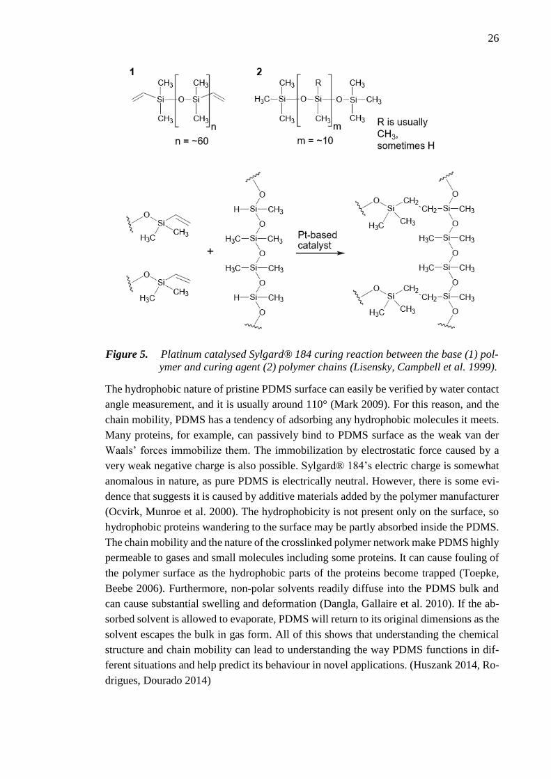
26
Figure 5. Platinum catalysed Sylgard® 184 curing reaction between the base (1) pol-
ymer and curing agent (2) polymer chains (Lisensky, Campbell et al. 1999).
The hydrophobic nature of pristine PDMS surface can easily be verified by water contact
angle measurement, and it is usually around 110° (Mark 2009). For this reason, and the
chain mobility, PDMS has a tendency of adsorbing any hydrophobic molecules it meets.
Many proteins, for example, can passively bind to PDMS surface as the weak van der
Waals’ forces immobilize them. The immobilization by electrostatic force caused by a
very weak negative charge is also possible. Sylgard® 184’s electric charge is somewhat
anomalous in nature, as pure PDMS is electrically neutral. However, there is some evi-
dence that suggests it is caused by additive materials added by the polymer manufacturer
(Ocvirk, Munroe et al. 2000). The hydrophobicity is not present only on the surface, so
hydrophobic proteins wandering to the surface may be partly absorbed inside the PDMS.
The chain mobility and the nature of the crosslinked polymer network make PDMS highly
permeable to gases and small molecules including some proteins. It can cause fouling of
the polymer surface as the hydrophobic parts of the proteins become trapped (Toepke,
Beebe 2006). Furthermore, non-polar solvents readily diffuse into the PDMS bulk and
can cause substantial swelling and deformation (Dangla, Gallaire et al. 2010). If the ab-
sorbed solvent is allowed to evaporate, PDMS will return to its original dimensions as the
solvent escapes the bulk in gas form. All of this shows that understanding the chemical
structure and chain mobility can lead to understanding the way PDMS functions in dif-
ferent situations and help predict its behaviour in novel applications. (Huszank 2014, Ro-
drigues, Dourado 2014)

27
3.3 Surface treatment of PDMS
As mentioned many times already in this thesis and elsewhere, PDMS elastomer’s surface
is unsuitable for cell culture in its pristine state. Its hydrophobicity is the main culprit as
cells have been shown to favour more hydrophilic substrates, or to be exact, substrates
with higher surface energy density (Zhao, Raines et al. 2007). Most surface treatments of
PDMS aim to increase the surface energy density to make the substrate more accessible
to water molecules and chemical reactions. In short, pristine PDMS surface does not offer
any attachment opportunities for the cells, which usually leads to quick cell death and
culture failure. While pristine PDMS is prone to adsorbing proteins, as stated previously,
it is not efficient enough to create sufficient substrate for all cell culture needs. To culture
cells on PDMS, these problems must be circumvented.
When the cell culture is dynamic, the requirements for a good substrate grow exponen-
tially. Additionally, these dynamic systems are usually designed for studying human stem
cells which tend to be more sensitive to the culture conditions, including the substrate,
than animal or immortal cancer cell lines (Rosler, Fisk et al. 2004). Luckily, there are
many possibilities to approach this issue. As the knowledge and technology has gone
forward, more methods for surface treatment have also been created and studied. Still, a
major part of surface treatment methods used for cell culture studies have relied on the
few well established and simple methods of plasma oxidation or physisorption (Wipff,
Majd et al. 2009, Kreutzer, Ikonen et al. 2013). On the other hand, optimizing novel coat-
ing methods is usually so laborious and complicated that cell culture researchers and tis-
sue engineers would rather focus on taking an existing method without the need to focus
on the chemistry of the treatments and optimization steps. Regardless, there are many
methods that can vary greatly in their utilization. In general, they can be classified into
physical and chemical methods depending on what type of interaction dominates during
the surface treatment. The division between the methods can be seen in Figure 6. This
classification is not fixed as some treatments could be labelled as both, or a mix of both,
as it will be explained in the following Sections. (Qiu, Wu et al. 2014)
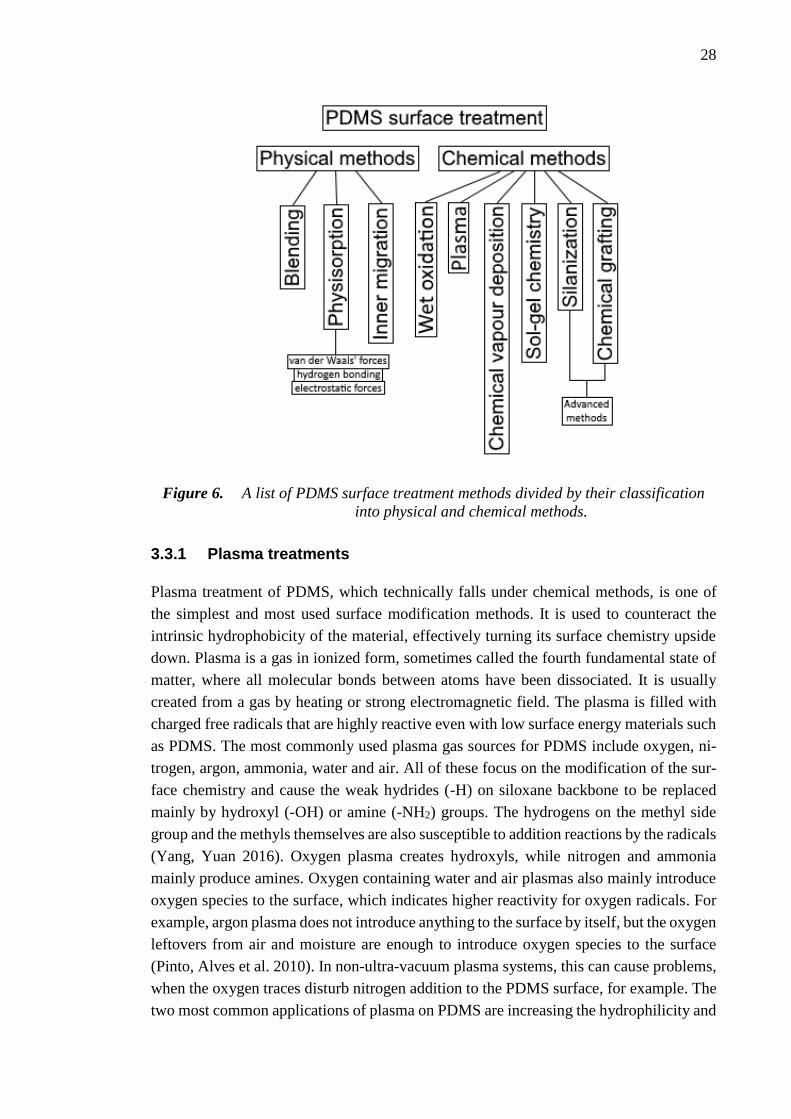
28
Figure 6. A list of PDMS surface treatment methods divided by their classification
into physical and chemical methods.
3.3.1 Plasma treatments
Plasma treatment of PDMS, which technically falls under chemical methods, is one of
the simplest and most used surface modification methods. It is used to counteract the
intrinsic hydrophobicity of the material, effectively turning its surface chemistry upside
down. Plasma is a gas in ionized form, sometimes called the fourth fundamental state of
matter, where all molecular bonds between atoms have been dissociated. It is usually
created from a gas by heating or strong electromagnetic field. The plasma is filled with
charged free radicals that are highly reactive even with low surface energy materials such
as PDMS. The most commonly used plasma gas sources for PDMS include oxygen, ni-
trogen, argon, ammonia, water and air. All of these focus on the modification of the sur-
face chemistry and cause the weak hydrides (-H) on siloxane backbone to be replaced
mainly by hydroxyl (-OH) or amine (-NH2) groups. The hydrogens on the methyl side
group and the methyls themselves are also susceptible to addition reactions by the radicals
(Yang, Yuan 2016). Oxygen plasma creates hydroxyls, while nitrogen and ammonia
mainly produce amines. Oxygen containing water and air plasmas also mainly introduce
oxygen species to the surface, which indicates higher reactivity for oxygen radicals. For
example, argon plasma does not introduce anything to the surface by itself, but the oxygen
leftovers from air and moisture are enough to introduce oxygen species to the surface
(Pinto, Alves et al. 2010). In non-ultra-vacuum plasma systems, this can cause problems,
when the oxygen traces disturb nitrogen addition to the PDMS surface, for example. The
two most common applications of plasma on PDMS are increasing the hydrophilicity and

29
adhesion. PDMS can be covalently bonded with itself or glass after oxygen plasma treat-
ment due to silanol-silanol (Si-OH) reaction, which produces a siloxane (Si-O-Si) bond
and releases water. Similarly, nitrogen plasma treated surface with amines is capable of
binding epoxy groups (Yang, Yuan 2016). Epoxy groups can be found on the surface of
SU-8, for example, which is a photoresist material commonly used in fabrication of mi-
crofluidic devices. While they cannot be classified as plasma, ozone treatment and ultra-
violet radiation have also been used for similar effect as oxygen plasma. They tend to be
much slower, but can reach deeper into the PDMS bulk. (Qiu, Wu et al. 2014)
3.3.2 Physical methods
The main characteristic for physical surface treatment methods is the type of interaction
between PDMS and the molecule used for the treatment. It is physical in nature, meaning
no chemical reaction happens between the molecule and PDMS. The most relevant phys-
ical methods can be further classified into blending, inner migration and physisorption.
Blending is one of the least used methods for PDMS surface treatment. Nevertheless,
when PDMS is in liquid phase, other polymers and materials can be blended in the bulk.
The molecules are spread out and immobilized inside the bulk after curing, while having
an effect also on the surface properties (Xiao, Yu et al. 2007). After blending, if the
blended molecules are small enough and have certain properties, it is possible to exploit
the relatively mobile bulk of PDMS to passively direct the blended molecules to the sur-
face. This method is called inner migration. Amphiphilic molecules, such as many of the
polymers in the pluronic family, can be belended with PDMS, and when it is placed in
water, the amphiphilic molecules begin to diffuse via gradient-induced migration. The
hydrophilic parts of the amphiphilic molecules migrate to the surface and orient them-
selves towards the water, which leads to hydrophilization of the PDMS surface that can
last for months (Wu, Hjort 2009).
Physisorption, or physical adsorption, utilizes the physical interaction between the exter-
nal molecules and the PDMS molecules on the surface. The physical interaction can be
van der Waals’ force, hydrogen bonding or electrostatic force depending on the paired
molecule or the state of the PDMS surface. By using a melt or a solution, the molecules
are brought into physical contact with the PDMS surface. If the PDMS is pristine, hydro-
phobic interactions dominate, and molecules can be adsorbed to the surface from their
hydrophobic functional groups. Amphiphilic molecules such as surfactants and large or
hydrophobic proteins can have a strong interaction with the PDMS surface. If the inter-
action is stronger than with the solution, they will stay on the surface after the solvent has
been removed. If the PDMS has been treated with oxygen plasma to have polar hydroxyls
on the surface, hydrophilic interactions, in other words hydrogen bonding, will dominate.
Molecules with functional groups capable of hydrogen bonding can then bind to the
PDMS surface. However, because water will also bind to these points, the binding affinity
of the molecules could be lower in aqueous solutions.

30
In electrostatic interaction, the negatively and positively charged functional groups in
molecules attract each other and potentially cause adsorption. Pristine PDMS of some
manufacturers, such as Dow Corning, is reportedly slightly negatively charged (Ocvirk,
Munroe et al. 2000) and capable of binding molecules with positively charged functional
groups. This can be useful for some big proteins which can have several different func-
tional groups, some with positive charge and some with hydrophobic inert groups. Treat-
ing the PDMS with plasma that introduces pH sensitive groups (acidic -OH, basic –NH2),
for example, can be useful to introduce either positive or negative charge to the surface
in a controlled way. Acidic groups have a leaving H+ ion that give a negatively charged
surface, while basic groups accept the H+ ion giving a positively charged surface. Elec-
trostatic interaction is also exploited in the more advanced layer-by-layer (LBL) method
that is revisited later in this Chapter. Physical methods described here have shown prom-
ise by being easy to implement and cheap, but in cell culture applications and most nota-
bly dynamic cell culture applications they tend to fall short by not being powerful enough.
The more advanced methods that focus on chemical bonding instead of physical interac-
tions tend to be more popular in those applications. (Qiu, Wu et al. 2014)
3.3.3 Chemical methods
In chemical surface treatment methods, a chemical reaction happens between the PDMS
surface and a molecule that is wanted on the surface. This causes changes in the covalent
bonds by either introducing new bonds and/or breaking old bonds. Here, the most relevant
chemical methods are divided into wet oxidation, chemical vapour deposition (CVD),
sol-gel chemistry, silanization and chemical grafting.
Wet oxidation is a special treatment method that is commonly used for the hydrophiliza-
tion of PDMS surface in place of a plasma treatment. By washing the PDMS surface with
the extremely reactive piranha solution, which is composed of sulphuric acid and hydro-
gen peroxide, the surface methyl groups are replaced by hydroxyl groups. The result is a
nearly complete hydrophilization of the surface.
CVD is a chemical process that is used to produce thin films or coatings from volatile
compounds. The substrate is exposed to the gas or vapour, which then reacts with the
surface potentially causing a change in the chemistry on the surface. One of the main
advantages of CVD treatment is that it is solvent free and can be completely dry. Many
of the most relevant CVD treatments for PDMS rely on the vaporization of monomers
that spontaneously polymerize on the PDMS creating a surface that can be tailored for
the needs of the application (Chen, McClelland et al. 2008). While usually being chemical
method, CVD polymer film does not necessarily bind covalently to pristine PDMS, but
rather by physical interaction. For covalent CVD, it may be necessary to pre-treat PDMS
with plasma, or other activating treatment (Bhushan, Hansford et al. 2006).

31
Sol-gel chemistry, or method, is a wet process that creates solid material from the mono-
mers dissolved in an aqueous solution: Sol stands for colloidal solution, and gel the
formed solid network. Similarly to CVD, the composition of the final coating can be tai-
lored to the needs of the application via the used monomers. Both polymer plastic (Su-
zuki, Yamada et al. 2010) or metal oxide (Roman, Culbertson 2006) coatings can be cre-
ated using this method. The advantage of this method is that it creates a homogenously
distributed coating. The density of the coating can be controlled, as the remaining liquid
is evaporated (Roman, Culbertson 2006). Biomolecules such as enzymes, proteins, or
whole cells can also be incorporated into the coating during its formation to create fully
biocompatible or bioactive coatings (Böttcher 2000).
The next method, silanization, is a widely used coating method for PDMS that utilizes a
class of molecules called silanes. Alkoxysilanes contain one to three alkoxy functional
groups (usually methoxy or ethoxy) around a silicon atom, while halosilanes contain one
to three halides (usually chloride, fluoride or mix of both). Additionally, at least one an-
other functional group is situated at the other end of the molecule; usually at the end of a
short carbon chain. Hydroxyl groups, which can easily be created on the PDMS surface,
can readily replace alkoxy or halide groups forming strong siloxane (Si-O-Si) bonds be-
tween PDMS and the silane. The clear advantage of this surface treatment method is its
capability to introduce a wide range of functional groups covalently to the PDMS surface.
Amine (Wipff, Majd et al. 2009, Séguin, McLachlan et al. 2010), epoxide (Moorcroft,
Meuleman et al. 2005) and thiol (Séguin, McLachlan et al. 2010) are common functional
groups in silanization treatments. During the silanization process, the silane self-assem-
bles into a monolayer. The carbon chain with the functional group points outwards from
the surface causing the surface to adopt the chemical properties of that functional group.
Groups such as amine and epoxide can be further utilized in binding reactions, for exam-
ple.
Chemical grafting is a broad term for chemically attaching polymers to a substrate; si-
lanization is basically chemical grafting that is focused on silanes. Chemical grafting can
be divided into “grafting to” and “grafting from” techniques, both of which focus on at-
taching polymers covalently to the substrate. In this case it is PDMS, so generally an
activation step, such as plasma treatment, is needed before anything can be attached to
the surface. “Grafting to” refers to attaching complete polymers to the surface. It usually
leads to rough and irregular surface, especially with long polymers, as they may form
clumps and kinks near the surface, as shown in Figure 7. However, “grafting to” is the
more straightforward method for creating a chemically tailored surface. Because the pol-
ymers are created and functionalized beforehand, there is no need to control the reactions
except the grafting itself. “Grafting from” refers to attaching monomers or oligomers to
the surface, which act as the starting point for a polymerization reaction. The polymeri-
zation continues until the reaction is chemically stopped, or when the monomers run out.
This is advantageous, because the polymers grow from the surface in a controlled fashion.

32
They grow outwards and tend to form dense tightly packed coatings (Hemmilä, Cauich-
Rodríguez et al. 2012) as visualized in Figure 7. The basic chemical methods presented
here often act as the starting points to the more specific and advanced surface treatment
methods required by cell stretching applications, for example. (Qiu, Wu et al. 2014)
Figure 7. Visualization of the tertiary structure of “grafting to” and “grafting from”
polymer coatings on PDMS.
3.3.4 Basis for advanced surface treatment of PDMS
When developing novel surface treatment methods, or when replicating already proven
methods, it is useful to realize the forces in work during the treatments. More often than
not, a chemical reaction between two functional groups of two different molecules dic-
tates the result of the treatment. While nowadays it is possible to buy many types of coat-
ing kits aimed at researchers, who are not experts in the field of surface treatment, the
lack of knowledge about the surface chemistry can lead to hard-to-decipher results in
biomedical applications. The chemistries between PDMS and the reactant molecules, be-
tween PDMS and various proteins, or between reactant molecules and cells can quickly
accumulate variables, which in face of problems or unexpected results can completely
ruin experiments. Factors such as temperature, pH, solvent, time, surface impurities, sur-
face state after plasma treatment etc. can have a critical effect on the outcome of a binding
reaction. For a chemist, such factors should not be alien, but for cell biologists, and bio-
medical engineers focusing on research in cell culture or biomedical implant and device
applications, the knowledge base about such factors is not as pronounced. Broad multi-
disciplinary approach is critical within complex topics, such as cell stretching applica-
tions, where expertise in cell culture, microfabrication, polymer science, chemistry, bio-
medical engineering, and tissue engineering have to meet in order to truly advance to the
next level of research. Tissues and organs build from ground up in a laboratory might be
far away now, but with the strong institution based multidisciplinary research mankind is
closer to the goal than ever before.
As stated many times earlier in this Chapter, pristine PDMS surface is unsuitable for cell
culture, and many other applications. However, as an elastomer material with other ben-
eficial properties, PDMS is too advantageous to be discarded. This has led to the many
different surface treatment methods aimed at overcoming the material’s shortcomings of

33
low reactivity, involuntary molecule adsorption, and hydrophobicity, as explained before
in this Chapter. However, these methods are very basic and not always ideal for many
applications. Therefore, some methods have been taken further by introducing multistep
treatments, immobilizations, layer-by-layer methods etc. The next section introduces var-
ious advanced surface treatment methods for PDMS that have been used in biomedical
research.
3.3.5 Advanced surface treatment of PDMS in biomedical research
Two generalizations can be made about the advanced surface treatment methods for
PDMS in biomedical research; 1) the focus is in layer-by-layer or step-by-step techniques
where multiple treatments are made into one method; 2) biocompatibility is the main con-
cern in PDMS based applications and some form of protein immobilization is usually
used to solve this issue. In biomedical field, PDMS is used as a base material in cell
culture, biomedical implant, microfluidics, or biosensor applications. For cell culture ap-
plications, whether they are dynamic or static, it is necessary to achieve some form of cell
adhesive surface. The most popular strategy is to bind ECM components covalently to
the surface to let the cells adhere there naturally. Plasma treatment is also a popular choice
to activate the inert PDMS surface for further binding reactions, as mentioned many times
before.
Séguin et al. used a multistep process to coat their PDMS based microfluidic assay. A
thin membrane of PDMS was covered by a steel mesh with circular micropatterns before
exposing the surface to argon plasma and sputtered aluminium. After the etching step, the
PDMS surface contained active and inactive sections, because of the steel mesh used for
masking parts of the surface. The surface was silanized with amine terminated silane and
thiol terminated silane. Amine reactive bissulfosuccinimidyl suberate was then used to
crosslink and immobilize protein-A covalently to the surface to act as a catcher for certain
immunoglobulins. The reaction is possible via bissulfosuccinimidyl suberate molecule’s
n-hydroxysuccinimide (NHS) esters on both ends. Sodium salt of this can be seen in Fig-
ure 8. The NHS acts as a leaving group in the reaction with the amine, forming a strong
amide bond. The method Séguin et al. proposed is a powerful way to inhibit the surface
treatment only to desired parts and showed that cells can be guided to these areas. (Séguin,
McLachlan et al. 2010)
Figure 8. Highly soluble sodium salt of bissulfosuccinimidyl suberate edited from
(Pierce Biotechnology 2012).
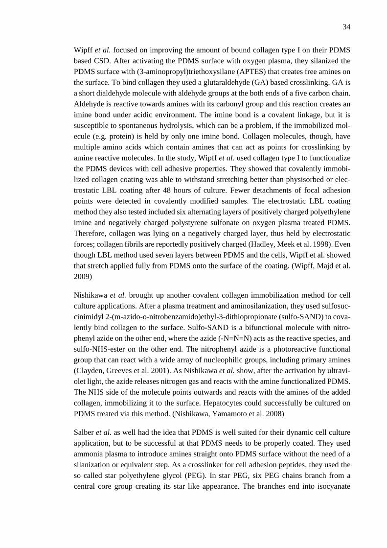
34
Wipff et al. focused on improving the amount of bound collagen type I on their PDMS
based CSD. After activating the PDMS surface with oxygen plasma, they silanized the
PDMS surface with (3-aminopropyl)triethoxysilane (APTES) that creates free amines on
the surface. To bind collagen they used a glutaraldehyde (GA) based crosslinking. GA is
a short dialdehyde molecule with aldehyde groups at the both ends of a five carbon chain.
Aldehyde is reactive towards amines with its carbonyl group and this reaction creates an
imine bond under acidic environment. The imine bond is a covalent linkage, but it is
susceptible to spontaneous hydrolysis, which can be a problem, if the immobilized mol-
ecule (e.g. protein) is held by only one imine bond. Collagen molecules, though, have
multiple amino acids which contain amines that can act as points for crosslinking by
amine reactive molecules. In the study, Wipff et al. used collagen type I to functionalize
the PDMS devices with cell adhesive properties. They showed that covalently immobi-
lized collagen coating was able to withstand stretching better than physisorbed or elec-
trostatic LBL coating after 48 hours of culture. Fewer detachments of focal adhesion
points were detected in covalently modified samples. The electrostatic LBL coating
method they also tested included six alternating layers of positively charged polyethylene
imine and negatively charged polystyrene sulfonate on oxygen plasma treated PDMS.
Therefore, collagen was lying on a negatively charged layer, thus held by electrostatic
forces; collagen fibrils are reportedly positively charged (Hadley, Meek et al. 1998). Even
though LBL method used seven layers between PDMS and the cells, Wipff et al. showed
that stretch applied fully from PDMS onto the surface of the coating. (Wipff, Majd et al.
2009)
Nishikawa et al. brought up another covalent collagen immobilization method for cell
culture applications. After a plasma treatment and aminosilanization, they used sulfosuc-
cinimidyl 2-(m-azido-o-nitrobenzamido)ethyl-3-dithiopropionate (sulfo-SAND) to cova-
lently bind collagen to the surface. Sulfo-SAND is a bifunctional molecule with nitro-
phenyl azide on the other end, where the azide (-N=N=N) acts as the reactive species, and
sulfo-NHS-ester on the other end. The nitrophenyl azide is a photoreactive functional
group that can react with a wide array of nucleophilic groups, including primary amines
(Clayden, Greeves et al. 2001). As Nishikawa et al. show, after the activation by ultravi-
olet light, the azide releases nitrogen gas and reacts with the amine functionalized PDMS.
The NHS side of the molecule points outwards and reacts with the amines of the added
collagen, immobilizing it to the surface. Hepatocytes could successfully be cultured on
PDMS treated via this method. (Nishikawa, Yamamoto et al. 2008)
Salber et al. as well had the idea that PDMS is well suited for their dynamic cell culture
application, but to be successful at that PDMS needs to be properly coated. They used
ammonia plasma to introduce amines straight onto PDMS surface without the need of a
silanization or equivalent step. As a crosslinker for cell adhesion peptides, they used the
so called star polyethylene glycol (PEG). In star PEG, six PEG chains branch from a
central core group creating its star like appearance. The branches end into isocyanate

35
functional groups (-N=C=O), which react towards amines, and crosslink the amine func-
tionalized PDMS with cell adhesive molecules. In their study, Salber et al. bound syn-
thetic peptide sequences known to interact with cell-binding sites derived from fibron-
ectin, laminin and collagen type IV. (Salber, Gräter et al. 2007)
Related to the previously mentioned study by Salber et al., Ahmed et al. used a similar
approach to coating stretchable PDMS substrates. After using ammonia plasma, they
treated the substrate with star shaped PEG-polypropylene glycol copolymer. The isocya-
nate groups were used to covalently bind fibronectin applied by stripe patterned PDMS
stamp, a method commonly referred to as micro contact printing. The mouse skeletal
myoblasts were successfully restricted to areas with fibronectin, and adhered cells were
able to survive the four days of dynamic culture. (Ahmed, Wolfram et al. 2010)
Trappmann et al. studied human epidermal stem cell differentiation on PDMS function-
alized with collagen type I. Without the use of plasma functionalization step, neverthe-
less, they were able to covalently crosslink collagen onto PDMS by using sulfosuccin-
imidyl-6-(4-azido-2-nitrophenylamino)hexanoate (sulfo-SANPAH). Sulfo-SANPAH is
chemically related to sulfo-SAND, and thus reacts under the same conditions in similar
manner. However, Trappmann et al. induced 365 nm UV-irradiation, as the solution con-
taining the crosslinker was in contact with pristine PDMS. Normally, no reaction would
occur, but the UV-irradiation can cause similar albeit weaker effect on PDMS surface as
oxygen plasma treatment (Qiu, Wu et al. 2014). The azide group is activated at the same
time via the UV-irradiation causing the crosslinker to immediately bind to the PDMS
surface, leaving the amine reactive NHS-ester side pointing outwards ready to be utilized
in covalent binding of collagen. Interestingly, the concentration of sulfo-SANPAH did
not seem to affect the amount of bound collagen, but the cell behaviour. Low concentra-
tion inhibited cell spreading and caused terminal differentiation; whereas higher concen-
tration kept them undifferentiated, while letting them proliferate and spread out.
(Trappmann, Gautrot et al. 2012)
At the time of the study by Trappmann et al., the usefulness of azido compounds in PDMS
surface treatment methods was not, however, a new concept. As demonstrated above by
Nishikawa et al., Gomez et al. in a similar fashion created a neural growth factor treated
grooved PDMS surface for supporting embryonic hippocampal neuron culture and axon
development. Their method for surface treatment included three steps. First, polyallyla-
mine (PAA), a polymer with amine group in every monomer, was conjugated with n-4-
(azidobenzoyloxy)succinimide (ABS), which is another molecule with phenyl azide and
NHS-ester functional groups. The NHS-ester side of ABS binds into the amine of PAA
leading into azido group terminated branched polymer structure. As a second step the
PAA-ABS is cast and physisorbed twice on PDMS and dried. The third and final step
includes the addition of neural growth factor solution and UV-irradiation. The UV-irra-
diation activates the azides, while also causing some functionalization of PDMS surface
similarly as in the later study by Trappmann et al. (Trappmann, Gautrot et al. 2012). The

36
azides in PAA-ABS bind to both the amines in the growth factors and the PDMS surface,
covalently immobilizing the neural growth factors on the PDMS. (Gomez, Lu et al. 2007)
LBL methods are also popular. In the studies by Brown et al. and Wang et al., for exam-
ple, a polyethylene imine and polystyrene sulfonate based LBL coating was used in sim-
ilar fashion as by Wipff et al in the study mentioned previously in this Section. Brown et
al. were able to show that vascular smooth muscle cells can be grown on LBL treated
PDMS even without any cell adhesion molecules; with comparable results to standard
tissue culture plastic. According to the study, LBL treated PDMS supported cell culture
better than physisorbed fibronectin coating. They also showed that decreasing the stiff-
ness of the PDMS substrate significantly increased the cell proliferation (Brown, Ookawa
et al. 2005). Wang et al. on the other hand report much lower CACO-2 cell numbers on
LBL treated PDMS surface than on standard tissue culture plastic; an indication that cell
type could have a major effect for the results (Wang, Sun et al. 2010). Chien et al. intro-
duced a photoactivated electrostatic LBL method for various substrates, including PDMS.
They introduced alternating layers of polyacrylic acid and polyacrylamide on oxygen
plasma treated PDMS. After several bilayers, there would be one azido functionalized
polyacrylic acid layer. The LBL process can be continued until the desired thickness is
achieved. Afterwards, when the layers are treated with UV-light, the interwoven polymers
with azido groups bind the layers together. As many times before, the azido functional-
ized polymers are further functionalized with cell adhesion molecules or cell adhesion
inhibitor molecules. The binding via azido groups happens only through layers that are
exposed to the UV-irradiation. By using custom designed photomasks, it is possible to
create micropatterned cell adhesive or inhibitive areas. The unbound parts of the polymer
layers are washed away. Chien et al. showed that cells can attach and grow on the LBL
treated surface, and that they will pattern along the grooves. One regular layer took
slightly over ten minutes to create and one azido functionalized layer took 30 minutes, so
the process can be lengthy, when creating thick LBL structures (Chien, Chang et al.
2009).
While this is not a complete review of all the research in the field, it shows the general
trend in the current most useful surface treatment methods for PDMS in biomedical and
cell culture research. In all of the studies introduced here, the authors had spent a great
effort to understand the chemical reactions behind the various types of surface treatment
methods. It is imperative to achieve comparable results, reproducible methods, and thus
useful studies, because even after eliminating the unknown variables in the surface treat-
ment process, the unpredictable nature of the cell culture still stands. While many of the
chemicals are extremely predictable and usually efficient in their chemical nature, their
realistic applicability for cell culture, for example, is a huge question mark. Chemicals
with crosslinking capability are usually extremely cytotoxic, and any unbound or unre-
acted molecules can act as unspecific fixatives to the delicate cellular organelles. In ad-
dition, many of the tailor made crosslinkers are very expensive, which further limits their

37
application in large scale. There is a wide variety of interesting molecules with capabili-
ties for crosslinking reactions that only wait to be exploited by adventurous researchers.
As an example, Tiller et al. showed that ascorbic acid (AA), or vitamin C, has this type
of capability and used it to bind enzymes to NH2 functionalized surfaces (Tiller, Berlin et
al. 1999). Despite its beneficial properties, ascorbic acid has not yet been utilized, how-
ever, in cell culture applications. This concept was taken further in this thesis work to
create novel AA crosslinker based surface treatment methods specifically for cell culture.
A multidisciplinary approach in the studies in this field is truly appreciated, if not essen-
tial.

38
EXPERIMENTAL PART

39
4. MATERIALS AND METHODS
To answer to the increasing need for basic research and more cell friendly solutions in
PDMS based cell stretching applications, four PDMS surface treatment methods in seven
variations were studied in static and dynamic conditions in this thesis work. The methods
were based on either physisorption or covalent chemical bonding of collagen type I on
PDMS. Physisorption was studied in two surface treatment variations PHY1 and PHY2
as well-founded references to the covalent methods. Covalent Method 1 with a single
variation COGA found in literature (Wipff, Majd et al. 2009) utilizes GA crosslinking. In
the study by Wipff et al, GA is used as a crosslinker between collagen type I and the
substrate, immobilizing the protein on the PDMS surface and it offers a reference covalent
method to the AA based methods developed in the thesis. The novel Covalent Method 2
with three variations COAA1, COAA2, and COAA3 utilizes AA based crosslinking as a
solution to the possible cytotoxic effect of GA. Additionally, the results were used to
propose and test Covalent Method 3, COGEL, that utilizes AA crosslinking and collagen
type I gel.
The methods were studied in cell free environment with double fluorescent labelling to
visually evaluate the amount of collagen type I bound on the PDMS surface. Covalent
Method 1 was also imaged after two days of stretching and compared to physisorption
methods. In addition to the cell free studies, Covalent Methods 1 and 2 were tested in
human adipose stem cell (hAdSC) culture for 14 days in static conditions, and for 13 days
in dynamic conditions to evaluate their suitability for extended dynamic culture. In the
final part of the study, Covalent Method 3 was preliminary tested in cell free environment
with Dulbecco’s phosphate buffered saline (DPBS) for nine days and with static hAdSC
culture for four days. The study was conducted in cooperation with BioMediTech, a joint
institution of Tampere University and Tampere University of Technology, and as part of
the Human Spare Parts and WoodBone projects. The cell culture work and live cell im-
aging were performed by Msc. Sanni Virjula of the Adult Stem Cell Group from Bio-
MediTech.
This Chapter describes the basic information about the experiments done in this thesis
work. Section 4.1 explains the manufacturing process of the CSDs and their counterparts,
the static reference wells, from PDMS, while Section 4.2 explains how the stretching
system works. The preparation of the different coatings can be found in Section 4.3. Chap-
ter ends with Section 4.4 with the description of the fluorescent characterization of the
coatings and the cell culture experiments.

40
4.1 PDMS device manufacture
Tailor-made CSDs from PDMS (Sylgard® 184, Dow Corning, MI, USA) and glass were
applied in the study. The design and preparation of the devices, shown in Figure 9 have
been described earlier by Kreutzer et al. (Kreutzer, Ikonen et al. 2013). The devices con-
sist of a PDMS bulk, where a circular vacuum chamber and a cylindrical PDMS-based
culture well are cut. The top of the device is sealed by a glass plate with a hole for creating
the vacuum, and the bottom is sealed by a 120 µm thick PDMS membrane. The PDMS
membrane provides the cell cultivation substrate. A small PDMS piece is added on top of
the glass plate for mounting the tubing for vacuum. When a partial vacuum is created in
the vacuum chamber, the elastomeric PDMS based culture well deflects, and deforms the
cell cultivation substrate (Figure 10). The dimensions are illustrated in Figure 9.
Figure 9. The pneumatic CSD with PDMS membrane (D), static reference device
with the same cell culture area (C), and separate polycarbonate rings of the
devices (A and B) for preventing PDMS membranes from touching the sur-
face below. The culture well is 12 mm in diameter (1.13 cm²) while the
whole CSD is 31 mm wide.
Figure 10. Visualization of the cross section of the CSD and the vacuum applied
stretch mechanism.

41
Curing of the PDMS was done uncovered in an oven (Binder GmbH, Tuttlingen, Ger-
many) in 60 °C for 10 hours to reduce the possibility of the ethylbenzene solvent residue.
The individual pieces were treated with oxygen plasma (Pico-SR-PCCE, low pressure
plasma system, Diener Elect., Ebhausen, Germany) to bond them permanently together.
The plasma cleaner parameters were the same in all treatments (power 30 W; treatment
time 20 s; chamber pressure 0.30; gas flow rate 1.4 sccm). The static reference wells
which were used for unstretched cell culture experiments were prepared with the same
dimensions for the cell culturing well as CSDs. The vacuum chamber was removed from
the reference wells to save manufacturing costs and time (Figure 9).
All devices were mounted on specifically developed polycarbonate rings in order to pre-
vent the PDMS membrane touching the surface under the devices. The polycarbonate
rings for the reference wells had an outer diameter of 25 mm and an inner diameter of 13
mm. The rings for CSDs have an outer diameter of 40 mm and inner diameter of 20 mm.
All rings were 0.8 mm thick.
The PDMS membrane was manufactured using the same PDMS curing protocol as for
the devices. In order to attach the membranes to the devices, the membranes were first
cleaned with isopropanol and deionized (DI) water, after which they were treated with
oxygen plasma and bonded to the devices.
4.2 Cell stretching system
The vacuum stretching system was similar to the one used by Kreutzer et al. (Kreutzer,
Ikonen et al. 2013) and it is composed of a laptop computer, LabVIEW-based controller
software, a measurement board (National Instruments, USB-6229 BNC, USA), a com-
puter controlled pressure regulator (T-2000, Marsh Bellofram, USA) attached to a high
pressure outlet, and an ejector pump (Festo OY, VAD-1/8, Finland), which creates the
vacuum. The CSDs on petri dishes were placed inside a cell culture incubator and attached
to the ejector pump outside the incubator using a silicone rubber tubing system. The
stretching was conducted under standard cell culture conditions in a humidified atmos-
phere (+37 °C, 5 % CO2). Cyclic equiaxial stretching strain magnitude for the cell free
durability test was 10 % with cyclic (sine wave, 0.5 Hz) stretching. Stretching for the cell
culture test was applied with an effective stretching period of 12 hours per day followed
by 12 hours of rest. The strain magnitude was increased from 2 % at the first stretching
period to 3.5 % at the second period and finally to 5 % for the rest of the dynamic culture
test.
4.3 Collagen type I coatings on PDMS
The membranes of the devices were functionalized with type I collagen from rat tail (Invi-
trogen, Life Technologies, Carlsbad, CA, USA) using different methods in five experi-
mental phases. In phase one (P1), two physisorption methods and Covalent Method 1

42
were studied and imaged in cell free static and dynamic tests. In phase two (P2), Covalent
Method 1 and 2 were imaged in cell free static test. In phase three (P3), one physisorption
method and Covalent Method 1 and 2 were studied in static cell culture test. In phase four
(P4), Covalent Method 1 and 2 were studied in dynamic cell culture test. In phase five
(P5), Covalent Method 3 was proposed and imaged after the coating preparation. It was
also preliminary studied in static cell culture test.
In this study, all the coated samples were used for characterization and cell culture within
24 hours of the coating preparation. For the cell culture experiments, the devices were
first wiped outside with 70 % ethanol, then placed in a laminar hood and finally sterilized
under mild UV-irradiation for 20 min. Finally, they were rinsed once with DPBS and
placed in sterile cell culture plates.
4.3.1 Physisorption methods
PHY1 and PHY2 were prepared with one difference between them. PHY1 was left with-
out plasma treatment while PHY2 samples were treated with oxygen plasma. All samples
were incubated at room temperature for 60 minutes with collagen solution (50 µg/ml;
diluted in 0.02 M acetic acid). The volume used to treat the wells contained 17 µg/cm² of
collagen type I. Following the incubation, the samples were washed thoroughly with de-
ionized (DI) water, dried for 15 minutes in a ventilation cabinet, and stored at +4 °C.
4.3.2 Covalent Method 1 with glutaraldehyde crosslinker
For Covalent Method 1, COGA, oxygen plasma, APTES (Sigma-Aldrich, St.Louis, MO,
USA), and GA (Sigma-Aldrich) were used to covalently bond collagen type I onto the
membrane (Wipff, Majd et al. 2009). After the plasma treatment, the membrane was
treated with 10 % APTES solution in methanol for two minutes at room temperature to
create a monolayer with primary amine groups on top. The samples were then washed
once with methanol and rinsed twice with DI water. Then the COGA samples were treated
with 3 % GA solution in DI water for 20 minutes and rinsed five times with DI water.
Finally, the devices were treated with collagen type I as in the PHY coatings. Figure 11
illustrates the stages present in COGA method.

43
Figure 11. A general illustration of Covalent Method 1 and the crosslinking mecha-
nism between APTES, GA and collagen type I.
4.3.3 Covalent Method 2 with ascorbic acid crosslinker
Covalent Method 2 was divided into three variations. The COAA1, COAA2 and COAA3
samples were prepared identically to the COGA samples, except the GA as the crosslinker
was replaced by AA (Sigma-Aldrich, cell culture grade, ≥98 %) and the incubation time
was prolonged from 20 minutes to 60 minutes followed by only two washings with DI
water. For the COAA1 coatings, the AA powder was dissolved in DPBS (200 mg/ml;
Lonza, BioWhittakerTM, Verviers, Belgium), whereas for the COAA2 and COAA3 coat-
ings the AA powder was dissolved in methanol (20 mg/ml). In the COAA3 coating pro-
tocol, 5 µl of 35 % hydrogen peroxide, which is 2 % of the used volume, was added to
the AA coating solution in methanol to improve AA reactivity (Tiller, Berlin et al. 1999).
4.3.4 Covalent Method 3 for collagen type I gel
Covalent Method 3, COGEL, was proposed and preliminary tested after the results from
the previous phases. The COGEL samples were prepared identically to the COAA1
method until the collagen type I treatment. In COGEL, collagen type I was dissolved and
diluted in methanol instead of acetic acid (50 µg/ml). To increase the effectiveness of the
dilution, the container was stirred with pipette tip and warmed by hands until no visible
collagen type I aggregates were seen. The samples were incubated with collagen type I-
methanol solution at room temperature for 60 min. During the incubation period, most of
the methanol evaporated leaving all of the collagen type I on the sample in a thick gel-
like layer. The same washing and storing protocol from the other methods was used to
finalize the treatment process.
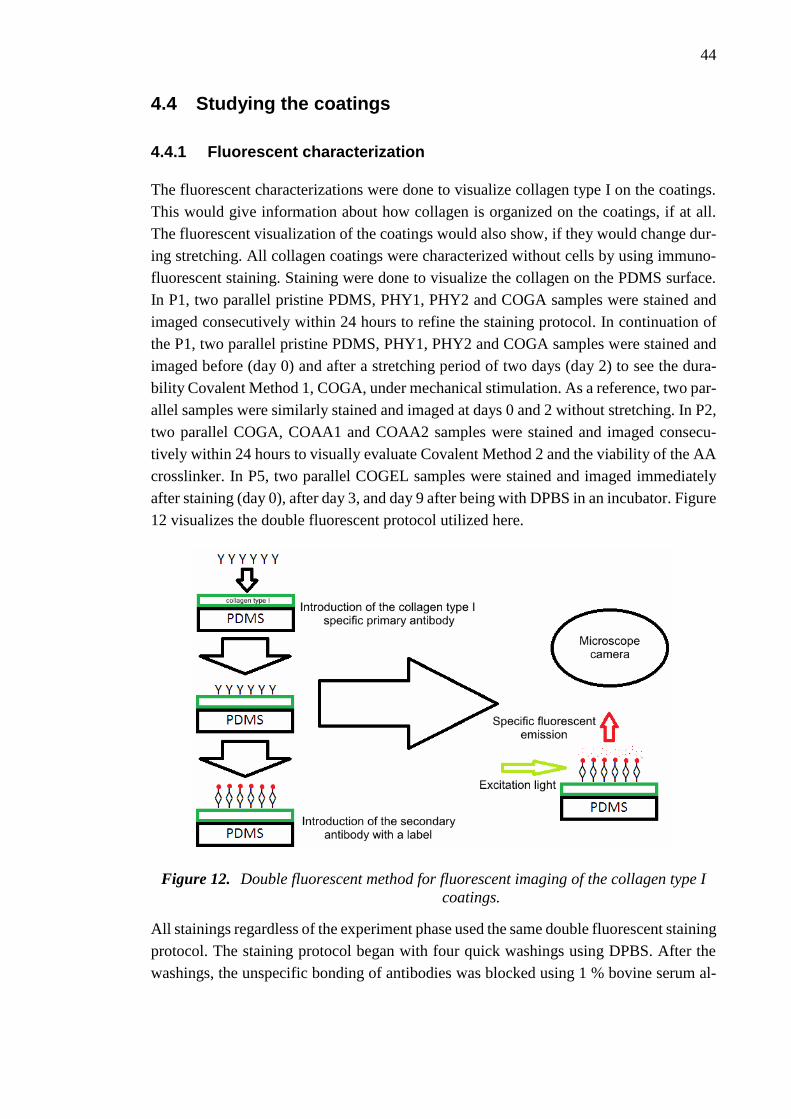
44
4.4 Studying the coatings
4.4.1 Fluorescent characterization
The fluorescent characterizations were done to visualize collagen type I on the coatings.
This would give information about how collagen is organized on the coatings, if at all.
The fluorescent visualization of the coatings would also show, if they would change dur-
ing stretching. All collagen coatings were characterized without cells by using immuno-
fluorescent staining. Staining were done to visualize the collagen on the PDMS surface.
In P1, two parallel pristine PDMS, PHY1, PHY2 and COGA samples were stained and
imaged consecutively within 24 hours to refine the staining protocol. In continuation of
the P1, two parallel pristine PDMS, PHY1, PHY2 and COGA samples were stained and
imaged before (day 0) and after a stretching period of two days (day 2) to see the dura-
bility Covalent Method 1, COGA, under mechanical stimulation. As a reference, two par-
allel samples were similarly stained and imaged at days 0 and 2 without stretching. In P2,
two parallel COGA, COAA1 and COAA2 samples were stained and imaged consecu-
tively within 24 hours to visually evaluate Covalent Method 2 and the viability of the AA
crosslinker. In P5, two parallel COGEL samples were stained and imaged immediately
after staining (day 0), after day 3, and day 9 after being with DPBS in an incubator. Figure
12 visualizes the double fluorescent protocol utilized here.
Figure 12. Double fluorescent method for fluorescent imaging of the collagen type I
coatings.
All stainings regardless of the experiment phase used the same double fluorescent staining
protocol. The staining protocol began with four quick washings using DPBS. After the
washings, the unspecific bonding of antibodies was blocked using 1 % bovine serum al-

45
bumin (Sigma-Aldrich) diluted in DPBS. The blocking solution was incubated in the sam-
ples for 60 minutes at room temperature. Then, the coatings were incubated overnight at
+4 °C with the anti-collagen type I primary antibody (ab90395, Abcam, Cambridge, UK)
diluted 1:500 in the blocking solution. Next day, the devices were washed four times for
three minutes with DPBS. The Alexa Fluor 488 ® conjugated secondary antibody (Life
technologies) was diluted 1:800 in the blocking solution and the devices were incubated
with the secondary antibody solution for 60 minutes at room temperature in dark. After
the incubation, the devices were washed again four times for three minutes with DPBS
and quickly rinsed once with DI water. The devices were imaged immediately after stain-
ing, or stored overnight at +4 °C in dark, and imaged latest in the morning. The greyscale
and coloured images without cells were taken with the fluorescent microscope at Tampere
University of Technology (Nikon Eclipse TS100 equipped for greyscale/coloured with
Allied Vision Manta/Canon EOS550D cameras, Tokyo, Japan). The fluorescent images
of COGEL coating in P5 were taken with Nikon Eclipse equipped with the Manta camera,
and coloured in post processing with GNU image manipulation program. The relief im-
ages in P5 were created using the same program as well.
4.4.2 Adipose stem cell culture
The cell culture studies in P3, P4 and P5 were conducted in the facilities and in coopera-
tion with the Adult Stem Cell Group from BioMediTech, and as part of the Human Spare
Parts and WoodBone projects. All cell culture studies were also done in accordance with
the Ethics Committee of the Pirkanmaa Hospital District. The hAdSCs were isolated from
adipose tissue samples at Tampere University Hospital (Tampere, Finland). The cells of
confirmed mesenchymal origin, the hAdSCs, were seeded onto pre-incubated static de-
vices and CSDs. After the three-to-four-hour pre-incubation period, the incubation me-
dium was first removed, 400 µl of fresh medium added and 1000 cells/cm2 were seeded
onto the samples in 100 µl of medium. Three parallel samples were used in static P3 and
dynamic P4 culture tests. Static P5 culture test used two parallel samples. In all experi-
ments, the culture medium was changed twice a week.
In the end of the static P3 culture test, the samples were imaged with light microscopy
(Zeiss Axio Scope.A1 equipped with AxioCam MRc5 camera, Carl Zeiss, Oberkochen,
Germany) after 14 days of culture. In the dynamic P4 culture test, the samples were im-
aged at days 3+0, 3+3, 3+7 and 3+10 with light microscopy. The P4 culture test included
a three day pre-stretching culture period (day 3+0), where the cells were allowed to adhere
to the devices, after which the stretching was started. In the end of the P4 culture test, the
proliferation and the cell number was evaluated with CyQuant® (Thermo Fisher Scien-
tific). In the end of P5 cell culture test at day 4, the cells were imaged with light micros-
copy to evaluate the cell attachment on COGEL and its suitability for cell culture tests in
the future.

46
5. RESULTS AND DISCUSSION
This Chapter presents and discusses results of this work. A major part of this thesis work
was the learning and optimization of the fabrication protocol for the CSDs and parts
thereof. However, the largest part and the main aim of the thesis was to find suitable
coating methods for the PDMS CSDs that; 1) make long term (> 48 hours) stem cell
dynamic culture practical; 2) are easy and straightforward to implement in a limited la-
boratory setting. The experimental part of the thesis work was divided in five phases to
portray the evolution of the experiments and the results from each phase are presented
and discussed in chronological order during this Chapter.
5.1 Phase one: Fluorescent characterization of Covalent
Method 1
The P1 was focused on the fluorescent characterization of Covalent Method 1 against the
physisorption methods and pristine PDMS control. COGA, PHY1, PHY2 and the control
samples were first characterized using fluorescent microscopy imaging in a simple setting
to visualize the collagen type I on the surfaces of the devices, and to visualize any differ-
ences in the physical form of collagen. The samples were imaged following their prepa-
ration and staining with the collagen type I specific labelled antibodies such that the in-
tensity, structure and distribution of the collagen on the substrate could be qualitatively
analysed. P1 acted as a preliminary experiment for the more advanced phases that follow.
Figure 13 shows the difference in the fluorescent signal belonging to collagen type I be-
tween the four high-magnified images of the samples. The image shows that collagen
type I can be labelled and seen from the substrate, and that pristine PDMS sample is dark,
as expected. According to the image, the PHY2 sample has adsorbed more collagen than
PHY1 due to the plasma treatment before the coating. The collagen appeared, however,
aggregated when compared to the COGA sample which had an evenly distributed layer
of collagen on the substrate. Figure 14 shows fluorescent images from the two-day dy-
namic experiment. COGA, PHY1, PHY2 and the control samples were imaged after
staining at day 0, after the two-day period of incubation in medium in static and stretched
conditions. Day 0 results are consistent with the previous results in Figure 13 except for
PHY2 collagen aggregation being less prominent in half of the samples (2/4). Static in-
cubation of two days did not visibly affect the coatings. The dynamic samples, however,
show that the aggregated collagen in PHY2 samples has dropped off, because of the strain
caused by the stretching substrate. If cells would have been living on top of the collagen
in PHY2 samples, while they could have had adhered there, they would have disengaged
with the collagen, which is unacceptable for any cell culture. COGA samples, however,

47
retained the same appearance, thus showing the superiority of the covalent immobiliza-
tion of collagen when compared to physisorption.
Figure 13. High magnification immunofluorescent images of pristine PDMS well and
wells coated with PHY1, PHY2, and COGA methods in P1. The images were
taken using 20x (scale bar 50 µm) objective on Nikon Eclipse equipped with
Manta camera.

48
Figure 14. Low magnification immunofluorescent images of pristine PDMS well and
wells coated with PHY1, PHY2, and COGA methods in P1 after the two-day
incubation period in static (left) and stretched (right) conditions. Images
were taken using 4x (scale bar 500 µm) objective on Nikon Eclipse
equipped with Manta camera.
5.2 Phase two: Fluorescent characterization of Covalent
Method 2
The Static experiments in P2 focused on the fluorescent characterization of the group of
novel covalent methods that utilize AA crosslinker and their comparison with COGA that
utilizes GA crosslinker. COGA, COAA1 and COAA2 were characterized by fluorescent
microscopy similarly as in P1.
Figure 15 shows the images which strongly suggest superior binding capacity of AA
crosslinker when compared to GA crosslinker. COGA maintains its evenly distributed
layer of collagen also seen in P1; it can be better seen with longer exposure times or image
post processing. However, COAA1 and to some extent COAA2 have completely different
appearance. COAA1 method has bound a massive amount of collagen that in some areas
seems to be in multiple layers; judged from the visible boundaries on the substrate. They
resemble the aggregations of collagen on PHY2 which could mean that the collagen is

49
only physisorbed in large quantities and it will come off due to dynamic load or cell in-
teraction. The fluorescent images show, however, that there is still an even collagen layer
in both COAA1 and COAA2 between the aggregations. Both COAA1 and COAA2 look
otherwise very similar when the largest collagen aggregations on COAA1 samples are
disregarded. The stripes seen on the surface are likely on the PDMS substrate itself as the
plasma oxidized thin PDMS membrane tends to form such stripes, likely due to the dam-
aging effect of the oxygen plasma. These small cracks that form when the thin membrane
deforms during manufacture, will then act as edges where molecules such as APTES, AA
or collagen may accumulate and show on the images.
Figure 15. Immunofluorescent images from two locations of two wells coated with ei-
ther COGA, COAA1, or COAA2 methods in P2. Images were taken using
10x (scale bar 200 µm) objective on Nikon Eclipse equipped with Canon
EOS550D colour camera.
5.3 Phase three: Static adipose stem cell culture
P3 of the experimental part focused on growing hAdSCs for 14 days on three parallel
PDMS static reference wells coated with PHY2, COGA, COAA1, COAA2 and COAA3
methods to validate their suitability for scientifically sound cell experiments. The cell
cultures were followed and imaged using light microscope during and after 14 days of
static culture and their viability and proliferation were qualitatively analysed.
Figure 16 shows the results of this experiment and differences between different coating
methods are apparent. While seeded cells initially started to adhere on the PHY2 coated
wells to some extent, along the way to day 14 almost all cells detached, leading to empty
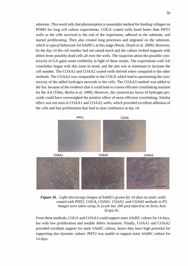
50
substrate. This result tells that physisorption is unsuitable method for binding collagen on
PDMS for long cell culture experiments. COGA coated wells fared better than PHY2
wells as the cells survived to the end of the experiment, adhered to the substrate, and
started proliferating. They also created long processes and migrated on the substrate,
which is typical behaviour for hAdSCs at this stage (Petrie, Doyle et al. 2009). However,
by the day 14 the cell number had not raised much and the culture looked stagnant with
debris from possibly dead cells all over the wells. The suspicion about the possible cyto-
toxicity of GA gains some credibility in light of these results. The experiments with AA
crosslinker began with this issue in mind, and the aim was at minimum to increase the
cell number. The COAA1 and COAA2 coated wells thrived when compared to the other
methods. The COAA3 was comparable to the COGA which lead to questioning the cyto-
toxicity of the added hydrogen peroxide to the cells. The COAA3 method was added to
the list, because of the evidence that it could lead to a more efficient crosslinking reaction
for the AA (Tiller, Berlin et al. 1999). However, the cytotoxicity factor of hydrogen per-
oxide could have overweighed the positive effect of more efficient crosslinking. Similar
effect was not seen in COAA1 and COAA2 wells, which provided excellent adhesion of
the cells and fast proliferation that lead to near confluence at day 14.
Figure 16. Light microscopy images of hAdSCs grown for 14 days on static wells
coated with PHY2, COGA, COAA1, COAA2, and COAA3 methods in P3.
Images were taken using 5x (scale bar 200 µm) objective on Zeiss Axio
Scope.A1.
From these methods, COGA and COAA3 could support static hAdSC culture for 14 days,
but with low proliferation and notable debris formation. Finally, COAA1 and COAA2
provided excellent support for static hAdSC culture, hence they have high potential for
supporting also dynamic culture. PHY2 was unable to support static hAdSC culture for
14 days.

51
5.4 Phase four: Dynamic adipose stem cell culture
P4 of the experimental part of the thesis was the dynamic culture of hAdSCs on COGA,
COAA1 and COAA2 coated samples. PHY2 and COAA3 were left out due to the insuf-
ficient results in P3 and to keep the sample number lower. The culture started as static for
three days to give the cells time to adhere onto the substrate and continued as dynamic
for 10 days for the half of the samples while the other half were kept static. The wells
were imaged using light microscopy after 3+10 days and their cell number was quantita-
tively analysed with CyQuant®.
Figure 17 shows the wells at day 3+10. It is immediately apparent after looking at the
images of the cells and the CyQuant® results in Figure 18 that COAA1 and COAA2
coated wells exhibit superior proliferation. Most of the COAA1 and COAA2 static wells
had reached confluence, which is the reason of stopping the experiment at day 3+10 and
not at day 3+11, which was initially planned. In one COAA2 sample, the layer of cells
had started to peel off already, as it can be seen in Figure 17. The results are in line with
the previous experiments in P3. In dynamic samples of COAA1, the cell number was at
day 3+10 about 32 % lower than in static samples and in COAA2 the number was about
17 % lower. However, the COAA2 result could include some error as the cells in the
static wells had begun to peel off, and therefore the cell number in the static sample could
have been even higher. Overall, the cell numbers between COAA1 and COAA2 samples
were quite similar. COGA samples, on the other hand, had significantly lower cell num-
bers at the end of the experiment. Unexpectedly, the static samples of COGA had lower
cell number than the dynamic samples, further telling of the unexpected and random na-
ture of COGA surface treatment method. However, this could also mean that the GA is
able to immobilize collagen well to the surface and withstand the dynamic strain caused
by the stretching keeping the cells attached to the substrate. This can cause the hypothet-
ical cytotoxic effect of GA be more detrimental to the cell culture than the stretching
itself. If this would be the case, the cell number disparity between static and dynamic
samples would diminish.
In respect of the results in P4, COAA1 and COAA2 surface treatment methods for PDMS
are suitable for dynamic hAdSC culture for 3+10 days. Due to the poor proliferation, the
COGA method is not well suited for cell culture applications in neither static nor dynamic
environment.

52
Figure 17. Light microscopy images of hAdSCs grown for 3+10 days on static and dy-
namic wells coated with COGA, COAA1, and COAA2 methods in P4. Im-
ages of COGA samples show areas in the same well with different number
of cells. The image of the static sample of COAA2 shows an area where the
confluent cells had begun to detach. Images were taken using 5x (scale bar
200 µm) objective on Zaiss Axio Scope.A1.
Figure 18. The cell number of hAdSCs on static and dynamic wells coated with COGA,
COAA1, and COAA2 methods in P4.

53
5.5 Phase five: Preliminary tests with Covalent Method 3
In P5 of the experimental part, preliminary functionality tests were conducted with the
COGEL coating, which was created with the results from P1 to P4 in mind. It was divided
in two parts for visualizing collagen in cell-free culture environment, and for testing its
suitability for hAdSC culture. The cell-free samples were imaged after day 0, day 3 and
day 9. The cell culture samples were imaged after four days of culture.
5.5.1 Cell free incubation test
Figure 19 shows a low magnification image from a well with COGEL coating. The
amount of collagen type I on the surface is very high, as expected. During the 60-minute
incubation with collagen type I-methanol solution, the dissolved collagen fibrils had
started aggregating into larger fibres, as it is apparent in Figure 19. Figure 20 shows a
high magnification image of the coating, as well as a relief meant to highlight the 3D
aspect. The areas in the samples showed varying amounts of collagen type I bundling. In
some areas, there were short thin fibres (Figure 20, upper), and in some areas, there were
clear matrix structures (Figure 20, lower). Multi-layered honeycomb-like collagen matri-
ces, as seen here, can commonly be found in natural reticular connective tissue and bone,
for example (Schwint, Labraga et al. 2004, Ushiki 2002), and it showcases collagen’s
ability for spontaneous fibrillogenesis without cellular influence. Figure 21 and Figure 22
show the samples from day 3 and 9 respectively. While some areas in the day 9 samples
were slightly darker than in the others, there is not a big difference in the non-incubated
day 0 samples and samples from day 3 or day 9. The presence of DPBS as a medium
substitute and temperature of 37 °C did not greatly affect the coatings, which show easily
identifiable fibrous and honeycomb-like structures even at day 9.
Figure 19. Low magnification immunofluorescent image of the well coated with CO-
GEL method at day 0 in P5. Image was taken using 4x (scale bar 500 µm)
objective on Nikon Eclipse equipped with Manta camera.

54
Figure 20. High magnification immunofluorescent images of the well coated with CO-
GEL method at day 0 in P5. The images on the left are shown as reliefs on
the right. Images were taken using 10x (scale bar 200 µm) objective on Ni-
kon Eclipse equipped with Manta camera. The reliefs were created using
GNU image manipulation program.
The results from the cell-free incubation test suggest that collagen type I takes a very
robust form, when the protein is forcefully packed onto the substrate via evaporation of
the solvent. In the other coating methods depicted in this thesis, collagen type I is only
passively coming into contact with the substrate, which results into lesser amount of col-
lagen available for fibrillogenesis, fibril formation. The result of this is most likely a coat-
ing that constitutes mostly of loosely bundled collagen fibrils that are structurally invisi-
ble in the scale of the images. When collagen is dissolved into a stock solution, it partially
loses its fibrous formation. However, some of the natural crosslinks between the collagen
alpha-helices are conserved, allowing the later self-assembly into larger fibrils and even
fibres. Closely packed small fibrils that cannot organize into larger fibrils would likely be
seen in the fluorescent images as smooth layers or structures; exactly what can be seen in
the images from P2 (Figure 15). This lack of organization of the fibrils may result from
the lower mobility and quantity of collagen available for bundling, as they are immobi-
lized by the crosslinker on the substrate. When the collagen amount is increased, the less
bundled surface collagen is buried under the excess, which then spontaneously crosslinks
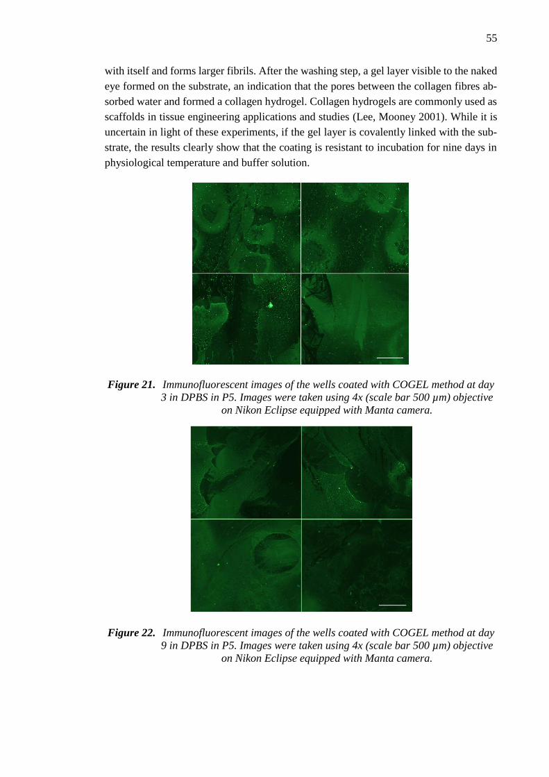
55
with itself and forms larger fibrils. After the washing step, a gel layer visible to the naked
eye formed on the substrate, an indication that the pores between the collagen fibres ab-
sorbed water and formed a collagen hydrogel. Collagen hydrogels are commonly used as
scaffolds in tissue engineering applications and studies (Lee, Mooney 2001). While it is
uncertain in light of these experiments, if the gel layer is covalently linked with the sub-
strate, the results clearly show that the coating is resistant to incubation for nine days in
physiological temperature and buffer solution.
Figure 21. Immunofluorescent images of the wells coated with COGEL method at day
3 in DPBS in P5. Images were taken using 4x (scale bar 500 µm) objective
on Nikon Eclipse equipped with Manta camera.
Figure 22. Immunofluorescent images of the wells coated with COGEL method at day
9 in DPBS in P5. Images were taken using 4x (scale bar 500 µm) objective
on Nikon Eclipse equipped with Manta camera.

56
5.5.2 Adipose stem cell culture test
Alongside the cell-free samples, two COGEL coated samples were plated with hAdSCs
similarly to the previous cell tests. The cells were cultured for four days and then imaged
as seen in Figure 23. After day 4 of culture, the cells were well attached to the substrate
and exhibited elongated processes from the main bodies, which is typical for stem cells
of this type (Petrie, Doyle et al. 2009). The debris that is visible in the images was hy-
pothesized to be small bubbles trapped inside the gel, as they seemed to be under the cells
and did not seem to affect the culture negatively. While there was no control in this small-
scale preliminary test, it can be said with reasonable certainty that hAdSCs can be suc-
cessfully cultured on COGEL coated PDMS. It is also worthwhile to mention that after
the culturing had been stopped and the cells removed from the incubator, the cells re-
mained attached to the surface overnight, even though their morphology was already
round, an indication of the incoming death. Nevertheless, while there are some clues
about the durability of the new COGEL coating, there are still many uncertainties regard-
ing the coating in a dynamic cell culture. For example, the amount of stretch that is trans-
ferred to the gel surface is unknown. These questions can only be answered by future
experiments.
Figure 23. Light microscopy images of hAdSCs grown for four days on static wells
coated with COGEL method in P5. Images were taken using 5x (scale bar
200 µm) and 10x (scale bar 100 µm) objectives respectively on Zeiss Axio
Scope.A1.
57
6. CONCLUSION
The aim of this thesis was to find and apply surface treatment methods for PDMS that are
suitable to be used in cell stretching applications, and to evaluate their capability to bind
collagen type I and to support long term static and dynamic hAdSC culture. The motiva-
tion behind this study was to allow researchers in the Human Spare Parts and Woodbone
projects to use the available custom made cell stretching system (Kreutzer, Ikonen et al.
2013) more effectively. The effect of stretching on cells has for a long time intrigued
researchers worldwide and factors such as the method used for coating the device can
have a significant effect on the end results.
The experimental study was divided in five phases that all focused on the various coating
methods for PDMS membrane, which was a part of the CSD, and meant for cell culture.
From these, the P1, P2 and P5 were fluorescent characterization experiments, while P3
and P4 were cell culture experiments. There were a total of seven different coating meth-
ods and a pristine PDMS control that were tested during the experiments of this thesis
work; PHY1, physisorbed collagen type I on PDMS; PHY2, physisorbed collagen on
plasma oxidized PDMS; COGA, immobilized collagen on APTES and GA; COAA1, im-
mobilized collagen on APTES and AA in DPBS; COAA2, immobilized collagen on
APTES and AA in methanol; COAA3, immobilized collagen on APTES and AA in meth-
anol with added hydrogen peroxide; COGEL, immobilized collagen gel on APTES and
AA.
P1, P2 and P5 showed that it is possible to label collagen on the substrates with a fluores-
cent dye and visualize its features with fluorescent microscope. The experiments in P1
also revealed that covalent binding of collagen by COGA is superior to physisorption
methods especially after 2 days of stretching. In P2, COAA1 and COAA2 are in turn
shown to bind superior amounts of collagen compared to COGA. P5 and the applied CO-
GEL method showed that collagen type I can spontaneously organize into visible fibrils
and honeycomb-like structures. The cell culture tests in P3 and P4 showed that COAA1
and COAA2 promoted cell adhesion and had superior proliferation of hAdSCs in all static
and dynamic samples. COGEL also supported hAdSC attachment and proliferation in
P5. In P3 and P4, COAA3 and COGA had both similarly low cell proliferation or high
cell mortality rate, which could be a telling sign about the possible cytotoxicity of the
used chemicals GA and hydrogen peroxide.
The main aim of this thesis work was to covalently bind collagen type I to PDMS CSDs
for long-term cell stretching experiments. Then, if applicable, the secondary aim was to
propose a novel surface treatment method to improve upon the existing methods. Both
aims were achieved in this thesis work. The novel AA based Covalent Method 2 was
successfully utilized in fluorescent imaging and cell stretching studies, improving the

58
binding of collagen and cell culture quality when compared to the popular physisorption
method or the GA based Covalent Method 1. Covalent Method 2 managed to do so with-
out complicating the treatment process or increasing the costs, thus proving to be a wel-
come addition to the repertoire of cell culture researchers. In addition, the coating meth-
ods developed in this thesis may improve functionalization efforts of tissue engineering
scaffolds and implants with durable but cell friendly coatings. The world around the cells
is constantly changing, but it is impossible to measure in vivo which specific forces affect
the cells in which way, especially when speaking about the elusive stem cells. Studying
dynamic cell culture is an important step in the search for the answers about the physical
cues that affect stem cell differentiation and mature cell culture. The answers that can
only be solved by rigorous basic research by tireless researchers worldwide.
59
REFERENCES
AHMED, W.W., KURAL, M.H. and SAIF, T.A., 2010. A novel platform for in situ in-
vestigation of cells and tissues under mechanical strain. Acta biomaterialia, 6(8), pp.
2979-2990.
AHMED, W.W., WOLFRAM, T., GOLDYN, A.M., BRUELLHOFF, K., RIOJA, B.A.,
MÖLLER, M., SPATZ, J.P., SAIF, T.A., GROLL, J. and KEMKEMER, R., 2010. My-
oblast morphology and organization on biochemically micro-patterned hydrogel coat-
ings under cyclic mechanical strain. Biomaterials, 31(2), pp. 250-258.
ALBERTS, B., BRAY, D., HOPKIN, K., JOHNSON, A., LEWIS, J., RAFF, M., ROB-
ERTS, K. and WALTER, P., 2010. Chapter 20: Cellular communities: Tissues, Stem
Cells, and Cancer. In: M. MORALES, ed, Essential Cell Biology. 3rd edition edn. NY,
USA & Abingdon, UK: Garland Science, pp. 689-730.
ANKRUM, J. and KARP, J.M., 2010. Mesenchymal stem cell therapy: Two steps for-
ward, one step back. Trends in molecular medicine, 16(5), pp. 203-209.
ASSADY, S., MAOR, G., AMIT, M., ITSKOVITZ-ELDOR, J., SKORECKI, K.L. and
TZUKERMAN, M., 2001. Insulin production by human embryonic stem cells. Diabe-
tes, 50(8), pp. 1691-1697.
BAROFFIO, A., BOCHATON-PIALLAT, M., GABBIANI, G. and BADER, C.R.,
1995. Heterogeneity in the progeny of single human muscle satellite cells. Differentia-
tion, 59(4), pp. 259-268.
BERTHIER, E., YOUNG, E.W.K. and BEEBE, D., 2012. Engineers are from PDMS-
land, Biologists are from Polystyrenia. Lab Chip, 12(7), pp. 1224-1237.
BHUSHAN, B., HANSFORD, D. and LEE, K.K., 2006. Surface modification of silicon
and polydimethylsiloxane surfaces with vapor-phase-deposited ultrathin fluorosilane
films for biomedical nanodevices. Journal of Vacuum Science & Technology A, 24(4),
pp. 1197-1202.
BIELBY, R.C., BOCCACCINI, A.R., POLAK, J.M. and BUTTERY, L.D., 2004. In
vitro differentiation and in vivo mineralization of osteogenic cells derived from human
embryonic stem cells. Tissue engineering, 10(9-10), pp. 1518-1525.
BJORNSON, C.R.R., RIETZE, R.L., REYNOLDS, B.A., MAGLI, M.C. and
VESCOVI, A.L., 1999. Turning Brain into Blood: A Hematopoietic Fate Adopted by
Adult Neural Stem Cells in Vivo. Science, 283(5401), pp. 534-537.
BODAS, D. and KHAN-MALEK, C., 2007. Hydrophilization and hydrophobic recov-
ery of PDMS by oxygen plasma and chemical treatment—An SEM investigation. Sen-
sors and Actuators B: Chemical, 123(1), pp. 368-373.
BONEWALD, L.F. and JOHNSON, M.L., 2008. Osteocytes, Mechanosensing and Wnt
Signaling. Bone, 42(4), pp. 606-615.

60
BOSCH, P., MUSGRAVE, D.S., LEE, J.Y., CUMMINS, J., SHULER, T., GHIVIZ-
ZANI, T.C., EVANS, T., ROBBINS, T.D. and HUARD, 2000. Osteoprogenitor cells
within skeletal muscle. Journal of orthopaedic research : official publication of the Or-
thopaedic Research Society, 18(6), pp. 933-944.
BÖTTCHER, H., 2000. Bioactive Sol-Gel Coatings. Journal für praktische Chemie,
342(5), pp. 427-436.
BRAAKMAN, S.T., PEDRIGI, R.M., READ, A.T., SMITH, J.A., STAMER, W.D.,
ETHIER, C.R. and OVERBY, D.R., 2014. Biomechanical strain as a trigger for pore
formation in Schlemm's canal endothelial cells. Experimental eye research, 127, pp.
224-235.
BROWN, X.Q., OOKAWA, K. and WONG, J.Y., 2005. Evaluation of polydime-
thylsiloxane scaffolds with physiologically-relevant elastic moduli: interplay of sub-
strate mechanics and surface chemistry effects on vascular smooth muscle cell response.
Biomaterials, 26(16), pp. 3123-3129.
BROXMEYER, H.E., ORSCHELL, C.M., CLAPP, D.W., HANGOC, G., COOPER, S.,
PLETT, P.A., LILES, W.C., LI, X., GRAHAM-EVANS, B., CAMPBELL, T.B., CAL-
ANDRA, G., BRIDGER, G., DALE, D.C. and SROUR, E.F., 2005. Rapid mobilization
of murine and human hematopoietic stem and progenitor cells with AMD3100, a
CXCR4 antagonist. The Journal of experimental medicine, 201(8), pp. 1307-1318.
CARPENTER, M.K., CUI, X., HU, Z., JACKSON, J., SHERMAN, S., SEIGER, Å and
WAHLBERG, L.U., 1999. In Vitro Expansion of a Multipotent Population of Human
Neural Progenitor Cells. Experimental neurology, 158(2), pp. 265-278.
CHEN, H., MCCLELLAND, A.A., CHEN, Z. and LAHANN, J., 2008. Solventless Ad-
hesive Bonding Using Reactive Polymer Coatings. Analytical Chemistry, 80(11), pp.
4119-4124.
CHENG, M.T., LIU, C.L., CHEN, T.H. and LEE, O.K., 2010. Comparison of potentials
between stem cells isolated from human anterior cruciate ligament and bone marrow for
ligament tissue engineering. Tissue engineering.Part A, 16(7), pp. 2237-2253.
CHEVALLAY, B. and HERBAGE, D., 2000. Collagen-based biomaterials as 3D scaf-
fold for cell cultures: applications for tissue engineering and gene therapy. Medical and
Biological Engineering and Computing, 38(2), pp. 211-218.
CHIEN, H., CHANG, T. and TSAI, W., 2009. Spatial control of cellular adhesion using
photo-crosslinked micropatterned polyelectrolyte multilayer films. Biomaterials,
30(12), pp. 2209-2218.
CHONG, P.P., SELVARATNAM, L., ABBAS, A.A. and KAMARUL, T., 2012. Hu-
man peripheral blood derived mesenchymal stem cells demonstrate similar characteris-
tics and chondrogenic differentiation potential to bone marrow derived mesenchymal
stem cells. Journal of orthopaedic research : official publication of the Orthopaedic Re-
search Society, 30(4), pp. 634-642.

61
CLAYDEN, J., GREEVES, N., WARREN, S. and WOTHERS, P., 2001. Synthesis and
reactions of carbenes. Organic chemistry. 1 edn. United States, New York: Oxford Uni-
versity Press, pp. 1053-1077.
CONRAD, S., RENNINGER, M., HENNENLOTTER, J., WIESNER, T., JUST, L.,
BONIN, M., AICHER, W., BüHRING, H., MATTHEUS, U., MACK, A., WAG-
NER, H., MINGER, S., MATZKIES, M., REPPEL, M., HESCHELER, J., SIEVERT,
K., STENZL, A. and SKUTELLA, T., 2008. Generation of pluripotent stem cells from
adult human testis. Nature, 456(7220), pp. 344-349.
COVAS, D.T., PANEPUCCI, R.A., FONTES, A.M., SILVA, W.A.,Jr, ORELLANA,
M.D., FREITAS, M.C., NEDER, L., SANTOS, A.R., PERES, L.C., JAMUR, M.C. and
ZAGO, M.A., 2008. Multipotent mesenchymal stromal cells obtained from diverse hu-
man tissues share functional properties and gene-expression profile with CD146+ peri-
vascular cells and fibroblasts. Experimental hematology, 36(5), pp. 642-654.
DANGLA, R., GALLAIRE, F. and BAROUD, C.N., 2010. Microchannel deformations
due to solvent-induced PDMS swelling. Lab on a Chip, 10(21), pp. 2972-2978.
DAVIDENKO, N., CAMPBELL, J.J., THIAN, E.S., WATSON, C.J. and CAMERON,
R.E., 2010. Collagen–hyaluronic acid scaffolds for adipose tissue engineering. Acta Bi-
omaterialia, 6(10), pp. 3957-3968.
DOW CORNING, 04/08/2016, 2016-last update, Sylgard 184 silicone elastomer
kit CURING AGENT safety data sheet [Homepage of Dow Corning], [Online]. Availa-
ble: http://www.dowcorning.com/DataFiles/090277018296f32a.pdf [05/19, 2016].
DOW CORNING, 12/03/2015, 2015-last update, Sylgard 184 silicone elastomer kit
BASE safety data sheet [Homepage of Dow Corning], [Online]. Available:
http://www.dowcorning.com/DataFiles/090277018276d832.pdf [05/19, 2016].
EDDINGTON, D.T., PUCCINELLI, J.P. and BEEBE, D.J., 2006. Thermal aging and
reduced hydrophobic recovery of polydimethylsiloxane. Sensors and Actuators B:
Chemical, 114(1), pp. 170-172.
ERDÖ, F., BÜHRLE, C., BLUNK, J., HOEHN, M., XIA, Y., FLEISCHMANN, B.,
FÖCKING, M., KÜSTERMANN, E., KOLOSSOV, E. and HESCHELER, J., 2003.
Host-dependent tumorigenesis of embryonic stem cell transplantation in experimental
stroke. Journal of Cerebral Blood Flow & Metabolism, 23(7), pp. 780-785.
EVERAERT, E.P., VAN, D.M. and BUSSCHER, H.J., 1996. Hydrophobic recovery of
repeatedly plasma-treated silicone rubber. Part 2. A comparison of the hydrophobic re-
covery in air, water, or liquid nitrogen. Journal of Adhesion Science and Technology,
10(4), pp. 351-359.
FIGUEROA, D.S., KEMENY, S.F. and CLYNE, A.M., 2011. Glycated collagen im-
pairs endothelial cell response to cyclic stretch. Cellular and Molecular Bioengineering,
4(2), pp. 220.

62
FRIEDENSTEIN, A.J., CHAILAKHJAN, R.K. and LALYKINA, K.S., 1970. THE DE-
VELOPMENT OF FIBROBLAST COLONIES IN MONOLAYER CULTURES OF
GUINEA-PIG BONE MARROW AND SPLEEN CELLS. Cell proliferation, 3(4), pp.
393-403.
GEIGER, B., SPATZ, J.P. and BERSHADSKY, A.D., 2009. Environmental sensing
through focal adhesions. Nature reviews. Molecular cell biology, 10(1), pp. 21-33.
GOERS, L., FREEMONT, P. and POLIZZI, K.M., 2014. Co-culture systems and tech-
nologies: taking synthetic biology to the next level. Journal of the Royal Society, Inter-
face, 11(96), pp. 10.1098/rsif.2014.0065.
GOFFIN, J.M., PITTET, P., CSUCS, G., LUSSI, J.W., MEISTER, J.J. and HINZ, B.,
2006. Focal adhesion size controls tension-dependent recruitment of alpha-smooth mus-
cle actin to stress fibers. The Journal of cell biology, 172(2), pp. 259-268.
GOLLNICK, P.D., ARMSTRONG, R.B., SALTIN, B., SAUBERT, C.W.,4th, SEM-
BROWICH, W.L. and SHEPHERD, R.E., 1973. Effect of training on enzyme activity
and fiber composition of human skeletal muscle. Journal of applied physiology, 34(1),
pp. 107-111.
GOMEZ, N., LU, Y., CHEN, S. and SCHMIDT, C.E., 2007. Immobilized nerve growth
factor and microtopography have distinct effects on polarization versus axon elongation
in hippocampal cells in culture. Biomaterials, 28(2), pp. 271-284.
GRAD, S., EGLIN, D., ALINI, M. and STODDART, M.J., 2011. Physical stimulation
of chondrogenic cells in vitro: a review. Clinical Orthopaedics and Related Research®,
469(10), pp. 2764-2772.
GREINER, A.M., HOFFMANN, P., BRUELLHOFF, K., JUNGBAUER, S., SPATZ,
J.P., MOELLER, M., KEMKEMER, R. and GROLL, J., 2014. Stable Biochemically
Micro‐patterned Hydrogel Layers Control Specific Cell Adhesion and Allow Long
Term Cyclic Tensile Strain Experiments. Macromolecular bioscience, 14(11), pp. 1547-
1555.
GRIGORIEV, A.I., OGANOV, V.S., BAKULIN, A.V., POLIAKOV, V.V., VORO-
NIN, L.I., MORGUN, V.V., SHNAÄDER, V., MURASHKO, L.V., NOVIKOV, V.E.,
LEBLANK, A. and SHAKLEFORD, L., 1998. Clinical and physiological evaluation of
bone changes among astronauts after long-term space flights]. Aviakosmicheskaia i
Ekologicheskaia Meditsina = Aerospace and Environmental Medicine, 32(1), pp. 21-25.
GRODZINSKY, A.J., LEVENSTON, M.E., JIN, M. and FRANK, E.H., 2000. Cartilage
tissue remodeling in response to mechanical forces. Annual Review of Biomedical Engi-
neering, 2, pp. 691-713.
GRONTHOS, S., MANKANI, M., BRAHIM, J., ROBEY, P.G. and SHI, S., 2000.
Postnatal human dental pulp stem cells (DPSCs) in vitro and in vivo. Proceedings of the
National Academy of Sciences, 97(25), pp. 13625-13630.
63
GUAN, K., NAYERNIA, K., MAIER, L.S., WAGNER, S., DRESSEL, R., LEE, J.H.,
NOLTE, J., WOLF, F., LI, M., ENGEL, W. and HASENFUSS, G., 2006. Pluripotency
of spermatogonial stem cells from adult mouse testis. Nature, 440(7088), pp. 1199-
1203.
GUYETTE, J.P., CHAREST, J.M., MILLS, R.W., JANK, B.J., MOSER, P.T., GILPIN,
S.E., GERSHLAK, J.R., OKAMOTO, T., GONZALEZ, G., MILAN, D.J., GAU-
DETTE, G.R. and OTT, H.C., 2016. Bioengineering Human Myocardium on Native
Extracellular Matrix. Circulation research, 118(1), pp. 56-72.
HADLEY, J.C., MEEK, K.M. and MALIK, N.S., 1998. Glycation changes the charge
distribution of type I collagen fibrils. Glycoconjugate journal, 15(8), pp. 835-840.
HATZIMICHAEL, E. and TUTHILL, M., 2010. Hematopoietic stem cell transplanta-
tion. Stem cells and cloning : advances and applications, 3, pp. 105-117.
HE, J.Q., MA, Y., LEE, Y., THOMSON, J.A. and KAMP, T.J., 2003. Human embry-
onic stem cells develop into multiple types of cardiac myocytes: action potential charac-
terization. Circulation research, 93(1), pp. 32-39.
HEMMILÄ, S., CAUICH-RODRÍGUEZ, J.V., KREUTZER, J. and KALLIO, P., 2012.
Rapid, simple, and cost-effective treatments to achieve long-term hydrophilic PDMS
surfaces. Applied Surface Science, 258(24), pp. 9864-9875.
HIRAMATSU, K., SASAGAWA, S., OUTANI, H., NAKAGAWA, K., YOSHI-
KAWA, H. and TSUMAKI, N., 2011. Generation of hyaline cartilaginous tissue from
mouse adult dermal fibroblast culture by defined factors. The Journal of clinical investi-
gation, 121(2), pp. 640-657.
HUANG, P., HE, Z., JI, S., SUN, H., XIANG, D., LIU, C., HU, Y., WANG, X. and
HUI, L., 2011. Induction of functional hepatocyte-like cells from mouse fibroblasts by
defined factors. Nature, 475(7356), pp. 386-389.
HUEY, D.J., HU, J.C. and ATHANASIOU, K.A., 2012. Unlike Bone, Cartilage Regen-
eration Remains Elusive. Science, 338(6109), pp. 917.
HUH, D., MATTHEWS, B.D., MAMMOTO, A., MONTOYA-ZAVALA, M., HSIN,
H.Y. and INGBER, D.E., 2010. Reconstituting Organ-Level Lung Functions on a Chip.
Science, 328(5986), pp. 1662.
HUSZANK, R., 2014. Irradiation induced chemical and physical effects in silicones. In:
A. TIWARI and M. SOUCEK D., eds, Concise Encyclopedia of High Performance Sili-
cones. 1st edition edn. MA & NJ, USA: Scrivener Publishing & Wiley, pp. 75-84.
IEDA, M., FU, J., DELGADO-OLGUIN, P., VEDANTHAM, V., HAYASHI, Y.,
BRUNEAU, B.G. and SRIVASTAVA, D., 2010. Direct reprogramming of fibroblasts
into functional cardiomyocytes by defined factors. Cell, 142(3), pp. 375-386.
JIANG, Y., JAHAGIRDAR, B.N., REINHARDT, R.L., SCHWARTZ, R.E., KEENE,
C.D., ORTIZ-GONZALEZ, X., REYES, M., LENVIK, T., LUND, T., BLACKSTAD,

64
M., DU, J., ALDRICH, S., LISBERG, A., LOW, W.C., LARGAESPADA, D.A. and
VERFAILLIE, C.M., 2002. Pluripotency of mesenchymal stem cells derived from adult
marrow. Nature, 418(6893), pp. 41-49.
KAJSTURA, J., ROTA, M., HALL, S.R., HOSODA, T., D'AMARIO, D., SANADA,
F., ZHENG, H., OGÓREK, B., RONDON-CLAVO, C., FERREIRA-MARTINS, J.,
MATSUDA, A., ARRANTO, C., GOICHBERG, P., GIORDANO, G., HALEY, K.J.,
BARDELLI, S., RAYATZADEH, H., LIU, X., QUAINI, F., LIAO, R., LERI, A., PER-
RELLA, M.A., LOSCALZO, J. and ANVERSA, P., 2011. Evidence for Human Lung
Stem Cells. N Engl J Med, 364(19), pp. 1795-1806.
KAUFMAN, D.S., HANSON, E.T., LEWIS, R.L., AUERBACH, R. and THOMSON,
J.A., 2001. Hematopoietic colony-forming cells derived from human embryonic stem
cells. Proceedings of the National Academy of Sciences of the United States of America,
98(19), pp. 10716-10721.
KLEIN-NULEND, J., VAN DER PLAS, A., SEMEINS, C.M., AJUBI, N.E., FRAN-
GOS, J.A., NIJWEIDE, P.J. and BURGER, E.H., 1995. Sensitivity of osteocytes to bio-
mechanical stress in vitro. FASEB journal : official publication of the Federation of
American Societies for Experimental Biology, 9(5), pp. 441-445.
KLINGBERG, F., CHOW, M.L., KOEHLER, A., BOO, S., BUSCEMI, L., QUINN,
T.M., COSTELL, M., ALMAN, B.A., GENOT, E. and HINZ, B., 2014. Prestress in the
extracellular matrix sensitizes latent TGF-beta1 for activation. The Journal of cell biol-
ogy, 207(2), pp. 283-297.
KREUTZER, J., IKONEN, L., HIRVONEN, J., PEKKANEN-MATTILA, M.,
AALTO-SETALA, K. and KALLIO, P., 2013. Pneumatic cell stretching system for car-
diac differentiation and culture. Medical engineering & physics, .
KUDDANNAYA, S., CHUAH, Y.J., LEE, M.H.A., MENON, N.V., KANG, Y. and
ZHANG, Y., 2013. Surface Chemical Modification of Poly(dimethylsiloxane) for the
Enhanced Adhesion and Proliferation of Mesenchymal Stem Cells. ACS Applied Mate-
rials & Interfaces, 5(19), pp. 9777-9784.
LAVKER, R.M. and SUN, T., 1983. Epidermal stem cells. Journal of Investigative
Dermatology, 81(s 1), pp. 121-127.
LEE, K.Y. and MOONEY, D.J., 2001. Hydrogels for tissue engineering. Chemical re-
views, 101(7), pp. 1869-1880.
LEE, A.A., DELHAAS, T., WALDMAN, L.K., MACKENNA, D.A., VILLARREAL,
F.J. and MCCULLOCH, A.D., 1996. An equibiaxial strain system for cultured cells.
The American Journal of Physiology, 271(4 Pt 1), pp. C1400-8.
LEONG, W.S., WU, S.C., PAL, M., TAY, C.Y., YU, H., LI, H. and TAN, L.P., 2012.
Cyclic tensile loading regulates human mesenchymal stem cell differentiation into neu-
ron‐like phenotype. Journal of tissue engineering and regenerative medicine, 6(S3), pp.
s68-s79.
65
LEUNG, D.Y.M., GLAGOV, S. and MATHEWS, M.B., 1977. A new in vitro system
for studying cell response to mechanical stimulation. Experimental cell research,
109(2), pp. 285-298.
LEVENBERG, S., GOLUB, J.S., AMIT, M., ITSKOVITZ-ELDOR, J. and LANGER,
R., 2002. Endothelial cells derived from human embryonic stem cells. Proceedings of
the National Academy of Sciences of the United States of America, 99(7), pp. 4391-
4396.
LI, W., LUCIONI, T., GUO, X., SMELSER, A. and GUTHOLD, M., 2015. Highly
stretchable, biocompatible, striated substrate made from fugitive glue. Materials, 8(6),
pp. 3508-3518.
LISENSKY, G.C., CAMPBELL, D.J., BECKMAN, K.J., CALDERON, C.E., DOO-
LAN, P.W., OTTOSEN, R.M. and ELLIS, A.B., 1999. Replication and compression of
surface structures with polydimethylsiloxane elastomer. Journal of chemical education,
76(4), pp. 537.
LUO, T., MOHAN, K., IGLESIAS, P.A. and ROBINSON, D.N., 2013. Molecular
mechanisms of cellular mechanosensing. Nat Mater, 12(11), pp. 1064-1071.
MAJD, H., QUINN, T.M., WIPFF, P. and HINZ, B., 2011. Dynamic expansion culture
for mesenchymal stem cells. Mesenchymal Stem Cell Assays and Applications, , pp.
175-188.
MARK, J.E., 2009. Polymer Data Handbook. 2nd edition edn. Oxford University Press.
MAYER, M., RABINDRANATH, R., BöRNER, J., HöRNER, E., BENTZ, A.,
SALGADO, J., HAN, H., BöSE, H., PROBST, J., SHAMONIN, M., MONKMAN,
G.J. and SCHLUNCK, G., 2013. Ultra-Soft PDMS-Based Magnetoactive Elastomers as
Dynamic Cell Culture Substrata. PLOS ONE, 8(10), pp. e76196.
MEZEY, E., CHANDROSS, K.J., HARTA, G., MAKI, R.A. and MCKERCHER, S.R.,
2000. Turning blood into brain: cells bearing neuronal antigens generated in vivo from
bone marrow. Science (New York, N.Y.), 290(5497), pp. 1779-1782.
MOORCROFT, M.J., MEULEMAN, W.R., LATHAM, S.G., NICHOLLS, T.J., EGE-
LAND, R.D. and SOUTHERN, E.M., 2005. In situ oligonucleotide synthesis on
poly(dimethylsiloxane): a flexible substrate for microarray fabrication. Nucleic acids re-
search, 33(8), pp. e75.
MORGAN, J.E. and PARTRIDGE, T.A., 2003. Muscle satellite cells.
MORRISON, S.J. and KIMBLE, J., 2006. Asymmetric and symmetric stem-cell divi-
sions in development and cancer. Nature, 441(7097), pp. 1068-1074.
MURRY, C.E., SOONPAA, M.H., REINECKE, H., NAKAJIMA, H., NAKAJIMA,
H.O., RUBART, M., PASUMARTHI, K.B.S., ISMAIL VIRAG, J., BARTELMEZ,
S.H., POPPA, V., BRADFORD, G., DOWELL, J.D., WILLIAMS, D.A. and FIELD,

66
L.J., 2004. Haematopoietic stem cells do not transdifferentiate into cardiac myocytes in
myocardial infarcts. Nature, 428(6983), pp. 664-668.
NAKAHARA, H., GOLDBERG, V.M. and CAPLAN, A.I., 1991. Culture-expanded
human periosteal-derived cells exhibit osteochondral potential in vivo. Journal of Or-
thopaedic Research, 9(4), pp. 465-476.
NISHIKAWA, M., YAMAMOTO, T., KOJIMA, N., KIKUO, K., FUJII, T. and SA-
KAI, Y., 2008. Stable immobilization of rat hepatocytes as hemispheroids onto colla-
gen-conjugated poly-dimethylsiloxane (PDMS) surfaces: Importance of direct oxygena-
tion through PDMS for both formation and function. Biotechnology and bioengineering,
99(6), pp. 1472-1481.
OCVIRK, G., MUNROE, M., TANG, T., OLESCHUK, R., WESTRA, K. and HARRI-
SON, D.J., 2000. Electrokinetic control of fluid flow in native poly(dimethylsiloxane)
capillary electrophoresis devices. Electrophoresis, 21(1), pp. 107-115.
ORLIC, D., KAJSTURA, J., CHIMENTI, S., JAKONIUK, I., ANDERSON, S.M., LI,
B., PICKEL, J., MCKAY, R., NADAL-GINARD, B., BODINE, D.M., LERI, A. and
ANVERSA, P., 2001. Bone marrow cells regenerate infarcted myocardium. Nature,
410(6829), pp. 701-705.
PETRIE, R.J., DOYLE, A.D. and YAMADA, K.M., 2009. Random versus directionally
persistent cell migration. Nature reviews Molecular cell biology, 10(8), pp. 538-549.
PIERCE BIOTECHNOLOGY, 2012-last update, User Guide: DSS and BS3 Crosslink-
ers [Homepage of Thermo Fisher Scientific Inc.], [Online]. Available: https://tools.ther-
mofisher.com/content/sfs/manuals/MAN0011240_DSS_BS3_CrsLnk_UG.pdf [04/15,
2017].
PINS, G.D., CHRISTIANSEN, D.L., PATEL, R. and SILVER, F.H., 1997. Self-assem-
bly of collagen fibers. Influence of fibrillar alignment and decorin on mechanical prop-
erties. Biophysical journal, 73(4), pp. 2164-2172.
PINTO, S., ALVES, P., MATOS, C.M., SANTOS, A.C., RODRIGUES, L.R.,
TEIXEIRA, J.A. and GIL, M.H., 2010. Poly(dimethyl siloxane) surface modification by
low pressure plasma to improve its characteristics towards biomedical applications. Col-
loids and Surfaces B: Biointerfaces, 81(1), pp. 20-26.
PITTENGER, M.F. and KERR, C., 2015. Chapter 2 Stem Cells. Tissue Engineering.
2nd edn. USA, MA: Academic Press, Elsevier, pp. 23-64.
POOJARI, Y., 2014. Silicone Copolymers: Enzymatic Synthesis and Properties. In: A.
TIWARI and M. SOUCEK D., eds, Concise Encyclopedia of High Performance Sili-
cones. 1st edition edn. MA & NJ, USA: Scrivener Publishing & Wiley, pp. 19-26.
POTTEN, C.S. and LOEFFLER, M., 1990. Stem cells: attributes, cycles, spirals, pitfalls
and uncertainties. Lessons for and from the crypt. Development (Cambridge, England),
110(4), pp. 1001-1020.
67
QIU, W., WU, C. and WU, Z., 2014. Surface Modification of PDMS in Microfluidic
Devices. In: A. TIWARI and M. SOUCEK D., eds, Concise Encyclopedia of High Per-
formance Silicones. 1st edition edn. MA & NJ, USA: Scrivener Publishing & Wiley, pp.
141-150.
RAMBHATLA, L., CHIU, C., KUNDU, P., PENG, Y. and CARPENTER, M.K., 2003.
Generation of hepatocyte-like cells from human embryonic stem cells. Cell transplanta-
tion, 12(1), pp. 1-11.
REUBINOFF, B.E., ITSYKSON, P., TURETSKY, T., PERA, M.F., REINHARTZ, E.,
ITZIK, A. and BEN-HUR, T., 2001. Neural progenitors from human embryonic stem
cells. Nature biotechnology, 19(12), pp. 1134-1140.
RIES, C. and EGEA, V., 2012. Human Mesenchymal Stem Cell Transdifferentiation to
Neural Cells: Role of Tumor Necrosis Factor Alpha. In: M.A. HAYAT, ed, Stem Cells
and Cancer Stem Cells, Volume 8: Therapeutic Applications in Disease and Injury.
Dordrecht: Springer Netherlands, pp. 71-78.
RODRIGUES, L.R. and DOURADO, F., 2014. Functionalization of silicone rubber sur-
faces towards biomedical applications. In: A. TIWARI and M. SOUCEK D., eds, Con-
cise Encyclopedia of High Performance Silicones. 1st edition edn. MA & NJ, USA:
Scrivener Publishing & Wiley, pp. 111-122.
ROISEN, F.J., KLUEBER, K.M., LU, C.L., HATCHER, L.M., DOZIER, A.,
SHIELDS, C.B. and MAGUIRE, S., 2001. Adult human olfactory stem cells. Brain re-
search, 890(1), pp. 11-22.
ROLLAND, J.P., HAGBERG, E.C., DENISON, G.M., CARTER, K.R. and DE
SIMONE, J.M., 2004. High‐resolution soft lithography: Enabling materials for nano-
technologies. Angewandte Chemie, 116(43), pp. 5920-5923.
ROMAN, G.T. and CULBERTSON, C.T., 2006. Surface Engineering of Poly(dime-
thylsiloxane) Microfluidic Devices Using Transition Metal Sol−Gel Chemistry. Lang-
muir, 22(9), pp. 4445-4451.
ROSENZWEIG, D.H., MATMATI, M., KHAYAT, G., CHAUDHRY, S., HINZ, B.
and QUINN, T.M., 2012. Culture of primary bovine chondrocytes on a continuously ex-
panding surface inhibits dedifferentiation. Tissue Engineering Part A, 18(23-24), pp.
2466-2476.
ROSENZWEIG, D., CHICATUN, F., NAZHAT, S. and QUINN, T., 2013. Cartilagi-
nous constructs using primary chondrocytes from continuous expansion culture seeded
in dense collagen gels. Acta biomaterialia, 9(12), pp. 9360-9369.
ROSLER, E.S., FISK, G.J., ARES, X., IRVING, J., MIURA, T., RAO, M.S. and CAR-
PENTER, M.K., 2004. Long-term culture of human embryonic stem cells in feeder-free
conditions. Developmental Dynamics, 229(2), pp. 259-274.
RUOSLAHTI, E., 1996. RGD and other recognition sequences for integrins. Annual
Review of Cell and Developmental Biology, 12, pp. 697-715.
68
SALBER, J., GRÄTER, S., HARWARDT, M., HOFMANN, M., KLEE, D., DUJIC, J.,
JINGHUAN, H., DING, J., KIPPENBERGER, S., BERND, A., GROLL, J., SPATZ, J.
and MÖLLER, M., 2007. Influence of Different ECM Mimetic Peptide Sequences Em-
bedded in a Nonfouling Environment on the Specific Adhesion of Human-Skin
Keratinocytes and Fibroblasts on Deformable Substrates. Small, 3(6), pp. 1023-1031.
SCADDEN, D.T., 2006. The stem-cell niche as an entity of action. Nature, 441(7097),
pp. 1075-1079.
SCHIELE, N.R., MARTURANO, J.E. and KUO, C.K., 2013. Mechanical factors in em-
bryonic tendon development: potential cues for stem cell tenogenesis. Current opinion
in biotechnology, 24(5), pp. 834-840.
SCHURMANN, S., WAGNER, S., HERLITZE, S., FISCHER, C., GUMBRECHT, S.,
WIRTH-HUCKING, A., PROLSS, G., LAUTSCHAM, L.A., FABRY, B., GOLD-
MANN, W.H., NIKOLOVA-KRSTEVSKI, V., MARTINAC, B. and FRIEDRICH, O.,
2016. The IsoStretcher: An isotropic cell stretch device to study mechanical biosensor
pathways in living cells. Biosensors & bioelectronics, 81, pp. 363-372.
SCHWINT, O.A., LABRAGA, M., CERVINO, C.O., HAFFAR, M., SEQUEIROS,
P.H. and MARCOS, H.J.A., 2004. A modification of the staining technique of reticular
fibres for image analysis of the cardiac collagen network. Cardiovascular Pathology,
13(4), pp. 213-220.
SCOTT, A., DANIELSON, P., ABRAHAM, T., FONG, G., SAMPAIO, A. and UN-
DERHILL, T., 2011. Mechanical force modulates scleraxis expression in bioartificial
tendons. J Musculoskelet Neuronal Interact, 11(2), pp. 124-132.
SÉGUIN, C., MCLACHLAN, J.M., NORTON, P.R. and LAGUGNÉ-LABARTHET,
F., 2010. Surface modification of poly(dimethylsiloxane) for microfluidic assay applica-
tions. Applied Surface Science, 256(8), pp. 2524-2531.
SIMPSON, D.G., MAJESKI, M., BORG, T.K. and TERRACIO, L., 1999. Regulation
of cardiac myocyte protein turnover and myofibrillar structure in vitro by specific direc-
tions of stretch. Circulation research, 85(10), pp. e59-69.
SPEIER, J.L., WEBSTER, J.A. and BARNES, G.H., 1957. The Addition of Silicon Hy-
drides to Olefinic Double Bonds. Part II. The Use of Group VIII Metal Catalysts. Jour-
nal of the American Chemical Society, 79(4), pp. 974-979.
STAMM, C., WESTPHAL, B., KLEINE, H., PETZSCH, M., KITTNER, C., KLINGE,
H., SCHÜMICHEN, C., NIENABER, C.A., FREUND, M. and STEINHOFF, G., 2003.
Autologous bone-marrow stem-cell transplantation for myocardial regeneration. The
Lancet, 361(9351), pp. 45-46.
SUBRAMANIAN, A., KRISHNAN, U.M. and SETHURAMAN, S., 2009. Develop-
ment of biomaterial scaffold for nerve tissue engineering: Biomaterial mediated neural
regeneration. Journal of Biomedical Science, 16(1), pp. 108.

69
SUZUKI, Y., YAMADA, M. and SEKI, M., 2010. Sol–gel based fabrication of hybrid
microfluidic devices composed of PDMS and thermoplastic substrates. Sensors and Ac-
tuators B: Chemical, 148(1), pp. 323-329.
SZABO, E., RAMPALLI, S., RISUENO, R.M., SCHNERCH, A., MITCHELL, R.,
FIEBIG-COMYN, A., LEVADOUX-MARTIN, M. and BHATIA, M., 2010. Direct
conversion of human fibroblasts to multilineage blood progenitors. Nature, 468(7323),
pp. 521-526.
TAKAHASHI, K. and YAMANAKA, S., 2006. Induction of Pluripotent Stem Cells
from Mouse Embryonic and Adult Fibroblast Cultures by Defined Factors. Cell, 126(4),
pp. 663-676.
THOMSON, J.A., ITSKOVITZ-ELDOR, J., SHAPIRO, S.S., WAKNITZ, M.A.,
SWIERGIEL, J.J., MARSHALL, V.S. and JONES, J.M., 1998. Embryonic Stem Cell
Lines Derived from Human Blastocysts. Science, 282(5391), pp. 1145-1147.
TILL, J.E. and MCCULLOCH, E.A., 1961. A Direct Measurement of the Radiation
Sensitivity of Normal Mouse Bone Marrow Cells. Radiation research, 14(2), pp. 213-
222.
TILLER, J., BERLIN, P. and KLEMM, D., 1999. A novel efficient enzyme-immobili-
zation reaction on NH2 polymers by means of L-ascorbic acid. Biotechnology and ap-
plied biochemistry, 30(2), pp. 155-162.
TIRKKONEN, L., HAIMI, S., HUTTUNEN, S., WOLFF, J., PIRHONEN, E., SAN-
DOR, G.K. and MIETTINEN, S., 2013. Osteogenic medium is superior to growth fac-
tors in differentiation of human adipose stem cells towards bone-forming cells in 3D
culture. European cells & materials, 25, pp. 144-158.
TOEPKE, M.W. and BEEBE, D.J., 2006. PDMS absorption of small molecules and
consequences in microfluidic applications. Lab Chip, 6(12), pp. 1484-1486.
TRAPPMANN, B., GAUTROT, J.E., CONNELLY, J.T., STRANGE, D.G.T., LI, Y.,
OYEN, M.L., COHEN STUART, M.A., BOEHM, H., LI, B., VOGEL, V., SPATZ,
J.P., WATT, F.M. and HUCK, W.T.S., 2012. Extracellular-matrix tethering regulates
stem-cell fate. Nat Mater, 11(7), pp. 642-649.
TROUNSON, A., THAKAR, R.G., LOMAX, G. and GIBBONS, D., 2011. Clinical tri-
als for stem cell therapies. BMC Medicine, 9(1), pp. 1-7.
UEMATSU, K., HATTORI, K., ISHIMOTO, Y., YAMAUCHI, J., HABATA, T.,
TAKAKURA, Y., OHGUSHI, H., FUKUCHI, T. and SATO, M., 2005. Cartilage re-
generation using mesenchymal stem cells and a three-dimensional poly-lactic-glycolic
acid (PLGA) scaffold. Biomaterials, 26(20), pp. 4273-4279.
UGOLINI, G.S., RASPONI, M., PAVESI, A., SANTORO, R., KAMM, R., FIORE,
G.B., PESCE, M. and SONCINI, M., 2016. On-chip assessment of human primary car-
diac fibroblasts proliferative responses to uniaxial cyclic mechanical strain. Biotechnol-
ogy and bioengineering, 113(4), pp. 859-869.
70
USHIKI, T., 2002. Collagen fibers, reticular fibers and elastic fibers. A comprehensive
understanding from a morphological viewpoint. Archives of Histology and Cytology,
65(2), pp. 109-126.
UZGARE, A.R., XU, Y. and ISAACS, J.T., 2004. In vitro culturing and characteristics
of transit amplifying epithelial cells from human prostate tissue. Journal of cellular bio-
chemistry, 91(1), pp. 196-205.
VATS, A., BIELBY, R.C., TOLLEY, N., DICKINSON, S.C., BOCCACCINI, A.R.,
HOLLANDER, A.P., BISHOP, A.E. and POLAK, J.M., 2006. Chondrogenic differenti-
ation of human embryonic stem cells: the effect of the micro-environment. Tissue engi-
neering, 12(6), pp. 1687-1697.
VAZIN, T. and FREED, W.J., 2010. Human embryonic stem cells: Derivation, culture,
and differentiation: A review. Restorative Neurology and Neuroscience, 28(4), pp. 589-
603.
VIERBUCHEN, T., OSTERMEIER, A., PANG, Z.P., KOKUBU, Y., SÜDHOF, T.C.
and WERNIG, M., 2010. Direct conversion of fibroblasts to functional neurons by de-
fined factors. Nature, 463(7284), pp. 1035-1041.
WAGERS, A.J. and WEISSMAN, I.L., 2004. Plasticity of Adult Stem Cells. Cell,
116(5), pp. 639-648.
WANG, L., SUN, B., ZIEMER, K.S., BARABINO, G.A. and CARRIER, R.L., 2010.
Chemical and physical modifications to poly(dimethylsiloxane) surfaces affect adhesion
of Caco-2 cells. Journal of Biomedical Materials Research Part A, 93A(4), pp. 1260-
1271.
WANG, N., BUTLER, J. and INGBER, D., 1993. Mechanotransduction across the cell
surface and through the cytoskeleton. Science, 260(5111), pp. 1124-1127.
WEISSMAN, I.L., 2000. Stem cells: units of development, units of regeneration, and
units in evolution. Cell, 100(1), pp. 157-168.
WIPFF, P.J., MAJD, H., ACHARYA, C., BUSCEMI, L., MEISTER, J.J. and HINZ, B.,
2009. The covalent attachment of adhesion molecules to silicone membranes for cell
stretching applications. Biomaterials, 30(9), pp. 1781-1789.
WU, Z. and HJORT, K., 2009. Surface modification of PDMS by gradient-induced mi-
gration of embedded Pluronic. Lab on a chip, 9(11), pp. 1500-1503.
XIAO, Y., YU, X., XU, J. and CHEN, H., 2007. Bulk modification of PDMS micro-
chips by an amphiphilic copolymer. Electrophoresis, 28(18), pp. 3302-3307.
XIAO, Z., DALLAS, M., QIU, N., NICOLELLA, D., CAO, L., JOHNSON, M.,
BONEWALD, L. and QUARLES, L.D., 2011. Conditional deletion of Pkd1 in osteo-
cytes disrupts skeletal mechanosensing in mice. FASEB journal : official publication of
the Federation of American Societies for Experimental Biology, 25(7), pp. 2418-2432.

71
YAMADA, Y., BOO, J.S., OZAWA, R., NAGASAKA, T., OKAZAKI, Y., HATA, K.
and UEDA, M., 2003. Bone regeneration following injection of mesenchymal stem cells
and fibrin glue with a biodegradable scaffold. Journal of Cranio-Maxillofacial Surgery,
31(1), pp. 27-33.
YANG, C. and YUAN, Y.J., 2016. Investigation on the mechanism of nitrogen plasma
modified PDMS bonding with SU-8. Applied Surface Science, 364, pp. 815-821.
YANG, G., ROTHRAUFF, B.B., LIN, H., GOTTARDI, R., ALEXANDER, P.G. and
TUAN, R.S., 2013. Enhancement of tenogenic differentiation of human adipose stem
cells by tendon-derived extracellular matrix. Biomaterials, 34(37), pp. 9295-9306.
YOUNG, H.E., STEELE, T.A., BRAY, R.A., HUDSON, J., FLOYD, J.A., HAWKINS,
K., THOMAS, K., AUSTIN, T., EDWARDS, C., CUZZOURT, J., DUENZL, M., LU-
CAS, P.A. and BLACK, A.C.,Jr, 2001. Human reserve pluripotent mesenchymal stem
cells are present in the connective tissues of skeletal muscle and dermis derived from fe-
tal, adult, and geriatric donors. The Anatomical Record, 264(1), pp. 51-62.
YU, J., VODYANIK, M.A., SMUGA-OTTO, K., ANTOSIEWICZ-BOURGET, J.,
FRANE, J.L., TIAN, S., NIE, J., JONSDOTTIR, G.A., RUOTTI, V., STEWART, R.,
SLUKVIN, I.I. and THOMSON, J.A., 2007. Induced Pluripotent Stem Cell Lines De-
rived from Human Somatic Cells. Science, 318(5858), pp. 1917-1920.
ZHANG, D., JIANG, W., LIU, M., SUI, X., YIN, X., CHEN, S., SHI, Y. and DENG,
H., 2009. Highly efficient differentiation of human ES cells and iPS cells into mature
pancreatic insulin-producing cells. Cell research, 19(4), pp. 429-438.
ZHAO, Y., ZHOU, J., DAI, W., ZHENG, Y. and WU, H., 2011. A convenient platform
of tunable microlens arrays for the study of cellular responses to mechanical strains.
Journal of Micromechanics and Microengineering, 21(5), pp. 054017.
ZHAO, G., RAINES, A.L., WIELAND, M., SCHWARTZ, Z. and BOYAN, B.D.,
2007. Requirement for both micron- and submicron scale structure for synergistic re-
sponses of osteoblasts to substrate surface energy and topography. Biomaterials, 28(18),
pp. 2821-2829.
ZUK, P.A., ZHU, M., MIZUNO, H., HUANG, J., FUTRELL, J.W., KATZ, A.J., BEN-
HAIM, P., LORENZ, H.P. and HEDRICK, M.H., 2001. Multilineage cells from human
adipose tissue: implications for cell-based therapies. Tissue engineering, 7(2), pp. 211-
228.
Related Documents