JOHNS HOPKINS ALL CHILDREN’S HOSPITAL Kawasaki Disease Clinical Pathway JOHNS HOPKINS ALL CHILDREN’S HOSPITAL Kawasaki Disease Clinical Pathway

Welcome message from author
This document is posted to help you gain knowledge. Please leave a comment to let me know what you think about it! Share it to your friends and learn new things together.
Transcript

JOHNS HOPKINS ALL CHILDREN’S HOSPITAL
Kawasaki Disease Clinical Pathway
JOHNS HOPKINS ALL CHILDREN’S HOSPITAL
Kawasaki Disease Clinical Pathway

2
Johns Hopkins All Children’s Hospital
Kawasaki Disease Clinical Pathway
Table of Contents
1. Rationale 2. Background 3. Diagnosis & Diagnostic Studies
a. Classic Kawasaki Disease b. Incomplete Kawasaki Disease
4. Emergency Center Evaluation a. Classic KD Pathway b. Incomplete KD Pathway
5. Inpatient Evaluation a. Classic KD Pathway b. Incomplete KD Pathway
6. Inpatient Management a. Pharmacotherapy b. Echocardiogram
7. Discharge Criteria & Follow Up 8. Documentation Reminders 9. References 10. Outcome Measures
Updated July 2021
Owners: Shaila Siraj, MD; Meghan Martin, MD; Juan Dumois, MD; Ashish Shah, MD; Courtney Titus, PA-C
This pathway is intended as a guide for physicians, physician assistants, nurse practitioners and other healthcare providers. It should be adapted to the care of specific patient based on the patient’s individualized circumstances and the practitioner’s professional judgment.
3
Johns Hopkins All Children's Hospital
Kawasaki Disease Clinical Pathway
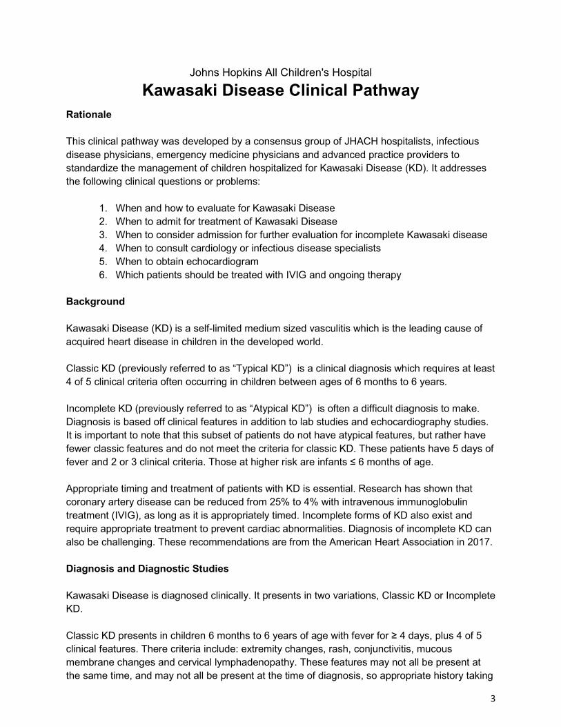
Rationale This clinical pathway was developed by a consensus group of JHACH hospitalists, infectious disease physicians, emergency medicine physicians and advanced practice providers to standardize the management of children hospitalized for Kawasaki Disease (KD). It addresses the following clinical questions or problems:
1. When and how to evaluate for Kawasaki Disease 2. When to admit for treatment of Kawasaki Disease 3. When to consider admission for further evaluation for incomplete Kawasaki disease 4. When to consult cardiology or infectious disease specialists 5. When to obtain echocardiogram 6. Which patients should be treated with IVIG and ongoing therapy
Background Kawasaki Disease (KD) is a self-limited medium sized vasculitis which is the leading cause of acquired heart disease in children in the developed world. Classic KD (previously referred to as “Typical KD”) is a clinical diagnosis which requires at least 4 of 5 clinical criteria often occurring in children between ages of 6 months to 6 years. Incomplete KD (previously referred to as “Atypical KD”) is often a difficult diagnosis to make. Diagnosis is based off clinical features in addition to lab studies and echocardiography studies. It is important to note that this subset of patients do not have atypical features, but rather have fewer classic features and do not meet the criteria for classic KD. These patients have 5 days of fever and 2 or 3 clinical criteria. Those at higher risk are infants ≤ 6 months of age. Appropriate timing and treatment of patients with KD is essential. Research has shown that coronary artery disease can be reduced from 25% to 4% with intravenous immunoglobulin treatment (IVIG), as long as it is appropriately timed. Incomplete forms of KD also exist and require appropriate treatment to prevent cardiac abnormalities. Diagnosis of incomplete KD can also be challenging. These recommendations are from the American Heart Association in 2017. Diagnosis and Diagnostic Studies Kawasaki Disease is diagnosed clinically. It presents in two variations, Classic KD or Incomplete KD. Classic KD presents in children 6 months to 6 years of age with fever for ≥ 4 days, plus 4 of 5 clinical features. There criteria include: extremity changes, rash, conjunctivitis, mucous membrane changes and cervical lymphadenopathy. These features may not all be present at the same time, and may not all be present at the time of diagnosis, so appropriate history taking

4
is critical to making the diagnosis of KD. Laboratory evaluation can assist with clinical decisions, but the diagnosis of KD is a clinical one. Classically, those with KD will have an elevated white blood cell count (WBC), and elevated markers of inflammation, elevated liver enzymes, low albumin levels and sterile pyuria. Thrombocytosis is common, but usually late in the clinical course. Clinical Criteria of Classic Kawasaki Disease:
Kawasaki Disease is defined by a set of clinical criteria listed below: 1. Fever ≥4 days duration 2. Presence of at least 4 of the following features:
-Polymorphous rash (desquamation, morbilliform, erythema multiforme) -Cervical lymphadenopathy > or = 1.5 cm, usually unilateral -Changes in lips/oral mucosa (strawberry tongue/ulcerations) -Extremity changes (swelling, erythema) -Nonexudative conjunctivitis with limbic sparing
If patient presents with fever for at least five days and two or three of the above criteria, further evaluation for Incomplete KD is warranted. Incomplete Kawasaki Disease Incomplete Kawasaki Disease is an often a difficult diagnosis to make. It is important to note that this subset of patients do not have atypical features, but rather have fewer classic features and do not meet criteria for Kawasaki disease. The patients have 5 days of fever with 2-3 clinical criteria. Those at higher risk for coronary artery lesions are infants < or = 6 months age. When suspecting incomplete disease, the incomplete KD algorithm may be beneficial. Of note, the current (2017) guidelines recommend considering KD on the differential for the following patients:
1. Infants <6 months with prolonged fever and irritability 2. Infants with unexplained aseptic meningitis 3. Infants or children with culture negative shock 4. Infants or children with prolonged fever and cervical lymphadenitis, retropharyngeal or
parapharyngeal phlegmon unresponsive to antibiotic therapy Differential Diagnoses of Patients Presenting with Features of KD5
The differential diagnosis includes other infectious and noninfectious conditions, including the following: • Multisystem Inflammatory Syndrome in Children (MIS-C) • Measles • Other viral infections (eg, adenovirus, enterovirus) • Staphylococcal and streptococcal toxin-mediated diseases (eg, scarlet fever and toxic shock
syndrome) • Drug hypersensitivity reactions, including Stevens Johnson syndrome • Systemic onset juvenile idiopathic arthritis
With epidemiologic risk factors: • Rocky Mountain spotted fever or other rickettsial infections • Leptospirosis

5
Emergency Center Evaluation
Patients often present emergency room for evaluation of prolonged fevers, a thorough history and physical exam will help distinguish between KD and other illnesses. Classic KD (previously referred to as “typical KD”) is diagnosed clinically, and EC management includes obtaining labs and admission for echocardiogram and IVIG. Incomplete KD (previously referred to as “Atypical KD”) is more difficult to diagnose, the Incomplete KD EC algorithm may help the EC provider with the diagnosis. Patients without elevation in inflammatory markers are less likely to have KD, though clinical judgement and provider experience will help with EC disposition. Classic KD should be considered for fevers of at least 4 consecutive days, measured over 38°C, with at least 4 clinical symptoms suggestive of KD such as mucositis, rash, and large lymph nodes. Fever alone does not support the diagnosis of Classic KD. It is important to recognize that some viral and bacterial illness can trigger KD, including influenza, and that the presence of another source of fever does not completely exclude KD. Illnesses such as adenovirus and strep pharyngitis make the diagnosis of KD less likely, however the presence of KD is not completely excluded and clinical judgement and provider experience is important in considering the diagnosis of KD. Consideration of the patient’s age is also a key factor since Classic KD is usually between 6 months to 6 years and Incomplete KD can occur at any age. Infectious Disease specialist consult should be considered when diagnosis is uncertain. Multisystem Inflammatory Syndrome in Children (MIS-C) In 2020 Pediatric Healthcare Institutions in the United Kingdom (UK) and New York City noted increased reports of previously healthy pediatric patients who presented with a severe inflammatory syndrome with Kawasaki disease-like features in patients who tested positive or had recent infection by SARS-CoV-2, the novel coronavirus which causes COVID-195,6. Because of the overlap in clinical features, MIS-C should be on the differential diagnosis (DDX) for any patient who present with signs and symptoms of KD with a history of COVID infection. Clinicians should refer to the Johns Hopkins All Children’s Hospital MIS-C Clinical Pathway for more information. Rashes There are some classic cutaneous and extremity changes noted with KD. In the acute phase, erythema or painful induration of hands and feet may be noted. An erythematous, usually diffuse, maculopapular eruption, can develop as early as day 5. Other generalized rashes such as scarlatiniform erythroderma, and erythema multiforme are common. Desquamation of fingers and toes around the nail beds usually begins around 2-3 weeks after the onset of fever and may extend into the palms and soles.
6
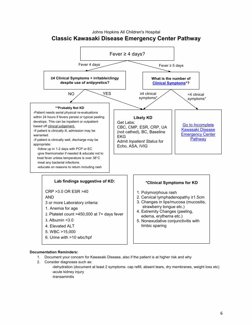
Johns Hopkins All Children's Hospital Classic Kawasaki Disease Emergency Center Pathway
Documentation Reminders:
1. Document your concern for Kawasaki Disease, also if the patient is at higher risk and why 2. Consider diagnoses such as:
-dehydration (document at least 2 symptoms- cap refill, absent tears, dry membranes, weight loss etc) -acute kidney injury -transaminitis
Fever ≥ 4 days?
≥4 Clinical Symptoms + irritable/clingy despite use of antipyretics?
What is the number of Clinical Symptoms*?
**Probably Not KD -Patient needs serial physical re-evaluations within 24 hours if fevers persist or typical peeling develops. This can be inpatient or outpatient based off clinical judgement. -If patient is clinically ill, admission may be warranted. -If patient is clinically well, discharge may be appropriate:
-follow up in 1-2 days with PCP or EC -give thermometer if needed & educate not to treat fever unless temperature is over 38°C -treat any bacterial infections -educate on reasons to return including rash
Likely KD Get Labs: CBC, CMP, ESR, CRP, UA (not cathed), BC, Baseline EKG Admit Inpatient Status for Echo, ASA, IVIG
*Clinical Symptoms for KD 1. Polymorphous rash 2. Cervical lymphadenopathy ≥1.5cm 3. Changes in lips/mucosa (mucositis, strawberry tongue etc.) 4. Extremity Changes (peeling, edema, erythema etc.) 5. Nonexudative conjunctivitis with limbic sparing
Fever ≥ 5 days Fever 4 days
YES NO ≥4 clinical symptoms*
Go to Incomplete Kawasaki Disease Emergency Center
Pathway
<4 clinical symptoms*
Lab findings suggestive of KD:
CRP >3.0 OR ESR >40 AND 3 or more Laboratory criteria: 1. Anemia for age 2. Platelet count >450,000 at 7+ days fever 3. Albumin <3.0 4. Elevated ALT 5. WBC >15,000 6. Urine with >10 wbc/hpf

7
Johns Hopkins All Children's Hospital Incomplete Kawasaki Disease Emergency Center Pathway
Fever for 5+ days with 2-3 clinical symptoms of KD
OR Infant <6m +fever for 7+days
OR Infants or children with prolonged fever and cervical lymphadenitis, retropharyngeal or parapharyngeal phlegmon unresponsive to
antibiotic therapy
Yes
Obtain the following studies: CBC, CMP, UA (not cathed) with micro, CRP and ESR, BC, Baseline EKG Consider the following or other testing as indicated: If ENT/Respiratory symptoms: RVP, optional sinus CT imaging, strep, CXR, mono If GI symptoms: GI PCR panel (for adenovirus testing)
Consider other DDX including MIS-C HLH EBV etc
CRP <3.0 AND ESR <40 (regardless of other lab findings)
CRP >3.0 OR ESR >40 AND 3 or more lab
criteria*
Admit Inpatient status for IVIG, ASA, Echo within 24 hours and consult Infectious disease, Cardiology
Infant <6 months with fever 7+ days
Admit to hospitalist for further evaluation and echocardiogram
Consider ID consult in any
patient with 5 or more days of
fever and suspicion for KD
*Lab findings suggestive of KD:
CRP >3.0 OR ESR >40 AND 3 or more Laboratory criteria: 1. Anemia for age 2. Platelet count >450,000 at 7+ days fever 3. Albumin <3.0 4. Elevated ALT 5. WBC >15,000 6. Urine with >10 wbc/hpf
**Probably Not KD -Patient needs serial physical re-evaluations within 24 hours if fevers persist or typical peeling develops. This can be inpatient or outpatient based off clinical judgement. -If patient is clinically ill, admission may be warranted. -If patient is clinically well, discharge may be appropriate:
-follow up in 1-2 days with PCP or EC -give thermometer if needed & educate
not to treat fever unless temperature is over 38°C
-treat any bacterial infections -educate on reasons to return including
rash
**Strep, Flu and other viral causes can trigger Kawasaki Disease; the presence of flu, strep pharyngitis or other source of illnesses makes diagnosis of KD less likely but does not completely exclude KD. Consult ID if necessary.
No

8
Inpatient Evaluation
Classic KD (previously referred to as “Typical KD”) is a clinical diagnosis which requires at least 4 days of fever and 4 of 5 clinical criteria. It often occurs in children between ages of 6 months to 6 years with slight male predominance. Initial management includes aspirin and IVIG therapy, with refractory cases requiring discussion with ID specialist. Incomplete (previously referred to as “Atypical KD”) is an often a difficult diagnosis to make. It is important to note that this subset of patients do not have atypical features, but rather have fewer classic features and do not meet criteria for Kawasaki disease. The patients have 5 days of fever with 2-3 clinical criteria. Those at higher risk for coronary artery lesions are infants ≤6 months age.
When suspecting incomplete disease, the Incomplete Kawasaki Disease Inpatient Pathway should be used.

9
Johns Hopkins All Children's Hospital Classic Kawasaki Disease Inpatient Pathway
≥ 4 Days of Fever
Does the patient have 4 out of 5 of the following Clinical Criteria: a. Polymorphous rash b. Cervical Lymphadenopathy ≥ 1.5cm c. Changes in lips/oral mucosa d. Extremity changes e. Nonexudative conjunctivitis with limbic sparing
KD Unlikely, Reevaluate Diagnosis
YES NO
YES NO
1. Consider DDX including MIS-C, HLH, EBV, etc 2. Consult Infectious Disease, especially for patients
with only 4 days of fever. 3. Consult Cardiology 4. Begin IVIG (should not be delayed for consultation
or echo in patients who have conclusive KD) 5. Obtain Echocardiogram
Are there 2-3 out of 5 Clinical Criteria?
NO YES
Still consider these patients for incomplete disease: Infants <6 m with prolonged (7 day) fever and irritability OR Infants with unexplained aseptic meningitis OR Infants or children with culture negative shock OR Infants or children with prolonged fever and cervical lymphadenitis, retropharyngeal or parapharyngeal phlegmon unresponsive to antibiotic therapy
Refer to Incomplete KD Clinical Pathway
NO
YES
KD Unlikely, Reevaluate Diagnosis

10
Johns Hopkins All Children's Hospital Incomplete Kawasaki Disease Inpatient Pathway
Adapted from Circulation article (McCrindle)
(1) In the absence of a “gold standard” for diagnosis, this algorithm cannot be evidence based but rather represents the informed opinion of the expert committee. Consultation with an expert should be sought any time assistance is needed. (2) Clinical findings of Kawasaki disease are listed in Table 3 (McCrindle). Characteristics suggesting that another diagnosis should be considered include exudative conjunctivitis, exudative pharyngitis, ulcerative intraoral lesions, bullous or vesicular rash, generalized adenopathy or splenomegaly. (3) Infants ≤ 6 months of age are the most likely to develop prolonged fever without other clinical criteria for Kawasaki disease; these infants are at particularly high risk of developing coronary artery abnormalities. (4) Echocardiography is considered positive for purposes of this algorithm if any of 3 conditions are met: Z score of left anterior descending coronary artery or right coronary artery ≥ 2.5; coronary artery aneurysm is observed; or ≥ 3 other suggestive features exist, including decreased left ventricular function, mitral regurgitation, pericardial effusion, or Z scores in left anterior descending coronary artery or right coronary artery of 2 to 2.5. (5) If the echocardiogram is positive, treatment should be given within 10 days of fever onset or after the tenth day of fever in the presence of clinical and laboratory signs (C-reactive protein [CRP], erythrocyte sedimentation rate [ESR]) of ongoing inflammation. (6) Typical peeling begins under the nail beds of fingers and toes. ALT indicates alanine transaminase; and WBC, white blood cells.
Children with fever ≥ 5 days and 2 or 3 compatible clinical criteria2 OR Infants with fever for ≥ 7 days without other explanation3
Consider DDX including MIS-C, HLH, EBV, etc
Assess Laboratory Tests
CRP <3.0mg/dL and ESR <40mm/hr CRP ≥3.0mg/dL and/or ESR ≥40mm/hr
Serial clinical and laboratory re-evaluation if fevers persist
Echocardiogram if typical6 peeling develops
Refer to Discharge Criteria &
Follow Up
3 or more Laboratory Findings: 1. Anemia for age 2. Platelet count of ≥450,000 after the 7th day of fever
3. Albumin ≤3.0g/dL 4. Elevated ALT level 5. WBC count of ≥15,000/mm3 6. Urine ≥10 WBC/hpf
OR Positive echocardiogram if already done. (See footnote 4)
Patient with Incomplete KD Consult ID, Consult Cardiology
Order Echocardiogram Treat5
YES
NO

11
Inpatient Management
Pharmacotherapy
Note that if Kawasaki disease is suspected, treatment should NOT be delayed in order to obtain echocardiogram or specialty consultation
Primary treatment:
1. Patients with complete KD criteria and those who meet the algorithm for incomplete KD should be treated with high dose IVIG (2g/kg given as a single IV infusion) within 10 days of illness onset but as soon as possible after diagnosis (Class 1; Level of Evidence A)
2. IVIG generally should not be administered to patients beyond the tenth day of illness in the absence of fever, significant elevation of inflammatory markers, or coronary abnormalities (Class III, Level of Evidence C). It is reasonable to administer IVIG to children presenting after the 10th day of illness (ie in whom the diagnosis was missed earlier) if they have either persistent fever without other explanation or coronary artery abnormalities together with ongoing systemic inflammation, as manifested by elevated ESR or CRP >3 mg/dL (Class IIa, Level of Evidence B)
3. Single-dose pulse methylprednisolone should not be administered with IVIG as routine primary therapy for patients with KD (Class III; Level of Evidence B).
4. Administration of a longer course of corticosteroids (eg, tapering over 2–3 weeks), together with IVIG 2 g/kg and Aspirin (ASA), may be considered for treatment of high-risk patients with acute KD, when such high risk can be identified in patients before initiation of treatment (Class IIb; Level of Evidence B).
a. High risk: infants <6 months, patients with large or giant aneurysms (internal luminal diameter Z score ≥10 or absolute dimension ≥8 mm) are particularly high risk for coronary artery thrombosis.
b. In North America, there is no consensus on prediction scores for those at risk for IVIG resistance: Japanese scoring systems for IVIG resistance and aneurysms have low sensitivity in North American populations.
5. Administration of moderate (30–50 mg/kg/day) to high-dose (80–100 mg/kg/day) ASA is reasonable until the patient is afebrile, although there is no evidence that it reduces coronary artery aneurysms (Class IIa; Level of Evidence C).
6. Recommendations on when to transition to low dose aspirin is variable in the literature. Locally, our experts generally recommend transitioning to low dose ASA (3-5 mg/kg once daily) upon discharge from the hospital or when the patient returns to baseline. If patient has not returned to baseline within 7 days, he/she will need immediate follow up with physician (PCP, ER, ID, cardiology) (Evidence Level Low, Recommendation Strong)

12
Treatment in IVIG Resistant Patients: 1. IVIG resistance is defined as persistent or recrudescent fever at least 36 hours and <7
days after completion of first IVIG infusion. 2. Most frequently administered
a. IVIG: Second infusion Pooled polyclonal IG 2 g/kg IV b. IVIG + steroid: IVIG 2 g/kg IV + methylprednisolone 2 mg/kg/day IV divided every
8 h until afebrile, then prednisone or prednisolone orally until CRP normalized, then taper over 2–3 weeks.
c. Infliximab Monoclonal antibody against TNF-α Single infusion: 5 mg/kg IV given over 2 h
3. Administration of cyclosporine may be considered in patients with refractory KD in whom a second IVIG infusion, infliximab, or a course of steroids has failed (Class IIb; Level of Evidence C).
4. Administration of immunomodulatory monoclonal antibody therapy (except TNF-α blockers), cytotoxic agents, or (rarely) plasma exchange may be considered in highly refractory patients who have failed to respond to a second infusion of IVIG, an extended course of steroids, or infliximab (Class IIb; Level of Evidence C).
Echocardiography
Recommendations for Cardiovascular Assessment for Diagnosis and Monitoring During the Acute Illness
1. Echocardiography should be performed when the diagnosis of KD is considered, but unavailability or technical limitations should not delay treatment (Class 1, level of evidence B)
2. Coronary arteries should be imaged, and quantitative assessment of luminal dimensions, normalized as Z scores adjusted for body surface area should be performed (Class 1; Level of Evidence B)
Classification of Coronary Artery Abnormalities on Echocardiogram:
Z-score classification
1. No involvement: Always <2 2. Dilation only: 2 to <2.5. 3. Small aneurysm: >2.5 to <5 4. Medium aneurysm: >5 to <10 and absolute dimension <8 mm 5. Large or giant aneurysm: >10, or absolute dimension >8 mm
From the Incomplete KD pathway, a positive echocardiogram includes*:
Left anterior descending (LAD) or right coronary artery Z-score >2.5 OR
Any coronary artery aneurysm OR
3 or more of the following:
1. Decreased left ventricular function

13
2. Mitral regurgitation 3. Pericardial effusion 4. Left anterior descending (LAD) or right coronary artery Z-score of 2 to <2.5
*Please note that there may be other cardiac lesions associated with KD, including valvular disease and myocarditis. It is important to discuss each individual case with cardiology and infectious disease.
Discharge Criteria & Follow Up
Discharge Criteria & Considerations*
1. Patient afebrile >24 hours and with improved clinical status (decreased irritability, adequate hydration and voiding)
2. All medications are sent to pharmacy in advance 3. All consultations and follow up appointments are made 4. Recommend Influenza vaccine on all patients (unless contraindicated ie anaphylaxis
to vaccine or egg protein) to avoid Reye’s Syndrome. (Evidence Level Low, Recommendation Strong)
5. Ibuprofen should be avoided in children with coronary aneurysms taking aspirin for its antiplatelet effects (Evidence Level Low, Recommendation Strong)
6. Identify if there are transportation issues for families for discharge home and for follow up appointments
7. Family understands instructions for follow up, medications, and when to seek immediate medical attention
8. Other considerations- No live vaccines 11 months after administration of IVIG (MMR, varicella), identify all knowledge gaps
Follow Up*
1. Primary Care Physician within 1 week 2. Cardiology outpatient follow up in 1-2 week after discharge.
a. In uncomplicated cases, echocardiogram should be repeated in 1-2 weeks and again in 4-6 weeks after treatment (Class 1; Level of Evidence B)
b. For patients with evolving coronary artery abnormalities (Z score >2.5) more frequent echocardiography should be performed (at least twice per week) until luminal dimensions have stopped progressing to determine the risk for and presence of thrombosis (Class 1; Level of Evidence B)
c. Outpatient appointment should be made at 2 weeks, then 4-6 weeks. Both appointments should be scheduled prior to discharge..
3. Infectious Disease follow up in 1-2 weeks
*(Evidence Level Low, Recommendation Strong)

14
Documentation Reminders
1. Document your concern for Kawasaki Disease, also if the patient is at higher risk and why 2. Consider diagnoses such as:
-dehydration (document at least 2 symptoms- cap refill, absent tears, dry membranes, weight loss etc)
-acute kidney injury -transaminitis

15
References
1. Center for Disease Control: https://www.cdc.gov 2. Dajani, A., Taubert KA, Takahashi, M., Bierman, F., et al. (1994). Guidelines for long-term
management of patients with Kawasaki disease. Report from the committee on rheumatic fever, endocarditis, and Kawasaki disease, council on cardiovascular disease in the young, American Heart Association. Circulation, 89(2), 916.
3. Feldstein L.R., et al; Multisystem Inflammatory Syndrome in U.S. Children & Adolescents, N Engl J Med. 2020 Jul 23;383(4):334-346
4. LiJiang MDa†, et al COVID-19 and multisystem inflammatory syndrome in children and adolescents, The Lancet Infectious Diseases, Volume 20, Issue 11, November 2020, Pages e276-e288
5. McCrindle, B., Rowley, A., Newburger, J., et al. (2017). Diagnosis, treatment, and long-term management of Kawasaki disease: A scientific statement for health professionals from the American Heart Association. Circulation, 135, e927. http://circ.ahajournals.org/content/early/2017/03/29/CIR.0000000000000484
6. Newburger, J., Takahashi, M., Gerber, M., et al. (2004). Diagnosis, treatment, and long-term management of Kawasaki disease: A statement for health professionals from the committee on rheumatic fever, endocarditis and Kawasaki disease, council on cardiovascular disease in the young, American Heart Association. Circulation, 110(17), 2748. http://circ.ahajournals.org/content/110/17/2747.full
7. Weiss, J., Eberhard, B., Chowdhury, D., & Gottlieb, B. (2004). Infliximab as a novel therapy for refractory Kawasaki disease. Journal of Rheumatology, 31, 808.
8. Verdoni L, Mazza A, Gervasoni A, et al. An outbreak of severe kawasaki-like disease at the italian epicentre of the SARS-CoV-2 epidemic: An observational cohort study. The Lancet. https://doi.org/10.1016/S0140- 6736(20)31103-X. doi: 10.1016/S0140-6736(20)31103-X

16
Outcome Measures:
1. Length of Stay in EC 2. Days admission 3. Development of cardiac abnormalities (long term - 1 year follow up) 4. Number of admissions 5. Number echocardiograms performed for KD 6. Number of positive echocardiograms during initial admission (short term) 7. Number of patients with discharge dx of KD 8. Time of admission to time to dose of IVIG
Clinical Pathway Team Kawasaki Disease Clinical Pathway
Johns Hopkins All Children’s Hospital Owner(s): Shaila Siraj, MD; Meghan Martin, MD; Juan Dumois, MD; Ashish Shah, MD; Courtney Titus, PA-C, EMT-P
Initially Created by: Shaila Siraj, Hospitalist Initially Reviewed by: Infectious Disease Group: Allison Messina, David Berman, Juan Dumois, Matthew Thomas, Katie Namtu Cardiology Group: Ashish Shah, Grace Frieire, Gary Stapleton, Daniel Mauriello Dipti Amin, CMIO Revised January 2018 by: Hospitalist: Shaila Siraj, MD Emergency Medicine: Meghan Martin, MD; Joseph Perno, MD; Courtney Titus, PA-C Infectious Disease: Juan Dumois, MD Cardiology: Ashish Shah, MD Revised July 2021 by: Hospitalist: Shaila Siraj, MD Emergency Medicine: Meghan Martin, MD; Courtney Titus, PA-C Infectious Disease: Juan Dumois, MD Cardiology: Ashish Shah, MD

17
Disclaimer
Clinical Pathways are intended to assist physicians, physician assistants, nurse practitioners and other health care providers in clinical decision-making by describing a range of generally acceptable approaches for the diagnosis, management, or prevention of specific diseases or conditions. The ultimate judgment regarding care of a particular patient must be made by the physician in light of the individual circumstances presented by the patient.
The information and guidelines are provided "AS IS" without warranty, express or implied, and Johns Hopkins All Children’s Hospital, Inc. hereby excludes all implied warranties of merchantability and fitness for a particular use or purpose with respect to the information. Johns Hopkins All Children’s Hospital, Inc. shall not be liable for direct, indirect, special, incidental or consequential damages related to the user's decision to use the information contained herein.
Related Documents