Joe B. (Bill) Putnam, Jr., MD, FACS Ingram Professor of Surgery and Chairman Department of Thoracic Surgery Professor, Department of Biomedical Informatics Vanderbilt University Medical Center Nashville, Tennessee 37232-5734 Thoracic Surgery: A Pleural Problem Primer Vanderbilt University Medical Center February 14, 2007

Joe B. (Bill) Putnam, Jr., MD, FACS Ingram Professor of Surgery and Chairman Department of Thoracic Surgery Professor, Department of Biomedical Informatics.
Dec 30, 2015
Welcome message from author
This document is posted to help you gain knowledge. Please leave a comment to let me know what you think about it! Share it to your friends and learn new things together.
Transcript
Joe B. (Bill) Putnam, Jr., MD, FACSIngram Professor of Surgery and Chairman
Department of Thoracic Surgery
Professor, Department of Biomedical Informatics
Vanderbilt University Medical CenterNashville, Tennessee 37232-5734
Thoracic Surgery:A Pleural Problem Primer
Vanderbilt University Medical Center
February 14, 2007
STEP 1.
Take your own pulse.
STEP 2.
Relax. Take a deep breath.
If the patient is not arresting, think…
Reflect on the situation
Ask yourself the question ‘Why…..’
Save the patient’s life…..!
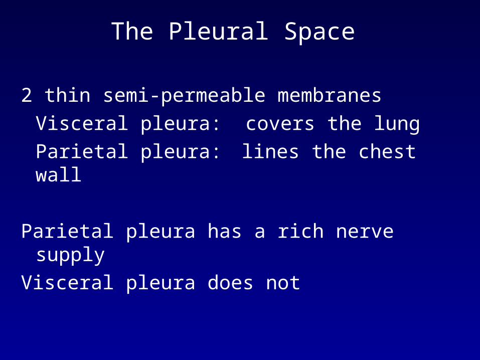
The Pleural Space
2 thin semi-permeable membranes
Visceral pleura: covers the lung
Parietal pleura: lines the chest wall
Parietal pleura has a rich nerve supply
Visceral pleura does not
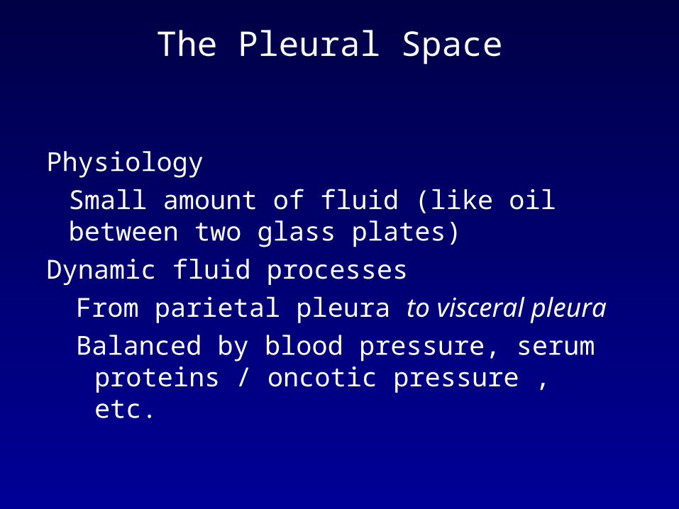
The Pleural Space
Physiology
Small amount of fluid (like oil between two glass plates)
Dynamic fluid processes
From parietal pleura to visceral pleura
Balanced by blood pressure, serum proteins / oncotic pressure , etc.


Diseases of the pleura
Air - Pneumothorax
Blood - Hemothorax
Infection - Empyema
Air - Pneumothorax
Chyle - Chylothorax
Tumor - ‘Tumor-thorax’ or tumor tamponade
Fluid - Pleurothorax

Pneumothorax
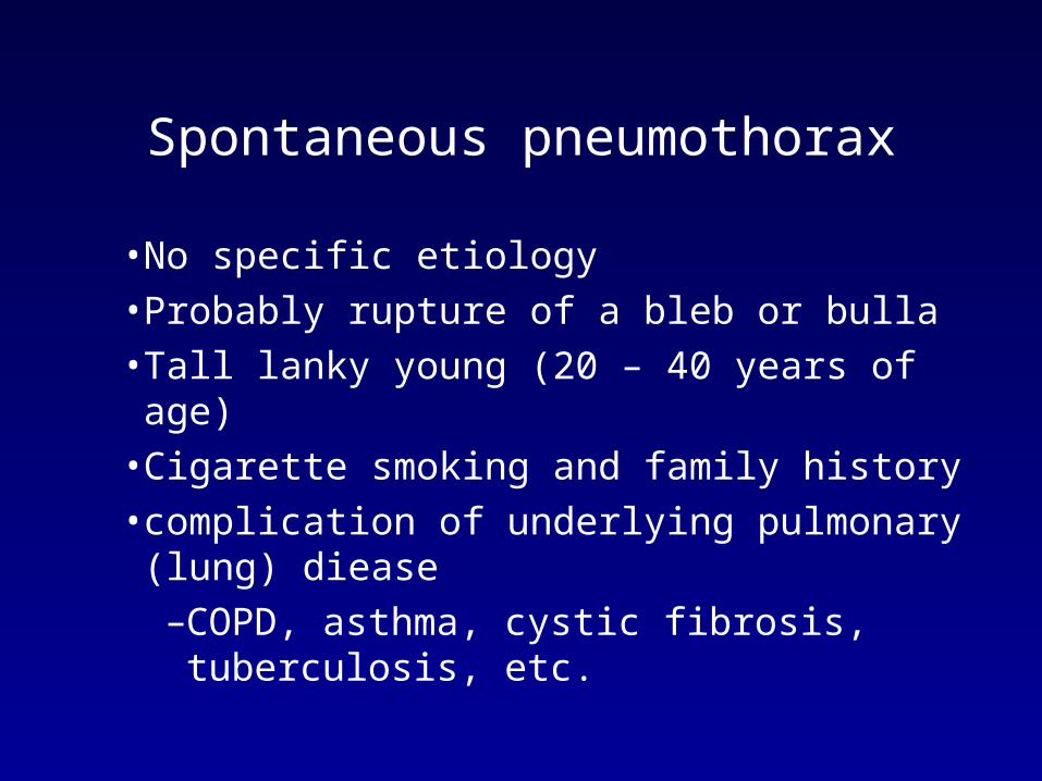
Spontaneous pneumothorax
• No specific etiology
• Probably rupture of a bleb or bulla
• Tall lanky young (20 – 40 years of age)
• Cigarette smoking and family history
• complication of underlying pulmonary (lung) diease
–COPD, asthma, cystic fibrosis, tuberculosis, etc.

Traumatic pneumothorax
• Penetrating
–GSW / stab
• Blunt trauma
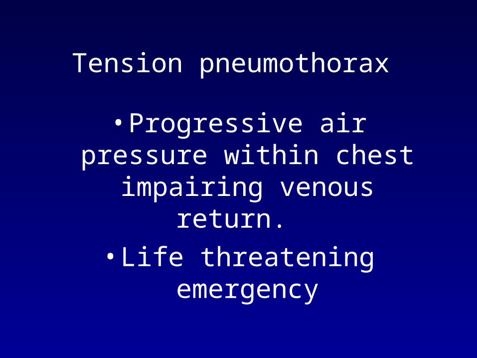
Tension pneumothorax
• Progressive air pressure within chest impairing venous return.
• Life threatening emergency
Tension Pneumothorax
Respiratory distressHyper-resonanceAbsent breath soundsTracheal deviation
NOT AN X-RAY DIAGNOSISRx: Immediate needle decompression,
chest tube
Tension Pneumothorax
• Tension pneumothorax is a clinical diagnosis and should not be made radiologically!
• Confused with hemothorax
• Hyperresonance is the key to diagnosis
• If in doubt - insert needle!
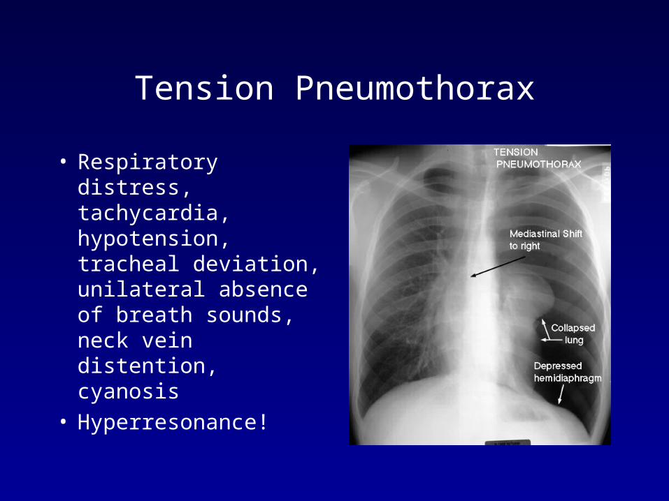
Tension Pneumothorax
• Respiratory distress, tachycardia, hypotension, tracheal deviation, unilateral absence of breath sounds, neck vein distention, cyanosis
• Hyperresonance!


•Spontaneous PTX More common males ages 20-40, smokersMay occur following a scream, valsalva, or coughSudden sharp pleuritic chest pain and dyspnea Tachypnea, tachycardia, subcutaneous
emphysema
Expiratory CXR may be needed to make diagnosis
Treatment: Observation for PTX less than 15-20% and no symptomsOthers: needle/catheter decompression and/or chest tube

Pneumomediastinumpneumopericardiumsubcutaneous emphysema
Barotrauma
Open Pneumothorax
• Large defect (>2/3 size of trachea)
• Removes normally negative intra-thoracic pressures - thus impedes respiration
• Prompt closure - sterile, occlusive
• Tape three sides - allowing a flap valve effect
• Surgery usually required

Massive Hemothorax
• More than 1500 ml. blood in chest - surgery
• Dullness to percussion, absent breath sounds, possible flat neck veins
• Insert large bore chest tube
• Be prepared for auto-transfusion
• >200 ml/hr may indicate need for surgery
• Most chest wounds do not require surgery
Massive Hemothorax
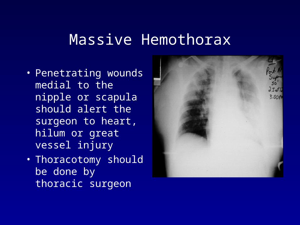
• Penetrating wounds medial to the nipple or scapula should alert the surgeon to heart, hilum or great vessel injury
• Thoracotomy should be done by thoracic surgeon
Massive Hemothorax
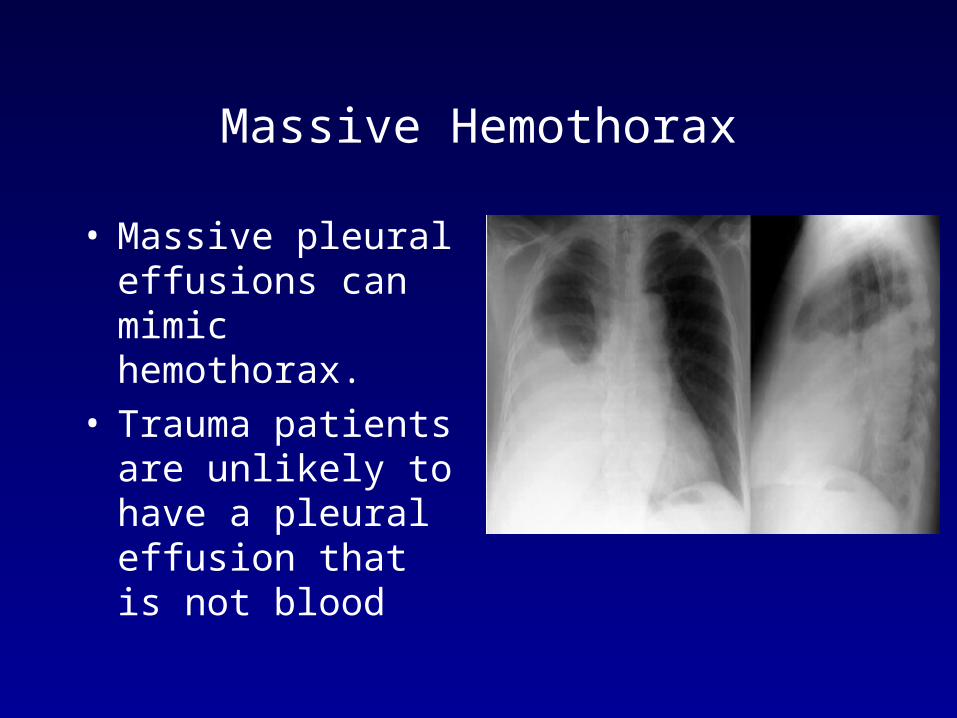
• Massive pleural effusions can mimic hemothorax.
• Trauma patients are unlikely to have a pleural effusion that is not blood

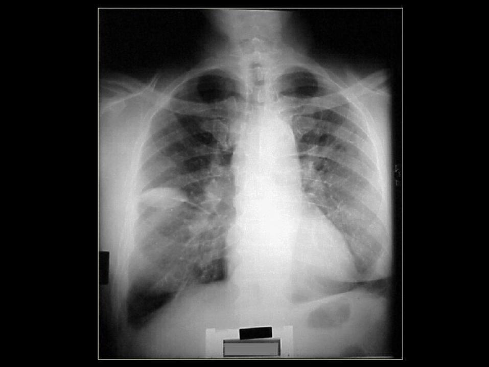
Pleural Effusion
• Fluid that accumulated in the pleural space–Trauma–Disease
•Heart failure•Cancer•Pulmonary embolism• Inflammation
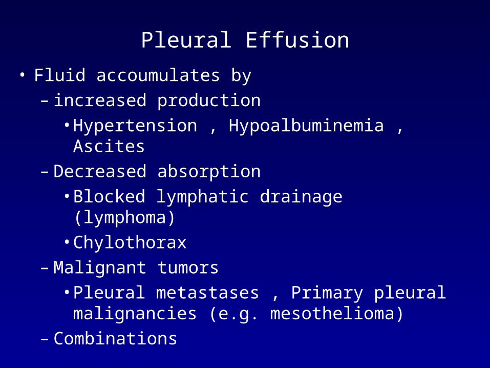
Pleural Effusion
• Fluid accoumulates by – increased production
• Hypertension , Hypoalbuminemia , Ascites– Decreased absorption
• Blocked lymphatic drainage (lymphoma)• Chylothorax
– Malignant tumors• Pleural metastases , Primary pleural
malignancies (e.g. mesothelioma)– Combinations


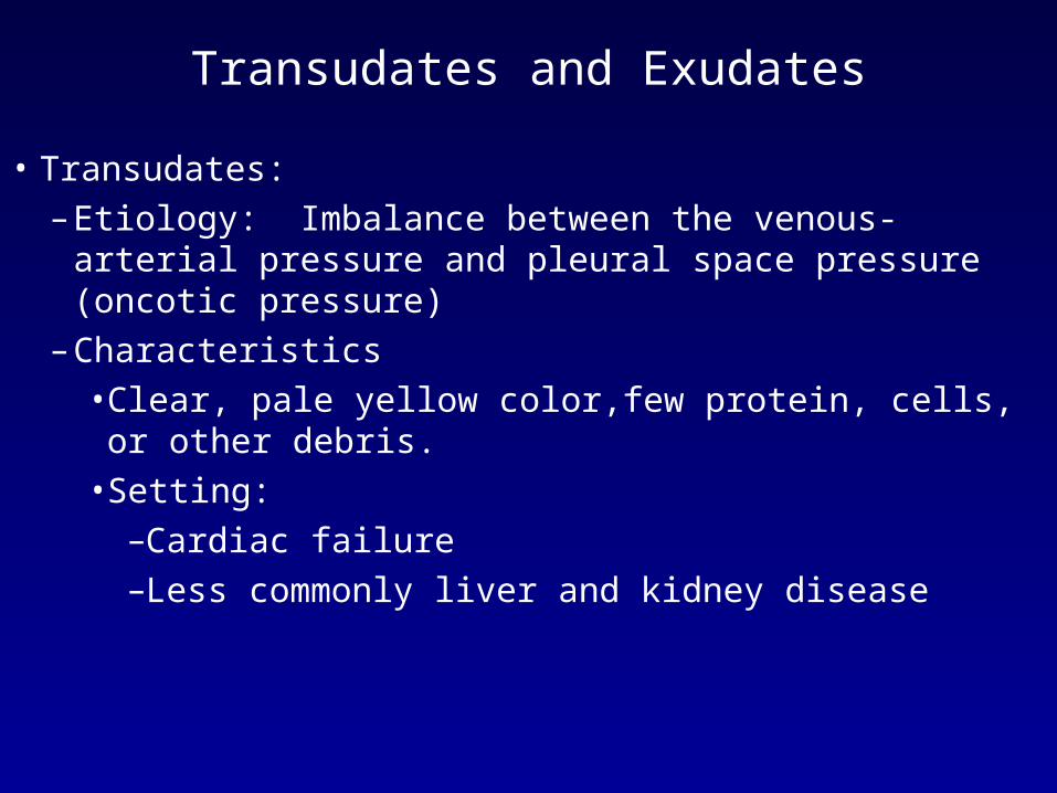
Transudates and Exudates
• Transudates:– Etiology: Imbalance between the venous-arterial
pressure and pleural space pressure (oncotic pressure) – Characteristics
• Clear, pale yellow color,few protein, cells, or other debris.
• Setting:–Cardiac failure–Less commonly liver and kidney disease
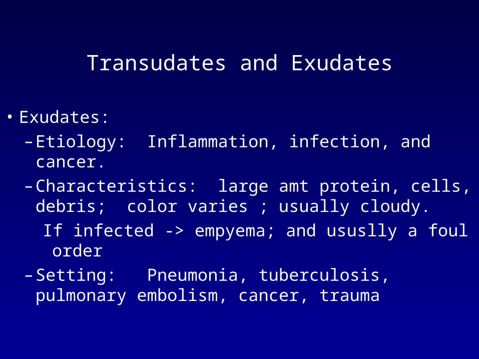
Transudates and Exudates
• Exudates:– Etiology: Inflammation, infection, and cancer. – Characteristics: large amt protein, cells,
debris; color varies ; usually cloudy.
If infected -> empyema; and ususlly a foul order
– Setting: Pneumonia, tuberculosis, pulmonary embolism, cancer, trauma

It’s time we face reality, my friends…..
We’re not exactly rocket scientists.”
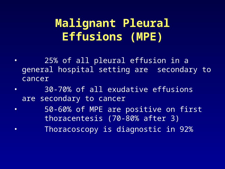
Malignant Pleural Effusions (MPE)
• 25% of all pleural effusion in a general hospital setting are secondary to cancer
• 30-70% of all exudative effusions are secondary to cancer
• 50-60% of MPE are positive on first thoracentesis (70-80% after 3)
• Thoracoscopy is diagnostic in 92%
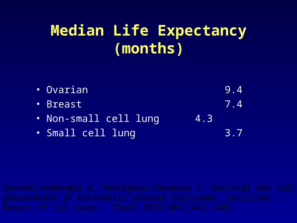
Median Life Expectancy(months)
• Ovarian 9.4• Breast 7.4• Non-small cell lung 4.3• Small cell lung 3.7
Sanchez-Armengol A, Rodriguez-Panadero F: Survival and talc pleurodesis in metastatic pleural carcinoma, revisited. Report of 125 cases. Chest 1993;104:1482-1485.
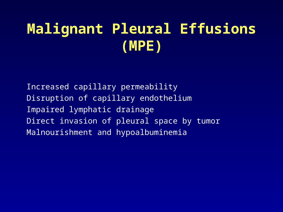
Malignant Pleural Effusions (MPE)
Increased capillary permeability
Disruption of capillary endothelium
Impaired lymphatic drainage
Direct invasion of pleural space by tumor
Malnourishment and hypoalbuminemia
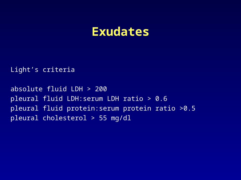
Exudates
Light’s criteria
absolute fluid LDH > 200
pleural fluid LDH:serum LDH ratio > 0.6
pleural fluid protein:serum protein ratio >0.5
pleural cholesterol > 55 mg/dl
Pleural Fluid Analysis
• Cell Count• Cytology• Cultures• LDH• Protein• Glucose• pH

Predicting Survival
• Pleural fluid pH (p=n.s.)• Pleural fluid glucose (p=n.s.)• Extent of pleural carcinomatosis
(p=n.s.)• Karnofsky Performance Score (<=
30 vs. >= 70, p<0.0001)34 d median vs. 395 days
Burrows CM, et al. Chest 117:73-78 (2000)

Treatment Goals for MPE
• Relieve or eliminate dyspnea
• Optimize patient function
• Minimize/eliminate hospitalization
• Minimize cost
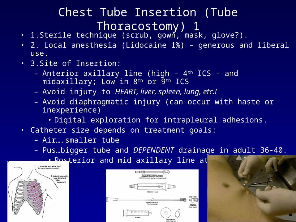
Chest Tube Insertion (Tube Thoracostomy) 1• 1.Sterile technique (scrub, gown, mask, glove?).• 2. Local anesthesia (Lidocaine 1%) – generous and liberal use.• 3.Site of Insertion:
– Anterior axillary line (high – 4th ICS - and midaxillary; Low in 8th or 9th ICS
– Avoid injury to HEART, liver, spleen, lung, etc.!– Avoid diaphragmatic injury (can occur with haste or inexperience)
• Digital exploration for intrapleural adhesions.• Catheter size depends on treatment goals:
– Air….smaller tube– Pus…bigger tube and DEPENDENT drainage in adult 36-40.
• Posterior and mid axillary line at level of 10th rib.
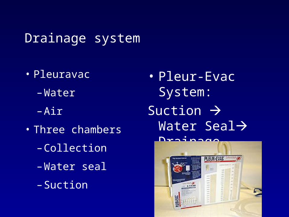
Drainage system
• Pleuravac
– Water
– Air
• Three chambers
– Collection
– Water seal
– Suction
• Pleur-Evac System:
Suction Water Seal Drainage

Treatment: Chest Tube Insertion (Tube Thoracostomy) 2
• Dissect and tunnel with a curved clamp over top of rib!– Neurovascular bundle runs along the inferior border of rib
• In a controlled fashion, puncture into the pleural space• Insert a finger into the pleural space to identify potential space
and guide chest tube. • With a clamp onto the end of the chest tube, and guided by the
finger track, insert the drain into the chest directing it towards the apex and posterior.
• ALL DRAIN HOLES to be within the pleural cavity– Special eye!


Chest Tube Insertion (Tube Thoracostomy) 3
• Check for leaks in the system (persistent air drainage, or inability to re-expand the lung)
• Persistent leak: ruptured bronchus, bronchopleural fistula, ruptured bleb.• Connect to underwater drainage system (Pleurovac). • Secure tube on skin.
– Vaseline gauze not needed ! • Remove when air leak or fluid drainage ceases
– A functionless tube is only a nidus for infection– Have patient take a deep breath in– As patient begins to exhale, remove the tube quikly– Patient involuntarily Valsalva’s, minimizing potential for sucking air into the
pleural cavity• Chest X-ray after removal.

Comments
• Maintain fluid levels in chambers
• Maintain hemostats and dressing at bedside
• Keep extra drainage system on nursing unit
• Do not clamp tubing -- unless getting ready
for removal

The last part of surgery, namely,
operations, is a reflection on the
healing art;
it is a tacit acknowledgement of
the insufficiency of surgery.
It is like an armed savage who
attempts to get that by force
which a civilized man would get
by strategem.
John
Hunter (1728-1793)

Congratulations and Good Luck!
Related Documents


