1 Table S1: GI numbers of proteins referred to. Csa3 Orthologues Csx1 orthologues Protein Name GI Number Protein Name GI Number Sso1445 15898283 Sso1389 1589823 Sso1444 15891282 Hbut_0709 124027589 MJ_0379 1499557 TVN0275 13541106 Smar_0319 126465231 Kcr_0447 170290067 Hbut_0646 124027528 PAE0117 18159125 Pars_1125 145591353 Msed_1154 146303923 Msed_1145 146303914 MJ_1666 1500569 Msed_1151 146309320 PF1127 18893202 APE1237 118431377 PH0168 14590105 AF_1869 2648681 PTO0057 48477129 Other TVN0114 13540945 VC1899 15641901 Aq_378 15605883 Pars_1134 145591362 Tpen_1272 119720178 TTE2636 20808981

Welcome message from author
This document is posted to help you gain knowledge. Please leave a comment to let me know what you think about it! Share it to your friends and learn new things together.
Transcript
1
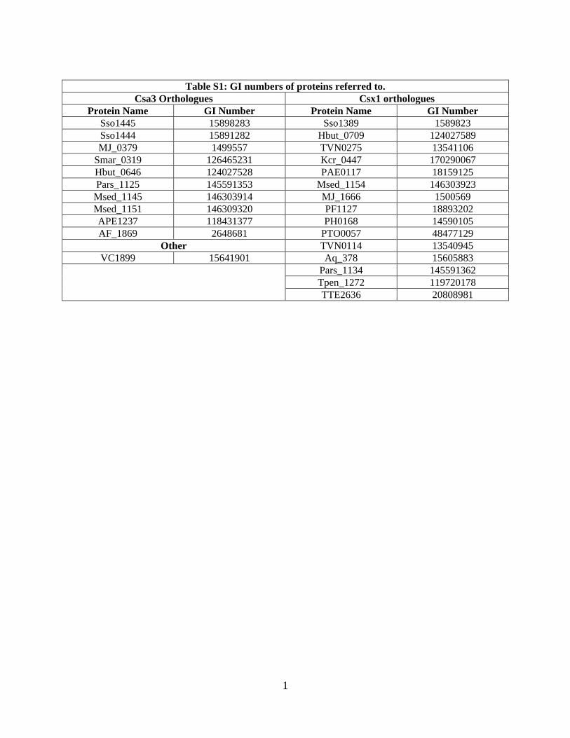
Table S1: GI numbers of proteins referred to.
Csa3 Orthologues Csx1 orthologues Protein Name GI Number Protein Name GI Number
Sso1445 15898283 Sso1389 1589823 Sso1444 15891282 Hbut_0709 124027589 MJ_0379 1499557 TVN0275 13541106
Smar_0319 126465231 Kcr_0447 170290067 Hbut_0646 124027528 PAE0117 18159125 Pars_1125 145591353 Msed_1154 146303923 Msed_1145 146303914 MJ_1666 1500569 Msed_1151 146309320 PF1127 18893202 APE1237 118431377 PH0168 14590105 AF_1869 2648681 PTO0057 48477129
Other TVN0114 13540945 VC1899 15641901 Aq_378 15605883
Pars_1134 145591362 Tpen_1272 119720178 TTE2636 20808981
2
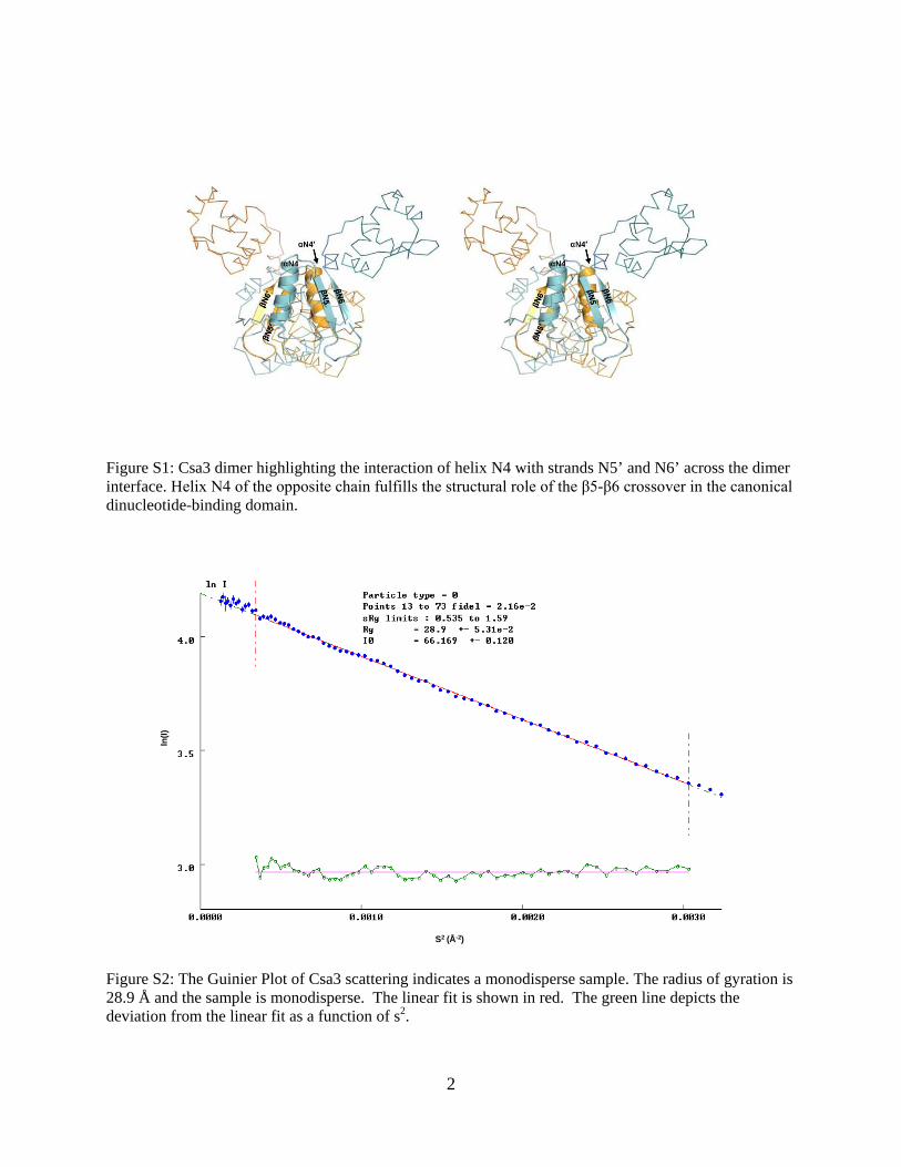
Figure S1: Csa3 dimer highlighting the interaction of helix N4 with strands N5’ and N6’ across the dimer interface. Helix N4 of the opposite chain fulfills the structural role of the β5-β6 crossover in the canonical dinucleotide-binding domain.
S2 (Å-2)
ln(I
)
Figure S2: The Guinier Plot of Csa3 scattering indicates a monodisperse sample. The radius of gyration is 28.9 Å and the sample is monodisperse. The linear fit is shown in red. The green line depicts the deviation from the linear fit as a function of s2.
3
90º
Thr175
Phe10
Arg17
Phe10
Arg17
Arg98Glu122
Glu122
Lys174
Asn178
90º
90º 90º
A
B
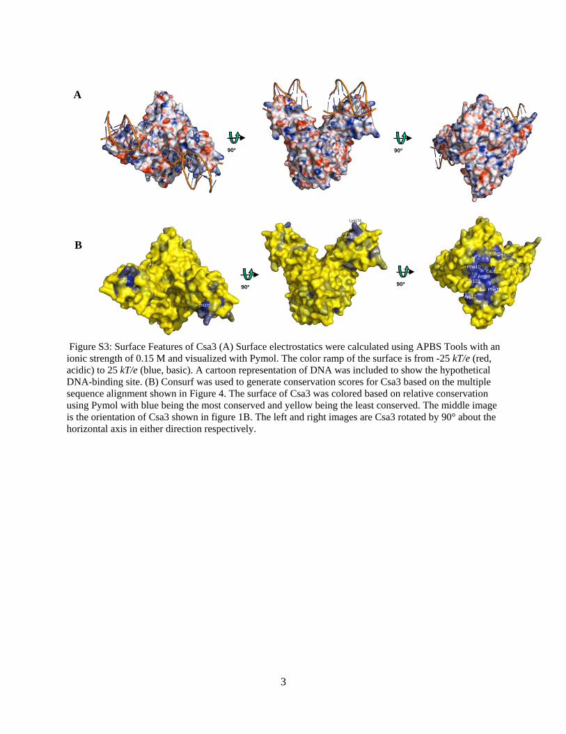
Figure S3: Surface Features of Csa3 (A) Surface electrostatics were calculated using APBS Tools with an ionic strength of 0.15 M and visualized with Pymol. The color ramp of the surface is from -25 kT/e (red, acidic) to 25 kT/e (blue, basic). A cartoon representation of DNA was included to show the hypothetical DNA-binding site. (B) Consurf was used to generate conservation scores for Csa3 based on the multiple sequence alignment shown in Figure 4. The surface of Csa3 was colored based on relative conservation using Pymol with blue being the most conserved and yellow being the least conserved. The middle image is the orientation of Csa3 shown in figure 1B. The left and right images are Csa3 rotated by 90° about the horizontal axis in either direction respectively.
4
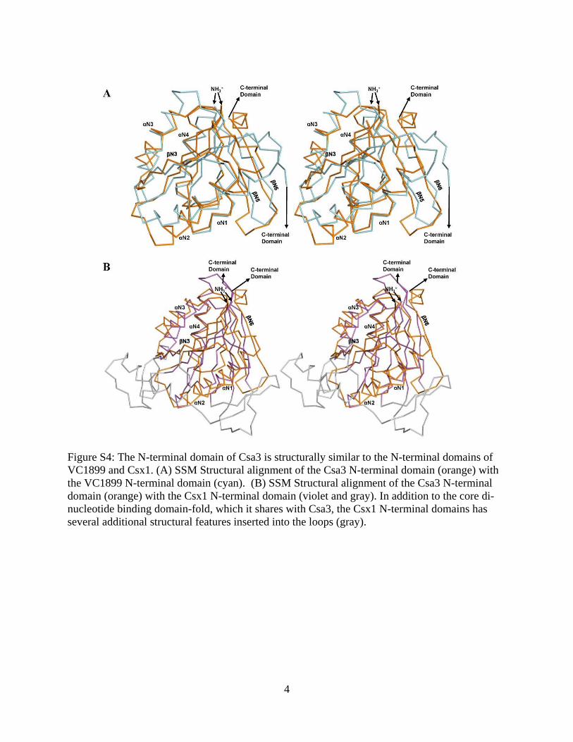
Figure S4: The N-terminal domain of Csa3 is structurally similar to the N-terminal domains of VC1899 and Csx1. (A) SSM Structural alignment of the Csa3 N-terminal domain (orange) with the VC1899 N-terminal domain (cyan). (B) SSM Structural alignment of the Csa3 N-terminal domain (orange) with the Csx1 N-terminal domain (violet and gray). In addition to the core di-nucleotide binding domain-fold, which it shares with Csa3, the Csx1 N-terminal domains has several additional structural features inserted into the loops (gray).

5
g
Figure S5: Csx1 Multiple sequence alignment (Continued on next page)

6
Figure S5: There are four sequence motifs that are conserved among Csx1 orthologues. All genomes in the comprehensive microbial resource databank were searched for Csx1 with the TIGR01897 HMM. All identified orthologues are from genomes that include at least one CRISPR. 15 retrieved sequences were retrieved and aligned using 3D-Coffee followed by manual adjustment around the gaps. Secondary structural elements were calculated using DSSP from the Sso1389 structure (2I71). Motifs 1 (Ser/Ala-h2-Gly-Asn/Asp-Pro-X7-Tyr), 2 (Asp-X-Thr-His-Gly-h-Asn-Tyr/Phe-h) and 3 (Tyr-Asn-Ser-Asp/Glu-Pro) are found in a cleft on the N-terminal domain that spans the dimer interface. Motif 4 (Arg-X3-Ala-His-Gly/Ala-Gly) is found in a cleft on the C-terminal domain that also spans the dimer interface and is distal to the winged HTH domain. GI numbers associated with the listed Csx1 proteins are presented in Table S1. The figure was generated using ALINE.
7
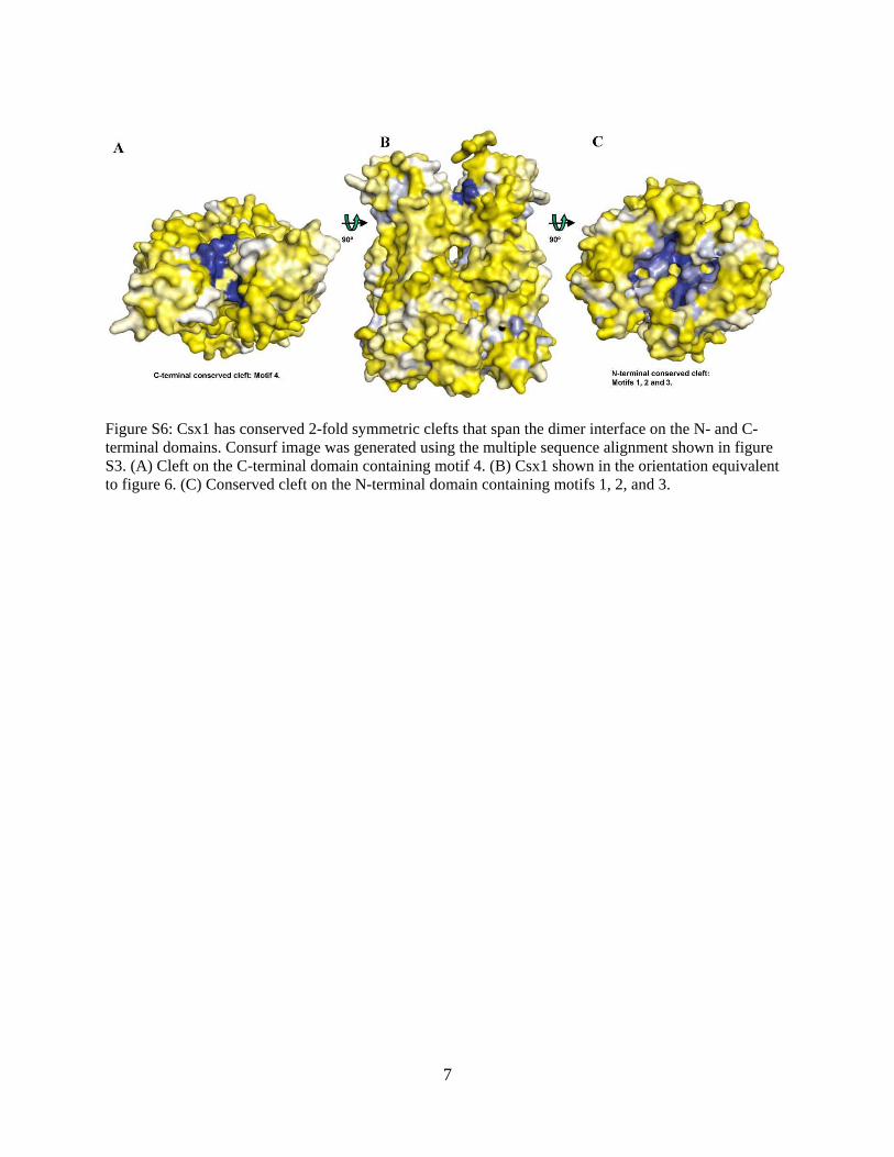
Figure S6: Csx1 has conserved 2-fold symmetric clefts that span the dimer interface on the N- and C-terminal domains. Consurf image was generated using the multiple sequence alignment shown in figure S3. (A) Cleft on the C-terminal domain containing motif 4. (B) Csx1 shown in the orientation equivalent to figure 6. (C) Conserved cleft on the N-terminal domain containing motifs 1, 2, and 3.
Related Documents