Neural map organization and development in the lateral-line system Jesús Pujol Martí DOCTORAL THESIS UPF - 2011 THESIS DIRECTOR Dr. Hernán López-Schier Cell and Developmental Biology Programme, Sensory Cell Biology and Organogenesis Laboratory, Centre de Regulació Genòmica (CRG).

Welcome message from author
This document is posted to help you gain knowledge. Please leave a comment to let me know what you think about it! Share it to your friends and learn new things together.
Transcript
Neural map organization and
development in the lateral-line system
Jesús Pujol Martí
DOCTORAL THESIS UPF - 2011
THESIS DIRECTOR
Dr. Hernán López-Schier
Cell and Developmental Biology Programme,
Sensory Cell Biology and Organogenesis Laboratory,
Centre de Regulació Genòmica (CRG).


“Knowledge is a complexity packed to break through the
reality that separates two minds.” Jorge Wagensberg.
This thesis is dedicated to you, dear readers.
You were the reason for writing it.
All the effort and passion
I put into this work
is dedicated to
my family,
friends &
fellows.


i
Acknowledgements
I am grateful to all the people who have been by my side during my
thesis years for making them some of the most challenging and exciting
years of my life.
I would especially like to thank my thesis director Dr. Hernán López-
Schier for giving me the opportunity to join his lab, for his insightful
guidance, confidence and exigency, as well as for sharing with me his
enthusiasm for science.
I also want to effusively thank Dr. Adèle Faucherre for her invaluable
mentoring, encouragement and patience. Her arrival to the lab
“brightened neurons”. Since then, I have benefited enormously from her
expertise and I have really enjoyed working in tandem with her.
I am also very thankful to Dr. Jean-Pierre Baudoin for his support, advice
and for the many stimulating discussions we had. Importantly, the work
he did in the lab represented a key step towards the findings I present
here.
My most sincere thanks also go to other past and present members of the
lab: Alessandro Mineo, Andrea Durán, Andrea Zecca, Filipe Pinto-
Teixeira, Dr. Indra Wibowo, Jacobo Cela, Dr. Mariana Muzzopappa,
Oriol Viader and Dr. Sabrina Desbordes. Their advice, help,
encouragement and friendship have been very important for the
completion of this thesis. They made everyday‟s work in the lab an
unforgettable experience.

ii
I would like to thank the members of my Thesis Advisory Committee Dr.
Berta Alsina, Dr. Matthieu Louis and Dr. Timo Zimmermann for their
advice over these years. I also want to thank many colleagues from the
CRG and UPF for creating such a stimulating environment for doing
research.
Last but not least, I am immensely grateful to my family and friends for
their unconditional support and love. It has been precious to have always
somebody trusting in me.

iii
Abstract
Sensory neurons project to the central nervous system in a spatially
ordered manner, assembling neural maps that represent attributes of
sensory stimuli and that are thought to be essential to interpret the
external world. I used the lateral-line system of the zebrafish larva as a
model to study sensory neural map organization and development.
Lateralis (lateral-line) sensory neurons organize a topographic neural
map, called somatotopy, which encodes the position of the sensory
stimulus. I demonstrated that the order of neurogenesis defines
somatotopy. In addition, I identified two sub-classes of lateralis sensory
neurons that differ in their central projection patterns and in their contacts
with a central output neuron: the Mauthner cell. I propose that such
neural-map dimorphism sub-serves appropriate behavioral reactions to
the sensory context. Importantly, I also demonstrated a contribution of
neuronal birth order to the assembly of the lateral-line dimorphic neural
map. Finally, additional results support that the observed neuronal
diversity and map topology occur normally in the absence of sensory
activity.

iv
Resum
Les neurones sensorials projecten al sistema nerviós central seguint una
distribució espacial ordenada, formant mapes neuronals que representen
propietats dels estímuls sensorials i que són considerats essencials per a
la interpretació del món extern. He utilitzat la línia lateral de la larva del
peix zebra com a model per a l‟estudi de l‟organització i el
desenvolupament dels mapes neuronals sensorials. Les neurones
sensorials de la línia lateral formen un mapa neuronal topogràfic,
anomenat somatotopia, que representa la posició de l‟estímul sensorial.
He demostrat que l‟ordre de neurogènesi defineix la somatotopia. A més,
he identificat dues subclasses de neurones sensorials de la línia lateral
que presenten diferències en els seus patrons de projecció central i en els
contactes amb una neurona central: la cèl·lula de Mauthner. Proposo que
aquest dimorfisme és important per a donar lloc a reaccions
comportamentals adients al context sensorial. També he demostrat una
contribució per part de l‟ordre de neurogènesi a la formació del mapa
neuronal dimòrfic de la línia lateral. Finalment, he obtingut resultats que
mostren que la diversitat neuronal i la topologia del mapa observades
ocorren amb normalitat en l‟absència d‟activitat sensorial.

v
Preface
The nervous system has seized scientists‟ attention throughout the ages.
Anatomical methods are the oldest way for studying the nervous system
and have uncovered basic principles of its organization, defining a
valuable groundwork for understanding its functions. Another important
source of information about nervous system function came from
analyzing the consequences of damage to specific regions of the brain.
More recently, the study of the brain activity has brought to light some
fundamental principles of nervous system function. Nowadays, it is even
possible to manipulate neuronal activity in intact behaving animals and
ask for its consequences. Because of the recent technological advances
and the many questions yet to be answered, these are exciting times to
study one of the greatest mysteries in modern biology: how the nervous
system works to exert its complex and fascintating functions.
In particular, I am attempting to understand the mechanisms that govern
the communication between sensory organs and the brain. In most
sensory systems, neurons project from the sensory receptors to the brain
in a spatially ordered manner forming neural maps that encode stimuli
attributes, such as identity or position. The formation of such precise
patterns of connectivity is thought to be essential for the brain in order to
process sensory information. One outstanding question for me is how a
sensory system can trigger seemingly opposite behavioral responses to
environmental stimuli. How sensory circuits are established during
development is the other central question that receives my attention.
vi
Most of the research on these issues has been carried out on the visual
and olfactory sensory systems. I have chosen the lateral-line sensory
system of the zebrafish larva because it is anatomically simple yet
functionally complex, mediating contrasting behaviours that are also
present in the adult fish. A decade ago, Ghysen‟s research group showed
that the lateralis (lateral-line) sensory neurons display a topographic
neural map. The same group shed some light on when and how this map
is established. Since their pioneering work, more research groups have
adopted the lateral-line system of the zebrafish as a model to study
sensory neural map organization, function and development. During my
thesis research, it has been exciting to see other laboratories‟
contributions to this field.
My thesis work has combined methods recently developed by other
groups together with novel tools we have developed in order to examine
the organization and development of the lateralis sensory neurons. I
believe that my findings provide important insights on the principles of
neural map organization with clear relevance to the mechanisms that
govern appropriate behavioral reactions to the sensory context. This
thesis also contains novel results regarding neural map development that
illustrate the overwhelming importance of time as a patterning factor and
provide a framework for future mechanistic interrogations. Furthermore,
I believe that other researchers will profit from the novel tools we have
generated.

Index
Acknowledgements ................................................................................ i
Abstract / Resum ................................................................................ iii
Preface ................................................................................................... v
Chapter 1: Introduction and Aims ........................................ 1
1.1 Sensory neural maps ...................................................................... 3
1.1.1 Structure .......................................................................................................... 3
1.1.2 Development ................................................................................................... 4
1.1.3 Functional significance ................................................................................. 11
1.1.4 Summary ....................................................................................................... 15
1.2 Using the zebrafish to study sensory neural map development
and function ........................................................................................ 16
1.3 The lateral-line sensory system ................................................... 18
1.3.1 Distribution and morphology of the sensory organs ..................................... 18
1.3.2 Natural stimuli and behavior ......................................................................... 20
1.3.3 From sensory organs to central nervous system ............................................ 24
1.3.4 Lateral-line maps .......................................................................................... 28
1.3.5 Lateral-line development .............................................................................. 33
1.3.6 Summary ....................................................................................................... 41
1.4 Aims of the thesis .......................................................................... 42
1.4.1 To study the initial assembly of the somatotopic map by the posterior
lateralis afferent neurons in the zebrafish larva ..................................................... 42
1.4.2 To search for heterogeneities among lateralis afferent neurons regarding the
connectivity with their central targets in the zebrafish larva .................................. 43
Chapter 2: Progressive neurogenesis defines lateralis
somatotopy ............................................................................. 45
2.1 Abstract ......................................................................................... 47
2.2 Introduction .................................................................................. 48
2.3 Results ........................................................................................... 50
2.3.1 BAPTI reveals growth anisotropy of the posterior lateralis ganglion ........... 50
2.3.2 The hppGFF53A enhancer-trap line expresses Gal4 in lateralis neurons ..... 55
2.3.3 Temporal progression of the innervation of posterior neuromasts ................ 58
2.3.4 The position of the somata within the ganglion predicts the neuron‟s choice
of target .................................................................................................................. 58
2.3.5 The position of the somata within the ganglion defines the neuron‟s central
projection along the somatotopic axis in the MON................................................ 64
2.4 Discussion ...................................................................................... 67
2.4.1 Tools for the study of the development and maintenance of somatotopy in
vivo ........................................................................................................................ 67
2.4.2 The temporal order of neuronal differentiation defines somatotopy ............. 68
2.4.3 Implications for the central encoding of the hydrodynamic field ................. 70
2.5 Materials and methods ................................................................ 73
2.5.1 Zebrafish strains and husbandry.................................................................... 73
2.5.2 Plasmid DNA constructs and injections ........................................................ 73
2.5.3 Labeling and imaging ................................................................................... 73
2.6 Supporting information ............................................................... 76

Chapter 3: Neuronal birth order delineates a dimorphic
sensorineural map ................................................................. 79
3.1 Abstract ......................................................................................... 81
3.2 Introduction .................................................................................. 82
3.3 Results ........................................................................................... 84
3.3.1 HGn39D is an insertion in cntnap2a ............................................................. 84
3.3.2 Lateralis afferent neurons are structurally diverse and diverge in the
hindbrain ................................................................................................................ 85
3.3.3 Neuronal sub-classification based on contacts with a central target ............. 88
3.3.4 Biased axonal projection pattern of large and small neurons ........................ 93
3.3.5 Neuronal projections and birth date .............................................................. 95
3.3.6 The lateralis neural map develops in the absence of sensory input ............... 96
3.4 Discussion .................................................................................... 101
3.5 Materials and methods .............................................................. 107
3.5.1 Zebrafish strains and husbandry.................................................................. 107
3.5.2 Selection of mutants .................................................................................... 107
3.5.3 Plasmid DNA constructs and injections ...................................................... 108
3.5.4 Generation of transgenic zebrafish .............................................................. 109
3.5.5 Whole-mount in situ hybridization ............................................................. 109
3.5.6 Neuronal labeling, birthdating and imaging ................................................ 110
3.5.7 Quantification of soma volume and peripheral axon diameter ................... 111
3.5.8 Laser-mediated cell ablation ....................................................................... 111
3.6 Supporting information ............................................................. 113
Chapter 4: Additional results ............................................ 115
4.1 Lateralis afferents contacting the Mauthner cell form a
somatotopic map ............................................................................... 117
4.2 The Mauthner cell receives input from hair-cells of opposing
polarities ............................................................................................ 118
4.3 Peripheral arborization and neuronal sub-classes .................. 119
Chapter 5: General discussion ........................................... 123
5.1 Structure and function of the lateral-line neural maps .......... 125
5.1.1 A new view of the neural maps built by the lateralis afferent neurons ....... 125
5.1.2 Functional implications of the lateral-line dimorphic neural map .............. 128
5.2 Neural map formation in the lateral-line system..................... 134
5.2.1 How can progressive neurogenesis build the lateral-line neural maps? ...... 135
5.2.2 A „circumstantial‟ assembly of the lateral-line neural maps ....................... 137
Chapter 6: Conclusions ...................................................... 141
References ............................................................................ 145
Appendix: Other contributions ......................................... 163

1
Chapter 1
INTRODUCTION and AIMS

2

3
1.1 Sensory neural maps
Animals perceive the external world by virtue of their sensory systems.
Sensory systems consist in: (1) receptors that detect external stimuli; (2)
neural pathways that convey sensory information to the brain; and (3)
central neurons organized in a series of relay nuclei that process this
information. Sensory systems ensure the interpretation of complex flows
of sensory information, by translating the basic features of a stimulus
-modality/identity, position, intensity and timing- into a coherent neural
code. This largely relies on the highly organized patterns of connectivity
between the distinct components of the sensory systems, which in many
cases shape neural maps of the external world (Gardner and Martin,
2000; Kaas, 1997; Lemke and Reber, 2005).
1.1.1 Structure
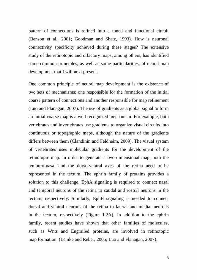
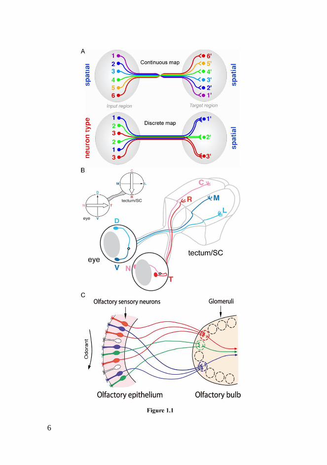
Neural maps are spatial arrangements of neuronal connections that
encode information. Both anatomical and physiological studies have
revealed neural maps in many sensory and motor systems. Neural maps
form a spectrum whose extremes are continuous neural maps, also called
topographic, and discrete neural maps. The main difference between the
two types is the nature of the attribute encoded by the map. In a
continuous map, nearby neurons in the input region connect to nearby
neurons in the target region. In other words, there is a point-to-point
match of connections between input and target regions. By this
arrangement, the map represents positional information. By contrast, in a
discrete map, spatially dispersed neurons of the same type in the input
region converge on the same cluster of neurons in the target region. In

4
this manner, the map represents discrete information such as neuronal
identity (Figure 1.1A) (Luo and Flanagan, 2007). The paradigm for
continuous maps is the retinotopic map in the visual system. In
vertebrates, retinal neurons convey visual information from the retina to
the optic tectum forming a map where neurons from the nasal and
temporal regions of the retina connect to neurons in the caudal and rostral
tectum, respectively. In addition, neurons from the dorsal and ventral
regions of the retina connect to neurons in the lateral and medial tectum,
respectively. Thus, the brain receives an intact two-dimensional image
from the retina (Figure 1.1B) (Lemke and Reber, 2005). The paradigm
for discrete maps is the olfactory map. The olfactory epithelium contains
olfactory sensory neurons, each of them expressing a single functional
odorant receptor. Neurons expressing the same odorant receptor are
spatially dispersed in the epithelium. However, central axons of neurons
expressing the same odorant receptor converge on the same region, called
glomerulus, in the olfactory bulb. Thus, the olfactory bulb contains an
odorant receptor map that encodes the identity of the odorant signal
received by the olfactory epithelium (Figure 1.1C) (Sakano, 2010).
1.1.2 Development
How are neural maps established during development? This has been a
central question for neurobiologists during decades. The final patterning
of neuronal connections that shape a neural map is the completion of a
continuous and complex process. First, neurons extend axons which
select specific paths to navigate through. Axons then recognize their
correct target and establish a widespread scaffold of contacts with a set of
neurons. Finally, axons select a specific subset of neurons and the initial
5
pattern of connections is refined into a tuned and functional circuit
(Benson et al., 2001; Goodman and Shatz, 1993). How is neuronal
connectivity specificity achieved during these stages? The extensive
study of the retinotopic and olfactory maps, among others, has identified
some common principles, as well as some particularities, of neural map
development that I will next present.
One common principle of neural map development is the existence of
two sets of mechanisms; one responsible for the formation of the initial
coarse pattern of connections and another responsible for map refinement
(Luo and Flanagan, 2007). The use of gradients as a global signal to form
an initial coarse map is a well recognized mechanism. For example, both
vertebrates and invertebrates use gradients to organize visual circuits into
continuous or topographic maps, although the nature of the gradients
differs between them (Clandinin and Feldheim, 2009). The visual system
of vertebrates uses molecular gradients for the development of the
retinotopic map. In order to generate a two-dimensional map, both the
temporo-nasal and the dorso-ventral axes of the retina need to be
represented in the tectum. The ephrin family of proteins provides a
solution to this challenge. EphA signaling is required to connect nasal
and temporal neurons of the retina to caudal and rostral neurons in the
tectum, respectively. Similarly, EphB signaling is needed to connect
dorsal and ventral neurons of the retina to lateral and medial neurons
in the tectum, respectively (Figure 1.2A). In addition to the ephrin
family, recent studies have shown that other families of molecules,
such as Wnts and Engrailed proteins, are involved in retinotopic
map formation (Lemke and Reber, 2005; Luo and Flanagan, 2007).
6
7
Although the visual system of invertebrates is very different from its
vertebrate equivalent, it also presents a similar retinotopic map. However,
the formation of the map relies on temporal gradients in addition to
molecular cues. Photoreceptors in the fly eye project axons into the
lamina in the brain, where they connect to target neurons shaping a
continuous map. Temporal gradients are essential for mapping the rostro-
caudal axis of the retina into the lamina. Photoreceptors from the caudal
region of the retina differentiate first and extend their axons to the caudal
region of the lamina. There, the arriving axons promote the
differentiation of target neurons and connect to them. Next, the same
process occurs in photoreceptors from more rostral regions of the retina,
which project axons to more rostral regions of the lamina, and so forth,
until all photoreceptors have differentiated and extended their axons. By
contrast, the mapping of the dorso-ventral axis of the fly retina into the
lamina depends on the Wnt family (Figure 1.2B) (Clandinin and
Feldheim, 2009). Temporal gradients also appear to be key factors for
building the retinotopic map in the visual system of crustaceans (Flaster
and Macagno, 1984).
8
The use of either molecular or temporal gradients is sufficient to form an
initial coarse map. However, other mechanisms are required to increase
the precision of the map. Neuronal activity can play a prominent role in
map refinement. In the visual system of vertebrates, neuronal activity
refines the retinotopic map. In fishes and amphibians, visual input evokes
the firing of retinal neurons during map formation. In mammals and
birds, the map forms before any visual experience has occurred. Retinal
neurons, however, generate spontaneous waves of action potentials that
spread across the retina during map formation. In both cases, the spatial
and temporal pattern of retinal neurons firing instructs the refinement of
their connections with target neurons in the tectum, in such a way that
“neurons that fire together wire together”. By contrast, neuronal activity
seems to play no role in the formation of the retinotopic map in the
invertebrate visual system (Goodman and Shatz, 1993; Luo and
Flanagan, 2007). In the olfactory system, neuronal activity produced by
odorant receptor signaling seems to regulate the expression of molecular
cues that instruct the formation of the olfactory map (Luo and Flanagan,
2007; Sakano, 2010).
Another common principle of neural map development is the use of local
interactions, both adhesive and repulsive, among axons. In the vertebrate
and invertebrate visual and olfactory systems, axon-axon interactions
ensure the ordering of axons during pathfinding and their proper spacing
into the target region (Luo and Flanagan, 2007). In the visual system,
such axon-axon interactions are mediated by cell-surface molecules like
cadherins and IgCAMs (Clandinin and Feldheim, 2009). In the olfactory
system, local sorting is mediated by the complementary expression of
9
Neuropilin-1 receptor and its repulsive ligand Semaphorin-3A (Mori and
Sakano, 2011).
An important question in the study of neural map formation is where
patterning instructions originate from. Three ways of establishing a
neural map between two groups of neurons, input and target neurons,
have been proposed. One possibility is that input neurons are prespecified
and instruct their target neurons which identity to acquire. Another
possibility is that input neurons are naïve and acquire their identity from
the prespecified target neurons they connect to. The last possibility is that
input and target neurons acquire their identities independently (Figure
1.3) (Jefferis et al., 2001). This is precisely what happens in the case of
the visual system of vertebrates and the olfactory system of both
vertebrates and invertebrates. In these systems, the patterning information
resides in both the input and the target neurons, which are specified and
sorted autonomously. By contrast, in the visual system of flies, the
patterning information resides in the input neurons that eventually
instruct target neurons which identity to acquire (Luo and Flanagan,
2007; Sakano, 2010).

10
11
1.1.3 Functional significance
Neural maps appear to be a universal solution; they are present in
phylogenetically distant animals and in diverse sensory systems.
Therefore, they must provide animals with some advantage. How does
the brain benefit from sensory neural maps to process sensory
information? What is the importance of sensory maps in guiding animal
behavior? Despite the notable advances made in deciphering the
mechanisms involved in neural map development, much less is known
about their functional significance. Furthermore, the rules used to extract
and process information from sensory maps are still obscure.
Since first evidence for neural maps came, researchers have proposed
diverse hypotheses about the relevance of neural maps, especially about
the importance of continuous or topographic maps for sensory
processing. Some authors have claimed that such maps are the basis of
perception, based on similarities between many aspects of perception and
structural aspects of the maps. In addition, some theories of visual
perception implicate topographic maps (Kaas, 1997). By contrast, other
authors have pointed out that the particular spatial arrangement found in
continuous maps may reflect an economical solution for the proper
wiring of the brain, or a solution for increasing communication velocity
by shortening connections between neurons (Chklovskii and Koulakov,
2004; Weinberg, 1997). With the advances in developmental and
molecular neurobiology, it is now possible to disrupt sensory neural map
formation and ask for its functional consequences in sensory processing
and behavior.

12
The functional significance of the retinotopic map has been recently
studied in mutant mice with disrupted projections from retinal neurons
into the optic tectum; in mammals more commonly called the superior
colliculus. Mutant mice with a disruption of spontaneous activity during
map development show an imprecise spatial arrangement of connections
between nasal/temporal retinal neurons and caudal/rostral superior
colliculus neurons. In animals with such alteration, superior colliculus
neurons have abnormal receptive fields along the temporo-nasal axis. In
other words, these neurons respond to the presence of a visual stimulus in
ectopic positions. Importantly, these animals fail to track visual stimuli
moving along the temporo-nasal axis, but are able to track it without
problems along the dorso-ventral axis (Wang et al., 2009). Other
mutations result in some dorsal retinal neurons connecting ectopically to
superior colliculus neurons that normally receive input from ventral
retinal neurons. This appears to be translated into ectopic receptive fields
along the dorso-ventral axis (Chandrasekaran et al., 2009). Superior
colliculus neurons are known to project to the visual cortex preserving
the retinotopic map. Disruptions of this map also have functional
consequences such as visual cortex neurons with abnormal receptive
fields and decreased visual acuity (Cang et al., 2008; Demyanenko et al.,
2011).
Researchers in the field of olfactory processing have followed a similar
approach. They have disrupted the olfactory map by different means and
asked for the consequences in sensory processing and behavior. As
mentioned previously, each olfactory sensory neuron expresses only one
of many odorant receptors. Neurons expressing the same receptor project
to the same spatially fixed glomerulus in the olfactory bulb. Importantly,
13
this anatomic map is translated into a functional map. Odorant signals
received in the olfactory epithelium are translated into an odor map of
activated glomeruli in the olfactory bulb (Mori and Sakano, 2011). Odor
representation in the olfactory bulb is sparse since each odorant signal
only activates few glomeruli (Lin da et al., 2006). Researchers have
engineered a mouse in which over 95% of the olfactory sensory neurons
express the same odorant receptor. This results in the activation of all
glomeruli by the presence of the odor that interacts with the predominant
odorant receptor. In this way, the representation of odors in the brain is
severely perturbed. It appears that these mice are able to smell, but they
show problems in odor discrimination and in olfactory behaviors,
especially in innate behaviors (Fleischmann et al., 2008). Other
approaches have consisted in the ablation of olfactory sensory neurons
from specific areas of the olfactory epithelium in mice. The ablation of
neurons from the dorsal region of the olfactory epithelium causes a
depletion of glomerular structures in the dorsal region of the olfactory
bulb. Animals with this perturbation fail to show innate responses to
aversive odors, although they are able to detect them and even perform
learned aversive responses to the same odors (Kobayakawa et al., 2007).
How neural maps facilitate sensorimotor transformations is another
fundamental question. This issue has been examined in the oculomotor
system. In many animals, eyes make very rapid movements or saccades
in order to sense with high resolution regions of a visual scene and build
up an internal representation of it. The superior colliculus, or tectum,
contains a well-defined retinotopic map where visual space is
represented. Importantly, this brain structure is involved in the
transformation of visual information into orienting behaviors, including
14
ocular motor commands. Several studies have shown that superior
colliculus neurons involved in sensorimotor transformations encode the
desired motor direction based on the coordinates provided by the
retinotopic map (Klier et al., 2001; Marino et al., 2008). Moreover,
topographic maps from several sensory modalities (visual, auditory and
somatosensory) are aligned with each other, as well as with a premotor
map, in the superior colliculus. This arrangement appears an effective
way to match incoming sensory information about a source with the
motor outputs necessary for orientation to it (Stein et al., 2009). The
analysis of some spinal cord reflex circuits similarly suggests that neural
maps play a role in adapting sensory input to motor output during
sensorimotor transformation (Levinsson et al., 2002).
Finally, the contribution of sensory maps to the learning of perceptual
tasks has also been examined using the rat whisker sensory system.
Sensory neurons innervating the whiskers on the snout project to the
thalamus preserving the spatial distribution of the whiskers and thus
forming a continuous map. Thalamic neurons project to the
somatosensory cortex maintaining this spatial order and forming a map of
the whiskers into the cortex (Petersen, 2007). Rats can cross a gap
between two elevated platforms by using their whiskers to touch and
locate the platform they have to reach for receiving a reward. A rat
possessing a single whisker can be trained to efficiently perform this task.
After, the whisker can be clipped and a prosthetic whisker can be placed
on the location of the trained whisker or in the location of any other
whisker. If the prosthetic whisker is placed in the location of the trained
whisker, the rat can efficiently perform the gap-crossing task again.
Interestingly, a period of relearning is necessary if it is placed in any

15
other whisker location and the duration of the relearning period is directly
proportional to the distance between the locations of trained and
prosthetic whiskers (Harris et al., 1999). This observation suggests that
the neural modifications associated with the learning of a perceptual task
are distributed according to the sensory map present in the sensory cortex
(Diamond et al., 1999).
1.1.4 Summary
The use of neural maps to represent information, such as position or
identity, is a fundamental organizational principle of the nervous system.
Neural maps are broadly present in the nervous system, from sensory to
motor components. Sensory neural maps have seized the attention of
researchers for many years. How are these maps established during
development? How are they used for sensory processing and for guiding
behavior? These are central questions in neurobiology. Despite the
notable advances in unraveling the developmental mechanisms involved
in sensory map formation, there are still many open questions. For
instance, the problem of how different forces act together to drive neural
map development is poorly understood. In addition, most of the research
has focused on a few neural maps and therefore there are still many
neural maps whose assembly needs to be analyzed. Besides this, very
little is known about the role of sensory maps in sensory processing and
behavior.

16
1.2 Using the zebrafish to study sensory neural map
development and function
To study sensory neural map development and function, it would be ideal
to have single species in which molecular, cellular and physiological
analyses, as well as perturbations, could be carried out in neurons during
development and once neuronal circuits are established. The zebrafish
(Danio rerio) has been historically used as a model for studying the basic
mechanisms of development. Importantly, in the recent years, it has also
become an organism of choice for analyzing how neuronal circuits
function and how they mediate behavior, especially at larval stages
(Fetcho and Liu, 1998).
The zebrafish compares favorably with other animal model systems to
study developmental neurobiology for several reasons. The zebrafish
embryo develops rapidly, externally and is optically transparent; which
makes it suitable for visualization and manipulation. It is also amenable
for mutational analysis. For example, it is possible to carry out large-
scale screenings for developmental defects using chemical or insertional
mutagenesis. Moreover, the generation of transgenic zebrafish is
technically simple. Reporter transgenic methods allow in vivo time-lapse
imaging of neurons. Engineered genes can also be expressed transiently,
facilitating functional studies by gain- or loss-of-gene function. By using
the zebrafish embryo, notable advances have been made in the fields of
patterning of the nervous system, axonal pathfinding and specification of
neuronal identity (Appel, 2006; Eisen, 1991; Nicolson, 2006).

17
The above-mentioned advantages also favor the use of the zebrafish as a
model for studying neuronal circuits function and behavior. The zebrafish
larva is amenable to neurophysiology, imaging and behavioral analyses.
At larval stages, the zebrafish displays simple and robust behaviors that
can be reliably evoked. In addition, some new tools provide the
opportunity to depict the patterns of connectivity between neurons; even
to reconstruct entire circuits. Other new tools allow monitoring neuronal
activity during behavior, in living intact animals, as well as manipulating
it; for example by activating and silencing neurons when desired. This is
especially of importance since it provides a powerful way to causally
relate neurons/circuits to behaviors. Last, the nervous system of the
zebrafish larva is relatively small in terms of size and number of neurons;
which indeed facilitates the analysis of neuronal circuits. Researchers
have already benefited from these advantages to gain insight into the
function of motor and sensory systems in the zebrafish larva (Del Bene
and Wyart, 2011; Fetcho and Liu, 1998; Friedrich et al., 2010).
Altogether the above-mentioned qualities make the zebrafish an ideal
model to close the gap between molecules, neurons, circuits and
behaviors. The hope is that the findings made in the zebrafish can be
extrapolated to other animals, since nervous system structure and gene
function is highly conserved among vertebrates.
18
1.3 The lateral-line sensory system
The lateral line is a sensory system found in fish and amphibians. It was
long ascribed to an auditory function since its sensory receptors, the hair
cells, are the same as in the inner ear. The current view is that the lateral
line mediates a „distant-touch‟ sense, because it responds to water
motions that occur within short-distances in the animal‟s surroundings
(Dijkgraaf, 1963). In this chapter, I will cover aspects of the lateral line
that are relevant to my thesis research focusing on our model organism:
the zebrafish larva. I will also present knowledge from the adult zebrafish
and from other fish species for comparisons and when there is no
information from our model.
1.3.1 Distribution and morphology of the sensory organs
The lateral line comprises a set of discrete sensory organs called
neuromasts. Neuromasts can occur superficially on the skin or within
sub-epidermal canals open to the water through pores. In the zebrafish
larva, all neuromasts are superficial and are arranged in stereotypic
patterns on each side of the animal. Neuromasts on the head and trunk
configure the anterior and posterior lateral-line branches, respectively.
The posterior lateral line can be further divided into two sub-branches
located in the lateral and dorsal aspects of the fish. In one-week-old
larvæ, the posterior lateral line comprises around 14 neuromasts (Figure
1.4A) (Ghysen and Dambly-Chaudière, 2007; Metcalfe et al., 1985). The
number of neuromasts dramatically increases over development. The
distribution and number of neuromasts in adult fish diverge notably
between different species. However, the organization of the system at

19
early stages of development appears to be conserved between species
(Nuñez et al., 2009).
Neuromasts are small sensory patches composed of a core of 20 to 30
mechanosensory hair cells surrounded by a similar number of non-
sensory supporting cells. Thus, they are structurally similar to the organs
of the inner ear (Ghysen and Dambly-Chaudière, 2007). Hair cells derive
their name from the hair bundle that projects from their apical domain. In
the neuromast, hair bundles protrude into a gelatinous cupula that
connects them to the surrounding water. The hair bundle comprises an
array of stereocilia arranged in rows of increasing length, like a staircase,
and a kinocilium eccentrically located adjacent to the tallest stereocilia.
Therefore, each hair bundle, and thus each hair cell, is polarized within
the plane of the neuromast. This represents a striking example of planar
cell polarity. A water motion over the cupula that deflects the stereocilia
towards the kinocilium depolarizes the hair cell. This triggers in turn an
increase in neurotransmitter release at the hair cell‟s basal domain and a
subsequent increase in the firing rate of the afferent (sensory) neuron
associated to the hair-cell. By contrast, a deflection in the opposite
direction hyperpolarizes the hair cell and reduces neurotransmitter release
which causes a decrease of the firing rate of the associated neuron
(Figure 1.4B). Therefore, the morphological polarization of the hair cell
determines its axis of mechanical sensitivity (Hudspeth, 2000).
Surprisingly, each neuromast consists in two intermingled populations of
hair cells, equal in number, of opposing hair-bundle polarities. Thus,
neuromasts are bidirectionally sensitive; one population of hair cells is
sensitive to deflections in a given direction, whereas the other population
20
is sensitive to deflections in the opposite direction (Figure 1.4B) (Flock
and Wersall, 1962; López-Schier et al., 2004). Furthermore, two types of
neuromasts have been described according to the polarization of their
hair cells across the animal‟s body axes. Parallel neuromasts contain hair
cells polarized across the antero-posterior (rostro-caudal) axis whereas
perpendicular neuromasts contain hair cells polarized across the dorso-
ventral axis. Consequently, parallel and perpendicular neuromasts are
differentially sensitive to mechanical stimulation across two orthogonal
axes (Figure 1.4C) (López-Schier et al., 2004).
1.3.2 Natural stimuli and behavior
The lateral line detects hydromechanic stimuli in the fish‟s surroundings,
specifically low frequency (<150 Hz) water motions (Bleckmann, 2008).
An important source of lateral-line stimuli is the water flow generated by
a predator. Fish are able to execute an extremely fast escape response,
known as the C-start reflex, after detecting the flow caused by a
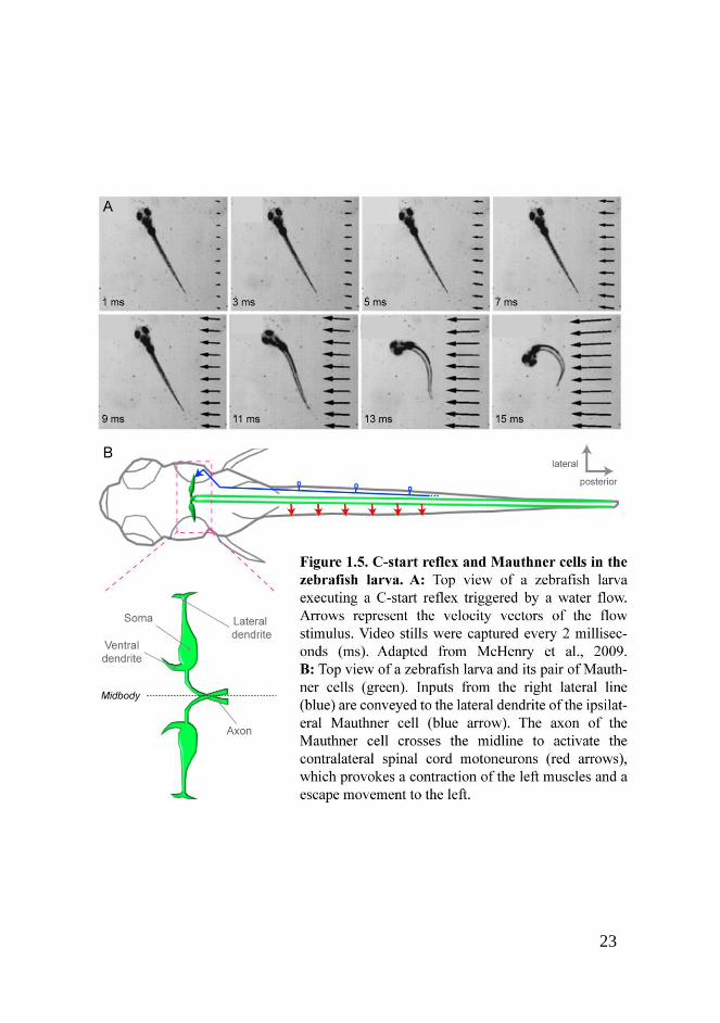
predator‟s strike. It has been shown recently that the zebrafish larva uses
its lateral line to detect and to escape from a water flow that emulates a
predator‟s strike (Figure 1.5A) (McHenry et al., 2009).
Another common source of hydromechanic stimuli is the swimming
movement of an animal. For instance, the wake behind a swimming fish
contains complex flow patterns and turbulences; and provides
information about the wake generator, such as the size, swimming style
and speed. Some fish use the lateral line to sense this information and
track the trails of prey fish. The lateral line can also sense water surface
waves. Surface feeding fish use their lateral line to detect preys in the

21
water surface, such as terrestrial insects falling into the water or animals
that contact the water-air interface from below to breathe or feed
(Bleckmann and Zelick, 2009).
Self-generated water motions and water currents in running water provide
continuous stimulation of the lateral line. The patterns of self-generated
water motions are modified when a fish approaches an object. These
changes provide information about the size, shape and distance of nearby
objects. This is especially used by blind cavefish to locate nearby
stationary objects and avoid obstacles during navigation (Bleckmann and
Zelick, 2009). Water flow information provided by the lateral line
appears to be important for rheotaxis, a behavioral orientation to swim
against water currents and avoid, thus, being washed out by the current
(Montgomery et al., 1997). The zebrafish larva clearly exhibits a
rheotactic response when exposed to a water flow
(http://rubenportugues.net/). Moreover, fish use lateral-line information
to make their swimming more efficient in running water (Liao, 2006).
Superficial and canal neuromasts present morphological differences
which result in different respond properties to the sources of
hydromechanic stimuli. The former detect flow velocity whereas the
latter detect the acceleration of water motions. Thus, superficial
neuromasts are efficient at detecting the flow created by a predator‟s
strike in still water. They also sense water currents and mediate rheotaxis.
By contrast, canal neuromasts are efficient at detecting small
hydromechanic stimuli against a background constant water flow
(Engelmann et al., 2000; Montgomery et al., 1997).

22
In summary, the lateral-line sensory system provides information that
fish use for predator avoidance, prey detection, object discrimination and
rheotaxis. Moreover, lateral-line receptors with different shapes, such as
superficial and canal neuromasts, convey sensory information that might
be used for different behaviors. How is the hydrodynamic information
captured by the neuromasts translated into the diverse behaviors mediated
by the lateral line? To comprehend this it is necessary to examine how
this information is conveyed to the brain and how it is further processed
centrally.
23

24
1.3.3 From sensory organs to central nervous system
As mentioned previously, a water motion over the cupula that bends the
stereocilia towards the kinocilium produces an increase in
neurotransmitter release at the hair cell‟s basal domain. The
neurotransmitter released by hair cells acts into the peripheral axonal
endings of lateralis (lateral-line) afferent neurons, generating an action
potential that travels along the neuron, from peripheral axon to central
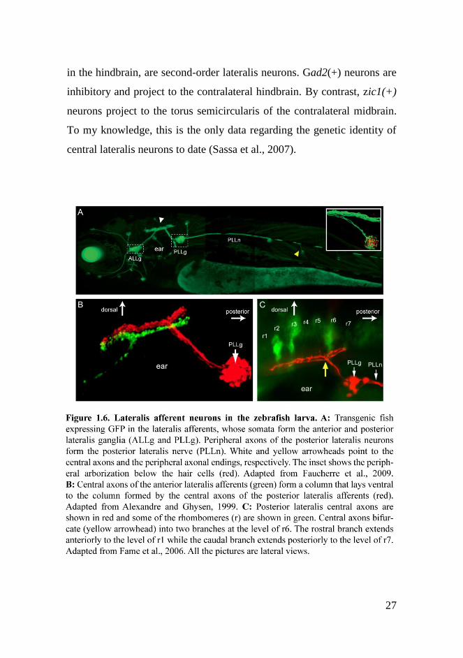
axon, passing by the neuronal soma. Axons from lateralis afferents are
grouped forming nerves, whereas somata coalesce forming small cephalic
ganglia near the ear. Somata from afferent neurons innervating the
anterior and posterior lateral-line branches form the anterior and posterior
lateralis ganglia (Figure 1.6A) (Metcalfe, 1985). In one-week-old larvæ,
the number of lateralis afferents is small; the posterior lateralis ganglion
comprises approximately 45 somata at around this stage (Liao, 2010). By
contrast, many more lateralis afferents are found in the adult zebrafish
(Metcalfe et al., 1985).
Sensory information from the lateral line arrives to the ipsilateral dorsal
hindbrain through the lateralis nerves. There, central axons from anterior
and posterior lateralis neurons form two contiguous yet non-overlapping
columns that course rostrocaudally (Figure 1.6B). In the zebrafish larva,
central axons terminate into a neuropil region ventral to the medial
octavolateralis nucleus (MON) in the hindbrain. Each axon bifurcates at
the level of rhombomere 6 into a rostral and a caudal branch and exhibits
terminal buttons along the entire rostrocaudal extent. The rostral branch
extends to rhombomere 1 whereas the caudal branch extends to
rhombomere 7/8 (Figure 1.6C). Although this is the general situation, few
25
neurons extend their central axons further into the ipsilateral cerebellum.
In the adult of several fish species, lateralis central axons end up clearly
within the MON, which also receives afferent neurons from the inner ear.
However, like in the larva, some central axons reach the ipsilateral
cerebellum (Bleckmann and Zelick, 2009; Fame et al., 2006; Metcalfe et
al., 1985).
Lateralis afferent neurons represent the first-order neurons of the lateral-
line sensory system, since they conduct sensory information from the
sensory receptor to the brainstem, specifically to the MON. There, they
synapse with second-order neurons. It has been shown that lateralis
central axons make monosynaptic contacts with the lateral dendrite of the
ispsilateral Mauthner cell, a command neuron that triggers the C-start
reflex, both in the zebrafish and in other species (Figure 1.5B) (Kimmel
et al., 1990; Zottoli and Van Horne, 1983). In the zebrafish larva, none of
the other reticulospinal neurons described, however, appear to extend
dendrites near the region of lateralis central axons terminals. Dendrites of
vestibulospinal neurons project into the column formed by lateralis
central projections, strongly suggesting that they synapse with lateralis
neurons (Metcalfe et al., 1985). The Mauthner cell and, very likely, the
vestibulospinal neurons are examples of second-order neurons that pick
up lateral-line information and send commands directly to motor centers
in the spinal cord; avoiding high-order sensory processing.
Most of the second-order lateralis neurons described in the zebrafish
larva and the adult of several fish species send lateral-line information to
higher-brain centers where is further processed. In the adult, the somata
from these second-order neurons are located in the MON. They send

26
axons largely to a midbrain nucleus called torus semicircularis, which is
equivalent to the inferior colliculus of mammals, a major target of
auditory information. The optic tectum, another midbrain structure, also
receives projections from the MON. In both cases, projections occur
bilaterally, with a contralateral predominance. In addition, second-order
neurons project to the contralateral MON (Figure 1.7) (Bleckmann and
Zelick, 2009). The same occurs in the zebrafish larva, although the
somata from second-order neurons are not located in a nucleus but extend
over a larger region, possibly over most of the hindbrain dorsal (alar)
plate. Furthermore, it appears that there are projections to neurons that
have not been previously identified as second-order targets in the adult
fish. It has been suggested that the pattern found in the larva is an
ancestral scaffold of connections from which some subsets will be
selected in different groups of vertebrates at later stages of development,
for their own purposes (Fame et al., 2006).
The next step in lateral-line sensory information transmission occurs
from the midbrain to the diencephalon. Third-order lateralis neurons
located in the torus semicircularis project axons into various diencephalic
nuclei. From the diencephalon, lateral-line sensory information finally
arrives to the telencephalon (Figure 1.7) (Bleckmann and Zelick, 2009).
Although we know well which the central relay stations of the lateral-line
information are, their roles in sensory processing still remain largely
unknown. Moreover, very little is known about the genetic identity of
lateralis central neurons. In the zebrafish larva, anatomical data strongly
suggest that both neurons expressing glutamate decarboxylase 2 (gad2)
and neurons expressing zic family member 1 (zic1), which are not mixed
27
in the hindbrain, are second-order lateralis neurons. Gad2(+) neurons are
inhibitory and project to the contralateral hindbrain. By contrast, zic1(+)
neurons project to the torus semicircularis of the contralateral midbrain.
To my knowledge, this is the only data regarding the genetic identity of
central lateralis neurons to date (Sassa et al., 2007).

28
1.3.4 Lateral-line maps
The distribution and morphology of neuromasts provide to the lateral-line
system a way to extract basic features from complex hydrodynamic
stimuli; which is essential to interpret them and to react appropriately.
This happens, at least, at four levels. First, each neuromast responds to
stimuli in its proximity; capturing, thus, sensory information from a
specific location on the animal‟s body. Second, superficial neuromasts
appear to be specialized to detect water velocity whereas canal
neuromasts are specialized to detect water acceleration. They represent
two submodalities within the system. Third, hair cells of opposing
polarities detect water motions in opposing directions. Fourth, parallel
and perpendicular neuromasts are sensitive to water movements along the
antero-posterior and dorso-ventral axes, respectively. Once these basic
features –position, submodality, direction and body axis- have been
extracted at the level of the periphery, they must be transmitted to the
central nervous system separately in parallel pathways or channels. The
29
first-order neurons, the lateralis afferents, are the responsible to do so.
Subsequently, this sensory information must be encoded at the level of
the central nervous system. This might be achieved by means of spatially
arranged neural maps, resembling what takes place in many other sensory
systems.
The receptive field of an individual lateralis afferent is defined by its
associated neuromasts. In the zebrafish larva, each lateralis neuron
innervates a single neuromast, with some exceptions. Multiple
innervation occurs largely in neurons innervating the terminal
neuromasts; a group of two to three consecutive neuromasts very close to
each other, located in the tip of the tail. Multiple innervation is rare in the
rest of the neuromasts; and when it occurs, the innervated neuromasts are
also spatially consecutive. In this way, lateralis afferents link several
organs to form multi-neuromast sensory units (Faucherre et al., 2009;
Nagiel et al., 2008). At a broader level, lateralis afferents are segregated
into anterior and posterior ganglia and nerves that form two adjacent but
segregated columns in the MON. Altogether these evidences indicate that
the stimuli captured at different locations on the animal‟s body are
relayed to the hindbrain in parallel channels. It has been revealed in
several fish species, including the zebrafish larva, that these channels are
arranged forming a continuous or topographic neural map. In the MON,
the column formed by the central axons of the anterior lateralis afferents
is always ventral to that formed by the central axons of the posterior
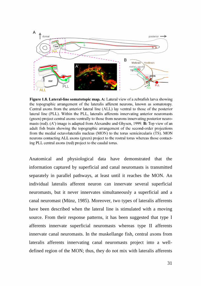
lateralis afferents (Figure 1.6B and Figure 1.8). Moreover, within each
column, lateralis afferents innervating anterior (rostral) neuromasts
project central axons ventrally to those from afferents innervating
posterior (caudal) neuromasts. Therefore, the distribution of neuromasts

30
along the antero-posterior (rostro-caudal) body axis is represented by a
dorso-ventral organization of lateralis afferents‟ central axons, known as
somatotopy (Figure 1.8A) (Alexandre and Ghysen, 1999; Bleckmann,
2008; Puzdrowski, 1989).
Is this somatotopic map maintained through the next steps of lateral-line
information relay? An anatomical study in midshipman fish has shown
that MON neurons project axons into the contralateral torus
semicircularis forming a coarse topographic map. MON neurons
contacting anterior lateralis central axons project to the rostral torus
whereas those contacting posterior lateralis central axons project to the
caudal torus (Figure 1.8B) (Weeg and Bass, 2000). In agreement with
that, physiological data from electric fish have shown that neurons in the
rostral torus respond to inputs from head neuromasts whereas neurons in
the caudal torus respond to inputs from tail neuromasts (Bleckmann et al.,
1987; Bleckmann and Zelick, 1993; Knudsen, 1977). Evidence for a
crude topographic organization of neurons in the torus semicircularis of
goldfish also exists (Engelmann and Bleckmann, 2004; Plachta et al.,
2003). However, recent studies have failed to find highly space selective
neurons in the MON and torus semicircularis, suggesting that single
neurons do not encode the spatial location of a stimulus (Künzel et al.,
2011; Voges and Bleckmann, 2011). These authors have suggested that,
by contrast, populations of lateralis central neurons encode the spatial
location of the stimulus. In this case, a strict topographic map would be
partially lost, for example to facilitate information coding in different
channels. Such a population code might be used by higher-brain centers
to rebuild a spatial map; by converging inputs from many neurons into
single neurons, for instance.
31
Anatomical and physiological data have demonstrated that the
information captured by superficial and canal neuromasts is transmitted
separately in parallel pathways, at least until it reaches the MON. An
individual lateralis afferent neuron can innervate several superficial
neuromasts, but it never innervates simultaneously a superficial and a
canal neuromast (Münz, 1985). Moreover, two types of lateralis afferents
have been described when the lateral line is stimulated with a moving
source. From their response patterns, it has been suggested that type I
afferents innervate superficial neuromasts whereas type II afferents
innervate canal neuromasts. In the muskellange fish, central axons from
lateralis afferents innervating canal neuromasts project into a well-
defined region of the MON; thus, they do not mix with lateralis afferents

32
innervating superficial neuromasts (Bleckmann, 2008). It is not clear yet
whether there is a separation of the information coming from superficial
and canal neuromasts at the torus semicircularis (Engelmann and
Bleckmann, 2004; Plachta et al., 2003).
Pioneering electrophysiological analyses in cichlid fish demonstrated that
all the hair cells innervated by a single lateralis afferent are functionally
polarized in the same direction (Münz, 1985). Both morphological
(Faucherre et al., 2009; Nagiel et al., 2008) and physiological (Liao,
2010; Obholzer et al., 2008) analyses in the zebrafish larva have shown
the same recently. Each neuromast is innervated by at least two lateralis
afferents. Each of these neurons synapses with hair cells of identical
polarity to divide the neuromast into synaptic planar-polarity
compartments (Figure 1.4B). This holds true even in the case of multiple
neuromasts innervation, where the innervated hair cells belong to
consecutive sensory organs. To date, there is no anatomical evidence
showing that neurons innervating hair cells of opposing polarities map
separately in the MON or in the torus semicircularis. Physiological
studies in the adult fish of several species have shown that many lateralis
central neurons from the MON and torus are sensitive to the direction of
water flow. These neurons change their responses when a water flow is
reversed from headward to tailward, for example. This might be
explained if individual central neurons receive input exclusively from one
of the two populations of hair cells of opposing polarities; in other words,
if the inputs from the two populations are kept separately along the
different central relay stations (Bleckmann, 2008). It might be, however,
that both inputs from hair cells of opposing polarities converge on other
lateralis central neurons, as recently observed in a single recording of a

33
neuron in the torus semicircularis of goldfish (Meyer, 2010). In any case,
the direction of the water flow might be encoded in the brain by other
means. Some authors have proposed that central lateralis neurons that
receive inputs from different neuromasts perform spatiotemporal cross-
correlations to determine water flow direction (Chagnaud et al., 2008).
In the same way as for the stimulus features addressed above, lateralis
afferents transmit the inputs from parallel and perpendicular neuromasts
to the brain in separate channels. In the zebrafish larva, a single lateralis
afferent neuron can innervate simultaneously a parallel and a
perpendicular neuromast. However, this only occurs in 10% of the cases
and the general situation is that a single neuron innervating a parallel
neuromast does not innervate a perpendicular one. Therefore, there is a
high degree of specificity in the innervation of parallel versus
perpendicular neuromasts by lateralis afferents (Sarrazin et al., 2010).
There are no indications that lateralis afferents innervating parallel and
perpendicular neuromasts map differentially in the brain (Bleckmann,
2008).
1.3.5 Lateral-line development
The development of the fish lateral line has been studied during the last
decades mainly in the zebrafish embryo and larva. Much of the collected
knowledge on this refers to the posterior lateral-line branch and their
associated afferent neurons. The development of the posterior lateral line
comprises several phases. First, at around 18 hours-post-fertilization
(hpf), a placode appears just posterior to the otic region. Within the next
hour of development, the placode splits into two groups of cells. The

34
rostral group consists of about 20 cells; which remain stationary and
further differentiate into lateralis afferent neurons, giving rise to the
posterior lateralis ganglion. The caudal group consists of about 100 cells;
which form a moving primordium, known as first primordium (primI).
PrimI migrates towards the tail along the horizontal myoseptum and
deposits group of cells in an anterior to posterior wave; each of them
eventually differentiating as a neuromast. By 48 hpf, 7 to 8 primI-derived
neuromasts (L1-L5 and terminal) configure the lateral branch of the
posterior lateral line (Figure 1.9) (Ghysen and Dambly-Chaudière, 2007;
Metcalfe, 1985). Some of the molecular mechanisms involved in
primordium migration and patterning, as well as in neuromast deposition,
have been already revealed. Cxcr4b and Cxcr7b chemokine receptors are
differentially expressed in primI. The former is expressed in the caudal
region whereas the latter in the rostral region of primI. Directional
migration of the primordium is driven by differential interactions
between these two chemokine receptors and their ligand Sdf1a, which is
expressed along the horizontal myoseptum. Moreover, Wnt and FGF
signaling play central roles in primordium patterning and neuromast
deposition (Ma and Raible, 2009).
At around 24 hpf, a second placode has appeared near the field where the
first one did. Within the next eight hours of development the new placode
gives rise to neurons, which are incorporated into the existing posterior
lateralis ganglion, and to a group of cells called D0. These cells further
split into three groups. One group of cells forms the D1 neuromast
whereas the two other groups form two new primordia; called second
primordium (primII) and dorsal primordium (primD). PrimII migrates
and deposits neuromasts along the same trail as primI whereas primD

35
follows a dorsal path. In one-week-old larvæ, 3 to 4 primII-derived
neuromasts (LII.1-LII.4) have been incorporated into the lateral branch
and few primD-derived neuromasts (D2-D4) configure, together with the
D1, the dorsal branch of the posterior lateral line (Figure 1.9). The
transition from the larval to the adult lateral-line system requires further
steps, such as the formation of more neuromasts from quiescent
precursors dropped by the distinct primordia (Grant et al., 2005; Nuñez et
al., 2009; Sapède et al., 2002; Sarrazin et al., 2010). Moreover, at the end
of the larval period in zebrafish some neuromasts from the anterior
lateral-line branch suffer morphogenetic changes giving rise to the canal
neuromasts (Webb and Shirey, 2003).
Parallel and perpendicular neuromasts originate from different primordia.
Both primI-derived neuromasts (L1-L5 and terminal) and D1 neuromast
are polarized parallel to the antero-posterior body axis. By contrast,
primII-derived neuromasts (LII.1-LII.4), as well as the neuromasts
deposited by primD by one week of development (D2-D4), are polarized
perpendicular to the antero-posterior body axis. This results in the
presence of both types of neuromasts, parallel and perpendicular, in both
the lateral and dorsal branches of the posterior lateral line (Figure 1.4C
and Figure 1.9) (López-Schier et al., 2004; Nuñez et al., 2009).
What is known about the development of the lateralis afferent neurons?
It has been recently shown that the choice of cell fate between lateralis
afferent neuron or primordium cell within the placode is regulated by
Notch signaling (Mizoguchi et al., 2011). Furthermore, the formation of
the lateralis afferents requires the expression of the proneural gene
neurogenin1 (Andermann et al., 2002). As soon as the first posterior

36
lateralis afferents differentiate in the postotic region, peripheral and
central axons grow out concurrently from each neuronal soma. Growing
central axons extend towards the hindbrain whereas growing peripheral
axons extend towards primI (Figure 1.9 and Figure 1.10). Peripheral
growth cones are found within primI before the onset of migration and
accompany it during the whole migratory process, eventually innervating
the deposited neuromasts. They do not appear to be attached to specific
cells but they move freely within primI. Moreover, they are never found
more posterior (leading) than the primordium (Gilmour et al., 2004).
However, other peripheral growth cones can be observed at different
positions along the developing posterior lateralis nerve, associated with
the axons of neurons whose growth cones accompany the primordium
(Gompel et al., 2001b; Metcalfe, 1985). Therefore, it appears that during
primI migration there are both „leading‟ and „following‟ lateralis
peripheral axons, whose growth cones are, respectively, within or behind
primI. It has been demonstrated that the „leading‟ peripheral axons are
guided by the primordium (Gilmour et al., 2004). Glial cell line-derived
neurotrophic factor (GDNF) signaling is a major determinant of this
process and it is thought to act at a short range (Schuster et al., 2010).
During peripheral axon extension, glial cell precursors migrate along the
developing axons and mediate nerve fasciculation and myelination.
Peripheral axons appear to be the source of instructive cues for migrating
glial precursos and sox10 is required for glial precursors to respond to
them (Gilmour et al., 2002). Both lateralis neurons and glia are
dispensable for primordium migration (Grant et al., 2005; López-Schier
and Hudspeth, 2005).

37
Very little is known about lateral-line neural map development. In the
zebrafish, research on this front has partly focused on finding out the
cellular mechanisms by which the lateralis afferent neurons establish the
somatotopic map. To gain insight into it, researchers have examined the
development of lateralis afferents with single-cell resolution. First, they
have shown that the neuromasts‟ constituent cells and their associated
neurons are not related by fixed lineage, ruling out the possibility that a
lateralis afferent neuron decides on a specific neuromast because they are
siblings. Second, they have registered differences between anterior and
posterior lateralis afferents and among those of the posterior lateral line
well before neuromasts are innervated. Central axons from anterior and
posterior lateralis neurons are topographically ordered as soon as they
project into the hindbrain. Moreover, posterior lateralis afferents show
differences in peripheral growth cone shape that are correlated to the
positions of the neuromasts they will innervate (Gompel et al., 2001b).
The authors of this work, then, have ruled out an instructive role of the
neuromasts in patterning the map. Alternatively, they have proposed that
each lateralis afferent neuron is somehow specified to innervate a
neuromast at a given position. External cues from the hindbrain or
instrinsic differences among the neurons might be key players in the
process (Ghysen and Dambly-Chaudière, 2004; Gompel et al., 2001b). In
agreement with this, genes differentially expressed among posterior
lateralis afferents have been found (Gompel, 2001).
Furthermore, how lateralis afferents discriminate between hair cells of
opposing polarities to innervate only those with the same orientation has
been the other main issue recently addressed. To date, there is no
evidence of molecular differences among the two populations of hair
38
cells. The only known difference involves evoked hair cell activity; a
bending of the neuromast cupula towards a given direction depolarizes
one population of hair cells whereas hyperpolarizes the other. It has been
shown that evoked hair cell activity modulates peripheral axon
arborization and hair cell polarity selection by lateralis afferents
(Faucherre et al., 2010).
By contrast, knowledge about the development of the lateralis central
neurons and the maps they shape is almost non-existent. The Mauthner
cell is probably the best-studied hindbrain neuron that receives input
from the lateralis afferents. In the zebrafish, the Mauthner cell is among
the earliest neurons to develop in the brain. The lateral dendrite of the
Mauthner cell starts growing at around 18 hpf and receives contacts from
the central axons of trigeminal, acoustico-vestibular and lateralis
afferents in a ventral to dorsal temporal sequence. This results in a
segregation of the inputs from different sensory modalities onto the
lateral dendrite. The first contacts between lateralis central axons and the
lateral dendrite of the Mauthner cell are observed at around 25 hpf and
occur on the most dorsal region (distal tip) of the dendrite (Kimmel et al.,
1990). Concerning other hindbrain neurons which are likely to receive
input from lateralis afferents, it has been demonstrated that the basic
helix-loop-helix transcription factor Atonal homolog 1a (Atoh1a) is
specifically required for the development of the zic1(+) neurons but not
for the gad2(+) neurons (Sassa et al., 2007).

39

40

41
1.3.6 Summary
The lateral line of the zebrafish larva is anatomically much simpler than
other sensory systems of vertebrates. At the peripheral level, it is
composed of a few neuromasts and a few lateralis afferent neurons. In
addition, the brain of the zebrafish larva is relatively small. These
attributes, together with the rapid and external development of the
zebrafish, facilitate the study of the peripheral and central components of
the lateral-line system during development and once neural circuits are
established. Despite of its anatomical simplicity, the larval lateral line
shares structural features with other sensory systems. Such features are
thought to be important for extracting basic attributes from complex
stimuli. Importantly, the larval lateral line is already functionally
complex since it appears to mediate contrasting behaviors that will be
present in the adult fish, such as the C-start reflex and rheotaxis. For all
the above-mentioned reasons, its study promises to shed light on a
problem that still remains obscure: how the brain uses the information
provided by a sensory system to generate appropriate behavioral
reactions to the sensory context. Importantly, the study of the lateral line
can also help to comprehend the developmental mechanisms of sensory
circuits‟ assembly. With the excellent tools available and the advantages
that the zebrafish larva exhibits, it should be possible to gain more insight
into these basic and broadly interesting problems.
42
1.4 Aims of the thesis
1.4.1 To study the initial assembly of the somatotopic map by
the posterior lateralis afferent neurons in the zebrafish larva
Since the somatotopic map was described in the zebrafish larva
(Alexandre and Ghysen, 1999) to the time I started my thesis research,
only one research paper focusing on its development has been published
(Gompel et al., 2001b). The authors of this work suggested that each
lateralis afferent is pre-specified to occupy a position in the map, either
by external cues from the brain or by intrinsic determinants.
Nevertheless, the results they showed are compatible with other
possibilities. Neuromasts from the posterior lateral line develop
progressively in an anterior to posterior wave. At the same time, lateralis
afferents might be born and extend axons progressively. A synchrony of
these two processes might be sufficient to generate a topographic map
without the need of a neuronal pre-specification mechanism. To gain
insight into the formation of the somatotopic map, I took advantage of
recently developed tools for neuronal dating, new transgenic lines and
live imaging of lateralis afferents during somatotopic map assembly.

43
1.4.2 To search for heterogeneities among lateralis afferent
neurons regarding the connectivity with their central targets in
the zebrafish larva
Lateralis afferents convey information about different basic features of a
complex stimulus to the brain in separate channels. To understand how
the brain uses this information to execute very diverse behaviors, we
certainly need to know about the connectivity between the lateralis
afferents and their central targets. Sensory information from different
channels might be segregated at the level of the central nervous system.
To test this hypothesis, I took advantage of new transgenic lines and
high-resolution imaging of lateralis central axons and a known central
target of the lateral line, the Mauthner cell. My hope is to set the stage for
future functional studies by examining the topology of the neural maps
shaped by the lateralis afferents.

44
45
Chapter 2
PROGRESSIVE NEUROGENESIS
DEFINES LATERALIS SOMATOTOPY
Pujol-Martí J, Baudoin JP, Faucherre A, Kawakami K, López-
Schier H. Progressive neurogenesis defines lateralis
somatotopy. Dev Dyn. 2010;239(7):1919-30.

46

78

79
Chapter 3
NEURONAL BIRTH ORDER
DELINEATES A DIMORPHIC
SENSORINEURAL MAP
Pujol-Martí J, Zecca A, Baudoin JP, Faucherre A, Asakawa K,
Kawakami K, López-Schier H. Neuronal birth order delineates
a dimorphic sensorineural map. J Neurosci. Under review.

80

81
3.1 Abstract
Spatially distributed sensory information is topographically mapped in
the brain by point-to-point correspondence of connections between
peripheral receptors and central target neurons. In fishes, for example, the
axonal projections from the mechanosensory lateral line organize a
somatotopic neural map. The lateral line provides hydrodynamic
information for intricate behaviors such as navigation and prey detection.
It also mediates fast startle reactions triggered by the Mauthner cell.
However, it is not known how the lateralis neural map is built to sub-
serve these contrasting behaviors. Here we reveal that birth order
diversifies lateralis afferent neurons in the zebrafish. We demonstrate that
early- and late-born lateralis afferents diverge along the main axes of the
hindbrain to synapse with hundreds of second-order targets. However,
early-born afferents projecting from primary neuromasts also assemble a
separate map by converging on the lateral dendrite of the Mauthner cell,
whereas projections from secondary neuromasts never make physical
contact with the Mauthner. We also show that neuronal diversity and map
topology occur normally in animals permanently deprived of
mechanosensory activity. We conclude that neuronal birth order
correlates with the assembly of neural sub-maps, whose combination is
likely to govern appropriate behavioral reactions to the sensory context.

82
3.2 Introduction
In aquatic environments, mechanical stimuli can convey information
about the location and trajectory of obstacles, conspecifics, prey and
predators, allowing animals to optimize navigation or to escape from a
threat (Bleckmann and Zelick, 2009; Dijkgraaf, 1963). The spatial
distribution of sensory information is topographically mapped in the
central nervous system by stereotypical connections between peripheral
receptors and target neurons, which is essential for the accurate
transmission of environmental stimuli to processing centers in the brain
(Luo and Flanagan, 2007). For example, hydromechanic variations along
the body of fishes and amphibians are captured by external
mechanosensory organs called neuromasts, which collectively form the
lateral line (Ghysen and Dambly-Chaudière, 2007). Locally acquired
mechanical signals by the sensory elements of the neuromast, called hair
cells, are transmitted to bipolar afferent neurons that project central axons
to the medial octavolateralis nucleus of the hindbrain (Alexandre and
Ghysen, 1999; Claas and Münz, 1981; Ghysen and Dambly-Chaudière,
2007). This first mechanosensory relay contains a somatotopic neural
map, in which the afferent central projections are stratified along a
dorsomedial-ventrolateral axis that reflects the spatial distribution of the
neuromasts (Alexandre and Ghysen, 1999; Claas and Münz, 1981;
Ghysen and Dambly-Chaudière, 2007). Lateral-line somatotopy suggests
that this sensory system builds a continuous neural map (Alexandre and
Ghysen, 1999; Luo and Flanagan, 2007).

83
The lateral line possesses a dual function. It plays a major role in
navigation, schooling, rheotaxis, prey detection and hunting (Coombs and
Patton, 2009; Montgomery et al., 1997; Montgomery et al., 2000). This
set of behaviors necessitates continuous input and involves fine and
complex processing of the hydrodynamic field. The lateral line also
mediates very fast responses to sudden mechanical stimuli, such as the C-
start escape behavior that is triggered by the activation of a reticulospinal
command neuron called Mauthner cell (McHenry et al., 2009). Because
the value of the escape response is its near immediate onset after
potentially threatening stimuli, it is best served by avoiding high-order
processing. It is currently not understood how the lateralis neural circuit
is built to sub-serve these contrasting behaviors. Here we investigate this
issue by characterizing the assembly of the posterior lateralis
sensorineural map.
84
3.3 Results
3.3.1 HGn39D is an insertion in cntnap2a
We have previously reported that the zebrafish transgenic line HGn39D
expresses the enhanced green-fluorescent protein (EGFP) in all the
afferent neurons of the lateral line (Faucherre et al., 2009). We sought to
determine the integration site in HGn39D by sequencing genomic DNA
flanking the transgene. This revealed a single insertion on chromosome
24 within a locus coding for a zebrafish homolog of the contactin-
associated protein-like 2/Caspr2 (which we herein call cntnap2a to
differentiate it from a second cntnap2 (cntnap2b) located on chromosome
2) (Figure 3.1A-B). CNTNAPs are members of the Neurexin family of
neuronal cell-adhesion proteins, which have been associated with autism
and epilepsy in humans and in the mouse (Peñagarikano et al., 2011).
Next, we used whole-mount in situ hybridization in 3 days-post
fertilization (dpf) zebrafish embryos to determine the expression pattern
of EGFP in HGn39D and of the endogenous cntnap2a, which revealed
strong expression of both genes in the anterior and posterior lateralis
ganglia (Figure 3.1C-D). Additional weak expression of cntnap2a may be
present throughout the central nervous system. This result suggests that
the specific expression in HGn39D may be replicated with new
transgenes. Towards this aim, we characterized regulatory regions of
cntnap2a in experimental constructs that were tested by transient
transgenesis by injecting them as plasmids into fertilized zebrafish eggs.
We started by cloning 2-kilobase (kb) fragments of genomic DNA
flanking the insertion site in HGn39D. We tried several combinations of
85
these genomic fragments placed upstream and/or downstream of the heat-
shock-70 promoter followed by a cytoplasmatic version of the
monomeric red-fluorescent protein mCherry. DNA fragments upstream
of the insertion site were not driving any visible transgene expression.
However, a 2 kb downstream fragment placed downstream of mCherry
drove robust gene expression in lateralis afferent neurons (data not
shown). We named this construct SILL1 (for Sensory Innervation of the
Lateral Line number 1) (Figure 3.1E). We subsequently generated a
stable transgenic line bearing the SILL1 construct, which fully
recapitulated the expression pattern of the original enhancer-trap line
(Figure 3.1F-J). We also constructed SILL2 by replacing mCherry with a
Gal4-VP16 transcription factor. Injecting the SILL2 plasmid into eggs
from the Tg[UAS:TdTomato-CAAX; HGn39D] stable transgenic line
activated mosaic expression of the red-fluorescent protein in the afferent
neurons of the lateral line (Figure 3.1K-O).
3.3.2 Lateralis afferent neurons are structurally diverse and
diverge in the hindbrain
Stochastic expression of SILL1 by DNA injection allowed us to obtain
unprecedented resolution of individual lateralis afferent neurons (Figure
3.2A). We selected for analysis animals in which single mCherry-labeled
axons were resolvable in the hindbrain. We combined this analysis with
neuronal labeling with fluorescent dextrans (see below). High-resolution
in vivo three-dimensional imaging and quantifications revealed that the
central projection of each neuron branches into approximately 60 bulged
spines or boutons (Figure 3.2A-D), which likely represent
synaptic contacts with second-order targets because they enriched a well-

86

87
characterized presynaptic marker (Figure 3.2E-G) (Hua et al., 2005). In
the course of this systematic anatomical study, we noticed that some
central axons consistently presented a ventral-pointing indentation in
their rostral ramus (Figure 3.2A). To know if this indentation identified a
particular neuronal population, we labeled all the neurons from a
neuromast by injecting dextrans in the vicinity of their peripheral
arborization below the hair cells. Dextran uptake and retrograde transport
highlights the entire neuron, including fine details of their central
projections (Figure 3.2H). Injections of magenta-dextran in terminal
neuromasts resulted in the labeling of up to four neurons (Figure 3.2I),
whose identity as afferent was established by the localization of their
soma within the posterior lateralis ganglion (Figure 3.2H). We
consistently observed that only around half of the labeled axons
presented an indentation. Indented axons always projected dorsally in the
lateralis column of the hindbrain (Figure 3.2I-L). Neurons without the
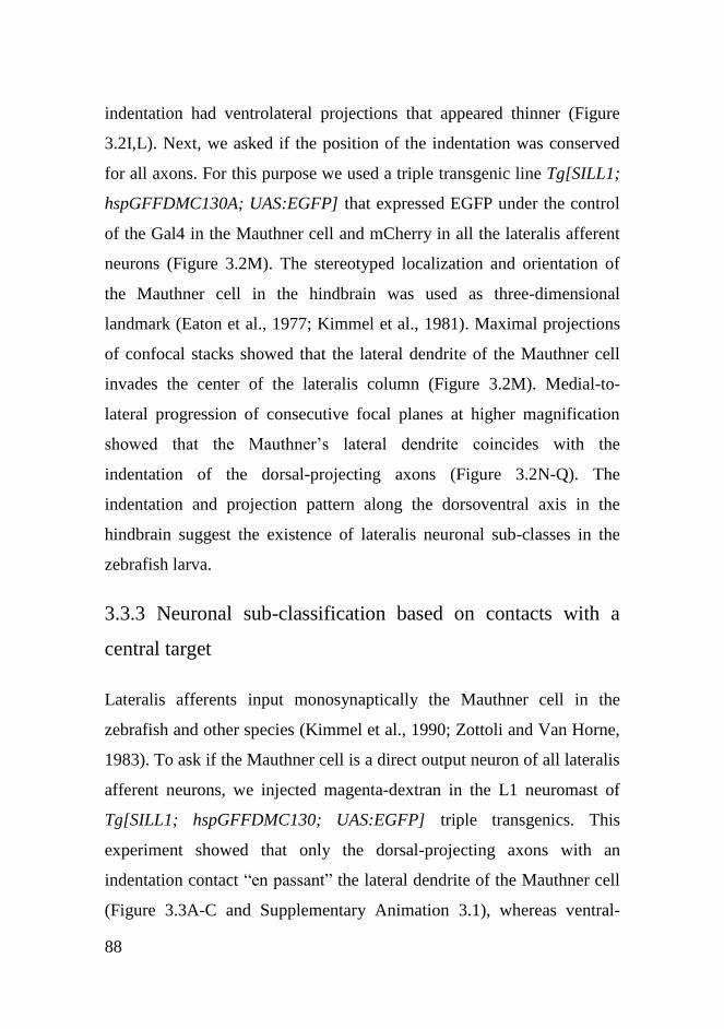
88
indentation had ventrolateral projections that appeared thinner (Figure
3.2I,L). Next, we asked if the position of the indentation was conserved
for all axons. For this purpose we used a triple transgenic line Tg[SILL1;
hspGFFDMC130A; UAS:EGFP] that expressed EGFP under the control
of the Gal4 in the Mauthner cell and mCherry in all the lateralis afferent
neurons (Figure 3.2M). The stereotyped localization and orientation of
the Mauthner cell in the hindbrain was used as three-dimensional
landmark (Eaton et al., 1977; Kimmel et al., 1981). Maximal projections
of confocal stacks showed that the lateral dendrite of the Mauthner cell
invades the center of the lateralis column (Figure 3.2M). Medial-to-
lateral progression of consecutive focal planes at higher magnification
showed that the Mauthner‟s lateral dendrite coincides with the
indentation of the dorsal-projecting axons (Figure 3.2N-Q). The
indentation and projection pattern along the dorsoventral axis in the
hindbrain suggest the existence of lateralis neuronal sub-classes in the
zebrafish larva.
3.3.3 Neuronal sub-classification based on contacts with a
central target
Lateralis afferents input monosynaptically the Mauthner cell in the
zebrafish and other species (Kimmel et al., 1990; Zottoli and Van Horne,
1983). To ask if the Mauthner cell is a direct output neuron of all lateralis
afferent neurons, we injected magenta-dextran in the L1 neuromast of
Tg[SILL1; hspGFFDMC130; UAS:EGFP] triple transgenics. This
experiment showed that only the dorsal-projecting axons with an
indentation contact “en passant” the lateral dendrite of the Mauthner cell
(Figure 3.3A-C and Supplementary Animation 3.1), whereas ventral-
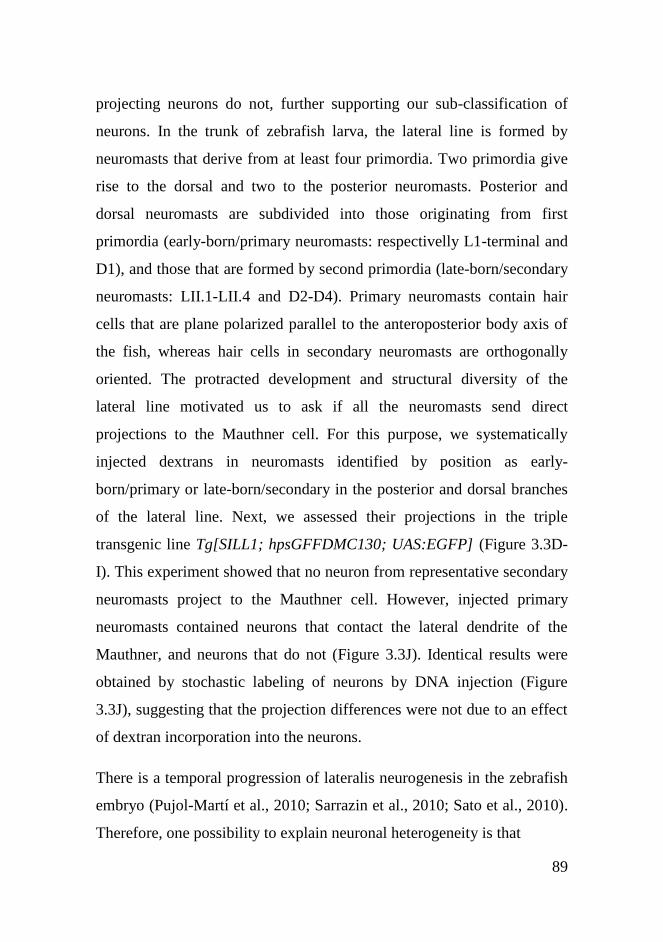
89
projecting neurons do not, further supporting our sub-classification of
neurons. In the trunk of zebrafish larva, the lateral line is formed by
neuromasts that derive from at least four primordia. Two primordia give
rise to the dorsal and two to the posterior neuromasts. Posterior and
dorsal neuromasts are subdivided into those originating from first
primordia (early-born/primary neuromasts: respectivelly L1-terminal and
D1), and those that are formed by second primordia (late-born/secondary
neuromasts: LII.1-LII.4 and D2-D4). Primary neuromasts contain hair
cells that are plane polarized parallel to the anteroposterior body axis of
the fish, whereas hair cells in secondary neuromasts are orthogonally
oriented. The protracted development and structural diversity of the
lateral line motivated us to ask if all the neuromasts send direct
projections to the Mauthner cell. For this purpose, we systematically
injected dextrans in neuromasts identified by position as early-
born/primary or late-born/secondary in the posterior and dorsal branches
of the lateral line. Next, we assessed their projections in the triple
transgenic line Tg[SILL1; hpsGFFDMC130; UAS:EGFP] (Figure 3.3D-
I). This experiment showed that no neuron from representative secondary
neuromasts project to the Mauthner cell. However, injected primary
neuromasts contained neurons that contact the lateral dendrite of the
Mauthner, and neurons that do not (Figure 3.3J). Identical results were
obtained by stochastic labeling of neurons by DNA injection (Figure
3.3J), suggesting that the projection differences were not due to an effect
of dextran incorporation into the neurons.
There is a temporal progression of lateralis neurogenesis in the zebrafish
embryo (Pujol-Martí et al., 2010; Sarrazin et al., 2010; Sato et al., 2010).
Therefore, one possibility to explain neuronal heterogeneity is that

90

91
neurons with the indentation or direct contact with the Mauthner cell are
born at different times than those without it. However, over time late-
born neurons may mature to resemble first-born. To test this possibility,
we followed samples in which both neuronal classes from a single
neuromast were labeled, and saw no differences in their central
projections or contacts with the Mauthner cell over a 7-day period
(Figure 3.3K). To rule out an effect of dextrans on the normal long-term
behavior of the axons, we also labeled the neurons from terminal
neuromasts in naïve HGn39D transgenic juveniles at 20 dpf and observed
them one day later. Again, we saw the presence of both neuronal sub-
classes (Figure 3.3L).
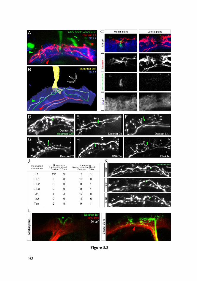
92

93
3.3.4 Biased axonal projection pattern of large and small
neurons
Having shown neuronal sub-classification in relation to central
projections and contact with the Mauthner cell, we next decided to search
for additional differences between these neurons. Because dextran uptake
eventually decorates the entire neuron, we looked at potential differences
in the ganglion and peripheral axons. Within the ganglion we consistently
found neurons with large and small somata (Figure 3.4A-B). We plotted
the neurons in a two-dimensional space in which each dot represented the
soma volume of individual neurons. The data was then grouped by both
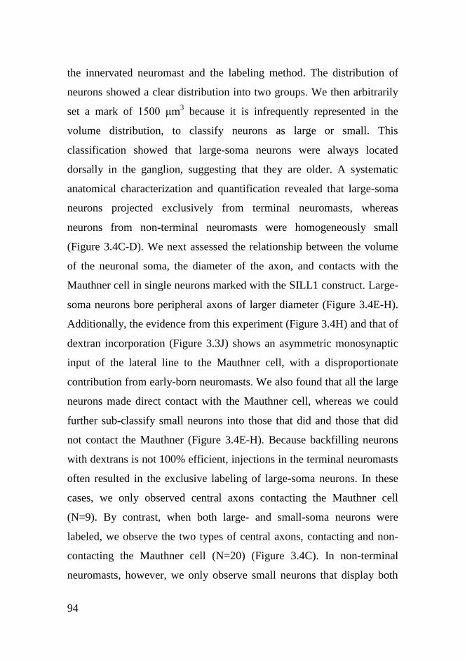
94
the innervated neuromast and the labeling method. The distribution of
neurons showed a clear distribution into two groups. We then arbitrarily
set a mark of 1500 μm3 because it is infrequently represented in the
volume distribution, to classify neurons as large or small. This
classification showed that large-soma neurons were always located
dorsally in the ganglion, suggesting that they are older. A systematic
anatomical characterization and quantification revealed that large-soma
neurons projected exclusively from terminal neuromasts, whereas
neurons from non-terminal neuromasts were homogeneously small
(Figure 3.4C-D). We next assessed the relationship between the volume
of the neuronal soma, the diameter of the axon, and contacts with the
Mauthner cell in single neurons marked with the SILL1 construct. Large-
soma neurons bore peripheral axons of larger diameter (Figure 3.4E-H).
Additionally, the evidence from this experiment (Figure 3.4H) and that of
dextran incorporation (Figure 3.3J) shows an asymmetric monosynaptic
input of the lateral line to the Mauthner cell, with a disproportionate
contribution from early-born neuromasts. We also found that all the large
neurons made direct contact with the Mauthner cell, whereas we could
further sub-classify small neurons into those that did and those that did
not contact the Mauthner (Figure 3.4E-H). Because backfilling neurons
with dextrans is not 100% efficient, injections in the terminal neuromasts
often resulted in the exclusive labeling of large-soma neurons. In these
cases, we only observed central axons contacting the Mauthner cell
(N=9). By contrast, when both large- and small-soma neurons were
labeled, we observe the two types of central axons, contacting and non-
contacting the Mauthner cell (N=20) (Figure 3.4C). In non-terminal
neuromasts, however, we only observe small neurons that display both

95
central-axon projection types (N=41) (Figure 3.4C). In conclusion,
neuronal labeling by dextran incorporation together with single-neuron
labeling by DNA injection, show that small neurons can be further
subdivided into those that contact the Mauthner cell and those that do not.
3.3.5 Neuronal projections and birth date
To ask about the timing of neuromast innervation relative to neuronal
size, we labeled neurons by fluorescent dextrans at different stages of
zebrafish development. This experiment confirmed that large neurons
arrive to the target neuromast first (Figure 3.5A-B). Lateralis
neurogenesis in the zebrafish embryo occurs in two discrete waves,
whose temporal sequence results in a strongly biased dorsoventral
localization of neurons in the ganglion (Pujol-Martí et al., 2010; Sarrazin
et al., 2010). Dorsalmost neurons belong to the first wave, suggesting that
they should have bigger somata. To test this hypothesis, we developed
BAIT (Birthdating combined with Anatomical analysis by Incorporation
of Tracers) by which timed fluorescent-protein photoconversion dates
cells, and dextran incorporation reveals their structure. BAIT showed that
older neurons are larger and are mostly located in the dorsal aspect of the
ganglion (Figure 3.5C-D). We have previously reported that the
transgenic line hspGFF53A is one of the earliest markers lateralis
afferent neurons, and that HGn39D begins to be expressed in the same
neurons several hours later (Pujol-Martí et al., 2010). A re-assessment of
these transgenic lines showed that the fluorescent signal from HGn39D
persists as neurons age, whereas that of hspGFF53A fades with time.
Neurons located in the ventral aspect of the ganglion (late-born)
expressed SILL1 (mCherry+) and hspGFF53A (EGFP+). However,
96
hspGFF53A was down-regulated in dorsally placed neurons (early-born),
whereas SILL1 expression was maintained (mCherry(+) / EGFP(-)). The
combination of these transgenics is therefore complementary to BAIT to
date neurons in vivo. Using the triple transgenics Tg[SILL1;
hspGFF53A; UAS:EGFP] injected with dextrans in terminal neuromasts
we analyzed the central projection of neurons of different ages by three-
dimensional confocal microscopy (Figure 3.5E-G) (N=17). This
experiment revealed that the oldest neurons (mCherry(+) / EGFP(-)) were
larger and localized to the ganglion‟s dorsum. When we examined the
central projections from these neurons we observed that they were always
characterized by and indentation in their rostral ramus (Figure 3.5F), and
projected axons dorsally in the hindbrain (Figure 3.5G). Ventrolateral
projections from younger neurons (mCherry(+) / EGFP(+)) did not have
an indentation. Form these results we confirm that axonal projections
correlate with the sequence of neuronal birth and differentiation.
3.3.6 The lateralis neural map develops in the absence of
sensory input
We wanted to know if evoked sensory activity played any role in
neuronal sub-classification or instructed their projection pattern in the
hindbrain. For this purpose, we combined the injection of a magenta-
dextran in terminal neuromasts whose large neurons always contact the
Mauthner cell, and red dextran in a secondary neuromast that are devoid
of neurons projecting to the Mauthner. These injections were made in
wild type animals and in mutants lacking hair-cell mechanoreception
(homozygous mutant for tmie), or lacking hair cells in the neuromasts
(homozygous mutant for atoh1a) (Figure 3.6A-C) (Faucherre et al.,
97
2010). We observed no differences in central projections or soma size
between all three conditions, demonstrating that evoked sensory input
does not instruct neuronal sub-classification or the pattern of central
projections. Another mechanism that could control map topology is
competition, whereby axons from early-born neurons repel those from
neurons born later from contacting the Mauthner cell. To test this
hypothesis it is useful to observe the axonal projections from late-born
neurons in the absence of early-born. If competition between neuronal
sub-classes does not play a role in axonal projections, late-born neurons
from experimental animals should be indistinguishable from those under
normal conditions. If, alternatively, interactions between axons are
responsible for the differences, the projections of late-born neurons
should be altered when early-born are absent. We tested this possibility
by eliminating early-born neurons in HGn39D transgenics by laser
ablation at 28-30 hpf, a stage in development between the differentiation
of both neuronal sub-classes. Subsequently, magenta-dextran was
injected in terminal neuromasts and central axonal projections were
analyzed by three-dimensional confocal microscopy. In all cases
analyzed the ablation of early-born neurons did not modify the axonal
projections from late-born neurons (Figure 3.6D-E). Collectively, these
results demonstrate that neither axonal competition, nor evoked sensory
activity play a significant role during the assembly of the lateralis neural
map.
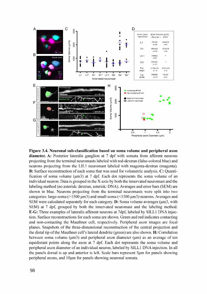
98
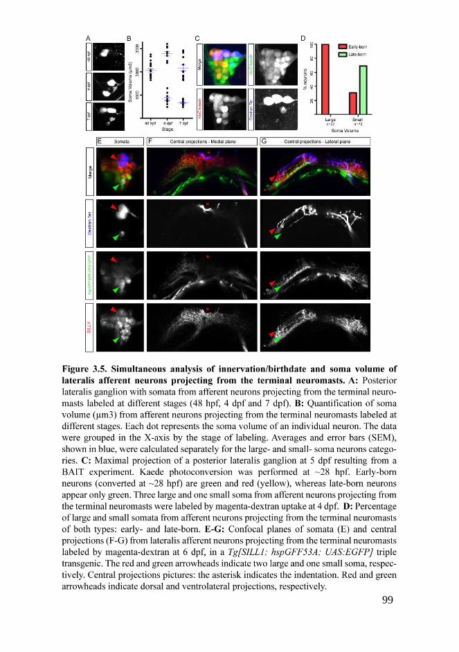
99

100
101
3.4 Discussion
The piscine lateralis afferent neurons assemble a somatotopic neural map,
in which axons are reproducibly positioned along a dorsoventral axis in
the hindbrain (Alexandre and Ghysen, 1999; Claas and Münz, 1981;
Ghysen and Dambly-Chaudière, 2007). Although somatotopy probably
forms a neuroanatomical code of the external hydrodynamic field, it is
not well understood how animals use it to command behaviors. In this
study we employed neuronal labeling and image registration to reveal
two classes of lateralis afferent neurons defined by birth order, soma size
and location, central projections and contacts with an identified output
neuron, to define a dimorphic sensorineural map. Next, we discuss our
results and make predictions on how lateralis map topology may underlie
appropriate behavioral responses according to the biological relevance of
mechanosensory input.
Lateralis afferent neurons are extremely interesting because they can shed
light on the mechanisms that initiate different behavioral programs by the
same sensory modality. Many studies have firmly demonstrated a role of
the lateral line in navigation, schooling and prey detection (Bleckmann,
2008; Coombs et al., 1998; Coombs and Patton, 2009; Dijkgraaf, 1963;
Montgomery et al., 1997; Montgomery et al., 2000) . It has also been
proposed that the lateral line can send powerful inputs to the Mauthner
cell to trigger the C-start escape response (McHenry et al., 2009). The
optical transparency, external and fast development of the zebrafish,
coupled with the accessibility and anatomical simplicity of its lateral line
provides a powerful model to study the neuroanatomical bases of these
contrasting behaviors. Because an essential part of investigations toward

102
this aim is the careful characterization of neuronal connectivity, we
generated two transgenic zebrafish lines for targeted gene expression.
The hspGFFDMC130A line contains the Gal4 transcriptional activator
stably integrated in the genome (Nagayoshi et al., 2008). We demonstrate
that hspGFFDMC130A can express fluorescent markers in the Mauthner
cell and few other tissues. We also generated the Tg[SILL1] transgenics
that expresses the mCherry red-fluorescent protein in all the lateralis
afferent neurons. Single-neuron labeling with the SILL1 construct
revealed unexpected neuronal diversification and neural-map
dimorphism. In a divergent neural sub-map, each neuron forms around 60
boutons that concentrate a well-characterized presynaptic marker. If we
assume that each bouton represents a synaptic contact with one output
neuron, the posterior lateral line has no more than 2,400 second-order
targets in the zebrafish larva (Fame et al., 2006; Sassa et al., 2007). In a
convergent sub-map, many lateralis neurons directly contact the lateral
dendrite of the Mauthner cell.
What determines map dimorphism and target selectivity? One possibility
is that sensory activity plays an instructive role (Luo and Flanagan,
2007). However, our results from the analysis of two types of mutants -a
strong loss-of-function in Tmie that blocks hair-cell mechanoreception,
and a loss-of-function in Atoh1a that prevents the development of hair
cells in neuromasts- indicate that sensory activity is not a major force in
sculpting the lateralis neural map. Axonal competition has been shown to
influence neural mapping in several systems. However, our selective
ablation of one neuronal subpopulation did not alter the projection of the
remaining neurons. Therefore, inter-class axonal competition does not
appear to instruct map topology.

103
We combined our transgenic lines with dextran injections for anatomical
tracing to develop BAIT. One caveat of BAIT is that it does not reveal
the actual birth dates of cells, but their relative age. Therefore, we made
the assumption that neurons differentiate at similar rates once they have
fate-committed. In doing so, we demonstrate a correlation between the
sequence of afferent neurogenesis and central projections, and that only
early-born neurons converge on the Mauthner cell. Although we cannot
currently rule out that molecular gradients in the target area refine map
topology much the same way as they do in other sensory systems, our
findings support a strong contribution of neurogenic timing in lateralis
neural map dimorphism and projection pattern (Clandinin and Feldheim,
2009; Fariñas et al., 2001; Feldheim and O'Leary, 2010; Luo and
Flanagan, 2007; Schuster et al., 2010). Such strategy is not likely to be
unique to the lateral line, however. For example, mechanosensory
neurons of the wing or photoreceptor neuron of the eye in Drosophila
also produce tiered central projections based on their time of
development (Morey et al., 2008; Palka et al., 1986; Petrovic and
Hummel, 2008). Earlier studies in Xenopus tested the involvement of
timing in generating the topographic organization of the neuronal
projection from the retina to the tectum, and concluded that it likely plays
a permissive rather than an instructive role in axonal organization (Holt,
1984). It would be interesting to test permissive versus instructive roles
of timing in the lateral line.
Neuromasts are directionally sensitive by virtue of the planar polarization
of their constituent hair cells. Posterior neuromasts have orthogonal
planar orientations. Parallel neuromasts develop earlier from first
primordia and are called “primary neuromasts”. Perpendicular

104
neuromasts develop subsequently from independent primordia and are
called “secondary” (Ghysen and Dambly-Chaudière, 2007; López-Schier
et al., 2004; Sarrazin et al., 2010). Our study demonstrates that waves of
afferent neurogenesis accompany the protracted development of the
neuromasts. Primary neuromasts project early-born neurons that diverge
along the hindbrain but some of which also converge on the Mauthner
cell. Secondary neuromasts, by contrast, only project late-born neurons
that do not converge on the Mauthner. A potentially equivalent
relationship between developmental timing and neuronal projection
patterns has been reported for the segregation of afferent projections for
the otic, lateralis and ampullary organs in the axolotl (Fritzsch et al.,
2005). In this case, however, the relationship is between organs rather
than within each organ.
Afferent input to the Mauthner from primary neuromasts may be an
elegant strategy to couple development with behavior because the earliest
born neurons and neuromasts could allow the animal to use the lateral
line for escape responses before it is able to swim. We show that in the
zebrafish larva, terminal neuromasts are innervated by large-diameter
afferent axons. The conduction velocity of myelinated axons in
vertebrates increases linearly with their diameter (Goldman and Albus,
1968; Holmes, 1941; Hursh, 1939). Therefore, early-born neurons
projecting from terminal neuromasts are likely to be fast conducing,
making them well suited to produce the first and fastest lateral-line
stimulus for the C-start response. This also suggests that terminal
neuromasts have a disproportionate relevance in the escape behavior
mediated by the Mauthner cell. Interestingly, the trout also shows
regional segregation of lateralis neurons relative to axon diameter and
105
conduction velocity, which can compensate for the increased axonal
length in large animals (Schellart and Kroese, 2002).
Are terminal neuromasts enough to trigger the C-start response? Unlike
neurons from other parts of the lateral line, each large neuron innervates
up to three terminal neuromasts, which may increase their depolarization
probability. This presents clear survival advantages because terminal
neuromasts may suffice to trigger a C-start reaction by sending strong
depolarizing inputs to the Mauthner cell with very short latencies.
However, in general fish should not startle by non-threatening stimuli,
which predicts that early-born neurons should have lower sensitivity than
late-born. Behaviors such as navigation, rheotaxis and schooling
necessitate continuous input and probably have lower activating
thresholds that the C-start. Afferent neurons with different excitability
and conduction velocities have been reported for the posterior lateral line
of the goldfish (Fukuda, 1974). Also, in cichlids, lateralis afferent
neurons with higher rate of spontaneous discharge are more sensitive
(Münz, 1985). Interestingly, the rate of spontaneous activity of individual
afferent neurons in the zebrafish larva varies along the lateral line (Liao,
2010). If these observations are extrapolated and combined with our
findings, one emergent possibility is that the C-start will only be
triggered by the coincident input on the Mauthner cell from “high
sensitivity/low conduction velocity” and “lower sensitivity/high
conduction velocity” neuronal classes. This model will safeguard the
animal from startling upon stimuli that would depolarize one neuronal
sub-class but not the other, and is reminiscent of the escape strategy of
crayfish, in which a mechanosensory stimulus activates parallel neuronal
pathways with different reaction times that trigger the startle when
106
arriving coincidently to an output command neuron (Mellon and
Christison-Lagay, 2008; Reichert and Wine, 1982). However, more
complex processing of hydrodynamic stimuli is possible, with multiple
neuromasts contributing to the probability of depolarizing excitatory
signals from the lateral line to the Mauthner cell (Korn et al., 1974).
Although spatiotemporal resolution may involve secondary neuromasts,
they may not contribute to the C-start response because of their discrete
location and their insensitivity to the direction of propagation of most of
the water flow generated by submerged predators. This may explain why
their afferent neurons do not form monosynaptic contacts with the
Mauthner cell. This begs the question of whether somatotopy has any
functional relevance to the C-start reaction. Currently, we favor the
hypothesis that lateralis somatotopy does not play a major role in the
escape behavior of the zebrafish larva. Thus, the lateral line would
sacrifice spatial accuracy for sensitivity and response speed. For
navigation, however, somatotopy is likely to be essential. Thus, the
lateral line may assemble a convergent sub-map for speed and a divergent
sub-map for accuracy. The combined activity of neurons can code for the
entire range of stimuli (spectrum) that a sensory organ can acquire.
Variation in the sensitivity of individual sensory receptors within the
organ can subtract aspects of a complex stimulus to direct behavior. This
subdivision of work is called range fractionation (Cohen, 1963).
However, variations in sensory transducers can also impact information
processing. Therefore, the next challenge is to determine if sensorineural
map dimorphism in the lateral line form the bases of range fractionation
by this sensory system (Braun et al., 2002; Voigt et al., 2000).

107
3.5 Materials and methods
3.5.1 Zebrafish strains and husbandry
Zebrafish were maintained under standardized conditions and
experiments were conducted in embryos of undetermined sex in
accordance with protocols approved by the PRBB‟s Ethical Committee of
Animal Experimentation. Tg[HuC:Kaede], HGn39D and hspGFF53A
transgenic lines have been published previously (Faucherre et al., 2009;
Pujol-Martí et al., 2010; Sato et al., 2006). The hspGFFDMC130A was
generated by random integration of an enhancer-trap construct (Asakawa
et al., 2008). The atoh1a_fh282 mutant strain carries a missense
mutation, aa126 arginine to tryptophan, and was obtained from C.
Moens. The tmie_ru01 mutant fish were described previously (Faucherre
et al., 2010; Gleason et al., 2009).
3.5.2 Selection of mutants
Wild type animals and homozygous mutants for tmie and atoh1a were
sorted based on DiASP incorporation into hair cells of the lateral line.
Homozygous mutants for tmie were genotyped by amplification using the
following primers and sequencing:
For: 5‟-CCAGCAGCTCTCGTAACCTC-3‟
Rev: 5‟-CCGCCATCACCAGTCTATTT-3‟
108
3.5.3 Plasmid DNA constructs and injections
For cloning the SILL enhancer, the 2 kb upstream of the HGn39D
insertion was amplified from genomic DNA and cloned in the Tol2kit
219 plasmid using the following primers:
EnUp-attb4-For:
5‟-GGGGACAACTTTGTATAGAAAAGTTGTAAAGAAATGTCAAGTGTTT-3‟
EnUp -attB1-Rev:
5‟-GGGGACTGCTTTTTTGTACAAACTTGGTTGGGATGGTGTACAGTAT-3‟
The 2 kb downstream of the HGn39D insertion was amplified and cloned
in the Tol2kit 220 plasmid using the following primers:
EnDo-attB2-For:
5‟-GGGGACAGCTTTCTTGTACAAAGTGGCCATCCCAACTCACTCACTATT-3‟
EnDO-attB3-Rev:
5‟-GGGGACAACTTTGTATAATAAAGTTGCCTGACATTTTCCGGAACAGG-3‟
The hsp70:mCherry-SILL (SILL1), hsp70:Gal4VP16-SILL (SILL2) and
hsp70:MCS-SILL constructs were obtained using the „„Tol2 kit‟‟ (Kwan
et al., 2007). Entry vectors were generated as described in the Invitrogen
Multisite Gateway manual. PCR were performed using primers to add att
sites onto the end of DNA fragments, using Platinum Pfx (Invitrogen).
The pEntry vectors containing the hsp70 promoter, the mCherry, the
Gal4VP16 and the MCS (multiple-cloning site) are from the „„Tol2 kit‟‟.
To generate the SILL:VAMP-GFP construct, VAMP-GFP was amplified

109
using the „UAS:VAMP-GFP;UAS:Kir2.1‟ plasmid as a template (Hua et
al., 2005), with the following primers:
For: 5‟-AGACGATATCATGTCTGCCCCAGATGCT-3‟
Rev: 5‟-TATGGATATCTTACTTGTACAGCTCGTC-3‟
The PCR product was digested by EcoRV and cloned into the
hsp70:MCS-SILL construct.
3.5.4 Generation of transgenic zebrafish
To generate the Tg[SILL1] stable transgenic line, 20 pg of the Tol2-
expression clone and 20 pg of the transposase synthetic RNA were
simultaneously injected into one-cell stage wild type eggs. The resulting
embryos were raised to adulthood and incrossed for visually screening of
germline transmission of the transgene. For mosaic expression in
neurons, 25–30 pg of DNA was injected into embryos at the one- or two-
cell stage.
3.5.5 Whole-mount in situ hybridization
We generated labeled RNA probes by in vitro transcription using the
DIG/Fluor RNA labeling Mix (Roche). Embryos were fixed in 4%
paraformaldehyde in PBS overnight at 4ºC and whole-mount in situ
hybridizations were carried using standard protocols.
To characterize cntnap2 expression, the following antisense probe was
used: Ensemble Zv9 ENSDART00000081960, nucleotides 3049-3357.
110
3.5.6 Neuronal labeling, birthdating and imaging
Neuronal labeling by fluorescent dextrans was performed as previously
described (Pujol-Martí et al., 2010). We used a red-fluorescent dextran
(tetramethylrhodamine, 3000 MW, anionic, fixable, Invitrogen
ref.D3308) and a magenta-fluorescent dextran (Alexa Fluor® 647, 10000
MW, anionic, fixable, Invitrogen ref.D22914). Imaging was done one
day after dextran injection. In all the samples, we imaged the dextran
injection site to confirm that only neurons innervating an individual
neuromast were labeled. We analyzed samples in which the neuronal
labeling was specific to the selected neuromast, with the exception of the
dextran injections into the D2 neuromast, in which neurons innervating
the more posterior and dorsal neuromasts were often also labeled.
For BAIT (Birthdating combined with Anatomical analysis by
Incorporation of Tracers) experiment, Tg[HuC:Kaede] eggs were kept in
the dark. At 28 hpf stage, embryos were exposed to 405 nm light for 2
min and subsequently kept in the dark for 3 days. Neurons innervating
terminal neuromasts were labeled by magenta-fluorescent dextran and the
posterior lateralis ganglion was imaged one day later.
For imaging, live samples were anæsthetized and mounted onto a glass-
bottom small Petri dish (MatTek, Ashland, MA) and covered with 1%
low-melting-point agarose with diluted anæsthesic. Images were acquired
with a Leica TCS SP5 inverted confocal laser scanning microscope with
a 20x Air objective, or a 40x Oil immersion objective. Z-stacks of central
and peripheral axons consisted of 1 μm-spaced images. Z-stacks of
neuronal somata consisted of 2.5 μm-spaced images. Three-dimensional

111
(3D) reconstructions and cropping of z-stacks were done with Imaris
software (Bitplane).
3.5.7 Quantification of soma volume and peripheral axon
diameter
To measure soma volume, a surface reconstruction of each soma imaged
was done using Imaris, with a surface area detail level of 0.5 μm and a
thresholding based on absolute intensity. Threshold was manually
adjusted to optimally match the surface reconstruction with the
fluorescent signal. Imaris automatically quantifies the volume of the
surface reconstruction. To measure peripheral axon diameter, we imaged
a ~100 μm segment of the peripheral axon just anterior to the peripheral
arborization at the level of the neuromast using a Leica TCS SP5 inverted
confocal micoscope with a 20x Air objective, with Zoom: 8. Diameters
were measured at ten equidistant locations of the imaged axonal segment
and the average was calculated.
3.5.8 Laser-mediated cell ablation
For neuronal ablation we used a Micropoint laser system (Photonic
Instruments Inc.) mounted on an Olympus IX81 inverted microscope
equipped with a 40x Air objective. HGn39D embryos at 28-30 hpf were
anesthetized, mounted on a glass-bottom dish (MatTek, Ashland, MA),
and covered with methylcellulose. A train of laser pulses was repeatedly
applied to the posterior lateralis ganglion until all EGFP fluorescence
disappeared. Embryos were allowed to recover for 2 hs and then assessed
for the presence of EGFP in the region of the posterior ganglion. Total

112
ablation occurred in samples with no green-fluorescent signal in that
region. One day later, we chose samples with new neurons forming a
ganglion. We next labeled neurons projecting from the terminal
neuromasts at 5-6 dpf by injecting fluorescent dextran at the neuromasts.
We did the same in non-ablated controls.
113
3.6 Supporting information
Supplementary animation 3.1: Interactive three-dimensional
reconstruction from the data shown in Figure 3.3A. Central axons from
lateralis afferent neurons innervating L1 neuromast (early-born/primary
neuromast) are labeled with magenta-dextran at 6 dpf, in a Tg[SILL1;
hspGFFDMC130A; UAS:EGFP] triple transgenic animal. Dorsal
projections contacting the Mauthner cell (yellow) are in green whereas
ventrolateral projections non-contacting the Mauthner are in red. The
lateralis column is shown in blue. The three-dimensional reconstruction
can be rotated by pressing the mouse left button on the picture and
moving the mouse; as well as it can be scaled by clicking on the “+” and
“−” icons. Before rotation, dorsal is towards top, anterior is towards left,
lateral is towards the observer.

114

115
Chapter 4
ADDITIONAL RESULTS

116
117
4.1 Lateralis afferents contacting the Mauthner cell
form a somatotopic map
A previous study in the zebrafish larva showed that lateralis afferents
assemble a somatotopic map where the position of each central axon
represents the position of the innervated neuromast (Alexandre and
Ghysen, 1999). In this study, the authors labeled neurons with fluorescent
dextrans. I noticed that neuronal backfilling with dextrans is not 100%
efficient; injections in terminal neuromasts often resulted in the exclusive
labeling of large-soma neurons with central axons contacting the
Mauthner cell. Moreover, when analyzing dextran-labeled neurons
projecting from either L1 or terminal neuromasts I observed that the ones
that contact the Mauthner cell are usually more strongly labeled than
those that do not. The less frequent and weaker labeling of neurons that
do not contact the Mauthner cell prompted me to think that the
somatotopic map was originally described by analyzing only lateralis
central axons from neurons that contact the Mauthner cell. By analyzing
Tg[hspGFFDMC130A; UAS:EGFP] double transgenic animals injected
with red-dextran in the terminal neuromasts and magenta-dextran in the
L1 neuromast, I confirmed that central axons contacting the Mauthner
cell are topographically ordered (N=5) (Figure 4.1).
118
4.2 The Mauthner cell receives input from hair-cells
of opposing polarities
Each neuromast is innervated by at least two lateralis afferent neurons;
each of them forming synapses with hair cells of identical polarity
(Faucherre et al., 2009; Nagiel et al., 2008). I sought to determine
whether lateralis afferents carrying inputs from hair-cells of opposing
polarities converge on the Mauthner cell. For this purpose, I assessed the
relationship between the presence of an indentation in the central axon,
indicative of a contact with the Mauthner, and the polarity of the hair-
cells innervated by single neurons marked with the SILL1 DNA
construct. To visualize the hair cells I used brn3c:memGFP transgenic
animals. Since hair cells are born in pairs of opposing polarity along a
single axis creating a line of mirror symmetry, all the cells located
anterior to this line are posteriorly polarized, whereas those located
posterior to the line of symmetry are anteriorly polarized. As a
consequence, hair-cell polarity can be predicted from the cell‟s position
with respect to the line of mirror symmetry (López-Schier and Hudspeth,
2006). I observed the indentation in the central axons of neurons
innervating posteriorly polarized hair cells (N=9) as well as in those of
neurons innervating anteriorly polarized hair cells (N=6) in the terminal
neuromasts (Figure 4.2). I conclude that the Mauthner cell receives
sensory inputs from hair-cells with an anterior polarity as well as from
hair-cells with a posterior polarity.

119
4.3 Peripheral arborization and neuronal sub-classes
I showed that primary neuromasts are innervated by lateralis afferents of
the two sub-classes, those that contact the Mauthner cell and those that do
not. I further confirmed that the former are born earlier than the latter.
This was possible thanks to the combination of Tg[SILL1] and
hspGFF53A transgenic lines that reveals neuronal relative age. This
combination also allowed me to ask whether the two neuronal sub-classes
differ at the level of the peripheral arborization of primary neuromasts. I
examined terminal neuromasts and observed that the peripheral axons of
early- and late-born neurons largely overlap occupying the entire
neuromast (N=3) (Figure 4.3). The presence of a bulged neurite in the
peripheral arbor predicts a synaptic contact between a neuron and a hair
cell (Faucherre et al., 2010; Nagiel et al., 2008). Importantly, bulged
neurites from early- and late-born neurons are often adjacent (N=3)
(Figure 4.3). Altogether these observations strongly suggest that, within a
given neuromast, neurons of the second sub-class carry sensory
information from the same hair cells as neurons of the first sub-class do.
This implies that late-born lateralis afferents non-contacting the
Mauthner cell convey information from hair-cells of opposing polarities.
Furthermore, there is evidence that strongly suggests that these neurons
convey the inputs from the two populations of hair cells in separate
channels. The experiments that revealed that each lateralis afferent
innervates hair-cells of identical polarity were carried out by using a
DNA construct encoding a fluorescent marker under the control of the
HuC promoter (Faucherre et al., 2009; Nagiel et al., 2008). Due to the
fact that the HuC promoter is a pan-neuronal promoter (Park et al., 2000)

120
and to the large amount of neurons analyzed in those experiments, it is
very likely that the two neuronal sub-classes were examined to conclude
that each lateralis afferent always selects hair-cells of identical polarity.

121

122

123
Chapter 5
GENERAL DISCUSSION

124
125
5.1 Structure and function of the lateral-line neural
maps
Lateralis afferent neurons segregate the sensory inputs acquired at
different locations along the animal‟s body and at the same time they
separate the inputs from parallel and perpendicular neuromasts.
Similarly, the inputs from hair-cells of opposing polarities are conveyed
to the brain in separate pathways (Faucherre et al., 2009; Nagiel et al.,
2008; Sarrazin et al., 2010). To better understand how the brain takes
advantage of the peripheral organization of the lateral line, I reexamined
the projection patterns of the lateralis afferent neurons in the zebrafish
larva, with an emphasis on their connectivity with a known central target:
the Mauthner cell (Kimmel et al., 1990). I will next discuss some
structural and functional aspects that emerge from my findings.
5.1.1 A new view of the neural maps built by the lateralis
afferent neurons
I demonstrated the existence of two sub-classes of lateralis afferents in
the posterior lateral line of the zebrafish larva, which differ in their
connectivity with the Mauthner cell and in their central axonal projection
patterns. The first sub-class comprises neurons whose central axons
contact the lateral dendrite of the Mauthner cell and are always located
dorsally in the lateralis column of the hindbrain. By contrast, the second
sub-class consists of neurons whose central axons do not contact the
Mauthner cell and occupy a ventrolateral position in the lateralis column.
Do they also differ in their peripheral axonal projection patterns?
Neurons of the first sub-class convey information from anterior and
posterior parallel neuromasts (Figure 5.1). Within these neuromasts, they

126
innervate hair cells of opposing polarities. Surprisingly, neurons of the
second sub-class also innervate the very same neuromasts that are
innervated by neurons of the first sub-class (Figure 5.1). Importantly,
within these neuromasts there is no apparent segregation of the peripheral
axon arbors of the two neuronal sub-classes; they rather overlap across
the entire organ. This strongly suggests that the two sub-classes convey
inputs from the same hair cells. Therefore, the second sub-class of
lateralis afferents also carries information from hair-cells of opposing
polarities, very likely in separate pathways. In addition, neurons of the
second sub-class convey information from the perpendicular neuromasts,
in contrast with neurons of the first sub-class (Figure 5.1).
A previous analysis in the zebrafish larva showed that the first lateral-line
sensory information relay in the brain contains a somatotopic map. In this
map, each lateralis central axon is reproducibly positioned in the
hindbrain reflecting the position of the peripheral axon and thus of the
innervated neuromast (Alexandre and Ghysen, 1999). However, the
systematic anatomical characterization and quantification I performed
revealed a more complex arrangement of the lateralis central and
peripheral axons. Lateralis afferents shape a sensory neural map
composed of two sub-classes of neurons that differ in their connectivity
with the Mauthner cell and in their central projections‟ positions. Each of
these sub-classes seems to convey sensory inputs from hair cells of
opposing polarities and from different locations along the animal‟s body.
With regard to this, I showed that within the first sub-class of neurons
central axons are topographically ordered building a somatotopic map.
Nevertheless, when one puts together the two sub-classes of neurons and
correlates the positions of the central axons to those of the peripheral

127
axons, there is no discernible topographic ordering (Figure 5.1).
Therefore, the organization of the lateralis afferents combines attributes
of both discrete and continuous maps. The two sub-classes of lateralis
afferents shape a dimorphic map that resembles a discrete neural map; the
spatial organization of their central axons reflects a discrete quality,
contacting or non-contacting the Mauthner cell, rather than the spatial
organization of their peripheral axons. Embedded in this map, however,
there is at least one sub-map built by the neurons of the first sub-class.
This is the somatotopic map, which resembles a continuous or
topographic map; the spatial organization of their central axons reflects
the spatial organization of their peripheral axons. It would be interesting
to examine whether there are other sub-maps embedded in the dimorphic
lateral-line neural map. For instance, the second sub-class of lateralis
afferents might assemble a second independent somatotopic map.
The functional and anatomical study of other sensory neural maps has
previously revealed a similar degree of complexity. The visual cortex
contains a continuous representation of the retina and embedded in this
map there are multiple, superimposed maps of different stimulus
attributes, such as eye dominance or motion direction preference (Luo
and Flanagan, 2007; Swindale, 2001). Similarly, somatosensory maps
normally consist of a continuous representation of the body surface in
which discrete units relaying sensory inputs from different modalities,
such as touch or pain, are embedded (Luo and Flanagan, 2007). In at least
one species, however, the projection patterns of the somatosensory
neurons appear to show a different topology. Cutaneous neurons from the
chick hindlimb project to the central nervous system forming two
separate bundles that represent sub-modalities of somatosensory
128
information. Each of these bundles assembles an independent
topographic map of the skin surface (Woodbury and Scott, 1991). This
organization resembles the map topology I describe here in the first step
of lateral-line sensory information relay in the brain.
5.1.2 Functional implications of the lateral-line dimorphic
neural map
The segregation of the lateralis central axons in dorsal and ventrolateral
projections does not account for a segregation of the sensory inputs
coming from different regions of the body or of the inputs coming from
hair-cells of opposing polarities. Besides hair-bundle polarity, there is no
evidence for other heterogeneities among the hair-cells of a given
neuromast. If this was the case, however, the two sub-classes of lateralis
neurons would not segregate inputs from the different receptors since
they seem to contact the same hair cells within a given neuromast.
Therefore, the two sub-classes of lateralis afferents rather convey the
same sensory information regarding both stimulus position and direction
across the antero-posterior body axis. In view of this, they might have a
redundant function. However, I showed that they differ in other
significant aspects. Importantly, they differ in their connectivity with the
Mauthner cell. My anatomical data also argue for the existence of “low
excitability/high conduction velocity” and “high excitability/low
conduction velocity” lateralis neurons, as discussed previously in chapter
3. The former are only found within the first neuronal sub-class whereas
the latter are found within each of the neuronal sub-classes. In addition,
sensory inputs from perpendicular neuromasts are exclusively conveyed
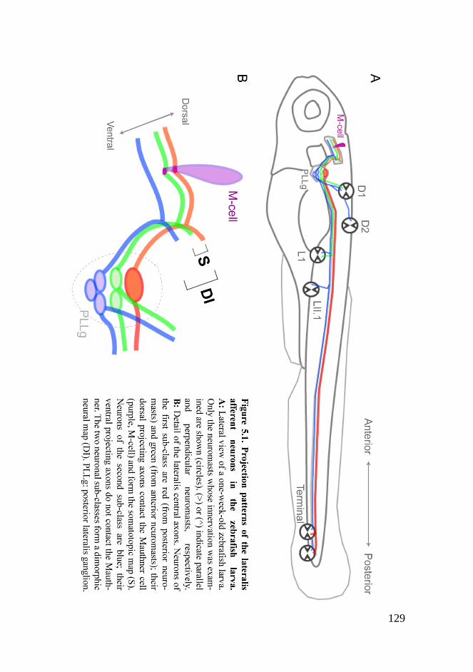
129
130
by the second sub-class of neurons. Finally, the two neuronal sub-classes
are born at different times. In light of these findings, I propose that the
two sub-classes I describe here might have contrasting functions within a
given neural circuit, thus sub-serving different components of the same
behavior. Alternatively, they might represent parallel channels of sensory
information used by different central neural circuits mediating distinct
behaviors. These possibilities might be analogous to the separate central
representation of submodalities observed in other sensory systems (Palka
et al., 1986; Sur et al., 1984; Szwed et al., 2003) and even in the adult
lateral-line system which consists in superficial and canal neuromasts
(Bleckmann, 2008). However, in these cases not only the sensory neurons
but also the peripheral receptors exhibit markedly physiological
differences between submodalities. I will next discuss previous findings
that reinforce these hypotheses and briefly propose some future
investigations on it.
The lateral line mediates very fast responses to abrupt stimuli, such as the
C-start reflex (McHenry et al., 2009). This escape behavior is triggered
by the activation of the Mauthner cell, which together with some
excitatory and inhibitory interneurons in the hindbrain constitute the
escape circuit. Importantly, a balance between direct sensory excitation
of the Mauthner and a feed-forward inhibition regulates the onset of the
C-start reflex (Faber et al., 1989; Faber et al., 1991; Koyama et al., 2010).
This is known to occur for the sensory inputs from the inner ear in the
zebrafish larva (Takahashi et al., 2002). Physiological data from the
goldfish also showed that the Mauthner cell receives both monosynaptic
excitatory and polysynaptic inhibitory inputs from the lateral line (Korn
and Faber, 1975). The lateralis afferents that contact the lateral dendrite

131
of the Mauthner cell must account for the monosynaptic excitatory input.
These very same neurons, however, might also account for the
polysynaptic inhibitory input by exciting interneurons that further inhibit
the Mauthner cell (Figure 5.2A). Alternatively, this could be done by the
lateralis afferents that do not contact the Mauthner cell (Figure 5.2B)
(Faber and Korn, 1975). Therefore, the heterogeneous anatomical
connectivity between lateralis afferents and the Mauthner cell that I
found might reflect a functional specialization where (1) only a subset of
the neurons participates in the escape circuit or (2) the two sub-classes of
neurons play opposite roles, excitatory versus inhibitory, within the same
escape circuit. To distinguish between these possibilities, it will be
necessary to dissect the connectivity between the two sub-classes of
lateralis afferents and the feed-forward inhibitory interneurons, which
have been recently identified in the zebrafish larva (Koyama et al., 2010).
Besides this, it would be very interesting to examine how the activities of
these two neuronal sub-classes influence the Mauthner cell firing and the
C-start behavior. This can be done by means of the recently developed
optogenetic tools, that provide a convenient way to manipulate and
monitor neuronal activity (Del Bene and Wyart, 2011).
Lateralis afferents synapse with hundreds of hindbrain second-order
neurons beyond the Mauthner cell. The hindbrain of the zebrafish larva
contains functional circuits that control diverse motor outputs, including
swimming and escape behaviors. Interestingly, many of these circuits
appear to be built from a basic ground plan. They are made of a set of
dorso-ventral stripes which consist of neurons that use the same
neurotransmitter. Neurons within a given stripe are arranged by structural
and functional properties, as well as by age. Dorsal neurons show high
132
excitability, are involved in slow swimming movements and are young.
By contrast, ventral neurons show low excitability, are involved in fast
swimming movements and are old (Kinkhabwala et al., 2010). Such
spatial organization of the hindbrain neurons, arranged by age and degree
of excitability, strikingly resembles that of the lateralis central axons.
This prompts me to speculate that lateralis afferents synapse with second-
order neurons in the hindbrain with similar properties that are well-suited
for a particular motor output (Figure 5.2C). For instance, an escape
behavior would take advantage of sensory and central neurons with low
excitability (high thresholds for activation) in order to exclusively detect
and respond to sudden and powerful threatening stimuli. By contrast,
rheotaxis would take advantage of neurons with high excitability (low
thresholds for activation) in order to detect and react to finer changes in
the environment. There is already some indirect evidence for a synaptic
match between lateralis afferents and central targets that share functional
properties. Dorsal hindbrain neurons (high excitability/young) send
dendrites to the ventral neuropil, where the lateralis ventrolateral axons
(high excitability/young) presumably reside. Ventral hindbrain neurons
(low excitability/old) project instead to the dorsal neuropil, where
lateralis dorsal axons (low excitability/old) are supposed to locate
(Kinkhabwala et al., 2010). It will be possible to further confirm this by
means of neuroanatomical methods and optogenetic tools. Moreover, it
would be exciting to directly test the role of the two sub-classes of
neurons in sub-serving different motor outputs. This can be done, for
instance, by using optogenetics or by cell ablations. These experiments
will certainly shed more light on the significance of the heterogeneities
among lateralis afferents delineated by my work.

133
134
5.2 Neural map formation in the lateral-line system
The development of the neural maps assembled by the lateralis afferents
has been the other central subject of my thesis research. I initially asked
how posterior lateral-line somatotopy is established. I demonstrated that
among the lateralis afferents that shape a somatotopic map, the ones with
dorsal projecting central axons and innervating posterior neuromasts are
older than those with ventral projecting central axons and innervating
anterior neuromasts. Therefore, there is a correlation between the time at
which a lateralis afferent neuron differentiates and its projection patterns.
I further found that these lateralis afferents make contacts with the
Mauthner cell and originate from a first wave of neurogenesis.
Importantly, a second wave of neurogenesis gives rise to another sub-
class of lateralis neurons, characterized by central axons that do not
contact the Mauthner cell and locate more ventrally in the lateralis
column. I propose a twofold contribution of progressive neurogenesis to
the diversification and patterning of the lateralis afferents. First, it
arranges a somatotopic map that encodes the position of the sensory
stimulus. Second, it delineates a dimorphic neural map with interesting
potential functional implications. Timing of neurogenesis is known to
contribute to sensory neural map assembly in other sensory systems, such
as the visual and olfactory systems of both invertebrates and vertebrates
(Clandinin and Feldheim, 2009; Holt, 1984; Imamura et al., 2011;
Jefferis et al., 2001; Petrovic and Hummel, 2008). Moreover, neurogenic
timing contributes to connectivity diversity in motor systems as well as in
other regions of the brain (Deguchi et al., 2011; McLean and Fetcho,
2009; Tripodi et al., 2011).

135
5.2.1 How can progressive neurogenesis build the lateral-line
neural maps?
Lateralis afferents face a considerable challenge during development;
they must connect the peripheral neuromasts to the central neurons in a
spatially orderly manner. How can the progressive birth of lateralis
afferents contribute to this task? One possibility is that lateralis afferents
are conferred with different properties on the basis of their differentiation
dates. Temporal fate or identity is well-known for bearing instructions to
create not only cell type heterogeneity but also neuronal projection
patterns diversity (Jefferis et al., 2001; Pearson and Doe, 2004; Petrovic
and Hummel, 2008). Each lateralis afferent could have an intrinsic
temporal identity that determines its final projection patterns. For
instance, lateralis neurons born at different times could express different
combinations of proteins (molecular codes) that might account for
connectivity specificity in the context of a Sperry-type chemoaffinity
mechanism (Sperry, 1963). A similar mechanism, based on molecular
heterogeneities within the retina and tectum governs retinotopic map
formation (Lemke and Reber, 2005). In the lateral-line sensory system,
contrary to what happens in the visual system, afferent neurons and
sensory receptors develop far from each other. In this scenario the
problem is more complicated and a chemoaffinity mechanism would
require not only the target hindbrain neurons (central target field) but also
the neuromasts (peripheral target field) to obey the same molecular code.
Temporal identity could also set up the distance at which a lateralis
peripheral axon extends along the antero-posterior axis and thus the
choice of neuromast, for instance through the slit/robo system (Ghysen

136
and Dambly-Chaudière, 2004). Alternatively, a complementary
expression of a guidance receptor and its repulsive ligand might occur in
neurons born at different times; which could account for a segregation of
their axons even before reaching their target fields (Imai et al., 2009). So
far, there is no evidence of molecular heterogeneities with a potential role
in neural map formation among lateralis afferents. Candidate genes
involved in the development of other neural maps need to be tested in the
future.
Another possibility is that temporal identity plays no role in neural map
formation in the lateral line. Progressive neurogenesis might instruct the
process without creating other differences among neurons than those in
differentiation and axogenesis timing. Topographic map formation in the
visual system of arthropods appears to occur in this way, without any
genetic contribution (Clandinin and Feldheim, 2009; Flaster and
Macagno, 1984). In the case of the lateral line, progressive neurogenesis
could simply give rise to neurons that do not differ in properties
instructing projection patterns diversity. However, these „identical‟
neurons appear progressively in a changing environment since
neuromasts are sequentially deposited by several primordia (Ghysen and
Dambly-Chaudière, 2007) and the hindbrain is under continuous growth
(Kinkhabwala et al., 2010). The final projection patterns of each neuron
might exclusively count on the interactions between its growing axons
and their surroundings. In such case, the position of each neuron within
the map would be circumstantial rather than an intrinsic property of the
neuron. This hypothesis is supported by the flexibility exhibited by
lateralis afferents in neuromast selection. First, when a neuron innervates
more than one neuromast there is no consistent pattern of coinnervation,

137
except for the terminal neuromasts (Nagiel et al., 2008). Second, lateralis
neurons coinnervate neuromasts originating from different primordia in
10% of the cases in normal conditions. Moreover, when early-born
neurons are ablated soon after their birth, the next differentiating neurons
show a very marked decrease of specificity in the innervation process:
around 80% of these neurons coinnervate neuromasts with different
origins (Sarrazin et al., 2010).
5.2.2 A „circumstantial‟ assembly of the lateral-line neural
maps
Based on my observations and on those from others (Gompel et al.,
2001b; Sarrazin et al., 2010; Sato et al., 2010; Schuster et al., 2010) I
propose the following model to explain the final projection patterns of the
lateralis peripheral axons (Figure 5.3). The earliest differentiating
lateralis neurons extend their peripheral axons when the first migrating
primordium (primI) is still adjacent to them. Therefore, their axons are
exposed to high levels of GDNF produced by primI which keeps them
growing as the primordium migrates, towing them to the posterior
primary neuromasts. Next neurons to differentiate extend their axons
when primI has migrated some distance; their peripheral axons are thus
less exposed to GDNF produced by primI, since GDNF is thought to act
at a short range. As a consequence, their towing by the migrating
primordium is weaker and, attracted by the already deposited neuromasts,
they abandon it earlier than the oldest axons. The following neurons
appear and extend axons when primI is very far from them. Their axons
do not sense GDNF from primI but follow instead the already existing
peripheral components of the lateral line. These neurons end up
138
innervating anterior primary neuromasts, which have not been innervated
by any neuron yet. A second wave of neurogenesis occurs when new
migrating primordia (primII and primD) arise. The axons of these
youngest neurons are towed by the new primordia and reach the
secondary neuromasts. Some of the youngest neurons, however,
innervate primary neuromasts although these have been already
innervated by older neurons.
On the other hand, almost nothing is known about the interaction of the
lateralis central axons and their targets in the hindbrain during
development. I have suggested above a synaptic match between lateralis
afferents and central targets with similar relative ages. This holds true at
least for the Mauthner cell, which is among the earliest neurons to
develop in the brain (Kimmel et al., 1990) and is exclusively contacted
by the central axons of early-born lateralis afferents. If this is the general
situation, a simple temporal code might match lateralis afferents with the
second-order neurons in the hindbrain (Figure 5.3). The earliest
differentiating lateralis afferents extend central axons that reach the
hindbrain first. These axons would occupy the most dorsal region of the
neuropil and associate with the earliest born second-order neurons. Next
lateralis afferents to differentiate extend central axons that would occupy
an adjacent ventral position to that of the earliest axons and associate
with younger second-order neurons. Lateralis and second-order neurons
that are born subsequently would synapse progressively, repeating the
process. The generation of transgenic animals that highlight second-order
neurons in the hindbrain and the use of neuroanatomical tools will allow
for the testing of this possibility.
139
In summary, I propose that neural map assembly in the lateral line largely
results from the coincident progressive development of each of its
components: the lateralis afferents and their central and peripheral
targets. Furthermore, it is very likely that the control of the progressive
development that takes places within one component is independent of
that taking place within the others. To test these hypotheses it will be
necessary to alter the development of the lateralis afferents, for instance,
without interfering with the development of their targets or with other
functions. Besides neurogenic timing, however, other factors might
influence to different extents neural map formation in the lateral line. It
has been recently shown that retinotopic map formation needs the
simultaneous action of molecular gradients, neural activity and interaxon
competition (Triplett et al., 2011). Moreover, olfactory map assembly
relies on the sequential arriving of axons to their target plus the
complementary expression of a guidance receptor and its repulsive ligand
by these axons (Takeuchi et al., 2010). My results indicate that sensory or
evoked activity does not play an instructive role in the coarse
arrangement of the lateralis central axons within the hindbrain. In
addition, lateralis axons of early-born neurons do not appear to repel
those from late-born neurons from contacting the Mauthner cell.
However, the role of these elements in connectivity specificity and map
refinement, if there was, still needs to be tested. Other factors, such as
intrinsic neural activity, might also be involved in the assembly of the
lateral-line neural maps.

140

141
Chapter 6
CONCLUSIONS

142

143
[1]
I characterized two transgenic lines that contain the Gal4 transcriptional
activator stably integrated in the genome and can drive expression of
UAS-driven transgenes in the lateralis afferent neurons (hspGFF53A)
and in the Mauthner cell (hspGFFDMC130A). Moreover, hspGFF53A
represents the earliest known marker with specificity for the lateralis
afferents.
[2]
My anatomical data revealed two sub-classes of lateralis afferent neurons
which differ in their connectivity with the Mauthner cell and in their
axonal projection patterns, defining a dimorphic neural map (discrete
map). Embedded in this map, there is at least one sub-map: the
somatotopic map (continuous map).
[3]
My anatomical data revealed that lateralis neuronal somata are
heterogeneous in size. Furthermore, large-soma neurons project
exclusively from terminal neuromasts whereas neurons from non-
terminal neuromasts are homogenously small.
[4]
I demonstrated growth anisotropy of the posterior lateralis ganglion that
occurs because new neurons are preferentially added to its ventromedial
side. This determines a conserved topological organization of the
neuronal somata within the ganglion that reflects the differentiation order
of the neurons.
144
[5]
The order of differentiation predicts the position of the neuron‟s central
axon along the somatotopic axis in the hindbrain and the neuron‟s choice
of peripheral target. I concluded that neuronal birth order defines lateral-
line somatotopy.
[6]
The order of differentiation predicts the position of the neuron‟s central
axon within the dimorphic neural map and the establishment of a contact
with the Mauthner cell. I concluded that neuronal birth order defines the
lateral-line dimorphic neural map. In addition, birth order also defines the
heterogeneity in neuronal soma size.
[7]
My results from the analysis of atoh1a and tmie mutants indicate that
sensory activity is not a major player in the formation of the lateral-line
dimorphic neural map. Moreover, competition among lateralis central
axons does not seem to influence the establishment of contacts with the
Mauthner cell.

145
REFERENCES

146

147
Alexandre, D. and Ghysen, A. (1999). Somatotopy of the lateral line projection in larval zebrafish. Proc Natl Acad Sci U S A 96, 7558-62. Andermann, P., Ungos, J. and Raible, D. W. (2002). Neurogenin1 defines zebrafish cranial sensory ganglia precursors. Dev Biol 251, 45-58. Appel, B. (2006). Transgenic and Imaging Methods for In Vivo Time-Lapse Microscopy of Zebrafish. In Using Zebrafish to Study Neuroscience, (ed. T. Nicolson), pp. 15-22. Atlanta, GA: Society for Neuroscience. Asakawa, K., Suster, M. L., Mizusawa, K., Nagayoshi, S., Kotani, T., Urasaki, A., Kishimoto, Y., Hibi, M. and Kawakami, K. (2008). Genetic dissection of neural circuits by Tol2 transposon-mediated Gal4 gene and enhancer trapping in zebrafish. Proc Natl Acad Sci U S A 105, 1255-60. Benson, D. L., Colman, D. R. and Huntley, G. W. (2001). Molecules, maps and synapse specificity. Nat Rev Neurosci 2, 899-909. Bleckmann, H. (2004). 3-D-orientation with the octavolateralis system. J Physiol Paris 98, 53-65. Bleckmann, H. (2008). Peripheral and central processing of lateral line information. J Comp Physiol A Neuroethol Sens Neural Behav Physiol 194, 145-58. Bleckmann, H., Bullock, T. H. and Jorgensen, J. M. (1987). The lateral line mechanoreceptive mesencephalic, diencephalic, and telencephalic regions in the thornback ray, Platyrhinoidis triseriata (Elasmobranchii). J Comp Physiol A 161, 67-84. Bleckmann, H. and Zelick, R. (1993). The responses of peripheral and central mechanosensory lateral line units of weakly electric fish to moving objects. J Comp Physiol A 172, 115-128. Bleckmann, H. and Zelick, R. (2009). Lateral line system of fish. Integr Zool 4, 13-25. Braun, C. B., Coombs, S. and Fay, R. R. (2002). What is the nature of multisensory interaction between octavolateralis sub-systems? Brain Behav. Evol. 59, 162-76.

148
Cang, J., Niell, C. M., Liu, X., Pfeiffenberger, C., Feldheim, D. A. and Stryker, M. P. (2008). Selective disruption of one Cartesian axis of cortical maps and receptive fields by deficiency in ephrin-As and structured activity. Neuron 57, 511-23. Caron, S. J., Prober, D., Choy, M. and Schier, A. F. (2008). In vivo birthdating by BAPTISM reveals that trigeminal sensory neuron diversity depends on early neurogenesis. Development 135, 3259-69. Claas, B. and Münz, H. (1981). Projection of lateral line afferents in a teleost's brain. Neurosci Lett 23, 287-90. Clandinin, T. R. and Feldheim, D. A. (2009). Making a visual map: mechanisms and molecules. Curr Opin Neurobiol 19, 174-80. Cohen, M. J. (1963). The crustacean myochordotonal organ as a proprioceptive system. Comp Biochem Physiol 8, 223-243. Coombs, S. and Conley, R. A. (1997). Dipole source localization by the mottled sculpin. II. The role of lateral line excitation patterns. J Comp Physiol A 180, 401-15. Coombs, S., Finneran, J. J. and Conley, R. A. (2000). Hydrodynamic image formation by the peripheral lateral line system of the Lake Michigan mottled sculpin, Cottus bairdi. Philos Trans R Soc Lond B Biol Sci 355, 1111-4. Coombs, S., Mogdans, J., Halstead, M. and Montgomery, J. (1998). Transformation of peripheral inputs by the first-order lateral line brainstem nucleus. J Comp Physiol A 182, 606-26. Coombs, S. and Patton, P. (2009). Lateral line stimulation patterns and prey orienting behavior in the Lake Michigan mottled sculpin (Cottus bairdi). J Comp Physiol A Neuroethol Sens Neural Behav Physiol 195, 279-97. Chagnaud, B. P., Brucker, C., Hofmann, M. H. and Bleckmann, H. (2008). Measuring flow velocity and flow direction by spatial and temporal analysis of flow fluctuations. J Neurosci 28, 4479-87. Chandrasekaran, A. R., Furuta, Y. and Crair, M. C. (2009). Consequences of axon guidance defects on the development of retinotopic receptive fields in the mouse colliculus. J Physiol 587, 953-63.

149
Chklovskii, D. B. and Koulakov, A. A. (2004). Maps in the brain: what can we learn from them? Annu Rev Neurosci 27, 369-92. Deguchi, Y., Donato, F., Galimberti, I., Cabuy, E. and Caroni, P. (2011). Temporally matched subpopulations of selectively interconnected principal neurons in the hippocampus. Nat Neurosci 14, 495-504. Del Bene, F. and Wyart, C. (2011). Optogenetics: A new enlightenment age for zebrafish neurobiology. Dev Neurobiol, [DOI:10.1002/dneu.20914]. Demyanenko, G. P., Riday, T. T., Tran, T. S., Dalal, J., Darnell, E. P., Brennaman, L. H., Sakurai, T., Grumet, M., Philpot, B. D. and Maness, P. F. (2011). NrCAM deletion causes topographic mistargeting of thalamocortical axons to the visual cortex and disrupts visual acuity. J Neurosci 31, 1545-58. Diamond, M. E., Petersen, R. S. and Harris, J. A. (1999). Learning through maps: functional significance of topographic organization in primary sensory cortex. J Neurobiol 41, 64-8. Dijkgraaf, S. (1963). The functioning and significance of the lateral-line organs. Biol Rev Camb Philos Soc 38, 51-105. Eaton, R. C., Farley, R. D., Kimmel, C. B. and Schabtach, E. (1977). Functional development in the Mauthner cell system of embryos and larvae of the zebra fish. J. Neurobiol. 8, 151-72. Eisen, J. S. (1991). Developmental neurobiology of the zebrafish. J Neurosci 11, 311-7. Engelmann, J. and Bleckmann, H. (2004). Coding of lateral line stimuli in the goldfish midbrain in still and running water. Zoology (Jena) 107, 135-51. Engelmann, J., Hanke, W., Mogdans, J. and Bleckmann, H. (2000). Hydrodynamic stimuli and the fish lateral line. Nature 408, 51-2. Faber, D. S., Fetcho, J. R. and Korn, H. (1989). Neuronal networks underlying the escape response in goldfish. General implications for motor control. Ann N Y Acad Sci 563, 11-33.

150
Faber, D. S. and Korn, H. (1975). Inputs from the posterior lateral line nerves upon the goldfish Mauthner cells. II. Evidence that the inhibitory components are mediated by interneurons of the recurrent collateral network. Brain Res 96, 349-56. Faber, D. S., Korn, H. and Lin, J. W. (1991). Role of medullary networks and postsynaptic membrane properties in regulating Mauthner cell responsiveness to sensory excitation. Brain Behav Evol 37, 286-97. Fame, R. M., Brajon, C. and Ghysen, A. (2006). Second-order projection from the posterior lateral line in the early zebrafish brain. Neural Dev 1, 4. Fariñas, I., Jones, K. R., Tessarollo, L., Vigers, A. J., Huang, E., Kirstein, M., de Caprona, D. C., Coppola, V., Backus, C., Reichardt, L. F. et al. (2001). Spatial shaping of cochlear innervation by temporally regulated neurotrophin expression. J. Neurosci. 21, 6170-80. Faucherre, A., Baudoin, J. P., Pujol-Martí, J. and López-Schier, H. (2010). Multispectral four-dimensional imaging reveals that evoked activity modulates peripheral arborization and the selection of plane-polarized targets by sensory neurons. Development 137, 1635-43. Faucherre, A., Pujol-Martí, J., Kawakami, K. and López-Schier, H. (2009). Afferent neurons of the zebrafish lateral line are strict selectors of hair-cell orientation. PLoS One 4, e4477. Feldheim, D. A. and O'Leary, D. D. (2010). Visual map development: bidirectional signaling, bifunctional guidance molecules, and competition. Cold Spring Harb. Perspect. Biol. 2, a001768. Fetcho, J. R. and Liu, K. S. (1998). Zebrafish as a model system for studying neuronal circuits and behavior. Ann N Y Acad Sci 860, 333-45. Flaster, M. S. and Macagno, E. R. (1984). Cellular interactions and pattern formation in the visual system of the branchiopod crustacean, Daphnia magna. III. The relationship between cell birthdates and cell fates in the optic lamina. J Neurosci 4, 1486-98. Fleischmann, A., Shykind, B. M., Sosulski, D. L., Franks, K. M., Glinka, M. E., Mei, D. F., Sun, Y., Kirkland, J., Mendelsohn, M., Albers, M. W. et al. (2008). Mice with a "monoclonal nose": perturbations in an olfactory map impair odor discrimination. Neuron 60, 1068-81.
151
Flock, Ǻ. (1971). The lateral line organ mechanoreceptors. In Fish Physiology, (ed. W. S. Hoar and D. J. Randall), pp. 241-264. London: Academic Press. Flock, Ǻ. and Wersall, J. (1962). A study of the orientation of the sensory hairs of the receptor cells in the lateral line organ of fish, with special reference to the function of the receptors. J Cell Biol 15, 19-27. Friedrich, R. W., Jacobson, G. A. and Zhu, P. (2010). Circuit neuroscience in zebrafish. Curr Biol 20, R371-81. Fritzsch, B., Gregory, D. and Rosa-Molinar, E. (2005). The development of the hindbrain afferent projections in the axolotl: evidence for timing as a specific mechanism of afferent fiber sorting. Zoology (Jena) 108, 297-306. Fukuda, J. (1974). Fiber composition of the posterior lateral-line nerve of goldfish, investigated by electrophysiological and microscopical techniques. J. Comp. Neurol. 155, 203-17. Gardner, E. P. and Martin, J. H. (2000). Coding of Sensory Information. In Principles of neural science, (ed. E. R. Kandel J. H. Schwartz and T. M. Jessell), pp. 411-428. New York: McGraw-Hill. Ghysen, A. and Dambly-Chaudière, C. (2004). Development of the zebrafish lateral line. Curr Opin Neurobiol 14, 67-73. Ghysen, A. and Dambly-Chaudière, C. (2007). The lateral line microcosmos. Genes Dev 21, 2118-30. Gilmour, D., Knaut, H., Maischein, H. M. and Nüsslein-Volhard, C. (2004). Towing of sensory axons by their migrating target cells in vivo. Nat Neurosci 7, 491-2. Gilmour, D. T., Maischein, H. M. and Nüsslein-Volhard, C. (2002). Migration and function of a glial subtype in the vertebrate peripheral nervous system. Neuron 34, 577-88. Gleason, M. R., Nagiel, A., Jamet, S., Vologodskaia, M., López-Schier, H. and Hudspeth, A. J. (2009). The transmembrane inner ear (Tmie) protein is essential for normal hearing and balance in the zebrafish. Proc. Natl. Acad. Sci. U.S.A. 106, 21347-52.

152
Goldman, L. and Albus, J. S. (1968). Computation of impulse conduction in myelinated fibers; theoretical basis of the velocity-diameter relation. Biophys. J. 8, 596-607. Gompel, N. (2001). Quelques aspects du développement de la ligne latérale du poisson zèbre, Danio rerio (Hamilton, 1822), (Actinopterygii, Cyprinidae). In Laboratoire de Neurogénétique : Évolution et Développement des Systèmes Sensoriels, (ed., pp. 1-109. Montpellier: Université Montpellier II. Gompel, N., Cubedo, N., Thisse, C., Thisse, B., Dambly-Chaudiere, C. and Ghysen, A. (2001a). Pattern formation in the lateral line of zebrafish. Mech Dev 105, 69-77. Gompel, N., Dambly-Chaudière, C. and Ghysen, A. (2001b). Neuronal differences prefigure somatotopy in the zebrafish lateral line. Development 128, 387-93. Goodman, C. S. and Shatz, C. J. (1993). Developmental mechanisms that generate precise patterns of neuronal connectivity. Cell 72 Suppl, 77-98. Grant, K. A., Raible, D. W. and Piotrowski, T. (2005). Regulation of latent sensory hair cell precursors by glia in the zebrafish lateral line. Neuron 45, 69-80. Haas, P. and Gilmour, D. (2006). Chemokine signaling mediates self-organizing tissue migration in the zebrafish lateral line. Dev Cell 10, 673-80. Harris, J. A., Petersen, R. S. and Diamond, M. E. (1999). Distribution of tactile learning and its neural basis. Proc Natl Acad Sci U S A 96, 7587-91. Holmes, W. (1941). The Structure and Conduction Velocity of the Medullated Nerve Fibres of Prawns. J. Exp. Biol. 18, 50-54. Holt, C. E. (1984). Does timing of axon outgrowth influence initial retinotectal topography in Xenopus? J Neurosci 4, 1130-52. Hua, J. Y., Smear, M. C., Baier, H. and Smith, S. J. (2005). Regulation of axon growth in vivo by activity-based competition. Nature 434, 1022-6.

153
Hudspeth, A. J. (2000). Sensory Transduction in the Ear. In Principles of neural science, (ed. E. R. Kandel J. H. Schwartz and T. M. Jessell), pp. 614-624. New York: McGraw-Hill. Hursh, J. B. (1939). Conduction velocity and diameter of nerve fibers. Am. J. Physiol. 127, 131–139. Imai, T., Yamazaki, T., Kobayakawa, R., Kobayakawa, K., Abe, T., Suzuki, M. and Sakano, H. (2009). Pre-target axon sorting establishes the neural map topography. Science 325, 585-90. Imamura, F., Ayoub, A. E., Rakic, P. and Greer, C. A. (2011). Timing of neurogenesis is a determinant of olfactory circuitry. Nat Neurosci 14, 331-7. Jefferis, G. S., Marin, E. C., Stocker, R. F. and Luo, L. (2001). Target neuron prespecification in the olfactory map of Drosophila. Nature 414, 204-8. Kaas, J. H. (1997). Topographic maps are fundamental to sensory processing. Brain Res Bull 44, 107-12. Kimmel, C. B., Ballard, W. W., Kimmel, S. R., Ullmann, B. and Schilling, T. F. (1995). Stages of embryonic development of the zebrafish. Dev. Dyn. 203, 253-310. Kimmel, C. B., Hatta, K. and Metcalfe, W. K. (1990). Early axonal contacts during development of an identified dendrite in the brain of the zebrafish. Neuron 4, 535-45. Kimmel, C. B., Sessions, S. K. and Kimmel, R. J. (1981). Morphogenesis and synaptogenesis of the zebrafish Mauthner neuron. J. Comp. Neurol. 198, 101-20. Kinkhabwala, A., Riley, M., Koyama, M., Monen, J., Satou, C., Kimura, Y., Higashijima, S. and Fetcho, J. (2010). A structural and functional ground plan for neurons in the hindbrain of zebrafish. Proc Natl Acad Sci U S A 108, 1164-9. Klier, E. M., Wang, H. and Crawford, J. D. (2001). The superior colliculus encodes gaze commands in retinal coordinates. Nat Neurosci 4, 627-32.
154
Knox, R. (1825). On the theory of the existence of a sixth sense in fishes; supposed to reside in certain peculiar tubular organs, found immediately under the integuments of the head in sharks and rays. The Edinburgh Journal of Science 2, 18-19. Knudsen, E. I. (1977). Distinct auditory and lateral line nuclei in the midbrain catfishes. J Comp Neurol 173, 417-31. Kobayakawa, K., Kobayakawa, R., Matsumoto, H., Oka, Y., Imai, T., Ikawa, M., Okabe, M., Ikeda, T., Itohara, S., Kikusui, T. et al. (2007). Innate versus learned odour processing in the mouse olfactory bulb. Nature 450, 503-8. Korn, H. and Faber, D. S. (1975). Inputs from the posterior lateral line nerves upon the goldfish Mauthner cell. I. Properties and synaptic localization of the excitatory component. Brain Res 96, 342-8. Korn, H., Faber, D. S. and Mariani, J. (1974). [Existence of projections of posterior nerves from the lateral line on the Mauthner cell; their antagonistic effect on the activation of this neuron by vestibular afferences]. C. R. Acad. Sci. Hebd. Seances Acad. Sci. D 279, 413-6. Koyama, M., Kinkhabwala, A., Satou, C., Higashijima, S. and Fetcho, J. (2010). Mapping a sensory-motor network onto a structural and functional ground plan in the hindbrain. Proc Natl Acad Sci U S A 108, 1170-5. Künzel, S., Bleckmann, H. and Mogdans, J. (2011). Responses of brainstem lateral line units to different stimulus source locations and vibration directions. J Comp Physiol A Neuroethol Sens Neural Behav Physiol 197, 773-87. Kwan, K. M., Fujimoto, E., Grabher, C., Mangum, B. D., Hardy, M. E., Campbell, D. S., Parant, J. M., Yost, H. J., Kanki, J. P. and Chien, C. B. (2007). The Tol2kit: a multisite gateway-based construction kit for Tol2 transposon transgenesis constructs. Dev Dyn 236, 3088-99. Ledent, V. (2002). Postembryonic development of the posterior lateral line in zebrafish. Development 129, 597-604. Lemke, G. and Reber, M. (2005). Retinotectal mapping: new insights from molecular genetics. Annu Rev Cell Dev Biol 21, 551-80.

155
Levinsson, A., Holmberg, H., Broman, J., Zhang, M. and Schouenborg, J. (2002). Spinal sensorimotor transformation: relation between cutaneous somatotopy and a reflex network. J Neurosci 22, 8170-82. Li, Q., Shirabe, K. and Kuwada, J. Y. (2004). Chemokine signaling regulates sensory cell migration in zebrafish. Dev Biol 269, 123-36. Liao, J. C. (2006). The role of the lateral line and vision on body kinematics and hydrodynamic preference of rainbow trout in turbulent flow. J Exp Biol 209, 4077-90. Liao, J. C. (2010). Organization and physiology of posterior lateral line afferent neurons in larval zebrafish. Biol Lett 6, 402-5. Lin da, Y., Shea, S. D. and Katz, L. C. (2006). Representation of natural stimuli in the rodent main olfactory bulb. Neuron 50, 937-49. López-Schier, H. and Hudspeth, A. J. (2005). Supernumerary neuromasts in the posterior lateral line of zebrafish lacking peripheral glia. Proc Natl Acad Sci U S A 102, 1496-501. López-Schier, H. and Hudspeth, A. J. (2006). A two-step mechanism underlies the planar polarization of regenerating sensory hair cells. Proc Natl Acad Sci U S A 103, 18615-20. López-Schier, H., Starr, C. J., Kappler, J. A., Kollmar, R. and Hudspeth, A. J. (2004). Directional cell migration establishes the axes of planar polarity in the posterior lateral-line organ of the zebrafish. Dev Cell 7, 401-12. Lowenstein, O. (1967). Lateral Line Detectors. In Lateral Line Detectors, (ed. P. Cahn), pp. 3-12. Bloomington: Indiana University Press. Luo, L. and Flanagan, J. G. (2007). Development of continuous and discrete neural maps. Neuron 56, 284-300. Ma, E. Y. and Raible, D. W. (2009). Signaling pathways regulating zebrafish lateral line development. Curr Biol 19, R381-6. Marino, R. A., Rodgers, C. K., Levy, R. and Muñoz, D. P. (2008). Spatial relationships of visuomotor transformations in the superior colliculus map. J Neurophysiol 100, 2564-76.

156
McLean, D. L. and Fetcho, J. R. (2009). Spinal interneurons differentiate sequentially from those driving the fastest swimming movements in larval zebrafish to those driving the slowest ones. J Neurosci 29, 13566-77. McHenry, M. J., Feitl, K. E., Strother, J. A. and Van Trump, W. J. (2009). Larval zebrafish rapidly sense the water flow of a predator's strike. Biol Lett 5, 477-9. Mellon, D., Jr. and Christison-Lagay, K. (2008). A mechanism for neuronal coincidence revealed in the crayfish antennule. Proc. Natl. Acad. Sci. U.S.A. 105, 14626-31. Metcalfe, W. K. (1985). Sensory neuron growth cones comigrate with posterior lateral line primordial cells in zebrafish. J Comp Neurol 238, 218-24. Metcalfe, W. K., Kimmel, C. B. and Schabtach, E. (1985). Anatomy of the posterior lateral line system in young larvae of the zebrafish. J Comp Neurol 233, 377-89. Meyer, G. (2010). Spatial sensitivity of midbrain lateral line units of the goldfish, Carassius auratus. In Institute of Zoology, (ed., pp. 1-77. Bonn: University of Bonn. Mizoguchi, T., Togawa, S., Kawakami, K. and Itoh, M. (2011). Neuron and sensory epithelial cell fate is sequentially determined by notch signaling in zebrafish lateral line development. J Neurosci 31, 15522-30. Montgomery, J., Baker, C. and Carton, A. (1997). The lateral line can mediate rheotaxis in fish. Nature 389, 960-963. Montgomery, J., Carton, G., Voigt, R., Baker, C. and Diebel, C. (2000). Sensory processing of water currents by fishes. Philos Trans R Soc Lond B Biol Sci 355, 1325-7. Morey, M., Yee, S. K., Herman, T., Nern, A., Blanco, E. and Zipursky, S. L. (2008). Coordinate control of synaptic-layer specificity and rhodopsins in photoreceptor neurons. Nature 456, 795-9. Mori, K. and Sakano, H. (2011). How is the olfactory map formed and interpreted in the mammalian brain? Annu Rev Neurosci 34, 467-99.
157
Münz, H. (1985). Single unit activity in the peripheral lateral-line system of the cichlid fish Sarotherodon niloticus. J Comp Physiol A 157, 555-568. Nagayoshi, S., Hayashi, E., Abe, G., Osato, N., Asakawa, K., Urasaki, A., Horikawa, K., Ikeo, K., Takeda, H. and Kawakami, K. (2008). Insertional mutagenesis by the Tol2 transposon-mediated enhancer trap approach generated mutations in two developmental genes: tcf7 and synembryn-like. Development 135, 159-69. Nagiel, A., Andor-Ardo, D. and Hudspeth, A. J. (2008). Specificity of afferent synapses onto plane-polarized hair cells in the posterior lateral line of the zebrafish. J Neurosci 28, 8442-53. Nicolson, T. (2006). Methods for Exploring the Auditory/Vestibular System in Zebrafish. In Using Zebrafish to Study Neuroscience, (ed. T. Nicolson). Atlanta, GA: Society for Neuroscience. Nuñez, V. A., Sarrazin, A. F., Cubedo, N., Allende, M. L., Dambly-Chaudière, C. and Ghysen, A. (2009). Postembryonic development of the posterior lateral line in the zebrafish. Evol Dev 11, 391-404. Obholzer, N., Wolfson, S., Trapani, J. G., Mo, W., Nechiporuk, A., Busch-Nentwich, E., Seiler, C., Sidi, S., Sollner, C., Duncan, R. N. et al. (2008). Vesicular glutamate transporter 3 is required for synaptic transmission in zebrafish hair cells. J Neurosci 28, 2110-8. Palka, J., Malone, M. A., Ellison, R. L. and Wigston, D. J. (1986). Central projections of identified Drosophila sensory neurons in relation to their time of development. J Neurosci 6, 1822-30. Park, H.-C., Kim, C.-H., Bae, Y.-K., Yeo, S.-Y., Kim, S.-H., Hong, S.-K., Shin, J., Yoo, K.-W., Hibi, M., Hirano, T. et al. (2000). Analysis of Upstream Elements in the HuC Promoter Leads to the Establishment of Transgenic Zebrafish with Fluorescent Neurons. Developmental Biology 227, 279-293. Parker, G. H. (1905). The Function of the Lateral-line Organs in Fishes. Bull US Bureau Fish 24, 185-207. Partridge, B. L. and Pitcher, T. J. (1980). The sensory basis of fish schools: Relative roles of lateral line and vision. J Comp Physiol A 135, 315-325.

158
Pearson, B. J. and Doe, C. Q. (2004). Specification of temporal identity in the developing nervous system. Annu Rev Cell Dev Biol 20, 619-47. Peñagarikano, O., Abrahams, B. S., Herman, E. I., Winden, K. D., Gdalyahu, A., Dong, H., Sonnenblick, L. I., Gruver, R., Almajano, J., Bragin, A. et al. (2011). Absence of CNTNAP2 Leads to Epilepsy, Neuronal Migration Abnormalities, and Core Autism-Related Deficits. Cell 147, 235-46. Petersen, C. C. (2007). The functional organization of the barrel cortex. Neuron 56, 339-55. Petrovic, M. and Hummel, T. (2008). Temporal identity in axonal target layer recognition. Nature 456, 800-3. Pitcher, T. J., Partridge, B. L. and Wardle, C. S. (1976). A blind fish can school. Science 194, 963-965. Plachta, D. T., Hanke, W. and Bleckmann, H. (2003). A hydrodynamic topographic map in the midbrain of goldfish Carassius auratus. J Exp Biol 206, 3479-86. Pohlmann, K., Atema, J. and Breithaupt, T. (2004). The importance of the lateral line in nocturnal predation of piscivorous catfish. J Exp Biol 207, 2971-8. Pujol-Martí, J., Baudoin, J. P., Faucherre, A., Kawakami, K. and López-Schier, H. (2010). Progressive neurogenesis defines lateralis somatotopy. Dev Dyn 239, 1919-30. Puzdrowski, R. L. (1989). Peripheral distribution and central projections of the lateral-line nerves in goldfish, Carassius auratus. Brain Behav Evol 34, 110-31. Reichert, H. and Wine, J. J. (1982). Neural mechanisms for serial order in a stereotyped behaviour sequence. Nature 296, 86-7. Sakano, H. (2010). Neural map formation in the mouse olfactory system. Neuron 67, 530-42. Sapède, D., Gompel, N., Dambly-Chaudière, C. and Ghysen, A. (2002). Cell migration in the postembryonic development of the fish lateral line. Development 129, 605-15.
159
Sarrazin, A. F., Nuñez, V. A., Sapède, D., Tassin, V., Dambly-Chaudière, C. and Ghysen, A. (2010). Origin and early development of the posterior lateral line system of zebrafish. J Neurosci 30, 8234-44. Sassa, T., Aizawa, H. and Okamoto, H. (2007). Visualization of two distinct classes of neurons by gad2 and zic1 promoter/enhancer elements in the dorsal hindbrain of developing zebrafish reveals neuronal connectivity related to the auditory and lateral line systems. Dev Dyn 236, 706-18. Sato, A., Koshida, S. and Takeda, H. (2010). Single-cell analysis of somatotopic map formation in the zebrafish lateral line system. Dev Dyn 239, 2058-65. Sato, T., Takahoko, M. and Okamoto, H. (2006). HuC:Kaede, a useful tool to label neural morphologies in networks in vivo. Genesis 44, 136-42. Scott, E. K. and Baier, H. (2009). The cellular architecture of the larval zebrafish tectum, as revealed by gal4 enhancer trap lines. Front Neural Circuits 3, 13. Scott, E. K., Mason, L., Arrenberg, A. B., Ziv, L., Gosse, N. J., Xiao, T., Chi, N. C., Asakawa, K., Kawakami, K. and Baier, H. (2007). Targeting neural circuitry in zebrafish using GAL4 enhancer trapping. Nat Methods 4, 323-6. Schellart, N. A. and Kroese, A. B. (2002). Conduction velocity compensation for afferent fiber length in the trunk lateral line of the trout. J Comp Physiol A Neuroethol Sens Neural Behav Physiol 188, 561-76. Schuster, K., Dambly-Chaudière, C. and Ghysen, A. (2010). Glial cell line-derived neurotrophic factor defines the path of developing and regenerating axons in the lateral line system of zebrafish. Proc Natl Acad Sci U S A 107, 19531-6. Sperry, R. W. (1963). Chemoaffinity in the Orderly Growth of Nerve Fiber Patterns and Connections. Proc Natl Acad Sci U S A 50, 703-10. Stein, B. E., Stanford, T. R. and Rowland, B. A. (2009). The neural basis of multisensory integration in the midbrain: its organization and maturation. Hear Res 258, 4-15.
160
Sur, M., Wall, J. T. and Kaas, J. H. (1984). Modular distribution of neurons with slowly adapting and rapidly adapting responses in area 3b of somatosensory cortex in monkeys. J Neurophysiol 51, 724-44. Swindale, N. (2001). Cortical cartography: what's in a map? Curr Biol 11, R764-7. Szwed, M., Bagdasarian, K. and Ahissar, E. (2003). Encoding of vibrissal active touch. Neuron 40, 621-30. Takahashi, M., Narushima, M. and Oda, Y. (2002). In vivo imaging of functional inhibitory networks on the mauthner cell of larval zebrafish. J Neurosci 22, 3929-38. Takeuchi, H., Inokuchi, K., Aoki, M., Suto, F., Tsuboi, A., Matsuda, I., Suzuki, M., Aiba, A., Serizawa, S., Yoshihara, Y. et al. (2010). Sequential arrival and graded secretion of Sema3F by olfactory neuron axons specify map topography at the bulb. Cell 141, 1056-67. Trevarrow, B., Marks, D. L. and Kimmel, C. B. (1990). Organization of hindbrain segments in the zebrafish embryo. Neuron 4, 669-79. Triplett, J. W., Pfeiffenberger, C., Yamada, J., Stafford, B. K., Sweeney, N. T., Litke, A. M., Sher, A., Koulakov, A. A. and Feldheim, D. A. (2011). Competition is a driving force in topographic mapping. Proc Natl Acad Sci U S A 108, 19060-5. Tripodi, M., Stepien, A. E. and Arber, S. (2011). Motor antagonism exposed by spatial segregation and timing of neurogenesis. Nature 479, 61-6. Voges, K. and Bleckmann, H. (2011). Two-dimensional receptive fields of midbrain lateral line units in the goldfish, Carassius auratus. J Comp Physiol A Neuroethol Sens Neural Behav Physiol 197, 827-37. Voigt, R., Carton, A. G. and Montgomery, J. C. (2000). Responses of anterior lateral line afferent neurones to water flow. J. Exp. Biol. 203, 2495-502. Wang, L., Rangarajan, K. V., Lawhn-Heath, C. A., Sarnaik, R., Wang, B. S., Liu, X. and Cang, J. (2009). Direction-specific disruption of subcortical visual behavior and receptive fields in mice lacking the beta2 subunit of nicotinic acetylcholine receptor. J Neurosci 29, 12909-18.
161
Webb, J. F. and Shirey, J. E. (2003). Postembryonic development of the cranial lateral line canals and neuromasts in zebrafish. Dev Dyn 228, 370-85. Weeg, M. S. and Bass, A. H. (2000). Central lateral line pathways in a vocalizing fish. J Comp Neurol 418, 41-64. Weinberg, R. J. (1997). Are topographic maps fundamental to sensory processing? Brain Res Bull 44, 113-6. Woodbury, C. J. and Scott, S. A. (1991). Somatotopic organization of
hindlimb skin sensory inputs to the dorsal horn of hatchling chicks (Gallus g. domesticus). J Comp Neurol 314, 237-56.
Wubbels, R. J. and Schellart, N. A. (1997). Neuronal encoding of
sound direction in the auditory midbrain of the rainbow trout. J. Neurophysiol. 77, 3060-74.
Zottoli, S. J. and Van Horne, C. (1983). Posterior lateral line afferent
and efferent pathways within the central nervous system of the goldfish with special reference to the Mauthner cell. J Comp Neurol 219, 100-11.

162

163
Appendix
OTHER
CONTRIBUTIONS
Faucherre A, Baudoin JP, Pujol-Martí J, López-Schier H.
Multispectral four-dimensional imaging reveals that evoked
activity modulates peripheral arborization and the selection of
plane-polarized targets by sensory neurons. Development.
2010;137(10):1635-43.
Faucherre A, Pujol-Martí J, Kawakami K, López-Schier H.
Afferent neurons of the zebrafish lateral line are strict selectors
of hair-cell orientation. PLoS ONE. 2009;4(2):e4477.

164
Related Documents