Jeffrey M Politsky, MD FRCP(C) Northeast Regional Epilepsy Group Atlantic Neuroscience institute

Jeffrey M Politsky, MD FRCP(C) Northeast Regional Epilepsy Group Atlantic Neuroscience institute.
Dec 24, 2015
Welcome message from author
This document is posted to help you gain knowledge. Please leave a comment to let me know what you think about it! Share it to your friends and learn new things together.
Transcript

Jeffrey M Politsky, MD FRCP(C)
Northeast Regional Epilepsy Group
Atlantic Neuroscience institute

Windows to the human brainWindows to the human brain
• Anatomical • Computerized X-ray Tomography (CT)• Magnetic Resonance Imaging (MRI)
• Functional, metabolic activity/blood flow• Positron Emission Tomography (PET)• Functional MRI (fMRI)• Near Infrared Spectroscopy (NIRS)
• Functional, information processing• Electroencephalography (EEG)• Magnetoencephalography (MEG)

Summary
3-T MRI V-EEG (at least 5 seizures separated
over time) PET MEG Functional Mapping Intra-cranial Monitoring

0
2
4
6
8
10
0.0001 0.001 0.01 0.1 1 10 100 1000 10000
Sp
atia
l re
solu
tio
n (
mm
)
MEGf MRI
CTMRI
PET
SPECTEEG
Temporal Resolution
Comparison of Methods
Comprehensive Epilepsy Center MEG
a technique for mapping brain activity by recording magnetic fields produced by inherently occurring electrical currents in the brain using arrays of SQUIDS (superconducting quantum interference devices);
Main Clinical Applications:○ Epilepsy Localization○ Functional brain mapping prior to brain tumor/lesion resection;
Clinical Research Applications: perceptual and cognitive brain processes; Psychiatric conditions, Autism Traumatic brain injury

First MEG (MIT – 1970’s)David Cohen and Jim Zimmerman

Failed ideas
Dual 14 (28)-Channel MEG System (1987)

Historical Development
1983by HUT4 channels30 mm in diameter(coverage: 7 cm2)Axial
1986by HUT7 channels93 mm in diameter(coverage: 68 cm2)Axial
1989by HUT24 channels125 mm in diameter(coverage:123 cm2)Planar
1992by Neuromag122 channelswhole head(coverage:1100 cm2)Planar12 Deliveries
1998by Neuromag306 channelswhole head(coverage:1220 cm2)Planar gradiometers &Magnetometers
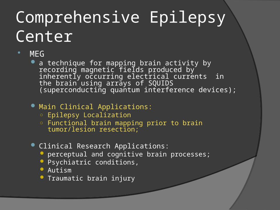
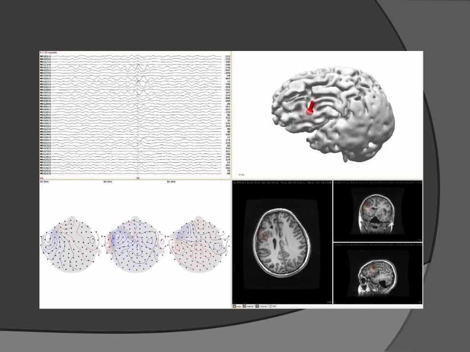
Patient undergoing MEG

MEG door

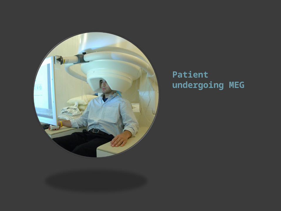
Magnetic fields

The MEG (and EEG) signals derive from the net effect of ionic currents flowing in neuronal dendrites during synaptic transmission.


Related Documents