Journal of Clinical Medicine Article Importance of Translabial Ultrasound for the Diagnosis of Pelvic Organ Prolapse and Its Correlation with the POP-Q Examination: Analysis of 363 Cases Gina Nam 1 , Sa-Ra Lee 2, * , Sung-Hoon Kim 2 and Hee-Dong Chae 2 Citation: Nam, G.; Lee, S.-R.; Kim, S.-H.; Chae, H.-D. Importance of Translabial Ultrasound for the Diagnosis of Pelvic Organ Prolapse and Its Correlation with the POP-Q Examination: Analysis of 363 Cases. J. Clin. Med. 2021, 10, 4267. https:// doi.org/10.3390/jcm10184267 Academic Editor: Angelo Cagnacci Received: 13 July 2021 Accepted: 17 September 2021 Published: 20 September 2021 Publisher’s Note: MDPI stays neutral with regard to jurisdictional claims in published maps and institutional affil- iations. Copyright: © 2021 by the authors. Licensee MDPI, Basel, Switzerland. This article is an open access article distributed under the terms and conditions of the Creative Commons Attribution (CC BY) license (https:// creativecommons.org/licenses/by/ 4.0/). 1 Department of Obstetrics and Gynecology, Chung-Ang University Hospital, Chung-Ang University College of Medicine, 102, Heukseok-ro, Dongjak-gu, Seoul 06973, Korea; [email protected] 2 Department of Obstetrics and Gynecology, Asan Medical Center, University of Ulsan College of Medicine, 88, Olympic-ro 43-gil, Songpa-gu, Seoul 05505, Korea; [email protected] (S.-H.K.); [email protected] (H.-D.C.) * Correspondence: [email protected]; Tel.: +82-2-3010-3648; Fax: +82-2-3010-3630 Abstract: The incidence of pelvic organ prolapse (POP) is increasing in our aging society. We aimed to evaluate the clinical usefulness of translabial ultrasound (TLUS) by comparing the findings of POP-Q examination and TLUS in advanced POP patients and we also aimed to evaluate the prevalence of rectocele and enterocele on the TLUS. We analyzed the TLUS and POP-Q exam findings of 363 symptomatic POP patients who visited our clinic from March 2019 to April 2021. We excluded three patients who had conditions mimicking POP, as revealed by the TLUS. The most common POP type was anterior compartment POP (68.61%), followed by apical compartment (38.61%) and posterior compartment (16.11%) POP. Agreement between the POP-Q exam and TLUS was tested using Cohen’s kappa (κ). p values < 0.05 were considered statistically significant. The incidence of rectocele or enterocele was only 1.67% (6/360) and there was no rectocele or enterocele in most patients (246/252, 96.63%) when the POP-Q exam revealed posterior compartment POP, suggesting that they only had posterior vaginal wall relaxation. The positive predictive value of the POP-Q exam for detecting rectocele or enterocele (as revealed by TLUS) was only 2.38%, whereas the negative predictive value was 100%. In conclusion, the application of TLUS is useful in the diagnosis of POP, especially for differentiation of true POP from conditions mimicking POP. The correlation between the POP-Q exam and TLUS is low, especially in posterior compartment POP, and therefore, patients with POP-Q exam findings suggesting posterior compartment POP should undergo TLUS to check for rectocele or enterocele. The use of TLUS in the diagnosis of POP patients can improve the accuracy of the diagnosis of POP patients in conjunction with a POP-Q exam. Keywords: enterocele; pelvic organ prolapse; pelvic organ prolapse quantification (POP-Q); rectocele; translabial ultrasound 1. Introduction Pelvic organ prolapse (POP) is a condition in which the pelvic organs such as the bladder, uterus, rectum, and sometimes the small bowel descend from their normal position through the vagina. The prevalence of POP is higher in older women, with a lifetime risk factor of 30% and 180 per 100,000 women aged 50 or older based on a database maintained by the Korea National Health Insurance [1]. Symptoms of POP include a sensation of vaginal bulge, overflow urinary incontinence, urinary retention, voiding difficulty, constipation, and splint behavior during urination or defecation [2]. Aging and parity are independent risk factors for POP [3]. The lifetime risk of surgery for POP in women is 19% [4]. The surgical repair of POP is the most common inpatient procedure performed in women aged 70 or older [5] and 1012 million USD is estimated as the economic burden of POP in our aging society [6]. J. Clin. Med. 2021, 10, 4267. https://doi.org/10.3390/jcm10184267 https://www.mdpi.com/journal/jcm

Welcome message from author
This document is posted to help you gain knowledge. Please leave a comment to let me know what you think about it! Share it to your friends and learn new things together.
Transcript
Journal of
Clinical Medicine
Article
Importance of Translabial Ultrasound for the Diagnosis ofPelvic Organ Prolapse and Its Correlation with the POP-QExamination: Analysis of 363 Cases
Gina Nam 1 , Sa-Ra Lee 2,* , Sung-Hoon Kim 2 and Hee-Dong Chae 2
�����������������
Citation: Nam, G.; Lee, S.-R.; Kim,
S.-H.; Chae, H.-D. Importance of
Translabial Ultrasound for the
Diagnosis of Pelvic Organ Prolapse
and Its Correlation with the POP-Q
Examination: Analysis of 363 Cases. J.
Clin. Med. 2021, 10, 4267. https://
doi.org/10.3390/jcm10184267
Academic Editor: Angelo Cagnacci
Received: 13 July 2021
Accepted: 17 September 2021
Published: 20 September 2021
Publisher’s Note: MDPI stays neutral
with regard to jurisdictional claims in
published maps and institutional affil-
iations.
Copyright: © 2021 by the authors.
Licensee MDPI, Basel, Switzerland.
This article is an open access article
distributed under the terms and
conditions of the Creative Commons
Attribution (CC BY) license (https://
creativecommons.org/licenses/by/
4.0/).
1 Department of Obstetrics and Gynecology, Chung-Ang University Hospital, Chung-Ang University Collegeof Medicine, 102, Heukseok-ro, Dongjak-gu, Seoul 06973, Korea; [email protected]
2 Department of Obstetrics and Gynecology, Asan Medical Center, University of Ulsan College of Medicine, 88,Olympic-ro 43-gil, Songpa-gu, Seoul 05505, Korea; [email protected] (S.-H.K.);[email protected] (H.-D.C.)
* Correspondence: [email protected]; Tel.: +82-2-3010-3648; Fax: +82-2-3010-3630
Abstract: The incidence of pelvic organ prolapse (POP) is increasing in our aging society. Weaimed to evaluate the clinical usefulness of translabial ultrasound (TLUS) by comparing the findingsof POP-Q examination and TLUS in advanced POP patients and we also aimed to evaluate theprevalence of rectocele and enterocele on the TLUS. We analyzed the TLUS and POP-Q exam findingsof 363 symptomatic POP patients who visited our clinic from March 2019 to April 2021. We excludedthree patients who had conditions mimicking POP, as revealed by the TLUS. The most commonPOP type was anterior compartment POP (68.61%), followed by apical compartment (38.61%) andposterior compartment (16.11%) POP. Agreement between the POP-Q exam and TLUS was testedusing Cohen’s kappa (κ). p values < 0.05 were considered statistically significant. The incidenceof rectocele or enterocele was only 1.67% (6/360) and there was no rectocele or enterocele in mostpatients (246/252, 96.63%) when the POP-Q exam revealed posterior compartment POP, suggestingthat they only had posterior vaginal wall relaxation. The positive predictive value of the POP-Q examfor detecting rectocele or enterocele (as revealed by TLUS) was only 2.38%, whereas the negativepredictive value was 100%. In conclusion, the application of TLUS is useful in the diagnosis of POP,especially for differentiation of true POP from conditions mimicking POP. The correlation betweenthe POP-Q exam and TLUS is low, especially in posterior compartment POP, and therefore, patientswith POP-Q exam findings suggesting posterior compartment POP should undergo TLUS to checkfor rectocele or enterocele. The use of TLUS in the diagnosis of POP patients can improve the accuracyof the diagnosis of POP patients in conjunction with a POP-Q exam.
Keywords: enterocele; pelvic organ prolapse; pelvic organ prolapse quantification (POP-Q); rectocele;translabial ultrasound
1. Introduction
Pelvic organ prolapse (POP) is a condition in which the pelvic organs such as thebladder, uterus, rectum, and sometimes the small bowel descend from their normal positionthrough the vagina. The prevalence of POP is higher in older women, with a lifetimerisk factor of 30% and 180 per 100,000 women aged 50 or older based on a databasemaintained by the Korea National Health Insurance [1]. Symptoms of POP include asensation of vaginal bulge, overflow urinary incontinence, urinary retention, voidingdifficulty, constipation, and splint behavior during urination or defecation [2]. Aging andparity are independent risk factors for POP [3]. The lifetime risk of surgery for POP inwomen is 19% [4]. The surgical repair of POP is the most common inpatient procedureperformed in women aged 70 or older [5] and 1012 million USD is estimated as the economicburden of POP in our aging society [6].
J. Clin. Med. 2021, 10, 4267. https://doi.org/10.3390/jcm10184267 https://www.mdpi.com/journal/jcm
J. Clin. Med. 2021, 10, 4267 2 of 9
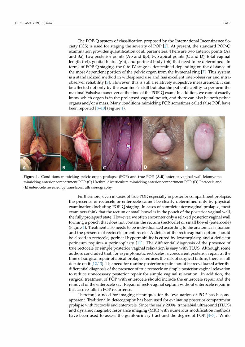
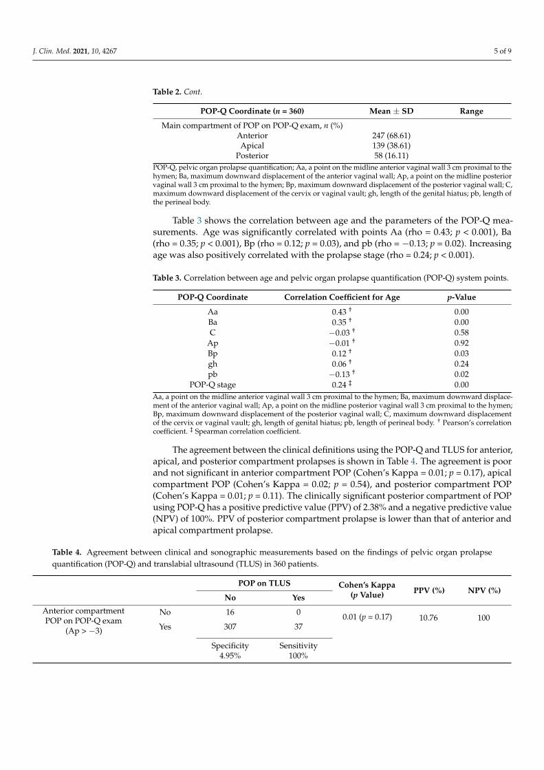
The POP-Q system of classification proposed by the International Incontinence So-ciety (ICS) is used for staging the severity of POP [2]. At present, the standard POP-Qexamination provides quantification of all parameters. There are two anterior points (Aaand Ba), two posterior points (Ap and Bp), two apical points (C and D), total vaginallength (tvl), genital hiatus (gh), and perineal body (pb) that need to be determined. Interms of POP-Q staging, the 0 to IV stage is determined depending on the distance ofthe most dependent portion of the pelvic organ from the hymenal ring [7]. This systemis a standardized method in widespread use and has excellent inter-observer and intra-observer reliability [3]. However, this is still a relatively subjective measurement; it canbe affected not only by the examiner’s skill but also the patient’s ability to perform themaximal Valsalva maneuver at the time of the POP-Q exam. In addition, we cannot exactlyknow which organ is in the prolapsed vaginal pouch, and there can also be both pelvicorgans and/or a mass. Many conditions mimicking POP, sometimes called false POP, havebeen reported [8–10] (Figure 1).
Figure 1. Conditions mimicking pelvic organ prolapse (POP) and true POP. (A,B) anterior vaginal wall leiomyomamimicking anterior compartment POP. (C) Urethral diverticulum mimicking anterior compartment POP. (D) Rectocele and(E) enterocele revealed by translabial ultrasonography.
Furthermore, even in cases of true POP, especially in posterior compartment prolapse,the presence of rectocele or enterocele cannot be clearly determined only by physicalexamination, including POP-Q staging. In cases of complete uterovaginal prolapse, mostexaminers think that the rectum or small bowel is in the pouch of the posterior vaginal wall,the fully prolapsed state. However, we often encounter only a relaxed posterior vaginal wallforming a pouch that does not contain the rectum (rectocele) or small bowel (enterocele)(Figure 1). Treatment also needs to be individualized according to the anatomical situationand the presence of rectocele or enterocele. A defect of the rectovaginal septum shouldbe closed in rectocele, perineal hypermobility is cured by levatorplasty, and a deficientperineum requires a perineoplasty [11]. The differential diagnosis of the presence oftrue rectocele or simple posterior vaginal relaxation is easy with TLUS. Although someauthors concluded that, for asymptomatic rectoceles, a concurrent posterior repair at thetime of surgical repair of apical prolapse reduces the risk of surgical failure, there is stilldebate on it [12,13]. The need for routine posterior repair should be reevaluated after thedifferential diagnosis of the presence of true rectocele or simple posterior vaginal relaxationto reduce unnecessary posterior repair for simple vaginal relaxation. In addition, thesurgical treatment of POP with enterocele should include the enterocele repair and theremoval of the enterocele sac. Repair of rectovaginal septum without enterocele repair inthis case results in POP recurrence.
Therefore, a need for imaging techniques for the evaluation of POP has becomeapparent. Traditionally, defecography has been used for evaluating posterior compartmentprolapse with rectocele and enterocele. Since the early 2000s, translabial ultrasound (TLUS)and dynamic magnetic resonance imaging (MRI) with numerous modification methodshave been used to assess the genitourinary tract and the degree of POP [4–7]. While
J. Clin. Med. 2021, 10, 4267 3 of 9
defecography is relatively unpleasant and MRI is costly, TLUS is a simple, inexpensive, andnon-harmful diagnostic modality that is easy to perform and widely applicable in mostgynecologic clinics. However, there are only a few reports on the relationship betweenclinical symptoms, POP-Q stage, and TLUS findings [11,14–18].
Therefore, we aimed to evaluate the clinical usefulness of TLUS by comparing thefindings of POP-Q examination and TLUS in advanced POP patients. Furthermore, wealso aimed to evaluate the prevalence of rectocele and enterocele on TLUS and the mainPOP type according to the age distribution in Korean POP women.
2. Materials and Methods
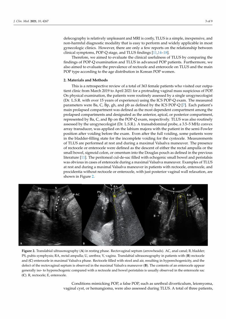
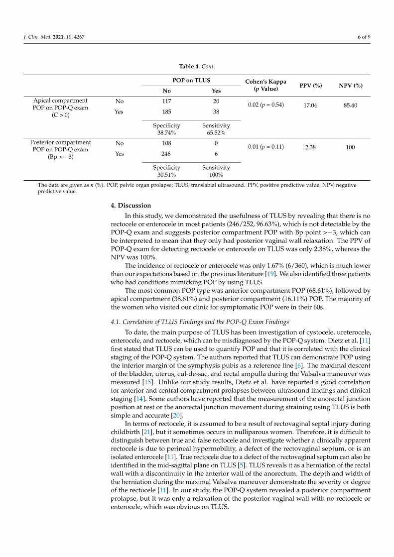
This is a retrospective review of a total of 363 female patients who visited our outpa-tient clinic from March 2019 to April 2021 for a protruding vaginal mass suspicious of POP.On physical examination, the patients were routinely assessed by a single urogynecologist(Dr. L.S.R. with over 15 years of experience) using the ICS POP-Q exam. The measuredparameters were Ba, C, Bp, gh, and pb as defined by the ICS POP-Q [7]. Each patient’smain prolapsed compartment was defined as the most dependent compartment among theprolapsed compartments and designated as the anterior, apical, or posterior compartment,represented by Ba, C, and Bp on the POP-Q exam, respectively. TLUS was also routinelyassessed by the urogynecologist (Dr. L.S.R.). A transabdominal probe, a 3.5–5 MHz convexarray transducer, was applied on the labium majora with the patient in the semi-Fowlerposition after voiding before the exam. Even after the full voiding, some patients werein the bladder-filling state for the incomplete voiding for the cystocele. Measurementsof TLUS are performed at rest and during a maximal Valsalva maneuver. The presenceof rectocele or enterocele were defined as the descent of either the rectal ampulla or thesmall bowel, sigmoid colon, or omentum into the Douglas pouch as defined in the previousliterature [11]. The peritoneal cul-de-sac filled with echogenic small bowel and peristalsiswas obvious in cases of enterocele during a maximal Valsalva maneuver. Examples of TLUSat rest and during a maximal Valsalva maneuver in patients with rectocele, enterocele, andprocidentia without rectocele or enterocele, with just posterior vaginal wall relaxation, areshown in Figure 2.
Figure 2. Translabial ultrasonography (A) in resting phase. Rectovaginal septum (arrowheads). AC, anal canal; B, bladder;PS, pubis symphysis; RA, rectal ampulla; U, urethra; V, vagina. Translabial ultrasonography in patients with (B) rectoceleand (C) enterocele in maximal Valsalva phase. Rectocele filled with stool and air, resulting in hyperechogenicity, and thedefect of the rectovaginal septum is observed in the maximal Valsalva maneuver (B). The contents of an enterocele appeargenerally iso- to hyperechogenic compared with a rectocele and bowel peristalsis is usually observed in the enterocele sac(C). R, rectocele; E, enterocele.
Conditions mimicking POP, a false POP, such as urethral diverticulum, leiomyoma,vaginal cyst, or hemangioma, were also assessed during TLUS. A total of three patients,
J. Clin. Med. 2021, 10, 4267 4 of 9
one patient with urethral diverticulum, one patient with an epidermoid cyst, and onepatient with an anterior vaginal leiomyoma, were excluded from this study.
Statistical Analysis
To assess the association between normally distributed continuous variables, weused Pearson’s correlation test. Very weak, weak, moderate, and strong correlations weredefined as r of 0 to 0.1, 0.1 to 0.3, 0.3 to 0.7, and 0.7 to 1.0. The strength of the associationbetween normally distributed continuous variables and ordinal variables was determinedby Spearman correlation coefficients. Very weak, weak, moderate, and strong correlationswere defined as rs of 0 to 0.1, 0.1 to 0.3, 0.3 to 0.7, and 0.7 to 1.0. Agreement between clinicaldefinition of posterior compartment POP using POP-Q and TLUS was tested using Cohen’skappa (κ). Poor, fair, moderate, good, and very good agreement were defined as κ of <0.2,0.21 to 0.40, 0.41 to 0.60, 0.61 to 0.80, and 0.81 to 1.00. p values < 0.05 were consideredstatistically significant. The statistical analysis was performed using SPSS for Windowsversion 20 (SPSS Inc., Chicago, IL, USA).
3. Results
Among 363 patients who visited our outpatient clinic due to a suspicion of POP, atotal of 360 POP patients who were diagnosed as true POP by the POP-Q exam and TLUSwere included in the final analysis. A total of 41 (11.39%) patients were vault prolapsepatients with a history of hysterectomy. All parameters were normally distributed onKolmogorov–Smirnov testing. The age distribution of the patients is summarized in Table 1.The mean age ± standard deviation of the participants was 59.34 ± 13.22 (range, 29–87)years. Among the patients, the largest proportion (125 (34.72%)) were in their 60s.
Table 1. Age distribution of 360 women who were diagnosed with pelvic organ prolapse.
Age n (%)
20–29 1 (0.27)30–39 37 (10.27)40–49 53 (14.72)50–59 61 (16.94)60–69 125 (34.72)70–79 66 (18.33)80–87 17 (4.72)
The mean POP-Q points, stage, and main compartment of prolapse in our studypopulation are listed in Table 2. On clinical examination, 360 women had POP of ICS stage2 or higher. The main compartment of prolapse was distributed as follows: anterior com-partment in 247 (68.61%), apical compartment in 139 (38.61%), and posterior compartmentin 58 (16.11%).
Table 2. International Continence Society Pelvic Organ Prolapse Quantification (POP-Q) exam findings.
POP-Q Coordinate (n = 360) Mean ± SD Range
Aa 0.89 ± 2.03 −4 to 5.5Ba 2.9 ± 2.54 −3 to 10C 0.63 ± 4.11 −9 to 10
Ap −1.65 ± 2.52 −4 to 10Bp 0.23 ± 3.02 −3 to 10gh 5.21 ± 1.27 −3.5 to 8pb 3.57 ± 0.93 −3 to 9
POP-Q stage 3.03 ± 0.44 2 to 4
J. Clin. Med. 2021, 10, 4267 5 of 9
Table 2. Cont.
POP-Q Coordinate (n = 360) Mean ± SD Range
Main compartment of POP on POP-Q exam, n (%)Anterior 247 (68.61)Apical 139 (38.61)
Posterior 58 (16.11)POP-Q, pelvic organ prolapse quantification; Aa, a point on the midline anterior vaginal wall 3 cm proximal to thehymen; Ba, maximum downward displacement of the anterior vaginal wall; Ap, a point on the midline posteriorvaginal wall 3 cm proximal to the hymen; Bp, maximum downward displacement of the posterior vaginal wall; C,maximum downward displacement of the cervix or vaginal vault; gh, length of the genital hiatus; pb, length ofthe perineal body.
Table 3 shows the correlation between age and the parameters of the POP-Q mea-surements. Age was significantly correlated with points Aa (rho = 0.43; p < 0.001), Ba(rho = 0.35; p < 0.001), Bp (rho = 0.12; p = 0.03), and pb (rho = −0.13; p = 0.02). Increasingage was also positively correlated with the prolapse stage (rho = 0.24; p < 0.001).
Table 3. Correlation between age and pelvic organ prolapse quantification (POP-Q) system points.
POP-Q Coordinate Correlation Coefficient for Age p-Value
Aa 0.43 † 0.00Ba 0.35 † 0.00C −0.03 † 0.58
Ap −0.01 † 0.92Bp 0.12 † 0.03gh 0.06 † 0.24pb −0.13 † 0.02
POP-Q stage 0.24 ‡ 0.00Aa, a point on the midline anterior vaginal wall 3 cm proximal to the hymen; Ba, maximum downward displace-ment of the anterior vaginal wall; Ap, a point on the midline posterior vaginal wall 3 cm proximal to the hymen;Bp, maximum downward displacement of the posterior vaginal wall; C, maximum downward displacementof the cervix or vaginal vault; gh, length of genital hiatus; pb, length of perineal body. † Pearson’s correlationcoefficient. ‡ Spearman correlation coefficient.
The agreement between the clinical definitions using the POP-Q and TLUS for anterior,apical, and posterior compartment prolapses is shown in Table 4. The agreement is poorand not significant in anterior compartment POP (Cohen’s Kappa = 0.01; p = 0.17), apicalcompartment POP (Cohen’s Kappa = 0.02; p = 0.54), and posterior compartment POP(Cohen’s Kappa = 0.01; p = 0.11). The clinically significant posterior compartment of POPusing POP-Q has a positive predictive value (PPV) of 2.38% and a negative predictive value(NPV) of 100%. PPV of posterior compartment prolapse is lower than that of anterior andapical compartment prolapse.
Table 4. Agreement between clinical and sonographic measurements based on the findings of pelvic organ prolapsequantification (POP-Q) and translabial ultrasound (TLUS) in 360 patients.
POP on TLUS Cohen’s Kappa(p Value) PPV (%) NPV (%)
No Yes
Anterior compartmentPOP on POP-Q exam
(Ap > −3)
No 16 0 0.01 (p = 0.17) 10.76 100Yes 307 37
Specificity4.95%
Sensitivity100%
J. Clin. Med. 2021, 10, 4267 6 of 9
Table 4. Cont.
POP on TLUS Cohen’s Kappa(p Value) PPV (%) NPV (%)
No Yes
Apical compartmentPOP on POP-Q exam
(C > 0)
No 117 20 0.02 (p = 0.54) 17.04 85.40Yes 185 38
Specificity38.74%
Sensitivity65.52%
Posterior compartmentPOP on POP-Q exam
(Bp > −3)
No 108 0 0.01 (p = 0.11) 2.38 100Yes 246 6
Specificity30.51%
Sensitivity100%
The data are given as n (%). POP, pelvic organ prolapse; TLUS, translabial ultrasound. PPV, positive predictive value; NPV, negativepredictive value.
4. Discussion
In this study, we demonstrated the usefulness of TLUS by revealing that there is norectocele or enterocele in most patients (246/252, 96.63%), which is not detectable by thePOP-Q exam and suggests posterior compartment POP with Bp point >−3, which canbe interpreted to mean that they only had posterior vaginal wall relaxation. The PPV ofPOP-Q exam for detecting rectocele or enterocele on TLUS was only 2.38%, whereas theNPV was 100%.
The incidence of rectocele or enterocele was only 1.67% (6/360), which is much lowerthan our expectations based on the previous literature [19]. We also identified three patientswho had conditions mimicking POP by using TLUS.
The most common POP type was anterior compartment POP (68.61%), followed byapical compartment (38.61%) and posterior compartment (16.11%) POP. The majority ofthe women who visited our clinic for symptomatic POP were in their 60s.
4.1. Correlation of TLUS Findings and the POP-Q Exam Findings
To date, the main purpose of TLUS has been investigation of cystocele, ureterocele,enterocele, and rectocele, which can be misdiagnosed by the POP-Q system. Dietz et al. [11]first stated that TLUS can be used to quantify POP and that it is correlated with the clinicalstaging of the POP-Q system. The authors reported that TLUS can demonstrate POP usingthe inferior margin of the symphysis pubis as a reference line [6]. The maximal descentof the bladder, uterus, cul-de-sac, and rectal ampulla during the Valsalva maneuver wasmeasured [15]. Unlike our study results, Dietz et al. have reported a good correlationfor anterior and central compartment prolapses between ultrasound findings and clinicalstaging [14]. Some authors have reported that the measurement of the anorectal junctionposition at rest or the anorectal junction movement during straining using TLUS is bothsimple and accurate [20].
In terms of rectocele, it is assumed to be a result of rectovaginal septal injury duringchildbirth [21], but it sometimes occurs in nulliparous women. Therefore, it is difficult todistinguish between true and false rectocele and investigate whether a clinically apparentrectocele is due to perineal hypermobility, a defect of the rectovaginal septum, or is anisolated enterocele [11]. True rectocele due to a defect of the rectovaginal septum can also beidentified in the mid-sagittal plane on TLUS [5]. TLUS reveals it as a herniation of the rectalwall with a discontinuity in the anterior wall of the anorectum. The depth and width ofthe herniation during the maximal Valsalva maneuver demonstrate the severity or degreeof the rectocele [11]. In our study, the POP-Q system revealed a posterior compartmentprolapse, but it was only a relaxation of the posterior vaginal wall with no rectocele orenterocele, which was obvious on TLUS.
J. Clin. Med. 2021, 10, 4267 7 of 9
4.2. Role of TLUS in Differentiation of the True POP from Conditions Mimicking POP
Many conditions, including various vaginal masses such as urethral diverticulum,vaginal leiomyoma, or urogenital cysts, can be confusing factors that mimic POP. A patientwith a protruding vaginal mass suggestive of an anterior compartment prolapse with symp-toms of urinary frequency and dyspareunia was revealed to have a urethral diverticulumon TLUS [10]. Braga et al. reported a patient who complained of vaginal bulging andoveractive bladder symptoms with POP-Q stage II anterior vaginal prolapse (Ba:0), and thehistopathological examination revealed an anterior vaginal leiomyoma [22]. Vulvovaginalhemangioma in Klippel–Trénaunay syndrome, vaginal wall cysts, and a perineal mass oreven a long cervical polyps can be misdiagnosed as POP [8,9] which manifests the same asour case shown in Figure 1.
Thus, imaging techniques have been studied to identify the defects of the pelvicfloor and the relationship with the adjacent organs as a complement to the POP-Q system.The defecation proctography has been regarded as a gold standard in conjunction witha physical examination in assisting diagnostic and surgical approaches to rectocele [23].MRI has also been introduced for pelvic floor disorders with high-resolution images of themuscles, ligaments, and sphincters, as well as functional information obtained throughdynamic MRI [24,25]. However, defecation proctography and MRI are rarely used in thedaily clinic because these modalities are relatively expensive, are not very acceptable bythe patients, and have risks of radiation exposure or allergic reactions to the contrast mediaused during the imaging studies. TLUS can be more widely used since ultrasonographyis the most widely available user-friendly imaging modality in gynecologic clinics, it isless expensive than MRI, it is easy to perform, it requires only a short time (approximately5 min), and it can give information as the other two modalities. The urogynecologist(L.S.R.) has been performing surgical treatments for POP patients without performingthese two more invasive imaging modalities. Considering the clinical practice in ourinstitute, preoperative defecography is rarely performed in patients who exhibit symptomsor signs of posterior compartment prolapse, even for those who undergo POP operations.Since 2019, the preoperative imaging findings have been evaluated using the TLUS insteadof defecography in our institute. We do not perform posterior colpoperineorrhaphy as aroutine concurrent procedure for patients with apical POP without symptoms or evidenceof rectocele or enterocele on TLUS. Even with this practice pattern, we did not observe ahigher de novo rectocele or enterocele after the apical POP operations in this case comparedwith that reported in the literature [26]. Therefore, the use of TLUS during the diagnosis ofPOP patients can improve the accuracy of their diagnosis combined with a POP-Q examwith minimal additional effort and cost.
4.3. Strengths and Limitations of this Study
This study has several strengths. First, to the best of our knowledge, this is the firststudy to evaluate the accuracy of TLUS for Korean POP women with a relatively largenumber of patients. Second, the POP-Q exam and TLUS were performed by a singleexperienced urogynecologist in all cases over a relatively short period of 25 months. Thiscan exclude interpersonal bias and minimize inter-observer reliability, which can affectaccuracy and consistency depending on the experience of the examiner. Finally, althoughthis is a retrospective study design, the data were prospectively collected using the sametechniques during routine urogynecologic clinical exams.
However, this study also has some limitations that should be considered. First, this isa single-center retrospective study and not a randomized controlled trial, and it includesno data on women without POP. Second, the degree of rectocele, demonstrated with thedepth and width of the rectocele on TLUS, was not analyzed in terms of the stage of POP-Qor Bp point measurement. The severity of POP was not analyzed by quantification ofthe TLUS using the inferior margin of the symphysis pubis as a reference line, as in thestudy of Dietz [14]. Only the presence of rectocele or enterocele was analyzed withoutassessing the burden of symptoms in relation to the TLUS findings. Third, all POP women
J. Clin. Med. 2021, 10, 4267 8 of 9
during the study periods were included, regardless of the operation, because the aim ofthis study was to compare the POP-Q examination with the TLUS. Therefore, womenwho did not undergo operations were also included in this study. Finally, this is a singleurogynecologist’s experience in a tertiary hospital and we cannot exclude the possibility ofselection bias for the analysis of patients who were referred to a tertiary hospital. Therefore,the nationwide Korean statistics cannot be represented with this data, especially for themain type and age distribution.
5. Conclusions
In conclusion, the application of TLUS is useful in the diagnosis of POP for the differ-entiation of true POP from conditions mimicking POP. The correlation between the POP-Qexam and TLUS is low, especially for posterior compartment POP, and therefore patientswith POP-Q exam findings suggesting posterior compartment POP should undergo TLUSto check for the presence of rectocele or enterocele.
Author Contributions: Conceptualization, S.-R.L.; formal analysis, G.N.; investigation, G.N. andS.-R.L.; resources, S.-R.L.; data curation, G.N. and S.-R.L.; writing—original draft preparation, G.N.and S.-R.L.; writing—review and editing, S.-R.L.; visualization, G.N. and S.-R.L.; supervision, S.-H.K.and H.-D.C.; project administration, S.-R.L. All authors have read and agreed to the published versionof the manuscript.
Funding: This research received no external funding.
Institutional Review Board Statement: This study was approved by the Asan Medical CenterInstitutional Review Board (approval No. 2020-1155).
Informed Consent Statement: Patient consent was waived by our Institutional Review Board be-cause of the retrospective chart review study design; however, informed consents were obtainedfrom the patients whose pictures were included in this study for the publication of their pictures.
Data Availability Statement: The Excel data used to support the findings of this study were suppliedby Sa-Ra Lee under license, and requests for access to these data should be made to Sa-Ra Lee,[email protected].
Conflicts of Interest: The authors have no potential conflicts of interest to disclose.
References1. Yuk, J.-S.; Lee, J.H.; Hur, J.-Y.; Shin, J.-H. The prevalence and treatment pattern of clinically diagnosed pelvic organ prolapse: A
Korean National Health Insurance Database-based cross-sectional study 2009–2015. Sci. Rep. 2018, 8, 1–6. [CrossRef] [PubMed]2. Digesu, G.A.; Chaliha, C.; Salvatore, S.; Hutchings, A.; Khullar, V. The relationship of vaginal prolapse severity tosymptoms and
quality of life. BJOG Int. J. Obstet. Gynaecol. 2005, 112, 971–976. [CrossRef] [PubMed]3. Nygaard, I.; Bradley, C.; Brandt, D. Pelvic organ prolapse in older women: Prevalence and risk factors. Obstet. Gynecol. 2004,
104, 489–497. [CrossRef] [PubMed]4. Smith, F.J.; Holman, C.A.J.; Moorin, R.E.; Tsokos, N. Lifetime risk of undergoing surgery for pelvic organ prolapse. Obstet. Gynecol.
2010, 116, 1096–1100. [CrossRef] [PubMed]5. Oliphant, S.S.; Jones, K.A.; Wang, L.; Bunker, C.H.; Lowder, J.L. Trends over time with commonly performed obstetric and
gynecologic inpatient procedures. Obstet. Gynecol. 2010, 116, 926. [CrossRef] [PubMed]6. Subak, L.L.; Waetjen, L.E.; Van Den Eeden, S.; Thom, D.H.; Vittinghoff, E.; Brown, J.S. Cost of pelvic organ prolapse surgery in the
United States. Obstet. Gynecol. 2001, 98, 646–651. [PubMed]7. Bump, R.C.; Mattiasson, A.; Bø, K.; Brubaker, L.P.; DeLancey, J.O.; Klarskov, P.; Shull, B.L.; Smith, A.R. The standardization of
terminology of female pelvic organ prolapse and pelvic floor dysfunction. Am. J. Obstet. Gynecol. 1996, 175, 10–17. [CrossRef]8. Nam, G.; Lee, S.R.; Eum, H.R.; Kim, S.H.; Chae, H.D.; Kim, G.J. A Huge Hemorrhagic Epidermoid Cyst of the Perineum with
Hypoechoic Semisolid Ultrasonographic Feature Mimicking Scar Endometriosis. Medicina 2021, 57, 276. [CrossRef]9. Nam, G.; Lee, S.R.; Choi, S. Clitoromegaly, Vulvovaginal Hemangioma Mimicking Pelvic Organ Prolapse, and Heavy Menstrual
Bleeding: Gynecologic Manifestations of Klippel-Trénaunay Syndrome. Medicina 2021, 57, 366. [CrossRef]10. Lee, S.R.; Lim, Y.-M.; Jeon, J.H.; Park, M.H. Diagnosis of urethral diverticulum mimicking pelvic organ prolapse with translabial
ultrasonography. Am. J. Obstet. Gynecol. 2017, 217, 482. [CrossRef]11. Dietz, H.P.; Steensma, A.B. Posterior compartment prolapse on two-dimensional and three-dimensional pelvic floor ultrasound:
The distinction between true rectocele, perineal hypermobility and enterocele. Ultrasound Obstet. Gynecol. 2005, 26, 73–77.[CrossRef]
J. Clin. Med. 2021, 10, 4267 9 of 9
12. Chang, O.H.; Davidson, E.R.W.; Thomas, T.N.; Paraiso, M.F.R.; Ferrando, C.A. Does concurrent posterior repair for an asymp-tomatic rectocele reduce the risk of surgical failure in patients undergoing sacrocolpopexy? Int. Urogynecol. J. 2020, 31, 2075–2080.[CrossRef] [PubMed]
13. Arunachalam, D.; Hale, D.S.; Heit, M.H. Posterior Compartment Surgery Provides No Differential Benefit for DefecatorySymptoms Before or After Concomitant Mesh-Augmented Apical Suspension. Female Pelvic Med. Reconstr. Surg. 2018, 24, 183–187.[CrossRef]
14. Dietz, H.; Haylen, B.; Broome, J. Ultrasound in the quantification of female pelvic organ prolapse. Ultrasound Obstet. Gynecol.2001, 18, 511–514. [CrossRef] [PubMed]
15. Dietz, H. Ultrasound imaging of the pelvic floor. Part I: Two-dimensional aspects. Ultrasound Obstet. Gynecol. 2004, 23, 80–92.[CrossRef] [PubMed]
16. Dietz, H.; Clarke, B.; Herbison, P. Bladder neck mobility and urethral closure pressure as predictors of genuine stress incontinence.Int. Urogynecol. J. 2002, 13, 289–293. [CrossRef] [PubMed]
17. Dietz, H. Ultrasound imaging of the pelvic floor. Part II: Three-dimensional or volume imaging. Ultrasound Obstet. Gynecol. 2004,23, 615–625. [CrossRef] [PubMed]
18. Samantray, S.R.; Mohapatra, I. Study of the Relationship Between Pelvic Organ Prolapse Quantification (POP-Q) Staging andDecubitus Ulcer in Pelvic Organ Prolapse. Cureus 2021, 13. [CrossRef]
19. Barber, M.D.; Maher, C. Epidemiology and outcome assessment of pelvic organ prolapse. Int. Urogynecol. J. 2013, 24, 1783–1790.[CrossRef] [PubMed]
20. Beer-Gabel, M.; Teshler, M.; Schechtman, E.; Zbar, A. Dynamic transperineal ultrasound vs. defecography in patients withevacuatory difficulty: A pilot study. Int. J. Colorectal Dis. 2004, 19, 60–67. [CrossRef]
21. Dietz, H.P.; Steensma, A. The role of childbirth in the aetiology of rectocele. BJOG Int. J. Obstet. Gynaecol. 2006, 113, 264–267.[CrossRef]
22. Braga, A.; Soave, I.; Caccia, G.; Regusci, L.; Ruggeri, G.; Pitaku, I.; Bassi, V.; Papadia, A.; Serati, M. What is this vaginal bulge? Anatypical case of vaginal paraurethral leiomyoma. A case report and literature systematic review. J. Gynecol. Obstet. Hum. Reprod.2021, 50, 101822. [CrossRef] [PubMed]
23. Kenton, K.; Shott, S.; Brubaker, L. The anatomic and functional variability of rectoceles in women. Int. Urogynecol. J. 1999,10, 96–99. [CrossRef]
24. Singh, K.; Reid, W.M.; Berger, L.A. Assessment and grading of pelvic organ prolapse by use of dynamic magnetic resonanceimaging. Am. J. Obstet. Gynecol. 2001, 185, 71–77. [CrossRef] [PubMed]
25. Rechi-Sierra, K.; Sánchez-Ballester, F.; García-Ibáñez, J.; Pardo-Duarte, P.; Flores-DelaTorre, M.; Monzó-Cataluña, A.; López-Alcina, E. Magnetic resonance imaging to evaluate anterior pelvic prolapse: H line is the key. Neurourol. Urodyn. 2021,40, 1042–1047. [CrossRef] [PubMed]
26. Russo, E.; Giannini, A.; Guevara, M.M.; Mannella, P.; Misasi, G.; Falcone, M.; Simoncini, T. Medium-term outcomes afterrobotic-assisted lateral suspension with mesh for advanced multi-compartmental prolapse. Int. Urogynecol. J. 2020, 31, 1647–1653.[CrossRef] [PubMed]
Related Documents