JACO Journal of the Academy of Chiropractic Orthopedists 2017 Volume 14 Issue 2 June, 2017

Welcome message from author
This document is posted to help you gain knowledge. Please leave a comment to let me know what you think about it! Share it to your friends and learn new things together.
Transcript

JACO Journal of the Academy of Chiropractic Orthopedists
2017
Volume 14
Issue 2
June, 2017
JACO Journal of the Academy of
Chiropractic Orthopedists
The Open Access, Peer-Reviewed and Indexed Publication of
the Academy of Chiropractic Orthopedists
June 2017 – Volume 14, Issue 2
Editorial Board Editor-In-Chief
Shawn M. Neff, DC, MAS, FACO
Managing Editor
Tracey A. Littrell, DC, DACBR, DACO, CCSP
Associate Editors
James Demetrious, DC, FACO
David Swensen, DC, FACO
Alicia M. Yochum, RN, DC, DACBR, RMSK
Current Events Editor
James R. Brandt, DC, MS, FACO
Editorial Advisory Board
James R. Brandt, DC, MS, FACO
Ronald C Evans, DC, FACO
James Demetrious, DC, FACO
Michael Henrie, DO
Robert Morrow, MD
Bruce Gundersen, DC, FACO
Editorial Review Board Scott D. Banks, DC MS Ward Beecher, D.C., FACO
Thomas F. Bergmann, DC Gary Carver, DC, FACO
Jeffrey R. Cates, DC, FACO Rick Corbett, DC, DACBR, FCCO(C)
Donald S. Corenman, MD, DC, FACO Clinton Daniels, DC, MS, DAAPM
Anthony Vincent D'Antoni, MS, DC, PhD James Demetrious, DC, FACO
Daniel P. Dock, DC, FACO Neil L. Erickson, DC, DABCO, CCSP
Simon John Forster, DC, DABCO Jaroslaw P. Grod, DC, FCCS(C)
Evan M. Gwilliam, DC, MBA Tony Hamm, DC, FACO
Dale Huntington, DC, FACO Charmaine Korporaal, M.Tech: Chiropractic
Ralph Kruse, DC, FACO Thomas Mack, DC, FACO
Joyce Miller, DC, FACO Loren C. Miller DC, FACO
William E. Morgan, DC, DAAPM Raymond S Nanko, DC, MD, DAAPM, FACO
Deanna O'Dwyer, DC, FACO Casey Okamoto, DC
Joni Owen, DC, FACO Gregory C. Priest, DC, FACO J
Christopher Roecker, DC, MS, DACO, DACSP Chris Romney, DC, FACO
Roger Russell, DC, MS, FACO Stephen M. Savoie, DC, FACO
Alec Schielke, DC Brandon Steele, DC
John Stites, DC, DACBR, DACO Larry L. Swank, DC, FACO
David Swensen, DC, FACO Cliff Tao, DC, DACBR
John M. Ventura, DC, FACO Michelle A Wessely BSc, DC, DACBR
Michael R. Wiles, DC, MEd, MS James A. Wyllie, DC DABCO
Steve Yeomans, DC, FACO Alicia M. Yochum, RN, DC, DACBR, RMSK
Articles, abstracts, opinions and comments appearing in this journal are the work of submitting authors, have been reviewed by
members of the editorial board and do not reflect the positions, opinions, endorsements or consensus of the Academy.
Journal of the Academy of Chiropractic Orthopedists June 2017- Volume 14, Issue 2

1
Journal of the Academy of Chiropractic Orthopedists
June 2017 – Volume 14, Issue 2
Editor’s Desk
❖ Shawn M. Neff, DC, MAS, FACO
Original Articles
❖ Roecker CB, Anzalone GA, Okamoto CS, Roes MJ: Benign Lower Thoracic Intradural Schwannoma Compressing the Conus Medullaris and Mimicking Thoracolumbar Myofascial Trigger Point Pain: A Case Report: JACO 2017, 14(2):3-20
❖ Kruse RA, Okamoto CS: Chiropractic Management of Cervicalgia in a Patient with
Diffuse Idiopathic Skeletal Hyperostosis Utilizing Cox Manual Cervical Distraction:
A Case Report: JACO 2017, 14(2):21-31
❖ Sergent A, Bowyer H: Abdominal Aortic Aneurysm and Spinal Manipulation, an Absolute Contraindication?: A Review of the Literature: JACO 2017, 14(1):32-37
Abstracts and Literature Review
❖ Dewan AK, et al: MRI of the Elbow: Techniques and Spectrum of Disease; Reviewed by Tao C. JACO 2017, 14(2):38-39
❖ Mayerhoefer ME, et al: Shoulder Impingement: Relationship of Clinical Symptoms and Imaging Criteria; Reviewed by Cates JR. JACO 2017, 14(2):40-42
Conference Proceedings
❖ ACCO Orthopedic Essentials 2017 pages 43-44
Radiology Corner
❖ Littrell TA: Interesting Finding in an Elderly Female Patient Without Reported Knee
Trauma: JACO 2017, 14(2):45-48
Ortho Quiz
❖ Kleinfield SL: Ortho Quiz. JACO 2017, 14(2):49
Current Events
❖ Examination
Answers to Ortho Quiz
❖ Check your knowledge on page 51

Journal of the Academy of Chiropractic Orthopedists
Volume 14, Issue 2
2
The Editor’s Desk
Shawn M. Neff, DC, MAS, FACO
Editor-in-Chief
Welcome to the June 2017 issue of the Journal of the Academy of Chiropractic Orthopedists. As
I finish my first year as the editor in chief I would like to express my gratitude. I want to thank
all our readers for supporting the Journal. I hope you are enjoying the journal and are excited for
the changes we are making. A thank you also to the Academy board for their trust in the team at
the journal. Lastly, I want to thank the editors, peer reviewers, illustrator and the clinician
researchers for their hard work and dedication to the field of chiropractic Orthopedics and to
JACO.
I want to extend an invitation to our readers to submit your research and case reports. If you
have an idea but need help bringing it to fruition, please reach out to us. We have a program to
pair you with an experienced researcher to co-author with you. We strive to foster not only a
community of consumers of research but also a community of contributors and this program is
moving us towards that goal.
I also would like to welcome our new managing editor this issue. Tracey Littrell, DC, DACBR.
DACO, CCSP is an associate professor in the Diagnosis and Radiology Department of Palmer
College of Chiropractic in Davenport, Iowa. She is a chiropractic orthopedist and radiologist and
has brought her energy and expertise to JACO. She will be a tremendous asset to the journal.
Be sure to check out the Radiology Corner this issue which she authored.
Sincerely,
-Shawn

3
Original Article
Benign Lower Thoracic Intradural Schwannoma Compressing the Conus
Medullaris and Mimicking Thoracolumbar Myofascial Trigger Point Pain: A
Case Report
Christopher B. Roecker, DC, MS, DACO1, Gerald A. Anzalone, DC2, Casey S. Okamoto, DC3,
Matthew J. Roes, DC, MD (deceased)4
1Assistant Professor, Palmer College of Chiropractic Life Science & Foundations Department 2 Adjunct Instructor, Kirkwood Community College
3 Doctor of Chiropractic, VA Medical Center Minneapolis, MN 4 Medical Doctor and Doctor of Chiropractic, Hiawatha, IA
Published: June 2017 Journal of the Academy of Chiropractic Orthopedists
June 2017, Volume 14, Issue 2
This is an Open Access article which permits unrestricted use, distribution, and reproduction in any medium, provided the original work is properly cited. The article copyright belongs to the author and the Academy of Chiropractic Orthopedists and is available at:
http://www.dcorthoacademy.com. © 2017 Roecker/Anzalone/Okamoto/Roes and the Academy of Chiropractic Orthopedists.
Abstract
Background The purpose of this report was to describe the clinical course, management, and
outcomes of a male with thoracolumbar spine pain associated with an intradural schwannoma.
Case presentation A 49-year-old male sought care at an interdisciplinary medical clinic with
rapid onset of paraspinal pain to the left of the thoracolumbar junction. The initial examination
indicated myofascial trigger point pain of the left quadratus lumborum. The patient's
management included manual myofascial trigger point pressure release, active and passive
muscle stretching, trigger point injections, prescription anti-inflammatory medication,
Journal of the Academy of Chiropractic Orthopedists
Volume 14, Issue 2
4
prescription muscle relaxant medication, and narcotic pain medications over an 8-week period
with a moderate reduction in pain. Following six treatment sessions, the patient reported a
progression of left thoracolumbar paraspinal pain intensity, nocturnal low back pain, left hip and
thigh pain, and bilateral leg weakness. The patient was referred for thoracolumbar spine
magnetic resonance imaging, which demonstrated a lower thoracic spine intradural tumor
effacing the conus medullaris. The patient was immediately referred for neurosurgical excision.
Following surgery, the patient experienced complete remission of thoracolumbar spine pain and
recovered his lower extremity strength. Histological evaluation later revealed the mass to be a
lower thoracic intradural extramedullary schwannoma causing compression of the conus
medullaris.
Discussion Clinicians managing persistent paraspinal trigger points with progressive pain and
neurological dysfunction should be aware of the possibility of undiagnosed co-morbidities as
complicating factors in clinical presentation. Progressive pain and neurological findings warrant
referral for advanced imaging to screen for undiagnosed complicating conditions, such as an
intradural mass. In this case, conus medullaris compression mimicked the clinical presentation of
myofascial trigger point syndrome within the quadratus lumborum musculature and was later
discovered to be associated with a lower thoracic benign intradural schwannoma.
Conclusion This case report describes the clinical presentation of a lower thoracic benign
intradural schwannoma initially presenting with characteristics of myofascial pain. Serious
neurological conditions may present with symptoms mimicking common musculoskeletal
disorders.
Keywords (MeSH TERMS)
Spinal Cord Tumor; Compression, Spinal Cord; Myelopathy, Compressive; Myofascial Pain
Syndrome; Trigger Point; Chiropractic
5
Background
Myofascial pain is pain that arises from muscles or fascia, and identification of this
condition is particularly relevant to musculoskeletal clinicians.1,2 Myofascial pain is estimated to
affect 44 million Americans annually3-5 and is one of the most common disorders encountered by
physicians in clinical practice.6 The defining characteristic of myofascial pain is the presence of a
myofascial trigger point (MTrP3,4,6-9, which manifests clinically as a hyperirritable nodule.2,3,10,11
MTrP pain has been reported to affect approximately 30% of pain patients reporting to general
practitioners,12 and is thought to be the leading diagnosis among pain management specialists.4,13
The presence of MTrP pain has become recognized as a legitimate entity that is clinically
significant within a musculoskeletal practice,1,6,13 but remains one of the most under-diagnosed
or misdiagnosed conditions.6The diagnosis of MTrP pain remains purely clinical,2 and there is no
universally accepted MTrP diagnostic criteria.2,3,10,11 While no well-validated diagnostic criteria
exist for MTrP, common clinical features have been identified as being the most consistent and
clinically relevant for diagnosing MTrP pain.2,9 A systematic review of the literature identified
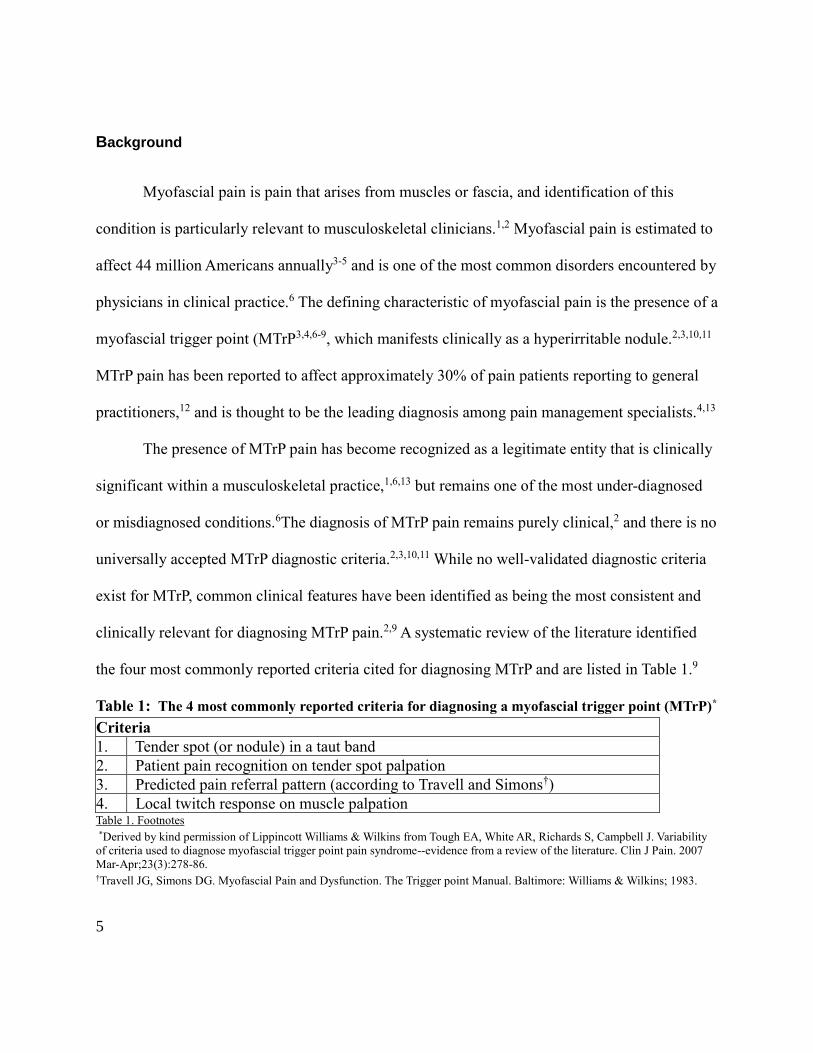
the four most commonly reported criteria cited for diagnosing MTrP and are listed in Table 1.9
Table 1: The 4 most commonly reported criteria for diagnosing a myofascial trigger point (MTrP)*
Criteria
1. Tender spot (or nodule) in a taut band
2. Patient pain recognition on tender spot palpation
3. Predicted pain referral pattern (according to Travell and Simons†)
4. Local twitch response on muscle palpation Table 1. Footnotes
*Derived by kind permission of Lippincott Williams & Wilkins from Tough EA, White AR, Richards S, Campbell J. Variability
of criteria used to diagnose myofascial trigger point pain syndrome--evidence from a review of the literature. Clin J Pain. 2007
Mar-Apr;23(3):278-86. †Travell JG, Simons DG. Myofascial Pain and Dysfunction. The Trigger point Manual. Baltimore: Williams & Wilkins; 1983.

Journal of the Academy of Chiropractic Orthopedists
Volume 14, Issue 2
6
Management of myofascial pain is usually directed toward treatment of the MTrP along
with the removal of perpetuating factors and should be systematically investigated for each
patient.2,14 Therapy intended to alleviate MTrP pain includes various forms of manual myofascial
release and soft tissue massage techniques, muscle stretching, acupuncture, therapeutic
ultrasound, drug treatments, and MTrP injection.2,3 Correction of perpetuating factors may
include addressing: abnormal posture, abnormal muscle activation patterns, anatomical defects
(e.g. limb length inequality), mood disorders, or nutritional inadequacies.2 Currently, MTrP
injection and muscle stretching is considered by some to be the standard management option for
myofascial pain and has been demonstrated to improve patient outcomes.2,15,16
The purpose of this paper is to describe the case of a man treated for myofascial pain with
manual trigger point release techniques, active and passive muscle stretching, soft tissue
massage, trigger point injections, and prescription anti-inflammatory, muscle relaxant, and
narcotic pain medications over an eight-week period. The patient's pain was initially diagnosed
as left thoracolumbar junction paraspinal MTrP pain, which occasionally referred pain to the left
lateral hip and sacroiliac region. The thoracolumbar MTrP pain persisted, despite temporary pain
relief immediately following treatment. Eight weeks after the patient's initial presentation,
neurological signs and symptoms rapidly developed. These progressive neurological findings
prompted a thoracolumbar spine magnetic resonance imaging (MRI), which revealed a lower
thoracic intradural extramedullary schwannoma and immediate neurosurgical decompression
was performed.
7
Case presentation
Consent for publication of clinical information was provided by the patient, provided
anonymity was preserved.
A 49-year-old white male sought chiropractic care for “moderate to severe”
thoracolumbar spine pain at an interdisciplinary medical clinic. The symptoms began one day
before seeking care and onset gradually following moderately intense housework. This initial
pain onset was severely intense and rated as a 9/10 on an 11-point Numeric Rating Scale (NRS).
The patient reported “dull” and “achy” pain localized to the left thoracolumbar paraspinal
musculature without any pain referral or radicular symptoms. Moderate physical activities also
provoked the patient’s pain.
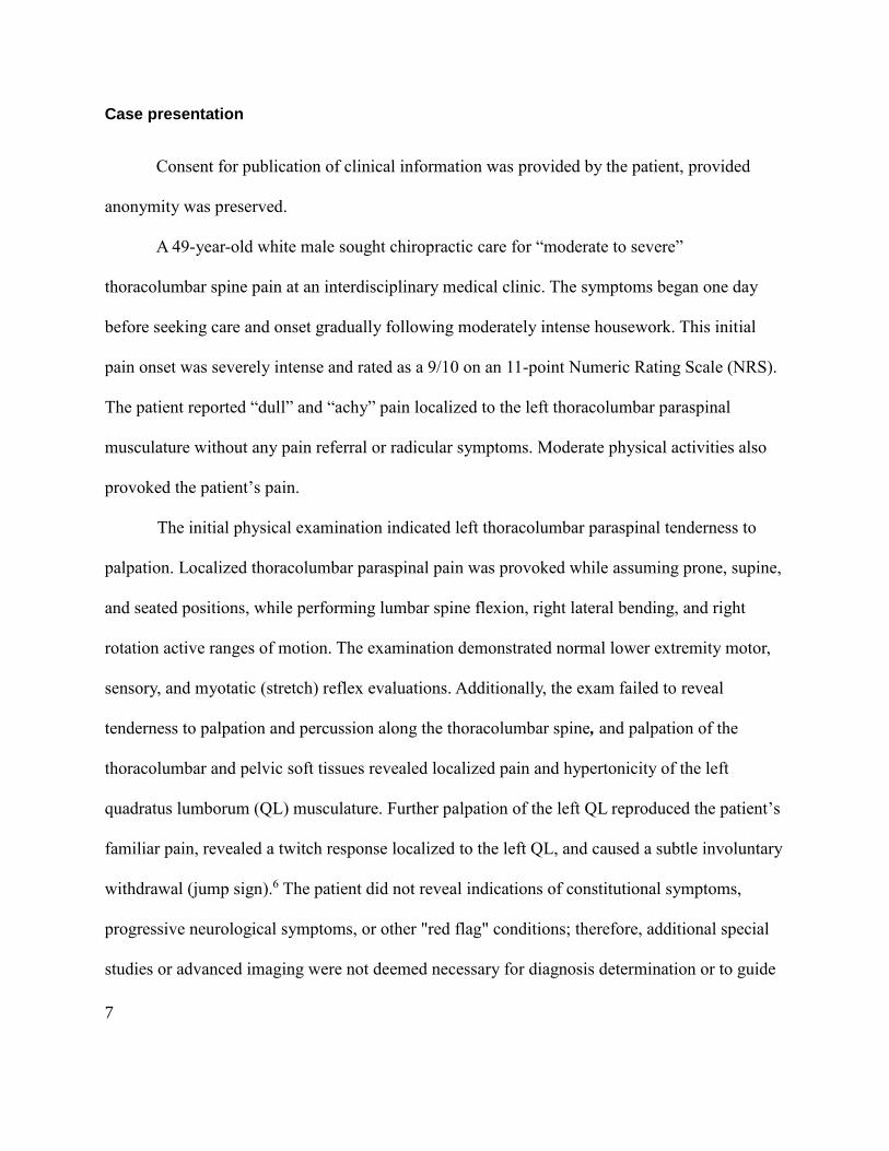
The initial physical examination indicated left thoracolumbar paraspinal tenderness to
palpation. Localized thoracolumbar paraspinal pain was provoked while assuming prone, supine,
and seated positions, while performing lumbar spine flexion, right lateral bending, and right
rotation active ranges of motion. The examination demonstrated normal lower extremity motor,
sensory, and myotatic (stretch) reflex evaluations. Additionally, the exam failed to reveal
tenderness to palpation and percussion along the thoracolumbar spine, and palpation of the
thoracolumbar and pelvic soft tissues revealed localized pain and hypertonicity of the left
quadratus lumborum (QL) musculature. Further palpation of the left QL reproduced the patient’s
familiar pain, revealed a twitch response localized to the left QL, and caused a subtle involuntary
withdrawal (jump sign).6 The patient did not reveal indications of constitutional symptoms,
progressive neurological symptoms, or other "red flag" conditions; therefore, additional special
studies or advanced imaging were not deemed necessary for diagnosis determination or to guide

Journal of the Academy of Chiropractic Orthopedists
Volume 14, Issue 2
8
treatment of this condition. Following the physical examination, the patient was diagnosed with
MTrP syndrome of the left QL.
The patient agreed to begin pain management care, involving combined chiropractic and
medical sessions, home-based low back stretching and strengthening activities, and prescription
medications pro re nata (PRN). The combined chiropractic and medical care consisted of manual
MTrP pressure release (ischemic compression) 2, active and passive muscle stretching, soft
tissue massage, postural modification, intramuscular trigger point injections (2.0 mL 2.0%
lidocaine, 2.0 mL 0.5% Marcaine, 2.0 mL of Traumeel®), and prescription medication (Table 2).
Immediately following the initial treatment, the patient casually reported an "80% improvement"
in his left thoracolumbar muscle pain. The patient was instructed to continue with home-based
strengthening and stretching, prescriptions medications as indicated, and to return to the clinic
PRN.
Table 2: Medication management prescribed by the medical doctor following the initial evaluation
Class Medication Dose Frequency Count Refills
Narcotic
analgesic
hydrocodone/APAP 5/500 mg PRN* 30 tablets none
NSAID Naproxen 500 mg BID 60 tablets none
Muscle
relaxant
Metaxalone 800 mg QID 40 tablets none
Table 2. Footnotes
APAP, N-acetyl-p-aminophenol (acetaminophen); PRN, pro re nata (as needed); NSAID, non-steroidal anti-inflammatory; BID,
bis in die (twice per day); QID, quater in die (four times per day) *Not to exceed 1 tablet every 6 hours
Following the first treatment, the patient returned six days later with the return of the
original dull and achy thoracolumbar pain along with emergence left sacroiliac (S-I) and hip
region pain. The progression of symptoms prompted a reexamination, which revealed normal
9
lower extremity sensory, motor, and myotatic reflex evaluations. The patient’s gait was
unremarkable with a normal base of support. The patient rated his pain as 8/10 on a NRS.
Thoracolumbar and pelvic soft tissue palpation revealed the persistence of the left QL MTrP pain
with accompanying QL MTrP pain referral pattern.17 Following reexamination, the patient
agreed to left QL MTrP pain management consisting of trigger point injection, manual trigger
point release, passive stretching, and soft tissue massage. The patient casually reported an
immediate and marked reduction of the thoracolumbar pain and abolition of the S-I region and
groin pain.
Treatment continued six times over the eight week management period, consisting of
continued trigger point injections, manual trigger point release, passive stretching, and soft tissue
massages. The patient consistently reported relief of his pain complaints following treatment
with a consistent and gradual return to pretreatment status. The patient reported overuse activities
(e.g. prolonged sitting and repetitive lumbar flexion or rotation) correlating with each
reemergence and overuse was thought to precipitate the left QL MTrP pain. The clinical
presentation remained consistent with MTrP pain of the left QL and there was consistently an
absence of abnormal neurological findings. Following each of the six visits, the patient reported
immediate relief of his pain pattern by approximately 80%, with marked pain reduction
following left QL MTrP injection therapy. Following six treatment sessions, at eight weeks after
initiation of care, the patient returned to the clinic with severe pain, rated as a 10/10 on the NRS,
and described the pain as “a different kind of pain." The patient described spontaneous onset of
nocturnal pain the previous night. He characterized this pain complaint as left localized
thoracolumbar paraspinal pain of an intense "deep achy” quality associated with radicular pain

Journal of the Academy of Chiropractic Orthopedists
Volume 14, Issue 2
10
into the left S-I and left hip regions, which caused him to assume an antalgic posture of
combined lumbar flexion and right lateral bending. Additionally, the rapid onset of pain was
associated with onset of bilateral lower extremity weakness while weight-bearing that was more
pronounced on the left side. The patient reported an inability to ascend or descend stairs resulting
from the lower extremity muscle weakness and antalgia. He also reported this recent onset of
pain prevented him from obtaining comfort in the seated, side-lying, or supine positions, which
prevented him from falling asleep. Again, the progression of symptoms prompted a
reexamination. The physical examination elicited the original left QL MTrP pain upon palpation
with reproduction of the referred pain pattern into the left sacroiliac and left hip regions. Lower
extremity motor examination revealed reduced strength in left hip flexion and left knee
extension, both graded 4/5 and lower extremity deep tendon reflexes revealed left patellar and
Achilles hyperreflexia, graded as 3+. Lower extremity sensory examination was within normal
limits and there was an absence of constitutional symptoms. A standard two-view lumbar spine
x-ray series (anteroposterior and lateral) was performed following the physical reexamination.
Lumbar spine plain film radiographs revealed the presence of mild lumbar levoscoliosis,
hypolordosis, mild degenerative disc disease, and lower lumbar facet arthrosis.
In this situation, the presence of increased pain intensity and progressive neurological
symptoms necessitated referral for a lumbar spine MRI to evaluate for the presence of neural
compromise or myelopathy. The patient underwent a non-enhanced MRI immediately following
the reexamination. The presence of abnormal signal patterns prompted the radiologist to suggest
performing an enhanced MRI, using intravenous gadolinium, to optimize visualization of a
suspected lesion. Impressions from the gadolinium-enhanced MRI revealed a heterogeneous high
T2-weighted signal structure measuring 1.8 x 1.7 cm in the anteroposterior and transverse

11
dimensions within the left aspect of the thecal sac, extending from mid T11 through the T12 level
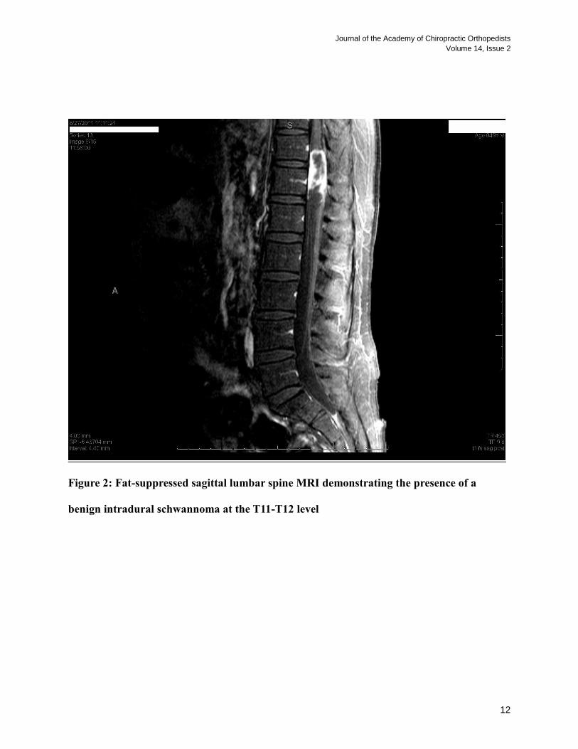
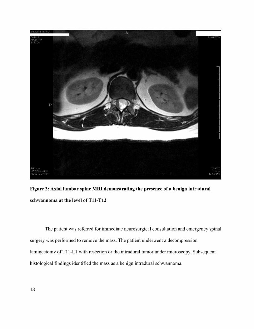
(Figures 1 and 2). The lesion appeared to arise from the left aspect of the conus medullaris with
effacement of the right aspect of the thecal sac (Figures 1, 2, and 3). Thin circumferential
contrast enhancement surrounded the tip of the filum terminale and extended to the L1 level.
Figures 1: Sagittal thoracolumbar spine MRI demonstrating the presence of a benign
intradural schwannoma at the T11-T12 level
Journal of the Academy of Chiropractic Orthopedists
Volume 14, Issue 2
12
Figure 2: Fat-suppressed sagittal lumbar spine MRI demonstrating the presence of a
benign intradural schwannoma at the T11-T12 level
13
Figure 3: Axial lumbar spine MRI demonstrating the presence of a benign intradural
schwannoma at the level of T11-T12
The patient was referred for immediate neurosurgical consultation and emergency spinal
surgery was performed to remove the mass. The patient underwent a decompression
laminectomy of T11-L1 with resection or the intradural tumor under microscopy. Subsequent
histological findings identified the mass as a benign intradural schwannoma.
Journal of the Academy of Chiropractic Orthopedists
Volume 14, Issue 2
14
Discussion
This case report describes a patient with an intradural schwannoma, mimicking a MTrP
referral pattern and initially responding to conservative management before rapid onset and
progression of neurologic symptoms. Re-evaluation, subsequent imaging including MRI with
and without gadolinium contrast, and post-surgical histological findings revealed that an
intradural schwannoma had arisen from and compressed the conus medullaris, resulting in conus
medullaris syndrome (CMS). Spinal pain is known to arise from intradural and epidural
tumors.18 Pain referral patterns from a MTrP located within the quadratus lumborum have been
identified to project into the sacroiliac joint, lower gluteal region, iliac crest, adjacent lower
abdomen, greater trochanter, and groin regions.17
CMS injuries occur in the region between the spinal cord and nerve roots; therefore,
resulting in a variable clinical presentation of upper and lower motor neuron manifestations.19
Although there are no definitive diagnostic criteria, CMS is commonly associated with rapid
onset of symmetrical neurological deficits. These neurological deficits typically involve a
combination of saddle anesthesia, bowel or bladder incontinence, lower extremity hyperreflexia,
and mild lower extremity weakness.19
This case was atypical in that the patient did not experience saddle anesthesia or bowel
and bladder dysfunction, possibly as the result of early symptom detection and subsequent
intervention. Also notable, the patient only exhibited hyperreflexia and motor weakness on the
left side. One explanation for this could be the uncommon location of the tumor. Schwann cells,
and thus schwannomas, are characteristically associated with the peripheral nervous system.
Surprisingly, this schwannoma arose not from the nerve roots but from the conus medullaris,
disproportionately affecting the left side. Intramedullary schwannomas are exceedingly rare,
15
accounting for only 50 cases between 1931 and 2002.20 Isolated CMS is commonly the result of
a non-traumatic primary intradural pathologic conditions (e.g. tumors or vascular lesions) and
treatment typically involves early surgical decompression to recover neurological integrity.19
Schwannomas are the most common intradural extramedullary spinal tumor, representing
43% to 67% of tumors in this category.21-24 Schwannomas typically demonstrate an onset
between the ages 30 to 50 years and may have a slight male predominance.25 Spinal
schwannomas occur at all spinal levels and are typically intradural.25,26 Safavi-Abbasi et al have
noted that the most frequent clinical presentation of schwannomas is pain and that most spinal
schwannomas in non-neurofibromatosis cases can be surgically removed with very few
postoperative deficits; however, preoperative autonomic dysfunction does not improve
significantly after surgical management.26
Regarding neuroimaging, conus medullaris tumors may have similar features on MRI
including infiltration of spinal cord tissue with compression of adjacent cord tissue, avid contrast
enhancement and cystic or hemorrhagic components. The two most common differential
diagnoses with these features include ependymoma and astrocytoma. As they have similar
features on MRI, it is important to remember that biopsy is necessary to confirm diagnosis of
conus medullaris tumors.
Musculoskeletal practitioners involved in the management of spinal pain conditions
and/or myofascial pain syndrome should be aware of the possibility of undiagnosed non-
musculoskeletal conditions that may mimic the clinical presentation of musculoskeletal pain
conditions. Emphasis should be placed upon reexamination and consideration of special studies
to inform an accurate diagnosis in the presence of unexplained progression of clinical

Journal of the Academy of Chiropractic Orthopedists
Volume 14, Issue 2
16
presentation. In this case, an undetected lower thoracic benign intradural schwannoma was
mistakenly managed as QL MTrP pain and later diagnosed as signs of CMS began to develop.
The accurate diagnosis allowed the patient to receive decompressive neurosurgery, resulting in
the immediate recovery of neurological functioning and elimination of thoracolumbar spine pain
and referred pain patterns.
Conclusion
Serious neurological conditions such as CMS may present with symptoms mimicking
common musculoskeletal disorders, including MTrP pain. Persistence or progression of
neurological signs or symptoms and increasing spinal pain suggest non-musculoskeletal etiology
and are indications for immediate advanced diagnostic imaging to evaluate for complicating
neurological involvement.27
Consent
Written informed consent was obtained from the patient for publication of this case report
and accompanying image. A copy of the written consent is available for review by the Editor-in-
Chief of this journal.
List of abbreviations
AP: anteroposterior
CMS: conus medullaris syndrome
e.g.: exempli gratia (for the sake of example)
L1: lumbar vertebra #1
17
L3: lumbar vertebra #3
L4: lumbar vertebra #4
MRI: magnetic resonance imaging
MTrP: myofascial trigger point
NRS: 11-point (0-10) Numeric Rating Scale
PRN: pro re nata (as needed)
S-I: sacroiliac joint
T2-weighted: spin–spin relaxation magnetic resonance imaging
T11: thoracic vertebra #11
T12: thoracic vertebra #12
QL: quadratus lumborum musculature
3+: a very brisk stretch reflex response (hyperreflexia)
Competing interests
The authors declare that they have no competing interests. Disclaimer: The views expressed in
this article are those of the authors and do not reflect the official policy or position of the
Department of Veterans Affairs or the US Government.
Authors' contributions
CBR participated in the conception of the report, the revision, and coordination of the final
manuscript. GAA was involved in the care of this patient, conducted the initial review of the
case, and helped to draft of the manuscript. CSO contributed to the drafting of this report and
made substantive contributions or the organization of this report. MJR was involved in the care
of this patient and assisted in the early drafting of this report, prior to his death. All authors made
substantive intellectual contributions to the report and meet the criteria for authorship.
Journal of the Academy of Chiropractic Orthopedists
Volume 14, Issue 2
18
Acknowledgements
The authors would like to thank Brenton L. Harris, MD for assisting with the neuroradiological
interpretation involved with this case. The authors would also would also like to thank Siri
Leech, DC, DABCR for her additional guidance on the interpretation of neuroimaging involved
with this case.
References
1. Vernon H, Schneider M: Chiropractic management of myofascial trigger points and
myofascial pain syndrome: a systematic review of the literature. J Manipulative
Physiol Ther 2009, 32:14-24.
2. Giamberardino MA, Affaitati G, Fabrizio A, Costantini R: Myofascial pain syndromes
and their evaluation. Best Pract Res Clin Rheumatol 2011, 25:185-198.
3. Bennett R: Myofascial pain syndromes and their evaluation. Best Pract Res Clin
Rheumatol 2007, 21:427-445.
4. Wheeler AH: Myofascial pain disorders: theory to therapy. Drugs 2004, 64:45-62.
5. Staud R: Future perspectives: pathogenesis of chronic muscle pain. Best Pract Res
Clin Rheumatol 2007, 21:581-596.
6. Cummings M, Baldry P: Regional myofascial pain: diagnosis and management. Best
Pract Res Clin Rheumatol 2007, 21:367-387.
7. Dommerholt J, Bron C, Franssen J: Myofascial trigger points: an evidence-informed
review. J Man Manip Ther 2006, 14:203-221.
8. Lavelle ED, Lavelle W, Smith HS: Myofascial trigger points. Med Clin North Am 2007,
91:229-239.
9. Tough EA, White AR, Richards S, Campbell J: Variability of criteria used to diagnose
myofascial trigger point pain syndrome--evidence from a review of the literature.
Clin J Pain 2007, 23:278-286.
10. Lucas N, Macaskill P, Irwig L, Moran R, Bogduk N: Reliability of physical
examination for diagnosis of myofascial trigger points: a systematic review of the
literature. Clin J Pain 2009, 25:80-89.
11. Malanga GA, Cruz Colon EJ: Myofascial low back pain: a review. Phys Med Rehabil
Clin N Am 2010, 21:711-724.
19
12. Skootsky SA, Jaeger B, Oye RK: Prevalence of myofascial pain in general internal
medicine practice. West J Med 1989, 151:157-160.
13. Harden RN, Bruehl SP, Gass S, Niemiec C, Barbick B: Signs and symptoms of the
myofascial pain syndrome: a national survey of pain management providers. Clin J
Pain 2000, 16:64-72.
14. Yap EC: Myofascial pain--an overview. Ann Acad Med Singapore 2007, 36:43-48.
15. Scott NA, Guo B, Barton PM, Gerwin RD: Trigger point injections for chronic non-
malignant musculoskeletal pain: a systematic review. Pain Med 2009, 10:54-69.
16. Alvarez DJ, Rockwell PG: Trigger points: diagnosis and management. Am Fam
Physician 2002, 65:653-660.
17. de Franca GG, Levine LJ: The quadratus lumborum and low back pain. J
Manipulative Physiol Ther 1991, 14:142-149.
18. Haldeman S, Kopansky-Giles D, Hurwitz EL, Hoy D, Mark EW, Dagenais S, Kawchuk
G, Stromqvist B, Walsh N: Advancements in the management of spine disorders. Best
Pract Res Clin Rheumatol 2012, 26:263-280.
19. Radcliff KE, Kepler CK, Delasotta LA, Rihn JA, Harrop JS, Hilibrand AS, Albert TJ,
Vaccaro AR: Current management review of thoracolumbar cord syndromes. Spine J
2011, 11:884-892.
20. Conti P, Pansini G, Mouchaty H, Capuano C, Conti R. Spinal neurinomas: Retrospective
analysis and long-term outcome of 179 consecutively operated cases and review of the
literature. Surg Neurol. 2004;61:34–43
21. Hufana V, Tan JS, Tan KK: Microsurgical treatment for spinal tumours. Singapore
Med J 2005, 46:74-77.
22. Prevedello DM, Koerbel A, Tatsui CE, Truite L, Grande CV, Ditzel LF, Araujo JC:
[Prognostic factors in the treatment of the intradural extramedullary tumors: a
study of 44 cases]. Arq Neuropsiquiatr 2003, 61:241-247.
23. el-Mahdy W, Kane PJ, Powell MP, Crockard HA: Spinal intradural tumours: Part I--
Extramedullary. Br J Neurosurg 1999, 13:550-557.
24. Garrido P, Laher-Mooncey S, Murphree NL, Jonker N, Levy LF, Makarawo S:
Neoplasms involving the spinal cord in Zimbabweans: an analysis of 262 cases. Cent
Afr J Med 1994, 40:201-204.
25. McCormick PC, Post KD, Stein BM: Intradural extramedullary tumors in adults.
Neurosurg Clin N Am 1990, 1:591-608.

Journal of the Academy of Chiropractic Orthopedists
Volume 14, Issue 2
20
26. Safavi-Abbasi S, Senoglu M, Theodore N, Workman RK, Gharabaghi A, Feiz-Erfan I,
Spetzler RF, Sonntag VK: Microsurgical management of spinal schwannomas:
evaluation of 128 cases. J Neurosurg Spine 2008, 9:40-47.
27. Schiff D, O'Neill BP, Suman VJ: Spinal epidural metastasis as the initial
manifestation of malignancy: clinical features and diagnostic approach. Neurology
1997, 49:452-456.
21
Original Article
Chiropractic Management of Cervicalgia in a Patient with Diffuse Idiopathic
Skeletal Hyperostosis Utilizing Cox Manual Cervical Distraction: A Case
Report
Ralph A. Kruse DC, FACO1,2, Casey S. Okamoto, DC3
1Private practice Chicago, IL 2Instructor, Cox Technique
3 Doctor of Chiropractic, VA Medical Center Minneapolis, MN
Published: June 2017 Journal of the Academy of Chiropractic Orthopedists
June 2017, Volume 14, Issue 2
This is an Open Access article which permits unrestricted use, distribution, and reproduction in any medium, provided the original work is
properly cited. The article copyright belongs to the author and the Academy of Chiropractic Orthopedists and is available at:
http://www.dcorthoacademy.com. © 2017 Kruse/Okamoto and the Academy of Chiropractic Orthopedists.
Abstract
Objective: The purpose of this case report is to describe the utilization of Cox Manual Cervical
Distraction for the treatment of cervicalgia in a patient with Diffuse Idiopathic Skeletal
Hyperostosis (DISH).
Clinical Features: A 59 year-old female presented with chronic constant neck pain and
stiffness which limited her ability to perform activities of daily living (ADLs). Cervical spine
radiographs revealed findings consistent with DISH.
Intervention and Outcome: This patient was treated with Cox manual cervical distraction
resulting in a decrease in the severity and frequency of her pain and improved ability to perform
ADLs. Protocol II was utilized to help promote normal facet mobility.
Conclusion: This case study describes the treatment of a 59 year old woman with chronic neck
pain in the setting of DISH.
Key Indexing Terms: Chiropractic, Diffuse Idiopathic Skeletal Hyperostosis, Cervical Spine
Journal of the Academy of Chiropractic Orthopedists
Volume 14, Issue 2
22
Introduction
Diffuse Idiopathic Skeletal Hyperostosis (DISH) is a spinal and extraspinal articular disorder
characterized by ligamentous calcification and ossification. This entity is distinct from
ankylosing spondylitis and degenerative joint disease and occurs with an incidence of 12 percent
in the United States, affecting middle-aged and elderly individuals. It is also known as
Forrestier’s disease, spondylosis hyperostotica, spondylitis ossificans ligamentosa, and senile
ankylosing hyperostosis.1
Numerous studies have documented the efficacy of flexion-distraction manipulation applied to
the lumbar spine for the treatment of conditions including disc herniation2, radiculopathy3,
stenosis4,5, synovial cysts6, post-surgical continued pain3, and pregnancy-related pain7.
Cox flexion-distraction tables which include the cervical headpiece allow for treatment of the
cervical spine. This component facilitates manual axial distraction as well as motion in flexion,
extension, rotation, lateral bending and coupled movements. These movements are primarily
designed to reduce intradiscal pressure and restore normal physiologic ranges of motion. Manual
distraction is a type of spinal traction wherein the doctor directly controls the application of force
in terms of both amplitude and vector.8
Treatment of this type for the cervical spine has been documented in case reports and
retrospective studies. Improvement has been observed in cases of radiculopathy caused by
cervical disc herniation9,10,11 as well as in cases involving cervical stenosis12, degenerative disc
disease13, adjacent segment disease related to congenital anomalies14, and surgical fusion.15
The purpose of this case report is to present a case of cervicalgia treated with Cox manual
cervical distraction in a patient with Diffuse Idiopathic Skeletal Hyperostosis.
Clinical Presentation
A 59 year-old female presented to a private chiropractic practice with complaints of constant
neck pain and stiffness of gradual onset over the past five years. She described all neck
movements as painful, limiting her ability to perform activities of daily living (ADLs) such as
driving, lifting, reading, and desk work, and resulting in disordered sleep. She described the
quality of her neck pain as throbbing, aching, and stiff, with radiation to her upper back and
shoulders bilaterally. She rated her pain as an 8 out of 10 on an 11-point Numeric Rating Scale.
Her Neck Disability Index (NDI) was rated at 36%. She denied any radiation of pain past the
shoulder and denied any numbness, tingling, or weakness of the upper extremity.

23
Prior interventions included an evaluation by a rheumatologist who diagnosed Fibromyalgia and
degenerative disc disease. She was prescribed medication including Lyrica and Desipramine
which she took for years but discontinued as she perceived no benefit. She was also prescribed
physical therapy which she discontinued due to a lack of progress.
Active cervical range of motion examination revealed the following: flexion and extension
limited to 25 degrees and provocative at end range, right and left rotation limited to 40 degrees
with mild provocation at end range, right lateral flexion limited and provocative at 20 degrees,
and left lateral flexion limited to 25 degrees, causing a pulling sensation in the contralateral
trapezius. Palpation revealed tenderness over the articular pillars from C3-7. Myotomes and
dermatomes were tested and intact at C5-T1. Myotatic reflexes were rated at 2+ bilaterally.
Tromner’s sign was absent. Hypertonicity to palpation was noted at the suboccipital, upper and
middle trapezius, levator scapulae, rhomboids and splenius capitus muscles bilaterally.
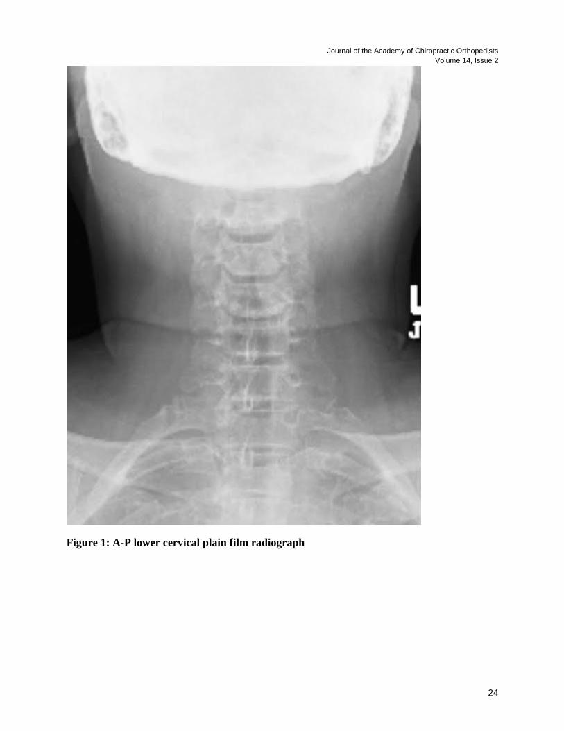
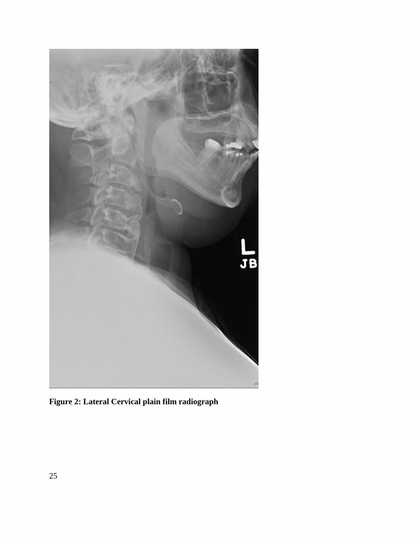
A-P lower cervical, lateral cervical, and cervicothoracic lateral plain film radiographs were
performed. A right list was noted in the coronal plane. Sagittal plane alignment demonstrated an
anterior head carriage. A mild loss of intervertebral disc space with uncinate proliferation was
noted at C4-C5 and C5-C6. Extensive anterior vertebral body osseous proliferation was seen
extending from C3 through C6 that spanned the anterior surface of the intervening disc space.
The intervertebral body heights and atlanto-dental interspace were within normal limits with no
evidence of osseous pathology. The radiographic impressions included minor multilevel
degenerative disc disease, uncovertebral arthrosis at C4-C5 and C5-C6, and diffuse idiopathic
skeletal hyperostosis (DISH).
Journal of the Academy of Chiropractic Orthopedists
Volume 14, Issue 2
24
Figure 1: A-P lower cervical plain film radiograph
25
Figure 2: Lateral Cervical plain film radiograph

Journal of the Academy of Chiropractic Orthopedists
Volume 14, Issue 2
26
Figure 3: Cervicothoracic lateral plain film radiograph
27
Treatment Intervention
Treatment consisted of Cox manual distraction manipulation applied to the cervical spine. Due to
the absence of a radicular component to the patient’s subjective and objective presentation, Cox
protocol II was used. The initial 3 treatments consisted of 10 repetitions of axial decompression
contacting the occiput. On the fourth visit, coupled movements of axial decompression and
rotation were performed to spinal levels C2 through C6 where the treating physician palpated
somatic dysfunction with suboptimal intersegmental rotation. Subsequent visits included the
addition of coupled motions of axial distraction with lateral flexion and rotation to the upper
thoracic spine with the aim of restoring normal physiologic motion to those segmental levels.
This patient was treated eight times over the course of four weeks and demonstrated progressive
subjective and objective improvement. Outcome measures were collected upon completion of
the course of care, at which time the patient rated her pain at 4/10 on a Numeric Rating Scale and
endorsed a Neck Disability Index of 22%. This was an improvement of 50% and 39%
respectively.
Discussion
Clinical characteristics of DISH are similar to degenerative joint disease including joint stiffness,
typically worse in the morning, and low grade musculoskeletal pain especially of the spine.
Approximately 20% of patients complain of dysphagia due to compression of the esophagus
from anterior cervical spine osseous proliferation. Extraspinal complaints may also be present
since osseous proliferation may occur in any ligamentous or tendinous attachment to bone.1
Radiographic features of the vertebral column show calcification followed by ossification of the
anterior longitudinal ligament (ALL) typically beginning in the middle of the vertebrae
extending to bridge the adjacent vertebral disc. This flowing exuberant hyperostosis, often over
one centimeter in thickness, causes the appearance of a bumpy anterior spinal contour and may
be described as “Candle Flame” hyperostosis. Initially the deep layers of the ALL may be
uninvolved resulting in a vertical radiolucent shadow. This lucency may be obliterated as the
ligamentous ossification progresses. Calcification may be inhibited due to anteriolateral fibrous
discal extensions from the outer annular fibers. This may result in horizontal radiolucent linear
clefts.1 In the cervical spine the bony hyperostosis is most exuberant in lower segments (C4-7).
There is relative preservation of intervertebral disc height, although minor disc degeneration may
be present, and a lack of significant apophyseal joint arthrosis which may allow for relatively
normal vertebral motion. A slight to moderate loss of the cervical lordosis and increase in
thoracic kyphosis is common.16
Though patients with DISH may present with stiffness and decreased range of motion due to
altered biomechanics, it remains unclear whether or to what degree DISH is associated with

Journal of the Academy of Chiropractic Orthopedists
Volume 14, Issue 2
28
pain.17 Holton et. al. report that few studies have evaluated the association of DISH and back
pain. The objective of Holton’s study was to estimate the prevalence of radiographic DISH in the
thoracic and lumbar spine among elderly men and determine its association with back pain in the
last 12 months. The findings indicated that those with DISH experienced less frequent and less
severe back pain than counterparts without DISH. 18
Few published studies document the management of patients with DISH with chiropractic
manipulation. Roberts et. al. demonstrated subjective and objective improvement utilizing
Activator-assisted spinal manipulative therapy in a 74 year-old man with low back pain and a
history of degenerative disc disease and DISH. 19 Hoffman reports on four cases of men over the
age of 75 with DISH and associated neurological signs and symptoms. Symptoms often appeared
to be minimal compared to the dramatic radiographic changes. Three of the four patients
responded favorably to chiropractic spinal manipulation.20 Troyanovich reported on a 60 year-
old man with DISH and a history of episodic disabling low back pain. Treatment consisted of
chiropractic manipulation including drop table adjustments, exercise and standing lumbar
traction. The patient demonstrated improvement with respect to flexibility, pain severity, and
engagement with activities of daily living lasting at least 19 months.21
In this case, the patient was treated with protocol II of Cox manual cervical distraction. Protocol
II is utilized to treat patients presenting with neck pain and associated non-radicular pain with the
aim of restoring physiologic ranges of motion. Treatments begin with axial distraction, followed
by a combination of distraction with rotation and lateral flexion as clinically indicated.22
There is a growing body of evidence to suggest that manual cervical distraction utilizing a Cox
headpiece may induce physiologic changes. These changes may be responsible for the
symptomatic relief reported for patients affected by discogenic or facetogenic pain conditions.
Gudavalli, et. al. measured changes in intradiscal pressure when performing manual cervical
distraction using the Cox headpiece. Pressure transducers were inserted into the nucleus pulposus
at the C4-5, C5-6, C6-7, and C7-T1 discs of cadavers using an anterior surgical approach.
Intradiscal pressure decreases were found in the levels tested during manual distraction, more
prominently in y-axis distraction and flexion.23
Kruse documented clinical relief utilizing Cox manual cervical distraction for radiculopathy
from a C5-C6 disc herniation13, radiculopathy from severe foraminal stenosis at C6-C79, central
and lateral recess stenosis from C4-C5 though C6-C711, and degenerative disc disease related to
C2-C3 block vertebrae.24 Schliesser, et. al studied 39 patients with cervical radiculopathy treated
with Cox cervical distraction and demonstrated a statistically significant decrease in Visual
Analog Scale (VAS) scores among those undergoing treatment, with a mean decrease of
41.4%.10
Joachim reported symptomatic improvement of neck pain with pain and numbness radiating to
both hands in a patient with spondylotic myelopathy and a history of cervical spine plate fusion

29
at C6-C7.15 Cox has also demonstrated relief from pain associated with disc degeneration
coupled with advanced facet osteoarthritic degeneration.25 Allen has documented successful
treatment using Cox spinal manipulation for the treatment of a patient with a C6-C7 disc
herniation, foraminal narrowing, and associated radiculopathy.26
Limitations
Because this is a single case report, it is not appropriate to generalize the effects from this patient
to others with neck pain and findings of DISH. A larger scale study would be needed to make
any determination regarding safety or efficacy of the applied intervention. Also, the long-term
effects of care for this patient are not reported.
Conclusion
This case study describes the treatment of a 59 year old woman with radiographic findings of
DISH and concomitant chronic neck pain that responded favorably to utilization of Cox manual
distraction.
Competing interests
The authors declare that they have no competing interests. Disclaimer: The views expressed in
this article are those of the authors and do not reflect the official policy or position of the
Department of Veterans Affairs or the US Government.
References
1. Terry R. Yochum, Lindsay J. Rowe. Yochum and Rowe's Essentials of Skeletal
Radiology. Philadelphia, Pa. : Lippincott Williams & Wilkins, c2005.; 2005 pp. 990-993.
2. Greenwood DM. Improvement in chronic low back pain in an aviation crash survivor
with adjacent segment disease following flexion distraction therapy: a case study. J
Chiropr Med. 2012;11(4):300-5.
3. Gudavalli MR, Olding K, Joachim G, Cox JM. Chiropractic Distraction Spinal
Manipulation on Postsurgical Continued Low Back and Radicular Pain Patients: A
Retrospective Case Series. J Chiropr Med. 2016;15(2):121-8.
4. Dupriest CM. Nonoperative management of lumbar spinal stenosis. J Manipulative
Physiol Ther. 1993;16(6):411-4.

Journal of the Academy of Chiropractic Orthopedists
Volume 14, Issue 2
30
5. Snow GJ. Chiropractic management of a patient with lumbar spinal stenosis. J
Manipulative Physiol Ther. 2001;24(4):300-4.
6. Cox JM. Chiropractic management of a patient with lumbar spine pain due to synovial
cyst: a case report. J Chiropr Med. 2012;11(1):7-15.
7. Kruse RA, Gudavalli S, Cambron J. Chiropractic treatment of a pregnant patient with
lumbar radiculopathy. J Chiropr Med. 2007;6(4):153-8.
8. Cox, James M. Neck, Shoulder, Arm Pain: Mechanism, Diagnosis, and Treatment. 4th
ed., Fort Wayne, Cox Technic Resource Center, Inc, 2014 pp. 51-52.
9. Gudavalli S, Kruse RA. Foraminal stenosis with radiculopathy from a cervical disc
herniation in a 33-year-old man treated with flexion distraction decompression
manipulation. J Manipulative Physiol Ther. 2008;31(5):376-80.
10. Schliesser JS, Kruse R, Fallon LF. Cervical radiculopathy treated with chiropractic
flexion distraction manipulation: A retrospective study in a private practice setting. J
Manipulative Physiol Ther. 2003;26(9):E19.
11. Manison AM. Chiropractic management using Cox cervical flexion-distraction technique
for a disk herniation with left foraminal narrowing in a 64-year-old man. J Chiropr Med.
2011;10(4):316-21.
12. Kruse RA, Gregerson D. Cervical Spinal Stenosis Resulting in Radiculopathy Treated
with Flexion Distraction Manipulation: A Case Study. J Neuromusculoskeletal System.
Winter 2002;10(4):141-147.
13. Kruse RA, Imbarlina F, De bono VF. Treatment of cervical radiculopathy with flexion
distraction. J Manipulative Physiol Ther. 2001;24(3):206-9.
14. Kruse, RA, Schliesser, J, DeBono, VF. Klippel-Feil syndrome with radiculopathy.
Chiropractic management utilizing flexion-distraction technique: a case study. J
Neuromusculoskeletal System. 2000;8:124–131.
15. Joachim GC. Cox decompression manipulation and guided rehabilitation of a patient with
a post surgical C6-C7 fusion with spondylotic myelopathy and concurrent L5-S1
radiculopathy. J Chiropr Med. 2014;13(2):110-5.
16. Juhl JH, Crummy AB, Kuhlman JE et al. Paul and Juhl's Essentials of Radiologic
Imaging. Lippincott Williams & Wilkins; 1998 pp. 111-112.
17. Foshang, Trevor H. et al. Diffuse idiopathic skeletal hyperostosis: A case of dysphagia J
Manipulative Physiol Ther. 2002;25(1):71-76.
18. Holton KF, Denard PJ, Yoo JU, et al. Diffuse idiopathic skeletal hyperostosis and its
relation to back pain among older men: the MrOS Study. Semin Arthritis Rheum.
2011;41(2):131-8.
19. Roberts JA, Wolfe TM. Chiropractic management of a veteran with lower back pain
associated with diffuse idiopathic skeletal hypertrophy and degenerative disk disease. J
Chiropr Med. 2012;11(4):293-9.
31
20. Hoffman L, et al. Diffuse idiopathic skeletal hyperostosis (DISH): a review of
radiographic features and report of four cases. J Manipulative Physiol Ther
1995;18(8):547-553.
21. Troyanovich S, Buettner M. A Structural chiropractic approach to the management of
diffuse idiopathic skeletal hyperostosis. J Manipulative Physiol Ther 2003;26:202-6.
22. Cox, James M. Neck, Shoulder, Arm Pain: Mechanism, Diagnosis, and Treatment. 4th
ed., Fort Wayne, Cox Technic Resource Center, Inc, 2014 pp. 75-76.
23. Gudavalli MR, Potluri T, Carandang G, et al. Intradiscal Pressure Changes during
Manual Cervical Distraction: A Cadaveric Study. Evid Based Complement Alternat Med.
2013;2013:954134.
24. Kruse R, Schliesser J, DeBono V. Klippel-Feil syndrome with radioculopathy.
Chiropractic management utilizing flexion-distraction technique: a case report. J
Neuromusculoskeletal System. 2000;8(4):124-131.
25. Cox, James M. Neck, Shoulder, Arm Pain: Mechanism, Diagnosis, and Treatment. 4th
ed., Fort Wayne, Cox Technic Resource Center, Inc, 2014 pp. 115.
26. Manison AM. Chiropractic management using Cox cervical flexion-distraction technique
for a disk herniation with left foraminal narrowing in a 64-year-old man. J Chiropr Med.
2011;10(4):316-21.
Journal of the Academy of Chiropractic Orthopedists
Volume 14, Issue 2
32
Original Article
Abdominal Aortic Aneurysm and Spinal Manipulation, an Absolute
Contraindication? A Review of the Literature
Adam Sergent, DC, CCSP1, Heather Bowyer DC, CCSP2 1Assistant Professor, Palmer College of Chiropractic Florida Clinical Affairs 2Associate Professor, Palmer College of Chiropractic Florida Clinical Affairs
Published: June 2017 Journal of the Academy of Chiropractic Orthopedists
June 2017, Volume 14, Issue 2
This is an Open Access article which permits unrestricted use, distribution, and reproduction in any medium, provided the original work is
properly cited. The article copyright belongs to the author and the Academy of Chiropractic Orthopedists and is available at:
http://www.dcorthoacademy.com. © 2017 Sergent and the Academy of Chiropractic Orthopedists.
Objective: To perform a topic review of chiropractic adjustments performed on patients
diagnosed with Abdominal Aortic Aneurysm.
Background: A need for this review was prompted when reviewing Medicare absolute
contraindications to a significant Abdominal Aortic Aneurysm without defining what significant
means.
Methods: Peer reviewed articles were accessed from PubMed from years 1986-2016, Index to
Chiropractic Literature 1995-2016; and Medline Complete 2012-1986, using the search terms
Chiropractic and Abdominal Aortic Aneurysm, A total of 10 articles with those search terms
were returned.
Discussion: Minimal research is currently available discussing spinal manipulative therapy
(SMT) in the presence of an abdominal aortic aneurysm (AAA). In the research that is available,
no documented adverse reactions to care are present therefore raising the question of whether an
AAA is in fact an absolute contraindication to SMT.
Conclusion: For patients diagnosed with abdominal aortic aneurysm, the current peer reviewed
literature is insufficient to determine whether chiropractic adjustments in the lumbar spine are
absolutely contraindicated. This diagnosis may not be an absolute contraindication to
chiropractic adjustments in the region of the aneurysm, as long as special consideration is given.
Key Indexing Terms: Chiropractic and Abdominal Aortic Aneurysm; AA; Manipulation;
Adjustment; Abdominal Aortic Aneurysm.

33
Introduction
Abdominal Aortic Aneurysm (AAA) is defined as a dilatation of the abdominal aorta measuring
more than 5cm in diameter. A normal diameter is approximately 3.5cm.1,2.3 Aneurysms are
classified into different groups. A saccular aneurysm is eccentric, localized and distended
affecting only part of the arterial wall. A true aneurysm is comprised of all layers of the aorta, a
partial aneurysm may only consist of one or two layers, and a dissecting aneurysm may
hemorrhage into the layers and cause separation.4
AAA’s are more prevalent in men than women, smokers than non-smokers and within an age
group of 60 to 80 years of age.3 Approximately 2-4 percent of the general population may have
an undetected AAA but that number jumps to 5.9 percent in the 60-80-year-old population.3 This
is especially important to the chiropractor who may not screen for AAA or image before the
initiation of a general treatment plan to address low back pain that is thought to be mechanical in
nature. AAA’s are missed completely or misdiagnosed in up to 30% of cases.5 Current evidence
based practices and standard of care allows for 4-6 weeks of treatment before imaging is
ordered.6 A PubMed search was performed and, though a number of studies were identified in
which a patient had both a dissecting AAA and concomitant LBP, none were identified in which
the AAA was determined to be the cause of LBP.
Patients presenting to the chiropractic office often have co-morbidities that must be considered
when developing a treatment plan that is safe and effective for the patient. Abdominal aortic
aneurysm is most often an incidental finding revealed upon imaging of a patient with low back
pain.
Current CMS guidelines regard AAA as an absolute contraindication to a dynamic thrust in the
event of “a significant major artery aneurysm near the proposed manipulation”; however, CMS
does not define “significant” or “near” and no such definition is found in the literature.7 The
American Association for Vascular Surgery and Society for Vascular Surgery guidelines have
found an increased risk of spontaneous rupture associated with larger aneurysm size. For an
aneurysm smaller than 5 cm, the risk is low compared to those larger than 5 cm. In fact, AAA
less than 4 cm were found to have a rupture rate of 0% annually. Surgical repair should be
considered at 5.5 cm or when an increase growth rate of >1cm per year is found.8 The purpose
of this review is to identify, by a review of available literature, whether the data is sufficient to
label AAA as an absolute contraindication to SMT. Further, does the size of an AAA and
proximity to the manipulation play a significant role? To date, there are no case studies in which
SMT has precipitated an AAA rupture or dissection.9,10
Journal of the Academy of Chiropractic Orthopedists
Volume 14, Issue 2
34
Methods
A literature search was performed using the terms “Chiropractic and Abdominal Aortic
Aneurysm” using PubMed returning 9 articles, Index for Chiropractic Literature (ICL) returning
10 articles and Medline Complete returning 9 articles. The search terms “Adjustment and
Abdominal Aortic Aneurysm” were limited; therefore, “Manipulation and Abdominal Aortic
Aneurysm” was also searched in the same 3 data bases. The second set of searches yielded 276
articles; however, no further articles were found pertaining to chiropractic and abdominal aortic
aneurysm. The term Abdominal Aortic Aneurysm was also searched, for information regarding
epidemiology, pathophysiology, and diagnostic criteria. Exclusion criteria included when an
article described the management of the AAA without being treated utilizing a chiropractic
adjustment. In some cases, the AAA was discovered and referred out as management of the case.
Inclusion criteria included articles that employed chiropractic adjustments as treatment in the
lumbar spine regardless of if the AAA was known before the manipulation was rendered or if it
was an incidental finding following a course of conservative chiropractic care. With the
inclusion/exclusion criteria there were only 2 papers reviewed that met the inclusion criteria of a
chiropractic adjustment in the region of the AAA. The remaining 8 papers discussed the
management of the AAA once found based on imaging or physical exam, there are statements in
these articles stating that it is not known if an AAA is an absolute contraindication for a
chiropractic adjustment.
Discussion
It is apparent that within the search terms used and the articles reviewed in most cases the
chiropractic adjustment did not cause any adverse effects related to the AAA In addition,
chiropractors play an important role in identifying a potential AAA and referring patients for
imaging and surgical consultation. The current literature available for this review is however
limited and it is apparent more research and or retrospective studies are needed. The 2 case
studies reviewed that met the inclusion criteria revealed that there were chiropractic adjustments
for mechanical back pain in patients with undiagnosed AAA who experienced improvement or
resolution of pain over the course of a few treatments without adverse event. In both cases an
AAA was discovered after undergoing chiropractic adjustments and both patients underwent a
successful surgical repair.
A case study by Hadida and Rajwani revealed a 74-year-old male who underwent 5 weeks of
manual adjusting using Thompson Drop technique, trigger point therapy and side posture lumbar
adjustment, with decreased back pain. Upon the 5th week the patient was put in a side posture
adjustment and had immediate relief. During that visit, the patient complained of abdominal pain
while lying supine. An abdominal exam was performed and a suspected AAA was found using
deep palpation. The patient was referred out for ultrasound and a 5.3cm AAA was revealed.

35
Surgery was performed 2 weeks later and abdominal pain resolved.10 The second case revealed a
25-year-old male with Marfan’s Syndrome undergoing 3 weeks of manual adjusting using a
compressive manipulative therapy in the thoracic spine, with resolution of pain in a patient. One
week later a routine exam was performed by his family physician and a dissecting aneurysm was
revealed and immediate surgical correction was performed. In this case, the patient was found to
have an old, healed dissection which correlated with a history of a physical altercation and
crushing chest pain on separate occasions one year prior. It is believed that the dissection
occurred during one of those reported incidents.11
Of these two articles that included an AAA and chiropractic adjustment, the chiropractic
adjustment was not thought to precipitate the development of AAA or result in an aortic
dissection. Of the remaining 10 articles that were reviewed 4 specifically state that AAA is not
an absolute contradiction to chiropractic adjustment while the others do not comment. The
chiropractic literature does not cite cases where spinal manipulative therapy (SMT) was a direct
cause of a AAA dissection.12 There is no published evidence that HVLA SMT may cause rupture
of an AAA. 9 It is not known if the forces utilized in a SMT are sufficient magnitude to cause
rupture of an AAA as all forces among chiropractors are different.13 While more research is
needed to determine if it is safe to apply a manipulation to the area with an AAA, it also must be
determined the size that makes it safe or unsafe.
While AAA’s are missed in up to 30% of cases, the chiropractic physician should still consider
screening for them in older populations.5 AAA’s are more prevalent in Caucasian men ages 60-
80 with a history of smoking. This age group of 60-80 year olds are very likely to present in the
chiropractor’s office with suspected mechanical low back pain. Knowing the patients age and
history should lead the chiropractor to determine if a screening is needed and if an abdominal
exam should be performed. Upon exam, there will be a palpable mass if present and abdominal
bruits heard. Imaging may be considered as well if AAA is still suspected though not found on
physical exam. Current guidelines by U.S Preventive Services Task Force (USPSTF)
recommends screening asymptomatic adults over the age of 50 as prevalence can be as high as
7.2%.14 The most current recommendations state that abdominal duplex ultrasonography is the
standard of care for AAA screening with a 94-100% specificity and a 98-100% specificity.14
With this being the gold standard it indicates that all suspected AAA should be referred out.
Calcification of the AAA is only seen on approximately 50% of x-rays and that is one of the
most reliable radiological signs seen by chiropractors.8 This suggests that even when a
chiropractor suspects a AAA if they try and rule in or out such diagnosis by imaging they often
have in their office up to 50% will still be missed.
With 2-4% of the population regardless of age having a AAA we must ask how many patients
with non-symptomatic AAA’s presenting for low back pain or wellness care, are being adjusted
as they do not meet the criteria of over 60 with a history of smoking and not necessarily being
Journal of the Academy of Chiropractic Orthopedists
Volume 14, Issue 2
36
screened for a AAA. We must consider what the prevalence of finding AAA’s in the office
before advanced imaging was readily available. How many people with an AAA that present for
suspected mechanical low back pain complete their chiropractic care plan with full resolution of
pain while having a AAA?
Conclusion
The current literature reviewed in this paper is sparse at best. More research is needed to
determine if adjusting a region near a significant AAA is an absolute contraindication. It also
must be determined what the CMS definition of a significant AAA is, and give measurements of
what defines significant and near. With the percentages of missed diagnosis of an AAA
anywhere from 30% on physical exam and 50% on x-ray imaging, physicians must be aware of
differential diagnosis that fall into specific patient populations.
Limitations
With the lack of available peer reviewed articles with the search terms Abdominal Aortic
Aneurysm and Chiropractic, one of the limitations is the depth of the paper in that there was a
lack of articles to review.
Competing Interests
The author declares that he has no competing interests.
References
1. Taylor John A, Bussières A. Diagnostic imaging for spinal disorders in the elderly: a
narrative review. Chiropractic & Manual Therapies, Vol 20, Iss 1, P 16 (2012)
2. de Boer N, Knaap S, de Zoete A. Clinical detection of abdominal aortic aneurysm in a 74-
year-old man in chiropractic practice. Journal Of Chiropractic Medicine [serial online].
March 2010;9(1):38-40
3. Beck R, Holt K, Fox M, Hurtgen-Grace K. Radiographic anomalies that may alter
chiropractic intervention strategies found in a New Zealand population. Journal Of
Manipulative & Physiological Therapeutics [serial online]. November 2004;27(9):554-6
4. Crawford C, Hurtgen-Grace K, Talarico E, Marley J. Review of the literature: Abdominal
aortic aneurysm: an illustrated narrative review. Journal Of Manipulative And
Physiological Therapeutics [serial online]. January 1, 2003;26:184-195.
5. Dargin J, Lowenstein R. Clinical communication: Adults: Ruptured Abdominal Aortic
Aneurysm Presenting as Painless Testicular Ecchymosis: The Scrotal Sign of Bryant
Revisited. Journal Of Emergency Medicine [serial online]. January 1, 2011;40:e45-e48

37
6. Bussières A, Taylor J, Peterson C. Diagnostic imaging practice guidelines for
musculoskeletal complaints in adults-an evidence-based approach-part 3: spinal disorders.
Journal Of Manipulative & Physiological Therapeutics. January 2008;31(1):33-88
7. Medicare benefit policy manual chapter 15 – Covered medical and other health service
https://www.cms.gov/Regulations-and-
Guidance/Guidance/Manuals/downloads/bp102c15.pdf Accessed May 31, 2017
8. Brewster D, Cronenwell J, Hallett J, Johnston K, Krupski W, Matsumura J. Guidelines
for the treatment of abdominal aortic aneurysms: Report of a subcommittee of the Joint
Council of the American Association for Vascular Surgery and Society for Vascular
Surgery. Journal of Vascular Surgery 2003 May;37(5):1106
9. Crawford C. Abdominal aortic aneurysm presenting as low back pain: a case report.
Chiropractic Journal Of Australia [serial online]. September 2003;33(3):83-88.
10. Hadida C, Rajwani M, Abdominal aortic aneurysms; case report. Journal of Canadian
Chiropractic Association 1998;42 (4)
11. Ruling J, Crowther E, McCord P. Clinical considerations in the chiropractic management
of the patient with Marfan syndrome. Journal Of Manipulative & Physiological
Therapeutics [serial online]. September 2000;23(7):498-502.
12. Stemper B, Hallman J, Peterson B. Original Article: An Experimental Study of Chest
Compression During Chiropractic Manipulation of the Thoracic Spine Using an
Anthropomorphic Test Device. Journal of Manipulative and Physiological Therapeutics
[serial online]. January 1, 2011;34:290-296
13. LeFevre M. Screening for abdominal aortic aneurysm: US preventive services task force
recommendation statement. Annals of internal medicine 2014; vol 161 (4) 281-290
14. Rowe L, Yochum T. From masqueraders of musculoskeletal disease. Essentials of
Skeletal Radiology, 3rd edition. Philadelphia: Lipincott Williams &Wilkins: 2005; 1818-
24
15. Brewster D, Cronenwell J, Hallett J, Johnston K, Krupski W, Matsumura J. Guidelines
for the treatment of abdominal aortic aneurysms: Report of a subcommittee of the Joint
Council of the American Association for Vascular Surgery and Society for Vascular
Surgery. J Vasc Surg 2003 May;37(5):1106

Journal of the Academy of Chiropractic Orthopedists
Volume 14, Issue 2
38
Editorial Review
MRI of the Elbow: Techniques and
Spectrum of Disease
Ashvin K. Dewan, MD, A. Bobby Chhabra, MD, A. Jay Khanna, MD, MBA, Mark W. Anderson, MD, Lance M. Brunton, MD
J Bone Joint Surg Am. 2013;95:e99(1-13)
JACO Editorial Reviewer: Cliff Tao DC DACBR
Published: June 2017
Journal of the Academy of Chiropractic Orthopedists
2017, Volume 14, Issue 2
The original article copyright belongs to the original publisher. This review is available from: www.dcorthoacademy.com © 2017 Tao and the
Academy of Chiropractic Orthopedists. This is an Open Access article which permits unrestricted use, distribution, and reproduction in any
medium, provided the original work is properly cited.
Author’s Abstract:
Background: Magnetic resonance imaging (MRI) of the elbow allows for high-resolution
evaluation of osseous and soft tissue structures, including ligaments, tendons, nerves, and
muscles. Multiple imaging techniques and pulse sequences exist. The purpose of this article is to
update orthopaedic surgeons on current MRI techniques and illustrate the spectrum of elbow
pathology detectable by MRI.
Methods: We searched MEDLINE with use of the keywords ‘‘MRI’’ and ‘‘elbow’’ for studies
less than five years old evaluating MRI techniques. These papers, our experience, and textbooks
reviewing elbow MRI provided the information for this article.
Results: We discuss the essentials and applications of the following techniques: (1)
conventional, non-gadolinium enhanced MRI; (2) gadolinium-enhanced MRI; and (3) magnetic
resonance arthrography. The classic MRI appearances of occult fractures, loose bodies, ulnar
collateral ligament injuries, lateral collateral ligament complex injuries, biceps tendon injuries,
triceps tendon injuries, lateral epicondylitis, medial epicondylitis, septic arthritis, osteomyelitis,
osteochondritis dissecans, compression neuropathies, synovial disorders, and various soft-tissue
masses are reviewed.
Conclusions: MRI is a valuable, noninvasive method of elbow evaluation. This article updates
orthopaedic surgeons on the various available MRI techniques and facilitates recognition of the
MRI appearances of the most commonly seen pathologic elbow conditions.

39
JACO Editorial Summary:
• This is an exhibit selection, which is way of bringing select recent exhibits from a
meeting which are felt to have potential reader interest, to publication. There is very little
or no peer review process.
• The authors of this exhibit/article are from the Department of Orthopaedic Surgery, The
Johns Hopkins University, Baltimore, Maryland; the Departments of Orthopaedic
Surgery and Radiology, University of Virginia, Charlottesville, Virginia; and the
University of Pittsburgh School of Medicine, Pittsburgh, Pennsylvania.
• The purpose of this article is to update the reader on general MRI sequences of the elbow
and to show examples of common elbow pathology on MRI. Of note the article
emphasizes the use of a fluid-sensitive sequence, such as STIR or fat-suppressed T2-
weighted imaging.
• The authors state a Medline search was used to procure studies not more than 5 years old
but do not describe the inclusion critieria. Of interest is that over half of the articles
referenced are at least 10 years old. The authors also used their experience and textbooks
to provide information for this article.
• A concise review of types of elbow MRI is presented with common indications.
Intravenous contrast-enhanced elbow MRI is commonly preferred for the evaluation of
soft tissue masses and synovial disorders including inflammatory arthritides. Bone
perfusion and viability are also shown with IV contrast enhanced MRI. MR arthrography
is indicated for intra-articular or periarticular pathology such as collateral ligament and
capsular tears, osteochondral lesions, and articular bodies.
• A brief synopsis of MR imaging features of various disorders related to trauma (occult
fracture, articular [loose] bodies, collateral ligament injury, biceps/triceps tendon injury),
degeneration (medial and lateral epicondylosis), infection (septic arthritis, osteomyelitis),
and other conditions such as osteochondral defects, compression neuropathies, synovial
disorders, and soft tissue masses is presented.
Summary:
This article is a good review to help the chiropractor and the chiropractic orthopedist by
suggesting when MRI, when intravenous and intra-articular contrast, and even which specific
sequences are indicated in the differential diagnosis of elbow pain. MRI is a valuable, non-
invasive tool in the evaluation of the elbow and this article shows examples of common
conditions that may be seen in the chiropractic office.

Journal of the Academy of Chiropractic Orthopedists
Volume 14, Issue 2
40
Editorial Review
Shoulder Impingement: Relationship of Clinical
Symptoms and Imaging Criteria
Marius E. Mayerhoefer, MD, Martin J. Breitenseher, MD, Christian Wurnig, MD,
and Andreas Roposch, MD, MSc
Clin J Sport Med Volume 19, Number 2, March 2009
Copyright 2009 by Lippincott Williams & Wilkins
JACO Editorial Reviewer: Jeffrey R. Cates, DC, MS
Published: June 2017
Journal of the Academy of Chiropractic Orthopedists
June 2017, Volume 14, Issue 2
The original article copyright belongs to the original publisher. This review is available from: http://www.dcorthoacademy.com © 2017
Jeffrey R. Cates, DC, MS and the Academy of Chiropractic Orthopedists. This is an Open Access article which permits unrestricted use,
distribution, and reproduction in any medium, provided the original work is properly cited.
Authors’ Abstract:
Objective: To establish, in patients with subacromial impingement syndrome, the relationship
between pain and shoulder function, as determined by the Constant score, and morphological
findings, as determined by radiographs and magnetic resonance imaging (MRI) and the
relationship between acromial shape and minimum acromiohumeral distance (AHD).
Design: Cross-sectional study.
Setting: Tertiary care center.
Patients: Forty-seven patients (33 males and 14 females; mean age, 51.7 years) with unilateral
subacromial impingement syndrome who had failed to respond to conservative therapy for at
least 6 months.
Interventions: The Constant score was determined preoperatively; acromial shape (type I, flat;
type II, curved; and type III, hooked) was evaluated on preoperative outlet view radiographs and
oblique sagittal T1-weighted MRIs; AHD was evaluated on preoperative anteroposterior
radiographs and oblique coronal T1-weighted MRIs.
Main Outcome Measures: Correlation coefficients and the simple kappa statistic were
calculated. Student t test and mean differences with 95% confidence limits were reported for
group comparisons.
41
Results: The Constant score was fairly correlated with AHD (r = 0.39, P , 0.01) but not with
acromial shape. Patients with an AHD 7 mm or less on MRI scored significantly lower than
those with an AHD more than 7 (mean difference, 18.5; P, 0.01). Acromial shape and AHD were
not correlated on radiographs or MRI.
Conclusions: AHD seems to better reflect the clinical status of patients with subacromial
impingement, but without rotator cuff tears, than acromial shape. Acromial shape is not a good
descriptor of subacromial space narrowing.
JACO Editorial Summary:
❖ The article was written by authors from the Department of Radiology, Medical
University of Vienna, Vienna, Austria; Institute of Radiology, Waldviertelklinikum
Horn, Horn, Austria; Second Orthopedic Department, Orthopaedic Hospital
Speising, Vienna, Austria; and Department of Orthopaedic Surgery, Great Ormond
Street Hospital for Children, Institute of Child Health, University College London,
London, UK.
❖ The purpose of this cross-sectional study was to evaluate the correlation between
imaged shoulder morphology and shoulder function as measured by the Constant
score.
❖ The Constant score is a validated assessment instrument that measures should
function across four domains that include pain, activities of daily living, shoulder
ROM, and muscle strength. The instrument uses a 100 point scale with a higher
score reflecting better function and lower pain.
❖ Forty seven patients with subacromial impingement syndrome received radiographs
and MRI's to assess acromion morphology and acromiohumeral distance (AHD).
❖ Acromial morphology was assessed by imaging as Type 1, 2 or 3 as described by
Bigliani, Epstien et al.
❖ Acromiohumeral distance was assessed by MRI and radiographic imaging. An AHD
of 7 mm or less is considered abnormal.
❖ The Constant score correlated fairly with AHD but not with acromial shape.
❖ Patients with an AHD 7 mm or less on MRI had a significantly lower Constant score
than those with an AHD greater than 7.

Journal of the Academy of Chiropractic Orthopedists
Volume 14, Issue 2
42
❖ Acromial shape and AHD were correlated, neither on radiographs nor on MRI.
Summary:
The results of this investigation suggest that assessment and reporting of acromiohumeral
distance (AHD) is to be preferred to morphology assessment in that it correlates better with
functional assessment of the shoulder.
43
ACCO Conference Proceedings 2017 April 27th, 2017 Las Vegas, NV
Submitted by Deanna O’Dwyer, DC, FACO
Published: June 2017
Journal of the Academy of Chiropractic Orthopedists
June 2017, Volume 14, Issue 2
This is an Open Access article which permits unrestricted use, distribution, and reproduction in any medium, provided the original work is properly cited. The article copyright belongs to the author and the Academy of Chiropractic Orthopedists and is available at:
http://www.dcorthoacademy.com. © 2017 ACCO and the Academy of Chiropractic Orthopedists.
Clinical Considerations and Interventions for Rotational Athletes. Joshua Satterlee, DC
Rotational athletes presents special challenge to the healthcare provider during highspeed
rotation, like that of the golf swing. Most of the body’s structures, including spine, muscles,
ligaments, and additional joints, are involved in rotational sports. The objectives for this course
are to offer Doctors of Chiropractic a systematic method for evaluation and treatment of injuries
in rotational athletes. Each participant will immediately gain an understanding and ability to
break down and understand these difficult cases
• Anatomy involved in rotational sports
• The logical roadmap to dissect rotational sports
• Clinical testing for rotational athletes lecture
• Lab Activity: Testing Rotational Athletes (diagnostic protocol)
• Treatments for Hip and Spine limitations
• Treatments for Shoulder, Ankle, and Tibial limitations
• Rehab progressions for Hip Rotation
• Rehab progressions for Spine limitations
• Rehab Progressions for the Shoulder
Connecting the Field to the Practice for the Functional Athlete
Sean D. Drake D.C.
Introduction to Sports Chiropractic and the approach by the on-field Chiropractor. Understand
the field and office dynamics of the functional Athlete Upper Extremity. Review of Rapid
Assessment & Emergency Protocol for the athlete.
Hands on - How to assess and approach performance with Chiropractic. On field assessment of
the athlete and qualifying when and when not go adjust. Upper Extremity overview and
application for the athlete.

Journal of the Academy of Chiropractic Orthopedists
Volume 14, Issue 2
44
Understand the field and office dynamics of the functional Athlete Lower Extremity. Review of
Rapid Assessment & Emergency Protocol for the athlete.
Hands on - How to assess and approach performance with Chiropractic. On field with the
athlete and qualifying when and when not go adjust. Lower Extremity overview and application
for the athlete.
A Chiropractic Orthopedic Approach to MRI Interpretation
James Demetrious, DC, FACO
An intuitive, evidence based course related to indications, utilization and interpretation of MRI
in the chiropractic setting. Course attendees will receive current and advanced perspectives that
are clinically relevant and useful.
• MRI physics/basic sequences
• MRI anatomy/physiology/injury manifestations
• ACR Appropriateness Criteria/Chiropractic Indications
• Clinically Relevant MRI manifestations
• Cervical Cranial Junction
• Intervertebral Disc
• Posterior Joints
• Musculoskeletal manifesations
• Case reviews/Chiropractic Recommendations/Treatment Protocols Based on MRI
findings
• New and exciting MRI technologies: fMRI, Upright MRI, Diffusion Tensor Imaging
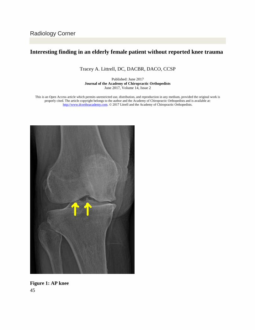
45
Radiology Corner
Interesting finding in an elderly female patient without reported knee trauma
Tracey A. Littrell, DC, DACBR, DACO, CCSP
Published: June 2017
Journal of the Academy of Chiropractic Orthopedists
June 2017, Volume 14, Issue 2
This is an Open Access article which permits unrestricted use, distribution, and reproduction in any medium, provided the original work is
properly cited. The article copyright belongs to the author and the Academy of Chiropractic Orthopedists and is available at:
http://www.dcorthoacademy.com. © 2017 Litrell and the Academy of Chiropractic Orthopedists.
Figure 1: AP knee
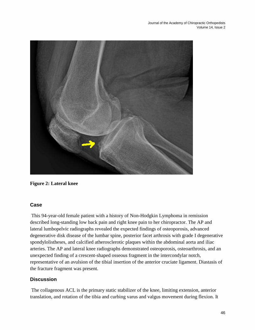
Journal of the Academy of Chiropractic Orthopedists
Volume 14, Issue 2
46
Figure 2: Lateral knee
Case
This 94-year-old female patient with a history of Non-Hodgkin Lymphoma in remission
described long-standing low back pain and right knee pain to her chiropractor. The AP and
lateral lumbopelvic radiographs revealed the expected findings of osteoporosis, advanced
degenerative disk disease of the lumbar spine, posterior facet arthrosis with grade I degenerative
spondylolistheses, and calcified atherosclerotic plaques within the abdominal aorta and iliac
arteries. The AP and lateral knee radiographs demonstrated osteoporosis, osteoarthrosis, and an
unexpected finding of a crescent-shaped osseous fragment in the intercondylar notch,
representative of an avulsion of the tibial insertion of the anterior cruciate ligament. Diastasis of
the fracture fragment was present.
Discussion
The collagenous ACL is the primary static stabilizer of the knee, limiting extension, anterior
translation, and rotation of the tibia and curbing varus and valgus movement during flexion. It
47
crosses the knee obliquely, from the anterior tibial eminence posterolaterally to the medial aspect
of the lateral femoral condyle.1
Anterior cruciate ligament injury is a pervasive injury in nearly all categories of patients, from
young to old, athletic to sedentary. It is one of the most common knee ligament injuries
evaluated by health care providers, with approximately 250,000 ACL tears diagnosed and
100,000 ACL surgical reconstructions performed every year in the United States.1
ACL injuries are expensive to treat with surgical reconstruction and rehabilitation and often
result in lasting impediments, including cartilaginous injuries, meniscal tears, biomechanical
instability, and early osteoarthrosis.1
Tibial spine avulsion fractures were first detailed in 1875 by Poncet, described erroneously as
pediatric-exclusive injuries.2,3,4 While these avulsions are more commonly seen in young
skeletally immature children and adolescents, their occurrence in adults is possible.3,4,5 In
children, the elasticity of the ACL, the weaker bony structures, and the open epiphyses are more
likely to lead to osseous avulsion of the ACL at the distal tibial attachment site than tear of the
ACL. In adults, ligament disruption is more common than osseous avulsion, occurring most
frequently at the tibial eminence rather than the femoral attachment.6 Bicycle falls account for
more than half of tibial avulsion fractures. Rotational and/or extension injury in both contact and
noncontact sports, such as soccer and skiing, make up the majority of the remaining
occurrences.7
In 1959, Meyers and McKeever described three tibial spine fracture patterns: Type I as a
nondisplaced fracture; Type II as a mildly displaced and elevated fracture of the anterior third
but with an intact posterior hinge; and Type III as a completely raised fracture. Tibial spine
fracture patterns with comminuted and rotated fragments were later termed Type IV fractures.2,5,7
Patients with tibial spine fractures typically present with knee pain and effusion, clinically
mimicking ACL tears. Commonly, the knee will be held in flexion with limited range of motion
possible, particularly extension. The osseous fragment may lead to joint locking. Orthopedic
examination to evaluate knee stability may be difficult to perform due to pain and muscle spasm;
Lachman’s test, anterior drawer test, and the pivot shift test would likely be positive.7
Well-positioned knee radiographs are essential for accurate diagnosis. Typically, the lateral view
will readily demonstrate the fracture. In this case, the findings on the lateral projection are subtle
and the diagnosis is clear on the suboptimally positioned AP view. A PA axial open-joint view
(Camp-Coventry or Homblad methods) of the knee would have been an appropriate addition to
this radiographic examination, resulting in better demonstration of the intercondylar fossa.

Journal of the Academy of Chiropractic Orthopedists
Volume 14, Issue 2
48
Examination with computed tomography (CT) aids in establishing comminution of fractures, the
degree of displacement, and in pre-operative planning. Due to the high likelihood of intra-
articular injury, examination with magnetic resonance imaging (MRI) is an indispensable tool in
the evaluation of concomitant meniscal and collateral ligament involvement.
Non-displaced fractures may be responsive to nonsurgical management, usually consisting of
hemarthrosis evacuation and immobilization bracing. Arthroscopic surgical management is
typically employed for displaced fractures.2,7
In this case of an adult with a tibial spine avulsion, the presence of osteoporosis most likely made
her susceptible to osseous avulsion over ACL rupture. Despite her knee pain and the diagnosis
of a displaced Type III tibial spine avulsion fracture, the patient declined further imaging and
medical orthopedic management.
References
1. Acevedo RJ, Rivera-Vega A, Miranda G, Micheo W. Anterior cruciate ligament injury:
identification of risk factors and prevention strategies. Curr Sports Med Rep. 2014 May-
Jun; 13(3):186-91.
2. Osti L, Buda M, Soldati F, Del Buono A, Osti R, Maffulli N. Arthroscopic treatment of
tibial eminence fracture: a systematic review of different fixation methods. Br Med Bull.
2016 Jun;118(1):73-90.
3. Myer, D. M., Purnell, G. J., Caldwell, P. E., & Pearson, S. E. (2013). ORV Arthroscopic
Reduction and Internal Fixation of Tibial Eminence Fractures. Arthroscopy
Techniques, 2(4), e341–e345. http://doi.org/10.1016/j.eats.2013.05.001
4. Kumar M, Malgonde M, Jain P. Avulsion fracture of tibial spine: A case report and
review of literature. Saudi J Sports Med 2013;13:102-4.
5. Kose O, Ozyurek S, Guler F, Canbora K. Avulsion fracture of the anterior cruciate
ligament in a 9-year-old child. BMJ Case Rep. 2013 Aug 8;2013. pii: bcr2013009426.
6. Shah SH, Porrino JA, Twaddle BC, Richardson ML. Osseous femoral avulsion of the
anterior cruciate ligament origin in an adult. Radiology Case Reports. (Online)
2015;10(2);1070. DOI: 10.2484/rcr.v10i2.1070
7. Little RM, Milewski MD. Physeal fractures about the knee. Curr Rev Musculoskelet Med
(2016) 9:478–486 DOI 10.1007/s12178-016-9370-7

49
Ortho Quiz
by Steven L. Kleinfield D.C., F.A.C.O.
1. When testing a myotome, you are essentially doing an ____________ exercise:
a. Isokinetic
b. Isometric
c. Isotonic
d. All of the above
2. While performing a myotome test, using the muscle grading scale, the patient presents with the ability to have complete range of motion against gravity only but no resistance. This would need to be graded as a:
a. 1/5 b. 2/5 c. 3/5 d. 4/5 e. 5/5
3. The term RUBOR means:
a. Pain b. Swelling c. Heat d. Cold e. Redness
4. The term TUMOR means:
a. Pain
b. Swelling
c. Heat
d. Cold
e. Redness
5. The term CALOR means:
a. Pain
b. Swelling
c. Heat
d. Cold
e. Redness

Journal of the Academy of Chiropractic Orthopedists
Volume 14, Issue 2
50
Current Events
❖ The Academy of Chiropractic Orthopedists announces the on-line Part I examination
dates will be July 20, 2017.
❖ Part II examination will be held September 30, 2017 ay Northwestern Health Sciences
University in Bloomington, MN.
Information about sitting the Board is available from the Executive Director Dr. Jerry
Wildenauer.
Jerrold R Wildenauer DC, FACO
1859 Warrior Drive
Mendota Heights, MN 55118
TEL: 612-454-1472
FAX: 651-846-5590
E-mail: [email protected]
❖ Apply for the Lipe Scholarship
Details at http://www.accoweb.org/lipescholarship.html

51
Answers to Ortho Quiz
1. b. Isometric
https://en.wikipedia.org/wiki/Myotome
2. c. 3/5 http://asia-spinalinjury.org/wp-content/uploads/2016/02/International_Stds_Diagram_Worksheet.pdf 3. e. Redness www.medilexicon.com/dictionary/79125
4. b. Swelling
http://www.medicinenet.com/script/main/art.asp?articlekey=24915
5. c. Heat
http://www.medicinenet.com/script/main/art.asp?articlekey=24915
Related Documents