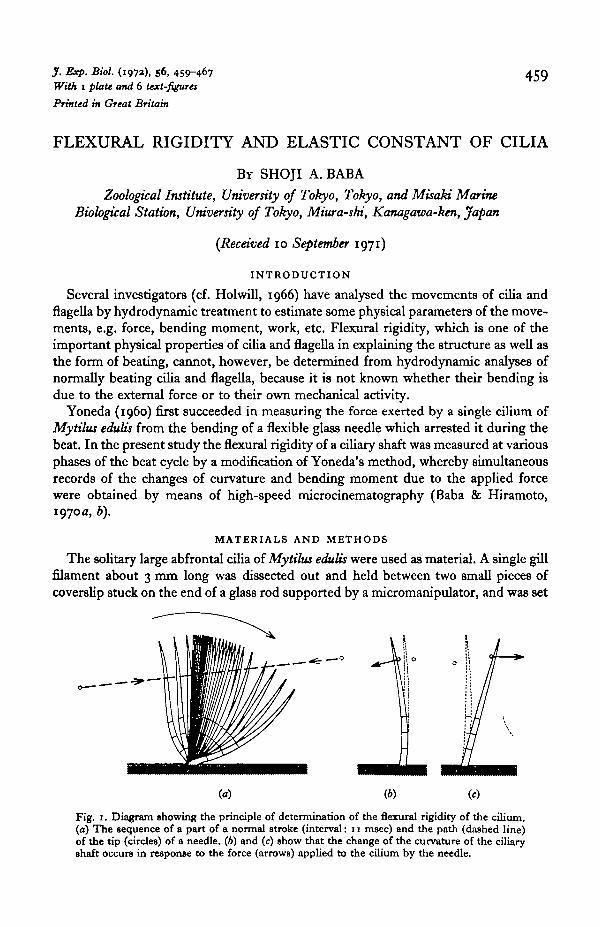
J. Exp. Bio/. (197a), 56, 459-467 With 1 plate and 6 text-figures Printed in Great Britain 459 FLEXURAL RIGIDITY AND ELASTIC CONSTANT OF CILIA BY SHOJI A. BABA Zoological Institute, University of Tokyo, Tokyo, and Misaki Marine Biological Station, University of Tokyo, Miura-shi, Kanagawa-ken, Japan (Received 10 September 1971) INTRODUCTION Several investigators (cf. Holwill, 1966) have analysed the movements of cilia and flagella by hydrodynamic treatment to estimate some physical parameters of the move- ments, e.g. force, bending moment, work, etc. Flexural rigidity, which is one of the important physical properties of cilia andflagellain explaining the structure as well as the form of beating, cannot, however, be determined from hydrodynamic analyses of normally beating cilia and flagella, because it is not known whether their bending is due to the external force or to their own mechanical activity. Yoneda (i960) first succeeded in measuring the force exerted by a single cilium of Mytilus edulis from the bending of aflexibleglass needle which arrested it during the beat. In the present study the flexural rigidity of a ciliary shaft was measured at various phases of the beat cycle by a modification of Yoneda's method, whereby simultaneous records of the changes of curvature and bending moment due to the applied force were obtained by means of high-speed microcinematography (Baba & Hiramoto, 1970a, b). MATERIALS AND METHODS The solitary large abfrontal cilia of Mytilus edulis were used as material. A single gill filament about 3 mm long was dissected out and held between two small pieces of coverslip stuck on the end of a glass rod supported by a micromanipulator, and was set iio (6) Fig. 1. Diagram showing the principle of determination of the flexural rigidity of the cilium. (a) The sequence of a part of a normal stroke (interval: 11 msec) and the path (dashed line) of the tip (circles) of a needle. (6) and (c) show that the change of the curvature of the ciliary shaft occurs in response to the force (arrows) applied to the cilium by the needle.

Welcome message from author
This document is posted to help you gain knowledge. Please leave a comment to let me know what you think about it! Share it to your friends and learn new things together.
Transcript
J. Exp. Bio/. (197a), 56, 459-467With 1 plate and 6 text-figuresPrinted in Great Britain
459
FLEXURAL RIGIDITY AND ELASTIC CONSTANT OF CILIA
BY SHOJI A. BABA
Zoological Institute, University of Tokyo, Tokyo, and Misaki MarineBiological Station, University of Tokyo, Miura-shi, Kanagawa-ken, Japan
(Received 10 September 1971)
INTRODUCTION
Several investigators (cf. Holwill, 1966) have analysed the movements of cilia andflagella by hydrodynamic treatment to estimate some physical parameters of the move-ments, e.g. force, bending moment, work, etc. Flexural rigidity, which is one of theimportant physical properties of cilia and flagella in explaining the structure as well asthe form of beating, cannot, however, be determined from hydrodynamic analyses ofnormally beating cilia and flagella, because it is not known whether their bending isdue to the external force or to their own mechanical activity.
Yoneda (i960) first succeeded in measuring the force exerted by a single cilium ofMytilus edulis from the bending of a flexible glass needle which arrested it during thebeat. In the present study the flexural rigidity of a ciliary shaft was measured at variousphases of the beat cycle by a modification of Yoneda's method, whereby simultaneousrecords of the changes of curvature and bending moment due to the applied forcewere obtained by means of high-speed microcinematography (Baba & Hiramoto,1970a, b).
MATERIALS AND METHODS
The solitary large abfrontal cilia of Mytilus edulis were used as material. A single gillfilament about 3 mm long was dissected out and held between two small pieces ofcoverslip stuck on the end of a glass rod supported by a micromanipulator, and was set
iio
(6)
Fig. 1. Diagram showing the principle of determination of the flexural rigidity of the cilium.(a) The sequence of a part of a normal stroke (interval: 11 msec) and the path (dashed line)of the tip (circles) of a needle. (6) and (c) show that the change of the curvature of the ciliaryshaft occurs in response to the force (arrows) applied to the cilium by the needle.
460 S. A. BAB A
in a position such that a cilium could be observed beating in a plane perpendicular tothe optical axis of a microscope (cf. Baba & Hiramoto, 1970 a, b).
The elasticity (flexural rigidity) of the cilium was, in principle, determined from theforce applied to the cilium by a micro-needle and the change in the curvature ofthe ciliary shaft in the following way. A flexible glass micro-needle supportedvertically is moved abruptly in the direction of the effective stroke of the cilium (tothe right in Text-fig. 1 a) or vice versa by means of a piezo-electric ceramic bimorphdriven by a train of square pulses. Because of the very short response time of thepiezo-electric device the rigid base of the needle shifts almost instantaneously tothe new position, whereas the flexible tip lags behind the base owing to the resistanceof the surrounding sea water. In the course of its movement (indicated by the dashedline in Text-fig, ia) the tip hits the cilium (by chance) at various stages of thestroke as shown in text-fig. 1 b and c. The force (F) applied to the cilium is the sum ofthe force due to the elasticity of the needle and the resistance of the medium tothe needle. Thus,
F = KD+R(dDldt), (1)
where D is the distance of the tip of the needle from its final steady position, dD\dt isthe velocity of the tip, K and R are elastic and viscous constants, respectively. Theconstant K was determined by the method similar to Yoneda's (i960) and the con-stant R was determined from the analysis of the movement of the tip of the needlefree from obstacles, which is given by D = Do g-W*)*, where Do is the distance of thetip from the final point at t = o. In several needles used in the present study the valuesof Klay in the range of 1-5 x io"6 dyne//mi; those of R, 5-8 x io"8 dyne.sec//tm; andthose of R/K which represents the time constant of the response of the tip, 10-̂ 70 msec.The movement of a single cilium and of the tip of the needle was recorded by 16 mmhigh-speed microcinematography similar to that reported previously (Baba & Hira-moto, 1970 a, b).
The bending moment about a given point on the cilium was calculated from the forceof the needle, which was determined from equation (1) using D and dD/dt obtainedfrom cine records, and the resistance of the medium acting on the different parts ofthe ciliary shaft (cf. Baba & Hiramoto, 19706).
The curvature (y) of a ciliary axis is defined by y = d<p/ds, where <j> is the angleformed by the tangent at the point in question to the axis of the cilium and a fixedreference line and s is the distance measured along the axis. In practice, y was obtainedfrom the difference of <}> at the two points 2-5 /an or 5 /an apart from the pointin question in opposite directions (one distal and the other proximal) along the axisdivided by the distance (i.e. 5 fim. or 10 /*m) between the two points.
The measurements of the flexural rigidity, which is the ratio of the bending momentto the corresponding change of curvature, were made using cilia normally beating insea water as well as cilia which were immobilized, maintaining the posture at the be-ginning of the effective stroke by treatment with sea water containing acetic acid
(PH4).For counting the number of component cilia in large abfrontal cilia, the optical
cross-section of the cilia whose beating was stopped at the end of the effective strokein sea water containing acetic acid (pH 4-5) was observed and photographed through
Flexural rigidity and elastic constant of cilia 461
a Zeiss Nomarski interference microscope using a x 100 oil-immersion objectivefollowing Dr Thurm's personal suggestion. For observation by electron microscopy,gill filaments were fixed with 2-5 % glutaraldehyde in sea water, stained with 20%phosphotungstic acid in methanol after dehydration, and embedded in Epon. Thecross-section of the cilia was examined with an electron microscope (JEM-T8).
RESULTS
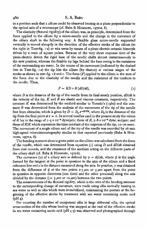
Fig. 2 (open circles) shows the changes in the curvature of the part at 20 /an fromthe base when a force is given by a glass micro-needle in the early effective phase(stage A in Fig. 4). The change of the curvature of the normal beat of the same cilium
-1 20 7 a
&-•-20
20 40 60Time (msec)
20 40 60Time (msec)
80
Fig. 2. Changes in the curvature of the ciliary shaft at 30 /tm from the base by the application offorce with a micro-needle. Open circles indicate the curvature and closed circles the bendingmoment in the early effective phase (stage A in Fig. 4). Dotted lines show the changes of thecurvature in the same stage of a normal beat. x^-$ °C.
is relatively slow at this stage as represented by dotted lines in Fig. 2. The force is ap-plied to the cilium in the direction of the effective stroke in Text-fig. 2 a (cf. Text-fig.1 c) or in the opposite direction in Fig. 26 (cf. Text-fig. 1 b). In either case the curvature(open circles) changes approximately in parallel with the bending moment (closedcircles) in its rising phase as well as in its falling phase and/or in its final state. Thecurvature recovers its initial value as the bending moment comes to zero again by therelease of the cilium from the needle. The parallelism of stress (bending moment)and strain (curvature) suggests that the deformation of the cilium in the present caseis regarded as an elastic one.
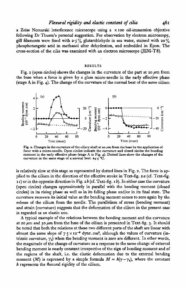
A typical example of the relations between the bending moment and the curvatureat 20 /tm and 30 jim from the base of the cilium is presented in Text-fig. 3. It shouldbe noted that both the relations at these two different parts of the shaft are linear withalmost the same slope of 7-5 x io"6 dyne.cm2, although the values of curvature (in-trinsic curvature, yt) when the bending moment is zero are different. In other words,the magnitude of the change of curvature as a response to the same change of externalbending moment is nearly constant irrespective of the sign of bending moment and ofthe regions of the shaft, i.e. the elastic deformation due to the external bendingmoment (M) is expressed by a simple formula M = k(y—yj), where the constantk represents the flexural rigidity of the cilium.
462 S. A. BABA
Fig. 3. Responses of the curvature at ao fun (closed circles) and at 30 fan (open circles) from thebase of a cilium to the bending moment due to the external force. The force acts on the cilium,either in the direction of the effective stroke or in that of the recovery stroke. Slopes of thestraight lines represent the flexural rigidity of the cilium. 04 °C.
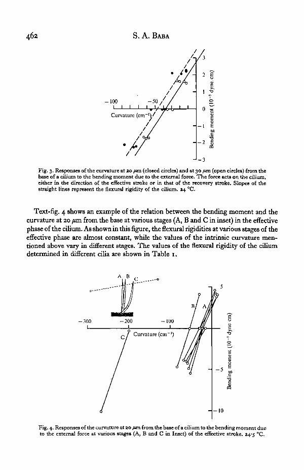
Text-fig. 4 shows an example of the relation between the bending moment and thecurvature at 20 /im from the base at various stages (A, B and C in inset) in the effectivephase of the cilium. As shown in this figure, the flexural rigidities at various stages of theeffective phase are almost constant, while the values of the intrinsic curvature men-tioned above vary in different stages. The values of the flexural rigidity of the ciliumdetermined in different cilia are shown in Table 1.
A B
- 3 0 0
-o
- 1 0 0
Curvature (cm ')
- - 1 0
Fig. 4. Responses of the curvature at ao /tin from the base of a cilium to the bending moment dueto the external force at various stages (A, B and C in Inset) of the effective stroke. 24-3 °C.
Flexural rigidity and elastic constant of cilia 463
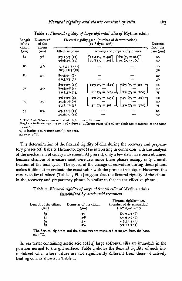
Table 1. Flexural rigidity of large abfrontal cilia of Mytilus edulis
Lengthof thecilium(jim)
82
80
80
7S
72
59
Diameter*of thecilium ,(/un)
3-6
3 6
3-2
3 0
3-3
2-4
Flexural rigidity ± s.D. (number of determination)(io~* dyne.cm1)
Effective phase
9-S±3-3 (17)9-6±3-4 (17)
13-3±3-5 (12)ia-9±2-3 (13)
8-7 ±3-9 (6)90±3-s (6)
8-9±2-3 (15)8-9±3-8(i5)7-9±3-°(iS)3-6±06 (9)4-5 ±i-8 (9)4'3 ±i-3 (S)
4-9±i'9(i3)4-4±i-3 (13)
Recovery and preparatory phases
fn -o (n = 4°)"] [60 (y< = 36o)"lLio-8 (y, = 3o)J, L7-4 (y< = i6o)J
— —
— —
rio-3 (y( = i8oo)"j p8-5 (y< = 50) n
L 61 (y< = 140) J , L7-3 (y< = i8oo)J
r a-o (y, = 1450)-! p - i (y< = 100) -1
L 31 (y< = 3°) J , L30 (y, = is°o)J— —
Distancefrom thebase Cum)
3030
ao3°2030
1030
3S
IO2O
as3030
• The diameters are measured at 20 /«n from the base.Brackets indicate that the pair of values at different ports of a ciliary shaft are measured at the samemoment.y{ is intrinsic curvature (cm"1), see text.23-5-34-5 °c.
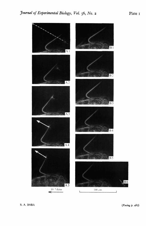
The determination of the flexural rigidity of cilia during the recovery and prepara-tory phases (cf. Baba & Hiramoto, 19706) is interesting in connexion with the analysisof the mechanism of ciliary movement. At present, only a few data have been obtainedbecause chances of measurement were few since these phases occupy only a smallfraction of the beat cycle. The speed of the change of curvature during these phasesmakes it difficult to evaluate the exact value with the present technique. However, theresults so far obtained (Table 1, PI. 1) suggest that the flexural rigidity of the ciliumin the recovery and preparatory phases is similar to that in the effective phase.
Table 2. Flexural rigidity of large abfrontal cilia of Mytilus edulisimmobilized by acetic acid treatment
Length of the ciliumOwn)
89817969
Diameter of the cilium(/un)
3 13-83-43-4
Flexural rigidity ± s.D.(number of determination)
(io~* dyne.cm1)
8-7 ±4-1 (6)5-5 ±°-6 (6)4-9 ±i-4 (8)3-O±I-I (4)
The flexural rigidities and the diameters are measured at ao /on from the base.34-5 °C
In sea water containing acetic acid (pH 4) large abfrontal cilia are immobile in theposition normal to the gill surface. Table 2 shows the flexural rigidity of such im-mobilized cilia, whose values are not significantly different from those of activelybeating cilia as shown in Table 1.
464 S. A. BABA
The cross-section of large abfrontal cilia was examined with Nomarski optics inliving material and with an electron microscope in fixed and sectional material. It wasfound that these compound cilia consist of a number of component cilia of 9 + 2pattern, which are separately enclosed with a membrane, lacking any visible connexionwith their neighbours and arranged in an approximately hexagonal array as observedby Thurm (1968).
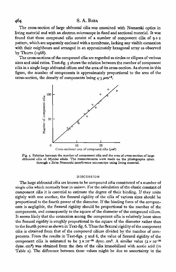
The cross-sections of the compound cilia are regarded as circles or ellipses of varioussizes and axial ratios. Text-fig. 5 shows the relation between the number of componentcilia in a single large abfrontal cilium and the area of its cross-section. As shown in thisfigure, the number of components is approximately proportional to the area of thecross-section, the density of components being 4-3 /tm~2.
100
c
I50
0 10 20Cross-seclional area of compound cilia (jtnf)
Fig. 5. Relation between the number of component cilia and the area of cross-section of largeabfrontal cilia of Mytiha edulis. The measurements were made on the photographs taken
through a Zeiss Nomarski interference microscope using living material.
DISCUSSION
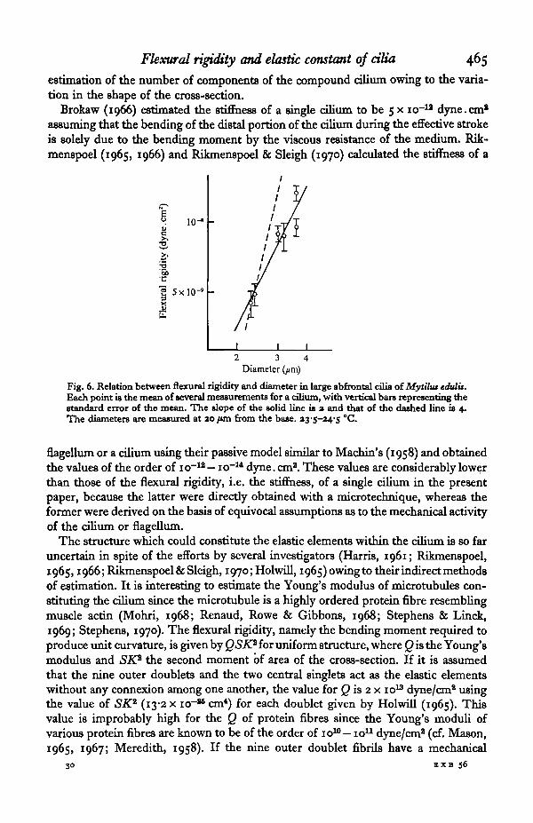
The large abfrontal cilia are known to be compound cilia constituted of a number ofsingle cilia which normally beat in unison. For the calculation of the elastic constant ofcomponent cilia it is essential to estimate the degree of their binding. If they unitetightly with one another, the flexural rigidity of the cilia of various sizes should beproportional to the fourth power of the diameter. If the binding force of the compon-ents is negligible, the flexural rigidity should be proportional to the number of thecomponents, and consequently to the square of the diameter of the compound cilium.It seems likely that the connexion among the component cilia is relatively loose sincethe flexural rigidity is roughly proportional to the square of the diameter rather thanto the fourth power as shown in Text-fig. 6. Thus the flexural rigidity of the componentcilia is obtained from that of the compound cilium divided by the number of com-ponents. From the results in Text-figs. 5 and 6, the value of flexural rigidity of thecomponent cilia is estimated to be 3 x io~10 dyne.cm8. A similar value (2x io~10
dyne. cm8) was obtained from the data of the cilia immobilized with acetic acid (inTable 2). The difference between these values might be due to uncertainty in the
Flexural rigidity and elastic constant of cilia 465
estimation of the number of components of the compound cilium owing to the varia-tion in the shape of the cross-section.
Brokaw (1966) estimated the stiffness of a single cilium to be 5 x io~u dyne.cm*assuming that the bending of the distal portion of the cilium during the effective strokeis solely due to the bending moment by the viscous resistance of the medium. Rik-menspoel (1965, 1966) and Rikmenspoel & Sleigh (1970) calculated the stiffness of a
10-c
T3
5xlO-»
2 34Diameter (//m)
Fig. 6. Relation between flexural rigidity and diameter in large abfrontal cilia oiMytilus edulis.Each point U the mean of several measurements for a cilium, with vertical bars representing thestandard error of the mean. The slope of the solid line is a and that of the dashed line is 4.The diameters are measured at ao fun from the base. zys-24'S °C.
flagellum or a cilium using their passive model similar to Machin's (1958) and obtainedthe values of the order of io"12 — io~14 dyne. cm2. These values are considerably lowerthan those of the flexural rigidity, i.e. the stiffness, of a single cilium in the presentpaper, because the latter were directly obtained with a microtechnique, whereas theformer were derived on the basis of equivocal assumptions as to the mechanical activityof the cilium or flagellum.
The structure which could constitute the elastic elements within the cilium is so faruncertain in spite of the efforts by several investigators (Harris, 1961; Rikmenspoel,1965,1966; Rikmenspoel & Sleigh, 1970; Holwill, 1965) owing to their indirect methodsof estimation. It is interesting to estimate the Young's modulus of microtubules con-stituting the cilium since the microtubule is a highly ordered protein fibre resemblingmuscle actin (Mohri, 1968; Renaud, Rowe & Gibbons, 1968; Stephens & Linck,1969; Stephens, 1970). The flexural rigidity, namely the bending moment required toproduce unit curvature, is given by QSK* for uniform structure, where Q is the Young'smodulus and SK* the second moment of area of the cross-section. If it is assumedthat the nine outer doublets and the two central singlets act as the elastic elementswithout any connexion among one another, the value for Q is 2 x io13 dyne/cma usingthe value of SK* (13-2 x i c r " cm4) for each doublet given by Holwill (1965). Thisvalue is improbably high for the Q of protein fibres since the Young's moduli ofvarious protein fibres are known to be of the order of IO10 — io11 dyne/cm2 (cf. Mason,1965, 1967; Meredith, 1958). If the nine outer doublet fibrils have a mechanical
30 EXB 56
466 S. A. BABA
connexion with one another by some transverse structures such as radial links, armsetc., SK* is given by 9
SK* = SdZx K\, (2)
where Sd is the cross-section of each doublet and K+ is the radius of gyration of the tthouter doublet about a line through the central pair and perpendicular to the plane ofbending. SK* is calculated to be 3 x icr*1 cm* from equation (2), if it is assumed thatthe nine outer doublets are equally spaced on circumference of a circle of radiuso-i /on and that the cross-section Sd is 6-5 x io~12 cm2 (calculated from the outsidediameter of 250 A and the wall thickness of 60 A for the outer singlet; cf. Holwill,1966). In this case the estimated Young's modulus for outer doublet microtubule is5-9 x io10 dyne/cm2, which is reasonable for protein fibres as mentioned above. Theabsence of any drastic change in the flexural rigidity during the beat cycle and thesimilarity in the flexural rigidity between actively beating cilia and immobilized ciliasuggest that the transverse connexion among outer doublets is maintained throughoutthe whole course of the ciliary movement and irrespective of the presence or absenceof the mechanical activity of the cilium.
Other possibilities are the membrane or the matrix. If the membrane has elasticity ofthe same order of magnitude as that of the surface structure of the sea-urchin egg (i.e.the cell membrane and the cortex; cf. Hiramoto, 1970), the fraction of the flexuralrigidity due to the membrane would be of the order of io~13 — io~ls dyne. cm2, whichis negligibly small as compared with the flexural rigidity of a single cilium in the pre-sent paper. The matrix would contribute much less than the membrane.
The movement of cilia is asymmetrical, differing from the ordinary movement ofmany flagella, i.e. they move through their effective stroke as rigid rods and throughtheir recovery stroke as though they were readily flexible (Gray, 1922; Carter, 1924).Gray (1930) foresaw the possibilities that the difference is due to an active change inthe elastic properties of the cilium and/or to permanent structural organization. Thefact that the flexural rigidity of the cilium is not different in various phases and inrelation to the direction of action of the force may indicate that the difference is mainlydue to an active bending and unbending of the cilium and that the asymmetry in themovement is of the nature of the active bending system in the cilium.
In conclusion, the cilia alter their intrinsic curvature throughout the beat cycle whilethey keep their flexural rigidity, which might be due to outer microtubules connectedwith one another, nearly constant. The nature of intrinsic curvature and themechanism of its regulation are to be investigated in future.
SUMMARY
1. The flexural rigidity of the large abfrontal cilia of Mytilus has been measured witha flexible glass micro-needle.
2. The same cilium has similar values to the flexural rigidity irrespective of thephases of beat cycle (including the recovery phase) and of the direction of force applied.
3. The values of 3-13 x io"8 dyne. cm2 have been obtained for the flexural rigidityof compound cilia of various sizes; 2-3 x io~10 dyne.cm2 for that of the componentcilia.

Journal of Experimental Biology, Vol. 56, No. 2 Plate 1
S. A. BABA (Facing p. 467)
Flexural rigidity and elastic constant of cilia 467
4. The Young's modulus of the microtubule is estimated to be 5-9 x io10 dyne/cm2
on the basis that the outer doublet microtubules are tightly connected with one another.
It is my great pleasure and privilege to offer this work to Professor H. Kinosita ofthe University of Tokyo as a part of the celebration of his sixtieth birthday. I wouldlike to thank him for his helpful suggestions during the course of this work and for hiscritical reading of the manuscript. I am also very grateful to Dr Y. Hiramoto for his care-ful discussion of the problems that were encountered in this work.
REFERENCES
BABA, S. A. & HIRAMOTO, Y. (1970a). High-speed microcinematography of ciliary movement. Zool.Mag., Tokyo 79, 8-13.
BABA, S. A. & HIRAMOTO, Y. (19706). A quantitative analysis of ciliary movement by means of high-speed microcinematography. J. exp. Biol. 53, 675-90.
BROKAW, C. J. (1966). Mechanics and energetics of cilia. Am. Rev. rap. Dis. 93 (3, Pa), 32-40.CARTER, G. S. (1924). On the structure and movements of the latero-frontal cilia of the gills of Mytihis.
Proc. R. Soc. hand. B 96, 115-22.GRAY, J. (1922). The mechanism of ciliary movement. Proc. R. Soc. Lond. B 93, 104-21.GRAY, J. (1930). The mechanism of ciliary movement. VI. Photographic and stroboscopic analysis of
ciliary movement. Proc. R. Soc. Lond. B 107, 313-32.HARRIS, J. E. (1961). The mechanics of ciliary movement. In The Cell and the Organism (ed. J. A.
Ramsay and V. B. Wigglesworth), pp. 22-36. London: Cambridge University Press.HIRAMOTO, Y. (1970). Rheological properties of sea urchin eggs. Biorheology 6, 201—34.HOLWTLL, M. E. J. (1965). The motion of Strigomonas oncopelti. J. exp. Biol. 42, 125-37.HOLWILL, M. E. J. (1966). Physical aspects of flagellar movement. Phytiol. Rev. 46, 696-785MACHIN, K. E. (1958). Wave propagation along flagella. J. exp. Biol. 35, 796-806.MASON, P. (1965). The viscoelasticity and structure of keratin and collagen. KoUoid-Z. Z. Polym. 203,
139-47-MASON, P. (1967). The viscoelasticity and structure of fibrous protein. II. Further dynamic measure-
ments of keratin. KoUoid-Z. Z. Polym. ai8, 46-52.MEREDITH, R. (1958). The rheology of fibers. In Rheology II (ed. F. R. Eirich), pp. 261-312. Academic
Press.MOHRI, H. (1968). Amino-acid composition of 'tubulin' constituting microtubules of sperm flagella.
Nature, Lond. 317, 1053—4.RENAUD, F. L., ROWE, A. J. & GIBBONS, I. R. (1968). Some properties of the protein forming the outer
fibers of cilia. J. Cell Biol. 36, 79-90.RIKMENSPOEL, R. (1965). The tail movement of bull spermatozoa. Observations and model calculations.
Biophyt. J. s, 365-92.RIKMBNSPOEL, R. (1966). Elastic properties of the sea urchin sperm flagellum. Biophyt. J. 6, 471-9.RiKMENSPOBL, R. & SLEIGH, M. A. (1970). Bending moments and elastic constants in cilia. J. theor. Biol.
28, 81-100.STEPHENS, R. E. (1970). Thermal fractionation of outer fiber doublet microtubules into A- and B-
subfiber components: A- and B-tubulin. J. tnolec. Biol. 47, 353—63.STEPHENS, R. E. & LINCK, R. W. (1969). A comparison of muscle actin and ciliary microtubule protein
in the mollusk Pecten irradians. J. molec. Biol. 40, 497-501.THURM, U. (1968). Steps in the transducer process of mechanoreceptors. Symp. tool. Soc. Lond. noi 33,
199-316.YONEDA, M. (i960). Force exerted by a single cilium of Mytihu edulis. I. J. exp. Biol. 37, 461-8.
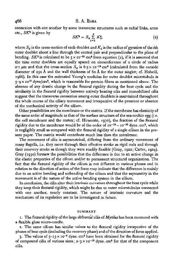
EXPLANATION OF PLATE I
PLATE I . Measurement of the flexural rigidity of a cilium in its recovery phase. A1-5 is a sequence ofthe response of the cilium in the experiment and B1-6 corresponding part of a normal beat. The intervalbetween successive frames is 3-1 msec. The tip of the needle that was first located at the position in-dicated by the dotted arrow in the lower-right corner of B6 moves to its final position (circle in Ai)along the course represented by the dashed line in Ai . In the course, the tip hits the cilium (A4) andexerts a force (shown by arrows in A4 and A 5) on the cilium. 24 "C.
30-3

Related Documents


